EP0410618A2 - Procédé pour visualiser la séquence de base des polymères d'acides nucléiques - Google Patents
Procédé pour visualiser la séquence de base des polymères d'acides nucléiques Download PDFInfo
- Publication number
- EP0410618A2 EP0410618A2 EP90307768A EP90307768A EP0410618A2 EP 0410618 A2 EP0410618 A2 EP 0410618A2 EP 90307768 A EP90307768 A EP 90307768A EP 90307768 A EP90307768 A EP 90307768A EP 0410618 A2 EP0410618 A2 EP 0410618A2
- Authority
- EP
- European Patent Office
- Prior art keywords
- tip
- nucleic acid
- polymer
- mercury
- dna
- Prior art date
- Legal status (The legal status is an assumption and is not a legal conclusion. Google has not performed a legal analysis and makes no representation as to the accuracy of the status listed.)
- Withdrawn
Links
Images

Classifications
-
- G—PHYSICS
- G01—MEASURING; TESTING
- G01Q—SCANNING-PROBE TECHNIQUES OR APPARATUS; APPLICATIONS OF SCANNING-PROBE TECHNIQUES, e.g. SCANNING PROBE MICROSCOPY [SPM]
- G01Q60/00—Particular types of SPM [Scanning Probe Microscopy] or microscopes; Essential components thereof
- G01Q60/10—STM [Scanning Tunnelling Microscopy] or apparatus therefor, e.g. STM probes
-
- C—CHEMISTRY; METALLURGY
- C07—ORGANIC CHEMISTRY
- C07H—SUGARS; DERIVATIVES THEREOF; NUCLEOSIDES; NUCLEOTIDES; NUCLEIC ACIDS
- C07H19/00—Compounds containing a hetero ring sharing one ring hetero atom with a saccharide radical; Nucleosides; Mononucleotides; Anhydro-derivatives thereof
- C07H19/02—Compounds containing a hetero ring sharing one ring hetero atom with a saccharide radical; Nucleosides; Mononucleotides; Anhydro-derivatives thereof sharing nitrogen
- C07H19/04—Heterocyclic radicals containing only nitrogen atoms as ring hetero atom
- C07H19/06—Pyrimidine radicals
- C07H19/10—Pyrimidine radicals with the saccharide radical esterified by phosphoric or polyphosphoric acids
-
- C—CHEMISTRY; METALLURGY
- C07—ORGANIC CHEMISTRY
- C07H—SUGARS; DERIVATIVES THEREOF; NUCLEOSIDES; NUCLEOTIDES; NUCLEIC ACIDS
- C07H19/00—Compounds containing a hetero ring sharing one ring hetero atom with a saccharide radical; Nucleosides; Mononucleotides; Anhydro-derivatives thereof
- C07H19/02—Compounds containing a hetero ring sharing one ring hetero atom with a saccharide radical; Nucleosides; Mononucleotides; Anhydro-derivatives thereof sharing nitrogen
- C07H19/04—Heterocyclic radicals containing only nitrogen atoms as ring hetero atom
- C07H19/16—Purine radicals
- C07H19/20—Purine radicals with the saccharide radical esterified by phosphoric or polyphosphoric acids
-
- C—CHEMISTRY; METALLURGY
- C07—ORGANIC CHEMISTRY
- C07H—SUGARS; DERIVATIVES THEREOF; NUCLEOSIDES; NUCLEOTIDES; NUCLEIC ACIDS
- C07H23/00—Compounds containing boron, silicon, or a metal, e.g. chelates, vitamin B12
-
- Y—GENERAL TAGGING OF NEW TECHNOLOGICAL DEVELOPMENTS; GENERAL TAGGING OF CROSS-SECTIONAL TECHNOLOGIES SPANNING OVER SEVERAL SECTIONS OF THE IPC; TECHNICAL SUBJECTS COVERED BY FORMER USPC CROSS-REFERENCE ART COLLECTIONS [XRACs] AND DIGESTS
- Y10—TECHNICAL SUBJECTS COVERED BY FORMER USPC
- Y10S—TECHNICAL SUBJECTS COVERED BY FORMER USPC CROSS-REFERENCE ART COLLECTIONS [XRACs] AND DIGESTS
- Y10S435/00—Chemistry: molecular biology and microbiology
- Y10S435/81—Packaged device or kit
-
- Y—GENERAL TAGGING OF NEW TECHNOLOGICAL DEVELOPMENTS; GENERAL TAGGING OF CROSS-SECTIONAL TECHNOLOGIES SPANNING OVER SEVERAL SECTIONS OF THE IPC; TECHNICAL SUBJECTS COVERED BY FORMER USPC CROSS-REFERENCE ART COLLECTIONS [XRACs] AND DIGESTS
- Y10—TECHNICAL SUBJECTS COVERED BY FORMER USPC
- Y10S—TECHNICAL SUBJECTS COVERED BY FORMER USPC CROSS-REFERENCE ART COLLECTIONS [XRACs] AND DIGESTS
- Y10S977/00—Nanotechnology
- Y10S977/84—Manufacture, treatment, or detection of nanostructure
- Y10S977/849—Manufacture, treatment, or detection of nanostructure with scanning probe
- Y10S977/852—Manufacture, treatment, or detection of nanostructure with scanning probe for detection of specific nanostructure sample or nanostructure-related property
- Y10S977/853—Biological sample
Definitions
- the present invention relates to means and methods for visualizing the base sequence of nucleic acid polymers and more particularly to determining and visualizing the nucleotide sequence of D.N.A and R.N.A.
- the present invention relates to a method for visualizing the base sequence of nucleic acid polymers and more particularly to methods of imaging nucleic acids using a scanning tunneling microscope (STM) or an atomic force microscope (AFM).
- STM scanning tunneling microscope
- AFM atomic force microscope
- the present invention allows fully hydrated DNA and RNA to be imaged undistorted by the metallic stains used in electron microscopy so that it is viewed in the exact configuration it maintains within living organisms.
- STM atomic force microscope
- the present disclosure is based on the remarkably unexpected discovery that the presence of a compound of a metal, such as mercury, in a neutral or in an ionized state, when complexed with the nucleic acid, leads to enhanced contrast in the STM thereby enabling the base sequence of nucleic acid polymers to be quickly and unequivocally elucidated.
- a compound of a metal such as mercury
- Another object of the present invention is to provide a novel and unique method to image DNA and RNA undistorted by the metal stains used in electron microscopy and while fully hydrated so that its viewed configuration exactly conforms to the configuration it maintains within living organisms.
- a further object of the present invention is to provide a novel and unique method for imaging DNA and RNA in which the presence of a preselected metal atom, bonded to the nucleic acid, provides enhanced contrast in the STM.
- the present invention relates to a method for determining and imaging nucleic acids under water using a scanning tunneling microscope ("STM") based on the presence of a metal atom, such as mercury, bonded to the nucleic acid.
- STM scanning tunneling microscope
- Thymidine, uridine, guanosine and cytidine nucleosides all contain oxygen in their thymine, uracil, guanine, and cytosine bases. Each has known derivatives in which one of its oxygen molecules is replaced by divalent sulfur. Examples are S-2 thymidine, S-4 thymidine, S-2 uridine, S-4 uridine, S-6 guanosine, and S-2 cytidine.
- the nucleoside bases can potentially also contain divalent sulfur, useful herein at other positions such as on the 5-methyl group of thymidine.
- the ribose portion of nucleosides also contain oxygen which, can be replaced by divalent sulfur, such as in the 2′-deoxy-2′-thionucleosides, an example of which is 2′-deoxy-2′-thiocytidine. Nucleotides containing such divalent sulfur are also useful in the practice of the present invention.
- Such base and/or sugar-modified nucleosides can be prepared as the analogous ribonucleotide 5′-triphosphates and used in the synthesis of RNA.
- Such base-modified 2′deoxynucleosides can also be prepared as the analogous 2′-deoxyribonucleotide- 5′-triphosphates and used in the synthesis of DNA.
- modified nucleosides 5′-triphosphates have been prepared in which the alpha-phosphate group contains a divalent sulfur replacing oxygen.
- Such 2′deoxy compounds have the general formula
- Such ribonucleotides have the general formula where, in both cases, the alpha-S isomer is the preferred substrate for nucleotide polymerizing enzymes.
- Base indicates the position of the five common nucleoside bases and their derivatives, namely cytosine, uracil, thymine, guanosine, or adenine.
- Nucleotides containing modified purine bases can also be used, such as inosine and 7-deazaguanosine nucleotides.
- Nucleotides containing modified pyrimidine bases can also be used, such as the 5-substituted pyrimidine nucleotides containing 5-bromo-, 5-fluoro-, or 5-iodouracil. Any base, phosphate, or sugar-substituted nucleotide used, which maintains base-pairing specificity, and is compatible with the presence of the essential sulfur atom needed for this invention can be used in this invention.
- base-2 results from incorporation of an alpha-thionucleoside-5′-triphosphate, while the other bases result from incorporation of non-sulfur-containing nucleoside-5′-triphosphates.
- divalent sulfur in these polynucleotides complexes with the metal atoms present in the compounds added for the purpose of specific nucleotide labeling. This results in selective staining of those sites in the polynucleotides that contain divalent sulfur.
- the mercury compounds or other other metal-containing compounds added to the solution for the purpose of labelling the DNA or RNA are present as cations and/or are bonded to other chemical groups.
- the present invention provides a method in which a metallic cluster may be formed between selected sites on the nucleic acid and the gold substrate. Subsequent imaging in water allows greater control of the deposition and the nature of the interface and is therefore preferred although favorable results can be obtained with a sample dried in air.
- a nucleic acid is sequenced by selecting a fragment for sequencing. While such a selection is a prerequisite for any sequencing technique, in STM it is straightforward to hold down and image at least 146 base-pair fragments (See: S.M. Lindsay et al, Science Vol. 244, pp. 1063-64 (1989)).
- Poly (U) and Poly (rU) are interchangeable and identify “polyuridylic acid”; poly (dT) identifies “polythymidylic acid”; and poly (s4U) identifies "poly-4-thiouridylic acid”.
- the original fragment is amplified using the general procedure described in U.S. Patent No. 4,683,202 but employing a sulfur-substituted variant for the monophosphate derivative of one of the bases.
- the resulting polymer is dissolved in a buffer that consists in part of a metal-containing inorganic compound or an organometallic compound such as sodium-4-hydroxy mercuribenzoate ("PCMB”), which binds the sulfur-containing sites, and yields enhanced contrast at mercury bound sites as described below.
- PCMB sodium-4-hydroxy mercuribenzoate
- the material is then synthesized with another divalent sulfur-labeled nucleotide, and the imaging is repeated.
- the sequence will be determined when three of the four normal nucleoides are replaced with a divalent sulfur-containing nucleotide.
- it will help to resolve ambiguities caused by lack of resolution of near neighbors to image the fourth base.
- Another possibility is to image the various combinations of pairs of bases in order to resolve ambiguities caused by inadequate resolution.
- RNA is a stiffer polymer than DNA, and the images obtained for poly (U) show features down to ⁇ 5A, a much better resolution than our best DNA pictures. It is possibly advantageous, therefore, to transcribe the RNA analog of the fragment to be sequenced.
- the present invention also provides a unique method for improving microscope tip insulation by etching them with a hard wax which insulates the tip and yet enables it to detect sample surface variations.
- a preferred wax for this has a softening point of 80-90°C, a vapor pressure of 10 ⁇ 3mm at 180°C, and is available as Apiezon wax from VWR Scientific (San Francisco, CA).
- the substrates are made by depositing between a few hundred and a few thousand A of gold epitaxially on freshly cleaved scratch-free mica in an ultra high vacuum (“UHV") system.
- UHV ultra high vacuum
- the substrate is preheated to 300°C in an oil-free system operated at 10 ⁇ 9 torr or better.
- the coated substrates are removed and stored under clean argon until use.
- the tips are etched from 0.01 inch diameter Pt-Ir wire (eg, Englehard material #26) using 3M NaCN containing 1M NaOH.
- the wire is immersed into the solution with 20V ac applied until 0.5A rms flows. It is then left until the waist at the liquid-air interface is just about to break.
- the current is stopped, and small bursts of current are applied to the junction as it is monitored under a microscope. When the lower region just parts, the etch is stopped.
- the overall shape of the end is quite rounded with a very small and sharp 'nipple' on the very end.
- the tip is insulated by being pushed through a film of Apiezon wax. This wax was found to be electrochemically inert in an aqueous environment. The temperature of the wax must be carefully controlled. If it is too hot, it flows off the tip as it penetrates, leaving too much metal exposed. If it is too cold, the end of the tip does not penetrate the wax.
- the wax is melted onto a slot in a thin metal plate, adjusting the heating of the plate so that the hottest part of the slot is about 200°C, and the open end somewhat below the melting temperature of the wax.
- a blob of wax is melted onto the slot so as to form a film over it.
- the tip is then pushed through the wax film until it can be seen to just push a bit of wax ahead of it as it breaks the top of the film.
- the tip is then translated into a slightly hotter region. SEM imaging shows that a micron (or less) of bare metal penetrates the wax. Once pushed up enough to be usefully coated, the tip is translated sideways out of the slot.
- the nucleic acid polymer whose sequence is to be determined is then prepared in the following manner.
- a sample of the unknown polymer is dissolved to a concentration of about 10 micrograms per ml in a buffer containing 10 mM tris and the appropriate mercury solution.
- Small amounts of EDTA, cacodylic acid and other preservatives may be used with no loss of resolution.
- a glass cell is pushed onto the substrate (it can be held with vacuum grease if its polish is inadequate for an interference fit).
- a few tens of microliters of sample solution are placed in the cell.
- About 1mm of Pt wire is pushed into the sample, and biased to cause hydrogen evolution (the substrate being held positive). This requires about 2V.
- the Faradaic deposition is continued for about two minutes.
- the STM tip is then lowered onto the substrate, and images are made in the usual way. With these conditions, the surface should not be uniformly coated, and isolated strands of polymer should be found in many regions.
- the mercury stained fragments show increased contrast with images of poly (rU) which has had the oxygen on the C4 atom replaced with sulfur (poly s4U).
- the mercury (II) needed to complex the sulfur, is provided by using a tris buffer containing about 2x10 ⁇ 3M sodium p-hydroxymercuribenzoate. While the exact concentrations do not appear to be critical, the conditions actually used to obtain the various images are described below.
- Solution III consisted of a 50-50 mix of Solutions I and II.
- Solution IV consisted of 23 micrograms per ml of a selected sequence fragment of RNA, a total of 1,300 bases long.
- the RNA is a transcript made in the presence of alpha-thio ATP selectively labeled by mercury.
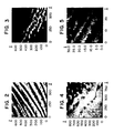
- FIG. 1 a high resolution image of the pure poly (rU) is shown in Figure 1.
- a 420 ⁇ by 420 ⁇ area of the substrate traversed by two molecules is shown.
- the relative contrast of the two molecules is somewhat different, depending on how each is embedded by surrounding reacted small anions, each shows the characteristic negative contrast as the molecule is approached (the tip appears to dip down relative to the substrate), followed by positive contrast, due, in part, to the constant current servo response. Internal structure is clearly resolved.
- the polymer appears to be a zig-zag with alternating 6 ⁇ and 8 ⁇ runs. This image is a perspective projection looking into the xy plane at an angle of about 45° with respect to all three axes.
- FIG. 2 shows a 700 ⁇ by 700 ⁇ region of deposits from solution I.
- FIG. 3 shows a similar view of deposits in a 500 ⁇ by 500 ⁇ region from solution II. While the absolute black to white scale is somewhat arbitrary (as illustrated by the differences in the two molecules imaged in FIG. 1), the degree of modulation in the immediate vicinity of the polymer, in relation to the background height, shows striking changes. This is because the 'heighest' parts of the image (with mercury stain) are many times the change from background to the dip as the tip approaches the molecule. This is not the case in the unstained polymer.
- FIG. 5 Such a heterogeneous staining is illustrated in FIG. 5 where several RNA polymers (which have been heterogeneously stained) are shown.
- the resolution is good enough to show each stained based clearly allowing the location of adenine residues to be read from the image directly.
- About twenty (20) of the bases are visible in this image.
- the DNA and RNA sample is dissolved to a concentration of 2 micrograms per milliliter in a 5 mM solution of tris (hydroxymethyl) aminothane in D.I. water.
- a 2.4 mM solution of sodium-p-hydroxymercuribenzoate is added in the amount of 2 microliter per microgram of DNA or RNA.
- RNA solution is inserted into the STM cell and deposited onto the gold substrate by holding the gold substrate at 1 volt positive with respect to a silver/silver chloride reference electrodes for one minute.
- the substrate is then scanned with the pre-prepared STM tip to form images such as that illustrated in FIGS. 1-5.
- the DNA solution is handled in a slightly different way because it must first be denatured.
- the DNA solution is placed in a capillary tube adjacent the STM cell and heated to 98°C for ten minutes to denature the DNA.
- the resulting heated solution of denatures single stranded DNA is then injected into the STM cell and deposited on the gold substrate by holding it at 1 volt positive with respect to an Ag/Ag Cl reference electrode for one minute.
- the substrate is then scanned with the STM tip to form images similar to those shown for RNA.
- the invention herein described is believed to be the first practical method ever devised for sequencing nucleic acids by visualization of the bases. Since the operation and data acquisition systems of the STM are usually under digital computer control, the entire process may be automated. The operator would guide the computer in locating target molecules and tracking their path. However, from that point on, the computer could trace the brightness contours along the backbones until the statistics were adequate for sequencing. Thus one machine could image at least 1000 base pairs a minute, a rate that would make the sequencing of the human genome a finite task (a few man years).
- a suitable computer-controlled system designed especially for scanning tunneling microscopy which acquires and displays the signal from the moving probe is commercially available from Angstrom Technology, Inc., Mesa, AZ (the TAK 2.0 Scanning Tunneling Microscope System).
- the sequencing method of the present invention is conducted using Lambda bacteriophage DNA as a sequencing target.
- a portion of this DNA is amplified using the polymerase chain reaction.
- two primers are used, and the sequences bounded by these primers are determined.
- the primer GAT GAG TTC GTG TCC GTA CAA CTG G together with the primer GGT TAT CGA AAT CAG CCA CAG CGC C, bounds a 500 base-pair target of the lambda bacteriophage genome.
- This segment is amplified under the following conditions: pH 8.3 10 mM tris-HCl, 50 mM KCL, 1.5 mM MgCl2, 0.01% gelatin, 0.2 mM each of dATP, dTTP, dCTP, dGTP, 0.2 micromolar concentration of each primer, 0.2 nanogram of template (lambda DNA) per 100 microliters final solution, and 2.5 units of TAQ polymerase per 100 microliters.
- each reaction one of the deoxynucleoside triphosphates is replaced with the analogous alpha-thio-deoxynucleoside triphosphate.
- dTTP is replaced in one reaction with alpha-thio-dTTP.
- Five separate reactions are done, one with each alpha thionucleoside triphosphate and one reaction with no alpha thioneucleoside triphosphate.
- the reaction with thioneucleoside triphosphates are done using the following temperature protocol:
- the resulting DNA preparations are then phenol-extracted to remove protein, purified on a membrane filter to remove unreacted primers and mononucleotides, and then sequenced by dissolving the DNA to a concentration of 2 g/mL in 3 mM solution of tris (hydroxymethyl) aminothane in distilled water. A 2.4 mM of sodium-p-hydroxymercuribenzoate is then added in the amount of 2 microliters per each microgram of the DNA.
- the DNA solution is then placed in a capillary tube adjacent to the STM cell and heated to 98°C for ten minutes to denature the DNA.
- the heated solution is then injected into the STM cell and deposited on the gold substrate by holding it at 1 volt positive with respect to a Ag/AgCl reference electrode for one minute.
- the substrate is then scanned with the STM tip and the data accumulated.
- Any filter that retains the DNA preparation but allows the unreacted starting materials to pass through can be used in the practice of the present invention so long as it does not bind nucleotides irreversibly. Typically, such a filter retains compounds with a molecular mass greater than 100,000 Daltons.
- a CENTRICON 100 filter is quite suitable for use herein.
- Example 2 Use of TAQ polymerase to amplify a pSP72 Multiple Cloning DNA Sequence in the presence of Sulfur-Containing Nucleoside Triphosphates, followeded by STM DNA Sequence Determination.
- a second example of the sequencing method involves the use of pSP72 vector DNA as a sequencing target. A portion of this DNA is amplified using the polymerase chain reaction. In this reaction, two primers are used, and the sequences bounded by these primers are determined. As an example, the primer ATTTA GGTGA CACTA TA, together with the primer TAATA CGAC TCAC TATA, bounds a 101 base-pair target of the PSP72 Vector sequence.
- This segment is amplified under the following conditions: pH 8.3 10 mM tris-HCl, 50 mM KCl, 1.5 mM MgCl2, 0.01% gelatin, 0.2 mM each of dATP, dTTP, dGTP, 0.2 micromolar concentration of each primer, 0.2 nanogram of template (PSP72 DNA) per 100 microliters final solution, and 2.5 units of TAQ polymerase per 100 microliters.
- one of the deoxynucleoside triphosphates is replaced with the analogous alpha-thio-deoxynucleoside triphosphate.
- dTTP is replaced in one reaction with alpha-thio-dTTP.
- Five separate reactions are done, one with each alpha thionucleoside triphosphate and one reaction with no alpha thionucleoside triphosphate.
- the reactions with thionucleoside triphosphates are done using the following temperature protocol:
- the resulting DNA preparations are phenol-extracted to remove protein, purified on a Centricon 100 filter to remove unreacted primers and mononucleotides, and then sequenced using the procedure described in Example 1.
- Example 3 Use of Reverse Transcriptase to Generate a cDNA Template that is Amplified by TAQ Polymerase in a Polymerase Chain Reaction in the presence of Sulfur-Containing Nucleoside Triphosphates, followed by STM DNA Sequence Determination.
- rabbit alpha-Globin messenger RNA is used as a sequencing target .
- This RNA is reverse-transcribed into DNA using reverse transcriptase, and the single-stranded DNA amplified using the polymerase chain reaction. In this reaction, two primers are used, and the sequences bounded by these primers are determined
- the oligodeoxynucleotide d(pT) 12-18 is used in the reverse transcriptase reaction to produce single-stranded cDNA.
- This cDNA is subjected to PCR amplification using d(pT) 12-18 as one primer and ACACTTCTGGTCCAGTCCGACTGAGA as the other primer, which together bound the following target alpha-globin sequence: 5′ end acacttctggtccagtccgactgagaaggaaccaccatggtgctgtctcccgct gacaagaccaacatcaagactgcctgggaaagatcggcagccacggtggcgag tatggcgccgaggccgtggagaggatgttcttgggcttccccaccaccaagacc tacttcccccacttcgacttcacccacggctctgagcagatcaaagcccacggc aagaa
- a reaction mixture is made that contains at pH 8.3, the following constitutes: 10 mM tris-HCl, 50 mM KCl, 1.5 mM MgCl2, 0.01% gelatin, 0.2 mM each of dATP, dTTP, dCTP, dGTP, 0.2 micromolar concentration of each primer, 5 microliters of cDNA solution per 100 microliters final solution, and 2.5 units of TAQ polymerase per 100 microliters.
- one of the deoxynucleoside triphosphates is replaced with the analogous alpha-thio-deoxynucleoside triphosphate.
- dTTP is replaced in one reaction with alpha-thio-dTTP.
- Five separate reactions are completed, one with each alphathionucleoside triphosphate and one reaction with no alpha thionucleoside triphosphate.
- the reaction with the thionucleoside triphosphates are conducted using the following temperature protocol:
- the resulting DNA preparations are phenol-extracted to remove protein, purified on a Centricon 100 filter to remove unreacted primers and mononucleotides, and then sequenced using the procedure of Example 1.
- Example 4 Use of Reverse Transcriptase to Generate cDNA Primer in the presence of Sulfur-Containing Nucleoside Triphosphates, followed by STM DNA Sequence Determination.
- the sequencing method is repeated using rabbit beta-Globin messenger RNA as a sequencing target in the absence of PCR amplification of cDNA.
- mRNA is reverse-transcribed into DNA using reverse transcriptase in the presence of sulfur-containing deoxynucleoside triphosphates.
- an oligodeoxynucleotide d(pT) 12-18 is used in the reverse transcriptase reaction to produce sulfur-substituted single-stranded cDNA. This is realized using the following conditions.
- mRNA 200 units of Maloney Murine Leukemia Virus reverse transcriptase, 0.3 micrograms of d(pT) 12-18 in 50 mM tris-HCl, pH 8.3 buffer containing 75 mM KCl, 3 mM MgCl2, 10 units of human placental RNAse inhibitor, 3 micrograms of bovine serum albumin, 10 nanomolar concentrations of each dATP, dTTP, dCTP, dGTP, and 10 mM dithiothreitol are incubated in 30 microliters for one hour at 37 degrees.
- one of the deoxynucleoside triphosphates is replaced with the analogous alpha-thio-deoxynucleoside triphosphate.
- dTTP is replaced in one reaction with alpha-thio-dTTP.
- Five separate reactions are done, one with each alpha thionucleoside triphosphate and one reaction with no alpha thionucleoside triphosphate.
- the resulting DNA preparations are phenol-extracted to remove protein, purified on a membrane filter (as shown in Example 1) to remove unreacted primers and mononucleotides, and then sequenced in the manner described in Example 1.
- RNA Polymerase to Generate RNA Transcripts in the presence of Sulfur-containing Ribonucleoside Triphosphates, followed by STM RNA Sequence Determination.
- Linearized plasmid DNA containing an SP6 promoter region is constructed using the state of the art (See: Melton et al, Nucleic Acids Research, vol. 12, pp. 7035-56 (1984).)
- An example of such a plasmid DNA is the PSP64 Vector. This contains the (ATTTA GGTCA CACTA TA) SP6 Promoter sequence.
- RNA generated by the interaction of this vector and SP6 RNA polymerase is sequenced by the following procedure:
- Five-fold concentrated translation buffer is made containing: 200 mM tris-HCl pH 7.5, 30 mM MgCl2, 10 mM Spermidine, and 50 mM NaCl.
- 20 microliters of the concentrated transcription buffer are added to 10 microliters of 100 mM dithiothreitol, 4 microliters of 25 units/ml human placental ribonuclease inhibitor, and 20 microliters of a solution that contains 12.5 mM for each of ATP, GTP, CTP, and UTP.
- 2 microliters (four micrograms) of linearized plasmid DNA and 50 units of SP6 RNA Polymerase are added. The final volume is increased to 100 microliters with autoclaved water. This mixture is incubated for two hours at 37 degrees.
- one of the ribonucleoside triphosphates is replaced with the analogous alpha-thio-ribonucleoside triphosphate.
- the UTP is replaced in one reaction with alpha-thio-UTP.
- Five separate reactions are done, one with each alpha thionucleoside triphosphate and one reaction with no alpha thionucleoside triphosphate.
- template DNA is removed by adding four units of bovine pancreas DNAse. The solutions are incubated for fifteen minutes at 37 degrees.
- RNA preparations are phenol-extracted to remove protein, chloroform extracted to remove phenol, and purified on a Centricon 100 filter to remove mononucleotides.
- the result RNA solution is then inserted into the STM cell and deposited onto the gold substrate by holding the gold substrate at 1 volt posititve with respect to a Ag/Ag Cl reference electrode for one minute. The substrate was then scanned with the STM tip to determine the sequence of the RNA.
- nicked dsDNA is reacted with E. Coli DNA Polymerase I in the absence of an exogenous primer.
- Ten-fold concentrated nick translation buffer consists of 500 mM tris-HCl, pH 7.2, 10 mM magnesium sulfate, and 1 mM dithiothreitol.
- nick translation buffer Five microliters of the nick translation buffer, five microliters (0.2 micrograms./ml) of target DNA (in this case lambda phage DNA), five microliters containing one unit/microliter DNA polymerase and 0.2 ng/microliter pancreatic DNAse, ten microliters of a solution containing three normal deoxyribonucleoside triphosphates, each at a concentration of 0.2 mM, five microliters of a 0.4 mM solution of the sulfur-containing deoxyribonucleoside triphosphate, and sufficient water for a final volume of fifty microliters are reacted for one hour at 15 degrees. Five microliters of 0.25 M EDTA are then added to stop the reaction.
- target DNA in this case lambda phage DNA
- pancreatic DNAse ten microliters of a solution containing three normal deoxyribonucleoside triphosphates, each at a concentration of 0.2 mM
- the resulting DNA preparation is phenol-extracted to remove protein, chloroform extracted to remove phenol, and purified on a membrane filter (as shown in Examples) to remove mononucleotides and primers.
- the DNA solution is then sequenced using the procedure of Example 1.
Landscapes
- Chemical & Material Sciences (AREA)
- Organic Chemistry (AREA)
- General Health & Medical Sciences (AREA)
- Health & Medical Sciences (AREA)
- Molecular Biology (AREA)
- Biotechnology (AREA)
- Biochemistry (AREA)
- Genetics & Genomics (AREA)
- Engineering & Computer Science (AREA)
- Life Sciences & Earth Sciences (AREA)
- Physics & Mathematics (AREA)
- General Physics & Mathematics (AREA)
- Nuclear Medicine, Radiotherapy & Molecular Imaging (AREA)
- Radiology & Medical Imaging (AREA)
- Measuring Or Testing Involving Enzymes Or Micro-Organisms (AREA)
- Measurement Of Length, Angles, Or The Like Using Electric Or Magnetic Means (AREA)
Applications Claiming Priority (2)
| Application Number | Priority Date | Filing Date | Title |
|---|---|---|---|
| US07/384,412 US5106729A (en) | 1989-07-24 | 1989-07-24 | Method for visualizing the base sequence of nucleic acid polymers |
| US384412 | 1989-07-24 |
Publications (2)
| Publication Number | Publication Date |
|---|---|
| EP0410618A2 true EP0410618A2 (fr) | 1991-01-30 |
| EP0410618A3 EP0410618A3 (en) | 1992-03-11 |
Family
ID=23517222
Family Applications (1)
| Application Number | Title | Priority Date | Filing Date |
|---|---|---|---|
| EP19900307768 Withdrawn EP0410618A3 (en) | 1989-07-24 | 1990-07-17 | Method for visualizing the base sequence of nucleic acid polymers |
Country Status (3)
| Country | Link |
|---|---|
| US (1) | US5106729A (fr) |
| EP (1) | EP0410618A3 (fr) |
| JP (1) | JPH03198798A (fr) |
Cited By (3)
| Publication number | Priority date | Publication date | Assignee | Title |
|---|---|---|---|---|
| EP0511662A1 (fr) * | 1991-04-30 | 1992-11-04 | Matsushita Electric Industrial Co., Ltd. | Sonde-microscope de balayage, procédé pour traiter des molécules utilisant cette sonde, et procédé pour détecter l'arrangement de bases d'ADN |
| GB2447679A (en) * | 2007-03-21 | 2008-09-24 | Jean Ernest Sohna Sohna | Scanning probe microscopy-based polynucleotide sequencing and detection |
| US10962535B2 (en) | 2016-01-12 | 2021-03-30 | Arizona Board Of Regents On Behalf Of Arizona State University | Porous material functionalized nanopore for molecular sensing apparatus |
Families Citing this family (36)
| Publication number | Priority date | Publication date | Assignee | Title |
|---|---|---|---|---|
| JPH05244997A (ja) * | 1992-03-04 | 1993-09-24 | Hitachi Ltd | Dnaまたはrnaの塩基配列決定法 |
| US5372930A (en) * | 1992-09-16 | 1994-12-13 | The United States Of America As Represented By The Secretary Of The Navy | Sensor for ultra-low concentration molecular recognition |
| US5472881A (en) * | 1992-11-12 | 1995-12-05 | University Of Utah Research Foundation | Thiol labeling of DNA for attachment to gold surfaces |
| US5314829A (en) * | 1992-12-18 | 1994-05-24 | California Institute Of Technology | Method for imaging informational biological molecules on a semiconductor substrate |
| FR2703693B1 (fr) * | 1993-04-06 | 1995-07-13 | Pasteur Institut | Procédé rapide de détermination d'une séquence d'ADN et application au séquençage et au diagnostic. |
| WO1995006138A1 (fr) * | 1993-08-25 | 1995-03-02 | The Regents Of The University Of California | Procede microscopique pour la detection de micro-mouvements |
| US5410910A (en) * | 1993-12-22 | 1995-05-02 | University Of Virginia Patent Foundation | Cryogenic atomic force microscope |
| US5497000A (en) * | 1994-01-27 | 1996-03-05 | The United States Of America As Represented By The Secretary Of The Navy | Method of electrochemical detection/identification of single organic molecules using scanning tunneling microscopy |
| US5601982A (en) * | 1995-02-07 | 1997-02-11 | Sargent; Jeannine P. | Method and apparatus for determining the sequence of polynucleotides |
| US5624845A (en) * | 1995-03-16 | 1997-04-29 | International Business Machines Corporation | Assembly and a method suitable for identifying a code |
| US5609744A (en) * | 1995-03-16 | 1997-03-11 | International Business Machines Corporation | Assembly suitable for identifying a code sequence of a biomolecule in a gel embodiment |
| US5576197A (en) * | 1995-04-07 | 1996-11-19 | Molecular Bio-Products | Polymerase chain reaction container and methods of using the same |
| US5874668A (en) * | 1995-10-24 | 1999-02-23 | Arch Development Corporation | Atomic force microscope for biological specimens |
| US5654546A (en) * | 1995-11-07 | 1997-08-05 | Molecular Imaging Corporation | Variable temperature scanning probe microscope based on a peltier device |
| US5821545A (en) * | 1995-11-07 | 1998-10-13 | Molecular Imaging Corporation | Heated stage for a scanning probe microscope |
| US6291164B1 (en) | 1996-11-22 | 2001-09-18 | Invitrogen Corporation | Methods for preventing inhibition of nucleic acid synthesis by pyrophosphate |
| US5992226A (en) * | 1998-05-08 | 1999-11-30 | The United States Of America As Represented By The Secretary Of The Navy | Apparatus and method for measuring intermolecular interactions by atomic force microscopy |
| US20030073250A1 (en) * | 1999-05-21 | 2003-04-17 | Eric Henderson | Method and apparatus for solid state molecular analysis |
| US20030186311A1 (en) * | 1999-05-21 | 2003-10-02 | Bioforce Nanosciences, Inc. | Parallel analysis of molecular interactions |
| US6573369B2 (en) * | 1999-05-21 | 2003-06-03 | Bioforce Nanosciences, Inc. | Method and apparatus for solid state molecular analysis |
| US20020042081A1 (en) * | 2000-10-10 | 2002-04-11 | Eric Henderson | Evaluating binding affinities by force stratification and force panning |
| US6897015B2 (en) * | 2000-03-07 | 2005-05-24 | Bioforce Nanosciences, Inc. | Device and method of use for detection and characterization of pathogens and biological materials |
| ATE402760T1 (de) * | 2000-08-15 | 2008-08-15 | Bioforce Nanosciences Inc | Vorrichtung zur bildung von nanomolekularen netzwerken |
| EP1360332A4 (fr) * | 2001-02-14 | 2005-02-09 | Univ Maryland | Decroissance radiative modifiee |
| AU2002329606A1 (en) * | 2001-07-17 | 2003-03-03 | Bioforce Nanosciences, Inc. | Combined molecular blinding detection through force microscopy and mass spectrometry |
| US7042488B2 (en) | 2001-09-27 | 2006-05-09 | Fujinon Corporation | Electronic endoscope for highlighting blood vessel |
| US20030215816A1 (en) * | 2002-05-20 | 2003-11-20 | Narayan Sundararajan | Method for sequencing nucleic acids by observing the uptake of nucleotides modified with bulky groups |
| US20050239193A1 (en) * | 2002-05-30 | 2005-10-27 | Bioforce Nanosciences, Inc. | Device and method of use for detection and characterization of microorganisms and microparticles |
| JP4398375B2 (ja) * | 2002-09-24 | 2010-01-13 | インテル・コーポレーション | フィードバック制御式カンチレバー偏向をモニターすることによる分子結合の検出方法 |
| US7270952B2 (en) * | 2002-09-24 | 2007-09-18 | Intel Corporation | Detecting molecular binding by monitoring feedback controlled cantilever deflections |
| JP2006512583A (ja) * | 2003-01-02 | 2006-04-13 | バイオフォース ナノサイエンシズ インコーポレイテッド | 小サンプル体積における分子分析の方法及び装置 |
| DE10346286B3 (de) | 2003-10-06 | 2005-04-14 | J. Eberspächer GmbH & Co. KG | Abgasreinigungsanordnung |
| US20090074594A1 (en) * | 2004-11-19 | 2009-03-19 | Gunther Strasser | Arrangement with a ventilator and a pump |
| CN101889074A (zh) * | 2007-10-04 | 2010-11-17 | 哈尔西恩莫尔丘勒公司 | 采用电子显微镜对核酸聚合物测序 |
| JP2010276488A (ja) * | 2009-05-29 | 2010-12-09 | Hitachi Ltd | プローブ顕微鏡 |
| US10364461B2 (en) | 2014-12-08 | 2019-07-30 | The Regents Of The University Of Colorado | Quantum molecular sequencing (QM-SEQ): identification of unique nanoelectronic tunneling spectroscopy fingerprints for DNA, RNA, and single nucleotide modifications |
Citations (3)
| Publication number | Priority date | Publication date | Assignee | Title |
|---|---|---|---|---|
| DE3312929A1 (de) * | 1982-06-02 | 1983-12-08 | Gesellschaft für Biotechnologische Forschung mbH (GBF), 3300 Braunschweig | Verfahren zur sequenzanalyse eines gegebenenfalls modifizierten oligoribonukleotids oder oligodesoxyribonukletids |
| JPS6111665A (ja) * | 1984-06-28 | 1986-01-20 | Hitachi Ltd | 核酸塩基検出装置 |
| GB2199945A (en) * | 1986-12-19 | 1988-07-20 | Karin D Rodland | Hybridization method and probe for detecting nucleic acid sequences |
Family Cites Families (1)
| Publication number | Priority date | Publication date | Assignee | Title |
|---|---|---|---|---|
| US4711955A (en) * | 1981-04-17 | 1987-12-08 | Yale University | Modified nucleotides and methods of preparing and using same |
-
1989
- 1989-07-24 US US07/384,412 patent/US5106729A/en not_active Expired - Fee Related
-
1990
- 1990-07-17 EP EP19900307768 patent/EP0410618A3/en not_active Withdrawn
- 1990-07-20 JP JP2190848A patent/JPH03198798A/ja active Pending
Patent Citations (3)
| Publication number | Priority date | Publication date | Assignee | Title |
|---|---|---|---|---|
| DE3312929A1 (de) * | 1982-06-02 | 1983-12-08 | Gesellschaft für Biotechnologische Forschung mbH (GBF), 3300 Braunschweig | Verfahren zur sequenzanalyse eines gegebenenfalls modifizierten oligoribonukleotids oder oligodesoxyribonukletids |
| JPS6111665A (ja) * | 1984-06-28 | 1986-01-20 | Hitachi Ltd | 核酸塩基検出装置 |
| GB2199945A (en) * | 1986-12-19 | 1988-07-20 | Karin D Rodland | Hybridization method and probe for detecting nucleic acid sequences |
Non-Patent Citations (10)
| Title |
|---|
| Abstract Database JAPS/JPO; & JP,A,61 011 665 (HITACHI SEISAKUSHO K.K.) 20 January 1986. * |
| Abstracts of Papers - American Chemical Society, Vol. 200, No. 1-2, 1990, Washington, US; S.M. LINDSAY: "Can the scanneling tunneling microscope sequence DNA", anyl 96. * |
| Biotechniques, Vol. 7, No. 2, 1989, pages 174-187, Natick, US; J.A.N. ZASADZINSKI: "Scanning tunneling microscopy with application to biological surfaces", the whole document. * |
| Nucleic Acids Research, Vol. 10, No. 13, 1982, pages 4009-4025, Arlington, Virginia, US; J.L. HARTLEY et al.: "A mercury-thiol affinity system for rapid generation of overlapping labeled DNA fragments for DNA sequencing", pages 4009-4011. * |
| Physical Review Letters, Vol. 49, No. 1, 5 July 1982, pages 57-61, Washington, US; G. BINNIG et al.: "Surface studies by scanning tunneling microscopy", page 59, column 2. * |
| Review of Scientific Instruments, Vol. 60, No. 10, October 1989, pages 3128-3130, New York, US; L.A. NAGAHARA et al.: "Preparation and characterization of STM tips for electrochemical studies", the whole document. * |
| Science, Vol. 240, 22 April 1988, pages 514-516, Lancaster, PA, US; M. AMREIN et al.: "Scanning tunneling microscopy of recA-DNA complexes coated with a conducting film", the whole document. * |
| Science, Vol. 242, 14 October 1988, pages 209-216, Lancaster, PA, US; P.K. HANSMA et al.: "Scanning tunneling microscopy and atomic force microscopy: application to biology and technology", pages 211-213. * |
| Science, Vol. 244, No. 4908, 2 June 1989, pages 1063, 1064, Lancaster, PA, US; S.M. LINDSAY et al.: "Images of the DNA double helix in water". * |
| Ultramicroscopy, Vol. 33, 1990, pages 107-116, Amsterdam, NL; L.A. NAGAHARA et al.: "Electrochemical deposition of molecular adsorbates for in situ scanning probe microscopy", page 109, column 2. * |
Cited By (5)
| Publication number | Priority date | Publication date | Assignee | Title |
|---|---|---|---|---|
| EP0511662A1 (fr) * | 1991-04-30 | 1992-11-04 | Matsushita Electric Industrial Co., Ltd. | Sonde-microscope de balayage, procédé pour traiter des molécules utilisant cette sonde, et procédé pour détecter l'arrangement de bases d'ADN |
| US5363697A (en) * | 1991-04-30 | 1994-11-15 | Matsushita Electric Industrial Co., Ltd. | Scanning probe microscope, molecular processing method using the scanning probe microscope and DNA base arrangement detecting method |
| US5730940A (en) * | 1991-04-30 | 1998-03-24 | Matsushita Electric Industrial Co., Ltd. | Scanning probe microscope and molecular processing method using the scanning probe microscope |
| GB2447679A (en) * | 2007-03-21 | 2008-09-24 | Jean Ernest Sohna Sohna | Scanning probe microscopy-based polynucleotide sequencing and detection |
| US10962535B2 (en) | 2016-01-12 | 2021-03-30 | Arizona Board Of Regents On Behalf Of Arizona State University | Porous material functionalized nanopore for molecular sensing apparatus |
Also Published As
| Publication number | Publication date |
|---|---|
| US5106729A (en) | 1992-04-21 |
| JPH03198798A (ja) | 1991-08-29 |
| EP0410618A3 (en) | 1992-03-11 |
Similar Documents
| Publication | Publication Date | Title |
|---|---|---|
| US5106729A (en) | Method for visualizing the base sequence of nucleic acid polymers | |
| US5601982A (en) | Method and apparatus for determining the sequence of polynucleotides | |
| JP5786235B2 (ja) | リガーゼ検出反応による核酸相違の適合性の高い検出 | |
| WO1996024689A9 (fr) | Procede et appareil pour determiner la sequence de polynucleotides | |
| EP1784512B1 (fr) | Detection d'hybridation d'acides nucleiques electrocatalytique | |
| US6974703B2 (en) | Electro-optical devices and methods for hybridization and detection | |
| US20090233280A1 (en) | Method of acquiring information regarding base sequence and information reading device for the same | |
| US7361470B2 (en) | Electrocatalytic nucleic acid hybridization detection | |
| JP2002510791A (ja) | インターカレート性のレドックス活性部分を使用する電気化学的センサ | |
| Fojta et al. | Osmium tetroxide complexes as versatile tools for structure probing and electrochemical analysis of biopolymers | |
| US7202037B2 (en) | Electrochemical sensor using intercalative, redox-active moieties | |
| US8124417B2 (en) | Method for analyzing nucleobases on a single molecular basis | |
| US5470707A (en) | Hydrogen bond labeling and base sequence determination methods for DNA or RNA | |
| JP2005519630A (ja) | 異なるレドックス電位を有する標識を用いた核酸反応 | |
| US20090134042A1 (en) | Polymerase-immobilized electrode | |
| JP2003530822A (ja) | 電子的核酸検出の増幅 | |
| KR100482718B1 (ko) | 핵산 프로브 고정화 기체 및 그것을 이용한 표적 핵산의존재를 검출하는 방법 | |
| CA2021002A1 (fr) | Methode de visualisation de la sequence des bases des polymeres d'acide nucleique | |
| JP4942192B2 (ja) | ポリメラーゼ固定化電極 | |
| JP2007195548A (ja) | 塩基配列に関する情報取得方法、及び情報取得装置 | |
| EP1244681A2 (fr) | Cluster metallique contenant des nucleotides et des acides nucleiques, et produits intermediaires associes | |
| Jeffrey et al. | Identification of DNA-cisplatin adducts in a blind trial of in situ scanning tunneling microscopy | |
| JP4007629B2 (ja) | 走査型プローブ顕微鏡を用いた核酸関連分析方法 | |
| JP2013160722A (ja) | トンネル電流測定に基づく遺伝子解析方法 |
Legal Events
| Date | Code | Title | Description |
|---|---|---|---|
| PUAI | Public reference made under article 153(3) epc to a published international application that has entered the european phase |
Free format text: ORIGINAL CODE: 0009012 |
|
| AK | Designated contracting states |
Kind code of ref document: A2 Designated state(s): AT BE CH DE DK ES FR GB GR IT LI LU NL SE |
|
| PUAL | Search report despatched |
Free format text: ORIGINAL CODE: 0009013 |
|
| AK | Designated contracting states |
Kind code of ref document: A3 Designated state(s): AT BE CH DE DK ES FR GB GR IT LI LU NL SE |
|
| 17P | Request for examination filed |
Effective date: 19920511 |
|
| 17Q | First examination report despatched |
Effective date: 19940405 |
|
| STAA | Information on the status of an ep patent application or granted ep patent |
Free format text: STATUS: THE APPLICATION IS DEEMED TO BE WITHDRAWN |
|
| 18D | Application deemed to be withdrawn |
Effective date: 19940817 |