EP0396115A2 - Procédé de formation des agglutinats dans les échantillons de sang - Google Patents
Procédé de formation des agglutinats dans les échantillons de sang Download PDFInfo
- Publication number
- EP0396115A2 EP0396115A2 EP90108316A EP90108316A EP0396115A2 EP 0396115 A2 EP0396115 A2 EP 0396115A2 EP 90108316 A EP90108316 A EP 90108316A EP 90108316 A EP90108316 A EP 90108316A EP 0396115 A2 EP0396115 A2 EP 0396115A2
- Authority
- EP
- European Patent Office
- Prior art keywords
- tray
- blood
- wells
- agglutinate
- well
- Prior art date
- Legal status (The legal status is an assumption and is not a legal conclusion. Google has not performed a legal analysis and makes no representation as to the accuracy of the status listed.)
- Withdrawn
Links
Images


Classifications
-
- G—PHYSICS
- G01—MEASURING; TESTING
- G01N—INVESTIGATING OR ANALYSING MATERIALS BY DETERMINING THEIR CHEMICAL OR PHYSICAL PROPERTIES
- G01N33/00—Investigating or analysing materials by specific methods not covered by groups G01N1/00 - G01N31/00
- G01N33/48—Biological material, e.g. blood, urine; Haemocytometers
- G01N33/50—Chemical analysis of biological material, e.g. blood, urine; Testing involving biospecific ligand binding methods; Immunological testing
- G01N33/80—Chemical analysis of biological material, e.g. blood, urine; Testing involving biospecific ligand binding methods; Immunological testing involving blood groups or blood types or red blood cells
-
- G—PHYSICS
- G01—MEASURING; TESTING
- G01N—INVESTIGATING OR ANALYSING MATERIALS BY DETERMINING THEIR CHEMICAL OR PHYSICAL PROPERTIES
- G01N33/00—Investigating or analysing materials by specific methods not covered by groups G01N1/00 - G01N31/00
- G01N33/48—Biological material, e.g. blood, urine; Haemocytometers
- G01N33/50—Chemical analysis of biological material, e.g. blood, urine; Testing involving biospecific ligand binding methods; Immunological testing
- G01N33/53—Immunoassay; Biospecific binding assay; Materials therefor
- G01N33/5302—Apparatus specially adapted for immunological test procedures
- G01N33/5304—Reaction vessels, e.g. agglutination plates
-
- Y—GENERAL TAGGING OF NEW TECHNOLOGICAL DEVELOPMENTS; GENERAL TAGGING OF CROSS-SECTIONAL TECHNOLOGIES SPANNING OVER SEVERAL SECTIONS OF THE IPC; TECHNICAL SUBJECTS COVERED BY FORMER USPC CROSS-REFERENCE ART COLLECTIONS [XRACs] AND DIGESTS
- Y10—TECHNICAL SUBJECTS COVERED BY FORMER USPC
- Y10S—TECHNICAL SUBJECTS COVERED BY FORMER USPC CROSS-REFERENCE ART COLLECTIONS [XRACs] AND DIGESTS
- Y10S435/00—Chemistry: molecular biology and microbiology
- Y10S435/967—Standards, controls, materials, e.g. validation studies, buffer systems
-
- Y—GENERAL TAGGING OF NEW TECHNOLOGICAL DEVELOPMENTS; GENERAL TAGGING OF CROSS-SECTIONAL TECHNOLOGIES SPANNING OVER SEVERAL SECTIONS OF THE IPC; TECHNICAL SUBJECTS COVERED BY FORMER USPC CROSS-REFERENCE ART COLLECTIONS [XRACs] AND DIGESTS
- Y10—TECHNICAL SUBJECTS COVERED BY FORMER USPC
- Y10S—TECHNICAL SUBJECTS COVERED BY FORMER USPC CROSS-REFERENCE ART COLLECTIONS [XRACs] AND DIGESTS
- Y10S436/00—Chemistry: analytical and immunological testing
- Y10S436/805—Optical property
Definitions
- This invention relates to a method of forming agglutinates in blood samples to permit diagnostic test procedures to be performed on such blood samples.
- This invention also relates to a non-centrifugal method of typing samples of blood based on the formation of agglutinates in certain of the samples and the determination if agglutinates are present or absent in the samples.
- Blood typing is a form of testing which must be performed by blood banks on all donated blood to ensure that each recipient of blood will be provided with blood that is compatible with the recipient's own blood type.
- the blood typing reactions which must be performed on donated blood include reactions for the various blood types within the ABO System, and reactions for the various blood types within the Rh System, Systems which are described, respectively, in Chapters 8 and 9 of the Technical Manual of the American Association of Blood Banks, Ninth Edition (1985).
- Blood typing involves agglutination reactions between antigens or antibodies which are characteristic of various blood types and blood type specific reagents which are selected for reaction with specific antigens.
- the blood samples in the form of twice diluted red cells or undiluted plasma, reagents and, in certain cases, certain enhancement compositions are placed in individual wells in a tray, which is initially positioned in a horizontal plane, and, preferably after vibratory agitation to enhance reaction, are allowed to incubate for a predetermined period of time.
- the purpose of the incubation is to allow agglutinates to form in the wells in which the blood type of the sample therein is such that an antigen or antibody in such blood type will so react with the reagent therein, and the agglutination reaction is further enhanced by periodically bi-directionally tilting the tray from the horizontal position by approximately at least 50°, first to one side of the horizontal and then to the other, with a dwell for a finite period of time in each tilted position, and preferably repeatedly bi-directionally tilting the tray with at least one dwell in the horizontal position after the first bi-directional tilting sequence.
- the bi-directional tilting of the tray with a dwell in each tilted position has the effect of causing the agglutinates, which form at the bottoms of the wells and which are generally crescent-shaped, to repeatedly fold over on themselves, thereby absorbing smaller agglutinates which have formed in the wells, to thereby enhance the size of the largest agglutinate in the well.
- agglutinates will form in two or more of the wells which contain samples from the same donor, and the pattern of the formation of agglutinates in various of the wells will identify the blood type of the donor.
- the detection of an agglutinate in a cell can be done by the naked eye or, preferably, by an electro optical reader which detects the presence of an agglutinate in the cell by variations in the optical characteristics of the contents of the well between a well which contains one or more agglutinates and a well which does not contain any agglutinates and which thus, has the red cells of the original sample still in suspension.
- Each well can be rather small, initially being provided with a blood sample of only approximately 30 - 50 microliters in a solution of only approximately 100 microliters, and the tray is manufactured from a transparent material, such as polystyrene, and is electro-optically read for the presence of agglutinates in its wells through the bottom to minimize variation in beam size at the detector due to the meniscus effect at the top of the contents of each well.
- the meniscus effect which can vary in magnitude from sample to sample, is, thus, minimized by positioning it last in the sequence of surfaces which must be traversed by the light as it passes from the light source to the detector, since all of the other surfaces in the sequence are substantially more uniform in their optical properties.
- the method of the present invention has a high degree of reliability, as measured by a low "No Type Determined" rate without repeated runs, in relationship to prior art mechanical blood typing systems, in spite of the fact that many of them require centrifugation during blood typing or manual visual confirmation of certain tests.
- an object of the present invention to provide an improved method of performing diagnostic test procedures on blood samples. More particularly, it is an object of the present invention to provide an improved method of typing samples of blood. Even more particularly, it is an object of the present invention to provide a method of typing samples of blood which can be performed mechanically and which does not require manual re-testing of Rh negative samples. It is also an object of the present invention to provide a method for enhancing an agglutination reaction of the type used in various blood testing techniques.
- FIG 1 illustrates a conventional blood typing tray 20 which is formed from a transparent thermoplastic material, for example, crystalline polystyrene, and which is provided with a multiplicity of generally cylindrical wells 22 which are arranged in an array of eight rows across, letters A - H inclusive, by a column of twelve deep in each row, numbers 1 - 12, inclusive.
- each of the wells 22 is frustoconical with a slightly tapering sidewall to facilitate the removal of the tray from the molding tooling used in its manufacture.
- Each of the wells 22 is relatively small, having an internal volume of only approximately 300 microliters, and preferably each of the wells has a generally flat bottom and a depth which is at least as great as its diameter.
- a well 22a which is the fourth well in the second row, such well may be referred to by its position in the tray 20 as well B4.
- a diluted sample of red cell or an undiluted sample of plasma which are obtained from the donated blood sample of a given donor following the centrifugation of such blood sample, is added by pipetting to a number of the twelve wells in a given row equal to the number of tests to be run.
- each sample is added to its well in the tray by pipetting a fixed volume of the sample into the well, for example, approximately 35 microliters of twice diluted red cells with a red cell concentration of approximately 2 - 3% in a bromelain saline dilutent, or approximately 30 microliters of undiluted plasma.
- samples from each of up to seven other donors are added to the wells in each of the other seven rows of the tray 20, although in some cases it is desirable to add samples from one or more control specimens to the wells in one or more of the rows, typically samples from two control specimens, one each to the wells in a given row, as a "control" to verify the accuracy of the readings of the samples in the wells of the other rows.
- a blood type specific reagent is added by pipetting to each of the eight wells 22 across in each column of wells; similarly, a second blood type specific reagent is added to each of the eight wells 22 across in each column of wells, and so on until the sample in each well has a blood type specific reagent mixed therewith, and each reagent is added to each of the eight wells in a given row either before the addition of a sample thereto or subsequently thereto according to conventional practices.
- the various reagents are selected based on their ability to form agglutinates as a result of a match between a particular reagent and an antigen or antibody which is characteristic of a specific blood type within the ABO or Rh Systems, and the formulation of an agglutinate within certain of the twelve wells in each of the eight rows of wells during a dwell interval, which is frequently referred to as incubation, will identify the blood type of the source of the samples within such row of wells.
- the agglutination reaction may be enhanced by the addition, by pipetting, of an enhancement solution, such as a high molecular weight polycation which is sold under the brand name polyvinylpyrollodone, along with triton to some or all of the wells to enhance any agglutination reaction which may be developing therein.
- an enhancement solution such as a high molecular weight polycation which is sold under the brand name polyvinylpyrollodone
- agglutinates within the wells 22, as heretofore described, and preferably after vibrating the tray to accelerate the start of the agglutination reaction may be enhanced in its quickness and in the formation of an agglutinate of sufficient size to be detectable visually or electro-optically by periodically tilting the tray 20 about its longitudinal central axis, preferably by an angle of at least approximately 50° from each side of a horizontal position, with a dwell period after each movement of the tray during the incubation period.
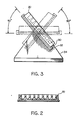
- the agglutinate which forms within a well where a positive reation is occurring will be generally crescent-shaped and will form at the bottom of the well since it will be more dense than the solution within the well, and predominately at the juncture or corner of the bottom and the side of the well due to surface tension and/or the tilted orientation of the tray, since a corner of each well 22 with a flat bottom will be positioned below all other positions of the well in a tilted orientation of the tray 20.
- the tilting of the tray will cause an initially small agglutinate, which will tend to be crescent-shaped, to fold over on itself by gravity as it moves to the opposite side of the well and in the process it will absorb smaller agglutinates which are forming within the well and thereby increase in size and will reduce the number of red cells in suspension in the sample in the well, and this will increase the light transmittance capability of the sample in the central portion of the well.
- Tilting of the tray 20 is accomplished by placing it in a holder 30 which is provided with a generally flat top portion 32 that is pivotal with respect to a fixed base portion 34, the tray 20 being positioned with its major longitudinal axis, that is, its axis which extends parallel to each of the eight rows of twelve wells, parallel to the tilting axis of the top portion 32 of the tray holder 30.
- the tray 20 is initially positioned in a horizontal position, either in the holder 30 or separately thereupon, while the blood samples for each test to be run, red cell solutions or plasma, as the case may be, are pipetted into the wells 22 of the tray 20 and the appropriate reagents and enhancement compositions, if any, are also pipetted into the wells 20, either before or after the pipetting of the samples, according to conventional practices.
- the tray 20 which must now be in the holder 30, is tilted in a first direction with respect to the horizon by pivoting the flat top portion 32 of the holder 30 with respect to the base portion 34 by at least approximately 50°, specifically to position the corner of each well 22 below all other portions of such well, and is maintained in such tilted position for approximately 41 ⁇ 2 - 5 minutes.
- the tray 20 is tilted in the opposite direction by approximately 50° for approximately 41 ⁇ 2 - 5 minutes, again to position a corner of a different portion of each well 22 below all other portions of such well, it is tilted back in the first direction to an angle of approximately 65° for approximately 41 ⁇ 2 - 5 minutes, it is placed in an untilted, horizontal position for approximately 41 ⁇ 2 - 5 minutes, it is tilted back in the second tilted direction to an angle of approximately 50° for approximately 41 ⁇ 2 - 5 minutes, it is placed in an untilted, horizontal position for approximately 41 ⁇ 2-5 minutes, it is tilted in the first tilted direction to an angle of approximately 50° for approximately 41 ⁇ 2 - 5 minutes and then it is transferred to an electro-optical reading station, indicated generally by reference numeral 40 in Figure 4, for detection of agglutinates in those wells having agglutinates therein by the variations in the optical characteristics between those wells having agglutinates therein and those wells not having agglutinate
- the reading which takes only 1 second for each well, or about 15 seconds for a tray when 8 cells across are done simultaneously, is done through the center of each well, and the light transmittance through a cell with an agglutinate will be much higher than the light transmittance through a well without one or more agglutinates therein. This is so since the red cells in a cell with an agglutinate will have been largely incorporated in one or more agglutinates which form at the sides of the well, away from the path of light transmittance, whereas in a well without an agglutinate the red cells will remain dispersed in the sample and will decrease light transmittance through the well. Thus, the total cycle time will be approximately 30 - 35 minutes.
- the use of at least one dwell in the horizontal position after the first bi-directional tilting sequence appears to be beneficial by allowing any agglutinate or agglutinates in a well to settle to the bottom thereof, which enhances the folding over of any such agglutinate in a subsequent bi-directional tilting sequence and, thus, the sweeping of the red cells and small agglutinates in the well by larger agglutinates.
- the electro-optical reading station 40 has a pair of spaced apart rails 42 and 44 for supporting a tray 20, which is shown in a horizontal position, a tungsten halogen lamp 46 for producing a source of illumination, a parabolic reflector 48 for directing illumination from the lamp 46 toward a fiber optic bundle 50 which has a plurality of individual fiber optic strands 52, each of which can be in the form of an individual bundle of multiple strands,leading therefrom, an optical band pass filter system indicated generally by reference numeral 54 being provided between the lamp 46 and the fiber optic bundle to limit the wavelength of the light received by the fiber optic bundle 50 to a range of 400 - 600 nanometers (nm).
- the light which passes through the fiber optic strands 52 is transmitted into a row of wells 22 through an imaging lens device 60, and preferably into the center of each of the wells 22 through the bottoms of the wells to minimize reading errors due to the meniscus of the sample in the well.
- a second imaging lens device 62 above the level of the tray 20 in the reading station 40 receives the light passing through the wells being read and, in turn, passes the light to a light detector 64 which converts the light to an electronic signal.
- the electronic signal for each from the light detector 64 is amplified by an amplifier 66, and, as amplified, is read by a reader 68.
- the digital signal from a given well 22 of the tray 20 will indicate the supernatent transmittance of the sample in the well, and this will be a function of the degree of completion of an agglutination reaction and the position of a settled agglutinate in a well, which will gravitate to a corner of the well by gravity resulting from the tilting of the tray 20 and/or by surface tension and which, therefore, will allow significant illumination to pass through the well, a characteristic that, represents a "positive” reading, as opposed to a "negative” reading in which significantly less illumination passes through the well due to the presence of unagglutinated blood cells in a suspension dispersed throughout the well being read.
- the digital output from each well is multiplied by a predetermined factor, which is proportional to the full signal output from that well, to normalize the various wells which are being read to one another. Differences in donor plasma that are unrelated to blood type may be obviated at this time by adding a control reagent in one of the plasma containing wells in each row to control for sample variables which are unrelated to blood type.
- the method of the present invention is especially effective for detection of the Du phenotype which, in known prior art methods, due to its weak reactivity, requires centrifugation of the sample being tested and washing of the test tray, and in many cases the addition of a second antibody reagent, factors which inhibit the mechanization of the testing method.
- a biotinylated anti-D agent is added, by pipetting, to the sample being tested, followed by the addition of a second anti-biotin antibody thereto. Since biotin is not normally found in human blood, no wading is needed before adding the second antibody thereto.
- the anti-biotin links red wells of the Du phenotype, which have been sensitized with biotinylated anti-D, so that agglutination can occur without the need for centrifugation.
- the method of the present invention is more sensitive, it does not require centrifugation of the sample being typed (as opposed to centrifugation of the original blood specimen to separate it into red well and plasma components), which simplifies the mechanization of the method, it minimizes donor-to-donor variations not related to blood type, and it can be used to perform Du testing without the need for washing, centrifugation and visual confirmation.
- the method of the present invention involves an agglutination reaction enhancement technique which has been specifically described in relation to a blood typing system
- other blood testing procedures for example, testing for syphilis and testing for HIV (AIDS virus) may also involve agglutination reactions
- the agglutination enhancement technique of the blood typing method of the present invention is adaptable to other blood testing procedures which utilize an agglutination reaction.
Landscapes
- Health & Medical Sciences (AREA)
- Life Sciences & Earth Sciences (AREA)
- Engineering & Computer Science (AREA)
- Immunology (AREA)
- Hematology (AREA)
- Chemical & Material Sciences (AREA)
- Urology & Nephrology (AREA)
- Biomedical Technology (AREA)
- Molecular Biology (AREA)
- Physics & Mathematics (AREA)
- Analytical Chemistry (AREA)
- Cell Biology (AREA)
- Pathology (AREA)
- Food Science & Technology (AREA)
- Medicinal Chemistry (AREA)
- Biotechnology (AREA)
- Microbiology (AREA)
- Biochemistry (AREA)
- General Health & Medical Sciences (AREA)
- General Physics & Mathematics (AREA)
- Chemical Kinetics & Catalysis (AREA)
- Investigating Or Analysing Biological Materials (AREA)
- Automatic Analysis And Handling Materials Therefor (AREA)
- Investigating Or Analysing Materials By The Use Of Chemical Reactions (AREA)
Applications Claiming Priority (2)
| Application Number | Priority Date | Filing Date | Title |
|---|---|---|---|
| US34667489A | 1989-05-03 | 1989-05-03 | |
| US346674 | 1989-05-03 |
Publications (2)
| Publication Number | Publication Date |
|---|---|
| EP0396115A2 true EP0396115A2 (fr) | 1990-11-07 |
| EP0396115A3 EP0396115A3 (fr) | 1991-07-24 |
Family
ID=23360526
Family Applications (1)
| Application Number | Title | Priority Date | Filing Date |
|---|---|---|---|
| EP19900108316 Withdrawn EP0396115A3 (fr) | 1989-05-03 | 1990-05-02 | Procédé de formation des agglutinats dans les échantillons de sang |
Country Status (6)
| Country | Link |
|---|---|
| US (1) | US5283178A (fr) |
| EP (1) | EP0396115A3 (fr) |
| JP (1) | JPH034170A (fr) |
| KR (1) | KR900018675A (fr) |
| AU (1) | AU635269B2 (fr) |
| CA (1) | CA2015941A1 (fr) |
Cited By (7)
| Publication number | Priority date | Publication date | Assignee | Title |
|---|---|---|---|---|
| US5476796A (en) * | 1991-06-18 | 1995-12-19 | Olympus Optical Co., Ltd. | Immunological test method |
| EP0779103A1 (fr) * | 1995-12-13 | 1997-06-18 | Lider S.à.r.l. | Dispositif de contrÔle visuel d'un liquide par mélange avec un liquide réactif |
| US6124139A (en) * | 1993-05-17 | 2000-09-26 | Fujirebio Inc. | Method and apparatus for indirect agglutination immunoassay |
| EP1186891A1 (fr) * | 2000-09-05 | 2002-03-13 | Tecan Schweiz AG | Support pour plaque de microtitrage |
| GB2472252A (en) * | 2009-07-31 | 2011-02-02 | Simon Stafford | Microplate holder for inclining a microplate |
| CN103412130A (zh) * | 2013-08-22 | 2013-11-27 | 四川省新成生物科技有限责任公司 | 快速测定尿液特定蛋白含量的试剂杯联、仪器以及方法 |
| GB2472321B (en) * | 2009-07-31 | 2014-03-05 | Oxley Hughes Ltd | Means for improved liquid handling in a microplate |
Families Citing this family (12)
| Publication number | Priority date | Publication date | Assignee | Title |
|---|---|---|---|---|
| US5583004A (en) * | 1994-12-23 | 1996-12-10 | Pincus; Mathew R. | Direct detection of unexpected alloantibodies in the serum of prospective transfusion recipients using a new hemagglutination inhibition assay |
| US5541417A (en) * | 1995-05-18 | 1996-07-30 | Abbott Laboratories | Quantative agglutination reaction analysis method |
| DE69913257T2 (de) * | 1998-07-17 | 2004-09-02 | Vertex Pharmaceuticals (San Diego) Llc, San Diego | Detektor und Siebvorrichtung für Ionenkanäle |
| US6349160B2 (en) | 1998-07-24 | 2002-02-19 | Aurora Biosciences Corporation | Detector and screening device for ion channels |
| US6608671B2 (en) * | 1998-07-17 | 2003-08-19 | Vertex Pharmaceuticals (San Diego) Llc | Detector and screening device for ion channels |
| JP3803345B2 (ja) * | 2004-02-16 | 2006-08-02 | 株式会社アイディエス | 血液検体の自動検知装置 |
| JP4638775B2 (ja) * | 2005-07-01 | 2011-02-23 | シスメックス株式会社 | 分析装置 |
| JP4829716B2 (ja) | 2006-08-18 | 2011-12-07 | シスメックス株式会社 | 血液凝固分析装置 |
| US7858924B2 (en) | 2006-12-12 | 2010-12-28 | Abbott Laboratories, Inc. | Device for use in normalizing readings on a testing machine |
| JP2011133364A (ja) * | 2009-12-24 | 2011-07-07 | Beckman Coulter Inc | 血球凝集像判定方法及び血球凝集像判定装置 |
| US9360433B1 (en) * | 2013-05-21 | 2016-06-07 | Indevr, Inc. | Detection of agglutination by optical density measurement |
| CN114007755B (zh) | 2019-07-03 | 2023-09-26 | 美国西门子医学诊断股份有限公司 | 用于细胞裂解和提纯的旋转平台以及使用方法 |
Citations (5)
| Publication number | Priority date | Publication date | Assignee | Title |
|---|---|---|---|---|
| US3457344A (en) * | 1966-06-07 | 1969-07-22 | Murex Welding Processes Ltd | Method for detecting antigens |
| US3492096A (en) * | 1967-10-05 | 1970-01-27 | Paul G Hattersley | Apparatus for and method of detecting the coagulation of whole blood |
| GB1270416A (en) * | 1969-07-07 | 1972-04-12 | Hans Peter Olof Unger | An improved method and apparatus for determining blood clotting time |
| US4447396A (en) * | 1982-01-04 | 1984-05-08 | Olympus Optical Co., Ltd. | System for discriminating a precipitation pattern of particles |
| EP0352139A2 (fr) * | 1988-07-21 | 1990-01-24 | Pak Leong Lim | Détection des antigènes ou anticorps |
Family Cites Families (14)
| Publication number | Priority date | Publication date | Assignee | Title |
|---|---|---|---|---|
| US3432268A (en) * | 1964-08-28 | 1969-03-11 | Peter Unger | Method and apparatus for testing cell suspensions |
| US3488156A (en) * | 1966-02-23 | 1970-01-06 | Lab Line Biomedical Products I | Automatic agglutinometer |
| US3574064A (en) * | 1968-05-09 | 1971-04-06 | Aerojet General Co | Automated biological reaction instrument |
| SE399768B (sv) * | 1975-09-29 | 1978-02-27 | Lilja Jan E | Kyvett for provtagning, blandning av, provet med ett reagensmedel och direkt utforande av, serskilt optisk, analys av det med reagensmedlet blandade provet |
| US4152390A (en) * | 1976-12-17 | 1979-05-01 | Eastman Kodak Company | Chemical analyzer |
| US4197088A (en) * | 1977-09-23 | 1980-04-08 | Akro-Medic Engineering, Inc. | Method for qualitative and quantitative determination of immunological reactions |
| FI56905C (fi) * | 1978-02-28 | 1980-04-10 | Osmo A Suovaniemi | Foerfarande och anordning foer automatisk maetning av agglutinationsprov t ex i spektrofotometer adsoptionsfotometer fluorometer eller nefelometer |
| JPS5933856B2 (ja) * | 1979-10-09 | 1984-08-18 | オリンパス光学工業株式会社 | 凝集反応測定方法およびそれに用いる反応容器 |
| ES275136Y (es) * | 1981-07-20 | 1984-10-01 | American Hospital Supply Corporation | Dispositivo de anclaje para peldanos en piezas de hormigon o similares. |
| JPS58105065A (ja) * | 1981-12-17 | 1983-06-22 | Olympus Optical Co Ltd | 免疫学的凝集反応に基く分析装置 |
| US4457894A (en) * | 1982-03-01 | 1984-07-03 | Clark George H | Semi-automated agglutination viewer for serology testing |
| JPS6086468A (ja) * | 1983-10-18 | 1985-05-16 | Olympus Optical Co Ltd | 抗原抗体反応の判定方法 |
| US4596695A (en) * | 1984-09-10 | 1986-06-24 | Cottingham Hugh V | Agglutinographic reaction chamber |
| US4737464A (en) * | 1985-09-26 | 1988-04-12 | Molecular Devices Corporation | Solid-state optical assay imaging apparatus |
-
1990
- 1990-05-02 CA CA002015941A patent/CA2015941A1/fr not_active Abandoned
- 1990-05-02 EP EP19900108316 patent/EP0396115A3/fr not_active Withdrawn
- 1990-05-02 AU AU54643/90A patent/AU635269B2/en not_active Ceased
- 1990-05-03 KR KR1019900006229A patent/KR900018675A/ko not_active Application Discontinuation
- 1990-05-07 JP JP2118498A patent/JPH034170A/ja active Pending
-
1992
- 1992-10-15 US US07/964,463 patent/US5283178A/en not_active Expired - Fee Related
Patent Citations (5)
| Publication number | Priority date | Publication date | Assignee | Title |
|---|---|---|---|---|
| US3457344A (en) * | 1966-06-07 | 1969-07-22 | Murex Welding Processes Ltd | Method for detecting antigens |
| US3492096A (en) * | 1967-10-05 | 1970-01-27 | Paul G Hattersley | Apparatus for and method of detecting the coagulation of whole blood |
| GB1270416A (en) * | 1969-07-07 | 1972-04-12 | Hans Peter Olof Unger | An improved method and apparatus for determining blood clotting time |
| US4447396A (en) * | 1982-01-04 | 1984-05-08 | Olympus Optical Co., Ltd. | System for discriminating a precipitation pattern of particles |
| EP0352139A2 (fr) * | 1988-07-21 | 1990-01-24 | Pak Leong Lim | Détection des antigènes ou anticorps |
Cited By (9)
| Publication number | Priority date | Publication date | Assignee | Title |
|---|---|---|---|---|
| US5476796A (en) * | 1991-06-18 | 1995-12-19 | Olympus Optical Co., Ltd. | Immunological test method |
| US6124139A (en) * | 1993-05-17 | 2000-09-26 | Fujirebio Inc. | Method and apparatus for indirect agglutination immunoassay |
| EP0779103A1 (fr) * | 1995-12-13 | 1997-06-18 | Lider S.à.r.l. | Dispositif de contrÔle visuel d'un liquide par mélange avec un liquide réactif |
| FR2742544A1 (fr) * | 1995-12-13 | 1997-06-20 | Lider Sarl | Procede de controle visuel d'un liquide par melange avec un liquide reactif et dispositif pour sa mise en oeuvre |
| EP1186891A1 (fr) * | 2000-09-05 | 2002-03-13 | Tecan Schweiz AG | Support pour plaque de microtitrage |
| WO2002021145A1 (fr) * | 2000-09-05 | 2002-03-14 | Tecan Trading Ag | Support pour plaque de microtitration |
| GB2472252A (en) * | 2009-07-31 | 2011-02-02 | Simon Stafford | Microplate holder for inclining a microplate |
| GB2472321B (en) * | 2009-07-31 | 2014-03-05 | Oxley Hughes Ltd | Means for improved liquid handling in a microplate |
| CN103412130A (zh) * | 2013-08-22 | 2013-11-27 | 四川省新成生物科技有限责任公司 | 快速测定尿液特定蛋白含量的试剂杯联、仪器以及方法 |
Also Published As
| Publication number | Publication date |
|---|---|
| AU5464390A (en) | 1990-11-08 |
| KR900018675A (ko) | 1990-12-22 |
| EP0396115A3 (fr) | 1991-07-24 |
| JPH034170A (ja) | 1991-01-10 |
| US5283178A (en) | 1994-02-01 |
| AU635269B2 (en) | 1993-03-18 |
| CA2015941A1 (fr) | 1990-11-03 |
Similar Documents
| Publication | Publication Date | Title |
|---|---|---|
| US5283178A (en) | Method of forming agglutinates in blood samples | |
| US4290997A (en) | Apparatus for automatic measurement of the results of agglutination tests | |
| US6114179A (en) | Method and test kit for detecting antigens and/or antibodies | |
| US5460940A (en) | Method for detecting antigens and/or antibodies | |
| US5188968A (en) | Method and reaction kit for agglutination detection | |
| US4148607A (en) | Apparatus and analysis for agglutination reaction | |
| US5631166A (en) | Specimen disk for blood analyses | |
| US5066465A (en) | Reaction apparatus | |
| EP1450159A2 (fr) | Procédé et appareil de détection de l'agglutinement des analyses | |
| Bromilow et al. | Evaluation of the ID‐gel test for antibody screening and identification | |
| US4130395A (en) | Process and apparatus for detection of specific biological factors by means of osmotic hemolysis | |
| US4319882A (en) | Method for detecting immunological agglutination and biochemical agent therefor | |
| US5017341A (en) | Agglutination analyzing vessel | |
| DE19856703C2 (de) | Verfahren zum Nachweis von Antikörpern oder Antigenen | |
| CN111480075B (zh) | 用于测试生物样品的方法和设备 | |
| CN103599817B (zh) | 免疫学化验系统和方法 | |
| EP0061541A1 (fr) | Analyse immunologique et agent biochimique pour cette analyse | |
| Trudell | Detection and identification of antibodies | |
| US4708850A (en) | Self-contained portable apparatus for blood typing | |
| EP0021236B1 (fr) | Procédé pour l'essai optique d'échantillons liquides | |
| EP0435245B1 (fr) | Kit de réaction | |
| JPS6113135A (ja) | 反応相手の測定法およびそのための装置 | |
| WO1985002259A1 (fr) | Methode de determination des resultats de reactions d'agglutination | |
| EP0426170B1 (fr) | Essai immunologique d'agglutation indirecte et appareil pour cela | |
| WO1982003462A1 (fr) | Analyse immunologique et agent biochimique pour une telle analyse |
Legal Events
| Date | Code | Title | Description |
|---|---|---|---|
| PUAI | Public reference made under article 153(3) epc to a published international application that has entered the european phase |
Free format text: ORIGINAL CODE: 0009012 |
|
| AK | Designated contracting states |
Kind code of ref document: A2 Designated state(s): AT BE CH DE ES FR GB IT LI NL |
|
| PUAL | Search report despatched |
Free format text: ORIGINAL CODE: 0009013 |
|
| AK | Designated contracting states |
Kind code of ref document: A3 Designated state(s): AT BE CH DE ES FR GB IT LI NL |
|
| 17P | Request for examination filed |
Effective date: 19920114 |
|
| 17Q | First examination report despatched |
Effective date: 19940323 |
|
| STAA | Information on the status of an ep patent application or granted ep patent |
Free format text: STATUS: THE APPLICATION IS DEEMED TO BE WITHDRAWN |
|
| 18D | Application deemed to be withdrawn |
Effective date: 19940606 |