EP0394152B1 - Matériau et procédé de restauration d'un défaut osseux par comblement par du tissu osseux - Google Patents
Matériau et procédé de restauration d'un défaut osseux par comblement par du tissu osseux Download PDFInfo
- Publication number
- EP0394152B1 EP0394152B1 EP90420194A EP90420194A EP0394152B1 EP 0394152 B1 EP0394152 B1 EP 0394152B1 EP 90420194 A EP90420194 A EP 90420194A EP 90420194 A EP90420194 A EP 90420194A EP 0394152 B1 EP0394152 B1 EP 0394152B1
- Authority
- EP
- European Patent Office
- Prior art keywords
- particles
- bone
- tissue
- bone tissue
- material according
- Prior art date
- Legal status (The legal status is an assumption and is not a legal conclusion. Google has not performed a legal analysis and makes no representation as to the accuracy of the status listed.)
- Expired - Lifetime
Links
- 210000000988 bone and bone Anatomy 0.000 title claims abstract description 78
- 230000007547 defect Effects 0.000 title claims abstract description 24
- 239000000463 material Substances 0.000 title claims abstract description 22
- 238000000034 method Methods 0.000 title abstract description 4
- 230000008569 process Effects 0.000 title abstract description 4
- 239000002245 particle Substances 0.000 claims abstract description 90
- VYPSYNLAJGMNEJ-UHFFFAOYSA-N Silicium dioxide Chemical compound O=[Si]=O VYPSYNLAJGMNEJ-UHFFFAOYSA-N 0.000 claims abstract description 35
- 239000008187 granular material Substances 0.000 claims abstract description 33
- DLYUQMMRRRQYAE-UHFFFAOYSA-N tetraphosphorus decaoxide Chemical compound O1P(O2)(=O)OP3(=O)OP1(=O)OP2(=O)O3 DLYUQMMRRRQYAE-UHFFFAOYSA-N 0.000 claims abstract description 20
- 239000000377 silicon dioxide Substances 0.000 claims abstract description 16
- 229910052681 coesite Inorganic materials 0.000 claims abstract description 13
- 229910052906 cristobalite Inorganic materials 0.000 claims abstract description 13
- 229910052682 stishovite Inorganic materials 0.000 claims abstract description 13
- 229910052905 tridymite Inorganic materials 0.000 claims abstract description 13
- KKCBUQHMOMHUOY-UHFFFAOYSA-N Na2O Inorganic materials [O-2].[Na+].[Na+] KKCBUQHMOMHUOY-UHFFFAOYSA-N 0.000 claims abstract description 12
- 238000011049 filling Methods 0.000 claims abstract description 12
- 235000012239 silicon dioxide Nutrition 0.000 claims abstract description 12
- 230000001788 irregular Effects 0.000 claims abstract description 4
- 239000011521 glass Substances 0.000 claims description 15
- 239000000203 mixture Substances 0.000 claims description 15
- 210000004027 cell Anatomy 0.000 claims description 14
- 210000000963 osteoblast Anatomy 0.000 claims description 13
- 230000004069 differentiation Effects 0.000 claims description 10
- 230000011164 ossification Effects 0.000 claims description 9
- 239000000843 powder Substances 0.000 claims description 8
- 239000002243 precursor Substances 0.000 claims description 7
- 239000000470 constituent Substances 0.000 claims description 4
- 238000009826 distribution Methods 0.000 claims description 3
- 230000008018 melting Effects 0.000 claims 1
- 238000002844 melting Methods 0.000 claims 1
- 238000000465 moulding Methods 0.000 claims 1
- 238000010298 pulverizing process Methods 0.000 claims 1
- 230000008929 regeneration Effects 0.000 claims 1
- 238000011069 regeneration method Methods 0.000 claims 1
- 230000008023 solidification Effects 0.000 claims 1
- 238000007711 solidification Methods 0.000 claims 1
- 230000001939 inductive effect Effects 0.000 abstract 1
- 210000001519 tissue Anatomy 0.000 description 16
- 238000009412 basement excavation Methods 0.000 description 14
- 239000001506 calcium phosphate Substances 0.000 description 14
- QORWJWZARLRLPR-UHFFFAOYSA-H tricalcium bis(phosphate) Chemical compound [Ca+2].[Ca+2].[Ca+2].[O-]P([O-])([O-])=O.[O-]P([O-])([O-])=O QORWJWZARLRLPR-UHFFFAOYSA-H 0.000 description 14
- 229910000389 calcium phosphate Inorganic materials 0.000 description 12
- 235000011010 calcium phosphates Nutrition 0.000 description 12
- 230000004820 osteoconduction Effects 0.000 description 12
- 238000002513 implantation Methods 0.000 description 10
- 238000004090 dissolution Methods 0.000 description 9
- 239000010410 layer Substances 0.000 description 9
- 230000015572 biosynthetic process Effects 0.000 description 7
- 230000012010 growth Effects 0.000 description 7
- 206010061218 Inflammation Diseases 0.000 description 6
- 238000006243 chemical reaction Methods 0.000 description 6
- 230000004054 inflammatory process Effects 0.000 description 6
- 239000002131 composite material Substances 0.000 description 5
- CSCPPACGZOOCGX-UHFFFAOYSA-N Acetone Chemical compound CC(C)=O CSCPPACGZOOCGX-UHFFFAOYSA-N 0.000 description 4
- 230000002349 favourable effect Effects 0.000 description 4
- 229910019142 PO4 Inorganic materials 0.000 description 3
- 230000009471 action Effects 0.000 description 3
- WUKWITHWXAAZEY-UHFFFAOYSA-L calcium difluoride Chemical compound [F-].[F-].[Ca+2] WUKWITHWXAAZEY-UHFFFAOYSA-L 0.000 description 3
- 229910001634 calcium fluoride Inorganic materials 0.000 description 3
- 238000000605 extraction Methods 0.000 description 3
- 210000002950 fibroblast Anatomy 0.000 description 3
- 239000007943 implant Substances 0.000 description 3
- 210000002540 macrophage Anatomy 0.000 description 3
- 230000003239 periodontal effect Effects 0.000 description 3
- 239000010452 phosphate Substances 0.000 description 3
- 241000282472 Canis lupus familiaris Species 0.000 description 2
- 102000008186 Collagen Human genes 0.000 description 2
- 108010035532 Collagen Proteins 0.000 description 2
- KRHYYFGTRYWZRS-UHFFFAOYSA-M Fluoride anion Chemical compound [F-] KRHYYFGTRYWZRS-UHFFFAOYSA-M 0.000 description 2
- CDBYLPFSWZWCQE-UHFFFAOYSA-L Sodium Carbonate Chemical compound [Na+].[Na+].[O-]C([O-])=O CDBYLPFSWZWCQE-UHFFFAOYSA-L 0.000 description 2
- 229910000287 alkaline earth metal oxide Inorganic materials 0.000 description 2
- PNEYBMLMFCGWSK-UHFFFAOYSA-N aluminium oxide Inorganic materials [O-2].[O-2].[O-2].[Al+3].[Al+3] PNEYBMLMFCGWSK-UHFFFAOYSA-N 0.000 description 2
- 229910052586 apatite Inorganic materials 0.000 description 2
- 239000011575 calcium Substances 0.000 description 2
- 229920001436 collagen Polymers 0.000 description 2
- 150000001875 compounds Chemical class 0.000 description 2
- 238000011161 development Methods 0.000 description 2
- 230000018109 developmental process Effects 0.000 description 2
- CGMRCMMOCQYHAD-UHFFFAOYSA-J dicalcium hydroxide phosphate Chemical compound [OH-].[Ca++].[Ca++].[O-]P([O-])([O-])=O CGMRCMMOCQYHAD-UHFFFAOYSA-J 0.000 description 2
- 230000008034 disappearance Effects 0.000 description 2
- 230000000694 effects Effects 0.000 description 2
- 239000012530 fluid Substances 0.000 description 2
- 238000000227 grinding Methods 0.000 description 2
- 230000003902 lesion Effects 0.000 description 2
- 239000007788 liquid Substances 0.000 description 2
- 230000007246 mechanism Effects 0.000 description 2
- 230000005012 migration Effects 0.000 description 2
- 238000013508 migration Methods 0.000 description 2
- VSIIXMUUUJUKCM-UHFFFAOYSA-D pentacalcium;fluoride;triphosphate Chemical compound [F-].[Ca+2].[Ca+2].[Ca+2].[Ca+2].[Ca+2].[O-]P([O-])([O-])=O.[O-]P([O-])([O-])=O.[O-]P([O-])([O-])=O VSIIXMUUUJUKCM-UHFFFAOYSA-D 0.000 description 2
- NBIIXXVUZAFLBC-UHFFFAOYSA-K phosphate Chemical compound [O-]P([O-])([O-])=O NBIIXXVUZAFLBC-UHFFFAOYSA-K 0.000 description 2
- 230000008439 repair process Effects 0.000 description 2
- 239000000741 silica gel Substances 0.000 description 2
- 229910002027 silica gel Inorganic materials 0.000 description 2
- 239000007787 solid Substances 0.000 description 2
- 239000000126 substance Substances 0.000 description 2
- 238000006467 substitution reaction Methods 0.000 description 2
- 235000019731 tricalcium phosphate Nutrition 0.000 description 2
- 229910000391 tricalcium phosphate Inorganic materials 0.000 description 2
- 229940078499 tricalcium phosphate Drugs 0.000 description 2
- 239000011800 void material Substances 0.000 description 2
- XLYOFNOQVPJJNP-UHFFFAOYSA-N water Substances O XLYOFNOQVPJJNP-UHFFFAOYSA-N 0.000 description 2
- 206010061728 Bone lesion Diseases 0.000 description 1
- ZOXJGFHDIHLPTG-UHFFFAOYSA-N Boron Chemical compound [B] ZOXJGFHDIHLPTG-UHFFFAOYSA-N 0.000 description 1
- OKTJSMMVPCPJKN-UHFFFAOYSA-N Carbon Chemical compound [C] OKTJSMMVPCPJKN-UHFFFAOYSA-N 0.000 description 1
- BVKZGUZCCUSVTD-UHFFFAOYSA-L Carbonate Chemical compound [O-]C([O-])=O BVKZGUZCCUSVTD-UHFFFAOYSA-L 0.000 description 1
- 206010013886 Dysaesthesia Diseases 0.000 description 1
- 102000009123 Fibrin Human genes 0.000 description 1
- 108010073385 Fibrin Proteins 0.000 description 1
- 208000005888 Periodontal Pocket Diseases 0.000 description 1
- 239000002253 acid Substances 0.000 description 1
- 239000000853 adhesive Substances 0.000 description 1
- 230000001070 adhesive effect Effects 0.000 description 1
- 230000004523 agglutinating effect Effects 0.000 description 1
- 230000002546 agglutinic effect Effects 0.000 description 1
- 239000003513 alkali Substances 0.000 description 1
- 238000000137 annealing Methods 0.000 description 1
- 239000011230 binding agent Substances 0.000 description 1
- 230000000975 bioactive effect Effects 0.000 description 1
- 230000017531 blood circulation Effects 0.000 description 1
- 210000002449 bone cell Anatomy 0.000 description 1
- 230000008468 bone growth Effects 0.000 description 1
- 229910052796 boron Inorganic materials 0.000 description 1
- 238000001354 calcination Methods 0.000 description 1
- 229910052791 calcium Inorganic materials 0.000 description 1
- FUFJGUQYACFECW-UHFFFAOYSA-L calcium hydrogenphosphate Chemical compound [Ca+2].OP([O-])([O-])=O FUFJGUQYACFECW-UHFFFAOYSA-L 0.000 description 1
- 238000005266 casting Methods 0.000 description 1
- 239000000919 ceramic Substances 0.000 description 1
- 210000001612 chondrocyte Anatomy 0.000 description 1
- 230000001684 chronic effect Effects 0.000 description 1
- 239000011248 coating agent Substances 0.000 description 1
- 238000000576 coating method Methods 0.000 description 1
- 238000004891 communication Methods 0.000 description 1
- 239000004020 conductor Substances 0.000 description 1
- 238000000151 deposition Methods 0.000 description 1
- 235000019700 dicalcium phosphate Nutrition 0.000 description 1
- 238000001035 drying Methods 0.000 description 1
- 238000001493 electron microscopy Methods 0.000 description 1
- 239000000835 fiber Substances 0.000 description 1
- 230000004927 fusion Effects 0.000 description 1
- 239000003292 glue Substances 0.000 description 1
- 229910002804 graphite Inorganic materials 0.000 description 1
- 239000010439 graphite Substances 0.000 description 1
- 210000003701 histiocyte Anatomy 0.000 description 1
- 229910052588 hydroxylapatite Inorganic materials 0.000 description 1
- 208000015181 infectious disease Diseases 0.000 description 1
- 230000005764 inhibitory process Effects 0.000 description 1
- 230000000977 initiatory effect Effects 0.000 description 1
- 238000003780 insertion Methods 0.000 description 1
- 230000037431 insertion Effects 0.000 description 1
- 150000002500 ions Chemical class 0.000 description 1
- 238000002386 leaching Methods 0.000 description 1
- 210000004698 lymphocyte Anatomy 0.000 description 1
- 230000014759 maintenance of location Effects 0.000 description 1
- 238000004519 manufacturing process Methods 0.000 description 1
- 239000011159 matrix material Substances 0.000 description 1
- 229910052751 metal Inorganic materials 0.000 description 1
- 239000002184 metal Substances 0.000 description 1
- 238000000386 microscopy Methods 0.000 description 1
- 239000004570 mortar (masonry) Substances 0.000 description 1
- 210000005036 nerve Anatomy 0.000 description 1
- 208000035824 paresthesia Diseases 0.000 description 1
- 230000001575 pathological effect Effects 0.000 description 1
- XYJRXVWERLGGKC-UHFFFAOYSA-D pentacalcium;hydroxide;triphosphate Chemical compound [OH-].[Ca+2].[Ca+2].[Ca+2].[Ca+2].[Ca+2].[O-]P([O-])([O-])=O.[O-]P([O-])([O-])=O.[O-]P([O-])([O-])=O XYJRXVWERLGGKC-UHFFFAOYSA-D 0.000 description 1
- 210000001539 phagocyte Anatomy 0.000 description 1
- 125000002467 phosphate group Chemical group [H]OP(=O)(O[H])O[*] 0.000 description 1
- 210000004180 plasmocyte Anatomy 0.000 description 1
- 230000000750 progressive effect Effects 0.000 description 1
- 230000035755 proliferation Effects 0.000 description 1
- 230000001737 promoting effect Effects 0.000 description 1
- 230000000717 retained effect Effects 0.000 description 1
- 229910000029 sodium carbonate Inorganic materials 0.000 description 1
- 210000004872 soft tissue Anatomy 0.000 description 1
- 238000005507 spraying Methods 0.000 description 1
- 239000002344 surface layer Substances 0.000 description 1
- 230000017423 tissue regeneration Effects 0.000 description 1
- 230000009466 transformation Effects 0.000 description 1
Images


Classifications
-
- C—CHEMISTRY; METALLURGY
- C03—GLASS; MINERAL OR SLAG WOOL
- C03C—CHEMICAL COMPOSITION OF GLASSES, GLAZES OR VITREOUS ENAMELS; SURFACE TREATMENT OF GLASS; SURFACE TREATMENT OF FIBRES OR FILAMENTS MADE FROM GLASS, MINERALS OR SLAGS; JOINING GLASS TO GLASS OR OTHER MATERIALS
- C03C4/00—Compositions for glass with special properties
- C03C4/0007—Compositions for glass with special properties for biologically-compatible glass
-
- A—HUMAN NECESSITIES
- A61—MEDICAL OR VETERINARY SCIENCE; HYGIENE
- A61L—METHODS OR APPARATUS FOR STERILISING MATERIALS OR OBJECTS IN GENERAL; DISINFECTION, STERILISATION OR DEODORISATION OF AIR; CHEMICAL ASPECTS OF BANDAGES, DRESSINGS, ABSORBENT PADS OR SURGICAL ARTICLES; MATERIALS FOR BANDAGES, DRESSINGS, ABSORBENT PADS OR SURGICAL ARTICLES
- A61L27/00—Materials for grafts or prostheses or for coating grafts or prostheses
- A61L27/02—Inorganic materials
- A61L27/10—Ceramics or glasses
-
- A—HUMAN NECESSITIES
- A61—MEDICAL OR VETERINARY SCIENCE; HYGIENE
- A61L—METHODS OR APPARATUS FOR STERILISING MATERIALS OR OBJECTS IN GENERAL; DISINFECTION, STERILISATION OR DEODORISATION OF AIR; CHEMICAL ASPECTS OF BANDAGES, DRESSINGS, ABSORBENT PADS OR SURGICAL ARTICLES; MATERIALS FOR BANDAGES, DRESSINGS, ABSORBENT PADS OR SURGICAL ARTICLES
- A61L27/00—Materials for grafts or prostheses or for coating grafts or prostheses
- A61L27/02—Inorganic materials
- A61L27/12—Phosphorus-containing materials, e.g. apatite
-
- C—CHEMISTRY; METALLURGY
- C03—GLASS; MINERAL OR SLAG WOOL
- C03C—CHEMICAL COMPOSITION OF GLASSES, GLAZES OR VITREOUS ENAMELS; SURFACE TREATMENT OF GLASS; SURFACE TREATMENT OF FIBRES OR FILAMENTS MADE FROM GLASS, MINERALS OR SLAGS; JOINING GLASS TO GLASS OR OTHER MATERIALS
- C03C3/00—Glass compositions
- C03C3/04—Glass compositions containing silica
- C03C3/076—Glass compositions containing silica with 40% to 90% silica, by weight
- C03C3/097—Glass compositions containing silica with 40% to 90% silica, by weight containing phosphorus, niobium or tantalum
-
- A—HUMAN NECESSITIES
- A61—MEDICAL OR VETERINARY SCIENCE; HYGIENE
- A61F—FILTERS IMPLANTABLE INTO BLOOD VESSELS; PROSTHESES; DEVICES PROVIDING PATENCY TO, OR PREVENTING COLLAPSING OF, TUBULAR STRUCTURES OF THE BODY, e.g. STENTS; ORTHOPAEDIC, NURSING OR CONTRACEPTIVE DEVICES; FOMENTATION; TREATMENT OR PROTECTION OF EYES OR EARS; BANDAGES, DRESSINGS OR ABSORBENT PADS; FIRST-AID KITS
- A61F2310/00—Prostheses classified in A61F2/28 or A61F2/30 - A61F2/44 being constructed from or coated with a particular material
- A61F2310/00005—The prosthesis being constructed from a particular material
- A61F2310/00179—Ceramics or ceramic-like structures
- A61F2310/00293—Ceramics or ceramic-like structures containing a phosphorus-containing compound, e.g. apatite
-
- A—HUMAN NECESSITIES
- A61—MEDICAL OR VETERINARY SCIENCE; HYGIENE
- A61L—METHODS OR APPARATUS FOR STERILISING MATERIALS OR OBJECTS IN GENERAL; DISINFECTION, STERILISATION OR DEODORISATION OF AIR; CHEMICAL ASPECTS OF BANDAGES, DRESSINGS, ABSORBENT PADS OR SURGICAL ARTICLES; MATERIALS FOR BANDAGES, DRESSINGS, ABSORBENT PADS OR SURGICAL ARTICLES
- A61L2430/00—Materials or treatment for tissue regeneration
- A61L2430/02—Materials or treatment for tissue regeneration for reconstruction of bones; weight-bearing implants
Definitions
- the invention relates to a material in the form of granules for rapid and complete filling of a bone defect by the formation of bone tissue.
- products such as dense or porous hydroxylapatite, tricalcium phosphate or bioverres can be used in the form of granules as filling material for bone lesions such as periodontal pockets, extraction sites, cystic lesions, bone defect. ..
- osteoconduction occurs (that is to say a growth of bone tissue along the surface of the implanted granules) and a progressive filling of the intergranular space.
- this intergranular growth is generally limited to particles located in the immediate vicinity of the walls of the bone cavity, the other particles surrounding themselves with fibrous tissues and thus giving a solid mass.
- osteoconduction leads to particles progressively coated by bone tissue which proliferates from the walls of the bone cavity, the particles reacting only best on the surface.
- said particles when placed in a cavity, for example an extraction site, they can migrate into the adjacent soft tissues, and be fixed in improper places. This can lead to complications such as dysaesthesia or paraesthesia of the nerve ..., pain, infection, perforation of the gum and loss of particles.
- the filling of the cavity becomes, after osteoconduction, a composite of bone, fibrous tissue and apatite or bioverre which has insufficient mechanical properties, lower than that of bone.
- the insertion of prosthetic implants into such a composite cannot generally be practiced because it risks presenting defects of compatibility, solidity or rigidity over time.
- the presence of particles in neighboring tissues is not desirable.
- Application EP 0206726 thus relates to the repair of periodontal defects (pockets located between the jaw bone and the root of the teeth) by placing a bioactive granule in said defect and illustrates the obtaining of such a bone-granule composite by osteoconduction, that is to say intergranular growth of bone tissue from the bone walls of the defect (p.15, 1.13-15 and 25-28).
- Said granule also has its own characteristics (chemical nature, selection of a particle size stack) which make it possible to obtain certain advantages, representing the main objective sought, such as good cohesion facilitating its positioning and its retention in place in the defect, as well as a blockage to stop the blood flow also occurring in the defect.
- This particular granule is obtained by a process consisting of a fusion of a mixture of powders followed by pouring into water in the form of frits which are washed with acetone and then ground before the particle size selection is carried out.
- the granule described in this document plays, so to speak, the role of a permanent implant, the object of osteoconduction, therefore gradually becoming coated with bone tissue which adheres to said implant and keeps it firmly in place; the repair of such a periodontal defect, leading to said type of bone-glass composite, does not moreover require any particular requirements of mechanical characteristics since it is not generally intended to receive subsequent implantations of prostheses.
- EP-A-145,210 describes another type of granulated biogreen whose spheroidal particles contain SiO2, Na2O, P2O5 CaO, (for example in the respective proportions by weight: 45 24.5 6 24.5) and are obtained by spraying of a solution containing the precursor compounds, drying and calcination.
- Document FR-A-2,606,282 describes a composition for filling the bone cavity containing two components consisting of an alumina or apatite powder with a particle size of between 250 and 1500 ⁇ m and a hydraulic or organic binder.
- the applicant sought to fill bone defects practically with bone tissue and to increase the speed of formation of said bone tissue. She thus sought to produce an osteostimulation, defined as being an osteogenesis in a bone site without contact with the neighboring bone, osteogenesis being the phenomenon leading to bone tissue, another objective being to improve the mechanical characteristics of the tissue bone filling said defect so as to be able to subsequently make successful implantations of prosthesis therein.
- the invention consists in using a bioreactive material for the treatment of bone tissue regeneration to fill a bone defect by placing in said defect said material in the form of a granule which causes osteogenesis by osteostimulation, the constituent particles of said granules being gradually disintegrated from the inside, to effect a differentiation of precursor cells into osteoblasts, then to be dissolved and quickly replaced by bone tissue, the particles having in combination, the following composition (in% by weight): SiO2 between 40 and 55% Na2O between 10 and 32% P2O5 between 0 and 12% CaO between 10 and 32%, a particle size such that at least 95% of them have a size of between 280 and 425 "m, sharp edges, an irregular profile and surface micro-defects or microcracks.
- the precursor cells are mesenchymal cells, originally present in the biological medium, which can differentiate into fibroblast and chondroblast cells which deposit fibrous or cartilaginous tissues (to be avoided in the present invention) and osteoblast cells which deposit tissue bone (sought in this invention). It is important to control the disintegration and the dissolution of the granule because these phenomena, if they are uncontrolled, could for example give rise to too intense inflammation which would prevent the appearance of conditions favorable to the differentiation of mesenchymal cells into osteoblasts. Furthermore, too rapid dissolution of the granules with respect to the rate of formation of bone tissues could no longer lead to a formation of these bone tissues but rather to the formation of undesirable fibrous tissues.
- the constituent particles are thus disintegrated from the inside, disappear and are gradually and rapidly replaced by bone tissue rather than by fibrous tissue; after disappearance of the material, a bone mass is obtained which is suitable, for example, perfectly for subsequent implantations of prostheses.
- This granule is also easy to use and apply; its particles remaining firmly in place in the defect, this avoids the use of artifices.
- bioreactive glasses used do not contain fluorinated products. They generally contain SiO2, Na2O, P2O5, an alkaline earth oxide, such as CaO. The presence of boron and alumina is not desirable.
- the disintegration and dissolution of the particles must be well controlled, thanks to the very narrow particle size distribution, the size having to be between 280 and 425 "m, that is to say that 95% of the powder must be retained on a sieve whose mesh void is equal to the recommended lower limit and 95% of the same powder must pass through a sieve whose mesh void is equal to the recommended upper limit.
- the material should be in the form of a powder made up of angular particles, having sharp edges and an irregular profile.
- the constituent particles must have surface micro-defects or microcracks. These microdefects or microcracks will be the initiation of small conduits which, as we will see later, connect the interior of the particles to the exterior and promote their disintegration from the interior.
- the particles according to the invention have been implanted in dogs, then samples have been taken after 1, 2, 3, 6 months, one year; after fixation treatment, they were cut out in a microtome and prepared to be observed by microscopy and analyzed.
- the Applicant has found that not only the bone tissue develops from the wall of the bone cavity, by osteoconduction, by forming a bone skeleton, but also unexpectedly, that the particles located in the middle of the cavity, therefore without contact with the walls, disintegrate and ossify simultaneously from their center, this disintegration not producing a harmful chronic inflammatory reaction.
- an excavation is formed by disintegration and dissolution of the central part under the action of physiological fluid and macrophage cells, histiocytes, plasma cells and lymphocytes. These central excavations are connected to the outside by small conduits.
- the wall of the excavation formed extends bone tissue covered by a layer of osteoid tissue and a strip of active osteoblast cells. It is thus noted that the desired but unexpected differentiation of the mesenchymal cells into osteoblasts rather than fibroblasts has taken place. A good number of these excavations are already practically filled with bone tissue at the end of 3 months. There is no direct connection between the bone tissue developing at the center of the particles and the external bone tissue.
- the implanted particles are exposed to the action of physiological fluid and cells; the latter operate a dissolution and penetrate inside said particles by means of surface defects or microcracks, due to the granule manufacturing process and accessible to said liquid; there are local changes in pH and composition of the glass, which are favorable to osteostimulation, this promoting the preferential transformation of precursor mesenchymal cells into osteoblasts depositing bone tissue; in particular, there is no development of fibroblast cells, generating annoying fibrous tissues, in contact with the reacted glass.
- the interior of the particles constitutes a very protected medium which makes it possible to create and maintain said conditions favorable to the differentiation of mesenchymal cells into osteoblasts.
- the filling granule is osteostimulating under the specific and limited conditions of the invention.
- the particles which are too small give too intense a reaction and lead to fibrous tissue, these conditions being unfavorable for the differentiation of mesenchymal cells into osteoblasts.
- a bone filling material is thus obtained in the bone defect, organically linked to the wall of said defect; this filling therefore has the properties of bone and can be used under the same conditions as the neighboring bone. In addition, no migration of particles is observed in the non- bone in the vicinity of said bone defect.
- the material according to the invention is not stable, it is on the contrary active, it is stimulator and unexpectedly causes the formation of bone tissue by osteostimulation. It therefore makes it possible to obtain a much faster ossification since it is done not only from the wall of the cavity by coating the particles, but also and simultaneously in the mass through the interior of the particles.
- the characteristics of size and morphology are essential to be able to obtain the desired results.
- a surface layer rich in calcium phosphate (CaP) is created, while on the contrary the underlying layer becomes rich in SiO2.
- CaP calcium phosphate
- the particles are too small, their interior consists of silica gel covered with a layer of CaP that is too thin and very fragile; the latter tends to break, the macrophage cells absorb the silica and the particles disappear too quickly without the medium, very protected and entertaining at differentiating precursor cells into osteoblast, constituted by the internal excavation of the particle, having had time to form and give birth to bone tissue. With this type of particle, there is no osteostimulation.
- said particles of too small dimensions are in too large an amount, the too intense reaction which they provoke can cause an inhibition of the osteostimulation which could occur with particles of larger size.
- the CaP layer is more solid and the center contains silica but no glass. As CaP no longer breaks, macrophage cells can enter the particle through micro-faults and create excavation; the highly protected environment, favorable to differentiation, can then develop; there is then osteogenesis by osteostimulation.
- composition, the particle size distribution and the morphology of the particles are essential factors for this osteostimulation to be observed by dissolution and disintegration of said particles from their interior, their disappearance and their replacement with bone tissue.
- the invention produces a preferential dissolution of glass in opposition to the reaction mechanisms of bioverres of comparable composition described in the prior art.
- the said mechanisms described are generally reactions of leaching of certain ions on the surface of the glass, without any dissolution of said glass being noted.
- the rough surface state of the granules acts in an agglutinative or nested manner and is such that the material is easy to place, the particles remaining in place in the cavity while agglutinating. between them and by gripping the walls of the bone cavity.
- the material according to the invention is obtained by first preparing a mixture of powders comprising silica, an alkaline earth oxide (preferably CaO and / or MgO), a carbonate to introduce the alkali into the less in part (sodium carbonate is preferably used), an acid phosphate or phosphate to introduce P2O5 (preferably CaHPO4).
- an alkaline earth oxide preferably CaO and / or MgO
- a carbonate to introduce the alkali into the less in part sodium carbonate is preferably used
- an acid phosphate or phosphate to introduce P2O5 (preferably CaHPO4).
- This mixture is melted and poured in the form of parts, usually small cylinders or discs, in relatively massive molds made of a material which is a good conductor of heat (for example graphite, metal, etc.). Annealing can be carried out. These parts are then reduced to granules by any means allowing to give cracked, rough particles having very many sharp angles. Crushing and / or grinding is preferably used, for example in mortar, hammer, bar. Then the desired particle size cut is made, for example by passing through vibrating screens.
- This mixture was melted and poured into small disks (diameter 4 cm, height 1 cm) which were then ground with a hammer. A size section between 300 and 425 ⁇ m was then made using a sieve.
- the granules obtained were implanted in bone cavities of the jaws of dogs; these cavities are either due to tooth extractions, or cavities created surgically, or pathological cavities, i.e. periodontal lesions.
- the implantation time varies between 1 month and 1 year, after which samples have been taken and sections made and prepared for examination. It is noted first of all that the particles have remained very well in place in the cavity. It has also been observed that the center of most of the particles in the mass has been rapidly disintegrated and dissolved, and has resulted in the differentiation of mesenchymal cells into osteoblasts. We also note, from the 1st month, in these particles, the presence of bone tissue covered by a layer of osteoid and a film of osteoblasts which allow the growth of said bone tissue. A large quantity of these particles are practically filled with bone tissue after 3 months without any connection being noted with the bone tissue obtained by external growth (osteoconduction) from the walls of the bone cavity.
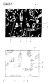
- FIGS. 1 and 2 show micrographic sections of grains obtained by back-scattered electron microscopy, with superimposed point analyzes of the Ca and Si content, carried out along a transverse line and indicative for the CaP and silica gel contents.
- FIG. 1 represents a particle according to the invention (magnification X 400) after one month of implantation.
- the analyzes were carried out along the transverse line (1); along this line, the Ca content is given by curve (2) and that of Si by curve (3).
- (21) the outer layer rich in CaP, while in the center of the particle the CaP content is poor (22).
- SiO2 we see several zones: an area poor both in SiO2 (32), in the process of disintegration, and in CaP (22), two areas (33) poor in SiO2 to outside of which the CaP layer (21) is being formed, two zones (31) containing silica which has not yet been dissolved.
- (4) we see the conduits connecting the excavation to the outside via, initially, surface micro-defects.
- Fig. 2 also represents a particle according to the invention after 3 months of implantation. While in fig. 1 the excavation is being formed, in fig. 2 this excavation is formed. The marks have the same meaning as in fig. 1. However, there is a peak (34) in Si concentration which has no particular significance.
- Figure 3 shows a set of particles (magnification x 60) after 3 months.
- the presence of bone tissue is visible in (3) in more or less quantity.
- the cut, being two-dimensional, may have been carried out near the top of certain particles, which explains the presence in FIG. 3 apparently small particles without excavation.
- This example shows the imperfect results obtained with a granule comprising an excess of large particles (greater than 500 "m).
- the experimental conditions are identical to those of Example 1; only the size section was chosen larger using screens from 425 to 850 ”m.
- bone tissue osteoconduction
- fibrous tissues consisting of densely packed collagen fibers.
- Large particles do not show central decay with resorption or dissolution of glass and substitution with bone tissue. It is not obvious that an ossification large particles occur after much longer implantation times, as they can become coated and enclosed in bone tissue formed by osteoconduction, this tissue preventing any increased reaction as it existed at the start of implantation.
- Example 1 It illustrates the imperfect restoration results due to the use of too fine granules.
- the experimental conditions are identical to those of Example 1; only the grain size section was chosen smaller, using screens of 212 and 300 "m.
- the differentiation of precursor cells into osteoblasts has not been made and the particles are surrounded by fibrous tissues before disappearing under the action of phagocytic cells. Only a few large particles show central disintegration with substitution of their heart by the cells described in Example 1, and by bone tissue.
- the granule according to the invention promotes the tissue reaction of osteogenesis by osteostimulation which occurs at all points of the bone defect (including the central area) inside the particles by destroying them and replacing them with tissue. bone, without preventing osteoconduction which tends to coat the particles, without destroying them, from the walls of the bone cavity.
Landscapes
- Chemical & Material Sciences (AREA)
- Health & Medical Sciences (AREA)
- Life Sciences & Earth Sciences (AREA)
- Engineering & Computer Science (AREA)
- Medicinal Chemistry (AREA)
- Epidemiology (AREA)
- Geochemistry & Mineralogy (AREA)
- Materials Engineering (AREA)
- Organic Chemistry (AREA)
- Inorganic Chemistry (AREA)
- Chemical Kinetics & Catalysis (AREA)
- Dermatology (AREA)
- Ceramic Engineering (AREA)
- Oral & Maxillofacial Surgery (AREA)
- Transplantation (AREA)
- General Chemical & Material Sciences (AREA)
- Animal Behavior & Ethology (AREA)
- General Health & Medical Sciences (AREA)
- Public Health (AREA)
- Veterinary Medicine (AREA)
- Molecular Biology (AREA)
- Materials For Medical Uses (AREA)
- Micro-Organisms Or Cultivation Processes Thereof (AREA)
- Investigating Or Analysing Biological Materials (AREA)
- Prostheses (AREA)
Description
- L'invention concerne un matériau sous forme de granulé pour procéder au comblement rapide et complet d'un défaut osseux par formation de tissus osseux.
- On sait que des produits comme l'hydroxylapatite dense ou poreux, le phosphate tricalcique ou les bioverres peuvent être utilisés sous forme de granulés comme matériau de remplissage pour des lésions osseuses telles que poches parodontales, sites d'extraction, lésions cystiques, défaut osseux...
- Dans le cas du remplissage de cavités osseuses, le rôle de ces granulés (notamment d'hydroxylapatite) est d'agir comme matrice en vue d'une restauration par croissance osseuse (c.à.d. prolifération normale de cellules osseuses). Il a ainsi été démontré qu'il se produit une ostéoconduction (c'est-à-dire une croissance de tissus osseux le long de la surface des granulés implantés) et un remplissage progressif de l'espace intergranulaire. Malheureusement cette croissance intergranulaire est généralement limitée aux particules situées à proximité immédiate des parois de la cavité osseuse, les autres particules s'entourant de tissus fibreux et donnant ainsi une masse solide. Ainsi, l'ostéoconduction conduit à des particules enrobées progressivement par du tissu osseux qui prolifère à partir des parois de la cavité osseuse, les particules ne réagissant au mieux qu'en surface.
- Par ailleurs, quand lesdites particules sont mises en place dans une cavité, par exemple un site d'extraction, elles peuvent migrer dans les tissus mous adjacents, et se trouver fixées dans des endroits impropres. Ceci peut entraîner des complications telles que dysaesthésie ou paraesthésie du nerf..., douleur, infection, perforation de la gencive et perte de particules.
- Pour ces raisons, le développement de l'emploi de particules, telles que celles d'hydroxylapatite, passe par l'amélioration de leurs propriétés de mise et de tenue en place. Ceci peut être obtenu par l'emploi d'artifices tels que mise en place à l'aide d'enveloppes résorbables, colles, collagène ou ses dérivés, fibrines adhésives etc...
- Ainsi, le remplissage de la cavité devient, après ostéoconduction, un composite de tissus osseux, fibreux et d'apatite ou de bioverre qui a des propriétés mécaniques insuffisantes, inférieures à celles de l'os. Par exemple, l'insertion d'implants prothétiques dans un tel composite ne peut pas être en général pratiqué car elle risque de présenter des défauts de compatibilité, de solidité ou de rigidité dans le temps. De plus, la présence de particules dans les tissus voisins n'est pas souhaitable.
- La demande EP 0206726 est ainsi relative à la réparation de défauts parodontaux (poches situées entre l'os de la mâchoire et la racine des dents) par mise en place d'un granulé bioactif dans ledit défaut et illustre l'obtention d'un tel composite os-granulé par ostéoconduction, c'est-à-dire croissance intergranulaire du tissu osseux à partir des parois osseuses du défaut (p.15, 1.13-15 et 25-28). Ledit granulé présente par ailleurs des caractéristiques propres (nature chimique, sélection d'un empilement granulométrique) qui permettent d'obtenir certains avantages, représentant l'objectif principal recherché, comme une bonne cohésion facilitant sa mise et son maintien en place dans le défaut, de même qu'un bouchage permettant d'arrêter l'écoulement sanguin se produisant également dans le défaut. Ce granulé particulier est obtenu par un procédé consistant en une fusion d'un mélange de poudres suivie d'une coulée dans de l'eau sous forme de frittes qui sont lavées à l'acétone puis broyées avant que la sélection granulométrique soit effectuée.
- Le granulé décrit dans ce document joue en quelque sorte le rôle d'un implant permanent, objet d'une ostéoconduction, donc s'enrobant progressivement de tissus osseux qui adhère audit implant et le maintient solidement en place; la réparation d'un tel défaut parodontal, conduisant audit type de composite os-verre, ne nécessite pas d'ailleurs des exigences particulières de caractéristiques mécaniques car elle n'est pas en général destinée à recevoir des implantation ultérieures de prothèses.
- Le document EP-A-145.210 décrit un autre type de bioverre granulé dont les particules sphéroïdales contiennent SiO₂ ,Na₂O ,P₂O₅ CaO, (par exemple dans les proportions respectives en poids : 45 24,5 6 24,5) et sont obtenues par pulvérisation d'une solution contenant les composés précurseurs, séchage et calcination.
- Le document FR-A-2.606.282 décrit une composition pour comblement de cavité osseuse contenant deux composants constitués d'une poudre d'alumine ou d'apatite de granulométrie comprise entre 250 et 1500 »m et d'un liant hydraulique ou organique.
- Quant au document EP-A-382.047, il décrit une céramique osseuse résorbable à base de phosphate tricalcique, remplacée progressivement par du tissu osseux et qui est obtenue à partir d'os naturel et d'un composé porteur de phosphate.
- Pour pallier les inconvénients résultant d'une ostéoconduction à partir des parois osseuses d'une cavité dans un espace intergranulaire et conduisant auxdits composites os-granulé, la demanderesse a recherché à combler des défauts osseux pratiquement par du tissu osseux et à accroître la rapidité de formation dudit tissu osseux. Elle a ainsi recherché à produire une ostéostimulation, définie comme étant une ostéogénèse dans un site osseux sans contact avec l'os avoisinant, l'ostéogénèse étant le phénomène conduisant à du tissu osseux, un autre objectif étant d'améliorer les caractéristiques mécaniques du tissu osseux comblant ledit défaut de façon à pouvoir y faire ultérieurement des implantations réussies de prothèse.
- L'invention consiste à mettre en oeuvre un matériau bioréactif pour le traitement de régénération de tissus osseux pour combler un défaut osseux en mettant en place dans ledit défaut ledit matériau sous forme d'un granulé qui provoque une ostéogénèse par ostéostimulation, les particules constitutives dudit granulé étant désintégrées progressivement de l'intérieur, pour opérer une différenciation de cellules précurseurs en ostéoblastes, puis être dissoutes et remplacées rapidement par du tissu osseux, les particules ayant en combinaison, la composition suivante (en % poids) :
SiO₂ compris entre 40 et 55 %
Na₂O compris entre 10 et 32 %
P₂O₅ compris entre 0 et 12 %
CaO compris entre 10 et 32 %,
une granulométrie telle qu'au moins 95 % d'entre elles ont une taille comprise entre 280 et 425 »m, des arêtes vives, un profil irrégulier et des microdéfauts de surface ou microfissures. - Les cellules précurseurs sont des cellules mesenchymales, présentes à l'origine dans le milieu biologique, qui peuvent se différencier en cellules fibroblastes et chondroblastes qui déposent des tissus fibreux ou cartilagineux (à éviter dans la présente invention) et en cellules ostéoblastes qui déposent du tissu osseux (recherchées dans la présente invention).
Il est important de contrôler la désintégration et la dissolution du granulé car ces phénomènes, s'ils sont incontrôlés, pourraient par exemple donner lieu à une inflammation trop intense qui empêcherait l'apparition des conditions favorables à la différenciation des cellules mesenchymales en ostéoblastes. En outre une dissolution trop rapide des granulés par rapport à la vitesse de formation des tissus osseux ne pourrait plus conduire à une formation de cesdits tissus osseux mais plutôt à la formation de tissus fibreux non désirables. - Les particules constitutives sont ainsi désintégrées de l'intérieur, disparaissent et sont remplacées progressivement et rapidement par du tissu osseux plutôt que par des tissus fibreux ; après disparition du matériau, on obtient une masse osseuse se prêtant par exemple parfaitement aux implantations ultérieures de prothèses.
Ce granulé est également d'utilisation et d'application facile ; ses particules restant bien en place dans le défaut, ceci évite l'emploi d'artifices. - Il est important que les verres bioréactifs utilisés ne contiennent pas de produits fluorés. Ils contiennent généralement SiO₂, Na₂O, P₂O₅, un oxyde alcalino-terreux, comme CaO. La présence de bore et d'alumine n'est pas souhaitable.
- La présence de produits fluorés tel que CaF₂ est néfaste car elle freine trop la restauration et, avec certaines granulométries, peut entraîner par ailleurs une réaction inflammatoire trop intense.
- La composition du verre est de préférence comprise dans les fourchettes suivantes :
- SiO₂
- 42- 48 %
- Na₂O
- 14-28 %
- P₂O₅
- 0-10 %
- CaO
- 20-29 %
- Comme cela a été dit la désintégration et la dissolution des particules doit être bien maîtrisée, grâce à la granulométrie des particules très étroite la taille devant être comprise entre 280 et 425 »m, c'est-à-dire que 95 % de la poudre doivent être retenus sur un tamis dont le vide de maille est égal à la limite inférieure préconisée et 95 % de la même poudre doivent passer à travers un tamis dont le vide de maille est égal à la limite supérieure préconisée.
- Il est avantageux qu'à l'intérieur de la coupe granulométrique 280-425 »m les 2/3 des particules soient compris dans une coupe 300-360 »m, les meilleurs résultats étant obtenus avec une coupe contenant au moins 90 % de particules comprises entre 300 et 360 »m.
- Le matériau doit être sous forme de poudre constituée de particules anguleuses, ayant des arêtes vives et un profil irrégulier. Les particules constitutives doivent présenter des microdéfauts de surface ou des microfissures. Ces microdéfauts ou microfissures seront l'amorce de petits conduits qui, comme on le verra plus loin, relient l'intérieur des particules à l'extérieur et favorisent leur désintégration par l'intérieur.
- Ces caractéristiques de taille et de morphologie sont essentielles pour pouvoir utiliser les bioverres décrits ci-dessus avec les résultats reherchés.
- En général, les particules selon l'invention ont été implantées dans des chiens, puis des échantillons ont été prélevés après 1, 2, 3, 6 mois, un an; après traitement de fixation, ils ont été découpés au microtome et préparés pour être observés par microscopie et analysés.
- Avec un matériau selon l'invention, la demanderesse a constaté que, non seulement le tissu osseux se développe à partir de la paroi de la cavité osseuse, par ostéoconduction, en formant un squelette osseux, mais encore de façon inattendue, que les particules situées au milieu de la cavité, donc sans contact avec les parois, se désintègrent et s'ossifient simultanément à partir de leur centre, cette désintégration ne produisant pas de réaction inflammatoire chronique nocive.
- A l'intérieur desdites particules, il se forme une excavation par désintégration et dissolution de la partie centrale sous l'action du liquide physiologique et de cellules macrophages, histiocytes, plasmocytes et lymphocytes. Ces excavations centrales sont reliées à l'extérieur par de petits conduits. Ensuite on note que, sur la paroi de l'excavation formée, s'étend du tissu osseux recouvert par une couche de tissu ostéoide et une bande de cellules actives ostéoblastes. On remarque ainsi qu'il s'est opéré la différentiation souhaitée mais inattendue des cellules mesenchymales en ostéoblastes plutôt qu'en fibroblastes. Un bon nombre de ces excavations est déjà pratiquement rempli de tissu osseux au boût de 3 mois. Il n'y a pas de liaison directe entre le tissu osseux se développant au centre des particules et le tissu osseux extérieur.
- Une explication peut être que les particules implantées sont exposées à l'action du liquide physiologique et de cellules; ces derniers opèrent une dissolution et pénètrent à l'intérieur desdites particules par l'intermédiaire de défauts de surface ou de microfissures, dus au procédé de fabrication du granulé et accessibles audit liquide; il se produit des changements locaux de pH et de composition du verre, qui sont favorables à l'ostéostimulation, celle-ci favorisant la transformation préférentielle des cellules mesenchymales précurseurs en ostéoblastes déposant du tissu osseux; il n'y a pas, en particulier, de développement des cellules fibroblastes, générant des tissus fibreux, gênants, au contact du verre ayant réagi. L'intérieur des particules constitue un milieu très protégé qui permet de créer et de maintenir lesdites conditions faborables à la différenciation des cellules mesenchymales en ostéoblastes. Cela signifie que le granulé de remplissage est ostéostimulant dans les conditions spécifiques et limitées de l'invention. En particulier les particules trop petites, donnent une réaction trop intense et conduisent à du tissus fibreux, ces conditions étant défavorables à la différenciation des cellules mesenchymales en ostéoblastes.
- En fin d'évolution on obtient ainsi, dans le défaut ossseux, une matière de remplissage également osseuse, organiquement liée à la paroi dudit défaut; ce remplissage a donc les propriétés de l'os et peut être utilisé dans les mêmes conditions que l'os voisin.
De plus, on ne constate pas de migration de particules dans les tissus non osseux avoisinant ledit défaut osseux. - Le matériau selon l'invention n'est pas stable, il est au contraire actif, il est stimulateur et entraîne de façon inattendue la formation du tissu osseux par ostéostimulation. Il permet donc d'obtenir une ossification beaucoup plus rapide puisqu'elle se fait non seulement à partir de la paroi de la cavité en enrobant les particules, mais également et simultanément dans la masse par l'intérieur des particules.
- Les caractéristiques de taille et de morphologie sont essentielles pour pouvoir obtenir les résultats recherchés.
En effet, quand on met les particules en contact avec le milieu physiologique, il se constitue une couche superficielle riche en phosphate de calcium (CaP) tandis qu'au contraire la couche sous-jacente devient riche en SiO₂.
Si les particules sont de trop petites dimensions, leur intérieur est constitué d'un gel de silice recouvert d'une couche de CaP trop mince et très fragile; celle-ci a tendance à se briser, les cellules macrophages absorbent la silice et les particules disparaissent trop rapidement sans que le milieu, très protégé et faborable à la différenciation des cellules précurseurs en ostéoblaste, constitué par l'excavation intérieure de la particule, ait eu le temps de se former et de donner naissance à du tissu osseux. Avec ce type de particule, il n'y a pas d'ostéostimulation.
De même si dans une coupe granulométrique lesdites particules de trop petites dimensions sont en quantité trop importante, la réaction trop intense qu'elles provoquent peut entraîner une inhibition de l'ostéostimulation qui pourrait se produire avec des particules de taille plus élevée. - Si les particules ont une granulométrie conforme à l'invention, la couche de CaP est plus solide et le centre contient de la silice mais pas de verre. Le CaP ne se brisant plus, les cellules macrophages peuvent pénétrer dans la particule par les microdéfauts et créer l'excavation; le milieu très protégé et favorable à la différenciation peut ensuite se développer; il y a alors ostéogénèse par ostéostimulation.
- Si les particules sont de trop grandes dimensions, il reste du verre à l'intérieur desdites particules, on ne note aucune formation d'excavation, et par conséquent la différenciation souhaitée ne se produit pas; les grosses particules subsistent.
- A la différence de l'art antérieur, on voit bien que la composition, la distribution granulométrique et la morphologie des particules sont des facteurs essentiels pour que l'on observe cette ostéostimulation par dissolution et désagrégation desdites particules de leur intérieur, leur disparition et leur remplacement par du tissu osseux.
- Etant donné que le but est de combler le défaut osseux aussi rapidement que possible, il est essentiel d'avoir un granulé entièrement dans la coupe granulométrique revendiquée. Les granulés en dehors de cette coupe ne produisent pas d'ostéostimulation et, par conséquent, l'effet recherché qui conduit à une formation osseuse précoce n'existe pas.
- On voit que l'invention produit une dissolution préférentielle du verre en opposition aux mécanismes de réaction des bioverres de composition comparable décrits dans l'art antérieur. En effet lesdits mécanismes décrits sont en général des réactions de lessivage de certains ions à la surface du verre, sans qu'il ne soit noté une dissolution dudit verre.
- Par ailleurs, l'état de surface rugueux des granulés, dû à leurs caractéristiques de forme, agit de façon agglutinante ou imbriquante et est tel que le matériau est de mise en place facile, les particules restant en place dans la cavité en s'agglutinant entre elles et en s'agrippant aux parois de la cavité osseuse.
Bien entendu, pour des applications particulières, il est possible d'utiliser d'autres artifices de mise en place. - D'une façon habituelle on obtient le matériau selon l'invention en préparant tout d'abord un mélange de poudres comprenant de la silice, un oxyde alcalino terreux (de préférence CaO et/ou MgO), un carbonate pour introduire l'alcalin au moins en partie (on utilise de préférence le carbonate de sodium), un phosphate ou phosphate acide pour introduire P₂O₅ (de préférence CaHPO₄).
- On fond ce mélange et on le coule sous forme de pièces, habituellement de petits cylindres ou disques, dans des moules relativement massifs en matériau bon conducteur de la chaleur (par exemple graphite, métal...). Un recuit peut être effectué. Ces pièces sont ensuite réduites en granulé par tous moyens permettant de donner des particules fissurées, rugueuses ayant de très nombreux angles vifs. On emploie de préférence le concassage et/ou le broyage, par exemple en mortier, à marteau, à barre.
Puis on réalise la coupe granulométrique souhaitée par exemple par passage sur tamis vibrants. - Il est important qu'avant broyage les pièces coulées ne soient pas en contact avec un liquide (par exemple eau, acétone...) ou tout autre élément pouvant réagir avec le verre. En effet, il pourrait alors se produire des modifications (chimiques et/ou physique) telles que les microdéfauts ou microfissures n'apparaissent plus.
- Pour illustrer l'invention, on a comparé un matériau selon l'invention à d'autres matériaux ne répondant pas aux conditions.
- Cet exemple illustre l'invention.
On est parti d'un mélange de poudres contenant (% poids) : - SiO₂
- 45 %
- CaO
- 24,5 %
- Na₂O
- 24,5 %
- P₂O₅
- 6 %
- Ce mélange a été fondu et coulé sous forme de petits disques (diamètre 4 cm, hauteur 1 cm) qui ont été ensuite broyés au marteau.
On a ensuite réalisé une coupe granulométrique entre 300 et 425 »m à l'aide de tamis. Les granulés obtenus ont été implantés dans des cavités osseuses de mâchoire de chiens; ces cavités sont soit dues à des extractions de dents, soit des cavités créées chirurgicalement, soit des cavités pathologiques, soit des lésions parodontales. - La durée d'implantation varie entre 1 mois et 1 an, après quoi des échantillons ont été prélevés et des coupes faites et préparées pour être examinées.
On note tout d'abord que les particules sont très bien restées en place dans la cavité.
On a également observé que le centre de la plupart des particules situées dans la masse a été désintégré et dissout rapidement, et a donné lieu à la différenciation des cellules mesenchymales en ostéoblastes.
On note également, dès le 1er mois, dans ces particules, la présence de tissus osseux recouvert par une couche d'ostéoïde et une pellicule d'ostéoblastes qui permettent la croissance dudit tissu osseux.
Une grande quantité de ces particules sont pratiquement remplies de tissus osseux au bout de 3 mois sans qu'on note de liaison avec le tissu osseux obtenu par croissance externe (ostéoconduction) à partir des parois de la cavité osseuse. - Après un an la cavité a été complètement envahie de tissus osseux.
De plus, on ne note pas d'inflammation, ni de migration de particules dans les tissus adjacents. - Les figures 1 et 2 présentent des coupes micrographiques de grains obtenues par microscopie à électrons rétrodiffusés, avec en surimpression les analyses ponctuelles de la teneur en Ca et en Si, effectuées selon une ligne transversale et indicatives pour les teneurs en CaP et en gel de silice.
La figure 1 représente une particule selon l'invention (grossissement X 400) après un mois d'implantation. On observe, en clair, la couche extérieure riche en CaP et, en sombre, l'excavation centrale en partie désintégrée. Les analyses ont été effectuées selon la ligne transversale (1); le long de cette ligne, la teneur en Ca est donnée par la courbe (2) et celle en Si par la courbe (3).
On voit en (21) la couche extérieure riche en CaP, alors qu'au centre de la particule la teneur en CaP est pauvre (22). En ce qui concerne SiO₂ on voit plusieurs zones : une zone pauvre à la fois en SiO₂ (32), en cours de désintégration, et en CaP (22), deux zones (33) pauvres en SiO₂ à l'extérieur desquelles la couche de CaP (21) est en train de se former, deux zones (31) contenant de la silice non encore dissoute. En (4) on voit les conduits reliant l'excavation à l'extérieur par l'intermédiaire, au départ, de microdéfauts de surface. - La fig. 2 représente également une particule selon l'invention après 3 mois d'implantation. Alors que dans la fig. 1 l'excavation est en cours de formation, dans la fig. 2 cette excavation est formée. Les repères ont la même signification que dans la fig. 1.
On note cependant un pic (34) de concentration en Si qui n'a pas de signification particulière. - La figure 3 représente un ensemble de particules (grossissement x 60) après 3 mois. On voit, en sombre, les excavations intérieures (1) à chaque particule entourées de la couche claire (2) riche en CaP. La présence de tissu osseux est visible en (3) en plus ou moins grande quantité.
On voit en (4) les conduits de communication de l'excavation avec l'extérieur. Comme cela a été indiqué précédemment, on ne note pas la présence de tissu osseux dans le milieu extérieur.
La coupe, étant bidimensionnelle, peut avoir été effectuée près du sommet de certaines particules, ce qui explique la présence sur la fig. 3 de particules apparemment petites et sans excavation. - Cet exemple montre les résultats imparfaits obtenus avec un granulé comportant un excès de grosses particules (supérieures à 500 »m).
Les conditions d'expérimentation sont identiques à celles de l'exemple 1; seulement la coupe granulométrique a été choisie plus grosse en utilisant des tamis de 425 à 850 »m.
Dans ce cas, on note toujours une croissance externe du tissu osseux (ostéoconduction), à partir de la paroi de la cavité osseuse, entre les particules et le long de leurs surfaces. Au centre de la cavité, lesdites particules sont entourées de tissus fibreux, consistant en fibres collagènes entassées de façon dense. Les grosses particules ne montrent pas de désintégration centrale avec résorbtion ou dissolution de verre et substitution par du tissu osseux. Il n'est pas évident qu'une ossification des grosses particules se produise après des durées d'implantation beaucoup plus longues, car elles peuvent se trouver enrobées et enfermées dans du tissus osseux formé par ostéoconduction, ce tissu empêchant toute réaction accrue telle qu'elle existait au début de l'implantation. - Il illustre les résultats imparfaits de restauration dûs à l'emploi de granulés trop fins.
Les conditions d'expérimentation sont identiques à celles de l'exemple 1; seule la coupe granulométrique a été choisie plus petite, en utilisant des tamis de 212 et 300 »m.
On note également dans ce cas une croissance externe du tissu osseux par ostéoconduction à partir de la paroi de la cavité et entre les particules, lequel tissu enrobe et se soude auxdites particules. Au centre de la cavité osseuse, la différenciation des cellules précurseurs en ostéoblastes ne s'est pas faite et les particules sont entourées de tissus fibreux avant de disparaître sous l'action de cellules phagocytantes.
Seule quelques grosses particules montrent une désintégration centrale avec substitution de leur coeur par les cellules décrites dans l'exemple 1, et par du tissu osseux. - Il illustre les mauvais résultats obtenus avec un granulé contenant un fluorure. Le verre a la composition suivante (% poids) :
- SiO₂
- 52 %
- CaO
- 16 %
- Na₂O
- 10 %
- P₂O₅
- 6 %
- CaF₂
- 16 %
- On voit que le granulé selon l'invention favorise la réaction tissulaire d'ostéogénèse par ostéostimulation qui se produit en tous points du défaut osseux (y compris la zone centrale) à l'intérieur des particules en les détruisant et en les remplaçant par du tissu osseux, sans empêcher par ailleurs une ostéoconduction qui tend à enrober les particules, sans les détruire, à partir des parois de la cavité osseuse.
et on a utilisé une granulométrie 425-800 »m.
Après implantation de 3 mois on note, malgré une grosse granulométrie, une réaction inflammatoire trop intense, phénomène nocif qui n'a pas été observé avec le granulé de l'invention. Cette réaction inflammatoire est attribuable à la présence de fluorure.
Claims (6)
- Matériau bioréactif mis en oeuvre dans le traitement de régénération de tissus osseux pour combler un défaut osseux, se présentant sous forme de granulé mis en place dans le défaut osseux caractérisé en ce que les particules constitutives dudit granulé ont en combinaison la composition suivante (en % poids) :
SiO₂ compris entre 40 et 55 %
Na₂O compris entre 10 et 32 %
P₂O₅ compris entre 0 et 12 %
CaO compris entre 10 et 32 %
une granulométrie telle qu'au moins 95 % d'entre elles sont situées dans une coupe granulométrique comprise entre 280-425 »m, des arêtes vives, un profil irrégulier et des microdéfauts de surfaces ou microfissures,
et que le granulé provoque une ostéogénèse par ostéostimulation , lesdites particules étant désintégrées de l'intérieur pour opérer une différenciation de cellules précurseurs en ostéoblastes, puis être dissoutes et remplacées rapidement par du tissu osseux. - Matériau selon la revendication 1, caractérisé en ce que, dans la coupe granulométrique, les 2/3 % des particules sont comprises entre 300-360 »m.
- Matériau selon la revendication 2, caractérisé en ce qu'au moins 90 % des particules sont situées dans une coupe granulométrique comprise entre 300 et 360 »m.
- Matériau selon l'une quelconque des revendications 1 à 3, caractérisé en ce que la composition du verre bioréactif est la suivante (en % poids) :
SiO₂ compris entre 42 et 48 %
Na₂O compris entre 14 et 28 %
P₂O₅ compris entre 0 et 10 %
CaO compris entre 20 et 29 % - Matériau selon l'une quelconque des revendications 1 à 4, caractérisé en ce que les particules anguleuses sont obtenues par fusion d'un mélange de poudres, coulée et solidification sous forme de pièces, sans contact avec tout élément pouvant réagir avec le verre, qui sont ensuite broyées.
- Matériau selon la revendication 5, caractérisé en ce que le broyage est un broyage à marteaux.
Applications Claiming Priority (2)
| Application Number | Priority Date | Filing Date | Title |
|---|---|---|---|
| FR8905504 | 1989-04-20 | ||
| FR898905504A FR2646084B1 (fr) | 1989-04-20 | 1989-04-20 | Materiau bioreactif de remplissage de cavites osseuses |
Publications (4)
| Publication Number | Publication Date |
|---|---|
| EP0394152A1 EP0394152A1 (fr) | 1990-10-24 |
| EP0394152B1 true EP0394152B1 (fr) | 1994-06-22 |
| EP0394152B2 EP0394152B2 (fr) | 2006-03-22 |
| EP0394152B9 EP0394152B9 (fr) | 2006-10-25 |
Family
ID=9381126
Family Applications (1)
| Application Number | Title | Priority Date | Filing Date |
|---|---|---|---|
| EP90420194A Expired - Lifetime EP0394152B9 (fr) | 1989-04-20 | 1990-04-18 | Matériau et procédé de restauration d'un défaut osseux par comblement par du tissu osseux |
Country Status (11)
| Country | Link |
|---|---|
| US (1) | US5204106A (fr) |
| EP (1) | EP0394152B9 (fr) |
| JP (1) | JPH03136664A (fr) |
| AT (1) | ATE107519T1 (fr) |
| BE (1) | BE1005342A5 (fr) |
| CA (1) | CA2014940C (fr) |
| DD (1) | DD293728A5 (fr) |
| DE (1) | DE69010066T3 (fr) |
| DK (1) | DK0394152T3 (fr) |
| ES (1) | ES2055386T5 (fr) |
| FR (1) | FR2646084B1 (fr) |
Cited By (1)
| Publication number | Priority date | Publication date | Assignee | Title |
|---|---|---|---|---|
| US6228386B1 (en) | 1999-04-23 | 2001-05-08 | Unicare Biomedical, Inc. | Compositions and methods to repair osseous defects |
Families Citing this family (99)
| Publication number | Priority date | Publication date | Assignee | Title |
|---|---|---|---|---|
| US5344456A (en) * | 1989-06-06 | 1994-09-06 | Tdk Corporation | Materials for living hard tissue replacements |
| US6990982B1 (en) | 1990-06-28 | 2006-01-31 | Bonutti Ip, Llc | Method for harvesting and processing cells from tissue fragments |
| US5269785A (en) * | 1990-06-28 | 1993-12-14 | Bonutti Peter M | Apparatus and method for tissue removal |
| US5329846A (en) * | 1991-08-12 | 1994-07-19 | Bonutti Peter M | Tissue press and system |
| US6503277B2 (en) * | 1991-08-12 | 2003-01-07 | Peter M. Bonutti | Method of transplanting human body tissue |
| US5270300A (en) * | 1991-09-06 | 1993-12-14 | Robert Francis Shaw | Methods and compositions for the treatment and repair of defects or lesions in cartilage or bone |
| FR2681787B1 (fr) * | 1991-09-26 | 1993-11-26 | Pachoud Christine | Procede pour la realisation d'une poudre destinee a la fabrication d'os de synthese, poudre et produits ainsi realises. |
| CA2142282A1 (fr) * | 1992-08-13 | 1994-03-03 | Paul Ducheyne | Modele bioactif pour la synthese in vitro de tissus osseux |
| US5658332A (en) * | 1994-06-30 | 1997-08-19 | Orthovita, Inc. | Bioactive granules for bone tissue formation |
| US5817327A (en) * | 1994-07-27 | 1998-10-06 | The Trustees Of The University Of Pennsylvania | Incorporation of biologically active molecules into bioactive glasses |
| US5628630A (en) * | 1994-12-15 | 1997-05-13 | Univ. Of Alabama At Birmingham | Design process for skeletal implants to optimize cellular response |
| US5681872A (en) * | 1995-12-07 | 1997-10-28 | Orthovita, Inc. | Bioactive load bearing bone graft compositions |
| DE69731184T2 (de) | 1996-01-29 | 2005-10-13 | University Of Maryland, Baltimore | Bioaktive glaszusammensetzungen zur verwendung zur behandlung von zahnstrukturen |
| WO1997027148A1 (fr) * | 1996-01-29 | 1997-07-31 | Usbiomaterials Corporation | Composition de verre bioactif et methodes de traitement utilisant ce verre bioactif |
| US5735942A (en) | 1996-02-07 | 1998-04-07 | Usbiomaterials Corporation | Compositions containing bioactive glass and their use in treating tooth hypersensitivity |
| US5788976A (en) * | 1996-02-12 | 1998-08-04 | Wbk, Inc. | Method for effecting bone repair |
| EP0891421A4 (fr) | 1996-03-18 | 2001-07-25 | Univ Pennsylvania | Substrat de materiau bioactif pour une fixation et une fonction cellulaires accrues |
| WO1997041079A1 (fr) * | 1996-05-01 | 1997-11-06 | Usbiomaterials | Ceramiques bioactives et procede pour preparer ces ceramiques bioactives |
| US5830480A (en) * | 1996-05-09 | 1998-11-03 | The Trustees Of The University Of Pennsylvania | Stabilization of sol-gel derived silica-based glass |
| US6224913B1 (en) | 1996-05-09 | 2001-05-01 | The Trustees Of The University Of Pennsylvania | Conditioning of bioactive glass surfaces in protein containing solutions |
| US6416774B1 (en) * | 1996-05-09 | 2002-07-09 | The Trustees Of The University Of Pennsyvania | Hollow bone mineral-like calcium phosphate particles |
| US5964807A (en) * | 1996-08-08 | 1999-10-12 | Trustees Of The University Of Pennsylvania | Compositions and methods for intervertebral disc reformation |
| US5718717A (en) | 1996-08-19 | 1998-02-17 | Bonutti; Peter M. | Suture anchor |
| US5914356A (en) * | 1996-12-06 | 1999-06-22 | Orthovita, Inc. | Bioactive load bearing bone bonding compositions |
| US20030203036A1 (en) * | 2000-03-17 | 2003-10-30 | Gordon Marc S. | Systems and processes for spray drying hydrophobic drugs with hydrophilic excipients |
| US5874101A (en) * | 1997-04-14 | 1999-02-23 | Usbiomaterials Corp. | Bioactive-gel compositions and methods |
| FI103715B1 (fi) * | 1997-04-21 | 1999-08-31 | Heimo Ylaenen | Uusi komposiitti ja sen käyttö |
| IN191261B (fr) | 1997-09-18 | 2003-10-18 | Univ Maryland | |
| US6306925B1 (en) | 1997-12-19 | 2001-10-23 | Usbiomaterials Corporation | Tape cast multi-layer ceramic/metal composites |
| US6045551A (en) | 1998-02-06 | 2000-04-04 | Bonutti; Peter M. | Bone suture |
| GB9820369D0 (en) * | 1998-09-19 | 1998-11-11 | Giltech Ltd | Material |
| FI110062B (fi) * | 1998-12-11 | 2002-11-29 | Antti Yli-Urpo | Uusi komposiitti ja sen käyttö |
| US6383519B1 (en) * | 1999-01-26 | 2002-05-07 | Vita Special Purpose Corporation | Inorganic shaped bodies and methods for their production and use |
| US6482427B2 (en) | 1999-04-23 | 2002-11-19 | Unicare Biomedical, Inc. | Compositions and methods for repair of osseous defects and accelerated wound healing |
| US6368343B1 (en) | 2000-03-13 | 2002-04-09 | Peter M. Bonutti | Method of using ultrasonic vibration to secure body tissue |
| US6447516B1 (en) | 1999-08-09 | 2002-09-10 | Peter M. Bonutti | Method of securing tissue |
| US6458162B1 (en) * | 1999-08-13 | 2002-10-01 | Vita Special Purpose Corporation | Composite shaped bodies and methods for their production and use |
| US6592625B2 (en) | 1999-10-20 | 2003-07-15 | Anulex Technologies, Inc. | Spinal disc annulus reconstruction method and spinal disc annulus stent |
| EP1974698A3 (fr) | 1999-10-20 | 2009-01-28 | Anulex Technologies, Inc. | Endoprothèse vasculaire d'anneau intervertébral |
| US7004970B2 (en) | 1999-10-20 | 2006-02-28 | Anulex Technologies, Inc. | Methods and devices for spinal disc annulus reconstruction and repair |
| WO2001028464A1 (fr) | 1999-10-20 | 2001-04-26 | Anulex Technologies, Inc. | Procede de reconstruction de l'annulus du disque intervertebral et stent d'annulus de disque intervertebral |
| US8128698B2 (en) | 1999-10-20 | 2012-03-06 | Anulex Technologies, Inc. | Method and apparatus for the treatment of the intervertebral disc annulus |
| US7615076B2 (en) | 1999-10-20 | 2009-11-10 | Anulex Technologies, Inc. | Method and apparatus for the treatment of the intervertebral disc annulus |
| US7935147B2 (en) | 1999-10-20 | 2011-05-03 | Anulex Technologies, Inc. | Method and apparatus for enhanced delivery of treatment device to the intervertebral disc annulus |
| US7951201B2 (en) | 1999-10-20 | 2011-05-31 | Anulex Technologies, Inc. | Method and apparatus for the treatment of the intervertebral disc annulus |
| US8632590B2 (en) | 1999-10-20 | 2014-01-21 | Anulex Technologies, Inc. | Apparatus and methods for the treatment of the intervertebral disc |
| US7052516B2 (en) | 1999-10-20 | 2006-05-30 | Anulex Technologies, Inc. | Spinal disc annulus reconstruction method and deformable spinal disc annulus stent |
| US6635073B2 (en) | 2000-05-03 | 2003-10-21 | Peter M. Bonutti | Method of securing body tissue |
| BR0109603A (pt) * | 2000-03-27 | 2004-02-25 | Schott Glas | Novas composições cosméticas, de higiene pessoal, agente de limpeza e de suplemento nutricional e métodos para fabricar e utilizar as mesmas |
| US6709744B1 (en) | 2001-08-13 | 2004-03-23 | The Curators Of The University Of Missouri | Bioactive materials |
| US7750063B2 (en) | 2001-10-24 | 2010-07-06 | Pentron Clinical Technologies, Llc | Dental filling material |
| US7303817B2 (en) * | 2001-10-24 | 2007-12-04 | Weitao Jia | Dental filling material |
| US7204875B2 (en) * | 2001-10-24 | 2007-04-17 | Pentron Clinical Technologies, Llc | Dental filling material |
| US7204874B2 (en) * | 2001-10-24 | 2007-04-17 | Pentron Clinical Technologies, Llc | Root canal filling material |
| US7211136B2 (en) * | 2001-10-24 | 2007-05-01 | Pentron Clinical Technologies, Llc | Dental filling material |
| US6719765B2 (en) | 2001-12-03 | 2004-04-13 | Bonutti 2003 Trust-A | Magnetic suturing system and method |
| US20030220692A1 (en) * | 2002-02-09 | 2003-11-27 | Shapiro Irving M. | Preparations of nucleus pulposus cells and methods for their generation, identification, and use |
| US7299805B2 (en) * | 2002-06-07 | 2007-11-27 | Marctec, Llc | Scaffold and method for implanting cells |
| US7789885B2 (en) | 2003-01-15 | 2010-09-07 | Biomet Manufacturing Corp. | Instrumentation for knee resection |
| US7887542B2 (en) | 2003-01-15 | 2011-02-15 | Biomet Manufacturing Corp. | Method and apparatus for less invasive knee resection |
| US8551100B2 (en) | 2003-01-15 | 2013-10-08 | Biomet Manufacturing, Llc | Instrumentation for knee resection |
| US7837690B2 (en) | 2003-01-15 | 2010-11-23 | Biomet Manufacturing Corp. | Method and apparatus for less invasive knee resection |
| EP1682161A4 (fr) * | 2003-10-29 | 2011-12-07 | Gentis Inc | Emulsions polymerisables pour le genie tissulaire |
| US7488324B1 (en) | 2003-12-08 | 2009-02-10 | Biomet Manufacturing Corporation | Femoral guide for implanting a femoral knee prosthesis |
| US9220595B2 (en) | 2004-06-23 | 2015-12-29 | Orthovita, Inc. | Shapeable bone graft substitute and instruments for delivery thereof |
| US7695479B1 (en) | 2005-04-12 | 2010-04-13 | Biomet Manufacturing Corp. | Femoral sizer |
| US9907659B2 (en) | 2007-04-17 | 2018-03-06 | Biomet Manufacturing, Llc | Method and apparatus for manufacturing an implant |
| US8603180B2 (en) | 2006-02-27 | 2013-12-10 | Biomet Manufacturing, Llc | Patient-specific acetabular alignment guides |
| US10278711B2 (en) | 2006-02-27 | 2019-05-07 | Biomet Manufacturing, Llc | Patient-specific femoral guide |
| US9918740B2 (en) | 2006-02-27 | 2018-03-20 | Biomet Manufacturing, Llc | Backup surgical instrument system and method |
| US20150335438A1 (en) | 2006-02-27 | 2015-11-26 | Biomet Manufacturing, Llc. | Patient-specific augments |
| US8070752B2 (en) | 2006-02-27 | 2011-12-06 | Biomet Manufacturing Corp. | Patient specific alignment guide and inter-operative adjustment |
| US8407067B2 (en) | 2007-04-17 | 2013-03-26 | Biomet Manufacturing Corp. | Method and apparatus for manufacturing an implant |
| US9113971B2 (en) | 2006-02-27 | 2015-08-25 | Biomet Manufacturing, Llc | Femoral acetabular impingement guide |
| US9339278B2 (en) | 2006-02-27 | 2016-05-17 | Biomet Manufacturing, Llc | Patient-specific acetabular guides and associated instruments |
| US9345548B2 (en) | 2006-02-27 | 2016-05-24 | Biomet Manufacturing, Llc | Patient-specific pre-operative planning |
| US9173661B2 (en) | 2006-02-27 | 2015-11-03 | Biomet Manufacturing, Llc | Patient specific alignment guide with cutting surface and laser indicator |
| US8591516B2 (en) | 2006-02-27 | 2013-11-26 | Biomet Manufacturing, Llc | Patient-specific orthopedic instruments |
| US9289253B2 (en) | 2006-02-27 | 2016-03-22 | Biomet Manufacturing, Llc | Patient-specific shoulder guide |
| US7780672B2 (en) | 2006-02-27 | 2010-08-24 | Biomet Manufacturing Corp. | Femoral adjustment device and associated method |
| GB2436067A (en) * | 2006-03-17 | 2007-09-19 | Apatech Ltd | A flowable biomedical filler resisiting flow at higher shear stress or compressive force |
| GB0607605D0 (en) * | 2006-04-18 | 2006-05-24 | Smith & Nephew | Composition |
| US7695520B2 (en) | 2006-05-31 | 2010-04-13 | Biomet Manufacturing Corp. | Prosthesis and implementation system |
| US9795399B2 (en) | 2006-06-09 | 2017-10-24 | Biomet Manufacturing, Llc | Patient-specific knee alignment guide and associated method |
| AU2007265379B9 (en) | 2006-06-29 | 2014-05-29 | Orthovita, Inc. | Bioactive bone graft substitute |
| JP5557448B2 (ja) | 2006-09-21 | 2014-07-23 | 俊樹 大黒 | 硬組織再生促進剤 |
| US8163022B2 (en) | 2008-10-14 | 2012-04-24 | Anulex Technologies, Inc. | Method and apparatus for the treatment of the intervertebral disc annulus |
| ES2816576T3 (es) | 2009-07-16 | 2021-04-05 | Univ Missouri | Andamios con oligoelementos para la regeneración de tejidos en mamíferos |
| US8287896B2 (en) * | 2010-01-06 | 2012-10-16 | The Curators Of The University Of Missouri | Scaffolds with trace element for tissue regeneration in mammals |
| US8481066B2 (en) * | 2009-07-16 | 2013-07-09 | The Curators Of The University Of Missouri | Scaffold for tissue regeneration in mammals |
| US8337875B2 (en) * | 2009-07-16 | 2012-12-25 | The Curators Of The University Of Missouri | Controlling vessel growth and directionality in mammals and implantable material |
| US8778378B2 (en) | 2009-12-21 | 2014-07-15 | Orthovita, Inc. | Bioactive antibacterial bone graft materials |
| US8460319B2 (en) | 2010-01-11 | 2013-06-11 | Anulex Technologies, Inc. | Intervertebral disc annulus repair system and method |
| US9968376B2 (en) | 2010-11-29 | 2018-05-15 | Biomet Manufacturing, Llc | Patient-specific orthopedic instruments |
| US9241745B2 (en) | 2011-03-07 | 2016-01-26 | Biomet Manufacturing, Llc | Patient-specific femoral version guide |
| RU2665361C2 (ru) | 2011-10-24 | 2018-08-29 | СИНЕРДЖИ БАЙОМЕДИКАЛ ЭлЭлСи | Композиции и их применение в регенерации костей |
| WO2014117107A1 (fr) | 2013-01-28 | 2014-07-31 | Cartiva, Inc. | Systèmes et procédés de réparation orthopédique |
| US9737294B2 (en) | 2013-01-28 | 2017-08-22 | Cartiva, Inc. | Method and system for orthopedic repair |
| US10722310B2 (en) | 2017-03-13 | 2020-07-28 | Zimmer Biomet CMF and Thoracic, LLC | Virtual surgery planning system and method |
Family Cites Families (14)
| Publication number | Priority date | Publication date | Assignee | Title |
|---|---|---|---|---|
| US3981736A (en) * | 1973-05-23 | 1976-09-21 | Ernst Leitz G.M.B.H. | Biocompatible glass ceramic material |
| US3905047A (en) * | 1973-06-27 | 1975-09-16 | Posta Jr John J | Implantable ceramic bone prosthesis |
| DE2501683C3 (de) * | 1975-01-17 | 1979-11-29 | Ernst Leitz Wetzlar Gmbh, 6300 Wetzlar | Polymeres Verbundmaterial für prothetische Zwecke und Verfahren zu seiner Herstellung |
| US4159358A (en) * | 1977-05-19 | 1979-06-26 | Board Of Regents, State Of Florida | Method of bonding a bioglass to metal |
| DD219017A3 (de) * | 1982-02-26 | 1985-02-20 | Univ Schiller Jena | Maschinell bearbeitbare bioaktive glaskeramik |
| BE895229A (fr) * | 1982-12-02 | 1983-03-31 | Wallone Region | Materiaux bioreactifs |
| US4608350A (en) * | 1983-10-27 | 1986-08-26 | E. I. Du Pont De Nemours And Company | Precursor solutions for biologically active glass |
| US4563350A (en) * | 1984-10-24 | 1986-01-07 | Collagen Corporation | Inductive collagen based bone repair preparations |
| US4851046A (en) * | 1985-06-19 | 1989-07-25 | University Of Florida | Periodontal osseous defect repair |
| US4889833A (en) * | 1986-10-06 | 1989-12-26 | Kuraray Co., Ltd. | Granular inorganic moldings and a process for production thereof |
| JPS63102762A (ja) * | 1986-10-20 | 1988-05-07 | 丸野 重雄 | 生体適合性複合体及びその製法 |
| FR2606282B1 (fr) * | 1986-11-12 | 1994-05-20 | Ecole Nale Sup Ceramique Indle | Composition durcissable pour le comblement des cavites osseuses |
| JPS63242263A (ja) * | 1987-03-31 | 1988-10-07 | 東海カ−ボン株式会社 | 人工骨材 |
| JPS645940A (en) * | 1987-06-30 | 1989-01-10 | Lion Corp | Ceramic composite sintered compact and its production |
-
1989
- 1989-04-20 FR FR898905504A patent/FR2646084B1/fr not_active Expired - Fee Related
-
1990
- 1990-04-18 ES ES90420194T patent/ES2055386T5/es not_active Expired - Lifetime
- 1990-04-18 AT AT90420194T patent/ATE107519T1/de not_active IP Right Cessation
- 1990-04-18 DK DK90420194.4T patent/DK0394152T3/da active
- 1990-04-18 DE DE69010066T patent/DE69010066T3/de not_active Expired - Fee Related
- 1990-04-18 EP EP90420194A patent/EP0394152B9/fr not_active Expired - Lifetime
- 1990-04-19 CA CA002014940A patent/CA2014940C/fr not_active Expired - Fee Related
- 1990-04-19 BE BE9000433A patent/BE1005342A5/fr not_active IP Right Cessation
- 1990-04-19 DD DD90339901A patent/DD293728A5/de not_active IP Right Cessation
- 1990-04-19 US US07/511,303 patent/US5204106A/en not_active Expired - Lifetime
- 1990-04-20 JP JP2105141A patent/JPH03136664A/ja active Granted
Cited By (1)
| Publication number | Priority date | Publication date | Assignee | Title |
|---|---|---|---|---|
| US6228386B1 (en) | 1999-04-23 | 2001-05-08 | Unicare Biomedical, Inc. | Compositions and methods to repair osseous defects |
Also Published As
| Publication number | Publication date |
|---|---|
| CA2014940C (fr) | 1995-05-16 |
| DK0394152T3 (da) | 1995-03-27 |
| US5204106A (en) | 1993-04-20 |
| FR2646084B1 (fr) | 1994-09-16 |
| JPH0587261B2 (fr) | 1993-12-16 |
| CA2014940A1 (fr) | 1990-10-20 |
| DE69010066T3 (de) | 2006-11-09 |
| JPH03136664A (ja) | 1991-06-11 |
| ES2055386T5 (es) | 2006-11-16 |
| BE1005342A5 (fr) | 1993-07-06 |
| ES2055386T3 (es) | 1994-08-16 |
| FR2646084A1 (fr) | 1990-10-26 |
| DE69010066T2 (de) | 1995-03-02 |
| EP0394152B9 (fr) | 2006-10-25 |
| EP0394152B2 (fr) | 2006-03-22 |
| DD293728A5 (de) | 1991-09-12 |
| ATE107519T1 (de) | 1994-07-15 |
| EP0394152A1 (fr) | 1990-10-24 |
| DE69010066D1 (de) | 1994-07-28 |
Similar Documents
| Publication | Publication Date | Title |
|---|---|---|
| BE1005342A5 (fr) | Materiau de restauration d'un defaut osseux par comblement par du tissu osseux. | |
| BE1006825A5 (fr) | Materiau bioreactif pour prothese ou implants composites. | |
| Schepers et al. | Bioactive glass particles of narrow size range for the treatment of oral bone defects: a 1–24 month experiment with several materials and particle sizes and size ranges | |
| EP0984902B1 (fr) | Procede de preparation d'un biomateriau a base d'hydroxyapatite, biomateriau obtenu et application chirurgicale ou dentaire | |
| EP2178805B1 (fr) | Verres bioactifs dopes en strontium | |
| CA2574712A1 (fr) | Bioverre poreux et procede de preparation | |
| EP2043587B1 (fr) | Nouveau compose phospho-calco-strontique et ses utilisations | |
| US6228386B1 (en) | Compositions and methods to repair osseous defects | |
| EP2307064B1 (fr) | Biomateriaux a base de phosphate de calcium | |
| EP2331151B1 (fr) | Substitut osseux à base de bioverre poreux et de sulfate de calcium | |
| EP3458117B1 (fr) | Produit et procede pour la preparation d'une composition injectable, a base de chitosane et de bioverres, destinee a la regeneration du tissu osseux | |
| EP0112319B1 (fr) | Matériaux bioréactifs | |
| Müller-Mai et al. | The early host and material response of bone-bonding and non-bonding glass-ceramic implants as revealed by scanning electron microscopy and histochemistry | |
| JPH01148812A (ja) | 無機ファイバーの製造方法 | |
| JPS5854023A (ja) | 無機フアイバ− | |
| Gatti et al. | Bioactivity of the active glasses | |
| Carrodeguas | Biological response to wollastonite doped α-tricalcium phosphate implants in hard and soft tissues in rats | |
| FR2728797A1 (fr) | Endoprothese avec revetement en calcite et procede d'obtention |
Legal Events
| Date | Code | Title | Description |
|---|---|---|---|
| PUAI | Public reference made under article 153(3) epc to a published international application that has entered the european phase |
Free format text: ORIGINAL CODE: 0009012 |
|
| AK | Designated contracting states |
Kind code of ref document: A1 Designated state(s): AT CH DE DK ES FR GB GR IT LI LU NL SE |
|
| RIN1 | Information on inventor provided before grant (corrected) |
Inventor name: DUCHEYNE, PAUL Inventor name: SCHEPERS, EVERT Inventor name: DE CLERCQ, MARCEL Inventor name: KEMPENEERS, RAYMOND |
|
| 17P | Request for examination filed |
Effective date: 19901112 |
|
| 17Q | First examination report despatched |
Effective date: 19930401 |
|
| GRAA | (expected) grant |
Free format text: ORIGINAL CODE: 0009210 |
|
| AK | Designated contracting states |
Kind code of ref document: B1 Designated state(s): AT CH DE DK ES FR GB GR IT LI LU NL SE |
|
| REF | Corresponds to: |
Ref document number: 107519 Country of ref document: AT Date of ref document: 19940715 Kind code of ref document: T |
|
| GBT | Gb: translation of ep patent filed (gb section 77(6)(a)/1977) |
Effective date: 19940628 |
|
| REF | Corresponds to: |
Ref document number: 69010066 Country of ref document: DE Date of ref document: 19940728 |
|
| ITF | It: translation for a ep patent filed | ||
| REG | Reference to a national code |
Ref country code: ES Ref legal event code: FG2A Ref document number: 2055386 Country of ref document: ES Kind code of ref document: T3 |
|
| REG | Reference to a national code |
Ref country code: GR Ref legal event code: FG4A Free format text: 3012309 |
|
| EAL | Se: european patent in force in sweden |
Ref document number: 90420194.4 |
|
| PLBI | Opposition filed |
Free format text: ORIGINAL CODE: 0009260 |
|
| REG | Reference to a national code |
Ref country code: DK Ref legal event code: T3 |
|
| 26 | Opposition filed |
Opponent name: U.S. BIOMATERIALS CORPORATION Effective date: 19950321 |
|
| NLR1 | Nl: opposition has been filed with the epo |
Opponent name: U.S. BIOMATERIALS CORPORATION |
|
| PLAW | Interlocutory decision in opposition |
Free format text: ORIGINAL CODE: EPIDOS IDOP |
|
| APAC | Appeal dossier modified |
Free format text: ORIGINAL CODE: EPIDOS NOAPO |
|
| APAE | Appeal reference modified |
Free format text: ORIGINAL CODE: EPIDOS REFNO |
|
| APAC | Appeal dossier modified |
Free format text: ORIGINAL CODE: EPIDOS NOAPO |
|
| RAP2 | Party data changed (patent owner data changed or rights of a patent transferred) |
Owner name: ORTHOVITA, INC. |
|
| NLT2 | Nl: modifications (of names), taken from the european patent patent bulletin |
Owner name: ORTHOVITA, INC. |
|
| REG | Reference to a national code |
Ref country code: GB Ref legal event code: 732E |
|
| PGFP | Annual fee paid to national office [announced via postgrant information from national office to epo] |
Ref country code: GR Payment date: 20000417 Year of fee payment: 11 |
|
| PGFP | Annual fee paid to national office [announced via postgrant information from national office to epo] |
Ref country code: DK Payment date: 20000419 Year of fee payment: 11 Ref country code: LU Payment date: 20000419 Year of fee payment: 11 |
|
| PGFP | Annual fee paid to national office [announced via postgrant information from national office to epo] |
Ref country code: AT Payment date: 20000425 Year of fee payment: 11 |
|
| PGFP | Annual fee paid to national office [announced via postgrant information from national office to epo] |
Ref country code: SE Payment date: 20000427 Year of fee payment: 11 |
|
| REG | Reference to a national code |
Ref country code: CH Ref legal event code: PUE Owner name: FBFC INTERNATIONAL S.A. TRANSFER- ORTHOVITA, INC. |
|
| NLS | Nl: assignments of ep-patents |
Owner name: ORTHOVITA, INC. |
|
| PGFP | Annual fee paid to national office [announced via postgrant information from national office to epo] |
Ref country code: NL Payment date: 20000914 Year of fee payment: 11 |
|
| REG | Reference to a national code |
Ref country code: FR Ref legal event code: TP |
|
| PG25 | Lapsed in a contracting state [announced via postgrant information from national office to epo] |
Ref country code: DK Free format text: LAPSE BECAUSE OF NON-PAYMENT OF DUE FEES Effective date: 20010418 Ref country code: AT Free format text: LAPSE BECAUSE OF NON-PAYMENT OF DUE FEES Effective date: 20010418 Ref country code: LU Free format text: LAPSE BECAUSE OF NON-PAYMENT OF DUE FEES Effective date: 20010418 |
|
| PG25 | Lapsed in a contracting state [announced via postgrant information from national office to epo] |
Ref country code: SE Free format text: LAPSE BECAUSE OF NON-PAYMENT OF DUE FEES Effective date: 20010419 |
|
| PG25 | Lapsed in a contracting state [announced via postgrant information from national office to epo] |
Ref country code: GR Free format text: LAPSE BECAUSE OF NON-PAYMENT OF DUE FEES Effective date: 20010430 |
|
| RAP2 | Party data changed (patent owner data changed or rights of a patent transferred) |
Owner name: VITA LICENSING INC. |
|
| RAP2 | Party data changed (patent owner data changed or rights of a patent transferred) |
Owner name: IMPLANT INNOVATIONS, INC. |
|
| REG | Reference to a national code |
Ref country code: GB Ref legal event code: 732E |
|
| NLT2 | Nl: modifications (of names), taken from the european patent patent bulletin |
Owner name: VITA LICENSING INC. |
|
| PG25 | Lapsed in a contracting state [announced via postgrant information from national office to epo] |
Ref country code: NL Free format text: LAPSE BECAUSE OF NON-PAYMENT OF DUE FEES Effective date: 20011101 |
|
| EUG | Se: european patent has lapsed |
Ref document number: 90420194.4 |
|
| NLT2 | Nl: modifications (of names), taken from the european patent patent bulletin |
Owner name: IMPLANT INNOVATIONS, INC. |
|
| REG | Reference to a national code |
Ref country code: GB Ref legal event code: IF02 |
|
| NLV4 | Nl: lapsed or anulled due to non-payment of the annual fee |
Effective date: 20011101 |
|
| PLBQ | Unpublished change to opponent data |
Free format text: ORIGINAL CODE: EPIDOS OPPO |
|
| PLAB | Opposition data, opponent's data or that of the opponent's representative modified |
Free format text: ORIGINAL CODE: 0009299OPPO |
|
| R26 | Opposition filed (corrected) |
Opponent name: U.S. BIOMATERIALS CORPORATION Effective date: 19950321 |
|
| APAC | Appeal dossier modified |
Free format text: ORIGINAL CODE: EPIDOS NOAPO |
|
| PLAY | Examination report in opposition despatched + time limit |
Free format text: ORIGINAL CODE: EPIDOSNORE2 |
|
| PLAY | Examination report in opposition despatched + time limit |
Free format text: ORIGINAL CODE: EPIDOSNORE2 |
|
| PLBC | Reply to examination report in opposition received |
Free format text: ORIGINAL CODE: EPIDOSNORE3 |
|
| PLAY | Examination report in opposition despatched + time limit |
Free format text: ORIGINAL CODE: EPIDOSNORE2 |
|
| PLBC | Reply to examination report in opposition received |
Free format text: ORIGINAL CODE: EPIDOSNORE3 |
|
| APAH | Appeal reference modified |
Free format text: ORIGINAL CODE: EPIDOSCREFNO |
|
| PUAH | Patent maintained in amended form |
Free format text: ORIGINAL CODE: 0009272 |
|
| STAA | Information on the status of an ep patent application or granted ep patent |
Free format text: STATUS: PATENT MAINTAINED AS AMENDED |
|
| 27A | Patent maintained in amended form |
Effective date: 20060322 |
|
| AK | Designated contracting states |
Kind code of ref document: B2 Designated state(s): AT CH DE DK ES FR GB GR IT LI LU NL SE |
|
| REG | Reference to a national code |
Ref country code: CH Ref legal event code: AEN Free format text: MAINTIEN DU BREVET DONT L'ETENDUE A ETE MODIFIEE |
|
| PGFP | Annual fee paid to national office [announced via postgrant information from national office to epo] |
Ref country code: IT Payment date: 20060430 Year of fee payment: 17 |
|
| REG | Reference to a national code |
Ref country code: ES Ref legal event code: DC2A Date of ref document: 20060622 Kind code of ref document: T5 |
|
| REG | Reference to a national code |
Ref country code: CH Ref legal event code: PUE Owner name: IMPLANT INNOVATIONS, INC. Free format text: VITA LICENSING INC.#300 DELAWARE AVENUE, SUITE 900#WILMINGTON, DE 19801 (US) -TRANSFER TO- IMPLANT INNOVATIONS, INC.#4555 RIVERSIDE DRIVE#PALM BEACH GARDENS FLORIDA 33410 (US) Ref country code: CH Ref legal event code: PUE Owner name: VITA LICENSING INC. Free format text: ORTHOVITA, INC.#45 GREAT VALLEY PARKWAY#MALVERN, PA 19355 (US) -TRANSFER TO- VITA LICENSING INC.#300 DELAWARE AVENUE, SUITE 900#WILMINGTON, DE 19801 (US) |
|
| GBTA | Gb: translation of amended ep patent filed (gb section 77(6)(b)/1977) |
Effective date: 20061204 |
|
| REG | Reference to a national code |
Ref country code: GB Ref legal event code: 9110 Free format text: EXTENSION ALLOWED: PERIOD(S) PRESCRIBED BY RULE(S) SCHEDULE 4 PARA 2 EXTENDED UNDER RULE 110(6) IN ACCORDANCE WITH THE DECISION OF THE COMPTROLLER DATED 20061108 |
|
| PGFP | Annual fee paid to national office [announced via postgrant information from national office to epo] |
Ref country code: DE Payment date: 20070419 Year of fee payment: 18 |
|
| PGFP | Annual fee paid to national office [announced via postgrant information from national office to epo] |
Ref country code: CH Payment date: 20070427 Year of fee payment: 18 Ref country code: ES Payment date: 20070427 Year of fee payment: 18 |
|
| PGFP | Annual fee paid to national office [announced via postgrant information from national office to epo] |
Ref country code: GB Payment date: 20070418 Year of fee payment: 18 |
|
| PGFP | Annual fee paid to national office [announced via postgrant information from national office to epo] |
Ref country code: FR Payment date: 20070425 Year of fee payment: 18 |
|
| PLAB | Opposition data, opponent's data or that of the opponent's representative modified |
Free format text: ORIGINAL CODE: 0009299OPPO |
|
| REG | Reference to a national code |
Ref country code: CH Ref legal event code: PL |
|
| GBPC | Gb: european patent ceased through non-payment of renewal fee |
Effective date: 20080418 |
|
| PG25 | Lapsed in a contracting state [announced via postgrant information from national office to epo] |
Ref country code: CH Free format text: LAPSE BECAUSE OF NON-PAYMENT OF DUE FEES Effective date: 20080430 Ref country code: LI Free format text: LAPSE BECAUSE OF NON-PAYMENT OF DUE FEES Effective date: 20080430 Ref country code: DE Free format text: LAPSE BECAUSE OF NON-PAYMENT OF DUE FEES Effective date: 20081101 |
|
| REG | Reference to a national code |
Ref country code: FR Ref legal event code: ST Effective date: 20081231 |
|
| PG25 | Lapsed in a contracting state [announced via postgrant information from national office to epo] |
Ref country code: FR Free format text: LAPSE BECAUSE OF NON-PAYMENT OF DUE FEES Effective date: 20080430 |
|
| REG | Reference to a national code |
Ref country code: ES Ref legal event code: FD2A Effective date: 20080419 |
|
| PG25 | Lapsed in a contracting state [announced via postgrant information from national office to epo] |
Ref country code: GB Free format text: LAPSE BECAUSE OF NON-PAYMENT OF DUE FEES Effective date: 20080418 |
|
| PG25 | Lapsed in a contracting state [announced via postgrant information from national office to epo] |
Ref country code: ES Free format text: LAPSE BECAUSE OF NON-PAYMENT OF DUE FEES Effective date: 20080419 |
|
| PG25 | Lapsed in a contracting state [announced via postgrant information from national office to epo] |
Ref country code: IT Free format text: LAPSE BECAUSE OF NON-PAYMENT OF DUE FEES Effective date: 20070418 |