EP0391330B1 - Verfahren zur Bestimmung von Antikörpern gegen Erreger von Infektionskrankheiten in Körperflüssigkeiten, Mittel dazu und ihre Verwendung in diesem Verfahren - Google Patents
Verfahren zur Bestimmung von Antikörpern gegen Erreger von Infektionskrankheiten in Körperflüssigkeiten, Mittel dazu und ihre Verwendung in diesem Verfahren Download PDFInfo
- Publication number
- EP0391330B1 EP0391330B1 EP90106330A EP90106330A EP0391330B1 EP 0391330 B1 EP0391330 B1 EP 0391330B1 EP 90106330 A EP90106330 A EP 90106330A EP 90106330 A EP90106330 A EP 90106330A EP 0391330 B1 EP0391330 B1 EP 0391330B1
- Authority
- EP
- European Patent Office
- Prior art keywords
- antibodies
- solid phase
- determination
- detection
- test
- Prior art date
- Legal status (The legal status is an assumption and is not a legal conclusion. Google has not performed a legal analysis and makes no representation as to the accuracy of the status listed.)
- Expired - Lifetime
Links
Images



Classifications
-
- G—PHYSICS
- G01—MEASURING; TESTING
- G01N—INVESTIGATING OR ANALYSING MATERIALS BY DETERMINING THEIR CHEMICAL OR PHYSICAL PROPERTIES
- G01N33/00—Investigating or analysing materials by specific methods not covered by groups G01N1/00 - G01N31/00
- G01N33/48—Biological material, e.g. blood, urine; Haemocytometers
- G01N33/50—Chemical analysis of biological material, e.g. blood, urine; Testing involving biospecific ligand binding methods; Immunological testing
- G01N33/53—Immunoassay; Biospecific binding assay; Materials therefor
- G01N33/569—Immunoassay; Biospecific binding assay; Materials therefor for microorganisms, e.g. protozoa, bacteria, viruses
- G01N33/56983—Viruses
-
- G—PHYSICS
- G01—MEASURING; TESTING
- G01N—INVESTIGATING OR ANALYSING MATERIALS BY DETERMINING THEIR CHEMICAL OR PHYSICAL PROPERTIES
- G01N33/00—Investigating or analysing materials by specific methods not covered by groups G01N1/00 - G01N31/00
- G01N33/48—Biological material, e.g. blood, urine; Haemocytometers
- G01N33/50—Chemical analysis of biological material, e.g. blood, urine; Testing involving biospecific ligand binding methods; Immunological testing
- G01N33/53—Immunoassay; Biospecific binding assay; Materials therefor
- G01N33/543—Immunoassay; Biospecific binding assay; Materials therefor with an insoluble carrier for immobilising immunochemicals
- G01N33/54353—Immunoassay; Biospecific binding assay; Materials therefor with an insoluble carrier for immobilising immunochemicals with ligand attached to the carrier via a chemical coupling agent
-
- G—PHYSICS
- G01—MEASURING; TESTING
- G01N—INVESTIGATING OR ANALYSING MATERIALS BY DETERMINING THEIR CHEMICAL OR PHYSICAL PROPERTIES
- G01N33/00—Investigating or analysing materials by specific methods not covered by groups G01N1/00 - G01N31/00
- G01N33/48—Biological material, e.g. blood, urine; Haemocytometers
- G01N33/50—Chemical analysis of biological material, e.g. blood, urine; Testing involving biospecific ligand binding methods; Immunological testing
- G01N33/53—Immunoassay; Biospecific binding assay; Materials therefor
- G01N33/569—Immunoassay; Biospecific binding assay; Materials therefor for microorganisms, e.g. protozoa, bacteria, viruses
- G01N33/56905—Protozoa
-
- G—PHYSICS
- G01—MEASURING; TESTING
- G01N—INVESTIGATING OR ANALYSING MATERIALS BY DETERMINING THEIR CHEMICAL OR PHYSICAL PROPERTIES
- G01N33/00—Investigating or analysing materials by specific methods not covered by groups G01N1/00 - G01N31/00
- G01N33/48—Biological material, e.g. blood, urine; Haemocytometers
- G01N33/50—Chemical analysis of biological material, e.g. blood, urine; Testing involving biospecific ligand binding methods; Immunological testing
- G01N33/53—Immunoassay; Biospecific binding assay; Materials therefor
- G01N33/569—Immunoassay; Biospecific binding assay; Materials therefor for microorganisms, e.g. protozoa, bacteria, viruses
- G01N33/56911—Bacteria
-
- G—PHYSICS
- G01—MEASURING; TESTING
- G01N—INVESTIGATING OR ANALYSING MATERIALS BY DETERMINING THEIR CHEMICAL OR PHYSICAL PROPERTIES
- G01N33/00—Investigating or analysing materials by specific methods not covered by groups G01N1/00 - G01N31/00
- G01N33/48—Biological material, e.g. blood, urine; Haemocytometers
- G01N33/50—Chemical analysis of biological material, e.g. blood, urine; Testing involving biospecific ligand binding methods; Immunological testing
- G01N33/53—Immunoassay; Biospecific binding assay; Materials therefor
- G01N33/569—Immunoassay; Biospecific binding assay; Materials therefor for microorganisms, e.g. protozoa, bacteria, viruses
- G01N33/571—Immunoassay; Biospecific binding assay; Materials therefor for microorganisms, e.g. protozoa, bacteria, viruses for venereal disease, e.g. syphilis, gonorrhoea
Definitions
- the invention relates to an immunochemical, competitive method for the detection and determination of antibodies against bacteria, viruses, parasites and fungi.
- the detection of the pathogen in body fluids is also possible.
- test kits for executing the method and its application are described.
- a special embodiment allows the quantitative detection of antibodies, especially in syphilis.
- the problem underlying the invention was to develop a highly specific and highly sensitive detection for antibodies against bacteria, viruses and parasites.
- This test should allow small and large series of tests to be carried out both manually and largely automatically, for example using the Behring ELISA Processor II.
- As a screening test it should above all enable safe positive / negative discrimination, taking into account the low prevalence of the diseases. It is important to be able to record the particularly infectious early primary stage with a generally low IgG titer in such a test with a high degree of certainty.
- the specificity of the test to be developed had to be high in order to keep the rate of false-positive results, which can only be verified with more complex tests, as low as possible.
- the invention relates to an immunochemical, competitive method for the detection and determination of antibodies against the pathogens of syphilis, toxoplasmosis, rubella, cytomegaly, amoebiasis, echinococcosis, tetanus and whooping cough using a solid phase consisting of a carrier and irreversibly bound membrane proteins of the respective Pathogens and labeled antibodies, directed against the essential antigens of the respective pathogen, the antibodies competing for the binding site on the solid phase, characterized in that the proteins are irreversibly bound to the support directly or via a non-immunochemically binding spacer.
- a method for the detection and determination of antibodies against the pathogens of syphilis, toxoplasmosis and rubella is preferred.
- the invention also relates to a test kit for the detection and determination of antibodies in an immunochemical, competitive process as described above, comprising a support to which membrane proteins of the respective pathogen are bound, or to which antibodies are bound, or which are bound by a non-immunochemically binding spacer Membrane proteins and possibly reagents for the detection of the label.
- the invention furthermore relates to the use of the above-mentioned test kit in a method for the detection and determination of antibodies against the pathogens of syphilis, toxoplasmosis, rubella, cytomegaly, amebiasis, echinococcosis, tetanus and whooping cough.
- the invention also relates to a method as described above, in which the signal reduction correlates linearly with the logarithm of the titer.
- the invention further relates to a method for the detection and determination of pathogens of syphilis, toxoplasmosis, rubella, cytomegaly, amoebiasis, echinococcosis, tetanus and whooping cough, the sample being preincubated first with the conjugate antibody in an uncoated vessel and then into that with antigens of the respective exciter coated measuring vessel is transmitted.
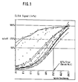
- the titration curves of syphilis-positive sera are linear over a wide titer range and are parallel to one another (FIG. 1).
- the ordinate shows only a measurement range of 50 to 100% signal reduction. More than 95% of the samples examined so far fall within this range.
- the qualitative version of the competitive ELISA enables, above all, but not exclusively, the testing of large sample quantities within approx. 3 hours. Due to the additional detection of specific IgM, even in the absence of specific IgG, very sensitive detection is possible even at very early stages of infection. In a special embodiment, ie after preincubation of the sample with the POD-labeled antibody and subsequent transfer into the measuring vessel, the detection of antigens is also possible.
- the quantitative version of the competitive ELISA provides important data in syphilis diagnostics for evaluating the stage of the disease or evaluating the success of antibiotic therapy (in addition to the IgM-specific tests). The good correlation with the TPHA must be emphasized here.
- Antigen preparations suitable for the solid phase can be obtained by methods known per se, e.g. B. Treponema pallidum according to WO 83/02886.
- the treponemes are obtained from the testicles of infected rabbits and extracted with the nonionic detergent n-octylglucoside.
- Suitable carrier materials for the solid phase are plastics such as polystyrene, polyvinyl chloride, polyamide and other synthetic polymers, natural polymers such as cellulose, and derivatized natural polymers such as cellulose acetate and nitrocellulose as well as glass, in particular as glass fiber.
- the supports can take the form of microparticles (0.01 to 1 »m), spheres, rods, tubes and microtest plates. Sheet-like structures such as striped paper, platelets and membranes are also suitable.
- the surface of the carrier can be permeable as well as impermeable to aqueous solutions.
- Preferred carriers are spheres, microparticles, tubes, strip papers and membranes. Microtest plates are particularly preferred supports.
- the solid phase is produced by irreversibly binding an antigen preparation to the carrier.
- the covalent bond is preferred in the case of the use of water-permeable carriers and the adsorptive bond in the case of the use of water-permeable as well as water-impermeable carriers.
- Direct adsorptive binding of antigen preparations to polystyrene treated with gamma rays is particularly preferred as the carrier.
- a pool serum from donors with positive serology for the respective pathogen serves as the starting material for the conjugate antibody.
- Monoclonal antibodies are also suitable.
- Radioactive isotopes, fluorescent and chemiluminescent dyes and enzymes can be used as markers, the detection of which is possible by means of chromogenic, luminogenic or fluorogenic substrate systems.
- the marking takes place according to methods which are described as state of the art for the markers mentioned.
- the method according to the invention can also be carried out using a diagnostic element which contains the solid phase and, in dry form, part or all of the necessary reagents.
- the method according to the invention is used for the detection and determination of pathogens.
- the sample and a solution of the labeled antibody are first incubated in an uncoated cavity of a microtest plate.
- the pre-incubated solution is transferred to a cavity coated with antigen. After a certain time of incubation, the solution is removed, the cavity is optionally washed with a buffer and the mark in the cavity is then measured.
- Treponema pallidum Spirochaeten are obtained from rabbit testicles that were infected with living pathogens 10 to 15 days beforehand.
- the treponemes are washed three times by centrifugation at 48,000 ⁇ g with 0.1 M phosphate buffer, pH 8.0, adjusted to a bacterial count of 109 / ml and, as described in WO 83/02886, extracted with the nonionic detergent n-octylglucoside.
- the antigen solution obtained in this way with a protein content of> 0.2 mg / ml is diluted in dilution levels of 1:50, 100, 150, 200, 300, 400, 600 and 800.
- test plate Divisible polystyrene microtitration plates from Nunc, (Roskilde, Denmark) are preferred as the test plate.
- a test plate with 100 »l per well is prepared for each dilution level.
- the test plates filled with the antigen solutions are left at 20.C for 18 h, then the solutions are suctioned off in the wells and the wells are thrice with tris / citric acid solution (0.05 M, pH 7.4) containing 0.2% bovine serum albumin. washed by filling and suction and then drying the test plates over silica gel at 20 ° C.
- a pool serum from donors with positive syphilis serology serves as the starting material for the conjugate antibody.
- T. pallidum western blot such sera show complex patterns with a large number of reactive bands.
- the serum is against buffer of 30 mM Na2HPO4 / Na2PO4, pH 7.2, dialyzed and placed on a column filled with DEAE cellulose DE 32 (Whatman) and equilibrated with the same buffer.
- the IgG fraction obtained in the run is dialyzed against PBS and adjusted to 10 mg / ml. It was verified that this method leads to no loss of certain antibody specificities and no significant decrease in titer.
- the conjugation of peroxidase is described in detail in EP-A-0 265 851.
- Serum, plasma or cerebrospinal fluid (25 »l each) was placed in wells of anti-T. pallidum test plates and incubated for 1 h at 37 ° C. The plates were taped up. Then 100 »l anti-T. pallidum / POD were added to each well and the test plate was incubated for a further hour at 37 ° C. The contents of the wells were removed by suction and the wells were washed four times with washing buffer. 100 »l TMB substrate preparation was added to each well, incubated for 30 min at 20-22 ° C and the incubation was ended by adding 100» l 1 N sulfuric acid. The OD 450 of the colored solution was measured against a blank of PBS.
- the test procedure corresponds to that described in Example 3.
- a high-titer pool serum (TPHA titer> 1: 10000) was diluted in the negative serum in stages 1: 1, 1: 4, 1:16, 1:64 and 1: 256. These diluted serum samples represent the expected titer range of native sera.
- the samples were then titrated in PBS, pH 7.2, in factor 3 dilution steps.
- the standard serum L3 was titrated over 8 levels (factor 3) and also (in duplicate) in the dilution levels 1:20 and 1: 500.
- the titration curves showed curves comparable to the curves shown in FIG. 1.
- the 50% titers of the 5 diluted serum samples were determined in accordance with FIG.
- the wells of a microtitration plate are filled with 100 »l of a solution of Toxoplasma gondii ultrasound antigen (from the RH strain) in 0.01 M bicarbonate buffer, pH 9.5 and left at room temperature for 20 h.
- the plates are washed and dried as described in Example 1.
- rabbits were immunized with the same antigen for 3 weeks using standard methods.
- the method of DE fractionation and GMBS conjugation to horseradish peroxidase is described in Example 2.
- 25 »l of the serum sample are initially placed in the test plate and 100» l of the conjugate antibody in PBS are added within 15 min. After a 1-hour incubation at 37 ° C., the test mixture is washed 4 times, as described in FIG. 3. After adding the TMB chromogen, the test is carried out and evaluated as described.
- the coating of the micro test plates and the test procedure correspond to the methodology described in 6.
- Rubella virus (RA27 / 3 strain) was grown in BHK cells, precipitated from the cell culture supernatant with polyethylene glycol (PEG) 6000 and a Tween® 80 / diethyl ether treatment according to Laufs and Thomssen (Arch.Ges.Virusforschung (1968) 24: 164-180).
- PEG polyethylene glycol
- Tween® 80 / diethyl ether treatment according to Laufs and Thomssen (Arch.Ges.Virusforschung (1968) 24: 164-180).
- the soluble antigen was dissolved in PBS, pH 7.2.
- a high-titer serum pool of more than 5 donors (ELISA-IgG titer each greater than 1: 10000) was fractionated according to the method described in EP 0 265 851 and the pure IgG fraction according to the GMBS Method coupled to horseradish peroxidase.
- care was taken to ensure that their reactivity to control antigen was as low as possible in the indirect IgG ELISA.
- a collective of 90 blood donor sera was tested in a comparative manner in the indirect IgG ELISA (serum dilution 1: 231; anti-human IgG from rabbits) and in the competitive ELISA analogously to the statements in example 6.
- conjugate titration is first carried out in the presence of a syphilis-negative donor serum.
- 25 »l of the serum sample are first incubated for 1 h at 37 ° C and then with 100» l of the anti-T. pallidum / POD conjugates in a dilution series of 1:50, 100, 150, 200, 300, 400, 600 and 800, (in Tris-HCl, 1.3 M, pH 7.2) and a further hour at 37 ° C incubated.
- PBS pH 7.2, containing 0.1% Tween® 20 »l TMB substrate transfer is then added to each well and incubated at 20 to 22 ° C for 30 min.
- the incubation is ended by adding 100 »10N sulfuric acid each.
- the absorbance of the solutions is measured against PBS at 450 nm.
- Those antigen / conjugate combinations which showed extinctions (OD 450) between 1.5 and 1.8 were selected for further optimization attempts.
- serum samples were tested with antigen / conjugate concentrations adjusted to one another, the content of T. pallidum-specific antibodies being known in conventional tests.
- the antigen / conjugate combination is selected as suitable in which the signal reduction correlates linearly with the logarithm of the titer. Optimal results are e.g. B.
- Fig. 1 Typical titration curves for the competitive syphilis ELISA (serum samples 1, 2 and 3 represent pure IgM fractions , taken up in negative serum).
- Fig. 2 Correlation of the signal reduction (in%) with the logarithm of the 50% titer (for 48 syphilis-positive serum samples).
- Fig. 3 Correlation of the competitive ELISA (Enzygnost-Sy philis) with the TPHA (Cellognost-Syphilis, H).
- Fig. 4 Frequency distribution (histogram) of syphilis positive ( ⁇ 60% signal of the negative control) and syphilis negative (> 70% signal of the negative control) serum samples over the entire measuring range.
- Fig. 6 Dependence of the signal reduction in the competitive ELISA (modified version) on the antigen concentration.
Landscapes
- Health & Medical Sciences (AREA)
- Life Sciences & Earth Sciences (AREA)
- Immunology (AREA)
- Engineering & Computer Science (AREA)
- Urology & Nephrology (AREA)
- Hematology (AREA)
- Biomedical Technology (AREA)
- Chemical & Material Sciences (AREA)
- Molecular Biology (AREA)
- Medicinal Chemistry (AREA)
- Biochemistry (AREA)
- Cell Biology (AREA)
- Pathology (AREA)
- Biotechnology (AREA)
- Food Science & Technology (AREA)
- Virology (AREA)
- Physics & Mathematics (AREA)
- Analytical Chemistry (AREA)
- Microbiology (AREA)
- General Health & Medical Sciences (AREA)
- General Physics & Mathematics (AREA)
- Tropical Medicine & Parasitology (AREA)
- Peptides Or Proteins (AREA)
- Medicines Containing Antibodies Or Antigens For Use As Internal Diagnostic Agents (AREA)
- Measuring Or Testing Involving Enzymes Or Micro-Organisms (AREA)
- Investigating Or Analysing Biological Materials (AREA)
Description
- Die Erfindung betrifft ein immunchemisches, kompetitives Verfahren zum Nachweis und zur Bestimmung von Antikörpern gegen Bakterien, Viren, Parasiten und Pilze. Mit einer Variante der beschriebenen Methode ist auch der Nachweis des Erregers in Körperflüssigkeiten möglich. Weiterhin werden Testbestecke zur Ausführung des Verfahrens und deren Anwendung beschrieben.
- Eine besondere Ausführungsform erlaubt den quantitativen Nachweis von Antikörpern, insbesondere bei Syphilis.
- In jüngster Zeit wurde die Bedeutung der Reaktivierung einer latenten Syphilis bei HIV-Patienten, welche häufig doppel-infiziert sind, erkannt. Besonders die Diagnose einer Neurosyphilis ist aufgrund der besonderen immunologischen Konstellation solcher Patienten schwierig. Die Nachweismethoden für Syphilis-Antikörper sind sehr vielfältig und reflektieren die frühzeitige diagnostische Beschäftigung (seit 1906) mit diesem Erreger, der noch Anfang dieses Jahrhunderts eine enorme gesundheitspolitische Bedeutung hatte.
Bei den Nachweismethoden für Syphilis-Antikörper können drei Kategorien unterschieden werden: - I. Nicht-Treponemen-spezifische Tests. Beispiele: Wassermann-Reaktion oder Cardiolipin-KBR, VDRL-Test oder Cardiolipin-Mikroflockungstest, RPR-Test: E.L. Reed, Public Health Lab. (1965) 23:96-103, VDRL-ELISA: N.S. Pedersen, O. Orum und S. Mouritsen, J. Clin.Microbiol. (1987) 25:1711-1716.
- II. Treponemen-spezifische Tests, z.B. die Reiter-KBR oder der sogenannte Flagellum-ELISA (R.V.W. Van Eijk ,H.E. Menke, G.J. Tidemann und E. Stolz, Genitourin.Med. (1988) 62:367-372).
- III. T. pallidum-spezifische Tests, z. B. der Nelson-Test (T.pallidum-Immobilisationstest), der "fluorescent treponemal antibody" (FTA)-Test (E.F. Hunter, W.E. Deacon und P.F. Meyer, Publ. Hlth.Rep. (1964) 79:410-412), der Treponema-pallidum-Hämagglutinationstest (TPHA: T. Tomizawa und S. Kasamatsu, Jap.J.med.Sci. Biol. (1966) 19:303-308).
Indirekte Enzymimunoassays zum IgG- bzw. IgM-Nachweis beruhen auf der Bindung spezifischer Antikörper an T.pallidum-Antigenpräparationen, welche (in der Regel adsorptiv) an Festphasen gebunden sind. Die Detektion der gebundenen Antikörper erfolgt über enzym-markierte Zweitantikörper, gerichtet gegen humanes IgG bzw. IgM. Die Methode des indirekten ELISA zum Syphilis-IgM-Nachweis ist in US-Patent Nr. 4, 288, 426 beschrieben. - Zusammenfassend läßt sich sagen, daß keiner der in der Literatur beschriebenen und auf dem Markt befindlichen Tests die Anforderungen an einen Screening-Test hinsichtlich des vertretbaren technischen Aufwandes, der objektiven und quantitativen Testbewertung, der weitgehend automatisierbaren Durchführung und des erforderlichen Höchstmaßes an Sensitivität und Spezifität erfüllt.
- Ein kompetitiver ELISA zum quantitativen Nachweis von Antikörpern gegen HIV ist in der europäischen Patentanmeldung EP-A-0 265 851 beschrieben.
- Eine spezielle Ausführungsform des kompetitiven ELISA zur Quantifizierung von Rubella-Virus in Zellkultur ist bei Y. Varela, E. Ortega und B. Gomez, J.Virol.Meth. (1988) 19:79-87 beschrieben. Diese Autoren verwenden allerdings keine direkt-markierten Rubella-spezifischen Antikörper. WPI 86-130689/20 beschreibt die Anwendung des kompetitiven Testprinzips zum Nachweis von Rubella-Antikörpern.
- Die der Erfindung zugrundeliegende Aufgabenstellung bestand darin, einen hochspezifischen und hochsensitiven Nachweis für Antikörper gegen Bakterien, Viren und Parasiten zu entwickeln. Dieser Test sollte es erlauben, kleine und große Serien von Tests sowohl manuell als auch weitgehend automatisiert, z.B. mit Hilfe des Behring-ELISA-Processor II durchzuführen. Als Screening-Test sollte er, unter Berücksichtigung der geringen Prävalenz der Erkrankungen, vor allem eine sichere positiv/negativ-Diskriminierung ermöglichen. Hierbei ist es wesentlich, in einem solchen Test mit hoher Sicherheit auch das besonders infektiöse frühe Primärstadium mit in der Regel geringem IgG-Titer zu erfassen. Andererseits mußte die Spezifität des zu entwickelnden Testes hoch sein, um die Rate von falschpositiven Ergebnissen, die nur mit aufwendigeren Tests verifiziert werden können, möglichst klein zu halten. Es wurde nun gefunden, daß ein kompetitives Verfahren zum Nachweis und zur Bestimmung von Antikörpern gegen die Erreger von Syphilis, Toxoplasmose, Röteln, Cytomegalie, Amoebiasis, Echinokokkosis, Tetanus und Keuchhusten sowohl der IgM- als auch der IgG-Klasse möglich ist, bei Verwendung einer Festphase, an die die Antigene, die auch bis zu einem gewissen Grad verunreinigt sein können, direkt, also ohne Antikörper-vermittelte Bindung gebunden sind, wenn ein entsprechend ausgewählter Konjugatantikörper verwendet wird.
In Experimenten mit reinen, IgG-freien IgM-Präparationen konnte gezeigt werden, daß auch spezifische Igm-Antikörper das Meßsignal in signifikanter Weise reduzieren (Fig. 1; Proben 1, 2, 3). - Gegenstand der Erfindung ist ein immunchemisches, kompetitives Verfahren zum Nachweis und zur Bestimmung von Antikörpern gegen die Erreger von Syphilis, Toxoplasmose, Röteln, Cytomegalie, Amoebiasis, Echinokokkosis, Tetanus und Keuchhusten unter Verwendung einer Festphase bestehend aus einem Träger und daran irreversibel gebundenen Membranproteinen der jeweiligen Erreger und markierten Antikörpern, gerichtet gegen die wesentlichen Antigene des jeweiligen Erregers, wobei die Antikörper um die Bindungstelle an der Festphase konkurrieren, dadurch gekennzeichnet, daß die Proteine irreversibel direkt oder über einen nicht immunchemisch bindenden Spacer an den Träger gebunden sind.
- Bevorzugt ist ein Verfahren zum Nachweis und zur Bestimmung von Antikörpern gegen die Erreger von Syphilis, Toxoplasmose und Röteln.
- Gegenstand der Erfindung ist außerdem ein Testbesteck zum Nachweis und zur Bestimmung von Antikörpern in einem immunchemischen, kompetitiven Verfahren wie oben beschrieben, enthaltend einen Träger, an dem Membranproteine des jeweiligen Erregers direkt oder über einen nicht immunchemisch bindenden Spacer gebunden sind, markierte Antikörper gegen die genannten Membranproteine und gegebenenfalls Reagenzien zum Nachweis der Markierung.
- Ferner ist Gegenstand der Erfindung die Verwendung des obengenannten Testbestecks in einem Verfahren zum Nachweis und zur Bestimmung von Antikörpern gegen die Erreger von Syphilis, Toxoplasmose, Röteln, Cytomegalie, Amoebiasis, Echinokokkosis, Tetanus und Keuchhusten.
- Gegenstand der Erfindung ist auch ein Verfahren wie oben beschrieben, worin die Signalreduktion linear mit dem Logarithmus des Titers korreliert.
- Gegenstand der Erfindung ist ferner ein Verfahren zum Nachweis und zur Bestimmung von Erregern von Syphilis, Toxoplasmose, Röteln, Cytomegalie, Amoebiasis, Echinokokkosis, Tetanus und Keuchhusten, wobei die Probe zunächst mit dem Konjugatantikörper in einem unbeschichteten Gefäß vorinkubiert wird und dann in das mit Antigenen des jeweiligen Erregers beschichtete Meßgefäß übertragen wird.
- Die Titrationskurven von Syphilis-positiven Seren verlaufen über einen weiten Titerbereich linear und sind zueinander parallel (Fig. 1).
- Diese Eigenschaft ist von Titrationskurven in indirekten ELISAs bekannt.
Tierische Hyperimmunsera zeigen eine vergleichbare Linearität und Parallelität und bieten sich als Basis für internationale Standards an. Zum quantitativen Vergleich des Antikörpergehaltes verschiedener Proben wurde z.B. der sogenannte 50%-Titer ermittelt, d.h. diejenige Serumverdünnung, bei welcher 50% des Signals der Negativ-Kontrolle erreicht werden (Fig. 1). Die Signalreduktion bei Testung der konzentrierten Serumprobe korreliert mit deren spezifischem Antikörpertiter.
Überraschenderweise zeigte es sich, daß bei Verwendung des erfindungsgemäßen Verfahrens die Signalreduktion (in % der Negativkontrolle) linear mit dem Logarithmus des Titers korreliert (Fig. 2). Diese Eigenschaft wird in einer speziellen Ausführungsform (siehe Beispiel 2) zur Quantifizierung der Syphilis-Antikörper herangezogen. Da nur solche Seren einen 50%-Titer aufweisen, die bei Testung der konzentrierten Proben das Meßsignal auf weniger als 50% der Negativkontrolle reduzieren, zeigt die Ordinate (Signalreduktion in % der Negativkontrolle) nur einen Meßbereich von 50 bis 100% Signalreduktion. In diesen Meßbereich fallen mehr als 95% der bisher untersuchten Proben. - Für Serumproben, die eine mehr als 90%-ige Signalreduktion aufweisen, gilt der lineare Zusammenhang zwischen der Signalreduktion und dem Logarithmus des Titers nicht mehr (siehe auch Fig. 1). Solche Proben müssen nach dem Screening-Test (mit unverdünntem Serum) nochmals nach Vorverdünnung (z.B. 1:20) nachgetestet werden.
Werden nun bei der quantitativen Ausführung der Technik mindestens 2 Standardseren (bzw. 2 Verdünnungsstufen eines Standardserums) mitgeführt, ist durch lineare Interpolation entsprechend Anlage 1 die Ermittlung des Titers bzw. des Antikörpergehaltes in internationalen Einheiten möglich. Diese lineare Interpolation kann auch rechnerisch, in der bevorzugten Ausführungsart der Auswertung auch mit Hilfe eines Computerprogrammes, durchgeführt werden. - Weiterhin wurde beobachtet, daß bei Titration von Serumproben und Testung nach der beschriebenen Methode die Titer des kompetitiven ELISA mit denjenigen des TPHA linear korrelieren (Fig. 3).
Die hohe Sensitivität des Testes wurde durch Prüfung einer großen Zahl klinisch definierter Serumproben von Patienten aus allen Stadien der Syphilis (Fig. 4) belegt. Die hohe Spezifität wird belegt durch den negativen Reaktionsausfall von Proben von Autoimmunpatienten, von Borreliose-Patienten und einer großen Zahl von gesunden Blutspendern (Fig. 4). - Überraschenderweise wurde weiterhin beobachtet, daß in einer besonderen Ausführungsform, nämlich durch Vorinkubation der Probe mit dem Konjugatantikörper in einem unbeschichteten Gefäß und anschließender Übertragung in das Gefäß, der Nachweis von Antigen möglich ist. Durch Versuche konnte verifiziert werden, daß losliche Bakterienäquivalente das Meßsignal in diesem Testansatz signifikant reduzieren (Fig. 5).
- Die qualitative Version des kompetitiven ELISAs ermöglicht durch ihre einfache technische Durchführung vor allem, aber nicht ausschließlich, die Austestung von großen Probenmengen innerhalb von ca. 3 Stunden.
Aufgrund der zusätzlichen Erfassung von spezifischen IgM, auch in Abwesenheit von spezifischem IgG, ist eine sehr sensitive Detektion auch sehr früher Infektionsstadien möglich. In einer besonderen Ausführungsform, d.h. nach Vorinkubation der Probe mit dem POD-markierten Antikörper und anschließender Übertragung in das Meßgefäß, ist auch der Nachweis von Antigenen möglich. Die quantitative Version des kompetitiven ELISA liefert z.B. bei der Syphilis-Diagnostik für die Bewertung des Stadiums der Erkrankung bzw. der Bewertung des Erfolges einer Antibiotika-Therapie (neben den IgM-spezifischen Tests) wichtige Daten. Hier muß die gute Korrelation zum TPHA betont werden.
Die Machbarkeit dieses Prinzips konnte weiterhin belegt werden für den Nachweis von Antikörpern gegen Toxoplasma gondii und gegen das Rubella-Virus. Besondere Vorteile werden in der Tatsache gesehen, daß durch Auswahl des Konjugatantikörpers die Spezifität des Testes verbessert und aus diesem Grunde auf eine aufwendige Hochreinigung von Antigenen unter Umständen verzichtet werden kann. - Für die Festphase geeignete Antigenpräparationen können nach an sich bekannten Verfahren gewonnen werden z. B. Treponema pallidum nach WO 83/02886. Dabei werden die Treponemen aus den Hoden von infizierten Kaninchen gewonnen und mit dem nichtionischen Detergenz n-Octylglucosid extrahiert.
- Als Trägermaterial für die Festphase sind Kunststoffe wie Polystyrol, Polyvinylchlorid, Polyamid und andere synthetische Polymere, natürliche Polymere wie Zellulose, sowie derivatisierte natürliche Polymere wie Zelluloseacetat und Nitrozellulose als auch Glas, insbesondere als Glasfaser geeignet.
- Die Träger können die Form von Micropartikeln (0.01 bis 1 »m), Kugeln, Stäbchen, Röhrchen und Microtestplatten haben. Flächenförmige Gebilde wie Streifenpapiere, Plättchen und Membranen sind ebenfalls geeignet. Die Oberfläche der Träger kann sowohl für wässrige Lösungen durchlässig als auch undurchlässig sein.
- Bevorzugte Träger sind Kugeln, Micropartikel, Röhrchen, Streifenpapiere und Membranen. Besonders bevorzugte Träger sind Microtestplatten.
- Die Festphase wird hergestellt, indem eine Antigenpräparation irreversibel an den Träger gebunden wird.
- Eine irreversible Bindung im Sinne der Erfindung liegt beispielsweise vor bei
- 1) einer adsorptiven Bindung, die nicht durch immunchemische Mittel, wie hochaffine Antikörper und auch nicht durch die in dem Verfahren verwendeten Mittel, wie markierte Antikörper, Verdunnungs- und Pufferlösungen gespalten wird,
- 2) einer über einen nicht immunchemisch bindenden Spacer vermittelte, bioaffine Bindung, wobei der Spacer aus Biotin und Avidin oder aus anderen Paarungen von Rezeptoren und Liganden bestehen kann,
- 3) einer kovalenten direkten Bindung
- 4) einer kovalenten, durch einen chemisch bifunktionellen Spacer vermittelten Bindung.
- Bevorzugt wird die kovalente Bindung im Falle der Verwendung von wasserdurchlässigen Trägern sowie die adsorptive Bindung im Falle der Verwendung von wasserdurchlässigen als auch wasserundurchlässigen Trägern.
- Besonders bevorzugt wird die direkte adsorptive Bindung von Antigenpräparationen an mit Gammastrahlen behandeltes Polystyrol als Träger.
- Als Ausgangsmaterial für den Konjugat-Antikörper dient ein Poolserum von Spendern mit für den jeweiligen Erreger positiver Serologie. Monoklonale Antikörper sind ebenfalls geeignet.
- Als Marker können verwendet werden radioaktive Isotope, fluoreszierende und chemilumineszierende Farbstoffe sowie Enzyme, deren Nachweis durch chromogene, luminogene oder fluorogene Substratsysteme möglich ist.
- Die Markierung erfolgt nach Verfahren, die für die genannten Marker als Stand der Technik beschrieben sind.
- Im Falle der Markierung der Antikörper mit Peroxidase kann die Periodat Technik nach Nakane et al., 1974, J. Histochem.Cytochem. 22, 1084-1090, angewandt werden oder ein Verfahren gemäß Ishikawa et al., 1983, J. Immunoassay 4, 209-327, in dem die Partner mit einem heterobifunktionellen Reagenz verknüpft werden.
- Das erfindungsgemäße Verfahren kann beispielsweise durchgeführt werden, indem in die Kavitäten (Wells) einer Microtestplatte, die eine zuvor beschriebene Antigenpräparation gebunden enthält,
- a) gleichzeitig die Probe und eine Lösung des markierten Antikörpers gegebenenfalls zuerst die Probe und nach einer gewissen Zeit eine Lösung des markierten Antikörpers gegeben, nach einer bestimmten Zeit der Inkubation die Lösung entfernt und die Wells mit einem Puffer gewaschen werden und anschließend die Markierung in den Wells gemessen wird,
oder - b) zuerst eine Lösung enthaltend den markierten Antikörper gegeben wird, nach einer bestimmten Zeit der Inkubation die Lösung entfernt und die Wells mit einem Puffer gewaschen, die Microtestplatte gegebenenfalls getrocknet, anschließend die verdünnte Probe zugegeben und eine bestimmt Zeit inkubiert, dann die Probe aus den Wells entfernt und die Markierung in der Probe gemessen wird.
- Das erfindungsgemäße Verfahren kann auch durchgeführt werden bei Verwendung eines diagnostischen Elements, das die Festphase und in trockener Form einen Teil oder auch alle erforderlichen Reagenzien enthält.
- In einer bevorzugten Ausführung wird das erfindungsgemäße Verfahren zum Nachweis und zur Bestimmung von Erregern eingesetzt. Dabei wird beispielsweise zunächst in einer unbeschichteten Kavität einer Microtestplatte die Probe und eine Lösung des markierten Antikörpers inkubiert.
- Nach einer bestimmten Zeit wird die vorinkubierte Lösung in eine mit Antigen beschichtete Kavität übertragen. Nach einer bestimmten Zeit der Inkubation wird die Lösung entfernt, die Kavität gegebenenfalls mit einem Puffer gewaschen und anschließend die Markierung in der Kavität gemessen.
- Die folgenden Beispiele sollen die Erfindung erläutern und stellen keine Einschränkung auf die in den Beispielen genannten Ausführungsformen dar.
- Treponema pallidum Spirochaeten werden aus Kaninchenhoden gewonnen, welche 10 bis 15 Tage vorher mit lebenden Erregern infiziert wurden. Die Treponemen werden durch Zentrifugation bei 48.000 x g mit 0,1 M Phosphatpuffer, pH 8,0 dreimal gewaschen, auf eine Keimzahl von 10⁹/ml eingestellt und wie in WO 83/02886 beschrieben, mit dem nichtionischen Detergenz n-Octylglucosid extrahiert.
Die solchermaßen gewonnene Antigenlösung mit einem Proteingehalt von > 0,2 mg/ml wird in Verdünnungsstufen von 1:50, 100, 150, 200, 300, 400, 600 und 800 verdünnt. Als Testplatte werden teilbare Polystyrol-Mikrotitrationsplatten der Fa. Nunc, (Roskilde, Dänemark) bevorzugt. Je Verdünnungsstufe wird eine Testplatte mit je 100 »l pro Vertiefung angelegt.
Die mit den Antigenlösungen gefüllten Testplatten werden 18 h bei 20.C belassen, dann werden die Lösungen in den Vertiefungen abgesaugt und die Näpfe dreimal mit Tris/Citronensäurelösung (0,05 M, pH 7,4), enthaltend 0,2% Rinderserumalbumin, durch Füllen und Absaugen gewaschen und die Testplatten anschliessend über Kieselgel bei 20°C getrocknet. - Als Ausgangsmaterial für den Konjugat-Antikörper dient ein Poolserum von Spendern mit positiver Syphilis-Serologie. Ein Mindesttiter im TPHA von > 1:10.000 und positive VDRL- und Cardiolipin-KBR-Titer werden gefordert. Im T.pallidum-Western-Blot zeigen solche Sera komplexe Muster mit einer Vielzahl von reaktiven Banden.
Das Serum wird gegen Puffer von 30 mM Na₂HPO₄/Na₂PO₄, pH 7,2, dialysiert und auf eine Säule, gefüllt mit DEAE-Cellulose DE 32 (Whatman) und equilibriert mit dem gleichen Puffer, gegeben. Die im Durchlauf erhaltene IgG-Fraktion wird gegen PBS dialysiert und auf 10 mg/ml eingestellt. Es wurde verifiziert, daß diese Methode zu keinem Verlust von bestimmten Antikörper-Spezifitäten und zu keinem signifikanten Titerrückgang führt. Die Konjugation der Peroxidase ist ausfuhrlich in der EP-A-0 265 851 beschrieben. - Serum, Plasma oder Liquor (je 25 »l) wurden in Wells von Anti- T.pallidum-Testplatten gegeben und 1 h bei 37°C inkubiert. Die Platten waren dabei abgeklebt. Anschließend wurden pro Vertiefung 100 »l Anti-T.pallidum/POD zugegeben und die Testplatte eine weitere Stunde bei 37°C inkubiert. Der inhalt der Vertiefungen wurde durch Absaugen entfernt und die Wells viermal mit Waschpuffer gewaschen.
Es wurden in jede Vertiefung 100 »l TMB-Substratzubereitung gegeben, 30 min bei 20 - 22°C inkubiert und die Inkubation durch Zugabe von 100 »l 1 N Schwefelsaure beendet. Die OD 450 der gefarbten Lösung wurde gegen einen Leerwert von PBS gemessen.
Als Anti-T.pallidum-positiv wurden solche Proben eingestuft, die einen OD-Wert von weniger als 60% des Wertes des negativen Kontroll-serums zeigten, als Anti-T.pallidum-grenzwertige Proben, deren OD 450-Werte im Bereich von 60 - 70% der Negativ-Kontrolle lagen und als Anti-T.pallidum-negativ Proben, die eine OD 450 von über 70% der Negativ-Proben zeigten.
Fig. 4 zeigt die Häufigkeitsverteilung (Histogramm) Von 128 Syphilis-positiven Proben, von 80 Serumproben von Autoimmunpatienten, von 53 Proben von Borreliose-Patienten und von 71 Proben klinisch gesunder Blutspender. - Aus der Abbildung ist ersichtlich, daß das Positiv-Kollektiv von dem Negativ-Kollektiv deutlich getrennt ist, wobei je eines der Seren der Autoimmunpatienten und von Normalspendern in den positiven Bereich fällt. Andererseits konnten alle Proben, die in diesem Test positiv anzeigten, durch Vergleichstests in anderen Techniken bestätigt werden.
- Die Testdurchführung entspricht derjenigen, die in Beispiel 3 beschrieben wurde. Zunächst wurde ein hochtitriges Poolserum (TPHA-Titer > 1:10000) im negativen Serum in den Stufen 1:1, 1:4, 1:16, 1:64 und 1:256 verdünnt. Diese verdünnten Serumproben repräsentieren den zu erwartenden Titerbereich nativer Seren. Die Proben wurden nun jeweils in PBS, pH 7,2, in Faktor-3-Verdünnungsstufen titriert.
Im gleichen Testansatz wurde das Standardserum L3 über 8 Stufen titriert (Faktor 3) und außerdem (in Doppelbestimmung) in den Verdünnungsstufen 1:20 und 1:500 mitgeführt. Die Titrationskurven zeigten den in Fig. 1 dargestellten Kurven vergleichbare Verläufe.
Zunächst wurden die 50%-Titer der 5 verdünnten Serumproben entsprechend Fig. 1 bestimmt und auf den 50%-Titer des Standardserums L3 (Sollwert 40 IU) bezogen. Schließlich wurde der Gehalt (in IU) der verdünnten Serumproben bei jeder Verdünnungsstufe rechnerisch ermittelt (Anlage 1) und gegen die graphisch (nach der 50%-Methode) ermittelten Werte aufgetragen. - Bei diesem Testansatz wurde anstelle einer Serumprobe 60 »l einer Antigenlösung, entsprechend der Beschreibung in Abschnitt 4, in PBS verdünnt (ab 10⁹ Bakterienäquivalente pro ml,Verdünnungsfaktor 3) und mit der als optimal ermittelten Konjugatverdünnung (60 »l) in einer unbeschichteten Mikrotiterplatte für 1 h bei 37°C inkubiert. Nach Übertragung von 100 »l dieses Ansatzes in die Testplatte wurde für eine weitere Stunde bei 37°C inkubiert und nach 4-maligem Waschen wurde der Test durch Substratzugabe und Auswertung, wie unter Beispiel 3 beschrieben, ausgeführt. Fig. 5 zeigt, daß mit dieser Methode Keimzahlen von mehr als 10⁶ bis 10⁷ Bakterienäquivalenten pro ml, entsprechend 0,1 - 1 »g/ml nachgewiesen werden können.
- Die Kavitäten einer Mikrotitrationsplatte werden mit je 100 »l einer Lösung von Toxoplasma gondii-Ultraschall-Antigen (vom RH-Stamm) in 0,01 M Bicarbonat-Puffer, pH 9,5 gefüllt und 20 h bei Raumtemperatur belassen. Das Waschen und Trocknen der Platten erfolgt wie in Beispiel 1 beschrieben.
Zur Gewinnung eines hochtitrigen Antiserums wurden Kaninchen nach Standardmethoden über 3 Wochen mit dem gleichen Antigen immunisiert. Die Methode der DE-Fraktionierung und der GMBS-Konjugation an Meerettich-Peroxidase ist in Beispiel 2 beschrieben.
Bei der Testdurchführung des kompetitiven ELISA werden zunächst je 25 »l der Serumprobe in die Testplatte vorgelegt und innerhalb von 15 min werden je 100 »l des Konjugatantikörpers in PBS zugefügt. Nach einer 1-stündigen Inkubation bei 37°C wird der Testansatz, wie in 3 beschrieben, 4-mal gewaschen. Nach Zugabe des Chromogens TMB wird der Test, wie beschrieben, durchgeführt und ausgewertet. - Zum Vergleich wurde ein Kollektiv von 80 Blutspendersera auch im indirekten IgG-ELISA, unter Verwendung eines Anti-Human-IgG/POD-Konjugates vom Kaninchen, in einer Serumverdünnung von 1:231 getestet (Enzygnost-Toxoplasmosis/IgG). Fig. 6 zeigt eine sehr gute Übereinstimmung der Methoden, wobei der kompetitive ELISA eine bessere Positiv/Negativ-Diskriminierung und eine technisch einfachere Durchführung bietet. Die vergleichbare Sensitivität ist aus dem Reaktionsausfall der Titrationsreihe eines positiven Standardserums (siehe Fig. 6) ersichtlich.
- Die Beschichtung der Mikrotestplatten und die Testdurchführung entspricht der in 6. beschriebenen Methodik.
Rubella-Virus (Stamm RA27/3) wurde in BHK-Zellen gezüchtet, aus dem Zellkulturüberstand mit Polyethylenglykol (PEG) 6000 präzipitiert und einer Tween® 80/Diethyläther-Behandlung nach Laufs und Thomssen (Arch.Ges.Virusforschung (1968) 24:164-180) unterzogen. Für die Plattenbeschichtung wurde das lösliche Antigen in PBS, pH 7,2, gelöst.
Für die Herstellung des Antikörper-POD-Konjugates wurde ein hochtitriger Serumpool von mehr als 5 Spendern (ELISA-IgG-Titer jeweils größer als 1:10000) nach der in EP 0 265 851 beschriebenen Methode fraktioniert und die reine IgG-Fraktion nach der GMBS-Methode an Meerettich-Peroxidase gekoppelt.
Bei der Auswahl der Sera wurde darauf geachtet, daß deren Reaktivität auf Kontrollantigen im indirekten IgG-ELISA möglichst niedrig war.
Ein Kollektiv von 90 Blutspendersera wurde vergleichend im indirekten IgG-ELISA (Serumverdünnung 1:231; Anti-Human-IgG vom Kaninchen) und im kompetitiven ELISA analog den Ausführungen in Beispiel 6. getestet. - Auf einzelnen Riegeln einer jeden Antigenverdünnungsstufe wird zunächst eine Konjugattitration in Gegenwart eines Syphilis-negativen Spenderserums durchgeführt. Hierzu werden zunächst je 25 »l der Serumprobe für 1 h bei 37° C inkubiert und anschließend mit je 100 »l des Anti-T. pallidum/POD-konjugates in einer Verdünnungsreihe von 1:50, 100, 150, 200, 300, 400, 600 und 800, (in Tris-HCl, 1,3 M, pH 7,2) versetzt und eine weitere Stunde bei 37° C inkubiert.
Nach 4-maligem Waschen mit PBS, pH 7,2, enthaltend 0,1 % Tween® 20 wird anschließend in jede Vertiefung 100 »l TMB Substratzübereitung gegeben und 30 min bei 20 bis 22° C inkubiert. Die Inkubation wird beendet durch Zugabe von jeweils 100 »l 0 N Schwefelsäure. Die Extinktion der Lösungen wird gegen PBS bei 450 nm gemessen. Es werden diejenigen Antigen-/Konjugatkombinationen für weitere Optimierungsversuche ausgewahlt, welche Extinktionen (OD 450) zwischen 1,5 und 1,8 zeigten.
In einem weiteren Testansatz wurden bei jeweils aufeinander eingestellten Antigen-/Konjugatkonzentrationen Serumproben getestet, deren Gehalt an T.pallidum-spezifischen Antikörpern in konventionellen Tests bekannt war.
Es wird diejenige Antigen-/Konjugatkombination als geeignet ausgewählt, bei der die Signalreduktion linear mit dem Logarithmus des Titers korreliert. Optimale Ergebnisse werden z. B. bei einer Antigenverdünnung von 1:300 (entspricht etwa 0,7 »g/ ml) und einer Konjugatverdünnung von 1:200 erzielt. Da bei der Bestimmung von Anti-T.pallidum eine Grenzwertfestlegung von OD 450 > 70 % der Extinktion der Negativ-Kontrolle für Negative und von OD 450 < 70 % für Syphilis-positive als günstig angesehen wird, wird für die Herstellung von Microtestplatten zur Bestimmung von Anti-T.pallidum bei der Behandlung der Wells, die auch als Beschichtung bezeichnet wird, die Konzentration von ca. 1 »g/ml gewählt. - Fig. 1: Für den kompetitiven Syphilis-ELISA typische Titrationskurven (Serumproben 1, 2 und 3 stellen reine IgM-Fraktionen, in negativem Serum aufgenommen, dar).
- Fig. 2: Korrelation der Signalreduktion (in %) mit dem Logarithmus des 50%-Titers (für 48 Syphilis-positive Serumproben).
- Fig. 3: Korrelation des kompetitiven ELISA (Enzygnost-Sy philis) mit dem TPHA (Cellognost-Syphilis, H).
- Fig. 4: Häufigkeitsverteilung (Histogramm) von Syphilispositiven (< 60% Signal der Negativ-Kontrolle) und Syphilis-negativen (> 70% Signal der Negativ-Kontrolle) Serumproben über den gesamten Meßbereich.
- Fig. 5: Korrelation der durch lineare Interpolation errechneten mit den graphisch ermittelten Titern (in IU). Dargestellt sind die Meßwerte für 5 unterschiedlich starke Serumproben in Abhängigkeit von der Probenverdünnung.
- Fig. 6: Abhängigkeit der Signalreduktion im kompetitiven ELISA (modifizierte Version) von der Antigenkonzentration.
- Fig. 7: Vergleich der Meßwerte des indirekten (IgG)-ELISA (Enzygnost-Toxo-plasmosis/IgG) und des kompetitiven Toxoplasma-Antikörper-ELISA (80 Sera gesunder, asymptomatischer Blutspender). Die Abbildung zeigt, daß der kompetitive ELISA eine deutlichere Positiv-/Negativ-Diskriminierung erlaubt als der indirekte ELISA.
Claims (8)
- Immunchemisches kompetitives Verfahren zum Nachweis und zur Bestimmung von Antikörpern gegen die Erreger von Syphilis, Toxoplasmose, Röteln, Cytomegalie, Amoebiasis, Echinokokkosis, Tetanus und Keuchhusten unter Verwendung einer Festphase bestehend aus einem Träger und daran irreversibel gebundenen Membranproteinen der jeweiligen Erreger und markierten Antikörpern, gerichtet gegen die wesentlichen Antigene des jeweiligen Erregers, wobei die Antikörper um die Bindungsstellen an der Festphase konkurrieren, dadurch gekennzeichnet, daß die Proteine irreversibel direkt oder über einen nicht immunchemisch bindenden Spacer an den Träger gebunden sind.
- Verfahren nach Anspruch 1, dadurch gekennzeichnet, daß die Erreger die Erreger von Syphilis, Toxoplasmose und Röteln sind.
- Verfahren nach Anspruch 1, dadurch gekennzeichnet, daß die Antigen-/Antikörperkonjugatkombination so eingestellt ist, daß die Signalreduktion linear mit dem Logarithmus des Titers korreliert.
- Verfahren nach Anspruch 3, dadurch gekennzeichnet, daß die Auswertung rechnerisch, insbesondere mit Hilfe eines entsprechenden Computerprogrammes erfolgt.
- Testbesteck zum Nachweis und zur Bestimmung von Antikörpern in einem immunchemischen kompetitiven Verfahren gemäß Anspruch 1, enthaltend einen Träger, an dem Membranproteine des jeweiligen Erregers direkt oder über einen nicht immunchemisch bindenden Spacer gebunden sind, markierte Antikörper gegen die genannten Membranproteine und gegebenenfalls Reagenzien zum Nachweis der Markierung.
- Testbesteck nach Anspruch 5, dadurch gekennzeichnet, daß es aus einem Element besteht, in dem die Festphase und in trockener Form die für den Nachweis und die Bestimmung erforderlichen Reagenzien ganz oder teilweise enthalten sind.
- Verwendung eines Testbestecks nach mindestens einem der Ansprüche 5 und 6 in einem Verfahren zum Nachweis und zur Bestimmung von Antikörpern gegen die Erreger von Syphilis, Toxoplasmose, Röteln, Cytomegalie, Amoebiasis, Echinokokkosis, Tetanus, und Keuchhusten.
- Immunchemisches, kompetitives Verfahren zum Nachweis und zur Bestimmung von Erregern von Syphilis, Toxoplasmose, Röteln, Cytomegalie, Amoebiasis, Echinokokkosis, Tetanus und Keuchhusten unter Verwendung einer Festphase bestehend aus einem Träger und daran irreversibel gebundenen Antigenen der jeweiligen Erreger und markierten Antikörper, gerichtet gegen die wesentlichen Antigene des jeweiligen Erregers, wobei zunächst außerhalb des Testgefäßes die Probe mit den markierten Antikörpern vorinkubiert wird und dann zu der Festphase hinzugegeben wird, wobei die Antikörper um die Bindungsstelle an der Festphase konkurrieren, dadurch gekennzeichnet, daß die Proteine irreversibel direkt oder über einen nicht immunchemisch bindenden Spacer an den Träger gebunden sind.
Applications Claiming Priority (2)
| Application Number | Priority Date | Filing Date | Title |
|---|---|---|---|
| DE3911361A DE3911361A1 (de) | 1989-04-07 | 1989-04-07 | Verfahren zur bestimmung von antikoerpern gegen erreger von infektionskrankheiten in koerperfluessigkeiten, mittel dazu und ihre verwendung in diesem verfahren |
| DE3911361 | 1989-04-07 |
Publications (3)
| Publication Number | Publication Date |
|---|---|
| EP0391330A2 EP0391330A2 (de) | 1990-10-10 |
| EP0391330A3 EP0391330A3 (de) | 1992-02-12 |
| EP0391330B1 true EP0391330B1 (de) | 1995-03-01 |
Family
ID=6378130
Family Applications (1)
| Application Number | Title | Priority Date | Filing Date |
|---|---|---|---|
| EP90106330A Expired - Lifetime EP0391330B1 (de) | 1989-04-07 | 1990-04-03 | Verfahren zur Bestimmung von Antikörpern gegen Erreger von Infektionskrankheiten in Körperflüssigkeiten, Mittel dazu und ihre Verwendung in diesem Verfahren |
Country Status (7)
| Country | Link |
|---|---|
| EP (1) | EP0391330B1 (de) |
| JP (1) | JPH02287258A (de) |
| AT (1) | ATE119291T1 (de) |
| AU (1) | AU645970B2 (de) |
| CA (1) | CA2014067A1 (de) |
| DE (2) | DE3911361A1 (de) |
| ES (1) | ES2070201T3 (de) |
Families Citing this family (2)
| Publication number | Priority date | Publication date | Assignee | Title |
|---|---|---|---|---|
| DE19536166C1 (de) * | 1995-09-29 | 1997-03-06 | Siegfried Dr Krell | Verfahren zur Bestimmung von Antikörpern gegen Treponema pallidum (Syphilis) |
| CN104360067B (zh) * | 2014-11-10 | 2016-11-16 | 厦门大学附属中山医院 | 梅毒螺旋体特异性抗体定量检测试剂盒及其制备方法 |
Family Cites Families (5)
| Publication number | Priority date | Publication date | Assignee | Title |
|---|---|---|---|---|
| EP0136798A3 (de) * | 1983-08-25 | 1986-02-19 | Biotech Research Laboratories Inc. | Titrierungsplatte und Testsatz zum Nachweis von Antikörpern in menschlichem Serum und ihre Herstellung und Verwendung |
| AT386079B (de) * | 1986-02-21 | 1988-06-27 | Waldheim Pharmazeutika Gmbh | Verfahren zum nachweis von antikoerpern, und mittel zum nachweis von antikoerpern und verfahren zur herstellung des mittels |
| EP0264866A3 (de) * | 1986-10-24 | 1990-12-27 | BEHRINGWERKE Aktiengesellschaft | Verfahren zur Bestimmung des Vorhandenseins von diagnostisch relevanten Substanzen, insbesondere Antikörpern oder Antigenen nach der ELISA-Methode durch photometrische Auswertung |
| DE3636540A1 (de) * | 1986-10-27 | 1988-04-28 | Behringwerke Ag | Verfahren zur bestimmung von anti-hiv, mittel dazu und ihre verwendung in diesem verfahren |
| IL82873A0 (en) * | 1987-06-15 | 1987-12-20 | Orgenics Ltd | Reversed competitive solid phase immunoassay for detecting single epitope analytes and kit therefor |
-
1989
- 1989-04-07 DE DE3911361A patent/DE3911361A1/de not_active Withdrawn
-
1990
- 1990-04-03 DE DE59008558T patent/DE59008558D1/de not_active Expired - Fee Related
- 1990-04-03 ES ES90106330T patent/ES2070201T3/es not_active Expired - Lifetime
- 1990-04-03 EP EP90106330A patent/EP0391330B1/de not_active Expired - Lifetime
- 1990-04-03 AT AT90106330T patent/ATE119291T1/de not_active IP Right Cessation
- 1990-04-06 JP JP2090492A patent/JPH02287258A/ja active Pending
- 1990-04-06 CA CA002014067A patent/CA2014067A1/en not_active Abandoned
- 1990-04-09 AU AU52991/90A patent/AU645970B2/en not_active Ceased
Also Published As
| Publication number | Publication date |
|---|---|
| AU5299190A (en) | 1990-10-11 |
| AU645970B2 (en) | 1994-02-03 |
| DE3911361A1 (de) | 1990-10-11 |
| ATE119291T1 (de) | 1995-03-15 |
| EP0391330A2 (de) | 1990-10-10 |
| EP0391330A3 (de) | 1992-02-12 |
| JPH02287258A (ja) | 1990-11-27 |
| DE59008558D1 (de) | 1995-04-06 |
| ES2070201T3 (es) | 1995-06-01 |
| CA2014067A1 (en) | 1990-10-07 |
Similar Documents
| Publication | Publication Date | Title |
|---|---|---|
| DE2851150C2 (de) | Verfahren zur immunologischen Untersuchung einer Serumprobe | |
| EP0944838B1 (de) | ANTIGENSPEZIFISCHER IgG-NACHWEIS | |
| EP0023989B1 (de) | Enzym-Immuntest zum Nachweis von Erreger-spezifischen Antikörpern und Testsatz für die Durchführung dieses Tests | |
| EP0363942B1 (de) | Verfahren zur Bestimmung einer spezifisch bindefähigen Substanz | |
| EP0944832B1 (de) | ANTIGENSPEZIFISCHER IgM-NACHWEIS | |
| DE68924878T2 (de) | Bestimmung eines felinen pathogen-specifischen Antikörpers und eines felinen pathogenen Antigens in einem Immuntest. | |
| EP0292810B1 (de) | Einschritt-Immuntest zur Bestimmung von antigen-spezifischen Antikörpern aus einer der Immunglobulin-Klassen A, M, D oder E und dazu geeignetes Mittel | |
| DE3882705T2 (de) | Immunoglobulin-bestimmungsverfahren. | |
| EP0544869B2 (de) | Immunologisches bestimmungsverfahren zur bestimmung von antikörpern in biologischen flüssigkeiten sowie kit zur durchführung des verfahrens | |
| DE68919828T2 (de) | Nanoteilchen mit gebundenen enzym und ligand zur verwendung bei analysen. | |
| DE3303793A1 (de) | Immunoassay fuer klassenspezifische immunoglobulinantikoerper | |
| EP0418635A1 (de) | Enzymimmunometrisches Bestimmungsverfahren unter Verwendung von Peroxidase als Markierungsenzym | |
| EP0265851B1 (de) | Verfahren zur Bestimmung von Anti-HIV, Mittel dazu und ihre Verwendung in diesem Verfahren | |
| WO2004040310A1 (de) | Mittel und verfahren zur diagnose einer treponemainfektion | |
| DE19629444A1 (de) | Sensitivitätssteigerung bei der immunchemischen Bestimmung eines Analyten | |
| EP0572845B1 (de) | Verfahren zur immunchemischen Bestimmung eines Analyten | |
| DE69606024T2 (de) | Feststellung von antikörperproduktion | |
| DE69612099T2 (de) | Elisa-serodiagnose von schweinpleuropneumonie der serotypen 1,9 und 11 | |
| DE19500862A1 (de) | Reaktionssäulen für simultane Mehrfachmessung und Verfahren zur Bestimmung von Verbindungen | |
| EP0391330A2 (de) | Verfahren zur Bestimmung von Antikörpern gegen Erreger von Infektionskrankheiten in Körperflüssigkeiten, Mittel dazu und ihre Verwendung in diesem Verfahren | |
| DE69119445T2 (de) | Analysemethode für infektiöse Pferdeanämie-Viren (EIA-Virus) | |
| DE2907228A1 (de) | Immunochemische bestimmung | |
| EP0781998B1 (de) | Immunologische Bestimmungsmethode | |
| DE69929468T2 (de) | VERBESSERTE SPEZIFITÄT IN DER DETEKTION DER ANTI-RUBELLA IgM ANTIKÖRPER | |
| DE3854603T2 (de) | Teststreifen für diagnose durch multi-immunoassay system. |
Legal Events
| Date | Code | Title | Description |
|---|---|---|---|
| PUAI | Public reference made under article 153(3) epc to a published international application that has entered the european phase |
Free format text: ORIGINAL CODE: 0009012 |
|
| AK | Designated contracting states |
Kind code of ref document: A2 Designated state(s): AT BE CH DE ES FR GB IT LI LU NL SE |
|
| 17P | Request for examination filed |
Effective date: 19901221 |
|
| PUAL | Search report despatched |
Free format text: ORIGINAL CODE: 0009013 |
|
| AK | Designated contracting states |
Kind code of ref document: A3 Designated state(s): AT BE CH DE ES FR GB IT LI LU NL SE |
|
| 17Q | First examination report despatched |
Effective date: 19940216 |
|
| GRAA | (expected) grant |
Free format text: ORIGINAL CODE: 0009210 |
|
| AK | Designated contracting states |
Kind code of ref document: B1 Designated state(s): AT BE CH DE ES FR GB IT LI LU NL SE |
|
| REF | Corresponds to: |
Ref document number: 119291 Country of ref document: AT Date of ref document: 19950315 Kind code of ref document: T |
|
| REF | Corresponds to: |
Ref document number: 59008558 Country of ref document: DE Date of ref document: 19950406 |
|
| ITF | It: translation for a ep patent filed | ||
| ET | Fr: translation filed | ||
| REG | Reference to a national code |
Ref country code: ES Ref legal event code: FG2A Ref document number: 2070201 Country of ref document: ES Kind code of ref document: T3 |
|
| GBT | Gb: translation of ep patent filed (gb section 77(6)(a)/1977) |
Effective date: 19950511 |
|
| PLBE | No opposition filed within time limit |
Free format text: ORIGINAL CODE: 0009261 |
|
| STAA | Information on the status of an ep patent application or granted ep patent |
Free format text: STATUS: NO OPPOSITION FILED WITHIN TIME LIMIT |
|
| 26N | No opposition filed | ||
| PGFP | Annual fee paid to national office [announced via postgrant information from national office to epo] |
Ref country code: CH Payment date: 19970326 Year of fee payment: 8 |
|
| REG | Reference to a national code |
Ref country code: CH Ref legal event code: PUE Owner name: BEHRINGWERKE AKTIENGESELLSCHAFT TRANSFER- BEHRING |
|
| PG25 | Lapsed in a contracting state [announced via postgrant information from national office to epo] |
Ref country code: LI Free format text: LAPSE BECAUSE OF NON-PAYMENT OF DUE FEES Effective date: 19980430 Ref country code: CH Free format text: LAPSE BECAUSE OF NON-PAYMENT OF DUE FEES Effective date: 19980430 |
|
| REG | Reference to a national code |
Ref country code: CH Ref legal event code: PFA Free format text: BEHRING DIAGNOSTICS GMBH TRANSFER- DADE BEHRING MARBURG GMBH |
|
| REG | Reference to a national code |
Ref country code: CH Ref legal event code: PL |
|
| NLS | Nl: assignments of ep-patents |
Owner name: BEHRING DIAGNOSTICS GMBH |
|
| NLT1 | Nl: modifications of names registered in virtue of documents presented to the patent office pursuant to art. 16 a, paragraph 1 |
Owner name: DADE BEHRING MARBURG GMBH |
|
| REG | Reference to a national code |
Ref country code: ES Ref legal event code: PC2A |
|
| REG | Reference to a national code |
Ref country code: FR Ref legal event code: TP |
|
| REG | Reference to a national code |
Ref country code: FR Ref legal event code: CD |
|
| PGFP | Annual fee paid to national office [announced via postgrant information from national office to epo] |
Ref country code: FR Payment date: 20000317 Year of fee payment: 11 |
|
| PGFP | Annual fee paid to national office [announced via postgrant information from national office to epo] |
Ref country code: SE Payment date: 20000320 Year of fee payment: 11 Ref country code: NL Payment date: 20000320 Year of fee payment: 11 Ref country code: DE Payment date: 20000320 Year of fee payment: 11 |
|
| PGFP | Annual fee paid to national office [announced via postgrant information from national office to epo] |
Ref country code: GB Payment date: 20000321 Year of fee payment: 11 Ref country code: AT Payment date: 20000321 Year of fee payment: 11 |
|
| PGFP | Annual fee paid to national office [announced via postgrant information from national office to epo] |
Ref country code: BE Payment date: 20000329 Year of fee payment: 11 |
|
| PGFP | Annual fee paid to national office [announced via postgrant information from national office to epo] |
Ref country code: LU Payment date: 20000404 Year of fee payment: 11 |
|
| PGFP | Annual fee paid to national office [announced via postgrant information from national office to epo] |
Ref country code: ES Payment date: 20000510 Year of fee payment: 11 |
|
| PG25 | Lapsed in a contracting state [announced via postgrant information from national office to epo] |
Ref country code: LU Free format text: LAPSE BECAUSE OF NON-PAYMENT OF DUE FEES Effective date: 20010403 Ref country code: GB Free format text: LAPSE BECAUSE OF NON-PAYMENT OF DUE FEES Effective date: 20010403 Ref country code: AT Free format text: LAPSE BECAUSE OF NON-PAYMENT OF DUE FEES Effective date: 20010403 |
|
| PG25 | Lapsed in a contracting state [announced via postgrant information from national office to epo] |
Ref country code: SE Free format text: LAPSE BECAUSE OF NON-PAYMENT OF DUE FEES Effective date: 20010404 Ref country code: ES Free format text: LAPSE BECAUSE OF NON-PAYMENT OF DUE FEES Effective date: 20010404 |
|
| PG25 | Lapsed in a contracting state [announced via postgrant information from national office to epo] |
Ref country code: FR Free format text: THE PATENT HAS BEEN ANNULLED BY A DECISION OF A NATIONAL AUTHORITY Effective date: 20010430 Ref country code: BE Free format text: LAPSE BECAUSE OF NON-PAYMENT OF DUE FEES Effective date: 20010430 |
|
| BERE | Be: lapsed |
Owner name: DADE BEHRING MARBURG G.M.B.H. Effective date: 20010430 |
|
| PG25 | Lapsed in a contracting state [announced via postgrant information from national office to epo] |
Ref country code: NL Free format text: LAPSE BECAUSE OF NON-PAYMENT OF DUE FEES Effective date: 20011101 |
|
| GBPC | Gb: european patent ceased through non-payment of renewal fee |
Effective date: 20010403 |
|
| EUG | Se: european patent has lapsed |
Ref document number: 90106330.5 |
|
| NLV4 | Nl: lapsed or anulled due to non-payment of the annual fee |
Effective date: 20011101 |
|
| PG25 | Lapsed in a contracting state [announced via postgrant information from national office to epo] |
Ref country code: DE Free format text: LAPSE BECAUSE OF NON-PAYMENT OF DUE FEES Effective date: 20020201 |
|
| REG | Reference to a national code |
Ref country code: FR Ref legal event code: ST |
|
| REG | Reference to a national code |
Ref country code: ES Ref legal event code: FD2A Effective date: 20030303 |
|
| PG25 | Lapsed in a contracting state [announced via postgrant information from national office to epo] |
Ref country code: IT Free format text: LAPSE BECAUSE OF NON-PAYMENT OF DUE FEES;WARNING: LAPSES OF ITALIAN PATENTS WITH EFFECTIVE DATE BEFORE 2007 MAY HAVE OCCURRED AT ANY TIME BEFORE 2007. THE CORRECT EFFECTIVE DATE MAY BE DIFFERENT FROM THE ONE RECORDED. Effective date: 20050403 |