EP0245509B1 - Method of immunoassay - Google Patents
Method of immunoassay Download PDFInfo
- Publication number
- EP0245509B1 EP0245509B1 EP86906453A EP86906453A EP0245509B1 EP 0245509 B1 EP0245509 B1 EP 0245509B1 EP 86906453 A EP86906453 A EP 86906453A EP 86906453 A EP86906453 A EP 86906453A EP 0245509 B1 EP0245509 B1 EP 0245509B1
- Authority
- EP
- European Patent Office
- Prior art keywords
- antigen
- antibody
- immobilized
- buffer solution
- assayed
- Prior art date
- Legal status (The legal status is an assumption and is not a legal conclusion. Google has not performed a legal analysis and makes no representation as to the accuracy of the status listed.)
- Expired
Links
Images
Classifications
-
- G—PHYSICS
- G01—MEASURING; TESTING
- G01N—INVESTIGATING OR ANALYSING MATERIALS BY DETERMINING THEIR CHEMICAL OR PHYSICAL PROPERTIES
- G01N33/00—Investigating or analysing materials by specific methods not covered by groups G01N1/00 - G01N31/00
- G01N33/48—Biological material, e.g. blood, urine; Haemocytometers
- G01N33/50—Chemical analysis of biological material, e.g. blood, urine; Testing involving biospecific ligand binding methods; Immunological testing
- G01N33/53—Immunoassay; Biospecific binding assay; Materials therefor
- G01N33/543—Immunoassay; Biospecific binding assay; Materials therefor with an insoluble carrier for immobilising immunochemicals
- G01N33/544—Immunoassay; Biospecific binding assay; Materials therefor with an insoluble carrier for immobilising immunochemicals the carrier being organic
- G01N33/549—Immunoassay; Biospecific binding assay; Materials therefor with an insoluble carrier for immobilising immunochemicals the carrier being organic with antigen or antibody entrapped within the carrier
Definitions
- This invention relates to an immunoassay method wherein fibroin-entrapped, immobilized antibodies or antigens are repeatedly used in the assay.
- Radioimmunoassays and enzyme immunoassays find ever-increasing utility as a microassay technique in various clinical tests.
- these assays are generally performed by the so-called "solid phase method" in which an antibody (or antigen) specific to an antigen (or antibody) to be assayed is immobilized by chemically binding, adsorbing or entrapping to a suitable insoluble carrier and the resulting immobilized antibody (or antigen) is complexed with the antigen (or antibody) to be assayed.
- US-A-3970429 Updike
- US-A-4009005 describe radioimmunoassay systems which may be suitable for continuous use in repeated cycles where immobilised antibody is regenerated for repeated use by rising with a suitable solvent or eluting solution. In both cases the regeneration systems described comprise the use of organic solvents.
- the antibody is immobilised in particles of acrylamide gel
- US-A-4009005 the antibody is covalently bound to a solid support.
- a fibroin film-entrapped, immobilized antibody or antigen specific to an antigen or antibody to be assayed may be repeatedly used in the immunoassay of said antigen or antibody by complexing said antigen or antibody and a labelled antibody or antigen, determining the amount of said antigen or antibody to be assayed based on the amount of labelled antibody or antigen so complexed, and immersing the immobilized antibody or antigen in a buffer solution having a pH of about 2 to 4 to remove complexed antigens or antibodies from the immobilized antibody or antigen.
- the immunoreactivity of the immobilized antibody or antigen is not lost significantly by this treatment so that the immobilized antibody or antigen may be used in the same immunoassay repeatedly.
- the present invention has been accomplished based on the above findings.
- Immobilized antibodies or antigens entrapped in fibroin films used in the present invention may be prepared by the method disclosed in Japanese Patent Publication (Kokai) 60-142259 or 60-155129 (see, Production Examples below). Both publications describe methods for entrapment of antigens or antibodies in fibroin films and how such films may be prepared.
- Examples of substances which may be assayed by the method of the present invention include:
- an immobilized antibody or antigen specific to an antigen or antibody to be assayed is used by entrapping the specific antibody or antigen in a fibroin film.
- the assay may be carried out either by the competitive method or the non-competitive method, i.e. the sandwich method.
- the antibody or antigen may be labeled with an enzyme, a fluorescent marker or a radioisotope.
- buffer solutions having a pH of about 2 to 4 include glycine-hydrochloric acid buffer and tartaric acid-sodium hydroxide buffer each having a concentration of 0.05 to 0.2 moles/liter.
- the above dissociation reaction may be facilitated further by incorporating 0.5 to 10 weight % of a neutral salt such as sodium chloride, potassium chloride or sodium sulfate to the buffer.
- a neutral salt such as sodium chloride, potassium chloride or sodium sulfate
- the immobilized antibodies or antigens may be used repeatedly in various immunoassays as shown in the test results below.
- the valuable antibodies or antigens may be recycled but also they may be easily adapted for use in an automated continuous assay system for handling a great number of samples easily.
- the fibroin film prepared in hereinafter-described Production Example 1 was cut to a segment of 7 x 12 mm size.
- the film was bonded at a short edge to one end of a polyester strip having a thickness of 200 ⁇ m (2 mm width x 10 cm length) with a cyanoacrylate adhesive.
- test tube having an inner diameter of 10 mm was added 0.5 ml of normal saline containing 0.5 weight % of bovine serum albumin (BSA).
- BSA bovine serum albumin
- the film removed from the tube was immersed in the buffer (a) shown in Table I for 5 minutes, washed with water and used in a blank test by repeating the above procedure without adding standard AFP. This cycle was repeated successively.
- the fibroin film prepared in hereinafter-described Production Example 2 was cut to a segment of 1.2 x 0.7 cm size and bonded to a polyester strip as in Experiment 1.
- the films removed from respective tubes were immersed in the buffer (a) shown in Table II for 5 minutes, washed with water and reused in the next run repeatedly. Each film was used at random for the series of solutions of standard hCG.
- 100 g of silk fiber was soaked in 5 liters of a 1.0 weight % aqueous solution of marseilles soap and degummed at 80°C for three hours. After washing with water, the fiber was soaked in 5 liters of a 0.5 weight % aqueous solution of marseilles soap and degummed at 80°C for three hours to obtain 72 g of fibroin material substantially free from sericin etc.
- 150 g of calcium chloride was dissolved in a mixture of 100 g of water and 80 g of ethanol in a kneader and the solution was heated at 75°C.
- 70 g of the above fibroin material was dissolved in the above solution with stirring for 1 hour and then diluted with 180 g of hot water (75°C). After cooling, the resulting fibroin solution was dialyzed in a hollow fiber dialyzer against tap water to give 1200 ml of a 5.7 weight % solution of fibroin.
- the residual calcium chloride concentration was 0.08 weight %.
- Monoclonal anti-human AFP antibody (raised in mice) dissolved in normal saline at a concentration of 250 ⁇ m/ml was applied on a glass plate surrounded by a square frame in an amount of 10 ⁇ g of the antibody/cm2 and dried at 15°C for 3 hours. After adding an amount of glycerine corresponding to 30 weight % of fibroin, the fibroin solution prepared in step (1) was cast on the antibody layer and dried at 20°C for 10 hours to give a fibroin film entrapping monoclonal anti-human AFP antibody having a thickness of 60 ⁇ m.
- Monoclonal anti-hCG antibody (raised in mice) dissolved in normal saline at a concentration of 250 ⁇ g/ml was applied on a glass plate surrounded by a square frame in an amount of 10 ⁇ g of the antibody/cm2 and dried at 20°C for 3 hours.
- the fibroin solution produced in Production Example 1 was concentrated to 15.7 weight % and mixed with an amount of glycerine corresponding to 30 weight % of fibroin.
- the fibroin film entrapping monoclonal anti-human AFP antibody prepared in Production Example 1 was cut to a strip of 0.25 x 10 cm size.
- a flow type assay system shown in Fig. 1 of the accompanying drawings was assembled by placing the film strip in a glass tube having an inner diameter of 0.3 cm and a length of 11 cm to form a reactor column.
- the reactor column was filled with distilled water. Then 0.7 ml of a series of solutions of 0, 1.25, 2.5, 5, 10, 20, and 40 ng/ml of standard AFP in normal saline containing 0.8 ⁇ g/ml of catalase-labelled anti-human AFP antibody as described below and 0.1 weight % of BSA was introduced into the reactor column and incubated for 20 minutes. The reactor column was washed by passing distilled water at a flow rate of 20 ml/minute for 1 minute. Then 0.7 ml of 0.1 M phosphate buffer, pH 7.0, containing 17.6 mM of hydrogen peroxide was introduced into the reactor column and allowed to react for 5 minutes.
- the reaction mixture was then displaced to a galvanic type oxygen electrode cell (Model AN, manufactured by Oriental Electric Co., Ltd.) by introducing distilled water at a flow rate of 0.5 ml/minute. Increase in potential was determined from the readings of electric current which were proportional to the oxygen concentration in the reaction mixture.
- a galvanic type oxygen electrode cell Model AN, manufactured by Oriental Electric Co., Ltd.
- the film was freed of bound antibody and antigen by passing 0.1 M glycine-HCl buffer (pH 2.5, containing 2% NaCl) through the reactor column at a flow rate of 0.5 ml/minute for 3 minutes. The column was then washed with distilled water at a flow rate of 20 ml/minute for 10 seconds.
- 0.1 M glycine-HCl buffer pH 2.5, containing 2% NaCl
- the catalase-labelled anti-human AFP antibody was produced as follows: 1 mg of the above antibody and 3 mg of catalase (sigma, bovine liver origin) were dissolved in 1 ml of 0.1 M phosphate buffer, pH 8.0. 10 ⁇ l of 10 weight % aqueous solution of glutaraldehyde was added to the solution. The mixture was allowed to react at 15°C for 3 hours. Then 2 mg of sodium borohydride was added. After allowing to react for 30 minutes, the reaction mixture was dialyzed in a dialysis membrane tube against 0.1 M phosphate buffer, pH 7.0, for 4 hours. This reaction mixture was purified using a TSK gel G-3000SW column (registered Trade Mark of TOKYO SODA MFG., Inc.) and a fraction containing the desired labelled antibody was collected.
- TSK gel G-3000SW column registered Trade Mark of TOKYO SODA MFG., Inc.
Landscapes
- Health & Medical Sciences (AREA)
- Immunology (AREA)
- Life Sciences & Earth Sciences (AREA)
- Engineering & Computer Science (AREA)
- Molecular Biology (AREA)
- Biomedical Technology (AREA)
- Chemical & Material Sciences (AREA)
- Hematology (AREA)
- Urology & Nephrology (AREA)
- Food Science & Technology (AREA)
- Biochemistry (AREA)
- Cell Biology (AREA)
- Biotechnology (AREA)
- Medicinal Chemistry (AREA)
- Physics & Mathematics (AREA)
- Analytical Chemistry (AREA)
- Microbiology (AREA)
- General Health & Medical Sciences (AREA)
- General Physics & Mathematics (AREA)
- Pathology (AREA)
- Peptides Or Proteins (AREA)
- Medicines Containing Antibodies Or Antigens For Use As Internal Diagnostic Agents (AREA)
- Medicines Containing Plant Substances (AREA)
- Preparation Of Compounds By Using Micro-Organisms (AREA)
- Steroid Compounds (AREA)
Abstract
Description
- This invention relates to an immunoassay method wherein fibroin-entrapped, immobilized antibodies or antigens are repeatedly used in the assay.
- It has been wide practice to detect or assay certain antigens and antibodies in a sample utilizing a highly specific antigen-antibody reaction as an aid of diagnosis or treatment of certain diseases. Radioimmunoassays and enzyme immunoassays, in particular, find ever-increasing utility as a microassay technique in various clinical tests.
- For the sake of accuracy and simplicity, these assays are generally performed by the so-called "solid phase method" in which an antibody (or antigen) specific to an antigen (or antibody) to be assayed is immobilized by chemically binding, adsorbing or entrapping to a suitable insoluble carrier and the resulting immobilized antibody (or antigen) is complexed with the antigen (or antibody) to be assayed.
- If the immobilized antibodies or antigens can be used repeatedly in the solid phase immunoassays, such valuable materials may be saved and adapted for use in an automated continuous assay system enabling a great number of samples to be handled easily.
US-A-3970429 ( Updike) and US-A-4009005 describe radioimmunoassay systems which may be suitable for continuous use in repeated cycles where immobilised antibody is regenerated for repeated use by rising with a suitable solvent or eluting solution. In both cases the regeneration systems described comprise the use of organic solvents. In the case of US-A-3970429, the antibody is immobilised in particles of acrylamide gel, whereas in US-A-4009005 the antibody is covalently bound to a solid support. - In order that an immobilized antibody or antigen can be used repeatedly, it is imperative that the antigen-antibody complex formed in the assay may be completely dissociated and the immunoreactivity of the immobilized antibody or antigen may not be lost significantly.
- As a result of our studies on the aforementioned points, we have found that a fibroin film-entrapped, immobilized antibody or antigen specific to an antigen or antibody to be assayed may be repeatedly used in the immunoassay of said antigen or antibody by complexing said antigen or antibody and a labelled antibody or antigen, determining the amount of said antigen or antibody to be assayed based on the amount of labelled antibody or antigen so complexed, and immersing the immobilized antibody or antigen in a buffer solution having a pH of about 2 to 4 to remove complexed antigens or antibodies from the immobilized antibody or antigen. The immunoreactivity of the immobilized antibody or antigen is not lost significantly by this treatment so that the immobilized antibody or antigen may be used in the same immunoassay repeatedly.
- The present invention has been accomplished based on the above findings.
- Immobilized antibodies or antigens entrapped in fibroin films used in the present invention may be prepared by the method disclosed in Japanese Patent Publication (Kokai) 60-142259 or 60-155129 (see, Production Examples below). Both publications describe methods for entrapment of antigens or antibodies in fibroin films and how such films may be prepared.
- Examples of substances which may be assayed by the method of the present invention include:
- (1) polypeptide hormones such as insulin, human chorionic gonadotropin, human placental lactogen, luteinizing hormone and their antibodies;
- (2) blood serum proteins such as IgG, IgA, IgM, IgE, alpha-fetoprotein, carcinoembryonic antigen, haptoglobin and their antibodies;
- (3) toxins and viruses such as E.coli toxin, cholera toxin, hepatitis virus, rubella virus, influenza virus and their antibodies; and
- (4) steroidal hormones and drugs such as estradiol, progesterone, testosterone, phenytoin, procainamide, kanamycin, penicillin, barbiturates and their antibodies.
- In the assay of the above-described substances, an immobilized antibody or antigen specific to an antigen or antibody to be assayed is used by entrapping the specific antibody or antigen in a fibroin film.
- The assay may be carried out either by the competitive method or the non-competitive method, i.e. the sandwich method.
- The antibody or antigen may be labeled with an enzyme, a fluorescent marker or a radioisotope.
- Examples of buffer solutions having a pH of about 2 to 4 include glycine-hydrochloric acid buffer and tartaric acid-sodium hydroxide buffer each having a concentration of 0.05 to 0.2 moles/liter. By immersing usually at a temperature of 10°C to 40°C for 3 to 10 minutes, the immobilized antibody or antigen may be easily freed of the antigen or antibody and the labelled antibody or antigen complexed thereto without losing its immunoreactivity so that it may be used in the same immunoassay repeatedly.
- At a pH higher than the above range, the dissociation of the antigen-antibody becomes harder to occur, whereas at a pH lower than the above range the immobilized antibody or antigen tends to become denatured undesirably.
- The above dissociation reaction may be facilitated further by incorporating 0.5 to 10 weight % of a neutral salt such as sodium chloride, potassium chloride or sodium sulfate to the buffer.
- According to the method of the present invention, the immobilized antibodies or antigens may be used repeatedly in various immunoassays as shown in the test results below. Thus, not only the valuable antibodies or antigens may be recycled but also they may be easily adapted for use in an automated continuous assay system for handling a great number of samples easily.
- The fibroin film prepared in hereinafter-described Production Example 1 was cut to a segment of 7 x 12 mm size. The film was bonded at a short edge to one end of a polyester strip having a thickness of 200 µm (2 mm width x 10 cm length) with a cyanoacrylate adhesive.
- To a test tube having an inner diameter of 10 mm was added 0.5 ml of normal saline containing 0.5 weight % of bovine serum albumin (BSA). The above fibroin film (n=2) was completely immersed in the liquid in the tube, and incubated at room temperature for 1 hour. After sucking off the liquid, 0.5 ml of a solution consisting of 80 ng/ml of standard AFP, 10 weight % of horse serum, peroxidase-labelled monoclonal anti-human AFP antibody (raised in mice but having a recognition site different from that of the immobilized monoclonal antibody, 1.2 µg/ml, enzyme labelling was carried out by the periodate method described in Journal of Histochemistry and Cytochemistry, 22, 1084 (1974)) and 0.1 weight % of BSA in normal saline was added to the test tube and incubated at 22°C for 1 hour. The film was washed with distilled water three times and transferred to a separate test tube having an inner diameter of 15 mm. Then 0.5 ml of a solution consisting of 19.1 mM o-phenylenediamine and 2.45 mM hydrogen peroxide in citric acid-disodium phosphate buffer, pH 5.0, was added to the tube and placed in the dark at room temperature for 30 minutes. After removing the film, 2 ml of 1N sulfuric acid was added to stop the reaction. Adsorption at 492 nm (A₄₉₂) of the resulting reaction mixture was measured.
- The film removed from the tube was immersed in the buffer (a) shown in Table I for 5 minutes, washed with water and used in a blank test by repeating the above procedure without adding standard AFP. This cycle was repeated successively.
- Next, similar tests were carried out using the buffer (b) shown in Table I as a dissociating solution.
- The results obtained are shown in Table I.
Table I Mean A₄₉₂ (n=2) Times of Assays AFP Concentration (ng/ml) Dissociating Solution Consisting of 0.1 M glycine-HCl (a) At pH 2.2 containing 1% NaCl (b) At pH 2.9 containing 5% KCl 1 80 1.119 1.077 2 0 0.025 0.027 3 80 1.017 1.155 4 0 0.029 0.034 5 80 1.083 1.172 6 0 0.024 0.036 7 80 1.152 1.106 8 0 0.033 0.029 9 80 1.143 1.128 10 0 0.031 0.030 11 80 1.054 1.039 12 0 0.027 0.035 13 80 1.121 1.014 14 0 0.035 0.033 15 80 1.027 1.185 16 0 0.033 0.038 17 80 1.035 1.065 18 0 0.028 0.031 19 80 1.077 1.118 20 0 0.032 0.035 - The fibroin film prepared in hereinafter-described Production Example 2 was cut to a segment of 1.2 x 0.7 cm size and bonded to a polyester strip as in Experiment 1.
- The above films (n=10 in each concentration) were immersed in 0.5 ml of a series of solutions of 0, 10, 30, 100 and 300 mIU/ml of standard hCG in normal saline containing 0.1 weight % of BSA, respectively, and incubated at 25°C for 1 hour. Thereafter the respective films were washed with water and then re-immersed in 0.5 ml of a solution consisting of horseradish peroxidase-labeled anti-hCG antibody (1.0 µg/ml, rabbit IgG, labeled by the same method as in Experiment 1) and 0.1 weight % of BSA in normal saline, and incubated at 25°C for 1 hour. After washing with distilled water to remove free portions of the labeled antibody, the films were subjected to the enzyme-developing reaction as in Experiment 1. A₄₉₂ of the resulting reaction mixtures were measured respectively.
- The films removed from respective tubes were immersed in the buffer (a) shown in Table II for 5 minutes, washed with water and reused in the next run repeatedly. Each film was used at random for the series of solutions of standard hCG.
- Next, similar tests were carried out using the buffer (c) shown in Table II as a dissociating solution.
- The results obtained are shown in Table II.
TABLE II Mean A₄₉₂ (n = 10) and Coefficient of Variation (%) hCG concentration (mIU/ml) Dissociating solution consisting of 0.1 M glycine-HCl (a) At pH 2.2 containing 1% NaCl (c) At pH 2.9 containing 5% NaCl 0 0.021 (9.1) 0.030 (12.3) 10 0.090 (7.6) 0.093 ( 8.4) 30 0.254 (7.8) 0.285 (10.7) 100 0.567 (5.3) 0.541 ( 6.1) 300 0.825 (6.2) 0.857 ( 7.5) - The invention will be described in more detail by making reference to the following Production Examples and Example.
-
- 100 g of silk fiber was soaked in 5 liters of a 1.0 weight % aqueous solution of marseilles soap and degummed at 80°C for three hours. After washing with water, the fiber was soaked in 5 liters of a 0.5 weight % aqueous solution of marseilles soap and degummed at 80°C for three hours to obtain 72 g of fibroin material substantially free from sericin etc.
- 150 g of calcium chloride was dissolved in a mixture of 100 g of water and 80 g of ethanol in a kneader and the solution was heated at 75°C. 70 g of the above fibroin material was dissolved in the above solution with stirring for 1 hour and then diluted with 180 g of hot water (75°C). After cooling, the resulting fibroin solution was dialyzed in a hollow fiber dialyzer against tap water to give 1200 ml of a 5.7 weight % solution of fibroin. The residual calcium chloride concentration was 0.08 weight %.
- Monoclonal anti-human AFP antibody (raised in mice) dissolved in normal saline at a concentration of 250 µm/ml was applied on a glass plate surrounded by a square frame in an amount of 10 µg of the antibody/cm² and dried at 15°C for 3 hours. After adding an amount of glycerine corresponding to 30 weight % of fibroin, the fibroin solution prepared in step (1) was cast on the antibody layer and dried at 20°C for 10 hours to give a fibroin film entrapping monoclonal anti-human AFP antibody having a thickness of 60 µm.
- Monoclonal anti-hCG antibody (raised in mice) dissolved in normal saline at a concentration of 250 µg/ml was applied on a glass plate surrounded by a square frame in an amount of 10 µg of the antibody/cm² and dried at 20°C for 3 hours.
- The fibroin solution produced in Production Example 1 was concentrated to 15.7 weight % and mixed with an amount of glycerine corresponding to 30 weight % of fibroin.
- Then the mixture was cast on the antibody layer and dried at 20°C for 8 hours to give a fibroin film entrapping monoclonal anti-hCG antibody having a thickness of 90 µm.
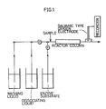
- The fibroin film entrapping monoclonal anti-human AFP antibody prepared in Production Example 1 was cut to a strip of 0.25 x 10 cm size. A flow type assay system shown in Fig. 1 of the accompanying drawings was assembled by placing the film strip in a glass tube having an inner diameter of 0.3 cm and a length of 11 cm to form a reactor column.
- The reactor column was filled with distilled water. Then 0.7 ml of a series of solutions of 0, 1.25, 2.5, 5, 10, 20, and 40 ng/ml of standard AFP in normal saline containing 0.8 µg/ml of catalase-labelled anti-human AFP antibody as described below and 0.1 weight % of BSA was introduced into the reactor column and incubated for 20 minutes. The reactor column was washed by passing distilled water at a flow rate of 20 ml/minute for 1 minute. Then 0.7 ml of 0.1 M phosphate buffer, pH 7.0, containing 17.6 mM of hydrogen peroxide was introduced into the reactor column and allowed to react for 5 minutes. The reaction mixture was then displaced to a galvanic type oxygen electrode cell (Model AN, manufactured by Oriental Electric Co., Ltd.) by introducing distilled water at a flow rate of 0.5 ml/minute. Increase in potential was determined from the readings of electric current which were proportional to the oxygen concentration in the reaction mixture.
- After the completion of the above procedure, the film was freed of bound antibody and antigen by passing 0.1 M glycine-HCl buffer (pH 2.5, containing 2% NaCl) through the reactor column at a flow rate of 0.5 ml/minute for 3 minutes. The column was then washed with distilled water at a flow rate of 20 ml/minute for 10 seconds.
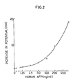
- The above procedures were repeated twice for each concentration of standard AFP. A standard curve as shown in Fig. 2 was obtained.
- All procedures are performed at room temperature.
- The catalase-labelled anti-human AFP antibody was produced as follows:
1 mg of the above antibody and 3 mg of catalase (sigma, bovine liver origin) were dissolved in 1 ml of 0.1 M phosphate buffer, pH 8.0. 10 µl of 10 weight % aqueous solution of glutaraldehyde was added to the solution. The mixture was allowed to react at 15°C for 3 hours. Then 2 mg of sodium borohydride was added. After allowing to react for 30 minutes, the reaction mixture was dialyzed in a dialysis membrane tube against 0.1 M phosphate buffer, pH 7.0, for 4 hours. This reaction mixture was purified using a TSK gel G-3000SW column (registered Trade Mark of TOKYO SODA MFG., Inc.) and a fraction containing the desired labelled antibody was collected. - A series of samples containing 0, 30, 50, 150 and 300 ng of standard AFP, respectively, was prepared by adding AFP into 1 ml of human serum from healthy adults (known to contain less than 5 ng/ml on radioimmunoassay). Each sample liquid was diluted 10 times with a 0.1 weight % solution of BSA in normal saline containing 0.89 µg/ml of the above catalase-labelled antibody.
- Using the above reactor column, the procedures of part (1) were repeated twice for each sample whereupon the results shown in Table III were obtained.
Table III AFP added (ng/ml) Mean AFP found (n=2) (ng/ml) 0 <12.5 30 34 50 57 80 83 150 165 300 290 - As shown in Table III, it was possible to assay AFP in human sera repeatedly.
Claims (9)
- An immunoassay method including the step of complexing an antibody or antigen immobilized by entrapment in fibroin film and specific to an antigen or antibody to be assayed with said antigen or antibody to be assayed, characterised in that after completion of the use in the assay of said immobilized antibody or antigen the latter is contacted with a buffer solution having a pH of about 2 to 4 to remove complexed antigens or antibodies therefrom by dissociation, so as to permit the immobilized antibody or antigen to be used in a further such assay.
- A method as claimed in claim 1 for assaying an antigen wherein a fibroin film entrapped, immobilized antibody to said antigen is employed.
- A method as claimed in claim 1 or claim 2 wherein said immobilized antibody or antigen is contacted with said buffer solution at a temperature of 10°C to 40°C.
- A method as claimed in any one of claims 1 to 3 wherein said immobilized antibody or antigen is contacted with said buffer solution for 3 to 10 minutes.
- A method as claimed in any one of claims 1 to 4 wherein said buffer solution is a glycine-HCl buffer or a tartaric acid-sodium hydroxide buffer having a concentration of 0.05 to 0.02M.
- A method as claimed in any one of claims 1 to 5 wherein said buffer solution includes 0.5 to 10 weight % of a salt selected from sodium chloride, potassium chloride and sodium sulphate.
- A method as claimed in claim 6 wherein said buffer solution is 0.1M glycine-HCl containing 1-5 weight % of NaCl or KCl at pH 2.2-2.9.
- A method as claimed in any one of claims 2 to 7 wherein an antigen to be assayed is further complexed with a labelled antibody.
- A method as claimed in claim 8 comprising the steps of:(a) incubating a fibroin film having entrapped, immobilized antibody specific to the antigen to be assayed with said antigen and the labelled antibody in a flow-type reaction vessel;(b) removing from said vessel the liquid phase of the complexing mixture and measuring the uncomplexed label;(c) passing through the reaction vessel at a flow rate of 0.5ml/minute for 3 minutes 0.1M glycine-HCl containing 2 weight % of Nacl at pH 2.5; and(d) washing the fibroin film with distilled water.
Priority Applications (1)
| Application Number | Priority Date | Filing Date | Title |
|---|---|---|---|
| AT86906453T ATE80467T1 (en) | 1985-11-05 | 1986-10-30 | IMMUNOTEST PROCEDURES. |
Applications Claiming Priority (2)
| Application Number | Priority Date | Filing Date | Title |
|---|---|---|---|
| JP248329/85 | 1985-11-05 | ||
| JP60248329A JPS62106369A (en) | 1985-11-05 | 1985-11-05 | Immunoassay |
Publications (3)
| Publication Number | Publication Date |
|---|---|
| EP0245509A1 EP0245509A1 (en) | 1987-11-19 |
| EP0245509A4 EP0245509A4 (en) | 1989-07-11 |
| EP0245509B1 true EP0245509B1 (en) | 1992-09-09 |
Family
ID=17176460
Family Applications (1)
| Application Number | Title | Priority Date | Filing Date |
|---|---|---|---|
| EP86906453A Expired EP0245509B1 (en) | 1985-11-05 | 1986-10-30 | Method of immunoassay |
Country Status (5)
| Country | Link |
|---|---|
| EP (1) | EP0245509B1 (en) |
| JP (1) | JPS62106369A (en) |
| AT (1) | ATE80467T1 (en) |
| DE (1) | DE3686717T2 (en) |
| WO (1) | WO1987002780A1 (en) |
Families Citing this family (8)
| Publication number | Priority date | Publication date | Assignee | Title |
|---|---|---|---|---|
| US20100046902A1 (en) | 2006-11-03 | 2010-02-25 | Trustees Of Tufts College | Biopolymer photonic crystals and method of manufacturing the same |
| EP2101975A2 (en) * | 2006-11-03 | 2009-09-23 | Trustees of Tufts College | Biopolymer sensor and method of manufacturing the same |
| EP2650112B1 (en) | 2006-11-03 | 2016-08-24 | Trustees Of Tufts College | Nanopatterned biopolymer optical device and method of manufacturing the same |
| WO2008127403A2 (en) | 2006-11-03 | 2008-10-23 | Trustees Of Tufts College | Biopolymer optofluidic device and method of manufacturing the same |
| WO2009061823A1 (en) | 2007-11-05 | 2009-05-14 | Trustees Of Tufts College | Fabrication of silk fibroin photonic structures by nanocontact imprinting |
| US8747886B2 (en) | 2009-02-12 | 2014-06-10 | Tufts University | Nanoimprinting of silk fibroin structures for biomedical and biophotonic applications |
| AU2010307268B2 (en) | 2009-07-20 | 2015-05-14 | Tufts University/Trustees Of Tufts College | All-protein implantable, resorbable reflectors |
| EP2474054A4 (en) | 2009-08-31 | 2013-03-13 | Tufts University Trustees Of Tufts College | SILK-BASED TRANSISTOR DEVICES |
Family Cites Families (8)
| Publication number | Priority date | Publication date | Assignee | Title |
|---|---|---|---|---|
| CA1036935A (en) * | 1973-03-19 | 1978-08-22 | Lavell R. Johnson | Method and apparatus for radioimmunoassay with regeneration of immunadsorbent |
| JPS58176547A (en) * | 1982-04-12 | 1983-10-17 | Denki Kagaku Kogyo Kk | Carrier for immunoaffinity-chromatography |
| JPS60108755A (en) * | 1983-11-18 | 1985-06-14 | Olympus Optical Co Ltd | Immunological analysis |
| DE3347092A1 (en) * | 1983-12-24 | 1985-07-18 | MTU Motoren- und Turbinen-Union München GmbH, 8000 München | METHOD AND DEVICE FOR OPTICALLY MEASURING THE FLOW OF A FLUID |
| JPS60142259A (en) * | 1983-12-29 | 1985-07-27 | Kanebo Ltd | Immobilized antibody |
| JPS60155129A (en) * | 1984-01-24 | 1985-08-15 | Kanebo Ltd | Preparation of immobilized antigen or antibody |
| JPS63209605A (en) * | 1987-02-24 | 1988-08-31 | 尾崎 公造 | Production of fan |
| JPH0513525A (en) * | 1991-07-05 | 1993-01-22 | Mitsubishi Electric Corp | IC Socket |
-
1985
- 1985-11-05 JP JP60248329A patent/JPS62106369A/en active Pending
-
1986
- 1986-10-30 AT AT86906453T patent/ATE80467T1/en not_active IP Right Cessation
- 1986-10-30 DE DE8686906453T patent/DE3686717T2/en not_active Expired - Fee Related
- 1986-10-30 EP EP86906453A patent/EP0245509B1/en not_active Expired
- 1986-10-30 WO PCT/JP1986/000552 patent/WO1987002780A1/en not_active Ceased
Also Published As
| Publication number | Publication date |
|---|---|
| ATE80467T1 (en) | 1992-09-15 |
| EP0245509A1 (en) | 1987-11-19 |
| JPS62106369A (en) | 1987-05-16 |
| DE3686717T2 (en) | 1993-01-28 |
| EP0245509A4 (en) | 1989-07-11 |
| DE3686717D1 (en) | 1992-10-15 |
| WO1987002780A1 (en) | 1987-05-07 |
Similar Documents
| Publication | Publication Date | Title |
|---|---|---|
| DK160108C (en) | Method and equipment for direct or indirect detection of reaction between a specific binding agent and the corresponding acceptor substance | |
| US5958790A (en) | Solid phase transverse diffusion assay | |
| DE3115115C2 (en) | Immunological procedure | |
| EP0149168B1 (en) | Immunoassay | |
| CA1098032A (en) | Solid phase assay of non-human antibody | |
| CA1123336A (en) | Method for the demonstration and determination of an antigen or antibody | |
| CH642458A5 (en) | Immunological method | |
| EP0207152B1 (en) | Solid phase diffusion assay | |
| US4894347A (en) | Erythrocyte agglutination assay | |
| US4350760A (en) | Method for the separation of a protein substance from a solution containing the same by affinity filtration and application of said method to enzymatic assays | |
| EP0245509B1 (en) | Method of immunoassay | |
| JPH0543600A (en) | Antibody-or antigen-immobilized silk fibroin membrane and sensor for measuring immune | |
| EP0155224A2 (en) | Solid-borne complex bearing chromagen responsive functionality for antibody, antigen, receptor, or ligand detection | |
| CA1334507C (en) | Simple method for immunological assay | |
| JP2736058B2 (en) | Manufacturing method of immunoassay device | |
| EP0683396A1 (en) | Method of assaying specific antibody | |
| JPH0694716A (en) | Immunity measuring method | |
| EP0308242B1 (en) | Agglutination assay | |
| AU703940B2 (en) | Regenerable solid phase for carrying out specific binding reactions | |
| JPS63106561A (en) | Reagent for immunological analysis | |
| JPS61122223A (en) | Purification of antibody | |
| JP4013270B2 (en) | Method for measuring bioactive peptide in urine and reagent for measurement | |
| JPH0792454B2 (en) | Antigen measurement method | |
| HK1000970B (en) | Solid phase diffusion assay | |
| JPS63101754A (en) | Manufacture of catalase-labeled antibody |
Legal Events
| Date | Code | Title | Description |
|---|---|---|---|
| PUAI | Public reference made under article 153(3) epc to a published international application that has entered the european phase |
Free format text: ORIGINAL CODE: 0009012 |
|
| 17P | Request for examination filed |
Effective date: 19870717 |
|
| AK | Designated contracting states |
Kind code of ref document: A1 Designated state(s): AT BE CH DE FR GB IT LI LU NL SE |
|
| A4 | Supplementary search report drawn up and despatched |
Effective date: 19890711 |
|
| 17Q | First examination report despatched |
Effective date: 19910417 |
|
| GRAA | (expected) grant |
Free format text: ORIGINAL CODE: 0009210 |
|
| AK | Designated contracting states |
Kind code of ref document: B1 Designated state(s): AT BE CH DE FR GB IT LI LU NL SE |
|
| REF | Corresponds to: |
Ref document number: 80467 Country of ref document: AT Date of ref document: 19920915 Kind code of ref document: T |
|
| PGFP | Annual fee paid to national office [announced via postgrant information from national office to epo] |
Ref country code: LU Payment date: 19920924 Year of fee payment: 7 |
|
| PGFP | Annual fee paid to national office [announced via postgrant information from national office to epo] |
Ref country code: AT Payment date: 19920925 Year of fee payment: 7 |
|
| ITF | It: translation for a ep patent filed | ||
| REF | Corresponds to: |
Ref document number: 3686717 Country of ref document: DE Date of ref document: 19921015 |
|
| ET | Fr: translation filed | ||
| PGFP | Annual fee paid to national office [announced via postgrant information from national office to epo] |
Ref country code: BE Payment date: 19921229 Year of fee payment: 7 |
|
| EPTA | Lu: last paid annual fee | ||
| PLBE | No opposition filed within time limit |
Free format text: ORIGINAL CODE: 0009261 |
|
| STAA | Information on the status of an ep patent application or granted ep patent |
Free format text: STATUS: NO OPPOSITION FILED WITHIN TIME LIMIT |
|
| 26N | No opposition filed | ||
| PGFP | Annual fee paid to national office [announced via postgrant information from national office to epo] |
Ref country code: SE Payment date: 19931005 Year of fee payment: 8 |
|
| PGFP | Annual fee paid to national office [announced via postgrant information from national office to epo] |
Ref country code: CH Payment date: 19931012 Year of fee payment: 8 |
|
| PGFP | Annual fee paid to national office [announced via postgrant information from national office to epo] |
Ref country code: GB Payment date: 19931026 Year of fee payment: 8 |
|
| PGFP | Annual fee paid to national office [announced via postgrant information from national office to epo] |
Ref country code: FR Payment date: 19931027 Year of fee payment: 8 |
|
| PG25 | Lapsed in a contracting state [announced via postgrant information from national office to epo] |
Ref country code: LU Free format text: LAPSE BECAUSE OF NON-PAYMENT OF DUE FEES Effective date: 19931030 Ref country code: AT Effective date: 19931030 |
|
| PG25 | Lapsed in a contracting state [announced via postgrant information from national office to epo] |
Ref country code: BE Effective date: 19931031 |
|
| PGFP | Annual fee paid to national office [announced via postgrant information from national office to epo] |
Ref country code: NL Payment date: 19931031 Year of fee payment: 8 |
|
| PGFP | Annual fee paid to national office [announced via postgrant information from national office to epo] |
Ref country code: DE Payment date: 19931230 Year of fee payment: 8 |
|
| BERE | Be: lapsed |
Owner name: KANEBO LTD Effective date: 19931031 |
|
| PG25 | Lapsed in a contracting state [announced via postgrant information from national office to epo] |
Ref country code: GB Effective date: 19941030 |
|
| PG25 | Lapsed in a contracting state [announced via postgrant information from national office to epo] |
Ref country code: SE Effective date: 19941031 Ref country code: LI Effective date: 19941031 Ref country code: CH Effective date: 19941031 |
|
| EAL | Se: european patent in force in sweden |
Ref document number: 86906453.5 |
|
| PG25 | Lapsed in a contracting state [announced via postgrant information from national office to epo] |
Ref country code: NL Effective date: 19950501 |
|
| NLV4 | Nl: lapsed or anulled due to non-payment of the annual fee | ||
| GBPC | Gb: european patent ceased through non-payment of renewal fee |
Effective date: 19941030 |
|
| PG25 | Lapsed in a contracting state [announced via postgrant information from national office to epo] |
Ref country code: FR Effective date: 19950630 |
|
| REG | Reference to a national code |
Ref country code: CH Ref legal event code: PL |
|
| PG25 | Lapsed in a contracting state [announced via postgrant information from national office to epo] |
Ref country code: DE Effective date: 19950701 |
|
| EUG | Se: european patent has lapsed |
Ref document number: 86906453.5 |
|
| REG | Reference to a national code |
Ref country code: FR Ref legal event code: ST |
|
| PG25 | Lapsed in a contracting state [announced via postgrant information from national office to epo] |
Ref country code: IT Free format text: LAPSE BECAUSE OF NON-PAYMENT OF DUE FEES;WARNING: LAPSES OF ITALIAN PATENTS WITH EFFECTIVE DATE BEFORE 2007 MAY HAVE OCCURRED AT ANY TIME BEFORE 2007. THE CORRECT EFFECTIVE DATE MAY BE DIFFERENT FROM THE ONE RECORDED. Effective date: 20051030 |