EP0220534A2 - Eichvorrichtung für einen optischen Katheter - Google Patents
Eichvorrichtung für einen optischen Katheter Download PDFInfo
- Publication number
- EP0220534A2 EP0220534A2 EP86113656A EP86113656A EP0220534A2 EP 0220534 A2 EP0220534 A2 EP 0220534A2 EP 86113656 A EP86113656 A EP 86113656A EP 86113656 A EP86113656 A EP 86113656A EP 0220534 A2 EP0220534 A2 EP 0220534A2
- Authority
- EP
- European Patent Office
- Prior art keywords
- catheter
- enclosure
- distal end
- reference element
- passage
- Prior art date
- Legal status (The legal status is an assumption and is not a legal conclusion. Google has not performed a legal analysis and makes no representation as to the accuracy of the status listed.)
- Granted
Links
- 230000003287 optical effect Effects 0.000 title claims abstract description 29
- 210000004369 blood Anatomy 0.000 claims description 15
- 239000008280 blood Substances 0.000 claims description 15
- 238000002496 oximetry Methods 0.000 claims description 15
- 239000003566 sealing material Substances 0.000 claims description 10
- 239000007787 solid Substances 0.000 claims description 6
- 239000000463 material Substances 0.000 claims description 4
- QVGXLLKOCUKJST-UHFFFAOYSA-N atomic oxygen Chemical compound [O] QVGXLLKOCUKJST-UHFFFAOYSA-N 0.000 description 9
- 229910052760 oxygen Inorganic materials 0.000 description 9
- 239000001301 oxygen Substances 0.000 description 9
- 102000001554 Hemoglobins Human genes 0.000 description 8
- 108010054147 Hemoglobins Proteins 0.000 description 8
- 238000000034 method Methods 0.000 description 8
- 238000005259 measurement Methods 0.000 description 7
- 238000012545 processing Methods 0.000 description 5
- 239000000835 fiber Substances 0.000 description 4
- 210000003743 erythrocyte Anatomy 0.000 description 3
- 238000004806 packaging method and process Methods 0.000 description 3
- 239000002245 particle Substances 0.000 description 3
- 238000012360 testing method Methods 0.000 description 3
- 230000005540 biological transmission Effects 0.000 description 2
- 230000008878 coupling Effects 0.000 description 2
- 238000010168 coupling process Methods 0.000 description 2
- 238000005859 coupling reaction Methods 0.000 description 2
- 238000012544 monitoring process Methods 0.000 description 2
- XUIMIQQOPSSXEZ-UHFFFAOYSA-N Silicon Chemical compound [Si] XUIMIQQOPSSXEZ-UHFFFAOYSA-N 0.000 description 1
- 238000010521 absorption reaction Methods 0.000 description 1
- 230000000747 cardiac effect Effects 0.000 description 1
- 238000011109 contamination Methods 0.000 description 1
- 230000002844 continuous effect Effects 0.000 description 1
- 230000007423 decrease Effects 0.000 description 1
- 239000000975 dye Substances 0.000 description 1
- 230000000694 effects Effects 0.000 description 1
- 239000013536 elastomeric material Substances 0.000 description 1
- 238000005534 hematocrit Methods 0.000 description 1
- 238000002329 infrared spectrum Methods 0.000 description 1
- 238000001802 infusion Methods 0.000 description 1
- 238000009533 lab test Methods 0.000 description 1
- 230000031700 light absorption Effects 0.000 description 1
- VTHJTEIRLNZDEV-UHFFFAOYSA-L magnesium dihydroxide Chemical compound [OH-].[OH-].[Mg+2] VTHJTEIRLNZDEV-UHFFFAOYSA-L 0.000 description 1
- 239000000347 magnesium hydroxide Substances 0.000 description 1
- 235000012254 magnesium hydroxide Nutrition 0.000 description 1
- 238000004519 manufacturing process Methods 0.000 description 1
- 230000007246 mechanism Effects 0.000 description 1
- 238000012986 modification Methods 0.000 description 1
- 230000004048 modification Effects 0.000 description 1
- 238000006213 oxygenation reaction Methods 0.000 description 1
- 230000008569 process Effects 0.000 description 1
- 230000001681 protective effect Effects 0.000 description 1
- 210000001147 pulmonary artery Anatomy 0.000 description 1
- 239000012925 reference material Substances 0.000 description 1
- 238000002310 reflectometry Methods 0.000 description 1
- 239000012858 resilient material Substances 0.000 description 1
- 239000011347 resin Substances 0.000 description 1
- 229920005989 resin Polymers 0.000 description 1
- 238000005070 sampling Methods 0.000 description 1
- 238000002798 spectrophotometry method Methods 0.000 description 1
- 230000001954 sterilising effect Effects 0.000 description 1
- 238000004659 sterilization and disinfection Methods 0.000 description 1
- 239000000126 substance Substances 0.000 description 1
- 239000000725 suspension Substances 0.000 description 1
- 210000003462 vein Anatomy 0.000 description 1
Images

Classifications
-
- G—PHYSICS
- G01—MEASURING; TESTING
- G01N—INVESTIGATING OR ANALYSING MATERIALS BY DETERMINING THEIR CHEMICAL OR PHYSICAL PROPERTIES
- G01N21/00—Investigating or analysing materials by the use of optical means, i.e. using sub-millimetre waves, infrared, visible or ultraviolet light
- G01N21/17—Systems in which incident light is modified in accordance with the properties of the material investigated
- G01N21/47—Scattering, i.e. diffuse reflection
- G01N21/4785—Standardising light scatter apparatus; Standards therefor
-
- A—HUMAN NECESSITIES
- A61—MEDICAL OR VETERINARY SCIENCE; HYGIENE
- A61B—DIAGNOSIS; SURGERY; IDENTIFICATION
- A61B5/00—Measuring for diagnostic purposes; Identification of persons
- A61B5/145—Measuring characteristics of blood in vivo, e.g. gas concentration or pH-value ; Measuring characteristics of body fluids or tissues, e.g. interstitial fluid or cerebral tissue
- A61B5/1495—Calibrating or testing of in-vivo probes
-
- A—HUMAN NECESSITIES
- A61—MEDICAL OR VETERINARY SCIENCE; HYGIENE
- A61B—DIAGNOSIS; SURGERY; IDENTIFICATION
- A61B2560/00—Constructional details of operational features of apparatus; Accessories for medical measuring apparatus
- A61B2560/02—Operational features
- A61B2560/0223—Operational features of calibration, e.g. protocols for calibrating sensors
- A61B2560/0228—Operational features of calibration, e.g. protocols for calibrating sensors using calibration standards
- A61B2560/0233—Optical standards
Definitions
- This invention relates to a calibration device and calibrating system for optical catheters used in a catheter oximetry system, and more particularly, it relates to a calibrating device which may remain with the sealed and sterilized distal end of the catheter within a package while the proximal end of the catheter is plugged into a computer or processor in order to perform the calibrating operation.
- a catheter oximetry system provides accurate, continuous, real-time measurement of mixed venous oxygen saturation using multiple wavelength reflection spectrophotometry.
- the color of red blood cells progressively changes from scarlet to purple as the amount of oxygen the red blood cells are carrying decreases.
- the amount of light backscattered, or reflected, at each wavelength depends upon the color, and therefore, oxygen level of the blood.
- Careful choice of wavelengths in the transmittal light allows accurate measurement of oxygenated hemoglobin with minimal interference by other blood characteristics such as temperature, pH, and hematocrit.
- the relative amounts of oxygenated hemoglobin and deoxygenated hemoglobin in the blood can be determined by measuring the relative absorption of light at different selected wavelengths.
- the percentage of hemoglobin which is in the oxygenated form is defined as the oxygen saturation of the blood in the equation: where HbO2 is the oxygenated hemoglobin concentration and Hb is the deoxygenated hemoglobin concentration.
- a widely used catheter oximetry system consists of three basic components: (1) a disposable fiberoptic pulmonary artery catheter that has a distal end adapted to be inserted into a vein of a patient and that interfaces at its other end with (2) an optical module containing light emitting diodes, a photodetector and associated electronics, which in turn, interfaces with the electrical leads of (3) a computer-based instrument that performs all of the data processing and control functions with displays, alarms and associated read-out devices.
- the instrument and optical module may be reused many times with different patients, but the catheter is used only with a single patient during a single operation or monitoring process.
- the catheters are disposable and are arranged to be separately packaged in sealed aseptic packages each with a specially designed optical connector plug adapted to be plugged into the optical module when the catheter is ready for use.
- each catheter be separately calibrated immediately before it is used so as to relate the actual light intensities received from the sample under test to the unknown concentrations of the substances being quantified in the sample under test. This may be accomplished by initially measuring a given sample of blood with the catheter and then wholly independently measuring the same blood in the laboratory by a different technique in order to match the laboratory calculated actual oxygen saturation content with the instrument calculated content and adjusting the latter accordingly.
- the catheter can be pulled loose from the tube and reference block and placed in a patient for obtaining blood oxygen saturation measurements in the manner intended.
- the initial calibration readings are obtained with the reference block and catheter remaining in the package in a sealed and sterilized condition while the connector plug end of the catheter is connected to the optical module and oximetry processor.
- a catheter having transmitting and receiving light guides therein is arranged to be packaged in a tray with the distal end of the catheter being received in a calibrating device.
- the calibrating device includes a tubular enclosure within which a reference element is urged into compliant engagement with the distal end of the catheter and with means being provided for tightly gripping and holding the catheter to the enclosure so that the catheter and calibrating device are ready for an immediate calibration operation in the package without any additional movement of either catheter or reference block being required.
- the package is sealed with a cover material that encloses the catheter and calibrating device in the tray in a sealed and sterile condition.
- a portion of the sealing material can be removed while the remainder is left in its sealed and sterile condition so as to only expose the optical connector at the proximal end of the catheter permitting it to be connected to an oximetry system to provide a calibration reading for the catheter -- all while the distal end and insertable major length of the catheter remains in a sealed, sterile condition.
- the remainder of the sealing material can be removed, the calibrating device readily removed from the distal end of the catheter, and the catheter placed directly in the patient for continuous blood oxygen saturation readings.
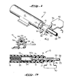
- the calibrating device 10 of the present invention is shown in Figures 1-3 wherein it will be seen to be comprised of a cylinder 12 which is closed at one end 13 and open at the other end thereof (Fig. 3). Received within the open end of the cylinder 12 is a plug 15 which is provided near the center thereof with a circumferencially extending rib 17 adapted to be received within a detent 18 just within the open end of the cylinder 12. The plug is inserted in the cylinder during the assembly of the calibrating device, and, as indicated in Figure 3, it is snapped into snug engagement therewith.
- an optical catheter 20 is adapted to be inserted through the plug 15 within an axial passage 22 extending therethrough.
- the axial passage 22 is just slightly larger than the outer diameter of the catheter and its balloon 21 so as to snugly confine the catheter therewithin.
- a reference block 24 and a coil spring 25 Positioned within the inner end of the tubular member 12 is a reference block 24 and a coil spring 25 with the spring urging the reference block into firm engagement with the flat distal end 20a of the catheter 20 at a position spaced inwardly from the end of the plug 15.
- the reference block 24 is the same as that shown and described in the aforementioned prior United States Patent No. 4,322,164 to Robert F. Shaw et al. Briefly, the reference block comprises a solid cylindrical element formed of a silicone resin and having a plurality of tiny particles scattered throughout its mass to provide scattering and reflecting surfaces for the light beams transmitted by the catheter 20. The particles will typically have dimensions within the range of from about 0.02 to about 20 microns and should be uniformly dis persed within the solid mass of the reference block 24.
- the mass is translucent is nature and has compliant characteristics at the surface thereof so that it will yield when pressed against the rigid surface 20a of the catheter thereby insuring a snug fit which will not become easily dislodged during handling of the catheter and attached calibrating device.
- the catheter 20 comprises a conventional optical catheter useful in oximetry measurements having a pair of separated lumens with a transmitting light guide 28 formed of a single fiber and a receiving light guide 29 likewise formed of a single fiber extending side-by-side along the length of the catheter to an exposed position at the flattened surface 20a at the very end of the catheter.
- Light carried along the transmitting fiber 28 is directed into the reference block 24 where it is backscattered and a portion thereof is reflected back into the receiving fiber 29 for transmission back to the oximetry processing apparatus to provide readings useful for calibrating the catheter and associated optical components.
- the strap includes an enlarged tab 40 at the outwardly projecting end thereof which tab is of a size whereby it can be readily gripped between the fingers in order to pull the strap loose from the prongs at the conclusion of the calibration operation in order to release the catheter from the calibrating device.
- FIG. 4 The use of the calibrating device 10 of the present invention in a catheter oximetry system is shown sequentially in Figures 4, 5 and 6.
- the catheter 20 is arranged to be packaged within conforming recesses set in a rectangularly shaped plastic tray 42.
- a piece of plastic sealing material 44 is laid atop the tray and sealed thereto, and the tray and enclosed catheter are then sterilized using conventional sterilization techniques.
- the distal end of the catheter is connected directly to the calibrating device 10 in the aforedescribed manner and clamped thereto by the strap 36 with the tab 40 of the strap extending to the side in a position adapting it to ready removability.
- the proximal end of the catheter includes the optical connector plug 46 and a plurality of other conven tional output connections including lumen connections for pressure readings, samplings, or infusion, a thermistor connection for cardiac outputs and a mechanism connected to pressurize the balloon 21 at the tip of the catheter -- all of such elements being conventional with the details thereof having no relevance with respect to the present invention.
- the first step in the calibration operation is to remove the plastic sealing material 44 from atop the tray to allow the fiberoptic connector plug 46 to be removed and coupled to the computer or processor 48.
- the sealing material 44 is provided with two sections separated by a seam or scoreline 43 whereby only one portion thereof is removed during the initial peeling of the material, as shown in Figure 5, exposing only the proximal end of the catheter and the connections thereto (including the connector plug 46) but leaving the main body of the catheter, which will later be placed in the patient, within the package in its original sealed and sterilized condition.
- the connector plug can then be placed in a receptacle in an optical module 50 which provides the electro-optical coupling between the connector plug 44 and the processing circuitry of the computer 48.
- the computer is turned on to provide signals to the optical module 50 creating the light sources which are directed via the coupling 46 down the length of the catheter to the reference block 24 wherein the light is backscattered and reflected back to the optical module.
- the module then converts these light signals into electrical signals for processing by the computer. In this way the appropriate calibration readings are obtained and stored in the computer.
- the catheter is calibrated and immediately ready for use in monitoring the blood oxygenation of a patient.
- the remainder of the sealing material 44 is then removed, and a simple pulling away of the strap 36 from its secured position on the calibrating device 10 leaves the catheter 20 free from its locked engagement therewith. The nurse or doctor can then directly take the catheter and place it in the patient.
- the calibrating device of the present invention permits the catheter to be directly locked to a calibrating device and packaged in such a manner so that no additional steps are required other than to connect the proximal end of the catheter to suitable processing circuitry in order to obtain appropriate calibration readings. Once the readings have been obtained, the catheter is ready for immediate use, and the protective and sealing material can be removed to permit the catheter to be immediately used. It has been found that the packaging method as aforedescribed will stand up under repeated jostling or dropping without dislodging the reference block from the catheter.
Landscapes
- Health & Medical Sciences (AREA)
- Life Sciences & Earth Sciences (AREA)
- Physics & Mathematics (AREA)
- General Health & Medical Sciences (AREA)
- Pathology (AREA)
- Optics & Photonics (AREA)
- Heart & Thoracic Surgery (AREA)
- General Physics & Mathematics (AREA)
- Immunology (AREA)
- Analytical Chemistry (AREA)
- Chemical & Material Sciences (AREA)
- Biophysics (AREA)
- Engineering & Computer Science (AREA)
- Biomedical Technology (AREA)
- Biochemistry (AREA)
- Medical Informatics (AREA)
- Molecular Biology (AREA)
- Surgery (AREA)
- Animal Behavior & Ethology (AREA)
- Public Health (AREA)
- Veterinary Medicine (AREA)
- Measurement Of The Respiration, Hearing Ability, Form, And Blood Characteristics Of Living Organisms (AREA)
- Investigating Or Analysing Materials By Optical Means (AREA)
Applications Claiming Priority (2)
| Application Number | Priority Date | Filing Date | Title |
|---|---|---|---|
| US791706 | 1985-10-28 | ||
| US06/791,706 US4650327A (en) | 1985-10-28 | 1985-10-28 | Optical catheter calibrating assembly |
Publications (3)
| Publication Number | Publication Date |
|---|---|
| EP0220534A2 true EP0220534A2 (de) | 1987-05-06 |
| EP0220534A3 EP0220534A3 (en) | 1987-12-09 |
| EP0220534B1 EP0220534B1 (de) | 1991-05-22 |
Family
ID=25154545
Family Applications (1)
| Application Number | Title | Priority Date | Filing Date |
|---|---|---|---|
| EP86113656A Expired EP0220534B1 (de) | 1985-10-28 | 1986-10-03 | Eichvorrichtung für einen optischen Katheter |
Country Status (5)
| Country | Link |
|---|---|
| US (1) | US4650327A (de) |
| EP (1) | EP0220534B1 (de) |
| JP (1) | JPH0667382B2 (de) |
| CA (1) | CA1277848C (de) |
| DE (1) | DE3679369D1 (de) |
Cited By (5)
| Publication number | Priority date | Publication date | Assignee | Title |
|---|---|---|---|---|
| EP0271304A1 (de) * | 1986-12-08 | 1988-06-15 | Spectramed, Inc. | Einwegkalibrierhülle für einen optischen kardiovaskulären Katheter |
| EP0366840A1 (de) * | 1986-12-16 | 1990-05-09 | BAXTER INTERNATIONAL INC. (a Delaware corporation) | Gerät zur Kalibrierung eines optisches Katheters und Verfahren |
| EP0397496A1 (de) * | 1989-05-12 | 1990-11-14 | Baxter International Inc. | Kalibrierungskappe für in-vitro-Eichung eines Sauerstoffsättigungsanzeigers und dessen Anwendungsverfahren |
| EP0572133A1 (de) * | 1992-05-22 | 1993-12-01 | Biomedical Sensors Ltd | Packung für eine aktive medizinische Vorrichtung |
| EP1401328A4 (de) * | 2001-05-31 | 2006-04-05 | Infraredx Inc | Optische bezugskatheter |
Families Citing this family (101)
| Publication number | Priority date | Publication date | Assignee | Title |
|---|---|---|---|---|
| US4968137A (en) * | 1986-12-05 | 1990-11-06 | The State Of Oregon Acting By And Through The State Board Of Higher Education On Behalf Of Oregon Health Sciences University | Devices and procedures for in vitro testing of pulse oximetry monitors |
| US4834532A (en) * | 1986-12-05 | 1989-05-30 | The State Of Oregon Acting By And Through The State Board Of Higher Education On Behalf Of Oregon Health Sciences University | Devices and procedures for in vitro calibration of pulse oximetry monitors |
| US5596986A (en) * | 1989-03-17 | 1997-01-28 | Scico, Inc. | Blood oximeter |
| US5460182A (en) * | 1992-09-14 | 1995-10-24 | Sextant Medical Corporation | Tissue penetrating apparatus and methods |
| US5772597A (en) * | 1992-09-14 | 1998-06-30 | Sextant Medical Corporation | Surgical tool end effector |
| US5762609A (en) * | 1992-09-14 | 1998-06-09 | Sextant Medical Corporation | Device and method for analysis of surgical tissue interventions |
| US5365925A (en) * | 1993-08-13 | 1994-11-22 | Ohmeda Inc. | Disposable calibration boot for multi-point calibration in fiber optic sensors |
| US5777731A (en) * | 1997-02-21 | 1998-07-07 | The Goodyear Tire & Rubber Company | Calibration of optical properties to measure depth of a liquid |
| US6271920B1 (en) | 1997-12-19 | 2001-08-07 | Chromatics Color Sciences International, Inc. | Methods and apparatus for color calibration and verification |
| JP2003508734A (ja) * | 1999-08-31 | 2003-03-04 | シーエムイー テレメトリックス インク. | スペクトル分析器の精度を検証するための装置 |
| EP1434522B1 (de) | 2000-10-30 | 2010-01-13 | The General Hospital Corporation | Optische systeme zur gewebeanalyse |
| US9295391B1 (en) | 2000-11-10 | 2016-03-29 | The General Hospital Corporation | Spectrally encoded miniature endoscopic imaging probe |
| US7865231B2 (en) * | 2001-05-01 | 2011-01-04 | The General Hospital Corporation | Method and apparatus for determination of atherosclerotic plaque type by measurement of tissue optical properties |
| US6923762B1 (en) * | 2001-11-01 | 2005-08-02 | Frank C. Creaghan, Jr. | Venoscope apparatus |
| US7355716B2 (en) | 2002-01-24 | 2008-04-08 | The General Hospital Corporation | Apparatus and method for ranging and noise reduction of low coherence interferometry LCI and optical coherence tomography OCT signals by parallel detection of spectral bands |
| US20110201924A1 (en) * | 2002-04-30 | 2011-08-18 | The General Hospital Corporation | Method and Apparatus for Improving Image Clarity and Sensitivity in Optical Tomography Using Dynamic Feedback to Control Focal Properties and Coherence Gating |
| JP2006516739A (ja) * | 2003-01-24 | 2006-07-06 | ザ・ジェネラル・ホスピタル・コーポレイション | 低コヒーレンス干渉計を用いて組織を識別するためのシステムおよび方法 |
| US7643153B2 (en) * | 2003-01-24 | 2010-01-05 | The General Hospital Corporation | Apparatus and method for ranging and noise reduction of low coherence interferometry LCI and optical coherence tomography OCT signals by parallel detection of spectral bands |
| EP2436307B1 (de) | 2003-03-31 | 2015-10-21 | The General Hospital Corporation | Fleckenreduzierung bei der optischen Kohärenztomografie durch pfadlängencodierte Winkelmischung |
| EP2008579B1 (de) | 2003-06-06 | 2016-11-09 | The General Hospital Corporation | Verfahren und Vorrichtung für eine Lichtquelle mit Abstimmung der Wellenlänge |
| KR101384553B1 (ko) | 2003-10-27 | 2014-04-11 | 더 제너럴 하스피탈 코포레이션 | 주파수 영역 간섭법을 이용하여 광 영상화를 수행하는 방법 및 장치 |
| DE102004024164A1 (de) * | 2004-05-14 | 2005-12-08 | Kaltenbach & Voigt Gmbh & Co. Kg | Zahnärztliches System zum Untersuchen der optischen Eigenschaften von Zahngewebe mit optischer Untersuchungsvorrichtung und Abgleicheinrichtung |
| KR101239250B1 (ko) | 2004-05-29 | 2013-03-05 | 더 제너럴 하스피탈 코포레이션 | 광간섭 단층촬영 화상 진단에서 반사층을 이용한 색 분산보상을 위한 프로세스, 시스템 및 소프트웨어 배열 |
| EP1771755B1 (de) * | 2004-07-02 | 2016-09-21 | The General Hospital Corporation | Endoskopische darstellungssonde mit doppelt kaschierter faser |
| WO2006017837A2 (en) | 2004-08-06 | 2006-02-16 | The General Hospital Corporation | Process, system and software arrangement for determining at least one location in a sample using an optical coherence tomography |
| US8965487B2 (en) * | 2004-08-24 | 2015-02-24 | The General Hospital Corporation | Process, system and software arrangement for measuring a mechanical strain and elastic properties of a sample |
| EP1793731B1 (de) | 2004-08-24 | 2013-12-25 | The General Hospital Corporation | Bildgebungsgerät mit einer Fluidabgabevorrichtung und einer Pull-Back-Vorrichntung |
| JP5215664B2 (ja) | 2004-09-10 | 2013-06-19 | ザ ジェネラル ホスピタル コーポレイション | 光学コヒーレンス撮像のシステムおよび方法 |
| US7366376B2 (en) | 2004-09-29 | 2008-04-29 | The General Hospital Corporation | System and method for optical coherence imaging |
| EP2278267A3 (de) * | 2004-11-24 | 2011-06-29 | The General Hospital Corporation | Interferometer mit gemeinsamem Pfad für endoskopische optische Kohärenztomographie |
| WO2006058346A1 (en) | 2004-11-29 | 2006-06-01 | The General Hospital Corporation | Arrangements, devices, endoscopes, catheters and methods for performing optical imaging by simultaneously illuminating and detecting multiple points on a sample |
| ATE451669T1 (de) | 2005-04-28 | 2009-12-15 | Gen Hospital Corp | Bewertung von bildmerkmalen einer anatomischen struktur in optischen kohärenztomographiebildern |
| EP1886121A1 (de) * | 2005-05-13 | 2008-02-13 | The General Hospital Corporation | Anordnungen, systeme und verfahren mit fähigkeit zur bereitstellung von optischer spektraldomänen-kohärenzrekflektometrie für einen empfindlichen nachweis chemischer und biologischer proben |
| WO2006130802A2 (en) * | 2005-06-01 | 2006-12-07 | The General Hospital Corporation | Apparatus, method and system for performing phase-resolved optical frequency domain imaging |
| KR101387454B1 (ko) * | 2005-08-09 | 2014-04-22 | 더 제너럴 하스피탈 코포레이션 | 광간섭 단층촬영법에서 편광 기반 직교 복조를 수행하기위한 장치, 방법 및 저장 매체 |
| CA2824033A1 (en) * | 2005-09-13 | 2007-03-22 | Edwards Lifesciences Corporation | Continuous spectroscopic measurement of total hemoglobin |
| CN101360447B (zh) * | 2005-09-29 | 2012-12-05 | 通用医疗公司 | 通过光谱编码进行光学成像的方法和装置 |
| US7889348B2 (en) | 2005-10-14 | 2011-02-15 | The General Hospital Corporation | Arrangements and methods for facilitating photoluminescence imaging |
| JP5680826B2 (ja) * | 2006-01-10 | 2015-03-04 | ザ ジェネラル ホスピタル コーポレイション | 1以上のスペクトルを符号化する内視鏡技術によるデータ生成システム |
| JP2009523574A (ja) * | 2006-01-18 | 2009-06-25 | ザ ジェネラル ホスピタル コーポレイション | 1つ又は複数の内視鏡顕微鏡検査法を使用してデータを生成するシステム及び方法 |
| EP2289396A3 (de) | 2006-01-19 | 2011-04-06 | The General Hospital Corporation | Verfahren und Systeme zur optischen Bildgebung von epithelialen Luminalorganen durch Strahlenabtastung dieser |
| US8145018B2 (en) | 2006-01-19 | 2012-03-27 | The General Hospital Corporation | Apparatus for obtaining information for a structure using spectrally-encoded endoscopy techniques and methods for producing one or more optical arrangements |
| WO2007084933A2 (en) * | 2006-01-20 | 2007-07-26 | The General Hospital Corporation | Systems and processes for providing endogenous molecular imaging with mid-infared light |
| JP5524487B2 (ja) * | 2006-02-01 | 2014-06-18 | ザ ジェネラル ホスピタル コーポレイション | コンフォーマルレーザ治療手順を用いてサンプルの少なくとも一部分に電磁放射を放射する方法及びシステム。 |
| US9186066B2 (en) * | 2006-02-01 | 2015-11-17 | The General Hospital Corporation | Apparatus for applying a plurality of electro-magnetic radiations to a sample |
| EP1984710A1 (de) * | 2006-02-07 | 2008-10-29 | AstraZeneca AB | Einrichtung und verfahren für ein spektrometrisches system |
| WO2007092911A2 (en) * | 2006-02-08 | 2007-08-16 | The General Hospital Corporation | Methods, arrangements and systems for obtaining information associated with an anatomical sample using optical microscopy |
| EP2982929A1 (de) * | 2006-02-24 | 2016-02-10 | The General Hospital Corporation | Verfahren und systeme zur durchführung von winkelaufgelöster optischer kohärenztomografie im fourier-bereich |
| EP2564769B1 (de) | 2006-04-05 | 2015-06-03 | The General Hospital Corporation | Vorrichtung zur polarisationsempfindlichen optischen Frequenzbereichsabbildung einer Probe |
| EP2517616A3 (de) * | 2006-05-10 | 2013-03-06 | The General Hospital Corporation | Prozesse, Anordnungen und Systeme zur Bereitstellung der Frequenzbereichsabbildung einer Probe |
| US7782464B2 (en) | 2006-05-12 | 2010-08-24 | The General Hospital Corporation | Processes, arrangements and systems for providing a fiber layer thickness map based on optical coherence tomography images |
| JP2010501877A (ja) * | 2006-08-25 | 2010-01-21 | ザ ジェネラル ホスピタル コーポレイション | ボリュメトリック・フィルタリング法を使用して光コヒーレンス・トモグラフィ画像形成の機能を向上させる装置及び方法 |
| US20080287808A1 (en) * | 2006-09-12 | 2008-11-20 | The General Hospital Corporation | Apparatus, probe and method for providing depth assessment in an anatomical structure |
| WO2008049118A2 (en) | 2006-10-19 | 2008-04-24 | The General Hospital Corporation | Apparatus and method for obtaining and providing imaging information associated with at least one portion of a sample and effecting such portion(s) |
| US7949019B2 (en) * | 2007-01-19 | 2011-05-24 | The General Hospital | Wavelength tuning source based on a rotatable reflector |
| WO2008115965A1 (en) * | 2007-03-19 | 2008-09-25 | The General Hospital Corporation | Apparatus and method for providing a noninvasive diagnosis of internal bleeding |
| EP2602651A3 (de) | 2007-03-23 | 2014-08-27 | The General Hospital Corporation | Verfahren, Anordnungen und Vorrichtung zur Verwendung eines wellenlängengewobbelten Lasers anhand von Winkelabtastungs- und Dispersionsverfahren |
| US10534129B2 (en) * | 2007-03-30 | 2020-01-14 | The General Hospital Corporation | System and method providing intracoronary laser speckle imaging for the detection of vulnerable plaque |
| WO2008131082A1 (en) | 2007-04-17 | 2008-10-30 | The General Hospital Corporation | Apparatus and methods for measuring vibrations using spectrally-encoded endoscopy techniques |
| US8115919B2 (en) * | 2007-05-04 | 2012-02-14 | The General Hospital Corporation | Methods, arrangements and systems for obtaining information associated with a sample using optical microscopy |
| WO2009018456A2 (en) | 2007-07-31 | 2009-02-05 | The General Hospital Corporation | Systems and methods for providing beam scan patterns for high speed doppler optical frequency domain imaging |
| US8040608B2 (en) * | 2007-08-31 | 2011-10-18 | The General Hospital Corporation | System and method for self-interference fluorescence microscopy, and computer-accessible medium associated therewith |
| US20090073439A1 (en) * | 2007-09-15 | 2009-03-19 | The General Hospital Corporation | Apparatus, computer-accessible medium and method for measuring chemical and/or molecular compositions of coronary atherosclerotic plaques in anatomical structures |
| US7933021B2 (en) | 2007-10-30 | 2011-04-26 | The General Hospital Corporation | System and method for cladding mode detection |
| US8100893B2 (en) * | 2007-11-28 | 2012-01-24 | The Spectranetics Corporation | Laser catheter calibrator |
| US7898656B2 (en) * | 2008-04-30 | 2011-03-01 | The General Hospital Corporation | Apparatus and method for cross axis parallel spectroscopy |
| WO2009137701A2 (en) * | 2008-05-07 | 2009-11-12 | The General Hospital Corporation | System, method and computer-accessible medium for tracking vessel motion during three-dimensional coronary artery microscopy |
| US8861910B2 (en) | 2008-06-20 | 2014-10-14 | The General Hospital Corporation | Fused fiber optic coupler arrangement and method for use thereof |
| EP2309923B1 (de) | 2008-07-14 | 2020-11-25 | The General Hospital Corporation | Vorrichtung und verfahren für eine farbendoskopie |
| EP2359121A4 (de) * | 2008-12-10 | 2013-08-14 | Gen Hospital Corp | Systeme, vorrichtung und verfahren zur erweiterung der bildgebungstiefenbereichs bei der optischen kohärenztomopgrafie mittels optischer unterabtastung |
| JP2012515576A (ja) * | 2009-01-20 | 2012-07-12 | ザ ジェネラル ホスピタル コーポレイション | 内視鏡生検装置、システム、及び方法 |
| EP2382456A4 (de) * | 2009-01-26 | 2012-07-25 | Gen Hospital Corp | System, verfahren und computermedium für mikroskopie mit weitem feld und sehr hoher auflösung |
| WO2010105197A2 (en) | 2009-03-12 | 2010-09-16 | The General Hospital Corporation | Non-contact optical system, computer-accessible medium and method for measuring at least one mechanical property of tissue using coherent speckle techniques(s) |
| BR112012001042A2 (pt) * | 2009-07-14 | 2016-11-22 | Gen Hospital Corp | equipamento e método de medição do fluxo de fluído dentro de estrutura anatômica. |
| WO2011044301A2 (en) * | 2009-10-06 | 2011-04-14 | The General Hospital Corporation | Apparatus and methods for imaging particular cells including eosinophils |
| EP2509488A4 (de) * | 2009-12-08 | 2014-04-09 | Gen Hospital Corp | Verfahren und anordnungen zur analyse, diagnose, behandlung und überwachung der stimmlippen durch optische kohärenztomographie |
| LT2542154T (lt) | 2010-03-05 | 2020-12-10 | The General Hospital Corporation | Ėminio švitinimo elektromagnetine spinduliuote aparatas |
| US9069130B2 (en) | 2010-05-03 | 2015-06-30 | The General Hospital Corporation | Apparatus, method and system for generating optical radiation from biological gain media |
| US9557154B2 (en) | 2010-05-25 | 2017-01-31 | The General Hospital Corporation | Systems, devices, methods, apparatus and computer-accessible media for providing optical imaging of structures and compositions |
| WO2011150069A2 (en) | 2010-05-25 | 2011-12-01 | The General Hospital Corporation | Apparatus, systems, methods and computer-accessible medium for spectral analysis of optical coherence tomography images |
| US10285568B2 (en) | 2010-06-03 | 2019-05-14 | The General Hospital Corporation | Apparatus and method for devices for imaging structures in or at one or more luminal organs |
| US9510758B2 (en) | 2010-10-27 | 2016-12-06 | The General Hospital Corporation | Apparatus, systems and methods for measuring blood pressure within at least one vessel |
| CN103607948A (zh) * | 2011-01-27 | 2014-02-26 | 皇家飞利浦电子股份有限公司 | 用于临床使用期间的光学形状感测校准的模板 |
| WO2013013049A1 (en) | 2011-07-19 | 2013-01-24 | The General Hospital Corporation | Systems, methods, apparatus and computer-accessible-medium for providing polarization-mode dispersion compensation in optical coherence tomography |
| US10241028B2 (en) | 2011-08-25 | 2019-03-26 | The General Hospital Corporation | Methods, systems, arrangements and computer-accessible medium for providing micro-optical coherence tomography procedures |
| US9341783B2 (en) | 2011-10-18 | 2016-05-17 | The General Hospital Corporation | Apparatus and methods for producing and/or providing recirculating optical delay(s) |
| EP2833776A4 (de) | 2012-03-30 | 2015-12-09 | Gen Hospital Corp | Abbildungssystem, verfahren und distaler anschluss zur multidirektionalen sichtfeldendoskopie |
| WO2013177154A1 (en) | 2012-05-21 | 2013-11-28 | The General Hospital Corporation | Apparatus, device and method for capsule microscopy |
| EP2948758B1 (de) | 2013-01-28 | 2024-03-13 | The General Hospital Corporation | Vorrichtung zur bereitstellung von gemeinsam mit optischer frequenzdomänenbildgebung aufgezeichneter diffuser spektroskopie |
| US10893806B2 (en) | 2013-01-29 | 2021-01-19 | The General Hospital Corporation | Apparatus, systems and methods for providing information regarding the aortic valve |
| WO2014121082A1 (en) | 2013-02-01 | 2014-08-07 | The General Hospital Corporation | Objective lens arrangement for confocal endomicroscopy |
| WO2014144709A2 (en) | 2013-03-15 | 2014-09-18 | The General Hospital Corporation | Methods and systems for characterizing an object |
| EP2997354A4 (de) | 2013-05-13 | 2017-01-18 | The General Hospital Corporation | Erkennung einer selbstinterferierenden fluoreszenzphase und amplitude |
| WO2015009932A1 (en) | 2013-07-19 | 2015-01-22 | The General Hospital Corporation | Imaging apparatus and method which utilizes multidirectional field of view endoscopy |
| WO2015010133A1 (en) | 2013-07-19 | 2015-01-22 | The General Hospital Corporation | Determining eye motion by imaging retina. with feedback |
| ES2893237T3 (es) | 2013-07-26 | 2022-02-08 | Massachusetts Gen Hospital | Aparato con una disposición láser que utiliza dispersión óptica para aplicaciones en la tomografía de coherencia óptica en el dominio de Fourier |
| WO2015105870A1 (en) | 2014-01-08 | 2015-07-16 | The General Hospital Corporation | Method and apparatus for microscopic imaging |
| US10736494B2 (en) | 2014-01-31 | 2020-08-11 | The General Hospital Corporation | System and method for facilitating manual and/or automatic volumetric imaging with real-time tension or force feedback using a tethered imaging device |
| US10228556B2 (en) | 2014-04-04 | 2019-03-12 | The General Hospital Corporation | Apparatus and method for controlling propagation and/or transmission of electromagnetic radiation in flexible waveguide(s) |
| EP3171766B1 (de) | 2014-07-25 | 2021-12-29 | The General Hospital Corporation | Einrichtung zur in-vivo-bildgebung und -diagnose |
| JP6833371B2 (ja) * | 2016-07-12 | 2021-02-24 | 浜松ホトニクス株式会社 | 光出力モニタ装置、光出力モニタ方法、保護キャップおよびアダプタ |
Citations (1)
| Publication number | Priority date | Publication date | Assignee | Title |
|---|---|---|---|---|
| US4322164A (en) | 1976-10-18 | 1982-03-30 | Oximetrix, Inc. | Sterilizable, disposable optical scattering reference medium and container assembly |
Family Cites Families (2)
| Publication number | Priority date | Publication date | Assignee | Title |
|---|---|---|---|---|
| CA1094341A (en) * | 1976-10-18 | 1981-01-27 | Robert F. Shaw | Sterilizable, disposable optical scattering reference medium |
| US4416285A (en) * | 1978-11-29 | 1983-11-22 | Oximetrix, Inc. | Improved optical catheter and method for making same |
-
1985
- 1985-10-28 US US06/791,706 patent/US4650327A/en not_active Expired - Lifetime
-
1986
- 1986-10-03 EP EP86113656A patent/EP0220534B1/de not_active Expired
- 1986-10-03 DE DE8686113656T patent/DE3679369D1/de not_active Expired - Fee Related
- 1986-10-22 CA CA000521131A patent/CA1277848C/en not_active Expired - Fee Related
- 1986-10-28 JP JP61254828A patent/JPH0667382B2/ja not_active Expired - Fee Related
Patent Citations (1)
| Publication number | Priority date | Publication date | Assignee | Title |
|---|---|---|---|---|
| US4322164A (en) | 1976-10-18 | 1982-03-30 | Oximetrix, Inc. | Sterilizable, disposable optical scattering reference medium and container assembly |
Cited By (6)
| Publication number | Priority date | Publication date | Assignee | Title |
|---|---|---|---|---|
| EP0271304A1 (de) * | 1986-12-08 | 1988-06-15 | Spectramed, Inc. | Einwegkalibrierhülle für einen optischen kardiovaskulären Katheter |
| EP0366840A1 (de) * | 1986-12-16 | 1990-05-09 | BAXTER INTERNATIONAL INC. (a Delaware corporation) | Gerät zur Kalibrierung eines optisches Katheters und Verfahren |
| EP0613654A1 (de) * | 1986-12-16 | 1994-09-07 | Baxter International Inc. | Eichvorrichtung für einen optischen Katheter |
| EP0397496A1 (de) * | 1989-05-12 | 1990-11-14 | Baxter International Inc. | Kalibrierungskappe für in-vitro-Eichung eines Sauerstoffsättigungsanzeigers und dessen Anwendungsverfahren |
| EP0572133A1 (de) * | 1992-05-22 | 1993-12-01 | Biomedical Sensors Ltd | Packung für eine aktive medizinische Vorrichtung |
| EP1401328A4 (de) * | 2001-05-31 | 2006-04-05 | Infraredx Inc | Optische bezugskatheter |
Also Published As
| Publication number | Publication date |
|---|---|
| DE3679369D1 (de) | 1991-06-27 |
| EP0220534B1 (de) | 1991-05-22 |
| JPH0667382B2 (ja) | 1994-08-31 |
| EP0220534A3 (en) | 1987-12-09 |
| CA1277848C (en) | 1990-12-18 |
| US4650327A (en) | 1987-03-17 |
| JPS62102737A (ja) | 1987-05-13 |
Similar Documents
| Publication | Publication Date | Title |
|---|---|---|
| US4650327A (en) | Optical catheter calibrating assembly | |
| EP0693900B1 (de) | Gerät und verfahren zur nichtinvasiven überwachung des hämatocrit-wertes | |
| US6266546B1 (en) | System for noninvasive hematocrit monitoring | |
| US4295470A (en) | Optical catheters and method for making same | |
| US4322164A (en) | Sterilizable, disposable optical scattering reference medium and container assembly | |
| US5499627A (en) | System for noninvasive hematocrit monitoring | |
| US4416285A (en) | Improved optical catheter and method for making same | |
| US20020038079A1 (en) | System for noninvasive hematocrit monitoring | |
| US8073517B1 (en) | System and method for measuring blood constituents using a catheter | |
| US4598715A (en) | Instrument for spectral measurement in the bloodstream | |
| US5048524A (en) | Blood parameter measurement | |
| US6754515B1 (en) | Stabilization of noisy optical sources in photoplethysmography | |
| US5944660A (en) | Disposable cartridge assembly with optional integrated temperature control system, and systems containing same | |
| US7729735B1 (en) | System and method for venous oximetry using a catheter | |
| DE3872891D1 (de) | Vorrichtung zur messung eines parameters ins blutgefaess. | |
| EP1207782A1 (de) | Segmentierter photoplethysmographiesensor mit universellem probenende | |
| US7933005B2 (en) | Modified method and apparatus for measuring analytes | |
| US20030109773A1 (en) | Method and system for determining bilirubin concentration | |
| US4856527A (en) | Amnioscope | |
| GB1595206A (en) | Apparatus for photometric analysis of a fluid | |
| GB2269012A (en) | Colour sensor; Fetal blood oximeter | |
| JPS6120806B2 (de) | ||
| Schlain et al. | Continuous arterial blood gas monitoring with transmitted light sensors and light emitting diode light sources | |
| Domanski et al. | Fiber-optic absorptive oximeter | |
| Van Woerkens et al. | Comparative Study of the Accuracy of Two Fiberoptic Mixed Venous Saturation Catheters (Spectracath® vs Opticath®) during Acute Changes in Hematocrit and Cardiac Output in Humans |
Legal Events
| Date | Code | Title | Description |
|---|---|---|---|
| PUAI | Public reference made under article 153(3) epc to a published international application that has entered the european phase |
Free format text: ORIGINAL CODE: 0009012 |
|
| AK | Designated contracting states |
Kind code of ref document: A2 Designated state(s): BE DE FR GB IT NL |
|
| PUAL | Search report despatched |
Free format text: ORIGINAL CODE: 0009013 |
|
| AK | Designated contracting states |
Kind code of ref document: A3 Designated state(s): BE DE FR GB IT NL |
|
| 17P | Request for examination filed |
Effective date: 19880526 |
|
| 17Q | First examination report despatched |
Effective date: 19891012 |
|
| GRAA | (expected) grant |
Free format text: ORIGINAL CODE: 0009210 |
|
| AK | Designated contracting states |
Kind code of ref document: B1 Designated state(s): BE DE FR GB IT NL |
|
| REF | Corresponds to: |
Ref document number: 3679369 Country of ref document: DE Date of ref document: 19910627 |
|
| ET | Fr: translation filed | ||
| ITF | It: translation for a ep patent filed | ||
| PLBE | No opposition filed within time limit |
Free format text: ORIGINAL CODE: 0009261 |
|
| STAA | Information on the status of an ep patent application or granted ep patent |
Free format text: STATUS: NO OPPOSITION FILED WITHIN TIME LIMIT |
|
| 26N | No opposition filed | ||
| REG | Reference to a national code |
Ref country code: GB Ref legal event code: IF02 |
|
| PGFP | Annual fee paid to national office [announced via postgrant information from national office to epo] |
Ref country code: GB Payment date: 20040915 Year of fee payment: 19 |
|
| PGFP | Annual fee paid to national office [announced via postgrant information from national office to epo] |
Ref country code: NL Payment date: 20040921 Year of fee payment: 19 |
|
| PGFP | Annual fee paid to national office [announced via postgrant information from national office to epo] |
Ref country code: FR Payment date: 20041004 Year of fee payment: 19 |
|
| PGFP | Annual fee paid to national office [announced via postgrant information from national office to epo] |
Ref country code: DE Payment date: 20041029 Year of fee payment: 19 |
|
| PGFP | Annual fee paid to national office [announced via postgrant information from national office to epo] |
Ref country code: BE Payment date: 20041105 Year of fee payment: 19 |
|
| PG25 | Lapsed in a contracting state [announced via postgrant information from national office to epo] |
Ref country code: IT Free format text: LAPSE BECAUSE OF NON-PAYMENT OF DUE FEES;WARNING: LAPSES OF ITALIAN PATENTS WITH EFFECTIVE DATE BEFORE 2007 MAY HAVE OCCURRED AT ANY TIME BEFORE 2007. THE CORRECT EFFECTIVE DATE MAY BE DIFFERENT FROM THE ONE RECORDED. Effective date: 20051003 Ref country code: GB Free format text: LAPSE BECAUSE OF NON-PAYMENT OF DUE FEES Effective date: 20051003 |
|
| PG25 | Lapsed in a contracting state [announced via postgrant information from national office to epo] |
Ref country code: BE Free format text: LAPSE BECAUSE OF NON-PAYMENT OF DUE FEES Effective date: 20051031 |
|
| PG25 | Lapsed in a contracting state [announced via postgrant information from national office to epo] |
Ref country code: NL Free format text: LAPSE BECAUSE OF NON-PAYMENT OF DUE FEES Effective date: 20060501 |
|
| PG25 | Lapsed in a contracting state [announced via postgrant information from national office to epo] |
Ref country code: DE Free format text: LAPSE BECAUSE OF NON-PAYMENT OF DUE FEES Effective date: 20060503 |
|
| GBPC | Gb: european patent ceased through non-payment of renewal fee |
Effective date: 20051003 |
|
| PG25 | Lapsed in a contracting state [announced via postgrant information from national office to epo] |
Ref country code: FR Free format text: LAPSE BECAUSE OF NON-PAYMENT OF DUE FEES Effective date: 20060630 |
|
| NLV4 | Nl: lapsed or anulled due to non-payment of the annual fee |
Effective date: 20060501 |
|
| REG | Reference to a national code |
Ref country code: FR Ref legal event code: ST Effective date: 20060630 |
|
| BERE | Be: lapsed |
Owner name: *OXIMETRIX INC. Effective date: 20051031 |