EP0171908A2 - Hepatitis B virus surface antigen and production thereof - Google Patents
Hepatitis B virus surface antigen and production thereof Download PDFInfo
- Publication number
- EP0171908A2 EP0171908A2 EP85304735A EP85304735A EP0171908A2 EP 0171908 A2 EP0171908 A2 EP 0171908A2 EP 85304735 A EP85304735 A EP 85304735A EP 85304735 A EP85304735 A EP 85304735A EP 0171908 A2 EP0171908 A2 EP 0171908A2
- Authority
- EP
- European Patent Office
- Prior art keywords
- dna
- hepatitis
- surface antigen
- virus surface
- recombinant dna
- Prior art date
- Legal status (The legal status is an assumption and is not a legal conclusion. Google has not performed a legal analysis and makes no representation as to the accuracy of the status listed.)
- Withdrawn
Links
- 108091007433 antigens Proteins 0.000 title claims abstract description 47
- 241000700721 Hepatitis B virus Species 0.000 title claims abstract description 43
- 102000036639 antigens Human genes 0.000 title claims abstract description 41
- 239000000427 antigen Substances 0.000 title claims abstract description 35
- 238000004519 manufacturing process Methods 0.000 title claims description 28
- 108020004414 DNA Proteins 0.000 claims abstract description 213
- 108020004511 Recombinant DNA Proteins 0.000 claims abstract description 38
- 108700039691 Genetic Promoter Regions Proteins 0.000 claims abstract description 16
- 238000000034 method Methods 0.000 claims description 87
- 240000004808 Saccharomyces cerevisiae Species 0.000 claims description 47
- 235000014680 Saccharomyces cerevisiae Nutrition 0.000 claims description 46
- 241000588724 Escherichia coli Species 0.000 claims description 45
- 238000011282 treatment Methods 0.000 claims description 15
- 230000001131 transforming effect Effects 0.000 claims description 9
- 102000008100 Human Serum Albumin Human genes 0.000 claims description 8
- 108091006905 Human Serum Albumin Proteins 0.000 claims description 8
- 238000000746 purification Methods 0.000 claims description 8
- QIVBCDIJIAJPQS-UHFFFAOYSA-N Tryptophan Natural products C1=CC=C2C(CC(N)C(O)=O)=CNC2=C1 QIVBCDIJIAJPQS-UHFFFAOYSA-N 0.000 claims description 5
- 241000700605 Viruses Species 0.000 claims description 5
- 244000005700 microbiome Species 0.000 claims description 5
- 229910019142 PO4 Inorganic materials 0.000 claims description 4
- 101710167911 Repressible acid phosphatase Proteins 0.000 claims description 4
- 239000003446 ligand Substances 0.000 claims description 4
- 208000006454 hepatitis Diseases 0.000 claims description 3
- 239000010452 phosphate Substances 0.000 claims description 3
- OSJPPGNTCRNQQC-UWTATZPHSA-N 3-phospho-D-glyceric acid Chemical group OC(=O)[C@H](O)COP(O)(O)=O OSJPPGNTCRNQQC-UWTATZPHSA-N 0.000 claims description 2
- 108091000080 Phosphotransferase Proteins 0.000 claims description 2
- 231100000283 hepatitis Toxicity 0.000 claims description 2
- 125000000430 tryptophan group Chemical group [H]N([H])C(C(=O)O*)C([H])([H])C1=C([H])N([H])C2=C([H])C([H])=C([H])C([H])=C12 0.000 claims 2
- 101710088194 Dehydrogenase Proteins 0.000 claims 1
- 102000020233 phosphotransferase Human genes 0.000 claims 1
- 239000012634 fragment Substances 0.000 description 94
- 239000013612 plasmid Substances 0.000 description 87
- LFQSCWFLJHTTHZ-UHFFFAOYSA-N Ethanol Chemical compound CCO LFQSCWFLJHTTHZ-UHFFFAOYSA-N 0.000 description 81
- 239000011541 reaction mixture Substances 0.000 description 74
- FAPWRFPIFSIZLT-UHFFFAOYSA-M Sodium chloride Chemical compound [Na+].[Cl-] FAPWRFPIFSIZLT-UHFFFAOYSA-M 0.000 description 73
- TWRXJAOTZQYOKJ-UHFFFAOYSA-L Magnesium chloride Chemical compound [Mg+2].[Cl-].[Cl-] TWRXJAOTZQYOKJ-UHFFFAOYSA-L 0.000 description 70
- DGVVWUTYPXICAM-UHFFFAOYSA-N β‐Mercaptoethanol Chemical compound OCCS DGVVWUTYPXICAM-UHFFFAOYSA-N 0.000 description 46
- 239000000499 gel Substances 0.000 description 45
- ISWSIDIOOBJBQZ-UHFFFAOYSA-N Phenol Chemical compound OC1=CC=CC=C1 ISWSIDIOOBJBQZ-UHFFFAOYSA-N 0.000 description 44
- 108091008146 restriction endonucleases Proteins 0.000 description 44
- 229910001629 magnesium chloride Inorganic materials 0.000 description 35
- 239000011780 sodium chloride Substances 0.000 description 34
- 102000012410 DNA Ligases Human genes 0.000 description 28
- 108010061982 DNA Ligases Proteins 0.000 description 28
- 238000001962 electrophoresis Methods 0.000 description 28
- 229920000936 Agarose Polymers 0.000 description 25
- QKNYBSVHEMOAJP-UHFFFAOYSA-N 2-amino-2-(hydroxymethyl)propane-1,3-diol;hydron;chloride Chemical compound Cl.OCC(N)(CO)CO QKNYBSVHEMOAJP-UHFFFAOYSA-N 0.000 description 21
- 229940024606 amino acid Drugs 0.000 description 21
- 150000001413 amino acids Chemical group 0.000 description 21
- 210000004027 cell Anatomy 0.000 description 21
- 230000014509 gene expression Effects 0.000 description 21
- 108090000623 proteins and genes Proteins 0.000 description 21
- 238000006243 chemical reaction Methods 0.000 description 20
- 239000000523 sample Substances 0.000 description 19
- VEXZGXHMUGYJMC-UHFFFAOYSA-N Hydrochloric acid Chemical compound Cl VEXZGXHMUGYJMC-UHFFFAOYSA-N 0.000 description 18
- 235000001014 amino acid Nutrition 0.000 description 18
- 239000000203 mixture Substances 0.000 description 18
- KCXVZYZYPLLWCC-UHFFFAOYSA-N EDTA Chemical compound OC(=O)CN(CC(O)=O)CCN(CC(O)=O)CC(O)=O KCXVZYZYPLLWCC-UHFFFAOYSA-N 0.000 description 17
- 230000003544 deproteinization Effects 0.000 description 17
- XSQUKJJJFZCRTK-UHFFFAOYSA-N Urea Chemical compound NC(N)=O XSQUKJJJFZCRTK-UHFFFAOYSA-N 0.000 description 16
- 239000002245 particle Substances 0.000 description 16
- 238000001556 precipitation Methods 0.000 description 16
- 239000006228 supernatant Substances 0.000 description 16
- 238000010276 construction Methods 0.000 description 15
- 230000007062 hydrolysis Effects 0.000 description 15
- 238000006460 hydrolysis reaction Methods 0.000 description 15
- AVKUERGKIZMTKX-NJBDSQKTSA-N ampicillin Chemical compound C1([C@@H](N)C(=O)N[C@H]2[C@H]3SC([C@@H](N3C2=O)C(O)=O)(C)C)=CC=CC=C1 AVKUERGKIZMTKX-NJBDSQKTSA-N 0.000 description 14
- 239000000872 buffer Substances 0.000 description 14
- 239000013604 expression vector Substances 0.000 description 13
- 229960000723 ampicillin Drugs 0.000 description 12
- 238000000605 extraction Methods 0.000 description 12
- 238000001502 gel electrophoresis Methods 0.000 description 12
- 239000000243 solution Substances 0.000 description 11
- 102000053602 DNA Human genes 0.000 description 10
- 102000039446 nucleic acids Human genes 0.000 description 10
- 108020004707 nucleic acids Proteins 0.000 description 10
- 150000007523 nucleic acids Chemical class 0.000 description 10
- 239000002953 phosphate buffered saline Substances 0.000 description 10
- 230000029087 digestion Effects 0.000 description 9
- 235000018102 proteins Nutrition 0.000 description 9
- 102000004169 proteins and genes Human genes 0.000 description 9
- 239000004202 carbamide Substances 0.000 description 8
- VHJLVAABSRFDPM-QWWZWVQMSA-N dithiothreitol Chemical compound SC[C@@H](O)[C@H](O)CS VHJLVAABSRFDPM-QWWZWVQMSA-N 0.000 description 8
- 239000003480 eluent Substances 0.000 description 8
- 238000004128 high performance liquid chromatography Methods 0.000 description 8
- 102000004594 DNA Polymerase I Human genes 0.000 description 7
- 108010017826 DNA Polymerase I Proteins 0.000 description 7
- 241001452028 Escherichia coli DH1 Species 0.000 description 7
- 238000004458 analytical method Methods 0.000 description 7
- 230000000694 effects Effects 0.000 description 7
- 239000011521 glass Substances 0.000 description 7
- 239000006166 lysate Substances 0.000 description 7
- 239000012064 sodium phosphate buffer Substances 0.000 description 7
- 229920002684 Sepharose Polymers 0.000 description 6
- 239000004098 Tetracycline Substances 0.000 description 6
- YBYRMVIVWMBXKQ-UHFFFAOYSA-N phenylmethanesulfonyl fluoride Chemical compound FS(=O)(=O)CC1=CC=CC=C1 YBYRMVIVWMBXKQ-UHFFFAOYSA-N 0.000 description 6
- 229960002180 tetracycline Drugs 0.000 description 6
- 229930101283 tetracycline Natural products 0.000 description 6
- 235000019364 tetracycline Nutrition 0.000 description 6
- 150000003522 tetracyclines Chemical class 0.000 description 6
- 230000009466 transformation Effects 0.000 description 6
- 102000002260 Alkaline Phosphatase Human genes 0.000 description 5
- 108020004774 Alkaline Phosphatase Proteins 0.000 description 5
- AGPKZVBTJJNPAG-WHFBIAKZSA-N L-isoleucine Chemical compound CC[C@H](C)[C@H](N)C(O)=O AGPKZVBTJJNPAG-WHFBIAKZSA-N 0.000 description 5
- AYFVYJQAPQTCCC-GBXIJSLDSA-N L-threonine Chemical compound C[C@@H](O)[C@H](N)C(O)=O AYFVYJQAPQTCCC-GBXIJSLDSA-N 0.000 description 5
- 102000016943 Muramidase Human genes 0.000 description 5
- 108010014251 Muramidase Proteins 0.000 description 5
- 108010062010 N-Acetylmuramoyl-L-alanine Amidase Proteins 0.000 description 5
- 229920001213 Polysorbate 20 Polymers 0.000 description 5
- MTCFGRXMJLQNBG-UHFFFAOYSA-N Serine Natural products OCC(N)C(O)=O MTCFGRXMJLQNBG-UHFFFAOYSA-N 0.000 description 5
- 230000009471 action Effects 0.000 description 5
- 238000001042 affinity chromatography Methods 0.000 description 5
- 229960000789 guanidine hydrochloride Drugs 0.000 description 5
- PJJJBBJSCAKJQF-UHFFFAOYSA-N guanidinium chloride Chemical compound [Cl-].NC(N)=[NH2+] PJJJBBJSCAKJQF-UHFFFAOYSA-N 0.000 description 5
- 229910052739 hydrogen Inorganic materials 0.000 description 5
- 208000015181 infectious disease Diseases 0.000 description 5
- 239000004325 lysozyme Substances 0.000 description 5
- 229960000274 lysozyme Drugs 0.000 description 5
- 235000010335 lysozyme Nutrition 0.000 description 5
- 239000000256 polyoxyethylene sorbitan monolaurate Substances 0.000 description 5
- 235000010486 polyoxyethylene sorbitan monolaurate Nutrition 0.000 description 5
- 239000000047 product Substances 0.000 description 5
- BDERNNFJNOPAEC-UHFFFAOYSA-N propan-1-ol Chemical compound CCCO BDERNNFJNOPAEC-UHFFFAOYSA-N 0.000 description 5
- XLYOFNOQVPJJNP-UHFFFAOYSA-N water Substances O XLYOFNOQVPJJNP-UHFFFAOYSA-N 0.000 description 5
- ZKHQWZAMYRWXGA-UHFFFAOYSA-N Adenosine triphosphate Natural products C1=NC=2C(N)=NC=NC=2N1C1OC(COP(O)(=O)OP(O)(=O)OP(O)(O)=O)C(O)C1O ZKHQWZAMYRWXGA-UHFFFAOYSA-N 0.000 description 4
- WQZGKKKJIJFFOK-GASJEMHNSA-N Glucose Natural products OC[C@H]1OC(O)[C@H](O)[C@@H](O)[C@@H]1O WQZGKKKJIJFFOK-GASJEMHNSA-N 0.000 description 4
- OAKJQQAXSVQMHS-UHFFFAOYSA-N Hydrazine Chemical compound NN OAKJQQAXSVQMHS-UHFFFAOYSA-N 0.000 description 4
- XUJNEKJLAYXESH-REOHCLBHSA-N L-Cysteine Chemical compound SC[C@H](N)C(O)=O XUJNEKJLAYXESH-REOHCLBHSA-N 0.000 description 4
- 108091034117 Oligonucleotide Proteins 0.000 description 4
- AYFVYJQAPQTCCC-UHFFFAOYSA-N Threonine Natural products CC(O)C(N)C(O)=O AYFVYJQAPQTCCC-UHFFFAOYSA-N 0.000 description 4
- 239000004473 Threonine Substances 0.000 description 4
- DTQVDTLACAAQTR-UHFFFAOYSA-N Trifluoroacetic acid Chemical compound OC(=O)C(F)(F)F DTQVDTLACAAQTR-UHFFFAOYSA-N 0.000 description 4
- 239000002253 acid Substances 0.000 description 4
- 238000009835 boiling Methods 0.000 description 4
- 239000003795 chemical substances by application Substances 0.000 description 4
- 239000012141 concentrate Substances 0.000 description 4
- 239000013613 expression plasmid Substances 0.000 description 4
- 239000008103 glucose Substances 0.000 description 4
- 238000009396 hybridization Methods 0.000 description 4
- 238000003018 immunoassay Methods 0.000 description 4
- AGPKZVBTJJNPAG-UHFFFAOYSA-N isoleucine Natural products CCC(C)C(N)C(O)=O AGPKZVBTJJNPAG-UHFFFAOYSA-N 0.000 description 4
- 229960000310 isoleucine Drugs 0.000 description 4
- 238000012986 modification Methods 0.000 description 4
- 230000004048 modification Effects 0.000 description 4
- 238000003756 stirring Methods 0.000 description 4
- CWERGRDVMFNCDR-UHFFFAOYSA-N thioglycolic acid Chemical compound OC(=O)CS CWERGRDVMFNCDR-UHFFFAOYSA-N 0.000 description 4
- WEVYAHXRMPXWCK-UHFFFAOYSA-N Acetonitrile Chemical compound CC#N WEVYAHXRMPXWCK-UHFFFAOYSA-N 0.000 description 3
- 244000063299 Bacillus subtilis Species 0.000 description 3
- 235000014469 Bacillus subtilis Nutrition 0.000 description 3
- SXRSQZLOMIGNAQ-UHFFFAOYSA-N Glutaraldehyde Chemical compound O=CCCCC=O SXRSQZLOMIGNAQ-UHFFFAOYSA-N 0.000 description 3
- DHMQDGOQFOQNFH-UHFFFAOYSA-N Glycine Chemical compound NCC(O)=O DHMQDGOQFOQNFH-UHFFFAOYSA-N 0.000 description 3
- 108010001336 Horseradish Peroxidase Proteins 0.000 description 3
- ROHFNLRQFUQHCH-YFKPBYRVSA-N L-leucine Chemical compound CC(C)C[C@H](N)C(O)=O ROHFNLRQFUQHCH-YFKPBYRVSA-N 0.000 description 3
- FFEARJCKVFRZRR-BYPYZUCNSA-N L-methionine Chemical compound CSCC[C@H](N)C(O)=O FFEARJCKVFRZRR-BYPYZUCNSA-N 0.000 description 3
- COLNVLDHVKWLRT-QMMMGPOBSA-N L-phenylalanine Chemical compound OC(=O)[C@@H](N)CC1=CC=CC=C1 COLNVLDHVKWLRT-QMMMGPOBSA-N 0.000 description 3
- QIVBCDIJIAJPQS-VIFPVBQESA-N L-tryptophane Chemical compound C1=CC=C2C(C[C@H](N)C(O)=O)=CNC2=C1 QIVBCDIJIAJPQS-VIFPVBQESA-N 0.000 description 3
- 239000000020 Nitrocellulose Substances 0.000 description 3
- 108091028043 Nucleic acid sequence Proteins 0.000 description 3
- 101150012394 PHO5 gene Proteins 0.000 description 3
- 108010021757 Polynucleotide 5'-Hydroxyl-Kinase Proteins 0.000 description 3
- 102000008422 Polynucleotide 5'-hydroxyl-kinase Human genes 0.000 description 3
- 239000013504 Triton X-100 Substances 0.000 description 3
- 229920004890 Triton X-100 Polymers 0.000 description 3
- 150000007513 acids Chemical class 0.000 description 3
- 210000004369 blood Anatomy 0.000 description 3
- 239000008280 blood Substances 0.000 description 3
- 150000001720 carbohydrates Chemical class 0.000 description 3
- 235000014633 carbohydrates Nutrition 0.000 description 3
- 238000007796 conventional method Methods 0.000 description 3
- 230000009089 cytolysis Effects 0.000 description 3
- SUYVUBYJARFZHO-RRKCRQDMSA-N dATP Chemical compound C1=NC=2C(N)=NC=NC=2N1[C@H]1C[C@H](O)[C@@H](COP(O)(=O)OP(O)(=O)OP(O)(O)=O)O1 SUYVUBYJARFZHO-RRKCRQDMSA-N 0.000 description 3
- RGWHQCVHVJXOKC-SHYZEUOFSA-N dCTP Chemical compound O=C1N=C(N)C=CN1[C@@H]1O[C@H](CO[P@](O)(=O)O[P@](O)(=O)OP(O)(O)=O)[C@@H](O)C1 RGWHQCVHVJXOKC-SHYZEUOFSA-N 0.000 description 3
- HAAZLUGHYHWQIW-KVQBGUIXSA-N dGTP Chemical compound C1=NC=2C(=O)NC(N)=NC=2N1[C@H]1C[C@H](O)[C@@H](COP(O)(=O)OP(O)(=O)OP(O)(O)=O)O1 HAAZLUGHYHWQIW-KVQBGUIXSA-N 0.000 description 3
- NHVNXKFIZYSCEB-XLPZGREQSA-N dTTP Chemical compound O=C1NC(=O)C(C)=CN1[C@@H]1O[C@H](COP(O)(=O)OP(O)(=O)OP(O)(O)=O)[C@@H](O)C1 NHVNXKFIZYSCEB-XLPZGREQSA-N 0.000 description 3
- 238000001514 detection method Methods 0.000 description 3
- 229940079593 drug Drugs 0.000 description 3
- 239000003814 drug Substances 0.000 description 3
- 238000010828 elution Methods 0.000 description 3
- 108010092809 exonuclease Bal 31 Proteins 0.000 description 3
- 108020004445 glyceraldehyde-3-phosphate dehydrogenase Proteins 0.000 description 3
- 208000002672 hepatitis B Diseases 0.000 description 3
- 230000002209 hydrophobic effect Effects 0.000 description 3
- 229930182817 methionine Natural products 0.000 description 3
- 229920001220 nitrocellulos Polymers 0.000 description 3
- 229920002401 polyacrylamide Polymers 0.000 description 3
- 108020003175 receptors Proteins 0.000 description 3
- 230000009467 reduction Effects 0.000 description 3
- 229960005486 vaccine Drugs 0.000 description 3
- 108010082737 zymolyase Proteins 0.000 description 3
- MTCFGRXMJLQNBG-REOHCLBHSA-N (2S)-2-Amino-3-hydroxypropansäure Chemical compound OC[C@H](N)C(O)=O MTCFGRXMJLQNBG-REOHCLBHSA-N 0.000 description 2
- HZAXFHJVJLSVMW-UHFFFAOYSA-N 2-Aminoethan-1-ol Chemical compound NCCO HZAXFHJVJLSVMW-UHFFFAOYSA-N 0.000 description 2
- LJGHYPLBDBRCRZ-UHFFFAOYSA-N 3-(3-aminophenyl)sulfonylaniline Chemical compound NC1=CC=CC(S(=O)(=O)C=2C=C(N)C=CC=2)=C1 LJGHYPLBDBRCRZ-UHFFFAOYSA-N 0.000 description 2
- 241000894006 Bacteria Species 0.000 description 2
- UXVMQQNJUSDDNG-UHFFFAOYSA-L Calcium chloride Chemical compound [Cl-].[Cl-].[Ca+2] UXVMQQNJUSDDNG-UHFFFAOYSA-L 0.000 description 2
- LEVWYRKDKASIDU-QWWZWVQMSA-N D-cystine Chemical compound OC(=O)[C@H](N)CSSC[C@@H](N)C(O)=O LEVWYRKDKASIDU-QWWZWVQMSA-N 0.000 description 2
- RTZKZFJDLAIYFH-UHFFFAOYSA-N Diethyl ether Chemical compound CCOCC RTZKZFJDLAIYFH-UHFFFAOYSA-N 0.000 description 2
- 241000588722 Escherichia Species 0.000 description 2
- WHUUTDBJXJRKMK-UHFFFAOYSA-N Glutamic acid Natural products OC(=O)C(N)CCC(O)=O WHUUTDBJXJRKMK-UHFFFAOYSA-N 0.000 description 2
- DCXYFEDJOCDNAF-REOHCLBHSA-N L-asparagine Chemical compound OC(=O)[C@@H](N)CC(N)=O DCXYFEDJOCDNAF-REOHCLBHSA-N 0.000 description 2
- CKLJMWTZIZZHCS-REOHCLBHSA-N L-aspartic acid Chemical compound OC(=O)[C@@H](N)CC(O)=O CKLJMWTZIZZHCS-REOHCLBHSA-N 0.000 description 2
- OUYCCCASQSFEME-QMMMGPOBSA-N L-tyrosine Chemical compound OC(=O)[C@@H](N)CC1=CC=C(O)C=C1 OUYCCCASQSFEME-QMMMGPOBSA-N 0.000 description 2
- ROHFNLRQFUQHCH-UHFFFAOYSA-N Leucine Natural products CC(C)CC(N)C(O)=O ROHFNLRQFUQHCH-UHFFFAOYSA-N 0.000 description 2
- KDXKERNSBIXSRK-UHFFFAOYSA-N Lysine Natural products NCCCCC(N)C(O)=O KDXKERNSBIXSRK-UHFFFAOYSA-N 0.000 description 2
- DBMJMQXJHONAFJ-UHFFFAOYSA-M Sodium laurylsulphate Chemical compound [Na+].CCCCCCCCCCCCOS([O-])(=O)=O DBMJMQXJHONAFJ-UHFFFAOYSA-M 0.000 description 2
- QAOWNCQODCNURD-UHFFFAOYSA-N Sulfuric acid Chemical compound OS(O)(=O)=O QAOWNCQODCNURD-UHFFFAOYSA-N 0.000 description 2
- 101001099217 Thermotoga maritima (strain ATCC 43589 / DSM 3109 / JCM 10099 / NBRC 100826 / MSB8) Triosephosphate isomerase Proteins 0.000 description 2
- 238000002835 absorbance Methods 0.000 description 2
- 238000005273 aeration Methods 0.000 description 2
- 239000011543 agarose gel Substances 0.000 description 2
- 210000004102 animal cell Anatomy 0.000 description 2
- HUMNYLRZRPPJDN-UHFFFAOYSA-N benzaldehyde Chemical compound O=CC1=CC=CC=C1 HUMNYLRZRPPJDN-UHFFFAOYSA-N 0.000 description 2
- 239000001110 calcium chloride Substances 0.000 description 2
- 229910001628 calcium chloride Inorganic materials 0.000 description 2
- 239000000969 carrier Substances 0.000 description 2
- 230000015556 catabolic process Effects 0.000 description 2
- 238000005119 centrifugation Methods 0.000 description 2
- 239000003153 chemical reaction reagent Substances 0.000 description 2
- 238000004587 chromatography analysis Methods 0.000 description 2
- 238000003776 cleavage reaction Methods 0.000 description 2
- 238000010367 cloning Methods 0.000 description 2
- 230000000295 complement effect Effects 0.000 description 2
- XVOYSCVBGLVSOL-UHFFFAOYSA-N cysteic acid Chemical compound OC(=O)C(N)CS(O)(=O)=O XVOYSCVBGLVSOL-UHFFFAOYSA-N 0.000 description 2
- 235000018417 cysteine Nutrition 0.000 description 2
- XUJNEKJLAYXESH-UHFFFAOYSA-N cysteine Natural products SCC(N)C(O)=O XUJNEKJLAYXESH-UHFFFAOYSA-N 0.000 description 2
- 229960003067 cystine Drugs 0.000 description 2
- OPTASPLRGRRNAP-UHFFFAOYSA-N cytosine Chemical compound NC=1C=CNC(=O)N=1 OPTASPLRGRRNAP-UHFFFAOYSA-N 0.000 description 2
- SUYVUBYJARFZHO-UHFFFAOYSA-N dATP Natural products C1=NC=2C(N)=NC=NC=2N1C1CC(O)C(COP(O)(=O)OP(O)(=O)OP(O)(O)=O)O1 SUYVUBYJARFZHO-UHFFFAOYSA-N 0.000 description 2
- 238000006731 degradation reaction Methods 0.000 description 2
- 239000005546 dideoxynucleotide Substances 0.000 description 2
- 239000012153 distilled water Substances 0.000 description 2
- 239000000284 extract Substances 0.000 description 2
- 238000013213 extrapolation Methods 0.000 description 2
- 238000000855 fermentation Methods 0.000 description 2
- 230000004151 fermentation Effects 0.000 description 2
- 239000000706 filtrate Substances 0.000 description 2
- 238000007710 freezing Methods 0.000 description 2
- 230000008014 freezing Effects 0.000 description 2
- ZDXPYRJPNDTMRX-UHFFFAOYSA-N glutamine Natural products OC(=O)C(N)CCC(N)=O ZDXPYRJPNDTMRX-UHFFFAOYSA-N 0.000 description 2
- UYTPUPDQBNUYGX-UHFFFAOYSA-N guanine Chemical compound O=C1NC(N)=NC2=C1N=CN2 UYTPUPDQBNUYGX-UHFFFAOYSA-N 0.000 description 2
- 210000003494 hepatocyte Anatomy 0.000 description 2
- 238000011534 incubation Methods 0.000 description 2
- 238000003780 insertion Methods 0.000 description 2
- 230000037431 insertion Effects 0.000 description 2
- 238000002955 isolation Methods 0.000 description 2
- 238000005304 joining Methods 0.000 description 2
- 230000014759 maintenance of location Effects 0.000 description 2
- 239000012528 membrane Substances 0.000 description 2
- 108020004999 messenger RNA Proteins 0.000 description 2
- 230000003647 oxidation Effects 0.000 description 2
- 238000007254 oxidation reaction Methods 0.000 description 2
- 101150047627 pgk gene Proteins 0.000 description 2
- COLNVLDHVKWLRT-UHFFFAOYSA-N phenylalanine Natural products OC(=O)C(N)CC1=CC=CC=C1 COLNVLDHVKWLRT-UHFFFAOYSA-N 0.000 description 2
- NBIIXXVUZAFLBC-UHFFFAOYSA-K phosphate Chemical compound [O-]P([O-])([O-])=O NBIIXXVUZAFLBC-UHFFFAOYSA-K 0.000 description 2
- 239000008363 phosphate buffer Substances 0.000 description 2
- 239000002504 physiological saline solution Substances 0.000 description 2
- SCVFZCLFOSHCOH-UHFFFAOYSA-M potassium acetate Chemical compound [K+].CC([O-])=O SCVFZCLFOSHCOH-UHFFFAOYSA-M 0.000 description 2
- 239000000843 powder Substances 0.000 description 2
- 238000002360 preparation method Methods 0.000 description 2
- 238000004445 quantitative analysis Methods 0.000 description 2
- 238000003127 radioimmunoassay Methods 0.000 description 2
- 238000011160 research Methods 0.000 description 2
- 230000007017 scission Effects 0.000 description 2
- 239000013605 shuttle vector Substances 0.000 description 2
- 235000019333 sodium laurylsulphate Nutrition 0.000 description 2
- 239000002904 solvent Substances 0.000 description 2
- 239000004094 surface-active agent Substances 0.000 description 2
- 239000000725 suspension Substances 0.000 description 2
- 238000012360 testing method Methods 0.000 description 2
- RWQNBRDOKXIBIV-UHFFFAOYSA-N thymine Chemical compound CC1=CNC(=O)NC1=O RWQNBRDOKXIBIV-UHFFFAOYSA-N 0.000 description 2
- 230000003612 virological effect Effects 0.000 description 2
- 239000011800 void material Substances 0.000 description 2
- OIXLLKLZKCBCPS-RZVRUWJTSA-N (2s)-2-azanyl-5-[bis(azanyl)methylideneamino]pentanoic acid Chemical compound OC(=O)[C@@H](N)CCCNC(N)=N.OC(=O)[C@@H](N)CCCNC(N)=N OIXLLKLZKCBCPS-RZVRUWJTSA-N 0.000 description 1
- 108091032973 (ribonucleotides)n+m Proteins 0.000 description 1
- GEYOCULIXLDCMW-UHFFFAOYSA-N 1,2-phenylenediamine Chemical compound NC1=CC=CC=C1N GEYOCULIXLDCMW-UHFFFAOYSA-N 0.000 description 1
- YVOOPGWEIRIUOX-UHFFFAOYSA-N 2-azanyl-3-sulfanyl-propanoic acid Chemical compound SCC(N)C(O)=O.SCC(N)C(O)=O YVOOPGWEIRIUOX-UHFFFAOYSA-N 0.000 description 1
- GHCZTIFQWKKGSB-UHFFFAOYSA-N 2-hydroxypropane-1,2,3-tricarboxylic acid;phosphoric acid Chemical compound OP(O)(O)=O.OC(=O)CC(O)(C(O)=O)CC(O)=O GHCZTIFQWKKGSB-UHFFFAOYSA-N 0.000 description 1
- SXOUIMVOMIGLHO-UHFFFAOYSA-N 3-(1h-indol-2-yl)prop-2-enoic acid Chemical compound C1=CC=C2NC(C=CC(=O)O)=CC2=C1 SXOUIMVOMIGLHO-UHFFFAOYSA-N 0.000 description 1
- GUPXYSSGJWIURR-UHFFFAOYSA-N 3-octoxypropane-1,2-diol Chemical compound CCCCCCCCOCC(O)CO GUPXYSSGJWIURR-UHFFFAOYSA-N 0.000 description 1
- OSJPPGNTCRNQQC-UHFFFAOYSA-N 3-phosphoglyceric acid Chemical compound OC(=O)C(O)COP(O)(O)=O OSJPPGNTCRNQQC-UHFFFAOYSA-N 0.000 description 1
- 108010051457 Acid Phosphatase Proteins 0.000 description 1
- 229930024421 Adenine Natural products 0.000 description 1
- GFFGJBXGBJISGV-UHFFFAOYSA-N Adenine Chemical compound NC1=NC=NC2=C1N=CN2 GFFGJBXGBJISGV-UHFFFAOYSA-N 0.000 description 1
- 208000002109 Argyria Diseases 0.000 description 1
- DCXYFEDJOCDNAF-UHFFFAOYSA-N Asparagine Natural products OC(=O)C(N)CC(N)=O DCXYFEDJOCDNAF-UHFFFAOYSA-N 0.000 description 1
- 125000001433 C-terminal amino-acid group Chemical group 0.000 description 1
- 0 CCC=CCC=CC*(P)=N*(CC)=C Chemical compound CCC=CCC=CC*(P)=N*(CC)=C 0.000 description 1
- 101100479039 Caenorhabditis elegans aars-1 gene Proteins 0.000 description 1
- 101001136869 Chlamydomonas reinhardtii Photosystem I reaction center subunit V, chloroplastic Proteins 0.000 description 1
- 206010008909 Chronic Hepatitis Diseases 0.000 description 1
- 101710117667 Constitutive acid phosphatase Proteins 0.000 description 1
- 102000016928 DNA-directed DNA polymerase Human genes 0.000 description 1
- 108010014303 DNA-directed DNA polymerase Proteins 0.000 description 1
- 102000004163 DNA-directed RNA polymerases Human genes 0.000 description 1
- 108090000626 DNA-directed RNA polymerases Proteins 0.000 description 1
- 102000004190 Enzymes Human genes 0.000 description 1
- 108090000790 Enzymes Proteins 0.000 description 1
- 239000004471 Glycine Substances 0.000 description 1
- MHAJPDPJQMAIIY-UHFFFAOYSA-N Hydrogen peroxide Chemical compound OO MHAJPDPJQMAIIY-UHFFFAOYSA-N 0.000 description 1
- 239000007836 KH2PO4 Substances 0.000 description 1
- QNAYBMKLOCPYGJ-REOHCLBHSA-N L-alanine Chemical compound C[C@H](N)C(O)=O QNAYBMKLOCPYGJ-REOHCLBHSA-N 0.000 description 1
- XVOYSCVBGLVSOL-REOHCLBHSA-N L-cysteic acid Chemical compound OC(=O)[C@@H](N)CS(O)(=O)=O XVOYSCVBGLVSOL-REOHCLBHSA-N 0.000 description 1
- 239000004472 Lysine Substances 0.000 description 1
- 241001465754 Metazoa Species 0.000 description 1
- NIPNSKYNPDTRPC-UHFFFAOYSA-N N-[2-oxo-2-(2,4,6,7-tetrahydrotriazolo[4,5-c]pyridin-5-yl)ethyl]-2-[[3-(trifluoromethoxy)phenyl]methylamino]pyrimidine-5-carboxamide Chemical compound O=C(CNC(=O)C=1C=NC(=NC=1)NCC1=CC(=CC=C1)OC(F)(F)F)N1CC2=C(CC1)NN=N2 NIPNSKYNPDTRPC-UHFFFAOYSA-N 0.000 description 1
- 125000001429 N-terminal alpha-amino-acid group Chemical group 0.000 description 1
- 241000282577 Pan troglodytes Species 0.000 description 1
- 102000011755 Phosphoglycerate Kinase Human genes 0.000 description 1
- ONIBWKKTOPOVIA-UHFFFAOYSA-N Proline Natural products OC(=O)C1CCCN1 ONIBWKKTOPOVIA-UHFFFAOYSA-N 0.000 description 1
- 244000253724 Saccharomyces cerevisiae S288c Species 0.000 description 1
- 235000004905 Saccharomyces cerevisiae S288c Nutrition 0.000 description 1
- 241001522306 Serinus serinus Species 0.000 description 1
- 238000002105 Southern blotting Methods 0.000 description 1
- 108091081024 Start codon Proteins 0.000 description 1
- CTCBPRXHVPZNHB-VQFZJOCSSA-N [[(2r,3s,4r,5r)-5-(6-aminopurin-9-yl)-3,4-dihydroxyoxolan-2-yl]methoxy-hydroxyphosphoryl] phosphono hydrogen phosphate;(2r,3r,4s,5r)-2-(6-aminopurin-9-yl)-5-(hydroxymethyl)oxolane-3,4-diol Chemical compound C1=NC=2C(N)=NC=NC=2N1[C@@H]1O[C@H](CO)[C@@H](O)[C@H]1O.C1=NC=2C(N)=NC=NC=2N1[C@@H]1O[C@H](COP(O)(=O)OP(O)(=O)OP(O)(O)=O)[C@@H](O)[C@H]1O CTCBPRXHVPZNHB-VQFZJOCSSA-N 0.000 description 1
- 239000008186 active pharmaceutical agent Substances 0.000 description 1
- 239000000654 additive Substances 0.000 description 1
- 229960000643 adenine Drugs 0.000 description 1
- 235000004279 alanine Nutrition 0.000 description 1
- 125000000539 amino acid group Chemical group 0.000 description 1
- BFNBIHQBYMNNAN-UHFFFAOYSA-N ammonium sulfate Chemical compound N.N.OS(O)(=O)=O BFNBIHQBYMNNAN-UHFFFAOYSA-N 0.000 description 1
- 229910052921 ammonium sulfate Inorganic materials 0.000 description 1
- 235000009582 asparagine Nutrition 0.000 description 1
- 229960001230 asparagine Drugs 0.000 description 1
- 235000003704 aspartic acid Nutrition 0.000 description 1
- 238000003556 assay Methods 0.000 description 1
- 238000000376 autoradiography Methods 0.000 description 1
- 239000011324 bead Substances 0.000 description 1
- OQFSQFPPLPISGP-UHFFFAOYSA-N beta-carboxyaspartic acid Natural products OC(=O)C(N)C(C(O)=O)C(O)=O OQFSQFPPLPISGP-UHFFFAOYSA-N 0.000 description 1
- 230000004071 biological effect Effects 0.000 description 1
- 230000015572 biosynthetic process Effects 0.000 description 1
- KGBXLFKZBHKPEV-UHFFFAOYSA-N boric acid Chemical compound OB(O)O KGBXLFKZBHKPEV-UHFFFAOYSA-N 0.000 description 1
- 239000004327 boric acid Substances 0.000 description 1
- 239000012888 bovine serum Substances 0.000 description 1
- 239000001913 cellulose Substances 0.000 description 1
- 229920002678 cellulose Polymers 0.000 description 1
- 239000007795 chemical reaction product Substances 0.000 description 1
- 239000003638 chemical reducing agent Substances 0.000 description 1
- 239000013611 chromosomal DNA Substances 0.000 description 1
- 208000019425 cirrhosis of liver Diseases 0.000 description 1
- 239000013599 cloning vector Substances 0.000 description 1
- 230000008878 coupling Effects 0.000 description 1
- 238000010168 coupling process Methods 0.000 description 1
- 238000005859 coupling reaction Methods 0.000 description 1
- 239000003431 cross linking reagent Substances 0.000 description 1
- 210000004748 cultured cell Anatomy 0.000 description 1
- ATDGTVJJHBUTRL-UHFFFAOYSA-N cyanogen bromide Chemical compound BrC#N ATDGTVJJHBUTRL-UHFFFAOYSA-N 0.000 description 1
- 229940104302 cytosine Drugs 0.000 description 1
- 239000007857 degradation product Substances 0.000 description 1
- 229940009976 deoxycholate Drugs 0.000 description 1
- KXGVEGMKQFWNSR-LLQZFEROSA-N deoxycholic acid Chemical compound C([C@H]1CC2)[C@H](O)CC[C@]1(C)[C@@H]1[C@@H]2[C@@H]2CC[C@H]([C@@H](CCC(O)=O)C)[C@@]2(C)[C@@H](O)C1 KXGVEGMKQFWNSR-LLQZFEROSA-N 0.000 description 1
- 238000003745 diagnosis Methods 0.000 description 1
- 239000000385 dialysis solution Substances 0.000 description 1
- LOKCTEFSRHRXRJ-UHFFFAOYSA-I dipotassium trisodium dihydrogen phosphate hydrogen phosphate dichloride Chemical compound P(=O)(O)(O)[O-].[K+].P(=O)(O)([O-])[O-].[Na+].[Na+].[Cl-].[K+].[Cl-].[Na+] LOKCTEFSRHRXRJ-UHFFFAOYSA-I 0.000 description 1
- 201000010099 disease Diseases 0.000 description 1
- 208000037265 diseases, disorders, signs and symptoms Diseases 0.000 description 1
- 230000008030 elimination Effects 0.000 description 1
- 238000003379 elimination reaction Methods 0.000 description 1
- 238000005516 engineering process Methods 0.000 description 1
- 238000006911 enzymatic reaction Methods 0.000 description 1
- 229940088598 enzyme Drugs 0.000 description 1
- 238000002474 experimental method Methods 0.000 description 1
- 235000013922 glutamic acid Nutrition 0.000 description 1
- 239000004220 glutamic acid Substances 0.000 description 1
- 102000006602 glyceraldehyde-3-phosphate dehydrogenase Human genes 0.000 description 1
- 230000009036 growth inhibition Effects 0.000 description 1
- SPSXSWRZQFPVTJ-ZQQKUFEYSA-N hepatitis b vaccine Chemical compound C([C@H](NC(=O)[C@H]([C@@H](C)O)NC(=O)[C@H]([C@@H](C)O)NC(=O)[C@H](CO)NC(=O)[C@H](CC(N)=O)NC(=O)[C@H](CC=1C2=CC=CC=C2NC=1)NC(=O)[C@H](CCC(N)=O)NC(=O)[C@@H](N)CCSC)C(=O)N[C@@H](CC1N=CN=C1)C(=O)N[C@@H](CCC(N)=O)C(=O)N[C@@H]([C@@H](C)O)C(=O)N[C@@H](CC(C)C)C(=O)N[C@@H](CCC(N)=O)C(=O)N[C@@H](CC(O)=O)C(=O)N1[C@@H](CCC1)C(=O)N[C@@H](CCCNC(N)=N)C(=O)N[C@@H](C(C)C)C(=O)OC(=O)CNC(=O)CNC(=O)[C@H](C)NC(=O)[C@H]1N(CCC1)C(=O)[C@H](CC=1C=CC=CC=1)NC(=O)[C@H](CC=1C=CC(O)=CC=1)NC(=O)[C@H](CC(C)C)NC(=O)CNC(=O)[C@@H](N)CCCNC(N)=N)C1=CC=CC=C1 SPSXSWRZQFPVTJ-ZQQKUFEYSA-N 0.000 description 1
- 229940124736 hepatitis-B vaccine Drugs 0.000 description 1
- HNDVDQJCIGZPNO-UHFFFAOYSA-N histidine Natural products OC(=O)C(N)CC1=CN=CN1 HNDVDQJCIGZPNO-UHFFFAOYSA-N 0.000 description 1
- 230000000977 initiatory effect Effects 0.000 description 1
- 239000007788 liquid Substances 0.000 description 1
- 201000007270 liver cancer Diseases 0.000 description 1
- UEGPKNKPLBYCNK-UHFFFAOYSA-L magnesium acetate Chemical compound [Mg+2].CC([O-])=O.CC([O-])=O UEGPKNKPLBYCNK-UHFFFAOYSA-L 0.000 description 1
- 239000011654 magnesium acetate Substances 0.000 description 1
- 235000011285 magnesium acetate Nutrition 0.000 description 1
- 229940069446 magnesium acetate Drugs 0.000 description 1
- 239000000463 material Substances 0.000 description 1
- 239000011259 mixed solution Substances 0.000 description 1
- 238000010369 molecular cloning Methods 0.000 description 1
- 229910000402 monopotassium phosphate Inorganic materials 0.000 description 1
- 230000003287 optical effect Effects 0.000 description 1
- NAHBVNMACPIHAH-HLICZWCASA-N p-ii Chemical compound C([C@H]1C(=O)N[C@@H](CCCNC(N)=N)C(=O)N[C@H](C(N[C@H]2CSSC[C@H](NC(=O)[C@H](CC=3C=CC=CC=3)NC(=O)CNC(=O)[C@H](CCCCN)NC(=O)[C@H](CC=3C=CC(O)=CC=3)NC2=O)C(=O)N[C@@H](CC=2C=CC(O)=CC=2)C(=O)N[C@@H](CCCNC(N)=N)C(=O)N[C@@H](CCCCN)C(=O)N[C@@H](CSSC[C@@H](C(=O)N1)NC(=O)[C@H](CC=1C2=CC=CC=C2NC=1)NC(=O)[C@H](CCCNC(N)=N)NC(=O)[C@@H](N)CCCNC(N)=N)C(=O)N[C@@H](CCCNC(N)=N)C(N)=O)=O)C(C)C)C1=CC=CC=C1 NAHBVNMACPIHAH-HLICZWCASA-N 0.000 description 1
- QNGNSVIICDLXHT-UHFFFAOYSA-N para-ethylbenzaldehyde Natural products CCC1=CC=C(C=O)C=C1 QNGNSVIICDLXHT-UHFFFAOYSA-N 0.000 description 1
- 244000052769 pathogen Species 0.000 description 1
- 230000001717 pathogenic effect Effects 0.000 description 1
- OQUKIQWCVTZJAF-UHFFFAOYSA-N phenol;sulfuric acid Chemical compound OS(O)(=O)=O.OC1=CC=CC=C1 OQUKIQWCVTZJAF-UHFFFAOYSA-N 0.000 description 1
- 229920003023 plastic Polymers 0.000 description 1
- 238000006116 polymerization reaction Methods 0.000 description 1
- 230000000379 polymerizing effect Effects 0.000 description 1
- 229920001296 polysiloxane Polymers 0.000 description 1
- 239000011148 porous material Substances 0.000 description 1
- 235000011056 potassium acetate Nutrition 0.000 description 1
- GNSKLFRGEWLPPA-UHFFFAOYSA-M potassium dihydrogen phosphate Chemical compound [K+].OP(O)([O-])=O GNSKLFRGEWLPPA-UHFFFAOYSA-M 0.000 description 1
- 239000008057 potassium phosphate buffer Substances 0.000 description 1
- 230000002265 prevention Effects 0.000 description 1
- 102000004196 processed proteins & peptides Human genes 0.000 description 1
- 108090000765 processed proteins & peptides Proteins 0.000 description 1
- 230000035755 proliferation Effects 0.000 description 1
- 239000012521 purified sample Substances 0.000 description 1
- 239000002994 raw material Substances 0.000 description 1
- 101150079601 recA gene Proteins 0.000 description 1
- 230000010076 replication Effects 0.000 description 1
- 239000011369 resultant mixture Substances 0.000 description 1
- 229920002477 rna polymer Polymers 0.000 description 1
- 238000012216 screening Methods 0.000 description 1
- 238000000926 separation method Methods 0.000 description 1
- 229940083575 sodium dodecyl sulfate Drugs 0.000 description 1
- 239000001488 sodium phosphate Substances 0.000 description 1
- 229910000162 sodium phosphate Inorganic materials 0.000 description 1
- 238000000527 sonication Methods 0.000 description 1
- 239000012798 spherical particle Substances 0.000 description 1
- 238000010186 staining Methods 0.000 description 1
- 239000012128 staining reagent Substances 0.000 description 1
- 238000003786 synthesis reaction Methods 0.000 description 1
- 239000012085 test solution Substances 0.000 description 1
- 238000010257 thawing Methods 0.000 description 1
- 229940113082 thymine Drugs 0.000 description 1
- PIEPQKCYPFFYMG-UHFFFAOYSA-N tris acetate Chemical compound CC(O)=O.OCC(N)(CO)CO PIEPQKCYPFFYMG-UHFFFAOYSA-N 0.000 description 1
- RYFMWSXOAZQYPI-UHFFFAOYSA-K trisodium phosphate Chemical compound [Na+].[Na+].[Na+].[O-]P([O-])([O-])=O RYFMWSXOAZQYPI-UHFFFAOYSA-K 0.000 description 1
- OUYCCCASQSFEME-UHFFFAOYSA-N tyrosine Natural products OC(=O)C(N)CC1=CC=C(O)C=C1 OUYCCCASQSFEME-UHFFFAOYSA-N 0.000 description 1
- 238000000108 ultra-filtration Methods 0.000 description 1
- 238000011144 upstream manufacturing Methods 0.000 description 1
- 238000005406 washing Methods 0.000 description 1
Images


Classifications
-
- C—CHEMISTRY; METALLURGY
- C12—BIOCHEMISTRY; BEER; SPIRITS; WINE; VINEGAR; MICROBIOLOGY; ENZYMOLOGY; MUTATION OR GENETIC ENGINEERING
- C12N—MICROORGANISMS OR ENZYMES; COMPOSITIONS THEREOF; PROPAGATING, PRESERVING, OR MAINTAINING MICROORGANISMS; MUTATION OR GENETIC ENGINEERING; CULTURE MEDIA
- C12N15/00—Mutation or genetic engineering; DNA or RNA concerning genetic engineering, vectors, e.g. plasmids, or their isolation, preparation or purification; Use of hosts therefor
- C12N15/09—Recombinant DNA-technology
- C12N15/63—Introduction of foreign genetic material using vectors; Vectors; Use of hosts therefor; Regulation of expression
- C12N15/79—Vectors or expression systems specially adapted for eukaryotic hosts
- C12N15/80—Vectors or expression systems specially adapted for eukaryotic hosts for fungi
- C12N15/81—Vectors or expression systems specially adapted for eukaryotic hosts for fungi for yeasts
-
- A—HUMAN NECESSITIES
- A61—MEDICAL OR VETERINARY SCIENCE; HYGIENE
- A61P—SPECIFIC THERAPEUTIC ACTIVITY OF CHEMICAL COMPOUNDS OR MEDICINAL PREPARATIONS
- A61P31/00—Antiinfectives, i.e. antibiotics, antiseptics, chemotherapeutics
- A61P31/12—Antivirals
-
- C—CHEMISTRY; METALLURGY
- C07—ORGANIC CHEMISTRY
- C07K—PEPTIDES
- C07K14/00—Peptides having more than 20 amino acids; Gastrins; Somatostatins; Melanotropins; Derivatives thereof
- C07K14/005—Peptides having more than 20 amino acids; Gastrins; Somatostatins; Melanotropins; Derivatives thereof from viruses
-
- C—CHEMISTRY; METALLURGY
- C12—BIOCHEMISTRY; BEER; SPIRITS; WINE; VINEGAR; MICROBIOLOGY; ENZYMOLOGY; MUTATION OR GENETIC ENGINEERING
- C12N—MICROORGANISMS OR ENZYMES; COMPOSITIONS THEREOF; PROPAGATING, PRESERVING, OR MAINTAINING MICROORGANISMS; MUTATION OR GENETIC ENGINEERING; CULTURE MEDIA
- C12N15/00—Mutation or genetic engineering; DNA or RNA concerning genetic engineering, vectors, e.g. plasmids, or their isolation, preparation or purification; Use of hosts therefor
- C12N15/09—Recombinant DNA-technology
- C12N15/63—Introduction of foreign genetic material using vectors; Vectors; Use of hosts therefor; Regulation of expression
- C12N15/74—Vectors or expression systems specially adapted for prokaryotic hosts other than E. coli, e.g. Lactobacillus, Micromonospora
- C12N15/78—Vectors or expression systems specially adapted for prokaryotic hosts other than E. coli, e.g. Lactobacillus, Micromonospora for Pseudomonas
-
- A—HUMAN NECESSITIES
- A61—MEDICAL OR VETERINARY SCIENCE; HYGIENE
- A61K—PREPARATIONS FOR MEDICAL, DENTAL OR TOILETRY PURPOSES
- A61K38/00—Medicinal preparations containing peptides
-
- C—CHEMISTRY; METALLURGY
- C12—BIOCHEMISTRY; BEER; SPIRITS; WINE; VINEGAR; MICROBIOLOGY; ENZYMOLOGY; MUTATION OR GENETIC ENGINEERING
- C12N—MICROORGANISMS OR ENZYMES; COMPOSITIONS THEREOF; PROPAGATING, PRESERVING, OR MAINTAINING MICROORGANISMS; MUTATION OR GENETIC ENGINEERING; CULTURE MEDIA
- C12N2730/00—Reverse transcribing DNA viruses
- C12N2730/00011—Details
- C12N2730/10011—Hepadnaviridae
- C12N2730/10111—Orthohepadnavirus, e.g. hepatitis B virus
- C12N2730/10122—New viral proteins or individual genes, new structural or functional aspects of known viral proteins or genes
Definitions
- This invention relates to hepatitis B virus surface antigen and production thereof. More particularly, this invention relates to (1) a recombinant DNA wherein a DNA coding for hepatitis B virus surface antigen P31 is inserted into the 3' end of a promoter, (2) production of said recombinant DNA, (3) a transformant carrying said recombinant DNA, (4) production of said transformant, (5) non-glycosylated hepatitis B virus surface antigen P31 protein and (6) production of hepatitis B virus surface antigen P31.
- Type B hepatitis is a viral disease frequently encountered in particular in tropical Africa, Southeast Asia and Far East and, epidemiologically, it is suggested that hepatitis B may cause chronic hepatitis, hepatic cirrhosis and, further, primary liver cancer.
- the pathogen is hepatitis B virus (hereinatter referred to as HBV for short), one of DN A viruses, which occurs as spherical particles having a diameter of 42 nm as called Dane .
- HBV surface antigen (hereinafter referred to as HBsAg for short), which is classifiable into subtypes such as adr, adw, ayr and ayw according to the differing antigenicity, and the subtypes adw and adr are popular in Japan.
- the blood of human infected patients is the only available source of HBsAg and the small-sized particles obtained can satisfy only the requirement for its use as a material for diagnostic reagents but are by no means sufficient for large-scale vaccine production.
- the location and base sequence of the HBsAg gene have been determined [Galibert, F. et al., Nature, 2 ⁇ 1, 646 (1979); Charnay, P. et al., Nucleic Acids Res., 7, 335 (1979)] and the expression of said gene as a hybrid protein in Escherichia coli has been reported [Charnay, P. et al., Nature, 286, 893 (1980); Edman, J. C. et al., Nature, 291, 503 (1981)].
- P31 is composed of P-I and 55 amino acid residues of the pre-S region as added to the N-terminal of P-I, and it has also been demonstrated that the polymerized human serum albumin (poly-HSA) receptor occurs in said region. Furthermore, the 1984 report cited above shows that the above-mentioned P31 protein has a sugar chain. On the other hand, it is considered that, since said receptor is also found. on the hepatocyte surface, the proliferation takes place after the occurrence of Dane particle-hepatocyte adhesion through poly-HSA. Therefore, it is expected that the HBV infection can be prevented more efficiently if the poly-HSA receptor on Dane particles can be masked with an antibody to P31, since, said particles are no more able to bind with hepatocytes. Disclosure of the Invention
- This invention relates to hepatitis B virus surface antigen and production thereof. More particularly, this invention provides (1) a recombinant DNA wherein a DNA coding for hepatitis B virus surface antigen P31 is inserted into the 3' end of a promoter, '(2) production of said recombinant DNA, (3) a transformant carrying said recombinant DNA, (4) production of said transformant, (5) non-glycosylated hepatitis B virus surface antigen P31 protein and (6) production of hepatitis B virus surface antigen P31.
- this invention provides a method for producing the hepatitis B virus surface antigen P31 protein which comprises cultivating a transformant carrying a DNA having a base sequence coding for hepatitis B virus surface antigen P31, accumulating the hepatitis B virus surface antigen P31 in the culture, collecting the P31-containing solution and subjecting the P31-containing solution to a purification process involving affinity-chromatographic treatment.
- the DNA coding for the hepatitis B virus surface antigen P31 may be of any subtype (adr, adw, ayr or ayw) and can be prepared, for example, by the methods described hereinbelow.
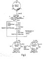
- the plasmid pBR322-EcoRI/HEV933 (hereinafter referred to as pHEV933 for short) with the 3.2 kb subtype adw HBV DNA inserted therein, which is described in Japanese published unexamined patent application (Kokai) No. 194897/1983 or in Nucleic Acids Res., 11, 1747 (1983), is subjected to double digestion with the restriction enzymes HpaI and EcoRI, to
- a DNA coding for P31 can be constructed by joining, to said fragment, an appropriate adapter con-5 ATGCAGTGG 3 ' t a i n i n g the sequence ( 3, ATGCAGTGG 5, ].
- pHBr330 the plasmid pBR322-BamHI/HBr330 (hereinafter referred to as pHBr330 for short) with the 3.19 kb subtype adr HBV DNA inserted therein, which is described in Japanese published unexamined patent application No.
- a DNA coding for P31 can be prepared by joining the above-mentioned adapter to this fragment.
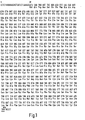
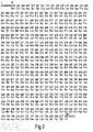
- the DNA coding for subtype adw HBsAg P31 is, for example, a DNA comprising the base sequence from the base pair No. 28 to No. 873 of the DNA sequence as shown in Fig. 1, and the DNA coding for subtype adr HBsAg P31 is, for example, a DNA comprising the base sequence from the base pair No. 10 to No. 855 of the DMA sequence as shown in Fig. 2.
- the P31-encoding DNA may be either of the viral origin or a chemically synthesized one.
- the DNA coding for subtype ayr or ayw HBsAg P31 can be prepared according to the same method as mentioned above.
- a recombinant DNA which allows the expression of the P31-encoding DNA can be constructed by insertion of this P31-encoding DNA at the 3'-end of a promoter region capable of functioning in each host (e.g.
- Escherichia coli Bacillus subtilis, yeast, animal cells).
- the promoter region may be any region which contains a site necessary for RNA polymerase binding and for the initiation of mRNA synthesis.
- a recombinant DNA which allows the expression of the P31-encoding DNA can be constructed by insertion of the P31-encoding DNA at the 3'-end of a promoter region capable of functioning in Escherichia coli.
- the P31-encoding DNA is inserted into an expression vector, such as pTRP601 or pTRP771, which is mentioned in Japanese published unexamined patent application No. 201796/1983, with the aid of T4 DNA ligase.
- a strain of Escherichia coli e.g. the strain C600, 294, W3110, RR1 or PR13
- the mixture by the known method [Cohen, S. N. et al., Proc. Natl. Acad. Sci. USA, 69, 2110 (1972)] or a modification thereof.
- the promoter to be used is not necessarily limited to the tro promoter (trp-p) but, for instance, the recA promoter [Japanese published unexamined patent application No. 65099/1984], lac promoter and ⁇ P L promoter may also be used.
- the transformant carrying the novel recombinant DNA containing the P31-encoding DNA as obtained in the above manner can be selected by using, for example, such phenotype as ampicillin resistance, tetracycline resistance or both ampicillin and tetracycline resistance.
- the following technique for finding out the strain carrying the novel recombinant plasmid DNA which contains the P31-encoding DNA from among the drug-resistant transformants obtained, the following technique, for instance, is used:
- One chain of the above-mentioned adapter 5' AATTCCACTGCATTGTAT 3 ' is labelled radioisotouically using Y- 32 P-ATP and T4 polynucleotide kinase.
- the thus-selected transformant is cultivated i0 1A71 908 per se known medium.
- the medium there may be mentioned, for example, L broth, Penassay broth, and M-9 medium supplemented with glucose and casamino acids [Miller, J., Experiments in Molecular Genetics, 431- 4 33 (Cold Spring Harbor Laboratory, New York, 1972)].
- an agent as 3 ⁇ -indolylacrylic acid for efficient promoter functioning.
- the cultivation of said transformant is carried out generally at 15°C to,43°C, preferably at 28°C to 40°C, for 2 to 24 hours, preferably for 4 to 16 hours, if necessary with aeration and/or stirring.
- the yeast transformant can be prepared in the following manner.
- Escherichia coli-yeast shuttle vector YEpl3 [Broach, J. R. et al., Gene, 8, 121 (1979)]
- pSHl5 or pSHl9 [Harashima, S. et al., Mol. Cell. B iol., 4, 771 (1984)]
- a yeast promoter region such as the reperessible acid phosphatase gene promoter region [Meyhack, B.
- the above-mentioned host strain of Escherichia coli is transformed by the method of Cohen et al. cited above.
- the thus-produced transformant which carries the novel recombinant DNA containing the P31-encoding DNA can be selected by using ampicillin resistance as the phenotype.
- the above-mentioned method is used in the same manner.
- the plasmid DNA is isolated from the thus-selected transformant by the alkaline extraction method [Birnboim, H. C. and Doly, J., Nucleic Acids Res., 7, 1513 (1979)] and used for the transformation of a yeast, for example a leucine-requiring strain of Saccharomyces cerevisiae such as AH 22 R - (leu2 his4 canl cir + pho ⁇ 0) [Proc. Natl. Acad. Sci.
- the yeast as the host is not limited to these but preferably is a strain of Saccharomyces cerevisiae.
- the yeast transformant obtained is cultivated on a per se known medium.
- the medium there may be mentioned, for example, Burkholder minimum medium [Bostian, K. L. et al., Proc. Natl. Acad. Sci. USA, 77, 4505 (1980)].
- the cultivation of the yeast transformant,obtained is carried out generally at 15°C to 40°C, preferably at 24°C to 37°C, for 10 to 96 hours, preferably for 24 to 72 hours, if necessary with aeration and/or stirring.
- the P31-encoding DNA is inserted at the 3'-end of a promoter region capable of functioning in Bacillus subtilis or aninal cells, and the host is transformed with the resultant recombinant DNA.
- P31 can be produced by cultivating the transformant.
- Escherichia coli and a yeast are more preferable as the host.
- the product P31 may be either in the glycosylated form or in the unglycosylated form.
- P31 obtainable from the transformants of Escherichia coli is substantially unglycosylated, but one molecule of carbohydrate may be bound thereto per molecule of P31.
- the P31 activity of the product can be determined, for example, by the direct immunoassay method [Fuiisawa, Y. et a l. , Nucleic Acids R es., 11, 3581 (1983)] which comprises binding the sample to a cyanogen bromide-activated cellulose paper followed by reaction with 125 I-anti-HBsAg antibody of Ausria II-125 (Dainabbott).
- the direct immunoassay method [Fuiisawa, Y. et a l. , Nucleic Acids R es., 11, 3581 (1983)] which comprises binding the sample to a cyanogen bromide-activated cellulose paper followed by reaction with 125 I-anti-HBsAg antibody of Ausria II-125 (Dainabbott).
- cells are collected by the conventional method.
- the cells may be subjected to an appropriate treatment such that the cells are suspended in a buffer containing a protein denaturing agent such as urea or guanidine hydrochloride and that the suspension is stirred at a cool place and then centrifuged to give a P31-containing supernatant, or that the cells are suspended in a buffer and disrupted by sonication, lysozyme, and/or freezing and thawing and then that a p 3l-containing supernatant is separated by centrifugation.
- a protein denaturing agent such as urea or guanidine hydrochloride
- the cells are disrupted by using Zymolyase (Kirin Brewery Co.) or mechanically by using glass beads.
- a surfactant such as Triton X-100 or deoxycholate, or a protein denaturing agent such as guanidine hydrochloride, whereby P31 can be extracted more advantageously.
- the separation and purification of the P31 protein from the above-mentioned extract is conducted in a purification process involving affinity-chromatographic treatment.
- affinity chromatography there may be mentioned affinity chromatography using polymerized human serum albumin (poly-HSA) as the ligand or antibody column treatment using an antibody to HBsAg, in particular a monoclonal antibody to HBsAg.
- poly-HSA polymerized human serum albumin
- the affinity chromatography using poly-HSA as the ligand is used most advantageously for the purification of the P31 protein.
- the carrier in affinity chromatography there may be used, for instance, Formyl-Cellulofine (Seika g aku Kogyo) or Affi-Gel 15 (Bio-Rad), and Formyl-Cellulofine is preferable anong others.
- Poly-HSA can be produced by polymerizing human serum albumin using a crosslinking agent (e. g . glutaraldehyde). This is allowed to bind to the above carrier using a reducing agent (e.g. NaCNBH 3 ), for instance, and the carrier-poly-HSA coupling product obtained, after washing if desired, is packed generally into a column for its use.
- a crosslinking agent e. g . glutaraldehyde
- a reducing agent e.g. NaCNBH 3
- the above-mentioned P31-containing solution (cell extract supernatant) is allowed to be adsorbed on the above-mentioned column equilibrated beforehand with a buffer [e.g. phosphate buffer] and then eluted with a buffer.
- Said buffer may contain an appropriate amount of a surfactant (e.g. Tween 20) or a protein denaturing agent (urea), for instance, and can be used as an adequate eluent by modifying the combination of such additives and the concentrations thereof.
- a surfactant e.g. Tween 20
- urea protein denaturing agent
- the P31 protein-containing eluate fractions are collected and, as desired, concentrated by ultrafiltration, for instance.
- the above concentrate desirably after reduction with an SH reagent such as dithiothreitol, be further subjected to chromatographic treatment using a hydrophobic column, such as high-performance liquid chromatography using a reversed-phase column or hydrophobic chromatography.
- an SH reagent such as dithiothreitol
- alkylated (C 1 to about C 18 ) silicone type carriers for example AP-0 202 300 A (C 8 ) and AP-224 300 A (C 8 ) (YMC-Shimakyu), Ultrapore RPSC (Beckman) and Hi-Pore RP-34 (Bio-Rad).
- AP-202 300 A (C 8 ) and AP-224 300 A (C 8 ) are preferable and AP-224 300 A (C 8 ) is more preferable.
- alkylated (C 1 to about C 18 ) carriers for exmaple Butyl-Toyopearl 650M (Toyo Soda Manufacturing) and Octyl-Sepharose CL-4B (Pharmacia).
- Butyl-Toyopearl 650M Toyo Soda Manufacturing
- Octyl-Sepharose CL-4B Pharmacia
- a C 1 to C 6 lower alkanol e.g. ethanol, propanol
- acetonitrile or the like is preferably used as the eluent, with the pH adjusted to 1.2 to 5.0 with trifluoroacetic acid, for instance.
- the rate of elution is preferably 0.1 to 100 ml/min, more preferably 0.5 to 30 ml/min.
- the P31 protein-containing fraction thus obtained may be lyophilized as desired to give a white powder.
- the enzyme immunoassay (ELISA) method which comprises reacting the sample with a poly-HSA-coated plastic plate and detecting the poly-HSA-bound P31 protein using a horseradish peroxidase (HRP)-coupled anti-HBsAg monoclonal antibody, or the radioimmunoassay (RIA) method which comprises detecting the poly-HSA-bound P31 protein using 125I-anti- HBsA g antibody of Ausria II - 125 ( D ainabbott, USA).
- ELISA enzyme immunoassay
- HRP horseradish peroxidase
- RIA radioimmunoassay
- the use of the production method according to the invention can give highly purified and substantially pure P 31 protein suitable for use as a drug, for instance.
- the HBsAg P31 protein which is substantially pure and has the following characteristic properties:
- the substantially pure P31 protein produced by the method of the invention has the same biological activities as the known HBsAg small particles produced from the blood of HBV-infected patients as the raw material have, and can be used as a vaccine for the diagnosis, prevention and/or treatment of HBV infection in the same manner as said HBsAg small particles.
- the determination of the activity (specific activity) of the P31 protein for the protein purity determination as described herein was performed by the ELISA method.
- a test solution P31 protein-containing solution
- Immunoplate II Nunc
- the poly-HSA obtained in Reference Example 4-(1) physically adsorbed thereon in advance After overnight reaction at 4°C, the plate was washed with PBS containing 5% bovine serum and 0.05% Tween 20 and, then, 100 ⁇ l of a horseradish peroxidase-bound anti-HBsAg monoclonal antibody solution was added to each well of the plate.
- the Escherichia coli plasmid pJA1 (50 ⁇ g) containing a Saccharomyces cerevisiae S288C-derived, repressible acid phosphatase gene (PH05)- and constitutive acid phosphatase gene (PH03)-containing, 7.9 kb DNA fragment [Kramer, R. A. and Anderson, N., Proc. Natl. Acad. Sci.
- a gel portion containing a 0.63 kb DNA fragment was sealed in a dialytie tube, the tube was immersed in a buffer for electrophoresis, and said DNA fragment was eluted electrically from the gel [McDonell, M. W. et al., J. Mol. Biol., 110, 119 (1977)].
- the liquid in the dialytic tube was extracted with phenol, followed by extraction with ether and addition of NaCl to a concentration of 0.2 M. Then, two volumes of cold ethanol was added and DNA precipitation was caused at -20°C.
- the plasmid pSH19 (1 ⁇ g) was treated with 2 units of the restriction enzyme BamEI and 2 units of the restriction enzyme SalI in 20 ⁇ l of a reaction mixture [10 mM Tris-HC1 (pH 8.0), 7 mM MgCl 2 , 100 mM NaCl, 2 mM 2-mercaptoethanol] at 37°C for 2 hours.
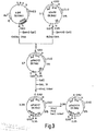
- the reaction mixture was subjected to electrophoresis using a 0.8% agarose slab gel, which was conducted under the same conditions as above. After electrophoresis, a 8.0 kb DNA fragment was isolated from the gel by the above-mentioned method. Following deproteinization with phenol, the DNA was precipitated with cold ethanol (cf. Fig. 3).
- a 400-ng portion of said 8.0 kb DNA fragment and a 200-ng portion of the above 0.63 kb DNA fragment were mixed and ligated together in 20 ⁇ l of a reaction mixture [66 mM Tris-HCl (pH 7.6), 6.6 mM MgCl 2 , 10 mM dithiothreitol, 1 mM ATP, 2 units of T4 DNA ligase (Takara Shuzo)] at 14°C. After overnight reaction, the reaction mixture was used for transforming Escherichia coli 294 by the above-cited method of Cohen et al.
- the plasmid DNA was isolated by the above-mentioned alkaline extraction method from each transformant selected based on ampicilline resistance as the indicator and examined for molecular weight and restriction enzyme cleavage pattern. In this way, a plasmid, pPH012, in which the 0.63 kb DNA fragment isolated from pJA1 had been inserted into the BamHI-SalI site of pSH19 was separated (cf. Fig. 3).
- a 3- ⁇ g portion of the plasmid pPHOl2 DNA was treated with 2 units of the restriction enzyme SalI in 20 ⁇ l of a reaction mixture [10 mM Tris-HC1 (pH 7.5), 7 mM MgCl 2 , 175 mM NaCl, 0.2 M EDTA, 7 mM 2-mercaptoethanol] at 37°C for 2 hours, followed by deproteinization with phenol and precipitation of DNA with cold ethanol.
- a reaction mixture 10 mM Tris-HC1 (pH 7.5), 7 mM MgCl 2 , 175 mM NaCl, 0.2 M EDTA, 7 mM 2-mercaptoethanol
- a 3- ⁇ g portion of this DNA was treated with 12 units of BAL31 nuclease (Bethesda Research Laboratories) in 50 ⁇ l of a reaction mixture [20 mM Tris-HC1 (pH 8.1), 12 mM CaCl 2 , 12 mM MgCl 2 , 1 mM EDTA] at 30°C for 2 minutes, followed by deproteinization with phenol and DNA precipitation with cold ethanol (cf. Fig. 3).
- the XhoI linker d(C CTCGAGG ) (200 ng) [New England BioLabs] was phosphorylated at the 5'-end thereof by treating with 3 units of T4 polynucleotide kinase [Takara Shuzo] in 50 ⁇ l of a reaction mixture [50 mM Tris-HCl (pH 7.6), 10 mM MgCl 2 , 10 mM 2-mercaptoethanol, 100 ⁇ M ATP] at 37°C for 1 hour.
- a 40-ng portion of the 5'-end phosphorylated XhoI linker [5'-P-d(CCTCGAGG)] and 400 ng of the above-mentioned BAL31-treated pPHO12 DNA were mixed and ligated together under the action of T4 DNA ligase under the same conditions as mentioned above.
- Escherichia coli 294 was transformed by the method of Cohen et al.
- the plasmid DNA was isolated by the alkaline extraction method mentioned above from each transformant selected on the basis of ampicillin resistance as the indicator, and a plasmid, pPH017, which gave a 0.55 fragment upon double digestion with BamHI and XhoI was selected.
- a 1-ug portion of the DNA precipitated was treated with 5 units of DNA polymerase I large fragment (New England BioLabs) in 30 ⁇ l of a reaction mixture [40 mM potassium phosphate buffer (pH 7.5), 6.6 mM MgCl 2 , 1 mM 2-mercaptoethanol, 33 ⁇ M dATP, 33 ⁇ M dGTP, 33 ⁇ M dTTP, 33 ⁇ M dCTP] at 12°C for 30 minutes to thereby convert the cohesive ends to blunt ends, followed by deproteinization with phenol and precipitation of DNA with cold ethanol.
- a reaction mixture [40 mM potassium phosphate buffer (pH 7.5), 6.6 mM MgCl 2 , 1 mM 2-mercaptoethanol, 33 ⁇ M dATP, 33 ⁇ M dGTP, 33 ⁇ M dTTP, 33 ⁇ M dCTP] at 12°C for 30 minutes to thereby convert the cohesive ends to blunt ends, followed by deproteinization with phenol
- the agarose g el was divided into fractions 1 to 10 depending on the size of DNA fragment.
- the agarose gel piece of each fraction was sealed in a dialytic tube and DNA was eluated electrically from the gel piece under the conditions described in Reference Example 1.
- the eluate was treated with phenol and the DNA was precipitated by addition of cold ethanol.
- a 0.5- ⁇ g portion of the DNA from each fraction was electrophoresed under the conditions described in Reference Example 1 using a 1% agarose slab gel and then the DNA was allowed to be adsorbed on a nitrocellulose filter (Schleicher and Schull) by the method of Southern [Southern, E.M., J. Mol. Biol., 98, 503 (1975)].
- Oligonuckotide 5'- TGAAGA TAAAGACAT-3', complementary to the oligonucleotide coding for 5 amino acids from the N-terminal of PGK [Dobson, M. J. et al., Nucleic Acids Res., 10, 2625 (1982)], was synthesized by the mehtod of Crea, R. et al. [Proc. Natl. Acad. Sci. USA, 75, .
- oligonucleotide was treated with 10 ⁇ Ci of Y-[ 32 P]ATP (Amersham) and 10 units of T4 polynucleotide kinase in 30 ⁇ l of a reaction mixture [50 mM Tris-HCl (pH 7.6), 10 mM MgCl 2 , 10 mM 2-mercaptoethanol] at 37°C for 30 minutes to thereby label the 5'-end with 32 P .
- a reaction mixture [50 mM Tris-HCl (pH 7.6), 10 mM MgCl 2 , 10 mM 2-mercaptoethanol] at 37°C for 30 minutes to thereby label the 5'-end with 32 P .
- 10 ul of 200 mM E DTA p H 8 .
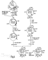
- a 50- ⁇ g portion of the plasmid p P KT3 D N A was treated with 50 units of the restriction enzyme HindIII in 100 ⁇ l of a reaction mixture [10 mM Tris-HCl (pH 7.5), 7 mM MgCl 2 , 60 mM NaCl] at 37°C for 2 hours, followed by 1% agarose slab gel electrophoresis under the conditions described in Refernce Example 1. After electrophoresis, the 2.95 kb DNA fragment was separated from the cel by the method described in Reference Example 1 (cf. Fig. 4).
- a 5- ⁇ g portion of said 2.95 kb DNA fragment was treated with 5 units of the restriction enzyme SalI in 20 ⁇ l of a reaction mixture [10 mM Tris-HC1 (pH 7.5), 7 mM MgCl 2 , 175 mM NaCl, 7 mM 2-mercaptoethanol] at 37°C for 3 hours and the digestion mixture was subjected to electrophoresis using a 1.2% agarose slab gel, which was conducted under the conditions described in Reference Example 1. After electrophoresis, a 2.1 kb DNA fragment was separated from the gel by the method described in Reference Example 1.
- a 0.5-pg portion of said 2.1 kb DNA fragment and 0.5 ⁇ g of a 3.74 kb DNA obtained by digestion of the plasmid pBR322 with HindIII and SalI were mixed and ligated together under the action of T4 DNA ligase under the conditions described in Reference Example 1.
- Escherichia coli DH1 was transformed, and a desired plasmid, pPKT101, was obtained from among the ampicilline-resistant transformants obtained (cf. Fig. 4).
- a 10-pg portion of said plasmid pPKT101 DNA was first treated with 10 units of the restriction enzyme SalI in 30 pl of a reaction mixture [10 mM Tris-HC1 (pH 7.5) , 7 mM MgCl 2 , 175 mM NaCl, 0.2 M EDTA, 7 mM 2-mercartoethanol] at 37°C for 3 hours, the digestion mixture was deproteinized with phenol, and DNA precipitation was caused with cold ethanol (SalI-digested pPTK101).
- a 1-pg portion of the Sail-digested pPTK101 was then treated with 10 units of BAL31 nuclease in 20 pl of a reaction mixtrue [20 mM Tris-HC1 (pH 8.1), 12 mM CaCl 2 , 12 mM MgCl 2 , 1 mM EDTA] at room temperature for 5 minutes. Immediately thereafter, 1 volume of phenol was added to terminate the reaction, followed by DNA precipitation with cold ethanol (B A L-digested pPTK101).
- a 50-ng portion of the phosphorylated Xhol linker described in Reference Example 1 and a 0.2-pg portion of BAL-digested pPTK101 were mixed and ligated together using T4 DNA ligase under the conditions described in Reference Example 1. Thereafter, Escherichia coli DH1 was transformed using the reaction mixtrue and, from among the ampicillin-resistant transformants obtained, a plasmid, pPKT567, with 0.69 kb from the Sall site of pPKT101 eliminated in the direction of the promoter region, was obtained. Analysis of the DNA base sequence by the dideoxynucleotide method proved that, in pPKT567, the BAL31 treatment had eliminated the PGK structural gene and up to 5'-vicinity region-24 (cf. Fig. 4).
- a 5-pg portion of the Escherichia coli-yeast shuttle vector p SHl9 was treated with 6 units of the restriction enzyme Sall in 20 ⁇ l of a reaction mixture [10 mM Tris-HC1 (pH 7.5), 7 mM MgCl 2 , 175 mM NaCl, 0.2 mM EDTA, 7 mM 2-mercaptoethanol] at 37°C for 2 hours, followed by deproteinization with phenol and precipitation with cold ethanol.
- a 1-pg portion of the DNA was treated with DNA polymerase I large fragment under the conditions as mentioned in Reference Example 1 were mixed and ligated cohesive end to a blunt end.
- a 500-ng portion of said DNA fragment and 50 ng of the phosphorylated XhoI linker as mentioned in Referance Example 1 were mixed and ligated together using T4 DNA ligase under the conditions described in Reference Example 1.
- Escherichia coli DH1 was transformed with the reaction mixture and, from among the amnicilin-resistant transformants obtained, a transformant carrying a plasmid, pSH19-l, in which the SalI site of pSH19 had been converted to a XhoI site was obtained (cf. Fig. 4).
- a 15-pg portion of said plasmid pSH19-1 DNA was treated with 24 units of the restriction enzyme HindIII in 100 pl of a reaction mixture [10 mM Tris-HCl (pH 7.5), 7 mM MgCl 2 , 60 mM NaCl] at 37°C for 10 minutes. Immediately thereafter, the reaction was terminated by adding 10 ⁇ l of 0.2 M EDTA. The reaction mixture was subjected to electrophoresis using a 0.7% agarose slab gel under the conditions described in Reference Example 1, and a 8.3 kb DNA fragment cleaved at one site with HindIII was separated from the gel by the method described in Reference Example 1.
- a 3-pg portion of said 8.3 kb DNA fragment was treated with 10 units of the restriction enzyme XhoI [Takara Shuzo] in 30 ⁇ l of a reaction mixtrue [10 mM Tris-HC1 (pH 7.5), 7 mM MgCl 2 , 100 mM NaCl, 7 mM 2-mercaptoethanol] at 37°C for 2 hours.
- the reaction mixture was electrophoresed using a 0.7% agarose slab gel under the conditions described in Reference Example 1. After electrophoresis, a 7.7 kb DNA fragment was separated from the gel by the method described in Reference Example 1 (cf. Fig. 4).
- a 10- ⁇ g portion of the plasmid pPKT567 DNA described in Reference Example 2-(2) was treated with 10 units each of the restriction enzymes HindIII and XhoI in 50 ⁇ l of a reaction mixture [50 mM Tris-HC1 (pH 7.6), 50 mM NaCl, 1 mM dithiothreitol, 10 mM MgCl 2 ] at 37°C for 2 hours. Thereafter, electrophoresis was performed using a 1.2% agarose slab gel under the conditions described in Reference Example 1, and a 1.40 kb DNA fragment was separated from the gel (cf. Fig. 4).
- 5'-AGCAACTCTAACCAT-3' complementary to that oligonucleotide of pgap491 [Holland, J. P. et al., J. Biol. Chem., 258, 5291 (1983)] of GLD which codes for the 5 amino acids from the N-terminal, was synthesized by the above-mentioned method of Crea, R. et al., labelled with 32P by the method described in Reference Example 2-(1), and used as the probe. Southern blotting was performed using the nitrocellulose filter described in Reference Example 2-(1) and said probe, whereupon the probe strongly hybridized with the sample of fraction No. 7 containing 2.0-2.3 kb DNA fragments.
- 100 ⁇ g of the plasmid pGLD9 DNA was treated with 50 units of the restriction enzyme HindIII in 200 ⁇ l of a reaction mixture [10 mM Tris-HCl (pH 7.5), 7 mM MgCl 2 , 60 mM NaCl] at 37°C for 3 hours, followed by electrophoresis using a 1.0% agarose slab gel under the conditions described in Reference Example 1. After electrophoresis, the 2.2 kb DNA fragment was separated from the gel by the method described in Reference Example 1.
- the percentage binding of poly-HSA to Cellulofine in the above procedure was about 73%, and there was obtained 40 ml of poly-HSA-Cellulofine containing about 24 mg of poly-HSA bound to each ml of Cellulofine.
- This gel was suspended in PBS containing 0.1 M ethanolamine, NaCNBH 3 (200 mg) was added and the mixture was shaken at room temperature for 3 hours, and the gel was then transferred onto a glass filter, washed in sequence with PBS, 3 M urea, 6 M guanidine hydrochloride and 25 mM sodium phosphate buffer (pH 7.5) and packed into a column having an inside diameter of 5 cm, giving a poly-HSA-Cellulofine column.
- the plasmid pBR322-BanHI/HBr330 DNA (also referred to as pHBr330 for short) as mentioned in Japanese published unexamined patent application No. 74985/1984 and in Nucleic Acids Res., 11, 1747 (1983) was prepared by the method described in Japanese published unexamined patent application No. 201796/1983 in Reference Example 1 thereof.
- Escherichia coli 294 was transformed and, from among the ampicillin-resistant transformants obtained, a plasmid pHBrP31 DNA composed of the above three DNAs ligated together was obtained (cf. Fig. 6).
- 1 ⁇ g of the plasmid pHBrP31 DNA was treated with 2 units of the restriction enzyme BamHI in 20 ⁇ l of a reaction mixture [10 mM Tris-HCl (pH 8.0), 7 mM MgCl 2 , 100 mM NaCl, 2 mM 2-mercaptoethanol] at 37°C for 2 hours, followed by deproteinization with phenol and DNA precipitation by addition of cold ethanol (BamHI-digested pHBrP31).
- a reaction mixture 10 mM Tris-HCl (pH 8.0), 7 mM MgCl 2 , 100 mM NaCl, 2 mM 2-mercaptoethanol
- pHBrP31-17 50 ⁇ g of said pHBrP31-17 was treated with 20 units each of the restriction enzymes ClaI and PstI [Takara Shuzo] in 100 ⁇ l of a reaction mixture [20 mM Tris-HC1 (pH 7.5), 10 mM MgCl 2 , 50 mM (NH 4 )-SO 4 ] at 37°C fpr 3 hours, followed by electrophoresis using a 1.0% agarose slab gel under the conditions described in Reference Example 1. After electrophoresis, a 1.42 kb DNA fragment was separated from the gel by the method described in Reference Example 1.
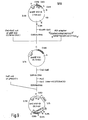
- pHBV933 The plasmid pBR-EcoRI/HBV933 DNA (referred to as pHBV933 for short) described in Japanese published unexamined patent applications Nos. 194897/1983 and 201796/1983 and in Nucleic Acids Res., 11, 1747 (1983) was prepared by the method described in Japanese published unexamined patent application No. 201796/1983 in Reference Example 1 thereof.
- Escherichia coli 294 was transformed using the reaction mixture, and the transformants obtained were screened by the method described in Reference Example 1, and a plasmid, pHBV933-5, in which the HpaI site of the plasmid pHBV933 had been converted to a PstI site was obtained (cf. Fig. 7).
- 500 ⁇ g of said plasmid pHBV933-5 DNA was treated with 500 units of the restriction enzyme PstI in 800 ⁇ l of a reaction mixture [20 mM Tris-HCl (pH 7.5), 10 mM MgCl 2 , 50 mM (NH 4 ) 2 SO 4 ] at 37°C for 20 minutes, immediately followed by deproteinization with phenol.
- Said reaction product was subjected to electrophoresis under the conditions described in Reference Example 1 using a 1.0% agarose slab gel. Thereafter, a 1.7 kb DNA fragment - a PstI partial digest - was separated from the gel by the method described in Reference Example 1 (cf. Fig. 7).
- a trp promoter-containing fragment of about 330 bp obtained by digesting the plasmid pTRP601 described in Japanese published unexamined patent application No. 201796/1983 with ClaI and HpaII [Takara Shuzo], a plasmid, pTRP P31-W2 (cf. Fig. 7), was completed.
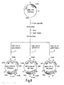
- Said plasmid separated from this transformant by the alkaline extraction method was used for transforming the host yeast Saccharomyces cerevisiae AH22R using the method of Hinnen et al. mentioned above, and a transformant yeast (AH22R /pPHO P31-R) carrying said plasmid was isolated (cf. Fig. 8).
- the host yeast strain K33-8D is transformed with said plasmid, and a yeast transformant (K33-8D/pPKT P31-R) is isolated (cf. Fig. 8).
- Example 3-(1) 200 ng of the 1.43 kb DNA fragment described in Example 3-(1) and the Sail-digested pGLD906-1 are ligated together under the conditions described in Reference Example 1 using T4 DNA ligase.
- a strain (294/pGLD P31-R) carrying a plasmid, pGLD P31-R, with the 1.43 kb DNA fragment containing a subtype adr P31-encoding DNA inserted therein in the same directionality as the GLD promoter is isolated.
- the host yeast K33-7B is transformed with said plasmid, and a yeast transformant (K33-7B/pGLD P31-R) is obtained (cf. Fig. 8).
- the reaction mixture was used for transforming Escherichia coli 294 and, from among the ampicillin-resistant transformants obtained, a plasmid, pHBV933-8, having a SalI site in lieu of the HpaI site of the plasmid pHBV933 was obtained (cf. Fig. 9).
- 100 ⁇ g of said plasmid pHBV933-8 was treated with 100 units each of the restriction enzymes EcoRI and Sail in 200 ⁇ l of a reaction mixture [100 mM Tris-HC1 (pH 7.5), 7 mM MgCl 2 , 50 mM NaCl, 7 mM 2-mercaptoethanol] at 37°C for 2 hours, and the reaction mixture was subjected to electrophoresis under the conditions described in Reference Example 1 using a 1.2% agarose slab gel. After electrophoresis, a 0.96 kb DNA fragment containing a subtype adw P31-encoding DNA fragment was separated from the gel by the method described in Reference Example 1 (cf. Fig. 9).
- Said plasmid was separated from said transformant by the alkaline extraction method and used for transforming the host yeast AH22R- by the method of Hinnen et al. mentioned above, and a yeast transformant (Saccharomyces cerevisiae AH22R /pPHO P31-W) carrying said plasmid was isolated (cf. Fig. 9).
- a plasmid containing a subtype adw P31-encoding DNA and the PGK promoter or GLD promoter can also be constructed by the above procedure.
- 50 ug of the above-mentioned plasmid pTRP P31- W 2 is treated with 20 units of the restriction enzyme PstI in 100 ⁇ l of a reaction mixture [10 mM Tris-HCl (pH 7.5), 10 mM MgCl 2 , 50 mM NaCl, 1 mM dithiothreitol] at 37°C for 20 minutes for partial digestion, and the reaction mixture is subjected to electrophoresis using a 0.8% agarose slab gel under the conditions described in Reference Example 1. Thereafter, a 4.6 kb linear DNA molecule which is a product of cleavage with PstI only at one site is separated from the gel by the method described in Reference Example 1 (cf. Fig. 10).
- 0.8 ⁇ g of said 0.98 kb DNA fragment is treated in 20 ⁇ l of a reaction mixture [33 mM Tris-acetate (pH 7.9), 66 mM potassium acetate, 10 mM magnesium acetate, 5 mM dithiothreitol] using 2.5 units of T4 DNA polymerase at 37°C for 15 minutes to render the cohesive ends blunt, followed by deproteinization with phenol and precipitation of a DNA with cold ethanol.
- a SalI linker is joined to said 0.98 kb DNA fragment, followed by SalI treatment to render the cohesive end blunt.
- a Sepharose 4B column a fraction containing a 0.99 kb DNA fragment (subtype adw P31-encoding DNA) is collected and said DNA is precipitated with cold ethanol.
- Said plasmid pPHO P31- W is separated from said transformant by the alkaline extraction method and used for transforming the host yeast AH22R by the method of Hinnen et al. mentioned above, and a yeast transformant (AH22R /pPHOP31-W) carrying said plasmid is isolated (cf. Fig. 10).
- Each transformant carrying a P31 gene expression plasmid as obtained in Example 1 or 2 was cultivated in M-9 medium containing 1.0% glucose and 1.0% casamino acids at 37°C for 6 hours and, then, cells were harvested and washed with a buffer [30 mM Tris-HC1 (pH 8.0), 50 mM NaCl, 5 mM EDTA].
- the cells were suspended in a lyzing solution comprising 10 mM Tris-HC1 (pH 8.0), 5 mM EDTA, 1 mM phenylmethylsulfonyl fluoride and 5 mg/ml lysozyme and lysis was effected.
- One unit of HBsAg corresponds to the count value obtained with that amount of the 125I-anti-HBsAg antibody attached to the A usria II-125 kit which is bound to 1 ng of HBsAg small particles.
- Each yeast transformant obtained in Example 3 or 4 and carrying a P31 gene expression plasmid was cultivated in Burkholder medium or a low phosphate level modification thereof at 30°C for 2 days and, thereafter, cells were harvested and washed with physiological saline.
- the cells were converted to the spheroplast state by treatment with Zymolyase [Seikagaku Kogyo] according to the method of Miyanohara, A. et al. Thereto was added 0.1% Triton X-100 for extraction of P31. The lysate was centrifuged at room temperature at 15,000 rpm for 15 minutes to give a supernatant. This supernatant was assyed for P31 activity using Auszyme II [Abbott]. The results obtained are shown in Table 3. The yields of P31 were each calculated in terms of the yield per liter of broth.
- the supernatant obtained above was passed through the poly-HSA-Cellulofine column (5 x 2 cm), which was obtained in Reference Example 4 and equilibrated with 25 mM sodium phosphate buffer (pH 7.5),for adsorpion of the P31 protein thereon.
- the column was washed in sequence with 300 ml of PBS containing 0.05% Tween 20, 220 ml of PBS containing 0.05% Tween 20 and 0.5 M NaCl, 80 ml of PBS containing 0.05% Tween 20 and 300 ml of 25 mM sodium phosphate buffer (pH 7.5) containing 4 M urea and then P31 was eluted with 300 ml of a sodium phosphate buffer (pH 7.5) containing 7.5 M urea.
- the active fraction (250 ml) was concentrated to 7.2 ml (2.3 x 10 6 units/ml) using a YM-5 memberane (Amicon, USA). To 7 ml of the obtained concentrate was added 140 mg of DTT and, after reduction at 80°C for 15 minutes, the concentrate was filtered through a membrane filter (ACRODISC; 0.2 ⁇ m in pore size; Gelman).
- a 500-ul portion of the filtrate obtained above 0 was allowed to be adsorbed on an AP202 300 A (C 8 ) reversed-phased column (YMC-Shimakyu) and high-performance liquid chromatography was performed using a trifluoroacetic acid-acetonitrile-n-propyl alcohol system as the eluent system.
- the substantially pure P31 protein (subtype adr) produced by the above method had the following characteristic properties:
- SDS-polyacrylamide slab gel electrophoresis was performed by the method of Laemmli [Nature, 227, 680 (1970)], followed by staining with the silver staining reagent "Daiichi” (Daiichi K agaku, Tokyo) and the P31 protein was detected as a single band (cf. Fig. 11).
- the molecular weight of the p31 protein was calculated to be about 32,000 daltons based on the result of SDS-polyacrylamide slab gel electrophoresis (cf. Fig. 11).
- a glass tube for hydrolysis there was placed 20 ug of the P31 protein. After addition of constant boiling point hydrochloric acid containing 4% thioglycolic acid, the tube was sealed under redcued pressure, and hydrolysis was performed at 110°C for 24; 48, 72 or 96 hours. After hydrolysis, the tube was opened, the hydrochloric acid was removed under reduced pressure, the residue was dissolved in 0.02 N hydrochloric acid, and amino acid analysis was performed using a Hitachi model 835 amino acid analyzer.
- Cystine and cysteine were determined, according to Hirs' method [Methods in Enzynol., 11, 197 (1967) ], as cysteric acid on the amino acid analyzer after performic acid oxidation of the P31 protein and hydrolysis in constant boiling point hydrochloric acid under reduced pressure for 24 hours.
- the amino acid analysis data were calculated each as an average of the values obtained by hydrolysis for 24, 48 and 72 hours.
- isoleucine, leucine and phenylalanine however, the values obtained after 96 hours of hydrolysis were employed and, for serine and threonine, the values were determined by extrapolation to hour 0 (zero) of hydrolysis. The results thus obtained are shown in Table 4.
- the N-terminal amino acid sequence was analyzed by applying, to 62 ⁇ g of the P31 protein, the automatic Edman degradation method using a Gas-phase Protein Se q uenator (Applied Biosystems model 470A, USA). Phenyl- thiohydantoin-amino acids (PTH-amino acids) were identified by high-performance liquid chromatography using a Micro Pak C18-3 SP-DS column (Varian, USA). The PTH-amino acids detected in the respective steps are shown in Table 5.
- Example 3-(1) 200 ng of the 1.43 kb DNA fragment described in Example 3-(1) and the SalI-digested pGLD906-1 were ligated together under the conditions described in Reference Example 1 using T4 DNA ligase.
- a strain (294/pGLD P31-R) carrying a plasmid, pGLD P31-R, with the 1.43 kb DNA fragment containing a subtype adr P31-encoding DNA inserted therein in the same directionality as the GLD promoter was isolated.
- the host yeast AH22R was transformed with said plasmid, and a yeast transformant ( A H22R-/pGLD P31-R) is obtained (cf. Fig. 8).
- Each yeast transformant obtained in Example 8 or 9 and carrying a P31 gene expression plasmid was cultivated in Burkholder medium or a low phosphate level modification thereof at 30°C for 2 days and, thereafter, cells were harvested and washed with physiological saline.
- the cells were converted to the spheroplast state by treatment with Zymolyase [Seikagaku Kogyp] according to the method of Miyanohara, A. et al. Thereto was added 0.1% Triton X-100 for extraction of P31. The lysate was centrifuged at room temperature at 15,000 rpm for 15 minutes to give a supernatant. This supernatant was assayed for P31 activity using Auszyme II [Abbott]. The results obtained are shown in Table 6. The fields of P31 were each calculated in terms of the yield per liter of broth.
- Plasmids were extracted from Escherichia coli 294 carrying the plasmid for expression of P31 gene as obtained in Examples 1 and 2, and Escherichia coli C600 were transformed with said plasmids to obtain Excherichia coli C600/pTRP P31-R and Escherichia coli C600/pTRP P31-W2.
- Escherichia coli C600 strain carrying a P31 gene expression plasmid as obtained in Example 11 was cultivated in M-9 medium containing 2.0% glucose and 1.0% casamino acids at 37°C for 8 hours and, then, cells were harvested and washed with a buffer [30 mM Tris-HCl (pH 8.0), 50 mM NaCl, 5 mM EDTA]. The cells were suspended in a lyzing solution comprising 10 mM Tris-HCl (pH 8.0), 5 mM EDTA, 1 mM phenylmethylsulfonyl fluoride and 5 mg/ml lysozyme and lysis was effected.
- One unit of HBsAg corresponds to the count value obtained with that amount of the 125 I-anti-HBsAg antibody attached to the Ausria II-I25 kit which is bound to 1 ng of HBsAg small particles.
- the substantially pure P31 protein (subtype adr) produced by the above method had the same characteristic properties as P31 protein (subtype adr) obtained in xample 7.
- Cystine and cysteine were determined, according to the method of Hirs as cysteic acid on the amino acid analyzer after performic acid oxidation of the P31 protein and hydrolysis in constant boiling point hydrochloric acid under reduced pressure for 24 hours. Quantitative analysis for threonine, serin and tryptophan was performed after hydrolysis of P31 protein in constant boilind hydrochloric acid containing 4% thioglycolic acid under reduced pressure for 16, 20, 24 and 30 hours. The amino acid analysis data were calculated each as an average of the values obtained by hydrolysis for 25, 50 and 180 minutes. For tryptophan, however, the value obtained after 24 hours of hydrolysis was employed and, for serine and threonine, the values were determined by extrapolation to hour 0 (zero) of hydrolysis. The results thus obtained are shown in Table 8.
- IFO and FRI represent "Institute for Fermentation, Osaka” (Japan) and “Fermentation Research Institute, Agency of Industrial Science and Technology, Ministry of International Trade and Industry, Japan", respectively.
- E. coli and S. cerevisiae represent Escherichia coli and Saccharomyces cerevisiae, respectively,
- FERM BP represents the accession number of a deposit under the Budapest Treaty
- FERM P represents the accession number before conversion to a deposit under the Budapest Treaty.
Landscapes
- Health & Medical Sciences (AREA)
- Life Sciences & Earth Sciences (AREA)
- Genetics & Genomics (AREA)
- Chemical & Material Sciences (AREA)
- Organic Chemistry (AREA)
- Engineering & Computer Science (AREA)
- Wood Science & Technology (AREA)
- Biotechnology (AREA)
- Biomedical Technology (AREA)
- General Engineering & Computer Science (AREA)
- Bioinformatics & Cheminformatics (AREA)
- General Health & Medical Sciences (AREA)
- Zoology (AREA)
- Biophysics (AREA)
- Biochemistry (AREA)
- Molecular Biology (AREA)
- Virology (AREA)
- Mycology (AREA)
- Microbiology (AREA)
- Medicinal Chemistry (AREA)
- Plant Pathology (AREA)
- Physics & Mathematics (AREA)
- Chemical Kinetics & Catalysis (AREA)
- Oncology (AREA)
- Nuclear Medicine, Radiotherapy & Molecular Imaging (AREA)
- Veterinary Medicine (AREA)
- Gastroenterology & Hepatology (AREA)
- General Chemical & Material Sciences (AREA)
- Pharmacology & Pharmacy (AREA)
- Proteomics, Peptides & Aminoacids (AREA)
- Animal Behavior & Ethology (AREA)
- Communicable Diseases (AREA)
- Public Health (AREA)
- Micro-Organisms Or Cultivation Processes Thereof (AREA)
- Preparation Of Compounds By Using Micro-Organisms (AREA)
- Peptides Or Proteins (AREA)
- Medicines Containing Antibodies Or Antigens For Use As Internal Diagnostic Agents (AREA)
- Saccharide Compounds (AREA)
Abstract
Description
- This invention relates to hepatitis B virus surface antigen and production thereof. More particularly, this invention relates to (1) a recombinant DNA wherein a DNA coding for hepatitis B virus surface antigen P31 is inserted into the 3' end of a promoter, (2) production of said recombinant DNA, (3) a transformant carrying said recombinant DNA, (4) production of said transformant, (5) non-glycosylated hepatitis B virus surface antigen P31 protein and (6) production of hepatitis B virus surface antigen P31.
- Type B hepatitis is a viral disease frequently encountered in particular in tropical Africa, Southeast Asia and Far East and, epidemiologically, it is suggested that hepatitis B may cause chronic hepatitis, hepatic cirrhosis and, further, primary liver cancer. The pathogen is hepatitis B virus (hereinatter referred to as HBV for short), one of DNA viruses, which occurs as spherical particles having a diameter of 42 nm as called Dane . particles after the discoverer, In the external layer of said particles, there exists HBV surface antigen (hereinafter referred to as HBsAg for short), which is classifiable into subtypes such as adr, adw, ayr and ayw according to the differing antigenicity, and the subtypes adw and adr are popular in Japan.
- In the blood of patients with hepatitis B, tiny particles and tubular particles are detected in addition to Dane particles and, in these particles, there is observed the same type of HBsAg as found in Dane particles. It is known, with other viruses, that an antibody to the surface antigen of a virus can prevent the infection with said virus and, in the case of HBV, too, there seems to be the possibility of vaccine production against hepatitis B on the basis of HBsAg. However, human and chimpanzee are the only animal species that can be infected with HBV, so that all the attempts so far made to cause infection of cultured cells with HBV have failed. Therefore, for the time being, the blood of human infected patients is the only available source of HBsAg and the small-sized particles obtained can satisfy only the requirement for its use as a material for diagnostic reagents but are by no means sufficient for large-scale vaccine production.
- Recent advancements in molecular biology have already made it possible to transform bacteria by introducing a DNA coding for a nonbacterial protein thereinto. If the expression of the structural gene coding for HBsAg (hereinafter referred to as HBsAg gene for short) in bacteria becomes possible as a result of application of recombinant DNA techniques, large-quantity production of HBsAg free of the risk of HBV infection will expectedly become possible and there will be opened the way for practical use of the hepatitis B vaccine.
- As for the subtype ayw, which is most frequently encountered in Europe and America among the currently known four subtypes adw, adr, ayw and ayr, the location and base sequence of the HBsAg gene have been determined [Galibert, F. et al., Nature, 2ε1, 646 (1979); Charnay, P. et al., Nucleic Acids Res., 7, 335 (1979)] and the expression of said gene as a hybrid protein in Escherichia coli has been reported [Charnay, P. et al., Nature, 286, 893 (1980); Edman, J. C. et al., Nature, 291, 503 (1981)].
- As regards the subtypes adw and adr, which are encountered frequently in Japan, some of the present inventors successfully created a DNA containing the HBsAg gene, determined the DNA base sequence of said gene and the location thereof on the genome and, furthermore, opened the way for large-quantity production of HBsAg by cultivating a transformant carrying said recombinant DNA (Japanese published unexamined patent applications Nos. 194897/1983, 201796/1983 and 74985/1984).
- Recently, Machida, A. et al. [Gastroenterology, 85, 268 (1983); ibid., 86, 910 (1984)] demonstrated that, in the small-size particles of HBsAg obtained from the plasma of patients positively responding to the hepatitis B virus e antigen, there exist the P31 protein (
molecular weight 31 kilodaltons) and P35 protein (conjugated protein which is sugar-bound p31 and has a molecular weight of 35 kilodaltons) in addition to the so-far identified main peptides such as P-I (molecular weight 22-24 kilodaltons) and P-II (molecular weight 25-29.5 kilodaltons) [Peterson, D. L. et al., Proc. Natl. Acad. Sci. USA, 74, 1530 (1977)]. P31 is composed of P-I and 55 amino acid residues of the pre-S region as added to the N-terminal of P-I, and it has also been demonstrated that the polymerized human serum albumin (poly-HSA) receptor occurs in said region. Furthermore, the 1984 report cited above shows that the above-mentioned P31 protein has a sugar chain. On the other hand, it is considered that, since said receptor is also found. on the hepatocyte surface, the proliferation takes place after the occurrence of Dane particle-hepatocyte adhesion through poly-HSA. Therefore, it is expected that the HBV infection can be prevented more efficiently if the poly-HSA receptor on Dane particles can be masked with an antibody to P31, since, said particles are no more able to bind with hepatocytes. Disclosure of the Invention - This invention relates to hepatitis B virus surface antigen and production thereof. More particularly, this invention provides (1) a recombinant DNA wherein a DNA coding for hepatitis B virus surface antigen P31 is inserted into the 3' end of a promoter, '(2) production of said recombinant DNA, (3) a transformant carrying said recombinant DNA, (4) production of said transformant, (5) non-glycosylated hepatitis B virus surface antigen P31 protein and (6) production of hepatitis B virus surface antigen P31. Further, this invention provides a method for producing the hepatitis B virus surface antigen P31 protein which comprises cultivating a transformant carrying a DNA having a base sequence coding for hepatitis B virus surface antigen P31, accumulating the hepatitis B virus surface antigen P31 in the culture, collecting the P31-containing solution and subjecting the P31-containing solution to a purification process involving affinity-chromatographic treatment.
- The DNA coding for the hepatitis B virus surface antigen P31, which is to be used in the practice of the invention, may be of any subtype (adr, adw, ayr or ayw) and can be prepared, for example, by the methods described hereinbelow. Thus, the plasmid pBR322-EcoRI/HEV933 (hereinafter referred to as pHEV933 for short) with the 3.2 kb subtype adw HBV DNA inserted therein, which is described in Japanese published unexamined patent application (Kokai) No. 194897/1983 or in Nucleic Acids Res., 11, 1747 (1983), is subjected to double digestion with the restriction enzymes HpaI and EcoRI, to
- give a 961 bp DNA fragment which contains a part of the pre-S region. A DNA coding for P31 can be constructed by joining, to said fragment, an appropriate adapter con-5 ATGCAGTGG3' taining the sequence (3,ATGCAGTGG5,]. Alternatively, the plasmid pBR322-BamHI/HBr330 (hereinafter referred to as pHBr330 for short) with the 3.19 kb subtype adr HBV DNA inserted therein, which is described in Japanese published unexamined patent application No. 74985/1984 or in Nucleic Acids Res., 11, 1747 (1983), is subjected to double digestion with the restriction enzymes EcoRI and BamHI, whereby a 1398 bp DNA fragment containing a part of the pre-S region can be obtained. A DNA coding for P31 can be prepared by joining the above-mentioned adapter to this fragment.
- The DNA coding for subtype adw HBsAg P31 is, for example, a DNA comprising the base sequence from the base pair No. 28 to No. 873 of the DNA sequence as shown in Fig. 1, and the DNA coding for subtype adr HBsAg P31 is, for example, a DNA comprising the base sequence from the base pair No. 10 to No. 855 of the DMA sequence as shown in Fig. 2.
- The P31-encoding DNA may be either of the viral origin or a chemically synthesized one.
- The DNA coding for subtype ayr or ayw HBsAg P31 can be prepared according to the same method as mentioned above.
- A recombinant DNA which allows the expression of the P31-encoding DNA can be constructed by insertion of this P31-encoding DNA at the 3'-end of a promoter region capable of functioning in each host (e.g.
- The promoter region may be any region which contains a site necessary for RNA polymerase binding and for the initiation of mRNA synthesis.
- For use of a strain of Escherichia coli, for instance, as the host, a recombinant DNA which allows the expression of the P31-encoding DNA can be constructed by insertion of the P31-encoding DNA at the 3'-end of a promoter region capable of functioning in Escherichia coli. For instance, the P31-encoding DNA is inserted into an expression vector, such as pTRP601 or pTRP771, which is mentioned in Japanese published unexamined patent application No. 201796/1983, with the aid of T4 DNA ligase. A strain of Escherichia coli (e.g. the strain C600, 294, W3110, RR1 or PR13) is transformed with the mixture by the known method [Cohen, S. N. et al., Proc. Natl. Acad. Sci. USA, 69, 2110 (1972)] or a modification thereof.
- The promoter to be used is not necessarily limited to the tro promoter (trp-p) but, for instance, the recA promoter [Japanese published unexamined patent application No. 65099/1984], lac promoter and λPL promoter may also be used.
- The transformant carrying the novel recombinant DNA containing the P31-encoding DNA as obtained in the above manner can be selected by using, for example, such phenotype as ampicillin resistance, tetracycline resistance or both ampicillin and tetracycline resistance. For finding out the strain carrying the novel recombinant plasmid DNA which contains the P31-encoding DNA from among the drug-resistant transformants obtained, the following technique, for instance, is used: One chain of the above-mentioned adapter 5' AATTCCACTGCATTGTAT 3' is labelled radioisotouically using Y-32P-ATP and T4 polynucleotide kinase. By using this as the probe and using the per se known colony hybridization method [Grunstein, M. and Hogness, D. S., Proc. Natl. Acad. Sci. USA, 72, 3961 (1975)], a positively responding clone can surely be found out from among the drug-resistant transformants already obtained.
- The thus-selected transformant is cultivated i0 1A71 908 per se known medium. As the medium, there may be mentioned, for example, L broth, Penassay broth, and M-9 medium supplemented with glucose and casamino acids [Miller, J., Experiments in Molecular Genetics, 431-433 (Cold Spring Harbor Laboratory, New York, 1972)]. There can be added, as necessary, such an agent as 3 β-indolylacrylic acid for efficient promoter functioning.
- The cultivation of said transformant is carried out generally at 15°C to,43°C, preferably at 28°C to 40°C, for 2 to 24 hours, preferably for 4 to 16 hours, if necessary with aeration and/or stirring.
- When a yeast, for instance, is used as the host, the yeast transformant can be prepared in the following manner. Into the Escherichia coli-yeast shuttle vector YEpl3 [Broach, J. R. et al., Gene, 8, 121 (1979)], pSHl5 or pSHl9 [Harashima, S. et al., Mol. Cell. Biol., 4, 771 (1984)], there is inserted a yeast promoter region such as the reperessible acid phosphatase gene promoter region [Meyhack, B. et al., EMBO J., 6, 675 (1982)], the promoter region of the glyceraldehyde-3-phosphate dehydrogenase gene [Holland, J. P. and Holland, M. J., J. Biol. Chem., 255, 2596 (1980)] or the promoter region of the 3-phosphoglycerate kinase gene [Dobson, M. J. et al., Nucleic Acids Res., 10, 2625 (1982)], and the P31-encoding DNA is joined thereto just behind said rerion under the action of T4 DNA ligase. Using this reaction mixture, the above-mentioned host strain of Escherichia coli is transformed by the method of Cohen et al. cited above. The thus-produced transformant which carries the novel recombinant DNA containing the P31-encoding DNA can be selected by using ampicillin resistance as the phenotype. For finding out the strain carrying the novel recombinant plasmid DNA having the P31- encoding DNA from among the resistant strains obtained, the above-mentioned method is used in the same manner.
- The plasmid DNA is isolated from the thus-selected transformant by the alkaline extraction method [Birnboim, H. C. and Doly, J., Nucleic Acids Res., 7, 1513 (1979)] and used for the transformation of a yeast, for example a leucine-requiring strain of Saccharomyces cerevisiae such as AH22R- (leu2 his4 canl cir+ phoε0) [Proc. Natl. Acad. Sci. USA, 80, 1 (1983)] or AH22R-- derived K33-7B (Pho80-AH22, pho8-2) or K33-8D (pho80-AH22, pho8-2 trpl) by the known method [Hinnen, A. et al., Proc. Katl. Acad. Sci. USA, 75, 1927 (1978)] or a modification thereof. The yeast as the host is not limited to these but preferably is a strain of Saccharomyces cerevisiae.
- The yeast transformant obtained is cultivated on a per se known medium. As the medium, there may be mentioned, for example, Burkholder minimum medium [Bostian, K. L. et al., Proc. Natl. Acad. Sci. USA, 77, 4505 (1980)].
- The cultivation of the yeast transformant,obtained is carried out generally at 15°C to 40°C, preferably at 24°C to 37°C, for 10 to 96 hours, preferably for 24 to 72 hours, if necessary with aeration and/or stirring.
- For using Bacillus subtilis or animal cells, for instance, as the host, the P31-encoding DNA is inserted at the 3'-end of a promoter region capable of functioning in Bacillus subtilis or aninal cells, and the host is transformed with the resultant recombinant DNA. P31 can be produced by cultivating the transformant. However, Escherichia coli and a yeast are more preferable as the host.
- The product P31 may be either in the glycosylated form or in the unglycosylated form. P31 obtainable from the transformants of Escherichia coli is substantially unglycosylated, but one molecule of carbohydrate may be bound thereto per molecule of P31.
- It is generally known that the growth of an HBsAg DNA-carrying transformant is inhbited by the production of surface antigen gene products. In accordance with the invention, however, the growth inhibition is removed and the yield of P31 is increased as a result of the use of the P31-encoding DNA.
- The P31 activity of the product can be determined, for example, by the direct immunoassay method [Fuiisawa, Y. et al., Nucleic Acids Res., 11, 3581 (1983)] which comprises binding the sample to a cyanogen bromide-activated cellulose paper followed by reaction with 125I-anti-HBsAg antibody of Ausria II-125 (Dainabbott).
- After cultivation, cells are collected by the conventional method. In the case of a transformant of Escherichia coli, the cells may be subjected to an appropriate treatment such that the cells are suspended in a buffer containing a protein denaturing agent such as urea or guanidine hydrochloride and that the suspension is stirred at a cool place and then centrifuged to give a P31-containing supernatant, or that the cells are suspended in a buffer and disrupted by sonication, lysozyme, and/or freezing and thawing and then that a p3l-containing supernatant is separated by centrifugation. It is preferable among others, however, to suspend the cells collected in a buffer, add lysozyme, homogenize for causing lysis, add a buffer containing urea (3-10 M), stir the mixture (at 0 to 10°C for 0.5 to 8 hours) and then collect a supernatant by centrifugation.
- In the case of a yeast transformant, the cells are disrupted by using Zymolyase (Kirin Brewery Co.) or mechanically by using glass beads. Thereto is added a surfactant such as Triton X-100 or deoxycholate, or a protein denaturing agent such as guanidine hydrochloride, whereby P31 can be extracted more advantageously.
- The separation and purification of the P31 protein from the above-mentioned extract is conducted in a purification process involving affinity-chromatographic treatment.
- As the affinity chromatography, there may be mentioned affinity chromatography using polymerized human serum albumin (poly-HSA) as the ligand or antibody column treatment using an antibody to HBsAg, in particular a monoclonal antibody to HBsAg.
- The affinity chromatography using poly-HSA as the ligand is used most advantageously for the purification of the P31 protein.
- As the carrier in affinity chromatography, there may be used, for instance, Formyl-Cellulofine (Seikagaku Kogyo) or Affi-Gel 15 (Bio-Rad), and Formyl-Cellulofine is preferable anong others.
- Poly-HSA can be produced by polymerizing human serum albumin using a crosslinking agent (e.g. glutaraldehyde). This is allowed to bind to the above carrier using a reducing agent (e.g. NaCNBH3), for instance, and the carrier-poly-HSA coupling product obtained, after washing if desired, is packed generally into a column for its use.
- For purifying the P31 protein by affinity chromatography with poly-HSA as the ligand, the above-mentioned P31-containing solution (cell extract supernatant) is allowed to be adsorbed on the above-mentioned column equilibrated beforehand with a buffer [e.g. phosphate buffer] and then eluted with a buffer. Said buffer may contain an appropriate amount of a surfactant (e.g. Tween 20) or a protein denaturing agent (urea), for instance, and can be used as an adequate eluent by modifying the combination of such additives and the concentrations thereof.
- The P31 protein-containing eluate fractions are collected and, as desired, concentrated by ultrafiltration, for instance.
- It is preferable that the above concentrate, desirably after reduction with an SH reagent such as dithiothreitol, be further subjected to chromatographic treatment using a hydrophobic column, such as high-performance liquid chromatography using a reversed-phase column or hydrophobic chromatography.
- As the carrier in the above-mentioned high-performance liquid chromatography, there may be mentioned alkylated (C1 to about C18) silicone type carriers, for example AP-0 202 300 A (C8) and AP-224 300 A (C8) (YMC-Shimakyu), Ultrapore RPSC (Beckman) and Hi-Pore RP-34 (Bio-Rad). Among others AP-202 300 A (C8) and AP-224 300 A (C8) are preferable and AP-224 300 A (C8) is more preferable.
- As the carrier in hydrophobic chromatography, there may by mentioned alkylated (C1 to about C18) carriers, for exmaple Butyl-Toyopearl 650M (Toyo Soda Manufacturing) and Octyl-Sepharose CL-4B (Pharmacia). The use of high-performance liquid chromatography using a reversed-phase column is particularly advantageous.
- In the above-mentioned high-performance liquid chromatography using a reversed-phase column, water, a C1 to C6 lower alkanol (e.g. ethanol, propanol), acetonitrile or the like is preferably used as the eluent, with the pH adjusted to 1.2 to 5.0 with trifluoroacetic acid, for instance. The rate of elution is preferably 0.1 to 100 ml/min, more preferably 0.5 to 30 ml/min.
- The P31 protein-containing fraction thus obtained may be lyophilized as desired to give a white powder.
- For determining the purity (specific acitivity) of the thus-produced P31 protein, there can be used, for example, the enzyme immunoassay (ELISA) method which comprises reacting the sample with a poly-HSA-coated plastic plate and detecting the poly-HSA-bound P31 protein using a horseradish peroxidase (HRP)-coupled anti-HBsAg monoclonal antibody, or the radioimmunoassay (RIA) method which comprises detecting the poly-HSA-bound P31 protein using 125I-anti- HBsAg antibody of Ausria II-125 (Dainabbott, USA).
- The use of the production method according to the invention can give highly purified and substantially pure P31 protein suitable for use as a drug, for instance.
- For example, by purifying P31 produced in Escherichia coli carrying a DNA having a base sequence coding for HBsAg P31 using the method of the invention, there can be obtained the HBsAg P31 protein which is substantially pure and has the following characteristic properties:
- (1) In SDS-polyacryiamide slab gel electrophoresis, it is homogeneous and its molecular weight as determined by said electrophoresis (under reducing conditions) is 32,000 + 1,000 daltons.
- (2) It contains methionine (or formylated methionine) or glutamine as the amino-terminal amino acid.
- (3) It contains isoleucine as the carboxy-terminal amino acid.
- (4) It binds poly-HSA.
- (5) It has a specific acitivity of not less than 1 x 10⁶ units/mg.
- The substantially pure P31 protein produced by the method of the invention has the same biological activities as the known HBsAg small particles produced from the blood of HBV-infected patients as the raw material have, and can be used as a vaccine for the diagnosis, prevention and/or treatment of HBV infection in the same manner as said HBsAg small particles.
- The determination of the activity (specific activity) of the P31 protein for the protein purity determination as described herein was performed by the ELISA method. Thus, a test solution (P31 protein-containing solution) was distributed, in 100-µl portions, into the wells of an Immunoplate II (Nunc) with the poly-HSA obtained in Reference Example 4-(1) physically adsorbed thereon in advance. After overnight reaction at 4°C, the plate was washed with PBS containing 5% bovine serum and 0.05% Tween 20 and, then, 100 µl of a horseradish peroxidase-bound anti-HBsAg monoclonal antibody solution was added to each well of the plate. After 2 hours of reaction at 37°C, the plate was washed again with the buffer mentioned above, followed by addition of 100 µl of citrate-phosphate buffer (pH 5.0) containing o-phenylenediamine (4 mg/10 ml) and aqueous hydrogen peroxide solution (4 µl/10 ml). After 30 minutes of reaction at room temperature, the enzymatic reaction was terminated by adding 50 µl of 2 N sulfuric acid and the absorbance at 492 nm was measured using a Titertek Multiskan MC. In this assay, the purified sample obtained in Example 7 was used as the standard and the results were given in terms of the number of units regarding the absorbance value (0.05 O.D.) given by 1 ng of the standard as representing 1 unit.
- In the present soecification, drawings and claims, the bases, amino acids and so on, when indicated by abbreviations, are indicated by the abbreviations according to the IUPAC-IUB Commission on Biochemical Nomenclature or by the conventional abbreviations in the relevant field. The following is a list of examples of such abbreviations. Where there is the possibility of optical isomerism relative to an amino acid, it is to be noted that said amino acid is in the L form unless otherwise specifically indicated.
-
- DNA Deoxyribonucleic acid
- RNA Ribonucleic acid
- mRNA Messenger-RNA
- A Adenine
- T Thymine
- G Guanine
- C Cytosine
- dATP Deoxyadenosine triphosphate
- dTTP Deoxythymidine triphosphate
- dGTP Deoxyguanosine triphosphate
- dCTP Deoxycytidine triphosphate
- ATP Adenosine triphosphate
- EDTA Ethylenediaminetetraacetic acid
- SDS Sodium dodecylsulfate
- DTT Dithiothreitol
- Gly Glycine
- Ala Alanine
- Val Valine
- Leu Leucine
- Ile Isoleucine
- Ser Serine
- Thr Threonine
- Cys Cysteine
- Cys Half cystine
- Met Methionine
- Glu Glutamic acid
- Asp Aspartic acid
- Lys Lysine
- Arg Arginine
- His Histidine
- Phe Phenylalanine
- Tyr Tyrosine
- Trp Tryptophan
- Pro Proline
- Asn Asparagine
- Gin Glutamine
- APr Ampicillin resistance gene
- Tcr Tetracycline resistance gene
- P R λPR promoter
-
ars 1Autonomous replication sequence 1 - IR Inverted repeat
-
- Fig. 1 shows a DNA sequence coding for subtype adr HBsAg P31 (upper row) and the corresponding amino acid sequence (lower row);
- Fig. 2 shows a DNA sequence coding for subtype adw HBsAg P31 (upper row) and the corresponding amino acid sequence (lower row);
- Fig. 3 is a construction scheme for the plasmid pPH017-1, the symbols E, S, B, H and X standing for EcoRI, SalI, BamHI, HindIII and XhoI, respectively;
- Fig. 4 is a construction scheme for the plasmid pPKT700-1, the symbols E, S, B, H and X standing for EcoRI, SalI, BamHI, HindIII and XhoI, respectively;
- Fig. 5 is a construction scheme for the plasmid pGLD906-1, the symbols E, S, B, H and X standing for EcoRI, SalI, BamHI, HindIII and XhoI, respectively;
- Fig. 6 is a construction scheme for the plasmid pTRP P31-R for the expression of subtype adr HBsAg P31 in Escherichia coli, the symbols E, B, C and P standing for EcoRI, BamEI, ClaI and PstI, respectively;
- Fig. 7 is a construction scheme for the plasmid pTRP P31-W2 for the expression of subtype adw HBsAg P31 in Escherichia coli, the symbols E, B, C and P standing for EcoRI, BamHI, ClaI and PstI, respectively;
- Fig. 8 is a construction schema for the plasmids pGLD P31-R, pPHO P31-R AND pPKT P31-R for the expression of subtype adr HBsAg P31 in yeast, the symbols E, B, S, H, X and C standing for EcoRI, BamHI, SalI, HindIII, XhoI and ClaI, respectively;
- Fig. 9 and Fig. 10 each is a construction scheme for the plasmid pPHO P31-W for the expression of subtype adw HBsAg P31 in yeast, the symbols E, P, B, C, S and H standing for EcoRI, PstI, BamHI, ClaI, SalI and HindIII, respectively; and
- Fig. 11 shows the results of SDS-polyacrylamide slab gel electrophoresis as described in paragraphs (1) and (2) in the last portion of Example 7.
- The Escherichia coli plasmid pJA1 (50 µg) containing a Saccharomyces cerevisiae S288C-derived, repressible acid phosphatase gene (PH05)- and constitutive acid phosphatase gene (PH03)-containing, 7.9 kb DNA fragment [Kramer, R. A. and Anderson, N., Proc. Natl. Acad. Sci. USA, 77, 6541 (1980)] was treated with 20 units of the restriction enzyme BamHI [Takara Shuzo] and 20 units of the restriction enzyme SalI [Takara Shuzo] in 100 µl of a reaction mixture [10 mM Tris-HC1 (pH 8.0), 7 mM MaCl2, 100 mM NaCl, 2 mM 2-mercaptoethanol] at 37°C for 3 hours, followed by 1.0% agarose (Sigma) slab gel electrophoresis in a buffer [100 mM Tris-HCl, 100 mM boric acid, 2 mM EDTA (pH 8.3] at 140 V for 2 hours. Thereafter, a gel portion containing a 0.63 kb DNA fragment was sealed in a dialytie tube, the tube was immersed in a buffer for electrophoresis, and said DNA fragment was eluted electrically from the gel [McDonell, M. W. et al., J. Mol. Biol., 110, 119 (1977)]. The liquid in the dialytic tube was extracted with phenol, followed by extraction with ether and addition of NaCl to a concentration of 0.2 M. Then, two volumes of cold ethanol was added and DNA precipitation was caused at -20°C.
- The plasmid pSH19 (1 µg) was treated with 2 units of the restriction enzyme BamEI and 2 units of the restriction enzyme SalI in 20 µl of a reaction mixture [10 mM Tris-HC1 (pH 8.0), 7 mM MgCl2, 100 mM NaCl, 2 mM 2-mercaptoethanol] at 37°C for 2 hours. The reaction mixture was subjected to electrophoresis using a 0.8% agarose slab gel, which was conducted under the same conditions as above. After electrophoresis, a 8.0 kb DNA fragment was isolated from the gel by the above-mentioned method. Following deproteinization with phenol, the DNA was precipitated with cold ethanol (cf. Fig. 3).
- A 400-ng portion of said 8.0 kb DNA fragment and a 200-ng portion of the above 0.63 kb DNA fragment were mixed and ligated together in 20 µl of a reaction mixture [66 mM Tris-HCl (pH 7.6), 6.6 mM MgCl2, 10 mM dithiothreitol, 1 mM ATP, 2 units of T4 DNA ligase (Takara Shuzo)] at 14°C. After overnight reaction, the reaction mixture was used for transforming Escherichia coli 294 by the above-cited method of Cohen et al. The plasmid DNA was isolated by the above-mentioned alkaline extraction method from each transformant selected based on ampicilline resistance as the indicator and examined for molecular weight and restriction enzyme cleavage pattern. In this way, a plasmid, pPH012, in which the 0.63 kb DNA fragment isolated from pJA1 had been inserted into the BamHI-SalI site of pSH19 was separated (cf. Fig. 3).
- A 3-µg portion of the plasmid pPHOl2 DNA was treated with 2 units of the restriction enzyme SalI in 20 µl of a reaction mixture [10 mM Tris-HC1 (pH 7.5), 7 mM MgCl2, 175 mM NaCl, 0.2 M EDTA, 7 mM 2-mercaptoethanol] at 37°C for 2 hours, followed by deproteinization with phenol and precipitation of DNA with cold ethanol. A 3-µg portion of this DNA was treated with 12 units of BAL31 nuclease (Bethesda Research Laboratories) in 50 µl of a reaction mixture [20 mM Tris-HC1 (pH 8.1), 12 mM CaCl2, 12 mM MgCl2, 1 mM EDTA] at 30°C for 2 minutes, followed by deproteinization with phenol and DNA precipitation with cold ethanol (cf. Fig. 3). The XhoI linker d(CCTCGAGG) (200 ng) [New England BioLabs] was phosphorylated at the 5'-end thereof by treating with 3 units of T4 polynucleotide kinase [Takara Shuzo] in 50 µl of a reaction mixture [50 mM Tris-HCl (pH 7.6), 10 mM MgCl2, 10 mM 2-mercaptoethanol, 100 µM ATP] at 37°C for 1 hour.
- A 40-ng portion of the 5'-end phosphorylated XhoI linker [5'-P-d(CCTCGAGG)] and 400 ng of the above-mentioned BAL31-treated pPHO12 DNA were mixed and ligated together under the action of T4 DNA ligase under the same conditions as mentioned above. Using this reaction mixture, Escherichia coli 294 was transformed by the method of Cohen et al. The plasmid DNA was isolated by the alkaline extraction method mentioned above from each transformant selected on the basis of ampicillin resistance as the indicator, and a plasmid, pPH017, which gave a 0.55 fragment upon double digestion with BamHI and XhoI was selected. Analysis of the DNA base sequence by the dideoxynucleotide method [Sanger, F. et al., Proc. Natl. Acad. Sci. USA, 74, 5463 (1977)] revealed that the BAL31 nuclease treatment had resulted in elimination of 20 bp upstream from the start codon ATG in PH05 (cf. Fig. 3).
- Then, 2 µg of said plasmid pPH017 was treated with 4 units of the restriction enzyme XhoI [Takara Shuzo] in 20 µl of a reaction mixture [6 mM Tris-HCl (pH 7.9), 150 mM NaCl, 6 mM MgCl2, 5 mM 2-mercaptoethanol] at 37°C for 2 hours, followed by deproteinization with phenol and DNA precipitation with cold ethanol.
- A 1-ug portion of the DNA precipitated was treated with 5 units of DNA polymerase I large fragment (New England BioLabs) in 30 µl of a reaction mixture [40 mM potassium phosphate buffer (pH 7.5), 6.6 mM MgCl2, 1 mM 2-mercaptoethanol, 33 µM dATP, 33 µM dGTP, 33 µM dTTP, 33 µM dCTP] at 12°C for 30 minutes to thereby convert the cohesive ends to blunt ends, followed by deproteinization with phenol and precipitation of DNA with cold ethanol. A 500-ng portion of said DNA fragment and 50 ng of SalI linker phosphorylated under the conditions mentioned above [5'-P-d(GGTCGACC)] (New England BioLabs) were mixed and ligated together under the action of T4 DNA ligase under the conditions mentioned above. Using this reaction mixtre, Escherichia coli 294 was transformed by the method of Cohen et al. From among the ampicillin-resistant transformants, a plasmid, pPHO17-1, in which the XhoI site of the plasmid pPHO17 had been converted to a SalI site was selected (cf. Fig. 3). Reference Example 2
- A 350-ug portion of the chromosomal DNA of the Saccharomyces cerevisiae strain Kyokai 3 (available from IFO) as prepared by the method of Cryer, D. R. et al. [Methods in Cell Biology, Vol. XII, pages 39-44 (1975)] was treated with 200 units of the restriction enzyme HindIII [Takara Shuzo] in 1 ml of a reaction mixture [10 mM Tris-HCl (pH 7.5), 7 mM MgCl2, 60 mM NaCl] at 37°C for 3 hours, followed by 1% agarose slab gel electrophoresis under the conditions described in Reference Example 1. After electrophoresis, the agarose gel was divided into
fractions 1 to 10 depending on the size of DNA fragment. The agarose gel piece of each fraction was sealed in a dialytic tube and DNA was eluated electrically from the gel piece under the conditions described in Reference Example 1. The eluate was treated with phenol and the DNA was precipitated by addition of cold ethanol. A 0.5-µg portion of the DNA from each fraction was electrophoresed under the conditions described in Reference Example 1 using a 1% agarose slab gel and then the DNA was allowed to be adsorbed on a nitrocellulose filter (Schleicher and Schull) by the method of Southern [Southern, E.M., J. Mol. Biol., 98, 503 (1975)]. Oligonuckotide, 5'-TGAAGATAAAGACAT-3', complementary to the oligonucleotide coding for 5 amino acids from the N-terminal of PGK [Dobson, M. J. et al., Nucleic Acids Res., 10, 2625 (1982)], was synthesized by the mehtod of Crea, R. et al. [Proc. Natl. Acad. Sci. USA, 75, . 5765 (1978)] and a 1-ug portion of said oligonucleotide was treated with 10 µCi of Y-[32P]ATP (Amersham) and 10 units of T4 polynucleotide kinase in 30 µl of a reaction mixture [50 mM Tris-HCl (pH 7.6), 10 mM MgCl2, 10 mM 2-mercaptoethanol] at 37°C for 30 minutes to thereby label the 5'-end with 32P. To the reaction mixture, there was added 10 ul of 200 mM EDTA (pH 8.0), and the mixture was deproteinized with 1 volume of phenol and then applied to a Sepharose 4B (Pharmacia) column (0.25 x 25 cm) equilibrated with TEN buffer [10 mM Tris-HCl (pH 8.0), 200 mM NaCl, 1 mM EDTA]. The labelled oligonucleotide eluted in the vicinity of void volume was collected and used as the probe for screening for the PGK gene. Blotting was performed by the above-mentioned method of Southern using the above-mentioned nitrocellulose filter and said probe, whereupon the probe hybridized strongly with the sample of fraction No. 3 containing 2.6 kb to 2.9 kb DNA fragments. - Then, 10 µg of the cloning vector pTR262 [Roberts, T. M. et al., Gene, 12, 123 (1980)] was treated with 10 units of the restriction enzyme HindIII in 50 µl of a reaction mixture [10 mM Tris-HCl (pH 7.5), 7 mM MgCl2, 60 mM NaCl] at 37°C for 2 hours, followed by deproteinization with phenol and DNA precipitation with cold ethanol (HindIII-digested pTR262). A 0.1-µg portion of the HindIII-digested pTR262 was mixed with 0.2 µg of the DNA of fraction No. 3 and they are ligated together under the action of T4 DNA ligase under the conditions mentioned in Reference Example 1. Using the reaction mixture, Escherichia coli DH1 [Maniatis, T. et al., Molecular Cloning, Cold Spring Harbor Laboratory, 254-255 (1982)] was transformed, and there were obtained about 1,300 transformants showing tetracycline resistance. From among them, there was selected a PGK gene-carrying transformant was selected by colony hybridization [Suggs, S. V. et al.; Proc. Natl. Acad. Sci. USA, 78, 6613 (1981)] using the above-mentioned 32 P-labelled synthetic probe.
- From a tranformant which gave a strong signal in autoradiography, a plasmid, pPKT3, was isolated by the above-mentioned alkaline extraction method. Digestion with HindIII led to detection of a 2.95 kb DNA insert and test by the method of Southern confirmed that said insert hybridized with said probe.
- A 50-µg portion of the plasmid pPKT3 DNA was treated with 50 units of the restriction enzyme HindIII in 100 µl of a reaction mixture [10 mM Tris-HCl (pH 7.5), 7 mM MgCl2, 60 mM NaCl] at 37°C for 2 hours, followed by 1% agarose slab gel electrophoresis under the conditions described in Refernce Example 1. After electrophoresis, the 2.95 kb DNA fragment was separated from the cel by the method described in Reference Example 1 (cf. Fig. 4).
- A 5-µg portion of said 2.95 kb DNA fragment was treated with 5 units of the restriction enzyme SalI in 20 µl of a reaction mixture [10 mM Tris-HC1 (pH 7.5), 7 mM MgCl2, 175 mM NaCl, 7 mM 2-mercaptoethanol] at 37°C for 3 hours and the digestion mixture was subjected to electrophoresis using a 1.2% agarose slab gel, which was conducted under the conditions described in Reference Example 1. After electrophoresis, a 2.1 kb DNA fragment was separated from the gel by the method described in Reference Example 1. A 0.5-pg portion of said 2.1 kb DNA fragment and 0.5 µg of a 3.74 kb DNA obtained by digestion of the plasmid pBR322 with HindIII and SalI were mixed and ligated together under the action of T4 DNA ligase under the conditions described in Reference Example 1. Using the reaction mixture, Escherichia coli DH1 was transformed, and a desired plasmid, pPKT101, was obtained from among the ampicilline-resistant transformants obtained (cf. Fig. 4).
- Then, for eliminating the structural gene region of the PGK gene, a 10-pg portion of said plasmid pPKT101 DNA was first treated with 10 units of the restriction enzyme SalI in 30 pl of a reaction mixture [10 mM Tris-HC1 (pH 7.5) , 7 mM MgCl2, 175 mM NaCl, 0.2 M EDTA, 7 mM 2-mercartoethanol] at 37°C for 3 hours, the digestion mixture was deproteinized with phenol, and DNA precipitation was caused with cold ethanol (SalI-digested pPTK101). A 1-pg portion of the Sail-digested pPTK101 was then treated with 10 units of BAL31 nuclease in 20 pl of a reaction mixtrue [20 mM Tris-HC1 (pH 8.1), 12 mM CaCl2, 12 mM MgCl2, 1 mM EDTA] at room temperature for 5 minutes. Immediately thereafter, 1 volume of phenol was added to terminate the reaction, followed by DNA precipitation with cold ethanol (BAL-digested pPTK101). A 50-ng portion of the phosphorylated Xhol linker described in Reference Example 1 and a 0.2-pg portion of BAL-digested pPTK101 were mixed and ligated together using T4 DNA ligase under the conditions described in Reference Example 1. Thereafter, Escherichia coli DH1 was transformed using the reaction mixtrue and, from among the ampicillin-resistant transformants obtained, a plasmid, pPKT567, with 0.69 kb from the Sall site of pPKT101 eliminated in the direction of the promoter region, was obtained. Analysis of the DNA base sequence by the dideoxynucleotide method proved that, in pPKT567, the BAL31 treatment had eliminated the PGK structural gene and up to 5'-vicinity region-24 (cf. Fig. 4).
- A 5-pg portion of the Escherichia coli-yeast shuttle vector pSHl9 was treated with 6 units of the restriction enzyme Sall in 20 µl of a reaction mixture [10 mM Tris-HC1 (pH 7.5), 7 mM MgCl2, 175 mM NaCl, 0.2 mM EDTA, 7 mM 2-mercaptoethanol] at 37°C for 2 hours, followed by deproteinization with phenol and precipitation with cold ethanol. A 1-pg portion of the DNA was treated with DNA polymerase I large fragment under the conditions as mentioned in Reference Example 1 were mixed and ligated cohesive end to a blunt end. A 500-ng portion of said DNA fragment and 50 ng of the phosphorylated XhoI linker as mentioned in Referance Example 1 were mixed and ligated together using T4 DNA ligase under the conditions described in Reference Example 1. Escherichia coli DH1 was transformed with the reaction mixture and, from among the amnicilin-resistant transformants obtained, a transformant carrying a plasmid, pSH19-l, in which the SalI site of pSH19 had been converted to a XhoI site was obtained (cf. Fig. 4).
- A 15-pg portion of said plasmid pSH19-1 DNA was treated with 24 units of the restriction enzyme HindIII in 100 pl of a reaction mixture [10 mM Tris-HCl (pH 7.5), 7 mM MgCl2, 60 mM NaCl] at 37°C for 10 minutes. Immediately thereafter, the reaction was terminated by adding 10 µl of 0.2 M EDTA. The reaction mixture was subjected to electrophoresis using a 0.7% agarose slab gel under the conditions described in Reference Example 1, and a 8.3 kb DNA fragment cleaved at one site with HindIII was separated from the gel by the method described in Reference Example 1. A 3-pg portion of said 8.3 kb DNA fragment was treated with 10 units of the restriction enzyme XhoI [Takara Shuzo] in 30 µl of a reaction mixtrue [10 mM Tris-HC1 (pH 7.5), 7 mM MgCl2, 100 mM NaCl, 7 mM 2-mercaptoethanol] at 37°C for 2 hours. The reaction mixture was electrophoresed using a 0.7% agarose slab gel under the conditions described in Reference Example 1. After electrophoresis, a 7.7 kb DNA fragment was separated from the gel by the method described in Reference Example 1 (cf. Fig. 4).
- A 10-µg portion of the plasmid pPKT567 DNA described in Reference Example 2-(2) was treated with 10 units each of the restriction enzymes HindIII and XhoI in 50 µl of a reaction mixture [50 mM Tris-HC1 (pH 7.6), 50 mM NaCl, 1 mM dithiothreitol, 10 mM MgCl2] at 37°C for 2 hours. Thereafter, electrophoresis was performed using a 1.2% agarose slab gel under the conditions described in Reference Example 1, and a 1.40 kb DNA fragment was separated from the gel (cf. Fig. 4).
- A 0.2-µg portion of said 1.40 kb DNA fragment and 0.5 µg of the above-mentioned 7.7 kb DNA fragment were mixed and ligated together using T4 DNA ligase under the conditions described in Reference Example 1.
Escherichia coli DH 1 was transformed with the reaction mixture and, fron among the ampicillin-resistant transforraants obtained, there was obtained a transformant carrying the desired plasmid pPKT700. Then, a plasmid, pPKT700-1, with the XhoI site of said plasmid pPKT700 converted to a SalI site was constructed by the method described in Reference Example 1 (cf. Fig. 4). Reference Example 3 - 5'-AGCAACTCTAACCAT-3', complementary to that oligonucleotide of pgap491 [Holland, J. P. et al., J. Biol. Chem., 258, 5291 (1983)] of GLD which codes for the 5 amino acids from the N-terminal, was synthesized by the above-mentioned method of Crea, R. et al., labelled with 32P by the method described in Reference Example 2-(1), and used as the probe. Southern blotting was performed using the nitrocellulose filter described in Reference Example 2-(1) and said probe, whereupon the probe strongly hybridized with the sample of fraction No. 7 containing 2.0-2.3 kb DNA fragments.
- 0.1 ug of the HindIII-digested pTR262 described in Reference Example 2-(1) and 0.2 ug of the DNA of fraction No. 7 were mixed and ligated together using T4 DNA ligase under the conditions described in Reference Example 1. Using the reaction mixture, Escherichia coli DH1 was transformed by the method described in Reference Example 2-(1) and, from among about 1,200 tetracycline-resistant transformants obtained, a transformant capable of hybridizing strongly with the 32P-labelled probe was isolated by colony hybridization. A plasmid, pGLD9, was isolated from this transformant by the above-mentioned alkaline extraction method. Upon digestion with HindIII, a 2.2 kb insert DNA was detected, and test by the method of Southern confirmed that this insert DNA hybridized with said probe (cf. Fig. 5).
- 100 µg of the plasmid pGLD9 DNA was treated with 50 units of the restriction enzyme HindIII in 200 µl of a reaction mixture [10 mM Tris-HCl (pH 7.5), 7 mM MgCl2, 60 mM NaCl] at 37°C for 3 hours, followed by electrophoresis using a 1.0% agarose slab gel under the conditions described in Reference Example 1. After electrophoresis, the 2.2 kb DNA fragment was separated from the gel by the method described in Reference Example 1. 10 µg of said 2.2 kb DNA fragment was treated with 10 units of the restriction enzyme HinfI [Takara Shuzo] in 50 µl of a reaction mixture [10 mM Tris-HC1 (pH 7.5), 7 mM MgCl2, 100 mM Nacl, 7 mM 2-mercaptoethanol] at 37°C for 2 hours and then hybridization was performed by the method of Southern using the probe for GLD, whereupon said probe was bound to a 0.5 kb DNA fragment (cf. Fig. 5).
- 5 µg of said 0.5 kb DNA fragment was treated with 10 units each of the restriction enzymes HhaI [Takara Shuzo] and TaaI (New England BioLabs) in 30 µl of a reaction mixture [10 mM Tris-HC1 (pH 7.5), 50 mM NaCl, 10 mM Mgcl2, 1 mM dithiothreitol] at 37°C for 3 hours, followed by 1.5% agarose slab gel electrophoresis, which was conducted under the conditions described in Reference Example 1. Thereafter, a 0.36 kb DNA fragment was separated from the gel by the method described in Reference Example 1 (cf. Fig. 5).
- 1 µg of said 0.36 kb DNA fragment was treated with DNA polymerase I large fragment under the conditions described in Reference Example 1 to thereby render the TaqI cohesive end blunt. Then, 1 pg of this fragment and 50 ng of the phosphorylated XhoI linker described in Reference Example 1 were mixed and ligated together using T4 DNA ligase under the conditions described in Reference Example 1. After reaction, XhoI was added in excess and, after 4 hours of reaction at 37°C, a linker-bound 0.36 kb DNA fragment was separated using a Sepharose 4B column under the conditions described in Reference Example 2-(1).
- Separately, 10 µg of the above-mentioned 2.2 kb DNA fragment was treated with DNA polymerase I large fragment under the conditions described in Reference Example 1 to render the cohesive end blunt and thereafter ligated with 50 ng of the phosphcrylated BamHI linker [5'-P-d(CGCGGATCCGCG) (new England BioLabs) described in Reference Example 1 using T4 DNA ligase under the conditions described in Reference Example 1. After reaction, 20 units of BamHI was added and, after 3 hours of reaction at 37°C, a linker-bound 2.2. kb DNA fragment was separated using a Sepharose 4B column under the conditions described in Reference Example 2-(1). 6 pg of said DNA fragment was treated with 2 units of the restriction enzyme HhaI in 50 pl of a reaction mixture [10 mM Tris-HCl (pH 7.5) , 50 mM NaCl, 10 mM MgCl2, 1 mM dithiothreitol] at'37°C for 2 hours. Thereafter, the mixture was electrophoresed using a 1.0% agarose slab gel under the conditions described in Reference Example 1. After electrophoresis, a 0.75 kb DNA fragment was separated from the gel by the method described in Reference Example I (cf. Fig. 5).
- 10 µg of the plasmid pSH19-1 described in Reference Example 2-(3) was treated with 10 units each of the restriction enzymes BamHI and XhoI in 50 µl of a reaction mixture [10 mM Tris-HCl (pH 7.5) , 7 mM MgCl2, 100 mM NaCl, 7 mM 2-mercaptoethanol] at 37°C for 2 hours, followed by 1.0% agarose slab gel electrophoresis under the conditions described in Reference Example 1. Thereafter, an 8.0 kb DNA fragment was separated from the gel by the method described in Reference Example 1.
- 500 ng of said 8.0 kb DNA fragment, 200 ng of the 0.36 kb DNA fragment described in Reference Example 3-(2) and 200 ng of the 0.75 kb DNA fragment described in Reference Example 3-(2) were mixed and ligated together using T4 DNA ligase under the conditions described in Reference Example 1. Using the: reaction mixture,
Escherichia coli DH 1 was transformed and, from among the ampicillin-resistant transformants obtained, a transformant carrying a plasmid, pGLD906, composed of the three DNA fragments ligated together was separated. Then, a plasmid, pGLD906-l, was constructed by converting the XhoI site of said plasmid pGLD906 to a Sail site (cf. Fig. 5). - To 20 ml of a human serum albumin (Albumin-Nichiyaku; Nippon Seiyaku, Tokyo) solution (containing 4 g of human serum albumin), there were added 180 ml of phosphate- buffered saline (PBS; 8.1 mM HaH2PO4, 1.5 mM KH2PO4, 2.7 mM KC1, 137 mM NaCl, pH 7.2) and 60 ml of 2% aqueous glutaraldehyde. The mixture was stirred to attain homogeneity and then allowed to stand at room temperature for 1 hour. The polymerization reaction was then terminated by dialyzing the above mixed solution against 10 liters of distilled water at 5°C. Thereafter, the dialyzing fluid was exchanged three times so that the reamining glutaraldehyde was completely removed from the reaction mixture. There was thus obtained 275 ml of a polymerized human serum albumin (poly-HSA) solution.
- To 90 ml of the poly-HSA solution obtained in the above manner, there was added 10 ml of 10 times concentrated PBS, followed by addition of 40 ml of Formyl-Cellulofine. After stirring, the reaction was allowed to proceed at room temperature for 30 minutes. Then, 1.6 g of NaCNBH3 was added to that mixture and, after tight stoppering, the whole mixture was shaken at room temperature for 18 hours. The mixture was transferred onto a glass filter and the gel was washed with PBS to thereby remove that portion of poly-HSA that remained unbound to the Formyl-Cellulofine. The percentage binding of poly-HSA to Cellulofine in the above procedure was about 73%, and there was obtained 40 ml of poly-HSA-Cellulofine containing about 24 mg of poly-HSA bound to each ml of Cellulofine. This gel was suspended in PBS containing 0.1 M ethanolamine, NaCNBH3 (200 mg) was added and the mixture was shaken at room temperature for 3 hours, and the gel was then transferred onto a glass filter, washed in sequence with PBS, 3 M urea, 6 M guanidine hydrochloride and 25 mM sodium phosphate buffer (pH 7.5) and packed into a column having an inside diameter of 5 cm, giving a poly-HSA-Cellulofine column.
- The plasmid pBR322-BanHI/HBr330 DNA (also referred to as pHBr330 for short) as mentioned in Japanese published unexamined patent application No. 74985/1984 and in Nucleic Acids Res., 11, 1747 (1983) was prepared by the method described in Japanese published unexamined patent application No. 201796/1983 in Reference Example 1 thereof. 50 µg of said plasmid pHBr330 was treated with 20 units each of the restriction enzymes EcoRI [Takara Shuzo] and BamHI in 100 µl of a reaction mixture [100 mM Tris-HCl (pH 7.5), 7 mM MgCl2, 50 mM NaCl, 7 mM 2-mercaptoethanol] at 37°C for 3 hours, followed by electrophoresis using a 1.0% agarose slab gel under the conditions described in Reference Example 1. After electrophoresis, a 1.4 kb DNA fragment was separated from the gel by the method described in Reference Example 1 (cf. Fig. 6).
- 2 µa of the plasmid pBR322 DNA was treated with 2 units each of the restriction enzymes BamHI and ClaI (New England BioLabs) in 20 µl of a reaction mixture [10 mM Tris-HC1 (pH 8.0), 7 mM MgCl2, 100 mM NaCl, 2 mM 2-mercaptoethanol] at 37°C for 2 hours, followed by 0.8% agarose slab gel electrophoresis, which was conducted under the conditions described in Reference Example 1. Thereafter, a 4.01 kb DNA fragment was separated from the gel by the method described in Reference Example 1.
- 500 ng of the above 4.01 kb DNA fragment, 500 ng of the above 1.4 kb DNA fragment and 50 ng of a 5' CGATACAATGCAGTGG 3' synthetic adapter 3'TATGTTACGTCACCTTAA5' phosphorylated at the 5'-end by the method described in Reference Example 1 were ligated together under the conditions described in Reference Example 1 using T4 DNA ligase. The above-mentioned adapter was chemically synthesized.by the phosphotriester method [Crea, R. et al., Prcc. Natl. Acad. Sci. USA, 75, 5765 (1978)]. Using the above reaction mixture, Escherichia coli 294 was transformed and, from among the ampicillin-resistant transformants obtained, a plasmid pHBrP31 DNA composed of the above three DNAs ligated together was obtained (cf. Fig. 6).
- 1 µg of the plasmid pHBrP31 DNA was treated with 2 units of the restriction enzyme BamHI in 20 µl of a reaction mixture [10 mM Tris-HCl (pH 8.0), 7 mM MgCl2, 100 mM NaCl, 2 mM 2-mercaptoethanol] at 37°C for 2 hours, followed by deproteinization with phenol and DNA precipitation by addition of cold ethanol (BamHI-digested pHBrP31).
- 500 ng of the BamHI-digested pHBrP31 was treated with DNA polymerase I large fragment under the conditions described in Reference Example 1 to render the cohesive ends blunt, followed by deproteinization with phenol and DNA precipitation with cold ethanol. 300 ng of the DNA fragment obtained and 50 ng of a PstI linker phosphorylated at the 5'-end by the method described in Reference Example 1 [5'-P-d(GCTGCAGC)] (New England BioLabs) were ligated together under the conditions described in Reference Example 1 using T4 DNA ligase. Using the reaction mixture, Escherichia coli 294 was transformed, the transformants obtained were screened by the method described in Reference Example 1 and a plasmid, pHBrP31-17, wherein the BamHI site of the plasmid pHBrP31 had been converted to a PstI site, was separated (cf. Fig. 6).
- 50 µg of said pHBrP31-17 was treated with 20 units each of the restriction enzymes ClaI and PstI [Takara Shuzo] in 100 µl of a reaction mixture [20 mM Tris-HC1 (pH 7.5), 10 mM MgCl2, 50 mM (NH4)-SO4] at 37°C fpr 3 hours, followed by electrophoresis using a 1.0% agarose slab gel under the conditions described in Reference Example 1. After electrophoresis, a 1.42 kb DNA fragment was separated from the gel by the method described in Reference Example 1.
- 50 µg of the expression vector pTRP771 described in Japanese published unexamined patent application No. 201796/1983 and in Nucleic Acids Res., 11, 3581 (1983) was treated with the restriction enzymes Clal and PstI in 100 µl of the above reaction mixture under the same conditions and then the reaction mixture was subjected to electrophoresis using a 1.0% agarose slab gel under the conditions described in Reference Example 1. After electrophoresis, a 3.3 kb DNA fragment was separated from the gel by the method described in Reference
- 200 ng of said 1.42 kb DNA (DNA coding for P31) and 500 ng of the 3.3 kb DNA were ligated together under the conditions described in Reference Example 1 using T4 DNA ligase. Said reaction mixture was used for transforming Escherichia coli 294 and an Escherichia coli strain (294/pTRP P31-R) carrying a plasmid, pTRP P31-R, in which said P31-encoding DNA was inserted into the expression vector was isolated by the method of Reference Example 1 (cf. Fig. 6).
- The plasmid pBR-EcoRI/HBV933 DNA (referred to as pHBV933 for short) described in Japanese published unexamined patent applications Nos. 194897/1983 and 201796/1983 and in Nucleic Acids Res., 11, 1747 (1983) was prepared by the method described in Japanese published unexamined patent application No. 201796/1983 in Reference Example 1 thereof. 2 ng of said plasmid was treated with 2 units of the restriction enzyme HpaI [Takara Shuzo] in 20 µl of a reaction mixture [10 mM Tris-HC1 (pH 7.5), 7 mM MgCl2, 100 mM KCl, 7 mM 2-mercaptoethanol] at 37°C for 2 hours, followed by deproteinization with phenol and precipitation of a DNA with cold ethanol. 300 ng of said DNA and 50 ng of a phosphorylated PstI linker [5'-P-(GCTGCAGC)] were ligated together under the conditions described in Reference Example 1. Escherichia coli 294 was transformed using the reaction mixture, and the transformants obtained were screened by the method described in Reference Example 1, and a plasmid, pHBV933-5, in which the HpaI site of the plasmid pHBV933 had been converted to a PstI site was obtained (cf. Fig. 7).
- 500 µg of said plasmid pHBV933-5 DNA was treated with 500 units of the restriction enzyme PstI in 800 µl of a reaction mixture [20 mM Tris-HCl (pH 7.5), 10 mM MgCl2, 50 mM (NH4)2SO4] at 37°C for 20 minutes, immediately followed by deproteinization with phenol. Said reaction product was subjected to electrophoresis under the conditions described in Reference Example 1 using a 1.0% agarose slab gel. Thereafter, a 1.7 kb DNA fragment - a PstI partial digest - was separated from the gel by the method described in Reference Example 1 (cf. Fig. 7).
- 3 µg of said 1.7 kb DNA was treated with 6 units of the restriction enzyme EcoRI in 20 µl of a reaction mixture [100 mM Tris-HC1 (pH 7.5), 7 mM NgCl2, 50 mM NaCl, 7 mM 2-mercaptoethanol] at 37°C for 1 hour, followed by electrophoresis using a 1.0% agarose slab gel, which was conducted under the conditions described in Reference Example 1. Thereafter, a 0.97 kb DNA fragment was separated from the gel by the method described in Reference Example 1 (cf. Fig. 7).
- 500 ng of said 0.97 kb DNA fragment, 500 ng of the 3.3 kb DNA (ClaI-PstI digest of pTRP771) described in Example 1 and 50 ng of the phosphorylated adapter d (5' CGATACAATGCAGTGG3' ) described in Example 1 3'TATGTTACGTCACCTTAA5' were ligated together under the conditions described in Refernce Example 1 using T4 DNA ligase. The reaction mixture was used for transforming Escherichia coli 294 and, from a tetracycline-resistant transformant obtained, a plasmid, pTRP P31-W, composed of the above three DNAs ligated together was separated by the method described in Reference Example 1. By inserting, into said plasmid at the ClaI site, a trp promoter-containing fragment of about 330 bp obtained by digesting the plasmid pTRP601 described in Japanese published unexamined patent application No. 201796/1983 with ClaI and HpaII [Takara Shuzo], a plasmid, pTRP P31-W2 (cf. Fig. 7), was completed.
- (1) 50 µg of the plasmid pHBrP31 DNA described in Example 1 was treated with 20 units each of the restriction enzymes ClaI and BamHI in 100 ul of a reaction mixture [100 mM Tris-HCl (pH 8.0), 7 mM MgCl2, 100 mM NaCl, 2 mM 2-mercaptoethanol] at 37°C for 3 hours, and the reaction mixture was subjected to 1.0% agarose slab gel electrophoresis under the conditions described in Reference Example 1. After electrophoresis, a 1.42 kb DNA fragment was separated from the gel by the method described in Reference Example 1.
- 2 µg of said 1.42 kb DNA fragment was treated with DNA polymerase I large fragment under the conditions described in Reference Example 1 to render the cohesive ends blunt, followed by deproteinization with phenol and precipitation of a DNA with cold ethanol. 1.5 µg of said DNA and 50 ng of the phosphorylated Sail linker described in Reference Example 1 were ligated together under the conditions described in Reference Example 1 using T4 DNA ligase. 10 units of the restriction enzyme SalI was added to the reaction mixture, and the reaction was conducted at 37°C for 3 hours to render the ends cohesive. After reaction, deproteinization with phenol was performed and the sample was subjected to a Sepharose 4B column under the conditions described in Reference Example 2-(1). A 1.43 kb DNA fragment (subtype adr P31-encoding DNA)-containing fraction eluted in the vicinity of void volume was collected and said DNA was precipitated with cold ethanol (cf. Fig. 8).
- 1 ug of the expression vector pPH017-1 DNA described in Reference Example 1 was treated with 2 units of the restriction enzyme SalI in 20 µl of a reaction mixture [6 mM Tris-HC1 (pH 7.5), 6 mM MgCl2, 150 mM NaCl, 6 mM 2-mercaptoethanol] at 37°C for 2 hours and, then, following addition of 0.1 unit of alkaline phosphatase, the reaction was continued at 65°C for 30 minutes. Thereafter, deproteinization with phenol was conducted and a DNA was precipitated by adding cold ethanol (SalI-digested pPHO17-1).
- Then, 200 ng of the above 1.43 kb DNA fragment and 200 ng of the SaII-digested pPH017-1 were ligated together under the conditions described in Reference Example 1 using T4 DNA ligase. Using the reaction mixture, Escherichia coli 294 was transformed, the. transformants obtained were screened by the method described in Reference Example 1, and a transformant (Escherichia coli 294/pPHO P31-R) carrying a plasmid, pPHO P31-R, in which the 1.43 kb DNA fragment containing a subtype adr P31-encoding DNA had been inserted in the same directionality as the PHO 5 promoter was isolated. Said plasmid separated from this transformant by the alkaline extraction method was used for transforming the host yeast Saccharomyces cerevisiae AH22R using the method of Hinnen et al. mentioned above, and a transformant yeast (AH22R /pPHO P31-R) carrying said plasmid was isolated (cf. Fig. 8).
- (2) 1 ug of the expression vector pPKT700-1 DNA described in Reference Example 2-(3) is treated with 2 units of the restriction enzyme SalI at 37°C for 2 hours and then further treated with 0.1 unit of alkaline phosphatase under the conditions described in Example 3-(1). Thereafter, a DNA is precipitated with cold ethanol (SalI-digested pPKT700-1).
- Then, 200 ng of the 1.43 kb DNA fragment described in Example 3-(1) and 200 ng of the Sail-digested pPKT700-1 are ligated together under the conditions described in Reference Example 1 using T4 DNA ligase. Using the reaction mixture and following the procedure described in Example 3-(1), a strain (294/pPKT P31-R) carrying a plasmid, pPKT P31-R, with the 1.43 kb DNA fragment containing a subtype adr P31-encoding DNA inserted therein in the same directionality as the PGK promoter is isolated.
- The host yeast strain K33-8D is transformed with said plasmid, and a yeast transformant (K33-8D/pPKT P31-R) is isolated (cf. Fig. 8).
- (3) 1 µg of the expression vector pGLD906-1 DNA described in Reference Example 3-(3) is treated with 2 units of the restriction enzyme SalI at 37°C for 2 hours and then further treated with 0.1 unit of alkaline phosphatase under the conditions described in Example 3-(1). Thereafter, a DNA is precipitated with cold ethanol (SalI-digested pGLD906-1).
- Then, 200 ng of the 1.43 kb DNA fragment described in Example 3-(1) and the Sail-digested pGLD906-1 are ligated together under the conditions described in Reference Example 1 using T4 DNA ligase. By the method described in Example 3-(1) using the reaction mixture, a strain (294/pGLD P31-R) carrying a plasmid, pGLD P31-R, with the 1.43 kb DNA fragment containing a subtype adr P31-encoding DNA inserted therein in the same directionality as the GLD promoter is isolated. The host yeast K33-7B is transformed with said plasmid, and a yeast transformant (K33-7B/pGLD P31-R) is obtained (cf. Fig. 8).
- When these transformants are cultivated by a conventional method, the desired P31 can be obtained.
- (1) 2 µg of the plasmid pHBV933 DNA described in Example 2 was treated with 2 units of the restriction enzyme HpaI in 20 ul of a reaction mixture [10 mM Tris-HC1 (pH 7.5), 7 mM MgCl2, 100 mM KC1, 7 mM 2-mercaptoethanol] at 37°C for 2 hours, followed by deproteinization with phenol and precipitation of a DNA with cold ethanol. 300 ng of said DNA and 50 ng of the phosphorylated SalI linker described in Reference Example 1 were mixed and ligated together under the conditions described in Reference Example 1 using T4 DNA ligase. The reaction mixture was used for transforming Escherichia coli 294 and, from among the ampicillin-resistant transformants obtained, a plasmid, pHBV933-8, having a SalI site in lieu of the HpaI site of the plasmid pHBV933 was obtained (cf. Fig. 9).
- 100 µg of said plasmid pHBV933-8 was treated with 100 units each of the restriction enzymes EcoRI and Sail in 200 µl of a reaction mixture [100 mM Tris-HC1 (pH 7.5), 7 mM MgCl2, 50 mM NaCl, 7 mM 2-mercaptoethanol] at 37°C for 2 hours, and the reaction mixture was subjected to electrophoresis under the conditions described in Reference Example 1 using a 1.2% agarose slab gel. After electrophoresis, a 0.96 kb DNA fragment containing a subtype adw P31-encoding DNA fragment was separated from the gel by the method described in Reference Example 1 (cf. Fig. 9).
- 20 ug of the plasmid pBR322 DNA was treated with 20 units each of the restriction enzymes ClaI and SalI in 100 µl of a reaction mixture [6 mM Tris-HCl (pH 7.9), 150 mM NaCl, 6 mM MgCl2, 6 mM 2-mercaptoethanol] at 37°C for 2 hours, followed by electrophoresis using a 1.0% agarose slab gel, which was conducted under the conditions described in Reference Example 1. Thereafter, a 3.74 kb DNA fragment was separated from the gel by the method described in Reference Example 1.
- 500 ng of said 3.74 kb DNA fragment, 500 ng of the above-mentioned 0.96 kb DNA fragment and 50 ng of (5'CGATACAATGCAGTGG3' the phosphorylated adapter d (3'TATGTTACGTCACCTTAA5') described in Example 1 were ligated together under the conditions described in Reference Example 1 using T4 DNA ligase. The reaction mixture was used for transforming Escherichia coli 294 and, from an ampicillin-resistant transformant, a plasmid, pHBV P31 DNA, composed of the above three DNAs ligated together was obtained (cf. Fig. 9).
- 50 µg of said plasmid pHBVP31 was treated with 20 units each of the restriction enzymes SalI and ClaI in 100 µl of a reaction mixture [10 mM Tris-HC1 (pH 7.5), 7 mM MgCl2, 175 mM NaCl, 0.2 mM EDTA, 7 mM 2-mercaptoethanol] at 37°C for 3 hours, and the reaction mixture was subjected to electrophoresis using a 1.0% agarose slab gel, which was conducted under the conditions described in Reference Example 1. Thereafter, a 0.98 kb DNA fragment was separated from the gel by the method described in Reference Example 1 (cf. Fig. 9).
- 2 µg of said 0.98 kb DNA fragment was treated with DNA polymerase I large fragment under the conditions described in Reference Example 1 to render the cohesive ends blunt, followed by deproteinization with phenol and precipitation of a DNA with cold ethanol. To said 0.98 kb DNA fragment was joined a SalI linker [5'-P-d(CGTCGACG), Bethesda Researchl, followed by SaiI treatment to render the cohesive ends blunt. Using a Sepharose 4B column, a fraction containing a 0.99 kb DNA fragment (subtype adw P31-encoding DNA) was collected and said DNA was precipitated with cold ethanol (cf. Fig. 9).
- 200 ng of the SalI-digested pPHO17-1 described in Example 3-(1) and 200 ng of the above 0.99 kb DNA fragment were ligated together under the conditions described in Reference Example 1 using T4 DNA ligase. Escherichia coli 294 was transformed using the reaction mixture, the transformants obtained were screened by the method described in Reference Example 1, and a strain (Escherichia coli 294/pPHO P31-W) carrying a plasmid, pPHO P31-M, having the 0.99 kb DNA fragment containing a subtype adw P31-encoding DNA as inserted therein in the same directionality as the PHO 5 promoter was isolated. Said plasmid was separated from said transformant by the alkaline extraction method and used for transforming the host yeast AH22R- by the method of Hinnen et al. mentioned above, and a yeast transformant (Saccharomyces cerevisiae AH22R /pPHO P31-W) carrying said plasmid was isolated (cf. Fig. 9).
- A plasmid containing a subtype adw P31-encoding DNA and the PGK promoter or GLD promoter can also be constructed by the above procedure.
- 50 ug of the above-mentioned plasmid pTRP P31-W2 is treated with 20 units of the restriction enzyme PstI in 100 µl of a reaction mixture [10 mM Tris-HCl (pH 7.5), 10 mM MgCl2, 50 mM NaCl, 1 mM dithiothreitol] at 37°C for 20 minutes for partial digestion, and the reaction mixture is subjected to electrophoresis using a 0.8% agarose slab gel under the conditions described in Reference Example 1. Thereafter, a 4.6 kb linear DNA molecule which is a product of cleavage with PstI only at one site is separated from the gel by the method described in Reference Example 1 (cf. Fig. 10).
- 5 µg of said 4.6 kb DNA molecule is treated with 5 units of the restriction enzyme ClaI in 30 µl of a reaction mixture [10 mM Tris-HC1 (pH 7.5), 7 mM MaCl2, 10 mM NaCl] at 37°C for 3 hours, and the reaction mixture is then subjected to 1.0% agarose slab gel electrophoresis under the conditions described in Reference Example 1. Thereafter, a 0.98 kb DNA fragment is separated from the gel by the method described in Reference Example 1.
- 0.8 µg of said 0.98 kb DNA fragment is treated in 20 µl of a reaction mixture [33 mM Tris-acetate (pH 7.9), 66 mM potassium acetate, 10 mM magnesium acetate, 5 mM dithiothreitol] using 2.5 units of T4 DNA polymerase at 37°C for 15 minutes to render the cohesive ends blunt, followed by deproteinization with phenol and precipitation of a DNA with cold ethanol. Using the method described in Example 3-(1), a SalI linker is joined to said 0.98 kb DNA fragment, followed by SalI treatment to render the cohesive end blunt. Using a Sepharose 4B column, a fraction containing a 0.99 kb DNA fragment (subtype adw P31-encoding DNA) is collected and said DNA is precipitated with cold ethanol.
- 200 ng of the SalI-digested pPH017-1 described in Example 3-(1) and 200 ng of the above 0.99 kb DNA fragment are ligated together under the conditions described in Reference Example 1 using T4 DNA ligase. Using the reaction mixture, Escherichia coli 294 is transformed and the transformants obtained are screened by the method described in Reference Example 1, and a strain (294/pPHO P31-W) carrying a plasmid, pPHO P31-W) having the 0.99 kb DNA fragment containing a subtype adw P31-encoding DNA as inserted therein in the same directionality as the PHO 5 promoter is isolated. Said plasmid pPHO P31-W is separated from said transformant by the alkaline extraction method and used for transforming the host yeast AH22R by the method of Hinnen et al. mentioned above, and a yeast transformant (AH22R /pPHOP31-W) carrying said plasmid is isolated (cf. Fig. 10).
- When this transformant is cultivated by a conventional method, the desired P31 can be obtained.
- Each transformant carrying a P31 gene expression plasmid as obtained in Example 1 or 2 was cultivated in M-9 medium containing 1.0% glucose and 1.0% casamino acids at 37°C for 6 hours and, then, cells were harvested and washed with a buffer [30 mM Tris-HC1 (pH 8.0), 50 mM NaCl, 5 mM EDTA]. The cells were suspended in a lyzing solution comprising 10 mM Tris-HC1 (pH 8.0), 5 mM EDTA, 1 mM phenylmethylsulfonyl fluoride and 5 mg/ml lysozyme and lysis was effected. To said lysate was added guanidine hydrochloride to a final concentration of 5 M. After incubation at 37°C for 2 hours, the lysate was centrifuged at 15,000 rpm and room temperature for 15 minutes to give a supernatant. The P31 activity of this supernatant was determined by the direct immunoassay mentioned hereinabove. The results thus obtained are shown in Table 2. The yields of P31 were each obtained in terms of the yield per milliliter of broth. The yields so calculated were found to be higher than the surface antigen yields described in Japanese published unexamined patent application No. 201796/1983.

- One unit of HBsAg corresponds to the count value obtained with that amount of the 125I-anti-HBsAg antibody attached to the Ausria II-125 kit which is bound to 1 ng of HBsAg small particles.
- Each yeast transformant obtained in Example 3 or 4 and carrying a P31 gene expression plasmid was cultivated in Burkholder medium or a low phosphate level modification thereof at 30°C for 2 days and, thereafter, cells were harvested and washed with physiological saline.
- The cells were converted to the spheroplast state by treatment with Zymolyase [Seikagaku Kogyo] according to the method of Miyanohara, A. et al. Thereto was added 0.1% Triton X-100 for extraction of P31. The lysate was centrifuged at room temperature at 15,000 rpm for 15 minutes to give a supernatant. This supernatant was assyed for P31 activity using Auszyme II [Abbott]. The results obtained are shown in Table 3. The yields of P31 were each calculated in terms of the yield per liter of broth.

- P31 was assayed using Auszyme II with HBsAg particles as the standard.
- 100 g of Escherichia coli 294/pTRP P31-R cells obtained by cultivating by the method described in Example 6 and freezing at -20°C and stored in the frozen state were uniformly suspended in 200 ml of a buffer (pH 7.5) containing 10 mM EDTA and 25 mM sodium phosphate. To this suspension were added 696 mg of phenylmethylsulfonyl fluoride and 100 mg of lysozyme, and the mixture was warmed at 37°C for 15 minutes. To the mixture was added 200 ml of 25 mM sodium phosphate buffer (pH 7.5) containing 8 M urea, and the whole mixture was homogenized by two 30-second treatments at 4,000 rpm using an Omni- mixer (Servall). After further addition of 600 ml of 25 mM sodium phosphate buffer containing 8 M urea to the homegenate, the whole mixture was stirred at 4°C for 4 hours. To this lysate was further added 3,000 ml of 25 mM sodium phosphate buffer (pH 7.5) and the resultant mixture was stirred for further 1 hour and then centrifuged at 18,900 x g for 70 minutes to give 3,900 ml of a supernatant (9.7 x 10³ units/ml).
- The supernatant obtained above was passed through the poly-HSA-Cellulofine column (5 x 2 cm), which was obtained in Reference Example 4 and equilibrated with 25 mM sodium phosphate buffer (pH 7.5),for adsorpion of the P31 protein thereon. The column was washed in sequence with 300 ml of PBS containing 0.05% Tween 20, 220 ml of PBS containing 0.05% Tween 20 and 0.5 M NaCl, 80 ml of PBS containing 0.05% Tween 20 and 300 ml of 25 mM sodium phosphate buffer (pH 7.5) containing 4 M urea and then P31 was eluted with 300 ml of a sodium phosphate buffer (pH 7.5) containing 7.5 M urea. The active fraction (250 ml) was concentrated to 7.2 ml (2.3 x 10⁶ units/ml) using a YM-5 memberane (Amicon, USA). To 7 ml of the obtained concentrate was added 140 mg of DTT and, after reduction at 80°C for 15 minutes, the concentrate was filtered through a membrane filter (ACRODISC; 0.2 µm in pore size; Gelman).
- A 500-ul portion of the filtrate obtained above 0 was allowed to be adsorbed on an AP202 300 A (C8) reversed-phased column (YMC-Shimakyu) and high-performance liquid chromatography was performed using a trifluoroacetic acid-acetonitrile-n-propyl alcohol system as the eluent system.
- Column, AP202 300 A (C8) (4.6 x 150 mm); column temperature, 30°C; eluent A, 0.1% trifuloroacetic acid-99.9% water; eluent B, 0.1% trifluoroacetic acid-49.95% acetonitrile-49.95% n-propyl alcohol; elution program, minute 0 (45% A + 55% B)-minute 25 (25% A + 75% B)-minute 47 (25% A + 75% B)-minute 49 (5% A + 95% B)-minute 50 (45% A + 55% B); elution rate, 1 ml/min.; detection wavelength, 280 nm. A fraction showing a retention time of about 41 minutes under these conditions (4.8 ml; 3.1 x 10⁴ units/ml) was collected and lyophilized. Thus was obtained a white powder containing 149 µg (1 x 10⁶ units/mg) of sunstantially pure P31 protein (subtype adr).
- The substantially pure P31 protein (subtype adr) produced by the above method had the following characteristic properties:
- SDS-polyacrylamide slab gel electrophoresis was performed by the method of Laemmli [Nature, 227, 680 (1970)], followed by staining with the silver staining reagent "Daiichi" (Daiichi Kagaku, Tokyo) and the P31 protein was detected as a single band (cf. Fig. 11).
- The molecular weight of the p31 protein was calculated to be about 32,000 daltons based on the result of SDS-polyacrylamide slab gel electrophoresis (cf. Fig. 11).
- In a glass tube for hydrolysis, there was placed 20 ug of the P31 protein. After addition of constant boiling point hydrochloric acid containing 4% thioglycolic acid, the tube was sealed under redcued pressure, and hydrolysis was performed at 110°C for 24; 48, 72 or 96 hours. After hydrolysis, the tube was opened, the hydrochloric acid was removed under reduced pressure, the residue was dissolved in 0.02 N hydrochloric acid, and amino acid analysis was performed using a Hitachi model 835 amino acid analyzer. Cystine and cysteine were determined, according to Hirs' method [Methods in Enzynol., 11, 197 (1967) ], as cysteric acid on the amino acid analyzer after performic acid oxidation of the P31 protein and hydrolysis in constant boiling point hydrochloric acid under reduced pressure for 24 hours. The amino acid analysis data were calculated each as an average of the values obtained by hydrolysis for 24, 48 and 72 hours. For isoleucine, leucine and phenylalanine, however, the values obtained after 96 hours of hydrolysis were employed and, for serine and threonine, the values were determined by extrapolation to hour 0 (zero) of hydrolysis. The results thus obtained are shown in Table 4.
-

- The N-terminal amino acid sequence was analyzed by applying, to 62 µg of the P31 protein, the automatic Edman degradation method using a Gas-phase Protein Sequenator (Applied Biosystems model 470A, USA). Phenyl- thiohydantoin-amino acids (PTH-amino acids) were identified by high-performance liquid chromatography using a Micro Pak C18-3 SP-DS column (Varian, USA). The PTH-amino acids detected in the respective steps are shown in Table 5.
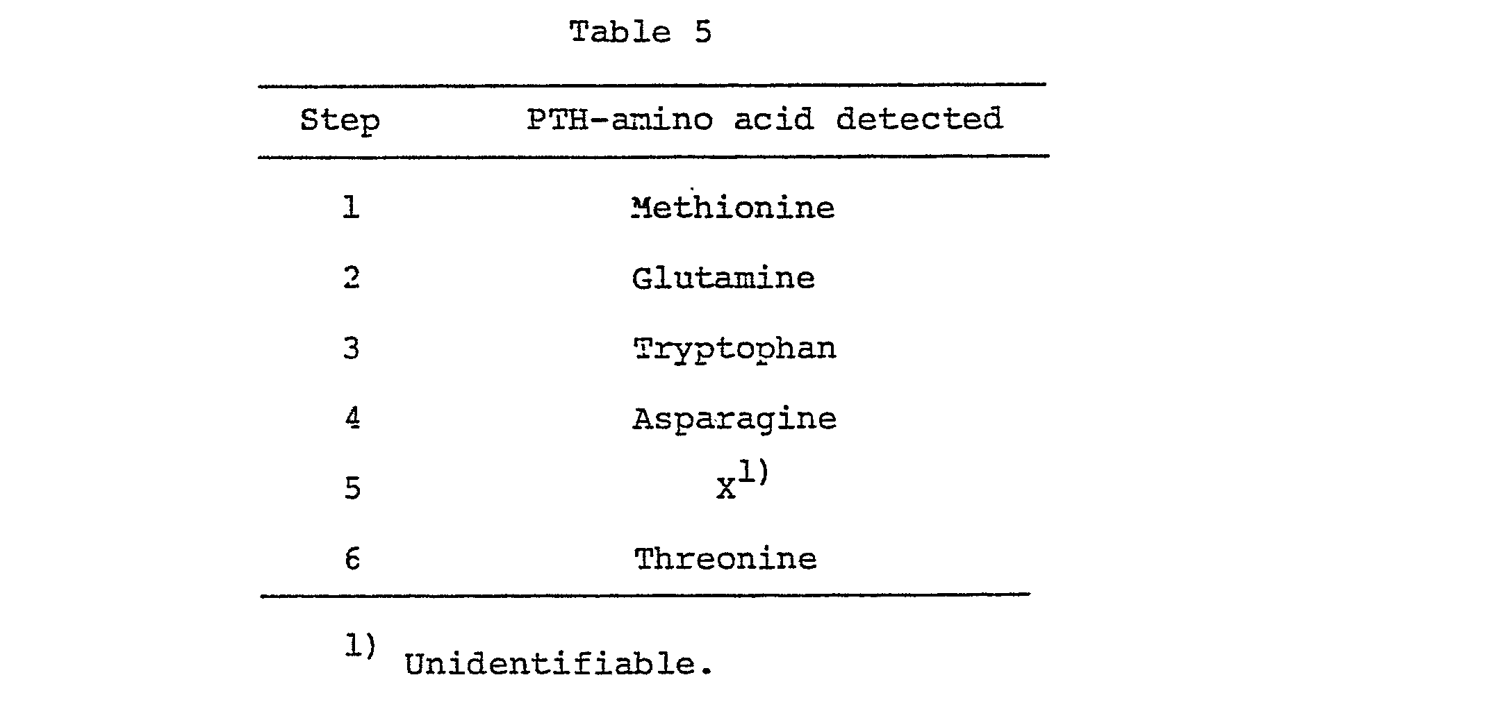
- 620 µg of said P31 protein was placed in a glass tube for degradation with hydrazine and 0.1 ml of anhydrous hydrazine was added. The tube was sealed under reduced pressure, and the whole was heated at 100°C for 6 hours. The degradation product obtained was lyophilized and dissolved in distilled water. Benzaldehyde was added to that solution and the resulting mixture was stirred at room temperature for 1 hour and, then, centrifuged to give a supernatant. This supernatant was lyophilized and subjected to amino acid analysis using a Hitachi model 835 amino acid analyzer. As a result, 47 nanomoles of isoleucine was detected.
- Also the use of cells of Escherichia coli 294/pTRP P31-W2 as obtained by cultivating said strain by the method described in Example 6 can lead to production of the P31 protein (subtype adw) in substantially pure form in the same manner as above.
- 1 µg of the expression vector pPKT700-1 DNA described in Reference Example 2-(3) was treated with 2 units of the restriction enzyme SalI at 37°C for 2 hours and then farther treated with 0.1 unit of alkaline phosphatase under the conditions described in Example 3-(1). Thereafter, a DNA was precipitated with cold ethanol (SalI-digested pPKT700-1).
- Then, 200 ng of the 1.43 kb DNA fragment described in Example 3-(1) and 200 ng of the SalI-digested pPKT700-1 were ligated together under the conditions described in Reference Example 1 using T4 DNA ligase. Using the reaction mixture and following the procedure described in Example 3-(1), a strain (294/pPKT P31-R) carrying a plasmid, pPKT P31-R, with the 1.43 kb DNA fragment containing a subtype adr P31-encoding DNA inserted therein in the same directionality as the PGK promoter was isolated. The host yeast strain AH22R was transformed with said plasmid, and a yeast transformant (AH22R /pPKT P31-R) was isolated (cf. Fig. 8).
- 1 µg of the expression vector pGLD906-l DNA described in Reference Example 3-(1) was treated with 2 units of the restriction enzyme SalI at 37°C for 2 hours and then further treated with 0.1 unit of alkaline phosphatase under the conditions described in Example 3-(1). Thereafter, a DNA was precipitated with cold ethanol SalI-digested pGLD906-1).
- Then, 200 ng of the 1.43 kb DNA fragment described in Example 3-(1) and the SalI-digested pGLD906-1 were ligated together under the conditions described in Reference Example 1 using T4 DNA ligase. By the method describe in Example 3-(1) using the reaction mixture, a strain (294/pGLD P31-R) carrying a plasmid, pGLD P31-R, with the 1.43 kb DNA fragment containing a subtype adr P31-encoding DNA inserted therein in the same directionality as the GLD promoter was isolated. The host yeast AH22R was transformed with said plasmid, and a yeast transformant (AH22R-/pGLD P31-R) is obtained (cf. Fig. 8).
- Each yeast transformant obtained in Example 8 or 9 and carrying a P31 gene expression plasmid was cultivated in Burkholder medium or a low phosphate level modification thereof at 30°C for 2 days and, thereafter, cells were harvested and washed with physiological saline.
- The cells were converted to the spheroplast state by treatment with Zymolyase [Seikagaku Kogyp] according to the method of Miyanohara, A. et al. Thereto was added 0.1% Triton X-100 for extraction of P31. The lysate was centrifuged at room temperature at 15,000 rpm for 15 minutes to give a supernatant. This supernatant was assayed for P31 activity using Auszyme II [Abbott]. The results obtained are shown in Table 6. The fields of P31 were each calculated in terms of the yield per liter of broth.

- Plasmids were extracted from Escherichia coli 294 carrying the plasmid for expression of P31 gene as obtained in Examples 1 and 2, and Escherichia coli C600 were transformed with said plasmids to obtain Excherichia coli C600/pTRP P31-R and Escherichia coli C600/pTRP P31-W2.
- Escherichia coli C600 strain carrying a P31 gene expression plasmid as obtained in Example 11 was cultivated in M-9 medium containing 2.0% glucose and 1.0% casamino acids at 37°C for 8 hours and, then, cells were harvested and washed with a buffer [30 mM Tris-HCl (pH 8.0), 50 mM NaCl, 5 mM EDTA]. The cells were suspended in a lyzing solution comprising 10 mM Tris-HCl (pH 8.0), 5 mM EDTA, 1 mM phenylmethylsulfonyl fluoride and 5 mg/ml lysozyme and lysis was effected. To said lysate was added guanidine hydrochloride to a final concentration of 7 M. After incubation at 37°C for 2 hours, the lysate was centrifuged at 15,000 rpm and room temperature for 15 minutes to give a supernatant. The P31 activity of this supernatant was determined by the direct immunoassay mentioned hereinabove. The results thus obtained are shown in Table 7. The yields of P31 were each calculated in terms of the yield per milliliter of broth.

- One unit of HBsAg corresponds to the count value obtained with that amount of the 125I-anti-HBsAg antibody attached to the Ausria II-I25 kit which is bound to 1 ng of HBsAg small particles.
- The concentrate (0.6 ml, 2.1 x 10⁶ units/ml) of the eluate from poly-HSA-Cellulofine column as obtained by the method described in Example 7 using Escherichia coli C600/ pTRP P31-R, was added 6 mg of DTT and, after reduction at 37°C for 1 hour, the mixture was filtered through a membrane filter. A 500-µl portion of the filtrate was allowed to be 0 adsorbed on an AP224 300 A (C8) reversed-phased column (YMC-Shimakyu) and high-performance liquid chromatography was performed using a trifluoroacetic acid-acetonitrile-n-propyl alcohol system as the eluent system.
- 0 Column, AP224 300 A (C8) (1 x 30 cm); column temperature, 30°C; eluent A, 0.1% trifuloroacetic acid-99.9% water; eluent B, 0.1% trifuloroacetic acid-49.95% acetonitrile-49.95% n-propyl alcohol; elution program, minute 0 (70% A + 30% B)-minute 10 (28% A + 72% B)-minute 30 (24% A + 76% B)-minute 50 (14% A + 86% B)-minute 55 (5% A + 95% B)-minute 58 (5% A + 95% B)-minute 59 (70% A + 30% B); elution rate, 2.5 ml/min.; detection wavelength, 280 nm. A fraction showing retention time of about 42 minutes under these conditions (20 ml; 1.7 x 10⁴ units/ml) was collected and reserved at -20°C.
- The substantially pure P31 protein (subtype adr) produced by the above method had the same characteristic properties as P31 protein (subtype adr) obtained in xample 7.
- (1) 1 mg of P31 protein obtained in Example 13 was put into a glass tube, and the solvent was removed unde reduced pressure. According to phenol-sulfuric acid method [
Analytical Chemistry 28, 350 (1956)], quantitative analysis c: carbohydrate was performed using glucose as a standard. The reult was that carbohydrate detected in the sample was not geater than 0.3 molecule per molecule of P31 protein on calculating the molecular weight of P31 protein as 31,000 daltons Therefore, the obtained P31 protein seems to have no carboy rate. - (2) In a glass tube for hydrolysis, there waf Placed 30 pg of the P31 protein obtained in Example 13, foleved by removal of the solvent. After addition of trfruoroacetic acid/constant boiling point hydrochloric acid [1:2 (v/v)], the tube was sealed under reduced pressure, and hydrolysis was performed at 170°C for 25, 50 and 180 minutes. After hydrolysis, the tube was opened, the trifluoroacetic acid and hydrochloric acid were removed, the residue was dissolved in 0.02 N hydrochloric acid, and amino acid analysis was performed using a Hitachi model 835 amino acid analyzer. Cystine and cysteine were determined, according to the method of Hirs as cysteic acid on the amino acid analyzer after performic acid oxidation of the P31 protein and hydrolysis in constant boiling point hydrochloric acid under reduced pressure for 24 hours. Quantitative analysis for threonine, serin and tryptophan was performed after hydrolysis of P31 protein in constant boilind hydrochloric acid containing 4% thioglycolic acid under reduced pressure for 16, 20, 24 and 30 hours. The amino acid analysis data were calculated each as an average of the values obtained by hydrolysis for 25, 50 and 180 minutes. For tryptophan, however, the value obtained after 24 hours of hydrolysis was employed and, for serine and threonine, the values were determined by extrapolation to hour 0 (zero) of hydrolysis. The results thus obtained are shown in Table 8.

- Institutes at which microorganisms have been deposited and the accession numbers are shown in Table 9.
- In the Table, "IFO" and "FRI" represent "Institute for Fermentation, Osaka" (Japan) and "Fermentation Research Institute, Agency of Industrial Science and Technology, Ministry of International Trade and Industry, Japan", respectively. E. coli and S. cerevisiae represent Escherichia coli and Saccharomyces cerevisiae, respectively,
- "FERM BP" number represents the accession number of a deposit under the Budapest Treaty and "FERM P" number in parentheses represents the accession number before conversion to a deposit under the Budapest Treaty.
-

Claims (34)
Applications Claiming Priority (8)
| Application Number | Priority Date | Filing Date | Title |
|---|---|---|---|
| WOPCT/JP84/00356 | 1984-07-11 | ||
| PCT/JP1984/000356 WO1986000640A1 (en) | 1984-07-11 | 1984-07-11 | Recombinant dna and its use |
| WOPCT/JP84/00423 | 1984-09-04 | ||
| PCT/JP1984/000423 WO1986001534A1 (en) | 1984-09-04 | 1984-09-04 | Recombinant dna and its use |
| WOPCT/JP84/00585 | 1984-12-12 | ||
| PCT/JP1984/000585 WO1986003411A1 (en) | 1984-12-12 | 1984-12-12 | Process for preparing novel protein |
| PCT/JP1985/000306 WO1986007384A1 (en) | 1985-06-03 | 1985-06-03 | Process for preparing hapatitis b virus surface antigen p31 |
| WOPCT/JP85/00306 | 1985-06-03 |
Related Child Applications (2)
| Application Number | Title | Priority Date | Filing Date |
|---|---|---|---|
| EP19900202097 Division EP0401941A3 (en) | 1984-07-11 | 1985-07-03 | Hepatitis b virus surface antigen and production thereof |
| EP90202097.3 Division-Into | 1985-07-03 |
Publications (2)
| Publication Number | Publication Date |
|---|---|
| EP0171908A2 true EP0171908A2 (en) | 1986-02-19 |
| EP0171908A3 EP0171908A3 (en) | 1987-07-15 |
Family
ID=27466908
Family Applications (2)
| Application Number | Title | Priority Date | Filing Date |
|---|---|---|---|
| EP85304735A Withdrawn EP0171908A3 (en) | 1984-07-11 | 1985-07-03 | Hepatitis b virus surface antigen and production thereof |
| EP19900202097 Withdrawn EP0401941A3 (en) | 1984-07-11 | 1985-07-03 | Hepatitis b virus surface antigen and production thereof |
Family Applications After (1)
| Application Number | Title | Priority Date | Filing Date |
|---|---|---|---|
| EP19900202097 Withdrawn EP0401941A3 (en) | 1984-07-11 | 1985-07-03 | Hepatitis b virus surface antigen and production thereof |
Country Status (3)
| Country | Link |
|---|---|
| EP (2) | EP0171908A3 (en) |
| JP (2) | JPH082306B2 (en) |
| CN (1) | CN85106190A (en) |
Cited By (17)
| Publication number | Priority date | Publication date | Assignee | Title |
|---|---|---|---|---|
| EP0205625A4 (en) * | 1984-12-28 | 1987-06-09 | Japan Found Cancer | Hepatitis b virus vaccine and process for its preparation. |
| EP0174444A3 (en) * | 1984-06-18 | 1987-07-29 | Chiron Corporation | Hepatitis surface antigen particle vaccine |
| EP0235430A1 (en) * | 1985-08-20 | 1987-09-09 | Takeda Chemical Industries, Ltd. | Novel protein and production thereof |
| EP0257507A1 (en) * | 1986-08-17 | 1988-03-02 | Yeda Research And Development Ltd. | Hepatitis vaccine |
| US4742158A (en) * | 1986-04-25 | 1988-05-03 | Merck & Co., Inc. | Purification of hepatitis pre-S antigens by polymerized serum albumin affinity binding |
| FR2606281A1 (en) * | 1986-06-18 | 1988-05-13 | Univ Osaka Res Found | HEPATITIS B VIRUS ANTIGEN AND METHOD FOR THE PRODUCTION THEREOF |
| EP0216117A3 (en) * | 1985-08-20 | 1988-06-15 | Takeda Chemical Industries, Ltd. | Novel dna and use thereof |
| EP0278940A3 (en) * | 1987-01-30 | 1988-12-07 | Smithkline Biologicals S.A. | Hepatitis b virus surface antigens and hybrid antigens containing them |
| EP0291146A3 (en) * | 1987-03-09 | 1989-12-06 | Merck & Co. Inc. | Purification of recombinant hepatitis b surface antigen |
| EP0314240A3 (en) * | 1987-10-26 | 1990-03-28 | Merck & Co. Inc. | Process for purifying recombinant hepatitis antigens |
| EP0315242A3 (en) * | 1987-10-26 | 1990-04-11 | Merck & Co. Inc. | Process for purifying recombinant hepatitis antigens |
| US5026828A (en) * | 1987-02-27 | 1991-06-25 | Merck & Co., Inc. | Method of purifying recombinant pres-1/S-2/S/S hepatitis B antigen from yeast |
| US5182195A (en) * | 1987-11-13 | 1993-01-26 | Takeda Chemical Industries, Ltd. | Method for increasing gene expression using protease deficient yeasts |
| EP0533263A3 (en) * | 1991-09-20 | 1994-06-08 | Merck & Co Inc | A multivalent hepatitis b virus vaccine |
| US6020167A (en) * | 1990-12-19 | 2000-02-01 | Medeva Holdings B.V. | Composition used as a therapeutic agent against chronic viral hepatic diseases |
| US6072049A (en) * | 1987-06-22 | 2000-06-06 | Medeva Holdings B.V. | Hepatitis B surface antigen vaccine |
| US6322789B1 (en) | 1991-08-26 | 2001-11-27 | Epimmune, Inc. | HLA-restricted hepatitis B virus CTL epitopes |
Families Citing this family (7)
| Publication number | Priority date | Publication date | Assignee | Title |
|---|---|---|---|---|
| JPS6137738A (en) * | 1984-07-31 | 1986-02-22 | Tetsuo Nakamura | Vaccine for hepatitis b |
| JPS63230639A (en) * | 1987-03-19 | 1988-09-27 | Nippon Chem Res Kk | Novel hepatitis b virus surface antigen particle and production thereof |
| IL90161A0 (en) * | 1988-05-13 | 1989-12-15 | Phillips Petroleum Co | Expression of hepatitis b s and pres2 proteins in methylotrophic yeasts |
| NZ229260A (en) * | 1988-06-03 | 1994-02-25 | Merck & Co Inc | Hepatitis b virus, expression cassette for pre-s domain, host cells and |
| LT3988B (en) | 1992-02-17 | 1996-06-25 | Fermentas Biotech Inst | Recombinant plasmides pfs19, pfps2-48 and pjlfds1 codingsynthesis of human hepatite b of surfice virus antigenes, methods fof producing thereof |
| JP5212537B1 (en) | 2011-12-13 | 2013-06-19 | ダイキン工業株式会社 | Refrigeration equipment |
| JP5403095B2 (en) | 2011-12-20 | 2014-01-29 | ダイキン工業株式会社 | Refrigeration equipment |
Family Cites Families (10)
| Publication number | Priority date | Publication date | Assignee | Title |
|---|---|---|---|---|
| LU88258I2 (en) * | 1978-12-22 | 1994-02-03 | ||
| IE52036B1 (en) * | 1979-05-24 | 1987-05-27 | Univ California | Non-passageable viruses |
| FR2480780B1 (en) * | 1980-04-22 | 1985-12-06 | Pasteur Institut | PROCESS FOR THE TRANSFORMATION OF CELLS, ESPECIALLY EUKARYOTES, BY CIRCULAR DNA SUCH AS THAT OF HEPATITIS B VIRUS AND PREPARATIONS CONTAINING THE EXPRESSION PRODUCTS OF SAID DNA |
| NZ201705A (en) * | 1981-08-31 | 1986-03-14 | Genentech Inc | Recombinant dna method for production of hepatitis b surface antigen in yeast |
| DE3381566D1 (en) * | 1982-08-16 | 1990-06-21 | Agency Science & Tech | RECOMBINANT PLASMIDE CONTAINING HEPATITIS B VIRUS GENE, Yeast TRANSFORMED WITH THIS RECOMBINANT PLASMIDE AND PRODUCTION OF HEPATITIS B VIRUS SURFACE ANTIQUE. |
| JPS5974985A (en) * | 1982-10-19 | 1984-04-27 | Takeda Chem Ind Ltd | Novel dna, its preparation and transformant having the same |
| JPS5980615A (en) * | 1982-10-29 | 1984-05-10 | Takeda Chem Ind Ltd | Dna and its use |
| EP0135435A3 (en) * | 1983-08-22 | 1987-03-25 | Merck & Co. Inc. | Immunogenic hbsag derived from transformed yeast |
| FR2559159B1 (en) * | 1984-02-02 | 1986-09-12 | Inst Nat Sante Rech Med | VIRAL VECTORS FOR THE CLONING AND EXPRESSION OF A PROTEIN IN A EUKARYOTIC CELL, COMPRISING AT LEAST ONE PART OF THE GENOME OF A RETROVIRUS; TRANSFECTED EUKARYOTIC CELLS; PROCESS USING SUCH CELLS AND PROTEINS OBTAINED |
| ATE73349T1 (en) * | 1984-06-18 | 1992-03-15 | Chiron Corp | HEPATITIS SURFACE ANTIGEN PARTICLE VACCINATION. |
-
1985
- 1985-07-03 EP EP85304735A patent/EP0171908A3/en not_active Withdrawn
- 1985-07-03 EP EP19900202097 patent/EP0401941A3/en not_active Withdrawn
- 1985-07-10 JP JP60153238A patent/JPH082306B2/en not_active Expired - Lifetime
- 1985-07-12 CN CN85106190A patent/CN85106190A/en not_active Withdrawn
-
1993
- 1993-05-14 JP JP5113354A patent/JPH0690781A/en active Pending
Cited By (22)
| Publication number | Priority date | Publication date | Assignee | Title |
|---|---|---|---|---|
| EP0174444A3 (en) * | 1984-06-18 | 1987-07-29 | Chiron Corporation | Hepatitis surface antigen particle vaccine |
| EP0205625A4 (en) * | 1984-12-28 | 1987-06-09 | Japan Found Cancer | Hepatitis b virus vaccine and process for its preparation. |
| EP0235430A1 (en) * | 1985-08-20 | 1987-09-09 | Takeda Chemical Industries, Ltd. | Novel protein and production thereof |
| EP0216117A3 (en) * | 1985-08-20 | 1988-06-15 | Takeda Chemical Industries, Ltd. | Novel dna and use thereof |
| US4742158A (en) * | 1986-04-25 | 1988-05-03 | Merck & Co., Inc. | Purification of hepatitis pre-S antigens by polymerized serum albumin affinity binding |
| BE1000167A4 (en) * | 1986-06-18 | 1988-07-05 | Univ Osaka Res Found | HEPATITIS B VIRUS ANTIGEN AND METHOD FOR THE PRODUCTION THEREOF. |
| FR2606281A1 (en) * | 1986-06-18 | 1988-05-13 | Univ Osaka Res Found | HEPATITIS B VIRUS ANTIGEN AND METHOD FOR THE PRODUCTION THEREOF |
| EP0257507A1 (en) * | 1986-08-17 | 1988-03-02 | Yeda Research And Development Ltd. | Hepatitis vaccine |
| EP0278940A3 (en) * | 1987-01-30 | 1988-12-07 | Smithkline Biologicals S.A. | Hepatitis b virus surface antigens and hybrid antigens containing them |
| US5026828A (en) * | 1987-02-27 | 1991-06-25 | Merck & Co., Inc. | Method of purifying recombinant pres-1/S-2/S/S hepatitis B antigen from yeast |
| EP0291146A3 (en) * | 1987-03-09 | 1989-12-06 | Merck & Co. Inc. | Purification of recombinant hepatitis b surface antigen |
| US6072049A (en) * | 1987-06-22 | 2000-06-06 | Medeva Holdings B.V. | Hepatitis B surface antigen vaccine |
| US6589530B1 (en) | 1987-06-22 | 2003-07-08 | Medeva Holdings B.V. | Hepatitis B surface antigen vaccine |
| US6117653A (en) * | 1987-06-22 | 2000-09-12 | Medeva Holdings B.V. | Hepatitis B surface antigen vaccine |
| US6100065A (en) * | 1987-06-22 | 2000-08-08 | Medeva Holdings B.V. | Hepatitis B surface antigen vaccine |
| EP0314240A3 (en) * | 1987-10-26 | 1990-03-28 | Merck & Co. Inc. | Process for purifying recombinant hepatitis antigens |
| US5011915A (en) * | 1987-10-26 | 1991-04-30 | Merck & Co., Inc. | Process for purifying recombinant hepatitis antigens |
| EP0315242A3 (en) * | 1987-10-26 | 1990-04-11 | Merck & Co. Inc. | Process for purifying recombinant hepatitis antigens |
| US5182195A (en) * | 1987-11-13 | 1993-01-26 | Takeda Chemical Industries, Ltd. | Method for increasing gene expression using protease deficient yeasts |
| US6020167A (en) * | 1990-12-19 | 2000-02-01 | Medeva Holdings B.V. | Composition used as a therapeutic agent against chronic viral hepatic diseases |
| US6322789B1 (en) | 1991-08-26 | 2001-11-27 | Epimmune, Inc. | HLA-restricted hepatitis B virus CTL epitopes |
| EP0533263A3 (en) * | 1991-09-20 | 1994-06-08 | Merck & Co Inc | A multivalent hepatitis b virus vaccine |
Also Published As
| Publication number | Publication date |
|---|---|
| EP0401941A3 (en) | 1991-04-17 |
| JPS6143991A (en) | 1986-03-03 |
| CN85106190A (en) | 1986-06-10 |
| JPH0690781A (en) | 1994-04-05 |
| EP0171908A3 (en) | 1987-07-15 |
| JPH082306B2 (en) | 1996-01-17 |
| EP0401941A2 (en) | 1990-12-12 |
Similar Documents
| Publication | Publication Date | Title |
|---|---|---|
| EP0171908A2 (en) | Hepatitis B virus surface antigen and production thereof | |
| KR940005588B1 (en) | Method of producing hepatitis virus protein in yeast | |
| US4977092A (en) | Expression of exogenous polypeptides and polypeptide products including hepatitis B surface antigen in yeast cells | |
| KR930002263B1 (en) | Method for preparing lyophilized hepatitis B vaccine | |
| DK169152B1 (en) | Method for Preparing an HBsAg Antibody | |
| KR890000096B1 (en) | Method for preparing hepatitis B virus surface antigen | |
| EP0204680A2 (en) | Process for isolating and purifying the hepatitis B surface antigen | |
| EP0155007B1 (en) | Method for purification of hbs antigen | |
| EP0312159A2 (en) | Method for producing hepatitis B virus proteins in yeast | |
| EP0248410A2 (en) | Improved yeast promoter and proces for preparing heterologous protein | |
| CZ226993A3 (en) | Set of surface proteins of particle-forming hepatitis b virus | |
| JPS5974985A (en) | Novel dna, its preparation and transformant having the same | |
| EP0288198A2 (en) | Production of Peptide | |
| US4983520A (en) | DNA sequence encoding modified hepatitis B virus surface antigen P31 protein | |
| US5164485A (en) | Modified hepatitis B virus surface antigen P31 and production thereof | |
| JPS6170989A (en) | Recombinant dna and its use | |
| EP0109559A1 (en) | High-efficiency yeast vector plasmid and method of its use | |
| JP2648122B2 (en) | Novel polypeptide and method for producing the same | |
| WO1986003411A1 (en) | Process for preparing novel protein | |
| JP2749010B2 (en) | Novel DNA and method for producing polypeptide using the same | |
| JPH02449A (en) | Production of peptide | |
| CN100368438C (en) | A kind of anti-hepatitis B virus fusion protein and its coding gene and application | |
| WO1986007384A1 (en) | Process for preparing hapatitis b virus surface antigen p31 | |
| JP2625379B2 (en) | DNA fragment for high expression vector plasmid of yeast | |
| JPH06153962A (en) | Dna coding p31 modified protein |
Legal Events
| Date | Code | Title | Description |
|---|---|---|---|
| PUAI | Public reference made under article 153(3) epc to a published international application that has entered the european phase |
Free format text: ORIGINAL CODE: 0009012 |
|
| AK | Designated contracting states |
Designated state(s): AT BE CH DE FR GB IT LI LU NL SE |
|
| PUAL | Search report despatched |
Free format text: ORIGINAL CODE: 0009013 |
|
| AK | Designated contracting states |
Kind code of ref document: A3 Designated state(s): AT BE CH DE FR GB IT LI LU NL SE |
|
| 17P | Request for examination filed |
Effective date: 19870908 |
|
| 17Q | First examination report despatched |
Effective date: 19890314 |
|
| RAP1 | Party data changed (applicant data changed or rights of an application transferred) |
Owner name: TAKEDA CHEMICAL INDUSTRIES, LTD. |
|
| STAA | Information on the status of an ep patent application or granted ep patent |
Free format text: STATUS: THE APPLICATION IS DEEMED TO BE WITHDRAWN |
|
| 18D | Application deemed to be withdrawn |
Effective date: 19910622 |
|
| RIN1 | Information on inventor provided before grant (corrected) |
Inventor name: IKEYAMA, SHUICHI Inventor name: NISHIMURA, OSAMU Inventor name: KIKUCHI, MASAKAZU Inventor name: FUJISAWA, YUKIO |