EP0160939B1 - Procédé autoradiographique - Google Patents
Procédé autoradiographique Download PDFInfo
- Publication number
- EP0160939B1 EP0160939B1 EP85105368A EP85105368A EP0160939B1 EP 0160939 B1 EP0160939 B1 EP 0160939B1 EP 85105368 A EP85105368 A EP 85105368A EP 85105368 A EP85105368 A EP 85105368A EP 0160939 B1 EP0160939 B1 EP 0160939B1
- Authority
- EP
- European Patent Office
- Prior art keywords
- phosphor sheet
- photosensitive material
- stimulable phosphor
- support medium
- read
- Prior art date
- Legal status (The legal status is an assumption and is not a legal conclusion. Google has not performed a legal analysis and makes no representation as to the accuracy of the status listed.)
- Expired - Lifetime
Links
- 238000000034 method Methods 0.000 title claims description 71
- 230000008569 process Effects 0.000 title claims description 36
- OAICVXFJPJFONN-UHFFFAOYSA-N Phosphorus Chemical compound [P] OAICVXFJPJFONN-UHFFFAOYSA-N 0.000 claims description 231
- 239000000463 material Substances 0.000 claims description 100
- 239000000126 substance Substances 0.000 claims description 71
- 230000005855 radiation Effects 0.000 claims description 38
- 230000004936 stimulating effect Effects 0.000 claims description 31
- 230000001678 irradiating effect Effects 0.000 claims description 6
- 239000010410 layer Substances 0.000 description 60
- 238000000376 autoradiography Methods 0.000 description 20
- 239000000499 gel Substances 0.000 description 19
- 238000012545 processing Methods 0.000 description 13
- 239000012634 fragment Substances 0.000 description 12
- -1 polyethylene terephthalate Polymers 0.000 description 10
- 230000001681 protective effect Effects 0.000 description 10
- 239000006185 dispersion Substances 0.000 description 9
- 239000011230 binding agent Substances 0.000 description 8
- 238000003776 cleavage reaction Methods 0.000 description 8
- 229910052693 Europium Inorganic materials 0.000 description 7
- 239000011248 coating agent Substances 0.000 description 7
- 238000000576 coating method Methods 0.000 description 7
- 230000007017 scission Effects 0.000 description 7
- 238000012800 visualization Methods 0.000 description 7
- ZWEHNKRNPOVVGH-UHFFFAOYSA-N 2-Butanone Chemical compound CCC(C)=O ZWEHNKRNPOVVGH-UHFFFAOYSA-N 0.000 description 6
- 239000000839 emulsion Substances 0.000 description 6
- 239000000203 mixture Substances 0.000 description 6
- 230000002285 radioactive effect Effects 0.000 description 6
- 230000015572 biosynthetic process Effects 0.000 description 5
- 238000001514 detection method Methods 0.000 description 5
- 229920000139 polyethylene terephthalate Polymers 0.000 description 5
- 239000005020 polyethylene terephthalate Substances 0.000 description 5
- 239000000243 solution Substances 0.000 description 5
- 238000011282 treatment Methods 0.000 description 5
- 239000012790 adhesive layer Substances 0.000 description 4
- 229920001222 biopolymer Polymers 0.000 description 4
- 238000002474 experimental method Methods 0.000 description 4
- 239000002245 particle Substances 0.000 description 4
- 230000035945 sensitivity Effects 0.000 description 4
- 239000002904 solvent Substances 0.000 description 4
- 238000001712 DNA sequencing Methods 0.000 description 3
- VYPSYNLAJGMNEJ-UHFFFAOYSA-N Silicium dioxide Chemical compound O=[Si]=O VYPSYNLAJGMNEJ-UHFFFAOYSA-N 0.000 description 3
- 229910052771 Terbium Inorganic materials 0.000 description 3
- 230000003321 amplification Effects 0.000 description 3
- 238000012790 confirmation Methods 0.000 description 3
- 238000001962 electrophoresis Methods 0.000 description 3
- 239000011521 glass Substances 0.000 description 3
- 229910052749 magnesium Inorganic materials 0.000 description 3
- 239000011777 magnesium Substances 0.000 description 3
- 229910052751 metal Inorganic materials 0.000 description 3
- 239000002184 metal Substances 0.000 description 3
- 238000003199 nucleic acid amplification method Methods 0.000 description 3
- 108020004707 nucleic acids Proteins 0.000 description 3
- 102000039446 nucleic acids Human genes 0.000 description 3
- 150000007523 nucleic acids Chemical class 0.000 description 3
- 108090000623 proteins and genes Proteins 0.000 description 3
- 102000004169 proteins and genes Human genes 0.000 description 3
- 238000000926 separation method Methods 0.000 description 3
- 229910052684 Cerium Inorganic materials 0.000 description 2
- RTZKZFJDLAIYFH-UHFFFAOYSA-N Diethyl ether Chemical compound CCOCC RTZKZFJDLAIYFH-UHFFFAOYSA-N 0.000 description 2
- 108010010803 Gelatin Proteins 0.000 description 2
- TWRXJAOTZQYOKJ-UHFFFAOYSA-L Magnesium chloride Chemical compound [Mg+2].[Cl-].[Cl-] TWRXJAOTZQYOKJ-UHFFFAOYSA-L 0.000 description 2
- LRHPLDYGYMQRHN-UHFFFAOYSA-N N-Butanol Chemical compound CCCCO LRHPLDYGYMQRHN-UHFFFAOYSA-N 0.000 description 2
- 239000000020 Nitrocellulose Substances 0.000 description 2
- 239000007983 Tris buffer Substances 0.000 description 2
- 235000010724 Wisteria floribunda Nutrition 0.000 description 2
- 230000008901 benefit Effects 0.000 description 2
- 239000007853 buffer solution Substances 0.000 description 2
- 229910052793 cadmium Inorganic materials 0.000 description 2
- 229910052791 calcium Inorganic materials 0.000 description 2
- 229910052801 chlorine Inorganic materials 0.000 description 2
- 239000011247 coating layer Substances 0.000 description 2
- 238000007796 conventional method Methods 0.000 description 2
- 230000006866 deterioration Effects 0.000 description 2
- 229920000159 gelatin Polymers 0.000 description 2
- 239000008273 gelatin Substances 0.000 description 2
- 235000019322 gelatine Nutrition 0.000 description 2
- 235000011852 gelatine desserts Nutrition 0.000 description 2
- 238000002372 labelling Methods 0.000 description 2
- 229920001220 nitrocellulos Polymers 0.000 description 2
- 230000035515 penetration Effects 0.000 description 2
- 239000002985 plastic film Substances 0.000 description 2
- 229920002401 polyacrylamide Polymers 0.000 description 2
- 229920000728 polyester Polymers 0.000 description 2
- 238000003672 processing method Methods 0.000 description 2
- 239000000941 radioactive substance Substances 0.000 description 2
- 238000002601 radiography Methods 0.000 description 2
- 239000011347 resin Substances 0.000 description 2
- 229920005989 resin Polymers 0.000 description 2
- 108091008146 restriction endonucleases Proteins 0.000 description 2
- GGCZERPQGJTIQP-UHFFFAOYSA-N sodium;9,10-dioxoanthracene-2-sulfonic acid Chemical compound [Na+].C1=CC=C2C(=O)C3=CC(S(=O)(=O)O)=CC=C3C(=O)C2=C1 GGCZERPQGJTIQP-UHFFFAOYSA-N 0.000 description 2
- 229910052712 strontium Inorganic materials 0.000 description 2
- 238000012360 testing method Methods 0.000 description 2
- 230000000007 visual effect Effects 0.000 description 2
- 229910052725 zinc Inorganic materials 0.000 description 2
- NIXOWILDQLNWCW-UHFFFAOYSA-M Acrylate Chemical compound [O-]C(=O)C=C NIXOWILDQLNWCW-UHFFFAOYSA-M 0.000 description 1
- 239000004925 Acrylic resin Substances 0.000 description 1
- 229920000178 Acrylic resin Polymers 0.000 description 1
- 239000004215 Carbon black (E152) Substances 0.000 description 1
- 229920000623 Cellulose acetate phthalate Polymers 0.000 description 1
- KCXVZYZYPLLWCC-UHFFFAOYSA-N EDTA Chemical compound OC(=O)CN(CC(O)=O)CCN(CC(O)=O)CC(O)=O KCXVZYZYPLLWCC-UHFFFAOYSA-N 0.000 description 1
- 229910052691 Erbium Inorganic materials 0.000 description 1
- 241000588724 Escherichia coli Species 0.000 description 1
- LFQSCWFLJHTTHZ-UHFFFAOYSA-N Ethanol Chemical compound CCO LFQSCWFLJHTTHZ-UHFFFAOYSA-N 0.000 description 1
- QUSNBJAOOMFDIB-UHFFFAOYSA-N Ethylamine Chemical compound CCN QUSNBJAOOMFDIB-UHFFFAOYSA-N 0.000 description 1
- 208000034454 F12-related hereditary angioedema with normal C1Inh Diseases 0.000 description 1
- 229910052688 Gadolinium Inorganic materials 0.000 description 1
- 229910052689 Holmium Inorganic materials 0.000 description 1
- 229910052779 Neodymium Inorganic materials 0.000 description 1
- 239000004698 Polyethylene Substances 0.000 description 1
- 239000004372 Polyvinyl alcohol Substances 0.000 description 1
- YSMRWXYRXBRSND-UHFFFAOYSA-N TOTP Chemical compound CC1=CC=CC=C1OP(=O)(OC=1C(=CC=CC=1)C)OC1=CC=CC=C1C YSMRWXYRXBRSND-UHFFFAOYSA-N 0.000 description 1
- 229910052775 Thulium Inorganic materials 0.000 description 1
- 241001648319 Toronia toru Species 0.000 description 1
- 229910052769 Ytterbium Inorganic materials 0.000 description 1
- 230000009102 absorption Effects 0.000 description 1
- 238000010521 absorption reaction Methods 0.000 description 1
- 230000004913 activation Effects 0.000 description 1
- 229910052784 alkaline earth metal Inorganic materials 0.000 description 1
- 150000001342 alkaline earth metals Chemical class 0.000 description 1
- 229910052782 aluminium Inorganic materials 0.000 description 1
- XAGFODPZIPBFFR-UHFFFAOYSA-N aluminium Chemical compound [Al] XAGFODPZIPBFFR-UHFFFAOYSA-N 0.000 description 1
- 238000004458 analytical method Methods 0.000 description 1
- QVQLCTNNEUAWMS-UHFFFAOYSA-N barium oxide Chemical compound [Ba]=O QVQLCTNNEUAWMS-UHFFFAOYSA-N 0.000 description 1
- 229910001864 baryta Inorganic materials 0.000 description 1
- 229910052797 bismuth Inorganic materials 0.000 description 1
- 229920002301 cellulose acetate Polymers 0.000 description 1
- 239000000460 chlorine Substances 0.000 description 1
- 125000001309 chloro group Chemical group Cl* 0.000 description 1
- 229910052681 coesite Inorganic materials 0.000 description 1
- 229910052906 cristobalite Inorganic materials 0.000 description 1
- 239000003431 cross linking reagent Substances 0.000 description 1
- 230000002939 deleterious effect Effects 0.000 description 1
- 230000000994 depressogenic effect Effects 0.000 description 1
- 208000028659 discharge Diseases 0.000 description 1
- VHJLVAABSRFDPM-QWWZWVQMSA-N dithiothreitol Chemical compound SC[C@@H](O)[C@H](O)CS VHJLVAABSRFDPM-QWWZWVQMSA-N 0.000 description 1
- 238000001035 drying Methods 0.000 description 1
- 230000000694 effects Effects 0.000 description 1
- 150000002148 esters Chemical class 0.000 description 1
- OGPBJKLSAFTDLK-UHFFFAOYSA-N europium atom Chemical compound [Eu] OGPBJKLSAFTDLK-UHFFFAOYSA-N 0.000 description 1
- 230000029142 excretion Effects 0.000 description 1
- 239000011888 foil Substances 0.000 description 1
- 238000001502 gel electrophoresis Methods 0.000 description 1
- 208000016861 hereditary angioedema type 3 Diseases 0.000 description 1
- 229930195733 hydrocarbon Natural products 0.000 description 1
- 150000002430 hydrocarbons Chemical class 0.000 description 1
- VEXZGXHMUGYJMC-UHFFFAOYSA-N hydrochloric acid Substances Cl VEXZGXHMUGYJMC-UHFFFAOYSA-N 0.000 description 1
- 230000006872 improvement Effects 0.000 description 1
- 239000012535 impurity Substances 0.000 description 1
- 239000004615 ingredient Substances 0.000 description 1
- 230000007794 irritation Effects 0.000 description 1
- 238000002955 isolation Methods 0.000 description 1
- 150000002576 ketones Chemical class 0.000 description 1
- 229910052746 lanthanum Inorganic materials 0.000 description 1
- 229910052745 lead Inorganic materials 0.000 description 1
- 229910001629 magnesium chloride Inorganic materials 0.000 description 1
- 229910052748 manganese Inorganic materials 0.000 description 1
- 239000003550 marker Substances 0.000 description 1
- 230000004060 metabolic process Effects 0.000 description 1
- 230000005012 migration Effects 0.000 description 1
- 238000013508 migration Methods 0.000 description 1
- 238000004816 paper chromatography Methods 0.000 description 1
- 230000037361 pathway Effects 0.000 description 1
- 239000013612 plasmid Substances 0.000 description 1
- 229920006255 plastic film Polymers 0.000 description 1
- 229920003229 poly(methyl methacrylate) Polymers 0.000 description 1
- 229920001225 polyester resin Polymers 0.000 description 1
- 239000004645 polyester resin Substances 0.000 description 1
- 229920000573 polyethylene Polymers 0.000 description 1
- 239000004926 polymethyl methacrylate Substances 0.000 description 1
- 229920002635 polyurethane Polymers 0.000 description 1
- 239000004814 polyurethane Substances 0.000 description 1
- 229920002689 polyvinyl acetate Polymers 0.000 description 1
- 239000011118 polyvinyl acetate Substances 0.000 description 1
- 229920002451 polyvinyl alcohol Polymers 0.000 description 1
- 239000000700 radioactive tracer Substances 0.000 description 1
- 238000001454 recorded image Methods 0.000 description 1
- 230000004044 response Effects 0.000 description 1
- 230000000630 rising effect Effects 0.000 description 1
- 238000006748 scratching Methods 0.000 description 1
- 230000002393 scratching effect Effects 0.000 description 1
- 230000035939 shock Effects 0.000 description 1
- 239000000741 silica gel Substances 0.000 description 1
- 229910002027 silica gel Inorganic materials 0.000 description 1
- 239000000377 silicon dioxide Substances 0.000 description 1
- 229910052709 silver Inorganic materials 0.000 description 1
- 239000004332 silver Substances 0.000 description 1
- 230000000638 stimulation Effects 0.000 description 1
- 229910052682 stishovite Inorganic materials 0.000 description 1
- 238000004381 surface treatment Methods 0.000 description 1
- 229920001059 synthetic polymer Polymers 0.000 description 1
- 238000004809 thin layer chromatography Methods 0.000 description 1
- 229920006352 transparent thermoplastic Polymers 0.000 description 1
- 229910052905 tridymite Inorganic materials 0.000 description 1
- 238000005406 washing Methods 0.000 description 1
- XLYOFNOQVPJJNP-UHFFFAOYSA-N water Substances O XLYOFNOQVPJJNP-UHFFFAOYSA-N 0.000 description 1
- 238000009736 wetting Methods 0.000 description 1
- 229910052727 yttrium Inorganic materials 0.000 description 1
Images

Classifications
-
- G—PHYSICS
- G03—PHOTOGRAPHY; CINEMATOGRAPHY; ANALOGOUS TECHNIQUES USING WAVES OTHER THAN OPTICAL WAVES; ELECTROGRAPHY; HOLOGRAPHY
- G03B—APPARATUS OR ARRANGEMENTS FOR TAKING PHOTOGRAPHS OR FOR PROJECTING OR VIEWING THEM; APPARATUS OR ARRANGEMENTS EMPLOYING ANALOGOUS TECHNIQUES USING WAVES OTHER THAN OPTICAL WAVES; ACCESSORIES THEREFOR
- G03B42/00—Obtaining records using waves other than optical waves; Visualisation of such records by using optical means
- G03B42/02—Obtaining records using waves other than optical waves; Visualisation of such records by using optical means using X-rays
-
- G—PHYSICS
- G03—PHOTOGRAPHY; CINEMATOGRAPHY; ANALOGOUS TECHNIQUES USING WAVES OTHER THAN OPTICAL WAVES; ELECTROGRAPHY; HOLOGRAPHY
- G03C—PHOTOSENSITIVE MATERIALS FOR PHOTOGRAPHIC PURPOSES; PHOTOGRAPHIC PROCESSES, e.g. CINE, X-RAY, COLOUR, STEREO-PHOTOGRAPHIC PROCESSES; AUXILIARY PROCESSES IN PHOTOGRAPHY
- G03C5/00—Photographic processes or agents therefor; Regeneration of such processing agents
- G03C5/16—X-ray, infrared, or ultraviolet ray processes
Definitions
- the present invention relates to an autoradiographic process.
- autoradiography or "radioautography” comprising the steps of: introducing a radioactively labeled substance into an organism; placing the organism or a part of the tissue of the organism (that is, a sample or specimen) and a radiographic film such as a high-speed type X-ray film together in layers for a certain period of time to expose said film thereto; and obtaining the locational information on the radioactively labeled substance in said sample from the resolved pattern of the film.
- the autoradiography has been utilized, for example, to investigate the pathway and state of metabolism, absorption and excretion of the substance introduced in the organism in detail.
- the autoradiography has been also utilized to obtain locational information on the radioactively labeled tissue of an organism and/or the radioactively labeled substances originating from an organism, which is present on a medium.
- an autoradiography comprising the steps of: labeling organism-originating biopolymers such as proteins or nucleic acids with a radioactive element; resolving a mixture of the radioactively labeled biopolymers, derivatives thereof, cleavage products thereof, or synthetic products thereof on a support medium through a resolving process such as gel electrophoresis; placing the gel support and a high-speed X-ray film together in layers for a certain period of time to expose said film to the gel support, developing said film, obtaining the locational information on the radioactively labeled substances from the developed film, and then performing the identification of the polymeric substances, determination of molecular weight of the polymeric substances and isolation of the polymeric substances based on the obtained locational information.
- the autoradiography has been effectively used especially for determining the base sequence of a nucleic acid such as DNA. Therefore, the autoradiography is thought to be a very useful means in the field of structural determination of polymeric substances originating from organisms.
- a support medium containing radioactively labeled substances is brought into contact in the form of layers with a radiographic film such as a high-speed X-ray film for a given time so that the film is exposed to the radiation and then a visible image indicating the positions of the radioactive substances is obtained.
- a radiographic film such as a high-speed X-ray film
- the primary drawback resides in that the exposure operation should be carried out at a low temperature (e.g., 0°C to -80°C), especially when the radiographic film is combined with a radiation intensifying screen, for a long period of time (e.g., several ten hours to several days).
- a low temperature e.g., 0°C to -80°C
- a latent image in the silver salt of the film formed by exposure to a radiation or light emission tends to fade at a relatively high temperature such as room temperature and to be undevelopable, and the silver salt is easily fogged chemically through migration of deleterious ingredients from the support medium carrying the sample thereto.
- the second drawback resides in that the exposure operation ought to be done in a dry state to prevent the radiographic film from wetting and being chemically fogged which decrease the quality of an image.
- the third drawback resides in that the radiographic film is readily influenced by physical irritation and produces fogging under application of physical pressure caused by the contact of the film with the hands of operators or the instrument in the exposure operation.
- high skill and caution must be taken in the handling of the film.
- the exposure over a long period of time causes natural radioactivities incorporated in the support medium to take part in the exposure of the radiographic film.
- the accuracy of the locational information on the labeled substances lowers.
- parallel experiments using control samples are generally performed to find out proper exposure time, but such more experiments make the procedure more complicated.
- kits employed therefor are described in Japanese Patent Applications No. 57(1982)-193418, No. 57(1982)-193419 and No. 58(1983)-30604 (corresponding to U.S. Patent Application No. 549,417 or European Patent Application No. 83110984.8 published under the number EP-A-0111154 after the priority date of the instant application).
- One of the kits is a separation type which comprises a stimulable phosphor sheet and a support medium for resolution, and the other one is an integrated type which comprises a stimulable phosphor sheet and a support medium provided thereon.
- the stimulable phosphor sheet is also called a radiation image storage panel, disclosed in, for example, U.S. Patent No. 4,239,968 and thus its general constitution is already known.
- the stimulable phosphor sheet comprises a stimulable phosphor, in which said phosphor is capable of absorbing radiation energy having passed through an object or radiated from an object; and releasing the radiation energy stored therein as stimulated emission when said sheet is excited with an electromagnetic wave (stimulating rays) such as visible or infrared rays.
- the stimulated emission is photoelectrically detected to obtain electric signals, which is then reproduced as a visible image on a display device such as CRT or on a recording medium such as a photographic film, or represented locational information in the form of symbols and/or numerals.
- the autoradiographic process using the stimulable phosphor sheet not only the exposure time is greatly shortened but also the accuracy of the locational information on the radioactively labeled substances is not lowered even when the exposure is carried out at an ambient temperature or a temperature therearound.
- the exposure operation previously taking many hours under chilled condition is made easy and hence, the autoradiographic procedure can be greatly simplified.
- a stimulable phosphor sheet in the autoradiography as a radiosensitive material substantially prevents either chemical fog or physical fog, both of which are unavoidable problems in the use of a conventional radiographic film.
- This provides an advantageous feature in the improvement of the accuracy of the location information and workability of the autoradiography. It is also possible to easily reduce or eliminate such a disadvantageous effect on the accuracy that is caused by the natural radioactivity or the radioactivity of impurities contained in the support medium, by applying a certain electric processing to the locational information stored in the stimulable phosphor sheet.
- the visualization is not always required to obtain the locational information on the radioactively labeled substances which are stored and recorded on the stimulable phosphor sheet, that is, the information can be obtained in the desired forms such as a visible image, symbols and/or numerical values and combinations thereof by scanning the phosphor sheet with stimulating rays such as a laser to read out the locational information. It is also possible to obtain the required information in various forms by further processing the obtained image information by use of an appropriate electric means. Namely, the information can be obtained as an alternative information by subjecting the electric signals or A/D converted digital signals having the image information to certain signal processing. For example, the electric signals or digital signals having the locational information on the labeled substances may be analyzed by means of a computer etc. to directly obtain a desired information on the organism.
- the signal processing method for digital signals to obtain the locational information on the radioactively labeled substances in the form of signals or numerals is described in Japanese Patent Application No. 58(1983)-1327 (corresponding to U.S. Patent Application No. 568,877 or European Patent Application No. 84100144.9 published under the number EP-A-0113672 after the priority date of the instance application).
- This method comprises obtaining the locational information on the radioactively labeled substances (for instance, radioactively labeled DNA fragments) resolved one-dimensionally on a support medium as digital signals and then subjecting the digital signals to a signal processing, to obtain the locational information (for instance, DNA sequencing) in the desired form of symbols and/or numerals.
- the conventional radiographic method has been almost predominantly utilized for autoradiography, so that it is requested to obtain the locational information on the radioactively labeled substances in the form of an image, which can be directly compared with another visible image obtained by the conventional method. Thus, it is desired to preserve the obtained locational information in the form of such an image.
- EP-A-0094843 discloses a radiation image recording and read-out method in which a radiation image is recorded on a recording member comprising a stimulable phosphor layer, the recording member is then scanned with a laser beam, and light emitted from the recording material is photoelectrically read out to reproduce a visible image.
- US ⁇ A ⁇ 4239968 discloses a method for recording and reproducing a radiation image wherein a radiation image is recorded on a stimulable phosphor and the recorded image is reproduced by utilizing the stimulability of the phosphor.
- an autoradiographic process for obtaining information on location, shape, concentration, distribution or combinations thereof of radioactively labeled substances originating from an organism and resolved in or on a support medium which comprises the steps of:
- the visualization of an autoradiograph can be performed at much milder conditions, which have been conventionally performed by directly placing a radiographic material on a support medium having radioactively labeled substances resolved thereon for many hours under chilling.
- a stimulable phosphor sheet on which the autoradiograph of the radioactively labeled substances is stored and recorded as radiation energy is irradiated with suitable stimulating rays in the state of having a photosensitive material thereon, the photosensitive material is exposed to light emitted by the phosphor sheet, and this exposure can be done at an ambient temperature for a short time. Accordingly, chemical and physical fogs hardly appear on the obtained image.
- the exposure of the photosensitive material can be done at milder conditions even when compared with the case of exposing it using a radiographic intensifying screen.
- the employment of the phosphor sheet means that the exposure operation is not needed, just after causing the phosphor sheet to absorb the radiation energy and is not restricted by time. It is also possible to expose plural photosensitive materials using only one phosphor sheet. Thus, the autoradiographic procedure is made remarkably easy.
- the visible image of the autoradiograph can be easily obtained because of no requirement of a special apparatus such as an image reproducing device and the costs for the autoradiographic procedure are reduced.
- the visible image is obtained by directly visualizing the autoradiograph of the radioactively labeled substances, which is done in a similar manner as in conventional radiography, so that the comparison thereof with another autoradiographic image becomes easy. A distortion of the image does not occur and the registration of the image can be automatically made, because the image is obtained by exposing the photosensitive material in contact with the phosphor sheet.
- the photosensitive material may be preferably placed on the surface side of the stimulable phosphor sheet to be irradiated with stimulating rays.
- the light emitted by the phosphor sheet is absorbed by the photosensitive substance in the photosensitive material (that is, the photosensitive material is exposed thereto) to contribute to the formation of an image.
- the photosensitive material is not substantially exposed to the stimulating rays but to the emitted light, because the wavelength region of the stimulating rays for the stimulable phosphor contained in the phosphor sheet is different from the wavelength region of the light emitted by the phosphor.
- the stimulating rays may slightly contribute to the exposure of the photosensitive material, but the photosensitive material has prominently lower sensitivity in the wavelength region of the stimulating rays than in the wavelength region of the maximum sensitivity.
- the locational information on the radioactively labeled substances can also be obtained as electric signals by reading out the stimulated emission under irradiation of the stimulable phosphor sheet with stimulating rays.
- the phosphor sheet is so suitably irradiated with stimulating rays stepwise at a certain time that at least a portion of the radiation energy remaining in the phosphor sheet can be efficiently released as stimulated emission.
- a portion of the stimulated emission under the first irradiation contributes to the formation of an image on the photosensitive material and another portion of the stimulated emission under the second or latter irradiation is detected and converted into electric signals.
- the operation of exposing the photosensitive material may be performed before or after the operation of reading out the stimulated emission.
- the locational information on the radioactively labeled substances can be obtained not only as electric signals but also as an image on the photosensitive material.
- the locational information can be kept in the form of electric signals or A/D converted digital signals in a magnetic tape and also kept in the form of an image on a photosensitive material.
- the stimulable phosphor sheet combined with the support medium in layers can be subjected to the exposure operation and/or the read-out operation. That is, the read-out operation can be performed without separating the support medium from the phosphor sheet after storing and recording the autoradiograph as radiation energy in the combined form thereof, and/or the exposure (visualization) operation can be performed after further placing the photosensitive material thereon.
- the exposure (visualization) operation can be performed after further placing the photosensitive material thereon.
- the support medium and phosphor sheet are in the integrated structure, it is not necessary to scratch the medium such as a gel off the phosphor sheet orto wash the medium off the phosphor sheet with an appropriate solvent so that the autoradiographic procedure can be simplified.
- the read-out system (being capable of functioning as an exposure device) for reading out the locational information stored and recorded on the stimulable phosphor sheet is shielded from penetration of light from outside, the exposure operation is not required to be conducted in a dark room. Accordingly, it becomes possible to combine the exposure operation with the read-out operation in one successive stage.
- the stimulable phosphor sheet combined with the photosensitive material in layers is read out, it is further possible to simultaneously perform the detection of electric signals and the exposure of the photosensitive material. That is, a portion of stimulated emission enters a photosensor such as a photomultiplier arranged in the vicinity of the phosphor sheet to be detected and converted into electric signals, and at the same time, the photosensitive material combined with the phosphor sheet is exposed to another portion of the stimulated emission to form an image thereon.
- a photosensor such as a photomultiplier arranged in the vicinity of the phosphor sheet to be detected and converted into electric signals
- the photosensitive material may be provided on the surface side of the stimulable phosphor sheet to be irradiated with stimulating rays.
- the light emitted by the phosphor sheet first enters the photosensitive material and a portion thereof is absorbed by the photosensitive substance in the photosensitive material to contribute to the formation of an image.
- the other portion of the emitted light having passed through the photosensitive material enters the photosensor and is detected thereby to obtain electric signals.
- the photosensitive material is substantially exposed only to the stimulated emission without being effected by the stimulating rays to form a desired image on the photosensitive material because of the difference of wavelength region therebetween as described above.
- the remainder of the stimulated emission can be obtained as electric signals with good accuracy without the influence of fluctuation in the sensitivity of the photosensitive material by adjusting a read-out gain in the read-out system to an appropriate value.
- the read-out operation together with the exposure of the photosensitive material can be conducted and the autoradiographic procedure can be more simplified than that of the second method.
- the photosensitive material comprising subjecting the three elements (i.e., the photosensitive material, the phosphor sheet and the support medium) in the combined form to the read-out operation
- the photosensitive material is further placed thereon and the read-out operation can be conducted without separating the phosphor sheet from the support medium.
- the above-mentioned operation for separating the medium from the phosphor sheet is not required. Further, it is not necessary to specifically conduct the exposure operation in a dark room in the case of the read-out system being shielded from penetration of light from the outside.
- the operation of storing and recording the autoradiograph on the phosphor sheet is conducted by allowing the phosphor sheet combined with the support medium and photosensitive material in layers to stand within the read-out system for a certain period of time, and subsequently the read-out operation together with the exposure operation can be conducted. Accordingly, it becomes possible to combine the storing and recording operation with an operation serving both as the read-out of the phosphor sheet and the exposure of the photosensitive material in one successive stage.
- the stimulable phosphor sheet used in the present invention basically comprises a support and and at least one phosphor layer, and the phosphor layer comprises a binder and a stimulable phosphor dispersed therein. Further, a transparent protective film is generally provided on the free surface (surface not facing the support) of the phosphor layer to keep the phosphor layer from chemical deterioration or physical shock.
- the stimulable phosphor sheet of such constitution can be prepared, for instance, by the following procedure.
- a material of the support of the stimulable phosphor sheet can be selected from those employed in conventional radiographic intensifying screens or those employed in known stimulable phosphor sheets.
- the support material include plastic films such as films of cellulose acetate and polyethylene terephthalate, a metal sheet such as aluminum foil, and paper sheets such as an ordinary paper, baryta paper and resin-coated paper.
- On the surface of the support to receive the phosphor layer may be provided for example, one or more of an adhesive layer, a light-reflecting layer or a light-absorbing layer.
- the phosphor layer-side surface of the support (or the surface of an adhesive layer, light-reflecting layer or light-absorbing layer in the case where such layers are provided on the phosphor layer) may be provided with protruded and depressed portions, as described in U.S. Patent Application No. 496,278 and European Patent Publication No. 92241.
- the phosphor layer basically comprises a binder and stimulable phosphor particles dispersed therein.
- the stimulable phosphor gives stimulated emission when excited with stimulating rays after exposure to a radiation.
- the stimulable phosphor is desired to give stimulated emission in the wavelength region of 350-500 nm when excited by stimulating rays in the wavelength region of 600-830 nm.
- Preferably employed in the process of the invention is an europium activated alkaline earth metal fluorohalide phosphor, but any other stimulable phosphor can be employed in the process of the invention.
- Examples of the stimulable phosphor include:
- LnOX:XA in which Ln is at least one element selected from the group consisting of La, Y, Gd and Lu, X is at least one element selected from the group consisting of CI and Br, A is at least one element selected from the group consisting of Ce and Tb, and x is a number satisfying the condition of 0 ⁇ x ⁇ 0.1, as described in the above-mentioned U.S. Patent No.
- M" is at least one divalent metal selected from the group consisting of Mg, Ca, Sr, Zn and Cd
- X is at least one element selected from the group consisting of Cl, Br and I
- A is at least one element selected from the group consisting of Eu, Tb, Ce, Tm, Dy, Pr, Ho, Nd, Yb and Er
- x and y are numbers satisfying the conditions of 0 ⁇ 0.6 and 0 ⁇ y ⁇ 0.2, respectively, as described in U.S. Patent No. 4,239,968.
- phosphor particles and a binder are added to an appropriate solvent (e.g., a lower alcohol, chlorine atom-containing hydrocarbon, ketone, ester, ether), and then they are well mixed to prepare a coating dispersion comprising the phosphor particles dispersed in the binder solution.
- an appropriate solvent e.g., a lower alcohol, chlorine atom-containing hydrocarbon, ketone, ester, ether
- binder examples include proteins such as gelatin and synthetic polymers such as polyvinyl acetate, nitrocellulose, polyurethane, polyvinyl alcohol, linear polyester and polyalkyl (meth)acrylate.
- the ratio between the binder and the phosphor in the coating dispersion generally is within the range of from 1:8 to 1:40 (binder:phosphor, by weight).
- the coating dispersion is then coated evenly on a support to form a coating layer, and the coating layer is gradually heated to dryness to prepare the phosphor layer on the support.
- the thickness of the phosphor layer generally ranges from 50 to 500 ⁇ m.
- a transparent protective film may be provided to protect the phosphor layer from physical and chemical deterioration.
- the material of the protective film include cellulose acetate, polymethyl methacrylate, polyethylene terephthalate and polyethylene.
- the thickness of the transparent protective film generally ranges from 0.1 to 20 ⁇ m.
- the support medium for resolving (or developing) radioactively labeled substances originating from an organism can be selected from those employed or proposed to employ in the conventional autoradiography.
- Representative examples of the support medium include a medium for electrophoresis such as a gel support e.g., polyacrylamide gel; a medium for paper chromatography such as a filter paper; and a medium for thin layer chromatography such as silica gel.
- the support medium is generally subjected to the exposure operation in a dry state, but it may be used in a wet state such as containing a resolving solvent, if desired. Further, the support medium can be encased or supported by an accessory means such as a glass plate or a plastic sheet.
- the resolving support medium may be originally provided on the stimulable phosphor sheet to give an integrated structure.
- the intensity of a radiation (such as a-rays or P-rays) radiating from the radioactively labeled substances is so low that the support medium is preferably provided directly on the surface of the phosphor layer (or the surface of the protective film when used) of the phosphor sheet.
- the surface of the stimulable phosphor sheet on which a support medium is superposed may be previously subjected to any of various surface treatments to increase the adhesion between the phosphor sheet and the support medium.
- the protective film-side surface (or the support-side surface) may be previously subjected to a surface activation treatment such as a glow discharge treatment or a roughing treatment to impart hydrophilic property thereto. Examples of the hydro- philically treated stimulable phosphor sheet are described in U.S. Patent Application No. 582,767 and European Patent Application No. 84101963.1.
- kits for the autoradiographic process comprising a support medium for resolution and a stimulable phosphor sheet as described above are described more in detail in the aforementioned U.S. Patent Application No. 549,417 and European Patent Application No. 83110984.8.
- the photosensitive material used in the present invention has a basic structure comprising a support and a photographic emulsion layer.
- the photographic emulsion layer comprises a binder such as gelatin and silver halide (photosensitive substance) dispersed therein.
- the photosensitive material is prepared by providing the emulsion layer onto the transparent support such as a polyethylene terephthalate sheet.
- a representative example of the photosensitive material includes a photographic film such as a high-speed type X-ray film.
- Examples of the sample to be resolved in the process of the invention include radioactively labeled organism-originating substances such as biopolymers, for instance, proteins, nucleic acids, their derivatives, their cleavage products, and their synthetic products.
- the organism-originating substances to be applied to the process of the invention are by no means restricted to the biopolymers as described above.
- the radioactivity label can be attached to the sample by introducing a radioactive element by appropriate means. Any radioactive element can be employed in the process of the invention, provided that the radioactive element emits a radiation such as a-rays, (3-rays, y-rays, neutron beams and X-rays. Typical examples of the radioactive elements include 32p, 14C, 35S, 3 H, and 125 I.
- the storing and recording operation is then carried out by placing the stimulable phosphor sheet and the support medium having the radioactively labeled substances resolved thereon together in layers, preferably in a dark room or in a light-shielded box for a certain period of time. Since the intensity of a radiation radiating from the labeled substances in the support medium is usually low, the phosphor sheet and the support medium can be superposed so as to bring the surface of the phosphor layer (or the surface of the protective film) into contact with the support medium. However, it is possible to superpose the support medium on the support-side surface of the phosphor sheet.
- an autoradiograph is recorded as a radiation energy-stored image on the phosphor sheet.
- the storing and recording time varies depending on the radiation intensity of the radioactively labeled substances contained in the support medium, the amount of said substances and the sensitivity of the stimulable phosphor sheet.
- the storing and recording time can be greatly shortened as compared with the exposure time required in the case using the conventional radiographic film.
- a precise control of the storing and recording time is not particularly required, since the locational information on the radioactively labeled substances can be suitably processed in the subsequent read-out operation through applying various electrical processing thereto according to the intensity and distribution of energy stored in the phosphor sheet and the desired information form, for example, by setting the amplification of electric signals to an appropriate value.
- the temperature employed for the storing and recording operation there is no specific limitation on the temperature employed for the storing and recording operation, and it is possible to perform the storing and recording at an ambient temperature within the range of 10 to 35°C in the autoradiography according to the present invention. If desired, the storing and recording operation may be naturally performed at a low temperature of approx. 5°C or lower as in the conventional autoradiography.
- the read-out system of the stimulable phosphor sheet When the read-out system of the stimulable phosphor sheet is shielded from light in the case of reading out (and exposing the photosensitive material) in the form of combining the support medium and the phosphor sheet in layers, the storing and recording can be performed therein after the support medium, the phosphor sheet and the photosensitive material are combined together in layers even in a light room as described hereinafter.
- the radiation energy stored in the phosphor sheet in the course of resolution of a sample on the support medium is released as light emission by irradiating the phosphor sheet with appropriate light or heat rays. More in detail, since the phosphor sheet is exposed to the natural radioactivity contained in a sample and to a radiation from the running radioactively labeled substances during the resolution, the radiation energy-stored image different from the objective autoradiograph is formed on the phosphor sheet to introduce a noise into the desired radiation energy-stored image. Thus, when the influence of the noise on the autoradiograph is not ignorable, it is preferred to erase the noise before the radiation energy-stored image having the desired autoradiograph is formed on the phosphor sheet.
- the noise-erasing operation may be applied to the support medium having the sample resolved thereon as such or after optionally applying thereto a drying treatment or a resolved substance-fixing treatment.
- the operation of visualizing the autoradiograph stored and recorded in the stimulable phosphor sheet on the photosensitive material is performed.
- the phosphor sheet superposed with the support medium in layers is subjected as such to the exposure operation.
- the support medium is separated from the phosphor sheet, and the photosensitive material and the phosphor sheet are then placed together in layers and subjected to the exposure operation.
- the support medium can be easily removed from the phosphor sheet, for instance, by peeling the medium off the phosphor sheet, scratching it off the sheet, or washing it off the sheet with a solvent such as water.
- the photosensitive material and the phosphor sheet may be placed together in layers before or after the storing and recording operation. However, when the erasing operation is conducted, the exposure operation must be conducted after the erasing operation. When the storing and recording operation and the read-out operation (or the exposure operation) are continously conducted in the read-out system, it is preferred to conduct the above operation before the storing and recording operation.
- Figure 1 shows typical embodiments according to the process of the present invention, wherein the former two of the stimulable phosphor sheet, the photosensitive material and the support medium and all the three are respectively arranged in a superposed form for the exposure operation (or the storing and recording operation and the read-out operation).
- numeral 1a represents the stimulable phosphor sheet comprising a support a, and a phosphor layer a 2
- numeral 1b represents the photosensitive material comprising a support b
- numeral 1c represents the support medium for resolution.
- the superposed form is kept in the subsequent read-out operation such that the photosensitive material and the stimulable phosphor sheet (or these elements and further the support medium) are desired to be sufficiently fixed not to slip off from each other.
- a combination of the stimulable phosphor sheet and the photosensitive material, or another combination of the stimulable phosphor sheet, the photosensitive material and the support medium may be tightly held by transparent glass plates to attain good contact with each other.
- the exposure operation according to one embodiment of the process of the invention for visualizing the autoradiograph having the locational information on the radioactively labeled substances which has been stored and recorded on the stimulable phosphor sheet can be performed, for example, as follows.
- the whole surface of the stimulable phosphor sheet is scanned with such a laser beam having a small spot size as employed in the read-out operation described hereinafter, whereby at least a portion of the radiation energy stored in the phosphor sheet is sequentially released as stimulated emission and the photosensitive material combined with the phosphor sheet is exposed thereto.
- the scanning with the laser beam can be done against the phosphor sheet-side (or the support medium-side in the case that the medium is superposed on the phosphor sheet) or the photosensitive material side.
- the laser beam used herein is selected such as to avoid overlapping of wavelength region thereof with the main wavelength region of the stimulated emission given by the phosphor sheet and not to expose the photosensitive material thereto.
- the employable laser beam depends on the nature of the stimulable phosphor in the phosphor sheet and on the photosensitive substance in the photosensitive material, and preferably employed is a laser beam having a wavelength in the red region.
- the exposure operation can be done in the same apparatus (read-out apparatus) as employed for the read-out operation.
- the stimulable phosphor sheet combined with the photosensitive material in layers may be scanned on the whole surface with a scanning light having a wide spot.
- the phosphor sheet may also be subjected to flooding with stimulating rays such as a lamp.
- stimulating rays such as a lamp.
- the phosphor sheet is arranged closely adjacent to the photosensitive material in such a manner that an image formation with a lens of the phosphor sheet surface is made on the surface of the photosensitive material, and the whole surface of the sheet is scanned with the wide-spot scanning light or subjected to the flooding (namely, lens image formation method).
- the photosensitive material on which the latent image is formed by the exposure operation is then separated from the phosphor sheet and developed to obtain a visible image corresponding to the autoradiograph of the radioactively labeled substances in the support medium.
- the thus obtained image is the same as the autoradiographic image obtained by the conventional radiography.
- the above-irradiated stimulable phosphor sheet is further irradiated with stimulating rays and the autoradiograph having the locational information which has been stored and recorded thereon can be obtained as electric signals, by adjusting the irradiation with the stimulating rays in the exposure operation, that is, causing the phosphor sheet to release only a certain portion of the radiation energy stored therein.
- the operation for reading out the locational information as electric signals can be carried out in a similar manner as the one described below, but the phosphor sheet only or the phosphor sheet combined with the support medium is subjected thereto.
- the irradiation with stimulating rays and the detection of stimulated emission are preferred to be done from the phosphor layer-side of the phosphor sheet.
- the read-out operation may be also done before the exposure operation of the photosensitive material.
- the read-out method comprising reading out once as described below
- it can also be employed by another method which comprises a preliminary read-out operation of scanning the stimulable phosphor sheet with stimulating rays to decide a one-dimensional direction of the resolution of the radioactively labeled substances based on the obtained digital signals and a final read-out operation of scanning a part of the phosphor sheet along the decided direction therewith, in the case of obtaining locational information on the labeled substances one-dimensionally resolved on the support medium such as DNA sequencing.
- a method for detecting signals is described more in detail in Japanese Patent Application No. 58-57417 (corresponding to U.S. Patent Application No. 595,470 or European Patent Application No.
- the preliminary read-out operation can be omitted by previously adjusting the scanning condition (scanning direction, scanning width, etc.) in the final read-out operation.
- the read-out operation can be further simplified.
- the visualization of the autoradiograph having the locational information on the radioactively labeled substances, which is stored and recorded on the stimulable phosphor sheet, and the read-out of the locational information can be carried out by using only one phosphor sheet.
- the autoradiographic process can be simplified to comprise three stages of resolving the sample on the support medium, exposing the photosensitive material with the phosphor sheet including storing and recording the autoradiograph on the phosphor sheet, and reading out the phosphor sheet. Further, when the exposure and the read-out are conducted in the same apparatus, the process can be more simplified to comprise only two stages.
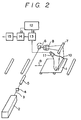
- the read-out operation for detecting the autoradiograph stored and recorded on the stimulable phosphor sheet as electric signals will be described briefly with respect to the third embodiment of the process of the invention, referring to an embodiment of a read-out system shown in Figure 2 of the accompanying drawings.
- the read-out operation is applied to the phosphor sheet combined with the photosensitive material so that the read-out operation also serves as the exposure operation of the photosensitive material.
- Figure 2 schematically shows an embodiment of the read-out system for reading out the information on one or two dimensional location of the radioactively labeled substances, which is stored and recorded on the photosensitive material-superposed stimulable phosphor sheet 1 shown in Figure 1-(1) wherein numeral 1a represents the stimulable phosphor sheet and 1 b represents the photosensitive material.
- the read-out operation is carried out in the following manner.
- Laser beam 3 generated by a laser source 2 first passes through a filter 4 to cut off a light beam in the wavelength region corresponding to the wavelength region of stimulated emission to be emitted from the phosphor sheet 1a in response to stimulation with the laser beam 3.
- the size of the beam diameter of the laser beam 3 passed through the filter 4 is strictly controlled by means of a beam expander 5.
- the laser beam is subsequently deflected by a beam deflector 6 such as a galvanometer mirror and reflected by a plane reflection mirror 7.
- the deflected beam then impinges one-dimensionally upon the photosensitive material 1 of the photosensitive material-superposed stimulable phosphor sheet 1.
- An f-8 lens 8 is provided between the beam deflector 6 and the plane reflection mirror 7 so that the beam speed is continuously kept constant when the deflected laser beam is scanned on the photosensitive material 1b.
- the laser source 2 used herein is selected as to avoid overlapping of the wavelength region of the laser beam 3 with the main wavelength region of the stimulated emission to be given by the stimulable phosphor sheet 1a. Further the wavelength region of the laser beam 3 is selected as to avoid the exposure of the photosensitive material 1 b.
- a preferred laser beam is one having a wavelength within the red region, though the employable wavelength region varies depending on the phosphor in the phosphor sheet and the photosensitive substance in the photosensitive material.
- the photosensitive material-superposed phosphor sheet 1 is then transferred to the direction along the arrow 9 under the irradiation of the above-mentioned deflected laser beam. Therefore, the whole surface of the phosphor sheet 1a a is subjected to the irradiation of the deflected laser beam through the photosensitive material 1 b.
- the phosphor sheet 1a When irradiated with the above-mentioned laser beam, the phosphor sheet 1a gives stimulated emission having the intensity proportional to the radiation energy stored therein. A portion of the emission is absorbed by the photosensitive material 1 b and an image (latent image) is formed on the photosensitive material 1b. On the other hand, the other portion of the emission enters through the photosensitive material 1 b into a light guiding sheet 10.
- the light guiding sheet 10 has a linear edge face for receiving the emission and the edge face is so positioned in the vicinity of the photosensitive material as to correspond to the scanning line on the photosensitive material 1b.
- the exit of the light guiding sheet 10 is in the form of a ring and is connected to a light-receiving face of a photosensor 11 such as a photomultiplier.
- the light guiding sheet 10 is made, for instance, by processing a sheet of a transparent thermoplastic resin such as a synthetic acrylic resin, and constituted such that the emission introduced from the linear edge face is transferred to the exit under total reflection within the sheet 10.
- the stimulated emission from the phosphor sheet 1a is guided in the interior of the light guiding sheet 10 to the exit, and received by the photosensor 11.
- a filter which allows only the light in the wavelength region of the stimulated emission to pass therethrough and cuts off the light in the wavelength region of the stimulating rays (laser beam) so as to detect only the stimulated emission.
- the stimulated emission detected by the photosensor 11 is converted to electric signals, amplified to electric signals adjusted to an appropriate level in an amplifier 13 according to an amplification degree setting value a provided from a control circuit 12 and transmitted to an A/D converter 14.
- the adjusted electric signals are then converted to digital signals according to an appropriate scale factor defined by a scale factor setting value b provided from the same control circuit 12, and supplied to a signal processing circuit 15.
- the digital signals are suitably processed to output digital data, which are then transmitted to a recording device (not shown), optionally upon storage in a storing means such as a magnetic tape.
- the amplification degree setting value a, and the scale factor setting value b provided from the control circuit 12 can be set, for instance, according to the stored and recorded information obtained by carrying out a preliminary read-out operation prior to the above read-out operation so as to obtain signals having good levels.
- the setting values can be experimentally set according to the exposure time of the phosphor sheet.
- the supplied digital signals are computerized so as to analyze the distribution and the radiation intensity of the radioactively labeled substances, and the locational information thereon can be obtained as symbols and/or numeralized digital data.
- a signal processing method for obtaining the locational information on one-dimensionally distributed radioactively labeled substances as symbols and/or numerals is described in, for instance, the aforementioned U.S. Patent Application No. 568,877 or European Patent Application No. 84100144.9.
- the digital signals are processed according to an image processing condition setting value c (not shown) provided from the control circuit 12 as to give a well-readable image having well adjusted concentration and contrast. Examples of the image processing include spatial frequency processing, gradation processing and substraction processing.
- Various recording devices based on various systems can be employed for the above-described purpose, for instance, a device for visualizing optically by scanning a photosensitive material with a laser beam, a display means for visualizing electrically on CRT, a means for printing a radiation image displayed on CRT by means of video printer, and a means for visualizing on a heat-sensitive recording material using thermic rays.
- the method for reading out the locational information on the radioactively labeled substances stored in the stimulable phosphor sheet is not restricted to those described above, and other suitable methods can be also used in the process of the present invention.
- the phosphor sheet may be irradiated with stimulating rays on the phosphor sheet-side (namely, the support-side thereof) and the stimulated emission may be also detected therefrom, instead of the irradiation and detection from the photosensitive material-side as described above.
- the photosensitive material having the latent image formed thereon by the exposure to the stimulated emission is separated from the phosphor sheet and developed to obtain a visible image corresponding to the autoradiograph of the radioactively labeled substances in the support medium.
- the read-out operation for the embodiment shown by Figure 1-(1) been described above (namely, the third method)
- the read-out of the stimulable phosphor sheet and the exposure of the photosensitive material for the other embodiments can be carried out in a similar manner to that described above by irradiating one side of the phosphor sheet combined with the photosensitive material and the support medium in layers, with stimulating rays (namely, the fourth embodiment).
- the fourth method it is preferred to conduct the irradiation of the stimulating rays and the detection of the stimulated emission on the phosphor layer-side.
- the confirmation and the further analysis of the resulting locational information on the radioactively labeled substances can be made by directly comparing the obtained digital data with the visualized auroradiograph on the photosensitive material. Further, this autoradiographic image can be compared with an image reproduced from the image-processed digital data.
- the read-out and the visualization of the autoradiograph having the locational information on the radioactively labeled substances stored and recorded on the stimulable phosphor sheet can be simultaneously conducted.
- the autoradiographic process can be simplified to substantially comprise the resolving stage and the read-out stage including the exposure.
- the support mediums for resolution which were used in the following examples were ones for electrophoresis, composed of a slab gel (1.5 mmx200 mmx200 mm) containing 8% of polyacrylamide (crosslinking agent content: 3%) prepared by a conventional method.
- the stimulable phosphor sheets were prepared by the following method.
- the coating dispersion was applied to a polyethylene terephthalate sheet (support, thickness: 250 ⁇ m) placed horizontally on a glass plate.
- the application of the coating dispersion was carried out using a doctor blade.
- the support having a layer of the coating dispersion was then placed in an oven and heated at a temperature gradually rising from 25 to 100°C.
- a phosphor layer having a thickness of 300 um was formed on the support.
- a stimulable phosphor sheet comprising a support, a phosphor layer and a protective film was prepared.
- Plasmid DNA of E. coli (pBR 322) was cleaved by the use of restriction enzyme Hind-III by the known method and 5'-end thereof was labeled with 32 P to obtain 1 ⁇ g of a double helix DNA ( 32 P-labeled substance).
- the double helix DNA (1 ⁇ g) and approx. 1 unit of the restriction enzyme Hae-III were added to 20 ⁇ l of 20 mM of tris[tris(hydroxylmethy-I)aminoethane]-hydrochloric acid buffer solution (pH 7.4) containing 5 mM of magnesium chloride and 1 mM of dithiothreitol.
- the resulting mixture was kept at 37°C for one hour to perform the specific cleavage reaction and a cleaved mixture solution containing cleavage products was obtained.
- the sample of cleaved mixture solution was charged on the slab gel support medium and electrophoresed at voltage of 500 V using 50 mM tris-borate buffer solution (pH 8.3) containing 1 mM of EDTA as an electrode solution.
- the electrophoresis was continued until the marker dye previously added to the sample reached the bottom end of the gel, and the starting position thereof was marked with a 32 P-containing ink.
- the above gel support medium and the stimulable phosphor sheet were superposed in layers and kept at room temperature (approx. 25°C) for 12.5 min to perform the storing and recording of an autoradiograph of the sample on the phosphor sheet.
- the support medium was separated from the phosphor sheet, and an X-ray film (RX type manufactured by Fuji Photo Film Co., Ltd.) was superposed on the protective film-side of the phosphor sheet.
- the X-ray film-superposed phosphor sheet was introduced into a read-out apparatus as shown in Figure 2 and then the phosphor sheet was scanned with an He-Ne laser beam (wavelength: 633 nm; light energy: 7x10- 4 J/cm 2 ) to expose the X-ray film to stimulated emission given by the divalent europium activated barium fluorobromide phosphor (peak wavelength: 390 nm).
- the X-ray film was developed.
- the electrophoretic pattern of the cleavage products with 32 P label was visualized as an image on the X-ray film.
- the phosphor sheet was introduced in the same read-out apparatus again and scanned with the laser beam in the same manner as described above, to read out locational information which represented the electrophoretic positions of the 32 P-labeled fragments (cleavage products) based on the starting position marked with the 32 P-containing ink. According to the thus-obtained locational information, the portions containing the 32 P-labeled fragments were cut out of the gel with a thin razor blade, and the gel portion segments were placed in a test tube.
- the residual gel (a part of which had been removed as above) was laid again on a stimulable phosphor sheet, and the read-out operation was conducted thereto in the read-out apparatus to examine absence of the 32 P-labeled fragment.
- the result of the examination indicated that the 32 P-labeled fragments had been completely removed from the gel.
- the accuracy of the locational information on 32 P-labeled fragments obtained by means of the above stimulable phosphor sheet provided with the support medium was sufficiently high.
- the gel support medium having the fragments (cleavage products) labeled with 32 P and the stimulable phosphor sheet were superposed in layers and kept at room temperature (approx. 25°C) for 12.5 min to perform the storing and recording of an autoradiograph of the sample.
- the support medium was then separated from the phosphor sheet, and an X-ray film (RX type manufactured by Fuji Photo Film Co., Ltd.) was superposed on the protective film-side of the phosphor sheet.
- the X-ray film-superposed phosphor sheet was introduced into the read-out apparatus as shown in Figure 2 and then read out to obtain locational information which represented the electrophoretic positions of the 32 P-labeled fragments based on the starting position marked with the 32 P-containing ink.
- an He-Ne laser beam (wavelength: 633 nm; light energy: 7 ⁇ 10 -4 J/cm 2 ) was used as the stimulating rays and the stimulated emission of the divalent europium activated barium fluorobromide phosphor (peak wavelength: 390 nm) was detected.
- the portions containing 32 P-labeled fragments were cut out of the gel with a thin razor blade, and the gel portion segments were placed in a test tube.
- the residual gel (a part of which had been removed as above) was laid again on the stimulable phosphor sheet, and the read-out operation was carried out thereto in the read-out apparatus to examine absence of the 32 P-labeled fragment.
- the result of the examination indicated that the 32 P-labeled fragments had been completely removed from the gel.
- the accuracy of the locational information on 32 P-labeled fragments obtained by means of the above stimulable phosphor sheet provided with the support medium was sufficiently high.
Landscapes
- Physics & Mathematics (AREA)
- General Physics & Mathematics (AREA)
- Conversion Of X-Rays Into Visible Images (AREA)
- Measurement Of Radiation (AREA)
Claims (6)
Applications Claiming Priority (4)
| Application Number | Priority Date | Filing Date | Title |
|---|---|---|---|
| JP8900284A JPS60233583A (ja) | 1984-05-02 | 1984-05-02 | オ−トラジオグラフ測定法 |
| JP89001/84 | 1984-05-02 | ||
| JP8900184A JPS60233582A (ja) | 1984-05-02 | 1984-05-02 | オ−トラジオグラフ測定法 |
| JP89002/84 | 1984-05-02 |
Publications (3)
| Publication Number | Publication Date |
|---|---|
| EP0160939A2 EP0160939A2 (fr) | 1985-11-13 |
| EP0160939A3 EP0160939A3 (en) | 1986-06-11 |
| EP0160939B1 true EP0160939B1 (fr) | 1990-08-16 |
Family
ID=26430320
Family Applications (1)
| Application Number | Title | Priority Date | Filing Date |
|---|---|---|---|
| EP85105368A Expired - Lifetime EP0160939B1 (fr) | 1984-05-02 | 1985-05-02 | Procédé autoradiographique |
Country Status (3)
| Country | Link |
|---|---|
| US (1) | US4734581A (fr) |
| EP (1) | EP0160939B1 (fr) |
| DE (1) | DE3579186D1 (fr) |
Families Citing this family (13)
| Publication number | Priority date | Publication date | Assignee | Title |
|---|---|---|---|---|
| US5260190A (en) * | 1982-11-05 | 1993-11-09 | Fuji Photo Film Co., Ltd. | Autoradiographic process |
| US5028793A (en) * | 1985-10-10 | 1991-07-02 | Quantex Corporation | Imaging screen for electrophoresis applications |
| JPS6285862A (ja) * | 1985-10-11 | 1987-04-20 | Fuji Photo Film Co Ltd | 核酸の塩基配列決定のための信号処理方法 |
| US4885696A (en) * | 1986-03-26 | 1989-12-05 | Fuji Photo Film Co., Ltd. | Signal processing method for determining base sequence of nucleic acid |
| JPS63167290A (ja) * | 1986-12-27 | 1988-07-11 | Fuji Photo Film Co Ltd | オ−トラジオグラフ解析のための信号処理方法 |
| JPH0664057B2 (ja) * | 1987-01-06 | 1994-08-22 | 富士写真フイルム株式会社 | オ−トラジオグラフ解析のための信号処理方法 |
| DE8717526U1 (fr) * | 1987-11-17 | 1989-04-06 | Siemens Ag, 1000 Berlin Und 8000 Muenchen, De | |
| JPH01253679A (ja) * | 1988-04-01 | 1989-10-09 | Hitachi Ltd | 放射線撮像素子 |
| US5347139A (en) * | 1991-06-04 | 1994-09-13 | Molecular Dynamics | Dual energy tracer quantitative analysis |
| US5128978A (en) * | 1991-09-20 | 1992-07-07 | Polaroid Corporation | Film holder for autoradiographic imaging |
| FR2699004B1 (fr) * | 1992-12-08 | 1995-02-10 | Georges Charpak | Procédé de représentation de la distribution spatiale d'éléments radioactifs au moyen d'un écran de type phosphore effaçable, et dispositif correspondant. |
| US5830629A (en) * | 1995-11-01 | 1998-11-03 | Eastman Kodak Company | Autoradiography assemblage using transparent screen |
| US20090039288A1 (en) * | 2006-12-18 | 2009-02-12 | Kulpinski Robert W | Single sided dual scanning for computed radiography |
Citations (1)
| Publication number | Priority date | Publication date | Assignee | Title |
|---|---|---|---|---|
| US4239968A (en) * | 1978-07-12 | 1980-12-16 | Fuji Photo Film Co., Ltd. | Method and apparatus for recording and reproducing a radiation image |
Family Cites Families (5)
| Publication number | Priority date | Publication date | Assignee | Title |
|---|---|---|---|---|
| US2523306A (en) * | 1947-09-23 | 1950-09-26 | Herman F Kaiser | Application of radiography to infrared phosphors |
| US3308438A (en) * | 1963-11-01 | 1967-03-07 | Baird Atomic Inc | Autofluoroscope |
| JPS5863931A (ja) * | 1981-10-12 | 1983-04-16 | Fuji Photo Film Co Ltd | X線画像の形成方法 |
| US4542523A (en) * | 1982-05-17 | 1985-09-17 | Polaroid Corporation | Radiographic intensifying screen manipulator |
| JPH0685045B2 (ja) * | 1982-05-19 | 1994-10-26 | 富士写真フイルム株式会社 | 放射線画像情報変換方法および装置 |
-
1985
- 1985-05-02 DE DE8585105368T patent/DE3579186D1/de not_active Expired - Fee Related
- 1985-05-02 EP EP85105368A patent/EP0160939B1/fr not_active Expired - Lifetime
-
1986
- 1986-09-08 US US06/904,865 patent/US4734581A/en not_active Expired - Lifetime
Patent Citations (1)
| Publication number | Priority date | Publication date | Assignee | Title |
|---|---|---|---|---|
| US4239968A (en) * | 1978-07-12 | 1980-12-16 | Fuji Photo Film Co., Ltd. | Method and apparatus for recording and reproducing a radiation image |
Also Published As
| Publication number | Publication date |
|---|---|
| US4734581A (en) | 1988-03-29 |
| EP0160939A2 (fr) | 1985-11-13 |
| DE3579186D1 (de) | 1990-09-20 |
| EP0160939A3 (en) | 1986-06-11 |
Similar Documents
| Publication | Publication Date | Title |
|---|---|---|
| US4617468A (en) | Stimulable phosphor sheet with hydrophilic surface | |
| US4865967A (en) | Autoradiographic gene-screening method | |
| US5260190A (en) | Autoradiographic process | |
| US5270162A (en) | Autoradiographic gene-screening method | |
| EP0160939B1 (fr) | Procédé autoradiographique | |
| EP0113672A2 (fr) | Dispositif de traitement d'un signal pour autoradiographie | |
| EP0079751B1 (fr) | Procédé pour éteindre le bruit des feuilles phosphore à stimuler | |
| EP0772059A1 (fr) | Assemblage pour autoradiographic avec l'écran transparent | |
| US4888695A (en) | Signal processing method in autoradiography | |
| EP0159523B1 (fr) | Procédé d'autoradiographie | |
| EP0725278B1 (fr) | Méthode pour la détection chimiluminescente et appareil | |
| EP0141382B1 (fr) | Méthode pour la détermination de la séquence des bases d'ADN ou fragments d'ADN utilisant l'autoradiographie | |
| EP0138086B1 (fr) | Méthode pour le tamisage autoradographique de gènes | |
| EP0111154B1 (fr) | Traitement d'autoradiographie | |
| US4871913A (en) | Signal processing method in autoradiography | |
| EP0115777B1 (fr) | Méthode pour la détermination de la séquence de base d'ADN on de fragment d'ADN | |
| US6236058B1 (en) | Image recording and reading system | |
| JPH0160784B2 (fr) | ||
| JPS60233582A (ja) | オ−トラジオグラフ測定法 | |
| JPH0465997B2 (fr) | ||
| US6066858A (en) | Autoradiographic process | |
| JPH0456259B2 (fr) | ||
| JPH1048399A (ja) | 蓄積性蛍光体シートおよび蓄積性蛍光体シートを用いた画像読み取り装置 |
Legal Events
| Date | Code | Title | Description |
|---|---|---|---|
| PUAI | Public reference made under article 153(3) epc to a published international application that has entered the european phase |
Free format text: ORIGINAL CODE: 0009012 |
|
| AK | Designated contracting states |
Designated state(s): DE FR NL |
|
| PUAL | Search report despatched |
Free format text: ORIGINAL CODE: 0009013 |
|
| AK | Designated contracting states |
Kind code of ref document: A3 Designated state(s): DE FR NL |
|
| 17P | Request for examination filed |
Effective date: 19860707 |
|
| 17Q | First examination report despatched |
Effective date: 19871008 |
|
| GRAA | (expected) grant |
Free format text: ORIGINAL CODE: 0009210 |
|
| AK | Designated contracting states |
Kind code of ref document: B1 Designated state(s): DE FR NL |
|
| REF | Corresponds to: |
Ref document number: 3579186 Country of ref document: DE Date of ref document: 19900920 |
|
| ET | Fr: translation filed | ||
| PLBE | No opposition filed within time limit |
Free format text: ORIGINAL CODE: 0009261 |
|
| STAA | Information on the status of an ep patent application or granted ep patent |
Free format text: STATUS: NO OPPOSITION FILED WITHIN TIME LIMIT |
|
| 26N | No opposition filed | ||
| PGFP | Annual fee paid to national office [announced via postgrant information from national office to epo] |
Ref country code: FR Payment date: 20020523 Year of fee payment: 18 |
|
| PGFP | Annual fee paid to national office [announced via postgrant information from national office to epo] |
Ref country code: NL Payment date: 20020524 Year of fee payment: 18 |
|
| PGFP | Annual fee paid to national office [announced via postgrant information from national office to epo] |
Ref country code: DE Payment date: 20020628 Year of fee payment: 18 |
|
| PG25 | Lapsed in a contracting state [announced via postgrant information from national office to epo] |
Ref country code: NL Free format text: LAPSE BECAUSE OF NON-PAYMENT OF DUE FEES Effective date: 20031201 |
|
| PG25 | Lapsed in a contracting state [announced via postgrant information from national office to epo] |
Ref country code: DE Free format text: LAPSE BECAUSE OF NON-PAYMENT OF DUE FEES Effective date: 20031202 |
|
| PG25 | Lapsed in a contracting state [announced via postgrant information from national office to epo] |
Ref country code: FR Free format text: LAPSE BECAUSE OF NON-PAYMENT OF DUE FEES Effective date: 20040130 |
|
| NLV4 | Nl: lapsed or anulled due to non-payment of the annual fee |
Effective date: 20031201 |
|
| REG | Reference to a national code |
Ref country code: FR Ref legal event code: ST |