EP0125412A1 - Strahlungsbildaufzeichnungs- und Wiedergabesystem - Google Patents
Strahlungsbildaufzeichnungs- und Wiedergabesystem Download PDFInfo
- Publication number
- EP0125412A1 EP0125412A1 EP84102584A EP84102584A EP0125412A1 EP 0125412 A1 EP0125412 A1 EP 0125412A1 EP 84102584 A EP84102584 A EP 84102584A EP 84102584 A EP84102584 A EP 84102584A EP 0125412 A1 EP0125412 A1 EP 0125412A1
- Authority
- EP
- European Patent Office
- Prior art keywords
- radiation
- image
- image recording
- stimulable phosphor
- conditions
- Prior art date
- Legal status (The legal status is an assumption and is not a legal conclusion. Google has not performed a legal analysis and makes no representation as to the accuracy of the status listed.)
- Granted
Links
- 230000005855 radiation Effects 0.000 title claims abstract description 75
- 238000012545 processing Methods 0.000 claims abstract description 75
- OAICVXFJPJFONN-UHFFFAOYSA-N Phosphorus Chemical compound [P] OAICVXFJPJFONN-UHFFFAOYSA-N 0.000 claims abstract description 38
- 230000004044 response Effects 0.000 claims abstract description 9
- 230000004936 stimulating effect Effects 0.000 claims abstract description 8
- 230000000994 depressogenic effect Effects 0.000 description 19
- 230000000881 depressing effect Effects 0.000 description 7
- 238000000034 method Methods 0.000 description 7
- 238000001514 detection method Methods 0.000 description 4
- 238000003745 diagnosis Methods 0.000 description 4
- 230000006870 function Effects 0.000 description 3
- 210000001015 abdomen Anatomy 0.000 description 2
- 230000004913 activation Effects 0.000 description 2
- 238000010586 diagram Methods 0.000 description 2
- 238000003780 insertion Methods 0.000 description 2
- 230000037431 insertion Effects 0.000 description 2
- 238000002601 radiography Methods 0.000 description 2
- 230000003213 activating effect Effects 0.000 description 1
- 238000002583 angiography Methods 0.000 description 1
- 238000010276 construction Methods 0.000 description 1
- 230000001747 exhibiting effect Effects 0.000 description 1
- 230000010365 information processing Effects 0.000 description 1
- 239000000463 material Substances 0.000 description 1
- 238000012544 monitoring process Methods 0.000 description 1
- 229910052709 silver Inorganic materials 0.000 description 1
- 239000004332 silver Substances 0.000 description 1
- -1 silver halide Chemical class 0.000 description 1
Images


Classifications
-
- G—PHYSICS
- G03—PHOTOGRAPHY; CINEMATOGRAPHY; ANALOGOUS TECHNIQUES USING WAVES OTHER THAN OPTICAL WAVES; ELECTROGRAPHY; HOLOGRAPHY
- G03B—APPARATUS OR ARRANGEMENTS FOR TAKING PHOTOGRAPHS OR FOR PROJECTING OR VIEWING THEM; APPARATUS OR ARRANGEMENTS EMPLOYING ANALOGOUS TECHNIQUES USING WAVES OTHER THAN OPTICAL WAVES; ACCESSORIES THEREFOR
- G03B42/00—Obtaining records using waves other than optical waves; Visualisation of such records by using optical means
- G03B42/02—Obtaining records using waves other than optical waves; Visualisation of such records by using optical means using X-rays
-
- A—HUMAN NECESSITIES
- A61—MEDICAL OR VETERINARY SCIENCE; HYGIENE
- A61B—DIAGNOSIS; SURGERY; IDENTIFICATION
- A61B6/00—Apparatus or devices for radiation diagnosis; Apparatus or devices for radiation diagnosis combined with radiation therapy equipment
- A61B6/44—Constructional features of apparatus for radiation diagnosis
- A61B6/4494—Means for identifying the diagnostic device
-
- G—PHYSICS
- G01—MEASURING; TESTING
- G01T—MEASUREMENT OF NUCLEAR OR X-RADIATION
- G01T1/00—Measuring X-radiation, gamma radiation, corpuscular radiation, or cosmic radiation
- G01T1/16—Measuring radiation intensity
- G01T1/20—Measuring radiation intensity with scintillation detectors
- G01T1/2012—Measuring radiation intensity with scintillation detectors using stimulable phosphors, e.g. stimulable phosphor sheets
Definitions
- This invention relates to a radiation image recording and reproducing system.
- This invention particularly relates to a radiation image recording and reproducing system wherein a stimulable phosphor is exposed to a radiation passing through an object to have a radiation image stored therein, the stimulable phosphor is scanned with stimulating rays which cause it to emit light in proportion to the radiation energy stored, the emitted light is photoelectrically detected and converted to an electric image signal, the electric image signal is processed, and a visible image is reproduced by use of the processed electric image signal.
- a radiation image recording and reproducing system using a stimulable phosphor is described, for example, in U.S. Patent Nos. 4,258,264, 4,276,473 and 4,315,318, U.S. Patent Appln. Serial No. 220,780, Japanese Unexamined Patent Publication No. 56(1981)-11395, and "Nikkan Kogyo Shinbun” (Daily Industrial Newspaper), November 6, 1982 edition.
- the radiation image recording and reproducing system comprises the steps of (i) exposing the stimulable phosphor to a radiation such as X-rays passing through an object to have a radiation image stored therein, (ii) scanning the stimulable phosphor with stimulating rays which cause it to emit light in proportion to the radiation energy stored, (iii) photoelectrically detecting the emitted light and converting it into an electric image signal, and (iv) reproducing a visible image by use of the obtained electric image signal.
- image recording can be conducted by use of a radiation exposure dose markedly lower than in the conventional radiography using a silver halide photographic material. Further, by processing the electric image signal in various manners, it is possible to obtain a radiation image having a markedly improved image quality, particularly a high diagnostic efficiency and accuracy. Thus this system is very advantageous in practical use, particularly for medical diagnosis.
- the aforesaid radiation image recording and reproducing system provides high diagnostic performance by appropriately processing the electric image signal obtained by photoelectrically reading out the radiation image stored in the stimulable phosphor. Therefore, image processings must be carried out appropriately according to the type of image recording. That is, most suitable image processing conditions should be selected according to factors such as the portion of the object to be image-recorded (the heart, the chest, or the like), the image recording method (plain image recording, contrasted image recording, subtraction image recording, or the like), and the diagnostic purpose (mass medical examination, close examination, or the like).
- the image processings embrace all possible image processings for improving the quality of radiation images according to the portion of the object, for example, contrast adjustments, density level adjustments, image gradation processings, frequency processings, and unsharp mask processings.
- the image processings also embrace image subtraction processings.
- exposure conditions of the radiation source should also preferably be adjusted as required with respect to the above-described type of image recording.
- image recording should be conducted under the most suitable radiation exposure conditions by changing, for example, the tube voltage, the tube current, the exposure time, and the focusing point size of the radiation source.
- the radiation exposure conditions of the radiation source and the image processing conditions be adjusted to the most suitable conditions according to the aforesaid type of image recording.
- the radiation image recording and reproducing system is continuously used for recording and reproducing radiation images of many patients or examination objects in mass medical examinations or the like, it is troublesome to adjust the radiation exposure conditions and the image processing conditions for each patient or each examination object every time image recording and reproducing are conducted. Also, in such a case, adjustment errors or operation errors readily arise.
- the stimulable phosphor used for recording image information in the aforesaid radiation image recording and reproducing system is fabricated into a sheet-like shape, and the stimulable phosphor sheets are provided with identification codes such as bar codes for identifying the stimulable phosphor sheets.
- the identification codes of the stimulable phosphor sheets are memorized in relation to the aforesaid object information, and used to clarify the relationship between the image information recorded in the stimulable phosphor sheets and the object information at the time of image recording and reproducing.
- the operations for correctly clarifying the relationship between the object information and the identification codes and for memorizing the object information and the identification codes in relation to each other are not always possible to conduct when a large number of objects have to be handled. Thus there is a risk of mistakes arising in such operations.
- the aforesaid operation must be carried out without fail for all objects. This problem must also be solved in practical use of the radiation image recording and reproducing system.
- the primary object of the present invention is to provide a radiation image recording and reproducing system free of operation error and exhibiting a high efficiency, wherein adjustment of the radiation exposure conditions for image recording and adjustment of the image processing conditions at the time of image processing are carried out by a single operation.
- Another object of the present invention is to provide a radiation image recording and reproducing system wherein operation errors are eliminated and the efficiency is improved by conducting the adjustment of the radiation exposure conditions, the adjustment of the image processing conditions, and read-out of an identification code of a stimulable phosphor by a single operation.
- the specific object of the present invention is to provide a radiation image recording and reproducing system wherein the adjustment of the radiation exposure conditions and the adjustment of the image processing conditions are carried out by a single operation, and read-out of the identification code of the stimulable phosphor is conducted automatically in response to an operation for memorizing the object information.
- the radiation image recording and reproducing system in accordance with the present invention comprises simultaneously adjusting the radiation exposure conditions and the image processing conditions by a single action for selecting preset conditions predetermined according to the type of image recording.
- the adjustment of the radiation exposure conditions, the adjustment of the image processing conditions, and read-out of an identification code of a stimulable phosphor are conducted by the same operation, thereby improving the efficiency and preventing operation errors.
- the radiation exposure conditions and the image processing conditions are simultaneously adjusted as described above, and read-out of the identification code of the stimulable phosphor is automatically conducted in response to an operation for memorizing the object information.
- the read-out of the identification code and memorizing of the object information are carried out simultaneously, thereby preventing operation errors.
- the radiation exposure conditions and the image processing conditions which should be determined for respective objects are simultaneously adjusted by a single operation.
- the radiation image recording and reproducing system of the present invention exhibits a high efficiency and is free of operation errors. This is very advantageous in practical use.
- single operation is meant the series of actions conducted by the operator (radiologist) as the selecting operation according to the type of image recording, for example, the series of actions of selecting and depressing the keys on a keyboard which specify the portion of the object (the abdomen, the frontal chest, or the like), and/or the image recording method (plain image recording, angiography, or the like), and/or the purpose of image recording.
- the operation of depressing these keys is called a single operation insofar as the depressing of these keys is a series of interrelated actions which have to be conducted for one type of image recording.
- read-out of an identification code of a stimulable phosphor is conducted by the same action as that for the adjustment of the radiation exposure conditions and the adjustment of the image processing conditions.
- this does not necessarily mean that the read-out of the identification code and the adjustments of the aforesaid conditions are carried out exactly simultaneously.
- the adjustments of the aforesaid conditions may first be conducted, and then the read-out of the identification code may be automatically carried out after several seconds insofar as the adjustments and the read-out are effected by a single operation (for example, by a series of actions of depressing the keys on the keyboard as described above).
- the adjustments of the aforesaid conditions and the read-out of the identification code should be regarded as being conducted by a single operation since the time lag therebetween is not caused by a different manual operation, but instead is a mechanical delay or an electrical delay.
- the read-out of the identification code of the stimulable phosphor is automatically conducted in response to an action of memorizing the object information.
- a magnetic card patient identification card or patient ID card
- the read-out of a bar code (identification code) of the stimulable phosphor may be started simultaneously with the start of the read-out of the magnetic card.
- the read-out of the bar code of the stimulable phosphor may be started in response to the manual entry of the aforesaid object information from a keyboard or the like (i.e. in response to the memorizing action).
- the data on the portion of the object in the aforesaid patient ID card is magnetically recorded together with the data on the name of the patient or the like in the patient ID card.
- the read-out of the identification code but also the selection of the aforesaid radiation exposure conditions and image processing conditions according to the type of image recording can be achieved only by the action of inserting the patient ID card into the magnetic card reader. In this case, the adjustments of the radiation exposure conditions and the image processing conditions, the read-out of the identification code, and memorizing of the object information are carried out by a single operation.
- Figure 1 shows the general arrangement of an embodiment of the radiation image recording and reproducing system in accordance with the present invention.
- the object is a patient and an X-ray source is used as the radiation source.
- An X-ray source 1 for emitting X-rays to a patient P is connected with a controller 2 for adjusting the radiation exposure conditions of the X-ray source 1, such as the tube voltage, the tube current, and the exposure time.
- the X-rays emitted from the X-ray source 1 pass through a predetermined portion of the patient P, and an image recording sheet 3 comprising a stimulable phosphor is exposed to the X-rays passing through the portion of the patient P to have an X-ray image of the portion stored therein.
- On the rear surface of the sheet 3 is provided in advance a bar code 3A (i.e. an identification code), which is read out by a bar code reader 4.
- the bar code reader 4 Upon reading out the bar code 3A, the bar code reader 4 generates a read-out signal 4a and sends it to the controller 2.
- a patient ID card reader for reading out the patient ID information (i.e. the object information) when a patient ID card 5 carrying the patient ID information stored therein is inserted into the patient ID card reader.
- the signals obtained by the read-out of the bar code 3A and the patient ID card 5 are sent from the controller 2 to an image processing and reproducing section 10.
- the bar code reader 4 and the patient ID card reader need not necessarily be connected with or incorporated in the controller 2. Thus the bar code reader 4 and the patient ID card reader may be installed separately from the controller 2 and connected with the image processing and reproducing section 10.
- the X-ray image stored in the image recording sheet 3 is then read out by a read-out section 11 of the image processing and reproducing section 10. Thereafter, an electric image signal obtained by the read-out is processed in an appropriate manner (under the image processing conditions adjusted by the controller 2).
- the image signal thus processed is used to reproduce a visible image on a monitor television 13 for monitoring the reproduced X-ray image.
- the image signal processed as described above is sent to an image reproducing section 14 in which a visible image 6A is reproduced in a photographic film by use of the image signal to form an image sheet 6 for diagnosis.
- the visible image 6A ultimately obtained in this manner has a high image quality which could not been obtained by the conventional radiography, and includes abundant diagnostic information.
- the controller 2 is provided with selection keys 2A for selecting the type of image recording and sending the signals specifying the radiation exposure conditions and the image processing conditions suitable for the type of image recording to the X-ray source 1 and the image processing section 12, thereby adjusting the radiation exposure conditions of the X-ray source 1 and the image processing conditions of the image processing section 12.
- selection keys 2A When necessary ones of the selection keys 2A are pressed, an image recording condition setting signal 2a and an image processing condition setting signal 2b are generated according to the type of image recording thus selected, and respectively sent to the X-ray source 1 and the image processing section 12.
- the image recording condition setting signal 2a is not sent to the outside of the controller 2.
- the signal 4a sent from the bar code reader 4 and the signal obtained by reading out the patient ID card 5 are fed as an ID signal 2c to the image processing and reproducing section 10.
- the image processing and reproducing section 10 is incorporated a computer for carrying out necessary memorizing and processing operations upon receiving the signals 2b and 2c.
- the electronic computer the relationship between the image to be reproduced and the ID signal is clarified by known methods of information processing, and necessary image processing is carried out.
- the controller 2 is also provided with a display section 2B for indicating the contents selected by the selection keys 2A on the keyboard. Further, input keys 2C such as numeric keys for manually inputting the ID contents are installed on the controller 2.
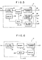
- Figures 4 and 5 show an embodiment of the architecture of the controller 2.
- a card reader 5A for reading out the ID information from the patient ID card 5 inserted thereinto, and a bar code reader controller 4A for controlling the bar code reader 4 are incorporated in the controller 2.
- the controller 2 also has a setting condition memory 20 in which the image recording conditions and the image processing conditions are memorized in advance for each type of image recording.
- the setting condition memory 20 receives a signal 21 representing the type of image recording which is selected by the selection keys 2A on the keyboard, and outputs signals specifying setting conditions corresponding to the selected type of image recording via an X-ray controller lA and an image processing controller 12A.
- the numeric keys 2C are positioned at a part of the keyboard so that the ID information and other necessary information can be entered manually by use of the numeric keys 2C.
- dials 2D, 2E and 2F for manually adjusting the tube voltage, the tube current and the exposure time of the X-ray source 1.
- the keyboard also has a density adjusting lever 2G and a contrast adjusting lever 2H for manually adjusting the image processing conditions, so that the image density and the image contrast can be manually adjusted to appropriate values.
- the bar code reader controller 4A activates the bar code reader 4 to read out the bar code 3A of the image recording sheet 3.
- the bar code reader controller 4A may be activated by the insertion of the patient ID card 5 into the card reader 5A instead of by depressing the ID pushbutton 2I.
- the ID pushbutton 2I is used only when the patient ID information is manually entered by use of the numeric keys 2C. Namely, when the patient ID information is entered by use of the numeric keys 2C, the ID pushbutton 2I is first depressed, and then the numeric keys 2C are depressed. At this time, the bar code reader 4 is activated as the ID pushbutton 2I is depressed. Or, conversely, the numeric keys 2C may be depressed first, and then the ID pushbutton 2I may be depressed, thereby starting the input of the ID information and the read-out of the bar code reader 4.
- the read-out signal 4a generated by the bar code reader 4 is returned to the bar code reader controller 4A, and sent to the image processing and reproducing section 10 together with the output of the ID card reader 5A.
- an X-ray exposure button 2J is used to cause X-rays to emit from the X-ray source 1.
- the X-ray exposure button 2J may be utilized also for inputting of the aforesaid image processing conditions (i.e. the action for inputting the set information into the image processing section 12), or for inputting of the ID information and the bar code information read out by the bar code reader 4 (i.e. the action for sending the inputted value and the read-out value to the image processing and reproducing section 10.
- the signals 2a, 2b and 2c are transmitted and, at the same time,' the X-ray source 1 is activated to emit X-rays.
- the bar code reader 4 is connected only with the controller 2. However, it is also possible to construct the system so that the bar code reader 4 receives a start signal 4a from the controller 2, and the output signal 4a of the bar code reader 4 is directly sent to the image processing and reproducing section 10 without passing though the controller 2.
- Figure 2 shows another embodiment of the radiation image recording and reproducing system in accordance with the present invention, which is constructed in the manner just described above.
- the bar code reader 4 is positioned at the rear of the image recording sheet 3 positioned for image recording in the system, so that the bar code 3A can be read while the image recording sheet 3 is positioned for image recording.
- the output signal 4a of the bar code reader 4 is directly sent to the image processing and reproducing section 10.
- the above-described embodiments of the radiation image recording and reproducing system in accordance with the present invention are operated in the manner as described below.
- the patient ID card 5 is received from the patient P and inserted into the card inlet of the controller 2.
- a cassette containing a single image recording sheet 3 or a magazine containing many image recording sheets 3 is loaded into the predetermined position.
- the ID pushbutton 21 on the keyboard of the controller 2 is depressed to activate the bar code reader 4.
- the read-out of the ID card 5 is carried out as the ID pushbutton 2I is depressed.
- the image recording portion of the patient P and the method of image recording e.g.
- the frontal chest and the plain image recording method are selected by use of the selection keys 2A on the keyboard.
- the signal 21 representing the selected type of image recording is entered into the setting condition memory 20, and the setting conditions corresponding to the selected type of image recording are selected from the data memorized in the memory 20 and sent to the X-ray controller lA and the image processing controller 12A.
- the X-ray controller lA controls the X-ray source 1 on the basis of the setting conditions so as to emit X-rays of a suitable dose from the X-ray source 1.
- the image processing controller 12A sends the image processing setting conditions suitable for the selected type of image recording to the image processing section 12 of the image processing and reproducing section 10, thereby adjusting the image processing conditions to desirable values.
- the bar code information read out by the bar code reader 4 and the patient ID information read out by the ID card reader 5A are entered into and memorized in the computer of the image processing and reproducing section 10. After the image recording is completed, the X-ray image stored in the image recording sheet 3 is read out by the read-out section 11.
- the sheet 3 is two- dimensionally scanned with stimulating rays such as a laser beam, and light emitted from the sheet 3 in proportion to the X-ray energy stored when the sheet 3 is exposed to the stimulating rays is photoelectrically detected and converted into an electric image signal.
- the electric image signal thus obtained is sent to the image processing section 12 and processed therein on the basis of the processing conditions specified by the image processing condition setting signal 2b and with reference to the signal 2c generated by the bar code reader 4.
- the image signal thus processed is displayed on the monitor television 13, so that the operator can monitor the image reproduced.
- the processed image signal is also sent to the image reproducing section 14 and used therein to reproduce the visible image 6A by point-by-point scanning on the image sheet 6 for diagnosis. At this time, the aforesaid patient ID information is also reproduced in the image sheet 6.
- the X-ray image of the patient p is recorded under predetermined appropriate image recording conditions, processed under predetermined appropriate image processing conditions, and reproduced in the image sheet 6 for diagnosis together with the necessary ID information.
- the image recording conditions and the image processing conditions are simultaneously adjusted by a single simple operation of selecting the type of image recording, no operation error arises, and a high efficiency is realized.
- the bar code reader 4 is activated by depressing the ID pushbutton 2I.
- the ID pushbutton 2I as a control start button which is depressed after the selection keys 2A are depressed, thereby starting the input of the setting conditions and the operation of the bar code reader 4.
- the activation of the bar code reader 4 can also be included in a single operation.
- the condition setting signals 2a and 2b are entered respectively into the X-ray source 1 and the image processing section 12 and, at the same time, the bar code reader 4 is activated to read out the bar code 3A.
- the ID card reader 5A may also be activated to start reading out as the I D pushbutton 2I used as the control start button is depressed. In this case, the exposure button 2J is used only for starting the exposure to X-rays.
- the activation of the bar code reader 4 and the read-out of the ID card 5 are conducted prior to the image recording.
- this sequence may be reversed. Namely, it is-also possible to construct the system so that the ID pushbutton 2I is depressed after the selection keys 2A are depressed.
- the bar code reader 4 is connected with the controller 2.
- the bar code reader 4 need not necessarily be connected to the controller 2.
- the bar code reader 4 may be positioned in the vicinity of the bar code 3A, and a sensor 4 B may be installed on an image recording table 30 on which the patient P lies.
- the sensor 4B detects the presence of the patient P on the image recording table 30 and sends a detection signal 4b' to the bar code reader 4.
- the bar code reader 4 starts reading out the bar code 3A.
- the ID card reader 5A is incorporated in the controller 2.
- the controller 2 comprises the keyboard provided with the keys having many functions, the setting condition memory 20, the X-ray controller lA, the image processing controller 12A, and the display section 2B.
- the read-out of the ID information is started in response to the start of the read-out of the bar code reader 4, or the read-out of the ID information and the read-out of the bar code reader 4 are started simultaneously when the ID pushbutton 2I is depressed.
- this sequence may be reversed. That is, the bar code reader 4 may be started in response to the insertion of the ID card 5 into the ID card reader 5A.
- the operation sequence is not limited to that described above, but may be modified in various manners.
Landscapes
- Health & Medical Sciences (AREA)
- Life Sciences & Earth Sciences (AREA)
- Physics & Mathematics (AREA)
- General Physics & Mathematics (AREA)
- Engineering & Computer Science (AREA)
- Molecular Biology (AREA)
- High Energy & Nuclear Physics (AREA)
- Medical Informatics (AREA)
- Biomedical Technology (AREA)
- Surgery (AREA)
- Pathology (AREA)
- Radiology & Medical Imaging (AREA)
- Nuclear Medicine, Radiotherapy & Molecular Imaging (AREA)
- Heart & Thoracic Surgery (AREA)
- Biophysics (AREA)
- Optics & Photonics (AREA)
- Animal Behavior & Ethology (AREA)
- General Health & Medical Sciences (AREA)
- Public Health (AREA)
- Veterinary Medicine (AREA)
- Spectroscopy & Molecular Physics (AREA)
- Radiography Using Non-Light Waves (AREA)
- Apparatus For Radiation Diagnosis (AREA)
- Facsimile Scanning Arrangements (AREA)
Applications Claiming Priority (2)
| Application Number | Priority Date | Filing Date | Title |
|---|---|---|---|
| JP40508/83 | 1983-03-11 | ||
| JP58040508A JPH0690405B2 (ja) | 1983-03-11 | 1983-03-11 | 放射線画像記録再生装置 |
Publications (2)
| Publication Number | Publication Date |
|---|---|
| EP0125412A1 true EP0125412A1 (de) | 1984-11-21 |
| EP0125412B1 EP0125412B1 (de) | 1988-10-26 |
Family
ID=12582484
Family Applications (1)
| Application Number | Title | Priority Date | Filing Date |
|---|---|---|---|
| EP84102584A Expired EP0125412B1 (de) | 1983-03-11 | 1984-03-09 | Strahlungsbildaufzeichnungs- und Wiedergabesystem |
Country Status (5)
| Country | Link |
|---|---|
| US (1) | US4641242A (de) |
| EP (1) | EP0125412B1 (de) |
| JP (1) | JPH0690405B2 (de) |
| CA (1) | CA1225167A (de) |
| DE (1) | DE3474864D1 (de) |
Cited By (7)
| Publication number | Priority date | Publication date | Assignee | Title |
|---|---|---|---|---|
| EP0181518A1 (de) * | 1984-10-16 | 1986-05-21 | Fuji Photo Film Co., Ltd. | Verfahren und Vorrichtung zur Aufzeichnung und Auslesen eines Strahlungsbildes |
| EP0182095A1 (de) * | 1984-10-16 | 1986-05-28 | Fuji Photo Film Co., Ltd. | Strahlungsbildaufzeichnungs- und -Wiedergabevorrichtung mit der Möglichkeit der Darstellung von Objektdaten |
| WO1988010435A1 (en) * | 1987-06-18 | 1988-12-29 | Eastman Kodak Company | Storage phosphor read-out method |
| EP0307760A2 (de) * | 1987-09-17 | 1989-03-22 | Agfa-Gevaert AG | Röntgenaufnahmekassette für blattförmiges Aufnahmematerial und Verfahren zu deren Verwendung |
| EP0307761A2 (de) * | 1987-09-17 | 1989-03-22 | Agfa-Gevaert AG | Verfahren zur Handhabung von Röntgenaufnahmekassetten mit einer phosphorbeschichteten Folie und zur Durchführung des Verfahrens geeignete Lesestation |
| WO1992014403A1 (en) * | 1991-02-15 | 1992-09-03 | Eastman Kodak Company | Computerized radiography and patient identification system |
| EP0634670A1 (de) * | 1993-06-30 | 1995-01-18 | Eastman Kodak Company | Speicherleuchtschirmleser, der die Bestrahlungsdauer und Grösse der Leuchtschirme erkennt |
Families Citing this family (29)
| Publication number | Priority date | Publication date | Assignee | Title |
|---|---|---|---|---|
| EP0077999B1 (de) * | 1981-10-26 | 1989-09-06 | Fuji Photo Film Co., Ltd. | Datenverarbeitungssystem für ein Strahlenbild-Wiedergabegerät |
| DE3586025D1 (de) * | 1984-09-12 | 1992-06-17 | Fuji Photo Film Co Ltd | Strahlungsbildlesevorrichtung und vorrichtung zur bezeichnung der wiedergabebedingungen. |
| JPS6197646A (ja) * | 1984-10-18 | 1986-05-16 | Fuji Photo Film Co Ltd | 放射線画像情報記録再生方法 |
| US5144687A (en) * | 1985-03-02 | 1992-09-01 | Kabushika Kaisha Toshiba | Image processing apparatus including spatial shift variant filter |
| JPH0824673B2 (ja) * | 1985-06-05 | 1996-03-13 | 富士写真フイルム株式会社 | 放射線画像情報記録装置 |
| JPH0812386B2 (ja) * | 1985-12-06 | 1996-02-07 | コニカ株式会社 | 放射線画像情報の読取装置 |
| US5231572A (en) * | 1986-10-20 | 1993-07-27 | Fuji Photo Film Co., Ltd. | Radiation image storage and reproduction system |
| US4854163A (en) * | 1987-09-28 | 1989-08-08 | Amoco Corporation | Beltless core conveyor system for wellsite analysis |
| JP2527361B2 (ja) * | 1988-03-19 | 1996-08-21 | 富士写真フイルム株式会社 | 放射線画像読取再生装置 |
| JPH0823664B2 (ja) * | 1988-03-31 | 1996-03-06 | 富士写真フイルム株式会社 | 放射線画像読取装置 |
| EP0387369B1 (de) * | 1989-03-14 | 1994-09-07 | Siemens Aktiengesellschaft | Röntgendiagnostikeinrichtung mit einem Speicherleuchtschirm |
| JPH02280462A (ja) * | 1989-04-20 | 1990-11-16 | Fuji Photo Film Co Ltd | 画像データ圧縮方法 |
| JPH04101278A (ja) * | 1990-08-20 | 1992-04-02 | Fuji Photo Film Co Ltd | 画像ファイリング装置 |
| KR930007678B1 (ko) * | 1991-01-31 | 1993-08-18 | 삼성전자 주식회사 | 인버터룸에어콘의 데이타 송수신 방법 |
| US5172419A (en) * | 1991-03-05 | 1992-12-15 | Lumisys, Inc. | Medical image processing system |
| US5272760A (en) * | 1992-05-29 | 1993-12-21 | Cimpiter Corporation | Radiographic image evaluation apparatus and method |
| US5384862A (en) * | 1992-05-29 | 1995-01-24 | Cimpiter Corporation | Radiographic image evaluation apparatus and method |
| US5264684A (en) * | 1992-11-25 | 1993-11-23 | Eastman Kodak Company | Storage phosphor radiography patient identification system |
| US5592374A (en) * | 1993-07-02 | 1997-01-07 | Eastman Kodak Company | Patient identification and x-ray exam data collection bar code system |
| US5865745A (en) * | 1996-11-27 | 1999-02-02 | Eastman Kodak Company | Remote health care information input apparatus |
| US6379044B1 (en) * | 1999-11-11 | 2002-04-30 | Agfa-Gevaert | Radiographic image recording method and apparatus |
| JP2002216107A (ja) * | 2001-01-16 | 2002-08-02 | Fuji Photo Film Co Ltd | 画像データ圧縮方法および装置 |
| JP4285623B2 (ja) * | 2001-04-26 | 2009-06-24 | 富士フイルム株式会社 | 画像表示方法および装置並びにプログラム |
| US20040031925A1 (en) * | 2002-08-16 | 2004-02-19 | Cti Pet Systems, Inc. | Intelligent coincidence transmission source and method for using |
| US20040186370A1 (en) * | 2003-02-10 | 2004-09-23 | Konica Minolta Holdings, Inc. | Medical image processing system, medical image pickup system and method of administrating medical images |
| JP4731795B2 (ja) | 2003-02-18 | 2011-07-27 | 株式会社根本杏林堂 | 薬液注入装置 |
| JP5675533B2 (ja) * | 2011-08-31 | 2015-02-25 | 富士フイルム株式会社 | 撮影条件決定支援装置及び撮影条件決定支援方法 |
| DE102012008998B4 (de) * | 2012-05-04 | 2021-07-22 | Alcon Inc. | Einrichtung für die lasergestützte Augenchirurgie |
| JP5968153B2 (ja) * | 2012-08-06 | 2016-08-10 | キヤノン株式会社 | 制御装置、制御方法、及びプログラム |
Citations (5)
| Publication number | Priority date | Publication date | Assignee | Title |
|---|---|---|---|---|
| FR2438856A1 (fr) * | 1978-10-05 | 1980-05-09 | Fuji Photo Film Co Ltd | Procede d'enregistrement d'une image de radiations |
| GB1600220A (en) * | 1977-06-23 | 1981-10-14 | Gen Electric | Programmers for diagnostic x-ray apparatus |
| US4350893A (en) * | 1979-05-01 | 1982-09-21 | Fuji Photo Film Co., Ltd. | Radiation image storage panel |
| EP0066008A1 (de) * | 1981-06-03 | 1982-12-08 | Siemens Aktiengesellschaft | Röntgendiagnostikanlage für angiographische Röntgenuntersuchungen |
| EP0077999A2 (de) * | 1981-10-26 | 1983-05-04 | Fuji Photo Film Co., Ltd. | Datenverarbeitungssystem für ein Strahlenbild-Wiedergabegerät |
Family Cites Families (4)
| Publication number | Priority date | Publication date | Assignee | Title |
|---|---|---|---|---|
| US4315318A (en) * | 1978-12-26 | 1982-02-09 | Fuji Photo Film Co., Ltd. | Method and apparatus for processing a radiation image |
| JPS55116340A (en) * | 1979-02-28 | 1980-09-06 | Fuji Photo Film Co Ltd | Method and device for processing gradation of radiation picture |
| JPS5618798A (en) * | 1979-07-24 | 1981-02-21 | Fuji Photo Film Co Ltd | Information recording method in radiation image record |
| JPS5824136A (ja) * | 1981-10-26 | 1983-02-14 | Fuji Photo Film Co Ltd | 放射線画像記録方式におけるデ−タ記録装置 |
-
1983
- 1983-03-11 JP JP58040508A patent/JPH0690405B2/ja not_active Expired - Lifetime
-
1984
- 1984-03-08 US US06/587,717 patent/US4641242A/en not_active Expired - Lifetime
- 1984-03-09 EP EP84102584A patent/EP0125412B1/de not_active Expired
- 1984-03-09 DE DE8484102584T patent/DE3474864D1/de not_active Expired
- 1984-03-09 CA CA000449292A patent/CA1225167A/en not_active Expired
Patent Citations (5)
| Publication number | Priority date | Publication date | Assignee | Title |
|---|---|---|---|---|
| GB1600220A (en) * | 1977-06-23 | 1981-10-14 | Gen Electric | Programmers for diagnostic x-ray apparatus |
| FR2438856A1 (fr) * | 1978-10-05 | 1980-05-09 | Fuji Photo Film Co Ltd | Procede d'enregistrement d'une image de radiations |
| US4350893A (en) * | 1979-05-01 | 1982-09-21 | Fuji Photo Film Co., Ltd. | Radiation image storage panel |
| EP0066008A1 (de) * | 1981-06-03 | 1982-12-08 | Siemens Aktiengesellschaft | Röntgendiagnostikanlage für angiographische Röntgenuntersuchungen |
| EP0077999A2 (de) * | 1981-10-26 | 1983-05-04 | Fuji Photo Film Co., Ltd. | Datenverarbeitungssystem für ein Strahlenbild-Wiedergabegerät |
Non-Patent Citations (1)
| Title |
|---|
| ELECTRO MEDICA, vol. 49, no. 2, 1981, pages 113-116, Erlangen, DE; G. BREITLING et al.: "Polyphos 300 - ein Mittelfrequenzröntgengenerator" * |
Cited By (10)
| Publication number | Priority date | Publication date | Assignee | Title |
|---|---|---|---|---|
| EP0181518A1 (de) * | 1984-10-16 | 1986-05-21 | Fuji Photo Film Co., Ltd. | Verfahren und Vorrichtung zur Aufzeichnung und Auslesen eines Strahlungsbildes |
| EP0182095A1 (de) * | 1984-10-16 | 1986-05-28 | Fuji Photo Film Co., Ltd. | Strahlungsbildaufzeichnungs- und -Wiedergabevorrichtung mit der Möglichkeit der Darstellung von Objektdaten |
| US4816680A (en) * | 1984-10-16 | 1989-03-28 | Fuji Photo Film Co., Ltd. | Radiation image recording and read-out apparatus |
| WO1988010435A1 (en) * | 1987-06-18 | 1988-12-29 | Eastman Kodak Company | Storage phosphor read-out method |
| EP0307760A2 (de) * | 1987-09-17 | 1989-03-22 | Agfa-Gevaert AG | Röntgenaufnahmekassette für blattförmiges Aufnahmematerial und Verfahren zu deren Verwendung |
| EP0307761A2 (de) * | 1987-09-17 | 1989-03-22 | Agfa-Gevaert AG | Verfahren zur Handhabung von Röntgenaufnahmekassetten mit einer phosphorbeschichteten Folie und zur Durchführung des Verfahrens geeignete Lesestation |
| EP0307761A3 (en) * | 1987-09-17 | 1989-08-09 | Agfa-Gevaert Ag | Handling method for x-ray-photography cassettes with a phosphorus-coated film and reading station for carrying out this method |
| EP0307760A3 (en) * | 1987-09-17 | 1989-10-04 | Agfa-Gevaert Ag | X-ray photography cassettes for record sheets material and method of application of this cassette |
| WO1992014403A1 (en) * | 1991-02-15 | 1992-09-03 | Eastman Kodak Company | Computerized radiography and patient identification system |
| EP0634670A1 (de) * | 1993-06-30 | 1995-01-18 | Eastman Kodak Company | Speicherleuchtschirmleser, der die Bestrahlungsdauer und Grösse der Leuchtschirme erkennt |
Also Published As
| Publication number | Publication date |
|---|---|
| DE3474864D1 (en) | 1988-12-01 |
| US4641242A (en) | 1987-02-03 |
| EP0125412B1 (de) | 1988-10-26 |
| JPH0690405B2 (ja) | 1994-11-14 |
| JPS59165047A (ja) | 1984-09-18 |
| CA1225167A (en) | 1987-08-04 |
Similar Documents
| Publication | Publication Date | Title |
|---|---|---|
| US4641242A (en) | Radiation image recording and reproducing system | |
| US5334851A (en) | Computed radiography patient identification system | |
| US7162068B2 (en) | Medical image displaying device, image obtaining and displaying device, method for displaying image in displaying device, and program for selecting display format | |
| US7769602B2 (en) | Medical image creating system, medical image creating method and display controlling program | |
| JP3506746B2 (ja) | X線イメージ信号の処理およびルーティング方法 | |
| EP0632400A2 (de) | Sammelvorrichtung für Untersuchungsdaten | |
| US5231572A (en) | Radiation image storage and reproduction system | |
| US6548823B2 (en) | Medical image reading apparatus | |
| EP0154131A2 (de) | Verfahren und Vorrichtung zum Auslesen und zur Gradationsverarbeitung von Strahlungsbildern | |
| US20050227154A1 (en) | Image output controlling method and image output controlling program | |
| JP3788510B2 (ja) | 医用画像装置及び該装置における表示画面の遷移方法並びに画面遷移プログラム | |
| US7006678B2 (en) | Image information processing system | |
| EP0679909B1 (de) | System zur Wiedergabe eines individuell gestalteter Strahlungsbildes | |
| US6188782B1 (en) | Automatic editing method for a digital medical imaging unit | |
| US6128400A (en) | Automatic editing method for a digital medical imaging unit and a unit for implementing the method | |
| JP3485339B2 (ja) | X線画像信号処理方法 | |
| US5652776A (en) | Reproduction or display of medical images with configurable text box | |
| JPH0775635A (ja) | 有効に付与された放射線量を制御する方法ならびに装置 | |
| US5233555A (en) | Method of erasing residual radiation information on stimulable phosphor sheet | |
| EP0421632A2 (de) | Verfahren und Vorrichtung zur digitalen Verarbeitung von Röntgenbildern | |
| US20020090125A1 (en) | Method and apparatus for handling image data | |
| JPH05309087A (ja) | 放射線画像撮影読み取り装置 | |
| JPH11276466A (ja) | X線撮影装置用付属装置 | |
| JPH06261252A (ja) | 放射線画像処理装置 | |
| JP2004283309A (ja) | 放射線撮影システム |
Legal Events
| Date | Code | Title | Description |
|---|---|---|---|
| PUAI | Public reference made under article 153(3) epc to a published international application that has entered the european phase |
Free format text: ORIGINAL CODE: 0009012 |
|
| AK | Designated contracting states |
Designated state(s): BE DE FR GB NL |
|
| 17P | Request for examination filed |
Effective date: 19841113 |
|
| 17Q | First examination report despatched |
Effective date: 19860908 |
|
| D17Q | First examination report despatched (deleted) | ||
| GRAA | (expected) grant |
Free format text: ORIGINAL CODE: 0009210 |
|
| AK | Designated contracting states |
Kind code of ref document: B1 Designated state(s): BE DE FR GB NL |
|
| REF | Corresponds to: |
Ref document number: 3474864 Country of ref document: DE Date of ref document: 19881201 |
|
| ET | Fr: translation filed | ||
| PLBI | Opposition filed |
Free format text: ORIGINAL CODE: 0009260 |
|
| 26 | Opposition filed |
Opponent name: SIEMENS AKTIENGESELLSCHAFT, BERLIN UND MUENCHEN Effective date: 19890630 |
|
| NLR1 | Nl: opposition has been filed with the epo |
Opponent name: SIEMENS AG |
|
| PLBN | Opposition rejected |
Free format text: ORIGINAL CODE: 0009273 |
|
| STAA | Information on the status of an ep patent application or granted ep patent |
Free format text: STATUS: OPPOSITION REJECTED |
|
| 27O | Opposition rejected |
Effective date: 19910121 |
|
| NLR2 | Nl: decision of opposition | ||
| REG | Reference to a national code |
Ref country code: GB Ref legal event code: IF02 |
|
| PGFP | Annual fee paid to national office [announced via postgrant information from national office to epo] |
Ref country code: GB Payment date: 20030213 Year of fee payment: 20 |
|
| PGFP | Annual fee paid to national office [announced via postgrant information from national office to epo] |
Ref country code: NL Payment date: 20030317 Year of fee payment: 20 |
|
| PGFP | Annual fee paid to national office [announced via postgrant information from national office to epo] |
Ref country code: FR Payment date: 20030318 Year of fee payment: 20 |
|
| PGFP | Annual fee paid to national office [announced via postgrant information from national office to epo] |
Ref country code: BE Payment date: 20030326 Year of fee payment: 20 |
|
| PGFP | Annual fee paid to national office [announced via postgrant information from national office to epo] |
Ref country code: DE Payment date: 20030528 Year of fee payment: 20 |
|
| PG25 | Lapsed in a contracting state [announced via postgrant information from national office to epo] |
Ref country code: GB Free format text: LAPSE BECAUSE OF EXPIRATION OF PROTECTION Effective date: 20040308 |
|
| PG25 | Lapsed in a contracting state [announced via postgrant information from national office to epo] |
Ref country code: NL Free format text: LAPSE BECAUSE OF EXPIRATION OF PROTECTION Effective date: 20040309 |
|
| BE20 | Be: patent expired |
Owner name: *FUJI PHOTO FILM CO. LTD Effective date: 20040309 |
|
| REG | Reference to a national code |
Ref country code: GB Ref legal event code: PE20 |
|
| NLV7 | Nl: ceased due to reaching the maximum lifetime of a patent |
Effective date: 20040309 |