CN111094988A - Pre-eclampsia biomarkers and related systems and methods - Google Patents
Pre-eclampsia biomarkers and related systems and methods Download PDFInfo
- Publication number
- CN111094988A CN111094988A CN201880059588.4A CN201880059588A CN111094988A CN 111094988 A CN111094988 A CN 111094988A CN 201880059588 A CN201880059588 A CN 201880059588A CN 111094988 A CN111094988 A CN 111094988A
- Authority
- CN
- China
- Prior art keywords
- proteins
- preeclampsia
- probes
- decorin
- kim1
- Prior art date
- Legal status (The legal status is an assumption and is not a legal conclusion. Google has not performed a legal analysis and makes no representation as to the accuracy of the status listed.)
- Pending
Links
- 201000011461 pre-eclampsia Diseases 0.000 title claims abstract description 461
- 238000000034 method Methods 0.000 title claims abstract description 275
- 239000000090 biomarker Substances 0.000 title abstract description 343
- 238000012360 testing method Methods 0.000 claims abstract description 52
- 238000012544 monitoring process Methods 0.000 claims abstract description 23
- 102000003973 Fibroblast growth factor 21 Human genes 0.000 claims description 345
- 108090000376 Fibroblast growth factor 21 Proteins 0.000 claims description 345
- 102100034459 Hepatitis A virus cellular receptor 1 Human genes 0.000 claims description 345
- 101710185991 Hepatitis A virus cellular receptor 1 homolog Proteins 0.000 claims description 345
- 102100028667 C-type lectin domain family 4 member A Human genes 0.000 claims description 331
- 101710183461 C-type lectin domain family 4 member A Proteins 0.000 claims description 331
- 102000004237 Decorin Human genes 0.000 claims description 324
- 108090000738 Decorin Proteins 0.000 claims description 324
- 108010036395 Endoglin Proteins 0.000 claims description 320
- 102100037241 Endoglin Human genes 0.000 claims description 320
- 102000003745 Hepatocyte Growth Factor Human genes 0.000 claims description 320
- 108090000100 Hepatocyte Growth Factor Proteins 0.000 claims description 320
- 102100024216 Programmed cell death 1 ligand 1 Human genes 0.000 claims description 227
- 101001117317 Homo sapiens Programmed cell death 1 ligand 1 Proteins 0.000 claims description 221
- 101000595923 Homo sapiens Placenta growth factor Proteins 0.000 claims description 211
- 102100035194 Placenta growth factor Human genes 0.000 claims description 211
- 239000000523 sample Substances 0.000 claims description 196
- 108090000623 proteins and genes Proteins 0.000 claims description 158
- 102000004169 proteins and genes Human genes 0.000 claims description 157
- 108010088411 Trefoil Factor-2 Proteins 0.000 claims description 122
- -1 PAPP-a2 Proteins 0.000 claims description 58
- 206010020772 Hypertension Diseases 0.000 claims description 52
- 230000035935 pregnancy Effects 0.000 claims description 51
- 230000014509 gene expression Effects 0.000 claims description 48
- 238000006243 chemical reaction Methods 0.000 claims description 45
- 239000000203 mixture Substances 0.000 claims description 45
- 230000027455 binding Effects 0.000 claims description 40
- 201000001474 proteinuria Diseases 0.000 claims description 40
- 208000024891 symptom Diseases 0.000 claims description 36
- 239000012472 biological sample Substances 0.000 claims description 35
- 238000004422 calculation algorithm Methods 0.000 claims description 35
- 238000011282 treatment Methods 0.000 claims description 32
- 238000003556 assay Methods 0.000 claims description 25
- 101000610209 Homo sapiens Pappalysin-2 Proteins 0.000 claims description 21
- 102100040154 Pappalysin-2 Human genes 0.000 claims description 21
- 238000003018 immunoassay Methods 0.000 claims description 20
- 210000004369 blood Anatomy 0.000 claims description 19
- 239000008280 blood Substances 0.000 claims description 19
- 210000002700 urine Anatomy 0.000 claims description 18
- 210000002966 serum Anatomy 0.000 claims description 17
- 102000008394 Immunoglobulin Fragments Human genes 0.000 claims description 15
- 108010021625 Immunoglobulin Fragments Proteins 0.000 claims description 15
- 230000035945 sensitivity Effects 0.000 claims description 14
- 238000002965 ELISA Methods 0.000 claims description 13
- 238000002560 therapeutic procedure Methods 0.000 claims description 13
- 108091006146 Channels Proteins 0.000 claims description 12
- QVGXLLKOCUKJST-UHFFFAOYSA-N atomic oxygen Chemical compound [O] QVGXLLKOCUKJST-UHFFFAOYSA-N 0.000 claims description 11
- 229910052760 oxygen Inorganic materials 0.000 claims description 11
- 239000001301 oxygen Substances 0.000 claims description 11
- 238000007477 logistic regression Methods 0.000 claims description 10
- 230000033115 angiogenesis Effects 0.000 claims description 8
- 210000002381 plasma Anatomy 0.000 claims description 8
- 238000002866 fluorescence resonance energy transfer Methods 0.000 claims description 7
- 239000003504 photosensitizing agent Substances 0.000 claims description 7
- 108010053096 Vascular Endothelial Growth Factor Receptor-1 Proteins 0.000 claims description 6
- 102100033178 Vascular endothelial growth factor receptor 1 Human genes 0.000 claims description 6
- 210000001808 exosome Anatomy 0.000 claims description 6
- 239000003550 marker Substances 0.000 claims description 6
- 238000002877 time resolved fluorescence resonance energy transfer Methods 0.000 claims description 6
- 238000012937 correction Methods 0.000 claims description 5
- 238000007637 random forest analysis Methods 0.000 claims description 5
- RPTUSVTUFVMDQK-UHFFFAOYSA-N Hidralazin Chemical compound C1=CC=C2C(NN)=NN=CC2=C1 RPTUSVTUFVMDQK-UHFFFAOYSA-N 0.000 claims description 4
- 239000000556 agonist Substances 0.000 claims description 4
- 239000000758 substrate Substances 0.000 claims description 4
- 229940127291 Calcium channel antagonist Drugs 0.000 claims description 3
- 102000033039 Pappalysin-2 Human genes 0.000 claims description 3
- 108091009503 Pappalysin-2 Proteins 0.000 claims description 3
- 210000004381 amniotic fluid Anatomy 0.000 claims description 3
- 238000012440 amplified luminescent proximity homogeneous assay Methods 0.000 claims description 3
- 230000003276 anti-hypertensive effect Effects 0.000 claims description 3
- 239000002220 antihypertensive agent Substances 0.000 claims description 3
- 239000002876 beta blocker Substances 0.000 claims description 3
- 239000000480 calcium channel blocker Substances 0.000 claims description 3
- 239000012530 fluid Substances 0.000 claims description 3
- 229940124549 vasodilator Drugs 0.000 claims description 3
- 239000003071 vasodilator agent Substances 0.000 claims description 3
- JWZZKOKVBUJMES-UHFFFAOYSA-N (+-)-Isoprenaline Chemical compound CC(C)NCC(O)C1=CC=C(O)C(O)=C1 JWZZKOKVBUJMES-UHFFFAOYSA-N 0.000 claims description 2
- YKFCISHFRZHKHY-NGQGLHOPSA-N (2s)-2-amino-3-(3,4-dihydroxyphenyl)-2-methylpropanoic acid;trihydrate Chemical group O.O.O.OC(=O)[C@](N)(C)CC1=CC=C(O)C(O)=C1.OC(=O)[C@](N)(C)CC1=CC=C(O)C(O)=C1 YKFCISHFRZHKHY-NGQGLHOPSA-N 0.000 claims description 2
- SGTNSNPWRIOYBX-UHFFFAOYSA-N 2-(3,4-dimethoxyphenyl)-5-{[2-(3,4-dimethoxyphenyl)ethyl](methyl)amino}-2-(propan-2-yl)pentanenitrile Chemical compound C1=C(OC)C(OC)=CC=C1CCN(C)CCCC(C#N)(C(C)C)C1=CC=C(OC)C(OC)=C1 SGTNSNPWRIOYBX-UHFFFAOYSA-N 0.000 claims description 2
- SGUAFYQXFOLMHL-UHFFFAOYSA-N 2-hydroxy-5-{1-hydroxy-2-[(4-phenylbutan-2-yl)amino]ethyl}benzamide Chemical compound C=1C=C(O)C(C(N)=O)=CC=1C(O)CNC(C)CCC1=CC=CC=C1 SGUAFYQXFOLMHL-UHFFFAOYSA-N 0.000 claims description 2
- GJSURZIOUXUGAL-UHFFFAOYSA-N Clonidine Chemical compound ClC1=CC=CC(Cl)=C1NC1=NCCN1 GJSURZIOUXUGAL-UHFFFAOYSA-N 0.000 claims description 2
- 108091034117 Oligonucleotide Proteins 0.000 claims description 2
- 239000002160 alpha blocker Substances 0.000 claims description 2
- 229940097320 beta blocking agent Drugs 0.000 claims description 2
- 229940083181 centrally acting adntiadrenergic agent methyldopa Drugs 0.000 claims description 2
- 229960002896 clonidine Drugs 0.000 claims description 2
- 230000000295 complement effect Effects 0.000 claims description 2
- 229960004042 diazoxide Drugs 0.000 claims description 2
- 230000001747 exhibiting effect Effects 0.000 claims description 2
- 229960002474 hydralazine Drugs 0.000 claims description 2
- 229960001632 labetalol Drugs 0.000 claims description 2
- HYIMSNHJOBLJNT-UHFFFAOYSA-N nifedipine Chemical compound COC(=O)C1=C(C)NC(C)=C(C(=O)OC)C1C1=CC=CC=C1[N+]([O-])=O HYIMSNHJOBLJNT-UHFFFAOYSA-N 0.000 claims description 2
- 229960001597 nifedipine Drugs 0.000 claims description 2
- IENZQIKPVFGBNW-UHFFFAOYSA-N prazosin Chemical compound N=1C(N)=C2C=C(OC)C(OC)=CC2=NC=1N(CC1)CCN1C(=O)C1=CC=CO1 IENZQIKPVFGBNW-UHFFFAOYSA-N 0.000 claims description 2
- 229960001289 prazosin Drugs 0.000 claims description 2
- 229960001722 verapamil Drugs 0.000 claims description 2
- 102000008816 Trefoil Factor-2 Human genes 0.000 claims 15
- 108010074708 B7-H1 Antigen Proteins 0.000 claims 6
- 239000007788 liquid Substances 0.000 claims 6
- 206010061481 Renal injury Diseases 0.000 claims 5
- 208000037806 kidney injury Diseases 0.000 claims 5
- YBJHBAHKTGYVGT-ZKWXMUAHSA-N (+)-Biotin Chemical compound N1C(=O)N[C@@H]2[C@H](CCCCC(=O)O)SC[C@@H]21 YBJHBAHKTGYVGT-ZKWXMUAHSA-N 0.000 claims 2
- 229940030600 antihypertensive agent Drugs 0.000 claims 2
- 230000004069 differentiation Effects 0.000 claims 2
- 238000008157 ELISA kit Methods 0.000 claims 1
- 108010090804 Streptavidin Proteins 0.000 claims 1
- 229960002685 biotin Drugs 0.000 claims 1
- 235000020958 biotin Nutrition 0.000 claims 1
- 239000011616 biotin Substances 0.000 claims 1
- 238000009396 hybridization Methods 0.000 claims 1
- 238000011005 laboratory method Methods 0.000 claims 1
- 238000003149 assay kit Methods 0.000 abstract description 10
- 238000007405 data analysis Methods 0.000 abstract description 5
- 238000002820 assay format Methods 0.000 abstract description 2
- 239000013610 patient sample Substances 0.000 abstract 1
- 102100039172 Trefoil factor 2 Human genes 0.000 description 107
- 208000037265 diseases, disorders, signs and symptoms Diseases 0.000 description 75
- 201000010099 disease Diseases 0.000 description 60
- 238000012545 processing Methods 0.000 description 31
- 239000012491 analyte Substances 0.000 description 30
- 239000000427 antigen Substances 0.000 description 25
- 108091007433 antigens Proteins 0.000 description 25
- 102000036639 antigens Human genes 0.000 description 25
- 102100035721 Syndecan-1 Human genes 0.000 description 24
- 108090000058 Syndecan-1 Proteins 0.000 description 24
- 102100035784 Decorin Human genes 0.000 description 23
- 239000011230 binding agent Substances 0.000 description 23
- 102100040055 Amyloid beta precursor like protein 1 Human genes 0.000 description 22
- 102100021943 C-C motif chemokine 2 Human genes 0.000 description 22
- 102100028668 C-type lectin domain family 4 member C Human genes 0.000 description 22
- 102100035350 CUB domain-containing protein 1 Human genes 0.000 description 22
- 101100107081 Danio rerio zbtb16a gene Proteins 0.000 description 22
- 102100020751 Dipeptidyl peptidase 2 Human genes 0.000 description 22
- 101710087012 Dipeptidyl-peptidase 7 Proteins 0.000 description 22
- 102100022086 GRB2-related adapter protein 2 Human genes 0.000 description 22
- 101000890407 Homo sapiens Amyloid beta precursor like protein 1 Proteins 0.000 description 22
- 101000897480 Homo sapiens C-C motif chemokine 2 Proteins 0.000 description 22
- 101000766907 Homo sapiens C-type lectin domain family 4 member C Proteins 0.000 description 22
- 101000737742 Homo sapiens CUB domain-containing protein 1 Proteins 0.000 description 22
- 101001000206 Homo sapiens Decorin Proteins 0.000 description 22
- 101000900690 Homo sapiens GRB2-related adapter protein 2 Proteins 0.000 description 22
- 101001015037 Homo sapiens Integrin beta-7 Proteins 0.000 description 22
- 101000760817 Homo sapiens Macrophage-capping protein Proteins 0.000 description 22
- 101001128156 Homo sapiens Nanos homolog 3 Proteins 0.000 description 22
- 101001124309 Homo sapiens Nitric oxide synthase, endothelial Proteins 0.000 description 22
- 101000983116 Homo sapiens Pancreatic prohormone Proteins 0.000 description 22
- 101000692259 Homo sapiens Phosphoprotein associated with glycosphingolipid-enriched microdomains 1 Proteins 0.000 description 22
- 101001095095 Homo sapiens Proline-rich acidic protein 1 Proteins 0.000 description 22
- 101000679851 Homo sapiens Tumor necrosis factor receptor superfamily member 4 Proteins 0.000 description 22
- 101001026790 Homo sapiens Tyrosine-protein kinase Fes/Fps Proteins 0.000 description 22
- 101100377226 Homo sapiens ZBTB16 gene Proteins 0.000 description 22
- 102100033016 Integrin beta-7 Human genes 0.000 description 22
- 102100024573 Macrophage-capping protein Human genes 0.000 description 22
- 102100031893 Nanos homolog 3 Human genes 0.000 description 22
- 102100026844 Pancreatic prohormone Human genes 0.000 description 22
- 102100026066 Phosphoprotein associated with glycosphingolipid-enriched microdomains 1 Human genes 0.000 description 22
- 108700003766 Promyelocytic Leukemia Zinc Finger Proteins 0.000 description 22
- 102100029981 Receptor tyrosine-protein kinase erbB-4 Human genes 0.000 description 22
- 101710100963 Receptor tyrosine-protein kinase erbB-4 Proteins 0.000 description 22
- 101000987219 Sus scrofa Pregnancy-associated glycoprotein 1 Proteins 0.000 description 22
- 101150109894 TGFA gene Proteins 0.000 description 22
- 102100022153 Tumor necrosis factor receptor superfamily member 4 Human genes 0.000 description 22
- 102100037333 Tyrosine-protein kinase Fes/Fps Human genes 0.000 description 22
- 102100031358 Urokinase-type plasminogen activator Human genes 0.000 description 22
- 102100040314 Zinc finger and BTB domain-containing protein 16 Human genes 0.000 description 22
- DDRJAANPRJIHGJ-UHFFFAOYSA-N creatinine Chemical compound CN1CC(=O)NC1=N DDRJAANPRJIHGJ-UHFFFAOYSA-N 0.000 description 22
- 101100229899 Mus musculus Gpnmb gene Proteins 0.000 description 21
- 238000004458 analytical method Methods 0.000 description 21
- 238000003860 storage Methods 0.000 description 20
- 238000003745 diagnosis Methods 0.000 description 19
- 239000000975 dye Substances 0.000 description 18
- 230000002159 abnormal effect Effects 0.000 description 16
- 239000003153 chemical reaction reagent Substances 0.000 description 14
- 208000035475 disorder Diseases 0.000 description 14
- 210000002889 endothelial cell Anatomy 0.000 description 14
- 230000001684 chronic effect Effects 0.000 description 13
- 206010070538 Gestational hypertension Diseases 0.000 description 11
- 230000006041 cell recruitment Effects 0.000 description 11
- 229940109239 creatinine Drugs 0.000 description 11
- 230000007115 recruitment Effects 0.000 description 11
- 238000011269 treatment regimen Methods 0.000 description 11
- 210000004027 cell Anatomy 0.000 description 10
- 238000005259 measurement Methods 0.000 description 10
- 238000001514 detection method Methods 0.000 description 9
- 102000004190 Enzymes Human genes 0.000 description 8
- 208000002296 eclampsia Diseases 0.000 description 8
- 230000000670 limiting effect Effects 0.000 description 8
- 238000012706 support-vector machine Methods 0.000 description 8
- 108090000790 Enzymes Proteins 0.000 description 7
- 150000001875 compounds Chemical class 0.000 description 7
- 238000004590 computer program Methods 0.000 description 7
- 239000007850 fluorescent dye Substances 0.000 description 7
- 239000012634 fragment Substances 0.000 description 7
- 230000036772 blood pressure Effects 0.000 description 6
- 230000035487 diastolic blood pressure Effects 0.000 description 6
- 230000006698 induction Effects 0.000 description 6
- 238000009595 pap smear Methods 0.000 description 6
- 230000035488 systolic blood pressure Effects 0.000 description 6
- 230000001225 therapeutic effect Effects 0.000 description 6
- MYMOFIZGZYHOMD-UHFFFAOYSA-N Dioxygen Chemical compound O=O MYMOFIZGZYHOMD-UHFFFAOYSA-N 0.000 description 5
- 201000005624 HELLP Syndrome Diseases 0.000 description 5
- 239000003795 chemical substances by application Substances 0.000 description 5
- 238000004891 communication Methods 0.000 description 5
- 239000003814 drug Substances 0.000 description 5
- 230000003287 optical effect Effects 0.000 description 5
- 230000002028 premature Effects 0.000 description 5
- 108091023037 Aptamer Proteins 0.000 description 4
- 102000004506 Blood Proteins Human genes 0.000 description 4
- 108010017384 Blood Proteins Proteins 0.000 description 4
- 208000005107 Premature Birth Diseases 0.000 description 4
- 230000008774 maternal effect Effects 0.000 description 4
- 210000002826 placenta Anatomy 0.000 description 4
- 230000002250 progressing effect Effects 0.000 description 4
- 230000001360 synchronised effect Effects 0.000 description 4
- 208000024320 Abnormal platelet count Diseases 0.000 description 3
- 206010000188 Abnormal weight gain Diseases 0.000 description 3
- 206010055690 Foetal death Diseases 0.000 description 3
- WQZGKKKJIJFFOK-GASJEMHNSA-N Glucose Natural products OC[C@H]1OC(O)[C@H](O)[C@@H](O)[C@@H]1O WQZGKKKJIJFFOK-GASJEMHNSA-N 0.000 description 3
- 206010019233 Headaches Diseases 0.000 description 3
- 102000017727 Immunoglobulin Variable Region Human genes 0.000 description 3
- 108010067060 Immunoglobulin Variable Region Proteins 0.000 description 3
- 206010067125 Liver injury Diseases 0.000 description 3
- 208000008589 Obesity Diseases 0.000 description 3
- 206010030113 Oedema Diseases 0.000 description 3
- 206010037423 Pulmonary oedema Diseases 0.000 description 3
- 208000007536 Thrombosis Diseases 0.000 description 3
- 102000003929 Transaminases Human genes 0.000 description 3
- 108090000340 Transaminases Proteins 0.000 description 3
- 208000021017 Weight Gain Diseases 0.000 description 3
- 239000011324 bead Substances 0.000 description 3
- 210000001124 body fluid Anatomy 0.000 description 3
- 230000037396 body weight Effects 0.000 description 3
- 238000013500 data storage Methods 0.000 description 3
- 238000011161 development Methods 0.000 description 3
- 230000018109 developmental process Effects 0.000 description 3
- 206010012601 diabetes mellitus Diseases 0.000 description 3
- 230000003205 diastolic effect Effects 0.000 description 3
- 230000000694 effects Effects 0.000 description 3
- 230000007717 exclusion Effects 0.000 description 3
- 230000002068 genetic effect Effects 0.000 description 3
- 239000008103 glucose Substances 0.000 description 3
- 231100000869 headache Toxicity 0.000 description 3
- 210000003734 kidney Anatomy 0.000 description 3
- 208000017169 kidney disease Diseases 0.000 description 3
- 210000004185 liver Anatomy 0.000 description 3
- 239000000463 material Substances 0.000 description 3
- 238000002552 multiple reaction monitoring Methods 0.000 description 3
- 102000039446 nucleic acids Human genes 0.000 description 3
- 108020004707 nucleic acids Proteins 0.000 description 3
- 150000007523 nucleic acids Chemical class 0.000 description 3
- 235000020824 obesity Nutrition 0.000 description 3
- 230000001991 pathophysiological effect Effects 0.000 description 3
- 230000007310 pathophysiology Effects 0.000 description 3
- 108090000765 processed proteins & peptides Proteins 0.000 description 3
- 208000005333 pulmonary edema Diseases 0.000 description 3
- 230000004044 response Effects 0.000 description 3
- 210000001519 tissue Anatomy 0.000 description 3
- 238000012546 transfer Methods 0.000 description 3
- 230000000007 visual effect Effects 0.000 description 3
- 230000004584 weight gain Effects 0.000 description 3
- 235000019786 weight gain Nutrition 0.000 description 3
- 229920001621 AMOLED Polymers 0.000 description 2
- BPYKTIZUTYGOLE-IFADSCNNSA-N Bilirubin Chemical compound N1C(=O)C(C)=C(C=C)\C1=C\C1=C(C)C(CCC(O)=O)=C(CC2=C(C(C)=C(\C=C/3C(=C(C=C)C(=O)N\3)C)N2)CCC(O)=O)N1 BPYKTIZUTYGOLE-IFADSCNNSA-N 0.000 description 2
- 208000017667 Chronic Disease Diseases 0.000 description 2
- 206010010071 Coma Diseases 0.000 description 2
- 208000001362 Fetal Growth Retardation Diseases 0.000 description 2
- 102100037362 Fibronectin Human genes 0.000 description 2
- 108010067306 Fibronectins Proteins 0.000 description 2
- 206010058558 Hypoperfusion Diseases 0.000 description 2
- CSNNHWWHGAXBCP-UHFFFAOYSA-L Magnesium sulfate Chemical compound [Mg+2].[O-][S+2]([O-])([O-])[O-] CSNNHWWHGAXBCP-UHFFFAOYSA-L 0.000 description 2
- 108030001694 Pappalysin-1 Proteins 0.000 description 2
- 102000005819 Pregnancy-Associated Plasma Protein-A Human genes 0.000 description 2
- 108010026552 Proteome Proteins 0.000 description 2
- 101100096709 Rattus norvegicus Ssbp1 gene Proteins 0.000 description 2
- 208000001647 Renal Insufficiency Diseases 0.000 description 2
- 230000005856 abnormality Effects 0.000 description 2
- 238000003016 alphascreen Methods 0.000 description 2
- 230000002491 angiogenic effect Effects 0.000 description 2
- 239000001961 anticonvulsive agent Substances 0.000 description 2
- 238000013528 artificial neural network Methods 0.000 description 2
- 230000008901 benefit Effects 0.000 description 2
- 239000000091 biomarker candidate Substances 0.000 description 2
- 239000010839 body fluid Substances 0.000 description 2
- 238000012875 competitive assay Methods 0.000 description 2
- 230000009918 complex formation Effects 0.000 description 2
- 230000034994 death Effects 0.000 description 2
- 231100000517 death Toxicity 0.000 description 2
- 238000002405 diagnostic procedure Methods 0.000 description 2
- 229910001882 dioxygen Inorganic materials 0.000 description 2
- 229940079593 drug Drugs 0.000 description 2
- 229940000406 drug candidate Drugs 0.000 description 2
- 238000011156 evaluation Methods 0.000 description 2
- 230000005284 excitation Effects 0.000 description 2
- 208000030941 fetal growth restriction Diseases 0.000 description 2
- 230000006870 function Effects 0.000 description 2
- 208000004104 gestational diabetes Diseases 0.000 description 2
- 231100000234 hepatic damage Toxicity 0.000 description 2
- 201000006370 kidney failure Diseases 0.000 description 2
- 210000000265 leukocyte Anatomy 0.000 description 2
- 239000004973 liquid crystal related substance Substances 0.000 description 2
- 230000008818 liver damage Effects 0.000 description 2
- 238000004949 mass spectrometry Methods 0.000 description 2
- 238000002493 microarray Methods 0.000 description 2
- 210000000056 organ Anatomy 0.000 description 2
- 239000002245 particle Substances 0.000 description 2
- 230000002093 peripheral effect Effects 0.000 description 2
- 230000003169 placental effect Effects 0.000 description 2
- 229920001184 polypeptide Polymers 0.000 description 2
- 230000001023 pro-angiogenic effect Effects 0.000 description 2
- 230000008569 process Effects 0.000 description 2
- 102000004196 processed proteins & peptides Human genes 0.000 description 2
- 238000003498 protein array Methods 0.000 description 2
- 238000002731 protein assay Methods 0.000 description 2
- 238000003127 radioimmunoassay Methods 0.000 description 2
- 230000005070 ripening Effects 0.000 description 2
- 210000003296 saliva Anatomy 0.000 description 2
- 239000000243 solution Substances 0.000 description 2
- 230000035882 stress Effects 0.000 description 2
- 239000000126 substance Substances 0.000 description 2
- 230000001052 transient effect Effects 0.000 description 2
- 230000006442 vascular tone Effects 0.000 description 2
- CEMAWMOMDPGJMB-UHFFFAOYSA-N (+-)-Oxprenolol Chemical compound CC(C)NCC(O)COC1=CC=CC=C1OCC=C CEMAWMOMDPGJMB-UHFFFAOYSA-N 0.000 description 1
- 208000009304 Acute Kidney Injury Diseases 0.000 description 1
- 206010001052 Acute respiratory distress syndrome Diseases 0.000 description 1
- 108010074415 Angiogenic Proteins Proteins 0.000 description 1
- 102000008076 Angiogenic Proteins Human genes 0.000 description 1
- 241001550224 Apha Species 0.000 description 1
- BSYNRYMUTXBXSQ-UHFFFAOYSA-N Aspirin Chemical compound CC(=O)OC1=CC=CC=C1C(O)=O BSYNRYMUTXBXSQ-UHFFFAOYSA-N 0.000 description 1
- BHELIUBJHYAEDK-OAIUPTLZSA-N Aspoxicillin Chemical compound C1([C@H](C(=O)N[C@@H]2C(N3[C@H](C(C)(C)S[C@@H]32)C(O)=O)=O)NC(=O)[C@H](N)CC(=O)NC)=CC=C(O)C=C1 BHELIUBJHYAEDK-OAIUPTLZSA-N 0.000 description 1
- 102000000844 Cell Surface Receptors Human genes 0.000 description 1
- 108010001857 Cell Surface Receptors Proteins 0.000 description 1
- 206010061452 Complication of pregnancy Diseases 0.000 description 1
- 108050006400 Cyclin Proteins 0.000 description 1
- 102000010834 Extracellular Matrix Proteins Human genes 0.000 description 1
- 108010037362 Extracellular Matrix Proteins Proteins 0.000 description 1
- 208000001951 Fetal Death Diseases 0.000 description 1
- 206010018910 Haemolysis Diseases 0.000 description 1
- 208000016988 Hemorrhagic Stroke Diseases 0.000 description 1
- 101000904724 Homo sapiens Transmembrane glycoprotein NMB Proteins 0.000 description 1
- 101000808011 Homo sapiens Vascular endothelial growth factor A Proteins 0.000 description 1
- 101000742579 Homo sapiens Vascular endothelial growth factor B Proteins 0.000 description 1
- 206010020802 Hypertensive crisis Diseases 0.000 description 1
- 208000001953 Hypotension Diseases 0.000 description 1
- 102000001706 Immunoglobulin Fab Fragments Human genes 0.000 description 1
- 108010054477 Immunoglobulin Fab Fragments Proteins 0.000 description 1
- 208000032382 Ischaemic stroke Diseases 0.000 description 1
- GAAKALASJNGQKD-UHFFFAOYSA-N LY-165163 Chemical compound C1=CC(N)=CC=C1CCN1CCN(C=2C=C(C=CC=2)C(F)(F)F)CC1 GAAKALASJNGQKD-UHFFFAOYSA-N 0.000 description 1
- 208000000091 Maternal Death Diseases 0.000 description 1
- 238000007476 Maximum Likelihood Methods 0.000 description 1
- 101100297639 Mus musculus Pigf gene Proteins 0.000 description 1
- 229940126655 NDI-034858 Drugs 0.000 description 1
- 241000290929 Nimbus Species 0.000 description 1
- 102400000050 Oxytocin Human genes 0.000 description 1
- XNOPRXBHLZRZKH-UHFFFAOYSA-N Oxytocin Natural products N1C(=O)C(N)CSSCC(C(=O)N2C(CCC2)C(=O)NC(CC(C)C)C(=O)NCC(N)=O)NC(=O)C(CC(N)=O)NC(=O)C(CCC(N)=O)NC(=O)C(C(C)CC)NC(=O)C1CC1=CC=C(O)C=C1 XNOPRXBHLZRZKH-UHFFFAOYSA-N 0.000 description 1
- 101800000989 Oxytocin Proteins 0.000 description 1
- CXOFVDLJLONNDW-UHFFFAOYSA-N Phenytoin Chemical compound N1C(=O)NC(=O)C1(C=1C=CC=CC=1)C1=CC=CC=C1 CXOFVDLJLONNDW-UHFFFAOYSA-N 0.000 description 1
- 208000006399 Premature Obstetric Labor Diseases 0.000 description 1
- 102100036691 Proliferating cell nuclear antigen Human genes 0.000 description 1
- 102000004022 Protein-Tyrosine Kinases Human genes 0.000 description 1
- 108090000412 Protein-Tyrosine Kinases Proteins 0.000 description 1
- 238000011529 RT qPCR Methods 0.000 description 1
- 102000003743 Relaxin Human genes 0.000 description 1
- 108090000103 Relaxin Proteins 0.000 description 1
- 208000033626 Renal failure acute Diseases 0.000 description 1
- 101710172711 Structural protein Proteins 0.000 description 1
- 229940100514 Syk tyrosine kinase inhibitor Drugs 0.000 description 1
- 102100023935 Transmembrane glycoprotein NMB Human genes 0.000 description 1
- 102100039037 Vascular endothelial growth factor A Human genes 0.000 description 1
- 102100038217 Vascular endothelial growth factor B Human genes 0.000 description 1
- 238000000862 absorption spectrum Methods 0.000 description 1
- 229960001138 acetylsalicylic acid Drugs 0.000 description 1
- 230000009471 action Effects 0.000 description 1
- 230000001154 acute effect Effects 0.000 description 1
- 201000011040 acute kidney failure Diseases 0.000 description 1
- 239000000654 additive Substances 0.000 description 1
- 230000002411 adverse Effects 0.000 description 1
- 230000002776 aggregation Effects 0.000 description 1
- 238000004220 aggregation Methods 0.000 description 1
- 238000011256 aggressive treatment Methods 0.000 description 1
- 229940124308 alpha-adrenoreceptor antagonist Drugs 0.000 description 1
- 238000000540 analysis of variance Methods 0.000 description 1
- 239000002870 angiogenesis inducing agent Substances 0.000 description 1
- 239000004037 angiogenesis inhibitor Substances 0.000 description 1
- 238000010171 animal model Methods 0.000 description 1
- 239000005557 antagonist Substances 0.000 description 1
- 230000001772 anti-angiogenic effect Effects 0.000 description 1
- 230000001773 anti-convulsant effect Effects 0.000 description 1
- 230000002082 anti-convulsion Effects 0.000 description 1
- 229940125681 anticonvulsant agent Drugs 0.000 description 1
- 229960003965 antiepileptics Drugs 0.000 description 1
- 238000013459 approach Methods 0.000 description 1
- 238000003491 array Methods 0.000 description 1
- 239000012620 biological material Substances 0.000 description 1
- 230000015572 biosynthetic process Effects 0.000 description 1
- 230000036765 blood level Effects 0.000 description 1
- 238000009530 blood pressure measurement Methods 0.000 description 1
- 210000004556 brain Anatomy 0.000 description 1
- UDSAIICHUKSCKT-UHFFFAOYSA-N bromophenol blue Chemical compound C1=C(Br)C(O)=C(Br)C=C1C1(C=2C=C(Br)C(O)=C(Br)C=2)C2=CC=CC=C2S(=O)(=O)O1 UDSAIICHUKSCKT-UHFFFAOYSA-N 0.000 description 1
- 230000015556 catabolic process Effects 0.000 description 1
- 210000003679 cervix uteri Anatomy 0.000 description 1
- 230000008859 change Effects 0.000 description 1
- 238000012767 chemiluminescent enzyme immunoassay Methods 0.000 description 1
- 238000004587 chromatography analysis Methods 0.000 description 1
- 238000003759 clinical diagnosis Methods 0.000 description 1
- 238000013264 cohort analysis Methods 0.000 description 1
- 230000002860 competitive effect Effects 0.000 description 1
- 239000013068 control sample Substances 0.000 description 1
- 238000011461 current therapy Methods 0.000 description 1
- 210000000805 cytoplasm Anatomy 0.000 description 1
- 230000006378 damage Effects 0.000 description 1
- 238000003066 decision tree Methods 0.000 description 1
- 230000007423 decrease Effects 0.000 description 1
- 230000007547 defect Effects 0.000 description 1
- 238000006731 degradation reaction Methods 0.000 description 1
- 230000003111 delayed effect Effects 0.000 description 1
- 238000007865 diluting Methods 0.000 description 1
- 238000009826 distribution Methods 0.000 description 1
- 238000001962 electrophoresis Methods 0.000 description 1
- 238000000295 emission spectrum Methods 0.000 description 1
- 230000003511 endothelial effect Effects 0.000 description 1
- 230000008753 endothelial function Effects 0.000 description 1
- 230000005281 excited state Effects 0.000 description 1
- 239000003777 experimental drug Substances 0.000 description 1
- 238000010195 expression analysis Methods 0.000 description 1
- 210000002744 extracellular matrix Anatomy 0.000 description 1
- 231100000479 fetal death Toxicity 0.000 description 1
- 238000002189 fluorescence spectrum Methods 0.000 description 1
- 238000001943 fluorescence-activated cell sorting Methods 0.000 description 1
- 150000004676 glycans Chemical class 0.000 description 1
- PCHJSUWPFVWCPO-UHFFFAOYSA-N gold Chemical compound [Au] PCHJSUWPFVWCPO-UHFFFAOYSA-N 0.000 description 1
- 230000012010 growth Effects 0.000 description 1
- 239000003102 growth factor Substances 0.000 description 1
- 230000008588 hemolysis Effects 0.000 description 1
- 230000002008 hemorrhagic effect Effects 0.000 description 1
- 230000002440 hepatic effect Effects 0.000 description 1
- 231100000753 hepatic injury Toxicity 0.000 description 1
- 230000013632 homeostatic process Effects 0.000 description 1
- 108091008147 housekeeping proteins Proteins 0.000 description 1
- 208000021822 hypotensive Diseases 0.000 description 1
- 230000001077 hypotensive effect Effects 0.000 description 1
- 230000003993 interaction Effects 0.000 description 1
- 208000020658 intracerebral hemorrhage Diseases 0.000 description 1
- 230000009545 invasion Effects 0.000 description 1
- 238000009533 lab test Methods 0.000 description 1
- 238000002372 labelling Methods 0.000 description 1
- 238000004895 liquid chromatography mass spectrometry Methods 0.000 description 1
- 230000033001 locomotion Effects 0.000 description 1
- 229910052943 magnesium sulfate Inorganic materials 0.000 description 1
- 235000019341 magnesium sulphate Nutrition 0.000 description 1
- 238000007726 management method Methods 0.000 description 1
- 239000011159 matrix material Substances 0.000 description 1
- 230000007246 mechanism Effects 0.000 description 1
- 230000001404 mediated effect Effects 0.000 description 1
- STEPQTYSZVCJPV-UHFFFAOYSA-N metazachlor Chemical compound CC1=CC=CC(C)=C1N(C(=O)CCl)CN1N=CC=C1 STEPQTYSZVCJPV-UHFFFAOYSA-N 0.000 description 1
- OJLOPKGSLYJEMD-URPKTTJQSA-N methyl 7-[(1r,2r,3r)-3-hydroxy-2-[(1e)-4-hydroxy-4-methyloct-1-en-1-yl]-5-oxocyclopentyl]heptanoate Chemical compound CCCCC(C)(O)C\C=C\[C@H]1[C@H](O)CC(=O)[C@@H]1CCCCCCC(=O)OC OJLOPKGSLYJEMD-URPKTTJQSA-N 0.000 description 1
- VKHAHZOOUSRJNA-GCNJZUOMSA-N mifepristone Chemical compound C1([C@@H]2C3=C4CCC(=O)C=C4CC[C@H]3[C@@H]3CC[C@@]([C@]3(C2)C)(O)C#CC)=CC=C(N(C)C)C=C1 VKHAHZOOUSRJNA-GCNJZUOMSA-N 0.000 description 1
- 229960003248 mifepristone Drugs 0.000 description 1
- 238000013508 migration Methods 0.000 description 1
- 230000005012 migration Effects 0.000 description 1
- 229960005249 misoprostol Drugs 0.000 description 1
- 230000000116 mitigating effect Effects 0.000 description 1
- NKAAEMMYHLFEFN-UHFFFAOYSA-M monosodium tartrate Chemical compound [Na+].OC(=O)C(O)C(O)C([O-])=O NKAAEMMYHLFEFN-UHFFFAOYSA-M 0.000 description 1
- 230000036963 noncompetitive effect Effects 0.000 description 1
- 230000008816 organ damage Effects 0.000 description 1
- 210000004789 organ system Anatomy 0.000 description 1
- 229960004570 oxprenolol Drugs 0.000 description 1
- 229940094443 oxytocics prostaglandins Drugs 0.000 description 1
- XNOPRXBHLZRZKH-DSZYJQQASA-N oxytocin Chemical compound C([C@H]1C(=O)N[C@H](C(N[C@@H](CCC(N)=O)C(=O)N[C@@H](CC(N)=O)C(=O)N[C@@H](CSSC[C@H](N)C(=O)N1)C(=O)N1[C@@H](CCC1)C(=O)N[C@@H](CC(C)C)C(=O)NCC(N)=O)=O)[C@@H](C)CC)C1=CC=C(O)C=C1 XNOPRXBHLZRZKH-DSZYJQQASA-N 0.000 description 1
- 229960001723 oxytocin Drugs 0.000 description 1
- 230000001936 parietal effect Effects 0.000 description 1
- 230000008506 pathogenesis Effects 0.000 description 1
- 230000001575 pathological effect Effects 0.000 description 1
- 239000000816 peptidomimetic Substances 0.000 description 1
- 238000011422 pharmacological therapy Methods 0.000 description 1
- 229960002036 phenytoin Drugs 0.000 description 1
- 230000006461 physiological response Effects 0.000 description 1
- 210000004180 plasmocyte Anatomy 0.000 description 1
- 230000003389 potentiating effect Effects 0.000 description 1
- 239000002243 precursor Substances 0.000 description 1
- 238000000513 principal component analysis Methods 0.000 description 1
- 230000035755 proliferation Effects 0.000 description 1
- 239000002599 prostaglandin synthase inhibitor Substances 0.000 description 1
- 150000003180 prostaglandins Chemical class 0.000 description 1
- QHGVXILFMXYDRS-UHFFFAOYSA-N pyraclofos Chemical compound C1=C(OP(=O)(OCC)SCCC)C=NN1C1=CC=C(Cl)C=C1 QHGVXILFMXYDRS-UHFFFAOYSA-N 0.000 description 1
- 238000011002 quantification Methods 0.000 description 1
- 238000006862 quantum yield reaction Methods 0.000 description 1
- 230000005855 radiation Effects 0.000 description 1
- 102000005962 receptors Human genes 0.000 description 1
- 108020003175 receptors Proteins 0.000 description 1
- 239000013074 reference sample Substances 0.000 description 1
- 230000001105 regulatory effect Effects 0.000 description 1
- 238000007634 remodeling Methods 0.000 description 1
- 238000011160 research Methods 0.000 description 1
- 238000003118 sandwich ELISA Methods 0.000 description 1
- 238000007790 scraping Methods 0.000 description 1
- 238000012216 screening Methods 0.000 description 1
- 230000028327 secretion Effects 0.000 description 1
- 238000000926 separation method Methods 0.000 description 1
- 238000011524 similarity measure Methods 0.000 description 1
- 150000003384 small molecules Chemical class 0.000 description 1
- 239000007787 solid Substances 0.000 description 1
- 208000011580 syndromic disease Diseases 0.000 description 1
- 230000008718 systemic inflammatory response Effects 0.000 description 1
- 239000013076 target substance Substances 0.000 description 1
- 229940126585 therapeutic drug Drugs 0.000 description 1
- 238000011285 therapeutic regimen Methods 0.000 description 1
- 239000010409 thin film Substances 0.000 description 1
- 210000002993 trophoblast Anatomy 0.000 description 1
- 238000001419 two-dimensional polyacrylamide gel electrophoresis Methods 0.000 description 1
- 238000002604 ultrasonography Methods 0.000 description 1
- 210000000685 uterine artery Anatomy 0.000 description 1
- 230000002792 vascular Effects 0.000 description 1
- 210000005166 vasculature Anatomy 0.000 description 1
- 230000003442 weekly effect Effects 0.000 description 1
Images


Classifications
-
- G—PHYSICS
- G01—MEASURING; TESTING
- G01N—INVESTIGATING OR ANALYSING MATERIALS BY DETERMINING THEIR CHEMICAL OR PHYSICAL PROPERTIES
- G01N33/00—Investigating or analysing materials by specific methods not covered by groups G01N1/00 - G01N31/00
- G01N33/48—Biological material, e.g. blood, urine; Haemocytometers
- G01N33/50—Chemical analysis of biological material, e.g. blood, urine; Testing involving biospecific ligand binding methods; Immunological testing
- G01N33/68—Chemical analysis of biological material, e.g. blood, urine; Testing involving biospecific ligand binding methods; Immunological testing involving proteins, peptides or amino acids
- G01N33/689—Chemical analysis of biological material, e.g. blood, urine; Testing involving biospecific ligand binding methods; Immunological testing involving proteins, peptides or amino acids related to pregnancy or the gonads
-
- A—HUMAN NECESSITIES
- A61—MEDICAL OR VETERINARY SCIENCE; HYGIENE
- A61K—PREPARATIONS FOR MEDICAL, DENTAL OR TOILETRY PURPOSES
- A61K31/00—Medicinal preparations containing organic active ingredients
- A61K31/13—Amines
- A61K31/135—Amines having aromatic rings, e.g. ketamine, nortriptyline
- A61K31/138—Aryloxyalkylamines, e.g. propranolol, tamoxifen, phenoxybenzamine
-
- A—HUMAN NECESSITIES
- A61—MEDICAL OR VETERINARY SCIENCE; HYGIENE
- A61K—PREPARATIONS FOR MEDICAL, DENTAL OR TOILETRY PURPOSES
- A61K31/00—Medicinal preparations containing organic active ingredients
- A61K31/16—Amides, e.g. hydroxamic acids
- A61K31/165—Amides, e.g. hydroxamic acids having aromatic rings, e.g. colchicine, atenolol, progabide
- A61K31/166—Amides, e.g. hydroxamic acids having aromatic rings, e.g. colchicine, atenolol, progabide having the carbon of a carboxamide group directly attached to the aromatic ring, e.g. procainamide, procarbazine, metoclopramide, labetalol
-
- A—HUMAN NECESSITIES
- A61—MEDICAL OR VETERINARY SCIENCE; HYGIENE
- A61K—PREPARATIONS FOR MEDICAL, DENTAL OR TOILETRY PURPOSES
- A61K31/00—Medicinal preparations containing organic active ingredients
- A61K31/185—Acids; Anhydrides, halides or salts thereof, e.g. sulfur acids, imidic, hydrazonic or hydroximic acids
- A61K31/19—Carboxylic acids, e.g. valproic acid
- A61K31/195—Carboxylic acids, e.g. valproic acid having an amino group
- A61K31/197—Carboxylic acids, e.g. valproic acid having an amino group the amino and the carboxyl groups being attached to the same acyclic carbon chain, e.g. gamma-aminobutyric acid [GABA], beta-alanine, epsilon-aminocaproic acid, pantothenic acid
- A61K31/198—Alpha-aminoacids, e.g. alanine, edetic acids [EDTA]
-
- A—HUMAN NECESSITIES
- A61—MEDICAL OR VETERINARY SCIENCE; HYGIENE
- A61K—PREPARATIONS FOR MEDICAL, DENTAL OR TOILETRY PURPOSES
- A61K31/00—Medicinal preparations containing organic active ingredients
- A61K31/275—Nitriles; Isonitriles
- A61K31/277—Nitriles; Isonitriles having a ring, e.g. verapamil
-
- A—HUMAN NECESSITIES
- A61—MEDICAL OR VETERINARY SCIENCE; HYGIENE
- A61K—PREPARATIONS FOR MEDICAL, DENTAL OR TOILETRY PURPOSES
- A61K31/00—Medicinal preparations containing organic active ingredients
- A61K31/33—Heterocyclic compounds
- A61K31/395—Heterocyclic compounds having nitrogen as a ring hetero atom, e.g. guanethidine or rifamycins
- A61K31/41—Heterocyclic compounds having nitrogen as a ring hetero atom, e.g. guanethidine or rifamycins having five-membered rings with two or more ring hetero atoms, at least one of which being nitrogen, e.g. tetrazole
- A61K31/4164—1,3-Diazoles
- A61K31/4168—1,3-Diazoles having a nitrogen attached in position 2, e.g. clonidine
-
- A—HUMAN NECESSITIES
- A61—MEDICAL OR VETERINARY SCIENCE; HYGIENE
- A61K—PREPARATIONS FOR MEDICAL, DENTAL OR TOILETRY PURPOSES
- A61K31/00—Medicinal preparations containing organic active ingredients
- A61K31/33—Heterocyclic compounds
- A61K31/395—Heterocyclic compounds having nitrogen as a ring hetero atom, e.g. guanethidine or rifamycins
- A61K31/435—Heterocyclic compounds having nitrogen as a ring hetero atom, e.g. guanethidine or rifamycins having six-membered rings with one nitrogen as the only ring hetero atom
- A61K31/44—Non condensed pyridines; Hydrogenated derivatives thereof
- A61K31/4422—1,4-Dihydropyridines, e.g. nifedipine, nicardipine
-
- A—HUMAN NECESSITIES
- A61—MEDICAL OR VETERINARY SCIENCE; HYGIENE
- A61K—PREPARATIONS FOR MEDICAL, DENTAL OR TOILETRY PURPOSES
- A61K31/00—Medicinal preparations containing organic active ingredients
- A61K31/33—Heterocyclic compounds
- A61K31/395—Heterocyclic compounds having nitrogen as a ring hetero atom, e.g. guanethidine or rifamycins
- A61K31/495—Heterocyclic compounds having nitrogen as a ring hetero atom, e.g. guanethidine or rifamycins having six-membered rings with two or more nitrogen atoms as the only ring heteroatoms, e.g. piperazine or tetrazines
- A61K31/50—Pyridazines; Hydrogenated pyridazines
- A61K31/502—Pyridazines; Hydrogenated pyridazines ortho- or peri-condensed with carbocyclic ring systems, e.g. cinnoline, phthalazine
-
- A—HUMAN NECESSITIES
- A61—MEDICAL OR VETERINARY SCIENCE; HYGIENE
- A61K—PREPARATIONS FOR MEDICAL, DENTAL OR TOILETRY PURPOSES
- A61K31/00—Medicinal preparations containing organic active ingredients
- A61K31/33—Heterocyclic compounds
- A61K31/395—Heterocyclic compounds having nitrogen as a ring hetero atom, e.g. guanethidine or rifamycins
- A61K31/495—Heterocyclic compounds having nitrogen as a ring hetero atom, e.g. guanethidine or rifamycins having six-membered rings with two or more nitrogen atoms as the only ring heteroatoms, e.g. piperazine or tetrazines
- A61K31/505—Pyrimidines; Hydrogenated pyrimidines, e.g. trimethoprim
- A61K31/517—Pyrimidines; Hydrogenated pyrimidines, e.g. trimethoprim ortho- or peri-condensed with carbocyclic ring systems, e.g. quinazoline, perimidine
-
- A—HUMAN NECESSITIES
- A61—MEDICAL OR VETERINARY SCIENCE; HYGIENE
- A61K—PREPARATIONS FOR MEDICAL, DENTAL OR TOILETRY PURPOSES
- A61K31/00—Medicinal preparations containing organic active ingredients
- A61K31/33—Heterocyclic compounds
- A61K31/395—Heterocyclic compounds having nitrogen as a ring hetero atom, e.g. guanethidine or rifamycins
- A61K31/54—Heterocyclic compounds having nitrogen as a ring hetero atom, e.g. guanethidine or rifamycins having six-membered rings with at least one nitrogen and one sulfur as the ring hetero atoms, e.g. sulthiame
- A61K31/549—Heterocyclic compounds having nitrogen as a ring hetero atom, e.g. guanethidine or rifamycins having six-membered rings with at least one nitrogen and one sulfur as the ring hetero atoms, e.g. sulthiame having two or more nitrogen atoms in the same ring, e.g. hydrochlorothiazide
-
- A—HUMAN NECESSITIES
- A61—MEDICAL OR VETERINARY SCIENCE; HYGIENE
- A61P—SPECIFIC THERAPEUTIC ACTIVITY OF CHEMICAL COMPOUNDS OR MEDICINAL PREPARATIONS
- A61P15/00—Drugs for genital or sexual disorders; Contraceptives
-
- G—PHYSICS
- G01—MEASURING; TESTING
- G01N—INVESTIGATING OR ANALYSING MATERIALS BY DETERMINING THEIR CHEMICAL OR PHYSICAL PROPERTIES
- G01N33/00—Investigating or analysing materials by specific methods not covered by groups G01N1/00 - G01N31/00
- G01N33/48—Biological material, e.g. blood, urine; Haemocytometers
- G01N33/50—Chemical analysis of biological material, e.g. blood, urine; Testing involving biospecific ligand binding methods; Immunological testing
- G01N33/53—Immunoassay; Biospecific binding assay; Materials therefor
- G01N33/543—Immunoassay; Biospecific binding assay; Materials therefor with an insoluble carrier for immobilising immunochemicals
- G01N33/54366—Apparatus specially adapted for solid-phase testing
-
- G—PHYSICS
- G16—INFORMATION AND COMMUNICATION TECHNOLOGY [ICT] SPECIALLY ADAPTED FOR SPECIFIC APPLICATION FIELDS
- G16B—BIOINFORMATICS, i.e. INFORMATION AND COMMUNICATION TECHNOLOGY [ICT] SPECIALLY ADAPTED FOR GENETIC OR PROTEIN-RELATED DATA PROCESSING IN COMPUTATIONAL MOLECULAR BIOLOGY
- G16B25/00—ICT specially adapted for hybridisation; ICT specially adapted for gene or protein expression
- G16B25/10—Gene or protein expression profiling; Expression-ratio estimation or normalisation
-
- G—PHYSICS
- G01—MEASURING; TESTING
- G01N—INVESTIGATING OR ANALYSING MATERIALS BY DETERMINING THEIR CHEMICAL OR PHYSICAL PROPERTIES
- G01N2333/00—Assays involving biological materials from specific organisms or of a specific nature
- G01N2333/435—Assays involving biological materials from specific organisms or of a specific nature from animals; from humans
- G01N2333/46—Assays involving biological materials from specific organisms or of a specific nature from animals; from humans from vertebrates
- G01N2333/47—Assays involving proteins of known structure or function as defined in the subgroups
- G01N2333/4701—Details
- G01N2333/4703—Regulators; Modulating activity
-
- G—PHYSICS
- G01—MEASURING; TESTING
- G01N—INVESTIGATING OR ANALYSING MATERIALS BY DETERMINING THEIR CHEMICAL OR PHYSICAL PROPERTIES
- G01N2333/00—Assays involving biological materials from specific organisms or of a specific nature
- G01N2333/435—Assays involving biological materials from specific organisms or of a specific nature from animals; from humans
- G01N2333/46—Assays involving biological materials from specific organisms or of a specific nature from animals; from humans from vertebrates
- G01N2333/47—Assays involving proteins of known structure or function as defined in the subgroups
- G01N2333/4701—Details
- G01N2333/471—Pregnancy proteins, e.g. placenta proteins, alpha-feto-protein, pregnancy specific beta glycoprotein
-
- G—PHYSICS
- G01—MEASURING; TESTING
- G01N—INVESTIGATING OR ANALYSING MATERIALS BY DETERMINING THEIR CHEMICAL OR PHYSICAL PROPERTIES
- G01N2333/00—Assays involving biological materials from specific organisms or of a specific nature
- G01N2333/435—Assays involving biological materials from specific organisms or of a specific nature from animals; from humans
- G01N2333/46—Assays involving biological materials from specific organisms or of a specific nature from animals; from humans from vertebrates
- G01N2333/47—Assays involving proteins of known structure or function as defined in the subgroups
- G01N2333/4701—Details
- G01N2333/4724—Lectins
-
- G—PHYSICS
- G01—MEASURING; TESTING
- G01N—INVESTIGATING OR ANALYSING MATERIALS BY DETERMINING THEIR CHEMICAL OR PHYSICAL PROPERTIES
- G01N2333/00—Assays involving biological materials from specific organisms or of a specific nature
- G01N2333/435—Assays involving biological materials from specific organisms or of a specific nature from animals; from humans
- G01N2333/475—Assays involving growth factors
- G01N2333/4753—Hepatocyte growth factor; Scatter factor; Tumor cytotoxic factor II
-
- G—PHYSICS
- G01—MEASURING; TESTING
- G01N—INVESTIGATING OR ANALYSING MATERIALS BY DETERMINING THEIR CHEMICAL OR PHYSICAL PROPERTIES
- G01N2333/00—Assays involving biological materials from specific organisms or of a specific nature
- G01N2333/435—Assays involving biological materials from specific organisms or of a specific nature from animals; from humans
- G01N2333/475—Assays involving growth factors
- G01N2333/50—Fibroblast growth factors [FGF]
-
- G—PHYSICS
- G01—MEASURING; TESTING
- G01N—INVESTIGATING OR ANALYSING MATERIALS BY DETERMINING THEIR CHEMICAL OR PHYSICAL PROPERTIES
- G01N2333/00—Assays involving biological materials from specific organisms or of a specific nature
- G01N2333/435—Assays involving biological materials from specific organisms or of a specific nature from animals; from humans
- G01N2333/475—Assays involving growth factors
- G01N2333/515—Angiogenesic factors; Angiogenin
-
- G—PHYSICS
- G01—MEASURING; TESTING
- G01N—INVESTIGATING OR ANALYSING MATERIALS BY DETERMINING THEIR CHEMICAL OR PHYSICAL PROPERTIES
- G01N2333/00—Assays involving biological materials from specific organisms or of a specific nature
- G01N2333/435—Assays involving biological materials from specific organisms or of a specific nature from animals; from humans
- G01N2333/705—Assays involving receptors, cell surface antigens or cell surface determinants
- G01N2333/70596—Molecules with a "CD"-designation not provided for elsewhere in G01N2333/705
-
- G—PHYSICS
- G01—MEASURING; TESTING
- G01N—INVESTIGATING OR ANALYSING MATERIALS BY DETERMINING THEIR CHEMICAL OR PHYSICAL PROPERTIES
- G01N2333/00—Assays involving biological materials from specific organisms or of a specific nature
- G01N2333/90—Enzymes; Proenzymes
- G01N2333/91—Transferases (2.)
- G01N2333/912—Transferases (2.) transferring phosphorus containing groups, e.g. kinases (2.7)
-
- G—PHYSICS
- G01—MEASURING; TESTING
- G01N—INVESTIGATING OR ANALYSING MATERIALS BY DETERMINING THEIR CHEMICAL OR PHYSICAL PROPERTIES
- G01N2333/00—Assays involving biological materials from specific organisms or of a specific nature
- G01N2333/90—Enzymes; Proenzymes
- G01N2333/914—Hydrolases (3)
- G01N2333/948—Hydrolases (3) acting on peptide bonds (3.4)
- G01N2333/95—Proteinases, i.e. endopeptidases (3.4.21-3.4.99)
- G01N2333/964—Proteinases, i.e. endopeptidases (3.4.21-3.4.99) derived from animal tissue
- G01N2333/96425—Proteinases, i.e. endopeptidases (3.4.21-3.4.99) derived from animal tissue from mammals
- G01N2333/96427—Proteinases, i.e. endopeptidases (3.4.21-3.4.99) derived from animal tissue from mammals in general
- G01N2333/9643—Proteinases, i.e. endopeptidases (3.4.21-3.4.99) derived from animal tissue from mammals in general with EC number
- G01N2333/96486—Metalloendopeptidases (3.4.24)
-
- G—PHYSICS
- G01—MEASURING; TESTING
- G01N—INVESTIGATING OR ANALYSING MATERIALS BY DETERMINING THEIR CHEMICAL OR PHYSICAL PROPERTIES
- G01N2800/00—Detection or diagnosis of diseases
- G01N2800/36—Gynecology or obstetrics
- G01N2800/368—Pregnancy complicated by disease or abnormalities of pregnancy, e.g. preeclampsia, preterm labour
-
- G—PHYSICS
- G01—MEASURING; TESTING
- G01N—INVESTIGATING OR ANALYSING MATERIALS BY DETERMINING THEIR CHEMICAL OR PHYSICAL PROPERTIES
- G01N2800/00—Detection or diagnosis of diseases
- G01N2800/60—Complex ways of combining multiple protein biomarkers for diagnosis
-
- G—PHYSICS
- G16—INFORMATION AND COMMUNICATION TECHNOLOGY [ICT] SPECIALLY ADAPTED FOR SPECIFIC APPLICATION FIELDS
- G16H—HEALTHCARE INFORMATICS, i.e. INFORMATION AND COMMUNICATION TECHNOLOGY [ICT] SPECIALLY ADAPTED FOR THE HANDLING OR PROCESSING OF MEDICAL OR HEALTHCARE DATA
- G16H20/00—ICT specially adapted for therapies or health-improving plans, e.g. for handling prescriptions, for steering therapy or for monitoring patient compliance
-
- G—PHYSICS
- G16—INFORMATION AND COMMUNICATION TECHNOLOGY [ICT] SPECIALLY ADAPTED FOR SPECIFIC APPLICATION FIELDS
- G16H—HEALTHCARE INFORMATICS, i.e. INFORMATION AND COMMUNICATION TECHNOLOGY [ICT] SPECIALLY ADAPTED FOR THE HANDLING OR PROCESSING OF MEDICAL OR HEALTHCARE DATA
- G16H50/00—ICT specially adapted for medical diagnosis, medical simulation or medical data mining; ICT specially adapted for detecting, monitoring or modelling epidemics or pandemics
- G16H50/20—ICT specially adapted for medical diagnosis, medical simulation or medical data mining; ICT specially adapted for detecting, monitoring or modelling epidemics or pandemics for computer-aided diagnosis, e.g. based on medical expert systems
Abstract
Disclosed herein are methods, kits, tests and systems for detecting, predicting, monitoring or eliminating preeclampsia in a pregnant woman. Also provided herein are novel diagnostic markers, data analysis methods, assay formats, and kits using such markers to improve one or more characteristics of a test for identifying or eliminating preeclampsia based on biomarkers from a patient sample.
Description
Cross-referencing
The benefit of U.S. provisional application 62/558,184 entitled "marker for preeclampsia" (PRECCLAMPSIAMARKERS), filed on 13/9/2017, the entire contents of which are incorporated herein by reference.
Background
Preeclampsia is a serious multisystemic complication of pregnancy. The incidence of the disease is generally considered to be between 2% and 8% of all pregnancies and the disease has significant morbidity and mortality risks to both the mother and the infant. Preeclampsia is the second leading cause of maternal/fetal death, contributing to approximately $ 200 million medical costs each year. In the united states, approximately one million women each year exhibit the typical symptoms of preeclampsia (hypertension and/or proteinuria after the 20 th month of gestation).
The etiology and pathogenesis of preeclampsia remains uncertain, and the use of the classic clinical symptoms of the disease to identify (or eliminate) preeclampsia is undesirable. The manifestations of classical clinical symptoms can be highly variable and these symptoms can be indicative of other different diseases such as chronic hypertension, gestational hypertension, transient hypertension, and gestational diabetes. Current laboratory tests (e.g., proteinuria tests) can be error prone or can only be used to detect preeclampsia at a relatively late stage in the development of the disease. Among other things, methods for more reliably determining whether a pregnant woman has preeclampsia may (1) result in a more timely diagnosis, (2) improve the accuracy of the diagnosis, and/or (3) prevent unnecessary episodes to treat a preeclampsia woman.
Is incorporated by reference
All publications, patents and patent applications mentioned in this specification are herein incorporated by reference to the same extent as if each individual publication, patent or patent application was specifically and individually indicated to be incorporated by reference.
Drawings
The features of the invention described herein are set forth with particularity in the appended claims. A better understanding of the features and advantages of the present invention will be obtained by reference to the following detailed description that sets forth illustrative embodiments, in which the principles of the invention are utilized, and the accompanying drawings of which:
figure 1A shows a plot of LogWorth versus effect size from an initial screen of ANOVA for markers of interest in example 3.
FIG. 1B provides a graphical representation of the data distribution of markers used to distinguish preeclampsia from non-preeclampsia.
Fig. 2 shows scatter plots of the predictive utility of the first 9 biomarkers determined in example 3 into 4 sub-cohorts based on the sFlt1/PlGF ratio for the sub-cohorts (a ═ non-PreE/true negative, B ═ PreE/true positive, C ═ PreE/false negative, D ═ non-PreE/false negative).
FIG. 3 shows four sub-cohort analyses for the literature-identified candidate analytes fibronectin, sFlt1, PlGF, and PAPP-A, which are similar to the four sub-cohort analysis of FIG. 2.
FIG. 4 shows the ROC curves and summary statistics for a logistic regression model of non-preeclampsia versus preeclampsia established from the first 9 predictor variables determined in the multi-stage gradient response based analysis of example 7.
FIG. 5 shows ROC curves and summary statistics for the logistic regression model established in example 7 for non-preeclampsia (non-PreE) and preeclampsia (PreE), which were established by sFltl and PlGF plus the first 2 predictors determined in example 3.
FIG. 6 illustrates a system for implementing the methods of the present disclosure.
FIGS. 7, 8, 9, 10, 11 and 12 show AlphaScreenTMSingleplex analysis of other candidate biomarkers by expression level by subcohort (a ═ non-PreE/true negative, B ═ PreE/true positiveC ═ PreE/false negative, D ═ non-PreE/false positive).
13A, 13B, 13C, 13D, 13E, 13F, 13G, 13H, 13I, 13J and 13K depict the logarithmic conversion determined for the expression levels of the first 11 markers for detecting preeclampsia in both preeclampsia and non-preeclampsia samples.
Fig. 14 depicts an exemplary procedure in which the Loess model was used for gestational age correction of biomarker (PlGF) expression levels.
FIG. 15 depicts principal component analysis plots of non-preeclamptic (-), preeclamptic (+), false positive (O), and false negative (X) samples, showing the aggregation of false negative samples with non-preeclamptic samples.
Fig. 16 is a graph showing exemplary functional roles of various markers in the pathophysiology of preeclampsia.
FIG. 17 provides a flow chart of a method for establishing a biomarker model suitable for identifying preeclampsia.
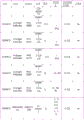
Figure 18 lists various antibodies or other antigen binding agents for use in some embodiments disclosed herein.
FIG. 19 provides a flow chart of a "stacked" decision structure for excluding preeclampsia.
Fig. 20 is a graph showing the degree to which various markers reveal orthogonal information by color to exclude preeclampsia.
Fig. 21 is a graph showing the relative prediction weights of various individual biomarkers for the exclusion of preeclampsia.
Detailed Description
The use of the classical clinical symptoms of the disease to detect preeclampsia is error prone, may have significant adverse consequences for the patient, and may burden the medical system with misdiagnosis. The measurement of proteinuria is prone to errors (e.g., false negatives and false positives), complications of preeclampsia may occur before proteinuria becomes evident, clinical manifestations of preeclampsia may vary greatly (from severe, rapidly progressing, early to late) and its symptoms (hypertension, proteinuria or both) may also indicate other diseases (chronic hypertension, gestational hypertension, transient hypertension and gestational diabetes) that may utilize less aggressive treatment courses. Thus, only patients suspected of preeclampsia are at great risk for over-treatment (e.g., delivery or early induction, thereby unnecessarily placing the infant at risk for premature complications and/or unnecessarily placing the mother at risk for surgical complications), and patients with silent or rapidly progressing preeclampsia may not be actively treated (e.g., with the infant at risk for intrauterine growth restriction or death, while the mother is at risk for post-eclampsia sequelae such as eclampsia episodes, kidney or liver damage, pulmonary edema, early parietal discs, and coma).
That is, hypertension and proteinuria reflect only the downstream consequences of the actual preeclampsia disease process. Traditional diagnosis of preeclampsia masks the disease because the only reliable treatment is labor. To improve the detection of preeclampsia, studies of the dysfunctional angiogenic processes associated with preeclampsia have been conducted to find better and/or more direct indicators of preeclampsia. One approach to this study has led to the use of the sflt/PlGF ratio in patient sera as a method to identify preeclampsia (see, e.g., Zeisler et al, NEJM 274(2017):13-22 or Verlohren et al, hypertension.63(2014): 346-52). However, the maximum sensitivity of this method was only 80% and the specificity was 78.3%. Thus, a large number of false negatives and positives are involved, making them of limited use in diagnosing preeclampsia and avoiding the over-treatment/under-treatment problem.
Accordingly, there is a need for methods, compositions, systems, and kits for improving the detection or prediction of preeclampsia in pregnant patients, particularly those capable of detecting or predicting preeclampsia with high negative predictive values and/or allowing a medical professional to rule out a diagnosis of preeclampsia over an extended period of time.
The present disclosure provides one or more tests with improved properties for assessing the risk of preeclampsia in a subject, where the test may be used to identify subjects who should not be treated for preeclampsia (e.g., in certain instances, subjects who exhibit one or more signs, symptoms, or risk factors for preeclampsia but who should not be treated for preeclampsia). In some embodiments, the test is a multi-marker serum or plasma protein assay. In some embodiments, the test is a multiplex serum/plasma protein assay. The one or more symptoms associated with preeclampsia may be diabetes (e.g., pregnancy, type I or type II), elevated normal blood glucose levels, hypertension (e.g., chronic or non-chronic), weight gain or sudden increase, elevated normal body weight, obesity, elevated normal Body Mass Index (BMI), abnormal weight gain, abnormal blood pressure, water retention, genetic factors, proteinuria abnormalities, headache, edema, abnormal protein/creatinine ratio, abnormal platelet count, stress, maternal failure, abnormal pap test results (pap smear), the onset of preeclampsia (e.g., personal history of PreE), family history of preeclampsia, preeclampsia at pregnancy, nephropathy, thrombosis, or any combination thereof.
The present disclosure provides tests, systems, and methods for assessing the risk of preeclampsia in a subject, e.g., to the exclusion of a patient having or in need of treatment for preeclampsia. In some embodiments, a test is used to discern whether a patient with one or more symptoms or risk factors associated with preeclampsia should be treated for preeclampsia. The one or more symptoms or risk factors associated with preeclampsia may be diabetes (e.g., pregnancy, type I or type II), above normal blood glucose levels, hypertension (e.g., chronic or non-chronic), weight gain or sudden increase, above normal body weight, obesity, above normal Body Mass Index (BMI), abnormal weight gain, abnormal blood pressure, water retention, genetic factors, abnormal proteinuria, headache, edema, abnormal protein/creatinine ratio, abnormal platelet count, stress, non-maternal, abnormal results of pap smears (pap smear), onset of preeclampsia (e.g., personal history of PreE), family history of preeclampsia, preeclampsia at previous pregnancy, nephropathy, thrombosis, or any combination thereof. Stated differently in some embodiments, the tests disclosed herein can be used to identify pregnant women who are symptomatic (and/or have one or more risk factors for preeclampsia) but do not have preeclampsia that may require preterm birth. In some embodiments, the test may be used for asymptomatic patients or patients with little or no risk-determining (or determinable) risk factors.
Definition of
As used in the specification and in the claims, the singular form of "a", "an", and "the" include plural referents unless the context clearly dictates otherwise.
The term "hypertension" refers to abnormal hypertension. Hypertension may be identified in any suitable manner, for example with reference to a systolic sitting pressure (sbp) greater than 140mmHg or a diastolic sitting pressure (sDBP) greater than 90 mmHg. Hypertension can be further classified into 1-type or 2-type hypertension, where 1-type is 140-149mmHg sSBP or 90-99mmHg sDBP, and 2-type is greater than 160mmHg sSBP or 100mmHg sDBP (see, for example, The Seventh Report of The journal Committee on prediction, Detection, Evaluation, and Treatment of Highblood pressure. JAMA 2003; 289: 2560-71). Other suitable criteria may also be used to identify hypertension, such as a systolic sitting pressure greater than 130 and/or a diastolic sitting pressure greater than 90 mmHg. Hypertension can also be determined according to the 2017AHA guidelines (see Whelton et al, 2017ACC/AHA/AAPA/ABC/ACPM/AGS/APHA/ASH/ASPC/NMA/PCNA guidelines the prediction, Detection, Evaluation, and Management of High Blood pressure additives, Journal of the American College of medicine (2017), doi: 10.1016/j.J.J.J.J.2017.11.006). Such guidelines identify "normal" blood pressure as being below 120/80mmHg, "elevated" as having a systolic pressure between 120-129mmHg and a diastolic pressure of less than 80mmHg, "stage 1" as having a systolic pressure between 120-139mmHg or a diastolic pressure between 80-89mmHg, "stage 2" as having a systolic pressure of at least 140mmHg or a diastolic pressure of at least 90mmHg, "hypertensive crisis" as having a systolic pressure of more than 180 and/or a diastolic pressure of more than 120.
The term "proteinuria" as used herein is defined as the presence of abnormal proteins in urine. A number of indicator dyes and reagents are available for semi-quantitative measurement of proteinuria (e.g., bromophenol blue). In some embodiments, the concentration of protein in urine can be determined by semi-quantitative "dipstick" analysis and can be classified as negative, trace (10-20mg/dL), 1+ (about 30mg/dL), 2+ (about 100mg/dL), 3+ (about 300mg/dL), or 4+ (about 1,000mg/dL), with 2+ typically used as a threshold for problematic proteinuria. In the alternative, the concentration of protein in urine may also be measured by collecting urine every 24 hours, where protein greater than or equal to 300mg indicates proteinuria. In the alternative, the concentration of protein in urine may be measured in a urine sample, wherein 30mg or more of protein per deciliter indicates proteinuria. In another alternative, proteinuria may also be expressed as a protein/creatinine ratio (Pr/Cr) in urine, where a Pr/Cr ratio >0.3 indicates problematic proteinuria.
As used herein, the term "antibody or fragment thereof" is used in the broadest sense and specifically includes intact monoclonal antibodies, polyclonal antibodies, multispecific antibodies (e.g., bispecific antibodies) formed from at least two intact antibodies, and antibody fragments. An antibody fragment is a portion that comprises an intact antibody that retains antigen binding activity. Examples include Fab, Fab', F (ab)2、F(abc)2And Fv fragments as well as diabodies, linear antibodies, scfvs, and multispecific antibodies formed from antibody fragments.
As used herein, the term "aptamer" refers to an oligonucleotide capable of forming a complex with an intended target substance. This complex formation is target specific, as other materials that may accompany the target do not complex with the aptamer. Complex formation and affinity are recognized as issues of degree; however, in this case, "target-specific" means that the aptamer binds to the target with a higher affinity than it binds to contaminating or "off-target" material.
As used herein, the term "preeclampsia" (or "PreE") refers to a pregnancy-specific disease involving multiple organ systems believed to result from abnormal placental formation, abnormal trophoblast development, defects in placental angiogenesis, and an increase in systemic inflammatory response in the mother. Preeclampsia, if left untreated, may progress to eclampsia, HELLP syndrome, hemorrhagic or ischemic stroke, liver injury, acute kidney injury, and Acute Respiratory Distress Syndrome (ARDS). Thus, although preeclampsia often exhibits symptoms such as hypertension and proteinuria, it is not identical to vascular tone and renal insufficiency alone (as evidenced by a series of non-overlapping complications that result from non-treatment), and thus the symptoms that reflect vascular tone and renal insufficiency are not themselves ideal for the predictive/diagnostic value of preeclampsia. Further information on the pathophysiology of preeclampsia can be found, for example, in Phipps et al, Clin J Am Soc Nephrol 11 (2016: 1102-. In some embodiments, the traditional diagnosis of preeclampsia is made by simultaneous detection of hypertension and proteinuria as defined above. In other embodiments (see American College of gynecology of obstetrics and gynecology) (American patent of austeritricia and gynecologists); Task Force on Hypertension in pregnancy. obstet gynecol.122(2013):1122-31, expressly incorporated herein by reference), a traditional diagnosis of preeclampsia is performed while detecting Hypertension (blood pressure with systolic pressure greater than or equal to 140mmHg or diastolic pressure greater than or equal to 90mmHg in two tests spaced at least 4 hours after 20 weeks gestation in a previously normotensive woman) and proteinuria (blood pressure with systolic pressure greater than or equal to 160mmHg or diastolic pressure greater than or equal to 110mmHg in a short interval of a few minutes) (greater than or equal to 300mg collected per 24 hours of urine that can be measured or extrapolated, or a protein creatinine ratio greater than or equal to 0.3, or a test paper reading of 1+), or when (a) a platelet count of less than 100,000 per milliliter, (b) conventional diagnosis of preeclampsia is performed while (a) new onset hypertension is achieved without proteinuria when the patient's baseline, or doubled, serum creatinine greater than 1.1mg/dL, or (c) hepatic transaminase blood concentration is twice or more than normal. In some cases, preeclampsia is performed without diagnosis of proteinuria, e.g., there is evidence of other end-stage organ damage.
The term "subject" may include a human or non-human animal. Thus, the methods described herein are applicable to both human and veterinary disease as well as animal models. Preferred subjects are "patients", i.e., living persons who are receiving treatment for a disease or condition. This includes people who are undergoing pathological examination without a definite disease.
The term "FRET pair" refers to a pair of dye molecules in which one dye molecule (acceptor or quencher dye) absorbs light at a wavelength at which the other emits light (donor dye), and the two dyes are spaced apart by a distance that allows energy transfer, the disclosed embodiments typically being within about 100 angstroms of each other, such as within about 50 angstroms, 20 angstroms, or 10 angstroms of each other (e.g., because they are attached to the same substrate moiety). Excitation of the donor dye causes excitation of the acceptor dye by a FRET mechanism, and a lower level of fluorescence is observed from the donor dye. The efficiency of FRET depends on the distance between dyes, the quantum yield of the donor dye, the fluorescence lifetime of the donor dye, and the overlap of the donor emission spectrum and the acceptor absorption spectrum.
The term "photosensitizer" refers to a photoactivatable compound or a biological precursor of a photoactivatable compound that produces a reactive species (e.g., oxygen) that has a photochemical effect on a biomolecule.
The term "oxygen sensitive dye" refers to a fluorescent dye that changes its fluorescence intensity or emission maximum upon binding to molecular oxygen (such dyes are used in the FOCI assay), or a chemiluminescent dye that emits light upon binding to molecular oxygen (such dyes are used in the LOCI assay).
As used herein, the term "affibody" refers to a small, highly stable protein that binds to a target molecule with a specificity and affinity similar to an antibody.
The term "unnecessary treatment of preeclampsia" may refer to a treatment of preeclampsia that is statistically unreasonable when the practitioner considers the results of the tests or procedures described herein (e.g., tests based on the determination of the amount or concentration of various protein biomarkers). Such unnecessary treatment may include induction of preterm labor based on one or more symptoms or risk factors of preeclampsia.
Test subject
In some embodiments, the methods, compositions, systems, and kits provided herein are used to detect or predict the condition of a pregnant human subject at any stage of pregnancy. In other embodiments, the pregnant human subject is after the 20 th week of gestation. In other embodiments, the pregnant human subject is the first or second, three months of pregnancy. In some embodiments, the pregnant human subject is after week 21, week 22, week 23, week 24, week 25, week 26, week 27, week 28, week 29, week 30, week 31, week 32, week 33, week 34, week 35, week 36, week 37, week 38, week 39, week 40, week 41 or week 42 of pregnancy.
The methods, compositions, systems and kits are suitable for detecting or predicting a condition in a pregnant subject, such as preeclampsia (PreE) or non-preeclampsia (NonPreE). In some embodiments, preeclampsia is further divided into very early (before 25 weeks gestation), early (before 34 weeks gestation) and late (after 34 weeks gestation) preeclampsia. Typically, a pregnant patient is considered non-pree (nonpre) when the patient does not develop hypertension or renal symptoms. Furthermore, when a pregnant patient shows only symptoms of hypertension without signs of proteinuria, the patient is usually only suspected of preeclampsia. However, since the measurement of proteinuria is prone to errors (e.g., false negatives and false positives), complications of preeclampsia may already occur before proteinuria becomes evident, and thus the clinical manifestations of preeclampsia may vary greatly (from severe, rapidly progressing, and early to late onset, or less severe), and symptoms (hypertension, proteinuria, or both) may indicate different diseases that may utilize different treatment courses. Thus, only patients suspected of preeclampsia are at unnecessarily significant treatment risk (e.g., delivery/induction of preterm birth puts the infant at risk for premature complications or the mother at risk for surgical complications), while silent or rapidly progressing patients are at risk for preeclampsia not being adequately treated positively (e.g., puts the infant at risk for intrauterine growth restriction or death, while the mother is at risk for post-eclampsia sequelae such as preeclampsia, kidney or liver damage, pulmonary edema, early paring of the placenta, and coma).
Thus, the subject may be a pregnant female with no known risk factors or one or more risk factors for symptoms such as PreE. In certain instances, hypertension and/or proteinuria may indicate that the subject is at risk for preeclampsia. In some cases, the urine protein content of a subject at risk for preeclampsia may be determined by dipstick assay to be 2+ (100mg/dL) or greater, with a urine collection of greater than or equal to 300mg per 24 hours, 30mg or greater per deciliter of protein in the urine sample, or a Pr/Cr ratio of greater than or equal to 30 mg/millimole in the urine. In some cases, a subject at risk for preeclampsia may have a systolic sitting pressure (sbp) greater than 140mmHg or a diastolic sitting pressure (sDBP) greater than 90mmHg, or both. In some cases, the sFlt1/PlGF ratio may be used to identify subjects at risk for pre-eclampsia (see, e.g., Zeisler et al, NEJM 274(2017):13-22 or Verlohren et al, Hypertension.63(2014): 346-52). In certain instances, both hypertension and proteinuria may be used to identify a subject at risk for preeclampsia. In certain instances, new onset hypertension associated with one or more symptoms selected from (a) a platelet count of less than 100,000 per milliliter, (b) serum creatinine greater than 1.1mg/dL or a doubling baseline value for the patient, or (c) a blood level of liver transaminase that is two times or more than normal may be used to identify subjects at risk for pre-eclampsia.
Sample (I)
Methods for detecting molecules (e.g., nucleic acids, proteins, etc.) in a pregnant subject to detect, diagnose, monitor, predict, or assess the pregnancy status or outcome are described in the present disclosure. In some cases, the molecule is a circulating molecule (e.g., not bound to cells and circulating freely in a bodily fluid such as blood, plasma, or serum). In some cases, the molecule is expressed in the cytoplasm of blood, endothelial cells, or organ cells. In some cases, the molecule is expressed on the surface of blood, endothelial cells, or organ cells.
The methods, kits, and systems disclosed herein can be used to classify one or more samples from one or more subjects. The sample can be any material comprising a tissue, cell, nucleic acid, gene fragment, expression product, protein, polypeptide, exosome, gene expression product, or gene expression product fragment of the subject to be tested. Samples may include, but are not limited to, tissue, cells, plasma, serum, or any other biological material from cells or derived from cells of an individual. The sample may be a heterogeneous or homogeneous population of cells or tissues. The sample may be a cell-free or cell-poor fluid (e.g., plasma or serum). In some cases, the sample is from a single patient. In some cases, the method includes analyzing multiple samples at a time, for example, by performing massively parallel, multiplex expression analysis on protein arrays and the like.
The sample is preferably a body fluid. The body fluid may be saliva, urine, blood and/or amniotic fluid. The sample may be part of any of these fluids, such as plasma, serum or exosomes (exemplary exosome separation techniques may be found, for example, in Li et al, therapeutics.7 (2017): 789-. In a preferred embodiment, the sample is a blood sample, a plasma sample or a serum sample.
The sample may be obtained using any method that can provide a sample suitable for use in the assay methods described herein. Samples can be obtained by non-invasive methods, such as pharyngeal swabs, buccal swabs, bronchial lavage, urine collection, scraping the cervix, cervical vaginal sample secretion collection (e.g., using an ophthalmic sponge, such as a Weck-Cel sponge), saliva collection, or stool collection. The sample may be obtained by a minimally invasive method such as blood drawing. Samples may be obtained by venipuncture.
As used herein, "obtaining a sample" includes obtaining a sample, either directly or indirectly. In some embodiments, the sample is obtained from the subject by the same party (e.g., a testing laboratory) that subsequently obtains biomarker data from the sample. In some embodiments, the sample is received (e.g., by a testing laboratory) from another entity that collects the sample from a subject (e.g., a physician, nurse, phlebotomist, or medical caregiver). In some embodiments, the sample is taken from the subject by a medical professional under the direction of an isolated entity (e.g., a testing laboratory) and then provided to the entity (e.g., a testing laboratory). In some embodiments, the sample is collected at home by the subject or a caregiver of the subject, and then provided to a party (e.g., a testing laboratory) who obtains biomarker data from the sample. Various kits suitable for self or home collection of biological samples have been described commercially and in the literature. See, for example, US20170023446a1 and US 4777964A.
Sample data
The methods, kits, and systems disclosed herein can include data relating to one or more samples or uses thereof. The data may represent the amount or concentration of one or more biomarkers (e.g., various proteins described herein). In other words, the data may be expression level data of a protein or polypeptide. The data on the expression levels of the biomarkers described herein can be obtained by protein arrays, proteomics, expression proteomics, mass spectrometry (e.g., liquid chromatography-mass spectrometry (LC-MS), Multiple Reaction Monitoring (MRM), Selective Reaction Monitoring (SRM), scheduled MRM, scheduled SRM), 2D PAGE, 3DPAGE, electrophoresis, proteome chips, proteome microarrays, Edman degradation, direct or indirect ELISA, immunoadsorption assays, immuno PCR (see, e.g., Sano et al, science.258(1992):120-2), proximity extension assays (see, e.g., Thorsen et al, Journal of relative medicine.11(2013):253, US20130288249a1, US9777315B2), Luminex assays or homogeneous assays (e.g., alpharesen (see, e.g., application notes, 5 (2008), US5898005A, US5861319A)), time-resolved fluorescence resonance energy transfer (see, e.g., TR 355635 f), time-resolved fluorescence energy transfer (see, e.g., TRF 355635 f), t 1998015830, t 2 f, 3, and t 35 f) Fluorescent Oxygen Channel Immunoassay (FOCI) or Luminescent Oxygen Channel Immunoassay (LOCI)TM(ii) a See, e.g., Ullman et al, Proc NatlAcad Sci U S A.91(1994): 5426-5430 or Ullman et al, Clin chem.1996Sep; 1518-26 to obtain exemplary methods and reagents).
In some embodiments, the compositions, methods, and devices described herein utilize marker molecules in various sandwich, competitive, or non-competitive assay formats to determine the expression levels of the biomarkers described herein. Such methods generate a signal related to the presence or amount of one or more of the proteins described herein. Suitable assay formats also include chromatography, mass spectrometry and western "blotting" methods. In addition, certain methods and devices, e.g. biosensors, optical immunoassays, immunoadsorptionsAssays, and enzyme immunoassays, can be used to determine the presence or quantity of an analyte without the need for a labeling molecule. Examples of Enzyme Immunoassays (EIA) include chemiluminescent enzyme immunoassays, electrochemiluminescent immunoassays (ECLIA), and enzyme-linked immunosorbent assays (ELISA), Kuhle, Jens et al, "diagnosis of the same analytical formats for the quantification of the same biochemical light chain in blood samples: ELISA, electrochemiluminescent immunoassays and Simoa," Clinical Chemistry and Laboratory Medicine (CCLM)54.10(2016): 1655-. Robotic instruments for performing these immunoassays are commercially available, including but not limited to Abbott Or
Or A system; biolog
 Or a CLC480 system; beckman Coulter
 ACCESS
 Or the Unicel Dxl 600/800 system; bioMerieux
 Or mini-
 A system; chimera Biotec GmbH
 An assay; dade Behring
 A system; diasorin LIAISON
 Or ETI-Max 300 systems; dynex
 A system; gold Standard diagnostics
 An analyzer; gyrolab xPlre
 A system; hudson Robotics ELISA workstation; ortho clinical diagnosis
 ECL or 3600 systems; hamiltron Robotics ELISA NIMBUS or STARlet system; luminex
 A system; perkinelmer ALPHA
 Or
Or Phadia laboratory system 100E, 250, 1000, 2500, or 5000; quanterix
 A system; quidel
 A POC system; radiometer AQT90 Flex system; roche diagnostics
 2010,
2010, 4000/6000/8000 Analyzer, or
 400 Plus; the analyzers family c111, c311, c501, c 502; seikisui diagnostics
 An IP automation system; siemens Dimension
 1500 systems, DPC Immulite 1000/2000 systems, or DCA
 An analyzer; sinculex Single Molecule Counting (SMC)TM) Measuring;
 a CS acute care diagnostic system; sysmex
 A ThermoFisher MGC 240 Benchtop analyzer; tosoh Bioscience
 -360 or AIA-600II systems; the UniCel Dxl 860i synchronous Access clinical system, the UniCel DxC 680i synchronous Access clinical system, the Access/Access2 immunoassay system, the 600 channel immunoassay system, the 600i synchronous channel clinical system, the Dxl 800 channel immunoassay system and the DxC 880i synchronous channel clinical system; and Vital diagnosis
 Instant chemiluminescence immunoassay analyzer. Any suitable immunoassay may be used, however, such as an enzyme-linked immunoassay (ELISA), Radioimmunoassay (RIA), competitive binding assay, and the like.
Other exemplary analysis systems include analysis systems, such as optical systems that include one or more radiation sources and/or one or more detectors. Such systems may use, for example, an illuminating light source and a sample, and a detector configured to detect light emitted by the sample (e.g., a fluorescence spectrum), an optical density (e.g., a portion of the light that passes through the sample), and/or light diffracted by the sample (e.g., diffractive optics). The assay system may use, for example, ELISA (enzyme linked immunosorbent assay). The assay system may use, for example, LOCI (luminescent oxygen channel immunoassay), FOCI (fluorescent oxygen channel immunoassay), or ALPHAscreen. The analytical technique may involve incubating and/or diluting the sample before or during analysis/determination of the sample.
In some embodiments, the compositions, methods, and devices described herein utilize Fluorescent Oxygen Channel Immunoassay (FOCI) compositions and methods. FOCI is generally described in U.S. patent nos. 5807675, 5,616,719, and 7,635,571, the entire contents of which are expressly incorporated herein by reference. In some embodiments, a first analyte binding agent capable of binding to an analyte and comprising a photosensitizer is used in combination with a second analyte binding agent comprising a fluorescent dye. In some embodiments, the photosensitizer of the first analyte binding agent generates singlet oxygen in an excited state, thereby causing the fluorescent dye of the second analyte binding agent to fluoresce upon reaction with singlet oxygen. In some embodiments, the emitted fluorescence may be detected, for example to determine the presence and/or absence of an analyte and/or to quantify and/or analyze an analyte in a sample. In some embodiments, the first and second analyte binding agents bind to the same region (e.g., epitope) of the analyte (e.g., protein). For example, in some embodiments, the first and second analyte binding agents comprise the same type of analyte binding moiety or reagent (e.g., the same antibody). In some embodiments, the first and second analyte binding agents bind to separate regions (e.g., epitopes) of the analyte (e.g., protein). In some embodiments, the first and second analyte binding agents bind to separate regions of the analyte (e.g., protein) that do not spatially overlap. In some embodiments, the first analyte binding agent and the second analyte binding agent are configured such that when both analyte binding agents bind to the analyte, singlet oxygen generated by the photosensitizer of the first analyte binding agent is in close proximity to the fluorescent dye of the second analyte binding agent. In some embodiments, the first and/or second analyte binding agent is an antigen binding agent (e.g., an antibody). In some embodiments, the first and/or second analyte binding agent is an affibody. In some embodiments, the first and/or second analyte binding agent is an antigen binding agent is an aptamer. In some embodiments, both the photosensitizer and the fluorescent dye are provided in the form of beads.
In some cases, the array may use different probes (e.g., antibodies, scFvs, Fab fragments) attached to different particles or beads. In such arrays, the identification of which probes are attached to those particles or beads can be determined from the encoding system. The probe may be an antibody or an antigen-binding fragment or derivative thereof.
The data relating to the sample may be compared to data relating to one or more control samples. In some cases, the control sample may be a sample of the same patient at a different time. In some cases, the one or more control samples may include one or more samples from healthy subjects, unhealthy subjects, or a combination thereof. The one or more control samples may include one or more samples from healthy subjects, subjects with pregnancy related diseases other than preeclampsia, subjects with chronic diseases as well as pregnancy related diseases, or subjects with chronic diseases only.
In some cases, expression level data for various samples is used to develop or train the algorithms or classifiers provided herein. In certain instances, the subject is a patient, e.g., a pregnant female; gene expression levels are measured in a sample from a patient and a classifier or algorithm (e.g., a trained algorithm) is applied to the resulting data to detect, predict, monitor, eliminate, or estimate the risk of a pregnancy-associated disease, such as preeclampsia.
In some cases, the analysis of the expression levels initially provides a measure of the expression level of each of several individual biomarkers. The expression level may be absolute with respect to the concentration of the biomarker, or relative with respect to the relative concentration of the expression product of interest relative to another biomarker in the sample. For example, the relative expression level of a protein may be expressed relative to the expression level of a housekeeping protein or a structural protein in the sample. Relative expression levels can also be determined by simultaneously analyzing differentially labeled samples bound to the same array. The expression level may also be expressed in any unit, e.g., related to signal intensity.
Classifier and classifier probe set
A classification system using a classifier including one or more classifiers is disclosed herein. In some cases, the classifier is a bi-directional classifier. In some cases, the two-way classifier may classify a sample of a pregnant patient into one of two categories, pre-eclampsia (PreE) and non-pre-eclampsia (nonPreE PreE). In some cases, a classifier may be used to classify a subject as not requiring treatment for preeclampsia. In some cases, a multi-directional classifier may be used (e.g., preeclampsia, non-preeclampsia, and indeterminate eclampsia).
A classifier and/or set of classifier probes (e.g., an antibody set) may be used to score (rule-in) or rule-out a sample from a patient to be treated for preeclampsia. For example, a classifier can be used to classify a sample as being from a healthy subject. Alternatively, the sample may be classified as from an unhealthy subject using a classifier. Alternatively, or in addition, the classifier may be used to score or exclude samples from subjects who should receive treatment for preeclampsia.
Data analysis system and method
The methods, kits, and systems disclosed herein can comprise an algorithm or use thereof. One or more algorithms may be used to classify one or more samples from one or more subjects. One or more algorithms may be applied to data from one or more samples. The data may include biomarker expression data.
The methods disclosed herein can include assigning a classification to one or more samples from one or more subjects. Assigning the classification to the sample may include applying an algorithm to the expression level. In some cases, the gene expression levels are input to a data analysis system that includes a trained algorithm for classifying the sample as one of the following diseases: preeclampsia, eclampsia, non-preeclampsia, chronic hypertension, gestational hypertension or HELLP (hemolysis, elevated liver enzymes) and low platelet count-see, e.g., Weinstein et al, Am J Obstet Gynecol.142(1982):159-67) syndrome. In some embodiments, the algorithm may, as part of its execution, calculate an index of the sample and compare the index of the sample to a threshold; the predetermined relationship may indicate a likelihood that the sample belongs to a particular classification.
The algorithm may provide a record of its output, including the classification and/or confidence of the sample. In some cases, the output of the algorithm may be the likelihood that the subject has a disease including preeclampsia, eclampsia, chronic hypertension, gestational hypertension, or HELLP syndrome.
The data analysis system may be a trained algorithm. The algorithm may include a linear classifier. In some cases, the linear classifier includes one or more of a linear discriminant analysis, fisher linear discriminant, naive bayes classifier, logistic regression, perceptron, support vector machine, or a combination thereof. The linear classifier may be a Support Vector Machine (SVM) algorithm. The algorithm may include a bi-directional classifier. The bi-directional classifier may include one or more decision trees, random forests, bayesian networks, support vector machines, neural networks, or logistic regression algorithms.
The algorithm may include one or more of Linear Discriminant Analysis (LDA), basic perceptron, elastic network, logistic regression, (kernel) Support Vector Machine (SVM), Diagonal Linear Discriminant Analysis (DLDA), Golub classifier, Parzen-based, (kernel) fisher discriminant classifier, k nearest neighbor, iterative mitigation, classification tree, maximum likelihood classifier, random forest, nearest centroid, microarray (PAM) prediction analysis, k median clustering, fuzzy C-means clustering, gaussian mixture model, ranked response (GR), Gradient Boosting Method (GBM), elastic network logistic regression, or a combination thereof. The algorithm may comprise a Diagonal Linear Discriminant Analysis (DLDA) algorithm. The algorithm may include a nearest centroid algorithm. The algorithm may comprise a random forest algorithm. In some embodiments, to distinguish between preeclampsia and non-preeclampsia, logistic regression, random forest, and gradient enhancement methods (GBM) perform better than Linear Discriminant Analysis (LDA), neural networks, and Support Vector Machines (SVMs).
biomarker/Gene expression products
The term "biomarker" refers to a measurable indication of a certain biological state or disease. In some cases, a biomarker may be a substance found in a subject, the amount of the substance, or some other indicator. For example, a biomarker may be the amount of protein and/or other gene expression products in a sample. In some embodiments, the biomarker is a full-length unmodified protein. In other embodiments, the biomarker is a spliced, post-translationally cleaved, or post-translationally chemically modified (e.g., methylated, phosphorylated, glycosylated, formylated, etc.) protein.
The methods, compositions, and systems described herein also relate to the use of the biomarker panels and/or gene expression products to identify, diagnose, classify, treat, or otherwise characterize various diseases in pregnant patients, including non-PreE, chronic hypertension, gestational hypertension, or HELLP syndrome. Sets of biomarkers and/or gene expression products for classifying biological samples are provided, as well as methods of obtaining such sets of biomarkers. Typically, a pattern of levels of gene expression biomarkers (also referred to as signatures) in a panel is determined from one or more reference samples and then used to evaluate the signatures of the same panel of biomarkers in a test sample, for example by a similarity measure between the test sample signature and the reference sample signature.
In some embodiments, the methods, compositions, and systems described herein may include detecting one or more biomarkers belonging to a particular functional class of biomarkers that are associated with one or more pathophysiological characteristics of preeclampsia (see fig. 16, which shows various pathophysiological characteristics or preeclampsia). While figure 16 shows various pathophysiological features and associated biomarkers thought to be associated with preeclampsia, many other methods for describing the relationship between pathophysiology and markers are possible. For example, KIM-1, CD274 and decorin can be considered proteins associated with kidney damage. Similarly, sFlt1, endoglin, pappalysin 2, and decorin may be considered angiogenesis-related proteins. Those skilled in the art will recognize that various other classification schemes may be similarly used.
Without wishing to be bound by theory, preeclampsia is thought to arise from abnormal invasion of feeder cells, leading to incomplete helical arterial remodeling and hypoperfusion of the placenta, which leads to signs and symptoms of preeclampsia, due to hypoperfusion of the placenta, this dysfunctional system may include angiogenesis and endothelial function, as evidenced by an imbalance between pro-angiogenic and anti-angiogenic factors, many of which are released from the placenta in response to the abnormal physiological response of preeclampsia and disrupt vascular homeostasis in the mother, SFLT1 (soluble FMS-like tyrosine kinase 1, a tyrosine protein kinase that may serve as cell surface receptors for VEGFA, VEGFB and PlGF and that decreases at term and plays an important role in the development of the embryonic vasculature), pllt (a growth factor, a pro-angiogenic protein that peaks at 30 Th week of pregnancy, may stimulate the growth, proliferation and migration of endothelial cells), DCN (core glycan protein is a functional component of the extracellular matrix, by binding to ECM molecules, by a regulatory factor that mediates the recruitment of endothelial cell recruitment, is thought to be a potent regulator of endothelial cell recruitment, is mediated by a leukocyte recruitment, because of the recruitment of endothelial cell recruitment, leukocyte recruitment, endothelial cell recruitment, recruitment of endothelial cell recruitment, recruitment of endothelial cell recruitment, recruitment of endothelial cell recruitment, recruitment of endothelial cell recruitment, recruitment of endothelial cell recruitment, recruitment of endothelial cell recruitment, recruitment of endothelial cell recruitment of endothelial.
The methods herein may include identifying or excluding a condition (e.g., preeclampsia) from one or more of the biomarkers listed in the table below (table a).
Table a: high priority biomarkers for identifying or excluding preeclampsia


The methods herein may include identifying or excluding a condition (e.g., preeclampsia) from one or more biomarkers listed in the following table (table B).
Table B: high priority biomarkers for identifying or excluding preeclampsia
| |
|
| 5 | |
| 6 | |
| 7 | |
| 8 | |
| 9 | |
| 10 | |
| 11 | PAPP.A2 |
The methods provided herein can include identifying or excluding a condition (e.g., preeclampsia) from (e.g., based on the analysis of) one or more biomarkers selected from table a and table B. In some cases, the methods provided herein may comprise identifying or excluding a condition (e.g., preeclampsia) from two or more biomarkers selected from table a and table B. In some cases, the methods provided herein can include identifying or excluding a disorder (e.g., preeclampsia) from three or more biomarkers selected from table a and table B. In some cases, the methods provided herein may comprise identifying or excluding a disorder (e.g., preeclampsia) from one biomarker selected from table a and table B. In some cases, the methods provided herein may include identifying or excluding a disorder (e.g., preeclampsia) from two biomarkers selected from table a and table B. In some cases, the methods provided herein can include identifying or excluding a disorder (e.g., preeclampsia) from three biomarkers selected from table a and table B. In some cases, the methods provided herein can include identifying or excluding a disorder (e.g., preeclampsia) from four biomarkers selected from table a and table B. In some cases, the methods provided herein may include identifying or excluding a disorder (e.g., preeclampsia) from five biomarkers selected from table a and table B. In some cases, the methods provided herein may include identifying or excluding a disorder (e.g., preeclampsia) from all biomarkers selected from table a and table B. In some cases, the methods provided herein can include identifying or excluding a disorder (e.g., preeclampsia) from no more than 3 biomarkers selected from table a and table B. In some cases, the methods provided herein can include identifying or excluding a disorder (e.g., preeclampsia) from no more than 4 biomarkers selected from table a and table B. In some cases, the methods provided herein can include identifying or excluding a condition (e.g., preeclampsia) from no more than 5 biomarkers selected from table a and table B. In some cases, the methods provided herein can include identifying or excluding a condition (e.g., preeclampsia) from no more than 6 biomarkers selected from table a and table B. In some cases, the methods provided herein can include identifying or excluding a condition (e.g., preeclampsia) from no more than 7 biomarkers selected from table a and table B. In some cases, the methods provided herein can include identifying or excluding a condition (e.g., preeclampsia) from no more than 8 biomarkers selected from table a and table B. In some cases, the methods provided herein can include identifying or excluding a condition (e.g., preeclampsia) from no more than 9 biomarkers selected from table a and table B. In some cases, the methods provided herein can include identifying or excluding a condition (e.g., preeclampsia) from no more than 10 biomarkers selected from table a and table B. In some cases, the methods provided herein can include identifying or excluding a condition (e.g., preeclampsia) from no more than 11 biomarkers selected from table a and table B.
The methods provided herein may comprise identifying or excluding a disease (e.g., preeclampsia) from one or more biomarkers selected from table a and one or more biomarkers selected from table B. In some cases, the methods provided herein may comprise identifying or excluding a disease (e.g., preeclampsia) from the 2 biomarkers selected from table a and the one or more biomarkers selected from table B. In some cases, the methods provided herein may comprise identifying or excluding a disease (e.g., preeclampsia) from the 3 biomarkers selected from table a and the one or more biomarkers selected from table B. In some cases, the methods provided herein may comprise identifying or excluding a disease (e.g., preeclampsia) from the 4 biomarkers selected from table a and the one or more biomarkers selected from table B. In some cases, the methods provided herein may comprise identifying or excluding a disease (e.g., preeclampsia) from the 3 biomarkers selected from table a and the one or more biomarkers selected from table B. In some cases, the methods provided herein can include identifying or excluding a disease (e.g., preeclampsia) from the 3 biomarkers selected from table a and the two or more biomarkers selected from table B. In some cases, the methods provided herein can include identifying or excluding a disease (e.g., preeclampsia) from the 3 biomarkers selected from table a and the three or more biomarkers selected from table B. In some cases, the methods provided herein can include identifying or excluding a disease (e.g., preeclampsia) from the 3 biomarkers selected from table a and the four or more biomarkers selected from table B. In some cases, the methods provided herein can include identifying or excluding a disease (e.g., preeclampsia) from the 3 biomarkers selected from table a and the five or more biomarkers selected from table B. In some cases, the methods provided herein can include identifying or excluding a disease (e.g., preeclampsia) from the 3 biomarkers selected from table a and the six or more biomarkers selected from table B. In some cases, the methods provided herein may comprise identifying or excluding a disease (e.g., preeclampsia) from 3 biomarkers selected from table a and one biomarker selected from table B. In some cases, the methods provided herein can include identifying or excluding a disease (e.g., preeclampsia) from the 3 biomarkers selected from table a and the two biomarkers selected from table B. In some cases, the methods provided herein can include identifying or excluding a disease (e.g., preeclampsia) from the 3 biomarkers selected from table a and the three biomarkers selected from table B. In some cases, the methods provided herein can include identifying or excluding a disease (e.g., preeclampsia) from the 3 biomarkers selected from table a and the four biomarkers selected from table B. In some cases, the methods provided herein may comprise identifying or excluding a disease (e.g., preeclampsia) from 3 biomarkers selected from table a and five biomarkers selected from table B. In some cases, the methods provided herein can include identifying or excluding a disease (e.g., preeclampsia) from the 3 biomarkers selected from table a and the six biomarkers selected from table B. In some cases, the methods provided herein can include identifying or excluding a disease (e.g., preeclampsia) from the 3 biomarkers selected from table a and the seven biomarkers selected from table B. The methods provided herein may comprise identifying or excluding a disease (e.g., preeclampsia) from no more than one biomarker selected from table a and no more than 2 biomarkers selected from table B. The methods provided herein may comprise identifying or excluding a disease (e.g., preeclampsia) from no more than two biomarkers selected from table a and no more than 2 biomarkers selected from table B. The methods provided herein may comprise identifying or excluding a disease (e.g., preeclampsia) from no more than three biomarkers selected from table a and no more than 2 biomarkers selected from table B. The methods provided herein may comprise identifying or excluding a disease (e.g., preeclampsia) from no more than one biomarker selected from table a and no more than 2 biomarkers selected from table B. The methods provided herein may comprise identifying or excluding a disease (e.g., preeclampsia) from no more than one biomarker selected from table a and no more than 3 biomarkers selected from table B. The methods provided herein may comprise identifying or excluding a disease (e.g., preeclampsia) from no more than two biomarkers selected from table a and no more than 3 biomarkers selected from table B. The methods provided herein may comprise identifying or excluding a disease (e.g., preeclampsia) from no more than three biomarkers selected from table a and no more than 3 biomarkers selected from table B. The methods provided herein may comprise identifying or excluding a disease (e.g., preeclampsia) from no more than one biomarker selected from table a and no more than 3 biomarkers selected from table B. The methods provided herein may comprise identifying or excluding a disease (e.g., preeclampsia) from no more than one biomarker selected from table a and no more than 4 biomarkers selected from table B. The methods provided herein may comprise identifying or excluding a disease (e.g., preeclampsia) from no more than two biomarkers selected from table a and no more than 4 biomarkers selected from table B. The methods provided herein may comprise identifying or excluding a disease (e.g., preeclampsia) from no more than three biomarkers selected from table a and no more than 4 biomarkers selected from table B. The methods provided herein may comprise identifying or excluding a disease (e.g., preeclampsia) from no more than one biomarker selected from table a and no more than 4 biomarkers selected from table B.
In some embodiments, the methods provided herein can include identifying or excluding a disorder (e.g., preeclampsia) from a set of markers including sFLT-1, PlGF, FGF21, CLEC4a, endoglin, CD274, and decorin.
The methods provided herein can include identifying or excluding a disease (e.g., preeclampsia) from (e.g., based on an analysis from) the following combinations: sFlt.1, PlGF, KIM1 and CLEC 4A; sFlt.1, PlGF, KIM1, CLEC4A and FGF 21; sFlt.1, PlGF, KIM1, CLEC4A and CD 274; sflt.1, PlGF, KIM1, CLEC4A and endoglin; sflt.1, PlGF, KIM1, CLEC4A and decorin; sflt.1, PlGF, KIM1, CLEC4A, FGF21 and endoglin; sFlt.1, PlGF, KIM1, CLEC4A, FGF21 and CD 274; sflt.1, PlGF, KIM1, CLEC4A, endoglin and CD 274; sflt.1, PlGF, KIM1, CLEC4A, endoglin and decorin; sflt.1, PlGF, KIM1, CLEC4A, FGF21, endoglin and CD 274; sflt.1, PlGF, KIM1, CLEC4A, FGF21, endoglin, CD274 and decorin; PlGF, KIM1, CLEC4A and endoglin; PlGF, KIM1, CLEC4A, endoglin and decorin; sflt.1, PlGF, KIM1, TFF2, FGF21 and decorin; sflt.1, PlGF, KIM1, CLEC4A, CD2741 and endoglin; or HGF, SYND1, CLEC4A, sflt.1, PlGF, KIM1, CLEC4A and FGF 21.
The methods provided herein can include identifying or excluding a disease (e.g., preeclampsia) from (e.g., based on analysis from) a set of four biomarkers and optionally in combination with PlGF: SFLT1, KIM1, CLEC4A, FGF 21; SFLT1, KIM1, CLEC4A, endoglin; SFLT1, KIM1, CLEC4A, decorin; SFLT1, KIM1, CLEC4A, CD 274; SFLT1, KIM1, CLEC4A, HGF; SFLT1, KIM1, CLEC4A, TFF 2; SFLT1, KIM1, CLEC4A, papp.a 2; SFLT1, KIM1, FGF21, endoglin; SFLT1, KIM1, FGF21, decorin; SFLT1, KIM1, FGF21, CD 274; SFLT1, KIM1, FGF21, HGF; SFLT1, KIM1, FGF21, TFF 2; SFLT1, KIM1, FGF21, papp.a 2; SFLT1, KIM1, endoglin, decorin; SFLT1, KIM1, endoglin, CD 274; SFLT1, KIM1, endoglin, HGF; SFLT1, KIM1, endoglin, TFF 2; SFLT1, KIM1, endoglin, papp.a 2; SFLT1, KIM1, decorin, CD 274; SFLT1, KIM1, decorin, HGF; SFLT1, KIM1, decorin, TFF 2; SFLT1, KIM1, decorin, papp.a 2; SFLT1, KIM1, CD274, HGF; SFLT1, KIM1, CD274, TFF 2; SFLT1, KIM1, CD274, papp.a 2; SFLT1, KIM1, HGF, TFF 2; SFLT1, KIM1, HGF, papp.a 2; SFLT1, KIM1, TFF2, papp.a 2; SFLT1, CLEC4A, FGF21, endoglin; SFLT1, CLEC4A, FGF21, decorin; SFLT1, CLEC4A, FGF21, CD 274; SFLT1, CLEC4A, FGF21, HGF; SFLT1, CLEC4A, FGF21, TFF 2; SFLT1, CLEC4A, FGF21, papp.a 2; SFLT1, CLEC4A, endoglin, decorin; SFLT1, CLEC4A, endoglin, CD 274; SFLT1, CLEC4A, endoglin, HGF; SFLT1, CLEC4A, endoglin, TFF 2; SFLT1, CLEC4A, endoglin, papp.a 2; SFLT1, CLEC4A, decorin, CD 274; SFLT1, CLEC4A, decorin, HGF; SFLT1, CLEC4A, decorin, TFF 2; SFLT1, CLEC4A, decorin, papp.a 2; SFLT1, CLEC4A, CD274, HGF; SFLT1, CLEC4A, CD274, TFF 2; SFLT1, CLEC4A, CD274, papp.a 2; SFLT1, CLEC4A, HGF, TFF 2; SFLT1, CLEC4A, HGF, papp. a 2; SFLT1, CLEC4A, TFF2, papp.a 2; SFLT1, FGF21, endoglin, decorin; SFLT1, FGF21, endoglin, CD 274; SFLT1, FGF21, endoglin, HGF; SFLT1, FGF21, endoglin, TFF 2; SFLT1, FGF21, endoglin, papp.a 2; SFLT1, FGF21, decorin, CD 274; SFLT1, FGF21, decorin, HGF; SFLT1, FGF21, decorin, TFF 2; SFLT1, FGF21, decorin, papp.a 2; SFLT1, FGF21, CD274, HGF; SFLT1, FGF21, CD274, TFF 2; SFLT1, FGF21, CD274, papp.a 2; SFLT1, FGF21, HGF, TFF 2; SFLT1, FGF21, HGF, papp.a 2; SFLT1, FGF21, TFF2, papp.a 2; SFLT1, endoglin, decorin, CD 274; SFLT1, endoglin, decorin, HGF; SFLT1, endoglin, decorin, TFF 2; SFLT1, endoglin, decorin, papp.a 2; SFLT1, endoglin, CD274, HGF; SFLT1, endoglin, CD274, TFF 2; SFLT1, endoglin, CD274, papp.a 2; SFLT1, endoglin, HGF, TFF 2; SFLT1, endoglin, HGF, papp.a 2; SFLT1, endoglin, TFF2, papp.a 2; SFLT1, decorin, CD274, HGF; SFLT1, decorin, CD274, TFF 2; SFLT1, decorin, CD274, papp.a 2; SFLT1, decorin, HGF, TFF 2; SFLT1, decorin, HGF, papp.a 2; SFLT1, decorin, TFF2, papp.a 2; SFLT1, CD274, HGF, TFF 2; SFLT1, CD274, HGF, papp.a 2; SFLT1, CD274, TFF2, papp.a 2; SFLT1, HGF, TFF2, papp.a 2; KIM1, CLEC4A, FGF21, endoglin; KIM1, CLEC4A, FGF21, decorin; KIM1, CLEC4A, FGF21, CD 274; KIM1, CLEC4A, FGF21, HGF; KIM1, CLEC4A, FGF21, TFF 2; KIM1, CLEC4A, FGF21, papp.a 2; KIM1, CLEC4A, endoglin, decorin; KIM1, CLEC4A, endoglin, CD 274; KIM1, CLEC4A, endoglin, HGF; KIM1, CLEC4A, endoglin, TFF 2; KIM1, CLEC4A, endoglin, papp.a 2; KIM1, CLEC4A, decorin, CD 274; KIM1, CLEC4A, decorin, HGF; KIM1, CLEC4A, decorin, TFF 2; KIM1, CLEC4A, decorin, papp.a 2; KIM1, CLEC4A, CD274, HGF; KIM1, CLEC4A, CD274, TFF 2; KIM1, CLEC4A, CD274, papp.a 2; KIM1, CLEC4A, HGF, TFF 2; KIM1, CLEC4A, HGF, papp.a 2; KIM1, CLEC4A, TFF2, papp.a 2; KIM1, FGF21, endoglin, decorin; KIM1, FGF21, endoglin, CD 274; KIM1, FGF21, endoglin, HGF; KIM1, FGF21, endoglin, TFF 2; KIM1, FGF21, endoglin, papp.a 2; KIM1, FGF21, decorin, CD 274; KIM1, FGF21, decorin, HGF; KIM1, FGF21, decorin, TFF 2; KIM1, FGF21, decorin, papp.a 2; KIM1, FGF21, CD274, HGF; KIM1, FGF21, CD274, TFF 2; KIM1, FGF21, CD274, papp.a 2; KIM1, FGF21, HGF, TFF 2; KIM1, FGF21, HGF, papp.a 2; KIM1, FGF21, TFF2, papp.a 2; KIM1, endoglin, decorin, CD 274; KIM1, endoglin, decorin, HGF; KIM1, endoglin, decorin, TFF 2; KIM1, endoglin, decorin, papp.a 2; KIM1, endoglin, CD274, HGF; KIM1, endoglin, CD274, TFF 2; KIM1, endoglin, CD274, papp.a 2; KIM1, endoglin, HGF, TFF 2; KIM1, endoglin, HGF, papp.a 2; KIM1, endoglin, TFF2, papp.a 2; KIM1, decorin, CD274, HGF; KIM1, decorin, CD274, TFF 2; KIM1, decorin, CD274, papp.a 2; KIM1, decorin, HGF, TFF 2; KIM1, decorin, HGF, papp.a 2; KIM1, decorin, TFF2, papp.a 2; KIM1, CD274, HGF, TFF 2; KIM1, CD274, HGF, papp.a 2; KIM1, CD274, TFF2, papp.a 2; KIM1, HGF, TFF2, papp.a 2; CLEC4A, FGF21, endoglin, decorin; CLEC4A, FGF21, endoglin, CD 274; CLEC4A, FGF21, endoglin, HGF; CLEC4A, FGF21, endoglin, TFF 2; CLEC4A, FGF21, endoglin, papp.a 2; CLEC4A, FGF21, decorin, CD 274; CLEC4A, FGF21, decorin, HGF; CLEC4A, FGF21, decorin, TFF 2; CLEC4A, FGF21, decorin, papp.a 2; CLEC4A, FGF21, CD274, HGF; CLEC4A, FGF21, CD274, TFF 2; CLEC4A, FGF21, CD274, papp.a 2; CLEC4A, FGF21, HGF, TFF 2; CLEC4A, FGF21, HGF, papp.a 2; CLEC4A, FGF21, TFF2, papp.a 2; CLEC4A, endoglin, decorin, CD 274; CLEC4A, endoglin, decorin, HGF; CLEC4A, endoglin, decorin, TFF 2; CLEC4A, endoglin, decorin, papp.a 2; CLEC4A, endoglin, CD274, HGF; CLEC4A, endoglin, CD274, TFF 2; CLEC4A, endoglin, CD274, papp.a 2; CLEC4A, endoglin, HGF, TFF 2; CLEC4A, endoglin, HGF, papp.a 2; CLEC4A, endoglin, TFF2, papp.a 2; CLEC4A, decorin, CD274, HGF; CLEC4A, decorin, CD274, TFF 2; CLEC4A, decorin, CD274, papp.a 2; CLEC4A, decorin, HGF, TFF 2; CLEC4A, decorin, HGF, papp.a 2; CLEC4A, decorin, TFF2, papp.a 2; CLEC4A, CD274, HGF, TFF 2; CLEC4A, CD274, HGF, papp.a 2; CLEC4A, CD274, TFF2, papp.a 2; CLEC4A, HGF, TFF2, papp.a 2; FGF21, endoglin, decorin, CD 274; FGF21, endoglin, decorin, HGF; FGF21, endoglin, decorin, TFF 2; FGF21, endoglin, decorin, papp.a 2; FGF21, endoglin, CD274, HGF; FGF21, endoglin, CD274, TFF 2; FGF21, endoglin, CD274, papp.a 2; FGF21, endoglin, HGF, TFF 2; FGF21, endoglin, HGF, papp.a 2; FGF21, endoglin, TFF2, papp.a 2; FGF21, decorin, CD274, HGF; FGF21, decorin, CD274, TFF 2; FGF21, decorin, CD274, papp.a 2; FGF21, decorin, HGF, TFF 2; FGF21, decorin, HGF, papp.a 2; FGF21, decorin, TFF2, papp.a 2; FGF21, CD274, HGF, TFF 2; FGF21, CD274, HGF, papp.a 2; FGF21, CD274, TFF2, papp.a 2; FGF21, HGF, TFF2, papp.a 2; endoglin, decorin, CD274, HGF; endoglin, decorin, CD274, TFF 2; endoglin, decorin, CD274, papp.a 2; endoglin, decorin, HGF, TFF 2; endoglin, decorin, HGF, papp.a 2; endoglin, decorin, TFF2, papp.a 2; endoglin, CD274, HGF, TFF 2; endoglin, CD274, HGF, papp.a 2; endoglin, CD274, TFF2, papp.a 2; endoglin, HGF, TFF2, papp.a 2; decorin, CD274, HGF, TFF 2; decorin, CD274, HGF, papp.a 2; decorin, CD274, TFF2, papp.a 2; or decorin, HGF, TFF2, papp.a 2; CD274, HGF, TFF2, papp.
The methods provided herein can include identifying or excluding a disease (e.g., preeclampsia) from (e.g., based on analysis from) a set of three biomarkers and optionally in combination with PlGF: SFLT1, KIM1, CLEC 4A; SFLT1, KIM1, FGF 21; SFLT1, KIM1, endoglin; SFLT1, KIM1, decorin; SFLT1, KIM1, CD 274; SFLT1, KIM1, HGF; SFLT1, KIM1, TFF 2; SFLT1, KIM1, papp.a 2; SFLT1, CLEC4A, FGF 21; SFLT1, CLEC4A, endoglin; SFLT1, CLEC4A, decorin; SFLT1, CLEC4A, CD 274; SFLT1, CLEC4A, HGF; SFLT1, CLEC4A, TFF 2; SFLT1, CLEC4A, papp.a 2; SFLT1, FGF21, endoglin; SFLT1, FGF21, decorin; SFLT1, FGF21, CD 274; SFLT1, FGF21, HGF; SFLT1, FGF21, TFF 2; SFLT1, FGF21, papp.a 2; SFLT1, endoglin, decorin; SFLT1, endoglin, CD 274; SFLT1, endoglin, HGF; SFLT1, endoglin, TFF 2; SFLT1, endoglin, papp.a 2; SFLT1, decorin, CD 274; SFLT1, decorin, HGF; SFLT1, decorin, TFF 2; SFLT1, decorin, papp.a 2; SFLT1, CD274, HGF; SFLT1, CD274, TFF 2; SFLT1, CD274, papp.a 2; SFLT1, HGF, TFF 2; SFLT1, HGF, papp.a 2; SFLT1, TFF2, papp.a 2; KIM1, CLEC4A, FGF 21; KIM1, CLEC4A, endoglin; KIM1, CLEC4A, decorin; KIM1, CLEC4A, CD 274; KIM1, CLEC4A, HGF; KIM1, CLEC4A, TFF 2; KIM1, CLEC4A, papp.a 2; KIM1, FGF21, endoglin; KIM1, FGF21, decorin; KIM1, FGF21, CD 274; KIM1, FGF21, HGF; KIM1, FGF21, TFF 2; KIM1, FGF21, papp.a 2; KIM1, endoglin, decorin; KIM1, endoglin, CD 274; KIM1, endoglin, HGF; KIM1, endoglin, TFF 2; KIM1, endoglin, papp.a 2; KIM1, decorin, CD 274; KIM1, decorin, HGF; KIM1, decorin, TFF 2; KIM1, decorin, papp.a 2; KIM1, CD274, HGF; KIM1, CD274, TFF 2; KIM1, CD274, papp.a 2; KIM1, HGF, TFF 2; KIM1, HGF, papp.a 2; KIM1, TFF2, papp.a 2; CLEC4A, FGF21, endoglin; CLEC4A, FGF21, decorin; CLEC4A, FGF21, CD 274; CLEC4A, FGF21, HGF; CLEC4A, FGF21, TFF 2; CLEC4A, FGF21, papp.a 2; CLEC4A, endoglin, decorin; CLEC4A, endoglin, CD 274; CLEC4A, endoglin, HGF; CLEC4A, endoglin, TFF 2; CLEC4A, endoglin, papp.a 2; CLEC4A, decorin, CD 274; CLEC4A, decorin, HGF; CLEC4A, decorin, TFF 2; CLEC4A, decorin, papp.a 2; CLEC4A, CD274, HGF; CLEC4A, CD274, TFF 2; CLEC4A, CD274, papp.a 2; CLEC4A, HGF, TFF 2; CLEC4A, HGF, papp.a 2; CLEC4A, TFF2, papp.a 2; FGF21, endoglin, decorin; FGF21, endoglin, CD 274; FGF21, endoglin, HGF; FGF21, endoglin, TFF 2; FGF21, endoglin, papp.a 2; FGF21, decorin, CD 274; FGF21, decorin, HGF; FGF21, decorin, TFF 2; FGF21, decorin, papp.a 2; FGF21, CD274, HGF; FGF21, CD274, TFF 2; FGF21, CD274, papp.a 2; FGF21, HGF, TFF 2; FGF21, HGF, papp.a 2; FGF21, TFF2, papp.a 2; endoglin, decorin, CD 274; endoglin, decorin, HGF; endoglin, decorin, TFF 2; endoglin, decorin, papp.a 2; endoglin, CD274, HGF; endoglin, CD274, TFF 2; endoglin, CD274, papp.a 2; endoglin, HGF, TFF 2; endoglin, HGF, papp.a 2; endoglin, TFF2, papp.a 2; decorin, CD274, HGF; decorin, CD274, TFF 2; decorin, CD274, papp.a 2; decorin, HGF, TFF 2; decorin, HGF, papp.a 2; decorin, TFF2, papp.a 2; CD274, HGF, TFF 2; CD274, HGF, papp.a 2; CD274, TFF2, papp.a 2; or HGF, TFF2, papp.a 2.
The methods provided herein can include identifying or excluding a disease (e.g., preeclampsia) from (e.g., based on analysis from) a set of three biomarkers and optionally in combination with PlGF: SFLT1, KIM 1; SFLT1, CLEC 4A; SFLT1, FGF 21; SFLT1, endoglin; SFLT1, decorin; SFLT1, CD 274; SFLT1, HGF; SFLT1, TFF 2; SFLT1, papp.a 2; KIM1, CLEC 4A; KIM1, FGF 21; KIM1, endoglin; KIM1, decorin; KIM1, CD 274; KIM1, HGF; KIM1, TFF 2; KIM1, papp.a 2; CLEC4A, FGF 21; CLEC4A, endoglin; CLEC4A, decorin; CLEC4A, CD 274; CLEC4A, HGF; CLEC4A, TFF 2; CLEC4A, papp.a 2; FGF21, endoglin; FGF21, decorin; FGF21, CD 274; FGF21, HGF; FGF21, TFF 2; FGF21, papp.a 2; endoglin, decorin; endoglin, CD 274; endoglin, HGF; endoglin, TFF 2; endoglin, papp.a 2; decorin, CD 274; decorin, HGF; decorin, TFF 2; decorin, papp.a 2; CD274, HGF; CD274, TFF 2; CD274, papp.a 2; HGF, TFF 2; HGF, papp.a 2; TFF2, papp. a 2.
The methods provided herein may include identifying or excluding a disease (e.g., preeclampsia) from (e.g., based on analysis from) the following group of biomarkers: PlGF, sFLT1, KIM 1; PlGF, sFLT1, CLEC 4A; PlGF, sFLT1, FGF 21; PlGF, sFLT1, decorin; PlGF, sFLT1, CD 274; PlGF, sFLT1, HGF; PlGF, sFLT1, TFF 2; PlGF, sFLT1, PAPP-A2; PlGF, endoglin, KIM 1; PlGF, endoglin, CLEC 4A; PlGF, endoglin, FGF 21; PlGF, endoglin, decorin; PlGF, endoglin, CD 274; PlGF, endoglin, HGF; PlGF, endoglin, TFF 2; PlGF, endoglin, PAPP-A2; PlGF, KIM1, CLEC 4A; PlGF, KIM1, FGF 21; PlGF, KIM1, decorin; PlGF, KIM1, CD 274; PlGF, KIM1, HGF; PlGF, KIM1, TFF 2; PlGF, KIM1, PAPP-A2; PlGF, CLEC4A, FGF 21; PlGF, CLEC4A, decorin; PlGF, CLEC4A, CD 274; PlGF, CLEC4A, HGF; PlGF, CLEC4A, TFF 2; PlGF, CLEC4A, PAPP-A2; PlGF, CD274, CLEC 4A; PlGF, CD274, FGF 21; PlGF, CD274, HGF; PlGF, CD274, TFF 2; PlGF, CD274, PAPP-A2; PlGF, decorin, CLEC 4A; PlGF, decorin, FGF 21; PlGF, decorin, HGF; PlGF, decorin, TFF 2; PlGF, decorin, PAPP-A2; PlGF, FGF21, TFF2, decorin; PlGF, FGF21, TFF2, CD 274; PlGF, FGF21, TFF2, HGF; PlGF, FGF21, TFF 2; PlGF, FGF21, TFF2, PAPP-A2; PlGF, endoglin, PAPP-a2, decorin, KIM 1; PlGF, endoglin, PAPP-a2, decorin, CLEC 4A; PlGF, endoglin, PAPP-a2, decorin, FGF 21; PlGF, endoglin, PAPP-a2, decorin, CD 274; PlGF, endoglin, PAPP-A2, decorin, HGF; PlGF, endoglin, PAPP-A2, decorin, TFF 2.
When the expression is given in a patient selected from the group consisting of table 2, table 3, table 4, table 5, cclp, table 4, table 2, table 2, table 2, table.
The methods provided herein may include identifying or excluding a condition (e.g., preeclampsia) in one or more biomarkers selected from table 2, table 3, table 4, or table 5 ("group 2"), and any combination thereof. In some cases, preeclampsia in a pregnant patient may be detected from one or more biomarkers selected from table 2, table 3, table 4, or table 5, and any combination thereof. In some cases, preeclampsia in a pregnant patient may be detected from two or more biomarkers selected from table 2, table 3, table 4, or table 5, and any combination thereof. In some cases, preeclampsia in a pregnant patient may be detected from three or more biomarkers selected from table 2, table 3, table 4, or table 5, and any combination thereof. In some cases, preeclampsia in a pregnant patient may be detected from four or more biomarkers selected from table 2, table 3, table 4, or table 5, and any combination thereof. In some cases, preeclampsia in a pregnant patient may be detected from five or more biomarkers selected from table 2, table 3, table 4, or table 5, and any combination thereof. In some cases, preeclampsia in a pregnant patient may be detected from six or more biomarkers selected from table 2, table 3, table 4, or table 5, and any combination thereof. In some cases, preeclampsia in a pregnant patient may be detected from seven or more biomarkers selected from table 2, table 3, table 4, or table 5, and any combination thereof. In some cases, preeclampsia in a pregnant patient may be detected from eight or more biomarkers selected from table 2, table 3, table 4, or table 5, and any combination thereof. In some cases, preeclampsia in a pregnant patient may be detected from nine or more biomarkers selected from table 2, table 3, table 4, or table 5, and any combination thereof. In some cases, preeclampsia in a pregnant patient may be detected from 10 or more biomarkers selected from table 2, table 3, table 4, or table 5, and any combination thereof. In some cases, preeclampsia in a pregnant patient may be detected from 11 or more biomarkers selected from table 2, table 3, table 4, or table 5, and any combination thereof. In some cases, preeclampsia in a pregnant patient may be detected from 12 or more biomarkers selected from table 2, table 3, table 4, or table 5, and any combination thereof. In some cases, preeclampsia in a pregnant patient may be detected from 13 or more biomarkers selected from table 2, table 3, table 4, or table 5, and any combination thereof. In some cases, preeclampsia in a pregnant patient may be detected from 14 or more biomarkers selected from table 2, table 3, table 4, or table 5, and any combination thereof. In some cases, preeclampsia in a pregnant patient may be detected from 15 or more biomarkers selected from table 2, table 3, table 4, or table 5, and any combination thereof. In some cases, preeclampsia in a pregnant patient may be detected from 16 or more biomarkers selected from table 2, table 3, table 4, or table 5, and any combination thereof. In some cases, preeclampsia in a pregnant patient may be detected from 17 or more biomarkers selected from table 2, table 3, table 4, or table 5, and any combination thereof. In some cases, preeclampsia in a pregnant patient may be detected from 18 or more biomarkers selected from table 2, table 3, table 4, or table 5, and any combination thereof. In some cases, preeclampsia in a pregnant patient may be detected from 19 or more biomarkers selected from table 2, table 3, table 4, or table 5, and any combination thereof. In some cases, preeclampsia in a pregnant patient may be detected from no more than 20 biomarkers selected from table 2, table 3, table 4, or table 5, and any combination thereof. In some cases, preeclampsia in a pregnant patient may be detected from no more than 19 biomarkers selected from table 2, table 3, table 4, or table 5, and any combination thereof. In some cases, preeclampsia in a pregnant patient may be detected from no more than 18 biomarkers selected from table 2, table 3, table 4, or table 5, and any combination thereof. In some cases, preeclampsia in a pregnant patient may be detected from no more than 17 biomarkers selected from table 2, table 3, table 4, or table 5, and any combination thereof. In some cases, preeclampsia in a pregnant patient may be detected from no more than 16 biomarkers selected from table 2, table 3, table 4, or table 5, and any combination thereof. In some cases, preeclampsia in a pregnant patient may be detected from no more than 15 biomarkers selected from table 2, table 3, table 4, or table 5, and any combination thereof. In some cases, preeclampsia in a pregnant patient may be detected from no more than 14 biomarkers selected from table 2, table 3, table 4, or table 5, and any combination thereof. In some cases, preeclampsia in a pregnant patient may be detected from no more than 13 biomarkers selected from table 2, table 3, table 4, or table 5, and any combination thereof. In some cases, preeclampsia in a pregnant patient may be detected from no more than 12 biomarkers selected from table 2, table 3, table 4, or table 5, and any combination thereof. In some cases, preeclampsia in a pregnant patient may be detected from no more than 11 biomarkers selected from table 2, table 3, table 4, or table 5, and any combination thereof. In some cases, preeclampsia in a pregnant patient may be detected from no more than 10 biomarkers selected from table 2, table 3, table 4, or table 5, and any combination thereof. In some cases, preeclampsia in a pregnant patient may be detected from no more than nine biomarkers selected from table 2, table 3, table 4, or table 5, and any combination thereof. In some cases, preeclampsia in a pregnant patient may be detected from no more than eight biomarkers selected from table 2, table 3, table 4, or table 5, and any combination thereof. In some cases, preeclampsia in a pregnant patient may be detected from no more than seven biomarkers selected from table 2, table 3, table 4, or table 5, and any combination thereof. In some cases, preeclampsia in a pregnant patient may be detected from no more than six biomarkers selected from table 2, table 3, table 4, or table 5, and any combination thereof. In some cases, preeclampsia in a pregnant patient may be detected from no more than five biomarkers selected from table 2, table 3, table 4, or table 5, and any combination thereof. In some cases, preeclampsia in a pregnant patient may be detected from no more than four biomarkers selected from table 2, table 3, table 4, or table 5, and any combination thereof. In some cases, preeclampsia in a pregnant patient may be detected from no more than three biomarkers selected from table 2, table 3, table 4, or table 5, and any combination thereof. In some cases, preeclampsia in a pregnant patient may be detected from 1, 2, 3, 4, 5,6, 7, 8, 9, 10, 11, 12, 13, 14, 15, 16, 17, 18, 19, or 20 biomarkers selected from table 2, table 3, table 4, or table 5, and any combination thereof.
The methods provided herein may include identifying or excluding a condition (e.g., preeclampsia) in one or more biomarkers selected from table 2, table 3, or table 5 ("set 3"), and any combination thereof. In some cases, preeclampsia in a pregnant patient may be detected from one or more biomarkers selected from table 2, table 3, or table 5, and any combination thereof. In some cases, preeclampsia in a pregnant patient may be detected from two or more biomarkers selected from table 2, table 3, or table 5, and any combination thereof. In some cases, preeclampsia in a pregnant patient may be detected from three or more biomarkers selected from table 2, table 3, or table 5, and any combination thereof. In some cases, preeclampsia in a pregnant patient may be detected from four or more biomarkers selected from table 2, table 3, or table 5, and any combination thereof. In some cases, preeclampsia in a pregnant patient may be detected from five or more biomarkers selected from table 2, table 3, or table 5, and any combination thereof. In some cases, preeclampsia in a pregnant patient may be detected from six or more biomarkers selected from table 2, table 3, or table 5, and any combination thereof. In some cases, preeclampsia in a pregnant patient may be detected from seven or more biomarkers selected from table 2, table 3, or table 5, and any combination thereof. In some cases, preeclampsia in a pregnant patient may be detected from eight or more biomarkers selected from table 2, table 3, or table 5, and any combination thereof. In some cases, preeclampsia in a pregnant patient may be detected from nine or more biomarkers selected from table 2, table 3, or table 5, and any combination thereof. In some cases, preeclampsia in a pregnant patient may be detected from 10 or more biomarkers selected from table 2, table 3, or table 5, and any combination thereof. In some cases, preeclampsia in a pregnant patient may be detected from 11 or more biomarkers selected from table 2, table 3, or table 5, and any combination thereof. In some cases, preeclampsia in a pregnant patient may be detected from 12 or more biomarkers selected from table 2, table 3, or table 5, and any combination thereof. In some cases, preeclampsia in a pregnant patient may be detected from 13 or more biomarkers selected from table 2, table 3, or table 5, and any combination thereof. In some cases, preeclampsia in a pregnant patient may be detected from 14 or more biomarkers selected from table 2, table 3, or table 5, and any combination thereof. In some cases, preeclampsia in a pregnant patient may be detected from 15 or more biomarkers selected from table 2, table 3, or table 5, and any combination thereof. In some cases, preeclampsia in a pregnant patient may be detected from 16 or more biomarkers selected from table 2, table 3, or table 5, and any combination thereof. In some cases, preeclampsia in a pregnant patient may be detected from 17 or more biomarkers selected from table 2, table 3, or table 5, and any combination thereof. In some cases, preeclampsia in a pregnant patient may be detected from 18 or more biomarkers selected from table 2, table 3, or table 5, and any combination thereof. In some cases, preeclampsia in a pregnant patient may be detected from 19 or more biomarkers selected from table 2, table 3, or table 5, and any combination thereof. In some cases, preeclampsia in a pregnant patient may be detected from no more than 20 biomarkers selected from table 2, table 3, or table 5, and any combination thereof. In some cases, preeclampsia in a pregnant patient may be detected from no more than 19 biomarkers selected from table 2, table 3, or table 5, and any combination thereof. In some cases, preeclampsia in a pregnant patient may be detected from no more than 18 biomarkers selected from table 2, table 3, or table 5, and any combination thereof. In some cases, preeclampsia in a pregnant patient may be detected from no more than 17 biomarkers selected from table 2, table 3, or table 5, and any combination thereof. In some cases, preeclampsia in a pregnant patient may be detected from no more than 16 biomarkers selected from table 2, table 3, or table 5, and any combination thereof. In some cases, preeclampsia in a pregnant patient may be detected from no more than 15 biomarkers selected from table 2, table 3, or table 5, and any combination thereof. In some cases, preeclampsia in a pregnant patient may be detected from no more than 14 biomarkers selected from table 2, table 3, or table 5, and any combination thereof. In some cases, preeclampsia in a pregnant patient may be detected from no more than 13 biomarkers selected from table 2, table 3, or table 5, and any combination thereof. In some cases, preeclampsia in a pregnant patient may be detected from no more than 12 biomarkers selected from table 2, table 3, or table 5, and any combination thereof. In some cases, preeclampsia in a pregnant patient may be detected from no more than 11 biomarkers selected from table 2, table 3, or table 5, and any combination thereof. In some cases, preeclampsia in a pregnant patient may be detected from no more than 10 biomarkers selected from table 2, table 3, or table 5, and any combination thereof. In some cases, preeclampsia in a pregnant patient may be detected from no more than nine biomarkers selected from table 2, table 3, or table 5, and any combination thereof. In some cases, preeclampsia in a pregnant patient may be detected from no more than eight biomarkers selected from table 2, table 3, or table 5, and any combination thereof. In some cases, preeclampsia in a pregnant patient may be detected from no more than seven biomarkers selected from table 2, table 3, or table 5, and any combination thereof. In some cases, preeclampsia in a pregnant patient may be detected from no more than six biomarkers selected from table 2, table 3, or table 5, and any combination thereof. In some cases, preeclampsia in a pregnant patient may be detected from no more than five biomarkers selected from table 2, table 3, or table 5, and any combination thereof. In some cases, preeclampsia in a pregnant patient may be detected from no more than four biomarkers selected from table 2, table 3, or table 5, and any combination thereof. In some cases, preeclampsia in a pregnant patient may be detected from no more than three biomarkers selected from table 2, table 3, or table 5, and any combination thereof. In some cases, preeclampsia in a pregnant patient may be detected from 1, 2, 3, 4, 5,6, 7, 8, 9, 10, 11, 12, 13, 14, 15, 16, 17, 18, 19, or 20 biomarkers selected from table 2, table 3, or table 5, and any combination thereof.
The methods provided herein may include identifying or excluding a condition (e.g., preeclampsia) from one or more biomarkers selected from table 2 or table 5 ("set 4"), and any combination thereof. In some cases, preeclampsia in a pregnant patient may be detected from one or more biomarkers selected from table 2 or table 5, and any combination thereof. In some cases, preeclampsia in a pregnant patient may be detected from two or more biomarkers selected from table 2 or table 5, and any combination thereof. In some cases, preeclampsia in a pregnant patient may be detected from three or more biomarkers selected from table 2 or table 5, and any combination thereof. In some cases, preeclampsia in a pregnant patient may be detected from four or more biomarkers selected from table 2 or table 5, and any combination thereof. In some cases, preeclampsia in a pregnant patient may be detected from five or more biomarkers selected from table 2 or table 5, and any combination thereof. In some cases, preeclampsia in a pregnant patient may be detected from six or more biomarkers selected from table 2 or table 5, and any combination thereof. In some cases, preeclampsia in a pregnant patient may be detected from seven or more biomarkers selected from table 2 or table 5, and any combination thereof. In some cases, preeclampsia in a pregnant patient may be detected from eight or more biomarkers selected from table 2 or table 5, and any combination thereof. In some cases, preeclampsia in a pregnant patient may be detected from nine or more biomarkers selected from table 2 or table 5, and any combination thereof. In some cases, preeclampsia in a pregnant patient may be detected from ten or more biomarkers selected from table 2 or table 5, and any combination thereof. In some cases, preeclampsia in a pregnant patient may be detected from 11, 12, 13, 14, 15, 16, 17, 18, or 19 or more biomarkers selected from table 2 or table 5, and any combination thereof. In some cases, preeclampsia in a pregnant patient may be detected from no more than 20, 19, 18, 17, 16, 15, 14, 13, 12, or 11 biomarkers selected from table 2 or table 5, and any combination thereof. In some cases, preeclampsia in a pregnant patient may be detected from no more than ten biomarkers selected from table 2 or table 5, and any combination thereof. In some cases, preeclampsia in a pregnant patient may be detected from no more than nine biomarkers selected from table 2 or table 5, and any combination thereof. In some cases, preeclampsia in a pregnant patient may be detected from no more than eight biomarkers selected from table 2 or table 5, and any combination thereof. In some cases, preeclampsia in a pregnant patient may be detected from no more than seven biomarkers selected from table 2 or table 5, and any combination thereof. In some cases, preeclampsia in a pregnant patient may be detected from no more than six biomarkers selected from table 2 or table 5, and any combination thereof. In some cases, preeclampsia in a pregnant patient may be detected from no more than five biomarkers selected from table 2 or table 5, and any combination thereof. In some cases, preeclampsia in a pregnant patient may be detected from no more than four biomarkers selected from table 2 or table 5, and any combination thereof. In some cases, preeclampsia in a pregnant patient may be detected from no more than three or more biomarkers selected from table 2 or table 5, and any combination thereof. In some cases, preeclampsia in a pregnant patient may be detected from 1, 2, 3, 4, 5,6, 7, 8, 9, 10, 11, 12, 13, 14, 15, 16, 17, 18, 19, or 20 biomarkers selected from table 2 or table 5, and any combination thereof.
The methods provided herein can include identifying or excluding a condition (e.g., preeclampsia) in one or more biomarkers selected from CCL2, CD134, DCN, HGF, NOS3, PlGF, CD274, CDCP1, FGF-21, TGFa, UPA, CLEC4A, CLEC4C, ZBTB16, APLP1, DPP7, GRAP2, ITGB7, PAG1, TFF2, AMN, CAPG, CLEC1a5, FES, KIM1, PGF, ERBB4, GPNMB, PPY, or SYND1, and any combination thereof ("group 5"). In some cases, preeclampsia in a pregnant patient may be detected from one or more biomarkers selected from CCL2, CD134, DCN, HGF, NOS3, PlGF, CD274, CDCP1, FGF-21, TGFa, UPA, CLEC4A, CLEC4C, ZBTB16, APLP1, DPP7, GRAP2, ITGB7, PAG1, TFF2, AMN, CAPG, CLEC1a5, FES, KIM1, PGF, ERBB4, nmb, PPY, or SYND1, and any combination thereof. In some cases, preeclampsia in a pregnant patient may be detected from two or more biomarkers selected from CCL2, CD134, DCN, HGF, NOS3, PlGF, CD274, CDCP1, FGF-21, TGFa, UPA, CLEC4A, CLEC4C, ZBTB16, APLP1, DPP7, GRAP2, ITGB7, PAG1, TFF2, AMN, CAPG, CLEC1a5, FES, KIM1, PGF, ERBB4, nmb, PPY, or SYND1, and any combination thereof. In some cases, preeclampsia in a pregnant patient may be detected from three or more biomarkers selected from CCL2, CD134, DCN, HGF, NOS3, PlGF, CD274, CDCP1, FGF-21, TGFa, UPA, CLEC4A, CLEC4C, ZBTB16, APLP1, DPP7, GRAP2, ITGB7, PAG1, TFF2, AMN, CAPG, CLEC1a5, FES, KIM1, PGF, ERBB4, nmb, PPY, or SYND1, and any combination thereof. In some cases, preeclampsia in a pregnant patient may be detected from four or more biomarkers selected from CCL2, CD134, DCN, HGF, NOS3, PlGF, CD274, CDCP1, FGF-21, TGFa, UPA, CLEC4A, CLEC4C, ZBTB16, APLP1, DPP7, GRAP2, ITGB7, PAG1, TFF2, AMN, CAPG, CLEC1a5, FES, KIM1, PGF, ERBB4, nmb, PPY, or SYND1, and any combination thereof. In some cases, preeclampsia in a pregnant patient may be detected from five or more biomarkers selected from CCL2, CD134, DCN, HGF, NOS3, PlGF, CD274, CDCP1, FGF-21, TGFa, UPA, CLEC4A, CLEC4C, ZBTB16, APLP1, DPP7, GRAP2, ITGB7, PAG1, TFF2, AMN, CAPG, CLEC1a5, FES, KIM1, PGF, ERBB4, nmb, PPY, or SYND1, and any combination thereof. In some cases, preeclampsia in a pregnant patient may be detected from six or more biomarkers selected from CCL2, CD134, DCN, HGF, NOS3, PlGF, CD274, CDCP1, FGF-21, TGFa, UPA, CLEC4A, CLEC4C, ZBTB16, APLP1, DPP7, GRAP2, ITGB7, PAG1, TFF2, AMN, CAPG, CLEC1a5, FES, KIM1, PGF, ERBB4, nmb, PPY, or SYND1, and any combination thereof. In some cases, preeclampsia in a pregnant patient may be detected from seven or more biomarkers selected from CCL2, CD134, DCN, HGF, NOS3, PlGF, CD274, CDCP1, FGF-21, TGFa, UPA, CLEC4A, CLEC4C, ZBTB16, APLP1, DPP7, GRAP2, ITGB7, PAG1, TFF2, AMN, CAPG, CLEC1a5, FES, KIM1, PGF, ERBB4, nmb, PPY, or SYND1, and any combination thereof. In some cases, preeclampsia in a pregnant patient may be detected from eight or more biomarkers selected from CCL2, CD134, DCN, HGF, NOS3, PlGF, CD274, CDCP1, FGF-21, TGFa, UPA, CLEC4A, CLEC4C, ZBTB16, APLP1, DPP7, GRAP2, ITGB7, PAG1, TFF2, AMN, CAPG, CLEC1a5, FES, KIM1, PGF, ERBB4, nmb, PPY, or SYND1, and any combination thereof. In some cases, preeclampsia in a pregnant patient may be detected from nine or more biomarkers selected from CCL2, CD134, DCN, HGF, NOS3, PlGF, CD274, CDCP1, FGF-21, TGFa, UPA, CLEC4A, CLEC4C, ZBTB16, APLP1, DPP7, GRAP2, ITGB7, PAG1, TFF2, AMN, CAPG, CLEC1a5, FES, KIM1, PGF, ERBB4, nmb, PPY, or SYND1, and any combination thereof. In some cases, preeclampsia in a pregnant patient may be detected from ten or more biomarkers selected from CCL2, CD134, DCN, HGF, NOS3, PlGF, CD274, CDCP1, FGF-21, TGFa, UPA, CLEC4A, CLEC4C, ZBTB16, APLP1, DPP7, GRAP2, ITGB7, PAG1, TFF2, AMN, CAPG, CLEC1a5, FES, KIM1, PGF, ERBB4, nmb, PPY, or SYND1, and any combination thereof. In some cases, preeclampsia in a pregnant patient may be detected from 11, 12, 13, 14, 15, 16, 17, 18, or 19 or more biomarkers selected from CCL2, CD134, DCN, HGF, NOS3, PlGF, CD274, CDCP1, FGF-21, TGFa, UPA, CLEC4A, CLEC4C, ZBTB16, APLP1, DPP7, GRAP2, ITGB7, PAG1, TFF2, AMN, CAPG, CLEC1a5, FES, KIM1, PGF, ERBB4, nmb, PPY, or SYND1, and any combination thereof. In some cases, preeclampsia in a pregnant patient may be detected from no more than 20, 19, 18, 17, 16, 15, 14, 13, 12, or 11 biomarkers selected from CCL2, CD134, DCN, HGF, NOS3, PlGF, CD274, CDCP1, FGF-21, TGFa, UPA, CLEC4A, CLEC4C, ZBTB16, APLP1, DPP7, GRAP2, ITGB7, PAG1, TFF2, AMN, CAPG, CLEC1a5, FES, KIM1, PGF, ERBB4, nmb, PPY, or SYND1, and any combination thereof. In some cases, preeclampsia in a pregnant patient may be detected from no more than ten biomarkers selected from CCL2, CD134, DCN, HGF, NOS3, PlGF, CD274, CDCP1, FGF-21, TGFa, UPA, CLEC4A, CLEC4C, ZBTB16, APLP1, DPP7, GRAP2, ITGB7, PAG1, TFF2, AMN, CAPG, CLEC1a5, FES, KIM1, PGF, ERBB4, nmb, PPY, or SYND1, and any combination thereof. In some cases, preeclampsia in a pregnant patient may be detected from no more than nine biomarkers selected from CCL2, CD134, DCN, HGF, NOS3, PlGF, CD274, CDCP1, FGF-21, TGFa, UPA, CLEC4A, CLEC4C, ZBTB16, APLP1, DPP7, GRAP2, ITGB7, PAG1, TFF2, AMN, CAPG, CLEC1a5, FES, KIM1, PGF, ERBB4, nmb, PPY, or SYND1, and any combination thereof. In some cases, preeclampsia in a pregnant patient may be detected from no more than eight biomarkers selected from CCL2, CD134, DCN, HGF, NOS3, PlGF, CD274, CDCP1, FGF-21, TGFa, UPA, CLEC4A, CLEC4C, ZBTB16, APLP1, DPP7, GRAP2, ITGB7, PAG1, TFF2, AMN, CAPG, CLEC1a5, FES, KIM1, PGF, ERBB4, nmb, PPY, or SYND1, and any combination thereof. In some cases, preeclampsia in a pregnant patient may be detected from no more than seven biomarkers selected from CCL2, CD134, DCN, HGF, NOS3, PlGF, CD274, CDCP1, FGF-21, TGFa, UPA, CLEC4A, CLEC4C, ZBTB16, APLP1, DPP7, GRAP2, ITGB7, PAG1, TFF2, AMN, CAPG, CLEC1a5, FES, KIM1, PGF, ERBB4, nmb, PPY, or SYND1, and any combination thereof. In some cases, preeclampsia in a pregnant patient may be detected from no more than six biomarkers selected from CCL2, CD134, DCN, HGF, NOS3, PlGF, CD274, CDCP1, FGF-21, TGFa, UPA, CLEC4A, CLEC4C, ZBTB16, APLP1, DPP7, GRAP2, ITGB7, PAG1, TFF2, AMN, CAPG, CLEC1a5, FES, KIM1, PGF, ERBB4, nmb, PPY, or SYND1, and any combination thereof. In some cases, preeclampsia in a pregnant patient may be detected from no more than five biomarkers selected from CCL2, CD134, DCN, HGF, NOS3, PlGF, CD274, CDCP1, FGF-21, TGFa, UPA, CLEC4A, CLEC4C, ZBTB16, APLP1, DPP7, GRAP2, ITGB7, PAG1, TFF2, AMN, CAPG, CLEC1a5, FES, KIM1, PGF, ERBB4, nmb, PPY, or SYND1, and any combination thereof. In some cases, preeclampsia in a pregnant patient may be detected from no more than four biomarkers selected from CCL2, CD134, DCN, HGF, NOS3, PlGF, CD274, CDCP1, FGF-21, TGFa, UPA, CLEC4A, CLEC4C, ZBTB16, APLP1, DPP7, GRAP2, ITGB7, PAG1, TFF2, AMN, CAPG, CLEC1a5, FES, KIM1, PGF, ERBB4, nmb, PPY, or SYND1, and any combination thereof. In some cases, preeclampsia in a pregnant patient may be detected from no more than three biomarkers selected from CCL2, CD134, DCN, HGF, NOS3, PlGF, CD274, CDCP1, FGF-21, TGFa, UPA, CLEC4A, CLEC4C, ZBTB16, APLP1, DPP7, GRAP2, ITGB7, PAG1, TFF2, AMN, CAPG, CLEC1a5, FES, KIM1, PGF, ERBB4, nmb, PPY, or SYND1, and any combination thereof. In some cases, preeclampsia in a pregnant patient may be detected from 1, 2, 3, 4, 5,6, 7, 8, 9, 10, 11, 12, 13, 14, 15, 16, 17, 18, 19, or 20 biomarkers selected from CCL2, CD134, DCN, HGF, NOS3, PlGF, CD274, CDCP1, FGF-21, TGFa, UPA, CLEC4A, CLEC4C, ZBTB16, APLP1, DPP7, GRAP2, ITGB7, PAG1, TFF2, AMN, CAPG, CLEC1a5, FES, KIM1, PGF, ERBB4, nmb, PPY, or SYND1, and any combination thereof.
The methods provided herein may comprise identifying or excluding a condition (e.g., preeclampsia) from one or more biomarkers selected from table 5. In some cases, preeclampsia in a pregnant patient may be detected from one or more biomarkers selected from table 5. In some cases, preeclampsia in a pregnant patient may be detected from two or more biomarkers selected from table 5. In some cases, preeclampsia in a pregnant patient may be detected from three or more biomarkers selected from table 5. In some cases, preeclampsia in a pregnant patient may be detected from four or more biomarkers selected from table 5. In some cases, preeclampsia in a pregnant patient may be detected from five or more biomarkers selected from table 5. In some cases, preeclampsia in a pregnant patient may be detected from six or more biomarkers selected from table 5. In some cases, preeclampsia in a pregnant patient may be detected from nine biomarkers selected from table 5. In some cases, preeclampsia in a pregnant patient may be detected from no more than eight biomarkers selected from table 5. In some cases, preeclampsia in a pregnant patient may be detected from no more than seven biomarkers selected from table 5. In some cases, preeclampsia in a pregnant patient may be detected from no more than six biomarkers selected from table 5. In some cases, preeclampsia in a pregnant patient may be detected from no more than five biomarkers selected from table 5. In some cases, preeclampsia in a pregnant patient may be detected from no more than four biomarkers selected from table 5. In some cases, preeclampsia in a pregnant patient may be detected from no more than three biomarkers selected from table 5. In some cases, preeclampsia in a pregnant patient may be detected from 1, 2, 3, 4, 5,6, 7, 8, or 9 biomarkers selected from table 5.
The methods provided herein may comprise identifying or excluding a condition (e.g., preeclampsia) from one or more biomarkers selected from table 2. In some cases, preeclampsia in a pregnant patient may be detected from one or more biomarkers selected from table 2. In some cases, preeclampsia in a pregnant patient may be detected from two or more biomarkers selected from table 2. In some cases, preeclampsia in a pregnant patient may be detected from three or more biomarkers selected from table 2. In some cases, preeclampsia in a pregnant patient may be detected from four or more biomarkers selected from table 2. In some cases, preeclampsia in a pregnant patient may be detected from five or more biomarkers selected from table 2. In some cases, preeclampsia in a pregnant patient may be detected from six or more biomarkers selected from table 2. In some cases, preeclampsia in a pregnant patient may be detected from nine biomarkers selected from table 2. In some cases, preeclampsia in a pregnant patient may be detected from no more than eight biomarkers selected from table 2. In some cases, preeclampsia in a pregnant patient may be detected from no more than seven biomarkers selected from table 2. In some cases, preeclampsia in a pregnant patient may be detected from no more than six biomarkers selected from table 2. In some cases, preeclampsia in a pregnant patient may be detected from no more than five biomarkers selected from table 2. In some cases, preeclampsia in a pregnant patient may be detected from no more than four biomarkers selected from table 2. In some cases, preeclampsia in a pregnant patient may be detected from no more than three biomarkers selected from table 2. In some cases, preeclampsia in a pregnant patient may be detected from 1, 2, 3, 4, 5,6, 7, 8, or 9 biomarkers selected from table 2.
The methods provided herein may comprise identifying or excluding a condition (e.g., preeclampsia) from one or more biomarkers selected from table 3. In some cases, preeclampsia in a pregnant patient may be detected from one or more biomarkers selected from table 3. In some cases, preeclampsia in a pregnant patient may be detected from two or more biomarkers selected from table 3. In some cases, preeclampsia in a pregnant patient may be detected from three or more biomarkers selected from table 3. In some cases, preeclampsia in a pregnant patient may be detected from four or more biomarkers selected from table 3. In some cases, preeclampsia in a pregnant patient may be detected from five or more biomarkers selected from table 3. In some cases, preeclampsia in a pregnant patient may be detected from six or more biomarkers selected from table 3. In some cases, preeclampsia in a pregnant patient may be detected from seven or more biomarkers selected from table 3. In some cases, preeclampsia in a pregnant patient may be detected from eight or more biomarkers selected from table 3. In some cases, preeclampsia in a pregnant patient may be detected from nine or more biomarkers selected from table 3. In some cases, preeclampsia in a pregnant patient may be detected from 11, 12, 13, 14, 15, 16, 17, 18, or 19 or more biomarkers selected from table 3. In some cases, preeclampsia in a pregnant patient may be detected from no more than 20, 19, 18, 17, 16, 15, 14, 13, 12, or 11 biomarkers selected from table 3. In some cases, preeclampsia in a pregnant patient may be detected from no more than ten biomarkers selected from table 3. In some cases, preeclampsia in a pregnant patient may be detected from no more than nine biomarkers selected from table 3. In some cases, preeclampsia in a pregnant patient may be detected from no more than eight biomarkers selected from table 3. In some cases, preeclampsia in a pregnant patient may be detected from no more than seven biomarkers selected from table 3. In some cases, preeclampsia in a pregnant patient may be detected from no more than six biomarkers selected from table 3. In some cases, preeclampsia in a pregnant patient may be detected from no more than five biomarkers selected from table 3. In some cases, preeclampsia in a pregnant patient may be detected from no more than four biomarkers selected from table 3. In some cases, preeclampsia in a pregnant patient may be detected from no more than three biomarkers selected from table 3. In some cases, preeclampsia in a pregnant patient may be detected from 1, 2, 3, 4, 5,6, 7, 8, or 9 biomarkers selected from table 3.
The methods provided herein may comprise identifying or excluding a condition (e.g., preeclampsia) from one or more biomarkers selected from table 4. In some cases, preeclampsia in a pregnant patient may be detected from one or more biomarkers selected from table 4. In some cases, preeclampsia in a pregnant patient may be detected from two or more biomarkers selected from table 4. In some cases, preeclampsia in a pregnant patient may be detected from three or more biomarkers selected from table 4. In some cases, preeclampsia in a pregnant patient may be detected from four or more biomarkers selected from table 4. In some cases, preeclampsia in a pregnant patient may be detected from five or more biomarkers selected from table 4. In some cases, preeclampsia in a pregnant patient may be detected from six or more biomarkers selected from table 4. In some cases, preeclampsia in a pregnant patient may be detected from seven or more biomarkers selected from table 4. In some cases, preeclampsia in a pregnant patient may be detected from eight or more biomarkers selected from table 4. In some cases, preeclampsia in a pregnant patient may be detected from nine or more biomarkers selected from table 4. In some cases, preeclampsia in a pregnant patient may be detected from 11, 12, 13, 14, 15, 16, 17, 18, or 19 or more biomarkers selected from table 4. In some cases, preeclampsia in a pregnant patient may be detected from no more than ten biomarkers selected from table 4. In some cases, preeclampsia in a pregnant patient may be detected from no more than nine biomarkers selected from table 4. In some cases, preeclampsia in a pregnant patient may be detected from no more than eight biomarkers selected from table 4. In some cases, preeclampsia in a pregnant patient may be detected from no more than seven biomarkers selected from table 4. In some cases, preeclampsia in a pregnant patient may be detected from no more than six biomarkers selected from table 4. In some cases, preeclampsia in a pregnant patient may be detected from no more than five biomarkers selected from table 4. In some cases, preeclampsia in a pregnant patient may be detected from no more than four biomarkers selected from table 4. In some cases, preeclampsia in a pregnant patient may be detected from no more than three biomarkers selected from table 4. In some cases, preeclampsia in a pregnant patient may be detected from 1, 2, 3, 4, 5,6, 7, 8, or 9 biomarkers selected from table 4.
In some embodiments, the methods provided herein may comprise detecting a condition (e.g., preeclampsia) from one or more biomarkers selected from the group consisting of: (a) candidate biomarkers of known preeclampsia reported in the literature (e.g., PAPP-A, sFlt1, pigf, or fibronectin), (b) the preeclampsia biomarkers specifically identified herein (e.g., those selected from group 1, group 2, group 3, group 4, group 5, table 2, table 3, table 4, or table 5), or (c) a combination of any of (a) and (b). In other embodiments, the methods provided herein can include detecting a condition (e.g., preeclampsia) from two or more, three or more, four or more, five or more, six or more, seven or more biomarkers selected from (a), (b), or (c). In other embodiments, the methods provided herein can include detecting a condition (e.g., preeclampsia) from no more than ten, no more than nine, no more than eight, no more than seven, no more than six, no more than five, no more than four, no more than three biomarkers selected from (a), (b), or (c). In other embodiments, the methods provided herein can include detecting a disorder (e.g., preeclampsia) from 1, 2, 3, 4, 5,6, 7, 8, 9, 10, 11, 12, 13, 14, 15, 16, 17, 18, 19, or 20 biomarkers selected from (a), (b), or (c).
Clinical/therapeutic applications
The methods, compositions, systems, and kits provided herein can be used to detect, diagnose, predict, or monitor the condition of a pregnant patient. In certain instances, the methods, compositions, systems, and kits described herein provide information to a practitioner that can be used to make clinical treatment decisions. Clinical and therapeutic decisions may include the following decisions: continuing with a particular therapy, modifying a particular therapy, changing the dosage of a particular therapy, stopping or terminating a particular therapy, changing the frequency of therapy, introducing a new therapy for use in combination with a current therapy, or any combination of the above. In some cases, the medical action taken may include an observation waiting or performing one or more other diagnostic tests of the same or different nature. In some cases, a clinical decision may be made to not induce labor or to continue dynamic monitoring of the subject. In certain instances, the methods provided herein can be applied in an experimental setting, such as a clinical trial. In certain instances, the methods provided herein can be used to monitor pregnant patients undergoing treatment with experimental drugs such as angiogenic/anti-angiogenic drugs, compounds, or therapeutic cell types. In certain instances, the methods provided herein can be used to determine whether administration of a test agent (e.g., an agonist, an antagonist, a peptidomimetic, a protein, a peptide, a nucleic acid, a therapeutic cell, a small molecule, or other drug candidate) to a subject reduces the risk of preeclampsia. Thus, the methods described herein can be used to determine whether a subject can be effectively treated with a test agent and to monitor the risk of preeclampsia in a subject.
Detecting/diagnosing a condition in a pregnant patient
The methods, compositions, systems and kits provided herein are particularly useful for detecting, diagnosing or excluding a condition of a pregnant patient, e.g., a condition of the pregnant patient at the time of testing. Exemplary conditions that may be detected, diagnosed or eliminated with the present methods include preeclampsia. The methods, compositions, systems and kits provided herein, in combination with other standard clinical data collected for pregnant patients, may also be used to rule out or rule out the diagnosis of preeclampsia, hypertension, gestational hypertension or HELLP syndrome. The methods provided herein are directed to exhibiting one or more novel symptoms associated with preeclampsia (e.g., hypertension, proteinuria, low platelet count, elevated serum creatinine levels, elevated liver enzymes, pulmonary edema, or brain/visual symptoms) prior to testing, thereby suspect that the patient has preeclampsia.
In some embodiments, the methods, compositions, systems, and kits provided herein can preclude the diagnosis of preeclampsia within a particular number of days in the future. In some cases, the particular number of days in the future is 1 to 30 days. In some cases, the specified number of days in the future is at least 1 day. In some cases, the specified number of days in the future is up to 30 days. In some cases, the particular number of days in the future is 1 to 5 days, 1 to 10 days, 1 to 30 days, 5 to 10 days, 5 to 30 days, or 10 to 30 days. In some cases, the particular number of days in the future is 1, 2, 3, 4, 5,6, 7, 8, 9, 10, 11, 12, 13, 14, 15, 16, 17, 18, 19, 20, 21, 22, 23, 24, 25, 26, 27, 28, 29, or 30 days. In some preferred embodiments, the specified number of days in the future is 5 to 10 days. In other preferred embodiments, the particular number of days in the future is 5,6, 7, 8, 9, or 10 days. In some embodiments, the methods, compositions, systems, and kits provided herein can exclude the mother to be delivered from admission and preterm delivery. In some embodiments, the methods, compositions, systems, and kits provided herein can preclude the diagnosis of preeclampsia within a particular number of weeks in the future. In some cases, the specified number of weeks is at least 1 week. In some cases, the specified number of weeks is at least 2 weeks. In some cases, the specified number of weeks is at least 3 weeks. In some cases, the specified number of weeks is at least 4 weeks. In some cases, the specified number of weeks is at least 5 weeks. In some cases, the specified number of weeks is at least 6 weeks. In some cases, the specified number of weeks is at most 1 week. In some cases, the specified number of weeks is at most 2 weeks. In some cases, the specified number of weeks is at most 3 weeks. In some cases, the specified number of weeks is up to 4 weeks. In some cases, the specified number of weeks is up to 5 weeks. In some cases, the specified number of weeks is up to 6 weeks.
In some embodiments, the methods, compositions, systems, and kits provided herein can exclude the diagnosis of preeclampsia in a particular number of weeks in the future using a particular PPV. In certain instances, the Positive Predictive Value (PPV) is at least about 30%, 31%, 32%, 33%, 34%, 35%, 36%, 37%, 38%, 39%, 40%, 41%, 42%, 43%, 44%, 45%, 46%, 47%, 48%, 49%, 50%, 51%, 52%, 53%, 54%, 55%, 56%, or 57%, or any range between these values.
In some embodiments, the methods, compositions, systems, and kits provided herein can use a particular NPV to exclude the diagnosis of preeclampsia in a particular number of weeks in the future. The NPV may be at least about 80%, 81%, 82%, 83%, 84%, 85%, 86%, 87%, 88%, 89%, 90%, 91%, 92%, 93%, 94%, 95%, 95.2%, 95.5%, 95.7%, 96%, 96.2%, 96.5%, 96.7%, 97%, 97.2%, 97.5%, 97.7%, 98%, 98.2%, 98.5%, 98.7%, 99%, 99.2%, 99.5%, 99.7%, or 99.9%, or any range between these values.
In some embodiments, the methods, compositions, systems, and kits provided herein can exclude the diagnosis of preeclampsia in a particular number of weeks in the future using a particular AUC. In some cases, the AUC may be at least about 50%, 53%, 55%, 57%, 60%, 63%, 65%, 67%, 70%, 72%, 75%, 77%, 78%, 79%, 80%, 81%, 82%, 83%, 84%, 85%, 86%, 87%, 88%, 89%, 90%, 91%, 92%, 93%, 94%, 95%, 96%, 97%, 98%, 99%, 99.1%, 99.2%, 99.3%, 99.4%, 99.5%, 99.6%, 99.7%, 99.8%, 99.9%, or any range between these values.
In some embodiments, the methods, compositions, systems, and kits provided herein can use a particular AUP to rule out the diagnosis of preeclampsia in a particular number of weeks in the future. The AUP can be at least about 50%, 53%, 55%, 57%, 60%, 63%, 65%, 67%, 70%, 72%, 75%, 77%, 78%, 79%, 80%, 81%, 82%, 83%, 84%, 85%, 86%, 87%, 88%, 89%, 90%, 91%, 92%, 93%, 94%, 95%, 96%, 97%, 98%, 99%, 99.1%, 99.2%, 99.3%, 99.4%, 99.5%, 99.6%, 99.7%, 99.8%, 99.9%, or any range between these values.
Diagnosing, detecting, or eliminating the condition of a pregnant patient is limiting the number of unnecessary invasive medical interventions performed on the patient and/or indicating alternative less invasive therapeutic interventions, such as pharmacological therapies (anticonvulsants, hypotensives, central α agonists, α receptor blockers, β receptor blockers, calcium channel blockers, vasodilators, cyclooxygenase inhibitors) for example, the methods provided herein can eliminate the diagnosis of preeclampsia in pregnant patients with high confidence (e.g., high negative predictive value by the methods, compositions, systems, and kits provided herein) thereby enabling the use of premature caesarean section or induction to be limited, delayed, or eliminated.
In another embodiment, the methods, compositions, systems, and kits provided herein may be used alone or in combination with other standard diagnostic methods currently used to detect, diagnose, or rule out a condition in a pregnant patient, such as, but not limited to, blood pressure measurements, urine protein measurements, platelet counts, serum creatinine level measurements, creatinine clearance measurements, urine protein/creatinine ratio measurements, serum transaminase level measurements, serum LDH level measurements, serum bilirubin level measurements, or doppler ultrasound indices (e.g., uterine artery indices). For example, hypertension in a pregnant patient may be predictive of a disease such as chronic hypertension, gestational hypertension, or preeclampsia; exclusion of diagnosis of preeclampsia by the methods, compositions, systems, and kits provided herein allows a patient to be correctly diagnosed with chronic hypertension or gestational hypertension.
Predicting a condition in a pregnant patient
In some embodiments, the methods provided herein can predict preeclampsia prior to the actual onset of a disease or symptoms associated with the disease (e.g., hypertension or proteinuria). In some cases, the methods provided herein can predict preeclampsia or other disorders in a pregnant patient at least 1 day, 1 week, 2 weeks, 3 weeks, 1 month, 2 months prior to onset of the disorder or symptoms associated with the disorder. In other instances, the methods provided herein can predict preeclampsia or other disease in a pregnant patient before onset for at least 1, 2, 3, 4, 5,6, 7, 8, 9, 10, 11, 12, 13, 14, 15, 16, 17, 18, 19, 20, 21, 22, 23, 24, 25, 26, 27, 28, 29, 30, or 31 days. In other cases, the methods provided herein can predict preeclampsia or other diseases in a pregnant patient at least 1, 2, 3, or 4 months prior to onset.
Monitoring the condition of a pregnant patient
Provided herein are methods, systems, kits, and compositions for monitoring the condition of a pregnant patient. Typically, monitoring is performed by a continuous test, such as a continuous non-invasive test, a continuous minimally invasive test (e.g., blood draw), or some combination thereof. Preferably, the monitoring is performed by performing a continuous non-invasive test or a continuous minimally invasive test (e.g., blood draw).
In some cases, a pregnant patient is monitored as needed (e.g., on an as needed basis) using the methods described herein. Additionally or alternatively, pregnant patients may be monitored weekly, monthly, or at any predetermined interval. In some cases, pregnant patients are monitored at least every 24 hours. In some cases, pregnant patients are monitored at least once every 1 to 30 days. In some cases, a pregnant patient is monitored at least once every 1 day. In some cases, pregnant patients are monitored at least once every 30 days. In certain instances, the pregnant patient is monitored at least once every 1 day to 5 days, 1 day to 10 days, 1 day to 15 days, 1 day to 20 days, 1 day to 25 days, 1 day to 30 days, 5 days to 10 days, 5 days to 15 days, 5 days to 20 days, 5 days to 25 days, 5 days to 30 days, 10 days to 15 days, 10 days to 20 days, 10 days to 25 days, 10 days to 30 days, 15 days to 20 days, 15 days to 25 days, 15 days to 30 days, 20 days to 25 days, 20 days to 30 days, or 25 days to 30 days (optionally on average). In some cases, a pregnant patient is monitored every 1, 2, 3, 4, 5,6, 7, 8, 9, 10, 11, 12, 13, 14, 15, 16, 17, 18, 19, 20, 21, 28, 29, 30, or 31 days. In some cases, pregnant patients are monitored at least once every 1, 2 or 3 months. In some cases, the frequency of monitoring pregnant patients by the methods described herein does not exceed one week, 10 days, two weeks, three weeks, or one month. In other words, the predictive value of some of the methods described herein may have clinical utility for at least one week, at least 10 days, at least two weeks, at least three weeks, or at least one month.
In some cases, the expression level of a biomarker in a patient may be measured, for example, within one, two, three, or four weeks after detection of one or more symptoms associated with preeclampsia (e.g., hypertension or proteinuria). In some methods, the expression level of the biomarker is determined periodically, for example every 1 week, 2 weeks, 3 weeks, 1 month, 2 months, or 3 months after pregnancy, after the beginning of the second pregnancy or after the beginning of the third pregnancy, after the 20 th week of pregnancy, or indefinitely, or until signs of illness are observed. In certain methods, biomarker expression levels are measured periodically after 20, 21, 22, 23, 24, 25, 26, 27, 28, 29, 30, 31, 32, 33, 34, 35, 36, 37, 38, 39, 40, 41, or 42 weeks. When evidence of a certain condition is observed, the monitoring frequency is sometimes increased. In some methods, a baseline value for the subject's expression level is determined in conjunction with measuring the expression level after the onset of symptoms prior to detecting one or more symptoms associated with preeclampsia (e.g., hypertension or proteinuria).
Treatment decision/protocol
The results of diagnosing, predicting, excluding, or monitoring the condition of a pregnant patient may be used to inform clinical or therapeutic decisions, such as determining or monitoring a therapeutic regimen.
In some embodiments, the entity from which the sample data is obtained and/or from which the sample is classified is different from the physician, caregiver, or medical institution performing the treatment. In some embodiments, the entity obtains sample data (e.g., levels of two or more proteins from tables A, B, 2, 3, 4, and 5), (calculates an index based, at least in part, on the levels of the proteins). Multiple protein biomarkers and/or determining risk of preeclampsia are third party testing services. Thus, in some embodiments, determining or monitoring a treatment regimen first includes receiving information from a third-party testing service, which may include, for example (but is not limited to): classifying the sample as having, being at risk of having, being associated with or being premature from preeclampsia, a level of a plurality of protein biomarkers (e.g., levels of two or more proteins in tables A, B, 2, 3, 4, and 5) in the sample associated with preeclampsia, or a pregnant patient may be premature.
In some embodiments, the entity that obtains sample data, determines the patient's risk of preterm birth from the sample, and/or classifies the sample from the patient as having a significant risk of preterm birth is the same as the entity undergoing treatment.
In certain instances, determining a treatment regimen may include administering a therapeutic drug. In some cases, determining the treatment regimen includes modifying, continuing, starting, or stopping the treatment regimen. In certain instances, determining a treatment regimen comprises treating a disease or condition (e.g., preeclampsia, eclampsia, gestational hypertension, or HELLP syndrome). In certain instances, the therapy is an antihypertensive therapy. In some cases, the therapy is an anti-Cyclooxygenase (COX) therapy. In some cases, the therapy is an anticonvulsant therapy.
Modifying the treatment regimen may include terminating the treatment. Changing the treatment regimen may include changing the treatment dosage. Modifying the treatment regimen may include changing the frequency of treatment. Modifying the treatment regimen may include administering a different treatment. In some cases, the results of diagnosing, predicting, or monitoring the condition of a pregnant patient may be used to inform treatment decisions such as caesarean sections. Other examples of treatment decisions may be cervical ripening and/or labor induction. Examples of agents that may be used for cervical ripening and/or induction of labour include prostaglandins, misoprostol, mifepristone, relaxin and oxytocin. Other examples of treatment decisions may be caesarean section.
Examples of treatment regimens may include administration of compounds or agents having antihypertensive properties (e.g., central α agonists such as methyldopa, vasodilators such as clonidine, diazoxide, hydralazine, and prazosin, calcium channel blockers such as nifedipine and verapamil, α -blockers such as labetalol or β -blockers such as oxprenolol), compounds or agents having anti-cyclooxygenase activity (e.g., acetylsalicylic acid), or compounds having anti-convulsive activity (e.g., phenytoin or magnesium sulfate), which compounds may be used alone or in combination.
In some cases, altering the treatment regimen may include treating the pregnant human in a manner that avoids unnecessary treatment of preeclampsia. For example, in some embodiments, managing a pregnant human subject identified as not at risk for preeclampsia includes ambulance monitoring, or the administration of no drugs for treating preeclampsia. In some cases, in patients who are not considered to be refractory to treatment for preeclampsia, a hypotensive drug may be prescribed and/or prescribed to the patient (rather than delivery).
Sensitivity, specificity, NPV, PPV, AUC, AUP and accuracy
The methods, kits, and systems disclosed herein for identifying, classifying (or excluding classification) or characterizing a sample can be characterized by having a specificity of at least about 80% using the methods disclosed herein. In some embodiments, the specificity of the method is at least about 85%. In some embodiments, the specificity of the method is at least about 90%. In some embodiments, the specificity of the method is at least about 95%. The specificity of the method can be at least about 80%, 81%, 82%, 83%, 84%, 85%, 86%, 87%, 88%, 89%, 90%, 91%, 92%, 93%, 94%, 95%, 96%, 97%, 98%, or 99% or any range between these values.
In some embodiments, the invention provides methods of identifying, classifying (or excluding classification) or characterizing a sample using the methods disclosed herein, which methods give a sensitivity of at least about 80%. In some embodiments, the method has a sensitivity of at least 85%. In some embodiments, the method has a sensitivity of at least 90%. In some embodiments, the method has a sensitivity of at least 95%. The sensitivity of the method may be at least about 80%, 81%, 82%, 83%, 84%, 85%, 86%, 87%, 88%, 89%, 90%, 91%, 92%, 93%, 94%, 95%, 96%, 97%, 98%, or 99% or any range between these values.
The methods, kits, and systems disclosed herein may improve methods of currently monitoring or predicting a pregnancy status or outcome (e.g., preeclampsia) or determining or excluding classification of a sample. The accuracy of the methods, kits, and systems disclosed herein can be at least about 50%, 53%, 55%, 57%, 60%, 63%, 65%, 67%, 70%, 72%, 75%, 77%, 78%, 79%, 80%, 81%, 82%, 83%, 84%, 85%, 86%, 87%, 88%, 89%, 90%, 91%, 92%, 93%, 94%, 95%, 96%, 97%, 98%, 99%, 99.1%, 99.2%, 99.3%, 99.4%, 99.5%, 99.6%, 99.7%, 99.8%, 99.9%, or any range between these values.
Methods, kits, and systems for identifying, classifying (or excluding classification) or characterizing a sample can be characterized by having a Negative Predictive Value (NPV) of greater than or equal to 90%. The NPV may be at least about 80%, 81%, 82%, 83%, 84%, 85%, 86%, 87%, 88%, 89%, 90%, 91%, 92%, 93%, 94%, 95%, 95.2%, 95.5%, 95.7%, 96%, 96.2%, 96.5%, 96.7%, 97%, 97.2%, 97.5%, 97.7%, 98%, 98.2%, 98.5%, 98.7%, 99%, 99.2%, 99.5%, 99.7%, or 99.9% or any range between these values. The NPV may be greater than or equal to 95%. The NPV may be greater than or equal to 96%. The NPV may be greater than or equal to 97%. The NPV may be greater than or equal to 98%.
The methods, kits, and/or systems disclosed herein for identifying, classifying (or excluding classification) or characterizing a sample (e.g., preeclampsia) may be characterized using the methods disclosed herein by having a Positive Predictive Value (PPV) of at least about 30%, 31%, 32%, 33%, 34%, 35%, 36%, 37%, 38%, 39%, 40%, 41%, 42%, 43%, 44%, 45%, 46%, 47%, 48%, 49%, 50%, 51%, 52%, 53%, 54%, 55%, 56%, or 57%, or any range between these values.
The methods, kits, and systems disclosed herein may improve AUC for methods that currently monitor or predict a state or outcome of pregnancy (e.g., preeclampsia) or determine or exclude classification of a sample. The AUC of the methods, kits, and systems disclosed herein can be at least about 50%, 53%, 55%, 57%, 60%, 63%, 65%, 67%, 70%, 72%, 75%, 77%, 78%, 79%, 80%, 81%, 82%, 83%, 84%, 85%, 86%, 87%, 88%, 89%, 90%, 91%, 92%, 93%, 94%, 95%, 96%, 97%, 98%, 99%, 99.1%, 99.2%, 99.3%, 99.4%, 99.5%, 99.6%, 99.7%, 99.8%, 99.9%, or any range between these values.
The methods, kits, and systems disclosed herein may improve the AUP of methods that currently monitor or predict the state or outcome of pregnancy (e.g., preeclampsia) or determine or exclude the classification of a sample. The AUP of the methods, kits, and systems disclosed herein can be at least about 50%, 53%, 55%, 57%, 60%, 63%, 65%, 67%, 70%, 72%, 75%, 77%, 78%, 79%, 80%, 81%, 82%, 83%, 84%, 85%, 86%, 87%, 88%, 89%, 90%, 91%, 92%, 93%, 94%, 95%, 96%, 97%, 98%, 99%, 99.1%, 99.2%, 99.3%, 99.4%, 99.5%, 99.6%, 99.7%, 99.8%, 99.9%, or any range between these values.
The methods, kits, and systems disclosed herein for diagnosing, prognosing, and/or monitoring the pregnancy status or outcome of a subject in need thereof may be characterized by having an accuracy of at least about 80%, 82%, 85%, 87%, 90%, 92%, 95%, or 97%, or any range between these values.
The methods, kits, and systems disclosed herein for diagnosing, prognosing, and/or monitoring the pregnancy status or outcome of a subject in need thereof may be characterized by having a specificity of at least about 80%, 82%, 85%, 87%, 90%, 92%, 95%, or 97%, or any range between these values.
The methods, kits, and systems disclosed herein for diagnosing, prognosing, and/or monitoring the pregnancy status or outcome of a subject in need thereof may be characterized by having a sensitivity of at least about 80%, 82%, 85%, 87%, 90%, 92%, 95%, or 97%, or any range between these values.
The methods, kits, and systems disclosed herein for diagnosing, prognosing, and/or monitoring the pregnancy status or outcome of a subject in need thereof may be characterized as having a Negative Predictive Value (NPV) of greater than or equal to 90%. The NPV may be at least about 90%, 91%, 92%, 93%, 94%, 95%, 95.2%, 95.5%, 95.7%, 96%, 96.2%, 96.5%, 96.7%, 97%, 97.2%, 97.5%, 97.7%, 98%, 98.2%, 98.5%, 98.7%, 99%, 99.2%, 99.5%, 99.7%, or 99.9% or any range between these values. The NPV may be greater than or equal to 95%. The NPV may be greater than or equal to 96%. The NPV may be greater than or equal to 97%. The NPV may be greater than or equal to 98%.
The methods, kits, and systems disclosed herein for diagnosing, prognosing, and/or monitoring the pregnancy status or outcome of a subject in need thereof may be characterized as having a Positive Predictive Value (PPV) of at least about 80%. In some embodiments, the methods, kits, and systems disclosed herein for diagnosing, prognosing, and/or monitoring the pregnancy status or outcome of a subject in need thereof may be characterized by having a Positive Predictive Value (PPV) of at least about 85%. In some embodiments, the methods, kits, and systems disclosed herein for diagnosing, prognosing, and/or monitoring the pregnancy status or outcome of a subject in need thereof may be characterized by having a Positive Predictive Value (PPV) of at least about 90%. The PPV may be at least about 80%, 85%, 90%, 95%, 95.2%, 95.5%, 95.7%, 96%, 96.2%, 96.5%, 96.7%, 97%, 97.2%, 97.5%, 97.7%, 98%, 98.2%, 98.5%, 98.7%, 99%, 99.2%, 99.5%, 99.7%, or 99.9% or any range between these values. The PPV may be greater than or equal to 95%. The PPV may be greater than or equal to 98%.
In some embodiments, the disclosure provides a test for confirming preeclampsia in a subject (preferably a pregnant subject), wherein the test is capable of identifying a subject without preeclampsia but with symptoms associated with preeclampsia from a subject with preeclampsia at an NPV of at least about 90%, 91%, 92%, 93%, 94%, 95%, 95.2%, 95.5%, 95.7%, 96%, 96.2%, 96.5%, 96.7%, 97%, 97.2%, 97.5%, 97.7%, 98%, 98.2%, 98.5%, 98.7%, 99%, 99.2%, 99.5%, 99.7%, or 99.9% or any range between these values. The one or more symptoms associated with preeclampsia may be diabetes (e.g., pregnancy, type I or type II), elevated normal blood glucose levels, hypertension (e.g., chronic or non-chronic), weight gain or sudden increase, elevated normal body weight, obesity above normal Body Mass Index (BMI), abnormal weight gain, abnormal blood pressure, water retention, genetic factors, proteinuria abnormalities, headache, edema, abnormal protein/creatinine ratio, abnormal platelet count, pressure, maternal failure, abnormal pap test results (pap smear), the onset of preeclampsia (e.g., personal history of PreE), family history of preeclampsia, preeclampsia at the time of pregnancy, nephropathy, thrombosis, or any combination thereof. Gestational age may also be used in tests, such as tests to rule out preeclampsia.
In some embodiments, the disclosure provides methods, kits, systems, or tests having a sensitivity of at least 79% and a specificity of at least 94%. In some embodiments, the method, kit, system or test has a sensitivity of at least 82% and a specificity of at least 80%. In some embodiments, the method, kit, system or test has a sensitivity of at least 90% and a specificity of at least 80%.
Computer program
The methods, kits, and systems disclosed herein may include at least one computer program or use thereof. The computer program may include a sequence of instructions written to perform specified tasks that are executable in the CPU (i.e., processor) of the digital processing apparatus. Computer readable instructions may be implemented as program modules, e.g., functions, objects, Application Programming Interfaces (APIs), data structures, etc., that perform particular tasks or implement particular abstract data types. Based on the disclosure provided herein, one of ordinary skill in the art will recognize that a computer program may be written in various versions of various languages.
The functionality of the computer readable instructions may be combined or distributed as desired in various environments. The computer program will typically provide a series of instructions from one location or a plurality of locations.
Further disclosed herein are systems for classifying (or excluding classification) one or more samples and uses thereof. The system may include: (a) a digital processing device comprising an operating system and a storage device configured to execute executable instructions; (b) a computer program comprising instructions executable by a digital processing device to classify a sample from a subject, comprising: (i) a first software module configured to receive a biomarker expression profile of one or more biomarkers from a sample from a subject; (ii) a second software module configured to analyze a biomarker expression profile from a subject; (iii) a third software module configured to classify a sample from the subject based on the classification system. In some embodiments, the classification system includes two categories. In other embodiments, the classification system includes two or more categories. The at least two categories may be selected from preeclampsia, non-preeclampsia (e.g., for at least a period of time), normal pregnancy, concurrent pregnancy, and gestational hypertension. Analyzing the biomarker expression profile from the subject may include applying an algorithm. Analyzing the biomarker expression profile may comprise normalizing the biomarker expression profile from the subject.
Fig. 6 illustrates a computer system (also referred to herein as "system") 401 programmed or otherwise configured to implement the methods of the present disclosure, e.g., to generate a set of selectors and/or for data analysis. The system 401 includes a central processing unit (CPU, also referred to herein as a "processor" and a "computer processor") 405, which may be a single or multi-core processor, or multiple processors for parallel processing. System 401 also includes memory 410 (e.g., random access memory, read only memory, flash memory), an electronic storage unit 415 (e.g., hard disk), a communication interface 420 (e.g., a network adapter) for communicating with one or more other systems, and peripherals 425 such as cache, other memory, data storage, and/or an electronic display adapter. Memory 410, storage unit 415, interface 420, and peripherals 425 communicate with CPU405 over a communication bus (solid lines), such as a motherboard. The storage unit 415 may be a data storage unit (or data repository) for storing data. System 401 is operatively coupled to a computer network ("network") 430 by way of a communication interface 420. Network 430 may be the internet, the internet and/or an extranet, or an intranet and/or extranet in communication with the internet. In some cases, network 430 is a telecommunications and/or data network. Network 430 may include one or more computer servers, which may enable distributed computing, such as cloud computing. In some cases, network 430 may implement a peer-to-peer network with the help of system 401, which may enable devices coupled to system 401 to act as clients or servers.
The system 401 is in communication with a processing system 435. The processing system 435 may be configured to implement the methods disclosed herein. In some examples, the processing system 435 is a microfluidic qPCR system. In other examples, processing system 435 is an ALPHA screening or other plate reader. In other examples, the processing system 435 is a FACS classifier or analyzer. The processing system 435 may communicate with the system 401 over the network 430 or through a direct (e.g., wired, wireless) connection. In some embodiments, raw data (e.g., biomarker expression profiles) from a processing system is uploaded to the system over a network for processing (e.g., sample classification or determining the probability of a certain classification). This data transfer may be direct (e.g., FTP, TCP, or other direct network connection between the processing system 435 and the system 401), or indirect (e.g., to a cloud storage system accessible by the system 401).
The methods described herein may be implemented by machine (or computer processor) executable code (or software) stored on an electronic storage location of the system 401, such as the memory 410 or the electronic storage unit 415. During use, code may be executed by processor 405. In some examples, code may be retrieved from storage unit 415 and stored on memory 410 for ready access by processor 405. In some cases, electronic storage unit 415 may be eliminated, and the machine executable instructions stored in memory 410.
Digital processing device
The methods, kits, and systems disclosed herein may include a digital processing device or use of a digital processing device. In further embodiments, the digital processing device includes one or more hardware Central Processing Units (CPUs) that perform the functions of the device. In other embodiments, the digital processing device further comprises an operating system configured to execute the executable instructions. In some embodiments, the digital processing device is optionally connected to a computer network. In a further embodiment, the digital processing device is optionally connected to the internet, such that it accesses the world wide web. In other embodiments, the digital processing device is optionally connected to a cloud computing infrastructure. In other embodiments, the digital processing device is optionally connected to an intranet. In other embodiments, the digital processing device is optionally connected to a data storage device.
Suitable digital processing devices include, by way of non-limiting example, server computers, desktop computers, laptop computers, notebook computers, sub-notebook computers, netbook computers, nettablet computers, set-top computers, handheld computers, internet appliances, mobile smartphones, tablets, personal digital assistants, video game consoles, and vehicles, in accordance with the description herein. Those skilled in the art will recognize that many smart phones are suitable for use with the system described herein. Those skilled in the art will also recognize that selected televisions, video players, and digital music players with optional computer network connectivity are suitable for use with the system described herein. Suitable tablet computers include those known to those skilled in the art having booklets, tablets and convertible configurations.
The digital processing device will typically include an operating system configured to execute executable instructions. An operating system is, for example, software including programs and data that manages the hardware of the device and provides services for executing application programs. Those skilled in the art will recognize that suitable server operating systems include, by way of non-limiting example, FreeBSD, OpenBSD, Linux、
 Mac OS X
 Windows
 And
And those skilled in the art will recognize that suitable personal computer operating systems include, by way of non-limiting example
 Mac OS
 And UNIX-like operating systems, e.g. GNU @
 In some embodiments, the operating system is provided by cloud computing. Those skilled in the art will also recognize that suitable mobile smartphone operating systems include, by way of non-limiting example, a mobile smartphone operating system
 OS、
OS、 Research
 BlackBerry
 OS、
OS、 OS、
OS、 And
And
the device typically includes a storage and/or memory device. The storage and/or memory device is one or more physical devices for temporarily or permanently storing data or programs. In some embodiments, the device is a volatile memory and requires power to maintain the stored information. In some embodiments, the device is a non-volatile memory and retains stored information when the digital processing device is not powered. In other embodiments, the non-volatile memory comprises flash memory. In some embodiments, the non-volatile memory comprises Dynamic Random Access Memory (DRAM). In some embodiments, the non-volatile memory comprises Ferroelectric Random Access Memory (FRAM). In some embodiments, the non-volatile memory includes phase change random access memory (PRAM). In other embodiments, the device is a storage device, which includes, by way of non-limiting example, CD-ROMs, DVDs, flash memory devices, disk drives, tape drives, optical disk drives, and cloud-based storage. In other embodiments, the storage and/or memory device is a combination of devices such as those disclosed herein.
Typically, a display that sends visual information to the user is initialized. Examples of displays include Cathode Ray Tubes (CRTs), Liquid Crystal Displays (LCDs), thin film transistor-liquid crystal displays (TFT-LCDs, Organic Light Emitting Diode (OLED) displays). In various other embodiments, on the OLED display is a passive matrix OLED (pmoled) or active matrix OLED (amoled) display. In some embodiments, the display may be a plasma display, a video projector, or a combination of devices such as those disclosed herein.
The digital processing device will typically include an input device to receive information from a user. The input device may be, for example, a keyboard, a pointing device (including, by way of non-limiting example, a mouse, trackball, trackpad, joystick, game controller, or stylus), a touch screen or multi-touch screen, a microphone for capturing voice or other sound input, a camera for capturing motion or visual input, or a combination of devices such as those disclosed herein.
Non-transitory computer-readable storage medium
The methods, kits, and systems disclosed herein may include one or more non-transitory computer-readable storage media encoded with a program comprising instructions executable by an operating system to perform and analyze the tests described herein; preferably to a networked digital processing device. The computer readable storage medium is a tangible component of a digital device that is optionally removable from a digital processing device. By way of non-limiting example, computer-readable storage media include CD-ROMs, DVDs, flash memory devices, solid state memory, disk drives, tape drives, optical disk drives, cloud computing systems and services, and the like. In some cases, programs and instructions are encoded on media permanently, substantially permanently, semi-permanently, or non-temporarily.
The non-transitory computer readable storage medium may be encoded with a computer program comprising instructions executable by a processor to create or use a classification system. The storage medium may include: (a) a database of one or more clinical characteristics of two or more control samples in computer memory, wherein (i) the two or more control samples can be from two or more subjects; (ii) the two or more control samples can be differentially classified based on a classification system comprising two or more classes; (b) a first software module configured to compare one or more clinical characteristics of two or more control samples; (c) a second software module configured to generate a set of classifiers based on the comparison of the one or more clinical features. At least two classifications may be selected from preeclampsia, non-preeclampsia, normal pregnancy, concurrent pregnancy, and gestational hypertension.
Antigen detection (e.g., antibodies)
In some embodiments, at least one antigen binding reagent is used to detect any biomarker identified herein. In some embodiments, the antigen binding agent can be an antibody (monoclonal or polyclonal), an antigen binding fragment of an antibody (e.g., Fab', F (ab))2、F(abc)2Or Fv fragments), or antibody derivatives (e.g., diabodies, restriction antibodies, or scfvs). In some embodiments, at least one antigen detecting moiety is an antibody from figure 18. In some embodiments, the antigen binding agent is an antigen binding fragment of any of the antibodies provided in figure 18 (e.g., Fab', f (ab))2、F(abc)2Or Fv fragments) or antibody derivatives (e.g. diabodies, linearAntibody or scFv).
Reagent kit
In some embodiments, the present disclosure provides an assay kit for analyzing any set of biomarkers included herein for detecting preeclampsia. In certain instances, the assay kit comprises one or more antigen binding reagents (e.g., a monoclonal or polyclonal antibody provided in fig. 18, or an antigen binding fragment or antibody derivative of an antibody provided in fig. 18). In some embodiments, the one or more antigen binding agents comprise a combination of antigen binding agents with the following antigen/biomarker specificities.
In some embodiments, an assay kit is provided comprising at least one antibody, antibody fragment or antibody derivative specific for each biomarker in one of the following sets (sets): sFlt.1, PlGF, KIM1 and CLEC 4A; sFlt.1, PlGF, KIM1, CLEC4A and FGF 21; sFlt.1, PlGF, KIM1, CLEC4A and CD 274; sflt.1, PlGF, KIM1, CLEC4A and endoglin; sflt.1, PlGF, KIM1, CLEC4A and decorin; sflt.1, PlGF, KIM1, CLEC4A, FGF21 and endoglin; sFlt.1, PlGF, KIM1, CLEC4A, FGF21 and CD 274; sflt.1, PlGF, KIM1, CLEC4A, endoglin and CD 274; sflt.1, PlGF, KIM1, CLEC4A, endoglin and decorin; sflt.1, PlGF, KIM1, CLEC4A, FGF21, endoglin and CD 274; sflt.1, PlGF, KIM1, CLEC4A, FGF21, endoglin, CD274 and decorin; PlGF, KIM1, CLEC4A and endoglin; PlGF, KIM1, CLEC4A, endoglin and decorin; sflt.1, PlGF, KIM1, TFF2, FGF21 and decorin; sflt.1, PlGF, KIM1, CLEC4A, CD2741 and endoglin; or HGF, SYND1, CLEC4A, sflt.1, PlGF, KIM1, CLEC4A and FGF 21.
In some embodiments, provided assay kits include at least one antibody, antibody fragment or antibody derivative specific for each biomarker in one of the following four groups and optionally in combination with PlGF: SFLT1, KIM1, CLEC4A, FGF 21; SFLT1, KIM1, CLEC4A, endoglin; SFLT1, KIM1, CLEC4A, decorin; SFLT1, KIM1, CLEC4A, CD 274; SFLT1, KIM1, CLEC4A, HGF; SFLT1, KIM1, CLEC4A, TFF 2; SFLT1, KIM1, CLEC4A, papp.a 2; SFLT1, KIM1, FGF21, endoglin; SFLT1, KIM1, FGF21, decorin; SFLT1, KIM1, FGF21, CD 274; SFLT1, KIM1, FGF21, HGF; SFLT1, KIM1, FGF21, TFF 2; SFLT1, KIM1, FGF21, papp.a 2; SFLT1, KIM1, endoglin, decorin; SFLT1, KIM1, endoglin, CD 274; SFLT1, KIM1, endoglin, HGF; SFLT1, KIM1, endoglin, TFF 2; SFLT1, KIM1, endoglin, papp.a 2; SFLT1, KIM1, decorin, CD 274; SFLT1, KIM1, decorin, HGF; SFLT1, KIM1, decorin, TFF 2; SFLT1, KIM1, decorin, papp.a 2; SFLT1, KIM1, CD274, HGF; SFLT1, KIM1, CD274, TFF 2; SFLT1, KIM1, CD274, papp.a 2; SFLT1, KIM1, HGF, TFF 2; SFLT1, KIM1, HGF, papp.a 2; SFLT1, KIM1, TFF2, papp.a 2; SFLT1, CLEC4A, FGF21, endoglin; SFLT1, CLEC4A, FGF21, decorin; SFLT1, CLEC4A, FGF21, CD 274; SFLT1, CLEC4A, FGF21, HGF; SFLT1, CLEC4A, FGF21, TFF 2; SFLT1, CLEC4A, FGF21, papp.a 2; SFLT1, CLEC4A, endoglin, decorin; SFLT1, CLEC4A, endoglin, CD 274; SFLT1, CLEC4A, endoglin, HGF; SFLT1, CLEC4A, endoglin, TFF 2; SFLT1, CLEC4A, endoglin, papp.a 2; SFLT1, CLEC4A, decorin, CD 274; SFLT1, CLEC4A, decorin, HGF; SFLT1, CLEC4A, decorin, TFF 2; SFLT1, CLEC4A, decorin, papp.a 2; SFLT1, CLEC4A, CD274, HGF; SFLT1, CLEC4A, CD274, TFF 2; SFLT1, CLEC4A, CD274, papp.a 2; SFLT1, CLEC4A, HGF, TFF 2; SFLT1, CLEC4A, HGF, papp. a 2; SFLT1, CLEC4A, TFF2, papp.a 2; SFLT1, FGF21, endoglin, decorin; SFLT1, FGF21, endoglin, CD 274; SFLT1, FGF21, endoglin, HGF; SFLT1, FGF21, endoglin, TFF 2; SFLT1, FGF21, endoglin, papp.a 2; SFLT1, FGF21, decorin, CD 274; SFLT1, FGF21, decorin, HGF; SFLT1, FGF21, decorin, TFF 2; SFLT1, FGF21, decorin, papp.a 2; SFLT1, FGF21, CD274, HGF; SFLT1, FGF21, CD274, TFF 2; SFLT1, FGF21, CD274, papp.a 2; SFLT1, FGF21, HGF, TFF 2; SFLT1, FGF21, HGF, papp.a 2; SFLT1, FGF21, TFF2, papp.a 2; SFLT1, endoglin, decorin, CD 274; SFLT1, endoglin, decorin, HGF; SFLT1, endoglin, decorin, TFF 2; SFLT1, endoglin, decorin, papp.a 2; SFLT1, endoglin, CD274, HGF; SFLT1, endoglin, CD274, TFF 2; SFLT1, endoglin, CD274, papp.a 2; SFLT1, endoglin, HGF, TFF 2; SFLT1, endoglin, HGF, papp.a 2; SFLT1, endoglin, TFF2, papp.a 2; SFLT1, decorin, CD274, HGF; SFLT1, decorin, CD274, TFF 2; SFLT1, decorin, CD274, papp.a 2; SFLT1, decorin, HGF, TFF 2; SFLT1, decorin, HGF, papp.a 2; SFLT1, decorin, TFF2, papp.a 2; SFLT1, CD274, HGF, TFF 2; SFLT1, CD274, HGF, papp.a 2; SFLT1, CD274, TFF2, papp.a 2; SFLT1, HGF, TFF2, papp.a 2; KIM1, CLEC4A, FGF21, endoglin; KIM1, CLEC4A, FGF21, decorin; KIM1, CLEC4A, FGF21, CD 274; KIM1, CLEC4A, FGF21, HGF; KIM1, CLEC4A, FGF21, TFF 2; KIM1, CLEC4A, FGF21, papp.a 2; KIM1, CLEC4A, endoglin, decorin; KIM1, CLEC4A, endoglin, CD 274; KIM1, CLEC4A, endoglin, HGF; KIM1, CLEC4A, endoglin, TFF 2; KIM1, CLEC4A, endoglin, papp.a 2; KIM1, CLEC4A, decorin, CD 274; KIM1, CLEC4A, decorin, HGF; KIM1, CLEC4A, decorin, TFF 2; KIM1, CLEC4A, decorin, papp.a 2; KIM1, CLEC4A, CD274, HGF; KIM1, CLEC4A, CD274, TFF 2; KIM1, CLEC4A, CD274, papp.a 2; KIM1, CLEC4A, HGF, TFF 2; KIM1, CLEC4A, HGF, papp.a 2; KIM1, CLEC4A, TFF2, papp.a 2; KIM1, FGF21, endoglin, decorin; KIM1, FGF21, endoglin, CD 274; KIM1, FGF21, endoglin, HGF; KIM1, FGF21, endoglin, TFF 2; KIM1, FGF21, endoglin, papp.a 2; KIM1, FGF21, decorin, CD 274; KIM1, FGF21, decorin, HGF; KIM1, FGF21, decorin, TFF 2; KIM1, FGF21, decorin, papp.a 2; KIM1, FGF21, CD274, HGF; KIM1, FGF21, CD274, TFF 2; KIM1, FGF21, CD274, papp.a 2; KIM1, FGF21, HGF, TFF 2; KIM1, FGF21, HGF, papp.a 2; KIM1, FGF21, TFF2, papp.a 2; KIM1, endoglin, decorin, CD 274; KIM1, endoglin, decorin, HGF; KIM1, endoglin, decorin, TFF 2; KIM1, endoglin, decorin, papp.a 2; KIM1, endoglin, CD274, HGF; KIM1, endoglin, CD274, TFF 2; KIM1, endoglin, CD274, papp.a 2; KIM1, endoglin, HGF, TFF 2; KIM1, endoglin, HGF, papp.a 2; KIM1, endoglin, TFF2, papp.a 2; KIM1, decorin, CD274, HGF; KIM1, decorin, CD274, TFF 2; KIM1, decorin, CD274, papp.a 2; KIM1, decorin, HGF, TFF 2; KIM1, decorin, HGF, papp.a 2; KIM1, decorin, TFF2, papp.a 2; KIM1, CD274, HGF, TFF 2; KIM1, CD274, HGF, papp.a 2; KIM1, CD274, TFF2, papp.a 2; KIM1, HGF, TFF2, papp.a 2; CLEC4A, FGF21, endoglin, decorin; CLEC4A, FGF21, endoglin, CD 274; CLEC4A, FGF21, endoglin, HGF; CLEC4A, FGF21, endoglin, TFF 2; CLEC4A, FGF21, endoglin, papp.a 2; CLEC4A, FGF21, decorin, CD 274; CLEC4A, FGF21, decorin, HGF; CLEC4A, FGF21, decorin, TFF 2; CLEC4A, FGF21, decorin, papp.a 2; CLEC4A, FGF21, CD274, HGF; CLEC4A, FGF21, CD274, TFF 2; CLEC4A, FGF21, CD274, papp.a 2; CLEC4A, FGF21, HGF, TFF 2; CLEC4A, FGF21, HGF, papp.a 2; CLEC4A, FGF21, TFF2, papp.a 2; CLEC4A, endoglin, decorin, CD 274; CLEC4A, endoglin, decorin, HGF; CLEC4A, endoglin, decorin, TFF 2; CLEC4A, endoglin, decorin, papp.a 2; CLEC4A, endoglin, CD274, HGF; CLEC4A, endoglin, CD274, TFF 2; CLEC4A, endoglin, CD274, papp.a 2; CLEC4A, endoglin, HGF, TFF 2; CLEC4A, endoglin, HGF, papp.a 2; CLEC4A, endoglin, TFF2, papp.a 2; CLEC4A, decorin, CD274, HGF; CLEC4A, decorin, CD274, TFF 2; CLEC4A, decorin, CD274, papp.a 2; CLEC4A, decorin, HGF, TFF 2; CLEC4A, decorin, HGF, papp.a 2; CLEC4A, decorin, TFF2, papp.a 2; CLEC4A, CD274, HGF, TFF 2; CLEC4A, CD274, HGF, papp.a 2; CLEC4A, CD274, TFF2, papp.a 2; CLEC4A, HGF, TFF2, papp.a 2; FGF21, endoglin, decorin, CD 274; FGF21, endoglin, decorin, HGF; FGF21, endoglin, decorin, TFF 2; FGF21, endoglin, decorin, papp.a 2; FGF21, endoglin, CD274, HGF; FGF21, endoglin, CD274, TFF 2; FGF21, endoglin, CD274, papp.a 2; FGF21, endoglin, HGF, TFF 2; FGF21, endoglin, HGF, papp.a 2; FGF21, endoglin, TFF2, papp.a 2; FGF21, decorin, CD274, HGF; FGF21, decorin, CD274, TFF 2; FGF21, decorin, CD274, papp.a 2; FGF21, decorin, HGF, TFF 2; FGF21, decorin, HGF, papp.a 2; FGF21, decorin, TFF2, papp.a 2; FGF21, CD274, HGF, TFF 2; FGF21, CD274, HGF, papp.a 2; FGF21, CD274, TFF2, papp.a 2; FGF21, HGF, TFF2, papp.a 2; endoglin, decorin, CD274, HGF; endoglin, decorin, CD274, TFF 2; endoglin, decorin, CD274, papp.a 2; endoglin, decorin, HGF, TFF 2; endoglin, decorin, HGF, papp.a 2; endoglin, decorin, TFF2, papp.a 2; endoglin, CD274, HGF, TFF 2; endoglin, CD274, HGF, papp.a 2; endoglin, CD274, TFF2, papp.a 2; endoglin, HGF, TFF2, papp.a 2; decorin, CD274, HGF, TFF 2; decorin, CD274, HGF, papp.a 2; decorin, CD274, TFF2, papp.a 2; or decorin, HGF, TFF2, papp.a 2; CD274, HGF, TFF2, papp.
In some embodiments, provided assay kits comprise at least one antibody, antibody fragment or antibody derivative specific for one of the following three sets (sets) and optionally each biomarker in combination with PlGF: SFLT1, KIM1, CLEC 4A; SFLT1, KIM1, FGF 21; SFLT1, KIM1, endoglin; SFLT1, KIM1, decorin; SFLT1, KIM1, CD 274; SFLT1, KIM1, HGF; SFLT1, KIM1, TFF 2; SFLT1, KIM1, papp.a 2; SFLT1, CLEC4A, FGF 21; SFLT1, CLEC4A, endoglin; SFLT1, CLEC4A, decorin; SFLT1, CLEC4A, CD 274; SFLT1, CLEC4A, HGF; SFLT1, CLEC4A, TFF 2; SFLT1, CLEC4A, papp.a 2; SFLT1, FGF21, endoglin; SFLT1, FGF21, decorin; SFLT1, FGF21, CD 274; SFLT1, FGF21, HGF; SFLT1, FGF21, TFF 2; SFLT1, FGF21, papp.a 2; SFLT1, endoglin, decorin; SFLT1, endoglin, CD 274; SFLT1, endoglin, HGF; SFLT1, endoglin, TFF 2; SFLT1, endoglin, papp.a 2; SFLT1, decorin, CD 274; SFLT1, decorin, HGF; SFLT1, decorin, TFF 2; SFLT1, decorin, papp.a 2; SFLT1, CD274, HGF; SFLT1, CD274, TFF 2; SFLT1, CD274, papp.a 2; SFLT1, HGF, TFF 2; SFLT1, HGF, papp.a 2; SFLT1, TFF2, papp.a 2; KIM1, CLEC4A, FGF 21; KIM1, CLEC4A, endoglin; KIM1, CLEC4A, decorin; KIM1, CLEC4A, CD 274; KIM1, CLEC4A, HGF; KIM1, CLEC4A, TFF 2; KIM1, CLEC4A, papp.a 2; KIM1, FGF21, endoglin; KIM1, FGF21, decorin; KIM1, FGF21, CD 274; KIM1, FGF21, HGF; KIM1, FGF21, TFF 2; KIM1, FGF21, papp.a 2; KIM1, endoglin, decorin; KIM1, endoglin, CD 274; KIM1, endoglin, HGF; KIM1, endoglin, TFF 2; KIM1, endoglin, papp.a 2; KIM1, decorin, CD 274; KIM1, decorin, HGF; KIM1, decorin, TFF 2; KIM1, decorin, papp.a 2; KIM1, CD274, HGF; KIM1, CD274, TFF 2; KIM1, CD274, papp.a 2; KIM1, HGF, TFF 2; KIM1, HGF, papp.a 2; KIM1, TFF2, papp.a 2; CLEC4A, FGF21, endoglin; CLEC4A, FGF21, decorin; CLEC4A, FGF21, CD 274; CLEC4A, FGF21, HGF; CLEC4A, FGF21, TFF 2; CLEC4A, FGF21, papp.a 2; CLEC4A, endoglin, decorin; CLEC4A, endoglin, CD 274; CLEC4A, endoglin, HGF; CLEC4A, endoglin, TFF 2; CLEC4A, endoglin, papp.a 2; CLEC4A, decorin, CD 274; CLEC4A, decorin, HGF; CLEC4A, decorin, TFF 2; CLEC4A, decorin, papp.a 2; CLEC4A, CD274, HGF; CLEC4A, CD274, TFF 2; CLEC4A, CD274, papp.a 2; CLEC4A, HGF, TFF 2; CLEC4A, HGF, papp.a 2; CLEC4A, TFF2, papp.a 2; FGF21, endoglin, decorin; FGF21, endoglin, CD 274; FGF21, endoglin, HGF; FGF21, endoglin, TFF 2; FGF21, endoglin, papp.a 2; FGF21, decorin, CD 274; FGF21, decorin, HGF; FGF21, decorin, TFF 2; FGF21, decorin, papp.a 2; FGF21, CD274, HGF; FGF21, CD274, TFF 2; FGF21, CD274, papp.a 2; FGF21, HGF, TFF 2; FGF21, HGF, papp.a 2; FGF21, TFF2, papp.a 2; endoglin, decorin, CD 274; endoglin, decorin, HGF; endoglin, decorin, TFF 2; endoglin, decorin, papp.a 2; endoglin, CD274, HGF; endoglin, CD274, TFF 2; endoglin, CD274, papp.a 2; endoglin, HGF, TFF 2; endoglin, HGF, papp.a 2; endoglin, TFF2, papp.a 2; decorin, CD274, HGF; decorin, CD274, TFF 2; decorin, CD274, papp.a 2; decorin, HGF, TFF 2; decorin, HGF, papp.a 2; decorin, TFF2, papp.a 2; CD274, HGF, TFF 2; CD274, HGF, papp.a 2; CD274, TFF2, papp.a 2; or HGF, TFF2, papp.a 2.
In some embodiments, provided assay kits include at least one antibody, antibody fragment or antibody derivative specific for each biomarker in one of the following two groups, and optionally in combination with PlGF: PlGF SFLT1, KIM 1; SFLT1, CLEC 4A; SFLT1, FGF 21; SFLT1, endoglin; SFLT1, decorin; SFLT1, CD 274; SFLT1, HGF; SFLT1, TFF 2; SFLT1, papp.a 2; KIM1, CLEC 4A; KIM1, FGF 21; KIM1, endoglin; KIM1, decorin; KIM1, CD 274; KIM1, HGF; KIM1, TFF 2; KIM1, papp.a 2; CLEC4A, FGF 21; CLEC4A, endoglin; CLEC4A, decorin; CLEC4A, CD 274; CLEC4A, HGF; CLEC4A, TFF 2; CLEC4A, papp.a 2; FGF21, endoglin; FGF21, decorin; FGF21, CD 274; FGF21, HGF; FGF21, TFF 2; FGF21, papp.a 2; endoglin, decorin; endoglin, CD 274; endoglin, HGF; endoglin, TFF 2; endoglin, papp.a 2; decorin, CD 274; decorin, HGF; decorin, TFF 2; decorin, papp.a 2; CD274, HGF; CD274, TFF 2; CD274, papp.a 2; HGF, TFF 2; HGF, papp.a 2; TFF2, papp. a 2.
In some embodiments, provided assay kits comprise at least one antibody, antibody fragment or antibody derivative specific for each biomarker in one of the following groups: PlGF, sFLT1, KIM 1; PlGF, sFLT1, CLEC 4A; PlGF, sFLT1, FGF 21; PlGF, sFLT1, decorin; PlGF, sFLT1, CD 274; PlGF, sFLT1, HGF; PlGF, sFLT1, TFF 2; PlGF, sFLT1, PAPP-A2; PlGF, endoglin, KIM 1; PlGF, endoglin, CLEC 4A; PlGF, endoglin, FGF 21; PlGF, endoglin, decorin; PlGF, endoglin, CD 274; PlGF, endoglin, HGF; PlGF, endoglin, TFF 2; PlGF, endoglin, PAPP-A2; PlGF, KIM1, CLEC 4A; PlGF, KIM1, FGF 21; PlGF, KIM1, decorin; PlGF, KIM1, CD 274; PlGF, KIM1, HGF; PlGF, KIM1, TFF 2; PlGF, KIM1, PAPP-A2; PlGF, CLEC4A, FGF 21; PlGF, CLEC4A, decorin; PlGF, CLEC4A, CD 274; PlGF, CLEC4A, HGF; PlGF, CLEC4A, TFF 2; PlGF, CLEC4A, PAPP-A2; PlGF, CD274, CLEC 4A; PlGF, CD274, FGF 21; PlGF, CD274, HGF; PlGF, CD274, TFF 2; PlGF, CD274, PAPP-A2; PlGF, decorin, CLEC 4A; PlGF, decorin, FGF 21; PlGF, decorin, HGF; PlGF, decorin, TFF 2; PlGF, decorin, PAPP-A2; PlGF, FGF21, TFF2, decorin; PlGF, FGF21, TFF2, CD 274; PlGF, FGF21, TFF2, HGF; PlGF, FGF21, TFF 2; PlGF, FGF21, TFF2, PAPP-A2; PlGF, endoglin, PAPP-a2, decorin, KIM 1; PlGF, endoglin, PAPP-a2, decorin, CLEC 4A; PlGF, endoglin, PAPP-a2, decorin, FGF 21; PlGF, endoglin, PAPP-a2, decorin, CD 274; PlGF, endoglin, PAPP-A2, decorin, HGF; PlGF, endoglin, PAPP-A2, decorin, TFF 2.
In some embodiments, the assay kits provided are suitable for use in multiplex homogeneous biomarker assays, suitable for detecting all analytes in a single reaction (e.g., in the same solution chamber). In such assays, multiple antibodies or antigen detection reagents to the same analyte/biomarker are provided that bind different epitopes, and the detection of simultaneous binding/interaction of two antibodies with the same molecular analyte/biomarker can be used to detect the analyte/biomarker in the sample. Thus, such kits provide two antibodies or antigen binding reagents for each analyte.
For a multiplex homogeneous biomarker assay detectable by TR-FRET, such kits provide a pair of antibodies or antigen binding reagents for each analyte coupled to a complementary FRET dye pair, wherein one pair of antibodies or antigen binding reagents binds to a FRET donor and the other binds to a FRET acceptor. In the case of a LOCI-detectable multiple cognate biomarker assay, such kits provide a pair of antibodies or antigen binding reagents, wherein the pair of antibodies or antigen binding reagents is conjugated to a photosensitizer and one of the pair of the other antibodies or antigen binding reagents is conjugated to an oxygen sensitive dye.
In some embodiments, the assay kits provided are suitable for use in multiplex heterogeneous biomarker assays suitable for detecting all analytes in separate reactions (e.g., in separate solution chambers). In some embodiments of such assays (e.g., sandwich ELISA), antibodies or antigen binding reagents directed against a relevant set of biomarkers are provided linked to a substrate (e.g., in a well of a multi-well plate or in a lateral flow assay channel). Also provided is a second free antibody to each of the provided biomarkers linked to a substrate. The antibody may be labeled (e.g., with a fluorescent dye, chemiluminescent enzyme, or luminescent enzyme) or unlabeled. Where an unlabeled antibody is provided, a second labeled (e.g., with a fluorescent dye, chemiluminescent enzyme, or luminescent enzyme) antibody or antigen binding reagent having binding specificity for the second free antibody is provided.
In some embodiments, the kit is used as an in vitro diagnostic kit comprising one or more kits with a test for testing on a third party platform, data collected from the third party can be uploaded to a server (cloud), processed through a model/algorithm, and results can be shared with a doctor or other medical practitioner. The kit may also contain instructions for use.
Exemplary embodiments
In certain aspects, the present disclosure provides a method for avoiding unnecessary treatment of preeclampsia, the method comprising: (a) contacting a biological sample collected from a pregnant human female with a plurality of different probes, wherein the plurality of different probes comprises probes having specific affinities for four or more proteins selected from the proteins listed in table a or table B; (b) determining an amount or concentration of each of the four or more proteins based on the binding of the plurality of different probes to the respective protein; and (c) treating said pregnant human in a manner that avoids unnecessary treatment of preeclampsia based at least in part on said amounts or concentrations of said four or more proteins determined in step (b). In some embodiments, the four or more proteins comprise: (a) placental growth factor (PlGF); (b) one or more angiogenesis-related proteins selected from the group consisting of soluble fms-like tyrosine kinase 1(sFlt1), endoglin, PAPP-a2, and decorin; and (c) one or more renal injury associated proteins selected from the group consisting of (1) renal injury molecule-1 (KIM1), (2) programmed cell death 1 ligand 1(CD274), and (3) decorin. In some embodiments, the four or more proteins further comprise one or more proteins selected from the group consisting of C-type lectin domain family 4 member a (CLEC4A), fibroblast growth factor 21(FGF21), trefoil factor 2(TFF2), and Hepatocyte Growth Factor (HGF). In some embodiments, the four or more proteins include PlGF, sFlt1, KIM1, and CLEC 4A. In some embodiments, the plurality of different probes comprises probes having specific affinity for fibroblast growth factor 21(FGF21), and the four or more proteins comprise FGF 21. In some embodiments, the plurality of different probes comprises probes having specific affinity for endoglin, and the four or more proteins comprise endoglin. In some embodiments, the plurality of different probes comprises probes having specific affinity for decorin, and the four or more proteins comprise decorin. In some embodiments, the plurality of different probes comprises a probe having specific affinity for cluster of differentiation 274(CD274), and the four or more proteins comprise CD 274. In some embodiments, the plurality of different probes comprises probes having specific affinity for Hepatocyte Growth Factor (HGF), and the four or more proteins comprise HGF. In some embodiments, the plurality of different probes comprises a probe having specific affinity for trefoil factor 2(TFF2), and the four or more proteins comprise TFF 2. In some embodiments, the plurality of different probes comprises a probe having specific affinity for pappalysin 2(PAPP-a2), and the four or more proteins comprise PAPP-a 2. In some embodiments, the biological sample is obtained from the pregnant female after week 20 of gestation. In some embodiments, the biological sample is collected from the pregnant female prior to week 30 of gestation. In some embodiments, the method further comprises: applying a classifier algorithm to the expression profiles of the four or more proteins, wherein the classifier algorithm calculates an index; and comparing the index to a reference value to determine whether to avoid unnecessary treatment of preeclampsia. In some embodiments, the classifier algorithm further comprises a correction for gestational age. In some embodiments, the correction to gestational age comprises a LOESS correction. In some embodiments, the classifier algorithm comprises logistic regression. In some embodiments, the classifier algorithm comprises a logistic regression with elastic network regularization. In some embodiments, the classifier algorithm comprises a random forest. In some embodiments, the biological sample is a urine, blood, amniotic fluid, exosome, plasma or serum sample. In some embodiments, the biological sample is from blood of the pregnant human female. In some embodiments, the amount or concentration of no more than 20, no more than 15, no more than 10, no more than 9, no more than 8, no more than 7, no more than 6, no more than 5, or no more than 4 proteins is determined. In some embodiments, one or more of the plurality of different probes is an antibody or antibody fragment. In some embodiments, each of the plurality of different probes is an antibody or antibody fragment. In some embodiments, the amount or concentration of at least one (or all (e.g., SYD1 and/or CLEC4A)) of the four or more proteins is determined using a luminescent oxygen channel immunoassay. In some embodiments, the amount or concentration of at least one (or all) of the four or more proteins is determined using a time-resolved fluorescence resonance energy transfer (TR-FRET) assay. In some embodiments, the amount or concentration of at least one (or all) of the four or more proteins is determined using a proximity extension assay. In some embodiments, the amount or concentration of at least one (or all) of the four or more proteins is determined using an enzyme-linked immunosorbent assay (ELISA). In some embodiments, the amount or concentration of at least one (or all) of the four or more proteins is determined using an amplified luminescent proximity homogeneous assay. In some embodiments, the lateral flow assay determines the amount or concentration of at least one (or all) of the four or more proteins. In some embodiments, the biological sample is obtained from a pregnant human in a peri-physician's office, a labor room, or a triage (ER). In some embodiments, the method further comprises openly placing the biological sample into a plurality of different reaction vessels, the plurality of reaction vessels comprising a first reaction vessel, a second reaction vessel, a third reaction vessel, and a fourth reaction vessel, wherein contacting the biological sample with the plurality of different probes comprises delivering probes having specific affinity for PlGF in the first reaction vessel, delivering probes having specific affinity for sFlt1 to the second reaction vessel, delivering probes having specific affinity for KIM1 to the third reaction vessel, and delivering probes having specific affinity for CLEC4A to the fourth reaction vessel. In some embodiments, the step of contacting the biological sample with the plurality of different probes occurs in a single reaction vessel. In some embodiments, the biological sample is obtained from a pregnant female after the pregnant female has exhibited one or more preeclampsia symptoms, wherein the preeclampsia symptoms are selected from the group consisting of (1) hypertension and (2) proteinuria. In some embodiments, the sample is obtained from the pregnant female after the pregnant female has exhibited both (1) hypertension and (2) proteinuria. In some embodiments, the plurality of different probes comprises two sets of probes having specific affinities for each of the four or more proteins, wherein each of the two sets of probes binds a different epitope.
In some aspects, the present disclosure provides a method, e.g., a laboratory method, for detecting and/or quantifying a plurality of proteins in a sample from a pregnant human female, the method comprising: contacting a biological sample from a pregnant human female with a plurality of probes, wherein the plurality of probes comprises probes having specific affinity for four or more proteins selected from the proteins listed in table a or table B; detecting the presence and/or amount of the four or more proteins based on the binding of a plurality of different probes to the respective proteins. In some embodiments, the four or more proteins comprise: (a) a placental growth factor; (b) one or more angiogenesis-related proteins selected from the group consisting of soluble fms-like tyrosine kinase 1(sFlt1), endoglin, PAPP-a2, and decorin; and (c) one or more renal injury associated proteins selected from the group consisting of (1) renal injury molecule-1 (KIM1), (2) programmed cell death 1 ligand 1(CD274), and (3) decorin. In some embodiments, the four or more proteins further comprise one or more proteins selected from the group consisting of C-type lectin domain family 4 member a (CLEC4A), fibroblast growth factor 21(FGF21), trefoil factor 2(TFF2), and Hepatocyte Growth Factor (HGF). In some embodiments, the four or more proteins include PlGF, sFlt1, KIM1, and CLEC 4A. In some embodiments, the plurality of different probes comprises probes having specific affinity for fibroblast growth factor 21(FGF21), and the four or more proteins comprise FGF 21. In some embodiments, the plurality of different probes comprises probes having specific affinity for endoglin, and the four or more proteins comprise endoglin. In some embodiments, the plurality of different probes comprises probes having specific affinity for decorin, and the four or more proteins comprise decorin. In some embodiments, the plurality of different probes comprises a probe having specific affinity for cluster of differentiation 274(CD274), and the four or more proteins comprise CD 274. In some embodiments, wherein the plurality of different probes comprises probes having specific affinity for Hepatocyte Growth Factor (HGF), and the four or more proteins comprise HGF. In some embodiments, the plurality of different probes comprises a probe having specific affinity for trefoil factor 2(TFF2), and the four or more proteins comprise TFF 2. In some embodiments, the plurality of different probes comprises a probe having specific affinity for pappalysin 2(PAPP-a2), and the four or more proteins comprise PAPP-a 2. In some embodiments, the biological sample is collected from the pregnant female after week 20 of gestation. In some embodiments, the biological sample is collected from the pregnant female prior to week 30 of gestation. In some embodiments, the biological sample is a urine, blood, amniotic fluid, exosome, plasma or serum sample. In some embodiments, the biological sample is from blood of the pregnant human female. In some embodiments, the amount or concentration of no more than 20, no more than 15, no more than 10, no more than 9, no more than 8, no more than 7, no more than 6, no more than 5, or no more than 4 proteins is determined. In some embodiments, one or more of the plurality of different probes is an antibody or antibody fragment. In some embodiments, each of the plurality of different probes is an antibody or antibody fragment. In some embodiments, the plurality of probes contacts the biological sample or a portion thereof in a single reaction vessel. In some embodiments, the plurality of probes contacts the biological sample or a portion thereof in separate reaction vessels for each of the four or more proteins. In some embodiments, the method further comprises a second plurality of probes, wherein said second plurality of probes comprises a probe set that specifically binds PlGF, a probe set that specifically binds sFlt1, a probe set that specifically binds KTM1, and a probe set that specifically binds CLEC4A, wherein the epitope to which the second plurality of probes binds their corresponding proteins is different from the epitope bound by each of the first plurality of probes. In some embodiments, simultaneous binding of at least one pair of the first and second plurality of probes to the same protein molecule in the sample is detected by a Luminescent Oxygen Channel Immunoassay (LOCI), a time-resolved fluorescence resonance energy transfer (TR-FRET) assay, an amplified luminescent proximity homogeneous assay, an enzyme-linked immunosorbent assay, an proximity extension assay, or a lateral flow assay.
In some aspects, the present disclosure provides a method for managing a pregnant human subject and determining that the pregnant human subject is not at risk for preeclampsia within a particular time period, the method comprising: (a) identifying a pregnant subject as not at risk of developing preeclampsia within a specified time period by a test having (1) a specificity greater than 80% and (2) a sensitivity greater than 85%, wherein the specified time period is between 1 week and 6 weeks; and (b) administering a pregnant human subject who is not at risk of developing preeclampsia within a specified time period by continuing dynamic monitoring therapy of the pregnant patient without treating the patient for preeclampsia. In some embodiments, the assay has a specificity of at least 82.0%, at least 84.0%, at least 85.0%, at least 87.0%, at least 88.0%, at least 89.0%, at least 90.0%, at least 90.5%, at least 91.0%, or at least 91.5%. In some embodiments, the test has a sensitivity of at least 86%, at least 87%, at least 88%, at least 89%, at least 90%, at least 91%, at least 92%, at least 93%, at least 94%, or at least 95%. In some embodiments, the test has a negative predictive value of at least about 95.0%, 96.0%, 97.0%, 98.0%, 98.2%, 98.4%, 98.5%, 98.7%, 99%, 99.2%, or 99.5% when applied to a random population of ethnic-diverse pregnant women who exhibit one or more of (1) hypertension and (2) proteinuria after 20 weeks gestation. In some embodiments, the test has a positive predictive value of at least about 30%, at least about 32%, at least about 35%, at least about 37%, at least about 40%, at least about 42%, at least about 45%, at least about 50%, at least about 55%, or at least about 57% when applied to a random population of ethnic-diverse pregnant women exhibiting one or more of (1) hypertension and (2) proteinuria after 20 weeks gestation. In some embodiments, the negative predictive value of the test is higher than the positive predictive value of the test. In some embodiments, the specific time period is between one and four weeks, between one and three weeks, or between one and two weeks. In some embodiments, the testing comprises determining the amount or concentration of each of four or more proteins selected from the proteins listed in table a or table B. In some embodiments, the four or more proteins include PlGF, sFlt-1, KIM1, and CLEC 4A. In some embodiments, the four or more proteins further comprise FGF 21.
In some aspects, the disclosure provides a method of treating a pregnant human subject, the method comprising obtaining a marker resulting at least in part from determining the levels of four or more proteins listed in table a or table B, and altering a clinical regimen for a pregnant human subject based at least in part on the obtained marker, in some embodiments, obtaining the marker comprises determining the levels of PlGF, sFlt1, KIM1 and CLEC4A, wherein the levels are determined by binding of each of PlGF, sFlt1, KIM1 and CLEC4A to a corresponding probe.
In some aspects, the present disclosure provides a mixture comprising: a liquid sample from a pregnant female subject; and a first plurality of different probes, wherein the first plurality of different probes comprises different probes, each probe having specific affinity for four or more proteins selected from the proteins listed in table a or table B. In some embodiments, the four or more proteins comprise: (a) placental growth factor (PlGF); (b) one or more angiogenesis-related proteins selected from the group consisting of soluble fms-like tyrosine kinase 1(sFlt1), endoglin, PAPP-a2, and decorin; and (c) one or more kidney injury-associated proteins selected from the group consisting of (1) kidney injury molecule-1 (KIM1), (2) programmed cell death 1 ligand 1(CD274), and (3) decorin. In some embodiments, the four or more proteins further comprise one or more proteins selected from the group consisting of C-type lectin domain family 4 member a (CLEC4A), fibroblast growth factor 21(FGF21), trefoil factor 2(TFF2), and Hepatocyte Growth Factor (HGF). In some embodiments, the four or more proteins include PlGF, sFlt1, KIM1, and CLEC 4A. In some embodiments, the four or more proteins comprise FGF 21. In some embodiments, the four or more proteins comprise endoglin. In some embodiments, the four or more proteins comprise decorin. In some embodiments, the four or more proteins comprise CD 274. In some embodiments, the four or more proteins comprise HGF. In some embodiments, the four or more proteins comprise TFF 2. In some embodiments, the four or more proteins comprise PAPP-a 2. In some embodiments, the liquid sample is collected after the 20 th week of pregnancy. In some embodiments, the liquid sample is collected from the pregnant female prior to week 30 of gestation. In some embodiments, the mixture includes no more than 20, no more than 16, no more than 14, no more than 12, no more than 10, no more than 8, no more than 7, no more than 6, no more than 5, or no more than 4 sets of probes designed to bind to different proteins in the liquid sample. In some embodiments, the fluid sample is from a blood, plasma, serum, or exosome sample. In some embodiments, the liquid sample is obtained from the subject after the subject has exhibited one or more symptoms of preeclampsia, wherein the symptoms of preeclampsia are selected from the group consisting of (1) hypertension and (2) proteinuria. In some embodiments, the liquid sample is obtained from the subject after the subject has exhibited both (1) hypertension and (2) proteinuria. In some embodiments, the first plurality of different probes comprises a first set of probes having specific affinity for each of the four or more proteins and a second set of probes having specific affinity for each of the four or more proteins, wherein each of the two sets of probes binds a different epitope. In some embodiments, the first set of probes and the second set of probes are conjugated to oligonucleotide pairs comprising complementary hybridization regions of each protein-specific probe pair. In some embodiments, for each protein-specific probe pair, the first set of probes and the second set of probes are conjugated to a unique FRET fluorophore pair. In some embodiments, for each protein-specific probe pair, one probe is conjugated to biotin or a streptavidin-binding analog thereof. In some embodiments, the mixture further comprises (a) a photosensitizer; (b) an oxygen-sensitive dye, wherein one of (a) and (b) is capable of binding to the first set of probes and the other is capable of binding to the second set of probes. In some embodiments, one or more of the first plurality of different probes is an antibody, an antibody fragment, or an antibody derivative. In some embodiments, each of the first plurality of different probes is an antibody, an antibody fragment, or an antibody derivative.
In some aspects, the present disclosure provides a reaction plate comprising a plurality of reaction wells, wherein the plurality of reaction wells comprises: a first well comprising (1) a first portion of a biological sample from a pregnant human subject, wherein the biological sample is obtained from a pregnant human subject after the 20 th week of gestation, and (2) a first set of probes for binding PlGF; a second well comprising (1) a second portion of a biological sample from the pregnant human subject, and (2) a second set of probes for binding to sFlt1, endoglin, PAPP-a2, or decorin; a third well comprising (1) a third portion of a biological sample from the pregnant human subject, and (2) a third set of probes for binding to KIM1, (2) programmed cell death 1 ligand 1(CD274), or decorin; and a fourth well comprising (1) a fourth portion of the biological sample from the pregnant human subject, and (2) a fourth set of probes for binding CLEC4A, FGF21, TFF2, or HGF. In some embodiments, the second set of probes in the second well is configured to bind to sFlt 1. In some embodiments, the third set of probes in the third well is configured to bind to KIM 1. In some embodiments, the fourth set of probes in the fourth well is configured to bind CLEC 4A. In some embodiments, the plurality of wells comprises a fifth well comprising (1) a fifth portion of a biological sample from a pregnant human subject and (2) a fifth set of probes for binding FGF 21. In some embodiments, the plurality of reaction wells comprises a well comprising a portion of the biological sample and a set of probes for binding endoglin. In some embodiments, the plurality of reaction wells comprises a well comprising a portion of the biological sample and a set of probes for binding decorin. In some embodiments, the plurality of reaction wells comprises a well comprising a portion of the biological sample and a set of probes for binding CD 274. In some embodiments, the plurality of reaction wells comprises a well comprising a portion of the biological sample and a set of probes for binding HGF. In some embodiments, the plurality of reaction wells comprises a well comprising a portion of the biological sample and a set of probes for binding TFF 2. In some embodiments, the plurality of reaction wells comprises a well comprising a portion of the biological sample and a set of probes for binding PAPP-a 2. In some embodiments, the biological sample is collected from a pregnant female prior to week 30 of gestation. In some embodiments, the biological sample is from blood of the pregnant human female. In some embodiments, the plurality of wells of the reaction comprise probes for specifically binding to no more than 20, no more than 15, no more than 12, no more than 10, no more than 8, no more than 7, no more than 6, no more than 5, or no more than 4 proteins. In some embodiments, the probe comprises an antibody or antibody fragment. In some embodiments, the biological sample is from a subject after the subject has exhibited one or more preeclampsia symptoms, wherein the preeclampsia symptoms are selected from the group consisting of (1) hypertension and (2) proteinuria. In some embodiments, the sample is from a subject who has exhibited (1) hypertension and (2) proteinuria. In some embodiments, each set of probes includes probes that specifically bind a different epitope.
In some aspects, the present disclosure provides a kit for excluding preeclampsia in a pregnant female subject, the kit comprising: (a) probes for determining the levels of four or more proteins selected from the proteins listed in tables 6 and 7; (b) wherein the kit is designed to measure the levels of no more than 20, no more than 15, no more than 10, no more than 8, no more than 7, no more than 6, no more than 5, or no more than 4 proteins. In some embodiments, the four or more proteins comprise: (a) placental growth factor (PlGF); (b) one or more angiogenesis-related proteins selected from the group consisting of soluble fms-like tyrosine kinase 1(sFlt1), endoglin, PAPP-a2, and decorin; and (c) one or more kidney injury-associated proteins selected from the group consisting of (1) kidney injury molecule-1 (KIM1), (2) programmed cell death 1 ligand 1(CD274), and (3) decorin. In some embodiments, the four or more proteins comprise one or more proteins selected from the group consisting of C-type lectin domain family 4 member a (CLEC4A), fibroblast growth factor 21(FGF21), trefoil factor 2(TFF2), and Hepatocyte Growth Factor (HGF). In some embodiments, the four or more proteins comprise PlGF, sFlt1, KIM1, and CLEC 4A. In some embodiments, the four or more proteins comprise FGF 21. In some embodiments, the four or more proteins comprise endoglin. In some embodiments, the four or more proteins comprise decorin. In some embodiments, the four or more proteins comprise CD 274. In some embodiments, the four or more proteins comprise HGF. In some embodiments, the four or more proteins comprise TFF 2. In some embodiments, the four or more proteins comprise PAPP-a 2. In some embodiments, the kit further comprises instructions for performing an immunoassay. In some embodiments, the kit is an enzyme-linked immunosorbent assay kit. In some embodiments, one or more of the probes is attached to a substrate. In some embodiments, the kit is for use in a lateral flow immunoassay.
In some aspects, the present disclosure provides a system for excluding preeclampsia in a pregnant female subject over a particular time period, the system comprising: a processor; an input module for inputting levels of at least four proteins in a biological sample, wherein the at least four proteins are selected from table 6 and table 7; a computer-readable medium containing instructions that, when executed by the processor, perform a first algorithm on input levels of at least four proteins; an output module that provides one or more markers based on the input levels of the at least four proteins, wherein the one or more markers indicate that the subject is free of preeclampsia for at least a specified period of time. In some embodiments, the system further comprises a probe for each of the at least four proteins. The system is designed to output one or more markers for no more than 15, no more than 10, no more than 8, no more than 7, no more than 6, no more than 5, or no more than 4 proteins based on the input level. In some embodiments, the at least four proteins comprise: (a) placental growth factor (PlGF); (b) one or more angiogenesis-related proteins selected from the group consisting of soluble fms-like tyrosine kinase 1(sFlt1), endoglin, PAPP-a2, and decorin; and (c) one or more kidney injury-associated proteins selected from the group consisting of (1) kidney injury molecule-1 (KIM1), (2) programmed cell death 1 ligand 1(CD274), and (3) decorin. In some embodiments, the at least four proteins comprise one or more proteins selected from the group consisting of C-type lectin domain family 4 member a (CLEC4A), fibroblast growth factor 21(FGF21), trefoil factor 2(TFF2), and Hepatocyte Growth Factor (HGF). In some embodiments, the at least four proteins include PlGF, sFlt1, KIM1, and CLEC 4A. In some embodiments, the at least four proteins comprise FGF 21. In some embodiments, the at least four proteins comprise decorin. In some embodiments, the at least four proteins comprise CD 274. In some embodiments, the at least four proteins comprise HGF. In some embodiments, the at least four proteins comprise TFF 2. In some embodiments, the at least four proteins comprise PAPP-a 2. In some embodiments, the algorithm comprises a gestational age-based correction. In some embodiments, the algorithm comprises logistic regression.
In one aspect, the present disclosure provides a method for assessing the risk of a female subject suffering from or developing preeclampsia within a particular time period, the method comprising: (a) obtaining a sample from a pregnant female subject; (b) measuring the levels of a plurality of proteins in a sample from a pregnant female subject, wherein at least two of the plurality of proteins are selected from the group of tables 2, 3, 4, and 5 ("group 2"); (c) calculating an index based at least in part on the levels of the plurality of proteins; (d) determining a risk of the female subject to suffer from or develop preeclampsia based on the index; wherein the specified period of time is at least one week. In some embodiments, the sample is a urine, blood, amniotic fluid, cervical-vaginal, exosome, plasma or serum sample. In some embodiments, the sample is a blood sample. In some embodiments, the sample is a serum sample. In some embodiments, the sample is a plasma sample. In some embodiments, measuring the level of a plurality of proteins from a sample comprises contacting the proteins with a plurality of probes specific for each protein. In some embodiments, the probe may comprise an antibody. In some embodiments, at least two of the plurality of proteins are selected from group 2. In some embodiments, at least three of the plurality of proteins are selected from group 2. In some embodiments, at least four of the plurality of proteins are selected from group 2. In some embodiments, at least five of the plurality of proteins are selected from group 2. In some embodiments, at least six of the plurality of proteins are selected from group 2. In some embodiments, at least seven of the plurality of proteins are selected from group 2. In some embodiments, the level of no more than ten proteins is measured. In some embodiments, the level of no more than nine proteins is measured. In some embodiments, the levels of no more than eight proteins are measured. In some embodiments, the level of no more than seven proteins is measured. In some embodiments, the level of no more than six proteins is measured. In some embodiments, the level of no more than five proteins is measured. In some embodiments, the levels of no more than four proteins are measured. In some embodiments, the level of no more than three proteins is measured. In some embodiments, the sample is obtained from the subject after the subject has exhibited one or more symptoms of preeclampsia, wherein the symptoms of preeclampsia are selected from the group consisting of (1) hypertension and (2) proteinuria. In some embodiments, the sample is obtained from the subject after the subject has exhibited both (1) hypertension and (2) proteinuria. In some embodiments, the sample is obtained from the subject after week 20 of pregnancy. In some embodiments, determining the risk of preeclampsia in the female subject comprises comparing the index to a threshold. In some embodiments, the predetermined relationship between the index and the threshold indicates that the subject does not have or does not develop preeclampsia when used in an unbiased population of pregnant women with both hypertension and proteinuria with negative predictive values of at least 80%, at least 85%, at least 90%, at least 91%, at least 92%, at least 93%, at least 94%, at least 95%, at least 96%, at least 97%, at least 97.5%, at least 98%, at least 98.5%, or at least 99%. In some embodiments, the predetermined relationship between the index and the threshold indicates that the subject does not have or does not develop preeclampsia with a negative predictive value of at least 90%. In some embodiments, the negative predictive value is higher than any negative predictive value of any test measuring only one or more of sFLT-1, PlGF, endoglin, and PAPP-A. In some embodiments, the plurality of proteins comprises soluble fms-like tyrosine kinase-1 ("sFLT-1"), and the index is calculated based in part on the level of sFLT-1 in the sample. In some embodiments, the plurality of proteins comprises placental growth factor ("PlGF"), and the index is calculated based in part on the level of PlGF in the sample. In some embodiments, the index is calculated based at least in part on the levels of sFLT-1 and PlGF. In some embodiments, the plurality of proteins comprises plasmcA protein cA associated with pregnancy (PAPP- cA), and the index is calculated based in part on the level of PAPP- cA in the sample. In some embodiments, the index is calculated based on the levels of only group 1 proteins, sFLT-1, PlGF, endoglin, and PAPP-A. In some embodiments, the levels of the plurality of proteins are determined by an immunoassay. In some embodiments, the levels of at least two proteins are determined by one or more enzyme-linked immunosorbent assays. In some embodiments, the levels of at least two proteins are determined by one or more Luminescent Oxygen Channel Immunoassays (LOCI). In some embodiments, the levels of the at least two proteins are determined by one or more of mass spectrometry, ELISPOT, nanoparticles, or radioimmunoassay. In some embodiments, the sample is obtained from a subject in a peridiagnostician's office, delivery room, or triage (ER).
In another aspect, the present disclosure provides a method for assessing whether a pregnant female subject currently has or will develop (or will not develop) preeclampsia within a particular time period, the method comprising: performing a binary classification test on a first sample from a pregnant female subject having both hypertension and proteinuria, wherein the test comprises measuring the level of one or more proteins in the first sample, wherein the one or more proteins are selected from tables 2, 3, 4, and 5; wherein the specified period is one to six weeks; and wherein the negative predictive value of the test is greater than 90% when used in an unbiased pregnant female population with both hypertension and proteinuria. In some embodiments, the binary classification test is a computer-implemented bi-directional classification algorithm. In some embodiments, the binary classification test is a decision tree, random forest, bayesian network, support vector machine, neural network, Linear Discriminant Analysis (LDA), Gradient Boosting Method (GBM), elastic network logistic regression, or logistic regression test. In some embodiments, the method further comprises obtaining a sample from the subject. In some embodiments, the sample is a urine, blood, amniotic fluid, exosome, plasma or serum sample. In some embodiments, the sample is a serum sample. In some embodiments, the sample is a plasma sample. In some embodiments, the binary classification test has a positive predictive power of at least 85% when used in an unbiased pregnant female population with both hypertension and proteinuria. In some embodiments, the binary classification test has a negative predictive value greater than a positive predictive value. In some embodiments, the binary classification test has a specificity of at least 85%. In some embodiments, the binary classification test has a sensitivity of at least 85%. In some embodiments, the specific time period is between 10 days and 3 weeks.
In another aspect, the present disclosure provides a method of classifying a pregnant human subject as having a low risk of developing preeclampsia within a particular time period, the method comprising: (a) obtaining a sample from a pregnant human subject who has been determined to have hypertension or proteinuria; (b) performing a test to obtain a protein expression profile, wherein the protein expression profile comprises levels of two or more proteins from tables 2, 3, 4, and 5; (c) applying a classifier algorithm to the expression profile, wherein the classifier algorithm calculates an index; and comparing the index to a reference value to determine that the pregnant subject has, or is at low risk of developing, pre-eclampsia.
In another aspect, the present disclosure provides a method of treatment comprising (a) classifying a pregnant subject as having a low risk of developing or developing preeclampsia according to any of the methods described herein, (b) altering the subject's treatment regimen according to the classification.
In another aspect, the present disclosure provides a kit for confirming the presence or absence of preeclampsia in a female subject, the kit comprising: (a) reagents for detecting one or more proteins selected from tables 2, 3, 4 and 5; and optionally (b) reagents for detecting PlGF and/or sFLT-1, wherein the kit is designed to measure levels of no more than 10 proteins. In some embodiments, the kit further comprises reagents for measuring PAPP- cA levels. In some embodiments, the kit is an enzyme-linked immunosorbent assay kit.
In another aspect, the present disclosure provides a system for assessing the likelihood that a pregnant subject has or will develop preeclampsia within a particular time period, the system comprising: (a) a first agent that selectively binds to one or more proteins of a sample from a pregnant subject, wherein the one or more proteins are selected from group 2; (b) optionally a second agent that selectively binds to placental growth factor (PlGF); (c) optionally a third agent that selectively binds to soluble fms-like tyrosine kinase 1 (sFlt-1); (d) an input module for inputting the levels of optional PlGF, optional sFlt-1, and one or more proteins selected from tables 2, 3, 4, and 5; (d) a processor; (e) a computer-readable medium comprising instructions that, when executed by a processor, perform a first algorithm on input levels of optional PlGF, optional sFlt-1, and one or more proteins from group 2; (f) an output module that provides one or more markers based on the input levels of optional PlGF, optional sFlt-1, and one or more proteins from group 2, wherein the one or more markers are representative of the presence or absence of pre-eclampsia in a pregnant subject. In one embodiment, the first agent and the second agent do not bind to the same protein. In another embodiment, the system is designed to output one or more markers based on input levels of no more than ten proteins.
In another aspect, the present disclosure provides a computer-implemented method for assessing the likelihood that a pregnant subject has or will develop preeclampsia within a particular time period, comprising: (a) receiving on a computer expression level data for a plasma or serum sample from a pregnant subject; (b) applying, by a computer, a classification algorithm to expression level data of a plasma or serum sample from a pregnant subject using a classification rule or a classification probability equation; (c) outputting, by a computer, a classification of the sample using a classification rule or a class probability equation, wherein the classification classifies the sample as having a likelihood of preeclampsia with a negative predictive value of greater than 80%, wherein the pregnant subject has hypertension or proteinuria. In some embodiments, the expression level data comprises levels of a protein selected from tables 2, 3, 4, and 5. In some embodiments, the classifier algorithm is a logistic regression. In some embodiments, the classifier algorithm is a decision tree, a random forest, a bayesian network, a support vector machine, a neural network, or a logistic regression algorithm.
Examples
Examples 1-7 describe an initial smaller study design to identify markers that can be used to rule out the need for treatment of preeclampsia. Examples 8-16 describe subsequent larger studies that provide additional information regarding the differentiation of biomarkers and biomarker combinations in assessing risk associated with, for example, women with one or more symptoms of preeclampsia.
Example 1 initial preliminary study design
Serum and urine samples for biomarker analysis were obtained from Progenity multicenter specimen procurement studies aimed at identifying biomarkers with improved performance for preeclampsia detection or diagnostic capabilities relative to the method of sFlt/PlGF ratio described by Zeisler et al (NEJM 274(2017):13-22), among others. More specifically, pregnant women aged 18 or over 18 years (20 to 39 weeks gestation at first diagnosis) are selected (based on new onset symptoms, elevated blood pressure, proteinuria, edema, or others) suspected of having preeclampsia. Baseline procedures were performed including collecting demographic, medical and obstetric history, concomitant medication list, weight, height, blood pressure and other clinical information, and obtaining blood and urine samples for biomarker determination. After discharge, all patients in the study received a medium term study visit every 14 days (+/-3 days). For patients who developed PreE, the time from baseline sampling (in days), gestational age at diagnosis, and severity of the disease were recorded. Patients who did not develop preeclampsia prior to or at parturition were included into a negative preeclampsia (non-PreE) control group. For these negative preeclampsia controls, the time from baseline sampling (in days) to delivery or loss of visit was recorded.
The middle study visit was performed every 14 days [ +/-3 days ] and continued until subjects: 1) up to 37 weeks gestation, 2) development of preeclampsia, 3) delivery or 4) blinding.
The results of labor (clinical information of mother and newborn) were collected for all subjects enrolled in the study. In addition, if admission at parturition is possible, blood and urine samples are taken for analysis at parturition.
The discovery sample set for further analysis included a total of 70 samples, which were classified as either non-preeclamptic or preeclamptic according to whether they were delivered 37 weeks prior to gestation: 40 non-preeclampsia (NonPreE) and 30 preeclampsia (PreE). Depending on the pre-eclamptic state and whether the sFlt/PlGF ratio is predictive, 70 samples were divided into four subgroups: (A) control patients who were delivered at term and not diagnosed with preeclampsia (true negative and false positive, n ═ 20); (B) a patient clinically diagnosed with pre-eclampsia having an sFlt/PlGF ratio of 38 or greater (true positive, n-20); (C) a patient clinically diagnosed with pre-eclampsia has an sFlt/PlGF ratio of less than 38 (false negative, n-10); (D) patients delivered at term and not diagnosed with pre-eclampsia and having an sFlt/PlGF ratio of 38 or higher (false positive, n ═ 20).
Table 1 provides a comparison of sub-population statistics for the current study. As a study by Zeisler, a highly pseudotyped based on the ratio sflt/PlGF as a criterion was observed: a significant fraction of patients determined to be preeclampsia by the ratio are false positives, while a significant fraction of patients actually developing preeclampsia have undetected preeclampsia (false negatives).
Table 1: sub-cohort (sub-cohort) statistics for this study and comparison with Zeisler et al.

Diagnostic criteria
The patient is diagnosed as suspected preeclampsia based on new onset hypertension (sSBP >140mmHg or sDBP >90mmHg or both), with proteinuria (defined as a cut-off of 2+ protein using a dipstick of > 300mg protein in urine per 24 hours, > 30mg protein per deciliter in urine sample, or a ratio of > 30mg protein per millimole to creatinine). The sFlt/PlGF standard suspected of being pre-eclamptic is a ratio of sFlt/PlGF greater than or equal to 38. For analytical purposes, patients diagnosed with suspected preeclampsia and preterm birth were classified as actual preeclampsia (true positive). For analytical purposes, patients diagnosed with suspected preeclampsia but not preterm birth were classified as concurrent pregnancies (false positives).
Exclusion criteria for patients in this study included non-pregnant males, under 18 or over 45 years of age, multiple gestation, gestation with a gestational age of less than 20 weeks or over 39 weeks, or pregnancies with known fetal abnormalities.
Example 2 quantification of protein biomarkers
Serum samples collected from 68 patients according to standard procedures were analyzed retrospectively (1 non-preeclampsia and 1 preeclampsia failed the quality control examination). Briefly, a filled red top blood collection tube was allowed to clot at room temperature for 30-60 minutes, centrifuged at 1300g for 20 minutes to remove clots, and then aliquoted for long-term storage below-80 ℃. Hemolysis, date and time of blood draw, and date and time of freezing were recorded. Single analyte analysis was performed on suspected biomarkers (fibronectin, PlGF, sFltl and PAPP- cA) and unbiased panel analysis (92 biomarkers included in each panel) was performed on proteins associated with inflammation, immune response, oncology, organ injury, immunooncology and metabolism. PlGF was reproduced in both single analyte assays and unbiased panel assays (rendered), whereas sFlt1, PAPP-A and fibronectin did not.
Single candidate analyte analysis
sFltl, PAPP-A, PlGF and fibronectin Individual analytes were detected by Biotin/fluorescein-based AlphaScreenTMThe assay was performed. The biotin-and fluorescein-labeled antibodies were freshly prepared each time by combining the 2.5 antibody mixture with 125. mu.l of dilution buffer and then placing the mixture on ice. The proximity mixture was placed in a single well of a standard white 96-well plate (Biorad, Hemel Hempstead, UK) and 2. mu.l of the antigen or sample of interest was added and diluted with 1-fold serum dilution buffer (SDB II,4483013, Life Technologies) if necessary. The protein-free control (NPC) consisted of 2. mu.l of the orthosteric mixture and 2. mu.l of 1 XSDB II. The plates were sealed using an optically clear heat seal with PX1 PCR plate sealant (Biorad, Hemel Hempstead, UK), centrifuged at 780g for 2 minutes (Rotina 380Rhettich Zentrifuge, Germany), and incubated at 20 ℃ for 1 h. After removal of the seal, 16. mu.l of anti-fluorescein acceptor beads (10. mu.gs) and St/. AV sensitizer beads (2. mu.g) (Perkin Elmer) were added to each well, plates were sealed again, rotated as before, and incubated at 37 ℃ for 60 minutes. The assay was read on a standard ALPHA screen reader.
Unbiased multiplex analyte analysis
To unbiased biomarker discovery, ortho extension analysis was used. A pair of oligonucleotide-conjugated antibodies specific for each protein was added to 1 μ l of serum. Antibody-protein-antibody sandwich structures are detected by hybridization of closely adjacent nucleotide pairs followed by extension reactions to produce unique sequence products. These sequences were then quantified by microfluidic qPCR. In this assay, a total of 552 different marker levels were determined, including markers associated with inflammation, immune response, tumor, organ damage, immunooncology, and metabolism.
Example 3-screening of Single proteins for differentiating between non-preeclampsia and preeclampsia
Three biomarkers (CLEC4A, SYND1, and PlGF) met FDR LogWorth significance criteria (>2), while the other 6 biomarkers (PGF, FES, TGF- α 2, APLP1, KIM1, and NOS32) showed more significant FDR LogWorth than most biomarkers.Table 2 provides summary information for these top ranked distinguishing markers, while FIG. 1B shows a visual representation of the data distribution for each of the top 3 biomarkers of non-preeclampsia and preeclampsia.
Table 2: first 9 biomarkers for non-preeclampsia and preeclampsia response screening
| Marker substance | P value | FDR P value | FDR LogWorth |
| CLEC4A | <.0001 | 0.0040 | 2.40 |
| SYND1 | <.0001 | 0.0040 | 2.40 |
| PlGF | <.0001 | 0.0040 | 2.40 |
| PGF | <.0001 | 0.0123 | 1.91 |
| APLP1 | 0.0028 | 0.1948 | 0.71 |
| TGF-α2 | 0.0025 | 0.1948 | 0.71 |
| FES | 0.0018 | 0.1948 | 0.71 |
| KIM1 | 0.0028 | 0.1948 | 0.71 |
| |
0.0034 | 0.2084 | 0.68 |
Example 4-subpopulation analysis of nine biomarkers from example 3
The expression levels of each of the first nine biomarkers identified in example 3 were further analyzed with respect to their expression levels in each of the subgroups identified in example 1 (a ═ non-PreE/true negative, B ═ PreE/true positive, C ═ PreE/false negative, D ═ non-PreE/false positive). The results are shown in FIG. 2. Consistent with the high false positive/negative rates analyzed for the sFltl/PlGF ratio, PlGF (fig. 2, top right panel) showed a weaker discriminatory power for groups C and D, even though it could discriminate a from B. However, other biomarkers (CLEC4A, figure 2, top left and SYND1, figure 2, middle) showed the ability to distinguish between the a and B cohorts and the C and D cohorts.
Example 5 random guided forest identification of predictive biomarkers
As an alternative to the method of example 3, random bootstrap forest predictor screening was performed on expression level data from 552 unbiased biomarkers in a multiplex screen. Table 3 shows the top 20 predictor variables found by this method, in order of contribution.
Table 3: predictive variables for the first 20 non-preeclampsia and preeclampsia derived from random Bootstrap forest analysis
| Predicted variables | Contribution to | In part | Sorting |
| SYND1 | 4.03 | 0.1013 | 1 |
| UPA | 1.63 | 0.0410 | 2 |
| AMN | 1.24 | 0.0313 | 3 |
| ZBTB16 | 1.24 | 0.0312 | 4 |
| PGF | 1.15 | 0.0290 | 5 |
| TGFα2 | 1.10 | 0.0278 | 6 |
| PlGF | 1.09 | 0.0275 | 7 |
| NOS3_2 | 1.08 | 0.0271 | 8 |
| ERBB4 | 1.01 | 0.0254 | 9 |
| HGF2 | 0.71 | 0.0179 | 10 |
| CXCL6 | 0.66 | 0.0167 | 11 |
| ALDH3A1 | 0.65 | 0.0165 | 12 |
| GDNF | 0.64 | 0.0163 | 13 |
| GLB1 | 0.63 | 0.0159 | 14 |
| CALCA | 0.57 | 0.0145 | 15 |
| CCL20 | 0.56 | 0.0141 | 16 |
| CD70 | 0.55 | 0.0140 | 17 |
| CLEC4A | 0.52 | 0.0132 | 18 |
| CAPG | 0.52 | 0.0131 | 19 |
| APLP1 | 0.51 | 0.0129 | 20 |
Example 6 four-cohort analysis of Single candidate analytes
Candidate analytes identified in previous work were measured and analyzed by four cohorts as described above: sFlt1, PlGF (e.g., Zeisler et al, NEJM 274(2017):13-22), PAPP-A (see, e.g., Spencer et al, PrenatDiagn.28(2008):7-10.), and fibronectin (see, e.g., Taylor et al, Am J Obstet Gynecol.165(1991): 895-. The scatter plot analyzed is shown in fig. 3. Although all have the ability to distinguish non-preeclampsia from preeclampsia (A vs. B), all show a disorganized queue due to the more similar A and C pairs, B and D pairs (when the ideal relationship would be A-B, C-D).
Example 7 development of a Multi-predictor model for preeclampsia
The 552 biomarker expression levels analyzed in the multiplex analysis were subjected to random bootstrap forest predictor variable screening (10 rounds) and the four candidate biomarker expression levels were combined into one comprehensive data set (556 markers). The top 50 markers derived by analysis are shown in the median order in table 3, where the first row represents the top 10 markers; the second row represents the last 10 markers, and so on.
Table 4: median-ordered randomized bootstrap predictor for 556 markers

To identify possible components of the logical model in an unbiased manner, the first 19 of these markers were then fitted to a hierarchical response (GR) model, followed by 250 bootstrap fits, this resulted in p-values <1 for 9 markers (CAPG, ZBTB16, SYD1, CLEC4C, TGF α 2, uPA, CLEC4A, PGF and AMN) (meaning that the non-zero correlation coefficient of the 250 models built from samples of bootstrap > 50%). these markers are listed in Table 5.
Table 5: first 9 tokens derived from multivariate analysis using a hierarchical response model
| 125_CAPG | 105_ZBTB16 | 111_SYND1 |
| 115_CLEC4C | 135_TGF-α2 | 112_uPA |
| 117_CLEC4A | 112_PGF | 111_AMN |
The generalized statistics and ROC curves of the logistic regression model used to distinguish non-preeclampsia are shown in fig. 4 for the results derived from preeclampsia using the 9 predictor variables of table 5. When applied to the data generated in this study, the AUC of the model was 0.992. Preliminary estimates based on the ROC curve of this data gave a specificity of approximately 90% for a specificity value of 95%.
For comparison, an additional logistic regression model was run using two candidate markers from Zeisler et al (sFlt1, PlGF) and additional CLEC4A and SYD1 (the first 2 single markers identified in example 3). FIG. 5 shows the generalized statistics and ROC curves established using a logistic regression model for distinguishing non-preeclampsia from preeclampsia using these four markers. When applied to the data generated in this study, the AUC of the model was 0.908.
Example 8 additional biomarker analysis
Other candidate biomarkers CCL2, CD134, DCN, HGF, NOS3, PlGF, CD274, CDCP1, FGF-21, TGFa, UPA, CLEC4A, CLEC4C, ZBTB16, APLP1, DPP7, GRAP2, ITGB7, PAG1, TFF2, AMN, CAPG, CLEC1A5, FES, KIM1, PGF, ERBB4, GPNMB, PPY and SYND1 other haplotypes were used in example 2TMThe assay was performed on serum samples and the expression levels of the sublevels are shown in example 4. Scattergrams of biomarker expression levels are shown in fig. 7-12.
Example 9 extended study design
An extended study was conducted using the same inclusion/exclusion criteria as in example 1 to further identify and validate biomarkers of preeclampsia. An overview of the procedure used is illustrated in the flow chart of fig. 17, which shows a method 100 in which the levels of biomarkers are determined 101, and the resulting data is subjected to logarithmic conversion 102 and a Loess correction 103 for gestational age. Machine learning 104 is then used to determine algorithms suitable for identifying subjects who do not need to treat preeclampsia. Table 6 details the samples collected from the patients. After filtration (duplicate samples were removed), the true (-) (bona fide (-)) and true (+) (bona fide (+)) samples collected from the study were further divided into training and test sets according to the ratio of 75/25, while samples in the (-) to (+) ratio were maintained. True (Bona fide) PreE positive (+) samples were from patients who had undergone preterm birth (i.e., less than 37 weeks gestational age) that were clinically diagnosed using 2013ACOG criteria, where samples were collected post-clinical and pre-partum and within 2 weeks of diagnosis of preeclampsia. The true PreE negative (-) sample is from a patient with at least one symptom of preeclampsia as defined in the ACOG 2013 guidelines, who delivered at term (i.e., at 37 weeks gestational age or later), but had no clinical diagnosis of preeclampsia at the current pregnancy, where the sample was collected prior to week 38 of gestational age.
Table 6: patient sample classification for extended preeclampsia studies

True PreE positive (+) samples were from patients clinically diagnosed using 2013ACOG criteria for preterm birth. According to 2013ACOG criteria, the true PreE negative (-) samples showed at least 1 symptom, but delivered at term. Samples that do not fit in either (+) or (-) categories will be excluded from algorithm development and testing. Table 6A provides race/ethnic information for the queue.
Table 6A: ethnicity/ethnicity subdivision for extended preeclampsia studies
| Nationality | Non-preeclampsia | Pre-eclampsia | Total of |
| Indian/Alaska Bentonian, |
1 | 1 | 2 |
| Asian | 3 | 0 | 3 |
| Black race/African Americans | 242 | 21 | 263 |
| Hawaii/ |
1 | 0 | 1 |
| Caucasian adult | 276 | 24 | 300 |
| Is unknown | 17 | 0 | 17 |
| Total of | 540 | 46 | 586 |
| Race of a ethnic group | Non-preeclampsia | Pre-eclampsia | Total of |
| Hispanic or Hispanic | 12 | 2 | 14 |
| Non-hispanic or hispanic | 295 | 39 | 334 |
| Is unknown | 233 | 5 | 238 |
| Total of | 540 | 46 | 586 |
Example 10 development of a naive multivariate model of preeclampsia
A series of 16 markers (CLEC4A, HGF, PlGF, KIM1, FGF-21, FN, DCN, SYND1, CD-274, TFF-2, PAPP-A, ADAM-12, sFLT1, PAPP-A2, ENG, and UPA) were selected by high-throughput protein expression hierarchical clustering (hierarchical clustering) on authentic (+) and (-) samples of example 9, where the authentic standards were the same as in example 9. After selection, assays for the protein level of each analyte were developed and the log (protein level) or ratio of log (protein level) with and without its binary interaction term was used for these markers as features to establish a naive multivariate model to predict preeclampsia. Starting with a set of logarithmically transformed expression data in the "training" set described in example 9, a model was built using Random Forest (RF), GBM (gradient boosting machine) and grid logistic regression ("enetr LR") algorithms. The following procedure was used in conjunction with the full set of 16 expression markers:
1) randomly dividing the sample into other training and testing subsets in a ratio of 10: 1;
2) down-sampling the training set to balance the classification;
3) performing 10-fold cross validation to optimize sensitivity;
4) repeating steps 2) -3)25 times;
5) repeating the steps 1) -4)40 times; and is
6) Compared to the "baseline" model of preeclampsia using only sflt/PlGF expression ratio of 58, NPV, PPV, sensitivity, specificity, AUC, and AUP (area under the exact recall curve) for each algorithm method are reported. The function of each naive algorithm and its contribution from this process are shown in table 7, and the performance characteristics are shown in table 8.
Table 7: features of the naive RF/GBM Enet LR model


Table 8: performance of the naive RF/GBM Enet LR model relative to the baseline sFlt1/PlGF model and the improved examples

The new models all showed improvements in specificity and PPV.
To improve false negative detection (NPV/sensitivity), algorithm optimization was first performed. The performance of each algorithm was examined in a single sample of the "false negative" category. 10/20 samples were classified as negative by RF/GBM/LR; 7/20 as negative according to GBM/LR; only 2/20 samples were classified as negative by LR.
Therefore, a "stacked" structure combining elastic network logistic regression and random forests was investigated as a method to reduce false negatives. Table 8 shows the performance of the stack model. FIG. 19 shows an exemplary depiction of a stacked model for diagnosing or excluding preeclampsia. As hypothesized, the stacked model structure improved sensitivity, NPV and AUC compared to the model alone and the sFltl/PlGF model.
In order to further improve the optimization of the model performance characteristics, the following characteristic optimization was performed.
I. Feature selection using rational selection and clustering
The first applied method attempted to more rationally select genes as features for improved detection. Fold up/down regulation of each biomarker signature was analyzed graphically in "false negative samples". This analysis is shown in FIG. 20. For these samples, PlGF, END, PAPP-A2, and sFlt1 all had low signals (in terms of changes in fold expression), indicating that they were insufficient to detect a subset of preeclampsia samples. However, KIM-1, FGF-21 and CLEC4A all had signals in both the false negative and true negative samples, indicating that they may broadly improve the detection of all subsets of preeclampsia samples. Therefore, the conventional Enet model and the "stacked" RF/Enet model were constructed using only KTM-1, FGF-21 and CLEC4A as features (see Table 8). This model has a significant improvement in NPV compared to the sFltl/PlGF baseline model, and some improvement in sensitivity, confirming that the model using KIM-1, FGF-21 and CLEC4A can increase the sensitivity of detection of preeclampsia.
Feature selection using principal component analysis
The second applied method examined the characteristics of the principal components (from PCA performed on the expression data) and examined the signal of each principal component in a true positive sample (where the true criteria are the same as in example 9). Analysis showed that the first four major components (PC1, PC2, PC4 and PC9) accounted for a variance of 61.5%; in particular, PC4 showed no signal in the false negative samples PC1 and PC 2. Most of this signal appears to come from CLEC4A, HGF, FGF21, KIM1 and TFF2, which are contributors to PC 4. Based on this assumption, the four principal components form the minimum set of classifications, and the model is generated using the first four principal components using the same algorithm used above. Table 9 shows the performance of the models constructed using these algorithms. The stacked model using the first four principal components as features showed higher properties than the sFltl/PlGF model in terms of NPV, PPV, specificity, sensitivity and AUC.
Table 9: performance of a model using principal components as features

Feature selection Using Lasso-LR
A third method is to use Lasso-LR to identify a set of biomarker signatures that can optimize performance on the full set of sample expression data after removing prevalent false negatives and false positives (if the frequency of the sample is reported as false negative by at least two prediction methods > -0.9, the sample is designated as prevalent false negative, and if the frequency of the sample is reported as false positive by a minimum of 3 prediction methods > -0.8, the sample is designated as prevalent false positive). laso-LR is run (using α ═ 1, λ ═ 0.01), 10 fold cross-validation is performed on 10 different seeds, repeated 500 times to produce the signature ratings shown in fig. 21. the expression level maps for the first 11 of these markers in preeclampsia and non-preeclampsia samples are shown in fig. 13. the first 2-10 signatures in this are used to generate the LR model with performance as shown in table 10.
Table 10: performance of models characterized by 2-10 biomarkers identified by Lasso-LR
| NPV | PPV | Degree of specificity | Sensitivity of the probe | AUP | AUC | |
| Lasso _2_ feature _ LR | 98.62% | 40.30% | 89.04% | 84.55% | 79.24% | 92.89% |
| Lasso _3_ feature _ LR | 98.91% | 41.67% | 89.52% | 87.73% | 80.52% | 93.64% |
| Lasso _4_ feature _ LR | 99.10% | 40.43% | 88.70% | 90.00% | 81.41% | 94.21% |
| Lasso _5_ feature _ LR | 99.09% | 39.60% | 88.30% | 90.00% | 81.14% | 94.04% |
| Lasso _6_ feature _ LR | 99.01% | 37.94% | 87.61% | 89.09% | 78.30% | 94.00% |
| Lasso _7_ feature _ LR | 98.99% | 37.52% | 87.44% | 88.86% | 78.31% | 93.95% |
| Lasso _8_ feature _ LR | 99.03% | 37.67% | 87.56% | 89.32% | 78.08% | 93.70% |
| Lasso _9_ feature _ LR | 98.97% | 37.73% | 87.65% | 88.64% | 77.42% | 93.46% |
| Lasso _10_ feature _ LR | 98.95% | 37.41% | 87.41% | 88.41% | 76.63% | 93.93% |
The use of 4-5 features (PlGF/sFlt1/KIM1/CLEC4A or PlGF/sFlt1/KIM1/CLEC4A/CD274) maximizes sensitivity, after which other protein marker features lead to reduced sensitivity. To estimate which or any of the 5 th markers contributed most to the sensitivity and other parameters in the model, cA 4 th marker combination (PlGF/sFLT1/KIM1/CLEC4A) plus the 5 th marker (CD274 or TFF2 or ADAM12 or DCN or END or HGF or FGF21 or PAPP- cA1 or FN or SYND1 or UPA or PAPP- cA) was used for modeling and their performance characteristics were compared (table 11).
Table 11: performance of the model with the first 4 marker + one additional marker


Example 11: correction of optimized multivariate models for gestational age-dependent expression
The model developed and evaluated in example 10 was next tested to see if inclusion of gestational age would improve the performance parameters of the model by adding Loess model adjustment of biomarker expression levels prior to applying logistic regression. Fig. 14 depicts an exemplary procedure in which the Loess model is used to perform gestational age correction of biomarker (PlGF) expression levels, while fig. 17 illustrates a procedure that may be incorporated into the model construction workflow. Table 12 describes the performance parameters of the models with and without Loess Gestational Age (GA) correction, where "Loess GA removal" corresponds to the model with gestational age taken into account and "no GA removal" corresponds to the model age without gestational age taken into account. Table 12 demonstrates that gestational age correction can improve the performance parameters of the model for the 5-and 4-biomarker models.
Table 12: performance of models with and without anterior 5 or 4 markers for gestational age correction

Example 12: performance (arbitration) of optimized multivariate models after reclassification of samples that are difficult to classify
In the modeling/analysis performed by example 12, some samples from preeclampsia studies were classified as negative or positive by the preeclampsia model despite having the opposite clinical markers. The clinician reevaluated 30 of these samples in a blinded fashion and changed the label in 9 of the 30 samples. A panel of independent specialist physicians were employed to judge and confirm or modify the initial expanded study classification status of real PreE positive and PreE negative samples (where the real criteria are the same as in example 9). The review is according to available clinical data based on ACOG (american society of gynecologists) guidelines. The performance parameters of the model trained for this updated patient population were calculated and shown in table 13.
Table 13: performance of models of the top 5 or top 4 markers of a patient population with corrected diagnostic label


At the discretion of the clinician, four samples were nevertheless classified as false negatives with high frequency. The model was constructed again by training on an updated patient population lacking these four high frequency false negatives (samples were designated as universal false negatives if their frequency was reported as false negatives by at least two prediction methods > -0.9). The performance parameters of these updated models are listed in table 14, indicating that the exclusion of these false negative samples from the training paradigm improves model performance in terms of AUP and AUC.
Table 14: performance of models with corrected diagnostic signature for patient populations and removal of top 4 false negative top 5 or top 4 markers

Example 13: performance of multivariate model optimized after threshold adjustment
Next, the sensitivity threshold is adjusted using values to see if the specificity threshold of the best performing model can be optimized. The results of the model with several different thresholds are shown in table 15. The results show that the specificity can be reduced to 81.7% and the sensitivity increased to 94.77%.
Table 15: sensitivity/specificity threshold optimization for optimal models

Example 14: performance of multivariate models optimized in labor time analysis
The performance of the endet-LR model using the top 5 or 4 biomarkers as features was evaluated in samples of pregnant patients at 1, 2, 4 or 6 weeks post parturition. The performance characteristics of the model in each case are listed in table 12. In general, sensitivity and PPV decrease with increasing time of labor, while specificity and NPV increase with increasing time of labor.
Table 16: performance of multivariate models optimized in labor time analysis

Example 15: interchangeability of high-signal markers in models exhibiting optimum performance
The best performing markers found in this assay, sFLT-1, KIM-1 and CLEC-4A, were examined to see if there are other combinations of lower signal markers that could be substituted for them in this study.
For sFLT-1, it was found that the combination of endoglin, PAPP-A2 and decorin could lead to a model with similar performance characteristics as sFLT-1, PlGF, KIM-1 and CLEC-4A. The results in Table 17 show that the model containing endoglin, PAPP-A2 and decorin in place of sFLT-1 has similar performance characteristics to the model containing sFLT-1 with only a small loss in AUC/AUP.
Table 17: performance of surrogate model for replacement of sFLT-1 with Endoglin, PAPP-A2, and decorin

For KIM-1, substitution with CD274 and/or decorin was found to have similar performance parameters, although the model containing only CD274 and no decorin had a better AUP than the model containing only decorin and no CD274 (see table 18).
Table 18: performance of a model with decorin and/or CD274 instead of KIM-1


For CLEC4A, a model replacing CLEC4A with FGF21, TFF2 and/or HGF was found to have similar performance characteristics (table 19). Table 19 also shows that, among these models, the AUP/AUC of the model using HGF or FGF21 is slightly higher than the model using TFF 2.
Table 19: performance of models replacing CLEC4A with FGF21, TFF2 and/or HGF

Example 16: independent queue validation for high performance models
Independent cohorts of patient samples collected as described in example 1 and example 9 were used to verify the performance of the high performance models developed in the previous examples. These consisted initially of 451 samples, which were reduced to 342 true positives or negatives (308 true negatives and 34 true positives) for preeclampsia samples. After a set of independent expert physicians to adjudicate and confirm or modify the initial extended study classification status of the true PreE positive and PreE negative samples (as shown in example 12), the cohort was reduced to a final set of 331 patient samples, 221 of which were true negative for preeclampsia and 32 of which were true positive for preeclampsia. Table 20 shows the model selected for validation on a 331 patient sample set. In table 20, where it is indicated that data is excluded from the training set, a sample is called False Negative (FN) if its frequency is reported as False Negative by at least two prediction methods > -0.9. If the frequency of a sample is reported as False Positive by at least 3 prediction methods > -0.8, the sample is called False Positive (FP).
Table 20: selecting details of models for validation in independent queues


Table 21 shows the validation data for the performance parameters of the models constructed in table 20. The 4.1 model (the endet logistic regression using sFLT-1, PlGF, KIM1 and END uses a cut-off value of 0.3, excludes false positive and negative samples, and corrects gestational age using Loess) had the highest performance when the models were ranked by performance. Notably, the AUC of the top 3 ranked model in this independent queue is consistent (equivalent or better) with the performance seen in the previous embodiment (e.g., embodiment 14).
Table 21: selecting performance parameters for a model for independent queue validation


Based on these positive performance data, 29 additional models were also constructed (44 models in total were created) for which performance characteristics of independent cohorts were calculated and listed in table 22. the corresponding biomarkers/features used in each model are listed in table 23, along with the model coefficients (the "β" value for each feature/biomarker at logistic regression, the "feature importance" value for each feature/biomarker in random forests or GBMs). where a "stacked" model is provided, the correlation coefficients for each step in table 23 are divided into different rows.
Table 22: performance parameters for other models of validation queues


Table 23: model coefficients of the model shown in Table 22


Ethnicity information for the 331 patient sample set used for validation in this example is provided in table 24.
Table 24: ethnicity/ethnicity segmentation of patients in 331 independent sample validation cohorts
| Nationality | Non-preeclampsia | Pre-eclampsia | Total of |
| Indian/ |
0 | 0 | 0 |
| Asian | 7 | 0 | 7 |
| Black race/African Americans | 36 | 6 | 42 |
| Hawaii/ |
3 | 0 | 3 |
| Caucasian adult | 253 | 26 | 279 |
| Total of | 299 | 32 | 331 |
| Race of a ethnic group | Non-preeclampsia | Pre-eclampsia | Total of |
| Hispanic or Hispanic | 10 | 1 | 11 |
| Non-hispanic or hispanic | 287 | 31 | 318 |
| Is unknown | 2 | 0 | 2 |
| Total of | 299 | 32 | 331 |
While preferred embodiments of the present invention have been shown and described herein, it will be obvious to those skilled in the art that such embodiments are provided by way of example only. Numerous variations, changes, and substitutions will now occur to those skilled in the art without departing from the invention. It should be understood that various alternatives to the embodiments of the invention described herein may be employed in practicing the invention. It is intended that the following claims define the scope of the invention and that methods and structures within the scope of these claims and their equivalents be covered thereby.
Claims (154)
1. A method of avoiding unnecessary treatment of preeclampsia, the method comprising:
(a) contacting a biological sample that has been collected from a pregnant human female with a plurality of different probes, wherein the plurality of different probes comprises probes having specific affinities for four or more proteins selected from the proteins listed in table a or table B;
(b) determining an amount or concentration of each of the four or more proteins based on the binding of the plurality of different probes to the respective protein; and is
(c) Treating said pregnant human in a manner that avoids unnecessary treatment of preeclampsia based at least in part on said amounts or concentrations of said four or more proteins determined in step (b).
2. The method of claim 1, wherein the four or more proteins comprise:
(a) placental growth factor (PlGF);
(b) one or more angiogenesis-related proteins selected from the group consisting of soluble fms-like tyrosine kinase 1(sFlt1), endoglin, PAPP-a2, and decorin; and
(c) one or more kidney injury-associated proteins selected from the group consisting of (1) kidney injury molecule-1 (KIM1), (2) programmed cell death 1 ligand 1(CD274), and (3) decorin.
3. The method of claim 2, wherein said four or more proteins further comprise one or more proteins selected from the group consisting of C-type lectin domain family 4 member A (CLEC4A), fibroblast growth factor 21(FGF21), trefoil factor 2(TFF2), and Hepatocyte Growth Factor (HGF).
4. The method of any one of claims 1-3, wherein the four or more proteins comprise PlGF, sFlt1, KIM1, and CLEC 4A.
5. The method of any one of claims 1-4, wherein the plurality of different probes comprises a probe having specific affinity for fibroblast growth factor 21(FGF21), and the four or more proteins comprise FGF 21.
6. The method of any one of claims 1-5, wherein the plurality of different probes comprises probes having specific affinity for endoglin and the four or more proteins comprise endoglin.
7. The method of any one of claims 1-6, wherein the plurality of different probes comprises probes having specific affinity for decorin, and the four or more proteins comprise decorin.
8. The method of any one of claims 1-7, wherein the plurality of different probes comprises probes having specific affinity for cluster of differentiation 274(CD274), and the four or more proteins comprise CD 274.
9. The method of any one of claims 1-8, wherein the plurality of different probes comprises probes having specific affinity for Hepatocyte Growth Factor (HGF), and the four or more proteins comprise HGF.
10. The method of any one of claims 1-9, wherein the plurality of different probes comprises a probe having specific affinity for trefoil factor 2(TFF2), and the four or more proteins comprise TFF 2.
11. The method of any one of claims 1-10, wherein the plurality of different probes comprises a probe having specific affinity for pappalysin 2(PAPP-a2), and the four or more proteins comprise PAPP-a 2.
12. The method of any one of claims 1-11, wherein the biological sample is obtained from the pregnant female after week 20 of gestation.
13. The method of any one of claims 1-12, wherein the biological sample is collected from the pregnant female prior to week 30 of gestation.
14. The method of any of claims 1-13, further comprising:
applying a classifier algorithm to the expression profiles of the four or more proteins, wherein the classifier algorithm calculates an index; and
comparing the index to a reference value to determine whether to avoid unnecessary treatment of preeclampsia.
15. The method according to any one of claims 1-14, wherein the classifier algorithm further comprises a correction for gestational age.
16. The method of claim 15 wherein the correction to gestational age comprises a LOESS correction.
17. The method of any of claims 14-16, wherein the classifier algorithm comprises logistic regression.
18. The method of any of claims 14-17, wherein the classifier algorithm comprises logistic regression with elastic network regularization.
19. The method of any one of claims 14-16, wherein the classifier algorithm comprises a random forest.
20. The method of any one of claims 1-19, wherein the biological sample is a urine, blood, amniotic fluid, exosome, plasma or serum sample.
21. The method of any one of claims 1-19, wherein the biological sample is from blood of the pregnant human female.
22. The method of any one of claims 1-21, wherein the amount or concentration of no more than 20, no more than 15, no more than 10, no more than 9, no more than 8, no more than 7, no more than 6, no more than 5, or no more than 4 proteins is determined.
23. The method of any one of claims 1-22, wherein one or more of the plurality of different probes is an antibody or antibody fragment.
24. The method of any one of claims 1-23, wherein each of the plurality of different probes is an antibody or antibody fragment.
25. The method of any one of claims 1-24, wherein the amount or concentration of at least one of the four or more proteins is determined using a luminescent oxygen channel immunoassay.
26. The method of any one of claims 1-25, wherein the amount or concentration of at least one of the four or more proteins is determined using a time-resolved fluorescence resonance energy transfer (TR-FRET) assay.
27. The method of any one of claims 1-26, wherein the amount or concentration of at least one of the four or more proteins is determined using a proximity extension assay.
28. The method of any one of claims 1-27, wherein the amount or concentration of at least one of the four or more proteins is determined using an enzyme-linked immunosorbent assay (ELISA).
29. The method of any one of claims 1-28, wherein the amount or concentration of at least one of the four or more proteins is determined using an amplified luminescent proximity homogeneous assay.
30. The method of any one of claims 1-29, wherein the amount or concentration of at least one of the four or more proteins is determined using a lateral flow assay.
31. The method of any one of claims 1-30, wherein the biological sample is obtained from a pregnant human in a peri-physician's office, a delivery room, or a triage (ER).
32. The method of any one of claims 1-31, further comprising openly placing the biological sample into a plurality of different reaction vessels, the plurality of reaction vessels comprising a first reaction vessel, a second reaction vessel, a third reaction vessel, and a fourth reaction vessel, wherein contacting the biological sample with the plurality of different probes comprises delivering probes having specific affinity for PlGF in the first reaction vessel, delivering probes having specific affinity for sFlt1 to the second reaction vessel, delivering probes having specific affinity for KIM1 to the third reaction vessel, and delivering probes having specific affinity for CLEC4A to the fourth reaction vessel.
33. The method of any one of claims 1-29, wherein the step of contacting the biological sample with the plurality of different probes occurs in a single reaction vessel.
34. The method of any one of claims 1-33, wherein the biological sample is obtained from a pregnant female after the pregnant female has exhibited one or more preeclampsia symptoms, wherein the preeclampsia symptoms are selected from the group consisting of (1) hypertension and (2) proteinuria.
35. The method of claim 34, wherein the sample is obtained from the pregnant female after the pregnant female has exhibited both (1) hypertension and (2) proteinuria.
36. The method of any one of claims 1-35, wherein the plurality of different probes comprises two sets of probes having specific affinities for each of the four or more proteins, wherein each of the two sets of probes binds a different epitope.
37. A method, such as a laboratory method, for detecting and/or quantifying a plurality of proteins in a sample from a pregnant human female, the method comprising:
contacting a biological sample from a pregnant human female with a plurality of probes, wherein the plurality of probes comprises probes having specific affinity for four or more proteins selected from the proteins listed in table a or table B;
detecting the presence and/or amount of the four or more proteins based on the binding of a plurality of different probes to the respective proteins.
38. The method of claim 37, wherein the four or more proteins comprise:
(a) a placental growth factor;
(b) one or more angiogenesis-related proteins selected from the group consisting of soluble fms-like tyrosine kinase 1(sFlt1), endoglin, PAPP-a2, and decorin; and
(c) one or more kidney injury-associated proteins selected from the group consisting of (1) kidney injury molecule-1 (KIM1), (2) programmed cell death 1 ligand 1(CD274), and (3) decorin.
39. The method of claim 38, wherein said four or more proteins further comprise one or more proteins selected from the group consisting of C-type lectin domain family 4 member a (CLEC4A), fibroblast growth factor 21(FGF21), trefoil factor 2(TFF2), and Hepatocyte Growth Factor (HGF).
40. The method of any one of claims 37-39, wherein the four or more proteins comprise PlGF, sFlt1, KIM1, and CLEC 4A.
41. The method of any one of claims 37-40, wherein the plurality of different probes comprises probes having specific affinity for fibroblast growth factor 21(FGF21), and the four or more proteins comprise FGF 21.
42. The method of any one of claims 37-41, wherein the plurality of different probes comprises probes having specific affinity for endoglin and the four or more proteins comprise endoglin.
43. The method of any one of claims 37-42, wherein the plurality of different probes comprises probes having specific affinity for decorin, and the four or more proteins comprise decorin.
44. The method of any one of claims 37-43, wherein the plurality of different probes comprises probes having specific affinity for cluster of differentiation 274(CD274), and the four or more proteins comprise CD 274.
45. The method of any one of claims 37-44, wherein the plurality of different probes comprise probes having specific affinity for Hepatocyte Growth Factor (HGF), and the four or more proteins comprise HGF.
46. The method of any one of claims 37-45, wherein the plurality of different probes comprises a probe having specific affinity for trefoil factor 2(TFF2), and the four or more proteins comprise TFF 2.
47. The method of any one of claims 37-46, wherein the plurality of different probes comprises a probe having specific affinity for pappalysin 2(PAPP-A2), and the four or more proteins comprise PAPP-A2.
48. The method of any one of claims 37-47, wherein the biological sample is collected from the pregnant female after week 20 of gestation.
49. The method of any one of claims 37-48, wherein the biological sample is collected from the pregnant female prior to week 30 of gestation.
50. The method of any one of claims 37-49, wherein the biological sample is a urine, blood, amniotic fluid, exosome, plasma or serum sample.
51. The method of any one of claims 37-49, wherein the biological sample is from blood of the pregnant human female.
52. The method of any one of claims 37-51, wherein the amount or concentration of no more than 20, no more than 15, no more than 10, no more than 9, no more than 8, no more than 7, no more than 6, no more than 5, or no more than 4 proteins is determined.
53. The method of any one of claims 37-52, wherein one or more of the plurality of different probes is an antibody or antibody fragment.
54. The method of any one of claims 37-53, wherein each of the plurality of different probes is an antibody or antibody fragment.
55. The method of any one of claims 37-54, wherein the plurality of probes contacts the biological sample or a portion thereof in a single reaction vessel.
56. The method of any one of claims 37-54, wherein the plurality of probes contacts the biological sample or a portion thereof in separate reaction vessels for each of the four or more proteins.
57. The method of any one of claims 37-56, further comprising a second plurality of probes, wherein said second plurality of probes comprises a probe set that specifically binds PlGF, a probe set that specifically binds sFlt1, a probe set that specifically binds KTM1, and a probe set that specifically binds CLEC4A, wherein the epitope to which said second plurality of probes binds to their corresponding proteins is different from the epitope to which each of the first plurality of probes binds.
58. The method of claim 57, wherein the simultaneous binding of at least one pair of the first and second plurality of probes to the same protein molecule in the sample is detected by a Luminescent Oxygen Channel Immunoassay (LOCI), a time-resolved fluorescence resonance energy transfer (TR-FRET) assay, an amplified luminescent orthohomogeneous assay, an enzyme-linked immunosorbent assay, an orthoextension assay, or a lateral flow assay.
59. A method for managing a pregnant human subject and determining that the pregnant human subject is not at risk for preeclampsia within a specified time period, the method comprising:
(a) identifying a pregnant subject as not at risk of developing preeclampsia within a specified time period by a test having (1) a specificity greater than 80% and (2) a sensitivity greater than 85%, wherein the specified time period is between 1 week and 6 weeks; and is
(b) Managing a pregnant human subject who is not at risk of developing preeclampsia within a specified time period by continuing dynamic monitoring therapy of said pregnant patient without treating the patient for preeclampsia.
60. The method of claim 59, wherein the test has a specificity of at least 82.0%, at least 84.0%, at least 85.0%, at least 87.0%, at least 88.0%, at least 89.0%, at least 90.0%, at least 90.5%, at least 91.0%, or at least 91.5%.
61. The method of claim 59 or 60, wherein the test has a sensitivity of at least 86%, at least 87%, at least 88%, at least 89%, at least 90%, at least 91%, at least 92%, at least 93%, at least 94%, or at least 95%.
62. The method of any one of claims 59-61, wherein the test has a negative predictive value of at least about 95.0%, 96.0%, 97.0%, 98.0%, 98.2%, 98.4%, 98.5%, 98.7%, 99%, 99.2%, or 99.5% when applied to a random population of ethnic-diverse pregnant women that exhibit one or more of (1) hypertension and (2) proteinuria after 20 weeks gestation.
63. The method of any one of claims 59-62, wherein the test has a positive predictive value of at least about 30%, at least about 32%, at least about 35%, at least about 37%, at least about 40%, at least about 42%, at least about 45%, at least about 50%, at least about 55%, or at least about 57% when applied to a random population of ethnic-diverse pregnant women exhibiting one or more of (1) hypertension and (2) proteinuria after 20 weeks gestation.
64. The method of any one of claims 59-63, wherein the negative predictive value of the test is higher than the positive predictive value of the test.
65. The method of any one of claims 59-63, wherein the specific time period is between one and four weeks, between one and three weeks, or between one and two weeks.
66. The method of any one of claims 59-65, wherein the testing comprises determining the amount or concentration of each of four or more proteins selected from the proteins listed in Table A or Table B.
67. The method of claim 66, wherein the four or more proteins comprise PlGF, sFlt-1, KIM1, and CLEC 4A.
68. The method of claim 67, wherein said four or more proteins further comprise FGF 21.
69. A method of treating a pregnant human subject, the method comprising:
obtaining markers resulting at least in part from determining the levels of four or more proteins listed in table a or table B; and is
Altering a clinical protocol for the pregnant human subject based at least in part on the obtained marker.
70. The method of claim 69, wherein obtaining the marker comprises: determining the levels of PlGF, sFlt1, KIM1, and CLEC4A, wherein said levels are determined by the binding of each of PlGF, sFlt1, KIM1, and CLEC4A to the corresponding probe.
71. The method of claim 70, wherein obtaining said marker further comprises determining the level of FGF21, wherein the level of FGF21 is determined by binding of FGF21 to a corresponding probe.
72. The method of any one of claims 69-71, further comprising classifying the pregnant subject as having or developing a low risk of preeclampsia within a particular time period.
73. The method of claim 72, wherein the pregnant subject is classified by any of the methods described herein.
74. The method of any one of claims 69-73, further comprising administering to the patient an antihypertensive.
75. The method of claim 74, wherein the antihypertensive agent is a central α agonist, a vasodilator, a calcium channel blocker, a α blocker, or a β blocker.
76. The method of claim 74, wherein the antihypertensive agent is methyldopa, labetalol, nifedipine, verapamil, clonidine, hydralazine, diazoxide, prazosin, or oxyenonol.
77. A mixture, comprising:
a liquid sample from a pregnant female subject; and
a first plurality of different probes, wherein the first plurality of different probes comprises different probes, each probe having specific affinity for four or more proteins selected from the proteins listed in Table A or Table B.
78. The mixture of claim 77, wherein the four or more proteins comprise:
(a) placental growth factor (PlGF);
(b) one or more angiogenesis-related proteins selected from the group consisting of soluble fms-like tyrosine kinase 1(sFlt1), endoglin, PAPP-a2, and decorin; and
(c) one or more kidney injury associated proteins selected from the group consisting of (1) kidney injury molecule-1 (KIM1), (2) programmed cell death 1 ligand 1(CD274), and (3) decorin.
79. The method of claim 78, wherein said four or more proteins further comprise one or more proteins selected from the group consisting of C-type lectin domain family 4 member A (CLEC4A), fibroblast growth factor 21(FGF21), trefoil factor 2(TFF2), and Hepatocyte Growth Factor (HGF).
80. The mixture according to any one of claims 77-79, wherein said four or more proteins comprise PlGF, sFlt1, KIM1, and CLEC 4A.
81. The mixture of any one of claims 77-80, wherein the four or more proteins comprise FGF 21.
82. The mixture of any one of claims 77-81, wherein the four or more proteins comprise endoglin.
83. The mixture of any one of claims 77-82, wherein the four or more proteins comprise decorin.
84. The mixture of any one of claims 77-83, wherein the four or more proteins comprise CD 274.
85. The mixture of any one of claims 77-84, wherein the four or more proteins comprise HGF.
86. The mixture of any one of claims 77-85, wherein the four or more proteins comprise TFF 2.
87. The mixture of any one of claims 77-86, wherein the four or more proteins comprise PAPP-A2.
88. The mixture of any one of claims 77-87, wherein the liquid sample is collected after the 20 th week of gestation.
89. The mixture of any one of claims 77-88, wherein said liquid sample is collected from said pregnant female prior to week 30 of gestation.
90. The mixture of any one of claims 77-89, wherein the mixture comprises no more than 20, no more than 16, no more than 14, no more than 12, no more than 10, no more than 8, no more than 7, no more than 6, no more than 5, or no more than 4 sets of probes designed to bind to different proteins in the liquid sample.
91. The mixture of any one of claims 77-90, wherein said fluid sample is from a blood, plasma, serum, or exosome sample.
92. The method of any of claims 77-91, wherein said liquid sample is obtained from the subject after the subject has exhibited one or more symptoms of preeclampsia, wherein the symptoms of preeclampsia are selected from the group consisting of (1) hypertension and (2) proteinuria.
93. The method of claims 77-92, wherein the liquid sample is obtained from the subject after the subject has exhibited both (1) hypertension and (2) proteinuria.
94. The method of any one of claims 77-93, wherein the first plurality of different probes comprises a first set of probes having specific affinity for each of the four or more proteins and a second set of probes having specific affinity for each of the four or more proteins, wherein each of the two sets of probes binds a different epitope.
95. The mixture of claim 94, wherein the first set of probes and the second set of probes are conjugated to oligonucleotide pairs comprising complementary hybridization regions of each protein-specific probe pair.
96. The mixture of claim 94, wherein for each protein-specific probe pair, the first set of probes and the second set of probes are conjugated to a unique FRET fluorophore pair.
97. The mixture of claim 94, wherein for each protein-specific probe pair, one probe is conjugated to biotin or a streptavidin-binding analog thereof.
98. The mixture of claim 94, further comprising (a) a photosensitizer; (b) an oxygen-sensitive dye, wherein one of (a) and (b) is capable of binding to the first set of probes and the other is capable of binding to the second set of probes.
99. The mixture of any one of claims 77-98, wherein one or more of said first plurality of different probes is an antibody or antibody fragment.
100. The mixture of any one of claims 77-99, wherein each of said first plurality of different probes is an antibody or antibody fragment.
101. A reaction plate comprising a plurality of reaction wells, wherein the plurality of reaction wells comprises:
a first well comprising (1) a first portion of a biological sample from a pregnant human subject, wherein the biological sample is obtained from a pregnant human subject after the 20 th week of gestation, and (2) a first set of probes for binding PlGF;
a second well comprising (1) a second portion of a biological sample from the pregnant human subject, and (2) a second set of probes for binding to sFlt1, endoglin, PAPP-a2, or decorin;
a third well comprising (1) a third portion of a biological sample from the pregnant human subject, and (2) a third set of probes for binding to KIM1, (2) programmed cell death 1 ligand 1(CD274), or decorin; and
a fourth well comprising (1) a fourth portion of the biological sample from the pregnant human subject, and (2) a fourth set of probes for binding CLEC4A, FGF21, TFF2, or HGF.
102. The reaction plate of claim 101, wherein said second set of probes in said second well are configured to bind to sFlt 1.
103. The reaction plate of claim 101 or claim 102, wherein said third set of probes in said third well is configured to bind to KIM 1.
104. The reaction plate of any one of claims 101-103, wherein the fourth set of probes in the fourth well is configured to bind CLEC 4A.
105. The reaction plate of any one of claims 101-104, wherein the plurality of wells comprises a fifth well comprising (1) a fifth portion of a biological sample from a pregnant human subject and (2) a fifth set of probes for binding FGF 21.
106. The reaction plate of any one of claims 101-105, wherein the plurality of reaction wells comprise a well comprising a portion of the biological sample and a set of probes for binding endoglin.
107. The reaction plate of any one of claims 101-106, wherein the plurality of reaction wells comprise a well comprising a portion of the biological sample and a set of probes for binding decorin.
108. The reaction plate of any one of claims 101-107, wherein the plurality of reaction wells comprise a well comprising a portion of the biological sample and a set of probes for binding CD 274.
109. The reaction plate of any one of claims 101-108, wherein the plurality of reaction wells comprise a well comprising a portion of the biological sample and a set of probes for binding HGF.
110. The reaction plate of any one of claims 101-109, wherein the plurality of reaction wells comprise a well comprising a portion of the biological sample and a set of probes for binding to TFF 2.
111. The reaction plate of any one of claims 101-110, wherein the plurality of reaction wells comprise a well comprising a portion of the biological sample and a set of probes for binding PAPP-a 2.
112. The reaction plate of any one of claims 101-111, wherein the biological sample is collected from a pregnant female prior to week 30 of pregnancy.
113. The reaction plate of any one of claims 101-112, wherein the biological sample is from blood of the pregnant human female.
114. The reaction plate of any one of claims 101-113, wherein a plurality of wells of the reaction comprise probes for specifically binding to no more than 20, no more than 15, no more than 12, no more than 10, no more than 8, no more than 7, no more than 6, no more than 5, or no more than 4 proteins.
115. The reaction plate of any one of claims 101-114, wherein the probes comprise antibodies or antibody fragments.
116. The reaction plate of any one of claims 101-115, wherein the biological sample is from a subject after the subject has exhibited one or more symptoms of preeclampsia, wherein the symptoms of preeclampsia are selected from the group consisting of (1) hypertension and (2) proteinuria.
117. The reaction plate of claim 101-116, wherein the sample is from a subject who has exhibited (1) hypertension and (2) proteinuria.
118. The reaction plate of any one of claims 101-117, wherein each set of probes comprises probes that specifically bind to a different epitope.
119. A kit for excluding preeclampsia in a pregnant female subject, the kit comprising:
(a) probes for determining the levels of four or more proteins selected from the proteins listed in tables 6 and 7;
(b) wherein the kit is designed to measure the levels of no more than 20, no more than 15, no more than 10, no more than 8, no more than 7, no more than 6, no more than 5, or no more than 4 proteins.
120. The kit of claim 119, wherein the four or more proteins comprise:
(a) placental growth factor (PlGF);
(b) one or more angiogenesis-related proteins selected from the group consisting of soluble fms-like tyrosine kinase 1(sFlt1), endoglin, PAPP-a2, and decorin; and
(c) one or more kidney injury associated proteins selected from the group consisting of (1) kidney injury molecule-1 (KIM1), (2) programmed cell death 1 ligand 1(CD274), and (3) decorin.
121. The kit of claim 120, wherein said four or more proteins comprise one or more proteins selected from the group consisting of C-type lectin domain family 4 member a (CLEC4A), fibroblast growth factor 21(FGF21), trefoil factor 2(TFF2), and Hepatocyte Growth Factor (HGF).
122. The kit of any one of claims 119-121, wherein the four or more proteins comprise PlGF, sFlt1, KIM1, and CLEC 4A.
123. The kit of any one of claims 119-122, wherein the four or more proteins comprise FGF 21.
124. The kit of any one of claims 119-123, wherein the four or more proteins comprise endoglin.
125. The kit of any one of claims 119-124, wherein the four or more proteins comprise decorin.
126. The kit of any one of claims 119-125, wherein the four or more proteins comprise CD 274.
127. The kit of any one of claims 119-126, wherein the four or more proteins comprise HGF.
128. The kit of any one of claims 119-127, wherein the four or more proteins comprise TFF 2.
129. The kit of any one of claims 119-128, wherein the four or more proteins comprise PAPP-a 2.
130. The kit of any one of claims 119-129, further comprising instructions for performing an immunoassay.
131. The kit of any one of claims 119-130, wherein the kit is an enzyme-linked immunosorbent assay kit.
132. The kit of any one of claims 119-131, wherein one or more of the probes are attached to a substrate.
133. The kit of any one of claims 119-132, wherein the kit is for use in a lateral flow immunoassay.
134. A system for excluding preeclampsia in a pregnant female subject during a specified time period, the system comprising:
a processor;
an input module for inputting levels of at least four proteins in a biological sample, wherein the at least four proteins are selected from table 6 and table 7;
a computer-readable medium containing instructions that, when executed by the processor, perform a first algorithm on input levels of at least four proteins; and
an output module that provides one or more markers based on the input levels of the at least four proteins, wherein the one or more markers indicate that the subject is free of preeclampsia for at least a specified period of time.
135. The system of claim 134, further comprising a probe for each of the at least four proteins.
136. The system of claim 134 or claim 135, wherein the system is designed to output one or more markers for no more than 15, no more than 10, no more than 8, no more than 7, no more than 6, no more than 5, or no more than 4 proteins based on input levels.
137. The system of any one of claims 134-136, wherein the at least four proteins comprise:
(a) placental growth factor (PlGF);
(b) one or more angiogenesis-related proteins selected from the group consisting of soluble fms-like tyrosine kinase 1(sFlt1), endoglin, PAPP-a2, and decorin; and
(c) one or more kidney injury associated proteins selected from the group consisting of (1) kidney injury molecule-1 (KIM1), (2) programmed cell death 1 ligand 1(CD274), and (3) decorin.
138. The system of any one of claims 134-137 wherein the at least four proteins comprise one or more proteins selected from the group consisting of C-type lectin domain family 4 member a (CLEC4A), fibroblast growth factor 21(FGF21), trefoil factor 2(TFF2), and Hepatocyte Growth Factor (HGF).
139. The system of any one of claims 134-138, wherein the at least four proteins comprise PlGF, sFlt1, KIM1, and CLEC 4A.
140. The system of any one of claims 134-139, wherein the at least four proteins comprise FGF 21.
141. The system of any one of claims 134-140, wherein the at least four proteins comprise decorin.
142. The system of any one of claims 134-141, wherein the at least four proteins comprise CD 274.
143. The system of any one of claims 134-142 wherein the at least four proteins comprise HGF.
144. The system of any one of claims 134-143, wherein the at least four proteins comprise TFF 2.
145. The system of any one of claims 134-144 wherein the at least four proteins comprise PAPP-a 2.
146. The system as set forth in any one of claims 134-145 wherein the algorithm comprises gestational age-based correction.
147. The system as set forth in any one of claims 134-146 wherein the algorithm comprises logistic regression. A method of classifying a pregnant human subject as not having and/or not developing preeclampsia within a particular time period, the method comprising:
obtaining a sample from a pregnant human subject who has been identified as having one or more symptoms or risk factors for preeclampsia;
performing a test to obtain a protein expression profile, wherein the protein expression profile comprises levels of two or more proteins from table a and table B;
applying a classifier algorithm to the expression profile, wherein the classifier algorithm calculates an index; and is
Comparing the index to a reference value to determine whether to classify the pregnant human subject as not having and/or not developing preeclampsia within a particular time period.
148. The method of claim 147, wherein the testing has:
a specificity of at least 80%, at least 82%, at least 85%, at least 87%, at least 90%, at least 92%, at least 95%, at least 97%, or at least 99%; and
a sensitivity of at least 80%, at least 82%, at least 85%, at least 87%, at least 90%, at least 92%, at least 95%, at least 97%, or at least 99%.
149. The method of claim 147 or claim 148, wherein the one or more symptoms comprise one or both of hypertension and proteinuria.
150. The method of any one of claims 147-149 wherein the specified time period is between one and six weeks (e.g., between one and four weeks).
151. The method of any one of claims 147-150, wherein the protein expression profile comprises levels of three or more proteins from tables a and B.
152. The method of any one of claims 147-151, wherein the protein expression profile comprises levels of four or more proteins from tables a and B.
153. The method of any one of claims 147-152, wherein the protein expression profile comprises levels of five or more proteins from tables a and B.
154. The method of any one of claims 147-153 wherein the expression profile is determined for no more than 15, 10, 8 or 6 proteins.
Applications Claiming Priority (3)
| Application Number | Priority Date | Filing Date | Title |
|---|---|---|---|
| US201762558184P | 2017-09-13 | 2017-09-13 | |
| US62/558,184 | 2017-09-13 | ||
| PCT/US2018/050893 WO2019055661A1 (en) | 2017-09-13 | 2018-09-13 | Preeclampsia biomarkers and related systems and methods |
Publications (1)
| Publication Number | Publication Date |
|---|---|
| CN111094988A true CN111094988A (en) | 2020-05-01 |
Family
ID=65723874
Family Applications (1)
| Application Number | Title | Priority Date | Filing Date |
|---|---|---|---|
| CN201880059588.4A Pending CN111094988A (en) | 2017-09-13 | 2018-09-13 | Pre-eclampsia biomarkers and related systems and methods |
Country Status (10)
| Country | Link |
|---|---|
| US (3) | US20200264188A1 (en) |
| EP (1) | EP3682250A4 (en) |
| JP (1) | JP2020533595A (en) |
| KR (1) | KR20200109293A (en) |
| CN (1) | CN111094988A (en) |
| AU (1) | AU2018331400A1 (en) |
| CA (1) | CA3075688A1 (en) |
| MX (1) | MX2020002788A (en) |
| SG (1) | SG11202002065VA (en) |
| WO (1) | WO2019055661A1 (en) |
Cited By (1)
| Publication number | Priority date | Publication date | Assignee | Title |
|---|---|---|---|---|
| CN113724873A (en) * | 2021-08-31 | 2021-11-30 | 陕西佰美基因股份有限公司 | Preeclampsia risk prediction method based on MLP multi-platform calibration |
Families Citing this family (3)
| Publication number | Priority date | Publication date | Assignee | Title |
|---|---|---|---|---|
| WO2019055661A1 (en) | 2017-09-13 | 2019-03-21 | Progenity, Inc. | Preeclampsia biomarkers and related systems and methods |
| EP4070113A4 (en) | 2019-12-04 | 2023-12-20 | Biora Therapeutics, Inc. | Assessment of preeclampsia using assays for free and dissociated placental growth factor |
| GB202012830D0 (en) * | 2020-08-17 | 2020-09-30 | Univ Tartu | Method of prognosing preeclampsia |
Family Cites Families (178)
| Publication number | Priority date | Publication date | Assignee | Title |
|---|---|---|---|---|
| US4777964A (en) | 1986-01-02 | 1988-10-18 | David Briggs | System for obtaining blood samples and submitting for testing of aids |
| US5223440A (en) | 1987-11-17 | 1993-06-29 | Adeza Biomedical Corporation | Ex vivo product of conception test to determine abortion |
| US5096830A (en) | 1987-11-17 | 1992-03-17 | Adeza Biomedical Corporation | Preterm labor and membrane rupture test |
| US5281522A (en) | 1988-09-15 | 1994-01-25 | Adeza Biomedical Corporation | Reagents and kits for determination of fetal fibronectin in a vaginal sample |
| US4919889A (en) | 1988-09-15 | 1990-04-24 | Aspen Diagnostics Corporation | Sample collection and transport fluid composition |
| WO1993009432A1 (en) | 1991-11-04 | 1993-05-13 | Adeza Biomedical Corporation | Screening method for identifying women at increased risk for imminent delivery |
| CA2121680C (en) | 1991-11-04 | 2005-03-22 | Andrew E. Senyei | Screening method for identifying women at increased risk for preterm delivery |
| US5516702A (en) | 1991-11-06 | 1996-05-14 | Adeza Biomedical Corporation | Screening method for identifying women at increased risk for imminent delivery |
| ES2132392T3 (en) | 1993-02-24 | 1999-08-16 | Dade Behring Inc | IMMOBILIZATION OF SPECIFIC LINK TEST REAGENTS. |
| US5898005A (en) | 1993-02-24 | 1999-04-27 | Dade Behring Inc. | Rapid detection of analytes with receptors immobilized on soluble submicron particles |
| JP3441076B2 (en) | 1993-03-23 | 2003-08-25 | ボロテリアク,ビクター | Detection method of amnion destruction in pregnant mammals |
| US5431171A (en) | 1993-06-25 | 1995-07-11 | The Regents Of The University Of California | Monitoring fetal characteristics by radiotelemetric transmission |
| DE69417242T2 (en) | 1993-09-03 | 1999-07-08 | Behringwerke Ag | FLUORESCENCE OXYGEN CHANNEL IMMUNITY TESTS |
| US5650394A (en) | 1993-11-04 | 1997-07-22 | Adeza Biomedical | Use of urinastatin-like compounds to prevent premature delivery |
| US5597700A (en) | 1994-04-28 | 1997-01-28 | California Research, Llc | Method for detecting free insulin-like growth-factor-binding protein 1 and a test device for detecting the ruptures of fetal membranes using the above method |
| US6140099A (en) | 1994-05-20 | 2000-10-31 | The Trustees Of The University Of Pennsylvania | Method of delaying fetal membrane rupture by inhibiting matrix metalloproteinase-9 activity |
| US5641636A (en) | 1994-05-20 | 1997-06-24 | University Of Pennsylvania | Method of predicting fetal membrane rupture based on matrix metalloproteinase-9 activity |
| WO1996023066A2 (en) | 1995-01-26 | 1996-08-01 | Merck Frosst Canada Inc. | Prostaglandin receptor, dp |
| CA2235845C (en) | 1995-10-31 | 2005-05-17 | Maruha Corporation | Prostaglandin d synthase-specific monoclonal antibody |
| US6678669B2 (en) | 1996-02-09 | 2004-01-13 | Adeza Biomedical Corporation | Method for selecting medical and biochemical diagnostic tests using neural network-related applications |
| US6120460A (en) | 1996-09-04 | 2000-09-19 | Abreu; Marcio Marc | Method and apparatus for signal acquisition, processing and transmission for evaluation of bodily functions |
| US6544193B2 (en) | 1996-09-04 | 2003-04-08 | Marcio Marc Abreu | Noninvasive measurement of chemical substances |
| FI963989A (en) | 1996-10-04 | 1998-04-05 | Wallac Oy | Homogeneous methods of determination based on the transmission of luminescence energy |
| CA2665139A1 (en) | 1997-03-07 | 1998-09-17 | Mount Sinai Hospital | Methods to diagnose a required regulation of trophoblast invasion |
| US6875567B1 (en) | 1997-11-10 | 2005-04-05 | Genentech, Inc. | Method of detecting cardiac hypertrophy through probe hybridization and gene expression analysis |
| US6610480B1 (en) | 1997-11-10 | 2003-08-26 | Genentech, Inc. | Treatment and diagnosis of cardiac hypertrophy |
| US20020031513A1 (en) | 1997-11-24 | 2002-03-14 | Shamir Leibovitz | Method and pharmaceutical composition for inhibiting premature rapture of fetal membranes, ripening of uterine cervix and preterm labor in mammals |
| US6394952B1 (en) | 1998-02-03 | 2002-05-28 | Adeza Biomedical Corporation | Point of care diagnostic systems |
| US6267722B1 (en) | 1998-02-03 | 2001-07-31 | Adeza Biomedical Corporation | Point of care diagnostic systems |
| US6506789B2 (en) | 1998-06-03 | 2003-01-14 | Shionogi & Co., Ltd. | Methods for the treatment of itching comprising administering PGD2 receptor antagonist |
| US6149590A (en) | 1998-07-22 | 2000-11-21 | Smith; Ramada S. | System for identifying premature rupture of membrane during pregnancy |
| US6126597A (en) | 1998-07-22 | 2000-10-03 | Smith; Ramada S. | System for identifying premature rupture of membrane during pregnancy |
| US20010025140A1 (en) | 1998-07-22 | 2001-09-27 | Torok Brian A. | System for identifying premature rupture of membrane during pregnancy |
| US7109044B1 (en) | 1998-09-04 | 2006-09-19 | Maruha Corporation | Method of detection and disease state management for renal diseases |
| TR200101882T2 (en) | 1998-10-29 | 2001-12-21 | Board Of Regents, The University Of Texas System | Use of thiazolidinedione derivatives to prevent uterine contractions or lactation in preterm labor |
| JP2000249709A (en) | 1999-02-26 | 2000-09-14 | Maruha Corp | Predicting method for restenosis after coronary blood vessel intervention execution |
| US6126616A (en) | 1999-06-21 | 2000-10-03 | Sanyal; Mrinal K. | Collection of biological products from human accessory reproductive organs by absorbent systems |
| CA2383135C (en) | 1999-08-23 | 2010-11-30 | Hiroyuki Hirai | Method of identifying properties of substance with respect to human prostaglandin d2 receptors |
| FR2809182B1 (en) | 2000-05-18 | 2003-08-15 | Univ Rene Descartes | DETECTION OF IL-6 FOR PREDICTING PREMATURE DELIVERY RISKS |
| US20010053876A1 (en) | 2000-06-15 | 2001-12-20 | Torok Brian A. | System for identifying premature rupture of membrane during pregnancy |
| US6878522B2 (en) | 2000-07-07 | 2005-04-12 | Baiyong Li | Methods for the identification of compounds useful for the treatment of disease states mediated by prostaglandin D2 |
| US7314727B2 (en) | 2000-07-21 | 2008-01-01 | Maruha Corporation | Methods to facilitate diagnosis of normal pressure hydrocephalus |
| US6410583B1 (en) | 2000-07-25 | 2002-06-25 | Merck Frosst Canada & Co. | Cyclopentanoindoles, compositions containing such compounds and methods of treatment |
| US20020046054A1 (en) | 2000-08-28 | 2002-04-18 | Morand Patrick G. | Use of blood and plasma donor samples and data in the drug discovery process |
| US7217725B2 (en) | 2000-09-14 | 2007-05-15 | Allergan, Inc. | Prostaglandin D2 antagonist |
| EP1351985A2 (en) | 2000-10-16 | 2003-10-15 | Bayer Aktiengesellschaft | Polynucleotide and polypeptide sequences of human dendritic cell immunoreceptors |
| US7635571B2 (en) | 2000-12-07 | 2009-12-22 | Siemens Healthcare Diagnostics Products Gmbh | Amplified signal in binding assays |
| US7144913B2 (en) | 2001-05-23 | 2006-12-05 | Merck Frosst Canada & Co. | Dihydropyrrolo[1,2-a]indole and tetrahydropyrido[1,2-a]-indole derivatives as prostaglandin d2 receptor antagonists |
| US20040185509A1 (en) | 2001-07-18 | 2004-09-23 | Francois Gervais | Eosinophil prostaglandin d2 receptor assays |
| US20040197834A1 (en) | 2001-07-20 | 2004-10-07 | Francois Gervais | Method to increase expression of pgd2 receptors and assays for identifying modulators of prostaglandin d2 receptors |
| US20050101841A9 (en) | 2001-12-04 | 2005-05-12 | Kimberly-Clark Worldwide, Inc. | Healthcare networks with biosensors |
| CA2491094A1 (en) | 2002-07-01 | 2004-01-08 | Univesity Of Rochester Medical Center | Method for determination of likelihood of occurrence of preterm labor in pregnant females |
| EA200500232A1 (en) | 2002-07-19 | 2005-12-29 | Бет Изрейэл Диконисс Медикал Сентер | METHODS OF DIAGNOSTICS AND TREATMENT OF PREECLAMPSIA OR ECLAMPSIA |
| US7435419B2 (en) | 2002-07-19 | 2008-10-14 | Beth Israel Deaconess Medical Center | Methods of diagnosing and treating pre-eclampsia or eclampsia |
| SI1535068T1 (en) | 2002-08-13 | 2010-07-30 | N Dia Inc | Devices and methods for detecting amniotic fluid in vaginal secretions |
| CA2497071C (en) | 2002-08-29 | 2009-12-22 | Hamutal Meiri | Method of diagnosis of pregnancy-related complications |
| EP1547598A1 (en) | 2002-09-30 | 2005-06-29 | Shionogi & Co., Ltd. | NOVEL USES OF PROSTAGLANDIN D sb 2 /sb , PROSTAGLANDIN D sb 2 /sb AGONIST AND PROSTAGLANDIN D sb 2 /sb ANTAGONIST |
| WO2004037206A2 (en) | 2002-10-23 | 2004-05-06 | University Of Hawaii | Methods for diagnosing and treating pre-term labor |
| US20040100376A1 (en) | 2002-11-26 | 2004-05-27 | Kimberly-Clark Worldwide, Inc. | Healthcare monitoring system |
| WO2004072638A1 (en) | 2003-02-05 | 2004-08-26 | Ciphergen Biosystems, Inc. | Non-invasive assessment of intra-amniotic environment |
| ATE553378T1 (en) | 2003-02-06 | 2012-04-15 | Hologic Inc | SCREENING AND TREATMENT PROCEDURES TO PREVENT PREMATURE BIRTH |
| US8068990B2 (en) | 2003-03-25 | 2011-11-29 | Hologic, Inc. | Diagnosis of intra-uterine infection by proteomic analysis of cervical-vaginal fluids |
| US7191068B2 (en) | 2003-03-25 | 2007-03-13 | Proteogenix, Inc. | Proteomic analysis of biological fluids |
| US8003765B2 (en) | 2003-04-09 | 2011-08-23 | Stony Brook Anaesthesiology, University Faculty Practice Corporation | Methods, antibodies and kits for detecting cerebrospinal fluid in a sample |
| US20040203079A1 (en) | 2003-04-09 | 2004-10-14 | Research Foundation Of The State University Of New York | Methods and kits for detecting cerebrospinal fluid in a sample |
| US20070003992A1 (en) | 2003-04-09 | 2007-01-04 | Pentyala Srinivas N | Methods and kits for detecting cerebrospinal fluid in a sample |
| US8060195B2 (en) | 2003-05-02 | 2011-11-15 | The Johns Hopkins University | Devices, systems and methods for bioimpedance measurement of cervical tissue and methods for diagnosis and treatment of human cervix |
| AU2004240705A1 (en) | 2003-05-20 | 2004-12-02 | Merck Frosst Canada & Co. | Fluoro-methanesulfonyl-substituted cycloalkanoindoles and their use as prostaglandin D2 antagonists |
| WO2005007223A2 (en) | 2003-07-16 | 2005-01-27 | Sasha John | Programmable medical drug delivery systems and methods for delivery of multiple fluids and concentrations |
| JP3897117B2 (en) | 2003-09-24 | 2007-03-22 | マルハ株式会社 | Method for determining and predicting the severity of pregnancy toxemia, and for evaluating fetal / placental function in pregnancy toxemia |
| WO2005031360A1 (en) | 2003-09-26 | 2005-04-07 | Maruha Corporation | Method of detecting or discriminating articular rheumatism and method of determining stage of disease or degree of dysfunction |
| US20050131287A1 (en) | 2003-12-16 | 2005-06-16 | Kimberly-Clark Worldwide, Inc. | Detection of premature rupture of the amniotic membrane |
| JP2008514900A (en) | 2004-07-30 | 2008-05-08 | アデザ・バイオメデイカル・コーポレイシヨン | Oncofetal fibronectin as a marker of disease and other conditions and methods for detection of oncofetal fibronectin |
| US20080254479A1 (en) | 2004-08-30 | 2008-10-16 | Cervimark, Llc | Methods and Kits For Predicting Risk For Preterm Labor |
| US20080090759A1 (en) | 2004-08-30 | 2008-04-17 | Robert Kokenyesi | Methods and kits for predicting risk for preterm labor |
| BRPI0515884A (en) | 2004-09-24 | 2008-08-12 | Beth Israel Hospital | methods of diagnosis and treatment of pregnancy complications |
| GB0422057D0 (en) | 2004-10-05 | 2004-11-03 | Astrazeneca Ab | Novel compounds |
| HN2005000795A (en) | 2004-10-15 | 2010-08-19 | Aventis Pharma Inc | PYRIMIDINS AS ANTAGONISTS OF PROSTAGLANDINA D2 RECEPTOR |
| JP5009811B2 (en) | 2004-12-21 | 2012-08-22 | イェール ユニバーシティ | Detection of preeclampsia |
| CA2528531A1 (en) | 2005-01-06 | 2006-07-06 | Mount Sinai Hospital | Markers of pre-term labor |
| DOP2006000016A (en) | 2005-01-26 | 2006-07-31 | Aventis Pharma Inc | 2-PHENYL-INDOLES AS ANTAGONISTS OF THE PROSTAGLANDIN RECEPTOR D2. |
| GB0505048D0 (en) | 2005-03-11 | 2005-04-20 | Oxagen Ltd | Compounds with PGD antagonist activity |
| US20060257901A1 (en) * | 2005-03-24 | 2006-11-16 | Beth Israel Deaconess Medical Center | Methods of diagnosing fetal trisomy 13 or a risk of fetal trisomy 13 during pregnancy |
| EP1910555A2 (en) | 2005-07-13 | 2008-04-16 | ECI Biotech Inc. | Substrates, sensors, and methods for assessing conditions in females |
| WO2007010965A1 (en) | 2005-07-22 | 2007-01-25 | Shionogi & Co., Ltd. | Azaindole derivative having pgd2 receptor antagonistic activity |
| US7863007B2 (en) | 2005-09-15 | 2011-01-04 | Swiss Asian Property Limited | Marker for prolonged rupture of membranes |
| GT200600457A (en) | 2005-10-13 | 2007-04-27 | Aventis Pharma Inc | DIHYDROGEN PHOSPHATE SALT AS ANTAGONIST OF PROSTAGLANDINA D2 RECEPTOR |
| WO2007047372A2 (en) | 2005-10-14 | 2007-04-26 | The Board Of Trustees Of The University Of Illinois | Pharmacological treatments for sleep disorders (apnoe) with prostanoid receptor antagonists |
| US20070111326A1 (en) | 2005-11-14 | 2007-05-17 | Abbott Laboratories | Diagnostic method for proteinaceous binding pairs, cardiovascular conditions and preeclampsia |
| US20080009552A1 (en) | 2006-03-23 | 2008-01-10 | Craig Pennell | Markers of pre-term labor |
| DOP2007000068A (en) | 2006-04-12 | 2007-10-31 | Sanofi Aventis Us Llc | AMINO-PYRIMIDINE COMPOUNDS 2,6-SUBSTITUTES-4-MONOSUSTITUTES AS ANTAGONISTS OF PROSTAGLANDINA D2 RECEIVER |
| PT2037967T (en) | 2006-06-16 | 2017-03-14 | Univ Pennsylvania | Prostaglandin d2 receptor antagonists for treating androgenetic alopecia |
| KR20090039735A (en) | 2006-07-25 | 2009-04-22 | 사노피-아벤티스 | 2-phenyl-indoles as prostaglandin d2 receptor antagonists |
| WO2008039941A2 (en) | 2006-09-27 | 2008-04-03 | The Government Of The Usa As Represented By The Secretary Of The Dpt. Of Health And Human Services | Scgb3a2 as a growth factor and anti-apoptotic agent |
| DK2918288T3 (en) | 2006-10-03 | 2017-11-27 | Genzyme Corp | Use of TGF-beta antagonists in the treatment of infants at risk of developing bronchopulmonary dysplasia |
| WO2008073491A1 (en) | 2006-12-11 | 2008-06-19 | University Of Florida Research Foundation, Inc. | System and method for analyzing progress of labor and preterm labor |
| CA2672373C (en) | 2006-12-19 | 2011-08-30 | Pfizer Products Inc. | Nicotinamide derivatives as inhibitors of h-pgds and their use for treating prostaglandin d2 mediated diseases |
| US8552364B2 (en) | 2006-12-26 | 2013-10-08 | Brigham Young University | Serum proteomics system and associated methods |
| EP2132196A1 (en) | 2007-02-26 | 2009-12-16 | Pfizer Products Inc. | Nicotinamide derivatives as inhibitors of h-pgds and their use for treating prostaglandin d2 mediated diseases |
| US20090058072A1 (en) | 2007-08-30 | 2009-03-05 | Shirlee Ann Weber | Record sheets with integrated themes |
| US20100251394A1 (en) | 2007-09-20 | 2010-09-30 | The Johns Hopkins University | Treatment and prevention of ischemic brain injury |
| WO2009059259A2 (en) | 2007-10-31 | 2009-05-07 | Children's Hospital Medical Center | Detection of worsening renal disease in subjects with systemic lupus erythematosus |
| US20110002866A1 (en) | 2007-10-31 | 2011-01-06 | Lubit Beverly W | Methods to prevent a hair-related side effect of treatment with a chemotherapeutic agent |
| US20100298368A1 (en) | 2007-11-06 | 2010-11-25 | Amira Pharmaceuticals, Inc. | Antagonists of pgd2 receptors |
| US20110065139A1 (en) | 2007-11-27 | 2011-03-17 | Jacob Mullerad | diagnostic device for identifying rupture of membrane during pregnancy |
| US20130177485A1 (en) | 2007-11-27 | 2013-07-11 | Momtec Life Ltd. Of Yazmot Haemek | Diagnostic device for identifying rupture of membrane during pregnancy |
| CA2707785C (en) | 2007-12-14 | 2015-11-03 | Pulmagen Therapeutics (Asthma) Limited | Indoles and their therapeutic use |
| US8835183B2 (en) | 2008-01-07 | 2014-09-16 | Ortho-Clinical Diagnostics, Inc. | Determination of sFlt-1:angiogenic factor complex |
| JP2011509985A (en) | 2008-01-18 | 2011-03-31 | プルマジェン セラピューティクス (アズマ) リミテッド | Indole active against CRTH2 receptor |
| EP2657697B1 (en) | 2008-01-25 | 2016-06-08 | Wallac Oy | Methods for determining the risk of chromosomal abnormality in a fetus |
| WO2009097579A1 (en) | 2008-01-30 | 2009-08-06 | Proteogenix, Inc. | Gestational age dependent proteomic changes of human maternal serum for monitoring maternal and fetal health |
| WO2009097584A1 (en) | 2008-01-30 | 2009-08-06 | Proteogenix, Inc. | Maternal serum biomarkers for detection of pre-eclampsia |
| KR101235961B1 (en) | 2008-02-01 | 2013-02-21 | 판미라 파마슈티칼스, 엘엘씨 | N,n-disubstituted aminoalkylbiphenyl antagonists of prostaglandin d2 receptors |
| EP2245002A4 (en) | 2008-02-01 | 2011-08-17 | Amira Pharmaceuticals Inc | N,n-disubstituted aminoalkylbiphenyl antagonists of prostaglandin d2 receptors |
| US8242145B2 (en) | 2008-02-14 | 2012-08-14 | Panmira Pharmaceuticals, Llc | Cyclic diaryl ether compounds as antagonists of prostaglandin D2 receptors |
| JP2011513242A (en) | 2008-02-25 | 2011-04-28 | アミラ ファーマシューティカルズ,インク. | Prostaglandin D2 receptor antagonist |
| EP2268611A2 (en) | 2008-04-02 | 2011-01-05 | Amira Pharmaceuticals, Inc. | Aminoalkylphenyl antagonists of prostaglandin d2 receptors |
| US20110112134A1 (en) | 2008-05-16 | 2011-05-12 | Amira Pharmaceuticals, Inc. | Tricyclic Antagonists of Prostaglandin D2 Receptors |
| CA2724542A1 (en) | 2008-05-16 | 2009-11-19 | Corthera, Inc. | Method of administering relaxin to reduce cervical dilation |
| EP2300425A4 (en) | 2008-06-24 | 2012-03-21 | Panmira Pharmaceuticals Llc | Cycloalkaneýb¨indole angtagonists of prostaglandin d2 receptors |
| EP2307362A4 (en) | 2008-07-03 | 2012-05-09 | Panmira Pharmaceuticals Llc | Antagonists of prostaglandin d2 receptors |
| FR2934681B1 (en) | 2008-07-29 | 2011-09-30 | Bastien Karkouche | DEVICE FOR CAPTURING BIOLOGICAL PARTICLES AND USE. |
| EP2316034A1 (en) | 2008-08-04 | 2011-05-04 | The Board of Regents of The University of Texas System | Multiplexed diagnostic test for preterm labor |
| KR101406433B1 (en) | 2008-09-22 | 2014-06-13 | 카이맨 케미칼 컴파니 인코포레이티드 | Multiheteroaryl compounds as inhibitors of h-pgds and their use for treating prostaglandin d2 mediated diseases |
| GB2463788B (en) | 2008-09-29 | 2010-12-15 | Amira Pharmaceuticals Inc | Heteroaryl antagonists of prostaglandin D2 receptors |
| WO2010039977A2 (en) | 2008-10-01 | 2010-04-08 | Amira Pharmaceuticals, Inc. | Heteroaryl antagonists of prostaglandin d2 receptors |
| US8524748B2 (en) | 2008-10-08 | 2013-09-03 | Panmira Pharmaceuticals, Llc | Heteroalkyl biphenyl antagonists of prostaglandin D2 receptors |
| GB2465062B (en) | 2008-11-06 | 2011-04-13 | Amira Pharmaceuticals Inc | Cycloalkane(B)azaindole antagonists of prostaglandin D2 receptors |
| WO2010057118A2 (en) | 2008-11-17 | 2010-05-20 | Amira Pharmaceuticals, Inc. | Heterocyclic antagonists of prostaglandin d2 receptors |
| US9180114B2 (en) | 2008-11-26 | 2015-11-10 | President And Fellows Of Harvard College | Neurodegenerative diseases and methods of modeling |
| WO2010085820A2 (en) | 2009-01-26 | 2010-07-29 | Amira Pharmaceuticals, Inc. | Tricyclic compounds as antagonists of prostaglandin d2 receptors |
| US20120046261A1 (en) | 2009-02-27 | 2012-02-23 | University Of Utah Research Foundation | Compositions and methods for diagnosing and preventing spontaneous preterm birth |
| GB0905964D0 (en) | 2009-04-06 | 2009-05-20 | King S College London | Marker |
| EP2462110A4 (en) | 2009-08-05 | 2013-04-03 | Panmira Pharmaceuticals Llc | Dp2 antagonist and uses thereof |
| US20110090048A1 (en) | 2009-09-29 | 2011-04-21 | Li Conan K | Data Transmission Device with User Identification Capability |
| EP2494363A1 (en) | 2009-10-29 | 2012-09-05 | The Trustees Of The University Of Pennsylvania | Method of predicting risk of preterm birth |
| JP5706438B2 (en) | 2009-11-12 | 2015-04-22 | サービレンツ・インコーポレイテッド | Equipment for cervical measurement |
| CN102667486B (en) | 2009-11-25 | 2016-03-09 | 霍洛吉克股份有限公司 | The detection of IAI |
| CA2807050C (en) | 2009-12-08 | 2019-06-18 | Ozlem Equils | Diagnostic biomarker to identify women at risk for preterm delivery |
| WO2011103473A2 (en) | 2010-02-18 | 2011-08-25 | The Johns Hopkins University | Preterm labor monitor |
| TW201200133A (en) | 2010-03-16 | 2012-01-01 | Aventis Pharma Inc | Substituted pyrimidines as prostaglandin D2 receptor antagonists |
| AU2011227420A1 (en) | 2010-03-16 | 2012-10-04 | Aventis Pharmaceuticals Inc. | A substituted pyrimidine as a prostaglandin D2 receptor antagonist |
| WO2011117798A1 (en) | 2010-03-22 | 2011-09-29 | Actelion Pharmaceuticals Ltd | 3-(heteroaryl-amino)-1,2,3,4-tetrahydro-9h-carbazole derivatives and their use as prostaglandin d2 receptor modulators |
| BR112012024065A2 (en) | 2010-03-23 | 2017-08-08 | Reproductive Research Tech Lp | operating method for predicting true premature labor and childbirth, and operating system for predicting true premature labor and childbirth |
| CN103038645B (en) * | 2010-04-13 | 2016-05-04 | 迈卡蒂斯股份有限公司 | For the biomarker of pregnant high blood pressure disease |
| WO2011143538A1 (en) * | 2010-05-14 | 2011-11-17 | Beth Israel Deaconess Medical Center | Extracorporeal devices and methods of treating complications of pregnancy |
| US20110312928A1 (en) | 2010-06-18 | 2011-12-22 | Lipocine Inc. | Progesterone Containing Oral Dosage Forms and Related Methods |
| US20110312927A1 (en) | 2010-06-18 | 2011-12-22 | Satish Kumar Nachaegari | Progesterone Containing Oral Dosage Forms and Related Methods |
| US20110318308A1 (en) | 2010-06-25 | 2011-12-29 | Winthrop-University Hospital | Lipocalin-type prostaglandin d2 synthase as a biomarker for lung cancer progression and prognosis |
| ES2553871T3 (en) | 2010-07-05 | 2015-12-14 | Actelion Pharmaceuticals Ltd. | 1-Phenyl-substituted heterocyclyl derivatives and their use as prostaglandin D2 receptor modulators |
| US10349820B2 (en) | 2010-07-12 | 2019-07-16 | Therasyn Sensors, Inc. | Device and methods for in vivo monitoring of an individual |
| US8765487B2 (en) | 2010-09-01 | 2014-07-01 | Clinical Innovations | Detection of amniotic fluid in vaginal secretions of pregnant women due to premature rupture of fetal membranes |
| JP6012052B2 (en) | 2010-10-01 | 2016-10-25 | ホロジック, インコーポレイテッドHologic, Inc. | Immunoassay test strips for use in diagnostic systems |
| WO2012049316A1 (en) | 2010-10-15 | 2012-04-19 | Olink Ab | Dynamic range methods |
| WO2012076553A2 (en) | 2010-12-06 | 2012-06-14 | Pronota N.V. | Biomarkers and parameters for hypertensive disorders of pregnancy |
| GB201101621D0 (en) | 2011-01-31 | 2011-03-16 | Olink Ab | Method and product |
| CA2821355A1 (en) | 2011-01-31 | 2012-08-09 | Esoterix Genetic Laboratories, Llc | Methods for enriching microparticles or nucleic acids using binding molecules |
| US8951996B2 (en) | 2011-07-28 | 2015-02-10 | Lipocine Inc. | 17-hydroxyprogesterone ester-containing oral compositions and related methods |
| CN103917875B (en) * | 2011-11-09 | 2016-11-09 | 霍夫曼-拉罗奇有限公司 | The ratio of SFlt-1 or Endoglin/PlGF is dynamically used as the indicant of critical pre-eclampsia and/or HELLP syndrome |
| CN110044855A (en) | 2012-02-06 | 2019-07-23 | 珀金埃尔默生物信号股份有限公司 | Amboceptor time resolution time-resolved fluorescence Resonance Energy Transfer |
| WO2013169751A1 (en) | 2012-05-08 | 2013-11-14 | The Board Of Trustees Of The Leland Stanford Junior University | Methods and compositions for providing a preeclampsia assessment |
| SG11201407667UA (en) | 2012-06-27 | 2015-02-27 | Hoffmann La Roche | MEANS AND METHODS APPLYING sFlt-1/PlGF OR ENDOGLIN/PlGF RATIO TO RULE-OUT ONSET OF PREECLAMPSIA WITHIN A CERTAIN TIME PERIOD |
| WO2014036440A2 (en) | 2012-08-30 | 2014-03-06 | Ansh Labs Llc | Papp-a2 as a marker for monitoring, predicting and diagnosing preeclampsia |
| US9797903B2 (en) | 2012-10-24 | 2017-10-24 | Winthrop-University Hospital | Non-invasive biomarker to identify subject at risk of preterm delivery |
| WO2014078622A1 (en) * | 2012-11-15 | 2014-05-22 | The Brigham And Women's Hospital, Inc. | Method and system for diagnosing and treating preeclampsia |
| US20160025738A1 (en) * | 2013-03-04 | 2016-01-28 | Iq Products B.V. | Prognostic marker to determine the risk for early-onset preeclampsia |
| EP3077825B1 (en) | 2013-12-03 | 2020-11-25 | Cézanne S.A.S. | Method for the selective determination of placental growth factor 2 |
| JP6568533B2 (en) | 2014-01-24 | 2019-08-28 | エフ.ホフマン−ラ ロシュ アーゲーF. Hoffmann−La Roche Aktiengesellschaft | Prediction of postpartum HELLP syndrome, postpartum eclampsia or postpartum preeclampsia |
| US10281475B2 (en) | 2014-03-27 | 2019-05-07 | Wayne State University | Systems and methods to identify and treat subjects at risk for obstetrical complications |
| WO2016019176A1 (en) | 2014-07-30 | 2016-02-04 | Matthew Cooper | Methods and compositions for diagnosing, prognosing, and confirming preeclampsia |
| JP6822412B2 (en) | 2015-02-18 | 2021-01-27 | アストン ユニヴァーシティー | Diagnostic Assays and Treatments for Preeclampsia |
| WO2016134324A1 (en) | 2015-02-20 | 2016-08-25 | Neoteryx, Llc | Method and apparatus for acquiring blood for testing |
| US20180114600A1 (en) | 2015-03-23 | 2018-04-26 | Adelaide Research & Innovation Pty Ltd | Methods and systems for determining risk of a pregnancy complication occurring |
| CN108700591A (en) | 2015-09-11 | 2018-10-23 | 洛斯安第斯大学 | In-vitro method for identifying pregnancy related disorder |
| NL2016967B1 (en) | 2016-06-15 | 2017-12-21 | Iq Products B V | Markers and their ratio to determine the risk for early-onset preeclampsia. |
| US20190353667A1 (en) | 2017-02-06 | 2019-11-21 | Astute Medical, Inc. | Methods and Compositions for Diagnosis and Prognosis of Renal Injury and Renal Failure |
| WO2019055661A1 (en) | 2017-09-13 | 2019-03-21 | Progenity, Inc. | Preeclampsia biomarkers and related systems and methods |
| US20190079097A1 (en) | 2017-09-13 | 2019-03-14 | Progenity, Inc. | Preeclampsia biomarkers and related systems and methods |
-
2018
- 2018-09-13 WO PCT/US2018/050893 patent/WO2019055661A1/en unknown
- 2018-09-13 JP JP2020515010A patent/JP2020533595A/en active Pending
- 2018-09-13 EP EP18856306.8A patent/EP3682250A4/en active Pending
- 2018-09-13 MX MX2020002788A patent/MX2020002788A/en unknown
- 2018-09-13 AU AU2018331400A patent/AU2018331400A1/en not_active Abandoned
- 2018-09-13 US US16/646,552 patent/US20200264188A1/en not_active Abandoned
- 2018-09-13 CN CN201880059588.4A patent/CN111094988A/en active Pending
- 2018-09-13 KR KR1020207009958A patent/KR20200109293A/en not_active Application Discontinuation
- 2018-09-13 SG SG11202002065VA patent/SG11202002065VA/en unknown
- 2018-09-13 CA CA3075688A patent/CA3075688A1/en active Pending
-
2021
- 2021-04-19 US US17/234,574 patent/US11333672B2/en active Active
-
2022
- 2022-03-10 US US17/691,399 patent/US20220268781A1/en active Pending
Cited By (2)
| Publication number | Priority date | Publication date | Assignee | Title |
|---|---|---|---|---|
| CN113724873A (en) * | 2021-08-31 | 2021-11-30 | 陕西佰美基因股份有限公司 | Preeclampsia risk prediction method based on MLP multi-platform calibration |
| CN113724873B (en) * | 2021-08-31 | 2024-01-12 | 陕西佰美基因股份有限公司 | Preeclampsia risk prediction method based on MLP multi-platform calibration |
Also Published As
| Publication number | Publication date |
|---|---|
| WO2019055661A8 (en) | 2020-03-26 |
| JP2020533595A (en) | 2020-11-19 |
| EP3682250A4 (en) | 2021-03-03 |
| SG11202002065VA (en) | 2020-04-29 |
| US11333672B2 (en) | 2022-05-17 |
| AU2018331400A1 (en) | 2020-04-02 |
| WO2019055661A1 (en) | 2019-03-21 |
| US20220268781A1 (en) | 2022-08-25 |
| US20200264188A1 (en) | 2020-08-20 |
| EP3682250A1 (en) | 2020-07-22 |
| CA3075688A1 (en) | 2019-03-21 |
| KR20200109293A (en) | 2020-09-22 |
| MX2020002788A (en) | 2020-09-14 |
| US20210293824A1 (en) | 2021-09-23 |
Similar Documents
| Publication | Publication Date | Title |
|---|---|---|
| US11333672B2 (en) | Preeclampsia biomarkers and related systems and methods | |
| Contro et al. | Cell-free fetal DNA for the prediction of pre-eclampsia at the first and second trimesters: a systematic review and meta-analysis | |
| JP2019207249A (en) | Cardiovascular risk event prediction and uses thereof | |
| AU2010240781B2 (en) | Methods for determining maternal health risks | |
| CN110596385A (en) | Methods for assessing the presence or risk of a colon tumor | |
| US20150168419A1 (en) | Prediction of a small-for-gestational age (sga) infant | |
| US11327071B2 (en) | Assessment of preeclampsia using assays for free and dissociated placental growth factor | |
| Huhn et al. | Maternal serum glycosylated fibronectin as a short-term predictor of preeclampsia: a prospective cohort study | |
| US10656163B2 (en) | Method for determining risk of pre-eclampsia | |
| Sabriá et al. | Addition of N‐terminal pro‐B natriuretic peptide to soluble fms‐like tyrosine kinase‐1/placental growth factor ratio> 38 improves prediction of pre‐eclampsia requiring delivery within 1 week: a longitudinal cohort study | |
| Morano et al. | Cell-free DNA (cfDNA) fetal fraction in early-and late-onset fetal growth restriction | |
| US20190079097A1 (en) | Preeclampsia biomarkers and related systems and methods | |
| Potkonjak et al. | Urinary troponin concentration as a marker of cardiac damage in pregnancies complicated with preeclampsia | |
| Chen et al. | Comprehensive maternal serum proteomics identifies the cytoskeletal proteins as non-invasive biomarkers in prenatal diagnosis of congenital heart defects | |
| Johnston et al. | Associations Between Placental Corticotropin–Releasing Hormone, Maternal Cortisol, and Birth Outcomes, Based on Placental Histopathology | |
| Than et al. | Molecular subclasses of preeclampsia characterized by a longitudinal maternal proteomics study: distinct biomarkers, disease pathways and options for prevention | |
| WO2021113710A1 (en) | Assessment of preeclampsia using assays for free and dissociated placental growth factor | |
| Degnes et al. | Placenta-derived proteins across gestation in healthy pregnancies—a novel approach to assess placental function? | |
| Shim et al. | Prenatal serum sFlt-1/PlGF ratio predicts the adverse neonatal outcomes among small-for-gestational-age fetuses in normotensive pregnant women: A prospective cohort study | |
| Shree et al. | Association of fetal fraction with hypertensive disorders of pregnancy incidence and disease severity | |
| JP2017516119A (en) | Early placental insulin-like peptide (pro-EPIL) | |
| US20140127703A1 (en) | Method for Diagnosing Preeclampsia | |
| Chen et al. | Maternal serum Lamin A is a potential biomarker that can predict adverse pregnancy outcomes | |
| WO2023102840A1 (en) | Use of gene marker in predicting risk of preeclampsia in pregnant woman | |
| Farisoğullari et al. | Can maternal serum midkine level predict chorionicity in twin pregnancies? |
Legal Events
| Date | Code | Title | Description |
|---|---|---|---|
| PB01 | Publication | ||
| PB01 | Publication | ||
| SE01 | Entry into force of request for substantive examination | ||
| SE01 | Entry into force of request for substantive examination | ||
| CB02 | Change of applicant information | ||
| CB02 | Change of applicant information |
Address after: California, USA Applicant after: Biola Therapeutics Inc. Address before: California, USA Applicant before: PROGENITY Inc. |
|
| WD01 | Invention patent application deemed withdrawn after publication | ||
| WD01 | Invention patent application deemed withdrawn after publication |
Application publication date: 20200501 |