CN108882902B - 用于图像中对象的可视化和表征的系统和方法 - Google Patents
用于图像中对象的可视化和表征的系统和方法 Download PDFInfo
- Publication number
- CN108882902B CN108882902B CN201780022416.5A CN201780022416A CN108882902B CN 108882902 B CN108882902 B CN 108882902B CN 201780022416 A CN201780022416 A CN 201780022416A CN 108882902 B CN108882902 B CN 108882902B
- Authority
- CN
- China
- Prior art keywords
- image
- color space
- multidimensional color
- applying
- transfer functions
- Prior art date
- Legal status (The legal status is an assumption and is not a legal conclusion. Google has not performed a legal analysis and makes no representation as to the accuracy of the status listed.)
- Active
Links
Images


Classifications
-
- G—PHYSICS
- G06—COMPUTING OR CALCULATING; COUNTING
- G06V—IMAGE OR VIDEO RECOGNITION OR UNDERSTANDING
- G06V10/00—Arrangements for image or video recognition or understanding
- G06V10/40—Extraction of image or video features
- G06V10/54—Extraction of image or video features relating to texture
-
- A—HUMAN NECESSITIES
- A61—MEDICAL OR VETERINARY SCIENCE; HYGIENE
- A61B—DIAGNOSIS; SURGERY; IDENTIFICATION
- A61B5/00—Measuring for diagnostic purposes; Identification of persons
- A61B5/43—Detecting, measuring or recording for evaluating the reproductive systems
- A61B5/4306—Detecting, measuring or recording for evaluating the reproductive systems for evaluating the female reproductive systems, e.g. gynaecological evaluations
- A61B5/4312—Breast evaluation or disorder diagnosis
-
- A—HUMAN NECESSITIES
- A61—MEDICAL OR VETERINARY SCIENCE; HYGIENE
- A61B—DIAGNOSIS; SURGERY; IDENTIFICATION
- A61B6/00—Apparatus or devices for radiation diagnosis; Apparatus or devices for radiation diagnosis combined with radiation therapy equipment
- A61B6/50—Apparatus or devices for radiation diagnosis; Apparatus or devices for radiation diagnosis combined with radiation therapy equipment specially adapted for specific body parts; specially adapted for specific clinical applications
- A61B6/502—Apparatus or devices for radiation diagnosis; Apparatus or devices for radiation diagnosis combined with radiation therapy equipment specially adapted for specific body parts; specially adapted for specific clinical applications for diagnosis of breast, i.e. mammography
-
- A—HUMAN NECESSITIES
- A61—MEDICAL OR VETERINARY SCIENCE; HYGIENE
- A61B—DIAGNOSIS; SURGERY; IDENTIFICATION
- A61B6/00—Apparatus or devices for radiation diagnosis; Apparatus or devices for radiation diagnosis combined with radiation therapy equipment
- A61B6/52—Devices using data or image processing specially adapted for radiation diagnosis
- A61B6/5211—Devices using data or image processing specially adapted for radiation diagnosis involving processing of medical diagnostic data
- A61B6/5217—Devices using data or image processing specially adapted for radiation diagnosis involving processing of medical diagnostic data extracting a diagnostic or physiological parameter from medical diagnostic data
-
- A—HUMAN NECESSITIES
- A61—MEDICAL OR VETERINARY SCIENCE; HYGIENE
- A61B—DIAGNOSIS; SURGERY; IDENTIFICATION
- A61B8/00—Diagnosis using ultrasonic, sonic or infrasonic waves
- A61B8/08—Clinical applications
- A61B8/0825—Clinical applications for diagnosis of the breast, e.g. mammography
-
- A—HUMAN NECESSITIES
- A61—MEDICAL OR VETERINARY SCIENCE; HYGIENE
- A61B—DIAGNOSIS; SURGERY; IDENTIFICATION
- A61B8/00—Diagnosis using ultrasonic, sonic or infrasonic waves
- A61B8/52—Devices using data or image processing specially adapted for diagnosis using ultrasonic, sonic or infrasonic waves
- A61B8/5215—Devices using data or image processing specially adapted for diagnosis using ultrasonic, sonic or infrasonic waves involving processing of medical diagnostic data
- A61B8/5223—Devices using data or image processing specially adapted for diagnosis using ultrasonic, sonic or infrasonic waves involving processing of medical diagnostic data for extracting a diagnostic or physiological parameter from medical diagnostic data
-
- G—PHYSICS
- G06—COMPUTING OR CALCULATING; COUNTING
- G06T—IMAGE DATA PROCESSING OR GENERATION, IN GENERAL
- G06T11/00—2D [Two Dimensional] image generation
- G06T11/001—Texturing; Colouring; Generation of texture or colour
-
- G—PHYSICS
- G06—COMPUTING OR CALCULATING; COUNTING
- G06T—IMAGE DATA PROCESSING OR GENERATION, IN GENERAL
- G06T7/00—Image analysis
- G06T7/0002—Inspection of images, e.g. flaw detection
- G06T7/0012—Biomedical image inspection
-
- G—PHYSICS
- G06—COMPUTING OR CALCULATING; COUNTING
- G06T—IMAGE DATA PROCESSING OR GENERATION, IN GENERAL
- G06T7/00—Image analysis
- G06T7/10—Segmentation; Edge detection
- G06T7/11—Region-based segmentation
-
- G—PHYSICS
- G06—COMPUTING OR CALCULATING; COUNTING
- G06T—IMAGE DATA PROCESSING OR GENERATION, IN GENERAL
- G06T7/00—Image analysis
- G06T7/10—Segmentation; Edge detection
- G06T7/174—Segmentation; Edge detection involving the use of two or more images
-
- G—PHYSICS
- G06—COMPUTING OR CALCULATING; COUNTING
- G06T—IMAGE DATA PROCESSING OR GENERATION, IN GENERAL
- G06T7/00—Image analysis
- G06T7/40—Analysis of texture
- G06T7/41—Analysis of texture based on statistical description of texture
- G06T7/48—Analysis of texture based on statistical description of texture using fractals
-
- G—PHYSICS
- G06—COMPUTING OR CALCULATING; COUNTING
- G06V—IMAGE OR VIDEO RECOGNITION OR UNDERSTANDING
- G06V10/00—Arrangements for image or video recognition or understanding
- G06V10/40—Extraction of image or video features
- G06V10/46—Descriptors for shape, contour or point-related descriptors, e.g. scale invariant feature transform [SIFT] or bags of words [BoW]; Salient regional features
- G06V10/462—Salient features, e.g. scale invariant feature transforms [SIFT]
-
- G—PHYSICS
- G06—COMPUTING OR CALCULATING; COUNTING
- G06V—IMAGE OR VIDEO RECOGNITION OR UNDERSTANDING
- G06V10/00—Arrangements for image or video recognition or understanding
- G06V10/40—Extraction of image or video features
- G06V10/56—Extraction of image or video features relating to colour
-
- G—PHYSICS
- G16—INFORMATION AND COMMUNICATION TECHNOLOGY [ICT] SPECIALLY ADAPTED FOR SPECIFIC APPLICATION FIELDS
- G16H—HEALTHCARE INFORMATICS, i.e. INFORMATION AND COMMUNICATION TECHNOLOGY [ICT] SPECIALLY ADAPTED FOR THE HANDLING OR PROCESSING OF MEDICAL OR HEALTHCARE DATA
- G16H50/00—ICT specially adapted for medical diagnosis, medical simulation or medical data mining; ICT specially adapted for detecting, monitoring or modelling epidemics or pandemics
- G16H50/30—ICT specially adapted for medical diagnosis, medical simulation or medical data mining; ICT specially adapted for detecting, monitoring or modelling epidemics or pandemics for calculating health indices; for individual health risk assessment
-
- H—ELECTRICITY
- H04—ELECTRIC COMMUNICATION TECHNIQUE
- H04N—PICTORIAL COMMUNICATION, e.g. TELEVISION
- H04N1/00—Scanning, transmission or reproduction of documents or the like, e.g. facsimile transmission; Details thereof
- H04N1/46—Colour picture communication systems
- H04N1/465—Conversion of monochrome to colour
-
- H—ELECTRICITY
- H04—ELECTRIC COMMUNICATION TECHNIQUE
- H04N—PICTORIAL COMMUNICATION, e.g. TELEVISION
- H04N1/00—Scanning, transmission or reproduction of documents or the like, e.g. facsimile transmission; Details thereof
- H04N1/46—Colour picture communication systems
- H04N1/56—Processing of colour picture signals
- H04N1/60—Colour correction or control
- H04N1/6027—Correction or control of colour gradation or colour contrast
-
- A—HUMAN NECESSITIES
- A61—MEDICAL OR VETERINARY SCIENCE; HYGIENE
- A61B—DIAGNOSIS; SURGERY; IDENTIFICATION
- A61B2503/00—Evaluating a particular growth phase or type of persons or animals
- A61B2503/40—Animals
-
- G—PHYSICS
- G06—COMPUTING OR CALCULATING; COUNTING
- G06T—IMAGE DATA PROCESSING OR GENERATION, IN GENERAL
- G06T2207/00—Indexing scheme for image analysis or image enhancement
- G06T2207/10—Image acquisition modality
- G06T2207/10116—X-ray image
-
- G—PHYSICS
- G06—COMPUTING OR CALCULATING; COUNTING
- G06T—IMAGE DATA PROCESSING OR GENERATION, IN GENERAL
- G06T2207/00—Indexing scheme for image analysis or image enhancement
- G06T2207/30—Subject of image; Context of image processing
- G06T2207/30004—Biomedical image processing
- G06T2207/30016—Brain
-
- G—PHYSICS
- G06—COMPUTING OR CALCULATING; COUNTING
- G06T—IMAGE DATA PROCESSING OR GENERATION, IN GENERAL
- G06T2207/00—Indexing scheme for image analysis or image enhancement
- G06T2207/30—Subject of image; Context of image processing
- G06T2207/30004—Biomedical image processing
- G06T2207/30056—Liver; Hepatic
-
- G—PHYSICS
- G06—COMPUTING OR CALCULATING; COUNTING
- G06T—IMAGE DATA PROCESSING OR GENERATION, IN GENERAL
- G06T2207/00—Indexing scheme for image analysis or image enhancement
- G06T2207/30—Subject of image; Context of image processing
- G06T2207/30004—Biomedical image processing
- G06T2207/30061—Lung
-
- G—PHYSICS
- G06—COMPUTING OR CALCULATING; COUNTING
- G06T—IMAGE DATA PROCESSING OR GENERATION, IN GENERAL
- G06T2207/00—Indexing scheme for image analysis or image enhancement
- G06T2207/30—Subject of image; Context of image processing
- G06T2207/30004—Biomedical image processing
- G06T2207/30068—Mammography; Breast
-
- G—PHYSICS
- G06—COMPUTING OR CALCULATING; COUNTING
- G06T—IMAGE DATA PROCESSING OR GENERATION, IN GENERAL
- G06T2207/00—Indexing scheme for image analysis or image enhancement
- G06T2207/30—Subject of image; Context of image processing
- G06T2207/30004—Biomedical image processing
- G06T2207/30081—Prostate
-
- G—PHYSICS
- G06—COMPUTING OR CALCULATING; COUNTING
- G06T—IMAGE DATA PROCESSING OR GENERATION, IN GENERAL
- G06T2207/00—Indexing scheme for image analysis or image enhancement
- G06T2207/30—Subject of image; Context of image processing
- G06T2207/30004—Biomedical image processing
- G06T2207/30084—Kidney; Renal
-
- G—PHYSICS
- G06—COMPUTING OR CALCULATING; COUNTING
- G06T—IMAGE DATA PROCESSING OR GENERATION, IN GENERAL
- G06T2207/00—Indexing scheme for image analysis or image enhancement
- G06T2207/30—Subject of image; Context of image processing
- G06T2207/30004—Biomedical image processing
- G06T2207/30096—Tumor; Lesion
-
- G—PHYSICS
- G06—COMPUTING OR CALCULATING; COUNTING
- G06V—IMAGE OR VIDEO RECOGNITION OR UNDERSTANDING
- G06V2201/00—Indexing scheme relating to image or video recognition or understanding
- G06V2201/03—Recognition of patterns in medical or anatomical images
Landscapes
- Engineering & Computer Science (AREA)
- Health & Medical Sciences (AREA)
- Life Sciences & Earth Sciences (AREA)
- Physics & Mathematics (AREA)
- Medical Informatics (AREA)
- General Physics & Mathematics (AREA)
- Theoretical Computer Science (AREA)
- General Health & Medical Sciences (AREA)
- Computer Vision & Pattern Recognition (AREA)
- Public Health (AREA)
- Pathology (AREA)
- Biomedical Technology (AREA)
- Nuclear Medicine, Radiotherapy & Molecular Imaging (AREA)
- Radiology & Medical Imaging (AREA)
- Animal Behavior & Ethology (AREA)
- Surgery (AREA)
- Molecular Biology (AREA)
- Veterinary Medicine (AREA)
- Biophysics (AREA)
- Heart & Thoracic Surgery (AREA)
- Multimedia (AREA)
- Optics & Photonics (AREA)
- High Energy & Nuclear Physics (AREA)
- Physiology (AREA)
- Signal Processing (AREA)
- Oral & Maxillofacial Surgery (AREA)
- Dentistry (AREA)
- Quality & Reliability (AREA)
- Probability & Statistics with Applications (AREA)
- Reproductive Health (AREA)
- Gynecology & Obstetrics (AREA)
- Epidemiology (AREA)
- Databases & Information Systems (AREA)
- Data Mining & Analysis (AREA)
- Primary Health Care (AREA)
- Image Processing (AREA)
- Apparatus For Radiation Diagnosis (AREA)
- Ultra Sonic Daignosis Equipment (AREA)
- Image Analysis (AREA)
- Measuring And Recording Apparatus For Diagnosis (AREA)
Applications Claiming Priority (3)
| Application Number | Priority Date | Filing Date | Title |
|---|---|---|---|
| US201662292413P | 2016-02-08 | 2016-02-08 | |
| US62/292413 | 2016-02-08 | ||
| PCT/US2017/016999 WO2017139367A1 (en) | 2016-02-08 | 2017-02-08 | System and method for the visualization and characterization of objects in images |
Publications (2)
| Publication Number | Publication Date |
|---|---|
| CN108882902A CN108882902A (zh) | 2018-11-23 |
| CN108882902B true CN108882902B (zh) | 2022-08-30 |
Family
ID=59564041
Family Applications (1)
| Application Number | Title | Priority Date | Filing Date |
|---|---|---|---|
| CN201780022416.5A Active CN108882902B (zh) | 2016-02-08 | 2017-02-08 | 用于图像中对象的可视化和表征的系统和方法 |
Country Status (9)
| Country | Link |
|---|---|
| US (3) | US10873681B2 (enExample) |
| EP (2) | EP3399916B1 (enExample) |
| JP (2) | JP7065038B2 (enExample) |
| KR (1) | KR20180115725A (enExample) |
| CN (1) | CN108882902B (enExample) |
| CA (1) | CA3013926A1 (enExample) |
| MA (1) | MA43593A (enExample) |
| MX (1) | MX2018009566A (enExample) |
| WO (1) | WO2017139367A1 (enExample) |
Families Citing this family (39)
| Publication number | Priority date | Publication date | Assignee | Title |
|---|---|---|---|---|
| JP5554927B2 (ja) | 2006-02-15 | 2014-07-23 | ホロジック, インコーポレイテッド | トモシンセシスシステムを使用した乳房バイオプシおよびニードル位置特定 |
| CN106420066B (zh) | 2009-10-08 | 2020-08-25 | 霍罗吉克公司 | 将穿刺活检组件引导到目标位置的方法及x射线成像系统 |
| WO2012122399A1 (en) | 2011-03-08 | 2012-09-13 | Hologic, Inc. | System and method for dual energy and/or contrast enhanced breast imaging for screening, diagnosis and biopsy |
| JP2014534042A (ja) | 2011-11-27 | 2014-12-18 | ホロジック, インコーポレイテッドHologic, Inc. | マンモグラフィーおよび/またはトモシンセシス画像データを使用して2d画像を生成するためのシステムおよび方法 |
| EP3315072B1 (en) | 2012-02-13 | 2020-04-29 | Hologic, Inc. | System and method for navigating a tomosynthesis stack using synthesized image data |
| JP6392309B2 (ja) | 2013-03-15 | 2018-09-19 | ホロジック インコーポレイティッド | 自動合焦を含む、トモシンセシススタックをナビゲートするためのシステム |
| JP6388347B2 (ja) | 2013-03-15 | 2018-09-12 | ホロジック, インコーポレイテッドHologic, Inc. | 腹臥位におけるトモシンセシス誘導生検 |
| CA3013926A1 (en) | 2016-02-08 | 2017-08-17 | Imago Systems, Inc. | System and method for the visualization and characterization of objects in images |
| US11944486B2 (en) * | 2016-06-27 | 2024-04-02 | Taihao Medical Inc. | Analysis method for breast image and electronic apparatus using the same |
| US20180174294A1 (en) * | 2016-12-16 | 2018-06-21 | General Electric Company | Multi-Layer Color Display In Synthetic 2D Images |
| US10430984B2 (en) * | 2016-12-16 | 2019-10-01 | General Electric Company | Fused slice or cine-loop image for multi-mode DBT acquisitions |
| US10187637B2 (en) | 2017-03-07 | 2019-01-22 | Filmic Inc. | Inductive micro-contrast evaluation method |
| US11399790B2 (en) | 2017-03-30 | 2022-08-02 | Hologic, Inc. | System and method for hierarchical multi-level feature image synthesis and representation |
| WO2018183549A1 (en) | 2017-03-30 | 2018-10-04 | Hologic, Inc. | System and method for synthesizing low-dimensional image data from high-dimensional image data using an object grid enhancement |
| US10789712B2 (en) * | 2017-07-11 | 2020-09-29 | MESC Health Ltd | Method and system for image analysis to detect cancer |
| US11026641B2 (en) * | 2017-10-16 | 2021-06-08 | Mayo Foundation For Medical Education And Research | System and method for tomography-based radiomic mass analysis |
| JP7153261B2 (ja) * | 2017-11-16 | 2022-10-14 | 国立大学法人九州大学 | 画像処理装置および画像処理装置の作動方法並びに画像処理プログラム |
| CN109003248B (zh) * | 2018-07-23 | 2020-12-08 | 中国石油大学(华东) | 一种细粒沉积岩纹层结构的表征方法 |
| WO2020068851A1 (en) * | 2018-09-24 | 2020-04-02 | Hologic, Inc. | Breast mapping and abnormality localization |
| CN109949274B (zh) | 2019-02-25 | 2020-12-25 | 腾讯科技(深圳)有限公司 | 一种图像处理方法、装置及系统 |
| EP3709262A1 (en) * | 2019-03-13 | 2020-09-16 | Koninklijke Philips N.V. | Correlated image analysis for 3d biopsy |
| KR102039138B1 (ko) * | 2019-04-02 | 2019-10-31 | 주식회사 루닛 | 적대적 학습에 기반한 도메인 어댑테이션 방법 및 그 장치 |
| US11209817B2 (en) * | 2019-05-06 | 2021-12-28 | Argo AI, LLC | Method and system for real-time diagnostics and fault monitoring in a robotic system |
| JP7209599B2 (ja) * | 2019-07-29 | 2023-01-20 | 富士フイルム株式会社 | 画像処理装置、方法およびプログラム |
| JP7203705B2 (ja) | 2019-09-17 | 2023-01-13 | 富士フイルム株式会社 | 画像処理装置、方法およびプログラム、並びに画像表示装置、方法およびプログラム |
| CN110751648B (zh) * | 2019-10-29 | 2023-02-28 | 国网黑龙江省电力有限公司电力科学研究院 | 基于图像处理技术的杆塔基础异常检测方法 |
| WO2021176347A1 (en) * | 2020-03-02 | 2021-09-10 | Vayyar Imaging Ltd. | Imaging system and device for breast cancer detection |
| CN111583320B (zh) * | 2020-03-17 | 2023-04-07 | 哈尔滨医科大学 | 融合深度卷积网络和影像组学特征的乳腺癌超声图分型方法、系统及存储介质 |
| CN112164020B (zh) * | 2020-03-31 | 2024-01-23 | 苏州润迈德医疗科技有限公司 | 精确提取血管中心线的方法、装置、分析系统和存储介质 |
| KR102736798B1 (ko) | 2020-07-20 | 2024-12-03 | 펄스나인 주식회사 | 인공지능을 이용한 편집된 이미지 검출 방법 및 시스템 |
| US11151755B1 (en) * | 2020-07-29 | 2021-10-19 | Adobe Inc. | Image processing for increasing visibility of obscured patterns |
| US20220101518A1 (en) * | 2020-09-25 | 2022-03-31 | GE Precision Healthcare LLC | System and method for stylizing a medical image |
| CN112164012B (zh) * | 2020-10-14 | 2023-05-12 | 上海影卓信息科技有限公司 | 人像彩色浮雕效果的实现方法及系统 |
| CA3195928A1 (en) * | 2020-11-14 | 2022-05-19 | Robert Dimitri Angelopoulos | Biometric ocular measurements using deep learning |
| US12254586B2 (en) | 2021-10-25 | 2025-03-18 | Hologic, Inc. | Auto-focus tool for multimodality image review |
| KR20240118765A (ko) | 2021-11-29 | 2024-08-05 | 홀로직, 인크. | 관심 객체들을 상관시키기 위한 시스템들 및 방법들 |
| US20240104701A1 (en) * | 2022-09-16 | 2024-03-28 | Imago Systems, Inc., | System and method for non-invasive visualization and characterization of lumen structures and flow therewithin |
| WO2024112579A1 (en) * | 2022-11-23 | 2024-05-30 | Subtle Medical, Inc. | Systems and methods for mri contrast synthesis under light-weighted framework |
| CN117315415B (zh) * | 2023-07-13 | 2024-06-11 | 东南大学 | 一种店铺招牌健康性能评估方法与装置 |
Citations (3)
| Publication number | Priority date | Publication date | Assignee | Title |
|---|---|---|---|---|
| CN1273516A (zh) * | 1997-08-28 | 2000-11-15 | 特性计算公司 | 从数字乳房x射线照片中自动检测成簇的微钙化的方法和系统 |
| CN101496061A (zh) * | 2006-07-31 | 2009-07-29 | 皇家飞利浦电子股份有限公司 | 用于对图像数据集进行基于尺度可视化的方法、装置和计算机可读介质 |
| US20140233826A1 (en) * | 2011-09-27 | 2014-08-21 | Board Of Regents Of The University Of Texas System | Systems and methods for automated screening and prognosis of cancer from whole-slide biopsy images |
Family Cites Families (54)
| Publication number | Priority date | Publication date | Assignee | Title |
|---|---|---|---|---|
| IL106691A (en) | 1993-08-13 | 1998-02-08 | Sophis View Tech Ltd | System and method for diagnosis of living tissue diseases |
| GB9619119D0 (en) * | 1996-09-12 | 1996-10-23 | Discreet Logic Inc | Processing image |
| US5859891A (en) | 1997-03-07 | 1999-01-12 | Hibbard; Lyn | Autosegmentation/autocontouring system and method for use with three-dimensional radiation therapy treatment planning |
| US6031935A (en) | 1998-02-12 | 2000-02-29 | Kimmel; Zebadiah M. | Method and apparatus for segmenting images using constant-time deformable contours |
| DE10139708A1 (de) * | 2001-08-11 | 2003-02-20 | Philips Corp Intellectual Pty | Vorrichtung und Verfahren zur Verarbeitung von Digitalbildern |
| US20050123181A1 (en) | 2003-10-08 | 2005-06-09 | Philip Freund | Automated microscope slide tissue sample mapping and image acquisition |
| US20050078861A1 (en) | 2003-10-10 | 2005-04-14 | Usikov Daniel A. | Tomographic system and method for iteratively processing two-dimensional image data for reconstructing three-dimensional image data |
| JP2005111946A (ja) * | 2003-10-10 | 2005-04-28 | Tokukin Kagi Kofun Yugenkoshi | ハーフトーンスクリーンの非線形較正方法 |
| US7840048B2 (en) | 2004-05-26 | 2010-11-23 | Guardian Technologies International, Inc. | System and method for determining whether there is an anomaly in data |
| US7496218B2 (en) | 2004-05-26 | 2009-02-24 | Ramsay Thomas E | System and method for identifying objects of interest in image data |
| US7907762B2 (en) | 2004-05-26 | 2011-03-15 | Guardian Technologies International, Inc. | Method of creating a divergence transform for identifying a feature of interest in hyperspectral data |
| US20060269140A1 (en) | 2005-03-15 | 2006-11-30 | Ramsay Thomas E | System and method for identifying feature of interest in hyperspectral data |
| JP2008513032A (ja) | 2004-09-22 | 2008-05-01 | トリパス イメージング, インコーポレイテッド | 乳癌の予後を評価するための方法および組成物 |
| US20060127880A1 (en) | 2004-12-15 | 2006-06-15 | Walter Harris | Computerized image capture of structures of interest within a tissue sample |
| US20090324097A1 (en) | 2005-03-15 | 2009-12-31 | Ramsay Thomas E | System and method for using a template in a predetermined color space that characterizes an image source |
| US8538099B2 (en) | 2005-03-23 | 2013-09-17 | General Electric Company | Method and system for controlling image reconstruction |
| US20100266179A1 (en) * | 2005-05-25 | 2010-10-21 | Ramsay Thomas E | System and method for texture visualization and image analysis to differentiate between malignant and benign lesions |
| ATE512421T1 (de) | 2005-11-23 | 2011-06-15 | Cedara Software Corp | Verfahren und system zur verbesserung von digitalen bildern |
| US7768652B2 (en) | 2006-03-16 | 2010-08-03 | Carl Zeiss Meditec, Inc. | Methods for mapping tissue with optical coherence tomography data |
| EP2029020A4 (en) | 2006-06-22 | 2010-04-21 | Wisconsin Alumni Res Found | USE OF STROMA COLLAGEN IN THE DIAGNOSIS AND CHARACTERIZATION OF BREAST CANCER |
| US8571287B2 (en) | 2006-06-26 | 2013-10-29 | General Electric Company | System and method for iterative image reconstruction |
| US8897528B2 (en) | 2006-06-26 | 2014-11-25 | General Electric Company | System and method for iterative image reconstruction |
| JP2008067296A (ja) * | 2006-09-11 | 2008-03-21 | Canon Inc | 画像処理方法及び画像処理装置 |
| US7916912B2 (en) | 2006-09-14 | 2011-03-29 | Siemens Israel Ltd. | Efficient border extraction of image feature |
| US20080085040A1 (en) | 2006-10-05 | 2008-04-10 | General Electric Company | System and method for iterative reconstruction using mask images |
| WO2008157843A1 (en) | 2007-06-21 | 2008-12-24 | Guardian Technologies International Inc. | System and method for the detection, characterization, visualization and classification of objects in image data |
| US7949181B2 (en) | 2007-06-28 | 2011-05-24 | General Electric Company | Segmentation of tissue images using color and texture |
| US8270691B2 (en) | 2007-10-09 | 2012-09-18 | Siemens Aktiengesellschaft | Method for fusing images acquired from a plurality of different image acquiring modalities |
| US8295575B2 (en) | 2007-10-29 | 2012-10-23 | The Trustees of the University of PA. | Computer assisted diagnosis (CAD) of cancer using multi-functional, multi-modal in-vivo magnetic resonance spectroscopy (MRS) and imaging (MRI) |
| US8582916B2 (en) | 2007-12-25 | 2013-11-12 | Medic Vision—Brain Technologies Ltd. | Noise reduction of images |
| US20110060755A1 (en) | 2008-03-06 | 2011-03-10 | Koninklijke Philips Electronics N.V. | Method of selectively and interactively processing data sets |
| SG191664A1 (en) | 2008-04-08 | 2013-07-31 | Univ Singapore | Retinal image analysis systems and methods |
| US8358453B2 (en) | 2008-05-27 | 2013-01-22 | Xerox Corporation | Control based iterative profiling methods |
| US8660330B2 (en) | 2008-06-27 | 2014-02-25 | Wolfram Jarisch | High efficiency computed tomography with optimized recursions |
| EP2257636A4 (en) * | 2008-07-03 | 2014-10-15 | Nec Lab America Inc | EPITHELIAL LAYER DETECTOR AND RELATED METHODS |
| WO2010027476A1 (en) | 2008-09-03 | 2010-03-11 | Rutgers, The State University Of New Jersey | System and method for accurate and rapid identification of diseased regions on biological images with applications to disease diagnosis and prognosis |
| JP2010218271A (ja) | 2009-03-17 | 2010-09-30 | Tokyo Institute Of Technology | パラメータ制御処理装置及び画像処理装置 |
| WO2011034596A1 (en) | 2009-09-18 | 2011-03-24 | Rutgers, The State University | High-throughput biomarker segmentation utilizing hierarchical normalized cuts |
| KR101388291B1 (ko) | 2009-10-12 | 2014-04-22 | 벤타나 메디컬 시스템즈, 인코포레이티드 | 조직에서 강화된 병리학적 결정 및 다중 분석물 검출을 위한 다중 기법 콘트라스트 및 명시야 컨텍스트 렌더링 |
| US8897544B2 (en) | 2009-12-10 | 2014-11-25 | Indiana University Research And Technology Corp. | System and method for segmentation of three-dimensional image data |
| WO2011137409A1 (en) | 2010-04-30 | 2011-11-03 | Vucomp, Inc. | Malignant mass detection and classification in radiographic images |
| US9996929B2 (en) | 2010-10-27 | 2018-06-12 | Varian Medical Systems International Ag | Visualization of deformations using color overlays |
| US8705833B2 (en) | 2011-04-25 | 2014-04-22 | The General Hospital Corporation | Computer-aided staining of multispectral images |
| US9159129B2 (en) | 2011-07-12 | 2015-10-13 | Definiens Ag | Generating image-based diagnostic tests by optimizing image analysis and data mining of co-registered images |
| US9607374B2 (en) | 2011-11-10 | 2017-03-28 | Cadess Medical Ab | Color decomposition in histology |
| US9256977B2 (en) | 2012-02-01 | 2016-02-09 | Siemens Medical Solutions Usa, Inc. | System for reconstruction of virtual frequency selective inversion MR images |
| US8594407B2 (en) | 2012-02-03 | 2013-11-26 | Siemens Aktiengesellschaft | Plane-by-plane iterative reconstruction for digital breast tomosynthesis |
| DE102012217301B4 (de) | 2012-09-25 | 2021-10-14 | Bayer Pharma Aktiengesellschaft | Kombination aus Kontrastmittel und Mammographie-CT-System mit vorgegebenem Energiebereich und Verfahren zur Erzeugung tomographischer Mammographie-CT-Aufnahmen durch diese Kombination |
| US9730655B2 (en) | 2013-01-21 | 2017-08-15 | Tracy J. Stark | Method for improved detection of nodules in medical images |
| WO2015054666A1 (en) | 2013-10-10 | 2015-04-16 | Board Of Regents, The University Of Texas System | Systems and methods for quantitative analysis of histopathology images using multi-classifier ensemble schemes |
| DE102015204957A1 (de) | 2014-03-27 | 2015-10-01 | Siemens Aktiengesellschaft | Bildgebendes Tomosynthesesystem, insbesondere Mammographiesystem |
| GB2528283B (en) * | 2014-07-16 | 2020-08-05 | Barco Nv | Image colour calibration with multiple colour scales |
| US9805248B2 (en) | 2014-08-29 | 2017-10-31 | Definiens Ag | Applying pixelwise descriptors to a target image that are generated by segmenting objects in other images |
| CA3013926A1 (en) | 2016-02-08 | 2017-08-17 | Imago Systems, Inc. | System and method for the visualization and characterization of objects in images |
-
2017
- 2017-02-08 CA CA3013926A patent/CA3013926A1/en active Pending
- 2017-02-08 MX MX2018009566A patent/MX2018009566A/es unknown
- 2017-02-08 EP EP17750690.4A patent/EP3399916B1/en active Active
- 2017-02-08 KR KR1020187026011A patent/KR20180115725A/ko not_active Ceased
- 2017-02-08 MA MA043593A patent/MA43593A/fr unknown
- 2017-02-08 JP JP2018560735A patent/JP7065038B2/ja active Active
- 2017-02-08 WO PCT/US2017/016999 patent/WO2017139367A1/en not_active Ceased
- 2017-02-08 CN CN201780022416.5A patent/CN108882902B/zh active Active
- 2017-02-08 US US15/533,451 patent/US10873681B2/en active Active
- 2017-02-08 EP EP23192820.1A patent/EP4273799A3/en active Pending
-
2020
- 2020-12-21 US US17/129,133 patent/US11734911B2/en active Active
-
2022
- 2022-04-25 JP JP2022071438A patent/JP7765344B2/ja active Active
-
2023
- 2023-08-17 US US18/451,656 patent/US20240096048A1/en active Pending
Patent Citations (3)
| Publication number | Priority date | Publication date | Assignee | Title |
|---|---|---|---|---|
| CN1273516A (zh) * | 1997-08-28 | 2000-11-15 | 特性计算公司 | 从数字乳房x射线照片中自动检测成簇的微钙化的方法和系统 |
| CN101496061A (zh) * | 2006-07-31 | 2009-07-29 | 皇家飞利浦电子股份有限公司 | 用于对图像数据集进行基于尺度可视化的方法、装置和计算机可读介质 |
| US20140233826A1 (en) * | 2011-09-27 | 2014-08-21 | Board Of Regents Of The University Of Texas System | Systems and methods for automated screening and prognosis of cancer from whole-slide biopsy images |
Also Published As
| Publication number | Publication date |
|---|---|
| JP2019511342A (ja) | 2019-04-25 |
| EP3399916B1 (en) | 2023-08-23 |
| JP2022105079A (ja) | 2022-07-12 |
| CA3013926A1 (en) | 2017-08-17 |
| WO2017139367A1 (en) | 2017-08-17 |
| US10873681B2 (en) | 2020-12-22 |
| CN108882902A (zh) | 2018-11-23 |
| EP4273799A3 (en) | 2023-12-06 |
| US20180109698A1 (en) | 2018-04-19 |
| JP7765344B2 (ja) | 2025-11-06 |
| US11734911B2 (en) | 2023-08-22 |
| EP3399916C0 (en) | 2023-08-23 |
| MA43593A (fr) | 2018-11-14 |
| MX2018009566A (es) | 2019-05-30 |
| JP7065038B2 (ja) | 2022-05-11 |
| US20240096048A1 (en) | 2024-03-21 |
| US20210258451A1 (en) | 2021-08-19 |
| EP3399916A4 (en) | 2019-08-07 |
| EP4273799A2 (en) | 2023-11-08 |
| KR20180115725A (ko) | 2018-10-23 |
| EP3399916A1 (en) | 2018-11-14 |
Similar Documents
| Publication | Publication Date | Title |
|---|---|---|
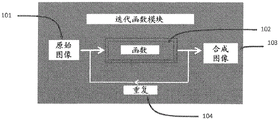
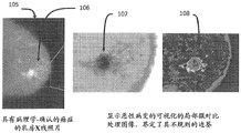
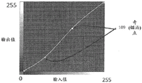
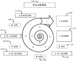
| CN108882902B (zh) | 用于图像中对象的可视化和表征的系统和方法 | |
| US20250045890A1 (en) | System And Method For The Visualization And Characterization Of Objects In Images | |
| US11669964B2 (en) | Systems and methods to facilitate review of liver tumor cases | |
| Altunbay et al. | Color graphs for automated cancer diagnosis and grading | |
| US5946407A (en) | System and method for scanning medical images using adjustable exposure time and brightness | |
| EP0811205B1 (en) | System and method for diagnosis of living tissue diseases | |
| CN101706843B (zh) | 一种乳腺cr图像交互式读片方法 | |
| Sinha et al. | Medical image processing | |
| EP3025306B1 (en) | Computer-implemented method for classification of a picture | |
| Kaur et al. | Computer-aided diagnosis of renal lesions in CT images: a comprehensive survey and future prospects | |
| CN104331864B (zh) | 基于非下采样轮廓波和视觉显著模型的乳腺影像处理 | |
| HK40000956B (en) | System and method for the visualization and characterization of objects in images | |
| HK40000956A (en) | System and method for the visualization and characterization of objects in images | |
| Anyfantis et al. | Revealing Occult Malignancies in Mammograms Through GAN-Driven Breast Density Transformation | |
| Anandan | Neural Network Model for Cancer Tumor Growth with Minimizing the Error in Learning | |
| Olaleke et al. | Automated Detection of Breast Cancer’s Indicators in Mammogram via Image Processing Techniques | |
| Naeppi et al. | Mammographic feature generator for evaluation of image analysis algorithms |
Legal Events
| Date | Code | Title | Description |
|---|---|---|---|
| PB01 | Publication | ||
| PB01 | Publication | ||
| SE01 | Entry into force of request for substantive examination | ||
| SE01 | Entry into force of request for substantive examination | ||
| REG | Reference to a national code |
Ref country code: HK Ref legal event code: DE Ref document number: 40000956 Country of ref document: HK |
|
| GR01 | Patent grant | ||
| GR01 | Patent grant |