CN103100112B - Preparation method and use of graft material in double membrane structure - Google Patents
Preparation method and use of graft material in double membrane structure Download PDFInfo
- Publication number
- CN103100112B CN103100112B CN201210525097.9A CN201210525097A CN103100112B CN 103100112 B CN103100112 B CN 103100112B CN 201210525097 A CN201210525097 A CN 201210525097A CN 103100112 B CN103100112 B CN 103100112B
- Authority
- CN
- China
- Prior art keywords
- cell
- stem cell
- diaphragm
- rich platelet
- fragment
- Prior art date
- Legal status (The legal status is an assumption and is not a legal conclusion. Google has not performed a legal analysis and makes no representation as to the accuracy of the status listed.)
- Active
Links
Images


Abstract
The invention belongs to the field of tissue engineering and biomaterials and discloses a preparation method and use of a graft material in a double membrane structure. The graft material comprises stem cell membrane patch segments and platelet-rich fibrin membrane particles, wherein the stem cells are firstly expanded-cultured, and then induction-cultured by using a membrane induction fluid to obtain a stem cell membrane patch, and the stem cell membrane patch is sheared to prepare the cell membrane patch segment; a platelet-rich fibrin gel is extruded, then liquid content of the extruded platelet-rich fibrin gel is removed to obtain the platelet-rich fibrin membrane, and the platelet-rich fibrin membrane is sheared to form particles; and the cell membrane patch segments and the platelet-rich fibrin membrane particles are mixed according to an optimal proportioning relation of in-vitro screening to prepare the needed graft material. The graft material can be widely applied to tissue repair and regeneration of small-area lesion in oral activity and other parts of the body, and the tissue repair effect is improved.
Description
Technical field
The invention belongs to organizational project and technical field of biological material, relate to a kind of pair of membrane structure graft materials, particularly the Preparation method and use of the two membrane structure graft materials of the compound rich platelet fibrin film of a kind of stem cell diaphragm fragment.
Background technology
At present, utilize stem cells technology to carry out Repair of tissue defect and conventionally adopt stem cell suspension and the compound method of exogenous timbering material, principal mode has three kinds: simple cell suspension local transplantation; Cell suspension inoculation is in exogenous timbering material; Cell suspension inoculation is external row dimensional culture after timbering material.Yet there is many deficiencies in above-mentioned implantation method: first, cell is obtaining in the process of suspension with trypsinization, and the structure of intercellular signal molecule and extracellular matrix is destroyed, and the activity of cell itself has also been subject to impact; Secondly, the amount of cell transplantation is difficult to control, and very little, cell is difficult to be colonizated in rapidly transplants part to cell concentration, and cell concentration is too many, can cause again local cells to pile up, and is unfavorable for that cell stretches and nutrition supply; Finally, the transplanting of timbering material and degraded may cause host toxicity reaction and immunoreation, and undegradable timbering material can cause fibers encapsulation, affects blood confession and organizational structure and rebuilds.Therefore, need to find a kind of new method of preparing stem cell compound support frame material, reduce the loss of stem cell, improve its biologic activity, reduce the absorption of timbering material and host's immunoreation, improve the effect of Repair of tissue defect.
Summary of the invention
In order to overcome above-mentioned the deficiencies in the prior art, the object of the present invention is to provide the graft materials and its production and use of two membrane structures of the compound PRF of a kind of CSF, CSF (cell sheet fragment) is cell patch fragment, PRF (platelet-rich fibrin) is rich platelet fibrin, graft materials of the present invention can be applied to, as the regeneration of the reparation of cartilage, soft tissue defects and dental pulp, periodontal membrane tissue, effectively improve the effect of tissue repair.
To achieve these goals, the technical solution used in the present invention is:
A pair membrane structure graft materials, is characterized in that: by stem cell diaphragm fragment and rich platelet fibrin film granulometric composition.
Preferably, described stem cell is selected from mesenchymal stem cells MSCs, fat stem cell, dental pulp stem cell or periodontal ligament stem cell.
Preferably, described rich platelet fibrin diaphragm granule obtains after via centrifugal blood.
Preferably, cell patch adopts six orifice plates to be prepared, and the ratio of cell patch fragment and rich platelet fibrin film is as follows: every six orifice plate cell patch quantity: for blood milliliter number=1 of centrifugal acquisition rich platelet fibrin diaphragm: 3.5~4.0.
Preferably, described stem cell and rich platelet fibrin all derive from same individuality.
Preferably, stem cell must be prepared into the form of cell patch fragment.
A preparation method for graft materials, is characterized in that its preparation process comprises the following steps:
The first step, the cultivation of stem cell diaphragm: primary stem cell is inoculated in six orifice plates after amplification culture, merges and with diaphragm induced liquid, to be cultivated to 80% time until growth, within 3 days, changes liquid once, until cell patch, around occurs when curling thinking that cell patch is ripe;
Second step, prepares stem cell diaphragm fragment: by the method for mechanical stripping, cell patch is peeled off from culture dish, with aseptic eye scissors, shredded, be prepared into cell patch fragment (big or small about 0.5mm * 0.5mm);
The 3rd step, extract rich platelet fibrin diaphragm granule: according to required ratio, take 10ml as unit, blood (not adding any anticoagulant) is placed in to the glass centrifuge tube of aseptic glass centrifuge tube or plastic bushing, with the speed of 3000rpm/min centrifugal 10 minutes immediately, after standing 5 minutes, aseptic tweezer clamp takes out the rich platelet fibrin gel of centrifuge tube mid portion, its bottom erythrocyte end is gently dipped in to remove unnecessary erythrocyte on sterile gauze, retain complete light yellow gel part and bottom and adhere on a small quantity firmly erythrocyte, the latter's tiling is placed between sterile gauze, the wherein liquid component of gently extruding out, be prepared into a faint yellow tough and tensile membrane structure, with aseptic eye scissors, shredded, be prepared into the rich platelet fibrin diaphragm granule of big or small about 0.5mm * 0.5mm * 0.5mm,
The 4th step, the cell patch fragment of preparation and rich platelet fibrin diaphragm granule are placed in to sterile petri dish, proportionally manually mixed, guarantee that the two fully mixes, can obtain two membrane structure graft materials of stem cell diaphragm fragment and the compound rear formation of rich platelet fibrin film.
Preferably, the ratio of cell patch fragment and rich platelet fibrin film is as follows: every six orifice plate cell patch quantity: for blood milliliter number=1 of centrifugal acquisition rich platelet fibrin diaphragm: 3.5~4.0.
Graft materials of the present invention is the application in Repair of tissue defect among a small circle in oromaxillo-facial region or whole body.
Graft materials of the present invention purposes in graft materials for Repair of tissue defect among a small circle in preparing oromaxillo-facial region or whole body.
The composite that graft materials of the present invention and other biological material form is being prepared the purposes in graft materials for Repair of tissue defect.
Two membrane structure graft materials for the compound rich platelet fibrin film of stem cell diaphragm fragment, by stem cell diaphragm fragment, rich platelet fibrin film granulometric composition.Stem cell form can be different according to the type of repair tissue, comprise mesenchymal stem cells MSCs, fat stem cell, dental pulp stem cell and periodontal ligament stem cell etc., and rich platelet fibrin film is that blood prepares after centrifugal.The external screening of carrying out the two proportioning, with the propagation of stem cell, minute be turned to evaluation index, determines that the two optimum volume ratio is: six orifice plates obtain cell patches: for blood=1 of centrifugal acquisition rich platelet fibrin diaphragm: 3.5~4.0.According to this proportioning, carry out the preparation of graft, then migrated in experimental animals, carry out the checking of experiment in body, result proves, the two membrane structure grafts under this proportioning have good tissue repair and tissue regeneration effect.
The mode that graft materials two components are mixed is: stem cell is prepared into diaphragm pieces, rich platelet fibrin film is prepared into graininess (big or small about 0.5mm * 0.5mm * 0.5mm), the two armstrong's patent in sterile petri dish mixes, and two components are fully mixed.
The present invention also provides the preparation method of described stem cell diaphragm fragment and rich platelet fibrin membrane granule, comprises the following steps:
The first step, the cultivation of stem cell diaphragm: primary stem cell is inoculated in six orifice plates after amplification culture, merges and with diaphragm induced liquid, to be cultivated to 80% time until growth, within 3 days, changes liquid once, until cell patch, around occurs when curling thinking that cell patch is ripe;
Second step, prepares stem cell diaphragm fragment: by the method for mechanical stripping, cell patch is peeled off from culture dish, with aseptic eye scissors, shredded, be prepared into cell patch fragment (big or small about 0.5mm * 0.5mm);
The 3rd step, extract rich platelet fibrin diaphragm granule: according to required ratio, take 10ml as unit, blood (not adding any anticoagulant) is placed in to the glass centrifuge tube of aseptic glass centrifuge tube or plastic bushing, with the speed of 3000rpm/min centrifugal 10 minutes immediately, after standing 5 minutes, aseptic tweezer clamp takes out the rich platelet fibrin gel of centrifuge tube mid portion, its bottom erythrocyte end is gently dipped in to remove unnecessary erythrocyte on sterile gauze, retain complete light yellow gel part and bottom and adhere on a small quantity firmly erythrocyte, the latter's tiling is placed between sterile gauze, the wherein liquid component of gently extruding out, be prepared into a faint yellow tough and tensile membrane structure, with aseptic eye scissors, shredded, be prepared into the rich platelet fibrin diaphragm granule of big or small about 0.5mm * 0.5mm * 0.5mm,
The 4th step, the cell patch fragment of preparation and rich platelet fibrin diaphragm granule are placed in to sterile petri dish, proportionally manually mixed, guaranteed that the two fully mixes, can be obtained the two membrane structure graft materials of the compound rich platelet fibrin film of stem cell diaphragm fragment.
Cartilage among a small circle and the reparation of soft tissue defects during two membrane structure graft materials of the described compound rich platelet fibrin of stem cell diaphragm fragment membrane granule can be used for, for operation better position, the visual field, can directly complex be placed in to damaged place, not good or cannot expose the case of defective region completely for the operation visual field, also graft materials can be placed in to syringe or adopt endoscopic procedures to be injected in damaged place.
The present invention compared with prior art has the following advantages:
The timbering material that existing tissue engineering technique is used is all almost exogenous material, seed cell is inoculated on exogenous material with the form of cell suspension, add again one or more exogenous growth factors, the weak point of this method is: ectogenic timbering material degraded needs certain hour, and has the inadequate possibility of degraded; And seed cell is inoculated and is transplanted to suspensions after tissue local, very easily run off, make it possible to the stable cell concentration playing a role of local field planting and obviously tail off; And the somatomedin kind of exogenous interpolation is less, completely different from multiple somatomedin eurythmy under physiological situation, the collaborative pattern playing a role, these all can affect the effect of tissue engineering technique clinical practice.The most outstanding feature of the present invention is the shortcoming that has fundamentally solved above-mentioned prior art, be in particular in following some:
1) low repulsion.Two kinds of compositions in cograft material prepared by this law, stem cell diaphragm fragment (CSF) and rich platelet fibrin film (PRF) source are sufficient, have splendid biologic activity, have hardly immunological rejection and toxic reaction.
2) proportioning is good.One of important feature of the present invention is the best proportioning that has filtered out stem cell population and rich platelet fibrin film.Under this conditions of mixture ratios, cell has good propagation and differentiation capability, is conducive to tissue repair and regeneration, and this proportioning usings six orifice plates and obtain the milliliter number of cell patch quantity and blood as the ultimate unit of proportioning, easily accurately grasps convenient operation.
3) high efficiency.Rich platelet fibrin film comes from blood, simple to operate, very easily obtains; Itself contain a large amount of somatomedin, comprise transforming growth factor β-1 (TGF β-1), platelet-derived growth factor (PDGF), insulin like growth factor (IGF), above-mentioned three kinds of somatomedin can promote cell migration, propagation, induction fibrin matrix is reinvented, and promote to play an important role the secretion of collagen stroma in initial organization healing; With the exception of this, PRF also contains epidermal growth factor (EGF) and the somatomedin relevant to wound healing and osteanagenesis such as VEGF (VEGF).PRF itself is exactly the three-dimensional rack structure that fibrin cross-linked network forms, and this structure can be so that somatomedin wherein slowly discharges.So the tissue repair of PRF and tissue regeneration effect mainly realize by two aspects, i.e. the regulating action of somatomedin and fibrin scaffold which effect.
4) easy to operate.The cell type of cell patch fragment (CSF) can be according to the difference of the region of repair deficiency and organization type and difference, comprise mesenchymal stem cells MSCs (BMSCs), fat stem cell (ASCs), dental pulp stem cell (DPSCs), periodontal ligament stem cell (PDLSCs) etc., above-mentioned cell has all been proved good tissue regeneration ability, and obtain relatively easily, be easy to amplification culture in the external short time; Cell patch technology itself has stronger tissue regeneration potential owing to having retained extracellular matrix and cytoactive compared with suspension cell; Simultaneously, diaphragm fragment technology has changed again traditional diaphragm and has transplanted pattern, diaphragm is prepared into a certain size fragment, can make cell fragment fully mix with timbering material on the one hand, cell rapid field planting is in timbering material surface and stretch out synapse to extend to support gap inner, the two forms an integral body (Fig. 7), cell can be injected on the other hand to the region that is difficult to expose completely the operation visual field.
5) infection.The red end of PRF has concentrated a large amount of leukocyte, and these leukocyte are brought into play good antiinflammatory action on the one hand in part, transfer on the other hand the immunologic function of body; Meanwhile, PRF has also enlisted the services of a large amount of immunoregulatory factor in blood in preparation process, as IL-1 β, and IL-4, IL-6 and TNF-α, bring into play anti-infective effect jointly, is beneficial to organization healing.
Therefore the cograft material that, prepared by the inventive method has that source is abundant, indication extensively, preparation is simple, without immunological rejection and toxic reaction, without dispute of ethic, the advantage such as safe and effective.The method is of universal significance, and is applicable to every patient.
Accompanying drawing explanation
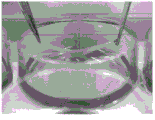
Fig. 1 sees (take dental pulp stem cell as example) after cell patch is cultivated maturation substantially.
Fig. 2 is the microstructural HE dyeing of cell patch (take dental pulp stem cell as example).
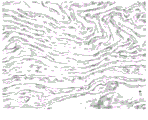
Fig. 3 is cell patch scanning electron microscope structure (take dental pulp stem cell as example).
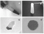
Fig. 4 is that rich platelet fibrin is seen substantially, and wherein A sees for centrifugal latter standing 5 minutes substantially; B is PRF gel; C is PRF film; D is PRF granule.
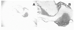
Fig. 5 is the microstructural HE dyeing of rich platelet fibrin film, and wherein A is 40 times of amplifications, and B is 100 times of amplifications.
Fig. 6 is rich platelet fibrin film scanning electron microscope structure, and wherein A is its upper end, is fibrin tridimensional network; B is its lower end, visible a large amount of platelet, leukocyte recruitment.
Fig. 7 is the scanning electron microscope of two membrane structure grafts of forming after the compound rich platelet fibrin of dental pulp stem cell diaphragm fragment membrane granule, visible cell secure adhesion is in PRF surface, and stretch out synapse cell to the space of PRF tridimensional network, the two strong bonded, forms whole.
Fig. 8 is cell proliferation situation under different conditions of mixture ratios, and wherein A is the impact of PRF on dental pulp stem cell propagation, and B is the impact of PRF on periodontal ligament stem cell propagation.
Fig. 9 be under different conditions of mixture ratios dental pulp stem cell to the expression of the differentiation phase DMP1 of odontoblast.
Figure 10 be under different conditions of mixture ratios periodontal ligament stem cell to the expression of periodontal ligament cell differentiation phase CP23.
Figure 11 is for adopting respectively two membrane structures of simple mesenchymal stem cells MSCs diaphragm fragment, simple PRF granule, the compound rich platelet fibrin of mesenchymal stem cells MSCs diaphragm fragment membrane granule to repair experimental result in the body that rabbit condylar cartilage is damaged, and this figure is that 2,4,8 weeks condyles of experiment are organized gross examination of skeletal muscle result.
Figure 12 is that Figure 11 organizes row HE coloration result.
Figure 13 is that Figure 11 organizes row Toluidine blue staining result.
Figure 14 is toluidine blue different cartogram of dying tinctorial strength analysis in neocartilage district in Figure 13, and abscissa represents group and time, and vertical coordinate represents the average optical density value of Yi Ran district tinctorial strength.
Figure 15 is for adopting respectively the interior experimental result of body of simple PRF granule, simple dental pulp stem cell diaphragm fragment, the two membrane structure row canine tooth marrow regeneration of the compound rich platelet fibrin of dental pulp stem cell diaphragm fragment membrane granule, and this figure is HE coloration result after January, February.
The specific embodiment
Below in conjunction with drawings and Examples, the present invention is carried out to more detailed explanation.
The preparation method of two membrane structure graft materials of the compound rich platelet fibrin film of stem cell diaphragm fragment, comprises checking two parts in external proportioning screening and body.
First: external proportioning screening
1. material and equipment
1.1 main agents
α-MEM culture medium (Hyclone company, the U.S.); 0.25% pancreatin (Hyclone company, the U.S.); Hyclone (Ilex purpurea Hassk.[I.chinensis Sims, Tian Hang bio tech ltd, Zhejiang).
1.2 instrument and equipment
Multitube frame autobalance centrifuge (TD25-WS, Hunan Xiang Yi Laboratory Instruments development corporation, Ltd.); Cell culture incubator (Heraeuc Hearcell, Thermo company, China); Six orifice plates (Nunc company, the U.S.); Superclean bench (BIOBASE, the U.S.).
2. operating procedure
The cultivation of 2.1 stem cell:
1) by primary stem cell after amplification culture, according to 1: 2~1: 3 ratio, go down to posterity;
2) get third generation stem cell and be inoculated in six orifice plates, cellar culture (α-MEM culture fluid of 10%FBS) is to cell fusion 80%.
2.2 rich platelet fibrin preparations:
1) will take 10ml as unit according to required ratio, blood (not adding any anticoagulant) will be placed in to the glass centrifuge tube of aseptic glass centrifuge tube or plastic bushing, with the speed of 3000rpm/min centrifugal 10 minutes immediately, standing 5 minutes;
2) aseptic tweezer clamp takes out the rich platelet fibrin gel of centrifuge tube mid portion, its bottom erythrocyte end is gently dipped in to remove unnecessary erythrocyte on sterile gauze, retain complete light yellow gel part and bottom and adhere on a small quantity firmly erythrocyte, the latter's tiling is placed between sterile gauze, the wherein liquid component of gently extruding out, is prepared into the faint yellow tough and tensile membrane structure of a rectangle;
3) be laid on sterile gauze, sterile scissors is cut off along its major axis, is prepared into 1/8 and 1/4 PRF, is placed in culture fluid standby.
2.3 stem cell and rich platelet fibrin film are cultivated altogether:
1) according to the ratio of 1/8,1/4,3/8,1/2 PRF, the PRF shearing is placed in to the six orifice plate culture dishs of third generation stem cell, cellar culture 7 days, changes liquid once in 3 days;
2) the two is cultivated altogether after 7 days and removes PRF, and stem cell continues normal cultivation, within 3 days, changes liquid once;
The detection of 2.4 stem cells hyperplasias, differentiation:
1) respectively at the two, cultivate altogether front and be total to the detection of carrying out stem cell population for 1~7 day after cultivating, observing the impact of PRF on cell proliferation;
2) the two is total to the Real-time PCR detection of 7,14,21 days row stem cell related gene expressions after cultivating, and observes the impact of PRF on cell differentiation.
3. experimental result
3.1 stem cells hyperplasia
PRF has obvious facilitation to the propagation of dental pulp stem cell and periodontal ligament stem cell, and is regular hour dependency.Two kinds of common features of stem cell are: PRF concentration is from 1/8~3/8, the impact of its on cell proliferation presents obvious concentration dependent, and the facilitation with 3/8PRF on cell proliferation is the most obvious, though 1/2PRF on cell proliferation has a certain promotion, not as good as 3/8PRF effect obvious (as shown in Figure 8).
3.2 differentiation of stem cells
The two is cultivated after 21 days altogether, the expression of the specific proteins DMP1 that PRF breaks up to odontoblast's direction dental pulp stem cell has obvious rise effect, and be obvious concentration dependent, best with 1/2PRF, but difference not statistically significant (Fig. 9) between itself and 3/8PRF; PRF also has obvious rise to the expression of periodontal ligament stem cell specific proteins CP23, with the concentration of 3/8PRF best (as shown in figure 10).
The result of comprehensive PRF on the impact of two kinds of dissimilar stem cells hyperplasia differentiation, we think that 3/8PRF all has obvious facilitation to the propagation of stem cell and differentiation, therefore select this concentration as optium concentration.Because every PRF film desired blood is 10mL, therefore finally determine that the two proportion relation is: every six orifice plate cell patch quantity: blood milliliter number=1: 3.75, in order to simplify the operation, we think 1: 3.5~4.0 all can.In order to verify transplantation effect in the body of the graft under this conditions of mixture ratios, we carry out part experiment in body.
Second portion: transplant checking in object
The first step, the cultivation of stem cell diaphragm: primary stem cell is inoculated in six orifice plates after amplification culture, merges and with diaphragm induced liquid, to be cultivated to 80% time until growth, within 3 days, changes liquid once, until cell patch, around occurs when curling thinking that cell patch is ripe;
Second step, prepares stem cell diaphragm fragment: by the method for mechanical stripping, cell patch is peeled off from culture dish, with aseptic eye scissors, shredded, be prepared into cell patch fragment (big or small about 0.5mm * 0.5mm);
The 3rd step, extracts rich platelet fibrin diaphragm granule: concrete grammar, with experiment in vitro part, is shredded the PRF film of preparing gained with aseptic eye scissors, be prepared into the rich platelet fibrin diaphragm granule of big or small about 0.5mm * 0.5mm * 0.5mm;
The 4th step, the cell patch fragment of preparation and rich platelet fibrin diaphragm granule are placed in to sterile petri dish, proportionally manually mixed, guaranteed that the two fully mixes, can be obtained the two membrane structure graft materials of the compound rich platelet fibrin film of stem cell diaphragm fragment.
For operation good position, the visual field, during use, the two membrane structure graft materials that prepare directly can be placed in to damaged place, not good or cannot expose the case of defective region completely for the operation visual field, also graft materials can be placed in to syringe or adopt endoscopic procedures to be injected in damaged place.
In order to verify the effect of two membrane structure graft materials of the compound rich platelet fibrin film of stem cell diaphragm fragment of the present invention, we have carried out respectively the two membrane structures of the compound rich platelet fibrin film of variety classes stem cell cell patch and have carried out the experiment of Repair of tissue defect, now take respectively " it is damaged that the two membrane structures of the compound rich platelet fibrin film of bone marrow mesenchymal stem cells diaphragm fragment are repaired rabbit condylar cartilage " and " the two membrane structures of the compound rich platelet fibrin film of canine tooth marrow stem cell diaphragm fragment are used for dental pulp regenerative therapy " be example, test as follows:
Experiment one: it is damaged that the two membrane structures of the compound rich platelet fibrin film of bone marrow mesenchymal stem cells diaphragm fragment are repaired rabbit condylar cartilage
1. material and equipment
1.1 main agents
α-MEM culture medium (Hyclone company, the U.S.); 0.25% pancreatin (Hyclone company, the U.S.); Hyclone (Ilex purpurea Hassk.[I.chinensis Sims, Tian Hang bio tech ltd, Zhejiang); Vitamin C (Sigma company, the U.S.); Pentobarbital sodium (Germany, Ke Hao biological engineering Co., Ltd subpackage).
1.2 instrument and equipment
Multitube frame autobalance centrifuge (TD25-WS, Hunan Xiang Yi Laboratory Instruments development corporation, Ltd.); Cell culture incubator (Heraeuc Hearcell, Thermo company, China); Six orifice plates (Nunc company, the U.S.); Superclean bench (BIOBASE, the U.S.); Nikon microscopy imaging system (XTJ30, Japan), analytical electron balance (FA1004, China), scanning electron microscope (S-4800 of Hitachi, Japan).
2. operating procedure
The cultivation of 2.1 bone marrow mesenchymal stem cells (BMSCs)
The primary mesenchymal stem cells MSCs of rabbit adopts the α-MEM culture medium cellar culture containing 10% hyclone, and the ratio according to 1: 2~1: 3 when cell fusion to 80% is carried out amplification culture, within every 2~3 days, changes liquid once;
The preparation of 2.2BMSCs diaphragm
1), when BMSCs is cultured to the third generation, be passaged to 6 orifice plates; Change diaphragm culture fluid (containing 10% hyclone, the ascorbic α-MEM of 20mg/ml culture medium), conventional continuation cultivated two weeks, and diaphragm culture fluid is changed once for every 2~3 days, notes lucifuge while changing liquid;
2) until cell patch, grow to periphery cell patch when slightly curling, think that cell patch is ripe, ripe cell patch is peeled off (as Fig. 1 from one end with ophthalmic tweezers, Fig. 2, shown in Fig. 3), be placed in sterile petri dish, adopt aseptic eye scissors to be cut into the fragment of big or small about 0.5mm * 0.5mm, standby.
The preparation of 2.3PRF
1) according to required ratio, take 10ml as unit, rabbit blood (not adding any anticoagulant) is placed in to the glass centrifuge tube of aseptic glass centrifuge tube or plastic bushing, with the speed of 3000rpm/min centrifugal 10 minutes immediately, standing 5 minutes;
2) now blood is divided into three layers in visible centrifuge tube: lower floor is erythrocyte layer, the faint yellow clear liquid that upper strata is minute quantity, and intermediate layer is faint yellow translucent rich platelet fibrin gel (as shown in Figure 4 A);
3) with aseptic ophthalmic tweezers by the complete taking-up of mid portion gel, its bottom erythrocyte end is gently dipped in to remove unnecessary erythrocyte on sterile gauze, retain complete light yellow gel part and bottom and adhere on a small quantity firmly erythrocyte (as shown in Figure 4 B), the latter's tiling is placed between sterile gauze, and the wherein liquid component of gently extruding out, is prepared into a faint yellow tough and tensile membrane structure, be rich platelet fibrin film, be PRF film (as Fig. 4 C, Fig. 5, shown in Fig. 6);
4) with aseptic eye scissors, shredded, be prepared into the rich platelet fibrin diaphragm granule of big or small about 0.5mm * 0.5mm * 0.5mm, standby (as shown in Figure 4 D).
The preparation of the two membrane structures of 2.4BMSCs/PRF
The BMSCs cell patch fragment of preparation and PRF diaphragm granule are placed in to sterile petri dish, according to six orifice plates, obtain cell patches: for blood=1 of centrifugal acquisition rich platelet fibrin diaphragm: 3.5~4.0 ratio, manually mixed, guarantee that the two fully mixes, can obtain two membrane structure graft materials of the compound rich platelet fibrin of stem cell diaphragm fragment membrane granule, standby (as shown in Figure 7).
The foundation of 2.5 rabbit Temporomandibular Joint Cartilage defect models
1) anesthesia: under aseptic condition, the anesthesia of 3% pentobarbital sodium (1mL/kg) auricular vein, separately imposes local anaesthesia with 1% lignocaine in modeling side;
2) preoperative preparation: art side remporomandibular joint art district preserved skin, iodophor disinfection, drape;
3) outside outer canthus, 5mm place to external auditory meatus direction is made the long skin incision of about 2cm;
4) separated subcutaneous tissue, cuts joint capsule;
5) expose the prominent joint of condyle, with the electronic condyle front bevel central authorities of dashing forward that are drilled in, drill through the hole of 1 diameter 3mm;
6) row embolia, sews up joint capsule, skin suture layer.
2.6 experiment groupings
According to experimental design, the graft at the damaged place of condylar cartilage to be implanted is divided into 4 groups: 1. blank group: the damaged place of condylar cartilage is implantation graft not; 2. simple PRF groups of grains; 3. simple BMSCs cell patch slice groups; 4. two membrane structure groups of BMSCs/PRF.
3. experimental result
After using pentobarbital sodium anesthesia when 2w, the 4w after implanting respectively at each group, 8w, draw materials, 4% paraformaldehyde is fixed.After 15%EDTA decalcification, carry out gross examination of skeletal muscle, and organize microstructural observation after HE, Toluidine blue staining, result is as follows:
1) gross examination of skeletal muscle is visible: the two membrane structure group reparation situations of BMSCs/PRF are better than other group (as shown in figure 11) of same time point;
2) HE coloration result: blank group, in each time point defective region depression, has been showed no the filling of cartilage sample material; Independent PRF group and separate cell diaphragm group are along with the prolongation defect repair district of time has neocartilage to generate gradually, but cartilage level arrangement disorder; Two membrane structure groups, in the time of the 8th week, damage zone flushes substantially, repairs that cartilage layers is thinner, level is more clear, with contiguous each layer of normal cartilage continuous (as shown in figure 12);
3) Toluidine blue staining result: each is organized neocartilage region and is heterochromaty (as shown in figure 13), adopts IPP 6.0 softwares to carry out tinctorial strength analysis to each group Toluidine blue staining picture, calculates average optical density value.Result draws: except blank group, all the other 3 groups 2,4,8 weeks after surgery average OD values are increases trend (P < 0.05) gradually, and is significantly higher than the blank group (P < 0.05) of same time point; Separately PRF group and separate cell diaphragm group are in the average OD value of each time point there are no significant difference.4,8 weeks average OD values of two membrane structure groups are significantly higher than other group (P < 0.05) (as shown in figure 14) of same time point.
Experiment two: the two membrane structures of the compound rich platelet fibrin film of canine tooth marrow stem cell diaphragm fragment are for dental pulp regenerative therapy
1. material and equipment
1.1 main agents
α-MEM culture medium (Hyclone company, the U.S.); Type i collagen enzyme (GIBCO company, the U.S.); 0.25% pancreatin (Hyclone company, the U.S.); Hyclone (Ilex purpurea Hassk.[I.chinensis Sims, Tian Hang bio tech ltd, Zhejiang); Vitamin C (Sigma company, the U.S.); Pentobarbital sodium (Germany, Ke Hao biological engineering Co., Ltd subpackage).
1.2 instrument and equipment
Multitube frame autobalance centrifuge (TD25-WS, Hunan Xiang Yi Laboratory Instruments development corporation, Ltd.); Cell culture incubator (Heraeuc Hearcell, Thermo company, China); Six orifice plates (Nunc company, the U.S.); Superclean bench (BIOBASE, the U.S.); Nikon microscopy imaging system (XTJ30, Japan), analytical electron balance (FA1004, China), scanning electron microscope (S-4800 of Hitachi, Japan).
2. operating procedure
Separation and the cultivation of 2.1 dog dental pulp stem cells (DPSCs)
The primary dental pulp stem cell of dog adopts the α-MEM cellar culture containing 10%FBS, cultivates a plurality of clone cell of the appearance group after 3~5 days, when cloning cluster interior detail intracellular growth to 80%, with the ratio of 1: 2~1: 3, carries out amplification culture, within every 2~3 days, changes liquid once;
The preparation of 2.2DPSCs diaphragm
With experiment one.
The preparation of 2.3PRF
With experiment one.
The preparation of the two membrane structure graft materials of the compound rich platelet fibrin film of 2.4 canine tooth marrow stem cell diaphragm fragment
With experiment one.
The foundation of 2.5 canine tooth marrow regenerating models
1) the conventional anesthesia of dog (approximately 6 monthly age), method is the same;
2) preoperative preparation: in dog mouth, all premolarss adopt the capable gum of periodontal scaling apparatus manually to scrape and control up and down, remove tartar and soft dirt, the inside and outside povidone iodine strict sterilization in oral cavity, drape;
3), after randomized grouping, upper mandibular premolar (except frist premolar) is opened marrow, takes off marrow top, Exposed Pulp tissue, barbed broach is complete to be pulled out after dental pulp, adopts 30#K to determine after Open canal system, expand apical foramen of tooth and file to 70#K, make tip of a root open zone diameter reach 0.7mm;
4) disposable syringe is drawn a large amount of physiological saline solution and is carried out root canal irrigation, determines that Gen Guanneiwu pulp tissue and tissue of tooth chip are residual;
5) in 2.6, divide into groups, graft is placed in to root pipe, adopt auger conveyor that graft is delivered in whole pipe, reach root canal orifice place; Calcium hydroxide (Dycal, Deng Shibai company) is gently put in graft upper end, the about 1mm of thickness, and after calcium hydroxide knot is solid, finishing hole wall, adopts glass ion rebasing, the about 1mm of thickness, damaged place adopts light-cured resin filling;
2.6 experiment groupings
According to experimental design, the graft of pulp cavity to be implanted is divided into 4 groups: 1. blank group: in pulp cavity, do not implant any graft; 2. simple PRF groups of grains; 3. simple DPSCs cell patch slice groups; 4. two membrane structure groups of DPSCs/PRF.
3. experimental result
HE coloration result: the visible pulp tissue of Normal group, odontoblast, predentin and ripe dentin ordered arrangement, odontoblast is high column ordered arrangement; The more shallow predentin of the equal visible vessels of each experimental group and dyeing forms, but there are differences between blood vessel quantity, dentin thickness and each group of cell arrangement situation: the two membrane structure group cells of DPSCs/PRF are the loose multiple layer of StarNet's shape and arrange, and cellular morphology is by changing to close Dentinal vertical polarization growth gradually away from Dentinal Parallel Growth, the similar odontoblast of form, sees in pulp cavity that more new vessels forms; The two membrane structure groups of simple DPSCs cell patch group pulp cells are arranged loose, unordered, and dentin layer is thinner, but also visible more vascularization in pulp cavity; Simple PRF groups of grains cell is mostly rounded, without obviously stretching, arranges unordered and dentin wall is thin, but also visible a small amount of vascularization (as shown in figure 15) in pulp cavity.
4. Analysis on Mechanism
The effect of 4.1 stem cell diaphragm fragments (CSF)
Stem cell (stem cells, SC) be the cell that a class has the of self-replication capacity and Multidirectional Differentiation ability, under certain condition, it can be divided into several functions cell, the potential function with the various histoorgans of regeneration and human body, therefore be widely used in organizational project as seed cell at present.
Cell patch technology is to impel seed cell to produce at short notice a large amount of extracellular matrixs (extracellular matrix, ECM) by external evoked mode, and cell is closely linked and becomes a kind of laminated structure.ECM not only provides the support of connection, nutrition supply and mechanical property for cell and tissue, what is more important can regulate intercellular communication, and the basic vital movement of cell is brought into play to omnibearing dynamic adjustments effect.ECM has the multiformity of 26S Proteasome Structure and Function, the variation of its structure and composition will affect the structure of cytoskeleton, thereby determine shape and the activity of cell, affect the existence of cell, participate in and control differentiation and the migration of cell, generation, the growth of histoorgan, regenerate and maintain the physiological activity of 26S Proteasome Structure and Function of histoorgan in all there is extremely crucial effect.This unique advantage that can preserve iuntercellular autocrine signaling molecule performance of cell patch technology, can reduce the quantity of invalid transplanted cells, is conducive to reconstruction or the regeneration of tissue.And diaphragm fragment technology has changed traditional diaphragm transplanting pattern, complete cell patch is prepared as to a certain size fragment, the activity of having preserved seed cell on the one hand, and at utmost preserved the performance of ECM and iuntercellular autocrine signaling molecule, make on the other hand cell farthest evenly mix with support, be convenient to cell and be colonizated in rapidly rack surface, make the graft of the two formation become the integral body with good biological function.
The effect of 4.2 rich platelet fibrin films (PRF)
Rich platelet fibrin (PRF) fibrous reticular structure that to be whole blood form after once centrifugal fast.The main feature of PRF is to be rich in the somatomedin that a large amount of ratios approach physiological situation, as transforming growth factor (transforming growth factor β, TGF β), platelet-derived growth factor (platelet-derivedgrowth factors, PDGF), insulin like growth factor (insulin-like growth factors, IGF), above-mentioned three kinds of somatomedin play an important role in initial organization healing, they can promote cell migration and proliferation, induction fibrin matrix is reinvented, and promotes the secretion of collagen stroma; With the exception of this, in PRF, also contain the multiple somatomedin relevant to wound healing and osteanagenesis, comprise epidermal growth factor (epidermal growth factor, EGF) and VEGF (vascular endothelial growthfactor, VEGF) etc.
As the second filial generation product of platelet concentrate, compare with first generation product platelet rich plasma (platelet-richplasma, PRP), PRF has following characteristics:
1) preparation is simple.The preparation of PRP need to be added anticoagulant, in case Hemostatic Oral Liquid solidifies; Its preparation needs two times centrifugal, the supernatant after centrifugal need to be shifted, and complex operation, and there is no at present centrifugal speed and the time of standard, the method that different researchers is prepared it is held different viewpoints.The preparation of PRF is very simple, extracts after whole blood immediately with the speed of 3000rprm/min centrifugal 10 minutes, standing 5 minutes, can obtain PRF gel.
2) safety is good.First PRP needs to add anticoagulant in the preparation, next after two times centrifugal finishes, needs to add thrombin of beef and calcium chloride is activated, just gel can be formed to transplant in body, and PRP only just can make the de-particle release somatomedin of platelet alpha chain wherein after activating; Yet, adds exogenous material and likely cause host's immunoreation, and too much operating procedure has also increased the probability polluting.The preparation of PRF does not need to add any exogenous material, and single stepping completes, and safety is good.
3) growth factor slow-release.PRP is that formation is gelatinous rapidly after adding more thrombin to carry out artificial activation.A large amount of ectogenic thrombins make the rapid polymerization of fibrin, form two-way fine and close syndeton, this just makes the somatomedin that platelet discharges not enlisted the services of wherein, but remains in the suspension outside fibrous reticular structure, thereby presents quick, a large amount of features that discharge.And PRF is the polymerization methods slowly producing under self thrombin action, this gradual polymerization methods is enlisted the services of somatomedin free in peripheral blood to the tridimensional network of PRF to a greater extent.The distribution of somatomedin in free somatomedin and platelet in this three dimensional structure will inevitably cause its slowly releasing effect, because they only can just be released when reconstruction occurs its place fibre structure, and the fibril in PRF and fibrin form tetragonal connected mode, this elastomeric matrices structure is also more conducive to cell migration and shla molecule retains.
4) tridimensional network can be used as timbering material.What the fibrin in PRP formed is two-way fine and close syndeton, and this structure is unfavorable for the migration of cell and somatomedin.Fibrin in PRF forms tridimensional network, and this structure is not only beneficial to the migration of cell and somatomedin, and the place of proliferation and differentiation is also provided for tissue repair relevant cell, has brought into play important support effect.
5) contain a large amount of leukocyte.PRF obtains via whole blood is centrifugal, and in centrifugal process, in blood, most leukocyte are all distributed in PRF lower end, i.e. red end (as shown in B figure in Fig. 6); Therefore,, when two membrane structures migrate in body, concentrated leukocyte is brought into play good antiinflammatory action in part on the one hand, transfers on the other hand the immune system of body, brings into play good anti-inflammatory properties and immunoloregulation function; Meanwhile, PRF has also enlisted the services of a large amount of immunoregulatory factor in blood in preparation process, as IL-1 β, and IL-4, IL-6 and TNF-α etc., these factors protect from infection, promote the aspects such as healing all to play an important role after Repair of tissue defect.
The analysis of two membrane structure zoografting effects of the compound rich platelet fibrin film of 4.3 stem cell diaphragm fragment
First of the present invention application experiment in vitro is studied the proportion relation of stem cell cell patch and rich platelet fibrin film, has obtained best proportion relation between the two.Second portion, according to the two membrane structure grafts of this proportion relation preparation, is implanted in experimental animals, probes into the impact of two membrane structure grafts on tissue repair.
4.3.1 test in one, rich platelet fibrin film PRF granule is as cytoskeleton and somatomedin donor, carries out compoundly with mesenchymal stem cells MSCs diaphragm fragment, and it is mixed according to a certain percentage and is placed on tissue defect place, carries out the reparation of cartilage defect.PRF diaphragm has toughness, shreds and becomes very easily operation after graininess, can be placed in the tissue defect of any shape; Mesenchymal stem cells MSCs is made into after patching, owing to there is the connection of extracellular matrix, and the shortcoming of having avoided suspension cell in the past easily to run off.Our experimental result shows, compare with natural recovering group, simple PRF and simple cell patch group, it is best that two membrane structure groups are repaired the effect of cartilage defects, and in the time of the 8th week, damage zone flushes substantially, reparation cartilage layers is thinner, level is more clear, more continuous with contiguous each layer of normal cartilage.This has just proved our guess, that is: mesenchymal stem cells MSCs has been brought into play the effect of seed cell in part, in local microenvironment, can become specific cell type by proliferation and differentiation, and PRF diaphragm granule provides a large amount of collagen stroma compositions on the one hand, for cell provides supporting structure, the platelet wherein containing on the other hand is constantly activated along with the reconstruction of matrix components, discharge the somatomedin that a large amount of ratios approach physiological situation, for the propagation of stem cell and the reparation of differentiation and tissue provide nutritional support.
4.3.2 test in two, using PRF diaphragm as cytoskeleton and somatomedin donor, with dental pulp stem cell diaphragm fragment carry out compound, by its mix be according to a certain percentage placed on that dental pulp lacks as root tube chamber in, carry out the research of dental pulp regeneration.Found that, two membrane structure groups are compared with simple cell diaphragm and simple PRF group favorable regeneration effect.Visible dental pulp, odontoblast, predentin and ripe dentin ordered arrangement are observed in Normal group canine tooth longitudinal section HE dyeing, Yihong collagen stroma of having a liking for that dentin takes on a red color after demineralization is processed dyes, predentin near pulp cavity partly dyes more shallow, along dentin layer, arrange, monolayer odontoblast immediately predentin is high columnar arrangement.The two membrane structure group HE dyeing of DPSCs/PRF is found, cell is the multiple layer of loose StarNet shape and arranges, and cellular morphology is by changing to close Dentinal vertical polarization growth gradually away from Dentinal Parallel Growth, and the similar odontoblast of form, sees more vascularization in pulp cavity.Simple cell diaphragm group cell is the multiple layer of loose StarNet shape and arranges, but it is loose, unordered that two membrane structure groups are arranged, do not show the feature that significantly nearly dentin vertical polarization growth changes, and dentin layer is thinner, but cellular morphology stretches obviously, also visible more vascularization in pulp cavity.Simple PRF groups of grains cell is mostly rounded, without obviously stretching, arranges unorderedly, without obvious vertical polarization growth, changes, and dentin wall is thin, but also visible a small amount of vascularization in pulp cavity.The above results explanation, in dental pulp regenerative process, dental pulp stem cell is desirable seed cell, be made into the form of cell patch fragment, retained to the full extent extracellular matrix on the one hand, make iuntercellular related signaling molecules be able to complete preservation, on the other hand, broken away from the constraint of cell suspension, the form of diaphragm is convenient operation more, guarantees that there is seed cell q.s and that be difficult for loss pulp cavity part.After Open canal system, having guaranteed has enough blood fortune in pulp cavity, makes seed cell have certain nutritional support, like this, even without PRF, provides a large amount of somatomedin and support effects, the regeneration of pulp cavity Zhong Yeyou pulp tissue.On the other hand, after Open canal system, blood can be full of pulp cavity, and in circulating, itself there is undifferentiated mescenchymal stem cell, stem cell homing is to pulp cavity, and under the effect of a large amount of somatomedin and the dual function of pulp cavity local microenvironment of PRF particle release, the stem cell going back to the nest is bred, breaks up in pulp cavity part, reach the object of dental pulp regeneration, this may namely also can see and arrange the reason of regeneration dental pulp preferably in simple PRF groups of grains.And lacked PRF in pulp cavity, provide somatomedin and collagen scaffold, even if there is the stem cell going back to the nest, also may not necessarily form good pulp tissue.And the graft materials of the two membrane structures of the compound rich platelet fibrin of dental pulp stem cell diaphragm fragment membrane granule is placed in to pulp cavity, met the requirement of seed cell, three-dimensional rack and somatomedin simultaneously, therefore can obtain comparatively desirable dental pulp regeneration, this conforms to this experimental result.
In sum, experimental result confirms Analysis on Mechanism, and the novel biomaterial of the compound rich platelet fibrin of stem cell diaphragm fragment diaphragm shown in the present can significantly promote reparation and the tissue regeneration effect of tissue defect.Biological implantation material mixture ratio prepared by the inventive method is suitable, and source is abundant, has the general suitability, is applicable to every individual patients.The biological implantation material of preparing by the method, can be widely used in the reparation of dissimilar tissue defect, also has good tissue regeneration effect, for the expansion of organizational project related experiment and clinical repair tissue defect and tissue regeneration provide new approaches.
Claims (9)
1. two membrane structure graft materials, is characterized in that:
By stem cell cell patch fragment and rich platelet fibrin film granulometric composition;
Wherein, described stem cell is selected from mesenchymal stem cells MSCs, fat stem cell, dental pulp stem cell or periodontal ligament stem cell;
Described cell patch adopts six orifice plates to be prepared, wherein, the ratio of described stem cell cell patch fragment and rich platelet fibrin film is as follows: the cell patch quantity of every six orifice plates: for blood milliliter number=1 of centrifugal acquisition rich platelet fibrin diaphragm: 3.5~4.0.
2. graft materials as claimed in claim 1, is characterized in that: described rich platelet fibrin diaphragm granule obtains after via centrifugal blood.
3. graft materials as claimed in claim 1, is characterized in that: described stem cell and rich platelet fibrin all derive from same individuality.
4. graft materials as claimed in claim 1, is characterized in that: stem cell must be prepared into the form of cell patch fragment.
5. a preparation method for graft materials as claimed in claim 1, is characterized in that, described preparation process comprises the following steps:
The first step, the cultivation of stem cell diaphragm: primary stem cell is inoculated in six orifice plates after amplification culture, merges and with diaphragm induced liquid, to be cultivated to 80% time until growth, within 3 days, changes liquid once, until cell patch, around occurs when curling thinking that cell patch is ripe;
Second step, prepares stem cell diaphragm fragment: by the method for mechanical stripping, cell patch is peeled off from culture dish, with aseptic eye scissors, shredded, be prepared into cell patch fragment, the size of described cell patch fragment is 0.5mm * 0.5mm;
The 3rd step, extract rich platelet fibrin diaphragm granule: according to required ratio, take 10ml as unit, blood is placed in to the glass centrifuge tube of aseptic glass centrifuge tube or plastic bushing, described blood does not add any anticoagulant, with the speed of 3000rpm/min centrifugal 10 minutes immediately, after standing 5 minutes, aseptic tweezer clamp takes out the rich platelet fibrin gel of centrifuge tube mid portion, its bottom erythrocyte end is gently dipped in to remove unnecessary erythrocyte on sterile gauze, retain complete light yellow gel part and bottom and adhere on a small quantity firmly erythrocyte, the latter is laid between sterile gauze, the wherein liquid component of gently extruding out, be prepared into a faint yellow tough and tensile membrane structure, with aseptic eye scissors, shredded, be prepared into size for the rich platelet fibrin diaphragm granule of 0.5mm * 0.5mm * 0.5mm,
The 4th step, the cell patch fragment of preparation and rich platelet fibrin diaphragm granule are placed in to sterile petri dish, proportionally manually mixed, guaranteed that the two fully mixes, can be obtained the two membrane structure graft materials of the compound rich platelet fibrin film of stem cell diaphragm fragment.
6. preparation method as claimed in claim 5, is characterized in that: the ratio of cell diaphragm fragment and rich platelet fibrin film is as follows: every six orifice plate cell patch quantity: for blood milliliter number=1 of centrifugal acquisition rich platelet fibrin diaphragm: 3.5~4.0.
7. the application in Repair of tissue defect material among a small circle in for the preparation of oromaxillo-facial region or whole body of the graft materials described in claim 1-3 any one.
8. the purposes in graft materials for Repair of tissue defect among a small circle in preparing oromaxillo-facial region or whole body of the graft materials described in claim 1-3 any one.
9. the composite that the graft materials described in claim 1-3 any one and other biological material form is being prepared the purposes in graft materials for Repair of tissue defect.
Priority Applications (1)
| Application Number | Priority Date | Filing Date | Title |
|---|---|---|---|
| CN201210525097.9A CN103100112B (en) | 2012-12-07 | 2012-12-07 | Preparation method and use of graft material in double membrane structure |
Applications Claiming Priority (1)
| Application Number | Priority Date | Filing Date | Title |
|---|---|---|---|
| CN201210525097.9A CN103100112B (en) | 2012-12-07 | 2012-12-07 | Preparation method and use of graft material in double membrane structure |
Publications (2)
| Publication Number | Publication Date |
|---|---|
| CN103100112A CN103100112A (en) | 2013-05-15 |
| CN103100112B true CN103100112B (en) | 2014-04-16 |
Family
ID=48308522
Family Applications (1)
| Application Number | Title | Priority Date | Filing Date |
|---|---|---|---|
| CN201210525097.9A Active CN103100112B (en) | 2012-12-07 | 2012-12-07 | Preparation method and use of graft material in double membrane structure |
Country Status (1)
| Country | Link |
|---|---|
| CN (1) | CN103100112B (en) |
Families Citing this family (13)
| Publication number | Priority date | Publication date | Assignee | Title |
|---|---|---|---|---|
| CN103690999B (en) * | 2013-12-10 | 2015-08-05 | 北京大学口腔医学院 | The liquid-solid embedded material turning to gel film of a kind of PRF precursor |
| CN103690998B (en) * | 2013-12-10 | 2015-12-09 | 北京大学口腔医学院 | The bone-grafting material of bone material gel is embedded in a kind of PRF |
| CN105708858A (en) * | 2014-12-05 | 2016-06-29 | 国玺干细胞应用技术股份有限公司 | Preparation method of growth-factor-platelet-rich fibrin and releasate |
| CN105505855A (en) * | 2016-02-04 | 2016-04-20 | 关志广 | Preparation method of autologous epidermic cell APG gel with multiplication capacity |
| CN106178108A (en) * | 2016-07-19 | 2016-12-07 | 安徽惠恩生物科技股份有限公司 | A kind of for repair in trauma rich in the preparation method of the degradable biological film of stem cell extract |
| CN106139254B (en) * | 2016-08-04 | 2019-07-09 | 中国人民解放军第二军医大学 | A kind of ASCs combination POC-PLA electrostatic spinning sticking patch complex and its preparation method and application |
| CN106267350A (en) * | 2016-09-21 | 2017-01-04 | 吉林大学 | A kind of preparation method of rich platelet fibrin film |
| CN106267349A (en) * | 2016-09-21 | 2017-01-04 | 吉林大学 | A kind of preparation method of rich platelet fibrin film |
| WO2018067532A1 (en) * | 2016-10-06 | 2018-04-12 | 3M Innovative Properties Company | Methods of making fibrin compositions and articles |
| CN108704164A (en) * | 2018-05-17 | 2018-10-26 | 广东芙金干细胞再生医学有限公司 | A kind of injection cell auxiliary autologous fat transplantation object and preparation method thereof |
| CN111909898B (en) * | 2020-08-17 | 2021-03-23 | 深圳市茵冠生物科技有限公司 | Separation and amplification method of human dental pulp stem cells and application thereof |
| CN112295017A (en) * | 2020-10-28 | 2021-02-02 | 武汉齿欣生物医药科技有限责任公司 | Optimized preparation method of solid platelet-rich fibrin membrane |
| CN114869912A (en) * | 2022-05-23 | 2022-08-09 | 西安中美弘康生物科技有限公司 | Preparation method of human mesenchymal stem cell composite nPRF preparation |
Citations (4)
| Publication number | Priority date | Publication date | Assignee | Title |
|---|---|---|---|---|
| EP1857126A1 (en) * | 2005-02-28 | 2007-11-21 | Cellseed Inc. | Cultured cell sheet, production method thereof, and application method thereof |
| JP2012044970A (en) * | 2010-08-30 | 2012-03-08 | Tokyo Univ Of Agriculture & Technology | Method for producing cell sheet for transplantation, cell sheet for transplantation, and method for treating using cell sheet for transplantation |
| WO2012036225A1 (en) * | 2010-09-14 | 2012-03-22 | 学校法人 東京女子医科大学 | Method for manufacturing multilayered cell sheet, multilayered cell sheet having vascular network obtained thereby, method of use thereof |
| CN102614546A (en) * | 2011-01-26 | 2012-08-01 | 香港中文大学 | Cell sheet for tissue repair and bio-artificial tissue engineering, method of producing the same and method of using the same |
-
2012
- 2012-12-07 CN CN201210525097.9A patent/CN103100112B/en active Active
Patent Citations (4)
| Publication number | Priority date | Publication date | Assignee | Title |
|---|---|---|---|---|
| EP1857126A1 (en) * | 2005-02-28 | 2007-11-21 | Cellseed Inc. | Cultured cell sheet, production method thereof, and application method thereof |
| JP2012044970A (en) * | 2010-08-30 | 2012-03-08 | Tokyo Univ Of Agriculture & Technology | Method for producing cell sheet for transplantation, cell sheet for transplantation, and method for treating using cell sheet for transplantation |
| WO2012036225A1 (en) * | 2010-09-14 | 2012-03-22 | 学校法人 東京女子医科大学 | Method for manufacturing multilayered cell sheet, multilayered cell sheet having vascular network obtained thereby, method of use thereof |
| CN102614546A (en) * | 2011-01-26 | 2012-08-01 | 香港中文大学 | Cell sheet for tissue repair and bio-artificial tissue engineering, method of producing the same and method of using the same |
Also Published As
| Publication number | Publication date |
|---|---|
| CN103100112A (en) | 2013-05-15 |
Similar Documents
| Publication | Publication Date | Title |
|---|---|---|
| CN103100112B (en) | Preparation method and use of graft material in double membrane structure | |
| CN103079577B (en) | The preparation technology of wound restoration agent compositions, pipe and device | |
| CN113318274B (en) | Hydrogel and preparation method and application thereof | |
| CN102205147B (en) | Transplanting material of fat granule tissues compounded with SVFs (Stromal Vascular Fractions) and PRFs (Platelet-Rich Fibrins) as well as preparation method thereof | |
| US20110002895A1 (en) | Composition for autotransplantation or allotransplantation using dental pulp stem cell, and use of the composition | |
| CN101053679B (en) | Method for preparing polymer multiporous holder filled with fiber protein gel | |
| CN106061438A (en) | Tissue grafts and methods of making and using the same | |
| EP1242129A2 (en) | Biological joint construct | |
| CN104056304A (en) | Growth-factor-chitosan-microsphere loaded DBM support joint cartilage repairing material | |
| CN109182249B (en) | Preparation method of scaffold material for cell transplantation for in vivo repair | |
| JP5727174B2 (en) | Hollow fiber module for cell culture and cell culture method | |
| CN110237302A (en) | A kind of preparation method of articular cartilage repair materials-autologous platelet rich plasma combination hyaluronic acid gel | |
| CN107254431A (en) | A kind of novel tissue engineering skin preparation method | |
| CN107693844A (en) | A kind of composition gels and application | |
| CN101063109A (en) | Construction method and application for complexion adjustable organization engineering skin | |
| RU2428996C2 (en) | Biotransplant for correction of soft tissue defects (versions), method of biotransplant obtaining (versions) and method of correction of soft tissue defects | |
| CN106606512A (en) | Mixed cell preparation used for treating myocardial infarction as well as preparation method thereof and application thereof | |
| CN106139254B (en) | A kind of ASCs combination POC-PLA electrostatic spinning sticking patch complex and its preparation method and application | |
| CN108478864A (en) | Composite fibrous scaffold | |
| CN104826167A (en) | Injectable autologous bone repair material and preparation method thereof | |
| CN101628127A (en) | Orbital margin tissue engineering bone and application thereof | |
| CN1565643A (en) | Tissue engineered cartilage based on bone marrow mesenchymal stem cell | |
| KR100956267B1 (en) | A semi-floating composition for transplant comprising elastin as a stabilizing agent | |
| CN111150885A (en) | Cartilage microcapsule and preparation method and application thereof | |
| CN109517786A (en) | A kind of multipotential stem cell preparation and its application with repair function |
Legal Events
| Date | Code | Title | Description |
|---|---|---|---|
| C06 | Publication | ||
| PB01 | Publication | ||
| C10 | Entry into substantive examination | ||
| SE01 | Entry into force of request for substantive examination | ||
| C14 | Grant of patent or utility model | ||
| GR01 | Patent grant |