WO2025004524A1 - 微量物質の分析方法および微量物質分析用キット - Google Patents
微量物質の分析方法および微量物質分析用キット Download PDFInfo
- Publication number
- WO2025004524A1 WO2025004524A1 PCT/JP2024/016641 JP2024016641W WO2025004524A1 WO 2025004524 A1 WO2025004524 A1 WO 2025004524A1 JP 2024016641 W JP2024016641 W JP 2024016641W WO 2025004524 A1 WO2025004524 A1 WO 2025004524A1
- Authority
- WO
- WIPO (PCT)
- Prior art keywords
- enzyme
- analyzing
- trace substance
- thionad
- trace
- Prior art date
- Legal status (The legal status is an assumption and is not a legal conclusion. Google has not performed a legal analysis and makes no representation as to the accuracy of the status listed.)
- Ceased
Links
Images



Classifications
-
- C—CHEMISTRY; METALLURGY
- C12—BIOCHEMISTRY; BEER; SPIRITS; WINE; VINEGAR; MICROBIOLOGY; ENZYMOLOGY; MUTATION OR GENETIC ENGINEERING
- C12Q—MEASURING OR TESTING PROCESSES INVOLVING ENZYMES, NUCLEIC ACIDS OR MICROORGANISMS; COMPOSITIONS OR TEST PAPERS THEREFOR; PROCESSES OF PREPARING SUCH COMPOSITIONS; CONDITION-RESPONSIVE CONTROL IN MICROBIOLOGICAL OR ENZYMOLOGICAL PROCESSES
- C12Q1/00—Measuring or testing processes involving enzymes, nucleic acids or microorganisms; Compositions therefor; Processes of preparing such compositions
- C12Q1/26—Measuring or testing processes involving enzymes, nucleic acids or microorganisms; Compositions therefor; Processes of preparing such compositions involving oxidoreductase
- C12Q1/32—Measuring or testing processes involving enzymes, nucleic acids or microorganisms; Compositions therefor; Processes of preparing such compositions involving oxidoreductase involving dehydrogenase
-
- C—CHEMISTRY; METALLURGY
- C12—BIOCHEMISTRY; BEER; SPIRITS; WINE; VINEGAR; MICROBIOLOGY; ENZYMOLOGY; MUTATION OR GENETIC ENGINEERING
- C12Q—MEASURING OR TESTING PROCESSES INVOLVING ENZYMES, NUCLEIC ACIDS OR MICROORGANISMS; COMPOSITIONS OR TEST PAPERS THEREFOR; PROCESSES OF PREPARING SUCH COMPOSITIONS; CONDITION-RESPONSIVE CONTROL IN MICROBIOLOGICAL OR ENZYMOLOGICAL PROCESSES
- C12Q1/00—Measuring or testing processes involving enzymes, nucleic acids or microorganisms; Compositions therefor; Processes of preparing such compositions
- C12Q1/34—Measuring or testing processes involving enzymes, nucleic acids or microorganisms; Compositions therefor; Processes of preparing such compositions involving hydrolase
-
- C—CHEMISTRY; METALLURGY
- C12—BIOCHEMISTRY; BEER; SPIRITS; WINE; VINEGAR; MICROBIOLOGY; ENZYMOLOGY; MUTATION OR GENETIC ENGINEERING
- C12Q—MEASURING OR TESTING PROCESSES INVOLVING ENZYMES, NUCLEIC ACIDS OR MICROORGANISMS; COMPOSITIONS OR TEST PAPERS THEREFOR; PROCESSES OF PREPARING SUCH COMPOSITIONS; CONDITION-RESPONSIVE CONTROL IN MICROBIOLOGICAL OR ENZYMOLOGICAL PROCESSES
- C12Q1/00—Measuring or testing processes involving enzymes, nucleic acids or microorganisms; Compositions therefor; Processes of preparing such compositions
- C12Q1/34—Measuring or testing processes involving enzymes, nucleic acids or microorganisms; Compositions therefor; Processes of preparing such compositions involving hydrolase
- C12Q1/42—Measuring or testing processes involving enzymes, nucleic acids or microorganisms; Compositions therefor; Processes of preparing such compositions involving hydrolase involving phosphatase
-
- C—CHEMISTRY; METALLURGY
- C12—BIOCHEMISTRY; BEER; SPIRITS; WINE; VINEGAR; MICROBIOLOGY; ENZYMOLOGY; MUTATION OR GENETIC ENGINEERING
- C12Q—MEASURING OR TESTING PROCESSES INVOLVING ENZYMES, NUCLEIC ACIDS OR MICROORGANISMS; COMPOSITIONS OR TEST PAPERS THEREFOR; PROCESSES OF PREPARING SUCH COMPOSITIONS; CONDITION-RESPONSIVE CONTROL IN MICROBIOLOGICAL OR ENZYMOLOGICAL PROCESSES
- C12Q1/00—Measuring or testing processes involving enzymes, nucleic acids or microorganisms; Compositions therefor; Processes of preparing such compositions
- C12Q1/68—Measuring or testing processes involving enzymes, nucleic acids or microorganisms; Compositions therefor; Processes of preparing such compositions involving nucleic acids
- C12Q1/6813—Hybridisation assays
- C12Q1/6816—Hybridisation assays characterised by the detection means
-
- G—PHYSICS
- G01—MEASURING; TESTING
- G01N—INVESTIGATING OR ANALYSING MATERIALS BY DETERMINING THEIR CHEMICAL OR PHYSICAL PROPERTIES
- G01N33/00—Investigating or analysing materials by specific methods not covered by groups G01N1/00 - G01N31/00
- G01N33/48—Biological material, e.g. blood, urine; Haemocytometers
- G01N33/50—Chemical analysis of biological material, e.g. blood, urine; Testing involving biospecific ligand binding methods; Immunological testing
- G01N33/53—Immunoassay; Biospecific binding assay; Materials therefor
- G01N33/543—Immunoassay; Biospecific binding assay; Materials therefor with an insoluble carrier for immobilising immunochemicals
Definitions
- the present invention relates to a method for analyzing trace substances and a kit for analyzing trace substances.
- Patent Document 1 it was considered to improve detection sensitivity by combining enzyme immunoassay or nucleic acid probe assay with enzyme cycling to exponentially amplify thioNAD(P)H as a signal substance.
- Patent Document 1 discloses an enzyme measurement method using an antibody-enzyme complex, in which the amount of a reaction product by the enzyme of the antibody-enzyme complex is quantified by generating thioNADH and/or thioNADPH through an enzyme cycling reaction using NADH and/or NADPH, thioNAD and/or thioNADP, and dehydrogenase (DH), and measuring the amount of the generated thioNADH and/or thioNADPH, or by measuring a color change due to the generated thioNADH and/or thioNADPH.
- DH dehydrogenase
- Patent Document 1 also discloses a nucleic acid probe measurement method using an enzyme-labeled nucleic acid probe, in which the enzymatic reaction products of the enzyme-labeled nucleic acid probe are quantified by generating thioNADH and/or thioNADPH through an enzyme cycling reaction using NADH and/or NADPH, thioNAD and/or thioNADP, and dehydrogenase (DH), and measuring the amount of the generated thioNADH and/or thioNADPH, or by measuring the color change due to the generated thioNADH and/or thioNADPH.
- DH dehydrogenase
- the present invention provides an analytical method that can shorten the time required to detect thioNAD(P)H produced in an enzyme cycling reaction.
- a method for analyzing a trace substance comprising: A step A of binding, to the trace substance, an enzyme-labeled antibody consisting of an antibody specific to the trace substance and an enzyme ⁇ , or an enzyme-labeled nucleic acid probe consisting of a nucleic acid probe specific to the trace substance and an enzyme ⁇ ; a step B of allowing the enzyme ⁇ of the enzyme-labeled antibody or the enzyme ⁇ of the enzyme-labeled nucleic acid probe to act on a compound ⁇ that is a substrate of the enzyme ⁇ to generate a compound ⁇ that is a substrate of a dehydrogenase; A step C of generating polymerized thioNAD(P)H by an enzyme cycling reaction using the generated compound ⁇ , dehydrogenase, NAD(P)H, and polymerized thioNAD(P); A step D of
- the trace substance is an antigen, 2.
- the method for analyzing a trace substance according to 1. wherein in the step A, the enzyme-labeled antibody is bound to the antigen. 3.
- the method for analyzing a trace substance according to 2. wherein the analysis is carried out on a carrier carrying an antibody specific to the antigen.
- the method for analyzing a trace substance according to 2. wherein the method is carried out on a biological tissue section in which the antigen is present.
- the method for analyzing a trace substance according to 2. wherein the analysis is carried out on a membrane in which the antigen is present. 6.
- the trace substance is a nucleic acid, 2.
- the method for analyzing a trace substance according to 1. wherein in the step A, the enzyme-labeled nucleic acid probe is bound to the nucleic acid.
- the produced polymerized thioNAD(P)H is quantitatively analyzed.
- the produced polymerized thioNAD(P)H is quantitatively analyzed based on absorbance.
- the method for analyzing a trace substance according to any one of 1. to 7., wherein in the step D, the produced polymerized thioNAD(P)H is qualitatively analyzed. 11.
- the enzyme ⁇ comprises one or more enzymes selected from the group consisting of transferases represented by EC numbers 2.-, hydrolases represented by EC numbers 3.-, elimination-addition enzymes represented by EC numbers 4.-, and isomerases represented by EC numbers 5.-.
- the method for analyzing a trace substance according to 14. wherein the enzyme ⁇ includes alkaline phosphatase.
- the compound ⁇ includes a compound having an androsterone skeleton.
- An enzyme-labeled antibody consisting of an antibody specific to a trace substance to be measured and an enzyme ⁇ , or an enzyme-labeled nucleic acid probe consisting of a nucleic acid probe specific to the trace substance and an enzyme ⁇ ;
- a trace substance analysis kit comprising: 21.
- the trace substance is an antigen; 21.
- the trace substance analysis kit according to 20. comprising the enzyme-labeled antibody. 22.
- the kit for analyzing a trace substance according to 21. which is used on a carrier carrying an antibody specific to the antigen. 23.
- the kit for analyzing a trace substance according to 21. which is used on a biological tissue section in which the antigen is present.
- 24. The kit for analyzing a trace substance according to 21., which is used on a membrane on which the antigen is present.
- 25. The kit for analyzing a trace substance according to any one of 21. to 24., wherein the antigen is a protein antigen.
- 26. The trace substance is a nucleic acid; 21.
- 27. The kit for analyzing a trace substance according to any one of 20.
- the polymerized thioNAD (P) contains a structure derived from N 6 -carboxymethyl-thioNAD.
- the present invention provides an analytical method that can shorten the time required to detect thioNAD(P)H produced in an enzyme cycling reaction.
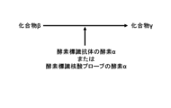
- FIG. 2 is a schematic diagram showing step A when the trace substance to be analyzed is an antigen.
- FIG. 2 is a schematic diagram showing step A when the trace substance to be analyzed is a nucleic acid.
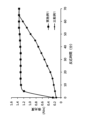
- FIG. FIG. 1 is a graph showing the change in absorbance over time in Example 1 and Comparative Example 1.
- NAD(P) means NAD and/or NADP
- NAD(P)H means NADH and/or NADPH.
- thioNAD(P) means thioNAD and/or thioNADP
- thioNAD(P)H means thioNADH and/or thioNADPH.
- polymerized thioNAD(P) means a polymer containing a unit derived from thioNAD(P) represented by the following general formula (1) (hereinafter referred to as a thioNAD(P) unit), and polymerized thioNAD(P)H means a polymer containing a unit derived from thioNAD(P)H represented by the following general formula (2) (hereinafter referred to as a thioNAD(P)H unit).
- R 1 is a hydroxy group or a phosphate group.
- general formula (1) represents a thioNAD unit.
- R 1 in general formula (1) is a phosphate group
- general formula (1) represents a thio-NADP unit.
- R 1 in general formula (2) is a hydroxy group
- general formula (2) represents a thio-NADH unit.
- R 1 in general formula (2) is a phosphate group
- general formula (2) represents a thio-NADPH unit.
- the method for analyzing a trace substance of this embodiment includes the steps of: Step A of binding an enzyme-labeled antibody consisting of an antibody specific to the trace substance and enzyme ⁇ , or an enzyme-labeled nucleic acid probe consisting of a nucleic acid probe specific to the trace substance and enzyme ⁇ , to the trace substance; Step B of allowing the enzyme ⁇ of the enzyme-labeled antibody or the enzyme ⁇ of the enzyme-labeled nucleic acid probe to act on compound ⁇ , which is a substrate for enzyme ⁇ , to generate compound ⁇ , which is a substrate for dehydrogenase; Step C of generating polymerized thioNAD(P)H by an enzyme cycling reaction using the generated compound ⁇ , dehydrogenase, NAD(P)H, and polymerized thioNAD(P); and Step D of detecting the trace substance using the generated polymerized thioNAD(P)H.
- Step A will be described separately for the case where the trace substance to be analyzed is an antigen and the case where the trace substance to be analyzed is a nucleic acid.
- an enzyme-labeled antibody consisting of an antibody that specifically binds to the antigen to be analyzed and an enzyme ⁇ is prepared, and the antibody of the enzyme-labeled antibody is allowed to bind to the antigen to be analyzed, as shown in Figure 1.
- an enzyme-labeled nucleic acid probe consisting of a nucleic acid probe that specifically binds to the nucleic acid to be analyzed and enzyme ⁇ is prepared, and the nucleic acid probe of the enzyme-labeled nucleic acid probe is allowed to bind to the nucleic acid to be analyzed, as shown in Figure 2.
- step B as shown in Figure 3, enzyme ⁇ of the enzyme-labeled antibody or enzyme ⁇ of the enzyme-labeled nucleic acid probe is allowed to act on compound ⁇ , which is the substrate of enzyme ⁇ , to produce compound ⁇ , which is the substrate of dehydrogenase.
- step C polymerized thioNAD(P)H is produced by an enzyme cycling reaction using the produced compound ⁇ , dehydrogenase, NAD(P)H, and polymerized thioNAD(P).
- reduced compound ⁇ is the starting point of the enzyme cycling reaction
- reduced compound ⁇ is oxidized to generate oxidized compound ⁇ .
- polymerized thioNAD(P)H is generated from polymerized thioNAD(P).
- oxidized compound ⁇ is reduced by dehydrogenase to generate reduced compound ⁇ .
- NAD(P) is generated from NAD(P)H.
- step D trace amounts of the target substance are detected using the polymerized thioNAD(P)H amplified in step C. Because the polymerized thioNAD(P)H is amplified in step C, highly sensitive analysis can be performed even if the target substance is in a trace amount.
- the time required for detecting thio-NAD(P)H produced in the enzyme cycling reaction can be shortened.
- the mechanism by which the time is shortened is not clear, it is presumed that the time shortening is possible due to the following mechanism.
- the detection of a trace substance by thio-NAD(P)H will be explained.
- the detection cannot be performed unless the concentration of thio-NAD(P)H reaches a certain level.
- thio-NAD(P)H is easily diffused on a plate or in a living tissue, it takes time for the concentration of thio-NAD(P)H to reach a certain level at the detection point.
- polymerized thio-NAD(P) is used. Therefore, it is considered that diffusion on a plate or in a biological tissue is suppressed, and the time required for the concentration of thio-NAD(P)H to reach a certain level or higher is shortened. This is considered to shorten the time required for the trace substances to be detected.
- the antibody of the enzyme-labeled antibody is bound to an antigen, which is a trace substance to be analyzed, as shown in Figure 1.
- the location where the analysis of a trace substance is carried out is not particularly limited, and for example, the analysis is carried out on a carrier carrying an antibody specific to an antigen.
- An example of an analytical method performed on a carrier carrying an antibody specific to an antigen is the enzyme-linked immunosorbent assay (ELISA).
- the ELISA method is a technique in which a sample solution is assayed on a carrier carrying an antibody, and the antigen in the sample solution is captured by the antibody, thereby detecting and quantifying the antigen.
- the carrier for carrying the antibody is not particularly limited, and for example, a microplate, a plastic tube, or magnetic beads can be used.
- the location where the analysis of the trace substance is carried out is not particularly limited, and for example, the analysis is carried out on a section of a biological tissue in which an antigen is present.
- An example of an analysis method performed on a biological tissue section in which an antigen exists is the immunohistochemical staining method (IHC method).
- the IHC method is a method for visualizing an antigen in situ on a biological tissue section.
- the IHC method has the advantage that it can analyze the biological tissue while retaining its anatomical and structural information, and can analyze in which compartment of the biological tissue the antigen is localized.
- the location where the analysis of the trace substance is carried out is not particularly limited, and for example, the analysis is carried out on a membrane where an antigen is present.
- An example of an analytical method performed on a membrane in the presence of an antigen is the Western blot method, in which proteins separated by electrophoresis are transferred onto a membrane and the proteins are detected and quantified using antibodies specific to the proteins to be analyzed.
- the type of antigen is not particularly limited, and may be any that can bind to an enzyme-labeled antibody, such as a protein, sugar, lipid, peptide, or low molecular weight compound that induces an immune response in the body, preferably a protein.
- proteins and sugars include proteins and sugars bound to lipids and/or nucleic acids.
- nucleic acid probe of the enzyme-labeled nucleic acid probe is bound to the nucleic acid, which is the trace substance to be analyzed.
- Examples of analytical methods in which an enzyme-labeled nucleic acid probe binds to a nucleic acid, which is a trace substance to be analyzed include the Southern blot method and the Northern blot method.
- the Southern blot method and the Northern blot method are techniques in which nucleic acids separated by electrophoresis are transferred to a membrane, and the nucleic acid to be analyzed is detected and quantified using a nucleic acid probe specific to the nucleic acid to be analyzed.
- the subject of analysis in the Southern blot method is DNA
- the subject of analysis in the Northern blot method is RNA.
- step D the polymerized thioNAD(P)H produced is detected.
- the detection method is not particularly limited, and for example, the polymerized thioNAD(P)H produced may be quantitatively analyzed.
- the method for qualitative analysis of polymerized thioNAD(P)H is not particularly limited, and for example, the generated polymerized thioNAD(P)H can be qualitatively analyzed visually. As mentioned above, polymerized thioNAD(P)H absorbs in the visible range, so visual qualitative analysis is possible.
- the structure of the polymerized thioNAD (P) is not particularly limited, but it is preferable that the polymerized thioNAD (P) contains a structure derived from N 6 -carboxymethyl-thioNAD represented by the following general formula (3), and it is more preferable that the polymerized thioNAD (P) contains a structure derived from thioNAD-N 6 -[N-(N-acryloyl-1-methoxycarbonyl-5-aminopentyl)-propioamide] represented by the following general formula (4).
- R 1 is a hydroxy group or a phosphate group.
- polymerized thioNAD(P) is not particularly limited, but it is preferable that polymerized thioNAD(P) contains a structure derived from acrylamide.
- the method for preparing polymerized thioNAD(P) is not particularly limited, but it can be prepared by referring to the methods described in Non-Patent Documents 1 and 2.
- the enzyme ⁇ of the enzyme-labeled antibody or enzyme-labeled nucleic acid probe is not particularly limited, but preferably contains one or more enzymes selected from the group consisting of the transferase group represented by EC number 2.-, the hydrolase group represented by EC number 3.-, the elimination-addition enzyme group represented by EC number 4.-, and the isomerase group represented by EC number 5.-, and more preferably contains alkaline phosphatase (ALP).
- ALP alkaline phosphatase
- compound ⁇ there are no particular limitations on compound ⁇ as long as it can be a substrate for enzyme ⁇ , but from the perspective of combination with dehydrogenase, which will be described later, it is preferable for it to contain a compound having an androsterone skeleton, and more preferably, it contains androsterone 3-phosphate (A3P).
- A3P androsterone 3-phosphate
- the dehydrogenase used in the enzyme cycling reaction of step C is not particularly limited, and may include, for example, one or more selected from group X consisting of malate dehydrogenase, lactate dehydrogenase, glucose dehydrogenase, galactose dehydrogenase, 3 ⁇ -hydroxysteroid dehydrogenase (abbreviated as 3 ⁇ -HSD), galactose dehydrogenase, glucose dehydrogenase, testosterone-17 ⁇ -dehydrogenase, and glycerol-3-phosphate dehydrogenase. From the viewpoint of improving the reaction rate, it is preferable to include hydroxysteroid dehydrogenase, and more preferably to include 3 ⁇ -HSD.
- group X consisting of malate dehydrogenase, lactate dehydrogenase, glucose dehydrogenase, galactose dehydrogenase, 3 ⁇ -hydroxysteroid dehydrogenase (abbreviated as 3 ⁇ -
- the combination of dehydrogenase and compound ⁇ is not particularly limited, but from the viewpoint of improving the reaction rate, a combination of 3 ⁇ -HSD and a compound having an androsterone skeleton as its substrate is preferred.
- the reaction rate when thioNAD is used as a coenzyme is within a few percent of that when NAD is used, whereas in the dehydrogenation reaction of androsterone by 3 ⁇ -HSD, the reaction rate with thioNAD is approximately 59% of the reaction rate with NAD, which has favorable properties as an enzyme for the cycling reaction in this embodiment.
- compounds having an androsterone skeleton include androsterone 3-phosphate (A3P), androsterone, 11 ⁇ -hydroxyandrosterone, 11-oxoandrosterone, 11 ⁇ -hydroxyandrosterone, etiocholanolone, tetrahydrocortisol, pregnanediol, lithocholic acid, deoxycholate, chenodeoxycholic acid, and cholic acid.
- A3P and 3 ⁇ -HSD is preferred from the viewpoint that it can be combined with ALP, which is a versatile enzyme for an enzyme-labeled antibody or enzyme-labeled nucleic acid probe.
- Compound ⁇ is not particularly limited as long as it can be a substrate for an enzyme cycling reaction using dehydrogenase. Therefore, it may be either a reduced substrate or an oxidized substrate.
- the properties of the enzyme ⁇ , compound ⁇ , and dehydrogenase are not particularly limited, but it is preferable that they have the following properties.
- the turnover rate of the labeling enzyme is high. Commercially available and widely used labeling enzymes can be used. The turnover number of the enzyme cycling is large.
- the dehydrogenase used in the enzyme cycling reaction is not contaminated with the labeled enzyme or does not have the same activity as the labeled enzyme.
- the substrate for the labeling enzyme is a commercially available compound or can be easily synthesized and is highly stable.
- the concentration of the enzyme-labeled antibody or enzyme-labeled nucleic acid probe in the reaction system is not particularly limited, but is, for example, 1 nM or more, preferably 10 nM or more, more preferably 20 nM or more, and is, for example, 100 nM or less, preferably 50 nM or less, more preferably 40 nM or less.
- the concentration of compound ⁇ in the reaction system is not particularly limited, but is, for example, 0.001 mM or more, preferably 0.01 mM or more, more preferably 0.05 mM or more, and even more preferably 0.1 mM or more, and is, for example, 500 mM or less, preferably 100 mM or less, more preferably 50 mM or less, even more preferably 10 mM or less, even more preferably 5 mM or less, even more preferably 1 mM or less, and even more preferably 0.5 mM or less.
- the concentration of NAD(P)H in the reaction system is not particularly limited, but is, for example, 0.001 mM or more, preferably 0.01 mM or more, more preferably 0.05 mM or more, even more preferably 0.1 mM or more, even more preferably 0.5 mM or more, even more preferably 1.0 mM or more, and is, for example, 500 mM or less, preferably 100 mM or less, more preferably 50 mM or less, even more preferably 10 mM or less, even more preferably 5 mM or less.
- the concentration of dehydrogenase in the reaction system is not particularly limited, but is, for example, 0.01 U/mL or more and 5000 U/mL or less, and from the viewpoint of improving the S/N ratio (signal/noise ratio), is preferably 0.1 U/mL or more and 500 U/mL or less, more preferably 1 U/mL or more and 100 U/mL or less, even more preferably 2 U/mL or more and 80 U/mL or less, even more preferably 5 U/mL or more and 60 U/mL or less, even more preferably 10 U/mL or more and 50 U/mL or less, and even more preferably 20 U/mL or more and 40 U/mL or less.
- the amount of thioNAD(P) units in the reaction system is not particularly limited, but from the viewpoint of improving the S/N ratio (signal/noise ratio), for 100 ⁇ L of a 30 U/mL dehydrogenase solution, i.e., 3 U of dehydrogenase, the amount is, for example, 1.00 ⁇ 10 3 mer to 1.00 ⁇ 10 5 mer, preferably 2.00 ⁇ 10 3 mer to 8.00 ⁇ 10 4 mer, more preferably 5.00 ⁇ 10 3 mer to 5.00 ⁇ 10 4 mer, even more preferably 7.00 ⁇ 10 3 mer to 3.00 ⁇ 10 4 mer, even more preferably 1.50 ⁇ 10 4 mer to 2.80 ⁇ 10 4 mer, and even more preferably 2.00 ⁇ 10 4 mer to 2.40 ⁇ 10 4 mer.
- the amount of thioNAD(P) units in the reaction system is represented by mer.
- Mer is a unit representing the number of thioNAD(P) units in the reaction system.
- the amount of thioNAD(P) units in the reaction system being 100mer means that the number of thioNAD(P) units in the reaction system is 100.
- the reaction temperature is not particularly limited, but is, for example, 0°C or higher, preferably 10°C or higher, more preferably 20°C or higher, and is, for example, 100°C or lower, preferably 80°C or lower, more preferably 60°C or lower, and even more preferably 40°C or lower.
- the number average molecular weight of the polymerized thioNAD(P) is not particularly limited, but from the viewpoint of further shortening the time required for detecting thioNAD(P)H produced in the enzyme cycling reaction, the number average molecular weight is preferably 1.0 ⁇ 10 4 or more and 1.0 ⁇ 10 9 or less, more preferably 5.0 ⁇ 10 4 or more and 5.0 ⁇ 10 8 or less, even more preferably 1.0 ⁇ 10 5 or more and 1.0 ⁇ 10 8 or less, even more preferably 6.5 ⁇ 10 5 or more and 7.0 ⁇ 10 7 or less, even more preferably 1.0 ⁇ 10 6 or more and 5.0 ⁇ 10 7 or less, even more preferably 5.0 ⁇ 10 6 or more and 3.0 ⁇ 10 7 or less, and even more preferably 1.0 ⁇ 10 7 or more and 2.0 ⁇ 10 7 or less.
- the number average molecular weight of the polymerized thioNAD(P) can be analyzed, for example, by gel permeation chromatography (GPC).
- the trace substance analysis kit of this embodiment includes an enzyme-labeled antibody consisting of an antibody specific to the trace substance to be measured and an enzyme ⁇ , or an enzyme-labeled nucleic acid probe consisting of a nucleic acid probe specific to the trace substance and an enzyme ⁇ , compound ⁇ which is a substrate for the enzyme ⁇ , a dehydrogenase, NAD(P)H, and polymerized thioNAD(P).
- the trace substance analysis kit of this embodiment includes an enzyme-labeled antibody.
- the trace substance to be analyzed is an antigen.
- the use of the trace substance analysis kit of this embodiment is not particularly limited, and it is used, for example, on a carrier carrying an antibody specific to an antigen. Specifically, it is used in the ELISA method, etc.
- the ELISA method is as described above.
- the application of the trace substance analysis kit of this embodiment is not particularly limited, and it is used, for example, on a biological tissue section in which an antigen is present. Specifically, it is used in the IHC method, etc.
- the IHC method is as described above.
- the application of the trace substance analysis kit of this embodiment is not particularly limited, and it is used, for example, on a membrane where an antigen is present. Specifically, it is used in the Western blotting method, etc.
- the Western blotting method is as described above.
- the type of antigen is not particularly limited, and may be any that can bind to an enzyme-labeled antibody, such as a protein, sugar, lipid, peptide, or low molecular weight compound that induces an immune response in the body, preferably a protein.
- proteins and sugars include proteins and sugars bound to lipids and/or nucleic acids.
- the trace substance analysis kit of this embodiment includes an enzyme-labeled nucleic acid probe.
- the trace substance to be analyzed is a nucleic acid.
- the use of the trace substance analysis kit is not particularly limited, and it is used, for example, in the Southern blot method and the Northern blot method. The Southern blot method and the Northern blot method are as described above.
- the structure of the polymerized thioNAD (P) is not particularly limited, but it is preferable that the polymerized thioNAD (P) contains a structure derived from N 6 -carboxymethyl-thioNAD represented by the above general formula (3), and it is more preferable that the polymerized thioNAD (P) contains a structure derived from thioNAD-N 6 -[N-(N-acryloyl-1-methoxycarbonyl-5-aminopentyl)-propioamide] represented by the above general formula (4).
- polymerized thioNAD(P) is not particularly limited, but it is preferable that polymerized thioNAD(P) contains a structure derived from acrylamide.
- the enzyme ⁇ of the enzyme-labeled antibody or enzyme-labeled nucleic acid probe is not particularly limited, but preferably contains one or more enzymes selected from the group consisting of the transferase group represented by EC number 2.-, the hydrolase group represented by EC number 3.-, the elimination-addition enzyme group represented by EC number 4.-, and the isomerase group represented by EC number 5.-, and more preferably contains alkaline phosphatase (ALP).
- ALP alkaline phosphatase
- compound ⁇ there are no particular limitations on compound ⁇ as long as it can be a substrate for enzyme ⁇ , but from the perspective of combination with the aforementioned dehydrogenase, it is preferable for it to contain a compound having an androsterone skeleton, and more preferably, it contains androsterone 3-phosphate (A3P).
- A3P androsterone 3-phosphate
- the dehydrogenase contained in the trace substance analysis kit of this embodiment is not particularly limited, and may, for example, include one or more types selected from the above-mentioned group X. From the viewpoint of improving the reaction rate as described above, it is preferable that it includes hydroxysteroid dehydrogenase, and more preferably that it includes 3 ⁇ -hydroxysteroid dehydrogenase (3 ⁇ -HSD).
- N 6 -carboxymethyl-thio NAD aqueous solution of iodoacetic acid (2.0 g) neutralized with 2 M LiOH, and the pH was adjusted to 6.5.
- the amount of thioNAD and compounds synthesized from thioNAD are expressed in mer.
- Mer is a unit representing the number of compounds synthesized from thioNAD and thioNAD.
- adding 1.8 ⁇ 10 15 mer of thioNAD to the aqueous solution of iodoacetic acid means that the number of thioNAD added to the aqueous solution of iodoacetic acid is 1.8 ⁇ 10 15 .
- the solution obtained in (1) above was diluted with the aqueous iodoacetic acid solution so that the amount of thio-NAD in the solution became 2.16 ⁇ 10 4 mer. To obtain 2.16 ⁇ 10 4 mer, the solution was diluted 1000-fold twice, 100-fold once, and 833-fold once. (3) The resulting solution was left to stand in the dark at room temperature for 10 days. The pH of the solution was appropriately checked and adjusted with 2 M LiOH.
- the polymerized thioNAD obtained by the above method was analyzed by gel permeation chromatography (GPC) and found to have a number average molecular weight of 1.5 ⁇ 10 7 .
- the GPC measurement conditions are as follows. Measurement device: 1260 Infinity II Bio-SEC system (Agilent) Column: PLgel 20 ⁇ m MIXED-A (size: 300 mm ⁇ 7.5 mm ID, Agilent) ⁇ 4 Column temperature: 25 ° C. Mobile phase: dimethylformamide Flow rate: 1.0 mL/min Detector: RI (Differential Refractive Index Detector) Standard substance: Polystyrene D-H2-10 kit (Agilent)
- Example 1 The following analysis subjects were analyzed using the following solution A by the following analytical method.
- GRP78 solution Glucose-Regulated Protein 78 kDa solution (hereinafter referred to as GRP78 solution) (BSA: 0.1%, Tween 20: 0.02% in PBS) (Solution A) 100 ⁇ L of solution A containing the following components and having a pH of 9.0 was obtained.
- Tris-HCl 100mM Polymerized thioNAD (total amount obtained in (30) above, amount of thioNAD unit: 2.16 ⁇ 10 4 mer) (Note that the amount of thioNAD unit was determined in Preliminary Experiment 2 below.) NADH (1.0mM) Androsterone 3-phosphate (A3P) (0.1mM) 3 ⁇ -Hydroxysteroid dehydrogenase (3 ⁇ -HSD) (30 U/mL) (The concentration of 3 ⁇ -HSD was determined in the preliminary experiment 1 described below.) (Analysis Method) A GRP78 solution was added to the microplate on which the anti-GRP78 antibody was immobilized, and the plate was incubated at 37° C. for 120 minutes.
- the solution in the wells was removed by suction, the inside of the wells was washed with saline, and 100 ⁇ L of an anti-GRP78 antibody solution labeled with alkaline phosphatase (ALP) (BSA: 0.1%, Tween 20: 0.02% in PBS) was added and incubated at 37° C. for 60 minutes.
- ALP alkaline phosphatase
- the solution in the wells was removed by suction, the inside of the wells was washed with physiological saline, and then the entire amount of solution A (100 ⁇ L) was added to the wells.
- Example 1 was 2.8, and the S/N ratio of Comparative Example 1 was 2.2. This shows that the analysis method of this embodiment can improve the S/N ratio.
- Solution B 100 ⁇ L of solution B containing the following components and having a pH of 9.0 was obtained.
- the entire amount of solution B (100 ⁇ L) was added to the wells, and while heating at 37° C., the absorbance was measured 5 minutes after the addition of solution B using a microplate reader (manufactured by Corona Electric Co., Ltd., model name: SH-1000, equipped with a 405 nm filter) to obtain a measured value A. If there was a significant difference between the measured value A and the blank value B, it was judged to be detectable, and the ALP concentration at the lowest ALP concentration among the detectable values was taken as the detection limit. The presence or absence of a significant difference was judged by the following method. First, the average value B AVE and standard deviation B SD of the blank value B were calculated.
- the reference value B X was calculated by the following formula (1).
- the reference value B X was compared with the measured value A, and if the values were different, it was judged that there was a significant difference.
- B X B AVE + B SD ⁇ 3 (1)
- the detection limit thus obtained was 2.24 ⁇ 10 ⁇ 20 mol/100 ⁇ L.
- the detection limit was obtained by the following method using the following solution C.
- Solution C 100 ⁇ L of TMB substrate solution (Thermodix) was prepared.
- method 100 ⁇ L of an HRP solution at pH 9.0 (HRP: 1.0 ⁇ 10 ⁇ 8 , 0.5 ⁇ 10 ⁇ 8 , 1.0 ⁇ 10 ⁇ 9 or 0.5 ⁇ 10 ⁇ 9 /100 ⁇ L, Tris-HCl: 100 mM) was added to the microplate.
- the entire amount of solution C (100 ⁇ L) was added to the wells, and while heating at 37° C., the absorbance was measured 10 minutes after the addition of solution C using a microplate reader (manufactured by Corona Electric Co., Ltd., model name: SH-1000, equipped with a 405 nm filter) to obtain a measurement value A. If there was a significant difference between the measured value A and the blank value B, it was judged to be detectable, and the HRP concentration at the lowest among the detectable values was taken as the detection limit. The presence or absence of a significant difference was judged by the method described above. The detection limit thus obtained was 4.31 ⁇ 10 ⁇ 12 mol/100 ⁇ L.
- the detection limit value of Reference Example 1 was approximately 1/108 of the detection limit value of Reference Comparative Example 1, which is an analysis method using HRP that is common in conventional IHC methods. From this, it is considered that the detection sensitivity can be improved by applying the analysis method according to this embodiment to the IHC method.
Landscapes
- Chemical & Material Sciences (AREA)
- Life Sciences & Earth Sciences (AREA)
- Health & Medical Sciences (AREA)
- Organic Chemistry (AREA)
- Engineering & Computer Science (AREA)
- Zoology (AREA)
- Proteomics, Peptides & Aminoacids (AREA)
- Immunology (AREA)
- Wood Science & Technology (AREA)
- Molecular Biology (AREA)
- General Health & Medical Sciences (AREA)
- Biotechnology (AREA)
- Microbiology (AREA)
- Analytical Chemistry (AREA)
- Physics & Mathematics (AREA)
- Biochemistry (AREA)
- Bioinformatics & Cheminformatics (AREA)
- General Engineering & Computer Science (AREA)
- Biophysics (AREA)
- Genetics & Genomics (AREA)
- Biomedical Technology (AREA)
- Hematology (AREA)
- Urology & Nephrology (AREA)
- Cell Biology (AREA)
- Food Science & Technology (AREA)
- Medicinal Chemistry (AREA)
- General Physics & Mathematics (AREA)
- Pathology (AREA)
- Measuring Or Testing Involving Enzymes Or Micro-Organisms (AREA)
Abstract
微量物質の分析方法であって、前記微量物質に、前記微量物質に特異的な抗体と酵素αとからなる酵素標識抗体、または、前記微量物質に特異的な核酸プローブと酵素αとからなる酵素標識核酸プローブを結合させる工程Aと、前記酵素標識抗体の前記酵素α、または、前記酵素標識核酸プローブの前記酵素αを、前記酵素αの基質である化合物βに作用させ、デヒドロゲナーゼの基質である化合物γを生成させる工程Bと、生成した前記化合物γと、デヒドロゲナーゼと、NAD(P)Hと、ポリマー化チオNAD(P)と、を用いて、酵素サイクリング反応により、ポリマー化チオNAD(P)Hを生成させる工程Cと、生成した前記ポリマー化チオNAD(P)Hにより、前記微量物質を検知する工程Dと、を含む、微量物質の分析方法。
Description
本発明は、微量物質の分析方法および微量物質分析用キットに関する。
タンパク質や核酸などの生体高分子を高感度に分析する方法として、酵素免疫測定法、ならびに、サザンブロット法、ノーザンブロット法およびウエスタンブロット法などの核酸プローブ測定法などが挙げられる。これらの分析方法においては、検出感度の向上が求められており、様々な検討がおこなわれてきた。
特許文献1では、酵素免疫測定法または核酸プローブ測定法と酵素サイクリング法を組み合わせ、シグナル物質としてのチオNAD(P)Hを幾何級数的に増幅することで、検出感度を向上させることが検討された。
特許文献1には、抗体酵素複合体を用いる酵素測定方法であって、抗体酵素複合体の酵素による反応生成物の定量は、NADHおよび/またはNADPH、チオNADおよび/またはチオNADP、並びにデヒドロゲナーゼ(DH)を用いて、酵素サイクリング反応によりチオNADHおよび/またはチオNADPHを生成させ、生成したチオNADHおよび/またはチオNADPHの量を測定するか、または生成したチオNADHおよび/またはチオNADPHによる色の変化を計測することで行う、前記方法が開示されている。
また、特許文献1には、酵素標識核酸プローブを用いる核酸プローブ測定方法であって、酵素標識核酸プローブの酵素による反応生成物の定量は、NADHおよび/またはNADPH、チオNADおよび/またはチオNADP、並びにデヒドロゲナーゼ(DH)を用いて、酵素サイクリング反応によりチオNADHおよび/またはチオNADPHを生成させ、生成したチオNADHおよび/またはチオNADPHの量を測定するか、または生成したチオNADHおよび/またはチオNADPHによる色の変化を計測することで行う、前記方法が開示されている。
また、特許文献1には、酵素標識核酸プローブを用いる核酸プローブ測定方法であって、酵素標識核酸プローブの酵素による反応生成物の定量は、NADHおよび/またはNADPH、チオNADおよび/またはチオNADP、並びにデヒドロゲナーゼ(DH)を用いて、酵素サイクリング反応によりチオNADHおよび/またはチオNADPHを生成させ、生成したチオNADHおよび/またはチオNADPHの量を測定するか、または生成したチオNADHおよび/またはチオNADPHによる色の変化を計測することで行う、前記方法が開示されている。
Lindberg M, Larsson PO, Mosbach K. A new immobilized NAD+ analogue, its application in affinity chromatography and as a functioning coenzyme. Eur J Biochem. 1973 Dec 3;40(1):187-93.
Muramatsu M, Urabe I, Yamada Y, Okada H. Synthesis and kinetic properties of a new NAD+ derivative carrying a vinyl group. Eur J Biochem. 1977 Oct 17;80(1):111-7.
特許文献1に記載された測定方法においては、酵素サイクリング反応で生成したチオNAD(P)Hが検知されるまでに要する時間の短縮が求められている。
本発明は、酵素サイクリング反応で生成したチオNAD(P)Hが検知されるまでに要する時間を短縮することが可能な分析方法を提供する。
すなわち、本発明によれば、以下に示す微量物質の分析方法および微量物質分析用キットが提供される。
1. 微量物質の分析方法であって、
前記微量物質に、前記微量物質に特異的な抗体と酵素αとからなる酵素標識抗体、または、前記微量物質に特異的な核酸プローブと酵素αとからなる酵素標識核酸プローブを結合させる工程Aと、
前記酵素標識抗体の前記酵素α、または、前記酵素標識核酸プローブの前記酵素αを、前記酵素αの基質である化合物βに作用させ、デヒドロゲナーゼの基質である化合物γを生成させる工程Bと、
生成した前記化合物γと、デヒドロゲナーゼと、NAD(P)Hと、ポリマー化チオNAD(P)と、を用いて、酵素サイクリング反応により、ポリマー化チオNAD(P)Hを生成させる工程Cと、
生成した前記ポリマー化チオNAD(P)Hにより、前記微量物質を検知する工程Dと、
を含む、微量物質の分析方法。
2. 前記微量物質が抗原であり、
前記工程Aにおいて、前記抗原に、前記酵素標識抗体を結合させる、1.に記載の微量物質の分析方法。
3. 前記抗原に特異的な抗体を担持させた担体上でおこなわれる、2.に記載の微量物質の分析方法。
4. 前記抗原が存在する生体組織切片上でおこなわれる、2.に記載の微量物質の分析方法。
5. 前記抗原が存在する膜上でおこなわれる、2.に記載の微量物質の分析方法。
6. 前記抗原がタンパク質抗原である、2.~5.のいずれかに記載の微量物質の分析方法。
7. 前記微量物質が核酸であり、
前記工程Aにおいて、前記核酸に、前記酵素標識核酸プローブを結合させる、1.に記載の微量物質の分析方法。
8. 前記工程Dにおいて、生成した前記ポリマー化チオNAD(P)Hを定量分析する、1.~7.のいずれかに記載の微量物質の分析方法。
9. 前記工程Dにおいて、生成した前記ポリマー化チオNAD(P)Hを吸光度により定量分析する、8.に記載の微量物質の分析方法。
10. 前記工程Dにおいて、生成した前記ポリマー化チオNAD(P)Hを定性分析する、1.~7.のいずれかに記載の微量物質の分析方法。
11. 前記工程Dにおいて、生成した前記ポリマー化チオNAD(P)Hを目視により定性分析する、10.に記載の微量物質の分析方法。
12. 前記ポリマー化チオNAD(P)が、N6-カルボキシメチル-チオNAD由来の構造を含む、1.~11.のいずれかに記載の微量物質の分析方法。
13. 前記ポリマー化チオNAD(P)が、アクリルアミド由来の構造を含む、1.~12.のいずれかに記載の微量物質の分析方法。
14. 前記酵素αが、EC番号2.-で表される転移酵素群、EC番号3.-で表される加水分解酵素群、EC番号4.-で表される除去付加酵素群およびEC番号5.-で表される異性化酵素群からなる群から選択される一種又は二種以上を含む、1.~13.のいずれかに記載の微量物質の分析方法。
15. 前記酵素αがアルカリホスファターゼを含む、14.に記載の微量物質の分析方法。
16. 前記化合物βがアンドロステロン骨格を有する化合物を含む、1.~15.のいずれかに記載の微量物質の分析方法。
17. 前記デヒドロゲナーゼがヒドロキシステロイドデヒドロゲナーゼを含む、1.~16.のいずれかに記載の微量物質の分析方法。
18. 前記化合物γがアンドロステロン骨格を有する化合物を含む、1.~17.のいずれかに記載の微量物質の分析方法。
19. 前記ポリマー化チオNAD(P)の数平均分子量が1.0×10⁴以上1.0×10⁹以下である、1.~18.のいずれかに記載の微量物質の分析方法。
20. 測定対象の微量物質に特異的な抗体と酵素αとからなる酵素標識抗体、または、前記微量物質に特異的な核酸プローブと酵素αとからなる酵素標識核酸プローブと、
前記酵素αの基質である化合物βと、
デヒドロゲナーゼと、
NAD(P)Hと、
ポリマー化チオNAD(P)と、
を含む微量物質分析用キット。
21. 前記微量物質が抗原であり、
前記酵素標識抗体を含む、20.に記載の微量物質分析用キット。
22. 前記抗原に特異的な抗体を担持させた担体上で用いられる、21.に記載の微量物質分析用キット。
23. 前記抗原が存在する生体組織切片上で用いられる、21.に記載の微量物質分析用キット。
24. 前記抗原が存在する膜上で用いられる、21.に記載の微量物質分析用キット。
25. 前記抗原がタンパク質抗原である、21.~24.のいずれかに記載の微量物質分析用キット。
26. 前記微量物質が核酸であり、
前記酵素標識核酸プローブを含む、20.に記載の微量物質分析用キット。
27. 前記ポリマー化チオNAD(P)が、N6-カルボキシメチル-チオNAD由来の構造を含む、20.~26.のいずれかに記載の微量物質分析用キット。
28. 前記ポリマー化チオNAD(P)が、アクリルアミド由来の構造を含む、20.~27.のいずれかに記載の微量物質分析用キット。
29. 前記酵素αが、EC番号2.-で表される転移酵素群、EC番号3.-で表される加水分解酵素群、EC番号4.-で表される除去付加酵素群およびEC番号5.-で表される異性化酵素群からなる群から選択される一種又は二種以上を含む、20.~28.のいずれかに記載の微量物質分析用キット。
30. 前記酵素αがアルカリホスファターゼを含む、29.に記載の微量物質分析用キット。
31. 前記化合物βがアンドロステロン骨格を有する化合物を含む、20.~30.のいずれかに記載の微量物質分析用キット。
32. 前記デヒドロゲナーゼがヒドロキシステロイドデヒドロゲナーゼを含む、20.~31.のいずれかに記載の微量物質分析用キット。
33. 前記ポリマー化チオNAD(P)の数平均分子量が1.0×10⁴以上1.0×10⁹以下である、20.~32.のいずれかに記載の微量物質分析用キット。
1. 微量物質の分析方法であって、
前記微量物質に、前記微量物質に特異的な抗体と酵素αとからなる酵素標識抗体、または、前記微量物質に特異的な核酸プローブと酵素αとからなる酵素標識核酸プローブを結合させる工程Aと、
前記酵素標識抗体の前記酵素α、または、前記酵素標識核酸プローブの前記酵素αを、前記酵素αの基質である化合物βに作用させ、デヒドロゲナーゼの基質である化合物γを生成させる工程Bと、
生成した前記化合物γと、デヒドロゲナーゼと、NAD(P)Hと、ポリマー化チオNAD(P)と、を用いて、酵素サイクリング反応により、ポリマー化チオNAD(P)Hを生成させる工程Cと、
生成した前記ポリマー化チオNAD(P)Hにより、前記微量物質を検知する工程Dと、
を含む、微量物質の分析方法。
2. 前記微量物質が抗原であり、
前記工程Aにおいて、前記抗原に、前記酵素標識抗体を結合させる、1.に記載の微量物質の分析方法。
3. 前記抗原に特異的な抗体を担持させた担体上でおこなわれる、2.に記載の微量物質の分析方法。
4. 前記抗原が存在する生体組織切片上でおこなわれる、2.に記載の微量物質の分析方法。
5. 前記抗原が存在する膜上でおこなわれる、2.に記載の微量物質の分析方法。
6. 前記抗原がタンパク質抗原である、2.~5.のいずれかに記載の微量物質の分析方法。
7. 前記微量物質が核酸であり、
前記工程Aにおいて、前記核酸に、前記酵素標識核酸プローブを結合させる、1.に記載の微量物質の分析方法。
8. 前記工程Dにおいて、生成した前記ポリマー化チオNAD(P)Hを定量分析する、1.~7.のいずれかに記載の微量物質の分析方法。
9. 前記工程Dにおいて、生成した前記ポリマー化チオNAD(P)Hを吸光度により定量分析する、8.に記載の微量物質の分析方法。
10. 前記工程Dにおいて、生成した前記ポリマー化チオNAD(P)Hを定性分析する、1.~7.のいずれかに記載の微量物質の分析方法。
11. 前記工程Dにおいて、生成した前記ポリマー化チオNAD(P)Hを目視により定性分析する、10.に記載の微量物質の分析方法。
12. 前記ポリマー化チオNAD(P)が、N6-カルボキシメチル-チオNAD由来の構造を含む、1.~11.のいずれかに記載の微量物質の分析方法。
13. 前記ポリマー化チオNAD(P)が、アクリルアミド由来の構造を含む、1.~12.のいずれかに記載の微量物質の分析方法。
14. 前記酵素αが、EC番号2.-で表される転移酵素群、EC番号3.-で表される加水分解酵素群、EC番号4.-で表される除去付加酵素群およびEC番号5.-で表される異性化酵素群からなる群から選択される一種又は二種以上を含む、1.~13.のいずれかに記載の微量物質の分析方法。
15. 前記酵素αがアルカリホスファターゼを含む、14.に記載の微量物質の分析方法。
16. 前記化合物βがアンドロステロン骨格を有する化合物を含む、1.~15.のいずれかに記載の微量物質の分析方法。
17. 前記デヒドロゲナーゼがヒドロキシステロイドデヒドロゲナーゼを含む、1.~16.のいずれかに記載の微量物質の分析方法。
18. 前記化合物γがアンドロステロン骨格を有する化合物を含む、1.~17.のいずれかに記載の微量物質の分析方法。
19. 前記ポリマー化チオNAD(P)の数平均分子量が1.0×10⁴以上1.0×10⁹以下である、1.~18.のいずれかに記載の微量物質の分析方法。
20. 測定対象の微量物質に特異的な抗体と酵素αとからなる酵素標識抗体、または、前記微量物質に特異的な核酸プローブと酵素αとからなる酵素標識核酸プローブと、
前記酵素αの基質である化合物βと、
デヒドロゲナーゼと、
NAD(P)Hと、
ポリマー化チオNAD(P)と、
を含む微量物質分析用キット。
21. 前記微量物質が抗原であり、
前記酵素標識抗体を含む、20.に記載の微量物質分析用キット。
22. 前記抗原に特異的な抗体を担持させた担体上で用いられる、21.に記載の微量物質分析用キット。
23. 前記抗原が存在する生体組織切片上で用いられる、21.に記載の微量物質分析用キット。
24. 前記抗原が存在する膜上で用いられる、21.に記載の微量物質分析用キット。
25. 前記抗原がタンパク質抗原である、21.~24.のいずれかに記載の微量物質分析用キット。
26. 前記微量物質が核酸であり、
前記酵素標識核酸プローブを含む、20.に記載の微量物質分析用キット。
27. 前記ポリマー化チオNAD(P)が、N6-カルボキシメチル-チオNAD由来の構造を含む、20.~26.のいずれかに記載の微量物質分析用キット。
28. 前記ポリマー化チオNAD(P)が、アクリルアミド由来の構造を含む、20.~27.のいずれかに記載の微量物質分析用キット。
29. 前記酵素αが、EC番号2.-で表される転移酵素群、EC番号3.-で表される加水分解酵素群、EC番号4.-で表される除去付加酵素群およびEC番号5.-で表される異性化酵素群からなる群から選択される一種又は二種以上を含む、20.~28.のいずれかに記載の微量物質分析用キット。
30. 前記酵素αがアルカリホスファターゼを含む、29.に記載の微量物質分析用キット。
31. 前記化合物βがアンドロステロン骨格を有する化合物を含む、20.~30.のいずれかに記載の微量物質分析用キット。
32. 前記デヒドロゲナーゼがヒドロキシステロイドデヒドロゲナーゼを含む、20.~31.のいずれかに記載の微量物質分析用キット。
33. 前記ポリマー化チオNAD(P)の数平均分子量が1.0×10⁴以上1.0×10⁹以下である、20.~32.のいずれかに記載の微量物質分析用キット。
本発明によれば、酵素サイクリング反応で生成したチオNAD(P)Hが検知されるまでに要する時間を短縮することが可能な分析方法を提供することができる。
以下、本発明を実施形態に基づいて説明する。
本実施形態において、数値範囲が段階的に記載されている場合、各数値範囲の上限及び下限は任意に組み合わせることができる。
本実施形態において、NAD(P)はNADおよび/またはNADPを意味し、NAD(P)HはNADHおよび/またはNADPHを意味する。
本実施形態において、チオNAD(P)はチオNADおよび/またはチオNADPを意味し、チオNAD(P)HはチオNADHおよび/またはチオNADPHを意味する。
本実施形態において、ポリマー化チオNAD(P)は、下記の一般式(1)で表されるチオNAD(P)由来のユニット(以下、チオNAD(P)ユニットと呼ぶ)を含むポリマーを意味し、ポリマー化チオNAD(P)Hは、下記の一般式(2)で表されるチオNAD(P)H由来のユニット(以下、チオNAD(P)Hユニットと呼ぶ)を含むポリマーを意味する。

一般式(1)および(2)において、R1はヒドロキシ基またはリン酸基である。
一般式(1)においてR1がヒドロキシ基である場合、一般式(1)はチオNADユニットを表す。
一般式(1)においてR1がリン酸基である場合、一般式(1)はチオNADPユニットを表す。
一般式(2)においてR1がヒドロキシ基である場合、一般式(2)はチオNADHユニットを表す。
一般式(2)においてR1がリン酸基である場合、一般式(2)はチオNADPHユニットを表す。
一般式(1)においてR1がヒドロキシ基である場合、一般式(1)はチオNADユニットを表す。
一般式(1)においてR1がリン酸基である場合、一般式(1)はチオNADPユニットを表す。
一般式(2)においてR1がヒドロキシ基である場合、一般式(2)はチオNADHユニットを表す。
一般式(2)においてR1がリン酸基である場合、一般式(2)はチオNADPHユニットを表す。
[微量物質の分析方法]
以下、本実施形態の微量物質の分析方法について説明する。
以下、本実施形態の微量物質の分析方法について説明する。
本実施形態の微量物質の分析方法は、前記微量物質に、前記微量物質に特異的な抗体と酵素αとからなる酵素標識抗体、または、前記微量物質に特異的な核酸プローブと酵素αとからなる酵素標識核酸プローブを結合させる工程Aと、前記酵素標識抗体の前記酵素α、または、前記酵素標識核酸プローブの前記酵素αを、前記酵素αの基質である化合物βに作用させ、デヒドロゲナーゼの基質である化合物γを生成させる工程Bと、生成した前記化合物γと、デヒドロゲナーゼと、NAD(P)Hと、ポリマー化チオNAD(P)と、を用いて、酵素サイクリング反応により、ポリマー化チオNAD(P)Hを生成させる工程Cと、生成した前記ポリマー化チオNAD(P)Hにより、前記微量物質を検知する工程Dと、を含む。
まず、本実施形態の微量物質の分析方法の各工程について説明する。
工程Aについて、分析対象の微量物質が抗原である場合と分析対象の微量物質が核酸である場合に分けて説明する。
分析対象の微量物質が抗原である場合、分析対象である抗原に特異的に結合する抗体と酵素αからなる酵素標識抗体を用意し、図1に示すように、当該酵素標識抗体の抗体を分析対象である抗原に結合させる。
分析対象の微量物質が核酸である場合、分析対象である核酸に特異的に結合する核酸プローブと酵素αからなる酵素標識核酸プローブを用意し、図2に示すように、当該酵素標識核酸プローブの核酸プローブを分析対象である核酸に結合させる。
分析対象の微量物質が抗原である場合、分析対象である抗原に特異的に結合する抗体と酵素αからなる酵素標識抗体を用意し、図1に示すように、当該酵素標識抗体の抗体を分析対象である抗原に結合させる。
分析対象の微量物質が核酸である場合、分析対象である核酸に特異的に結合する核酸プローブと酵素αからなる酵素標識核酸プローブを用意し、図2に示すように、当該酵素標識核酸プローブの核酸プローブを分析対象である核酸に結合させる。
工程Bでは、図3に示すように、酵素標識抗体の酵素α、または、酵素標識核酸プローブの酵素αを酵素αの基質である化合物βに作用させ、デヒドロゲナーゼの基質である化合物γを生成させる。
工程Cでは、図4に示すように、生成した化合物γと、デヒドロゲナーゼと、NAD(P)Hと、ポリマー化チオNAD(P)と、を用いて、酵素サイクリング反応により、ポリマー化チオNAD(P)Hを生成させる。
具体的には、還元型の化合物γが酵素サイクリング反応の起点となる場合、まず、還元型の化合物γが酸化され酸化型の化合物γが生成する。この際、ポリマー化チオNAD(P)からポリマー化チオNAD(P)Hが生成される。そして、酸化型の化合物γがデヒドロゲナーゼにより還元され還元型の化合物γが生成する。この際、NAD(P)HからNAD(P)が生成される。これで酵素サイクリングの1サイクルが完結する。ポリマー化チオNAD(P)とNAD(P)Hが供給される限り、このサイクルが繰り返され、ポリマー化チオNAD(P)Hが増幅される。
具体的には、還元型の化合物γが酵素サイクリング反応の起点となる場合、まず、還元型の化合物γが酸化され酸化型の化合物γが生成する。この際、ポリマー化チオNAD(P)からポリマー化チオNAD(P)Hが生成される。そして、酸化型の化合物γがデヒドロゲナーゼにより還元され還元型の化合物γが生成する。この際、NAD(P)HからNAD(P)が生成される。これで酵素サイクリングの1サイクルが完結する。ポリマー化チオNAD(P)とNAD(P)Hが供給される限り、このサイクルが繰り返され、ポリマー化チオNAD(P)Hが増幅される。
工程Dでは、工程Cで増幅されたポリマー化チオNAD(P)Hにより分析対象の微量物質を検知する。工程Cでポリマー化チオNAD(P)Hが増幅されるため、分析対象が微量であっても高感度の分析をおこなうことができる。
本実施形態の微量物質の分析方法によると、酵素サイクリング反応で生成したチオNAD(P)Hが検知されるまでに要する時間を短縮できる。時間短縮のメカニズムは明らかではないが、以下のメカニズムにより時間短縮が可能になると推測される。
まず、チオNAD(P)Hによる微量物質の検知について説明すると、チオNAD(P)Hにより微量物質を検知する場合、チオNAD(P)Hが一定以上の濃度にならないと検知をおこなうことができない。そのため、チオNAD(P)Hがプレート上や生体組織上で拡散しやすいと、検知をおこなう地点においてチオNAD(P)Hの濃度が一定以上になるまでに時間がかかってしまう。
本実施形態の微量物質の分析方法においては、ポリマー化したチオNAD(P)を用いる。そのため、プレート上や生体組織上における拡散が抑制されると考えられ、チオNAD(P)Hの濃度が一定以上になるまでに要する時間が短縮されると考えられる。これにより、微量物質が検知されるまでに要する時間が短縮されると考えられる。
まず、チオNAD(P)Hによる微量物質の検知について説明すると、チオNAD(P)Hにより微量物質を検知する場合、チオNAD(P)Hが一定以上の濃度にならないと検知をおこなうことができない。そのため、チオNAD(P)Hがプレート上や生体組織上で拡散しやすいと、検知をおこなう地点においてチオNAD(P)Hの濃度が一定以上になるまでに時間がかかってしまう。
本実施形態の微量物質の分析方法においては、ポリマー化したチオNAD(P)を用いる。そのため、プレート上や生体組織上における拡散が抑制されると考えられ、チオNAD(P)Hの濃度が一定以上になるまでに要する時間が短縮されると考えられる。これにより、微量物質が検知されるまでに要する時間が短縮されると考えられる。
以下、本実施形態の微量物質の分析方法のうち、酵素標識抗体を用いる場合について説明する。
酵素標識抗体を用いる場合、図1に示すように、酵素標識抗体の抗体を分析対象の微量物質である抗原に結合させる。
微量物質の分析をおこなう場所は特に限定されず、例えば、抗原に特異的な抗体を担持させた担体上でおこなわれる。
抗原に特異的な抗体を担持させた担体上でおこなわれる分析方法としては、例えば、酵素免疫測定法(ELISA法)を挙げることができる。ELISA法は、抗体を担持させた担体に試料溶液をアッセイし、試料溶液中の抗原を抗体で捕捉することにより、抗原の検出および定量をおこなう手法である。
抗体を担持させる担体は特に限定されず、例えば、マイクロプレート、プラスチックチューブまたは磁気ビーズ等を用いることができる。
抗原に特異的な抗体を担持させた担体上でおこなわれる分析方法としては、例えば、酵素免疫測定法(ELISA法)を挙げることができる。ELISA法は、抗体を担持させた担体に試料溶液をアッセイし、試料溶液中の抗原を抗体で捕捉することにより、抗原の検出および定量をおこなう手法である。
抗体を担持させる担体は特に限定されず、例えば、マイクロプレート、プラスチックチューブまたは磁気ビーズ等を用いることができる。
微量物質の分析をおこなう場所は特に限定されず、例えば、抗原が存在する生体組織切片上でおこなわれる。
抗原が存在する生体組織切片上でおこなわれる分析方法としては、例えば、免疫組織化学染色法(IHC法)を挙げることができる。IHC法は、生体切片上で生体内原位置(in situ)の抗原を視覚化する手法である。IHC法には、生体組織の解剖学的、構造的情報を保持したまま解析できるという特長があり、生体組織のどの区画に抗原が局在するかを解析できる。
抗原が存在する生体組織切片上でおこなわれる分析方法としては、例えば、免疫組織化学染色法(IHC法)を挙げることができる。IHC法は、生体切片上で生体内原位置(in situ)の抗原を視覚化する手法である。IHC法には、生体組織の解剖学的、構造的情報を保持したまま解析できるという特長があり、生体組織のどの区画に抗原が局在するかを解析できる。
微量物質の分析をおこなう場所は特に限定されず、例えば、抗原が存在する膜上でおこなわれる。
抗原が存在する膜上でおこなわれる分析方法としては、例えば、ウエスタンブロット法を挙げることができる。ウエスタンブロット法は、電気泳動によって分離したタンパク質を膜に転写し、分析対象のタンパク質に特異的な抗体によりそのタンパク質の検出および定量をおこなう手法である。
抗原が存在する膜上でおこなわれる分析方法としては、例えば、ウエスタンブロット法を挙げることができる。ウエスタンブロット法は、電気泳動によって分離したタンパク質を膜に転写し、分析対象のタンパク質に特異的な抗体によりそのタンパク質の検出および定量をおこなう手法である。
抗原の種類は特に限定されず、酵素標識抗体と結合できるものであればよく、例えば、タンパク質、糖、脂質、ペプチドまたは生体に免疫応答を引き起こす低分子化合物であり、好ましくはタンパク質である。ここで、タンパク質および糖には、脂質および/または核酸が結合した、タンパク質および糖も含まれる。
以下、本実施形態の微量物質の分析方法のうち、酵素標識核酸プローブを用いる場合について説明する。
酵素標識核酸プローブを用いる場合、図2に示すように、酵素標識核酸プローブの核酸プローブを分析対象の微量物質である核酸に結合させる。
分析対象の微量物質である核酸に酵素標識核酸プローブの核酸プローブが結合する分析方法としては、例えば、サザンブロット法およびノーザンブロット法を挙げることができる。サザンブロット法およびノーザンブロット法は、電気泳動によって分離した核酸を膜に転写し、分析対象の核酸に特異的な核酸プローブによりその核酸の検出および定量をおこなう手法である。サザンブロット法の分析対象はDNAであり、ノーザンブロット法の分析対象はRNAである。
工程Dでは生成したポリマー化チオNAD(P)Hを検知する。検知方法は特に限定されず、例えば、生成したポリマー化チオNAD(P)Hを定量分析してもよい。
ポリマー化チオNAD(P)Hの定量分析の方法は特に限定されず、例えば、吸光度により定量分析することができる。なお、ポリマー化チオNAD(P)Hは可視域に吸収を示すため(極大吸収波長:400nm、モル吸光係数=11,900)、例えば、普及型の吸光光度計や比色測定用マイクロプレートリーダーを用いて測定できる。
工程Dでは生成したポリマー化チオNAD(P)Hを検知する。検知方法は特に限定されず、例えば、生成したポリマー化チオNAD(P)Hを定性分析してもよい。
ポリマー化チオNAD(P)Hの定性分析の方法は特に限定されず、例えば、生成したポリマー化チオNAD(P)Hを目視により定性分析することができる。前述の通り、ポリマー化チオNAD(P)Hは可視域に吸収を示すため、目視による定性分析が可能である。
ポリマー化チオNAD(P)の構造は特に限定されないが、ポリマー化チオNAD(P)が下記の一般式(3)で表されるN6-カルボキシメチル-チオNAD由来の構造を含むことが好ましく、下記の一般式(4)で表されるチオNAD-N6-[N-(N-アクリロイル-1-メトシキカルボニル-5-アミノペンチル)-プロピオアミド]由来の構造を含むことがより好ましい。
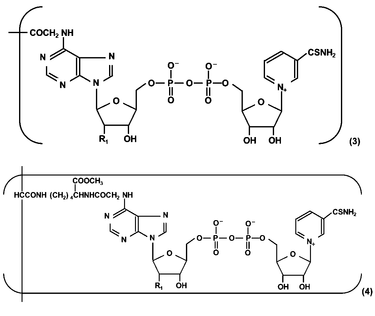
一般式(3)および(4)において、R1はヒドロキシ基またはリン酸基である。
ポリマー化チオNAD(P)の構造は特に限定されないが、ポリマー化チオNAD(P)がアクリルアミド由来の構造を含むことが好ましい。
ポリマー化チオNAD(P)の調製方法は特に限定されないが、非特許文献1および2に記載の方法を参考に調製することができる。
酵素標識抗体または酵素標識核酸プローブの酵素αは特に限定されないが、EC番号2.-で表される転移酵素群、EC番号3.-で表される加水分解酵素群、EC番号4.-で表される除去付加酵素群およびEC番号5.-で表される異性化酵素群からなる群から選択される一種又は二種以上を含むことが好ましく、アルカリホスファターゼ(ALP)を含むことがより好ましい。
化合物βは酵素αの基質となり得るものであれば特に限定されないが、後述するデヒドロゲナーゼとの組み合わせの観点から、アンドロステロン骨格を有する化合物を含むことが好ましく、Androsterone 3-phosphate(A3P)を含むことがより好ましい。
工程Cの酵素サイクリング反応で用いるデヒドロゲナーゼは特に限定されず、例えば、マレートデヒドロゲナーゼ、ラクテートデヒドロゲナーゼ、グルコースデヒドロゲナーゼ、ガラクトースデヒドロゲナーゼ、3α-ヒドロキシステロイドデヒドロゲナーゼ(3α-Hydroxysteroid dehydrogenase、略称:3α-HSD)、ガラクトースデヒドロゲナーゼ、グルコースデヒドロゲナーゼ、テストステロン-17β-デヒドロゲナーゼおよびグリセロール-3-ホスフェートデヒドロゲナーゼからなる群Xから選択される一種または二種以上を含み、反応速度向上の観点から、ヒドロキシステロイドデヒドロゲナーゼを含むことが好ましく、3α-HSDを含むことがより好ましい。
デヒドロゲナーゼと化合物βの組み合わせは特に限定されないが、反応速度向上の観点から、3α-HSDとその基質となるアンドロステロン骨格を有する化合物の組み合わせが好ましい。
反応速度向上について具体例を挙げて説明すると、多くの脱水素酵素では、補酵素としてチオNADを用いた場合の反応速度がNADを用いた場合の数%以内であるのに対し、3α-HSDによるアンドロステロンの脱水素反応では、チオNADでの反応速度がNADでの反応速度の約59%もあり、本実施形態に係るサイクリング反応の酵素として好ましい性質を有している。
アンドロステロン骨格を有する化合物の具体例としては、例えば、Androsterone 3-phosphate(A3P)、アンドロステロン、11β-ヒドロキシアンドロステロン、11-オキソアンドロステロン、11α-ヒドロキシアンドロステロン、エチオコラノロン、テトラハイドロコルチゾール、プレグナンジオール、リトコール酸、デオキシコレート、ケノデオキシコール酸およびコール酸などが挙げられる。
3α-HSDとアンドロステロン骨格を有する化合物の組み合わせの中でも、酵素標識抗体または酵素標識核酸プローブの酵素として汎用的なALPとの組み合わせが可能であるという観点から、A3Pと3α-HSDの組み合わせが好ましい。
反応速度向上について具体例を挙げて説明すると、多くの脱水素酵素では、補酵素としてチオNADを用いた場合の反応速度がNADを用いた場合の数%以内であるのに対し、3α-HSDによるアンドロステロンの脱水素反応では、チオNADでの反応速度がNADでの反応速度の約59%もあり、本実施形態に係るサイクリング反応の酵素として好ましい性質を有している。
アンドロステロン骨格を有する化合物の具体例としては、例えば、Androsterone 3-phosphate(A3P)、アンドロステロン、11β-ヒドロキシアンドロステロン、11-オキソアンドロステロン、11α-ヒドロキシアンドロステロン、エチオコラノロン、テトラハイドロコルチゾール、プレグナンジオール、リトコール酸、デオキシコレート、ケノデオキシコール酸およびコール酸などが挙げられる。
3α-HSDとアンドロステロン骨格を有する化合物の組み合わせの中でも、酵素標識抗体または酵素標識核酸プローブの酵素として汎用的なALPとの組み合わせが可能であるという観点から、A3Pと3α-HSDの組み合わせが好ましい。
化合物γはデヒドロゲナーゼを用いた酵素サイクリング反応の基質となり得るものであれば特に限定されない。そのため、還元型基質でも酸化型基質でもよい。
本実施形態の微量物質の分析方法において、酵素α、化合物β、デヒドロゲナーゼの性質は特に限定されないが、以下の性質を持つことが好ましい。
標識酵素のターンオーバー数が大きい。
市販および汎用されている標識酵素が使用できる。
酵素サイクリングのターンオーバー数が大きい。
酵素サイクリング反応に使用する脱水素酵素に標識酵素のコンタミや標識酵素と同様の活性がない。
標識酵素の基質が市販の化合物であるか、または容易に合成ができ、かつ安定性が高い。
標識酵素のターンオーバー数が大きい。
市販および汎用されている標識酵素が使用できる。
酵素サイクリングのターンオーバー数が大きい。
酵素サイクリング反応に使用する脱水素酵素に標識酵素のコンタミや標識酵素と同様の活性がない。
標識酵素の基質が市販の化合物であるか、または容易に合成ができ、かつ安定性が高い。
本実施形態の微量物質の分析方法において、反応系中における酵素標識抗体または酵素標識核酸プローブの濃度は特に限定されないが、例えば1nM以上、好ましくは10nM以上、より好ましくは20nM以上であり、そして、例えば100nM以下、好ましくは50nM以下、より好ましくは40nM以下である。
本実施形態の微量物質の分析方法において、反応系中における化合物βの濃度は特に限定されないが、例えば0.001mM以上、好ましくは0.01mM以上、より好ましくは0.05mM以上、さらに好ましくは0.1mM以上であり、そして、例えば500mM以下、好ましくは100mM以下、より好ましくは50mM以下、さらに好ましくは10mM以下、さらに好ましくは5mM以下、さらに好ましくは1mM以下、さらに好ましくは0.5mM以下である。
本実施形態の微量物質の分析方法において、反応系中におけるNAD(P)Hの濃度は特に限定されないが、例えば0.001mM以上、好ましくは0.01mM以上、より好ましくは0.05mM以上、さらに好ましくは0.1mM以上、さらに好ましくは0.5mM以上、さらに好ましくは1.0mM以上であり、そして、例えば500mM以下、好ましくは100mM以下、より好ましくは50mM以下、さらに好ましくは10mM以下、さらに好ましくは5mM以下である。
本実施形態の微量物質の分析方法において、反応系中におけるデヒドロゲナーゼの濃度は特に限定されないが、例えば0.01U/mL以上5000U/mL以下であり、S/N比(シグナル/ノイズ比)向上の観点から、好ましくは0.1U/mL以上500U/mL以下、より好ましくは1U/mL以上100U/mL以下、さらに好ましくは2U/mL以上80U/mL以下、さらに好ましくは5U/mL以上60U/mL以下、さらに好ましくは10U/mL以上50U/mL以下、さらに好ましくは20U/mL以上40U/mL以下である。
本実施形態の微量物質の分析方法において、反応系中におけるチオNAD(P)ユニットの量は特に限定されないが、S/N比(シグナル/ノイズ比)向上の観点から、30U/mLのデヒドロゲナーゼ溶液100μL、つまり3Uのデヒドロゲナーゼに対して、例えば1.00×103mer以上1.00×105mer以下、好ましくは2.00×103mer以上8.00×104mer以下、より好ましくは5.00×103mer以上5.00×104mer以下、さらに好ましくは7.00×103mer以上3.00×104mer以下、さらに好ましくは1.50×104mer以上2.80×104mer以下、さらに好ましくは2.00×104mer以上2.40×104mer以下である。
なお、本実施形態において、反応系中におけるチオNAD(P)ユニットの量はmerで表す。merは反応系中におけるチオNAD(P)ユニットの個数を表す単位であり、例えば、反応系中におけるチオNAD(P)ユニットの量が100merであるということは、反応系中におけるチオNAD(P)ユニットの個数が100であるということを意味する。
なお、本実施形態において、反応系中におけるチオNAD(P)ユニットの量はmerで表す。merは反応系中におけるチオNAD(P)ユニットの個数を表す単位であり、例えば、反応系中におけるチオNAD(P)ユニットの量が100merであるということは、反応系中におけるチオNAD(P)ユニットの個数が100であるということを意味する。
本実施形態の微量物質の分析方法において、反応温度は特に限定されないが、例えば0℃以上、好ましくは10℃以上、より好ましくは20℃以上であり、そして、例えば100℃以下、好ましくは80℃以下、より好ましくは60℃以下、さらに好ましくは40℃以下である。
本実施形態の微量物質の分析方法において、ポリマー化チオNAD(P)の数平均分子量は特に限定されないが、酵素サイクリング反応で生成したチオNAD(P)Hが検知されるまでに要する時間をより一層短縮できる観点から、好ましくは1.0×10⁴以上1.0×10⁹以下、より好ましくは5.0×10⁴以上5.0×10⁸以下、さらに好ましくは1.0×10⁵以上1.0×10⁸以下、さらに好ましくは6.5×10⁵以上7.0×10⁷以下、さらに好ましくは1.0×10⁶以上5.0×10⁷以下、さらに好ましくは5.0×10⁶以上3.0×10⁷以下、さらに好ましくは1.0×10⁷以上2.0×10⁷以下である。
ポリマー化チオNAD(P)の数平均分子量は、例えば、ゲル浸透クロマトグラフィー(GPC)により分析することができる。
ポリマー化チオNAD(P)の数平均分子量は、例えば、ゲル浸透クロマトグラフィー(GPC)により分析することができる。
[微量物質分析用キット]
以下、本実施形態の微量物質の分析用キットについて説明する。
以下、本実施形態の微量物質の分析用キットについて説明する。
本実施形態の微量物質分析用キットは、測定対象の微量物質に特異的な抗体と酵素αとからなる酵素標識抗体、または、前記微量物質に特異的な核酸プローブと酵素αとからなる酵素標識核酸プローブと、前記酵素αの基質である化合物βと、デヒドロゲナーゼと、NAD(P)Hと、ポリマー化チオNAD(P)と、を含む。
以下、本実施形態の微量物質分析用キットが酵素標識抗体を含む場合について説明する。
本実施形態の微量物質分析用キットが酵素標識抗体を含む場合、分析対象の微量物質は抗原である。
本実施形態の微量物質分析用キットの用途は特に限定されず、例えば、抗原に特異的な抗体を担持させた担体上で用いられる。具体的には、ELISA法などに用いられる。ELISA法については前述の通りである。
本実施形態の微量物質分析用キットの用途は特に限定されず、例えば、抗原が存在する生体組織切片上で用いられる。具体的には、IHC法などに用いられる。IHC法については前述の通りである。
本実施形態の微量物質分析用キットの用途は特に限定されず、例えば、抗原が存在する膜上で用いられる。具体的には、ウエスタンブロット法などに用いられる。ウエスタンブロット法については前述の通りである。
抗原の種類は特に限定されず、酵素標識抗体と結合できるものであればよく、例えば、タンパク質、糖、脂質、ペプチドまたは生体に免疫応答を引き起こす低分子化合物であり、好ましくはタンパク質である。ここで、タンパク質および糖には、脂質および/または核酸が結合した、タンパク質および糖も含まれる。
以下、本実施形態の微量物質分析用キットが酵素標識核酸プローブを含む場合について説明する。
本実施形態の微量物質分析用キットが酵素標識核酸プローブを含む場合、分析対象の微量物質は核酸である。微量物質分析用キットの用途は特に限定されず、例えば、サザンブロット法およびノーザンブロット法などに用いられる。サザンブロット法およびノーザンブロット法については前述の通りである。
ポリマー化チオNAD(P)の構造は特に限定されないが、ポリマー化チオNAD(P)が上記の一般式(3)で表されるN6-カルボキシメチル-チオNAD由来の構造を含むことが好ましく、上記の一般式(4)で表されるチオNAD-N6-[N-(N-アクリロイル-1-メトシキカルボニル-5-アミノペンチル)-プロピオアミド]由来の構造を含むことがより好ましい。
ポリマー化チオNAD(P)の構造は特に限定されないが、ポリマー化チオNAD(P)がアクリルアミド由来の構造を含むことが好ましい。
酵素標識抗体または酵素標識核酸プローブの酵素αは特に限定されないが、EC番号2.-で表される転移酵素群、EC番号3.-で表される加水分解酵素群、EC番号4.-で表される除去付加酵素群およびEC番号5.-で表される異性化酵素群からなる群から選択される一種又は二種以上を含むことが好ましく、アルカリホスファターゼ(ALP)を含むことがより好ましい。
化合物βは酵素αの基質となり得るものであれば特に限定されないが、前述したデヒドロゲナーゼとの組み合わせの観点から、アンドロステロン骨格を有する化合物を含むことが好ましく、Androsterone 3-phosphate(A3P)を含むことがより好ましい。
本実施形態の微量物質分析用キットが含むデヒドロゲナーゼは特に限定されず、例えば、前述した群Xから選択される一種または二種以上を含み、前述した反応速度向上の観点から、ヒドロキシステロイドデヒドロゲナーゼを含むことが好ましく、3α-Hydroxysteroid dehydrogenase(3α-HSD)を含むことがより好ましい。
以上、本発明の実施形態について述べたが、これらは本発明の例示であり、上記以外の様々な構成を採用することもできる。また、本発明は前述の実施形態に限定されるものではなく、本発明の目的を達成できる範囲での変形、改良等は本発明に含まれるものである。
以下、実施例により本発明の有用性を更に詳細に説明するが、本発明はこれらに限定されるのもではない。
[ポリマー化チオNADの調製]
以下の手順によりポリマー化チオNADを得た。
以下の手順によりポリマー化チオNADを得た。
<N6-カルボキシメチル-チオNADの調製>
(1)2MのLiOHで中和したヨード酢酸(2.0g)水溶液にオリエンタル酵母工業株式会社製のチオNAD(2.0μg、1.8×10¹⁵mer)を加え、pHを6.5に調整した。なお、本実施例において、チオNADおよびチオNADから合成される化合物の量はmerで表す。merはチオNADおよびチオNADから合成される化合物の個数を表す単位であり、つまり、ヨード酢酸水溶液に1.8×10¹⁵merのチオNADを加えたということは、ヨード酢酸水溶液に加えたチオNADの個数が1.8×10¹⁵であるということを意味する。
(2)上記(1)で得られた溶液を上記のヨード酢酸水溶液で希釈し、溶液中のチオNADの量が2.16×10⁴merになるようにした。2.16×10⁴merとする場合、1000倍希釈を2回、100倍希釈を1回、833倍希釈を1回おこなった。
(3)得られた溶液を暗所・室温で10日間静置した。溶液のpHは適宜確認し、2MのLiOHで調整した。
(4)3MのHClで溶液のpHを3.5に調節し、氷上で冷却した。
(5)冷却した溶液を10倍量のアセトン(温度:-5℃)に注いだ。
(6)沈殿物を回収し、エーテルで洗浄し、50mLの水に溶解した。
(7)得られた溶液を亜ジチオン酸ナトリウム溶液(1g/100mL、1%NaHCO3)と混合し、100℃で5分間静置した。
(8)溶液を室温まで冷まし、15分間酸素を供給した。
(9)1MのNaOHで溶液のpHを11.3に調節し、75℃で1時間静置した。
(10)得られた溶液をアルコール脱水素酵素と反応させるため、まず、溶液の温度を室温まで下げ、Trisの濃度が0.1MとなるようにTris緩衝液を加えた。
(11)3MのHClで溶液のpHを7.5に調整し、1mLのアセトアルデヒドと1000Uのアルコール脱水素酵素を添加した。
(12)340nmの吸光度の変動がなくなったとき、脱水素(酸化)が完了したとみなし、得られた溶液を、溶液の10倍過剰の冷エーテル(-5℃)に注いだ。
(13)3℃で一晩静置した後、油状沈殿物を回収し、アセトンとエーテルを用いて洗浄し、1Lの水に溶解させ、0.1MのNaOHでpH7.5に調整した。
(14)得られた溶液をカラムで洗浄し、N6-カルボキシメチル-チオNADの溶液を得た。なお、上記したN6-カルボキシメチル-チオNADの調製においては未反応物、副反応物および精製によるロスが殆ど残存しなかった。このことから、チオNADの仕込み量(2.16×10⁴mer)と同量のN6-カルボキシメチル-チオNADが得られたとみなした。
(1)2MのLiOHで中和したヨード酢酸(2.0g)水溶液にオリエンタル酵母工業株式会社製のチオNAD(2.0μg、1.8×10¹⁵mer)を加え、pHを6.5に調整した。なお、本実施例において、チオNADおよびチオNADから合成される化合物の量はmerで表す。merはチオNADおよびチオNADから合成される化合物の個数を表す単位であり、つまり、ヨード酢酸水溶液に1.8×10¹⁵merのチオNADを加えたということは、ヨード酢酸水溶液に加えたチオNADの個数が1.8×10¹⁵であるということを意味する。
(2)上記(1)で得られた溶液を上記のヨード酢酸水溶液で希釈し、溶液中のチオNADの量が2.16×10⁴merになるようにした。2.16×10⁴merとする場合、1000倍希釈を2回、100倍希釈を1回、833倍希釈を1回おこなった。
(3)得られた溶液を暗所・室温で10日間静置した。溶液のpHは適宜確認し、2MのLiOHで調整した。
(4)3MのHClで溶液のpHを3.5に調節し、氷上で冷却した。
(5)冷却した溶液を10倍量のアセトン(温度:-5℃)に注いだ。
(6)沈殿物を回収し、エーテルで洗浄し、50mLの水に溶解した。
(7)得られた溶液を亜ジチオン酸ナトリウム溶液(1g/100mL、1%NaHCO3)と混合し、100℃で5分間静置した。
(8)溶液を室温まで冷まし、15分間酸素を供給した。
(9)1MのNaOHで溶液のpHを11.3に調節し、75℃で1時間静置した。
(10)得られた溶液をアルコール脱水素酵素と反応させるため、まず、溶液の温度を室温まで下げ、Trisの濃度が0.1MとなるようにTris緩衝液を加えた。
(11)3MのHClで溶液のpHを7.5に調整し、1mLのアセトアルデヒドと1000Uのアルコール脱水素酵素を添加した。
(12)340nmの吸光度の変動がなくなったとき、脱水素(酸化)が完了したとみなし、得られた溶液を、溶液の10倍過剰の冷エーテル(-5℃)に注いだ。
(13)3℃で一晩静置した後、油状沈殿物を回収し、アセトンとエーテルを用いて洗浄し、1Lの水に溶解させ、0.1MのNaOHでpH7.5に調整した。
(14)得られた溶液をカラムで洗浄し、N6-カルボキシメチル-チオNADの溶液を得た。なお、上記したN6-カルボキシメチル-チオNADの調製においては未反応物、副反応物および精製によるロスが殆ど残存しなかった。このことから、チオNADの仕込み量(2.16×10⁴mer)と同量のN6-カルボキシメチル-チオNADが得られたとみなした。
<N(ε)-アクリロイル-L-リジンメチルエステル塩酸塩の調製>
(15)L-リジン-塩酸塩(27.6g)を熱水(70mL)に溶解させ、CuCO3・Cu(OH)2(15g)を添加した。
(16)得られた溶液に4MのNaOH(37.5mL)を加え、さらにジブチルヒドロキシトルエン(50mg)を加え、-5℃に冷却した。
(17)得られた溶液と塩化アクリロイル(13.7g)、4MのNaOH(37.5mL)を-5℃条件下で1.5時間かけて攪拌しながら混合した。
(18)混合後、5MのHClを用いて溶液のpHを6.0に調整した。
(19)得られた沈殿物をろ過により回収し、超純水を用いて洗浄した。
(20)洗浄した沈殿物を超純水(150mL)に懸濁させ、pHを1.5まで下げ沈殿物を溶解させた。
(21)溶け残った沈殿物をろ過によって取り除き、Na2CO3で溶液のpHを6.0に戻し再度沈殿させた。
(22)沈殿物を回収し、超純水(150mL)で懸濁し、5MのHClを添加し溶解させた。
(23)硫化水素を溶液に導入し、生成した硫化銅をろ過除去した。
(24)ろ液を減圧下30℃で蒸発させた後、得られた残渣にエタノールを加え、蒸発させ、結晶を得た。
(25)得られた結晶を21%メタノール塩化水素(200mL)により、室温、48時間処理し、N(ε)-アクリロイル-L-リジンメチルエステル塩酸塩を得た。
(15)L-リジン-塩酸塩(27.6g)を熱水(70mL)に溶解させ、CuCO3・Cu(OH)2(15g)を添加した。
(16)得られた溶液に4MのNaOH(37.5mL)を加え、さらにジブチルヒドロキシトルエン(50mg)を加え、-5℃に冷却した。
(17)得られた溶液と塩化アクリロイル(13.7g)、4MのNaOH(37.5mL)を-5℃条件下で1.5時間かけて攪拌しながら混合した。
(18)混合後、5MのHClを用いて溶液のpHを6.0に調整した。
(19)得られた沈殿物をろ過により回収し、超純水を用いて洗浄した。
(20)洗浄した沈殿物を超純水(150mL)に懸濁させ、pHを1.5まで下げ沈殿物を溶解させた。
(21)溶け残った沈殿物をろ過によって取り除き、Na2CO3で溶液のpHを6.0に戻し再度沈殿させた。
(22)沈殿物を回収し、超純水(150mL)で懸濁し、5MのHClを添加し溶解させた。
(23)硫化水素を溶液に導入し、生成した硫化銅をろ過除去した。
(24)ろ液を減圧下30℃で蒸発させた後、得られた残渣にエタノールを加え、蒸発させ、結晶を得た。
(25)得られた結晶を21%メタノール塩化水素(200mL)により、室温、48時間処理し、N(ε)-アクリロイル-L-リジンメチルエステル塩酸塩を得た。
<チオNAD-N6-[N-(N-アクリロイル-1-メトシキカルボニル-5-アミノペンチル)-プロピオアミド]の調製>
(26)上記(25)で得られたN(ε)-アクリロイル-L-リジンメチルエステル塩酸塩(5.0g)を超純水(10mL)に溶解し、5MのNaOHを用いて、pHを4.5に調整し、さらに、上記(14)で得られたN6-カルボキシメチル-チオNADの溶液の全量(2.16×10⁴mer)と、ヒドロキノン(10mg)と、N-(3-ジメチルアミノプロピル)-N’-エチルカルボジイミド塩酸塩1.34gを溶解させた水溶液(1mL)と、を加え、混合した。
(27)1MのHClを用いて溶液のpHを保ちながら、室温で20時間攪拌を続けた後、溶液を冷アセトン・エタノール(1:1)中に注いだ。
(28)遠心分離により沈殿物を集め、集めた沈殿物をアセトンで洗浄し、水(5mL)に溶解し、カラムを通して脱塩し、チオNAD-N6-[N-(N-アクリロイル-1-メトシキカルボニル-5-アミノペンチル)-プロピオアミド]を得た。なお、上記したチオNAD-N6-[N-(N-アクリロイル-1-メトシキカルボニル-5-アミノペンチル)-プロピオアミド]の調製においては未反応物、副反応物および精製によるロスが殆ど残存しなかった。このことから、N6-カルボキシメチル-チオNADの仕込み量(2.16×10⁴mer)と同量のチオNAD-N6-[N-(N-アクリロイル-1-メトシキカルボニル-5-アミノペンチル)-プロピオアミド]が得られたとみなした。
(26)上記(25)で得られたN(ε)-アクリロイル-L-リジンメチルエステル塩酸塩(5.0g)を超純水(10mL)に溶解し、5MのNaOHを用いて、pHを4.5に調整し、さらに、上記(14)で得られたN6-カルボキシメチル-チオNADの溶液の全量(2.16×10⁴mer)と、ヒドロキノン(10mg)と、N-(3-ジメチルアミノプロピル)-N’-エチルカルボジイミド塩酸塩1.34gを溶解させた水溶液(1mL)と、を加え、混合した。
(27)1MのHClを用いて溶液のpHを保ちながら、室温で20時間攪拌を続けた後、溶液を冷アセトン・エタノール(1:1)中に注いだ。
(28)遠心分離により沈殿物を集め、集めた沈殿物をアセトンで洗浄し、水(5mL)に溶解し、カラムを通して脱塩し、チオNAD-N6-[N-(N-アクリロイル-1-メトシキカルボニル-5-アミノペンチル)-プロピオアミド]を得た。なお、上記したチオNAD-N6-[N-(N-アクリロイル-1-メトシキカルボニル-5-アミノペンチル)-プロピオアミド]の調製においては未反応物、副反応物および精製によるロスが殆ど残存しなかった。このことから、N6-カルボキシメチル-チオNADの仕込み量(2.16×10⁴mer)と同量のチオNAD-N6-[N-(N-アクリロイル-1-メトシキカルボニル-5-アミノペンチル)-プロピオアミド]が得られたとみなした。
<ポリマー化チオNADの調製>
(29)上記(28)で得られたチオNAD-N6-[N-(N-アクリロイル-1-メトシキカルボニル-5-アミノペンチル)-プロピオアミド]の全量(2.16×10⁴mer)と、300mMのアクリルアミドと、50mMの3-(ジメチルアミノ)プロピオニトリルと、4.4mMの過硫酸アンモニウムと、を混合した溶液を調製した。
(30)調製した溶液に窒素ガスを1分間バブリングし、氷中で1時間静置した後、水を用いて透析し、ポリマー化チオNADを得た。なお、上記したポリマー化チオNADの調製においては未反応物、副反応物および精製によるロスが殆ど残存しなかった。このことから、チオNAD-N6-[N-(N-アクリロイル-1-メトシキカルボニル-5-アミノペンチル)-プロピオアミド]の仕込み量(2.16×10⁴mer)と同量のチオNADを含有するポリマー化チオNADが得られたとみなした。
(29)上記(28)で得られたチオNAD-N6-[N-(N-アクリロイル-1-メトシキカルボニル-5-アミノペンチル)-プロピオアミド]の全量(2.16×10⁴mer)と、300mMのアクリルアミドと、50mMの3-(ジメチルアミノ)プロピオニトリルと、4.4mMの過硫酸アンモニウムと、を混合した溶液を調製した。
(30)調製した溶液に窒素ガスを1分間バブリングし、氷中で1時間静置した後、水を用いて透析し、ポリマー化チオNADを得た。なお、上記したポリマー化チオNADの調製においては未反応物、副反応物および精製によるロスが殆ど残存しなかった。このことから、チオNAD-N6-[N-(N-アクリロイル-1-メトシキカルボニル-5-アミノペンチル)-プロピオアミド]の仕込み量(2.16×10⁴mer)と同量のチオNADを含有するポリマー化チオNADが得られたとみなした。
<ポリマー化チオNADの分析>
上記の方法により得られたポリマー化チオNADをゲル浸透クロマトグラフィー(GPC)で分析した結果、その数平均分子量は1.5×10⁷であった。
GPC測定条件は以下のとおりである。
測定装置:1260 Infinity II Bio-SECシステム(Agilent社製)
カラム:PLgel 20μm MIXED-A(サイズ:300mm×7.5mmI.D.、Agilent社製)×4本
カラム温度:25℃
移動相:ジメチルホルムアミド
流速:1.0mL/min
検出器:RI(示差屈折率検出器)
標準物質:Polystyrene D-H2-10キット(Agilent社製)
上記の方法により得られたポリマー化チオNADをゲル浸透クロマトグラフィー(GPC)で分析した結果、その数平均分子量は1.5×10⁷であった。
GPC測定条件は以下のとおりである。
測定装置:1260 Infinity II Bio-SECシステム(Agilent社製)
カラム:PLgel 20μm MIXED-A(サイズ:300mm×7.5mmI.D.、Agilent社製)×4本
カラム温度:25℃
移動相:ジメチルホルムアミド
流速:1.0mL/min
検出器:RI(示差屈折率検出器)
標準物質:Polystyrene D-H2-10キット(Agilent社製)
[酵素免疫測定法(ELISA法)]
<実施例1>
下記の分析対象を、下記の溶液Aを用いて、下記の分析方法により分析した。
<実施例1>
下記の分析対象を、下記の溶液Aを用いて、下記の分析方法により分析した。
(分析対象)
Glucose-Regulated Protein 78 kDa溶液(以下、GRP78溶液と呼ぶ)(BSA:0.1%、Tween 20:0.02% in PBS)
(溶液A)
以下の成分を含むpH9.0の溶液Aを100μL得た。
Tris-HCl(100mM)
ポリマー化チオNAD(上記(30)により得られた全量、チオNADユニットの量:2.16×10⁴mer)(なお、チオNADユニットの量は下記の予備実験2により決定した。)
NADH(1.0mM)
Androsterone 3-phosphate(A3P)(0.1mM)
3α-Hydroxysteroid dehydrogenase(3α-HSD)(30U/mL)(なお、3α-HSDの濃度は下記の予備実験1により決定した。)
(分析方法)
抗GRP78抗体を固定化したマイクロプレートにGRP78溶液を加え、37℃で120分間インキュベートした。
次いで、ウェル内の溶液を吸引除去した後、ウェル内を生理食塩水で洗浄してから、アルカリホスファターゼ(ALP)で標識した抗GRP78抗体溶液(BSA:0.1%、Tween 20:0.02% in PBS)を100μL加え、37℃で60分間インキュベートした。
次いで、ウェル内の溶液を吸引除去した後、ウェル内を生理食塩水で洗浄してから、ウェルに溶液Aの全量(100μL)を加え、37℃で加温しながら、マイクロプレートリーダー(コロナ電気株式会社製、機種名:SH-1000、405nmのフィルターを装着)を用いて、溶液Aを加えてからの吸光度を5分ごとに測定した。結果を図5に示す。
(予備実験1)
3α-HSDの濃度を1、10、30、50または100U/mLにして、上記の測定方法により溶液Aを加えてから5分後の吸光度を測定し、各濃度でのS/N比(シグナル/ノイズ比)を比較した。その結果、30U/mLのときのS/N比が最も大きかったことから、3α-HSDの濃度は30U/mLとした。
(予備実験2)
ポリマー化チオNADのチオNADユニットの量を7.20×10³、1.44×10⁴、2.16×10⁴または2.88×10⁴merにして、上記の測定方法により溶液Aを加えてから5分後の吸光度を測定し、各濃度でのS/N比(シグナル/ノイズ比)を比較した。その結果、2.16×104merのときのS/N比が最も大きかったことから、チオNADユニットの量を2.16×104merとした。
Glucose-Regulated Protein 78 kDa溶液(以下、GRP78溶液と呼ぶ)(BSA:0.1%、Tween 20:0.02% in PBS)
(溶液A)
以下の成分を含むpH9.0の溶液Aを100μL得た。
Tris-HCl(100mM)
ポリマー化チオNAD(上記(30)により得られた全量、チオNADユニットの量:2.16×10⁴mer)(なお、チオNADユニットの量は下記の予備実験2により決定した。)
NADH(1.0mM)
Androsterone 3-phosphate(A3P)(0.1mM)
3α-Hydroxysteroid dehydrogenase(3α-HSD)(30U/mL)(なお、3α-HSDの濃度は下記の予備実験1により決定した。)
(分析方法)
抗GRP78抗体を固定化したマイクロプレートにGRP78溶液を加え、37℃で120分間インキュベートした。
次いで、ウェル内の溶液を吸引除去した後、ウェル内を生理食塩水で洗浄してから、アルカリホスファターゼ(ALP)で標識した抗GRP78抗体溶液(BSA:0.1%、Tween 20:0.02% in PBS)を100μL加え、37℃で60分間インキュベートした。
次いで、ウェル内の溶液を吸引除去した後、ウェル内を生理食塩水で洗浄してから、ウェルに溶液Aの全量(100μL)を加え、37℃で加温しながら、マイクロプレートリーダー(コロナ電気株式会社製、機種名:SH-1000、405nmのフィルターを装着)を用いて、溶液Aを加えてからの吸光度を5分ごとに測定した。結果を図5に示す。
(予備実験1)
3α-HSDの濃度を1、10、30、50または100U/mLにして、上記の測定方法により溶液Aを加えてから5分後の吸光度を測定し、各濃度でのS/N比(シグナル/ノイズ比)を比較した。その結果、30U/mLのときのS/N比が最も大きかったことから、3α-HSDの濃度は30U/mLとした。
(予備実験2)
ポリマー化チオNADのチオNADユニットの量を7.20×10³、1.44×10⁴、2.16×10⁴または2.88×10⁴merにして、上記の測定方法により溶液Aを加えてから5分後の吸光度を測定し、各濃度でのS/N比(シグナル/ノイズ比)を比較した。その結果、2.16×104merのときのS/N比が最も大きかったことから、チオNADユニットの量を2.16×104merとした。
<比較例1>
ポリマー化チオNADの代わりに2.16×10⁴merのチオNADを用いたこと以外は実施例1と同様にしてELISA法をおこなった。結果を図5に示す。
ポリマー化チオNADの代わりに2.16×10⁴merのチオNADを用いたこと以外は実施例1と同様にしてELISA法をおこなった。結果を図5に示す。
図5に示すように、実施例1は比較例1よりも吸光度の上昇速度が速かった。このことから、本実施形態の分析方法によると、酵素サイクリング反応で生成したチオNAD(P)Hが検知可能な濃度に到達するまでに要する時間、つまり検知されるまでに要する時間を短縮できることがわかる。
また、実施例1のS/N比は2.8であり、比較例1のS/N比は2.2であった。このことから、本実施形態の分析方法によると、S/N比を向上できることがわかる。
[免疫組織化学染色法(IHC法)への適用のための予備実験]
本実施形態に係る分析方法をIHC法へ適用するための予備実験をおこなった。具体的には、本実施形態に係る分析方法と、従来のIHC法において一般的な西洋ワサビペルオキシダーゼ(HRP)による分析方法との検出限界を比較した。
本実施形態に係る分析方法をIHC法へ適用するための予備実験をおこなった。具体的には、本実施形態に係る分析方法と、従来のIHC法において一般的な西洋ワサビペルオキシダーゼ(HRP)による分析方法との検出限界を比較した。
<参考例1>
下記の溶液Bを用いて、下記の方法により検出限界の値を得た。
(溶液B)
以下の成分を含むpH9.0の溶液Bを100μL得た。
Tris-HCl(100mM)
ポリマー化チオNAD(上記(30)により得られた全量、チオNADユニットの量:2.16×10⁴mer)
NADH(1.0mM)
Androsterone 3-phosphate(A3P)(0.1mM)
3α-Hydroxysteroid dehydrogenase(3α-HSD)(30U/mL)
(方法)
マイクロプレートにpH9.0のALP溶液(ALP:5.0×10-17,1.0×10-17,5.0×10-18または1.0×10⁻¹⁸/100μL,Tris-HCl:100mM)を100μL加えた。
次いで、ウェルに溶液Bの全量(100μL)を加え、37℃で加温しながら、マイクロプレートリーダー(コロナ電気株式会社製、機種名:SH-1000、405nmのフィルターを装着)を用いて、溶液Bを加えてから5分後の吸光度を測定し、測定値Aを得た。
そして、測定値Aとブランク値Bの間に有意差があった場合、検出可能と判断し、検出可能となった場合のうちALP濃度が最も小さかった場合のALP濃度を検出限界の値とした。
なお、有意差の有無の判断は、以下の方法によりおこなった。まず、ブランク値Bの平均値BAVEおよび標準偏差BSDを求めた。次いで、下記式(1)により基準値BXを求めた。次いで、基準値BXと測定値Aを比較し、異なる値であれば、有意差があると判断した。
BX=BAVE+BSD×3 (1)
このようにして得られた検出限界の値は2.24×10⁻²⁰mol/100μLであった。
下記の溶液Bを用いて、下記の方法により検出限界の値を得た。
(溶液B)
以下の成分を含むpH9.0の溶液Bを100μL得た。
Tris-HCl(100mM)
ポリマー化チオNAD(上記(30)により得られた全量、チオNADユニットの量:2.16×10⁴mer)
NADH(1.0mM)
Androsterone 3-phosphate(A3P)(0.1mM)
3α-Hydroxysteroid dehydrogenase(3α-HSD)(30U/mL)
(方法)
マイクロプレートにpH9.0のALP溶液(ALP:5.0×10-17,1.0×10-17,5.0×10-18または1.0×10⁻¹⁸/100μL,Tris-HCl:100mM)を100μL加えた。
次いで、ウェルに溶液Bの全量(100μL)を加え、37℃で加温しながら、マイクロプレートリーダー(コロナ電気株式会社製、機種名:SH-1000、405nmのフィルターを装着)を用いて、溶液Bを加えてから5分後の吸光度を測定し、測定値Aを得た。
そして、測定値Aとブランク値Bの間に有意差があった場合、検出可能と判断し、検出可能となった場合のうちALP濃度が最も小さかった場合のALP濃度を検出限界の値とした。
なお、有意差の有無の判断は、以下の方法によりおこなった。まず、ブランク値Bの平均値BAVEおよび標準偏差BSDを求めた。次いで、下記式(1)により基準値BXを求めた。次いで、基準値BXと測定値Aを比較し、異なる値であれば、有意差があると判断した。
BX=BAVE+BSD×3 (1)
このようにして得られた検出限界の値は2.24×10⁻²⁰mol/100μLであった。
<参考比較例1>
下記の溶液Cを用いて、下記の方法により検出限界の値を得た。
(溶液C)
TMB基質液(サーモディクス社製)を100μL用意した。
(方法)
マイクロプレートにpH9.0のHRP溶液(HRP:1.0×10-8,0.5×10-8,1.0×10-9または0.5×10⁻⁹/100μL,Tris-HCl:100mM)を100μL加えた。
次いで、ウェルに溶液Cの全量(100μL)を加え、37℃で加温しながら、マイクロプレートリーダー(コロナ電気株式会社製、機種名:SH-1000、405nmのフィルターを装着)を用いて、溶液Cを加えてから10分後の吸光度を測定し、測定値Aを得た。
そして、測定値Aとブランク値Bの間に有意差があった場合、検出可能と判断し、検出可能となった場合のうちHRP濃度が最も小さかった場合のHRPを検出限界の値とした。
なお、有意差の有無の判断は、上記の方法によりおこなった。
このようにして得られた検出限界の値は4.31×10⁻¹²mol/100μLであった。
下記の溶液Cを用いて、下記の方法により検出限界の値を得た。
(溶液C)
TMB基質液(サーモディクス社製)を100μL用意した。
(方法)
マイクロプレートにpH9.0のHRP溶液(HRP:1.0×10-8,0.5×10-8,1.0×10-9または0.5×10⁻⁹/100μL,Tris-HCl:100mM)を100μL加えた。
次いで、ウェルに溶液Cの全量(100μL)を加え、37℃で加温しながら、マイクロプレートリーダー(コロナ電気株式会社製、機種名:SH-1000、405nmのフィルターを装着)を用いて、溶液Cを加えてから10分後の吸光度を測定し、測定値Aを得た。
そして、測定値Aとブランク値Bの間に有意差があった場合、検出可能と判断し、検出可能となった場合のうちHRP濃度が最も小さかった場合のHRPを検出限界の値とした。
なお、有意差の有無の判断は、上記の方法によりおこなった。
このようにして得られた検出限界の値は4.31×10⁻¹²mol/100μLであった。
参考例1の検出限界の値は、従来のIHC法において一般的なHRPによる分析方法である参考比較例1の検出限界の値の約108分の1であった。このことから、本実施形態に係る分析方法をIHC法に適用すると、検出感度を向上できると考えられる。
この出願は、2023年6月30日に出願された日本出願特願2023-108447号を基礎とする優先権を主張し、その開示の全てをここに取り込む。
Claims (33)
- 微量物質の分析方法であって、
前記微量物質に、前記微量物質に特異的な抗体と酵素αとからなる酵素標識抗体、または、前記微量物質に特異的な核酸プローブと酵素αとからなる酵素標識核酸プローブを結合させる工程Aと、
前記酵素標識抗体の前記酵素α、または、前記酵素標識核酸プローブの前記酵素αを、前記酵素αの基質である化合物βに作用させ、デヒドロゲナーゼの基質である化合物γを生成させる工程Bと、
生成した前記化合物γと、デヒドロゲナーゼと、NAD(P)Hと、ポリマー化チオNAD(P)と、を用いて、酵素サイクリング反応により、ポリマー化チオNAD(P)Hを生成させる工程Cと、
生成した前記ポリマー化チオNAD(P)Hにより、前記微量物質を検知する工程Dと、
を含む、微量物質の分析方法。 - 前記微量物質が抗原であり、
前記工程Aにおいて、前記抗原に、前記酵素標識抗体を結合させる、請求項1に記載の微量物質の分析方法。 - 前記抗原に特異的な抗体を担持させた担体上でおこなわれる、請求項2に記載の微量物質の分析方法。
- 前記抗原が存在する生体組織切片上でおこなわれる、請求項2に記載の微量物質の分析方法。
- 前記抗原が存在する膜上でおこなわれる、請求項2に記載の微量物質の分析方法。
- 前記抗原がタンパク質抗原である、請求項2~5のいずれかに記載の微量物質の分析方法。
- 前記微量物質が核酸であり、
前記工程Aにおいて、前記核酸に、前記酵素標識核酸プローブを結合させる、請求項1に記載の微量物質の分析方法。 - 前記工程Dにおいて、生成した前記ポリマー化チオNAD(P)Hを定量分析する、請求項1~7のいずれかに記載の微量物質の分析方法。
- 前記工程Dにおいて、生成した前記ポリマー化チオNAD(P)Hを吸光度により定量分析する、請求項8に記載の微量物質の分析方法。
- 前記工程Dにおいて、生成した前記ポリマー化チオNAD(P)Hを定性分析する、請求項1~7のいずれかに記載の微量物質の分析方法。
- 前記工程Dにおいて、生成した前記ポリマー化チオNAD(P)Hを目視により定性分析する、請求項10に記載の微量物質の分析方法。
- 前記ポリマー化チオNAD(P)が、N6-カルボキシメチル-チオNAD由来の構造を含む、請求項1~11のいずれかに記載の微量物質の分析方法。
- 前記ポリマー化チオNAD(P)が、アクリルアミド由来の構造を含む、請求項1~12のいずれかに記載の微量物質の分析方法。
- 前記酵素αが、EC番号2.-で表される転移酵素群、EC番号3.-で表される加水分解酵素群、EC番号4.-で表される除去付加酵素群およびEC番号5.-で表される異性化酵素群からなる群から選択される一種又は二種以上を含む、請求項1~13のいずれかに記載の微量物質の分析方法。
- 前記酵素αがアルカリホスファターゼを含む、請求項14に記載の微量物質の分析方法。
- 前記化合物βがアンドロステロン骨格を有する化合物を含む、請求項1~15のいずれかに記載の微量物質の分析方法。
- 前記デヒドロゲナーゼがヒドロキシステロイドデヒドロゲナーゼを含む、請求項1~16のいずれかに記載の微量物質の分析方法。
- 前記化合物γがアンドロステロン骨格を有する化合物を含む、請求項1~17のいずれかに記載の微量物質の分析方法。
- 前記ポリマー化チオNAD(P)の数平均分子量が1.0×10⁴以上1.0×10⁹以下である、請求項1~18のいずれかに記載の微量物質の分析方法。
- 測定対象の微量物質に特異的な抗体と酵素αとからなる酵素標識抗体、または、前記微量物質に特異的な核酸プローブと酵素αとからなる酵素標識核酸プローブと、
前記酵素αの基質である化合物βと、
デヒドロゲナーゼと、
NAD(P)Hと、
ポリマー化チオNAD(P)と、
を含む微量物質分析用キット。 - 前記微量物質が抗原であり、
前記酵素標識抗体を含む、請求項20に記載の微量物質分析用キット。 - 前記抗原に特異的な抗体を担持させた担体上で用いられる、請求項21に記載の微量物質分析用キット。
- 前記抗原が存在する生体組織切片上で用いられる、請求項21に記載の微量物質分析用キット。
- 前記抗原が存在する膜上で用いられる、請求項21に記載の微量物質分析用キット。
- 前記抗原がタンパク質抗原である、請求項21~24のいずれかに記載の微量物質分析用キット。
- 前記微量物質が核酸であり、
前記酵素標識核酸プローブを含む、請求項20に記載の微量物質分析用キット。 - 前記ポリマー化チオNAD(P)が、N6-カルボキシメチル-チオNAD由来の構造を含む、請求項20~26のいずれかに記載の微量物質分析用キット。
- 前記ポリマー化チオNAD(P)が、アクリルアミド由来の構造を含む、請求項20~27のいずれかに記載の微量物質分析用キット。
- 前記酵素αが、EC番号2.-で表される転移酵素群、EC番号3.-で表される加水分解酵素群、EC番号4.-で表される除去付加酵素群およびEC番号5.-で表される異性化酵素群からなる群から選択される一種又は二種以上を含む、請求項20~28のいずれかに記載の微量物質分析用キット。
- 前記酵素αがアルカリホスファターゼを含む、請求項29に記載の微量物質分析用キット。
- 前記化合物βがアンドロステロン骨格を有する化合物を含む、請求項20~30のいずれかに記載の微量物質分析用キット。
- 前記デヒドロゲナーゼがヒドロキシステロイドデヒドロゲナーゼを含む、請求項20~31のいずれかに記載の微量物質分析用キット。
- 前記ポリマー化チオNAD(P)の数平均分子量が1.0×10⁴以上1.0×10⁹以下である、請求項20~32のいずれかに記載の微量物質分析用キット。
Priority Applications (1)
| Application Number | Priority Date | Filing Date | Title |
|---|---|---|---|
| JP2025529471A JPWO2025004524A1 (ja) | 2023-06-30 | 2024-04-30 |
Applications Claiming Priority (2)
| Application Number | Priority Date | Filing Date | Title |
|---|---|---|---|
| JP2023-108447 | 2023-06-30 | ||
| JP2023108447 | 2023-06-30 |
Publications (1)
| Publication Number | Publication Date |
|---|---|
| WO2025004524A1 true WO2025004524A1 (ja) | 2025-01-02 |
Family
ID=93938209
Family Applications (1)
| Application Number | Title | Priority Date | Filing Date |
|---|---|---|---|
| PCT/JP2024/016641 Ceased WO2025004524A1 (ja) | 2023-06-30 | 2024-04-30 | 微量物質の分析方法および微量物質分析用キット |
Country Status (3)
| Country | Link |
|---|---|
| JP (1) | JPWO2025004524A1 (ja) |
| TW (1) | TW202503265A (ja) |
| WO (1) | WO2025004524A1 (ja) |
Citations (2)
| Publication number | Priority date | Publication date | Assignee | Title |
|---|---|---|---|---|
| WO2012128338A1 (ja) * | 2011-03-23 | 2012-09-27 | Ito Etsuro | タンパク質および核酸の超高感度測定法およびキット、並びに新規な酵素基質 |
| WO2017169565A1 (ja) * | 2016-03-30 | 2017-10-05 | 悦朗 伊藤 | タンパク質及び核酸の超高感度測定方法 |
-
2024
- 2024-04-30 JP JP2025529471A patent/JPWO2025004524A1/ja active Pending
- 2024-04-30 WO PCT/JP2024/016641 patent/WO2025004524A1/ja not_active Ceased
- 2024-05-02 TW TW113116324A patent/TW202503265A/zh unknown
Patent Citations (2)
| Publication number | Priority date | Publication date | Assignee | Title |
|---|---|---|---|---|
| WO2012128338A1 (ja) * | 2011-03-23 | 2012-09-27 | Ito Etsuro | タンパク質および核酸の超高感度測定法およびキット、並びに新規な酵素基質 |
| WO2017169565A1 (ja) * | 2016-03-30 | 2017-10-05 | 悦朗 伊藤 | タンパク質及び核酸の超高感度測定方法 |
Non-Patent Citations (2)
| Title |
|---|
| SUYE, S.I. ; ZHENG, H. ; OKADA, H. ; HORI, T.: "Assembly of alternating polymerized mediator, polymerized coenzyme, and enzyme modified electrode by layer-by-layer adsorption technique", SENSORS AND ACTUATORS B: CHEMICAL, ELSEVIER BV, NL, vol. 108, no. 1-2, 22 July 2005 (2005-07-22), NL , pages 671 - 675, XP027810678, ISSN: 0925-4005 * |
| WANG LE, SUN PENGXUE, YANG YIYU, QIAO HANZHEN, TIAN HAILONG, WU DAPENG, YANG SHUOYE, YUAN QIPENG, WANG JINSHUI: "Preparation of ZIF@ADH/NAD-MSN/LDH Core Shell Nanocomposites for the Enhancement of Coenzyme Catalyzed Double Enzyme Cascade", NANOMATERIALS, MDPI, vol. 11, no. 9, pages 2171, XP093254121, ISSN: 2079-4991, DOI: 10.3390/nano11092171 * |
Also Published As
| Publication number | Publication date |
|---|---|
| JPWO2025004524A1 (ja) | 2025-01-02 |
| TW202503265A (zh) | 2025-01-16 |
Similar Documents
| Publication | Publication Date | Title |
|---|---|---|
| JP2600065B2 (ja) | 高密度リポ蛋白中のコレステロールの定量法 | |
| Chen et al. | Cascade signal amplification strategy by coupling chemical redox-cycling and Fenton-like reaction: Toward an ultrasensitive split-type fluorescent immunoassay | |
| JP5781603B2 (ja) | 結合反応の電気化学的検出方法 | |
| Ge et al. | Backfilling rolling cycle amplification with enzyme-DNA conjugates on antibody for portable electrochemical immunoassay with glucometer readout | |
| Ming et al. | Portable and sensitive detection of copper (II) ion based on personal glucose meters and a ligation DNAzyme releasing strategy | |
| Povedano et al. | Magnetic microbeads-based amperometric immunoplatform for the rapid and sensitive detection of N6-methyladenosine to assist in metastatic cancer cells discrimination | |
| Sun et al. | Assembly of “carrier free” enzymatic nano-reporters for improved ELISA | |
| Bhatia et al. | Electrochemical detection of interleukin-8 in human saliva using a polyenzyme label based on diaphorase and neutravidin | |
| Li et al. | CRISPR/Cas12a-based fluorescence immunoassay: combination of efficient signal generation with specific molecule recognition | |
| EP3438279B1 (en) | Method for measuring protein and nucleic acid with ultrahigh sensitivity | |
| Li et al. | Immobilized enzymatic fluorescence capillary biosensor for determination of sulfated bile acid in urine | |
| Rani et al. | Measurement of bile acid in serum and bile with arylamine-glass-bound 3α-hydroxysteroid dehydrogenase and diaphorase | |
| US11155851B2 (en) | Compounds and methods relating to lysosomal storage disorders | |
| Akram et al. | Development and validation of an economical uric acid-Fe3+/Fe2+-ferrozine-based colorimetric assay to estimate uric acid level of pure and biological samples | |
| CN105051536A (zh) | 用于测定酶活性的电化学发光(ecl)检测试剂和相关方法 | |
| WO2025004524A1 (ja) | 微量物質の分析方法および微量物質分析用キット | |
| Zhu et al. | Ultrasensitive detection of β-lactamase-associated drug-resistant bacteria using a novel mass-tagged probe-mediated cascaded signal amplification strategy | |
| EP3234104B1 (en) | Accurate assay measurement of hydrophobic haptenic analytes | |
| JPWO2008117816A1 (ja) | タンパク質および核酸の超高感度測定法 | |
| Piguillem et al. | Microfluidic fluorescence immunosensor using ZnONFs for invasive aspergillosis determination | |
| Longo et al. | Nanoelectrode ensemble immunosensor platform for the anodic detection of anti-tissue transglutaminase isotype IgA | |
| Messina et al. | Continuous-flow/stopped-flow system using an immunobiosensor for quantification of human serum IgG antibodies to Helicobacter pylori | |
| KR20110130384A (ko) | 효소가 집적된 미세튜브를 이용한 정량분석법 | |
| CN101389766A (zh) | 脂蛋白传感器 | |
| Zhang et al. | Exo-III enzyme based colorimetric small extracellular vesicles (sEVs) detection via G-quadruplex-based signal quenching strategy |
Legal Events
| Date | Code | Title | Description |
|---|---|---|---|
| 121 | Ep: the epo has been informed by wipo that ep was designated in this application |
Ref document number: 24831398 Country of ref document: EP Kind code of ref document: A1 |
|
| ENP | Entry into the national phase |
Ref document number: 2025529471 Country of ref document: JP Kind code of ref document: A |
|
| WWE | Wipo information: entry into national phase |
Ref document number: 2025529471 Country of ref document: JP |
|
| NENP | Non-entry into the national phase |
Ref country code: DE |