WO2024117232A1 - 免疫原性複合体及び医薬組成物 - Google Patents
免疫原性複合体及び医薬組成物 Download PDFInfo
- Publication number
- WO2024117232A1 WO2024117232A1 PCT/JP2023/042983 JP2023042983W WO2024117232A1 WO 2024117232 A1 WO2024117232 A1 WO 2024117232A1 JP 2023042983 W JP2023042983 W JP 2023042983W WO 2024117232 A1 WO2024117232 A1 WO 2024117232A1
- Authority
- WO
- WIPO (PCT)
- Prior art keywords
- ova
- mdo
- antigen
- administration
- mannoprotein
- Prior art date
- Legal status (The legal status is an assumption and is not a legal conclusion. Google has not performed a legal analysis and makes no representation as to the accuracy of the status listed.)
- Ceased
Links
Images





















Classifications
-
- A—HUMAN NECESSITIES
- A61—MEDICAL OR VETERINARY SCIENCE; HYGIENE
- A61K—PREPARATIONS FOR MEDICAL, DENTAL OR TOILETRY PURPOSES
- A61K39/00—Medicinal preparations containing antigens or antibodies
-
- A—HUMAN NECESSITIES
- A61—MEDICAL OR VETERINARY SCIENCE; HYGIENE
- A61K—PREPARATIONS FOR MEDICAL, DENTAL OR TOILETRY PURPOSES
- A61K39/00—Medicinal preparations containing antigens or antibodies
- A61K39/35—Allergens
-
- A—HUMAN NECESSITIES
- A61—MEDICAL OR VETERINARY SCIENCE; HYGIENE
- A61K—PREPARATIONS FOR MEDICAL, DENTAL OR TOILETRY PURPOSES
- A61K45/00—Medicinal preparations containing active ingredients not provided for in groups A61K31/00 - A61K41/00
-
- A—HUMAN NECESSITIES
- A61—MEDICAL OR VETERINARY SCIENCE; HYGIENE
- A61K—PREPARATIONS FOR MEDICAL, DENTAL OR TOILETRY PURPOSES
- A61K47/00—Medicinal preparations characterised by the non-active ingredients used, e.g. carriers or inert additives; Targeting or modifying agents chemically bound to the active ingredient
- A61K47/50—Medicinal preparations characterised by the non-active ingredients used, e.g. carriers or inert additives; Targeting or modifying agents chemically bound to the active ingredient the non-active ingredient being chemically bound to the active ingredient, e.g. polymer-drug conjugates
- A61K47/51—Medicinal preparations characterised by the non-active ingredients used, e.g. carriers or inert additives; Targeting or modifying agents chemically bound to the active ingredient the non-active ingredient being chemically bound to the active ingredient, e.g. polymer-drug conjugates the non-active ingredient being a modifying agent
- A61K47/62—Medicinal preparations characterised by the non-active ingredients used, e.g. carriers or inert additives; Targeting or modifying agents chemically bound to the active ingredient the non-active ingredient being chemically bound to the active ingredient, e.g. polymer-drug conjugates the non-active ingredient being a modifying agent the modifying agent being a protein, peptide or polyamino acid
-
- A—HUMAN NECESSITIES
- A61—MEDICAL OR VETERINARY SCIENCE; HYGIENE
- A61K—PREPARATIONS FOR MEDICAL, DENTAL OR TOILETRY PURPOSES
- A61K47/00—Medicinal preparations characterised by the non-active ingredients used, e.g. carriers or inert additives; Targeting or modifying agents chemically bound to the active ingredient
- A61K47/50—Medicinal preparations characterised by the non-active ingredients used, e.g. carriers or inert additives; Targeting or modifying agents chemically bound to the active ingredient the non-active ingredient being chemically bound to the active ingredient, e.g. polymer-drug conjugates
- A61K47/69—Medicinal preparations characterised by the non-active ingredients used, e.g. carriers or inert additives; Targeting or modifying agents chemically bound to the active ingredient the non-active ingredient being chemically bound to the active ingredient, e.g. polymer-drug conjugates the conjugate being characterised by physical or galenical forms, e.g. emulsion, particle, inclusion complex, stent or kit
-
- A—HUMAN NECESSITIES
- A61—MEDICAL OR VETERINARY SCIENCE; HYGIENE
- A61P—SPECIFIC THERAPEUTIC ACTIVITY OF CHEMICAL COMPOUNDS OR MEDICINAL PREPARATIONS
- A61P37/00—Drugs for immunological or allergic disorders
- A61P37/02—Immunomodulators
Definitions
- the present invention relates to immunogenic conjugates and pharmaceutical compositions.
- This application claims priority based on Japanese Patent Application No. 2022-191916, filed on November 30, 2022, the contents of which are incorporated herein by reference.
- Allergies are inflammatory diseases in which an excessive immune response occurs against specific allergen proteins.
- the current radical treatment for allergies is desensitization therapy, which involves continuously administering small amounts of allergens and using dendritic cells (DCs) to induce regulatory T cells (Tregs) and establish immune tolerance.
- DCs dendritic cells
- Tregs regulatory T cells
- current drugs use the allergens as is, and there is a risk that the allergens will bind to IgE antibodies, causing a severe allergic reaction. Therefore, current drugs can only be administered in small amounts, which causes treatment to take a long time. If it were possible to deliver large amounts of allergens to DCs while avoiding binding to IgE antibodies, these problems could be solved.
- Non-Patent Document 1 As a ligand for MR, mannoprotein (MAN) derived from budding yeast, which contains highly branched mannose polymers and shows high affinity for MR, is considered to be effective.
- MAN mannoprotein derived from budding yeast, which contains highly branched mannose polymers and shows high affinity for MR, is considered to be effective.
- Patent Document 1 describes an immunogenic complex in which MAN is bound to an allergen via a reactive dialdehyde.
- the present invention aims to provide an immunogenic complex that has low reactivity to IgE antibodies and is highly capable of inducing tolerant DCs, and a pharmaceutical composition containing the immunogenic complex.
- the present invention includes the following aspects.
- An immunogenic complex comprising an antigen and a mannoprotein, wherein the antigen and the mannoprotein are bound by a disulfide bond.
- the immunogenic complex according to [1] wherein the immunogenic complex forms a nanoparticle comprising a shell and a core, the shell comprising the mannan chain of the mannoprotein, and the core comprising the antigen.
- the antigen is an allergen or an autoantigen.
- the immunomodulator is at least one selected from the group consisting of a retinoic acid receptor agonist, a retinoid X receptor agonist, a vitamin D receptor agonist, an aromatic hydrocarbon receptor agonist, a histone deacetylase inhibitor, and an mTOR inhibitor.
- a pharmaceutical composition comprising the immunogenic complex according to any one of [1] to [6].
- the present invention provides an immunogenic complex that has low reactivity to IgE antibodies and high ability to induce tolerant DCs, and a pharmaceutical composition containing the immunogenic complex.
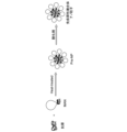
- An example of a method for preparing an immunogenic complex of one embodiment is shown.
- An example of a method for preparing an immunogenic complex of one embodiment is shown.
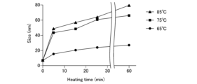
- the results of evaluating the effect of heating temperature and heating time on the particle size of disulfide-crosslinked MAN-OVA nanoparticles (MDO) are shown. "65°C”, "75°C”, and "85°C” indicate heating temperatures.
- OVA ovalbumin; 5 min: MDO (heated for 5 minutes); 15 min: MDO (heated for 15 minutes); 25 min: MDO (heated for 25 minutes); 60 min: MDO (heated for 60 minutes).
- OVA dNP disulfide cross-linked OVA nanoparticles
- MDO(5) MDO with 5 mg/mL MAN
- MDO(10) MDO with 10 mg/mL MAN
- MDO(15) MDO with 15 mg/mL MAN
- MDO(20) MDO with 20 mg/mL MAN.
- MDO-15 w/o H 2 O 2 MDO with 15 mg/mL MAN (without hydrogen peroxide treatment); MDO-15 w H 2 O 2 : MDO with 15 mg/mL MAN (with hydrogen peroxide treatment).
- SDS treatment SDS + DDT treatment.
- OVA ovalbumin
- Man mannoprotein
- OVA dNP disulfide-crosslinked OVA nanoparticles
- MO(5) MAN-OVA nanoparticles (MO) with 5 mg/mL MAN
- MDO(5) MDO with 10 mg/mL MAN
- MO(15) MO with 15 mg/mL MAN
- OVA ovalbumin
- OVA dNP disulfide-crosslinked OVA nanoparticles
- MDO disulfide-crosslinked MAN-OVA nanoparticles
- MGO glutaraldehyde-crosslinked MAN-OVA nanoparticles.
- Fluorescence microscopy images evaluating the uptake of each nanoparticle into DCs are shown.
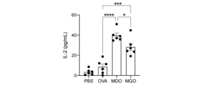
- the results of evaluating antigen presentation by bone marrow dendritic cells (BMDCs) stimulated with each nanoparticle, based on IL-2 production, are shown.
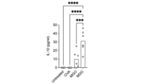
- the results of evaluating IL-10 production from bone marrow dendritic cells (BMDCs) stimulated with each type of nanoparticle are shown.
- ATRA reductase RALDH
- MDO (1:1): mixed with MP:OVA 1:1
- MDO (3:1): mixed with MP:OVA 3:1
- MDO (4:1): mixed with MP:OVA 4:1.
- the results of evaluating the reactivity of each nanoparticle with anti-OVA antibody are shown.
- the administration schedule of the experiment evaluating the risk of anaphylactic shock due to administration of each nanoparticle is shown below.
- Alum Alum.
- the results of evaluating the change in rectal temperature after administration of each nanoparticle as an index of anaphylactic shock are shown.
- 1 shows the administration schedule for an experiment evaluating the preventive effect of each nanoparticle on airway allergic reaction by pre-oral administration.
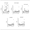
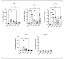
- the results of evaluating the preventive effect of airway allergic reactions by pre-oral administration of each nanoparticle are shown.
- the results of evaluating the preventive effect of airway allergic reactions by pre-oral administration of each nanoparticle are shown.
- the results of evaluating the preventive effect of airway allergic reactions by pre-oral administration of each nanoparticle are shown.
- 1 shows the administration schedule for an experiment evaluating the preventive effect of each nanoparticle on airway allergic reaction by pre-subcutaneous administration.
- the results of evaluating the preventive effect of airway allergic reactions by pre-subcutaneous administration of each nanoparticle are shown.
- the results of evaluating the preventive effect of airway allergic reactions by pre-subcutaneous administration of each nanoparticle are shown.
- the results of evaluating the preventive effect of airway allergic reactions by pre-subcutaneous administration of each nanoparticle are shown.
- the results of evaluating the preventive effect of airway allergic reactions by pre-subcutaneous administration of each nanoparticle are shown. 1 shows the administration schedule for an experiment evaluating the therapeutic effect of each nanoparticle on airway allergic reaction by post-oral administration.
- the results of evaluating the therapeutic effect of each nanoparticle on airway allergic reaction after post-oral administration are shown.
- the results of evaluating the therapeutic effect of each nanoparticle on airway allergic reaction after post-oral administration are shown.
- the results of evaluating the therapeutic effect of each nanoparticle on airway allergic reaction after post-oral administration are shown.
- 1 shows the administration schedule for an experiment evaluating the therapeutic effect of each nanoparticle on airway allergic reaction by post-subcutaneous administration.
- the results of evaluating the therapeutic effect of each nanoparticle on airway allergic reaction after post-subcutaneous administration are shown.
- the results of evaluating the therapeutic effect of each nanoparticle on airway allergic reaction after post-subcutaneous administration are shown.
- the results of evaluating the therapeutic effect of each nanoparticle on airway allergic reaction after post-subcutaneous administration are shown.
- the results of evaluating the therapeutic effect of each nanoparticle on airway allergic reaction after post-subcutaneous administration are shown.
- the results of evaluating the inhibition of mononuclear cell infiltration by administration of each nanoparticle are shown.
- 1 shows the administration schedule for an experiment evaluating the inhibitory effect of pre-oral administration of each nanoparticle on type IV allergic response.
- the results of evaluating the inhibitory effect of pre-oral administration of each nanoparticle on type IV allergic response are shown.
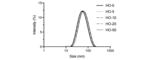
- the figures show the results of evaluating the effect of the OVA content on the particle size of disulfide-crosslinked HSA (human serum albumin)-OVA nanoparticles (HO): HO-0: HO with 0% OVA content; HO-5: HO with 5% OVA content; HO-10: HO with 10% OVA content; HO-20: HO with 20% OVA content; HO-50: HO with 50% OVA content.
- Non-reduced SDS treatment; reduced: SDS + DDT treatment.
- HO-5 HO with 5% OVA content
- HO-10 HO with 10% OVA content
- HO-20 HO with 20% OVA content
- HO-50 HO with 50% OVA content
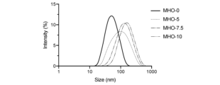
- MHO-10 10 mg/mL disulfide-crosslinked MAN-HSA-OVA nanoparticles (MHO) of MAN.
- MHO-0 0 mg/mL MHO of MAN
- MHO-5 5 mg/mL MHO of MAN
- MHO-7.5 7.5 mg/mL MHO of MAN
- MHO-10 10 mg/mL MHO of MAN.
- Fluorescence microscopy images evaluating the uptake of each nanoparticle into DCs are shown.
- the results of evaluating the change in DC phenotype due to stimulation with each nanoparticle are shown below: (a) MHC class II (IA b ) expression; (b) CD80 expression; (c) CD86 expression; (d) PD-L1 expression.
- MHO-10-R MHO-10 + rapamycin.
- the antigen presentation of BMDCs stimulated with each nanoparticle was evaluated based on IL-2 production.
- the results of evaluating the reactivity of each nanoparticle with anti-OVA antibody are shown. 1 shows the results of a basophil activation test in peripheral blood from healthy subjects.
- the peripheral blood from healthy subjects after each stimulation was stained with CRTH2 (CD294)-FITC, CD203c-PE, and CD3-PC7, and analyzed by a flow cytometer.
- the figures show the results of a basophil activation test in peripheral blood from patients with Japanese cedar pollen allergy.
- the figures show the results of staining peripheral blood from healthy subjects with CRTH2 (CD294)-FITC, CD203c-PE, and CD3-PC7 after each stimulation and analyzing the blood using a flow cytometer.
- 1 shows the results of a basophil activation test in peripheral blood from healthy subjects and from patients with cedar pollen allergy.
- the term “comprises” means that it may contain components other than the target component.
- the term “consists of” means that it does not contain components other than the target component.
- the term “consists essentially of” means that it does not contain components other than the target component in a form that exerts a special function (such as a form that completely loses the effect of the invention). In this specification, when “comprises,” it includes aspects that "consist of” and aspects that “consist essentially of.”
- a numerical range expressed using " ⁇ ” means that the range includes the numbers written before and after " ⁇ " as the lower and upper limits.
- a first aspect of the present disclosure is an immunogenic complex.
- the immunogenic complex of this aspect includes an antigen and a mannoprotein.
- the antigen and the mannoprotein are bound by a disulfide bond.
- Antigen refers to any substance capable of inducing an immune response, such as humoral and cellular immune responses, in a target organism.
- An antigen may be any substance capable of inducing proliferation, activation, maturation, etc. of immune cells when in contact with immune cells, or any substance capable of inducing cytokine production, antibody production, etc. from immune cells.
- the antigen is a protein or peptide.
- An antigen may be a protein that may be an allergen, a protein of an infectious agent (such as a virus, bacteria, fungus, etc.), a protein derived from a neoplastic cell (such as a tumor cell), a peptide or fragment of said protein, or a recombinant protein of said protein.
- An antigen may be an autoantigen.
- the antigen is an allergen or an autoantigen.
- Autoantigen refers to a substance possessed by an individual itself that may be an antigen.
- allergen refers to a substance that can cause an allergy in an individual.
- An allergen is a substance that is recognized as a foreign substance by an individual's immune system and causes an immune response, primarily the production of immunoglobulin E (IgE).
- allergens include pollen allergens, arthropod-derived allergens, food-derived allergens, and allergens present in insect saliva, nails, or needles.
- the allergen is a pollen allergen.
- pollen allergens include allergens derived from cedar pollen, cypress pollen, white birch pollen, rice pollen, ragweed pollen, mugwort pollen, and Japanese knotweed pollen.
- Antigens may be used alone or in combination of two or more types.
- Mannoproteins are glycoproteins composed of mannan and protein. Mannan is a polysaccharide whose main component is mannose. Mannoproteins are known in nature as one of the components of the cell walls of microorganisms, particularly yeast. For example, in yeast, mannoproteins exist in a structure in which a polypeptide chain is bound to the end of ⁇ -mannan.
- mannoproteins are composed of an internal chain portion N-glycosidically linked to an asparagine residue of a polypeptide, a polymeric mannan chain portion on the outside of the internal chain portion, and an oligosaccharide chain portion O-glycosidically linked to a serine or threonine residue of a peptide.
- Mannoproteins may be obtained from natural sources or by known chemical synthesis methods. Mannoproteins may be obtained, for example, from yeast.
- yeast include Saccharomyces, Pichia, and Candida, with Saccharomyces yeasts being preferred.
- Saccharomyces yeasts include S. bayanus, S. boulardii, S. bulderi, S. cariocanus, S. cariocus, S. cerevisiae, S. chevalieri, S. dairenensis, S. S. ellipsoides, S. eubayanus, S. exigus, S. florentinus, S. kluyveri, S. martiniae, S. monacensis, S.
- yeast is S. cerevisiae.
- the method for obtaining mannoprotein is not particularly limited, and known methods can be used.
- Methods for obtaining mannoprotein from yeast include, for example, a method that utilizes the solubility of mannoprotein in water. Examples include a method of extracting mannoprotein from yeast with hot water, and a method of extracting mannoprotein from yeast with alkali.
- mannoprotein can be obtained by extracting mannoprotein from yeast with hot water, followed by precipitation with cetablon (hexadecyltrimethylammonium bromide). Commercially available mannoprotein may be used.
- the antigen and the mannoprotein are bound by a disulfide bond.
- a thiol group (-SH) of the antigen and a thiol group of the polypeptide chain of the mannoprotein are bound by a disulfide bond to form a nanoparticle.
- the antigen is a protein or peptide
- the thiol group may be a thiol group of a cysteine residue of the protein or peptide.
- the thiol group of the mannoprotein may be a thiol group of a cysteine residue of the polypeptide chain of the mannoprotein.
- Disulfide bonds can be formed between thiol groups of the antigen; between a thiol group of the antigen and a thiol group of the mannoprotein; and between thiol groups of the mannoprotein.
- the immunogenic complex of this embodiment may form nanoparticles by binding the antigen and the mannoprotein via a disulfide bond.
- the size of the nanoparticles is not particularly limited, but may have an average particle diameter of about 10 to 2000 nm, for example. The average particle diameter tends to increase when the amount of mannoprotein used is increased.
- the average particle diameter of the nanoparticles may be about 30 to 1000 nm, about 50 to 500 nm, or about 50 to 300 nm.
- the average particle diameter of the nanoparticles can be measured using a dynamic light scattering spectrophotometer.
- the particle diameter polydispersity (PDI) of the nanoparticles is not particularly limited, but may be, for example, about 0.1 to 0.8, or about 0.2 to 0.6.
- the particle diameter polydispersity of the nanoparticles can be measured using a dynamic light scattering spectrophotometer.
- the nanoparticle comprises a shell and a core.
- the shell of the nanoparticle comprises mannan chains of a mannoprotein.
- the core of the nanoparticle comprises an antigen.
- the core may further comprise a polypeptide chain of a mannoprotein.
- the core of the nanoparticle comprises an antigen and a polypeptide chain of a mannoprotein, which are linked by disulfide bonds to form the core (see FIG. 1).
- the immunogenic complex of this embodiment may contain optional components in addition to the antigen and mannoprotein, such as matrix proteins, immunomodulators, and the like.
- Microx protein refers to a protein other than the antigen of interest that forms an immunogenic complex together with the antigen and mannoprotein.
- the matrix protein can be used, for example, when the cost of the antigen is high, to allow a small amount of antigen to form an immunogenic complex.
- the matrix protein is not particularly limited. In order to prevent the induction of an unexpected immune response, it is preferable to use a protein derived from the same organism as the subject of administration of the immunogenic complex.
- a human protein can be used as the matrix protein.
- a specific example of the matrix protein is albumin.
- human serum albumin can be preferably used as the matrix protein.
- the matrix protein may be used alone or in combination of two or more kinds.
- the matrix protein is present in the core of the nanoparticle and is bound to either or both of the antigen and the polypeptide chain of the mannoprotein by disulfide bonds to form the core together with them.
- Immunomodulator means a drug that has the effect of modulating immune function.
- the immunomodulator is an immunosuppressant that suppresses immune function.
- immunomodulators include retinoic acid receptor agonists, retinoid X receptor agonists, vitamin D receptor agonists, aromatic hydrocarbon receptor agonists, histone deacetylase inhibitors, and mTOR inhibitors.
- An "agonist” is a compound that binds to a receptor and activates an intracellular signal transduction system.
- An example of a retinoic acid receptor agonist is all-trans-retinoic acid.
- An example of a retinoid X receptor agonist is bexarotene.
- An example of a vitamin D receptor agonist is vitamin D3.
- An example of an aromatic hydrocarbon receptor agonist is 2-(1'H-indole-3'-carbonyl)-thiazole-4-carboxylic acid methyl ester.
- An example of a histone deacetylase inhibitor is suberoylanilide hydroxamic acid (SAHA).
- An example of an mTOR inhibitor is rapamycin.
- the immunomodulatory agent may be used alone or in combination of two or more kinds.
- the immunomodulator is thought to be present in the core of the nanoparticle due to intermolecular forces, etc.
- FIG. 1 An example of a method for producing an immunogenic complex according to an embodiment is shown in Fig. 1.
- Fig. 1 an antigen and mannoprotein (MAN) are used as materials for the immunogenic complex.
- Fig. 1 shows only one thiol group each of the antigen and mannoprotein, but each of the antigen and mannoprotein may have two or more thiol groups.
- Figure 2 shows an example of a method for producing an immunogenic complex according to one embodiment.
- an antigen, mannoprotein (MAN), and matrix protein are used as materials for the immunogenic complex.
- the antigen, mannoprotein, and matrix protein are shown to have only one thiol group each, but each may have two or more thiol groups.
- the method for producing the immunogenic complex may include, for example, a step of heating a solution in which an antigen and a mannoprotein are dissolved to form nanoparticles (hereinafter also referred to as “step (i)”), and a step of oxidizing the nanoparticles obtained in step (i) (hereinafter also referred to as “step (ii)").
- the antigen and mannoprotein are dissolved in an appropriate buffer solution to prepare a solution containing the antigen and mannoprotein.
- the buffer solution is not particularly limited, and can be one commonly used in the field of biochemistry.
- the buffer solution include phosphate buffer (PB), phosphate buffered saline (PBS), acetate buffer, citrate buffer, citrate phosphate buffer, borate buffer, tartaric acid ornamental solution, Tris buffer, HEPES buffer, etc.
- the buffer solution preferably contains a salt (e.g., sodium chloride).
- the salt concentration include 10 to 100 mM, preferably 20 to 80 mM, more preferably 30 to 70 mM, and even more preferably 40 to 60 mM.
- the pH of the buffer solution is not particularly limited, but is preferably near neutral, and preferably 6.0 to 8.0.
- a specific example of the buffer solution is 10 mM phosphate buffer solution (pH 7.4) to which 50 mM NaCl has been added.
- the mixing ratio of the antigen and the mannoprotein is not particularly limited. When the ratio of the mannoprotein is increased, the size of the nanoparticles tends to increase.
- the mannoprotein concentration in the antigen/mannoprotein solution is not particularly limited, but may be, for example, 1 to 50 mg/mL, preferably 5 to 30 mg/mL, more preferably 5 to 20 mg/mL, and even more preferably 5 to 15 mg/mL.
- the mannoprotein concentration is within the above-mentioned preferred range, the PDI tends to be small.
- the antigen concentration in the antigen/mannoprotein solution is not particularly limited, but may be, for example, 1 to 50 mg/mL, preferably 1 to 10 mg/mL, and more preferably 1 to 5 mg/mL.
- the matrix protein When a matrix protein is used, the matrix protein is dissolved in a buffer together with the antigen and mannoprotein to prepare a solution containing the antigen, mannoprotein and matrix protein (see FIG. 2).
- the total mass of the antigen and matrix protein can be considered as the mass of the antigen mentioned above.
- the ratio of the antigen to the total mass (100% by mass) of the antigen and the matrix protein is, for example, 1 to 90% by mass, and may be 3 to 80% by mass, 5 to 70% by mass, 10 to 60% by mass, 15 to 50% by mass, 15 to 40% by mass, or 20 to 40% by mass.
- the immunomodulator When an immunomodulator is used, the immunomodulator is dissolved in a buffer solution together with the antigen and mannoprotein to prepare a solution containing the antigen, mannoprotein and immunomodulator (and optionally the matrix protein).
- the amount of immunomodulator used is not particularly limited and can be appropriately selected depending on the type of immunomodulator.
- the concentration of rapamycin in the solution can be, for example, 1 to 100 ⁇ g/mL, or can be 5 to 50 ⁇ g/mL or 10 to 30 ⁇ g/mL.
- the heat treatment temperature can be set appropriately depending on the denaturation temperature of the antigen (and matrix protein).
- the heat treatment temperature can be set to a temperature at which the antigen (and matrix protein) denatures. By setting the heat treatment temperature to a temperature at which the antigen (and matrix protein) denatures, nanoparticles are stably formed. In addition, reactivity of the antigen with the antibody is suppressed.
- Specific examples of heat treatment temperatures include 60 to 90°C, with 65 to 90°C being preferred, 70 to 90°C being more preferred, and 75 to 85°C being even more preferred.
- the heat treatment time can be set to a time sufficient for denaturing the antigen (and matrix protein) at the heat treatment temperature.
- the heat treatment time can be, for example, 1 minute or more, preferably 5 minutes or more, more preferably 10 minutes or more, even more preferably 15 minutes or more, and particularly preferably 20 minutes or more.
- the treatment time may be 30 minutes or more, or may be 60 minutes or more.
- the upper limit of the treatment time is not particularly limited, but can be, for example, 100 minutes or less, or 80 minutes or less.
- step (i) the antigen and mannoprotein, and optionally the matrix protein and the immunomodulator, are aggregated in the solution.
- Pre-NP nanoparticle
- the mixture may be cooled using an ice bath or the like. Cooling can stop the formation of nanoparticles.
- the oxidation treatment of the nanoparticles (Pre-NP) can be carried out using an oxidizing agent.
- the oxidizing agent include hydrogen peroxide, oxidized glutathione (GSSG), potassium nitrate, perchlorate, hypochlorite, nitric acid, permanganate, ozone, etc.
- the oxidizing agent is hydrogen peroxide.
- the oxidizing agent may be used alone or in combination of two or more kinds.
- the amount of the oxidizing agent used can be appropriately set depending on the type of the oxidizing agent.
- the oxidizing agent is hydrogen peroxide, it can be added to the solution obtained in step (i) so that the final concentration becomes, for example, 0.1 to 1 mass %.
- the reaction conditions for the oxidation reaction may be any conditions that allow the thiol groups in the nanoparticles to form disulfide bonds.
- the reaction temperature is, for example, 0 to 30°C, preferably 1 to 20°C, more preferably 2 to 10°C, and even more preferably 3 to 5°C.
- the reaction time is, for example, 1 hour or more, preferably 5 hours or more, more preferably 8 hours or more, even more preferably 10 hours or more, and particularly preferably 12 hours or more.
- the upper limit of the treatment time is not particularly limited, but can be, for example, 30 hours or less, or 20 hours or less.
- step (ii) disulfide bonds are formed between the thiol groups in the nanoparticles. This makes it possible to obtain nanoparticles of an immunogenic complex in which the antigen and mannoprotein are bound by disulfide bonds.
- nanoparticles of the immunogenic complex may be collected by ultrafiltration or the like.
- ultrafiltration By performing ultrafiltration, free antigens, mannoproteins, oxidizing agents, matrix proteins, immunomodulators, etc. can be removed.
- an ultrafiltration membrane with a molecular weight cutoff of 100 kDa can be used.
- the recovered nanoparticles of the immunogenic complex may be freeze-dried and stored.
- the nanoparticles may be suspended in a 1% by weight glucose solution and freeze-dried.
- the immunogenic complex has a shape in which the antigen is covered with mannan chains of mannoprotein. Therefore, it is easily taken up into dendritic cells by the mannose receptor (MR) and CD209 of DC.
- the immunogenic complex taken up into DC is cleaved within the DC as the disulfide bonds are reduced to thiol groups. This releases the antigen from the immunogenic complex.
- the antigen released into the DC cell is degraded within the DC cell to a peptide of about 15 to 30 amino acid residues. This peptide is presented by MHC molecules as a T cell epitope. This induces tolerant DC.
- the immunogenic complex of this embodiment has low reactivity with antigen-specific antibodies because the antigen is not exposed. Furthermore, by thermally denaturing the antigen during the preparation of the immunogenic complex, the reactivity with antigen-specific antibodies is further reduced. This suppresses binding between the antigen and IgE antibodies. Therefore, with the immunogenic complex of this embodiment, it is possible to administer many antigens to the subject while avoiding binding with IgE antibodies.
- a second aspect of the present disclosure is a pharmaceutical composition.
- the pharmaceutical composition of this aspect contains the immunogenic complex of the first aspect.
- the pharmaceutical composition of this aspect contains the immunogenic complex of the first aspect as an active ingredient.
- the pharmaceutical composition of this embodiment may contain an optional component in addition to the immunogenic complex of the first embodiment.
- the optional component may include, for example, a pharma- ceutically acceptable carrier.
- pharmaceutical-ceutically acceptable carrier refers to a carrier that does not inhibit the physiological activity of the active ingredient and does not show substantial toxicity to the subject to which it is administered.
- not substantially toxic refers to a carrier that does not show toxicity to the subject to which it is administered at a dose that is normally used.
- the pharma-ceutically acceptable carrier is a carrier that does not inhibit the immunogenicity of the immunogenic complex of the first embodiment and does not show substantial toxicity to the subject to which it is administered.
- the pharma-ceutically acceptable carrier includes any known pharma-ceutically acceptable component that is typically considered to be an inactive component.
- the pharma-ceutically acceptable carrier is not particularly limited, but examples thereof include solvents, diluents, vehicles, excipients, flow enhancers, binders, granulating agents, dispersing agents, suspending agents, wetting agents, lubricants, disintegrants, solubilizers, stabilizers, emulsifiers, and fillers.
- the pharma- ceutical acceptable carrier may be used alone or in combination of two or more kinds.
- the pharmaceutical composition of this embodiment may contain, for example, a buffer solution as a solvent. Examples of the buffer solution include those mentioned above.
- the pharmaceutical composition may contain other ingredients in addition to the above ingredients.
- the other ingredients are not particularly limited, and any ingredient commonly used in the pharmaceutical field may be used without any particular restriction.
- examples of the other ingredients include pharmaceutical additives other than those mentioned above.
- pharmaceutical additives include, but are not limited to, preservatives (e.g., antioxidants), chelating agents, flavoring agents, sweeteners, thickeners, buffers, colorants, etc. These ingredients may be used alone or in combination of two or more kinds.
- the pharmaceutical composition may contain an active ingredient other than the immunogenic complex of the first aspect.
- the active ingredient include, but are not limited to, antiviral agents, antibiotics, anti-inflammatory agents, antipyretics, analgesics, etc.
- One type of active ingredient may be used alone, or two or more types may be used in combination.
- the pharmaceutical composition may contain an adjuvant.
- adjuvants include, but are not limited to, aluminum hydroxide, calcium phosphate, monophosphoryl lipid A, chitosan, etc.
- One type of adjuvant may be used alone, or two or more types may be used in combination.
- the dosage form of the pharmaceutical composition is not particularly limited, and may be any dosage form commonly used as a pharmaceutical preparation.
- the pharmaceutical composition of this embodiment may be an oral preparation or a parenteral preparation.
- Oral preparations include, for example, tablets, coated tablets, pills, powders, granules, capsules, syrups, fine granules, liquids, drops, emulsions, etc.
- Parenteral preparations include, for example, injections, suppositories, nasal drops, enteral preparations, inhalants, etc.
- Pharmaceutical compositions of these dosage forms may be formulated according to standard methods (for example, methods described in the Japanese Pharmacopoeia).
- the route of administration of the pharmaceutical composition of this embodiment is not particularly limited, and may be oral or parenteral.
- parenteral administration include sublingual administration, intravenous administration, intranasal administration, subcutaneous administration, intradermal administration, intramuscular administration, intraperitoneal administration, and enteral administration.
- the pharmaceutical composition can be administered in a therapeutically effective amount of the immunogenic complex of the first aspect.
- therapeutically effective amount means an amount of a drug effective for treating or preventing a target disease.
- a therapeutically effective amount of an immunogenic complex can be an amount effective for inducing tolerant DCs.
- the therapeutically effective amount may be appropriately determined depending on the symptoms, body weight, age, and sex of the patient, as well as the dosage form and administration method of the pharmaceutical composition.
- the pharmaceutical composition can be administered in a single dose of 0.001 to 1000 mg of antigen per kg of the body weight of the subject. The dose may be 0.005 to 500 mg/kg, 0.01 to 300 mg/kg, 0.02 to 200 mg/kg, or 0.03 to 100 mg/kg.
- the pharmaceutical composition may contain a therapeutically effective amount of the immunogenic complex per unit dosage form.
- the content of the immunogenic complex in the pharmaceutical composition may be 0.01 to 90% by mass, 0.05 to 80% by mass, or 0.1 to 60% by mass.
- the pharmaceutical composition may be administered in a single dose or repeatedly.
- the administration interval may be appropriately determined depending on the symptoms, weight, age, and sex of the patient, as well as the dosage form of the pharmaceutical composition and the administration method.
- the administration interval may be, for example, every few hours, 2-3 times a day, once a day, once every 2-3 days, once a week, once a month, once every few months, etc.
- the subject to which the pharmaceutical composition is administered is not particularly limited.
- the subject to which the pharmaceutical composition is administered is preferably a mammal, and may be a human or a non-human mammal.
- non-human mammals include primates (monkeys, gorillas, chimpanzees, marmosets, etc.), rodents (mice, rats, guinea pigs, hamsters, etc.), pets (dogs, cats, rabbits, ferrets, etc.), and livestock (cows, pigs, horses, goats, sheep, etc.).
- the pharmaceutical composition can be used for treating or preventing allergies or autoimmune diseases.
- the allergies or autoimmune diseases to which the pharmaceutical composition can be applied are not particularly limited. Examples of allergies include, but are not limited to, hay fever, food allergies, dust mite allergies, atopic dermatitis, allergic asthma, allergic rhinitis, allergic urticaria, and the like. Examples of autoimmune diseases include, but are not limited to, Graves' disease, rheumatoid arthritis, Hashimoto's thyroiditis, type 1 diabetes, systemic lupus erythematosus, vasculitis, and the like.
- the disclosure provides a method of treating or preventing allergy or an autoimmune disease comprising administering to a subject an immunogenic conjugate of the first aspect.
- the immunogenic conjugate may be administered in a therapeutically effective amount.
- the disclosure provides an immunogenic conjugate of the first aspect for treating or preventing an allergy or autoimmune disease.
- the disclosure provides the use of the immunogenic complex of the first aspect in the manufacture of a pharmaceutical composition for treating or preventing an allergy or an autoimmune disease.
- the disclosure provides the use of the immunogenic conjugate of the first aspect for treating or preventing allergy or an autoimmune disease.
- Ovalbumin Ovalbumin
- MAN mannoprotein
- ConA concanavalin A
- Tris tris(hydroxymethyl)aminomethane
- LPS lipopolysaccharide
- Hydrogen peroxide H2O2 , 30 wt% in water
- dithiothreitol DTT
- sulfuric acid 98%, H2SO4
- calcium chloride CaCl2
- magnesium chloride hexahydrate MgCl2 ⁇ 6H2O
- sodium dodecyl sulfate SDS
- glycine sodium chloride
- NaCl NaCl
- Mildform 10N 10% formalin neutral buffer
- CBB solution for protein assay 4% paraformaldehyde solution (PFA), disodium hydrogen phosphate, and sodium dihydrogen phosphate were purchased from Nacalai Tesque, Inc.
- D-glucose was purchased from Tokyo Chemical Industry Co., Ltd.
- Biotin anti-mouse IgE antibody, biotin anti-mouse IgG1 antibody, and biotin anti-mouse IgG2a antibody were purchased from BioLegend.
- NPs Mannan-Coated OVA Nanoparticles
- PB phosphate buffer
- OVA and MAN were mixed at different concentrations to obtain working solutions. In the following tests, the concentrations of OVA and MAN are expressed using the final concentrations in the working solutions.
- Protein nanoparticles (NPs) were prepared by heating 1 mL of the working solution at 65°C, 75°C, or 85°C. The heating times were 0, 5, 15, 25, or 60 minutes, respectively. The NP formation was stopped by cooling in an ice bath.
- MAN-OVA NPs (MO) without disulfide bonds.
- H 2 O 2 was added to the MO solution up to 0.3 wt %, and the mixture was reacted overnight at 4°C to form intermolecular disulfide bonds.
- Free OVA, MAN, and H2O2 were removed by ultrafiltration (100 kDa Amicon, Merck Millipore) to recover disulfide-crosslinked MAN-OVA NPs (MDO).
- MDO was freeze-dried in a 1 wt% glucose solution to ensure long-term storage.
- 20 mg/mL OVA and 20 mg/mL MAN were mixed in equal amounts and conjugated with glutaraldehyde (final concentration 25 mM) to prepare a MAN-OVA conjugate (MGO).
- Nanoparticle size and zeta potential The particle size, particle size distribution and zeta potential of the nanoparticles were measured at 25° C. using a dynamic light scattering spectrophotometer (DLS, Zetasizer Pro ZSU3200).
- OVA and MAN OVA concentration were measured using a protein assay CBB solution based on the Bradford method.
- OVA standard solution was serially diluted 2-fold, and a standard curve was created by linear regression. Samples were mixed with CBB solution and incubated at room temperature for 10 minutes. Absorbance was measured at 595 nm using a plate reader (Infinite200PRO M Plex, Tecan).
- a phenol-sulfuric acid assay was used to measure MAN concentration.
- a standard curve was prepared by gradient dilution of MAN. 5% (w/v) phenol was mixed with the sample, and H2SO4 was added and mixed well. After incubation at room temperature for 40 minutes, the absorbance was measured at 490 nm.
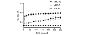
- ConA agglutination assay was performed as previously reported (Cui, Z.; J Control Release 2002, 81 (1-2), 173-184.). Briefly, ConA was dissolved in phosphate-buffered saline (PBS, 10 mM, pH 7.4), and 5 mM CaCl2 and 5 mM MgCl2 were added to obtain a 1 mg/mL ConA solution. 40 ⁇ L of sample was mixed with 200 ⁇ L of ConA solution. Turbidity was measured at 36 nm for 300 seconds.
- PBS phosphate-buffered saline
- ⁇ Mouse> C57BL/6J mice (7-8 weeks old, male) and BALB/c mice (6-8 weeks old, female) were purchased from Kyudou Co., Ltd.
- OT-II Tg/wt,Ly5.1/Ly5.1 mice were fed CE-2 (Kyudou Co., Ltd.) and had access to bedding and water.
- Cage changing was performed in a laminar flow hood. Mice were housed under a 12-hour light/12-hour dark cycle. Animal experiments were performed in accordance with the guidelines of the Kyushu University Animal Care and Management Committee.
- BMDCs Myeloid Dendritic Cells
- BMDCs were first negatively selected from bone marrow cells with biotin anti-mouse F4/80 and streptavidin nanobeads, and then positively selected with anti-mouse CD11c nanobeads.
- OVA-rhodamine was used to prepare fluorescently labeled NPs.
- BMDCs (2 ⁇ 10 5 cells/well) were seeded in 12-well plates and incubated with rhodamine-labeled OVA, OVA dNPs (disulfide-crosslinked OVA nanoparticles), MDO, and MGO for different times.
- MAN was added to the medium at a final concentration of 1 mg/mL 30 min before sample addition. Dead cells were stained with SYTOX TM Green (Thermo Fisher Scientific).
- MFI mean fluorescence intensity
- BMDCs were seeded in a 96-well plate (5 ⁇ 10 4 cells/well) and pulsed with PBS, OVA, MDO, or MGO for 3 hours. Then, BMDCs and OT-II CD4 + T cells (2.5 ⁇ 10 5 cells/well) were cocultured for 24 hours.
- OT-II CD4 + T cells were isolated from the spleen of OT-II mice using a Mouse CD4 T Cell Isolation Kit (Biolegend). The IL-2 concentration in the culture medium was measured using an IL-2 Mouse ELISA Kit (Thermo Fisher Scientific).
- mice were sensitized with 200 ⁇ L PBS containing 10 ⁇ g OVA and 1 mg alum by intraperitoneal injection three times at 7-day intervals. Seven days after the last sensitization, the mice were sacrificed and peripheral blood was collected by retro-orbital bleeding. Serum was obtained from the peripheral blood.
- An indirect immunoassay (indirect-ELISA) protocol was used. 100 ⁇ L of OVA, OVA-dNP, MDO, and MGO (as 100 ⁇ g/mL of OVA) were coated onto a 96-well plate at 4°C for 24 hours. After blocking with ELISA-ELISPOT diluent, serially diluted sera were added to the plate and incubated overnight at 4°C. Biotin anti-mouse IgE, biotin anti-mouse IgG1, and biotin anti-mouse IgG2a antibodies were added to the plate, respectively. After incubation, streptavidin-HRP was added, and TMB solution was used as a substrate. Finally, the reaction was stopped, and the optical density (OD) of each well was measured at 450 nm.
- mice were sensitized by intraperitoneal injection of 200 ⁇ L PBS containing 10 ⁇ g OVA and 1 mg alum on days 0 and 14. On day 28, mice were intraperitoneally injected with 200 ⁇ g OVA, OVA dNP, MDO, and MGO (containing 200 ⁇ g OVA). After intraperitoneal injection, rectal temperature was measured every 10 min using a thermometer (Physitemp PTM1, Muromachi Kikai Co., Ltd.).
- mice received (a) oral administration of PBS, OVA, MDO and MGO (containing 0.5 mg OVA) on days 0-4 and 7-11, or (b) subcutaneous administration of PBS, OVA, MDO and MGO (containing 20 ⁇ g OVA) on days 0, 2, 4, 7, 9 and 11.
- mice were sensitized by intraperitoneal injection of 10 ⁇ g OVA and 1 mg alum.
- mice were intranasally administered 25 ⁇ g OVA.
- mice were sacrificed and bronchoalveolar lavage fluid (BAL), peripheral blood and lung tissue were collected.
- BAL bronchoalveolar lavage fluid
- mice were first sensitizing mice with intraperitoneal injections of 10 ⁇ g OVA and 1 mg alum on days 0 and 7. Next, mice received (a) oral administration of PBS, OVA, MDO, and MGO (containing 0.5 mg OVA) on days 14-18 and 21-25, or (b) subcutaneous administration of PBS, OVA, MDO, and MGO (containing 20 ⁇ g OVA) on days 14, 16, 18, 21, 23, and 25. Finally, mice were intranasally challenged with 25 ⁇ g OVA on days 32-35. Mice were sacrificed on day 36, and bronchoalveolar lavage fluid (BAL), peripheral blood, and lung tissue were collected.
- BAL bronchoalveolar lavage fluid
- cytokines in BAL fluid IL-4, IL-5, IL-6, IL-10, IL-13, and IFN- ⁇ in the BAL fluid were detected using a BD Cytometric Bead Array (BD Life Sciences-Biosciences), followed by measurement using a flow cytometer (CyroFLEX-S, Beckman Coulter Inc.).
- thiol groups were not detected in native OVA (0 min), but after heating, thiol groups appeared on the OVA surface and became detectable.
- the free thiol group concentration of NPs not treated with H2O2 was significantly higher than that of dNPs treated with H2O2 (Fig . 6), indicating the formation of disulfide bonds in dNPs.
- the addition of MAN reduced the detectable thiol groups, which may be due to the inhibition of the reaction of DTNB with SH by the coating of MAN.
- ConA is a tetrameric lectin with four binding sites for ⁇ -mannose.
- the turbidity of OVA dNP did not change significantly.
- the turbidity of MDO increased significantly, and the turbidity increased with increasing MAN content. This suggested that a large amount of mannan was present on the surface of MDO-20. Due to the unimodal distribution and high MAN content of MDO-15, MDO-15 was used in subsequent experiments.
- BMDCs ⁇ Induction of regulatory T cells by MDO>
- T cell stimulating ability of BMDCs was measured using OT-II CD4 + T cells that have a specific TCR that recognizes the OVA peptide/I-AB complex and secrete IL-2 in response to it.
- BMDCs were pulsed with OVA, MDO, and MGO for 3 hours, respectively, and then co-cultured with OT-II CD4 + T cells for 24 hours, and the secreted IL-2 concentration in the medium was measured using an ELISA kit.
- MDO treatment significantly increased the IL-2 concentration compared to OVA treatment and MGO treatment ( Figure 11).
- MDO treatment increased the IL-10 concentration compared to OVA treatment and MGO treatment ( Figure 12).
- MDO treatment increased the activity of ATRA reductase (RALDH) compared to OVA treatment ( Figure 13).
- MGO and MDO treatments increased the proportion of regulatory T cells compared to OVA treatment ( Figure 14).
- mice were administered two routes of administration (oral and subcutaneous).
- oral administration mice were administered PBS, 500 ⁇ g OVA, or 500 ⁇ g OVA as MDO or MGO on days 0-4 and 7-11.
- Mice were sensitized by subcutaneous injection of OVA and alum on days 14 and 21, and finally administered OVA intranasally on days 28-31 ( Figure 18).
- serum, bronchoalveolar lavage fluid (BAL), and lungs were collected.
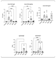
- Anti-OVA IgE levels were reduced by OVA pre-treatment and more strongly by MDO and MGO pre-treatment ( Figure 19).
- Anti-OVA IgG2a levels were significantly increased by MDO treatment, whereas IgG1 levels were not different (Figure 19).
- Pre-administration of OVA, MDO, and MGO suppressed the increase in eosinophils and neutrophils and reduced the total cell count in the BAL fluid, but no significant difference was observed in the number of macrophage cells ( Figure 20).
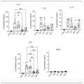
- the levels of IL-4, IL-5, IL-6, and IL-13 mediated by Th2 cells in the BAL fluid were significantly reduced by MDO pre-administration and MGO pre-administration compared to PBS treatment ( Figure 21).
- Pre-administration of OVA also reduced Th2 cytokines, but not as significantly as MDO pre-administration and MGO pre-administration.
- mice were given 20 ⁇ g of PBS or OVA, or 20 ⁇ g of OVA as MDO or MGO on days 0, 2, 4, 7, 9, and 11.
- the subsequent course was the same as for oral administration ( Figure 22).
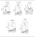
- Pre-administration of OVA, pre-administration of MDO, and pre-administration of MGO showed similar IgE production inhibitory effects (Figure 23).
- IgG2a levels increased with MDO pre-administration, but IgG1 levels did not differ among all groups.
- IgG2a/IgE and IgG2a/IgG1 values indicated that MDO pre-administration and MGO pre-administration showed superior IgE inhibitory effects ( Figure 23).
- mice were sensitized with OVA and alum on days 0 and 7 and orally administered PBS, 20 ⁇ g OVA, or 500 ⁇ g OVA as MDO or MGO on days 14-18 and 21-25 (Figure 26).
- Nasal administration of OVA was performed on days 32-35.
- Anti-OVA IgE levels were decreased after OVA post-administration and MDO post-administration ( Figure 27). There was a tendency for a decrease after MGO post-administration, but there was no statistical difference (Figure 27).
- mice were administered PBS, 20 ⁇ g OVA, or 20 ⁇ g OVA as MDO or MGO on days 14, 16, 18, 21, 23, and 25 (Figure 30).
- MDO post-administration reduced anti-OVA IgE production ( Figure 31).
- BAL fluid neutrophil, eosinophil, and total cell counts were reduced by MDO post-administration ( Figure 32).
- IL-4, IL-5, and IL-6 were reduced by MDO post-administration (Figure 33).
- Mononuclear cell infiltration was reduced by OVA pre-administration and MGO post-administration, but more dramatically by MDO post-administration (Figure 34: subcutaneous treatment).
- HSA Human serum albumin
- OVA ovalbumin
- MAN mannoprotein
- ConA concanavalin A
- LPS lipopolysaccharide
- Hydrogen peroxide H 2 O 2 , 30 wt % in water
- sulfuric acid (98%, H 2 SO 4 )
- calcium chloride CaCl 2
- magnesium chloride hexahydrate MgCl 2 ⁇ 6H 2 O
- sodium chloride NaCl
- DTNB 2,2'-dinitro-5,5'-dithiodibenzoic acid
- 5(6)-Carboxytetramethylrhodamine N-succinimidyl ester HNS-Rho was purchased from Thermo Fisher Scientific.
- NuPAGE sample reducing agent (10x), NuPAGE LDS sample buffer (4x), ELISA-ELISPOT diluent (5x), TMB (3,3',5,5'-tetramethylbenzidine) solution (1x), and HRP-conjugated Streptavidin (horseradish peroxidase) were purchased from Invitrogen.
- QuickBlue staining solution was purchased from Bio Dynamics Institute Co., Ltd.
- 4% paraformaldehyde solution (PFA), disodium hydrogen phosphate, and sodium dihydrogen phosphate were purchased from Nacalai Tesque, Inc.
- Biotin anti-mouse IgE antibody, biotin anti-mouse IgG1 antibody, and biotin anti-mouse IgG2a antibody were purchased from BioLegend.
- HSA NPs were prepared under various conditions [buffer (10 mM phosphate buffer containing 0 mM, 50 mM or 135 mM NaCl (pH 7.4)]; heating temperature (65°C, 75°C or 85°C); heating time (5 min, 10 min, 30 min or 60 min)]. HSA was dissolved in the buffer and left at 4°C overnight. The HSA dispersion was filtered through a 0.22 ⁇ m membrane to remove aggregates. 1 mL of the HSA solution was heated in a water bath and immediately cooled in an ice bath.
- H2O2 was added to the NP solution to a final concentration of 0.3 wt%, and the solution was kept at 4°C overnight to form disulfide bonds inside the NPs.
- Free HSA and H2O2 were removed by ultrafiltration (100 kDa Amicon, Merck Millipore).
- HSA-OVA NP(HO) HSA-OVA NP(HO)
- rapamycin 10 ⁇ L of rapamycin (1 mg/mL) was added to 1 mL of the mixed solution of MAN, HSA, and OVA.
- the buffer used for dissolving HSA, OVA, and MAN was 10 mM PB + 50 mM NaCl (pH 7.4).
- the mixed solution was heated at 85°C for 60 minutes. After cooling the mixed solution, H 2 O 2 was added, and the solution was left to stand overnight at 4°C, and the NP solution was collected. Ultrafiltration was performed using a 100 kDa filter to collect the NP.
- Rapamycin was quantified by a validated high performance liquid chromatography (HPLC) method using an Inertsil C8 column (5 ⁇ m, 4.6 mm ⁇ 150 mm) with an Elite Lachrom L-2455 diode array detector (Hitachi). The mobile phase was changed from 70% methanol and 30% Milli-Q water to 90% methanol and 10% Milli-Q water within 20 min. The flow rate was set at 1.0 mL/min, injection volume was 20 ⁇ L, and column oven temperature was set at 25° C. Rapamycin was detected using a photodiode array detector at a wavelength of 277 nm.
- HPLC high performance liquid chromatography
- BMDC phenotype analysis BMDCs were seeded in 12-well plates (3 ⁇ 10 6 cells/well), stimulated with 100 ng/mL LPS for 24 hours, and treated with PBS, OVA, HO-20, MHO-10, or MHO-10-R (MHO-10 encapsulated with rapamycin) for 24 hours. BMDCs were collected, and nonspecific binding of immunoglobulins to Fc receptors was inhibited with anti-mouse CD16/32 antibody. BMDCs were stained with anti-mouse I-Ab-FITC, CD86-APC, CD80-BV421, and propidium iodide (PI) (Biolegend). Subsequent analysis was performed by flow cytometry.
- PI propidium iodide
- NP preparation conditions The optimal conditions for preparing NPs from HSA, a matrix protein, were screened. HSA is neutral and negatively charged, so HSA aggregates are stably dispersed. The HSA solution was heated at a specific temperature (65-85°C) for up to 60 minutes. Then, H2O2 was added to crosslink the disulfide bonds. The size of HSA NPs increased with heating temperature (data not shown). This is thought to be due to the progression of denaturation with temperature. HSA is known to denature at 65-80°C. Since HSA is expected to be completely denatured and aggregation promoted at 85°C, 85°C was selected as the preparation temperature. When sodium chloride is added, the aggregate size increases, possibly due to the suppression of electrostatic repulsion. 50 mM NaCl was selected because excess sodium chloride destabilizes NPs.
- the effect of the amount of MAN added on the preparation conditions of HSA/OVA NPs was evaluated with a fixed HSA/OVA ratio of 4:1.
- the particle size and PDI of the obtained NPs (MHO series) increased with increasing MAN concentration ( Figure 39, Tables 3 and 4).
- the MAN content in MHO quantified by the phenol-sulfuric acid method increased with increasing MAN concentration, indicating that the incorporation of MAN into NPs is concentration-dependent (Tables 3 and 4). Since the mannan portion of MAN is hydrophilic, mannan coats the surface of the NPs.
- MHO-10 was dispersed in a buffer solution (10 mM PB + 50 mM NaCl, pH 7.4) and storage stability at 4°C was confirmed. No size change was observed after storage for at least 28 days (data not shown).
- rapamycin an immunosuppressant
- HPLC analysis of the rapamycin remaining in the filtrate of the NP dispersion we confirmed that rapamycin had been incorporated almost quantitatively (data not shown).
- NPs (MHCryj) were prepared by the above method using 4.5 mg/mL HSA, 0.5 mg/mL Cry j, and 10 mg/mL MAN. MHCryj was slightly larger than MHO-10, but showed a uniform size distribution ( Figure 41, Table 5). In the PAGE analysis of MHCryj, bands of Cry j and HSA were observed under reducing conditions ( Figure 42), and no bands were observed under non-reducing conditions.
- NPs have the effect of inducing tolerant DCs.
- BMCs stimulated with LPS expressed high levels of MHC class II (I-A b ), costimulatory factors CD80 and CD86, indicating that BMDCs were mature.
- I-A b MHC class II
- CD80 and CD86 costimulatory factors
- MHO-10 and MHO-10-R have the ability to suppress the activation of BMDCs, and that MHO-10-R may induce tolerant DCs due to the effect of rapamycin.
- MAN induces signal transduction via mannose receptor and DC-SIGN, causing activation of NF- ⁇ B. Rapamycin enhances activation of NF- ⁇ B through inhibition of mammalian target of rapamycin, which is a signal transduction pathway different from that of MAN. Therefore, MAN and rapamycin may synergistically induce tolerant DC.
- ⁇ Reactivity to anti-OVA antibodies> The reactivity of HO and MHO with anti-OVA antibodies was examined by ELISA. Anti-OVA antibodies were obtained from the serum of mice in which OVA-specific allergy was induced. As shown in FIG. 47, HO-20 and MHO-10 had much lower reactivity with anti-OVA antibodies than OVA, and there was no significant difference between these two NPs. This result indicates that MHO induces a weak IgE-mediated allergic reaction. In allergy treatment, this indicates that MHO is safer than native antigens used in conventional antigen-specific immunotherapy.
- Example 3 To evaluate whether Cry j NP induces an allergic reaction, a basophil activation test was performed using human peripheral blood.
- MHCryj Materials and Methods ⁇ Preparation of MHCryj> NP (MHCryj) was prepared in the same manner as above using 4.75 mg/mL HSA, 0.25 mg/mL Cry j 1/2 (a mixture of Cry j 1 and Cry j 2), and 7.5 mg/mL MAN. HSA and Cry j 1/2 were dissolved in PB and left to stand overnight at 4°C. The solution was filtered through a 0.22 ⁇ m membrane to remove aggregates. The solution was then heated in a water bath (85°C, 10 minutes) and immediately cooled in an ice bath.
- H 2 O 2 was added to the solution to a final concentration of 0.1 wt%, and the solution was kept at 4°C overnight to form disulfide bonds inside the NP.
- Free HSA and H 2 O 2 were removed by ultrafiltration (100 kDa Amicon, Merck Millipore).
- the size and PDI of the obtained MHCryj were as follows: Size: 123.6 ⁇ 1.6 (d.nm) PDI: 0.34 ⁇ 0.04
- the basophil activation test was performed using an Allergenecity kit (BECKMAN COULTER, Tokyo, Japan) according to the protocol attached to the Allergenecity kit.
- peripheral blood from healthy subjects and peripheral blood from patients with cedar pollen allergy were used, collected using blood collection tubes containing ethylenediaminetetraacetic acid (EDTA).
- EDTA ethylenediaminetetraacetic acid
- Peripheral blood was stimulated using Cry j 1/2 or MHCryj as an allergen.
- Cry j 1/2 was diluted with PBS so that the amount of Cry j 1/2 added per 100 ⁇ L of peripheral blood was 0.01 ⁇ g, 0.1 ⁇ g, or 1 ⁇ g, and 20 ⁇ L was added to 100 ⁇ L of peripheral blood.
- MHCryj was diluted with PBS so that the amount of Cry j 1/2 added per 100 ⁇ L of peripheral blood was 0.01 ⁇ g, 0.1 ⁇ g, or 1 ⁇ g, and 20 ⁇ L was added to 100 ⁇ L of peripheral blood.
- Anti-IgE antibody was used as a positive control.
- PBS was used as a negative control.
- the samples used to stimulate each peripheral blood are summarized in Table 6. The values in Table 6 indicate the amount of Cry j 1/2 added ( ⁇ g) per 100 ⁇ L of peripheral blood.
- each peripheral blood sample was stained with CRTH2 (CD294)-FITC, CD203c-PE, and CD3-PC7, and analyzed by a flow cytometer. Basophil activation was evaluated based on the analysis results. The rate of basophil activation was determined by calculating the percentage of basophils with increased CD203c fluorescence intensity compared to the negative control (PBS) in the CRTH2 (CD294) and CD203c double positive basophil population in the CD3 negative mononuclear cell region.
- PBS negative control
- CD203c double positive basophil population in the CD3 negative mononuclear cell region.
- the positive control increased the fluorescence intensity of CD203c compared to the negative control (PBS), and an increase in the proportion of activated basophils was confirmed.
- stimulation with Cry j (0.01 ⁇ g, 0.1 ⁇ g, 1 ⁇ g) also increased the fluorescence intensity of CD203c compared to the negative control (PBS), and an increase in the proportion of activated basophils was confirmed ( Figures 48 and 50, right).
- the present invention provides an immunogenic complex that has low reactivity to IgE antibodies and high ability to induce tolerant DCs, and a pharmaceutical composition containing the immunogenic complex.
Landscapes
- Health & Medical Sciences (AREA)
- Life Sciences & Earth Sciences (AREA)
- Public Health (AREA)
- Veterinary Medicine (AREA)
- Medicinal Chemistry (AREA)
- Pharmacology & Pharmacy (AREA)
- Chemical & Material Sciences (AREA)
- Animal Behavior & Ethology (AREA)
- General Health & Medical Sciences (AREA)
- Epidemiology (AREA)
- Immunology (AREA)
- Bioinformatics & Cheminformatics (AREA)
- Engineering & Computer Science (AREA)
- Microbiology (AREA)
- Mycology (AREA)
- Chemical Kinetics & Catalysis (AREA)
- General Chemical & Material Sciences (AREA)
- Nuclear Medicine, Radiotherapy & Molecular Imaging (AREA)
- Organic Chemistry (AREA)
- Proteomics, Peptides & Aminoacids (AREA)
- Peptides Or Proteins (AREA)
Abstract
抗原と、マンノプロテインと、を含み、前記抗原と前記マンノプロテインとがジスルフィド結合で結合している、免疫原性複合体。前記免疫原性複合体を含有する医薬組成物。
Description
本発明は、免疫原性複合体及び医薬組成物に関する。
本願は、2022年11月30日に、日本に出願された特願2022-191916号に基づき優先権を主張し、その内容をここに援用する。
本願は、2022年11月30日に、日本に出願された特願2022-191916号に基づき優先権を主張し、その内容をここに援用する。
アレルギーは、特定のアレルゲンタンパク質に対して免疫応答が過剰に起こる炎症性疾患である。現在行われているアレルギーの根治療法は減感作療法であり、少量のアレルゲンを継続的に投与し、樹状細胞(DC)により、制御性T細胞(Treg)を誘導することで免疫寛容を成立させることを目的とする。しかし、現在の薬剤は、アレルゲンをそのまま用いるため、アレルゲンがIgE抗体と結合することにより、重篤なアレルギー反応を生じるリスクがある。したがって、現在の薬剤は少量しか投与できず、治療に長期間を要する原因となる。IgE抗体との結合を回避しつつ、大量のアレルゲンをDCに送達できれば、これらの問題を解決し得る。
DCに発現する受容体のうち、マンノース受容体(MR)はマンノースと結合し、寛容型の樹状細胞(tDC)を誘導することが報告されている(非特許文献1)。MRのリガンドとしては、高分岐のマンノースのポリマーを含み、MRに対して高い親和性を示す、出芽酵母由来のマンノプロテイン(MAN)が有効と考えられる。例えば、特許文献1には。MANとアレルゲンとを、反応ジアルデヒドを介して結合させた、免疫原性複合体が記載されている。
S. Sirvent, et al., Novel vaccines targeting dendritic cells by coupling allergoids to nonoxidized mannan enhance allergen uptake and induce functional regulatory T cells through programmed death ligand 1. J. Allergy Clin. Immunol. 2016, 138, 558.
IgE抗体に対する反応性が低く、且つ寛容型DCの誘導能が高いアレルギー治療薬の開発が求められる。
そこで、本発明は、IgE抗体に対する反応性が低く、且つ寛容型DCの誘導能の高い、免疫原性複合体、及び前記免疫原性複合体を含有する医薬組成物を提供することを目的とする。
本発明は以下の態様を含む。
[1]抗原と、マンノプロテインと、を含み、前記抗原と前記マンノプロテインとがジスルフィド結合で結合している、免疫原性複合体。
[2]前記免疫原性複合体が、外殻とコアとを含むナノ粒子を形成しており、前記外殻が前記マンノプロテインのマンナン鎖を含み、前記コアが前記抗原を含む、[1]に記載の免疫原性複合体。
[3]前記抗原がアレルゲン又は自己抗原である、[1]又は[2]に記載の免疫原性複合体。
[4]マトリックスタンパク質をさらに含み、前記マトリックスタンパク質と、前記抗原及び前記マンノプロテインの少なくとも一方とが、ジスルフィド結合で結合している、[1]~[3]のいずれか一つに記載の免疫原性複合体。
[5]免疫調整剤をさらに含む、[1]~[4]のいずれか一つに記載の免疫原性複合体。
[6]前記免疫調整剤が、レチノイン酸受容体アゴニスト、レチノイドX受容体アゴニスト、ビタミンD受容体アゴニスト、芳香族炭化水素受容体アゴニスト、ヒストン脱アセチル化酵素阻害剤、及びmTOR阻害剤からなる群より選択される少なくとも1種である、[5]に記載の免疫原性複合体。
[7][1]~[6]のいずれか一つに記載の免疫原性複合体を含有する、医薬組成物。
[8]アレルギー又は自己免疫疾患を治療又は予防するための、[7]に記載の医薬組成物。
[1]抗原と、マンノプロテインと、を含み、前記抗原と前記マンノプロテインとがジスルフィド結合で結合している、免疫原性複合体。
[2]前記免疫原性複合体が、外殻とコアとを含むナノ粒子を形成しており、前記外殻が前記マンノプロテインのマンナン鎖を含み、前記コアが前記抗原を含む、[1]に記載の免疫原性複合体。
[3]前記抗原がアレルゲン又は自己抗原である、[1]又は[2]に記載の免疫原性複合体。
[4]マトリックスタンパク質をさらに含み、前記マトリックスタンパク質と、前記抗原及び前記マンノプロテインの少なくとも一方とが、ジスルフィド結合で結合している、[1]~[3]のいずれか一つに記載の免疫原性複合体。
[5]免疫調整剤をさらに含む、[1]~[4]のいずれか一つに記載の免疫原性複合体。
[6]前記免疫調整剤が、レチノイン酸受容体アゴニスト、レチノイドX受容体アゴニスト、ビタミンD受容体アゴニスト、芳香族炭化水素受容体アゴニスト、ヒストン脱アセチル化酵素阻害剤、及びmTOR阻害剤からなる群より選択される少なくとも1種である、[5]に記載の免疫原性複合体。
[7][1]~[6]のいずれか一つに記載の免疫原性複合体を含有する、医薬組成物。
[8]アレルギー又は自己免疫疾患を治療又は予防するための、[7]に記載の医薬組成物。
本発明によれば、IgE抗体に対する反応性が低く、且つ寛容型DCの誘導能の高い、免疫原性複合体、及び前記免疫原性複合体を含有する医薬組成物が提供される。
「を含む」(comprise)という用語は、対象となる構成要素以外の構成要素を含んでいてもよいことを意味する。「からなる」(consist of)という用語は、対象となる構成要素以外の構成要素を含まないことを意味する。「から本質的になる」(consist essentially of)という用語は、対象となる構成要素以外の構成要素を特別な機能を発揮する態様(発明の効果を完全に喪失させる態様など)では含まないことを意味する。本明細書において、「を含む」(comprise)と記載する場合、「からなる」(consist of)態様、及び「から本質的になる」(consist essentially of)態様を包含する。
「~」を用いて表される数値範囲は、「~」の前後に記載される数値を下限値及び上限値として含む範囲を意味する。
[免疫原性複合体]
本開示の第1態様は、免疫原性複合体である。本態様の免疫原性複合体は、抗原と、マンノプロテインと、を含む。前記抗原と前記マンノプロテインとは、ジスルフィド結合で結合している。
本開示の第1態様は、免疫原性複合体である。本態様の免疫原性複合体は、抗原と、マンノプロテインと、を含む。前記抗原と前記マンノプロテインとは、ジスルフィド結合で結合している。
<抗原>
「抗原」は、対象の生物において、液性免疫応答及び細胞性免疫応答の免疫応答を誘導し得る任意の物質をいう。抗原は、免疫細胞と接触したときに、免疫細胞の増殖、活性化、成熟等を任意に誘導し得る物質であってもよく、免疫細胞からのサイトカイン産生、抗体産生等を誘導し得る任意の物質であってもよい。一実施形態において、抗原は、タンパク質又はペプチドである。抗原は、アレルゲンであり得るタンパク質、感染因子(ウイルス、細菌、真菌等)のタンパク質、新生物細胞(腫瘍細胞等)由来のタンパク質、前記タンパク質のペプチド若しくは断片、前記タンパク質の組換えタンパク質であり得る。抗原は、自己抗原であってもよい。一実施形態において、抗原は、アレルゲン又は自己抗原である。「自己抗原」とは、個体自身が有する物質であって、抗原となり得る物質をいう。
「抗原」は、対象の生物において、液性免疫応答及び細胞性免疫応答の免疫応答を誘導し得る任意の物質をいう。抗原は、免疫細胞と接触したときに、免疫細胞の増殖、活性化、成熟等を任意に誘導し得る物質であってもよく、免疫細胞からのサイトカイン産生、抗体産生等を誘導し得る任意の物質であってもよい。一実施形態において、抗原は、タンパク質又はペプチドである。抗原は、アレルゲンであり得るタンパク質、感染因子(ウイルス、細菌、真菌等)のタンパク質、新生物細胞(腫瘍細胞等)由来のタンパク質、前記タンパク質のペプチド若しくは断片、前記タンパク質の組換えタンパク質であり得る。抗原は、自己抗原であってもよい。一実施形態において、抗原は、アレルゲン又は自己抗原である。「自己抗原」とは、個体自身が有する物質であって、抗原となり得る物質をいう。
「アレルゲン」とは、個体においてアレルギーを引き起こし得る物質をいう。アレルゲンは、個体の免疫系により異物として認識され、免疫反応、主に免疫グロブリンE(IgE)産生を引き起こす物質である。アレルゲンの例としては、例えば、花粉アレルゲン、節足動物由来アレルゲン、食品由来アレルゲン、昆虫の唾液、爪若しくは針に存在するアレルゲン等が挙げられる。一実施形態において、アレルゲンは、花粉アレルゲンである。花粉アレルゲンとしては、例えば、スギ花粉、ヒノキ花粉、シラカンバ花粉、イネ花粉、ブタクサ花粉、ヨモギ花粉、カナムグラ花粉等に由来するアレルゲンが挙げられる。
抗原は、1種を単独で用いてもよく、2種以上を併用してもよい。
<マンノプロテイン(MAN)>
マンノプロテインは、マンナンとタンパク質とから構成される糖タンパク質である。マンナンは、マンノースを主な構成成分とする多糖類である。マンノプロテインは、自然界では微生物、特に酵母の細胞壁の構成成分の一つとして知られている。マンノプロテインは、例えば、酵母では、α-マンナンの末端にポリペプチド鎖が結合した構造で存在している。一実施形態において、マンノプロテインは、ポリペプチドのアスパラギン残基にN-グリコシド結合した内部鎖部分、内部鎖部分の外側にある高分子マンナン鎖部分、及びペプチドのセリン又はスレオニン残基にO-グリコシド結合したオリゴ糖鎖部分から構成される。
マンノプロテインは、マンナンとタンパク質とから構成される糖タンパク質である。マンナンは、マンノースを主な構成成分とする多糖類である。マンノプロテインは、自然界では微生物、特に酵母の細胞壁の構成成分の一つとして知られている。マンノプロテインは、例えば、酵母では、α-マンナンの末端にポリペプチド鎖が結合した構造で存在している。一実施形態において、マンノプロテインは、ポリペプチドのアスパラギン残基にN-グリコシド結合した内部鎖部分、内部鎖部分の外側にある高分子マンナン鎖部分、及びペプチドのセリン又はスレオニン残基にO-グリコシド結合したオリゴ糖鎖部分から構成される。
マンノプロテインは、天然起源から得てもよく、公知の化学合成法により得てもよい。マンノプロテインは、例えば、酵母から得ることができる。酵母としては、例えば、サッカロミセス属(Saccharomyces)、ピキア属(Pichia)、カンジダ属(Candida)等が挙げられるが、サッカロミセス属酵母が好ましい。サッカロミセス属の酵母としては、例えば、S.バヤナス(S.bayanus)、S.ブラウディ(S.boulardii)、S.ブルデリ(S.bulderi)、S.カリオカヌス(S.cariocanus)、S.カリオカス(S.cariocus)、S.セレビシエ(S.cerevisiae)、S.シュバリエ(S.chevalieri)、S.ダイレネンシス(S.dairenensis)、S.エリプソイデス(S.ellipsoideus)、S.ユーバヤヌス(S.eubayanus)、S.エキシグス(S.exiguus)、S.フロレンティヌス(S.florentinus)、S.クルイベリ(S.kluyveri)、S.マーティニエ(S.martiniae)、S.モンセンシス(S.monacensis)、S.ノルベンシス(S.norbensis)、S.パラドキサス(S.paradoxus)、S.パストリアヌス(S.pastorianus)、S.スペンセロラム(S.spencerorum)、S.ツリセンシス(S.turicensis)、S.ユニスポラス(S.unisporus)、S.ウバルム(S.uvarum)、S.ソナタス(S.zonatus)等が挙げられる。一実施形態において、酵母はS.セレビシエ(S.cerevisiae)である。
マンノプロテインを得る方法は、特に限定されず、公知の方法を用いることができる。酵母からマンノプロテインを得る方法としては、例えば、マンノプロテインの水への溶解性を利用する方法が挙げられる。例えば、酵母からマンノプロテインを熱水抽出する方法、酵母からマンノプロテインをアルカリ抽出する方法等が挙げられる。例えば、酵母からマンノプロテインを熱水抽出した後、セタブロン(臭化ヘキサデシルトリメチルアンモニウム)沈殿することにより、マンノプロテインを得ることができる。マンノプロテインは、市販のものを用いてもよい。
本態様の免疫原性複合体では、抗原とマンノプロテインとが、ジスルフィド結合により結合している。本態様の免疫原性複合体では、例えば、抗原が有するチオール基(-SH)及びマンノプロテインのポリペプチド鎖が有するチオール基がジスルフィド結合により結合し、ナノ粒子を形成している。抗原がタンパク質又はペプチドである場合、前記チオール基は、当該タンパク質又はペプチドが有するシステイン残基のチオール基であってよい。マンノプロテインが有するチオール基は、マンノプロテインのポリペプチド鎖が有するシステイン残基のチオール基であってもよい。ジスルフィド結合の形成は、抗原のチオール基どうし;抗原のチオール基とマンノプロテインのチオール基;及びマンノプロテインのチオール基どうしの間で形成され得る。
本態様の免疫原性複合体は、抗原とマンノプロテインとが、ジスルフィド結合により結合することにより、ナノ粒子を形成してもよい。ナノ粒子のサイズは、特に限定されないが、例えば、平均粒子径10~2000nm程度が挙げられる。マンノプロテインの使用量を多くすると、平均粒子径が大きくなる傾向がある。ナノ粒子の平均粒子径は、30~1000nm程度、50~500nm程度、又は50~300nm程度であってもよい。ナノ粒子の平均粒子径は、動的光散乱分光光度計により測定することができる。
ナノ粒子の粒子径の多分散度(PDI)は、特に限定されないが、例えば、0.1~0.8程度、又は0.2~0.6程度であってもよい。ナノ粒子の粒子径の多分散度は、動的光散乱分光光度計により測定することができる。
一実施形態において、ナノ粒子は、外殻とコアとを含む。一実施形態において、ナノ粒子の外殻は、マンノプロテインのマンナン鎖を含む。一実施形態において、ナノ粒子のコアは、抗原を含む。コアは、さらに、マンノプロテインのポリペプチド鎖を含んでもよい。一実施形態において、ナノ粒子のコアは、抗原とマンノプロテインのポリペプチド鎖とを含み、それらがジスルフィド結合により結合してコアを形成している(図1参照)。
<任意成分>
本態様の免疫原性複合体は、抗原及びマンノプロテインに加えて、任意成分を含んでもよい。任意成分としては、例えば、マトリックスタンパク質、免疫調整剤等が挙げられる。
本態様の免疫原性複合体は、抗原及びマンノプロテインに加えて、任意成分を含んでもよい。任意成分としては、例えば、マトリックスタンパク質、免疫調整剤等が挙げられる。
(マトリックスタンパク質)
「マトリックスタンパク質」とは、目的の抗原以外のタンパク質であって、抗原及びマンノプロテインと共に、免疫原性複合体を形成するタンパク質を意味する。マトリックスタンパク質は、例えば、抗原のコストが高い場合に、少量の抗原で免疫原性複合体を形成させるために用いることができる。
「マトリックスタンパク質」とは、目的の抗原以外のタンパク質であって、抗原及びマンノプロテインと共に、免疫原性複合体を形成するタンパク質を意味する。マトリックスタンパク質は、例えば、抗原のコストが高い場合に、少量の抗原で免疫原性複合体を形成させるために用いることができる。
マトリックスタンパク質は、特に限定されない。マトリックスタンパク質は、予期しない免疫応答の誘発を防ぐため、免疫原性複合体の投与対象と同種の生物に由来するタンパク質を用いることが好ましい。例えば、免疫原性複合体の投与対象が、ヒトである場合、マトリックスタンパク質としては、ヒトタンパク質を用いることができる。マトリックスタンパク質の具体例としては、例えば、アルブミンが挙げられる。投与対象がヒトである場合、マトリックスタンパク質としてヒト血清アルブミンを好適に用いることができる。
マトリックスタンパク質は、1種を単独で用いてもよく、2種以上を併用してもよい。
マトリックスタンパク質は、1種を単独で用いてもよく、2種以上を併用してもよい。
免疫原性複合体のナノ粒子がマトリックスタンパク質を含む場合、マトリックスタンパク質はナノ粒子のコアに存在し、抗原及びマンノプロテインのポリペプチド鎖のいずれか又は両方とジスルフィド結合で結合して、これらと共にコアを形成する。
(免疫調整剤)
「免疫調整剤」とは、免疫機能を変調させる作用を有する薬剤を意味する。一実施形態において、免疫調整剤は、免疫機能を抑制する免疫抑制剤である。免疫調整剤としては、例えば、レチノイン酸受容体アゴニスト、レチノイドX受容体アゴニスト、ビタミンD受容体アゴニスト、芳香族炭化水素受容体アゴニスト、ヒストン脱アセチル化酵素阻害剤、及びmTOR阻害剤等が挙げられる。「アゴニスト」とは、受容体に結合して、細胞内シグナル伝達系を作動させる化合物である。
「免疫調整剤」とは、免疫機能を変調させる作用を有する薬剤を意味する。一実施形態において、免疫調整剤は、免疫機能を抑制する免疫抑制剤である。免疫調整剤としては、例えば、レチノイン酸受容体アゴニスト、レチノイドX受容体アゴニスト、ビタミンD受容体アゴニスト、芳香族炭化水素受容体アゴニスト、ヒストン脱アセチル化酵素阻害剤、及びmTOR阻害剤等が挙げられる。「アゴニスト」とは、受容体に結合して、細胞内シグナル伝達系を作動させる化合物である。
レチノイン酸受容体アゴニストとしては、例えば、all-trans-レチノイン酸が挙げられる。レチノイドX受容体アゴニストとしては、例えば、ベキサロテンがあげられる。ビタミンD受容体アゴニストとしては、例えば、ビタミンD3が挙げられる。芳香族炭化水素受容体アゴニストとしては、例えば、2-(1’H-indole-3’-carbonyl)-thiazole-4-carboxylic acid methyl esterが挙げられる。ヒストン脱アセチル化酵素阻害剤としては、例えば、スベロイルアニリドヒドロキサム酸(SAHA)が挙げられる。mTOR阻害剤としては、例えば、ラパマイシンが挙げられる。
免疫調整剤は、1種を単独で用いてもよく、2種以上を併用してもよい。
免疫調整剤は、1種を単独で用いてもよく、2種以上を併用してもよい。
免疫原性複合体のナノ粒子が免疫調整剤を含む場合、免疫調整剤は分子間力等により、ナノ粒子のコアに存在すると考えられる。
<免疫原性複合体の作製方法>
図1に、一実施形態の免疫原性複合体の作製方法の一例を示す。図1では、免疫原性複合体の材料として、抗原及びマンノプロテイン(MAN)を用いている。図1では、模式的に、抗原及びマンノプロテインが有するチオール基をそれぞれ1個のみ示しているが、チオール基はそれぞれ2個以上有してもよい。
図1に、一実施形態の免疫原性複合体の作製方法の一例を示す。図1では、免疫原性複合体の材料として、抗原及びマンノプロテイン(MAN)を用いている。図1では、模式的に、抗原及びマンノプロテインが有するチオール基をそれぞれ1個のみ示しているが、チオール基はそれぞれ2個以上有してもよい。
図2に、一実施形態の免疫原性複合体の作製方法の一例を示す。図2では、免疫原性複合体の材料として、抗原、マンノプロテイン(MAN)及びマトリックスタンパク質を用いている。図2では、模式的に、抗原、マンノプロテイン及びマトリックスタンパク質が有するチオール基をそれぞれ1個のみ示しているが、チオール基はそれぞれ2個以上有してもよい。
免疫原性複合体の作製方法は、例えば、抗原及びマンノプロテインを溶解した溶液を加熱処理してナノ粒子を形成させる工程(以下、「工程(i)」ともいう)、及び工程(i)で得たナノ粒子を酸化処理する工程(以下、「工程(ii)」ともいう)を含んでもよい。
≪工程(i)≫
適切な緩衝液に、抗原及びマンノプロテインを溶解し、抗原及びマンノプロテインを含む溶液を調製する。緩衝液は、特に限定されず、生化学分野で常用されるものを用いることができる。緩衝液としては、例えば、リン酸緩衝液(PB)、リン酸緩衝食塩水(PBS)、酢酸緩衝液、クエン酸緩衝液、クエン酸リン酸緩衝液、ホウ酸緩衝液、酒石酸鑑賞液、トリス緩衝液、HEPES緩衝液等が挙げられる。緩衝液は、塩(例えば、塩化ナトリウム)を含むものが好ましい。塩濃度としては、例えば、10~100mMが挙げられ、20~80mMが好ましく、30~70mMがより好ましく、40~60mMがさらに好ましい。塩濃度が前記好ましい範囲内であると、ナノ粒子が安定化しやすい。緩衝液のpHは、特に限定されないが、中性付近が好ましく、pH6.0~8.0が好ましい。緩衝液の具体例としては、50mMのNaClを添加した10mMリン酸緩衝液(pH7.4)が挙げられる。
適切な緩衝液に、抗原及びマンノプロテインを溶解し、抗原及びマンノプロテインを含む溶液を調製する。緩衝液は、特に限定されず、生化学分野で常用されるものを用いることができる。緩衝液としては、例えば、リン酸緩衝液(PB)、リン酸緩衝食塩水(PBS)、酢酸緩衝液、クエン酸緩衝液、クエン酸リン酸緩衝液、ホウ酸緩衝液、酒石酸鑑賞液、トリス緩衝液、HEPES緩衝液等が挙げられる。緩衝液は、塩(例えば、塩化ナトリウム)を含むものが好ましい。塩濃度としては、例えば、10~100mMが挙げられ、20~80mMが好ましく、30~70mMがより好ましく、40~60mMがさらに好ましい。塩濃度が前記好ましい範囲内であると、ナノ粒子が安定化しやすい。緩衝液のpHは、特に限定されないが、中性付近が好ましく、pH6.0~8.0が好ましい。緩衝液の具体例としては、50mMのNaClを添加した10mMリン酸緩衝液(pH7.4)が挙げられる。
抗原及びマンノプロテインの混合比率は、特に限定されない。マンノプロテインの比率を多くすると、ナノ粒子のサイズが大きくなる傾向がある。抗原とマンノプロテインとの混合比率(質量比)は、例えば、抗原:マンノプロテイン=2:1~1:10が挙げられ、1:1~1:6が好ましく、1:1~1:4がさらに好ましく、1:2~1:3が特に好ましい。混合比率が前記好ましい範囲内であると、PDIが小さくなりやすい。
抗原/マンノプロテイン溶液中のマンノプロテイン濃度は、特に限定されないが、例えば、1~50mg/mLが挙げられ、5~30mg/mLが好ましく、5~20mg/mLがより好ましく、5~15mg/mLがさらに好ましい。マンノプロテイン濃度が前記好ましい範囲内であると、PDIが小さくなりやすい。
抗原/マンノプロテイン溶液中のマンノプロテイン濃度は、特に限定されないが、例えば、1~50mg/mLが挙げられ、5~30mg/mLが好ましく、5~20mg/mLがより好ましく、5~15mg/mLがさらに好ましい。マンノプロテイン濃度が前記好ましい範囲内であると、PDIが小さくなりやすい。
抗原/マンノプロテイン溶液中の抗原濃度は、特に限定されないが、例えば、1~50mg/mLが挙げられ、1~10mg/mLが好ましく、1~5mg/mLがより好ましい。
マトリックスタンパク質を用いる場合、抗原及びマンノプロテインと共に、マトリックスタンパク質を緩衝液に溶解して、抗原、マンノプロテイン及びマトリックスタンパク質を含む溶液を調製する(図2参照)。この場合、抗原及びマトリックスタンパク質の合計質量を、上記で挙げた抗原の質量として考えればよい。
抗原及びマトリックスタンパク質の合計質量(100質量%)に対する抗原の割合は、例えば、1~90質量%が挙げられ、3~80質量%、5~70質量%、10~60質量%、15~50質量%、15~40質量%、又は20~40質量%であってもよい。
抗原及びマトリックスタンパク質の合計質量(100質量%)に対する抗原の割合は、例えば、1~90質量%が挙げられ、3~80質量%、5~70質量%、10~60質量%、15~50質量%、15~40質量%、又は20~40質量%であってもよい。
免疫調整剤を用いる場合、抗原及びマンノプロテインと共に、免疫調整剤を緩衝液に溶解して、抗原、マンノプロテイン及び免疫調整剤(及び任意にマトリックスタンパク質)を含む溶液を調製する。免疫調整剤の使用量は、特に限定されず、免疫調整剤の種類に応じて適宜選択することができる。免疫調整剤がラパマイシンである場合、溶液中のラパマイシンの濃度としては、例えば、1~100μg/mLが挙げられ、5~50μg/mLでもよく、10~30μg/mLでもよい。
熱処理温度は、抗原(及びマトリックスタンパク質)の変性温度に応じて適宜設定することができる。熱処理温度は、抗原(及びマトリックスタンパク質)が変性する温度に設定することができる。熱処理温度を、抗原(及びマトリックスタンパク質)が変性する温度に設定することにより、ナノ粒子が安定して形成される。また、抗原に対する抗体との反応性が抑制される。熱処理温度の具体例としては、60~90℃が挙げられ、65~90℃が好ましく、70~90℃がより好ましく、75~85℃がさらに好ましい。
熱処理時間は、上記熱処理温度により、抗原(及びマトリックスタンパク質)が変性するのに十分な時間とすることができる。熱処理時間としては、例えば、1分以上が挙げられ、5分以上が好ましく、10分以上がより好ましく、15分以上がさらに好ましく、20分以上が特に好ましい。処理時間は、30分以上であってもよく、60分以上であってもよい。処理時間の上限は、特に限定されないが、例えば、100分以下、又は80分以下とすることができる。
工程(i)により、溶液中で、抗原及びマンノプロテイン、並びに任意にマトリックスタンパク質及び免疫調整剤が凝集する。これにより、マンノプロテインのマンナン鎖を含む外殻と、抗原及びマンノプロテインのポリペプチド鎖、並びに任意にマトリックスタンパク質及び免疫調整剤を含むコアと、を含むナノ粒子(Pre-NP)が形成される。
熱処理後、氷冷バス等を用いて冷却してもよい。冷却により、ナノ粒子の形成を停止することができる。
≪工程(ii)≫
ナノ粒子(Pre-NP)の酸化処理は、酸化剤を用いて行うことができる。酸化剤としては、例えば、過酸化水素、グルタチオン酸化体(GSSG)、硝酸カリウム、過塩素酸塩、次亜塩素酸塩、硝酸、過マンガン酸塩、オゾン等が挙げられる。一実施形態において、酸化剤は、過酸化水素である。
ナノ粒子(Pre-NP)の酸化処理は、酸化剤を用いて行うことができる。酸化剤としては、例えば、過酸化水素、グルタチオン酸化体(GSSG)、硝酸カリウム、過塩素酸塩、次亜塩素酸塩、硝酸、過マンガン酸塩、オゾン等が挙げられる。一実施形態において、酸化剤は、過酸化水素である。
酸化剤は、1種を単独で用いてもよく、2種以上を併用してもよい。
酸化剤の使用量は、酸化剤の種類に応じて、適宜設定することができる。酸化剤が過酸化水素である場合、例えば、最終濃度が0.1~1質量%となるように、工程(i)で得られた溶液に添加することができる。
酸化剤の使用量は、酸化剤の種類に応じて、適宜設定することができる。酸化剤が過酸化水素である場合、例えば、最終濃度が0.1~1質量%となるように、工程(i)で得られた溶液に添加することができる。
酸化反応の反応条件は、ナノ粒子内のチオール基がジスルフィド結合を形成可能な条件であればよい。
反応温度としては、例えば、0~30℃が挙げられ、1~20℃が好ましく、2~10℃がより好ましく、3~5℃がさらに好ましい。
反応時間としては、例えば、1時間以上が挙げられ、5時間以上が好ましく、8時間以上がより好ましく、10時間以上がさらに好ましく、12時間以上が特に好ましい。処理時間の上限は、特に限定されないが、例えば、30時間以下、又は20時間以下とすることができる。
反応温度としては、例えば、0~30℃が挙げられ、1~20℃が好ましく、2~10℃がより好ましく、3~5℃がさらに好ましい。
反応時間としては、例えば、1時間以上が挙げられ、5時間以上が好ましく、8時間以上がより好ましく、10時間以上がさらに好ましく、12時間以上が特に好ましい。処理時間の上限は、特に限定されないが、例えば、30時間以下、又は20時間以下とすることができる。
工程(ii)により、ナノ粒子内のチオール基間でジスルフィド結合が形成される。これにより、抗原とマンノプロテインとがジスルフィド結合で結合している、免疫原性複合体のナノ粒子を得ることができる。
酸化反応後、限外ろ過等により、免疫原性複合体のナノ粒子を回収してもよい。限外ろ過を行うことにより、遊離の抗原、マンノプロテイン、酸化剤、マトリックスタンパク質、免疫調整剤等を除去することができる。限外ろ過膜としては、例えば、分画分子量100kDaのものを用いることができる。
回収した免疫原性複合体のナノ粒子は、凍結乾燥して保存してもよい。例えば、ナノ粒子を1質量%のグルコース溶液に懸濁し、凍結乾燥してもよい。
本態様の免疫原性複合体は、抗原がマンノプロテインのマンナン鎖により覆われた形状となっている。そのため、DCのマンノース受容体(MR)およびCD209により、樹状細胞内に取り込まれやすい。DC内に取り込まれた免疫原性複合体は、DC内でジスルフィド結合がチオール基に還元されて切断される。これにより、免疫原性複合体から抗原が放出される。DC細胞内に放出された抗原は、DC細胞内で分解されて15~30アミノ酸残基程度のペプチドとなる。このペプチドがT細胞エピトープとしてMHC分子により提示される。これにより。寛容型DCが誘導される。
一方、本態様の免疫原性複合体は、抗原が露出していないため、抗原特異的抗体との反応性が低い。また、免疫原性複合体の調製の過程で、抗原を熱変性させることにより、抗原特異的抗体との反応性はさらに低下する。これにより、抗原とIgE抗体との結合が抑制される。そのため、本態様の免疫原性複合体によれば、IgE抗体との結合を回避しつつ、多くの抗原を対象に投与することができる。
[医薬組成物]
本開示の第2の態様は、医薬組成物である。本態様の医薬組成物は、前記第1の態様の免疫原性複合体を含有する。本態様の医薬組成物は、第1の態様の免疫原性複合体を有効成分として含有する。
本開示の第2の態様は、医薬組成物である。本態様の医薬組成物は、前記第1の態様の免疫原性複合体を含有する。本態様の医薬組成物は、第1の態様の免疫原性複合体を有効成分として含有する。
本態様の医薬組成物は、第1の態様の免疫原性複合体に加えて、任意成分を含んでもよい。任意成分としては、例えば、薬学的に許容される担体が挙げられる。「薬学的に許容される担体」とは、有効成分の生理活性を阻害せず、且つ、その投与対象に対して実質的な毒性を示さない担体を意味する。「実質的な毒性を示さない」とは、その成分が通常使用される投与量において、投与対象に対して毒性を示さないことを意味する。本実施形態の医薬組成物においては、薬学的に許容される担体は、第1の態様の免疫原性複合体の免疫原性を阻害せず、且つその投与対象に対して実質的な毒性を示さない担体である。薬学的に許容される担体は、典型的には非活性成分とみなされる、公知のあらゆる薬学的に許容され得る成分を包含する。薬学的に許容される担体は、特に限定されないが、例えば、溶媒、希釈剤、ビヒクル、賦形剤、流動促進剤、結合剤、造粒剤、分散化剤、懸濁化剤、湿潤剤、滑沢剤、崩壊剤、可溶化剤、安定剤、乳化剤、充填剤等が挙げられる。薬学的に許容される担体は、1種を単独で用いてもよく、2種以上を併用してもよい。
本態様の医薬組成物は、例えば、溶媒として、緩衝液を含んでもよい。緩衝液としては、上述したものが挙げられる。
本態様の医薬組成物は、例えば、溶媒として、緩衝液を含んでもよい。緩衝液としては、上述したものが挙げられる。
医薬組成物は、上記成分に加えて、他の成分を含んでいてもよい。他の成分は、特に限定されず、医薬分野において常用されるものを特に制限なく使用することができる。他の成分としては、例えば、上記以外の医薬品添加剤が挙げられる。医薬品添加剤としては、例えば、保存剤(例えば、酸化防止剤)、キレート剤、矯味矯臭剤、甘味剤、増粘剤、緩衝剤、着色剤等が挙げられるが、これらに限定されない。これらの成分は、1種を単独で用いてもよく、2種以上を併用してもよい。
医薬組成物は、第1の態様の免疫原性複合体以外の活性成分を含んでいてもよい。活性成分としては、例えば、抗ウイルス剤、抗生物質、抗炎症剤、解熱剤、鎮痛剤等が挙げられるが、これらに限定されない。活性成分は、1種を単独で用いてもよく、2種以上を併用してもよい。
医薬組成物は、アジュバントを含んでもよい。アジュバントとしては、例えば、水酸化アルミニウム、リン酸カルシウム、モノホスホリルリピドA、キトサン等が挙げられるが、これらに限定されない。アジュバントは、1種を単独で用いてもよく、2種以上を併用してもよい。
医薬組成物の剤型は、特に制限されず、医薬品製剤として一般的に用いられる剤型とすることができる。本実施形態の医薬組成物は、経口製剤であってもよく、非経口製剤であってもよい。経口製剤としては、例えば、錠剤、被覆錠剤、丸剤、散剤、顆粒剤、カプセル剤、シロップ剤、細粒剤、液剤、ドロップ愛、乳剤等が挙げられる。非経口製剤としては、例えば、注射剤、坐剤、点鼻剤、経腸剤、吸入剤等が挙げられる。これらの剤型の医薬組成物は、定法(例えば、日本薬局方記載の方法)に従って、製剤化することができる。
本実施形態の医薬組成物の投与経路は、特に限定されず、経口でもよく、非経口でもよい。非経口投与としては、例えば、舌下投与、静脈投与、鼻腔内投与、皮下投与、皮内投与、筋肉内投与、腹腔内投与、経腸投与等が挙げられる。
医薬組成物は、第1の態様の免疫原性複合体の治療的有効量を投与することができる。「治療的有効量」とは、対象疾患の治療又は予防のために有効な薬剤の量を意味する。例えば、免疫原性複合体の治療的有効量は、寛容型DCの誘導に有効な量であり得る。治療的有効量は、患者の症状、体重、年齢、及び性別等、並びに医薬組成物の剤型、及び投与方法等によって適宜決定すればよい。例えば、医薬組成物は、抗原の1回の投与量として、投与対象の体重1kgあたり、0.001~1000mgとすることができる。前記投与量は、0.005~500mg/kgであってもよく、0.01~300mg/kgであってもよく、0.02~200mg/kgであってもよく、0.03~100mg/kgであってもよい。
医薬組成物は、単位投与形態あたり、治療的有効量の免疫原性複合体を含んでいてもよい。例えば、医薬組成物における免疫原性複合体の含有量は、0.01~90質量%であってもよく、0.05~80質量%であってもよく、0.1~60質量%であってもよい。
医薬組成物は、単回投与であってもよく、反復投与であってもよい。反復投与である場合、投与間隔は、患者の症状、体重、年齢、及び性別等、並びに医薬組成物の剤型、及び投与方法等によって適宜決定すればよい。投与間隔は、例えば、数時間毎、1日2~3回、1日1回、2~3日に1回、1週間に1回、1月に1回、数カ月に1回等とすることができる。
医薬組成物の投与対象は、特に限定されない。医薬組成物の投与対象は、哺乳動物が好ましく、ヒトであってもよく、ヒト以外の哺乳動物であってもよい。ヒト以外の哺乳動物としては、霊長類(サル、ゴリラ、チンパンジー、マーモセット等)、げっ歯類(マウス、ラット、モルモット、ハムスター等)、ペット(イヌ、ネコ、ウサギ、フェレット等)、家畜(ウシ、ブタ、ウマ、ヤギ、ヒツジ等)等が挙げられる。
医薬組成物は、アレルギー又は自己免疫疾患を治療又は予防するために用いることができる。適用対象のアレルギー又は自己免疫疾患は、特に限定されない。
アレルギーとしては、例えば、花粉症、食物アレルギー、ダニアレルギー、アトピー性皮膚炎、アレルギー性喘息、アレルギー性鼻炎、アレルギー性蕁麻疹等が挙げられるが、これらに限定されない。
自己免疫疾患としては、例えば、パセドウ病、関節リウマチ、橋本甲状腺炎、1型糖尿病、全身性エリテマトーデス、血管炎等が挙げられるが、これらに限定されない。
アレルギーとしては、例えば、花粉症、食物アレルギー、ダニアレルギー、アトピー性皮膚炎、アレルギー性喘息、アレルギー性鼻炎、アレルギー性蕁麻疹等が挙げられるが、これらに限定されない。
自己免疫疾患としては、例えば、パセドウ病、関節リウマチ、橋本甲状腺炎、1型糖尿病、全身性エリテマトーデス、血管炎等が挙げられるが、これらに限定されない。
[他の態様]
一態様において、本開示は、第1の態様の免疫原性複合体を対象に投与することを含む、アレルギー又は自己免疫疾患を治療又は予防する方法を提供する。免疫原性複合体は、治療的有効量を投与し得る。
一態様において、本開示は、アレルギー又は自己免疫疾患を治療又は予防するための、第1の態様の免疫原性複合体を提供する。
一態様において、本開示は、アレルギー又は自己免疫疾患を治療又は予防するための医薬組成物の製造における、第1の態様の免疫原性複合体の使用を提供する。
一態様において、本開示は、アレルギー又は自己免疫疾患を治療又は予防するための第1の態様の免疫原性複合体の使用を提供する。
一態様において、本開示は、第1の態様の免疫原性複合体を対象に投与することを含む、アレルギー又は自己免疫疾患を治療又は予防する方法を提供する。免疫原性複合体は、治療的有効量を投与し得る。
一態様において、本開示は、アレルギー又は自己免疫疾患を治療又は予防するための、第1の態様の免疫原性複合体を提供する。
一態様において、本開示は、アレルギー又は自己免疫疾患を治療又は予防するための医薬組成物の製造における、第1の態様の免疫原性複合体の使用を提供する。
一態様において、本開示は、アレルギー又は自己免疫疾患を治療又は予防するための第1の態様の免疫原性複合体の使用を提供する。
以下、実施例により本発明を説明するが、本発明は以下の実施例に限定されるものではない。
[実施例1]
(材料と方法)
<材料>
オバルブミン(OVA、グレードV)、マンノプロテイン(MAN)、コンカナバリンA(ConA)、L-システイン、トリス(ヒドロキシメチル)アミノメタン(Tris)及びリポポリサッカライド(LPS、O111:B4)は、Sigma-Aldrichから購入した。過酸化水素(H2O2、水中30wt%)、ジチオスレイトール(DTT)、硫酸(98%、H2SO4)、塩化カルシウム(CaCl2)、塩化マグネシウム六水和物(MgCl2・6H2O)、ドデシル硫酸ナトリウム(SDS)、グリシン、塩化ナトリウム(NaCl)、及びMildform 10N(10%ホルマリン中性緩衝液)は、富士フイルム和光純薬株式会社から購入した。2,2’-dinitro-5,57-dithiodi-benzoic acid(DTNB)は、同仁堂研究所から購入した。5(6)-Carboxytetramethylrhodamine N-succinimidyl ester(HNS-rhodamine)及びimjectTM Alumは、Thermo Fisher Scientificから購入した。NuPAGEサンプ還元剤(10x)、NuPAGE LDSサンプルバッファー(4x)、ELISA-ELISPOT希釈液(5x)、TMB(3,3’,5,5’-tetramethylbenzidine)溶液(1x)、HRP(西洋わさびペルオキシダーゼ)標識ストレプトアビジンは、Invitrogenから購入した。QuickBlue染色液は、株式会社バイオダイナミクス研究所から購入した。タンパク質アッセイ用CBB溶液、4%パラホルムアルデヒド溶液(PFA)、リン酸水素二ナトリウム及びリン酸二水素ナトリウムは、ナカライテスク株式会社から購入した。D-グルコースは、東京化成工業株式会社から購入した。ビオチン抗マウスIgE抗体、ビオチン抗マウスIgG1抗体、ビオチン抗マウスIgG2a抗体は、BioLegendから購入した。
(材料と方法)
<材料>
オバルブミン(OVA、グレードV)、マンノプロテイン(MAN)、コンカナバリンA(ConA)、L-システイン、トリス(ヒドロキシメチル)アミノメタン(Tris)及びリポポリサッカライド(LPS、O111:B4)は、Sigma-Aldrichから購入した。過酸化水素(H2O2、水中30wt%)、ジチオスレイトール(DTT)、硫酸(98%、H2SO4)、塩化カルシウム(CaCl2)、塩化マグネシウム六水和物(MgCl2・6H2O)、ドデシル硫酸ナトリウム(SDS)、グリシン、塩化ナトリウム(NaCl)、及びMildform 10N(10%ホルマリン中性緩衝液)は、富士フイルム和光純薬株式会社から購入した。2,2’-dinitro-5,57-dithiodi-benzoic acid(DTNB)は、同仁堂研究所から購入した。5(6)-Carboxytetramethylrhodamine N-succinimidyl ester(HNS-rhodamine)及びimjectTM Alumは、Thermo Fisher Scientificから購入した。NuPAGEサンプ還元剤(10x)、NuPAGE LDSサンプルバッファー(4x)、ELISA-ELISPOT希釈液(5x)、TMB(3,3’,5,5’-tetramethylbenzidine)溶液(1x)、HRP(西洋わさびペルオキシダーゼ)標識ストレプトアビジンは、Invitrogenから購入した。QuickBlue染色液は、株式会社バイオダイナミクス研究所から購入した。タンパク質アッセイ用CBB溶液、4%パラホルムアルデヒド溶液(PFA)、リン酸水素二ナトリウム及びリン酸二水素ナトリウムは、ナカライテスク株式会社から購入した。D-グルコースは、東京化成工業株式会社から購入した。ビオチン抗マウスIgE抗体、ビオチン抗マウスIgG1抗体、ビオチン抗マウスIgG2a抗体は、BioLegendから購入した。
<マンナンコートOVAナノ粒子(NP)の調製>
OVAとMANを、それぞれ50mM NaClを添加した10mMリン酸緩衝液(PB;pH7.4)に溶解し、4℃で一晩静置した。OVAとMANを異なる濃度で混合し、作業溶液を得た。以下の試験では、OVA及びMANの濃度は、作業溶液における最終濃度を用いて表す。作業溶液1mLを65℃、75℃又は85℃で加熱し、タンパク質ナノ粒子(NP)を調製した。加熱時間はそれぞれ0分、5分、15分、25分、又は60分とした。氷浴で冷却してNP形成を停止した。これにより、ジスルフィド結合未形成のMAN-OVA NP(MO)を得た。MO溶液にH2O2を0.3wt%まで添加し、4℃で一晩反応させて分子間ジスルフィド結合を形成させた。限外ろ過(100kDa Amicon, Merck Millipore)によって遊離OVA、MAN及びH2O2を除去し、ジスルフィド架橋MAN-OVA NP(MDO)を回収した。MDOは1wt%のグルコース溶液で凍結乾燥し、長期保存に耐えられるようにした。ポジティブコントロールとして、20mg/mLのOVAと20mg/mLのMANとを等量で混合し、グルタルアルデヒド(最終濃度 25 mM)で結合させたMAN-OVA結合体(MGO)を作製した。
OVAとMANを、それぞれ50mM NaClを添加した10mMリン酸緩衝液(PB;pH7.4)に溶解し、4℃で一晩静置した。OVAとMANを異なる濃度で混合し、作業溶液を得た。以下の試験では、OVA及びMANの濃度は、作業溶液における最終濃度を用いて表す。作業溶液1mLを65℃、75℃又は85℃で加熱し、タンパク質ナノ粒子(NP)を調製した。加熱時間はそれぞれ0分、5分、15分、25分、又は60分とした。氷浴で冷却してNP形成を停止した。これにより、ジスルフィド結合未形成のMAN-OVA NP(MO)を得た。MO溶液にH2O2を0.3wt%まで添加し、4℃で一晩反応させて分子間ジスルフィド結合を形成させた。限外ろ過(100kDa Amicon, Merck Millipore)によって遊離OVA、MAN及びH2O2を除去し、ジスルフィド架橋MAN-OVA NP(MDO)を回収した。MDOは1wt%のグルコース溶液で凍結乾燥し、長期保存に耐えられるようにした。ポジティブコントロールとして、20mg/mLのOVAと20mg/mLのMANとを等量で混合し、グルタルアルデヒド(最終濃度 25 mM)で結合させたMAN-OVA結合体(MGO)を作製した。
<ナノ粒子の粒径、ゼータ電位>
ナノ粒子の粒径、粒径分布及びゼータ電位は、動的光散乱分光光度計(DLS、Zetasizer Pro ZSU3200)を用いて25℃で測定した。
ナノ粒子の粒径、粒径分布及びゼータ電位は、動的光散乱分光光度計(DLS、Zetasizer Pro ZSU3200)を用いて25℃で測定した。
<OVA及びMANの定量>
OVAの濃度測定には、Bradford法に基づくタンパク質アッセイCBB溶液を用いた。OVA標準液を2倍連続希釈し、線形回帰により標準曲線を作成した。サンプルはCBB溶液と混合し、室温で10分間インキュベートした。吸光度は、プレートリーダー(Infinite200PRO M Plex, Tecan)を用いて、595nmで測定した。
OVAの濃度測定には、Bradford法に基づくタンパク質アッセイCBB溶液を用いた。OVA標準液を2倍連続希釈し、線形回帰により標準曲線を作成した。サンプルはCBB溶液と混合し、室温で10分間インキュベートした。吸光度は、プレートリーダー(Infinite200PRO M Plex, Tecan)を用いて、595nmで測定した。
MAN濃度測定には、フェノール-硫酸アッセイを用いた。MANを勾配希釈し、標準曲線を作成した。5%(w/v)のフェノールをサンプルと混合し、H2SO4を加えてよく混和した。室温で40分間インキュベートした後、490nmで吸光度を測定した。
<チオール(SH)基の定量>
OVA、MAN、MO(H2O2処理なし)及びMDOの露出したSH基を、Ellman’s regentにより定量した。混合サンプル溶液とDTNB溶液を室温で15分間インキュベートし、412nmで吸光度を測定した。得られた値をL-システインについてプロットし、標準曲線を作成した。
OVA、MAN、MO(H2O2処理なし)及びMDOの露出したSH基を、Ellman’s regentにより定量した。混合サンプル溶液とDTNB溶液を室温で15分間インキュベートし、412nmで吸光度を測定した。得られた値をL-システインについてプロットし、標準曲線を作成した。
<ジスルフィド結合の確認>
MO及びMDOのサイズ変化は、それぞれ1%SDS、30mM DTT、及びその組み合わせの存在下で2時間インキュベートすることによって測定された。変性SDS-PAGEを用いて、MO及びMDOの形成メカニズムを検討した。2.5μLの試料を2.5μLの4×LDSサンプルバッファーと混合した。還元条件では、10×還元剤1μLと水4μLを加え、非還元条件では、水5μLを加えた。合計10μLの混合物を95℃、3分間加熱した。電気泳動、ゲル染色、及びゲル洗浄後、ゲルをスキャナで読み取った。
MO及びMDOのサイズ変化は、それぞれ1%SDS、30mM DTT、及びその組み合わせの存在下で2時間インキュベートすることによって測定された。変性SDS-PAGEを用いて、MO及びMDOの形成メカニズムを検討した。2.5μLの試料を2.5μLの4×LDSサンプルバッファーと混合した。還元条件では、10×還元剤1μLと水4μLを加え、非還元条件では、水5μLを加えた。合計10μLの混合物を95℃、3分間加熱した。電気泳動、ゲル染色、及びゲル洗浄後、ゲルをスキャナで読み取った。
<ConA凝集アッセイ>
ConA凝集アッセイは、以前の報告と同様に行った(Cui, Z.; J Control Release 2002, 81 (1-2), 173-184.)。簡潔に記載すると、ConAをリン酸緩衝生理食塩水(PBS、10mM、pH7.4)に溶解し、5mMのCaCl2及び5mMのMgCl2を加えて1mg/mLのConA溶液とした。40μLのサンプルを200μLのConA溶液と混合した。36nmで300秒間、濁度を測定した。
ConA凝集アッセイは、以前の報告と同様に行った(Cui, Z.; J Control Release 2002, 81 (1-2), 173-184.)。簡潔に記載すると、ConAをリン酸緩衝生理食塩水(PBS、10mM、pH7.4)に溶解し、5mMのCaCl2及び5mMのMgCl2を加えて1mg/mLのConA溶液とした。40μLのサンプルを200μLのConA溶液と混合した。36nmで300秒間、濁度を測定した。
<マウス>
C57BL/6Jマウス(7-8週齢、雄)及びBALB/cマウス(6-8週齢、雌)は、九動株式会社から購入した。OT-II Tg/wt,Ly5.1/Ly5.1マウスに、CE-2(九動株式会社)を与え、寝台と水にアクセス可能にした。ケージの交換は層流フード内で行った。マウスは、12時間明期、12時間暗期のサイクルで飼育した。動物実験は、九州大学動物愛護管理委員会のガイドラインに従って行った。
C57BL/6Jマウス(7-8週齢、雄)及びBALB/cマウス(6-8週齢、雌)は、九動株式会社から購入した。OT-II Tg/wt,Ly5.1/Ly5.1マウスに、CE-2(九動株式会社)を与え、寝台と水にアクセス可能にした。ケージの交換は層流フード内で行った。マウスは、12時間明期、12時間暗期のサイクルで飼育した。動物実験は、九州大学動物愛護管理委員会のガイドラインに従って行った。
<骨髄からの骨髄樹状細胞(BMDC)の単離と分化>
骨髄細胞は、C57BL/6Jマウスの脛骨と大腿骨から得た。骨髄細胞は、10%牛胎児血清(FBS、Thermo Fisher Scientific)、50μM 2-メルカプトエタノール、2mM GlutaMAXTM Supplement(Thermo Fisher Scientific)、100 U/mLペニシリン、100μg/mLストレプトマイシン、10mM HEPES buffer solution(Nacalai Tesque Inc)、20ng/mL組み換えマウス顆粒球マクロファージコロニー刺激因子(GM-CSF)、10ng/mL組み換えマウスインターロイキン4(IL-4)及び5ng/mL組み換えマウスFMS様チロシンキナーゼ3リガンド(FLT-3L)を含むRPMI 1640培地で、6日間増殖させた。その後、7日目に、LPSを培地に添加した。LPSで24時間刺激した後、まず骨髄細胞からビオチン抗マウスF4/80とストレプトアビジンのナノビーズでBMDCをネガティブ選択し、次に抗マウスCD11cナノビーズでポジティブ選択した。
骨髄細胞は、C57BL/6Jマウスの脛骨と大腿骨から得た。骨髄細胞は、10%牛胎児血清(FBS、Thermo Fisher Scientific)、50μM 2-メルカプトエタノール、2mM GlutaMAXTM Supplement(Thermo Fisher Scientific)、100 U/mLペニシリン、100μg/mLストレプトマイシン、10mM HEPES buffer solution(Nacalai Tesque Inc)、20ng/mL組み換えマウス顆粒球マクロファージコロニー刺激因子(GM-CSF)、10ng/mL組み換えマウスインターロイキン4(IL-4)及び5ng/mL組み換えマウスFMS様チロシンキナーゼ3リガンド(FLT-3L)を含むRPMI 1640培地で、6日間増殖させた。その後、7日目に、LPSを培地に添加した。LPSで24時間刺激した後、まず骨髄細胞からビオチン抗マウスF4/80とストレプトアビジンのナノビーズでBMDCをネガティブ選択し、次に抗マウスCD11cナノビーズでポジティブ選択した。
<細胞への吸収>
OVA-ローダミンは、蛍光標識NPの作製に使用した。BMDC(2×10⁵cells/well)を12wellプレートに播種し、ローダミン標識OVA、OVA dNP(ジスルフィド架橋OVAナノ粒子)、MDO、及びMGOと異なる時間で培養した。阻害アッセイのため、MANを、サンプル添加の30分前に最終濃度1mg/mLで培地に添加した。死細胞はSYTOXTM Green(Thermo Fisher Scientific)で染色した。BMDCにおけるローダミンの平均蛍光強度(MFI)は、フローサイトメトリー(CyroFLEX-S,Beckman Coulter Inc)により測定した。蛍光顕微鏡での観察のために、BMDC(5×10⁴cells/well)を96ウェルガラスプレートに播種した。ローダミン標識サンプルとインキュベートした後、4%PFAで固定し,10%Hoechstで染色した。BMDCは、蛍光顕微鏡(BZ-X800, KEYENCE)で、NP-rhodamineの取り込みを観察した。
OVA-ローダミンは、蛍光標識NPの作製に使用した。BMDC(2×10⁵cells/well)を12wellプレートに播種し、ローダミン標識OVA、OVA dNP(ジスルフィド架橋OVAナノ粒子)、MDO、及びMGOと異なる時間で培養した。阻害アッセイのため、MANを、サンプル添加の30分前に最終濃度1mg/mLで培地に添加した。死細胞はSYTOXTM Green(Thermo Fisher Scientific)で染色した。BMDCにおけるローダミンの平均蛍光強度(MFI)は、フローサイトメトリー(CyroFLEX-S,Beckman Coulter Inc)により測定した。蛍光顕微鏡での観察のために、BMDC(5×10⁴cells/well)を96ウェルガラスプレートに播種した。ローダミン標識サンプルとインキュベートした後、4%PFAで固定し,10%Hoechstで染色した。BMDCは、蛍光顕微鏡(BZ-X800, KEYENCE)で、NP-rhodamineの取り込みを観察した。
<MHCクラスII拘束性OVA提示>
BMDCsを96ウェルプレートに播種し(5×10⁴細胞/ウェル)、PBS、OVA、MDO、MGOでそれぞれ3時間パルス照射した。次に、BMDC及びOT-II CD4+T細胞(2.5×10⁵cells/well)を24時間共培養した。OT-II CD4+T細胞は、OT-IIマウスの脾臓からMouse CD4 T Cell Isolation Kit(Biolegend)により単離した。培養液中のIL-2濃度は、IL-2 Mouse ELISA Kit(Thermo Fisher Scientific)により測定した。
BMDCsを96ウェルプレートに播種し(5×10⁴細胞/ウェル)、PBS、OVA、MDO、MGOでそれぞれ3時間パルス照射した。次に、BMDC及びOT-II CD4+T細胞(2.5×10⁵cells/well)を24時間共培養した。OT-II CD4+T細胞は、OT-IIマウスの脾臓からMouse CD4 T Cell Isolation Kit(Biolegend)により単離した。培養液中のIL-2濃度は、IL-2 Mouse ELISA Kit(Thermo Fisher Scientific)により測定した。
<NPのOVA特異的抗体との反応性>
BALB/cマウスに、OVA10μg及びミョウバン1mgを含む200μL PBSを、7日おきに3回腹腔内投与して感作した。最後の感作から7日後に、マウスを犠牲にし、後眼窩出血により末梢血を採取した。末梢血から血清を得た。
BALB/cマウスに、OVA10μg及びミョウバン1mgを含む200μL PBSを、7日おきに3回腹腔内投与して感作した。最後の感作から7日後に、マウスを犠牲にし、後眼窩出血により末梢血を採取した。末梢血から血清を得た。
間接免疫測定法(indirect-ELISA)プロトコルを用いた。100μLのOVA、OVA dNP、MDO、及びMGO(OVAの100μg/mLとして)を、96ウェルプレートに、4℃、24時間でコートした。ELISA-ELISPOT希釈液でブロッキング後、連続希釈した血清をプレートに添加し、4℃で一晩インキュベートした。ビオチン抗マウスIgE抗体、ビオチン抗マウスIgG1抗体、ビオチン抗マウスIgG2a抗体をそれぞれプレートに添加した。インキュベート後、ストレプトアビジン-HRPを加え、TMB溶液を基質として使用した。最後に反応を停止し、各ウェルの光学濃度(OD)を450nmで測定した。
<受動的全身性アナフィラキシー>
BALB/cマウスに、0日目及び14日目に、10μgのOVA及び1mgのミョウバンを含む200μLのPBSを腹腔内注射して感作した。28日目に、200μgのOVA、OVA dNP、MDO、MGO(200μgのOVAを含む)をマウスに腹腔内注射した。腹腔内注射後、直腸温を温度計(Physitemp PTM1、室町機械株式会社)により10分毎に測定した。
BALB/cマウスに、0日目及び14日目に、10μgのOVA及び1mgのミョウバンを含む200μLのPBSを腹腔内注射して感作した。28日目に、200μgのOVA、OVA dNP、MDO、MGO(200μgのOVAを含む)をマウスに腹腔内注射した。腹腔内注射後、直腸温を温度計(Physitemp PTM1、室町機械株式会社)により10分毎に測定した。
<マウスアレルギー性喘息モデルにおけるNP投与による寛容性の誘導>
6-8週齢のBALB/cマウスをアレルギー性喘息モデルの樹立に使用した。投与前のプロトコルとして、マウスは、(a)0-4日目及び7-11日目に、PBS、OVA、MDO及びMGO(OVAを0.5mg含む)を経口投与、又は(b)0日目、2日目、4日目、7日目、9日目及び11日目に、PBS、OVA、MDO及びMGO(OVAを20μg含む)の皮下投与を受けた。14日目及び21日目に、10μgのOVA及び1mgのミョウバンを腹腔内注射することによってマウスを感作させた。最後に、28-31日目に、25μgのOVAを、マウスに鼻腔内投与した。32日目にマウスを犠牲にし、気管支肺胞洗浄液(BAL)、末梢血、及び肺組織を採取した。
6-8週齢のBALB/cマウスをアレルギー性喘息モデルの樹立に使用した。投与前のプロトコルとして、マウスは、(a)0-4日目及び7-11日目に、PBS、OVA、MDO及びMGO(OVAを0.5mg含む)を経口投与、又は(b)0日目、2日目、4日目、7日目、9日目及び11日目に、PBS、OVA、MDO及びMGO(OVAを20μg含む)の皮下投与を受けた。14日目及び21日目に、10μgのOVA及び1mgのミョウバンを腹腔内注射することによってマウスを感作させた。最後に、28-31日目に、25μgのOVAを、マウスに鼻腔内投与した。32日目にマウスを犠牲にし、気管支肺胞洗浄液(BAL)、末梢血、及び肺組織を採取した。
投与後のプロトコルは、まず、0日目及び7日目に、OVA10μg及びミョウバン1mgを腹腔内注射してマウスを感作させた。次に、マウスは、(a)14-18日目及び21-25日目に、PBS、OVA、MDO及びMGO(0.5mgのOVAを含む)の経口投与、又は(b)14日目、16日目、18日目、2日目1、23日目及び25日目に、PBS、OVA、MDO及びMGO(20μgのOVAを含む)の皮下投与を受けた。最後に、32-35日目に、25μgのOVAをマウスに鼻腔内投与した。36日目にマウスを犠牲にし、気管支肺胞洗浄液(BAL)、末梢血、及び肺組織を採取した。
<BAL液の細胞組成の分析>
マウス肺を1mLの冷PBSで気管カニューレを通して洗浄した。遠心分離後、細胞を500μlのPBSに懸濁し、計数した。Cytospin 4(Thermo Fisher Scientific)を用いてサイトスピンを調製し、Diff-Quik(シスメックス株式会社)を用いて染色した。細胞の違いは、標準的な形態により評価した。
マウス肺を1mLの冷PBSで気管カニューレを通して洗浄した。遠心分離後、細胞を500μlのPBSに懸濁し、計数した。Cytospin 4(Thermo Fisher Scientific)を用いてサイトスピンを調製し、Diff-Quik(シスメックス株式会社)を用いて染色した。細胞の違いは、標準的な形態により評価した。
<BAL液におけるサイトカインの定量>
BAL液中のIL-4、IL-5、IL-6、IL-10、IL-13、及びIFN-γを、BD Cytometric Bead Array(BD Life Sciences-Biosciences)を用いて検出した。その後、フローサイトメーター(CyroFLEX-S, Beckman Coulter Inc)にて測定した。
BAL液中のIL-4、IL-5、IL-6、IL-10、IL-13、及びIFN-γを、BD Cytometric Bead Array(BD Life Sciences-Biosciences)を用いて検出した。その後、フローサイトメーター(CyroFLEX-S, Beckman Coulter Inc)にて測定した。
<血清中のOVA特異的抗体の定量>
血液は、前述のように採取した。血清はELISA-ELISPOT希釈液で希釈し、OVA特異的なIgE、IgG1及びIgG2aを、前述のように間接-ELISAプロトコルで評価した。
血液は、前述のように採取した。血清はELISA-ELISPOT希釈液で希釈し、OVA特異的なIgE、IgG1及びIgG2aを、前述のように間接-ELISAプロトコルで評価した。
<肺組織の組織学的解析>
心臓の右心室にPBSを注入して肺に灌流させた。その後、マウスから肺を分離し、気管から10%ホルマリン溶液で肺を膨張させた。膨張した肺を10%ホルマリンで固定し、パラフィンに包埋し、ヘマトキシリン・エオジン(H&E)染色を行った。染色した切片は、肺の気道炎症の評価に使用した。
心臓の右心室にPBSを注入して肺に灌流させた。その後、マウスから肺を分離し、気管から10%ホルマリン溶液で肺を膨張させた。膨張した肺を10%ホルマリンで固定し、パラフィンに包埋し、ヘマトキシリン・エオジン(H&E)染色を行った。染色した切片は、肺の気道炎症の評価に使用した。
<統計解析>
統計解析は、GraphPad Prism 9(GraphPad Softwarea)を用いて実施した。一元配置分散分析(One-way ANOVA)を用いて、全群の平均値の有意性を判定した。その後、Tukeyの多重比較により、すべての平均値を他のすべての平均値と比較した。*:p<0.05、**:p<0.01、***:p<0.001、****:p<0.0001。
統計解析は、GraphPad Prism 9(GraphPad Softwarea)を用いて実施した。一元配置分散分析(One-way ANOVA)を用いて、全群の平均値の有意性を判定した。その後、Tukeyの多重比較により、すべての平均値を他のすべての平均値と比較した。*:p<0.05、**:p<0.01、***:p<0.001、****:p<0.0001。
[結果]
<MDOの調製条件の最適化>
MDOの調製条件を最適化するために、加熱温度、加熱時間、及びMAN濃度がNP形成に与える影響について検討した。まず、OVA及びMANを5mg/mLとした作業溶液を用いて、加熱条件を評価した。加熱処理後、H2O2を作業溶液に添加することにより分子間ジスルフィド結合を形成させた。サイズ分布と多分散性指数(PDI)から、MDOのサイズは加熱温度と時間に依存していることがわかった(表1、図3)。75℃及び85℃で調製されたMDO-5のサイズは、65℃よりも顕著に増加した(図4)。これは、OVAの変性後に疎水性相互作用により凝集体が形成されるためであり(Weijers, M.et al., Food Hydrocolloids 2002, 16 (3), 269-276.)、OVA変性が80℃付近で起こるためである(Chay Pak Ting, B. P.et al., In Separation, Extraction and Concentration Processes in the Food, Beverage and Nutraceutical Industries, Rizvi, S. S. H. Ed.; Woodhead Publishing, 2013; pp 595-618.)。凝集を促進するために、85℃、60分間の加熱により、OVAを完全に変性させる条件でMDOを調製することを選択した。
<MDOの調製条件の最適化>
MDOの調製条件を最適化するために、加熱温度、加熱時間、及びMAN濃度がNP形成に与える影響について検討した。まず、OVA及びMANを5mg/mLとした作業溶液を用いて、加熱条件を評価した。加熱処理後、H2O2を作業溶液に添加することにより分子間ジスルフィド結合を形成させた。サイズ分布と多分散性指数(PDI)から、MDOのサイズは加熱温度と時間に依存していることがわかった(表1、図3)。75℃及び85℃で調製されたMDO-5のサイズは、65℃よりも顕著に増加した(図4)。これは、OVAの変性後に疎水性相互作用により凝集体が形成されるためであり(Weijers, M.et al., Food Hydrocolloids 2002, 16 (3), 269-276.)、OVA変性が80℃付近で起こるためである(Chay Pak Ting, B. P.et al., In Separation, Extraction and Concentration Processes in the Food, Beverage and Nutraceutical Industries, Rizvi, S. S. H. Ed.; Woodhead Publishing, 2013; pp 595-618.)。凝集を促進するために、85℃、60分間の加熱により、OVAを完全に変性させる条件でMDOを調製することを選択した。

次に、MAN濃度(0mg/mL,5mg/mL,10mg/mL,15mg/mL,20mg/mL)が、MDO粒子径に与える影響について検討した。MAN濃度の増加により粒子径及びPDIが大きくなり、特に20mg/mLのMANではMDOの分布が二峰性になることが観察された(図5、表2)。MDO中のOVA及びMANの含有量を定量したところ、OVAの90%以上がMDOを形成し、MANの含有量はMAN濃度の増加とともに増加した(表2)。NP内の遊離チオール基濃度はEllman’s assayで確認した。図6に示すように、ネイティブOVA中のチオール基は検出されなかったが(0分)、加熱後、OVA表面にチオール基が出現し検出可能となった。H2O2で処理しなかったNPの遊離チオール基濃度は、H2O2で処理したdNPのそれよりも有意に高く(図6)、dNPにジスルフィド結合が形成されていることが示された。さらに、MANの添加により検出可能なチオール基が減少したが、これはMANのコーティングによりDTNBとSHの反応が阻害されたためと考えられる。
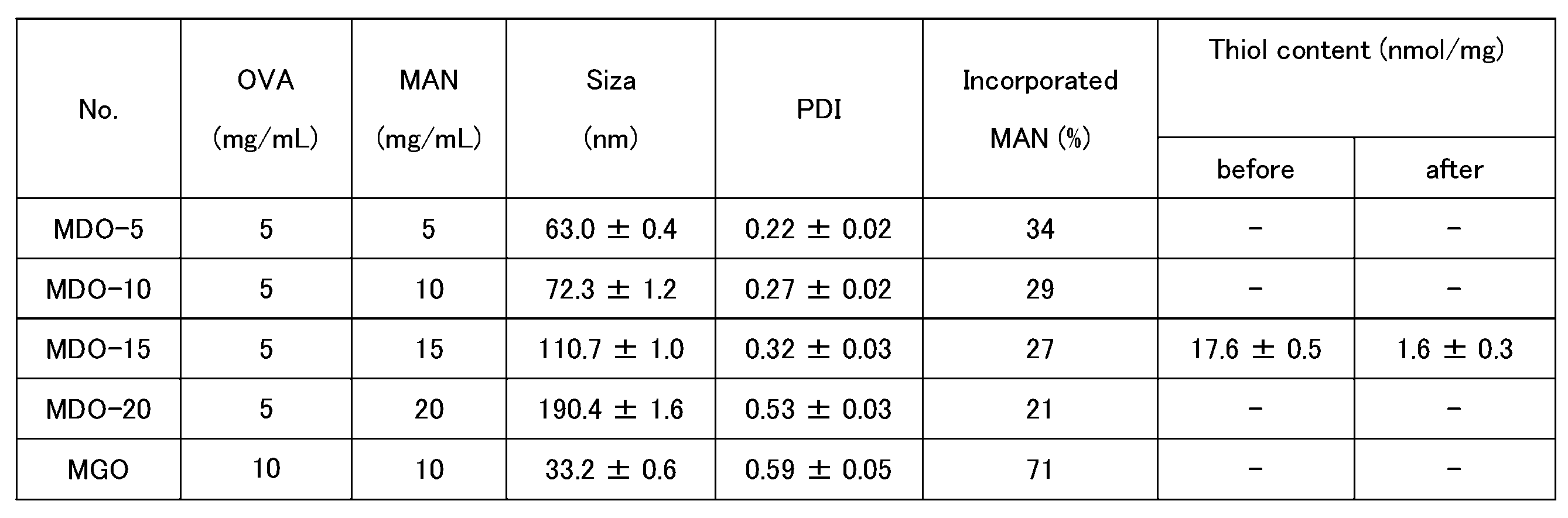
SDS-PAGEは、分子間ジスルフィド結合形成のさらなる証拠となった(図7)。非還元条件(SDSのみ)では、OVAのバンドはdNPでは観察されず、NPでは観察された。dNPのバンドはローディングウェル付近に観察され、OVAの一部がNPから放出されたことが明らかになった。一方、還元条件下(SDS+DTT)では、NP及びdNPはOVA1分子に解離した。SDS処理では、MO-15がMDO-15よりも大きくサイズダウンしたが、DTT処理では両者とも大きな変化はなく、SDSとDTTの併用処理では、粒子径が大きく減少した(図7)。SDS処理でMDO-15のサイズが小さくなったのは、MANがOVAと十分なジスルフィド結合を形成できないためと考えられる。これらの結果から、分子間ジスルフィド結合がMDO NP内に形成されていることが明らかとなった。
MDOの表面にマンナンが提示されていることは、ConA凝集アッセイによってさらに証明された。ConAは4量体レクチンであり、α-マンノースに対する結合部位を4個有している。図8に示すように、OVA dNPの濁度は大きく変化しなかった。一方、MDOでは濁度が有意に上昇し、MANの含有量が多いほど濁度は高くなった。MDO-20表面には、多くのマンナンが存在することが示唆された。MDO-15の単峰性の分布と高いMAN含有量から、以降の実験にはMDO-15を使用した。
<MDOのBMDCへの取り込み>
MDOのDCへの取り込み効率を評価するために、フローサイトメトリーにより平均蛍光強度(MFI)を調べた。OVA、OVA dNP、MDO、及びMGOは、均等にローダミンで標識した。図9に示すように、BMDCのMFIは、時間とともに増加した。MDO及びMGOは、OVA及びOVA dNPよりも効率的にBMDCsに取り込まれた。30分間インキュベート後の蛍光顕微鏡画像を図10に示した。MDO及びMGOでは、細胞質で特に明るい蛍光が観察された。これらの結果から、アレルゲン濃度を上げることなく、ナノ粒子の形成により取り込み効率が向上し、さらにMANの修飾により受容体を介したエンドサイトーシスに依存して取り込みが向上することが示された。これらの結果から、アレルギー治療への有効量を増加させることが可能であることが示された。
MDOのDCへの取り込み効率を評価するために、フローサイトメトリーにより平均蛍光強度(MFI)を調べた。OVA、OVA dNP、MDO、及びMGOは、均等にローダミンで標識した。図9に示すように、BMDCのMFIは、時間とともに増加した。MDO及びMGOは、OVA及びOVA dNPよりも効率的にBMDCsに取り込まれた。30分間インキュベート後の蛍光顕微鏡画像を図10に示した。MDO及びMGOでは、細胞質で特に明るい蛍光が観察された。これらの結果から、アレルゲン濃度を上げることなく、ナノ粒子の形成により取り込み効率が向上し、さらにMANの修飾により受容体を介したエンドサイトーシスに依存して取り込みが向上することが示された。これらの結果から、アレルギー治療への有効量を増加させることが可能であることが示された。
<MDOによる制御性T細胞の誘導>
T細胞応答の制御には、抗原が分解され、DCのMHCクラスIIに提示され、T細胞上のTCRによって認識されることが重要である。ここでは、OVAペプチド/I-AB複合体を認識する特異的TCRを有し、それに応答してIL-2を分泌するOT-II CD4+T細胞を用いて、BMDCのT細胞刺激能を測定した。BMDCをOVA、MDO、及びMGOでそれぞれ3時間パルスした後、OT-II CD4+T細胞と24時間共培養し、培地中の分泌IL-2濃度をELISAキットで測定した。MDO処理により、OVA処理及びMGO処理に比べて、IL-2濃度は有意に上昇した(図11)。また、MDO処理により、OVA処理及びMGO処理と比べて、IL-10濃度が上昇した(図12)。MDO処理により、OVA処理と比べて、ATRAの還元酵素(RALDH)の活性が上昇した(図13)。MGO処理及びMDO処理により、OVA処理に比べて、制御性T細胞の割合が上昇した(図14)。
T細胞応答の制御には、抗原が分解され、DCのMHCクラスIIに提示され、T細胞上のTCRによって認識されることが重要である。ここでは、OVAペプチド/I-AB複合体を認識する特異的TCRを有し、それに応答してIL-2を分泌するOT-II CD4+T細胞を用いて、BMDCのT細胞刺激能を測定した。BMDCをOVA、MDO、及びMGOでそれぞれ3時間パルスした後、OT-II CD4+T細胞と24時間共培養し、培地中の分泌IL-2濃度をELISAキットで測定した。MDO処理により、OVA処理及びMGO処理に比べて、IL-2濃度は有意に上昇した(図11)。また、MDO処理により、OVA処理及びMGO処理と比べて、IL-10濃度が上昇した(図12)。MDO処理により、OVA処理と比べて、ATRAの還元酵素(RALDH)の活性が上昇した(図13)。MGO処理及びMDO処理により、OVA処理に比べて、制御性T細胞の割合が上昇した(図14)。
<抗OVA抗体に対する反応性及びアナフィラキシーショックリスク評価>
MDOのアレルギー治療における安全性を確認するため、MDOと抗OVA抗体との反応性、及び全身性アナフィラキシー反応について検討した。BALB/cマウスにOVA及びミョウバンを0日目、7日目、及び14日目にプライミングし、21日目に血清を採取した。血清中には抗OVA抗体が含まれていた。OVA dNP及びMDOは、OVA及びMGOに比べて、抗OVA抗体との反応性が非常に低く、両者の間に有意差はなかった(図15)。このことから、OVAの変性は抗体結合部位の構造を破壊し、抗体の認識を抑制するのに十分であるが、グルタルアルデヒド抱合体は依然として抗体によって認識される危険性があることが示唆された。全身性アナフィラキシーモデルとして、BALB/cマウスを0日目と14日目にOVA及びミョウバンでプライミングした後、28日目にサンプルを腹腔内投与し、10分ごとに直腸温を検出した(図16)。OVA処理では40分までに基礎体温より3.5℃低い温度まで直腸温の漸減を示した(図17)。MDO及びMGOでは直腸温の低下は見られなかった。このことから、MDO及びMGOは全身性アナフィラキシーを惹起しないことが示唆された。
MDOのアレルギー治療における安全性を確認するため、MDOと抗OVA抗体との反応性、及び全身性アナフィラキシー反応について検討した。BALB/cマウスにOVA及びミョウバンを0日目、7日目、及び14日目にプライミングし、21日目に血清を採取した。血清中には抗OVA抗体が含まれていた。OVA dNP及びMDOは、OVA及びMGOに比べて、抗OVA抗体との反応性が非常に低く、両者の間に有意差はなかった(図15)。このことから、OVAの変性は抗体結合部位の構造を破壊し、抗体の認識を抑制するのに十分であるが、グルタルアルデヒド抱合体は依然として抗体によって認識される危険性があることが示唆された。全身性アナフィラキシーモデルとして、BALB/cマウスを0日目と14日目にOVA及びミョウバンでプライミングした後、28日目にサンプルを腹腔内投与し、10分ごとに直腸温を検出した(図16)。OVA処理では40分までに基礎体温より3.5℃低い温度まで直腸温の漸減を示した(図17)。MDO及びMGOでは直腸温の低下は見られなかった。このことから、MDO及びMGOは全身性アナフィラキシーを惹起しないことが示唆された。
<MDOのプレ投与による気道アレルギー反応の抑制>
MDOの気道アレルギー反応抑制能を評価するために、2種類の投与経路(経口投与と皮下投与)でマウスに投与した。経口投与では、マウスに、PBS、500μgのOVA又は500μgのOVAをMDO又はMGOとして0-4日目及び7-11日目に投与した。14日目及び21日目にOVA及びミョウバンをマウスに皮下注射して感作し、最後に28-31日目にOVAを鼻腔内投与した(図18)。OVA投与の1日後、血清、気管支肺胞洗浄液(BAL)及び肺を採取した。抗OVA IgE値は、OVAプレ投与により低下し、MDOプレ投与及びMGOプレ投与により、より強く低下した(図19)。抗OVA IgG2a値は、MDO処理により有意に上昇したが、IgG1値は差がなかった(図19)。OVAのプレ投与、MDOのプレ投与、及びMGOのプレ投与は、好酸球及び好中球の増加を抑制し、BAL液中の総細胞数を減少させたが、マクロファージ細胞数には有意差を認めなかった(図20)。BAL液中のTh2細胞を介するIL-4、IL-5、IL-6、及びIL-13値は、PBS処理と比較して、MDOプレ投与及びMGOプレ投与により有意に減少した(図21)。OVAのプレ投与でも、Th2サイトカインは減少したが、MDOプレ投与、MGOプレ投与ほど顕著ではなかった。一方、Th1サイトカインであるIFN-γは、BAL液中に検出されなかった(図21)。肺組織の組織学的検査では、ビヒクルプレ投与と比較して、無投与マウス(PBSプレ投与)の肺では、血管周囲および気管支周囲付近に単核細胞の浸潤が広範囲に見られた(図34:経口投与予防PBS)。一方、OVAプレ投与及びMGOプレ投与では、浸潤は減少し、炎症部位はまれであった(図34:経口投与予防OVA,MGO)。また、MDOのプレ投与により、組織の炎症はほぼ正常レベルにまで抑制されたことが印象的であった(図34:経口投与予防MDO)。このように、BALB/cマウスにおいて、MDOの経口投与により、IgEを介した気道アレルギー反応を抑制できることが示唆された。
MDOの気道アレルギー反応抑制能を評価するために、2種類の投与経路(経口投与と皮下投与)でマウスに投与した。経口投与では、マウスに、PBS、500μgのOVA又は500μgのOVAをMDO又はMGOとして0-4日目及び7-11日目に投与した。14日目及び21日目にOVA及びミョウバンをマウスに皮下注射して感作し、最後に28-31日目にOVAを鼻腔内投与した(図18)。OVA投与の1日後、血清、気管支肺胞洗浄液(BAL)及び肺を採取した。抗OVA IgE値は、OVAプレ投与により低下し、MDOプレ投与及びMGOプレ投与により、より強く低下した(図19)。抗OVA IgG2a値は、MDO処理により有意に上昇したが、IgG1値は差がなかった(図19)。OVAのプレ投与、MDOのプレ投与、及びMGOのプレ投与は、好酸球及び好中球の増加を抑制し、BAL液中の総細胞数を減少させたが、マクロファージ細胞数には有意差を認めなかった(図20)。BAL液中のTh2細胞を介するIL-4、IL-5、IL-6、及びIL-13値は、PBS処理と比較して、MDOプレ投与及びMGOプレ投与により有意に減少した(図21)。OVAのプレ投与でも、Th2サイトカインは減少したが、MDOプレ投与、MGOプレ投与ほど顕著ではなかった。一方、Th1サイトカインであるIFN-γは、BAL液中に検出されなかった(図21)。肺組織の組織学的検査では、ビヒクルプレ投与と比較して、無投与マウス(PBSプレ投与)の肺では、血管周囲および気管支周囲付近に単核細胞の浸潤が広範囲に見られた(図34:経口投与予防PBS)。一方、OVAプレ投与及びMGOプレ投与では、浸潤は減少し、炎症部位はまれであった(図34:経口投与予防OVA,MGO)。また、MDOのプレ投与により、組織の炎症はほぼ正常レベルにまで抑制されたことが印象的であった(図34:経口投与予防MDO)。このように、BALB/cマウスにおいて、MDOの経口投与により、IgEを介した気道アレルギー反応を抑制できることが示唆された。
皮下投与では、0日目、2日目、4日目、7日目、9日目、11日目に、PBS又はOVAを20μg、或いはMDO又はMGOとして20μgのOVAをマウスに投与した。その後の経過は経口投与と同じである(図22)。OVAのプレ投与、MDOのプレ投与、及びMGOのプレ投与は、同様のIgE産生抑制効果を示した(図23)。IgG2a値は、MDOプレ投与で増加したが、IgG1値は全群で差はなかった。IgG2a/IgE値及びIgG2a/IgG1値により、MDOプレ投与及びMGOプレ投与がより優れたIgE抑制効果を示すことが示された(図23)。BAL液においても経口投与前と同様に、OVAのプレ投与、MDOのプレ投与、及びMGOのプレ投与により好中球、好酸球の上昇が抑制され、MDOのプレ投与がより顕著な効果を示した(図24)。IL-4、IL-5、IL-6、IL-13は、MDOプレ投与で有意に減少した(図25)。単核球の浸潤は、OVAのプレ投与、及びMGOのプレ投与で減少したが、MDOのプレ投与でより劇的に減少した(図34:皮下投与予防)。これらの結果により、MDOの経口投与及び皮下投与のいずれも予防効果を有することが確認された。
<MDOのポスト投与による気道アレルギー反応の抑制>
既に感作された動物におけるMDO投与のアレルギー治療効果を評価するため、0日目及び7日目にマウスをOVA及びミョウバンで感作し、14-18日目及び21-25日目にPBS、20μgのOVA、或いはOVA500μgをMDO又はMGOとして経口投与した(図26)。32-35日目にOVA経鼻投与が行われた。OVAポスト投与及びMDOポスト投与では、抗OVA IgE値は低下した(図27)。MGOポスト投与では低下傾向が見られたが統計的な差はなかった(図27)。一方、抗OVA IgG1値、及び抗OVA IgG2a値は、すべての投与後群でPBS群との差は認められなかった(図27)。MDOポスト投与により、BAL液中の好中球、好酸球数の減少が観察された(図28)。マクロファージ数には有意な影響は見られなかった(図28)。MDOポスト投与及びMGOポスト投与により、BAL液中のIL-4及びIL-6濃度は低下したが、IL-5及びIL-13は低下しなかった(図29)。これらの結果は、MDOのポスト投与により、すでに誘発されたアレルギー性炎症をダウンレギュレートすることがより困難であることを明らかにした。単核球の浸潤は、OVAのプレ投与、及びMGOのポスト投与で減少したが、MDOのポスト投与でより劇的に減少した(図34:経口投与治療)。
既に感作された動物におけるMDO投与のアレルギー治療効果を評価するため、0日目及び7日目にマウスをOVA及びミョウバンで感作し、14-18日目及び21-25日目にPBS、20μgのOVA、或いはOVA500μgをMDO又はMGOとして経口投与した(図26)。32-35日目にOVA経鼻投与が行われた。OVAポスト投与及びMDOポスト投与では、抗OVA IgE値は低下した(図27)。MGOポスト投与では低下傾向が見られたが統計的な差はなかった(図27)。一方、抗OVA IgG1値、及び抗OVA IgG2a値は、すべての投与後群でPBS群との差は認められなかった(図27)。MDOポスト投与により、BAL液中の好中球、好酸球数の減少が観察された(図28)。マクロファージ数には有意な影響は見られなかった(図28)。MDOポスト投与及びMGOポスト投与により、BAL液中のIL-4及びIL-6濃度は低下したが、IL-5及びIL-13は低下しなかった(図29)。これらの結果は、MDOのポスト投与により、すでに誘発されたアレルギー性炎症をダウンレギュレートすることがより困難であることを明らかにした。単核球の浸潤は、OVAのプレ投与、及びMGOのポスト投与で減少したが、MDOのポスト投与でより劇的に減少した(図34:経口投与治療)。
皮下投与経路では、14日目、16日目、18日目、21日目、23日目、及び25日目に、PBS、20μgのOVA、或いは20μgのOVAをMDO又はMGOとしてマウスに投与した(図30)。MDOポスト投与は、抗OVA IgE産生を減少させた(図31)。BAL液中の好中球、好酸球、及び総細胞数は、MDOポスト投与により減少した(図32)。IL-4、IL-5、IL-6は、MDOポスト投与により減少した(図33)。単核球の浸潤は、OVAのプレ投与、及びMGOのポスト投与で減少したが、MDOのポスト投与でより劇的に減少した(図34:皮下投与治療)。これらの結果により、MDOの経口投与及び皮下投与のいずれも治療効果を有することが確認された。
<MDOのポスト投与によるアレルギー反応の抑制>
0日目及び7日目にマウスをOVA及びミョウバンで感作し、14-15日目及び21日目にSaline、20μgのOVA、或いはOVA500μgをMDO又はMGOとして経口投与した(図35)。32-35日目にOVA経鼻投与が行われた。IV型アレルギー応答を耳の厚みの変化により評価した。MDOポスト投与では、Salineポスト投与と比較して、IV型アレルギー応答が有意に抑制された(図36)。
0日目及び7日目にマウスをOVA及びミョウバンで感作し、14-15日目及び21日目にSaline、20μgのOVA、或いはOVA500μgをMDO又はMGOとして経口投与した(図35)。32-35日目にOVA経鼻投与が行われた。IV型アレルギー応答を耳の厚みの変化により評価した。MDOポスト投与では、Salineポスト投与と比較して、IV型アレルギー応答が有意に抑制された(図36)。
[実施例2]
(材料と方法)
<材料>
ヒト血清アルブミン(HSA)、オバルブミン(OVA、グレードV)、マンノプロテイン(MAN)、ラパマイシン、コンカナバリンA(ConA)、L-システイン及びリポポリサッカライド(LPS、O111:B4)は、Sigma-Aldrichから購入した。日本スギ花粉抗原(Cry j 1およびCry j 2)は、株式会社バイオダイナミクス研究所から購入した。過酸化水素(H2O2,水中30wt%)、硫酸(98%,H2SO4)、塩化カルシウム(CaCl2)、塩化マグネシウム六水和物(MgCl2・6H2O)及び塩化ナトリウム(NaCl)は、富士フイルム和光純薬株式会社から購入した。2,2’-dinitro-5,5’-dithiodi-benzoic acid(DTNB)は、同仁堂研究所から購入した。5(6)-Carboxytetramethylrhodamine N-succinimidyl ester(HNS-Rho)は、Thermo Fisher Scientificから購入した。NuPAGEサンプル還元剤(10×)、NuPAGE LDSサンプルバッファー(4×)、ELISA-ELISPOT希釈液(5×)、TMB(3,3’,5,5’-tetramethylbenzidine)溶液(1×)、HRP結合Streptavidin(ホースラディッシュパーオキシダーゼ)は、Invitrogenから購入した。QuickBlue染色液は、株式会社バイオダイナミクス研究所から購入した。4%パラホルムアルデヒド溶液(PFA)、リン酸水素二ナトリウム及びリン酸二水素ナトリウムは、ナカライテスク株式会社から購入した。ビオチン抗マウスIgE抗体、ビオチン抗マウスIgG1抗体、及びビオチン抗マウスIgG2a抗体は、BioLegendから購入した。
(材料と方法)
<材料>
ヒト血清アルブミン(HSA)、オバルブミン(OVA、グレードV)、マンノプロテイン(MAN)、ラパマイシン、コンカナバリンA(ConA)、L-システイン及びリポポリサッカライド(LPS、O111:B4)は、Sigma-Aldrichから購入した。日本スギ花粉抗原(Cry j 1およびCry j 2)は、株式会社バイオダイナミクス研究所から購入した。過酸化水素(H2O2,水中30wt%)、硫酸(98%,H2SO4)、塩化カルシウム(CaCl2)、塩化マグネシウム六水和物(MgCl2・6H2O)及び塩化ナトリウム(NaCl)は、富士フイルム和光純薬株式会社から購入した。2,2’-dinitro-5,5’-dithiodi-benzoic acid(DTNB)は、同仁堂研究所から購入した。5(6)-Carboxytetramethylrhodamine N-succinimidyl ester(HNS-Rho)は、Thermo Fisher Scientificから購入した。NuPAGEサンプル還元剤(10×)、NuPAGE LDSサンプルバッファー(4×)、ELISA-ELISPOT希釈液(5×)、TMB(3,3’,5,5’-tetramethylbenzidine)溶液(1×)、HRP結合Streptavidin(ホースラディッシュパーオキシダーゼ)は、Invitrogenから購入した。QuickBlue染色液は、株式会社バイオダイナミクス研究所から購入した。4%パラホルムアルデヒド溶液(PFA)、リン酸水素二ナトリウム及びリン酸二水素ナトリウムは、ナカライテスク株式会社から購入した。ビオチン抗マウスIgE抗体、ビオチン抗マウスIgG1抗体、及びビオチン抗マウスIgG2a抗体は、BioLegendから購入した。
<ナノ粒子(NP)の調製>
HSA NPを、様々な条件[バッファー(0mM、50mM又は135mMのNaCl(pH7.4)を含む10mMリン酸バッファー);加熱温度(65℃、75℃又は85℃);加熱時間(5分、10分、30分又は60分)]で調製した。HSAをバッファーに溶解し、4℃で一晩静置した。HSA分散液を0.22μmメンブレンでろ過し、凝集を除去した。1mLのHSA溶液をウォーターバスで加熱した後、直ちに氷浴で冷却した。最後に、H2O2を最終濃度0.3wt%になるようにNP溶液に加え、4℃で一晩保持し、NP内部にジスルフィド結合を形成させた。遊離HSA及びH2O2は、限外ろ過(100kDa Amicon,Merck Millipore)によって取り除いた。
HSA NPを、様々な条件[バッファー(0mM、50mM又は135mMのNaCl(pH7.4)を含む10mMリン酸バッファー);加熱温度(65℃、75℃又は85℃);加熱時間(5分、10分、30分又は60分)]で調製した。HSAをバッファーに溶解し、4℃で一晩静置した。HSA分散液を0.22μmメンブレンでろ過し、凝集を除去した。1mLのHSA溶液をウォーターバスで加熱した後、直ちに氷浴で冷却した。最後に、H2O2を最終濃度0.3wt%になるようにNP溶液に加え、4℃で一晩保持し、NP内部にジスルフィド結合を形成させた。遊離HSA及びH2O2は、限外ろ過(100kDa Amicon,Merck Millipore)によって取り除いた。
同様に、HSA-OVA NP(HO)におけるHSA及びOVAの比率(2.5:2.5、4:1、4.5:0.5、4.75:0.25mg/mL)及びMAN濃度(0mg/mL、5mg/mL、7mg/mL、10mg/mL)の影響を調査した。ラパマイシンを用いる場合、MAN、HSA及びOVAの混合溶液1mLに、1mg/mLのラパマイシンを10μL添加した。HSA、OVA及びMANを溶解するための緩衝液は、10mM PB+50mM NaCl(pH7.4)を用いた。混合液は85℃で60分間加熱した。混合液を冷却後、H2O2を添加し、4℃で一晩静置した後、NP溶液を回収した。100kDaのフィルターで限外ろ過を行い、NPを回収した。
<ラパマイシン負荷効率の測定>
ラパマイシンは、高速液体クロマトグラフィー(HPLC)バリデート法により、Inertsil C8カラム(5μm,4.6mm×150mm)にElite Lachrom L-2455 ダイオードアレイ検出器(日立)を用いて定量した。移動相は、20分以内に、70%メタノール及び30%ミリQ水から90%メタノール及び10%ミリQ水へと変更させた。流速は1.0mL/min、注入量は20μL、カラムオーブン温度は25℃に設定した。フォトダイオードアレイ検出器を用いて、波長277nmでラパマイシンを検出した。
ラパマイシンは、高速液体クロマトグラフィー(HPLC)バリデート法により、Inertsil C8カラム(5μm,4.6mm×150mm)にElite Lachrom L-2455 ダイオードアレイ検出器(日立)を用いて定量した。移動相は、20分以内に、70%メタノール及び30%ミリQ水から90%メタノール及び10%ミリQ水へと変更させた。流速は1.0mL/min、注入量は20μL、カラムオーブン温度は25℃に設定した。フォトダイオードアレイ検出器を用いて、波長277nmでラパマイシンを検出した。
<スギ花粉NPの調製>
HSA NPの他の抗原タンパク質の導入能力を調べるため、OVAをCry j 1とCry j 2の混合物であるスギ花粉抗原に置き換えて、NPを調製した。HSA及びCry jを、10mM PB+50mM NaClバッファーに4.5:0.5mg/mLの割合で溶解し、MANを最終濃度10mg/mLとなるように溶液に添加した。その後、85℃で60分間加熱し、氷上で冷却後、H2O2の添加によりジスルフィド結合を形成させた。限外ろ過後、MAN-HSA-Cry j NP(MHCryj)を得た。
HSA NPの他の抗原タンパク質の導入能力を調べるため、OVAをCry j 1とCry j 2の混合物であるスギ花粉抗原に置き換えて、NPを調製した。HSA及びCry jを、10mM PB+50mM NaClバッファーに4.5:0.5mg/mLの割合で溶解し、MANを最終濃度10mg/mLとなるように溶液に添加した。その後、85℃で60分間加熱し、氷上で冷却後、H2O2の添加によりジスルフィド結合を形成させた。限外ろ過後、MAN-HSA-Cry j NP(MHCryj)を得た。
<BMDC表現型解析>
BMDCを12ウェルプレートに播種し(3×10⁶cells/well)、100ng/mLのLPSで24時間刺激し、PBS、OVA、HO-20、MHO-10、又はMHO-10-R(MHO-10にラパマイシンを封入)で24時間処理した。BMDCを回収し、抗マウスCD16/32抗体で免疫グロブリンのFc受容体に対する非特異結合を阻害した。BMDCを抗マウスI-Ab-FITC、CD86-APC、CD80-BV421及びヨウ化プロピジウム(PI)(Biolegend)で染色した。その後の解析は、フローサイトメトリーで行った。
BMDCを12ウェルプレートに播種し(3×10⁶cells/well)、100ng/mLのLPSで24時間刺激し、PBS、OVA、HO-20、MHO-10、又はMHO-10-R(MHO-10にラパマイシンを封入)で24時間処理した。BMDCを回収し、抗マウスCD16/32抗体で免疫グロブリンのFc受容体に対する非特異結合を阻害した。BMDCを抗マウスI-Ab-FITC、CD86-APC、CD80-BV421及びヨウ化プロピジウム(PI)(Biolegend)で染色した。その後の解析は、フローサイトメトリーで行った。
<その他>
他の方法は、実施例1と同様の方法で、実施した。
他の方法は、実施例1と同様の方法で、実施した。
[結果]
<NPの調製条件の最適化>
マトリックスタンパク質であるHSAからNPを調製するための最適な条件をスクリーニングした。HSAは、中性で負の電荷を帯びているため、HSAの凝集体は安定に分散する。HSA溶液を特定の温度(65-85℃)で60分まで加熱した。その後、H2O2を添加し、ジスルフィド結合を架橋させた。HSA NPのサイズは加熱温度とともに増加した(データ示さず)。これは温度とともに変性が進行するためと考えられる。HSAは65~80℃で変性することが知られている。HSAは、85℃で完全に変性し、凝集が促進されると予想されるため、調製温度として85℃を選択した。塩化ナトリウムを添加すると、静電反発が抑制されるためか、凝集サイズが大きくなる。過剰な塩化ナトリウムはNPを不安定にするため、50mM NaClを選択した。
<NPの調製条件の最適化>
マトリックスタンパク質であるHSAからNPを調製するための最適な条件をスクリーニングした。HSAは、中性で負の電荷を帯びているため、HSAの凝集体は安定に分散する。HSA溶液を特定の温度(65-85℃)で60分まで加熱した。その後、H2O2を添加し、ジスルフィド結合を架橋させた。HSA NPのサイズは加熱温度とともに増加した(データ示さず)。これは温度とともに変性が進行するためと考えられる。HSAは65~80℃で変性することが知られている。HSAは、85℃で完全に変性し、凝集が促進されると予想されるため、調製温度として85℃を選択した。塩化ナトリウムを添加すると、静電反発が抑制されるためか、凝集サイズが大きくなる。過剰な塩化ナトリウムはNPを不安定にするため、50mM NaClを選択した。
凝集体形成に対するHSA及びOVAの混合比の影響を検討した。混合比率は、全濃度(5.0mg/mL)を一定で変化させた。調製したNP(HOシリーズ)のサイズ分布は、混合比に関係なく50nmの単峰性であった(図37、表3、4)。2種類のタンパク質がほぼ定量的にNPに変換されたことは、NP分散液のろ液にタンパク質が含まれないことから確認された(データ示さず)。2つのタンパク質がよく混ざり合っているのは、OVAとHSAとの変性温度範囲が一致しているためと考えられる。OVAは70-80℃で変性する。H2O2で処理すると、残った遊離チオール基が還元され、HOシリーズ NPにジスルフィド結合が形成されたことが確認できた(表3、4)。SDS-PAGEでも、NPにおけるジスルフィド結合形成が確認された(図38)。HOシリーズNPのバンドはローディングウェルの近くに確認された(図38、Not reduced)。一方、DTT処理による還元した場合には、NPは構成タンパク質であるHSAとOVAとに解離することが確認された(図38、Reduced)。
HSA/OVA比を4:1に固定し、HSA/OVA NPの調製条件に対するMAN添加量の効果を評価した。得られたNP(MHOシリーズ)は、MAN濃度の増加とともに粒子径とPDIが増加した(図39、表3、4)。フェノール硫酸法で定量したMHO中のMANの含有量は、MAN濃度の増加とともに増加し、MANのNPへの取り込みは濃度依存的であることが示された(表3、4)。MANのマンナン部分が親水性であるため、マンナンはNPの表面をコーティングすることになる。MHO-10を緩衝液(10mM PB+50mM NaCl,pH7.4)に分散し、4℃での保存安定性を確認した。少なくとも28日間の保存でサイズ変化は見られなかった(データ示さず)。
マンナンのNP表面への提示は、α-マンノースとの結合部位を4個有する4量体レクチンであるConAとNPとの凝集によって確認された。図40に示すように、ConA添加後のHO-20の濁度には変化がなかった。一方、MHO-10、MHO-5は、ConA添加により濁度が上昇し、MAN含量の高いMHO-10の方が、濁度が高くなった。これは、MHO-10がMHO-5よりもマンナン表面に高密度に存在しているためと考えられる。その後の検討にはMHO-10を使用した。
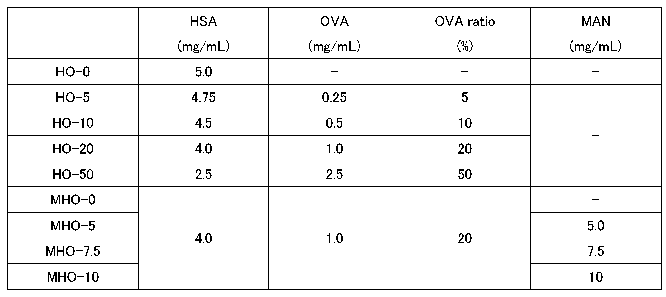
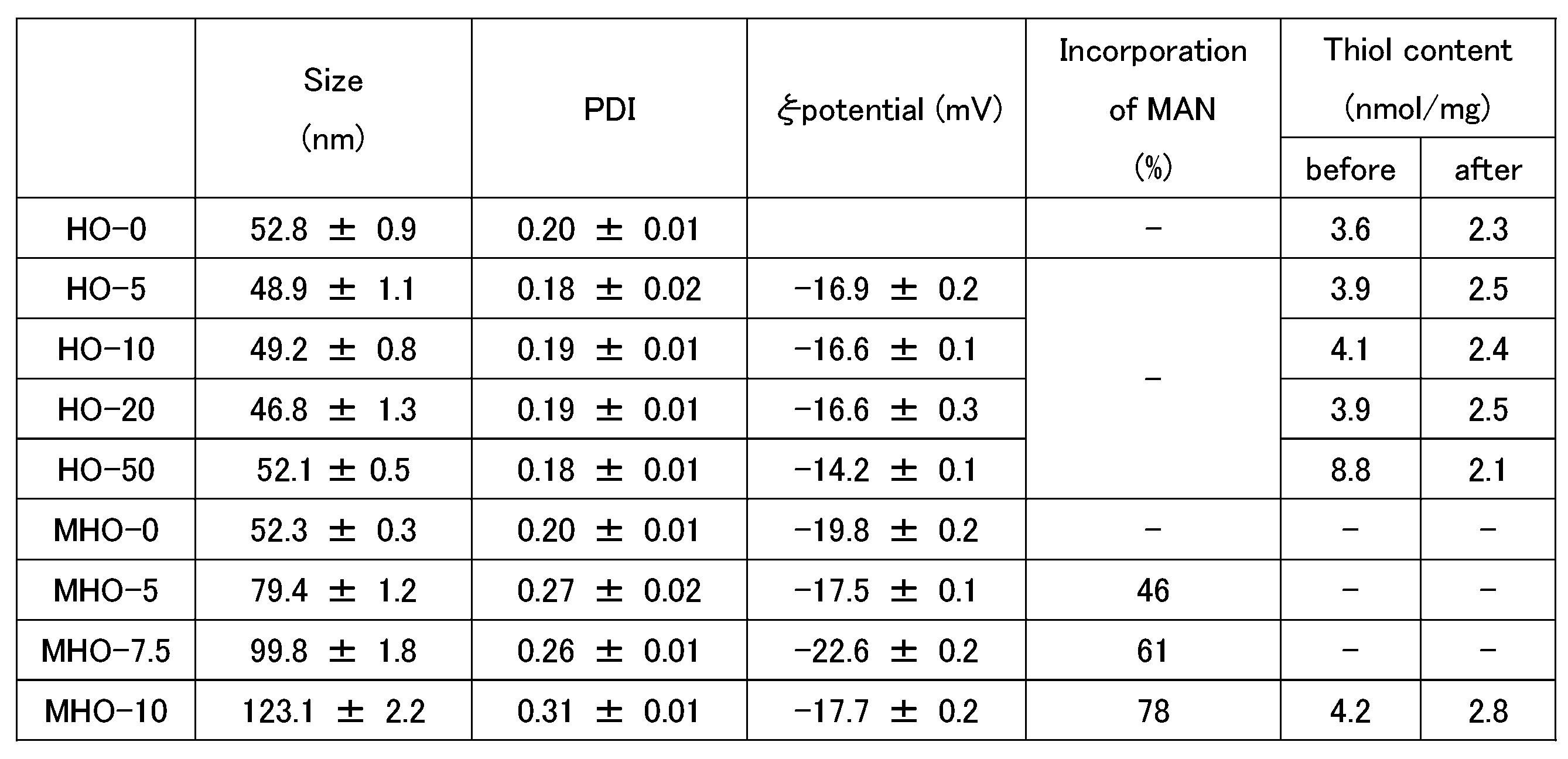
免疫抑制剤であるラパマイシンのMHO-10への封入を試みた。ラパマイシン(10μg/mL=10.9μM)をMHO-10の調製系に混合するだけで、ローディングが行われた。NP分散液の濾液中に残存するラパマイシンをHPLC分析することにより、ほぼ定量的にラパマイシンが取り込まれていることを確認した(データ示さず)。
<Cry j NPの調製>
MANでコートしたHSA NPが他の抗原を凝集する能力を確認するため、OVAをスギ花粉のアレルギー治療に使用されている日本スギ花粉抗原(Cry j 1及びCry j 2の混合物)に置き換えた。4.5mg/mLのHSA、0.5mg/mLのCry j、及び10mg/mLのMANを用いて、上記の方法でNP(MHCryj)を調製した。MHCryjは,MHO-10に比べるとやや大きいが、一様なサイズ分布を示した(図41、表5)。MHCryjのPAGE解析では、還元条件下でCry j及びHSAのバンドが観察され(図42)、非還元条件下ではバンドが観察されなかった。このことから、MHCryjにはジスルフィド結合を介してCry jがほぼ定量的に取り込まれていることが確認された。同様に、Cry jは、55~75℃の変性温度を有するため、Cry jのNP化はNPのサイズに影響を与えないことがわかった(図41)。
MANでコートしたHSA NPが他の抗原を凝集する能力を確認するため、OVAをスギ花粉のアレルギー治療に使用されている日本スギ花粉抗原(Cry j 1及びCry j 2の混合物)に置き換えた。4.5mg/mLのHSA、0.5mg/mLのCry j、及び10mg/mLのMANを用いて、上記の方法でNP(MHCryj)を調製した。MHCryjは,MHO-10に比べるとやや大きいが、一様なサイズ分布を示した(図41、表5)。MHCryjのPAGE解析では、還元条件下でCry j及びHSAのバンドが観察され(図42)、非還元条件下ではバンドが観察されなかった。このことから、MHCryjにはジスルフィド結合を介してCry jがほぼ定量的に取り込まれていることが確認された。同様に、Cry jは、55~75℃の変性温度を有するため、Cry jのNP化はNPのサイズに影響を与えないことがわかった(図41)。

<寛容型樹状細胞(DC)の誘導及び抗原特異的T細胞の活性化>
MHOのBMDCへのターゲティング能力は、フローサイトメトリーで推定した。ここでは、ローダミン標識OVAを使用してMHOを調製した。図43に示すように、BMDCの蛍光強度は時間とともに増加し、MHO-10とインキュベートしたBMDCは、各時点でOVA及びHO-20よりも強い蛍光を示した。インキュベート4時間後のBMDCを蛍光顕微鏡で観察すると、特にMHO-10で細胞質内に明るい蛍光が見られた(図44)。これらの結果は、ナノ粒子の形成がBMDCによる取り込み効率を向上させ、MANの修飾がBMDC上の受容体を標的として取り込みレベルを向上させることを示している。したがって、MHOは生体内におけるDCへの抗原及び薬剤の取り込み効率を向上させることが期待される。
MHOのBMDCへのターゲティング能力は、フローサイトメトリーで推定した。ここでは、ローダミン標識OVAを使用してMHOを調製した。図43に示すように、BMDCの蛍光強度は時間とともに増加し、MHO-10とインキュベートしたBMDCは、各時点でOVA及びHO-20よりも強い蛍光を示した。インキュベート4時間後のBMDCを蛍光顕微鏡で観察すると、特にMHO-10で細胞質内に明るい蛍光が見られた(図44)。これらの結果は、ナノ粒子の形成がBMDCによる取り込み効率を向上させ、MANの修飾がBMDC上の受容体を標的として取り込みレベルを向上させることを示している。したがって、MHOは生体内におけるDCへの抗原及び薬剤の取り込み効率を向上させることが期待される。
NPが寛容なDCを誘導する効果があるかどうかを評価するために、NPとのインキュベーショト後のBMDCの表現型の変化を測定した。図45に示すように、LPSで刺激したBMCは、MHCクラスII(I-Ab)、共刺激因子CD80及びCD86を高レベルで発現し、BMDCが成熟していることが示された。成熟したBMDCをOVAとHO-20で処理すると、I-Ab、CD80、CD86の発現は大きく変化しなかった。一方、MHO-10及びMHO-10-R(MHO-10にラパマイシンを封入)は、I-Ab、CD80、及びCD86の減少を引き起こし、特にMHO-10-Rは顕著な効果を示した。このことから、MHO-10とMHO-10-Rは、BMDCの活性化を抑制する能力を有し、MHO-10-Rはラパマイシンの効果により寛容型DCを誘導する可能性が示唆された。MANはマンノース受容体及びDC-SIGNを介したシグナル伝達を誘導し、NF-κBの活性化を引き起こす。ラパマイシンは、MANとは異なるシグナル伝達経路であるmammalian target of rapamycinの阻害を介してNF-κBの活性化を高める。そのため、MANとラパマイシンが相乗的に寛容型DCを誘導する可能性がある。
抗原特異的寛容性の誘導には、抗原が分解され、DCのMHCクラスIIに提示され、T細胞上のTCRによって認識されることが重要である。OVAペプチド/I-Ab複合体に特異的なTCRを有するOT-II CD4+T細胞を用いて、抗原提示と認識とを確認した。図46に示すように、刺激されたT細胞から分泌されたIL-2のレベルは、OVA群及びHO群で同等であり、HOがBMDC内で分解されOVAペプチドを提示できることが示唆された。MHO群では、PBS群に比べ、IL-2濃度が有意に上昇した。これは、MHOのBMDCへの取り込み効率が高いため、MHOをパルスしたBMDCがより多くのペプチドを提示したことを示唆している。このことから、MHOは、BMDCに抗原及び薬剤を効率よく取り込ませることができ、BMDCの寛容な表現型に合わせたペプチドの提示を可能にすることが確認された。
<抗OVA抗体に対する反応性>
HO及びMHOと抗OVA抗体との反応性をELISA法により検討した。抗OVA抗体は、OVA特異的アレルギーを誘発したマウスの血清から取得した。図47に示すように、HO-20及びMHO-10は、OVAよりも抗OVA抗体との反応性が非常に低く、これら2つのNPの間に有意差はなかった。この結果は、MHOが、弱いIgEを介したアレルギー反応を引き起こすことを示す。アレルギー治療において、MHOは、従来の抗原特異的免疫療法で使用されているネイティブ抗原よりも安全であることを示す。
HO及びMHOと抗OVA抗体との反応性をELISA法により検討した。抗OVA抗体は、OVA特異的アレルギーを誘発したマウスの血清から取得した。図47に示すように、HO-20及びMHO-10は、OVAよりも抗OVA抗体との反応性が非常に低く、これら2つのNPの間に有意差はなかった。この結果は、MHOが、弱いIgEを介したアレルギー反応を引き起こすことを示す。アレルギー治療において、MHOは、従来の抗原特異的免疫療法で使用されているネイティブ抗原よりも安全であることを示す。
[実施例3]
Cry j NPがアレルギー反応を起こさないことを評価するため、ヒト末梢血を用いて好塩基球活性化試験を行った。
Cry j NPがアレルギー反応を起こさないことを評価するため、ヒト末梢血を用いて好塩基球活性化試験を行った。
(材料と方法)
<MHCryjの調製>
4.75mg/mLのHSA、0.25mg/mLのCry j 1/2(Cry j 1とCry j 2の混合物)、及び7.5mg/mLのMANを用いて、上記と同様の方法で、NP(MHCryj)を調製した。HSA及びCry j 1/2をPBに溶解し、4℃で一晩静置した。この溶液を、0.22μmメンブレンでろ過し、凝集を除去した。その後、この溶液を、ウォーターバスで加熱(85℃、10分間)した後、直ちに氷浴で冷却した。最後に、H2O2を最終濃度0.1wt%になるようにこの溶液に加え、4℃で一晩保持し、NP内部にジスルフィド結合を形成させた。遊離HSA及びH2O2は、限外ろ過(100kDa Amicon,Merck Millipore)によって取り除いた。
<MHCryjの調製>
4.75mg/mLのHSA、0.25mg/mLのCry j 1/2(Cry j 1とCry j 2の混合物)、及び7.5mg/mLのMANを用いて、上記と同様の方法で、NP(MHCryj)を調製した。HSA及びCry j 1/2をPBに溶解し、4℃で一晩静置した。この溶液を、0.22μmメンブレンでろ過し、凝集を除去した。その後、この溶液を、ウォーターバスで加熱(85℃、10分間)した後、直ちに氷浴で冷却した。最後に、H2O2を最終濃度0.1wt%になるようにこの溶液に加え、4℃で一晩保持し、NP内部にジスルフィド結合を形成させた。遊離HSA及びH2O2は、限外ろ過(100kDa Amicon,Merck Millipore)によって取り除いた。
得られたMHCryjのサイズ及びPDIは、以下の通りであった。
サイズ:123.6±1.6(d.nm)
PDI:0.34±0.04
サイズ:123.6±1.6(d.nm)
PDI:0.34±0.04
<好塩基球活性化試験>
好塩基球活性化試験には、Allergenecityキット(BECKMAN COULTER、東京、日本)を用いた。試験は、前記Allergenecityキットに付属のプロトコルに従って行った。
好塩基球活性化試験には、Allergenecityキット(BECKMAN COULTER、東京、日本)を用いた。試験は、前記Allergenecityキットに付属のプロトコルに従って行った。
好塩基球活性化試験には、エチレンジアミン四酢酸(EDTA)を入れた採血管を用いて採取した健常者の末梢血とスギ花粉症患者の末梢血とを用いた。
アレルゲンとしてCry j 1/2、又はMHCryjを用いて、末梢血を刺激した。Cry j 1/2は、末梢血100μLに対して、Cry j 1/2の添加量が0.01μg、0.1μg、又は1μgとなるようにPBSで希釈し、20μLを末梢血100μLに添加した。MHCryjは、末梢血100μLに対して、Cry j 1/2の添加量が0.01μg、0.1μg、又は1μgとなるようにPBSで希釈し、20μLを末梢血100μLに添加した。ポジティブコントロールとして抗IgE抗体を用いた。ネガティブコントロールとしてPBSを用いた。各末梢血の刺激に用いたサンプルを表6にまとめた。表6中の数値は、末梢血100μLに対する、Cry j 1/2換算の添加量(μg)を示す。
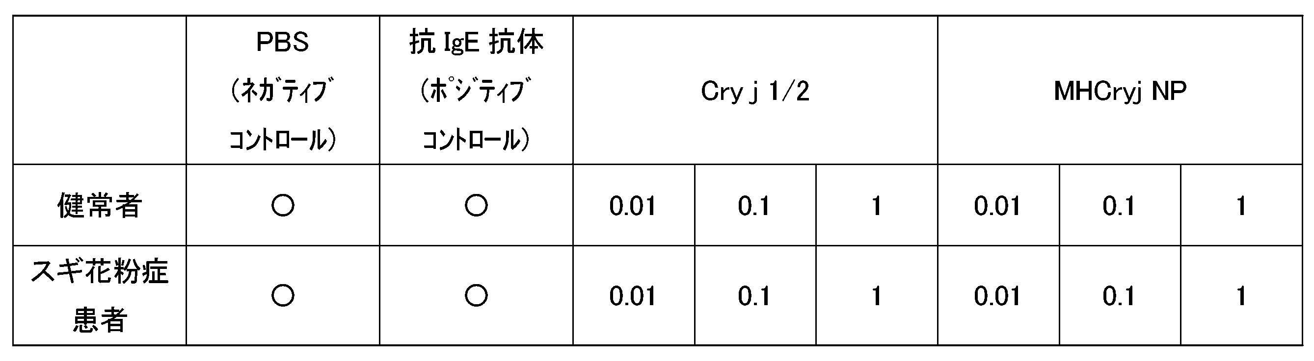
刺激後の各末梢血を、CRTH2(CD294)-FITC、CD203c-PE、及びCD3-PC7で染色し、フローサイトメーターで解析した。その解析結果から、好塩基球の活性化を評価した。
好塩基球の活性化の割合は、CD3陰性の単核球領域におけるCRTH2(CD294)及びCD203cのダブルポジティブの好塩基球集団において、ネガティブコントロール(PBS)と比較してCD203cの蛍光強度が増強した好塩基球の割合を求めた。
好塩基球の活性化の割合は、CD3陰性の単核球領域におけるCRTH2(CD294)及びCD203cのダブルポジティブの好塩基球集団において、ネガティブコントロール(PBS)と比較してCD203cの蛍光強度が増強した好塩基球の割合を求めた。
(結果)
常者の末梢血においては、ポジティブコントロール(抗IgE抗体)では、ネガティブコントロール(PBS)と比較してCD203cの蛍光強度が増強し、活性化好塩基球の割合の増加が確認された(図48、図50左図)。Cry j(0.01μg,0.1μg,1μg)、又はMHCryj(0.01μg,0.1μg,1μg)での刺激では、ネガティブコントロール(PBS)と比較してCD203cの蛍光強度は増強せず、好塩基球の活性化は確認されなかった(図48、図50左図)。
常者の末梢血においては、ポジティブコントロール(抗IgE抗体)では、ネガティブコントロール(PBS)と比較してCD203cの蛍光強度が増強し、活性化好塩基球の割合の増加が確認された(図48、図50左図)。Cry j(0.01μg,0.1μg,1μg)、又はMHCryj(0.01μg,0.1μg,1μg)での刺激では、ネガティブコントロール(PBS)と比較してCD203cの蛍光強度は増強せず、好塩基球の活性化は確認されなかった(図48、図50左図)。
スギ花粉症患者の末梢血においては、ポジティブコントロール(抗IgE抗体)では、ネガティブコントロール(PBS)と比較してCD203cの蛍光強度が増強し、活性化好塩基球の割合の増加が確認された。また、Cry j(0.01μg,0.1μg,1μg)での刺激でも、ネガティブコントロール(PBS)と比較してCD203cの蛍光強度が増強し、活性化好塩基球の割合の増加が確認された(図48、図50右図)。一方、MHCryj(0.01μg,0.1μg,1μg)での刺激では、ネガティブコントロール(PBS)と比較してCD203cの蛍光強度は増強せず、好塩基球の活性は確認されなかった(図48、図50右図)。
健常者の末梢血においては、刺激に用いたCry jに対して末梢血好塩基球の活性化が認められず、一方でスギ花粉症患者の末梢血においては、Cry jに対して末梢血好塩基球の活性化が認められた。この結果は、スギ花粉症患者の好塩基球がCry jに対して抗原特異的にアレルギー反応を起こすことを示している。さらに、スギ花粉症患者の末梢血において、Cry jに対して末梢血好塩基球の活性化が認められたが、MHCryjに対しては末梢血好塩基球の活性化が認められなかった。この結果は、MHCryj刺激では、スギ花粉症患者の好塩基球に対して抗原特異的なアレルギー反応が抑制されることを示している。これらの結果から、アレルギー治療において従来の抗原特異的免疫療法で使用されているネイティブ抗原であるCry jよりも、MHCryjの方が、安全性が高いことが示された。
本発明によれば、IgE抗体に対する反応性が低く、且つ寛容型DCの誘導能の高い、免疫原性複合体、及び前記免疫原性複合体を含有する医薬組成物が提供される。
以上、本発明の好ましい実施例を説明したが、本発明はこれら実施例に限定されることはない。本発明の趣旨を逸脱しない範囲で、構成の付加、省略、置換、およびその他の変更が可能である。本発明は前述した説明によって限定されることはなく、添付のクレームの範囲によってのみ限定される。
Claims (8)
- 抗原と、マンノプロテインと、を含み、
前記抗原と前記マンノプロテインとがジスルフィド結合で結合している、
免疫原性複合体。 - 前記免疫原性複合体が、外殻とコアとを含むナノ粒子を形成しており、
前記外殻が前記マンノプロテインのマンナン鎖を含み、
前記コアが前記抗原を含む、
請求項1に記載の免疫原性複合体。 - 前記抗原がアレルゲン又は自己抗原である、請求項1又は2に記載の免疫原性複合体。
- マトリックスタンパク質をさらに含み、
前記マトリックスタンパク質と、前記抗原及び前記マンノプロテインの少なくとも一方とが、ジスルフィド結合で結合している、
請求項1又は2に記載の免疫原性複合体。 - 免疫調整剤をさらに含む、請求項1又は2に記載の免疫原性複合体。
- 前記免疫調整剤が、レチノイン酸受容体アゴニスト、レチノイドX受容体アゴニスト、ビタミンD受容体アゴニスト、芳香族炭化水素受容体アゴニスト、ヒストン脱アセチル化酵素阻害剤、及びmTOR阻害剤からなる群より選択される少なくとも1種である、請求項5に記載の免疫原性複合体。
- 請求項1又は2に記載の免疫原性複合体を含有する、医薬組成物。
- アレルギー又は自己免疫疾患を治療又は予防するための、請求項7に記載の医薬組成物。
Priority Applications (1)
| Application Number | Priority Date | Filing Date | Title |
|---|---|---|---|
| JP2024561704A JPWO2024117232A1 (ja) | 2022-11-30 | 2023-11-30 |
Applications Claiming Priority (2)
| Application Number | Priority Date | Filing Date | Title |
|---|---|---|---|
| JP2022191916 | 2022-11-30 | ||
| JP2022-191916 | 2022-11-30 |
Publications (1)
| Publication Number | Publication Date |
|---|---|
| WO2024117232A1 true WO2024117232A1 (ja) | 2024-06-06 |
Family
ID=91323900
Family Applications (1)
| Application Number | Title | Priority Date | Filing Date |
|---|---|---|---|
| PCT/JP2023/042983 Ceased WO2024117232A1 (ja) | 2022-11-30 | 2023-11-30 | 免疫原性複合体及び医薬組成物 |
Country Status (2)
| Country | Link |
|---|---|
| JP (1) | JPWO2024117232A1 (ja) |
| WO (1) | WO2024117232A1 (ja) |
Citations (3)
| Publication number | Priority date | Publication date | Assignee | Title |
|---|---|---|---|---|
| JPH0393730A (ja) * | 1989-09-02 | 1991-04-18 | Hayashibara Biochem Lab Inc | 減感作剤 |
| JP2013527854A (ja) * | 2010-05-10 | 2013-07-04 | アセンド・バイオファーマシューティカルズ・ピーティーワイ・リミテッド | 免疫刺激組成物及びワクチン組成物 |
| JP2016515603A (ja) * | 2013-04-03 | 2016-05-30 | インムノテック・ソシエダッド・リミターダInmunotek, S.L. | ワクチン接種における使用のための免疫原性複合体およびこれを得る方法 |
-
2023
- 2023-11-30 JP JP2024561704A patent/JPWO2024117232A1/ja active Pending
- 2023-11-30 WO PCT/JP2023/042983 patent/WO2024117232A1/ja not_active Ceased
Patent Citations (3)
| Publication number | Priority date | Publication date | Assignee | Title |
|---|---|---|---|---|
| JPH0393730A (ja) * | 1989-09-02 | 1991-04-18 | Hayashibara Biochem Lab Inc | 減感作剤 |
| JP2013527854A (ja) * | 2010-05-10 | 2013-07-04 | アセンド・バイオファーマシューティカルズ・ピーティーワイ・リミテッド | 免疫刺激組成物及びワクチン組成物 |
| JP2016515603A (ja) * | 2013-04-03 | 2016-05-30 | インムノテック・ソシエダッド・リミターダInmunotek, S.L. | ワクチン接種における使用のための免疫原性複合体およびこれを得る方法 |
Non-Patent Citations (4)
| Title |
|---|
| BENITO-VILLALVILLA CRISTINA, SORIA IRENE, SUBIZA JOSé LUIS, PALOMARES OSCAR: "Novel vaccines targeting dendritic cells by coupling allergoids to mannan", ALLERGO JOURNAL INTERNATIONAL, vol. 27, no. 8, 1 December 2018 (2018-12-01), pages 256 - 262, XP055780369, DOI: 10.1007/s40629-018-0069-8 * |
| LI SHUNYI, MURAKAMI DAISUKE, NAGATOISHI SATORU, LIU YIWEI, TSUMOTO KOUHEI, KATAYAMA YOSHIKI, MORI TAKESHI: "One-pot preparation of mannan-coated antigen nanoparticles using human serum albumin as a matrix for tolerance induction", JOURNAL OF COLLOID AND INTERFACE SCIENCE, ACADEMIC PRESS,INC., US, vol. 649, 1 November 2023 (2023-11-01), US , pages 955 - 965, XP093177016, ISSN: 0021-9797, DOI: 10.1016/j.jcis.2023.06.170 * |
| LI SHUNYI, TORIUMI HIROKI, TAKAHASHI DAISUKE, KAMASAKI TOMOKO, FUJIOKA YOICHIRO, NAGATOISHI SATORU, LI JINTING, LIU YIWEI, HOSOKAW: "Safe and efficient oral allergy immunotherapy using one-pot-prepared mannan-coated allergen nanoparticles", BIOMATERIALS, ELSEVIER, AMSTERDAM, NL, vol. 303, 1 December 2023 (2023-12-01), AMSTERDAM, NL , pages 122381, XP093177019, ISSN: 0142-9612, DOI: 10.1016/j.biomaterials.2023.122381 * |
| SIRVENT SOFIA; SORIA IRENE; CIRAUQUI CRISTINA; CASES BARBARA; MANZANO ANA I.; DIEZ-RIVERO CARMEN M.; RECHE PEDRO A.; LóPEZ-RE: "Novel vaccines targeting dendritic cells by coupling allergoids to nonoxidized mannan enhance allergen uptake and induce functional regulatory T cells through programmed death ligand 1", JOURNAL OF ALLERGY AND CLINICAL IMMUNOLOGY, ELSEVIER, AMSTERDAM, NL, vol. 138, no. 2, 13 April 2016 (2016-04-13), AMSTERDAM, NL , pages 558, XP029672342, ISSN: 0091-6749, DOI: 10.1016/j.jaci.2016.02.029 * |
Also Published As
| Publication number | Publication date |
|---|---|
| JPWO2024117232A1 (ja) | 2024-06-06 |
Similar Documents
| Publication | Publication Date | Title |
|---|---|---|
| US20240000830A1 (en) | Immune-modifying nanoparticles for the treatment of inflammatory diseases | |
| EP2493487B1 (en) | Composition to induce specific immune tolerance | |
| Afify et al. | Bovine holo-beta-lactoglobulin cross-protects against pollen allergies in an innate manner in BALB/c mice: potential model for the farm effect | |
| JP6557140B2 (ja) | ペプチドコンジュゲート粒子 | |
| JP5191234B2 (ja) | 免疫疾患の予防ないし治療剤および方法 | |
| US20120189700A1 (en) | Nanoparticle Based Immunological Stimulation | |
| US20230381183A1 (en) | USE OF FOLIC ACID AND FOLATE MODIFICATION IN INDUCING B-CELL IMMUNE TOLERANCE AND TARGETING mIgM-POSITIVELY-EXPRESSED B-CELL LYMPHOMA | |
| JP2003527438A (ja) | アレルギーの予防及び/又は治療用組成物及びアレルギーの予防及び/又は治療方法 | |
| Wang et al. | Nanoparticles of Chitosan/Poly (D, L-Lactide-Co-Glycolide) enhanced the immune responses of haemonchus contortus HCA59 antigen in model mice | |
| Tobagus et al. | Adjuvant costimulation during secondary antigen challenge directs qualitative aspects of oral tolerance induction, particularly during the neonatal period | |
| Pali-Schöll et al. | Formulations for allergen immunotherapy in human and veterinary patients: new candidates on the horizon | |
| US20250319155A1 (en) | Preparation of tolerizing nanoparticles for the treatment of peanut allergy | |
| US20220304928A1 (en) | Protein producing nanoliposomes and uses thereof | |
| Afify et al. | Jensen-Jarolim E (2021) Bovine Holo-Beta-Lactoglobulin Cross-Protects Against Pollen Allergies in an Innate Manner in BALB/c Mice: Potential Model for the Farm Effect | |
| HK40114864A (en) | Immune-modifying particles for the treatment of inflammation | |
| RU2782223C2 (ru) | Композиция для индуцирования специфической иммунологической толерантности | |
| WO2019246408A2 (en) | Antibody linked peptide based immunotherapy for type 1 diabetes | |
| HK40009827A (en) | Immune-modifying particles for the treatment of inflammation |
Legal Events
| Date | Code | Title | Description |
|---|---|---|---|
| 121 | Ep: the epo has been informed by wipo that ep was designated in this application |
Ref document number: 23897878 Country of ref document: EP Kind code of ref document: A1 |
|
| WWE | Wipo information: entry into national phase |
Ref document number: 2024561704 Country of ref document: JP |
|
| NENP | Non-entry into the national phase |
Ref country code: DE |
|
| 122 | Ep: pct application non-entry in european phase |
Ref document number: 23897878 Country of ref document: EP Kind code of ref document: A1 |