WO2023276395A1 - キメラサイトカイン受容体 - Google Patents
キメラサイトカイン受容体 Download PDFInfo
- Publication number
- WO2023276395A1 WO2023276395A1 PCT/JP2022/016453 JP2022016453W WO2023276395A1 WO 2023276395 A1 WO2023276395 A1 WO 2023276395A1 JP 2022016453 W JP2022016453 W JP 2022016453W WO 2023276395 A1 WO2023276395 A1 WO 2023276395A1
- Authority
- WO
- WIPO (PCT)
- Prior art keywords
- cells
- receptor
- car
- seq
- amino acid
- Prior art date
- Legal status (The legal status is an assumption and is not a legal conclusion. Google has not performed a legal analysis and makes no representation as to the accuracy of the status listed.)
- Ceased
Links
Images








Classifications
-
- A—HUMAN NECESSITIES
- A61—MEDICAL OR VETERINARY SCIENCE; HYGIENE
- A61K—PREPARATIONS FOR MEDICAL, DENTAL OR TOILETRY PURPOSES
- A61K40/00—Cellular immunotherapy
- A61K40/10—Cellular immunotherapy characterised by the cell type used
- A61K40/11—T-cells, e.g. tumour infiltrating lymphocytes [TIL] or regulatory T [Treg] cells; Lymphokine-activated killer [LAK] cells
-
- A—HUMAN NECESSITIES
- A61—MEDICAL OR VETERINARY SCIENCE; HYGIENE
- A61K—PREPARATIONS FOR MEDICAL, DENTAL OR TOILETRY PURPOSES
- A61K40/00—Cellular immunotherapy
- A61K40/30—Cellular immunotherapy characterised by the recombinant expression of specific molecules in the cells of the immune system
-
- A—HUMAN NECESSITIES
- A61—MEDICAL OR VETERINARY SCIENCE; HYGIENE
- A61K—PREPARATIONS FOR MEDICAL, DENTAL OR TOILETRY PURPOSES
- A61K40/00—Cellular immunotherapy
- A61K40/30—Cellular immunotherapy characterised by the recombinant expression of specific molecules in the cells of the immune system
- A61K40/31—Chimeric antigen receptors [CAR]
-
- A—HUMAN NECESSITIES
- A61—MEDICAL OR VETERINARY SCIENCE; HYGIENE
- A61K—PREPARATIONS FOR MEDICAL, DENTAL OR TOILETRY PURPOSES
- A61K40/00—Cellular immunotherapy
- A61K40/30—Cellular immunotherapy characterised by the recombinant expression of specific molecules in the cells of the immune system
- A61K40/35—Cytokines
-
- A—HUMAN NECESSITIES
- A61—MEDICAL OR VETERINARY SCIENCE; HYGIENE
- A61K—PREPARATIONS FOR MEDICAL, DENTAL OR TOILETRY PURPOSES
- A61K40/00—Cellular immunotherapy
- A61K40/40—Cellular immunotherapy characterised by antigens that are targeted or presented by cells of the immune system
- A61K40/41—Vertebrate antigens
- A61K40/42—Cancer antigens
- A61K40/4202—Receptors, cell surface antigens or cell surface determinants
- A61K40/421—Immunoglobulin superfamily
- A61K40/4211—CD19 or B4
-
- A—HUMAN NECESSITIES
- A61—MEDICAL OR VETERINARY SCIENCE; HYGIENE
- A61K—PREPARATIONS FOR MEDICAL, DENTAL OR TOILETRY PURPOSES
- A61K40/00—Cellular immunotherapy
- A61K40/40—Cellular immunotherapy characterised by antigens that are targeted or presented by cells of the immune system
- A61K40/41—Vertebrate antigens
- A61K40/42—Cancer antigens
- A61K40/4202—Receptors, cell surface antigens or cell surface determinants
- A61K40/4214—Receptors for cytokines
- A61K40/4217—Receptors for interleukins [IL]
-
- A—HUMAN NECESSITIES
- A61—MEDICAL OR VETERINARY SCIENCE; HYGIENE
- A61K—PREPARATIONS FOR MEDICAL, DENTAL OR TOILETRY PURPOSES
- A61K40/00—Cellular immunotherapy
- A61K40/40—Cellular immunotherapy characterised by antigens that are targeted or presented by cells of the immune system
- A61K40/41—Vertebrate antigens
- A61K40/42—Cancer antigens
- A61K40/4254—Adhesion molecules, e.g. NRCAM, EpCAM or cadherins
- A61K40/4255—Mesothelin [MSLN]
-
- A—HUMAN NECESSITIES
- A61—MEDICAL OR VETERINARY SCIENCE; HYGIENE
- A61K—PREPARATIONS FOR MEDICAL, DENTAL OR TOILETRY PURPOSES
- A61K40/00—Cellular immunotherapy
- A61K40/40—Cellular immunotherapy characterised by antigens that are targeted or presented by cells of the immune system
- A61K40/41—Vertebrate antigens
- A61K40/42—Cancer antigens
- A61K40/4256—Tumor associated carbohydrates
- A61K40/4258—Gangliosides, e.g. GM2, GD2 or GD3
-
- A—HUMAN NECESSITIES
- A61—MEDICAL OR VETERINARY SCIENCE; HYGIENE
- A61P—SPECIFIC THERAPEUTIC ACTIVITY OF CHEMICAL COMPOUNDS OR MEDICINAL PREPARATIONS
- A61P35/00—Antineoplastic agents
-
- C—CHEMISTRY; METALLURGY
- C07—ORGANIC CHEMISTRY
- C07K—PEPTIDES
- C07K14/00—Peptides having more than 20 amino acids; Gastrins; Somatostatins; Melanotropins; Derivatives thereof
- C07K14/435—Peptides having more than 20 amino acids; Gastrins; Somatostatins; Melanotropins; Derivatives thereof from animals; from humans
- C07K14/46—Peptides having more than 20 amino acids; Gastrins; Somatostatins; Melanotropins; Derivatives thereof from animals; from humans from vertebrates
- C07K14/47—Peptides having more than 20 amino acids; Gastrins; Somatostatins; Melanotropins; Derivatives thereof from animals; from humans from vertebrates from mammals
- C07K14/4701—Peptides having more than 20 amino acids; Gastrins; Somatostatins; Melanotropins; Derivatives thereof from animals; from humans from vertebrates from mammals not used
- C07K14/473—Peptides having more than 20 amino acids; Gastrins; Somatostatins; Melanotropins; Derivatives thereof from animals; from humans from vertebrates from mammals not used alpha-Glycoproteins
-
- C—CHEMISTRY; METALLURGY
- C07—ORGANIC CHEMISTRY
- C07K—PEPTIDES
- C07K14/00—Peptides having more than 20 amino acids; Gastrins; Somatostatins; Melanotropins; Derivatives thereof
- C07K14/435—Peptides having more than 20 amino acids; Gastrins; Somatostatins; Melanotropins; Derivatives thereof from animals; from humans
- C07K14/705—Receptors; Cell surface antigens; Cell surface determinants
- C07K14/70503—Immunoglobulin superfamily
- C07K14/70521—CD28, CD152
-
- C—CHEMISTRY; METALLURGY
- C07—ORGANIC CHEMISTRY
- C07K—PEPTIDES
- C07K14/00—Peptides having more than 20 amino acids; Gastrins; Somatostatins; Melanotropins; Derivatives thereof
- C07K14/435—Peptides having more than 20 amino acids; Gastrins; Somatostatins; Melanotropins; Derivatives thereof from animals; from humans
- C07K14/705—Receptors; Cell surface antigens; Cell surface determinants
- C07K14/715—Receptors; Cell surface antigens; Cell surface determinants for cytokines; for lymphokines; for interferons
-
- C—CHEMISTRY; METALLURGY
- C07—ORGANIC CHEMISTRY
- C07K—PEPTIDES
- C07K14/00—Peptides having more than 20 amino acids; Gastrins; Somatostatins; Melanotropins; Derivatives thereof
- C07K14/435—Peptides having more than 20 amino acids; Gastrins; Somatostatins; Melanotropins; Derivatives thereof from animals; from humans
- C07K14/705—Receptors; Cell surface antigens; Cell surface determinants
- C07K14/715—Receptors; Cell surface antigens; Cell surface determinants for cytokines; for lymphokines; for interferons
- C07K14/7155—Receptors; Cell surface antigens; Cell surface determinants for cytokines; for lymphokines; for interferons for interleukins [IL]
-
- C—CHEMISTRY; METALLURGY
- C12—BIOCHEMISTRY; BEER; SPIRITS; WINE; VINEGAR; MICROBIOLOGY; ENZYMOLOGY; MUTATION OR GENETIC ENGINEERING
- C12N—MICROORGANISMS OR ENZYMES; COMPOSITIONS THEREOF; PROPAGATING, PRESERVING, OR MAINTAINING MICROORGANISMS; MUTATION OR GENETIC ENGINEERING; CULTURE MEDIA
- C12N15/00—Mutation or genetic engineering; DNA or RNA concerning genetic engineering, vectors, e.g. plasmids, or their isolation, preparation or purification; Use of hosts therefor
- C12N15/09—Recombinant DNA-technology
- C12N15/11—DNA or RNA fragments; Modified forms thereof; Non-coding nucleic acids having a biological activity
- C12N15/62—DNA sequences coding for fusion proteins
-
- A—HUMAN NECESSITIES
- A61—MEDICAL OR VETERINARY SCIENCE; HYGIENE
- A61K—PREPARATIONS FOR MEDICAL, DENTAL OR TOILETRY PURPOSES
- A61K2239/00—Indexing codes associated with cellular immunotherapy of group A61K40/00
- A61K2239/46—Indexing codes associated with cellular immunotherapy of group A61K40/00 characterised by the cancer treated
- A61K2239/48—Blood cells, e.g. leukemia or lymphoma
-
- A—HUMAN NECESSITIES
- A61—MEDICAL OR VETERINARY SCIENCE; HYGIENE
- A61K—PREPARATIONS FOR MEDICAL, DENTAL OR TOILETRY PURPOSES
- A61K2239/00—Indexing codes associated with cellular immunotherapy of group A61K40/00
- A61K2239/46—Indexing codes associated with cellular immunotherapy of group A61K40/00 characterised by the cancer treated
- A61K2239/54—Pancreas
-
- C—CHEMISTRY; METALLURGY
- C07—ORGANIC CHEMISTRY
- C07K—PEPTIDES
- C07K2319/00—Fusion polypeptide
-
- C—CHEMISTRY; METALLURGY
- C07—ORGANIC CHEMISTRY
- C07K—PEPTIDES
- C07K2319/00—Fusion polypeptide
- C07K2319/01—Fusion polypeptide containing a localisation/targetting motif
- C07K2319/02—Fusion polypeptide containing a localisation/targetting motif containing a signal sequence
-
- C—CHEMISTRY; METALLURGY
- C07—ORGANIC CHEMISTRY
- C07K—PEPTIDES
- C07K2319/00—Fusion polypeptide
- C07K2319/01—Fusion polypeptide containing a localisation/targetting motif
- C07K2319/03—Fusion polypeptide containing a localisation/targetting motif containing a transmembrane segment
Definitions
- the present invention relates to methods for producing chimeric cytokine receptors and chimeric antigen receptor (CAR)-introduced cells with long-lasting cytotoxic activity.
- CAR chimeric antigen receptor
- Adoptive immunotherapy is a treatment method that involves collecting immune cells from a donor such as the patient themselves, culturing, stimulating, manipulating, and/or growing them outside the body, and then introducing the cells into the patient.
- Examples of adoptive immunotherapy include tumor-infiltrating lymphocyte therapy, TCR transgenic T-cell therapy, and chimeric antigen receptor-T (CAR-T) cell therapy for cancer patients.
- CAR-T cell therapy is a chimeric antigen receptor that combines a single-chain antibody that recognizes a cancer cell surface antigen with a transmembrane domain and a signaling domain of a co-stimulatory molecule involved in T cell activation. (CAR) into T cells.
- CAR-T cell therapy aims to kill tumor cells by introducing the CAR gene, which encodes the CAR, into T cells taken from the patient and then returned to the patient.
- CAR-T cell therapy treatment methods that target the CD19 molecule for B-cell leukemia and lymphoma are approved in Europe, the United States, and Japan. Its response rate is extremely high, and it has been reported that remission exceeding 80% can be obtained even in refractory cases. Furthermore, the development of CAR-T cell therapy targeting other antigens and cancer types is underway, and the market size is expected to expand in the future.
- Adoptive immunotherapy is expected to be a treatment method that can cure recurrent and refractory tumors, but it has not yet shown a sustained therapeutic effect in many cancers, with the exception of some cancer types.
- CAR-T cell therapy does not provide sustained therapeutic effects is that the cells used for treatment have weakened functions such as proliferative ability.
- cytokine release syndrome associated with CAR-T cell therapy
- cytokines such as IL-6 are released from macrophages activated by GM-CSF secreted by CAR-T cells, resulting in hypotension and multiple organ damage.
- neurotoxic syndrome cytokines such as IL-1 ⁇ are released from macrophages and the like activated by CAR-T cells, resulting in aphasia, psychosis, convulsions, cerebral edema, and the like (Non-Patent Documents 3 and 4).
- adoptive immunotherapy typified by CAR-T cell therapy
- CAR-T cell therapy faces two challenges: improving therapeutic efficacy and reducing side effects.
- the improvement of therapeutic effect correlates with the incidence and grade of side effects. Therefore, it is thought that trying to increase the therapeutic effect will increase the side effects, and conversely, trying to reduce the side effects will also reduce the therapeutic effect, making it difficult to achieve both improved therapeutic effects and reduced side effects at the same time. .
- the purpose of the present invention is to improve the therapeutic effect and reduce the side effects of adoptive immunotherapy. It is to provide a new technique for imparting to immune cells.
- a chimeric receptor based on multiple cytokine receptors as a new artificial receptor that enables both enhancement of functions such as proliferation and cytokine capture in CAR-T cells.
- a cytokine receptor was generated.
- This chimeric cytokine receptor consists of an N-terminal cytokine-binding domain for cytokine capture and a C-terminal T-cell activation domain based on the constitutively active IL-7 (interleukin-7) receptor ⁇ -chain. Configured.
- CAR-T cells introduced with the chimeric cytokine receptor together with the chimeric antigen receptor have enhanced functions such as proliferative potential and cytotoxic activity, and that the CAR-T cells It was found that it acquires the activity of scavenging external cytokines. Furthermore, the present inventors also found that CAR-T cells introduced with the chimeric cytokine receptor of the present invention together with CAR exhibit long-term viability and long-lasting anti-tumor effects in vivo.
- a chimeric cytokine receptor Containing a ligand binding region on the N-terminal side and a T cell activation region on the C-terminal side, said ligand binding domain consists of a cytokine binding domain of a cytokine receptor; the T cell activation region comprises a transmembrane domain and an intracellular domain of IL-7 (interleukin-7) receptor ⁇ chain;
- the transmembrane domain is (a) the amino acid sequence shown in SEQ ID NO: 2 between positions 243 and 244 in the amino acid sequence shown in SEQ ID NO: 1; (b) the amino acid sequence shown in SEQ ID NO: 3 between positions 241 and 242 in the amino acid sequence shown in SEQ ID NO: 1; (c) the amino acid sequence shown in SEQ ID NO: 4 between positions 244 and 245 in the amino acid sequence shown in SEQ ID NO: 1; (d) the amino acid sequence shown in SEQ ID NO:5
- the cytokine receptor is IL-6 (interleukin-6) receptor, IL-1 (interleukin-1) receptor type 2, granulocyte-macrophage colony-stimulating factor (GM-CSF) receptor ⁇ and GM-CSF receptor ⁇ chain.
- IL-6 interleukin-6 receptor
- IL-1 interleukin-1 receptor type 2
- GM-CSF granulocyte-macrophage colony-stimulating factor
- GM-CSF receptor ⁇ chain GM-CSF receptor ⁇ chain.
- a cell preparation comprising the host cell according to any one of (8) to (12).
- a method for producing chimeric antigen receptor (CAR)-introduced cells having long-lasting cytotoxic activity comprising: isolating peripheral blood mononuclear cells from peripheral blood derived from a subject; A step of introducing the gene expression vector of claim 7 and a CAR expression vector into isolated peripheral blood mononuclear cells, wherein the CAR expression vector comprises a base sequence encoding a chimeric antigen receptor (CAR).
- CAR chimeric antigen receptor
- chimeric cytokine receptor of the present invention By introducing the chimeric cytokine receptor of the present invention into immune cells used for adoptive immunotherapy, their functions such as proliferation ability can be enhanced. Furthermore, immune cells into which the chimeric cytokine receptor of the present invention has been introduced can capture extracellular cytokines.
- FIG. 1 shows the structure of the IL-7 receptor ⁇ chain.
- SP indicates a signal peptide.
- FIG. 3 shows the structures of chimeric cytokine receptors IL6R-ca7R and GP130-IL6R-ca7R.
- Figure 2A shows the structure of IL6R-ca7R.
- Figure 2B shows the structure of GP130-IL6R-ca7R.
- SP indicates a signal peptide
- PPCL indicates a constitutively active insertion mutation of 4 amino acid residues Pro-Pro-Cys-Leu (SEQ ID NO: 2).
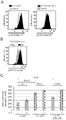
- FIG. 2 shows the results of analysis by flow cytometry of the kinetics of receptor internalization at each time point after administration of recombinant IL-6 to GP130-IL6R-ca7R-introduced CAR-T cells or control cells.
- FIG. 4 is a diagram showing the results of quantification by flow cytometry;
- FIG. 5A shows fluorescence intensity detected by an anti-IL6RA antibody in GP130-IL6R-ca7R-transduced CAR-T cells or control cells at each time point after administration of IL-6.
- FIG. 4 shows the measurement results of cytokine (IL-6) scavenging ability of each CAR-T cell.
- Figure 6A shows the experimental procedure.
- FIG. 6B shows the results of adding IL-6 to each CAR-T cell and measuring the IL-6 concentration in the supernatant.
- FIG. 6C shows the results measured by the same method as in FIG. 6B in "control_cell”, “IL6R-ca7R_cell” and “GP130-IL6R-ca7R_cell”.
- the results of measuring the IL-6 concentration in the supernatant after adding IL-6 to the culture supernatant isolated from each CAR-T cell are "control_sup”, “IL6R-ca7R_sup”, and " GP130-IL6R-ca7R_sup”.
- P values shown in the figure indicate statistical significance by one-way ANOVA.
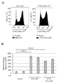
- the results of evaluating phosphorylated STAT3 (pSTAT3) in each CAR-T cell are shown.
- FIG. 7A shows the results of pSTAT3-based flow cytometry in each CAR-T cell.
- Figure 7B shows the results of pSTAT3-based flow cytometry in the presence (IL6+) or absence (IL6-) of IL-6 in each CAR-T cell.
- FIG. 8A shows the results of pSTAT5-based flow cytometry in the absence of IL-6 in each CAR-T cell.
- FIG. 3 shows the cell proliferation rate of each CAR-T cell. Each CD19-targeted CAR-T cell was cultured with the CD19-positive tumor cell line NALM6, and the fold change in the number of CAR-T cells after 7 days relative to initiation of co-culture is shown.
- FIG. 10 shows the results of analyzing the amount of Granzyme B (GZMB) produced after each CAR-T cell was co-cultured with the K562-CD19 cell line or the Raji cell line.
- FIG. 11A shows the results of analyzing the amount of Granzyme B produced after co-culturing with the K562-CD19 cell line.
- FIG. 3 shows the survival rate of tumor cells (K562-CD19 cell line or NALM6 cell line) co-cultured with each CAR-T cell.
- FIG. 10 shows the effect of activity-suppressing mutations Y449F, M452L, or Y456F in the chimeric cytokine receptor GP130-IL6R-ca7R.
- Figure 13A shows the positions of Y449F, M452L, and Y456F in the IL-7 receptor alpha chain.
- FIG. 13B shows the results of Western blot analysis of STAT3, STAT5, and Akt phosphorylation states in CAR-T cells co-introduced with chimeric cytokine receptor GP130-IL6R-ca7R having each mutation.
- FIG. 10 shows the results of functional analysis of CAR-T cells co-introduced with chimeric cytokine receptor GP130-IL6R-ca7R (M452L).
- FIG. 14A shows the fold change in each CAR-T cell number after 7 days of co-culture with the CD19-positive tumor cell line NALM6. P values shown in the figure indicate statistical significance by one-way ANOVA.
- FIG. 14B shows the results of flow cytometry analysis of memory traits of each CAR-T cell after co-culture with NALM6 for 7 days.
- FIG. 14C shows fold changes in the number of CAR-T cells with undifferentiated memory traits (CD62L-positive and CCR7-positive). P values indicate statistical significance by one-way ANOVA.
- FIG. 14A shows the fold change in each CAR-T cell number after 7 days of co-culture with the CD19-positive tumor cell line NALM6. P values shown in the figure indicate statistical significance by one-way ANOVA.
- FIG. 14B shows the results of flow
- FIG. 3 shows the cell proliferation rate of each CAR-T cell.
- Fig. 2 shows the results of co-culture of each mesothelin-targeted CAR-T cell with the K562-mesothelin cell line, staining with anti-IFN- ⁇ antibody and anti-TNF- ⁇ antibody, and analysis by flow cytometry.
- FIG. 1 shows the results of co-culture of each mesothelin-targeted CAR-T cell with the K562-mesothelin cell line, staining with anti-IFN- ⁇ antibody and anti-TNF- ⁇ antibody, and analysis
- FIG. 16A shows the results of flow cytometry.
- FIG. 16B shows the percentage of IFN- ⁇ producing cells.
- Figure 16C shows the percentage of TNF- ⁇ producing cells.
- FIG. 2 shows the results of co-culture of GD2-targeted CAR-T cells with NALM6-GD2 cell line, staining with anti-IFN- ⁇ antibody and anti-TNF- ⁇ antibody, and analysis by flow cytometry.
- FIG. 17A shows the percentage of IFN- ⁇ producing cells.
- Figure 17B shows the percentage of TNF- ⁇ producing cells.
- FIG. 2 shows the structure of chimeric cytokine receptor GP130-IL6R-CD28 (T195P).
- Figure 18A shows the structure of the IL6 receptor alpha subunit (IL6RA).
- Figure 18B shows the structure of GP130-IL6R-CD28(T195P).
- SP indicates a signal peptide
- T195P indicates a constitutively active mutation of CD28.
- FIG. 18C is a diagram showing the cell proliferation rate of each CAR-T cell.
- FIG. 2 shows the structures of chimeric cytokine receptors CSF2RA-ca7R, CSF2RB-ca7R, and IL1R2-ca7R.
- Figure 19A shows the structure of CSF2RA-ca7R.
- Figure 19B shows the structure of CSF2RB-ca7R.
- Figure 19C shows the structure of IL1R2-ca7R.
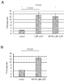
- FIG. 3 shows the results of functional analysis of CAR-T cells co-introduced with both CSF2RA-ca7R and CSF2RB-ca7R.
- FIG. 20A shows the fold change in CAR-T cell numbers after 7 days of co-culturing each CAR-T cell with the CD19 positive tumor cell line NALM6.
- FIG. 20B shows the results of measuring the GM-CSF capture ability of each CAR-T cell. GM-CSF was added to each CAR-T cell, and the results of measuring the GM-CSF concentration in the supernatant are shown.
- Figures 20C and 20D show GM-CSF concentration (Figure 20C) and IL-6 concentration (Figure 20C) and IL-6 concentration (Figure 20C) in the culture medium after each CAR-T cell was co-cultured with NALM6 cells and then monocytic leukemia cell line THP1 was added. 20D) is shown. P values shown in the figure indicate statistical significance by Student's t-test.
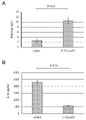
- FIG. 2 shows the measurement results of cell proliferation rate and cytokine (IL-1 ⁇ ) scavenging ability of each CAR-T cell.
- FIG. 21A shows the fold change in the number of CAR-T cells after 7 days of culturing each CD19-targeted CAR-T cell with the CD19-positive tumor cell line NALM6.
- FIG. 1 shows the results of measuring blood IL-6 concentrations 1 hour, 2 hours, and 4 hours after human IL-6 was administered to NSG mice administered with each CAR-T cell via the tail vein. The P value shown in the figure indicates statistical significance by Student's t-test of the logarithmic value of the concentration.
- FIG. 22A shows blood IL-6 concentrations after 1 hour.
- FIG. 22B shows blood IL-6 concentrations after 2 hours.
- FIG. 22C shows blood IL-6 concentrations after 4 hours.
- FIG. 3 shows the results of measurement of cytokine (IL-6 and IL-1 ⁇ ) scavenging ability of CAR-T cells co-introduced with chimeric cytokine receptors and full-length IL-1 receptor type 2.
- FIG. Figure 23-1A shows the experimental procedure.
- FIG. 23-1B shows the results of adding recombinant IL-6 to the CAR-T cells and measuring the IL-6 concentration in the supernatant. "nd" indicates that IL-6 was not detected.
- Figure 23-2D shows the experimental procedure.
- FIG. 23-2E shows the results of culturing the CAR-T cells with the CD19-positive tumor cell line NALM6, adding the monocytic cell line THP-1, and measuring the IL-6 concentration in the supernatant. . "nd" indicates that IL-6 was not detected.
- Graph showing measurement results of CAR-T cells co-introduced with GP130-IL6R-ca7R(M452L) and FMC63-28z for scavenging IL-6 produced from monocyte THP1-CD19/EGFP-Luc2 in vivo. is.
- Figure 24A shows the experimental procedure.
- FIG. 24B shows IL-6 concentrations in plasma 1, 5, and 8 days after administration of CAR-T cells.
- FIG. 2 shows the antitumor effect of GP130-IL6R-ca7R(M452L)-co-introduced CAR-T cells in an in vivo leukemia model.
- Figure 25-1A shows the experimental procedure.
- FIG. 25-2C shows the results of measuring the amount of NALM6 tumor as luciferase luminescence by IVIS Imaging 10, 24, 38, and 60 days after administration of CAR-T cells.
- FIG. 25-3E shows the results of analysis of the progression-free survival rate based on the date of NALM6-GL cell transplantation. P values shown in the figure indicate statistical significance by Log-rank test.
- FIG. 26-1A shows the experimental procedure.
- FIG. 26-2C shows the results of luciferase luminescence measurement of second-generation CAR-T cells that infiltrated into subcutaneous tumors by IVIS Imaging 14 to 35 days after administration of second-generation CAR-T cells.
- FIG. 26-2D shows the quantitative results of logarithmically transforming luciferase luminescence levels.
- FIG. 26-3F shows the results of analysis of the progression-free survival rate based on the date of transplantation of AsPC-1 cells. P values shown in the figure indicate statistical significance by Log-rank test.
- a first aspect of the present invention is a chimeric cytokine receptor.
- the chimeric cytokine receptor of the present invention comprises a ligand-binding domain on the N-terminal side and a T-cell activating domain on the C-terminal side, and the T-cell activating domain is the IL-7 (interleukin-7) receptor ⁇ chain. It contains a transmembrane domain and an intracellular domain.
- the N-terminal ligand-binding domain consists of the cytokine-binding domains of cytokine receptors such as IL-6 receptor, IL-1 receptor type 2, GM-CSF receptor ⁇ chain, or GM-CSF receptor ⁇ chain.
- immune cells such as T cells
- adoptive immunotherapy is a therapeutic method that involves collecting cells such as immune cells from a donor, ex vivo culturing, stimulating, manipulating, and proliferating the cells, and then introducing the cells into the recipient. For example, targeting cancer patients, the cytotoxic activity of immune cells against cancer cells can be enhanced.
- adoptive immunotherapy include tumor-infiltrating lymphocyte therapy, TCR transgenic T cell therapy, chimeric antigen receptor-T (CAR-T) cell therapy, and the like.
- adoptive immunotherapy may also include leukapheresis.
- Adoptive immunotherapy mainly applies to autologous transplantation, in which the donor who provides the cells and the recipient into which the cells are introduced are the same individual, but allogeneic transplantation, in which the donor and recipient are different individuals or xenotransplantation) are also included.
- Tumor-infiltrating lymphocyte therapy refers to collecting lymphocytes (Tumor-Infiltrating Lymphocytes; TIL) that infiltrate tumors from donors, subjecting them to extracorporeal activation, amplification, etc., and then introducing them into recipients. It is a method of treatment that includes
- TCR gene-transferred T cell therapy refers to the introduction of a cancer antigen-specific T cell receptor (TCR) gene into immune cells such as T cells collected from the peripheral blood of a donor.
- a method of treatment comprising introduction into an ent.
- CAR-T cell therapy refers to the introduction of a chimeric antigen receptor (CAR) gene into immune cells such as T cells collected from a donor's peripheral blood to kill cancer cells. It is a therapeutic method that involves generating CAR-T cells that can recognize and attack and introducing them into the recipient, such as by transfusion.
- CAR-T cell therapy is approved for B-cell hematological malignancies such as leukemia and lymphoma, and is expected to be an epoch-making cancer treatment technology following immune checkpoint inhibition therapy.
- CAR Chimeric Antigen Receptor
- a fusion protein comprising an extracellular domain capable of binding to an antigen such as a cancer antigen, a transmembrane domain, and an intracellular domain having signaling activity.
- an antigen such as a cancer antigen
- a transmembrane domain More specifically, a single-chain antibody (scFv) in which variable regions (V L and V H ) of light and heavy chains derived from a monoclonal antibody that recognizes a cancer cell surface antigen is bound, is added to the transmembrane domain. and an artificial antigen receptor that combines the signaling domains of co-stimulatory molecules involved in T cell activation.
- CARs can be broadly classified into first to third generations based on the structure and type of signaling domains contained in CARs.
- First-generation CARs consist solely of CD3 ⁇ (also known as CD247) in the signaling domain.
- Second-generation CARs have a signaling domain containing CD3 ⁇ plus one co-stimulatory molecule such as CD28 or CD137 (4-lBB).
- Representative examples of second-generation CARs include FMC63-28z CAR containing a portion of CD28 as a costimulatory molecule and FMC63-BBz CAR containing a portion of 4-lBB as a costimulatory molecule.
- Both FMC63-28z CAR and FMC63-BBz CAR contain anti-CD19 scFv as scFv.
- CAR-T cells chimeric antigen receptor-T cells
- Cytokine release syndrome refers to symptoms or pathologies caused by the release of cytokines such as inflammatory cytokines. Cytokine release syndrome may develop accompanying administration of drugs such as antibody drugs and CAR-T cell therapy. In CAR-T cell therapy, cytokines such as IL-6 are released from monocytes, macrophages, etc. activated by CAR-T cells, and the cytokines induce fever and increased vascular permeability. Multiple organ failure may result. Cytokine release syndrome developed in 77% of patients receiving CAR-T cell therapy for B-cell acute lymphoblastic leukemia (Maude S.L., et al., N Engl J Med, 2018, 378(5):439-448 .).
- Neurotoxic syndrome refers to symptoms or pathological conditions based on neurotoxicity caused by the administration of drugs, etc.
- CAR-T cell therapy cytokines such as IL-1 ⁇ are released from monocytes, macrophages, etc. activated by CAR-T cells, resulting in neurotoxicity, resulting in aphasia, psychosis, convulsions, cerebral edema, etc.
- cytokines such as IL-1 ⁇
- CAR-T cell therapy cytokines such as IL-1 ⁇ are released from monocytes, macrophages, etc. activated by CAR-T cells, resulting in neurotoxicity, resulting in aphasia, psychosis, convulsions, cerebral edema, etc.
- Approximately 40% of patients receiving CAR-T cell therapy for B-cell acute lymphoblastic leukemia developed neurotoxic syndrome (Maude S.L., et al., N Engl J Med, 2018, 378(5):439 -448.).
- Cytokines is a general term for proteins with a relatively small molecular weight that are secreted from cells. Cytokines are mainly secreted from immune cells and are responsible for information transmission between cells. Examples of cytokines include interleukins, interferons, chemokines, hematopoietic factors, cell growth factors, tumor necrosis factors, and the like. Specific examples of interleukins include IL-1, IL-6, and IL-7 described below. Specific examples of hematopoietic factors include GM-CSF, which will be described later.
- IL-1 interleukin-1
- IL-1 includes IL-1 ⁇ and IL-1 ⁇ .
- IL-1 includes IL-1 derived from any species.
- Specific examples of IL-1 include human IL-1, including human IL-1 ⁇ and human IL-1 ⁇ .
- IL-1 ⁇ can be released from monocytes, macrophages, etc. in neurotoxic syndrome.
- IL-6 interleukin-6
- IL-6 is known as an inflammatory cytokine. IL-6 is produced by T cells, macrophages and the like. As used herein, the term “IL-6” is intended to include IL-6 derived from any species. A specific example of IL-6 is human IL-6. In cytokine release syndrome, IL-6 can be released from monocytes, macrophages, and the like.
- IL-7 interleukin-7
- IL-7 is known as a hematopoietic growth factor. IL-7 is secreted from stromal cells and the like. As used herein, the term “IL-7” is intended to include IL-7 derived from any species. A specific example of IL-7 is human IL-7.
- GM-CSF Granulocyte Macrophage Colony-Stimulating Factor
- CSF2 is a type of cytokine that functions as a hematopoietic growth factor and immunoregulatory factor. It is secreted by T cells, macrophages, NK cells, etc.
- GM-CSF includes GM-CSF derived from any species.
- a specific example of GM-CSF is human GM-CSF.
- a "cytokine receptor” is a receptor to which a cytokine binds as a ligand.
- cytokine receptors include interleukin receptors, interferon receptors, chemokine receptors, hematopoietic factor receptors, cell growth factor receptors, tumor necrosis factor receptors, and the like.
- interleukin receptors include IL-1 receptor, IL-6 receptor, IL-7 receptor ⁇ chain, gp130, and common ⁇ chain described later, as well as IL-2 receptor and IL-3 receptor.
- hematopoietic factor receptors include the GM-CSF receptor described below.
- a specific example of a cell growth factor receptor is the TGF- ⁇ receptor.
- tumor necrosis factor receptors include TNF- ⁇ receptors (eg, types 1 and 2), and FAS receptors.
- IL-1 receptor is a receptor to which IL-1 binds as a ligand, and includes IL-1 receptor type 1 (also referred to herein as IL1R1) and IL-1 receptor. 1 receptor type 2 (also referred to herein as IL1R2) is known.
- IL-1 receptor type 1 includes wild-type and mutant IL-1 receptor type 1 derived from any species.
- human IL-1 receptor type 1 consisting of the amino acid sequence shown in SEQ ID NO: 7 and its orthologues can be mentioned.
- mutant IL-1 receptor type 1 or IL-1 receptor type 1 orthologs include an amino acid sequence in which one or more amino acids are deleted, substituted or added in the amino acid sequence shown in SEQ ID NO: 7, or 60% or more, 70% or more, 80% or more, 85% or more, 90% or more, 95% or more, 96% or more, 97% or more, 98% or more, or 99% of the amino acid sequence shown in SEQ ID NO: 7
- An IL-1 receptor type 1 consisting of an amino acid sequence having the above identity is included.
- IL-1 receptor type 1 gene is a gene that encodes IL-1 receptor type 1.
- a specific example of the IL-1 receptor type 1 gene is an IL-1 receptor type 1 gene encoding IL-1 receptor type 1 consisting of the amino acid sequence shown in SEQ ID NO: 7, such as the base shown in SEQ ID NO: 8.
- a human IL-1 receptor type 1 gene consisting of a sequence is included.
- a nucleotide sequence in which one or more bases are deleted, substituted, or added in the nucleotide sequence shown by SEQ ID NO: 8, or 60% or more, 70% or more, or 80% of the nucleotide sequence shown by SEQ ID NO: 8 % or more, 85% or more, 90% or more, 95% or more, 97% or more, 98% or more, or 99% or more of the base identity includes IL-1 receptor type 1 genes.
- IL-1 receptor type 2 includes wild-type and mutant IL-1 receptor type 2 derived from any species. Examples thereof include human IL-1 receptor type 2 consisting of the amino acid sequence shown in SEQ ID NO: 9 and its orthologs. Examples of mutant IL-1 receptor type 2 or IL-1 receptor type 2 orthologs include an amino acid sequence in which one or more amino acids are deleted, substituted or added in the amino acid sequence shown in SEQ ID NO: 9, or 60% or more, 70% or more, 80% or more, 85% or more, 90% or more, 95% or more, 96% or more, 97% or more, 98% or more, or 99% of the amino acid sequence shown in SEQ ID NO: 9 An IL-1 receptor type 2 consisting of an amino acid sequence having the above identity is included.
- the extracellular domain is included in the region located on the N-terminal side of the transmembrane domain after signal peptide cleavage, eg, positions 14 to 343 in the amino acid sequence shown in SEQ ID NO:9.
- the cytokine-binding region is a region contained in the extracellular domain and capable of binding to a cytokine. It is from 1st to 296th.
- IL-1 receptor type 2 gene is a gene that encodes IL-1 receptor type 2.
- a specific example of the IL-1 receptor type 2 gene is an IL-1 receptor type 2 gene encoding IL-1 receptor type 2 consisting of the amino acid sequence shown in SEQ ID NO: 9, such as the base shown in SEQ ID NO: 10.
- a human IL-1 receptor type 2 gene consisting of a sequence is included.
- a nucleotide sequence in which one or more bases are deleted, substituted or added in the nucleotide sequence shown by SEQ ID NO: 10, or 60% or more, 70% or more, or 80% of the nucleotide sequence shown by SEQ ID NO: 10 % or more, 85% or more, 90% or more, 95% or more, 97% or more, 98% or more, or 99% or more of the base identity includes IL-1 receptor type 2 genes.
- IL-6 receptor is a receptor to which IL-6 binds as a ligand.
- the IL-6 receptor is also known as IL-6 receptor alpha subunit or CD126, also denoted as IL6R or IL6RA.
- the IL-6 receptor binds IL-6 together with gp130 (glycoprotein 130) described later to form a heterotrimer consisting of IL-6, IL-6 receptor, and gp130.
- IL-6 receptors include wild-type and mutant IL-6 receptors derived from any species. Examples thereof include the human IL-6 receptor consisting of the amino acid sequence shown in SEQ ID NO: 11 and its orthologs.
- mutant IL-6 receptors or IL-6 receptor orthologs include an amino acid sequence in which one or more amino acids are deleted, substituted, or added in the amino acid sequence shown in SEQ ID NO: 11, or SEQ ID NO: 11 60% or more, 70% or more, 80% or more, 85% or more, 90% or more, 95% or more, 96% or more, 97% or more, 98% or more, or 99% or more identity to the amino acid sequence shown in and an IL-6 receptor consisting of an amino acid sequence having
- the extracellular domain is included in the region located on the N-terminal side of the transmembrane domain after signal peptide cleavage, eg, positions 21 to 365 in the amino acid sequence shown in SEQ ID NO:11.
- the cytokine-binding region is a region contained in the extracellular domain and capable of binding to a cytokine. It is 338th.
- IL-6 receptor gene is a gene that encodes the IL-6 receptor.
- an IL-6 receptor gene encoding an IL-6 receptor consisting of the amino acid sequence shown in SEQ ID NO: 11, for example, a human IL-6 receptor gene consisting of the nucleotide sequence shown in SEQ ID NO: 12. 6 receptor genes are included.
- a nucleotide sequence in which one or more bases are deleted, substituted or added in the nucleotide sequence shown by SEQ ID NO: 12, or 60% or more, 70% or more, or 80% of the nucleotide sequence shown by SEQ ID NO: 12 % or more, 85% or more, 90% or more, 95% or more, 97% or more, 98% or more, or 99% or more of the base identity includes an IL-6 receptor gene consisting of a nucleotide sequence.
- gp130 (glycoprotein 130) is also known as IL6ST or CD130.
- gp130 includes wild-type and mutant gp130 derived from any species. Examples thereof include human gp130 consisting of the amino acid sequence shown in SEQ ID NO: 13, and its orthologue.
- mutant gp130 or gp130 orthologs include an amino acid sequence in which one or more amino acids are deleted, substituted, or added in the amino acid sequence shown in SEQ ID NO: 13, or 60 amino acids in the amino acid sequence shown in SEQ ID NO: 13 % or more, 70% or more, 80% or more, 85% or more, 90% or more, 95% or more, 96% or more, 97% or more, 98% or more, or gp130 consisting of an amino acid sequence having 99% or more identity mentioned.
- the extracellular domain is included in the region located on the N-terminal side of the transmembrane domain after signal peptide cleavage, for example, positions 23 to 619 in the amino acid sequence shown in SEQ ID NO:13.
- the cytokine-binding region of human gp130 is a region contained in the extracellular domain and capable of binding to cytokines.
- gp130 gene is a gene that encodes gp130. Specific examples of the gp130 gene include the gp130 gene encoding gp130 having the amino acid sequence shown in SEQ ID NO:13, such as the human gp130 gene having the nucleotide sequence shown in SEQ ID NO:14.
- nucleotide sequence in which one or more bases are deleted, substituted or added in the nucleotide sequence shown by SEQ ID NO: 14, or 60% or more, 70% or more, or 80% of the nucleotide sequence shown by SEQ ID NO: 14 % or more, 85% or more, 90% or more, 95% or more, 97% or more, 98% or more, or 99% or more of the base identity includes the gp130 gene consisting of a nucleotide sequence.
- IL-7 receptor (interleukin-7 receptor) is the receptor to which IL-7 binds as a ligand, the IL-7 receptor ⁇ chain and common ⁇ chain (also known as ⁇ c chain or CD132). It is a heterodimer consisting of The major signaling pathways activated by IL-7 binding to the IL-7 receptor include the Jak-Stat and PI3K-Akt pathways. Binding of IL-7 to the IL-7 receptor results in phosphorylation of the Jak kinase and subsequent phosphorylation of tyrosine residues (Y401, Y449, Y456, etc.) in the IL-7 receptor ⁇ -chain.
- pY449 the phosphorylated tyrosine residue at position 449 (pY449) is known to be particularly important (Jiang Q, et al., Mol Cell Biol., 2004, 24(14):6501-13.).
- pY449 serves as a docking site for SH2 domain proteins containing Stat family transcription factors activated by phosphorylation by Jak.
- SOCS family proteins are also known to be involved in IL-7 receptor signaling.
- IL-7 receptor signaling in lymphocytes is known to lead to survival, proliferation and differentiation depending on the developmental stage of lymphocytes.
- IL-7 receptor ⁇ chain is also known as CD127, and is also referred to herein as IL7RA or IL7R ⁇ .
- the IL-7 receptor ⁇ -chain includes wild-type and mutant IL-7 receptor ⁇ -chains derived from any species. Examples thereof include the human IL-7 receptor ⁇ chain consisting of the amino acid sequence shown in SEQ ID NO: 1 and its orthologue.
- mutant IL-7 receptor ⁇ -chain or IL-7 receptor ⁇ -chain orthologue include an amino acid sequence in which one or more amino acids are deleted, substituted or added in the amino acid sequence shown in SEQ ID NO: 1, and 60% or more, 70% or more, 80% or more, 85% or more, 90% or more, 95% or more, 96% or more, 97% or more, 98% or more, or 99% of the amino acid sequence shown in SEQ ID NO: 1
- An IL-7 receptor ⁇ chain consisting of an amino acid sequence having the above identity is included.
- the extracellular domain can be included in the region located on the N-terminal side of the transmembrane domain after signal peptide cleavage, for example, positions 21 to 239 in the amino acid sequence shown in SEQ ID NO:1.
- the transmembrane domain can be positions 240-264 in the amino acid sequence shown in SEQ ID NO:1.
- the intracellular domain may be included at positions 265-459 in the amino acid sequence shown in SEQ ID NO:1.
- the intracellular domain of the human IL-7 receptor ⁇ -chain contains several functional motifs, such as the JAK binding motif (also known as ⁇ BOX1 motif''), the STAT3 association motif ( ⁇ STAT3 association motif”), and the STAT5/PI3K-related motif (also known as the “SH2 domain binding motif”) (Fig. 1).
- JAK binding motif also known as ⁇ BOX1 motif''
- STAT3 association motif ⁇ STAT3 association motif
- STAT5/PI3K-related motif also known as the “SH2 domain binding motif”
- the JAK binding motif is at positions 272-280 (Val-Trp-Pro-Ser-Leu-Pro-Asp-His-Lys sequence, SEQ ID NO: 15), STAT3-related motifs at positions 456-459 (Tyr-Gln-Asn-Gln sequence, SEQ ID NO: 16), and STAT5/PI3K-related motifs at positions 449-452 (Tyr-Val-Thr-Met sequence, sequence number 17).
- a constitutively active mutation is known in the IL-7 receptor ⁇ chain.
- "Constitutively active IL-7 receptor ⁇ chain” or "IL-7 receptor ⁇ chain having a constitutively active mutation” is an IL that can be activated even under conditions where IL-7 is not bound as a ligand. It is a variant of the -7 receptor alpha chain. In cells expressing constitutively active IL-7 receptor ⁇ -chain, at least part of the downstream signaling pathway of IL-7 receptor ⁇ -chain can be activated even in the absence of IL-7.
- mutations that suppress the activity of the above-mentioned functional motif in the intracellular domain of the IL-7 receptor ⁇ chain are known (Fig. 13A).
- Examples of mutations that suppress the activity of STAT5/PI3K-related motifs include the Y449F mutation that replaces the Tyr residue at position 449 with a Phe residue in the amino acid sequence shown in SEQ ID NO: 1, and the amino acid sequence shown in SEQ ID NO: 1 at position 452. and the M452L mutation that replaces the Met residue of with a Leu residue.
- An example of a mutation that suppresses the activity of the STAT3-related motif is the Y456F mutation that substitutes a Phe residue for the Tyr residue at position 456 in the amino acid sequence shown in SEQ ID NO:1.
- IL-7 receptor ⁇ chain gene is a gene that encodes the IL-7 receptor ⁇ chain.
- a specific example of the IL-7 receptor ⁇ -chain gene is an IL-7 receptor ⁇ -chain gene encoding an IL-7 receptor ⁇ -chain consisting of the amino acid sequence shown in SEQ ID NO: 1, such as the base shown in SEQ ID NO: 19.
- a human IL-7 receptor alpha chain gene consisting of a sequence is included.
- a nucleotide sequence in which one or more bases are deleted, substituted or added in the nucleotide sequence shown by SEQ ID NO: 19, or 60% or more, 70% or more, or 80% of the nucleotide sequence shown by SEQ ID NO: 19 % or more, 85% or more, 90% or more, 95% or more, 97% or more, 98% or more, or 99% or more of the base identity includes an IL-7 receptor ⁇ -chain gene consisting of a nucleotide sequence.
- GM-CSF receptor is a receptor to which GM-CSF binds as a ligand, and is composed of GM-CSF receptor ⁇ chain and GM-CSF receptor ⁇ chain. It is a heterodimer that is
- GM-CSF receptor ⁇ chain is also known as CD116, and is also referred to as CSF2RA in this specification.
- the GM-CSF receptor ⁇ -chain includes wild-type and mutant GM-CSF receptor ⁇ -chains derived from any species. Examples thereof include the human GM-CSF receptor ⁇ chain consisting of the amino acid sequence shown in SEQ ID NO: 20 and its orthologue.
- mutant GM-CSF receptor ⁇ -chains or GM-CSF receptor ⁇ -chain orthologues include amino acid sequences in which one or more amino acids are deleted, substituted, or added in the amino acid sequence shown in SEQ ID NO: 20; and 60% or more, 70% or more, 80% or more, 85% or more, 90% or more, 95% or more, 96% or more, 97% or more, 98% or more, or 99% of the amino acid sequence shown in SEQ ID NO: 20
- a GM-CSF receptor ⁇ chain consisting of an amino acid sequence having the above identity is included.
- the extracellular domain is included in the region located on the N-terminal side of the transmembrane domain after signal peptide cleavage, eg, positions 23 to 320 in the amino acid sequence shown in SEQ ID NO:20.
- the cytokine-binding region in the human GM-CSF receptor ⁇ -chain is a region contained in the extracellular domain and capable of binding to cytokines, and consists of positions 25 to 320 in the amino acid sequence shown in SEQ ID NO: 20, for example. region, or the full-length extracellular domain consisting of positions 23-320.
- GM-CSF receptor ⁇ chain gene is a gene that encodes the GM-CSF receptor ⁇ chain.
- a specific example of the GM-CSF receptor ⁇ -chain gene is a GM-CSF receptor ⁇ -chain gene encoding the GM-CSF receptor ⁇ -chain consisting of the amino acid sequence shown in SEQ ID NO:20, such as the base shown in SEQ ID NO:21.
- a human GM-CSF receptor alpha chain gene consisting of a sequence.
- nucleotide sequence in which one or more bases are deleted, substituted or added in the nucleotide sequence shown by SEQ ID NO: 21, or 60% or more, 70% or more, or 80% of the nucleotide sequence shown by SEQ ID NO: 21 GM-CSF receptor ⁇ -chain genes having 90% or more, 85% or more, 90% or more, 95% or more, 97% or more, 98% or more, or 99% or more base identity are included.
- GM-CSF receptor ⁇ chain includes wild-type and mutant GM-CSF receptor ⁇ chains derived from any species. Examples thereof include the human GM-CSF receptor ⁇ chain consisting of the amino acid sequence shown in SEQ ID NO: 22 and its orthologs.
- mutant GM-CSF receptor ⁇ chain or GM-CSF receptor ⁇ chain orthologue include an amino acid sequence in which one or more amino acids are deleted, substituted or added in the amino acid sequence shown in SEQ ID NO: 22; and 60% or more, 70% or more, 80% or more, 85% or more, 90% or more, 95% or more, 96% or more, 97% or more, 98% or more, or 99% of the amino acid sequence shown in SEQ ID NO: 22
- a GM-CSF receptor ⁇ chain consisting of an amino acid sequence having the above identity is included.
- the extracellular domain is included in the region located on the N-terminal side of the transmembrane domain after signal peptide cleavage, eg, positions 17 to 443 in the amino acid sequence shown in SEQ ID NO:22.
- the cytokine-binding region is a region contained in the extracellular domain and capable of binding to cytokines. For example, positions 17 to 132 and/or Or 339th to 436th.
- GM-CSF receptor ⁇ chain gene is a gene that encodes the GM-CSF receptor ⁇ chain.
- a specific example of the GM-CSF receptor ⁇ chain gene is a GM-CSF receptor ⁇ chain gene encoding a GM-CSF receptor ⁇ chain consisting of the amino acid sequence shown in SEQ ID NO:22, such as the base shown in SEQ ID NO:23.
- a human GM-CSF receptor beta chain gene consisting of a sequence.
- nucleotide sequence in which one or more bases are deleted, substituted or added in the nucleotide sequence shown by SEQ ID NO: 23, or 60% or more, 70% or more, or 80% of the nucleotide sequence shown by SEQ ID NO: 23 GM-CSF receptor ⁇ -chain genes having 90% or more, 85% or more, 90% or more, 95% or more, 97% or more, 98% or more, or 99% or more base identity are included.
- chimeric cytokine receptor means a fusion protein containing portions derived from two or more cytokine receptors.
- immune cells include cell types that can function as part of the immune system, as well as undifferentiated cells and progenitor cells (eg, immune progenitor cells) that can differentiate into such cell types.
- specific examples of immune cells include lymphocytes, granulocytes, dendritic cells, macrophages, and monocytes.
- lymphocytes include T cells, B cells, and natural killer cells (NK cells). Lymphocytes may be tumor infiltrating lymphocytes.
- T cells include killer T cells (cytotoxic T cells), helper T cells, and regulatory T cells.
- T cells may be either CD8-positive T-cells or CD4-positive T-cells.
- T cells can be classified into naive T cells, memory T cells, and effector T cells, but any of them may be used. It is known that naive T cells and memory T cells are abundantly contained in peripheral blood, while effector T cells are rarely contained in peripheral blood. Examples of granulocytes include neutrophils, eosinophils, and basophils. In a narrow sense, immune cells may not include stem cells, but in the present specification, stem cells capable of differentiating into lymphocytes (eg, hematopoietic stem cells) are also included.
- stem cells capable of differentiating into lymphocytes (eg, hematopoietic stem cells) are also included.
- a “signal peptide” is an extracellular translocation signal that is required for extracellular secretion of a protein biosynthesized by gene expression, and is also called a signal peptide.
- a signal sequence may comprise a region composed of hydrophobic amino acids. After translation, the signal peptide is cleaved off by a signal peptidase before it is translocated out of the cell. Signal peptide sequences are present at the N-terminus of many secretory and membrane proteins and are, for example, 15-30 amino acids long.
- the signal peptide may be derived from any species and may be human or non-human, such as insect cells or viruses, but is preferably of human origin. A specific example of the signal peptide is a signal peptide derived from human Oncostatin M.
- linker peptide is a peptide that can be inserted between portions in the fusion protein such as the chimeric cytokine receptor of the present invention so that each fused portion performs its intended function.
- the length of the linker peptide is not limited, it is usually 3 to 100 amino acids long, preferably 5 to 50 amino acids long. Peptides containing many amino acids with relatively small side chains, such as serine and glycine, are often used.
- a "tag peptide” is a short peptide consisting of tens of amino acids to several tens of amino acids that can be used for protein labeling, and is used for protein detection and purification. Usually, labeling is carried out by ligating a base sequence encoding a tag peptide to the 5'-end or 3'-end of a gene encoding a protein to be labeled and expressing it as a fusion protein with the tag peptide.
- tag peptides have been developed in the field, and any tag peptide may be used. Specific examples of tag peptides include FLAG, HA, His, PA, and myc.
- the term "capture” refers to binding of a ligand such as a cytokine by the ligand-binding domain contained in the chimeric cytokine receptor or fragment thereof.
- a cytokine is captured by a chimeric cytokine receptor or the like, the function of the cytokine, such as signaling through endogenous cytokine receptors, can be suppressed or inhibited.
- Cytokines captured by chimeric cytokine receptors on cell membranes can be taken up intracellularly.
- plural refers to an integer of 2 or more, for example, an integer of 2 to 10, 2 to 7, 2 to 5, 2 to 4, or 2 to 3.
- amino acid identity refers to the amino acid sequences of two polypeptides to be compared, so that the number of matching amino acid residues is maximized, if necessary, one or both It refers to the ratio (%) of the number of matching amino acid residues to the total number of amino acid residues when aligned with appropriate gaps inserted.
- Base identity nucleotide sequence identity
- amino acid substitution refers to substitution between 20 types of amino acids that constitute natural proteins. Amino acid substitutions are preferably within conservative amino acid groups with similar properties such as charge, side chain, polarity and aromaticity. For example, uncharged polar amino acids with low polarity side chains (Gly, Asn, Gln, Ser, Thr, Cys, Tyr), branched chain amino acids (Leu, Val, Ile), neutral amino acids (Gly, Ile , Val, Leu, Ala, Met, Pro), neutral amino acids with hydrophilic side chains (Asn, Gln, Thr, Ser, Tyr, Cys), acidic amino acids (Asp, Glu), basic amino acids ( Arg, Lys, His), substitutions within the group of aromatic amino acids (Phe, Tyr, Trp).
- uncharged polar amino acids with low polarity side chains Gly, Asn, Gln, Ser, Thr, Cys, Tyr
- branched chain amino acids Leu, Val, Ile
- neutral amino acids Gly, I
- the chimeric cytokine receptor of the present invention comprises an N-terminal ligand binding domain and a C-terminal T cell activation domain.
- the term "ligand binding region” means a region capable of binding to a ligand in the extracellular region of the chimeric cytokine receptor of the present invention.
- the ligand binding domain consists of the cytokine binding domain of the cytokine receptor.
- the cytokine receptor from which the cytokine binding domain is derived is, in principle, a cytokine receptor other than the IL-7 receptor ⁇ -chain.
- cytokine-binding domain is a domain that can bind to a cytokine.
- Cytokines to which the cytokine binding domain binds are not limited and include, for example, IL-6, IL-1, or GM-CSF.
- cytokine-binding domain constituting the ligand-binding domain of the chimeric cytokine receptor of the present invention cytokine-binding domains derived from cytokine receptors other than the IL-7 receptor ⁇ -chain can be used.
- the cytokine binding domain of the IL-6 receptor may be any suitable cytokine binding domain.
- the cytokine binding domain of the IL-1 receptor type 2 may be any cytokine binding domain.
- the cytokine binding domain of the GM-CSF receptor alpha chain may be any suitable cytokine binding domain.
- the cytokine binding domain of the GM-CSF receptor beta chain may be any suitable cytokine binding domain.
- the cytokine-binding domain of the IL-6 receptor is a domain contained in the extracellular domain of the IL-6 receptor and capable of binding to cytokines such as IL-6. More specifically, it may be a region containing the D2 domain and/or D3 domain, which are known to be important for IL-6 binding in the IL-6 receptor (Schwantner A., et al., J. Biol Chem., 2004, 279(1):571-6; Yawata H., et al., EMBO J, 1993, 12(4):1705-1712.). Specific examples of the IL-6 receptor cytokine-binding region include the region consisting of positions 110 to 365 or 110 to 338 in the amino acid sequence shown in SEQ ID NO: 11, and the extracellular region consisting of positions 21 to 365. mentioned.
- the cytokine-binding region of IL-1 receptor type 2 is a region contained in the extracellular domain of IL-1 receptor type 2 and capable of binding to cytokines such as IL-1. More specifically, it may be a region containing three IgC2 domains and a protease cleavage region in IL-1 receptor type 2, and an example of such a region is the extracellular region of IL-1 receptor type 2. (Liu C, et al., J Biol Chem, 1996, 271(34):20965-20972). Specific examples of IL-1 receptor type 2 cytokine-binding regions include the region consisting of positions 14 to 296 and the full-length extracellular region consisting of positions 14 to 343 in the amino acid sequence shown in SEQ ID NO:9.
- the cytokine-binding region of the GM-CSF receptor ⁇ -chain is a region contained in the extracellular domain of the GM-CSF receptor ⁇ -chain and capable of binding to cytokines such as GM-CSF. More specifically, the D1 domain known to be important for GM-CSF binding in the GM-CSF receptor ⁇ chain (eg, the region consisting of positions 25 to 113 in the amino acid sequence shown in SEQ ID NO: 20). and / or D2-D3 domain (also known as Class I cytokine receptor homology module, exemplified by the region consisting of positions 122 to 320 in the amino acid sequence shown in SEQ ID NO: 20).
- a specific example of such a region is the extracellular region of the GM-CSF receptor ⁇ chain, such as the extracellular region consisting of positions 25 to 320 in the amino acid sequence shown in SEQ ID NO:20.
- the cytokine-binding region of the GM-CSF receptor ⁇ chain is a region contained in the extracellular domain of the GM-CSF receptor ⁇ chain, and is a region that can bind to cytokines such as GM-CSF.
- a region containing the D1 domain and/or D4 domain known to be important for GM-CSF binding in the GM-CSF receptor ⁇ chain more preferably a large amount of the ⁇ and ⁇ chains D2 domain and/or D3 domain known to be important for somatogenesis (Example of ⁇ chain binding region in GM-CSF receptor ⁇ chain: positions 133 to 240 and 241 in the amino acid sequence shown in SEQ ID NO: 22 positions to 338) (Hansen G., et al., Cell, 2008, 134(3):496-507; Haman A., et al., J Biol Chem, 1999, 274 (48):34155-63.).
- Such regions include the extracellular region containing positions 17 to 132 and/or positions 339 to 436 in the amino acid sequence shown in SEQ ID NO: 22, for example, the full length of the extracellular region consisting of positions 17 to 443, A region consisting of positions 17-132 and/or positions 339-436 can be mentioned.
- the cytokine receptor may comprise the cytokine binding domain of IL-6 receptor and the ligand binding domain of gp130 (glycoprotein 130).
- the ligand-binding domain of gp130 may be positioned either on the N-terminal side or the C-terminal side of the IL-6 receptor cytokine-binding domain, but is preferably on the N-terminal side.
- the ligand-binding region of gp130 is a region contained in the extracellular domain of gp130, which is capable of binding cytokines such as IL-6. More specifically, it may be a region containing the D1 domain, D2 domain, and/or D3 domain, which are known to be important for IL-6 binding in gp130 (Chow D., et al., Science Roo S., Biochem J, 2001, 356(Pt 2):605-12.).
- a specific example of such a region is the extracellular region of gp130, for example, the extracellular region consisting of positions 23-326 in the amino acid sequence shown in SEQ ID NO:13.
- the "T cell activation domain” is a domain capable of activating T cells in the chimeric cytokine receptor of the present invention.
- the T cell activation domain can activate functions such as T cell proliferative ability and cytotoxic activity.
- the T-cell activating region of the chimeric cytokine receptor of the invention comprises the transmembrane and intracellular domains of the IL-7 (interleukin-7) receptor alpha chain.
- transmembrane domain of the IL-7 receptor ⁇ chain refers to a portion of the IL-7 receptor ⁇ chain consisting of an amino acid sequence that penetrates the cell membrane.
- the transmembrane domain of the IL-7 receptor ⁇ -chain can be determined by known techniques based on the amino acid sequence of the IL-7 receptor ⁇ -chain.
- the transmembrane domain of human IL-7 receptor ⁇ -chain can be positions 240-264 in the amino acid sequence shown in SEQ ID NO:1.
- IL-7 receptor ⁇ -chain intracellular domain refers to a portion of the IL-7 receptor ⁇ -chain consisting of an amino acid sequence located intracellularly.
- the intracellular domain of the IL-7 receptor is a region included in the portion located on the C-terminal side of the transmembrane domain of the IL-7 receptor ⁇ -chain described above.
- the Box1 motif in the amino acid sequence of the IL-7 receptor ⁇ chain for example, positions 272 to 280 in the amino acid sequence shown in SEQ ID NO: 1 and/or the SH2 domain binding motif (for example, position 449 in the amino acid sequence shown in SEQ ID NO: 1) 452), such as the intracellular region consisting of positions 265 to 459 in the amino acid sequence shown in SEQ ID NO:1.
- transmembrane domain and intracellular domain of IL-7 receptor ⁇ chain means the transmembrane domain of IL-7 receptor ⁇ chain and the intracellular domain of IL-7 receptor ⁇ chain described above. It means to include both domains.
- T-cell activating region containing the transmembrane domain and intracellular domain of the IL-7 receptor ⁇ -chain is the region containing positions 240-459 in the amino acid sequence shown in SEQ ID NO:1.
- the T cell activation region in the chimeric cytokine receptor of the present invention preferably has a constitutively active mutation in the transmembrane domain of IL-7 receptor.
- the constitutively active mutation may be any mutation capable of activating T cells, and any constitutively active mutation known in the art can be used.
- a constitutively active mutation may be an insertion mutation. Constitutively active insertional mutations are known (Srochat C., et al., J Exp Med, 2011, 208(5):901-8).
- one or more motifs selected from the group consisting of JAK-binding motifs, STAT3-related motifs, and STAT5/PI3K-related motifs contained in the intracellular domain of the IL-7 receptor ⁇ chain are It can have mutations that suppress activity. By adding mutations to the amino acid sequences that make up these motifs, it is possible to suppress only specific downstream signals, thereby enhancing the therapeutic effect of the chimeric cytokine receptor of the present invention.
- the chimeric cytokine receptor of the present invention may have a mutation at the Y449 residue and/or the M452 residue in the STAT5/PI3K-related motif contained in the intracellular domain of the IL-7 receptor ⁇ chain. can. Also, the chimeric cytokine receptor of the present invention can have a mutation at the Y456 residue in the STAT3-associated motif contained in the intracellular domain of the IL-7 receptor ⁇ -chain.
- the Y449 residue is known to be an important residue involved in the recruitment of STAT3 and STAT5 proteins, and mutation of the Y449 residue can attenuate the cell proliferation signal induced by IL-7 (Lin J.X., et al., Immunity 1995 , 2(4):331-9; Corcoran, A.E., et al., EMBO J, 1996, 15(8): 1924-1932.).
- the M452 residue is known to be involved in the recruitment of PI3K protein, and mutation of the M452 residue attenuates PI3K-Akt signaling, while maintaining STAT3/5 signaling. It does not affect.
- mutation of the M452 residue can promote the formation of memory T cells with long-term viability (Cui G., et al. al., J Immunol, 2020, 204(4):844-857). Mutation of the Y456 residue is also known to attenuate the proliferation signal (Zhong J., et al., BMC Immunol, 2010, 11:5.). Specific examples of Y449, M452 and/or Y456 mutations include Y449F mutation, M452L mutation and/or Y456F mutation.
- the chimeric cytokine receptor of the invention has a M452L mutation in the STAT5/PI3K-associated motif contained in the intracellular domain of the IL-7 receptor ⁇ -chain.
- the chimeric cytokine receptor of the present invention can contain signal peptides, linker peptides, and/or tag peptides, etc., as necessary, in addition to the ligand-binding domain and T-cell activation domain described above. If the ligand-binding region does not contain a signal peptide, the chimeric cytokine receptor of the invention preferably contains a signal peptide at the N-terminal side of the ligand-binding region.
- the ligand binding domain of the chimeric cytokine receptor of the invention is derived from the IL-6 receptor and the T cell activation domain is derived from the IL-7 receptor ⁇ chain.
- Such an example includes a chimeric cytokine receptor consisting of the amino acid sequence shown in SEQ ID NO:24.
- the ligand binding domain of the chimeric cytokine receptor of the invention is derived from the IL-6 receptor
- the T cell activation domain is derived from the IL-7 receptor alpha chain
- the chimeric cytokine receptor is IL- N-terminally to the ligand-binding domain derived from the 6 receptor, it further contains the ligand-binding domain of gp130.
- Such an example includes a chimeric cytokine receptor consisting of the amino acid sequence shown in SEQ ID NO:25.
- the ligand binding domain of the chimeric cytokine receptor of the invention is derived from IL-1 receptor type 2 and the T cell activation domain is derived from IL-7 receptor ⁇ chain.
- IL-1 receptor type 2 IL-1 receptor type 2
- T cell activation domain is derived from IL-7 receptor ⁇ chain.
- Such an example includes a chimeric cytokine receptor consisting of the amino acid sequence shown in SEQ ID NO:26.
- the ligand-binding domain of the chimeric cytokine receptor of the invention is derived from the GM-CSF receptor ⁇ -chain and the T cell activation domain is derived from the IL-7 receptor ⁇ -chain.
- An example of such is a chimeric cytokine receptor consisting of the amino acid sequence shown in SEQ ID NO:27.
- the ligand binding domain of the chimeric cytokine receptor of the invention is derived from the GM-CSF receptor ⁇ chain and the T cell activation domain is derived from the IL-7 receptor ⁇ chain.
- Such an example includes a chimeric cytokine receptor consisting of the amino acid sequence shown in SEQ ID NO:28.
- the chimeric cytokine receptor of the present invention can activate immune cells and capture extracellular cytokines. Based on these two functions, it is possible to achieve both improved therapeutic effects and reduced side effects of adoptive immunotherapy.
- the ligand-binding domain of the chimeric cytokine receptor of the present invention can suppress cytokine activity by capturing extracellular cytokines such as IL-6, IL-1 ⁇ , and GM-CSF. This can suppress or avoid cytokine release syndrome and neurotoxicity syndrome associated with adoptive immunotherapy such as CAR-T cell therapy. More specific suppression/avoidance mechanisms are not limited, but can be exemplified as follows.
- a ligand-binding domain derived from the IL-6 receptor may mediate uptake of IL-6 into the cell.
- the ligand-binding domain derived from IL-1 receptor type 2 is cleaved and released extracellularly as a soluble receptor, traps IL-1 ⁇ , and/or uncleaves IL-1 ⁇ into the cell. May mediate absorption.
- Ligand-binding domains derived from the GM-CSF receptor ⁇ / ⁇ chains reduce activation of monocytes and/or macrophages by GM-CSF by sequestering GM-CSF and thus IL from monocytes/macrophages. It can suppress cytokine secretion such as -6.
- the chimeric cytokine receptor of the present invention can be used regardless of the type of co-introduced T cell receptor or CAR. Therefore, it can be widely applied to adoptive immunotherapy.
- a second aspect of the invention is a nucleic acid encoding a chimeric cytokine receptor.
- nucleic acid encoding a chimeric cytokine receptor may be nucleic acid encoding any of the chimeric cytokine receptors described in the first aspect.
- the base sequences of such nucleic acids are not limiting.
- a nucleic acid comprising, in frame, a nucleic acid encoding a ligand-binding domain on the 5'-end side and a nucleic acid encoding a T-cell activation domain on the 3'-end side.
- a codon-optimized base sequence and a base sequence with an initiation codon (ATG) added to the 5' end are also exemplified.
- the nucleic acid of this aspect is a nucleic acid encoding a chimeric cytokine receptor in which the ligand binding region is derived from the IL-6 receptor and the T cell activation region is derived from the IL-7 receptor ⁇ chain.
- examples thereof include a nucleic acid encoding a chimeric cytokine receptor consisting of the amino acid sequence shown in SEQ ID NO:24, such as a nucleic acid consisting of the base sequence shown in SEQ ID NO:29.
- the nucleic acid of this aspect has a ligand binding region derived from the IL-6 receptor, a T cell activation region derived from the IL-7 receptor ⁇ chain, and a ligand derived from the IL-6 receptor.
- a nucleic acid encoding a chimeric cytokine receptor further comprising a gp130 ligand-binding domain on the N-terminal side of the binding domain. Examples thereof include a nucleic acid encoding a chimeric cytokine receptor consisting of the amino acid sequence shown in SEQ ID NO:25, such as a nucleic acid consisting of the base sequence shown in SEQ ID NO:30.
- the nucleic acid of this aspect is a nucleic acid encoding a chimeric cytokine receptor in which the ligand binding region is derived from IL-1 receptor type 2 and the T cell activation region is derived from IL-7 receptor ⁇ chain. is.
- examples thereof include a nucleic acid encoding a chimeric cytokine receptor consisting of the amino acid sequence shown in SEQ ID NO:26, such as a nucleic acid consisting of the base sequence shown in SEQ ID NO:31.
- the nucleic acid of this aspect is a nucleic acid encoding a chimeric cytokine receptor in which the ligand binding region is derived from the GM-CSF receptor ⁇ chain and the T cell activation region is derived from the IL-7 receptor ⁇ chain.
- examples thereof include a nucleic acid encoding a chimeric cytokine receptor consisting of the amino acid sequence shown in SEQ ID NO:27, such as a nucleic acid consisting of the base sequence shown in SEQ ID NO:32.
- the nucleic acid of this aspect is a nucleic acid encoding a chimeric cytokine receptor in which the ligand binding region is derived from the GM-CSF receptor ⁇ chain and the T cell activation region is derived from the IL-7 receptor ⁇ chain.
- examples thereof include a nucleic acid encoding a chimeric cytokine receptor consisting of the amino acid sequence shown in SEQ ID NO:28, such as a nucleic acid consisting of the base sequence shown in SEQ ID NO:33.
- a third aspect of the present invention is a gene vector (hereinafter referred to as "chimeric cytokine receptor expression vector") containing a nucleic acid encoding a chimeric cytokine receptor in an expressible state.
- the gene expression vector of this aspect is a gene expression vector that contains the nucleic acid of the second aspect and a promoter, and is capable of expressing the chimeric cytokine receptor in cells.
- the gene expression vector may contain constituent elements such as a marker gene (selection marker), an enhancer, a terminator, an origin of replication, and a poly A signal, in addition to the nucleic acid and promoter which are the constituent elements.
- the gene expression vector of this aspect may contain, as the nucleic acid described in the second aspect, a nucleic acid encoding one type of chimeric cytokine receptor, or a combination of nucleic acids encoding two or more types of chimeric cytokine receptors. may be included.
- the combination may be any combination, and the chimeric cytokine receptor and the GM-CSF receptor comprising the cytokine-binding region of the GM-CSF receptor ⁇ chain Combinations with chimeric cytokine receptors containing the cytokine binding region of the ⁇ chain are exemplified. Nucleic acids encoding two or more chimeric cytokine receptors may be contained within the same gene expression vector.
- gene expression vector refers to a vector that contains a gene or gene fragment (hereinafter referred to as "gene, etc.”) in an expressible state and that contains an expression unit that can control the expression of the gene, etc. .
- a gene expression vector may be a plasmid vector or a viral vector.
- the term "state capable of expression” means that a gene or the like to be expressed is arranged in the downstream region of the promoter under the control of the promoter.
- Vectors include plasmid vectors, virus vectors, and the like, and any vector can be used. Plasmid vectors, which can be easily manipulated for gene recombination, or viral vectors, which can easily introduce genes into immune cells, are usually sufficient.
- the plasmid vector may be, for example, a commercially available mammalian cell expression vector such as Promega's pCI vector or pSI vector, or a shuttle vector replicable between mammalian cells and bacteria such as E. coli.
- Viral vectors include, for example, retroviral vectors (including oncoretroviral vectors, lentiviral vectors, and pseudotyped vectors), adenoviral vectors, adeno-associated viral (AAV) vectors, simian viral vectors, vaccinia viral vectors, Sendai viral vectors, Viral vectors such as Epstein-Barr virus (EBV) vectors and HSV vectors can be used. Viral vectors may be used that are replication deficient so that they do not replicate in infected cells.
- retroviral vectors including oncoretroviral vectors, lentiviral vectors, and pseudotyped vectors
- adenoviral vectors including oncoretroviral vectors, lentiviral vectors, and pseudotyped vectors
- AAV adeno-associated viral vectors
- simian viral vectors simian viral vectors
- vaccinia viral vectors Sendai viral vectors
- Viral vectors such as Epstein-Barr virus (EB
- retroviral particles can be produced by selecting a suitable packaging cell and packaging signal sequence based on the LTR sequence.
- packaging cells include PG13 (ATCC® CRL-10686TM), PA317 (ATCC® CRL-9078TM), GP+E-86 and GP+envAm-12 ( U.S. Patent No. 5,278,056), and Psi-Crip (Proceedings of the National Academy of Sciences of the United States of America, vol. 85, pp. 6460-6464 (1988)).
- Retroviral particles can also be produced using 293 cells or 293T cells with high transfection efficiency. Many types of viral vectors produced based on retroviruses and packaging cells that can be used for packaging retroviral vectors are commercially available from many companies.
- promoter refers to a gene expression regulatory region capable of controlling the expression of genes, etc. placed downstream (3' end side) in cells into which a gene expression vector has been introduced. Promoters can be classified into ubiquitous promoters (systemic promoters) and site-specific promoters, based on where genes under expression control are expressed.
- a ubiquitous promoter is a promoter that controls the expression of a target gene or the like (target gene or the like) in all cells, that is, in the entire host individual.
- a site-specific promoter is a promoter that controls the expression of a target gene or the like only in specific cells or tissues.
- the promoter contained in the gene expression vector of the present invention may be either a ubiquitous promoter or a site-specific promoter, preferably capable of inducing expression in immune cells.
- promoters are classified into constitutively active promoters, expression-inducible promoters, and time-specific active promoters based on the timing of expression.
- a constitutively active promoter can constantly express a target gene or the like in a cell.
- An expression-inducible promoter can induce the expression of a target gene or the like in cells at any time.
- the time-specific active promoter can induce the expression of a target gene or the like in cells only at a specific time during the developmental stage. Both promoters can be understood as overexpression type promoters because they can bring about overexpression of target genes in host cells.
- the promoter contained in the gene expression vector of the present invention is preferably a constitutively active promoter that enables long-lasting therapeutic effects.
- the promoter is a promoter capable of inducing the expression of a nucleic acid encoding a chimeric cytokine receptor in cells such as immune cells.
- Target cells into which the gene expression vector of the present invention is introduced are, in principle, mammalian cells, particularly human-derived cells, such as human-derived immune cells. Just do it. Examples thereof include CMV promoter (CMV-IE promoter), SV40 early promoter, RSV promoter, EF1 ⁇ promoter, Ub promoter, 5'LTR promoter and the like.
- CMV promoter CMV-IE promoter
- SV40 early promoter SV40 early promoter
- RSV promoter EF1 ⁇ promoter
- Ub promoter Ub promoter
- 5'LTR promoter a nucleic acid encoding a chimeric cytokine receptor can be placed downstream of the 5' LTR promoter to direct its gene expression.
- a "marker gene” is a gene that encodes a marker protein, also called a selectable marker or reporter protein.
- the term "marker protein” refers to a peptide that can determine the presence or absence of expression of a marker gene based on its activity. Detection of the activity may be carried out by directly detecting the activity of the labeled protein itself, or indirectly through metabolites such as dyes generated by the activity of the labeled protein. good.
- Detection includes biological detection (including detection by binding of peptides and nucleic acids such as antibodies and aptamers), chemical detection (including enzymatic detection), physical detection (including behavioral analysis detection), or detection Any sensory detection of a person (including detection by sight, touch, smell, hearing, and taste) may be used.
- labeling protein encoded by the labeling gene is not particularly limited as long as its activity can be detected by a method known in the art.
- Label proteins that are less invasive to transformants for detection are preferred. Examples thereof include tag peptides, drug-resistant proteins, chromoproteins, fluorescent proteins, and luminescent proteins.
- a "drug-resistant protein” is a protein that imparts resistance to drugs such as antibiotics added to the medium, etc., and is often an enzyme.
- ⁇ -lactamase confers resistance to ampicillin
- aminoglycoside 3′ phosphotransferase confers resistance to kanamycin
- tetracycline efflux transporter confers resistance to tetracycline
- chloramphenicol examples include CAT (chloramphenicol acetyltransferase) that imparts resistance.
- a "chromoprotein” is a protein involved in the biosynthesis of a pigment, or a protein that enables chemical detection of a transformant with a pigment by applying a substrate, usually an enzyme.
- pigment refers to a low-molecular-weight compound or peptide capable of imparting a pigment to a transformant, regardless of the type thereof. Examples include ⁇ -galactosidase (LacZ), ⁇ -glucuronidase (GUS), melanin-based pigment synthetic protein, ommochrome-based pigment, or pteridine-based pigment.
- Fluorescent protein refers to a protein that emits fluorescence of a specific wavelength when irradiated with excitation light of a specific wavelength. It may be either natural or non-natural. Also, the excitation wavelength and fluorescence wavelength are not particularly limited. Specific examples include CFP, RFP, DsRed (including derivatives such as 3xP3-DsRed), YFP, PE, PerCP, APC, GFP (including derivatives such as EGFP and 3xP3-EGFP), and the like. be done.
- Photoprotein refers to a substrate protein capable of emitting light without the need for excitation light or an enzyme that catalyzes the luminescence of that substrate protein. Examples include luciferin or aequorin as a substrate protein and luciferase as an enzyme.
- the “enhancer” is not particularly limited as long as it can enhance the expression efficiency of the gene or fragment thereof in the vector.
- a "terminator” is a sequence capable of terminating transcription of a gene or the like expressed by the activity of the promoter.
- the type of terminator is not particularly limited. A terminator derived from the same species as the promoter is preferred. A terminator paired with the promoter on the genome in a single gene expression control system is particularly preferred.
- a fourth aspect of the present invention is a host cell.
- the host cell of this embodiment comprises a genetic vector that expressably contains a nucleic acid encoding a chimeric cytokine receptor.
- a host cell of this embodiment is, for example, a CAR-T cell containing a chimeric antigen receptor (CAR) expression vector.
- CAR chimeric antigen receptor
- the host cell of this embodiment contains a chimeric cytokine receptor expression vector as an essential component and a CAR expression vector and/or an IL-1 receptor type 2 expression vector as an optional component.
- the chimeric cytokine receptor expression vector which is an essential component, conforms to the description in the third embodiment.
- the host cell of this embodiment may contain one type of chimeric cytokine receptor expression vector, or may contain two or more types of chimeric cytokine receptor expression vectors in combination.
- the combination may be any combination.
- host cell in this embodiment is not limited.
- host cells include immune cells, peripheral blood mononuclear cells (PBMC), cord blood mononuclear cells, skin keratinocytes, mesenchymal stem cells, hematopoietic stem cells, various cancer cell lines, and neural stem cells, iPS cells, and ES cells.
- Preferred host cells are immune cells.
- the immune cells may be T cells, NK cells, or macrophages. T cells are preferably naive T cells and memory T cells.
- Host cells such as immune cells may be cells derived from living organisms, immortalized cell lines, or cells differentiated from ES cells or induced pluripotent stem cells (iPS cells).
- a "chimeric antigen receptor (CAR) expression vector” is a gene expression vector that contains a nucleic acid encoding a chimeric antigen receptor (CAR) and a promoter, and is capable of expressing the CAR intracellularly.
- the CAR expression vector may contain constituent elements such as a marker gene (selection marker), an enhancer, a terminator, a replication origin, and a poly A signal, in addition to the nucleic acid and promoter which are the constituent elements.
- a CAR expression vector may be a plasmid vector or a viral vector.
- the type of CAR expressed by the CAR expression vector is not limited, nor is the type of antigen targeted by the CAR.
- antigens include viral antigens, bacterial antigens, parasite antigens, cell surface markers on target cells associated with particular disease states, tumor antigens, and surface molecules of immune cells.
- CARs preferably target antigens on the surface of tumor cells.
- IL-1 receptor type 2 expression vector is a gene expression vector that contains a nucleic acid encoding IL-1 receptor type 2 and a promoter, and is capable of expressing IL-1 receptor type 2 in cells. .
- the IL-1 receptor type 2 expression vector in addition to the nucleic acid and promoter which are the constituent elements, may optionally include constituent elements such as a marker gene (selection marker), an enhancer, a terminator, a replication origin, and a poly(A) signal. may contain
- the IL-1 receptor type 2 expression vector may be a plasmid vector or a virus vector.
- the host cell of the present invention can capture cytokines extracellularly and optionally absorb cytokines into the cells. Furthermore, based on the T cell activation domain, host cell proliferation ability and cytotoxic activity are enhanced, and therapeutic effects based on cytotoxic activity can be sustained for a long period of time.
- the host cell of this embodiment containing a CAR expression vector can be used as a CAR-T cell for CAR-T cell therapy.
- the host cell of this embodiment comprises a gene expression vector encoding a chimeric cytokine receptor whose ligand-binding region is derived from IL-6 receptor and an IL-1 receptor type 2 expression vector
- the host cell of this embodiment Cells are able to sequester both IL-6 and IL-1 ⁇ . Therefore, both cytokine release syndrome and neurotoxicity syndrome can be suppressed.
- a fifth aspect of the present invention is a cell preparation.
- the cell preparation of this aspect contains the host cell of the fourth aspect as an active ingredient and can be used for adoptive immunotherapy. According to the cell preparation of this embodiment, cytokine release syndrome and neurotoxic syndrome can be suppressed in adoptive immunotherapy, and therapeutic effects can be maintained for a long period of time.
- the term "subject" refers to an object to which the cell preparation of this embodiment is applied.
- a tissue, organ, or individual In the case of an individual, it is, for example, a mammal, preferably a human individual.
- a human individual may be a patient, such as a cancer patient.
- subject information refers to various information related to subject characteristics and conditions. For example, when the subject is a human individual, age, body weight, sex, general health, presence or absence of disease, disease progression and severity, drug sensitivity, presence or absence of concomitant drugs, tolerance to treatment, etc. .
- treatment refers to alleviation or elimination of symptoms associated with disease, prevention or suppression of disease progression, and cure of disease.
- Disease is not limited in this specification. Diseases include, for example, cancer, inflammatory diseases, autoimmune diseases, hepatitis, infectious diseases, and the like. Infectious diseases include viral infections such as influenza and HIV, bacterial infections, and fungal infections. The disease is preferably cancer.
- cancer is not limited, but examples include adenocarcinoma, squamous cell carcinoma, small cell carcinoma and large cell carcinoma.
- Specific cancer types include, for example, malignant melanoma, oral cavity cancer, laryngeal cancer, pharyngeal cancer, thyroid cancer, lung cancer, breast cancer, esophageal cancer, stomach cancer, colon cancer (colon cancer and rectal cancer), small intestine cancer, bladder cancer, prostate cancer, testicular cancer, endometrial cancer, cervical cancer, endometrial cancer, ovarian cancer, stomach cancer, kidney cancer, liver cancer cancer, pancreatic cancer, biliary tract cancer (including gallbladder cancer and bile duct cancer), brain tumor, head and neck cancer, mesothelioma, osteosarcoma, soft tissue sarcoma, glioma, neuroblastoma Pediatric tumors, blood cancers, lymphomas, myeloma, and the like.
- hematological cancers include leukemia (eg, B-cell leukemia, acute lymphoblastic leukemia, chronic lymphocytic leukemia), lymphoma (eg, non-Hodgkin's lymphoma), myeloma (eg, multiple myeloma). mentioned.
- leukemia eg, B-cell leukemia, acute lymphoblastic leukemia, chronic lymphocytic leukemia
- lymphoma eg, non-Hodgkin's lymphoma
- myeloma eg, multiple myeloma
- cancer may be either solid cancer or blood cancer.
- the cell preparation of this aspect includes the host cell of the fourth aspect as an essential active ingredient.
- the cell preparations of this embodiment can contain one or more host cells.
- the content of active ingredients contained in the cell preparation of the present invention is not particularly limited. Generally, the content varies depending on the type of active ingredient, the dosage form, and the type of solvent and carrier that are other constituents described later. Therefore, it is sufficient to determine them as appropriate in consideration of each condition.
- a single dose of cell preparation may contain an effective amount of the active ingredient. However, when it is necessary to administer a large amount of the cell preparation to the subject in order to obtain the pharmacological effect of the active ingredient, it may be administered in several divided doses in order to reduce the burden on the subject. In this case, the total amount of the active ingredients should be an effective amount.
- Effective amount refers to the amount necessary to exhibit the function as an active ingredient, and the amount that imparts little or no harmful side effects to the subject to which it is applied. This effective amount may vary depending on various conditions such as subject information, route of application, and number of applications. Therefore, when using the cell preparation of this aspect as a medicine, the content of the active ingredient is ultimately determined by the judgment of a doctor, pharmacist, or the like.
- the amount of host cells contained in the cell preparation of this embodiment is, for example, 10 5 to 10 10 cells or 10 6 to 10 9 cells, preferably 10 7 to 10 8 cells.
- 0.2 to 5.0 ⁇ 10 6 cells/kg for children weighing 50 kg or less and 0.1 to 2.5 ⁇ 10 8 cells for children weighing over 50 kg are examples.
- For adults, 0.6-6.0 ⁇ 10 8 cells are exemplified.
- the cell preparation of the present invention can contain a pharmaceutically acceptable solvent if necessary.
- “Pharmaceutically acceptable solvent” refers to a solvent commonly used in the field of formulation technology. Examples include water or an aqueous solution, or an organic solvent.
- Aqueous solutions include, for example, saline, isotonic solutions containing glucose or other adjuvants, phosphate buffers, phosphate-buffered saline, sodium acetate buffers, glycol or ethanol solutions.
- Adjuvants include, for example, D-sorbitol, D-mannose, D-mannitol, sodium chloride, low-concentration nonionic surfactants, polyoxyethylene sorbitan fatty acid esters, and the like.
- Organic solvents include ethanol.
- the cell preparation of the present invention can contain a pharmaceutically acceptable carrier if necessary.
- “Pharmaceutically acceptable carrier” refers to additives commonly used in the field of formulation technology. Examples include excipients, human serum albumin, and the like.
- Excipients include, for example, sugars such as monosaccharides, disaccharides, cyclodextrins and polysaccharides, inorganic acid salts such as hydrochlorides, hydrobromides, phosphates or sulfates, acetates, propionates. salts of organic acids such as acid salts, malonates or benzoates, metal salts, citric acid, tartaric acid, glycine, polyethylene glycol, kaolin, silicic acid, or combinations thereof.
- sugars such as monosaccharides, disaccharides, cyclodextrins and polysaccharides
- inorganic acid salts such as hydrochlorides, hydrobromides, phosphates or sulfates, acetates, propionates. salts of organic acids such as acid salts, malonates or benzoates, metal salts, citric acid, tartaric acid, glycine, polyethylene glycol, kaolin, silicic
- solubilizers In addition to the above, if necessary, solubilizers, suspending agents, diluents, dispersing agents, surfactants, soothing agents, stabilizers, absorption enhancers, bulking agents commonly used in pharmaceutical compositions, etc. Preservatives, preservatives, antioxidants, buffers, tonicity agents and the like can also be included as appropriate.
- Carriers are used to avoid or suppress decomposition of the active ingredient by enzymes or the like in the body of the subject, facilitate formulation and administration methods, and maintain the dosage form and efficacy. It can be used appropriately.
- the dosage form of the cell preparation of the present invention is not particularly limited. Any form may be used as long as it can be delivered to the target site without deactivating the active ingredient in the body of the subject.
- the specific dosage form differs depending on the application method described later.
- Application methods can be broadly divided into parenteral administration and oral administration, with parenteral administration being preferred.
- the preferred dosage form is a liquid formulation that can be administered directly to the target site or systemically administered via the circulatory system.
- liquid formulations include injections. Injections can be prepared by appropriately combining solvents, excipients, suspending agents, surfactants, stabilizers, pH adjusters, etc., and mixing them in unit dosage forms generally accepted for pharmaceutical practice. Can be formulated.
- the application method of the cell preparation of the present invention is not particularly limited, and the administration route is not limited.
- parenteral administration may be used.
- Parenteral administration can be further subdivided into systemic administration and topical administration.
- Local administration includes, for example, intradermal administration, intramuscular administration, subcutaneous administration, intraperitoneal administration, intranasal administration, intratumoral administration, tissue administration, and organ administration;
- Intracirculatory administration including intravenous administration (intravenous injection), intraarterial administration and intralymphatic administration.
- a preferred route of administration is intravenous administration, which may also be an infusion administered by infusion (eg, a single intravenous infusion).
- the cell preparation of this aspect provides a method for treating and/or preventing a disease, which comprises administering the cell preparation of the present invention to a subject.
- the disease may be cancer, for example, and the subject may be a cancer patient.
- the therapeutic/preventive methods are characterized by long-lasting effects such as cytotoxic activity and reduced side effects such as cytokine release syndrome and neurotoxic syndrome.
- This therapeutic/prophylactic method may be adoptive immunotherapy, such as CAR-T cell therapy.
- a method for providing anti-tumor immunity to a subject includes the step of administering the cell preparation to the subject.
- chimeric cytokine receptor gene expression vector, host cell, or cell preparation of the present invention in the manufacture of a medicament for treating and/or preventing diseases such as cancer.
- the sixth aspect of the present invention is a method for producing chimeric antigen receptor (CAR)-introduced cells with long-lasting cytotoxic activity.
- the preparation method of this embodiment includes a peripheral blood mononuclear cell isolation step and a vector introduction step as essential steps. According to the production method of this embodiment, CAR-introduced cells having long-lasting cytotoxic activity can be produced. CAR-introduced cells produced by the production method of this embodiment can be used for CAR-T cell therapy in which therapeutic effects are maintained for a long period of time.
- long-lasting cytotoxic activity means long-lasting cytotoxic activity against target cells such as cancer cells.
- long-lasting means, for example, that the therapeutic effects of therapeutic cells in conventional adoptive immunotherapy last longer. More specifically, it means that the therapeutic effect lasts longer than CAR-introduced cells into which the chimeric cytokine receptor expression vector of the present invention has not been introduced.
- detectable cytotoxic activity is 2 days or more, 3 days or more, 4 days or more, 5 days or more, 6 days or more, 1 week or more, 10 days or more, 2 weeks or more, 3 weeks or more, 24 days or more, 4 weeks or more, 1 month or more, 5 weeks or more, 38 days or more, 40 days or more, 6 weeks or more, 45 days or more, 7 weeks or more, 50 days or more, 55 days or more, 60 days or more, 2 months or more, 3 months or more, 4 months or more, 5 months or more, 6 months or more, or 1 year or more.
- the cytotoxic activity "sustains" means that at least detectable cytotoxic activity is maintained over any of the above periods.
- the chimeric cytokine receptor expression vector of the present invention is not introduced. It is sufficient if the cytotoxic activity is maintained higher than that of the CAR-introduced cells.
- the method for producing CAR-introduced cells of this embodiment includes a peripheral blood mononuclear cell isolation step and a vector introduction step as essential steps, and an expansion culture step as a selection step. Each step will be specifically described below.
- the “peripheral blood mononuclear cell isolation step” is a step of isolating peripheral blood mononuclear cells from peripheral blood derived from a subject. The purpose of this step is to isolate peripheral blood mononuclear cells containing immune cells.
- peripheral blood mononuclear cells refers to cells or cells containing monocytes and/or lymphocytes such as T cells, isolated from the peripheral blood of humans or animals. means group.
- peripheral blood mononuclear cells can be separated by density gradient centrifugation or hemolysis.
- Density gradient centrifugation can be performed by layering a diluted whole blood sample on a solvent and then centrifuging.
- Ficoll-hypaque (registered trademark) or the like can be used as a solvent, and a solvent adjusted to a density of 1.077 g/mL is exemplified.
- the mononuclear cell component can be separated from red blood cells, granulocytes, and plasma as an intermediate layer.
- Hemolysis is a method of removing red blood cells with a hypotonic solution, but this method does not remove the granulocyte component. Therefore, this step can usually be performed by density gradient centrifugation.
- the "vector introduction step” refers to a gene expression vector containing a nucleic acid encoding the chimeric cytokine receptor according to the third aspect in peripheral blood mononuclear cells isolated in the peripheral blood mononuclear cell isolation step (hereinafter referred to as (referred to as a "chimeric cytokine receptor expression vector”) and a step of introducing a CAR expression vector.
- a CAR expression vector is a gene expression vector containing a nucleic acid encoding a chimeric antigen receptor (CAR) in an expressible state, and its specific configuration conforms to the description of the fourth aspect.
- Cells into which a chimeric cytokine receptor expression vector and a CAR expression vector have been introduced in this step can be used, for example, as CAR-T cells for adoptive immunotherapy such as CAR-T cell therapy.
- each vector of the chimeric cytokine receptor expression vector and the CAR expression vector is not particularly limited.
- each vector is a viral vector
- methods for virus infection of cells are known in the art.
- functional substances that improve viral infection efficiency such as fibronectin and fibronectin fragments (for example, retronectin (registered trademark) or Vecofusin-1 (registered trademark), which are fibronectin fragments having a heparin-binding site), etc. of functional substances may be used.
- RetroNectin The following methods are exemplified as virus infection methods using RetroNectin. After treating the cell culture plate with Retronectin, the bottom of the plate is blocked with a 2% BSA/PBS solution for 30 minutes. Then, after washing with PBS, a retrovirus solution derived from PG13 packaging cells is loaded, and the culture plate is centrifuged at 32°C and 2000g for 2 hours. After centrifugation, the virus solution is removed and the cells are plated.
- each vector is a non-viral vector such as a plasmid
- a non-viral vector such as a plasmid
- gene introduction methods transformation methods
- the lipofection method, electroporation method, microinjection method, calcium phosphate method, DEAE-Dextran method, particle bombardment and the like can be mentioned.
- peripheral blood mononuclear cells into which the vector has been introduced in this step can usually be used as they are, but if necessary, only the cells into which the vector has been introduced may be separated.
- the “expansion culture step” is a step of stimulating the peripheral blood mononuclear cells obtained after the peripheral blood mononuclear cell isolation step with an anti-CD3 antibody, and the above-described peripheral blood mononuclear cell isolation step and vector introduction step. can be performed as a selection step between
- the anti-CD3 antibody used in this step is not limited as long as it can stimulate peripheral blood mononuclear cells.
- cells expressing soluble anti-CD3 antibody or membrane-bound anti-CD3 antibody may be used.
- proliferation of peripheral blood mononuclear cells can also be maintained by culturing peripheral blood mononuclear cells stimulated with anti-CD3 antibody in the presence of cytokines such as IL-2.
- the expansion culture process can improve the efficiency of T cell proliferation and CAR introduction.
- Example 1 Preparation of chimeric cytokine receptor IL6R-ca7R and GP130-IL6R-ca7R> (Purpose)
- a chimeric cytokine containing part of the interleukin-6 receptor ⁇ subunit (hereinafter referred to as "IL6RA") and part of the interleukin-7 receptor ⁇ subunit (hereinafter referred to as "IL7RA”) Create a receptor IL6R-ca7R.
- IL6RA interleukin-6 receptor ⁇ subunit
- IL7RA interleukin-7 receptor ⁇ subunit
- the signal peptide used was the silkworm Fibroin L protein-derived signal peptide (SEQ ID NO: 34).
- the ligand-binding region consists of the extracellular domain of IL6RA. Specifically, it consists of positions 20 to 365 in full-length human IL6RA shown in SEQ ID NO:11.
- the T cell activation region consists of the hinge region to the intracellular domain in IL7RA with constitutively active mutations. Specifically, at positions 232 to 459 of the full-length human IL7RA shown in SEQ ID NO: 1, a constitutively active insertion mutation Pro-Pro-Cys-Leu (SEQ ID NO: 2) between positions 243 and 244 consists of an amino acid sequence into which is inserted.
- IL6R-ca7R gene The full-length amino acid sequence of IL6R-ca7R is shown in SEQ ID NO:24.
- SEQ ID NO: 29 shows the base sequence of the gene encoding IL6R-ca7R (hereinafter referred to as "IL6R-ca7R gene").
- GP130-IL6R-ca7R consists of gp130-derived ligand-binding region, linker peptide, IL6RA-derived ligand-binding region, and T cell activation region in order from the N-terminal side. It was produced as a tethered artificial receptor (Fig. 2B). Each component is as follows.
- the gp130-derived ligand-binding region consists of the extracellular domain of gp130 and contains a gp130-derived signal peptide at its N-terminus. Specifically, it consists of positions 1 to 326 in full-length human gp130 shown in SEQ ID NO:13.
- the IL6RA-derived ligand-binding region consists of the extracellular domain of IL6RA. Specifically, it consists of positions 110 to 365 in full-length human IL6RA shown in SEQ ID NO:11.
- the T-cell activating region has a constitutively active insertion mutation Pro-Pro -Cys-Leu (SEQ ID NO: 2) is inserted into the amino acid sequence.
- SEQ ID NO:25 The full-length amino acid sequence of GP130-IL6R-ca7R is shown in SEQ ID NO:25.
- SEQ ID NO: 30 shows the nucleotide sequence of the gene encoding GP130-IL6R-ca7R (hereinafter referred to as "GP130-IL6R-ca7R gene").
- Example 2 Cell surface expression of chimeric cytokine receptor> (Purpose) Chimeric cytokine receptors IL6R-ca7R and GP130-IL6R-ca7R are introduced into T cells together with a chimeric antigen receptor (CAR) and their cell surface expression is analyzed by flow cytometry.
- CAR chimeric antigen receptor
- RPMI 1640 medium supplemented with 10% FBS, penicillin (100 units/mL), streptomycin (100 ⁇ g/mL), and recombinant IL2 (100 IU/mL) was used as the medium for T cells.
- peripheral blood mononuclear cells HHU20180703 or HHU20180821
- peripheral blood mononuclear cells HHU20180703 or HHU20180821
- anti-CD3 antibody initial stimulation
- the K562 cell line expressing the single-chain variable region fragment (scFV) derived from anti-CD3 antibody (clone OKT3) and the costimulatory molecule CD80 on the cell surface was used.
- the K562 cell line and peripheral blood mononuclear cells were co-cultured at a ratio of 1:7.
- the primary stimulation was performed in all of the following Examples for the purpose of improving T cell proliferation and CAR introduction efficiency.
- the IL6R-ca7R gene or GP130-IL6R-ca7R gene produced in Example 1 was retrovirally introduced into T cells together with the FMC63-28z CAR gene.
- the pMX plasmid (Kitamura T. et al., Exp Hematol., 2003, 31:1007-1014.) as a retroviral plasmid was transiently transfected with TranslT293 (Mirus Bio) to generate Plat-E.
- the resulting ecotropic retroviral vector was stably transfected into PG13 packaging cells.
- the resulting PG13 cell-derived viral vector was introduced into T cells using RetroNectin (Takara Bio).
- FMC63-28z CAR is an anti-CD19 antibody (clone FMC63; Nicholson et al., Mol Immunol. 1997, 34(16-17):1157-65.)-derived single-chain variable region fragment, transmembrane region of CD28 and the cytoplasmic domain, and the cytoplasmic domain of CD3z.
- chimeric cytokine receptor and/or IL6RA on the cell surface of T cells in the CD8-positive fraction was labeled with an anti-IL6RA antibody (BioLegend, 352812) and analyzed by flow cytometry. . Specifically, after staining cells with antibodies, data were acquired with BD LSR Fortessa (BD Biosciences) and analyzed using Flowjo software (BD Biosciences).
- FIG. 3 A representative flow cytometry plot is shown in Fig. 3. As shown in FIG. 3, 90% and 80% or more of the CAR-T cells transfected with the chimeric cytokine receptor IL6R-ca7R or GP130-IL6R-ca7R were positive, respectively. On the other hand, almost no positive cells were detected in control CAR-T cells transfected with only the FMC63-28z CAR gene.
- Example 3 Kinetics of chimeric cytokine receptor internalization associated with IL-6 administration> (Purpose) We will administer recombinant IL-6 (interleukin-6) to CAR-T cells co-introduced with chimeric cytokine receptors, and analyze the internalization dynamics of chimeric cytokine receptors.
- IL-6 interleukin-6
- the GP130-IL6R-ca7R gene was retrovirally introduced into T cells together with the NGFR-FMC63-28z CAR gene in the same manner as in Example 2 (hereinafter referred to as "GP130-IL6R-ca7R-introduced CAR-T cells"). .
- NGFR-FMC63-28z CAR means tNGFR-Furin-SGSG-P2A-FMC63-28z CAR, which includes, in order from the N-terminal side, a truncated NGFR labeling marker, a Furin protease cleavage sequence (RAKR), a flexible linker (SGSG ), a P2A sequence from porcine tessho virus, and a FMC63-28z CAR.
- RAKR Furin protease cleavage sequence
- SGSG flexible linker
- P2A sequence from porcine tessho virus and a FMC63-28z CAR.
- IL-6 Recombinant IL-6 (PEPROTECH, 200-06) was added at 50 ng/mL to GP130-IL6R-ca7R-introduced CAR-T cells. After allowing to stand in the presence of IL-6 for 30 minutes, IL-6 was removed by replacing the medium with IL-6-free medium, followed by further culturing for 3.5 hours.
- CD8 T cells in the positive fraction were analyzed by flow cytometry after labeling the cell surface chimeric cytokine receptor with an anti-IL6RA antibody.
- T cells transfected with only the NGFR-FMC63-28z CAR gene were used as control cells.
- Fig. 4 shows the analysis results by flow cytometry.
- the chimeric cytokine receptor was internalized in the presence of IL-6 (Fig. 4, 30 min), and after IL-6 was removed, the chimeric cytokine receptor was internalized. Recycled to the surface (Fig. 4, 4 h). In contrast, such kinetics of receptor internalization were not observed in control cells.
- Fig. 5 shows the results of flow cytometry quantification.
- cell surface chimeric cytokine receptors decreased in the presence of IL-6 (Fig. 5A, 0 min vs. 30 min) and recovered after IL-6 was removed. (Fig. 5A, 30 min vs. 4 h).
- the percentage of cells labeled by anti-IL6RA antibody decreased in the presence of IL-6 (Fig. 5B, 0 min vs. 30 min) and then recovered after IL-6 was removed (Fig. 5B, 30 min vs. 4 h).
- Such receptor internalization kinetics were not observed in control cells.
- Example 4 IL-6 trapping ability of chimeric cytokine receptor-introduced CAR-T cells> (Purpose) After adding recombinant IL-6 to CAR-T cells co-introduced with chimeric cytokine receptors, the IL-6 trapping ability is evaluated by measuring the IL-6 concentration in the supernatant.
- Figure 6B shows the results of measuring the IL-6 concentration in the culture supernatant.
- IL6R-ca7R-transduced CAR-T cells had significantly reduced levels of IL-6 in the supernatant compared to control cells and captured most of the added IL-6.
- GP130-IL6R-ca7R-transfected CAR-T cells showed a further decrease in IL-6 concentration in the supernatant, indicating that almost all IL-6 was captured.
- IL6R-ca7R-introduced CAR-T cells or GP130-IL6R-ca7R-introduced CAR-T cells were plated on a 48-well plate at 2.5 ⁇ 10 5 cells/well, and after 48 hours, only the culture supernatant was transferred to another 48-well plate, Recombinant IL-6 was added there to a final concentration of 3000 pg/mL. After 24 hours, the supernatant was collected and the IL-6 concentration was measured by ELISA.
- IL-6 was added to the culture supernatants of control cells, IL6R-ca7R-introduced CAR-T cells, and GP130-IL6R-ca7R-introduced CAR-T cells, and the IL-6 concentration in the supernatants was increased.
- the measurement results are shown as "control_sup”, "IL6R-ca7R_sup” and "GP130-IL6R-ca7R_sup”.
- IL-6 was added to the cells, and the IL-6 concentration in the supernatant was measured.
- IL6R-ca7R_cell As shown in FIG. 6C, no significant scavenging effect of IL-6 was detected when IL-6 was added to the cell culture supernatant.
- Example 5 Activation of the JAK-STAT pathway based on chimeric cytokine receptors> (Purpose) We measure phosphorylated STAT3/STAT5 levels in chimeric cytokine receptor-introduced CAR-T cells to verify activation of the JAK-STAT pathway.
- FIG. 7 shows the results of pSTAT3 analysis.
- Figure 7 shows the distribution of pSTAT3 signal intensity after IL6R-ca7R-introduced CAR-T cells and GP130-IL6R-ca7R-introduced CAR-T cells were rested in cytokine-free medium for 1 day (Fig. 7A), after addition of IL-6.
- the distribution of pSTAT3 signal intensity (Fig. 7B) and the results of quantifying the fluorescence intensity indicating pSTAT3 (Fig. 7C) are shown.
- FIG. 8 shows the results of pSTAT5 analysis.
- FIG. 8 shows the distribution of pSTAT5 signal intensity after 1-day rest (FIG. 8A), and the results of quantification of fluorescence intensity indicating pSTAT5 before and after IL-6 addition after 1-day rest (FIG. 8B).
- a CAR gene targeting CD19 (FMC63-28z CAR gene or FMC63-BBz CAR gene) and IL6R-ca7R gene or GP130-IL6R-ca7R gene were co-introduced into T cells.
- both a CAR gene (FMC63-28z CAR gene or FMC63-BBz CAR gene) targeting CD19 and a gene encoding a chimeric cytokine receptor (IL6R-ca7R gene or GP130-IL6R-ca7R gene) are introduced.
- FMC63-BBz CAR is a CAR containing a single-chain variable region fragment derived from an anti-CD19 antibody (clone FMC63) and a portion of 4-lBB, and is described in the literature (Milone MC, et al., Mol Ther, 2009, 17 ( 8):1453-64.) was prepared based on the sequence described.
- the chimeric cytokine receptor-introduced CD19-targeted CAR-T cells and control cells into which only the CAR gene was introduced were seeded in 48-well plates at 2.5 ⁇ 10 5 cells/well, and the CD19-positive tumor cell line NALM6 (Tohoku It was co-cultured at 1:1 with University Institute of Aging Medicine/Medical Cell Resource Center, TKG 0413). Culture was performed in RPMI 1640 medium containing 10% FBS, penicillin (100 units/mL), and streptomycin (100 ⁇ g/mL) without addition of cytokines, and the medium was changed on day 5 of culture. rice field. After 7 days from the start of co-culture, the number of chimeric cytokine receptor-introduced CD19-targeted CAR-T cells and control cells was counted to evaluate their cell proliferation ability.
- Fig. 9A shows the fold change in the number of cells from the start of coculture when the FMC63-28z CAR gene was used as the CAR gene.
- the proliferation rate during co-culture was less than 3-fold, whereas the proliferation rates of IL6R-ca7R-transduced CD19-targeted CAR-T cells and GP130-IL6R-ca7R-transduced CD19-targeted CAR-T cells was also more than 12 times higher. Similar results were obtained when the FMC63-BBz CAR gene was used as the CAR gene (Fig. 9B).
- Example 7 Functional evaluation of chimeric cytokine receptor-introduced CD19-targeted CAR-T cells> (Purpose) Chimeric cytokine receptor-loaded CD19-targeted CAR-T cells are evaluated for their function. Specifically, the ratio of IFN- ⁇ -producing cells, the amount of Granzyme B produced, and the survival rate of tumor cells are examined.
- IFN- ⁇ -producing cells GP130-IL6R-ca7R-introduced CD19-targeted CAR-T cells were co-cultured with CD19-positive tumor cell line NALM6 at a ratio of 1:1. After 2 hours from the initiation of coculture, 1/1000 diluted Brefeldin A (BioLegend, 420601) was administered. Four hours later, T cells were stained with an anti-IFN- ⁇ antibody (BioLegend, 502528), and IFN- ⁇ -producing cells in the CD4-positive T-cell fraction and the CD8-positive T-cell fraction were analyzed by flow cytometry.
- GP130-IL6R-ca7R-transduced CD19-targeted CAR-T cells had a significantly increased proportion of IFN- ⁇ -producing cells compared with control cells.
- Fig. 11A shows the distribution of Granzyme B (GZMB) production after co-culture with the K562-CD19 cell line.
- GZMB Granzyme B
- GP130-IL6R-ca7R-transfected CD19-targeted CAR-T cells showed increased production of Granzyme B compared to control cells (FMC63-28z CAR-T cells).
- Fig. 11B shows the quantitative results of Granzyme B production when co-cultured with different target cells. Whether using the K562-CD19 cell line or the Raji cell line, GP130-IL6R-ca7R-transfected CD19-targeted CAR-T cells showed increased Granzyme B production compared to control cells.
- tumor cell viability GP130-IL6R-ca7R-introduced CD19-targeted CAR-T cells and CD19-positive tumor cells were co-cultured 1:1 overnight, and tumors that survived Cell numbers were counted by flow cytometry.
- Surviving tumor cells are identified by fluorescence from the EGFP gene stably transfected into each cell line and by the LIVE/DEAD Fixable Near-IR Dead Cell Stain Kit (Thermo Fisher Scientific, L10119) that selectively stains dead cells. did. The viability of tumor cells under each condition was calculated, with the number of viable tumor cells in wells in which only tumor cells were cultured being defined as 100%.
- Fig. 12 shows the results of measuring tumor cell viability.
- GP130-IL6R-ca7R transduced CD19-targeted CAR-T cells had significantly lower tumor cell viability compared to control cells. Therefore, GP130-IL6R-ca7R-introduced CD19-targeted CAR-T cells were shown to have increased cytotoxic activity against tumor cells.
- Y449F, M452L, and Y456F> Effects of activity-suppressing mutations Y449F, M452L, and Y456F> (Purpose) Y449F, M452L, and Y456F shown in FIG. 13A are known as mutations that suppress the activity of each functional motif in the intracellular domain of the IL-7 receptor ⁇ chain. We will introduce these activity-suppressing mutations into the chimeric cytokine receptor GP130-IL6R-ca7R and examine their effects.
- GP130-IL6R-ca7R(M452L) gene and GP130-IL6R-ca7R(Y456F) gene into which amino acid substitution mutation Y456F was introduced were used.
- intracellular proteins were harvested and analyzed for phosphorylated STAT3 (pSTAT3), phosphorylated STAT5 (pSTAT5), and phosphorylated Akt (pAkt). was detected by Western blot.
- pSTAT3 phosphorylated STAT3
- pSTAT5 phosphorylated STAT5
- pAkt phosphorylated Akt
- CAR-T cells co-transfected with GP130-IL6R-ca7R(WT), GP130-IL6R-ca7R(Y449F), or GP130-IL6R-ca7R(Y456F) showed enhanced JAK-STAT and Akt pathways compared to control cells. activation was observed.
- the Akt pathway was not activated, and only the JAK-STAT pathway was selectively activated.
- Fig. 14B shows the results of analyzing the cell surface expression of CD62L and CCR7 for each proliferated CAR-T cell by flow cytometry.
- CD62L and CCR7 are known to be highly expressed in undifferentiated memory T cells (Gattinoni, L., et al., Nat Med., 2011, 17(10):1290-1297).
- CAR-T cells co-transfected with GP130-IL6R-ca7R(WT) the proportion of CD62L-positive CCR7-positive T cells, which correspond to undifferentiated memory T cells, decreased after proliferation compared with control cells. Progression of differentiation was accelerated (Fig. 14B, 47.1% vs. 13.4%).
- Fig. 14C shows the fold change from the start of co-culture of CAR-T cells with undifferentiated traits.
- CAR-T cells co-transfected with GP130-IL6R-ca7R(M452L) showed higher levels of undifferentiated memory T than both control cells and CAR-T cells co-transfected with GP130-IL6R-ca7R(M452L).
- Cell proliferation rate increased significantly.
- Example 9 Proliferative ability of chimeric cytokine receptor-introduced mesothelin/GD2-targeted CAR-T cells> (Purpose) The effect of chimeric cytokine receptor introduction on the cell proliferation ability of CAR-T cells targeting mesothelin or GD2, which are antigens other than CD19, is evaluated.
- CAR genes targeting mesothelin or ganglioside GD2 and GP130-IL6R-ca7R genes were co-transfected into T cells.
- CAR genes targeting mesothelin have been described in the literature (Li, Q., et al., Anticancer Res., 2004, 24(3a):1327-35; Carpenito, C., et al., Proc Natl Acad Sci U S A ., 2009, 106(9):3360-3365) and used a gene synthesized based on the amino acid sequence of the antibody clone ss1.
- T cells transduced with both a CAR gene targeting mesothelin or GD2 and a gene encoding a chimeric cytokine receptor are referred to as "chimeric cytokine receptor-introduced mesothelin-targeted CAR-T cells.
- chimeric cytokine receptor-introduced mesothelin-targeted CAR-T cells are referred to as "chimeric cytokine receptor-introduced mesothelin-targeted CAR-T cells.
- chimeric cytokine receptor-introduced mesothelin-targeted CAR-T cells or "chimeric cytokine receptor-introduced GD2-targeted CAR-T cells" (“GP130-IL6R-ca7R-introduced GD2-targeted CAR-T cells”) .
- the above CAR-T cells and control cells were cultured in the presence of IL-2, and after removing IL-2 on day 5 of culture, they were sown in a 48-well plate at 2.5 ⁇ 10 5 cells/well.
- GP130-IL6R-ca7R-transfected mesothelin-targeted CAR-T cells were co-cultured with a K562 cell line stably transfected with mesothelin (referred to as "K562-mesothelin cell line”) at a ratio of 4:1.
- the K562-mesothelin cell line was obtained by introducing the mesothelin gene into the K562 cell line (JCRB cell bank, JCRB0019) using a retroviral vector and isolating mesothelin-positive cells by flow cytometry.
- GP130-IL6R-ca7R-transfected GD2-targeted CAR-T cells were co-cultured with GD2-stably transfected NALM6 cell line (referred to as "NALM6-GD2 cell line”) at a 1:1 ratio.
- the NALM6-GD2 cell line stably expresses GD2 on the cell surface after retroviral co-introduction of the B4GALNT1 gene, which encodes the GM2/GD2 synthase, and the ST8SIA1 gene, which encodes the GD3 synthase, into the NALM6 cell line described above. I used the one obtained from the clone that does. Co-culture was performed under the condition of not adding cytokine.
- Fig. 15 shows the fold change in cell number from the start of co-culture.
- GP130-IL6R-ca7R-introduced mesothelin-targeted CAR-T cells and GP130-IL6R-ca7R-introduced GD2-targeted CAR-T cells showed significantly enhanced proliferative potential compared to control cells.
- Example 10 Cytokine-producing ability of chimeric cytokine receptor-introduced mesothelin/GD2-targeted CAR-T cells> (Purpose) The cytokine-producing ability of chimeric cytokine receptor-introduced mesothelin or GD2-targeted CAR-T cells is evaluated.
- GP130-IL6R-ca7R-transduced mesothelin-targeted CAR-T cells were co-cultured 2:1 with the K562-mesothelin cell line. Brefeldin A was administered 2 hours after the start of coculture. After another 4 hours, T cells were stained with anti-IFN- ⁇ and anti-TNF- ⁇ antibodies. IFN- ⁇ -producing cells, TNF- ⁇ -producing cells, and IFN- ⁇ /TNF- ⁇ -producing cells in the CD8-positive T cell fraction were analyzed by flow cytometry.
- GP130-IL6R-ca7R-transduced mesothelin-targeted CAR-T cells showed IFN- ⁇ -producing cells (FIG. 16B), TNF- ⁇ -producing cells (FIG. 16C), and IFN- ⁇ /TNF- ⁇ -producing cells compared to control cells. The percentage of cells (Fig. 16D) increased.
- GP130-IL6R-ca7R-introduced GD2-targeted CAR-T cells were co-cultured 1:1 with the NALM6-GD2 cell line.
- the cytokine-producing ability of CD8-positive T cells was evaluated in the same manner as above.
- GP130-IL6R-ca7R-transduced GD2-targeted CAR-T cells showed IFN- ⁇ -producing cells (FIG. 17A), TNF- ⁇ -producing cells (FIG. 17B), and IFN- ⁇ /TNF- ⁇ -producing cells compared to control cells.
- the percentage of cells (Fig. 17C) increased.
- Example 11 Preparation and functional analysis of chimeric cytokine receptor GP130-IL6R-CD28 (T195P)> (Purpose) A chimeric cytokine receptor GP130-IL6R-CD28 (T195P) based on the CD28 receptor with a constitutively activating mutation is generated and its function evaluated.
- the signal peptide, gp130-derived ligand-binding region, linker peptide, and IL6RA-derived ligand-binding region of GP130-IL6R-CD28 have the same configuration as that of GP130-IL6R-ca7R described above. Only the configuration differs between them (Fig. 2B, Fig. 18B).
- the T cell activation region of GP130-IL6R-CD28 (T195P) is a constitutively active mutation, the T195P mutation (known to increase T cell proliferation ability; Yoo H.Y., et al. Nat Genet, 2014, 46(4) 371-5; Lee S. H., et al., Haematologica, 2015, 100(12): e505-e507).
- the T-cell activating region is the full-length human CD28 receptor shown in SEQ ID NO: 35, in which the Thr residue at position 195 is replaced with a Pro residue from position 114 to the constitutively active CD28 receptor. It consists of 220 places.
- SEQ ID NO:36 The full-length amino acid sequence of GP130-IL6R-CD28 (T195P) is shown in SEQ ID NO:36.
- SEQ ID NO: 18 shows the nucleotide sequence of the gene encoding GP130-IL6R-CD28(T195P) (hereinafter referred to as "GP130-IL6R-CD28(T195P) gene").
- Fig. 18C shows the fold change in cell number from the start of co-culture. No significant difference in cell proliferation ability was detected between IL6R-transduced CD19-targeted CAR-T cells and GP130-IL6R-CD28(T195P)-transduced CD19-targeted CAR-T cells compared to control cells.
- the chimeric cytokine receptor based on the constitutively active CD28 receptor did not have the effect of enhancing T cell proliferation.
- This result also indicated that it is important to obtain the beneficial effects of the present invention that the T-cell activating domain of the chimeric cytokine receptor is based on the constitutively active IL-7 receptor ⁇ -chain.
- ⁇ Example 12 Preparation and functional analysis of chimeric cytokine receptors CSF2RA-ca7R and CSF2RB-ca7R> (Purpose) Contains part of granulocyte-macrophage colony-stimulating factor (GM-CSF) receptor ⁇ chain (hereinafter referred to as "CSF2RA”) or ⁇ chain (hereinafter referred to as "CSF2RB”) and part of IL7RA
- GM-CSF granulocyte-macrophage colony-stimulating factor
- CSF2RB ⁇ chain
- the ligand-binding region of CSF2RA-ca7R consists of the signal peptide and extracellular domain of the GM-CSF receptor ⁇ chain (CSF2RA). Specifically, it consists of positions 1 to 320 in full-length human CSF2RA shown in SEQ ID NO:20.
- the ligand-binding region of CSF2RB-ca7R consists of the signal peptide and extracellular domain of GM-CSF receptor ⁇ chain (CSF2RB). Specifically, it consists of positions 1 to 443 in full-length human CSF2RB shown in SEQ ID NO:22.
- the T cell activation regions of CSF2RA-ca7R and CSF2RB-ca7R consist of the portion from the hinge region to the intracellular domain of IL7RA with constitutively activating mutations. More specifically, at positions 232 to 459 of full-length human IL7RA shown in SEQ ID NO: 1, a constitutively active insertion mutation Pro-Pro-Cys-Leu (SEQ ID NO: 2) is inserted between positions 243 and 244. ) is inserted.
- CSF2RA-ca7R and CSF2RB-ca7R are shown in SEQ ID NOs: 27 and 28.
- SEQ ID NOs: 32 and 33 show the nucleotide sequences of genes encoding CSF2RA-ca7R and CSF2RB-ca7R (hereinafter referred to as "CSF2RA-ca7R gene” and "CSF2RB-ca7R gene”).
- FIG. 20B shows the results of measuring the GM-CSF concentration in the culture supernatant.
- CSF2RA-ca7R/CSF2RB-ca7R-transfected CAR-T cells reduced the concentration of GM-CSF in the supernatant below the detection limit compared to control cells, indicating that added GM-CSF was trapped. .
- CSF2RA-ca7R/CSF2RB-ca7R-transfected CAR-T cells the concentration of GM-CSF in the supernatant was significantly reduced compared to control cells, and GM-CSF was secreted by the CAR-T cells themselves upon antigen stimulation with NALM6. GM-CSF captured.
- THP-1 a monocytic cell line, secretes IL-6 due to GM-CSF secretion associated with CAR-T cell activation. was gotten.
- Example 13 Preparation and functional analysis of chimeric cytokine receptor IL1R2-ca7R> (Purpose)
- a chimeric cytokine receptor IL1R2-ca7R containing a portion of IL-1 receptor type 2 (hereinafter referred to as "IL1R2”) and a portion of IL7RA is constructed and its function is evaluated.
- the ligand-binding region consists of the IL1R2 signal peptide and extracellular domain. Specifically, it consists of positions 1 to 343 in full-length human IL1R2 shown in SEQ ID NO:9.
- the T cell activation region consists of the portion from the hinge region to the intracellular domain of IL7RA with constitutively active mutations. Specifically, at positions 232 to 459 of the full-length human IL7RA shown in SEQ ID NO: 1, a constitutively active insertion mutation Pro-Pro-Cys-Leu (SEQ ID NO: 2) between positions 243 and 244 consists of an amino acid sequence into which is inserted.
- IL1R2-ca7R gene The full-length amino acid sequence of IL1R2-ca7R is shown in SEQ ID NO:26.
- SEQ ID NO: 31 shows the base sequence of the gene encoding IL1R2-ca7R (hereinafter referred to as "IL1R2-ca7R gene").
- Fig. 21A shows the fold change in cell number from the start of co-culture.
- the proliferation rate of control cells into which only the CAR gene was introduced was less than 3-fold, whereas the proliferation rate of IL1R2-ca7R-introduced CD19-targeted CAR-T cells was more than 10-fold.
- IL-1 ⁇ scavenging activity was analyzed by the same method as in Example 4.
- Recombinant IL-1 ⁇ PEPROTECH, 200-01B
- ILR2-ca7R-introduced CD19-targeted CAR-T cells was added to ILR2-ca7R-introduced CD19-targeted CAR-T cells, and IL-1 ⁇ concentration was measured in the supernatant.
- Figure 21B shows the results of measuring the IL-1 ⁇ concentration in the culture supernatant.
- ILR2-ca7R-transduced CD19-targeted CAR-T cells had significantly reduced levels of IL-1 ⁇ in the supernatant compared with control cells transfected with the CAR gene alone, and captured most of the added IL-1 ⁇ .
- Example 14 In vivo IL-6 capture/absorption effect by chimeric cytokine receptor-introduced CAR-T cells> (Purpose) GP130-IL6R-ca7R-introduced CAR-T cells were administered to NSG mice, and human IL-6 was administered through the tail vein to examine the effect on blood IL-6 concentration changes.
- NALM6-GL cell line stably transfected with EGFP-P2A-Luc2 (GL)
- NSG mice Charles River Japan, JAX Mice Stock No: 005557).
- mice Charles River Japan, JAX Mice Stock No: 005557.
- mice were administered via the tail vein.
- 2 ⁇ g of human IL-6 was intravenously administered, and blood was collected 1 hour, 2 hours, and 4 hours later, and IL-6 concentration in plasma was measured by ELISA.
- Control cells or GP130-IL6R-ca7R-introduced CAR-T cells administered to NSG mice were re-stimulated with NALM6-GL cell line on day 7 of culture to proliferate, and then cultured for 11 days. did.
- Fig. 22 NSG mice treated with GP130-IL6R-ca7R-transduced CAR-T cells, compared to NSG mice treated with control cells, 1 hour (Fig. 22A), 2 hours (Fig. 22B), Plasma IL-6 concentrations were significantly lower at both time points, and 4 hours later (Fig. 22C). These results indicated that administration of GP130-IL6R-ca7R-introduced CAR-T cells captured/absorbed IL-6 in vivo.
- Example 15 Effect of IL-6 and IL-1 ⁇ capture/uptake by CAR-T cells co-introduced with chimeric cytokine receptor and full-length IL-1 receptor type 2> (Purpose) IL-6 or IL-6 or IL-6 produced from recombinant or THP1 cell lines against CAR-T cells co-transfected with chimeric cytokine receptor GP130-IL6R-ca7R (M452L) and full-length IL-1 receptor type 2 Capability for IL-6 or IL-1 ⁇ is assessed by adding 1 ⁇ and measuring the concentration of IL-6 or IL-1 ⁇ in the supernatant.
- FMC63-28z-IL1R2 is a polypeptide containing full-length human IL-1 receptor type 2 consisting of FMC63-28z CAR, the P2A sequence derived from porcine tescho virus, and the amino acid sequence shown in SEQ ID NO:9.
- CAR-T cells co-transfected with the chimeric cytokine receptor GP130-IL6R-ca7R (M452L) and the full-length IL-1 receptor type 2 can release both IL-6 and IL-1 ⁇ with extremely high efficiency. It has been shown that it can be captured.
- Example 16 Capture/absorption effect on IL-6 produced from monocyte cells in vivo>
- GP130-IL6R-ca7R (M452L)-introduced CAR-T cells were administered to NSG mice that had been injected with the monocytic cell line THP-1 via the tail vein, and the blood concentration of IL-6 produced by the THP-1 cell line was measured.
- Method A monocytic cell line THP-1 stably transfected with CD19 and EGFP-Luc2 (hereinafter referred to as "THP1-CD19/EGFP-Luc2”) was administered to NSG mice at 3 ⁇ 10 6 /mouse through the tail vein.
- CAR-T cells co-introduced with GP130-IL6R-ca7R(M452L) gene and FMC63-28z gene (hereinafter referred to as "GP130-IL6R-ca7R(M452L)-introduced CAR-T cells")
- control cells transfected with only the FMC63-28z gene were administered to the tail vein at 5 ⁇ 10 6 cells/mouse.
- peripheral blood was collected, and plasma IL-6 concentration was measured by ELISA (Fig. 24A).
- Example 17 Evaluation of antitumor effect in in vivo leukemia model>
- GP130-IL6R-ca7R (M452L)-introduced CAR-T cells are administered to NSG mice to which NALM6-GL cell line was administered via the tail vein, and the antitumor effect is evaluated in an in vivo leukemia model.
- CAR-T cells co-introduced with GP130-IL6R-ca7R (M452L) gene and FMC63-28z gene (hereinafter referred to as "GP130-IL6R-ca7R (M452L)-introduced CAR-T cells"), or FMC63 Control cells transfected with only the -28z gene were administered to the tail vein at 0.5 ⁇ 10 6 cells/mouse (FIG. 25-1A).
- GP130-IL6R-ca7R(M452L)-introduced CAR-T cells or control cells administered to NSG mice were administered on day 9 of culture.
- Peripheral blood was collected 10, 24, and 38 days after administration of CAR-T cells, and the ratio of human CD45-positive T cells was measured by flow cytometry.
- NALM6-GL cells in vivo were detected by luciferase luminescence using IVIS (registered trademark) Spectrum in vivo imaging system (PerkinElmer).
- IVIS registered trademark Spectrum in vivo imaging system
- the rate of progression-free survival of the mice up to about 60 days after transplantation of NALM6-GL cells was analyzed. For analysis of progression-free survival, the endpoint was weight loss of 20% or more.
- FIG. 25-1B shows the percentage of human CD45-positive T cells 10, 24, and 38 days after administration of CAR-T cells.
- the number of human CD45-positive T cells in the peripheral blood 10 and 24 days after CAR-T cell administration was higher than in mice treated with control cells. was significantly high, indicating long-term persistence of GP130-IL6R-ca7R(M452L)-introduced CAR-T cells (Fig. 25-1B).
- Figures 25-2C and 25-2D show the results of measuring the amount of NALM6 tumor as the amount of luciferase luminescence by IVIS Imaging.
- mice treated with GP130-IL6R-ca7R(M452L)-transfected CAR-T cells NALM6 tumor burden was higher than that of control cells at 10, 24, and 38 days after CAR-T cell administration. was shown to be significantly lower (Fig. 25-2D). Furthermore, mice treated with GP130-IL6R-ca7R (M452L)-introduced CAR-T cells showed a significantly increased progression-free survival compared to mice treated with control cells ( Figure 25-3E ). These results revealed that GP130-IL6R-ca7R(M452L)-introduced CAR-T cells had enhanced antitumor effects over a long period of time.
- Example 18 Evaluation of antitumor effect in in vivo solid tumor model> (Purpose) GP130-IL6R-ca7R(M452L)-introduced CAR-T cells are administered to NSG mice subcutaneously implanted with mesothelin-positive pancreatic cancer cell line AsPC-1 as a solid tumor model, and the antitumor effect is evaluated in an in vivo solid tumor model. .
- CAR-T cells are labeled with luciferase.
- GP130-IL6R-ca7R(M452L)-introduced second-generation CAR-T cells or control cells administered to NSG mice were administered on day 10 of culture.
- Luc2-ss1-28z means Luc2-P2A-tNGFR-Furin-SGSG-P2A-ss1 28z, and from the N-terminal side, luciferase gene, P2A sequence, truncated NGFR, Furin protease cleavage sequence (RAKR), flexible linker (SGSG), a P2A sequence, and a polypeptide containing the ss1 28z CAR.
- ss1 28z CAR is a second generation CAR gene derived from the anti-mesothelin antibody clone ss1 and has a CD28 signal domain for mesothelin.
- Luc2 contained in Luc2-ss1-28z allows the localization of CAR-T cells to be observed over time from outside the body based on luciferase luminescence. Peripheral blood was collected 14, 21, 28, and 35 days after administration of CAR-T cells, and the proportion of human CD45-positive T cells was measured by flow cytometry.
- CAR-T cells infiltrating into subcutaneous tumors were detected by luciferase luminescence using IVIS (registered trademark) Imaging System (PerkinElmer) 14, 21, 28, and 35 days after administration of CAR-T cells.
- IVIS registered trademark
- Imaging System PerkinElmer 14 21, 28, and 35 days after administration of CAR-T cells.
- the progression-free survival rate of the mice up to about 80 days after transplantation of AsPC-1 cells was analyzed.
- the endpoint was the first time point in which the tumor volume was consistently ⁇ 200 mm 3 .
- Figure 26-1B shows the percentage of human CD45-positive T cells 14, 21, 28, and 38 days after administration of CAR-T cells.
- NSG mice treated with GP130-IL6R-ca7R(M452L)-transduced second-generation CAR-T cells showed significantly higher peripheral blood 14, 21, and 28 days after CAR-T cell administration than mice treated with control cells.
- the proportion of human CD45-positive T cells was significantly high, indicating long-term persistence of GP130-IL6R-ca7R(M452L)-introduced second-generation CAR-T cells (Fig. 26-1B).
- Figures 26-2C and 26-2D show the results of measuring luciferase luminescence of CAR-T cells that infiltrated into subcutaneous tumors by IVIS Imaging.
- NSG mice treated with GP130-IL6R-ca7R(M452L)-transduced second-generation CAR-T cells showed increased CAR compared with control cells at both 14, 21, and 28 days after CAR-T cell administration.
- -Infiltration of T cells into the tumor was significantly enhanced (Fig. 26-2D), and at the same time, tumor volume was significantly reduced (Fig. 26-3E).
- mice treated with GP130-IL6R-ca7R(M452L)-transduced second-generation CAR-T cells had a significantly increased progression-free survival compared with mice treated with control cells (Fig. 26-3F).
- Fig. 26-3F mice treated with control cells
- GP130-IL6R-ca7R(M452L)-introduced second-generation CAR-T cells have enhanced anti-tumor effects against solid tumors over the long term.
Landscapes
- Health & Medical Sciences (AREA)
- Life Sciences & Earth Sciences (AREA)
- General Health & Medical Sciences (AREA)
- Chemical & Material Sciences (AREA)
- Organic Chemistry (AREA)
- Veterinary Medicine (AREA)
- Public Health (AREA)
- Animal Behavior & Ethology (AREA)
- Epidemiology (AREA)
- Genetics & Genomics (AREA)
- Molecular Biology (AREA)
- Zoology (AREA)
- Medicinal Chemistry (AREA)
- Biochemistry (AREA)
- Biophysics (AREA)
- Proteomics, Peptides & Aminoacids (AREA)
- Gastroenterology & Hepatology (AREA)
- Toxicology (AREA)
- Immunology (AREA)
- Cell Biology (AREA)
- Engineering & Computer Science (AREA)
- Biomedical Technology (AREA)
- Bioinformatics & Cheminformatics (AREA)
- Wood Science & Technology (AREA)
- General Engineering & Computer Science (AREA)
- Biotechnology (AREA)
- Chemical Kinetics & Catalysis (AREA)
- Microbiology (AREA)
- Physics & Mathematics (AREA)
- Plant Pathology (AREA)
- General Chemical & Material Sciences (AREA)
- Nuclear Medicine, Radiotherapy & Molecular Imaging (AREA)
- Pharmacology & Pharmacy (AREA)
- Micro-Organisms Or Cultivation Processes Thereof (AREA)
- Peptides Or Proteins (AREA)
- Medicines Containing Material From Animals Or Micro-Organisms (AREA)
Priority Applications (3)
| Application Number | Priority Date | Filing Date | Title |
|---|---|---|---|
| EP22832556.9A EP4365295A4 (en) | 2021-06-28 | 2022-03-31 | CHIMERIC CYTOKINE RECEPTOR |
| JP2023531455A JPWO2023276395A1 (https=) | 2021-06-28 | 2022-03-31 | |
| US18/571,394 US20240352088A1 (en) | 2021-06-28 | 2022-03-31 | Chimeric cytokine receptor |
Applications Claiming Priority (2)
| Application Number | Priority Date | Filing Date | Title |
|---|---|---|---|
| JP2021106789 | 2021-06-28 | ||
| JP2021-106789 | 2021-06-28 |
Publications (1)
| Publication Number | Publication Date |
|---|---|
| WO2023276395A1 true WO2023276395A1 (ja) | 2023-01-05 |
Family
ID=84692672
Family Applications (1)
| Application Number | Title | Priority Date | Filing Date |
|---|---|---|---|
| PCT/JP2022/016453 Ceased WO2023276395A1 (ja) | 2021-06-28 | 2022-03-31 | キメラサイトカイン受容体 |
Country Status (4)
| Country | Link |
|---|---|
| US (1) | US20240352088A1 (https=) |
| EP (1) | EP4365295A4 (https=) |
| JP (1) | JPWO2023276395A1 (https=) |
| WO (1) | WO2023276395A1 (https=) |
Citations (5)
| Publication number | Priority date | Publication date | Assignee | Title |
|---|---|---|---|---|
| US5278056A (en) | 1988-02-05 | 1994-01-11 | The Trustees Of Columbia University In The City Of New York | Retroviral packaging cell lines and process of using same |
| JP2019528078A (ja) * | 2016-08-26 | 2019-10-10 | ベイラー カレッジ オブ メディスンBaylor College Of Medicine | 細胞治療のための構造的に活性なサイトカイン受容体 |
| JP2021503902A (ja) * | 2017-11-24 | 2021-02-15 | オートラス リミテッド | ポリペプチド |
| JP2021514677A (ja) * | 2018-03-02 | 2021-06-17 | アロジーン セラピューティクス,インコーポレイテッド | 誘導性キメラサイトカイン受容体 |
| JP2021106789A (ja) | 2019-12-27 | 2021-07-29 | 株式会社吉野工業所 | 気密型コンパクト容器 |
Family Cites Families (1)
| Publication number | Priority date | Publication date | Assignee | Title |
|---|---|---|---|---|
| CA3102641A1 (en) * | 2018-06-22 | 2019-12-26 | Kite Pharma, Inc. | Chimeric transmembrane proteins and uses thereof |
-
2022
- 2022-03-31 JP JP2023531455A patent/JPWO2023276395A1/ja active Pending
- 2022-03-31 US US18/571,394 patent/US20240352088A1/en active Pending
- 2022-03-31 WO PCT/JP2022/016453 patent/WO2023276395A1/ja not_active Ceased
- 2022-03-31 EP EP22832556.9A patent/EP4365295A4/en active Pending
Patent Citations (5)
| Publication number | Priority date | Publication date | Assignee | Title |
|---|---|---|---|---|
| US5278056A (en) | 1988-02-05 | 1994-01-11 | The Trustees Of Columbia University In The City Of New York | Retroviral packaging cell lines and process of using same |
| JP2019528078A (ja) * | 2016-08-26 | 2019-10-10 | ベイラー カレッジ オブ メディスンBaylor College Of Medicine | 細胞治療のための構造的に活性なサイトカイン受容体 |
| JP2021503902A (ja) * | 2017-11-24 | 2021-02-15 | オートラス リミテッド | ポリペプチド |
| JP2021514677A (ja) * | 2018-03-02 | 2021-06-17 | アロジーン セラピューティクス,インコーポレイテッド | 誘導性キメラサイトカイン受容体 |
| JP2021106789A (ja) | 2019-12-27 | 2021-07-29 | 株式会社吉野工業所 | 気密型コンパクト容器 |
Non-Patent Citations (38)
| Title |
|---|
| CARPENITO, C. ET AL., PROC NATL ACAD SCI U S A., vol. 106, no. 9, 2009, pages 3360 - 3365 |
| CHOW D. ET AL., SCIENCE, vol. 291, no. 5511, pages 2150 - 5 |
| CORCORAN, A.E. ET AL., EMBO J, vol. 15, no. 8, 1996, pages 1924 - 1932 |
| CUI G. ET AL., J IMMUNOL, vol. 204, no. 4, 2020, pages 844 - 857 |
| CUI GUANGWEI, SHIMBA AKIHIRO, MA GUANGYONG, TAKAHARA KAZUHIKO, TANI-ICHI SHIZUE, ZHU YUANBO, ASAHI TAKUMA, ABE AKIFUMI, MIYACHI HI: "IL-7R–Dependent Phosphatidylinositol 3-Kinase Competes with the STAT5 Signal to Modulate T Cell Development and Homeostasis", THE JOURNAL OF IMMUNOLOGY, WILLIAMS & WILKINS CO., US, vol. 204, no. 4, 15 February 2020 (2020-02-15), US , pages 844 - 857, XP093017759, ISSN: 0022-1767, DOI: 10.4049/jimmunol.1900456 * |
| GATTINONI, L. ET AL., NAT MED., vol. 17, no. 10, 2011, pages 1290 - 1297 |
| GIAVRIDIS T ET AL., NAT MED., vol. 24, no. 6, 2018, pages 731 - 738 |
| GIAVRIDIS THEODOROS; VAN DER STEGEN SJOUKJE J.; EYQUEM JUSTIN; HAMIEH MOHAMAD; PIERSIGILLI ALESSANDRA; SADELAIN MICHEL: "CAR T cell–induced cytokine release syndrome is mediated by macrophages and abated by IL-1 blockade", NATURE MEDICINE, NATURE PUBLISHING GROUP US, NEW YORK, vol. 24, no. 6, 30 June 2018 (2018-06-30), New York, pages 731 - 738, XP036519593, ISSN: 1078-8956, DOI: 10.1038/s41591-018-0041-7 * |
| GREENSAMBROOK: "Molecular Cloning: A Laboratory Manual", 2012, COLD SPRING HARBOR LABORATORY PRESS |
| HAMAN A. ET AL., J BIOL CHEM, vol. 274, no. 48, 1999, pages 34155 - 63 |
| HANSEN G. ET AL., CELL, vol. 134, no. 3, 2008, pages 496 - 507 |
| HORWACIKM I. ET AL., MOL CELL PROTEOMICS., vol. 14, no. 10, 2015, pages 2577 - 90 |
| JIANG Q ET AL., MOL CELL BIOL., vol. 24, no. 14, 2004, pages 6501 - 13 |
| KITAMURA T. ET AL., EXP HEMATOL., vol. 31, 2003, pages 1007 - 1014 |
| KOCHENDERFER J.N. ET AL., J IMMUNOTHER., vol. 32, no. 7, September 2009 (2009-09-01), pages 689 - 702 |
| LEE S. H ET AL., HAEMATOLOGICA, vol. 100, no. 12, 2015, pages 505 - 507 |
| LI, Q. ET AL., ANTICANCER RES., vol. 24, no. 3, 2004, pages 1327 - 35 |
| LIN J.X. ET AL., IMMUNITY, vol. 2, no. 4, 1995, pages 331 - 9 |
| LIU C ET AL., J BIOL CHEM, vol. 271, no. 34, 1996, pages 20965 - 20972 |
| MAUDE S.L. ET AL., N ENGL J MED, vol. 378, no. 5, 2018, pages 439 - 448 |
| MILONE M.C. ET AL., MOL THER, vol. 17, no. 8, 2009, pages 1453 - 64 |
| MIRZA S. ET AL., BIOCHEM J, vol. 426, no. 3, 2010, pages 307 - 17 |
| MOHIT SACHDEVA, PHILIPPE DUCHATEAU, STéPHANE DEPIL, LAURENT POIROT, JULIEN VALTON: "Granulocyte–macrophage colony-stimulating factor inactivation in CAR T-cells prevents monocyte-dependent release of key cytokine release syndrome mediators", JOURNAL OF BIOLOGICAL CHEMISTRY, AMERICAN SOCIETY FOR BIOCHEMISTRY AND MOLECULAR BIOLOGY, US, vol. 294, no. 14, 5 April 2019 (2019-04-05), US , pages 5430 - 5437, XP055639279, ISSN: 0021-9258, DOI: 10.1074/jbc.AC119.007558 * |
| NICHOLSON ET AL., MOL IMMUNOL., vol. 34, no. 16-17, 1997, pages 1157 - 65 |
| NORELLI M. ET AL., NAT MED, vol. 24, no. 6, 2018, pages 739 - 748 |
| NORELLI MARGHERITA; CAMISA BARBARA; BARBIERA GIULIA; FALCONE LAURA; PUREVDORJ AYURZANA; GENUA MARCO; SANVITO FRANCESCA; PONZONI MA: "Monocyte-derived IL-1 and IL-6 are differentially required for cytokine-release syndrome and neurotoxicity due to CAR T cells", NATURE MEDICINE, NATURE PUBLISHING GROUP US, NEW YORK, vol. 24, no. 6, 30 June 2018 (2018-06-30), New York, pages 739 - 748, XP036519591, ISSN: 1078-8956, DOI: 10.1038/s41591-018-0036-4 * |
| PFLANZ S., BIOCHEM J, vol. 356, 2001, pages 605 - 12 |
| PROCEEDINGS OF THE NATIONAL ACADEMY OF SCIENCES OF THE UNITED STATES OF AMERICA, vol. 85, 1988, pages 6460 - 6464 |
| RICHMAN, S. A. ET AL., CANCER IMMUNOL RES., vol. 6, no. 1, 2018, pages 36 - 46 |
| ROSSIG, C. ET AL., MED PEDIATR ONCOL., vol. 35, no. 6, 2000, pages 692 - 5 |
| SACHDEVA M. ET AL., J BIOL CHEM., vol. 294, no. 14, 2019, pages 5430 - 5437 |
| SCHWANTNER A. ET AL., J BIOL CHEM., vol. 279, no. 1, 2004, pages 571 - 6 |
| See also references of EP4365295A4 |
| SHANNON L. MAUDE, THEODORE W. LAETSCH, JOCHEN BUECHNER, SUSANA RIVES, MICHAEL BOYER, HENRIQUE BITTENCOURT, PETER BADER, MICHAEL R.: "Tisagenlecleucel in Children and Young Adults with B-Cell Lymphoblastic Leukemia", THE NEW ENGLAND JOURNAL OF MEDICINE, MASSACHUSETTS MEDICAL SOCIETY, US, vol. 378, no. 5, 1 February 2018 (2018-02-01), US , pages 439 - 448, XP055665831, ISSN: 0028-4793, DOI: 10.1056/NEJMoa1709866 * |
| SHOCHAT C. ET AL., J EXP MED, vol. 208, no. 5, 2011, pages 901 - 8 |
| YAWATA H. ET AL., EMBO J, vol. 12, no. 4, 1993, pages 1705 - 1712 |
| YOO H.Y. ET AL., NAT GENET, vol. 46, no. 4, 2014, pages 371 - 5 |
| ZHONG J. ET AL., BMC IMMUNOL, vol. 11, no. 5, 2010 |
Also Published As
| Publication number | Publication date |
|---|---|
| US20240352088A1 (en) | 2024-10-24 |
| JPWO2023276395A1 (https=) | 2023-01-05 |
| EP4365295A4 (en) | 2026-01-07 |
| EP4365295A1 (en) | 2024-05-08 |
Similar Documents
| Publication | Publication Date | Title |
|---|---|---|
| CN106659742B (zh) | 表达免疫应答刺激细胞因子以吸引和/或激活免疫细胞的基因修饰间充质干细胞 | |
| KR102830462B1 (ko) | 인간 메소텔린을 특이적으로 인식하는 세포 표면 분자, il-7, 및 ccl19 를 발현하는 면역 담당 세포 | |
| JP6971986B2 (ja) | 免疫療法の抗腫瘍活性を高めるための間葉系幹細胞 | |
| JP2021121198A (ja) | 免疫機能制御因子を発現する免疫担当細胞 | |
| EP4656653A2 (en) | Tethered interleukin-15 and interleukin-21 | |
| KR20210033436A (ko) | 신규 키메라 항원 수용체 암호화 유전자가 형질도입된 유전자 변형 nk 세포주 및 그의 용도 | |
| CN111918964B (zh) | 促进免疫细胞增殖的方法 | |
| WO2022029431A1 (en) | Chimeric receptor binding tgf-beta | |
| CN115552019A (zh) | 在髓样细胞和小胶质细胞中特异性表达治疗性蛋白的病毒载体 | |
| CN115885038A (zh) | 表达嵌合抗原受体的免疫活性细胞 | |
| KR20210057750A (ko) | 암 면역치료요법을 위한 mr1 제한된 t 세포 수용체 | |
| WO2025130934A1 (zh) | 表达膜结合型il-15融合蛋白的工程化til及其应用 | |
| KR102338957B1 (ko) | 형질전환된 t세포를 이용한 제대혈 유래 자연살해세포의 배양방법 | |
| KR20060035640A (ko) | 림프구 조혈 세포를 포유 동물에게 이식하기 위한 방법 | |
| EP2951302B1 (en) | Methods and compositions for treating gastrointestinal stromal tumor(gist) | |
| US20240352088A1 (en) | Chimeric cytokine receptor | |
| CN116606379A (zh) | 一种融合多肽及其用途 | |
| JP2024515577A (ja) | 修飾された顆粒球コロニー刺激因子(g-csf)及びこれに結合するキメラサイトカイン受容体 | |
| KR20240050428A (ko) | 키메라 항원 수용체(car) t 세포 요법 플랫폼 | |
| KR102770127B1 (ko) | 이종 면역세포에 대한 화학주성을 유도하는 형질전환된 면역세포 | |
| WO2023210661A1 (ja) | CAR-T細胞活性化、造血幹細胞増殖、iPS細胞分化を制御する組成物およびその用途 | |
| HK40109248A (zh) | 包含饲养细胞的自然杀伤细胞增殖用组合物 | |
| EP4731648A1 (en) | Constitutive cytokine receptors | |
| HK40114020A (zh) | 拴系白细胞介素-15和白细胞介素-21 | |
| HK40058243B (zh) | 使用转化的t细胞培养脐带血来源的自然杀伤细胞的方法 |
Legal Events
| Date | Code | Title | Description |
|---|---|---|---|
| 121 | Ep: the epo has been informed by wipo that ep was designated in this application |
Ref document number: 22832556 Country of ref document: EP Kind code of ref document: A1 |
|
| WWE | Wipo information: entry into national phase |
Ref document number: 2023531455 Country of ref document: JP |
|
| WWE | Wipo information: entry into national phase |
Ref document number: 18571394 Country of ref document: US |
|
| WWE | Wipo information: entry into national phase |
Ref document number: 2022832556 Country of ref document: EP |
|
| NENP | Non-entry into the national phase |
Ref country code: DE |
|
| ENP | Entry into the national phase |
Ref document number: 2022832556 Country of ref document: EP Effective date: 20240129 |