WO2023243732A1 - 皮膚老化のバイオマーカー、皮膚老化の判定方法、皮膚老化改善物質のスクリーニング方法 - Google Patents
皮膚老化のバイオマーカー、皮膚老化の判定方法、皮膚老化改善物質のスクリーニング方法 Download PDFInfo
- Publication number
- WO2023243732A1 WO2023243732A1 PCT/JP2023/022650 JP2023022650W WO2023243732A1 WO 2023243732 A1 WO2023243732 A1 WO 2023243732A1 JP 2023022650 W JP2023022650 W JP 2023022650W WO 2023243732 A1 WO2023243732 A1 WO 2023243732A1
- Authority
- WO
- WIPO (PCT)
- Prior art keywords
- subunit beta
- skin aging
- alpha chain
- protein
- subject
- Prior art date
- Legal status (The legal status is an assumption and is not a legal conclusion. Google has not performed a legal analysis and makes no representation as to the accuracy of the status listed.)
- Ceased
Links
Images









Classifications
-
- G—PHYSICS
- G01—MEASURING; TESTING
- G01N—INVESTIGATING OR ANALYSING MATERIALS BY DETERMINING THEIR CHEMICAL OR PHYSICAL PROPERTIES
- G01N33/00—Investigating or analysing materials by specific methods not covered by groups G01N1/00 - G01N31/00
- G01N33/48—Biological material, e.g. blood, urine; Haemocytometers
- G01N33/50—Chemical analysis of biological material, e.g. blood, urine; Testing involving biospecific ligand binding methods; Immunological testing
- G01N33/68—Chemical analysis of biological material, e.g. blood, urine; Testing involving biospecific ligand binding methods; Immunological testing involving proteins, peptides or amino acids
-
- C—CHEMISTRY; METALLURGY
- C07—ORGANIC CHEMISTRY
- C07K—PEPTIDES
- C07K14/00—Peptides having more than 20 amino acids; Gastrins; Somatostatins; Melanotropins; Derivatives thereof
- C07K14/435—Peptides having more than 20 amino acids; Gastrins; Somatostatins; Melanotropins; Derivatives thereof from animals; from humans
-
- C—CHEMISTRY; METALLURGY
- C12—BIOCHEMISTRY; BEER; SPIRITS; WINE; VINEGAR; MICROBIOLOGY; ENZYMOLOGY; MUTATION OR GENETIC ENGINEERING
- C12Q—MEASURING OR TESTING PROCESSES INVOLVING ENZYMES, NUCLEIC ACIDS OR MICROORGANISMS; COMPOSITIONS OR TEST PAPERS THEREFOR; PROCESSES OF PREPARING SUCH COMPOSITIONS; CONDITION-RESPONSIVE CONTROL IN MICROBIOLOGICAL OR ENZYMOLOGICAL PROCESSES
- C12Q1/00—Measuring or testing processes involving enzymes, nucleic acids or microorganisms; Compositions therefor; Processes of preparing such compositions
- C12Q1/68—Measuring or testing processes involving enzymes, nucleic acids or microorganisms; Compositions therefor; Processes of preparing such compositions involving nucleic acids
-
- C—CHEMISTRY; METALLURGY
- C12—BIOCHEMISTRY; BEER; SPIRITS; WINE; VINEGAR; MICROBIOLOGY; ENZYMOLOGY; MUTATION OR GENETIC ENGINEERING
- C12Q—MEASURING OR TESTING PROCESSES INVOLVING ENZYMES, NUCLEIC ACIDS OR MICROORGANISMS; COMPOSITIONS OR TEST PAPERS THEREFOR; PROCESSES OF PREPARING SUCH COMPOSITIONS; CONDITION-RESPONSIVE CONTROL IN MICROBIOLOGICAL OR ENZYMOLOGICAL PROCESSES
- C12Q1/00—Measuring or testing processes involving enzymes, nucleic acids or microorganisms; Compositions therefor; Processes of preparing such compositions
- C12Q1/68—Measuring or testing processes involving enzymes, nucleic acids or microorganisms; Compositions therefor; Processes of preparing such compositions involving nucleic acids
- C12Q1/6876—Nucleic acid products used in the analysis of nucleic acids, e.g. primers or probes
- C12Q1/6883—Nucleic acid products used in the analysis of nucleic acids, e.g. primers or probes for diseases caused by alterations of genetic material
-
- G—PHYSICS
- G01—MEASURING; TESTING
- G01N—INVESTIGATING OR ANALYSING MATERIALS BY DETERMINING THEIR CHEMICAL OR PHYSICAL PROPERTIES
- G01N33/00—Investigating or analysing materials by specific methods not covered by groups G01N1/00 - G01N31/00
- G01N33/48—Biological material, e.g. blood, urine; Haemocytometers
- G01N33/50—Chemical analysis of biological material, e.g. blood, urine; Testing involving biospecific ligand binding methods; Immunological testing
-
- C—CHEMISTRY; METALLURGY
- C12—BIOCHEMISTRY; BEER; SPIRITS; WINE; VINEGAR; MICROBIOLOGY; ENZYMOLOGY; MUTATION OR GENETIC ENGINEERING
- C12Q—MEASURING OR TESTING PROCESSES INVOLVING ENZYMES, NUCLEIC ACIDS OR MICROORGANISMS; COMPOSITIONS OR TEST PAPERS THEREFOR; PROCESSES OF PREPARING SUCH COMPOSITIONS; CONDITION-RESPONSIVE CONTROL IN MICROBIOLOGICAL OR ENZYMOLOGICAL PROCESSES
- C12Q2600/00—Oligonucleotides characterized by their use
- C12Q2600/158—Expression markers
-
- G—PHYSICS
- G01—MEASURING; TESTING
- G01N—INVESTIGATING OR ANALYSING MATERIALS BY DETERMINING THEIR CHEMICAL OR PHYSICAL PROPERTIES
- G01N2800/00—Detection or diagnosis of diseases
- G01N2800/70—Mechanisms involved in disease identification
- G01N2800/7042—Aging, e.g. cellular aging
Definitions
- the present invention relates to a biomarker for skin aging, a method for determining skin aging, and a method for screening skin aging improving substances.
- Non-Patent Document 1 Skin aging is a multifaceted process that is influenced by various endogenous factors (e.g., cell aging) and extrinsic factors (e.g., ultraviolet rays, irritants, etc.) (Non-Patent Document 1).
- endogenous factors e.g., cell aging
- extrinsic factors e.g., ultraviolet rays, irritants, etc.
- Non-Patent Document 2 a hypothesis has been proposed that molecular changes in blood associated with aging may contribute to the aging of living organisms.
- Non-Patent Document 3 a hypothesis has been proposed that molecular changes in blood associated with aging may contribute to the aging of living organisms.
- the present invention aims to provide a biomarker consisting of a protein or a fragment thereof for use in determining skin aging.
- the present invention aims to provide a method for determining skin aging in a subject, which is characterized by measuring a protein or a fragment thereof in a blood sample of the subject.
- the present inventors discovered for the first time a blood protein that increases in a gender-specific manner with aging, leading to the completion of the present invention. Furthermore, it was also discovered for the first time that these proteins suppress collagen fiber formation in fibroblasts, suppress the secretion of collagen precursors, and cause abnormalities in the fibril structure of the extracellular matrix. It was also revealed that these proteins suppress the barrier function in the epidermis and suppress the expression of type I collagen precursor in the dermis. Based on these findings, the present inventors have developed a biomarker for use in determining skin aging, a method for determining skin aging, and a method for screening skin aging improving substances.
- the gist of the present invention is as follows.
- Osteomodulin OMD
- chondroadherin CH1
- collagen type XI alpha 2 chain COL11A2
- leucine-rich repeat-containing protein 15 LHB
- lutropin subunit beta LHB
- glycoprotein hormones alpha chain/ lutropin subunit beta CGA/LHB
- glycoprotein hormones alpha chain/ follitropin subunit beta CGA/FSHB
- Osteomodulin OMD
- chondroadherin CH1
- collagen type XI alpha 2 chain collagen type alpha 2 chain (collagen type alpha 2 chain (COL11A2)
- leucine-rich repeat-containing protein 15 LRRC15
- glycoprotein hormone alpha chain/choriogonadotropin subunit beta 3/choriogonadotropin subunit beta 7 Glycoprotein hormones alpha chain/Choriogonadotropin subunit beta 3/Choriogonadotropin subunit beta 7 (CGA / CGB3 / CGB7)
- lutropin subunit beta LHB
- glycoprotein hormones alpha chain/Choriogonadotropin subunit beta (LHB) at least one species selected from the group consisting of glycoprotein hormones alpha chain/ follitropin subunit beta (CGA/FSHB)
- a method for screening skin aging-improving substances comprising examining changes in the expression level or concentration of proteins or fragments thereof.
- the skin aging biomarker of the present invention is a protein that increases with skin aging and thus serves as a physiological indicator of the progress of aging.
- the method for determining skin aging of the present invention measures proteins that increase in aging skin, that is, the skin aging biomarker of the present invention, and determines skin aging.
- a skin aging improving substance can be found by examining changes in the concentration of the skin aging biomarker protein of the present invention before and after ingesting a test substance. Since the skin aging-improving substance found by the method of the present invention can control symptoms related to skin aging, it can be expected to be effective against symptoms caused by progression of skin aging.
- FIG. 2A shows an immunofluorescence image of type I collagen fibers of NHDF cells among the evaluation results regarding osteomodulin collagen production in Test Example 2.
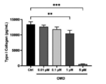
- FIG. 2B shows the results of collagen production among the evaluation results regarding osteomodulin collagen production in Test Example 2.
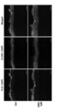
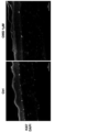
- FIG. 2C shows a scanning electron microscope image of the extracellular matrix of NHDF among the evaluation results regarding osteomodulin collagen production in Test Example 2.
- FIG. 2D shows the results of quantification and distribution of collagen fiber diameter among the evaluation results regarding osteomodulin collagen production in Test Example 2.
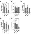
- FIG. 3 shows the results of gene expression in Test Example 3 (A: OCLN, B: OLDN1, C: FLG, D: LOR, E: KRT1).
- FIG. 3 shows the results of gene expression in Test Example 3 (A: OCLN, B: OLDN1, C: FLG, D: LOR, E: KRT1).
- FIG. 3 shows the results of gene expression in Test Example 3 (A: OCLN, B: OLDN1, C: FLG, D: LOR,
- FIG. 4A shows H&E staining of HSE among the results of histological analysis of human skin equivalent (HSE) in Test Example 4.
- FIG. 4B shows the measurement results of transepidermal water loss (TEWL) among the results of the histological analysis of human skin equivalent (HSE) in Test Example 4.
- FIG. 4C shows immunostaining of KRT1/KRT14 among the results of histological analysis of human skin equivalent (HSE) in Test Example 4.
- FIG. 4D shows immunostaining of FLG/CLDN1 among the results of histological analysis of human skin equivalent (HSE) in Test Example 4.
- FIG. 4E shows Ki67 immunostaining among the results of histological analysis of human skin equivalent (HSE) in Test Example 4.
- FIG. 4A shows H&E staining of HSE among the results of histological analysis of human skin equivalent (HSE) in Test Example 4.
- FIG. 4B shows the measurement results of transepidermal water loss (TEWL) among the results of the histological analysis of
- FIG. 4F shows the results of the gene expression levels of KRT1, FLG, LOR, OCLN, and CLDN1 in the epidermis of HSE, among the results of histological analysis of human skin equivalent (HSE) in Test Example 4.
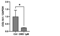
- FIG. 4G shows the results of the gene expression level of COL1A1 in the dermis of HSE, among the results of histological analysis of human skin equivalent (HSE) in Test Example 4.
- FIG. 5A shows an immunostaining fluorescence image of type I collagen fibers of NHDF cells among the results of evaluation regarding collagen production of CHAD and LRRC15 in Test Example 5.
- FIG. 5B shows the results of the collagen production amount among the evaluation results regarding collagen production of CHAD and LRRC15 in Test Example 5.
- the biomarker according to this embodiment is an aging biomarker, which means a factor that changes with aging, and is capable of determining and predicting aging, particularly aging conditions such as the degree of progress of skin aging. Further, by determining and predicting aging using the biomarker according to this embodiment, improvement and deterioration of aging can be determined and predicted.
- Ameliorating aging means inhibiting the aging state, inhibiting the progression of aging, and promoting reversal of aging (rejuvenation).
- aging refers to a decline in function that occurs in individuals, organs, tissues, cells, etc. that occurs after the growth period.
- skin aging refers to the decline in skin function that occurs with aging, and in a broader sense, it refers to the effects of various endogenous factors (e.g., cell aging) and extrinsic factors (e.g., ultraviolet rays, irritants, etc.). It refers to the decline in skin function that occurs as a result of being affected.
- the biomarker according to this embodiment can serve as an indicator of a physiological aging state or a pathological aging state, particularly a physiological aging state or a pathological aging state of the skin.
- Physiological aging is a decline in physiological function that inevitably progresses with age
- pathological aging refers to a decline in function that causes a pathological condition.
- the biomarkers according to this embodiment are biomarkers for use in determining skin aging, and include osteomodulin (OMD), chondroadherin (CHAD), collagen type XI alpha 2 chain ( collagen type XI alpha 2 chain (COL11A2), leucine-rich repeat-containing protein 15 (LRRC15), glycoprotein hormone alpha chain/choriogonadotropin subunit beta 3/choriogonadotropin subunit Beta 7 (Glycoprotein hormones alpha chain/Choriogonadotropin subunit beta 3/Choriogonadotropin subunit beta 7 (CGA / CGB3 / CGB7)), lutropin subunit beta (LHB), glycoprotein hormone alpha chain/Lutropin subunit beta ( A protein selected from the group consisting of glycoprotein hormones alpha chain/ lutropin subunit beta (CGA/LHB)) and glycoprotein hormone alpha chain/ follitropin subunit beta (CGA/FSHB) or a fragment thereof.
- OMD osteomodulin
- the above proteins or fragments thereof are a group of proteins that increase in the blood with aging, especially in women, and act on skin cells to accelerate aging.
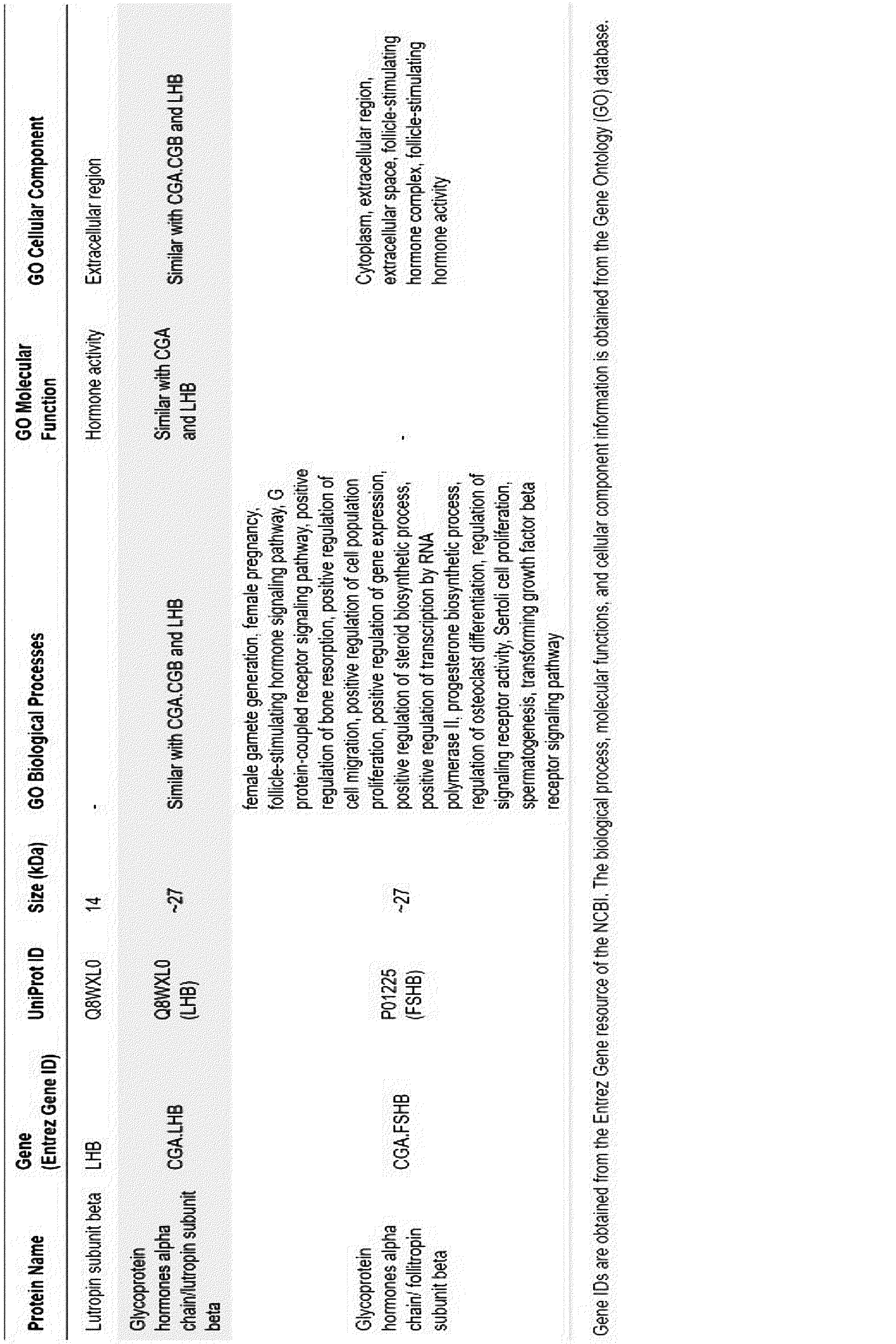
- Each of the above-mentioned biomarker proteins is a conventionally known protein and has the characteristics shown in Tables 1-1 and 1-2.
- the biomarker proteins according to this embodiment include osteomodulin (OMD), chondroadherin (CHAD), and leucine.
- OMD osteomodulin
- CHCD chondroadherin
- leucine leucine-rich repeat-containing protein 15
- the size of the protein of the biomarker is preferably 180 kDa or less, more preferably 150 kDa or less, even more preferably 120 kDa or less, even more preferably 80 kDa or less, and particularly preferably 66 kDa or less, from the viewpoint of sensitivity in determining skin aging.
- a body fluid sample such as blood, serum, plasma, urine, secretion, interstitial fluid, synovial fluid, cerebrospinal fluid, lymph fluid, etc.
- blood, serum, and plasma are more preferred, and serum and plasma are even more preferred.
- the protein or fragment thereof is contained in a subject sample, preferably a body fluid sample, more preferably a blood sample, even more preferably a serum sample.
- the above-mentioned subject is preferably a mammal or a human, more preferably a human, and even more preferably a biological female human.
- an increase in the expression or concentration of the protein or its fragment in a subject sample (preferably in a body fluid sample, more preferably in a blood sample, still more preferably in a serum sample)
- Figure 2 shows the progression of skin aging in a subject.
- a reduction in the expression or concentration of the protein or fragment thereof in a subject sample (preferably a body fluid sample, more preferably a blood sample, even more preferably a serum sample) improves skin aging in the subject. shows.
- the subject sample After the subject sample is collected from the subject, it may be appropriately prepared according to the measurement method.
- ELISA Enzyme-Linked Immunosorbent Assay
- RIA radioimmunoassay
- immunoprecipitation Western blotting, etc.
- ELISA is preferred because it is simple.
- a sample containing a biomarker protein according to this embodiment is exposed to a solid support (eg, a microtiter plate, etc.).
- a solid support eg, a microtiter plate, etc.
- Biomarker proteins in a sample are immobilized on a support and directly detected using an enzyme-conjugated antibody or the like specific to the biomarker proteins.
- the method for determining skin aging of the present invention uses osteomodulin (OMD), chondroadherin (CHAD), and collagen type XI alpha 2 chain in a blood sample of a subject. (COL11A2)), leucine-rich repeat-containing protein 15 (LRRC15), Glycoprotein hormones alpha chain/choriogonadotropin subunit beta 3/choriogonadotropin subunit beta 7 chain/Choriogonadotropin subunit beta 3/Choriogonadotropin subunit beta 7 (CGA / CGB3 / CGB7)), lutropin subunit beta (LHB), glycoprotein hormones alpha chain/ lutropin at least one protein selected from the group consisting of glycoprotein hormones alpha chain/follitropin subunit beta (CGA/FSHB); A method for determining skin aging in a subject, characterized by measuring fragments.
- the method for determining skin aging includes (i) measuring the amount or concentration of each of the above proteins or fragments thereof in a body fluid sample of a subject, preferably a blood sample of a subject, more preferably a serum sample of a subject; and (ii) determining skin aging of the subject by comparing the measured value obtained in (i) with a reference value serving as a standard for determining skin aging.
- the reference value may be a body fluid level of a healthy individual, preferably a blood level, more preferably a serum level, or may be an average value for healthy individuals of the subject's age, or may be a previous measurement of the subject. It may be a value, or it may be an average value for people who have the same situation as the subject.
- the measured value obtained in step (i) is higher than the reference value, it is determined that skin aging is more advanced than the standard, or that skin aging is more advanced than before.
- the measured value obtained in step (i) above is lower than the standard value, it means that skin aging is slower than normal, the progress of skin aging is slower than normal, or skin aging has improved from before. It is determined that the progression of skin aging has been suppressed or that the reversal of skin aging (rejuvenation) has occurred.
- the progress state of skin aging can be appropriately evaluated using biomarkers.
- the above-mentioned protein or fragment thereof used in the method corresponds to the above-mentioned biomarker of the present invention, and the description in the section on biomarkers (paragraphs 0012 to 0022) can be applied to the specific explanation.
- the screening method for skin aging-improving substances of the present invention includes osteomodulin (OMD), chondroadherin (CHAD), and collagen in blood samples of a subject before and after ingesting a test substance.
- OMD osteomodulin
- CHCD chondroadherin
- collagen collagen in blood samples of a subject before and after ingesting a test substance.
- collagen type XI alpha 2 chain (COL11A2), leucine-rich repeat-containing protein 15 (LRRC15), glycoprotein hormone alpha chain/choriogonadotropin subunit beta 3/Glycoprotein hormones alpha chain/Choriogonadotropin subunit beta 3/Choriogonadotropin subunit beta 7 (CGA / CGB3 / CGB7), lutropin subunit beta (LHB), glycoprotein hormone alpha chain glycoprotein hormones alpha chain/ lutropin subunit beta (CGA/LHB) and glycoprotein hormones alpha chain/ follitropin subunit beta (CGA/FSHB) It is characterized by examining changes in the expression level or concentration of at least one protein selected from the group or its fragment.
- the screening method for a skin aging improving substance includes (i) measuring the amount or concentration of the protein or its fragment in a blood sample of a subject before ingesting the test substance; (iii) After a certain period of time from step (ii) above, determining the amount of the protein or its fragment (same as that measured in step (i) above) in the blood sample of the subject; Step of measuring concentration, (iv) Comparing the measured values of (i) and (iii) above, if the value of (iii) is lower than (i), or if the values of (i) and (iii) are the same This includes the step of determining that the test substance is a skin aging improving substance, and determining that the test substance is not a skin aging improving substance if the value of (iii) is higher than (i).
- the method of the present invention it is possible to find healthcare materials, pharmaceutical materials, cosmetic materials, etc. that improve or prevent skin aging, suppress the progression of skin aging, and promote reversal of aging (rejuvenation).
- the above-mentioned protein or fragment thereof used in the method corresponds to the above-mentioned biomarker of the present invention, and the description in the section on biomarkers (paragraphs 0012 to 0022) can be applied to the specific explanation.
- test examples but the present invention is not limited thereto.
- blending amounts in the following test examples and the like indicate mass % unless otherwise specified.
- test conditions such as culture conditions in each test example were the same, except for the difference in the substances added, unless otherwise specified.
- controls and negative controls were provided for every test to accurately evaluate the effects of the biomarkers of the present invention.
- ⁇ Cell culture and materials> ⁇ Dermal fibroblasts: Newborn normal human dermal fibroblasts (NHDFs (NB)), lot number 05882 (Kurabo, Osaka, Japan) were used, and 10% fetal bovine serum and 1% antibiotics and antifungal agents were added. Cultured in Dulbecco's modified Eagle's medium (DMEM; Gibco, USA). - Cultured human keratinocytes: Newborn normal human epidermal keratinocytes (NHEK (NB)), lot number 7609 (Kurabo, Osaka, Japan) were used and cultured in Humedia-KG2 medium (Kurabo, Japan).
- DMEM Dulbecco's modified Eagle's medium
- - Cultured human keratinocytes Newborn normal human epidermal keratinocytes (NHEK (NB)), lot number 7609 (Kurabo, Osaka, Japan) were used and cultured in Humedia-KG2 medium (Kurabo, Japan
- -Cell culture conditions Cultured at 37°C in a 5% CO 2 incubator.
- - Recombinant human OMD (ab27632) was obtained from Abcam (Cambridge, UK).
- - Pro-Collagen 1 ELISA kit Human Pro-Collagen I alpha 1 DuoSet ELISA, DY6220-05 was purchased from R&D Systems (Minneapolis, MN, USA).
- ⁇ Method> ⁇ Method for counting the number of viable cells: It was stained with Hoechst 33342 (Dojindo, Kumamoto, Japan) and counted with ImageXpress® (Molecular Devices, California, USA).
- NHDF ⁇ Scanning electron microscope (SEM): NHDF was plated using a similar protocol to that sample. Recombinant OMD (0.01 ⁇ M, 0.1 ⁇ M, or 1 ⁇ M) was added to the treatment groups. After 6 days of treatment, the NHDF was fixed with a fixative and the sample was subjected to SEM observation.
- SEM Sccanning electron microscope
- This cDNA was mixed with SYBR TM Green PCR Master Mix and primers for KRT1, KRT10, OCLN, CLDN1, FLG, LOR, IVL, KRT1, KRT10, COL1A1, p21, and OMD (Thermo Fisher Scientific, Massachusetts, US). qPCR was performed using a real-time PCR device (Thermo Fisher Scientific) according to the manufacturer's protocol. Transcript levels were normalized to GAPDH.
- TEWL transepidermal water loss
- LDH -Lactic dehydrogenase assay: Performed using Viability/Cytotoxicity Multiplex Assay Kit (Dojindo, Kumamoto, Japan) according to the instructions.
- HSE Viability/Cytotoxicity Multiplex Assay Kit
- 100 ⁇ l of medium was transferred to a 96-well plate and LDH assay was performed.
- proteins such as OMD were added and left to stand for 48 hours, and then 100 ⁇ l of the supernatant was transferred to a 96-well plate and LDH assay was performed.
- This cluster is composed of eight proteins: osteomodulin (OMD), chondroadherin (CHAD), collagen type XI alpha 2 chain (COL11A2), and leucine.
- LRRC15 Rich repeat-containing protein 15
- CGA / CGB3 / CGB7 Glycoprotein hormones alpha chain/Choriogonadotropin subunit beta 3 /Choriogonadotropin subunit beta 7
- LHB lutropin subunit beta
- CGA/LHB glycoprotein hormones alpha chain/ lutropin subunit beta
- CGA/FSHB glycoprotein hormones alpha chain/follitropin subunit beta
- Figure 1 shows the results of a detailed analysis of age-related changes in these eight proteins.
- these eight proteins decreased between ages 20 and 40 and increased between ages 45 and 55.
- these proteins increased in abundance around age 65 and gradually decreased with age. Therefore, these eight proteins were found to vary specifically in women.
- the present inventors have identified osteomodulin (OMD), chondroadherin (CHAD), leucine-rich repeat-containing protein 15 (LRRC15), glycoprotein hormones alpha chain/Choriogonadotropin subunit beta 3/Choriogonadotropin subunit beta 7 (CGA / CGB3 / CGB7), Because the size of lutropin subunit beta (LHB), glycoprotein hormones alpha chain/ lutropin subunit beta (CGA/LHB), and glycoprotein hormones alpha chain/ follitropin subunit beta (CGA/FSHB) is less than 66 kDa, they can pass through blood vessels in the skin. Because it can reach the skin tissue, it was thought that it would affect the skin tissue and cells.
- the present inventors have revealed that OMD suppresses the production of type I collagen in fibroblasts and reduces the diameter of collagen fibers.
- OMD was thought to suppress the gene expression of OCLN, CLDN1, LOR, and KRT1 in NHEK and promote the decline of barrier function.
- RNA extraction For quantitative real-time polymerase chain reaction (RT-PCR), the epidermis and dermis of HSE were separated with forceps. Before RNA extraction, tissues were crushed with Biomasher II (Nippi, Tokyo, Japan), centrifuged at 10,000 ⁇ g for 10 min, and the supernatant was used for extraction with an RNeasy Mini Kit (Qiagen, Hilden, Germany). .
- NHDFs were plated in a 24-well plate at a density of 38,000 cells per well and cultured overnight in DMEM supplemented with 10% fetal bovine serum and 1% antibiotic-antimycotin. The next day, the medium was replaced with DMEM (without fetal bovine serum) supplemented with 1% antibiotic-antimycoplasma agent and 1 mg/ml L-ascorbic acid (013-12061, Fujifilm Wako Pure Chemical Industries, Ltd., Osaka, Japan). . OMD (0.01 ⁇ M, 0.1 ⁇ M, 1 ⁇ M, 5 ⁇ M) was added to the treatment groups.
- FIG. 4F shows that HSE containing OMD suppressed the expression level of COL1A1 in the dermis.
- OMD changes the shape of dermal fibroblasts into an elongated one, which is thought to be due to the disappearance of extracellular collagen and irregular cytoskeletal signal transmission.
- OMD affects the aggregates of collagen fibers and the diameter of collagen fibers, it is possible that OMD reduces the quality of type I collagen. It is also known that menopausal skin has higher extensibility and laxity than non-menopausal skin, and OMD, which increases with age, has been shown to affect the diameter size of collagen fibers. It was thought that this might be the cause.
- OMD Since the quality of collagen, such as orientation and fiber width, changes with endogenous aging playing a more central role, OMD, which reduces the quality and quantity of collagen in the skin, is a biological It is believed to be particularly useful as a marker.
- OMD which increases with age, is a useful biomarker because it affects both the epidermis and dermis of the skin.
- OMD is a useful biomarker for determining skin aging, so by targeting OMD in cells and tissues and examining changes in its expression level and concentration, we can screen for OMD-inhibiting components and OMD production-suppressing components. It is thought that it is possible to screen for ingredients that have the effect of suppressing aging of the dermis and epidermis.
- the amount of type I collagen in the culture supernatant under each condition was measured by collagen I ⁇ I enzyme-linked immunosorbent assay (ELISA). The measured value of each sample was calculated when the measured value of the negative control was set as 100 (FIG. 5B).
- a control was prepared by adding only L-ascorbic acid without adding CHAD or LRRC15.
- a negative control was one in which L-ascorbic acid and protein (CHAD, LRRC15) were not added.
- the control to which L-ascorbic acid was added showed high staining intensity, but the staining intensity was significantly lower in fibroblasts to which 1 ⁇ M OMD, CHAD, and LRRC15 were added in addition to L-ascorbic acid. decreased.
- the ELISA results showed that type I collagen production was suppressed in fibroblasts supplemented with 1 ⁇ M OMD, CHAD, and LRRC15. In particular, the effect of suppressing type I collagen production in fibroblasts to which CHAD and LRRC15 were added was remarkable.
- CHAD and LRRC15 which are proteins of 66 kDa or less, can pass through blood vessels in the skin and reach the skin tissue, so if the blood concentration increases due to aging, etc. It is thought to have a greater effect on cells.
- the present invention is applicable to all technical fields that require biomarkers for determining skin aging and their use, detection of the progress state of skin aging, determination of skin aging, and screening of skin aging improving substances, such as skin aging. It can be used to detect the occurrence of symptoms, predict the degree of symptom progression, predict the risk of symptom progression, investigate the causes of skin aging, confirm the effect of suppressing skin aging by drugs, etc., and confirm the therapeutic effect of skin aging. It can be used particularly in fields such as medicine, cosmetics, and food.
Landscapes
- Life Sciences & Earth Sciences (AREA)
- Chemical & Material Sciences (AREA)
- Health & Medical Sciences (AREA)
- Engineering & Computer Science (AREA)
- Organic Chemistry (AREA)
- Proteomics, Peptides & Aminoacids (AREA)
- Molecular Biology (AREA)
- Immunology (AREA)
- Analytical Chemistry (AREA)
- General Health & Medical Sciences (AREA)
- Biochemistry (AREA)
- Zoology (AREA)
- Physics & Mathematics (AREA)
- Genetics & Genomics (AREA)
- Biotechnology (AREA)
- Wood Science & Technology (AREA)
- Urology & Nephrology (AREA)
- Microbiology (AREA)
- Hematology (AREA)
- Biomedical Technology (AREA)
- Pathology (AREA)
- Biophysics (AREA)
- Medicinal Chemistry (AREA)
- General Engineering & Computer Science (AREA)
- Cell Biology (AREA)
- Bioinformatics & Cheminformatics (AREA)
- Food Science & Technology (AREA)
- General Physics & Mathematics (AREA)
- Toxicology (AREA)
- Gastroenterology & Hepatology (AREA)
- Investigating Or Analysing Biological Materials (AREA)
Abstract
Description
[1]皮膚老化の判定における使用のための、オステオモジュリン(osteomodulin (OMD))、コンドロアドヘリン(chondroadherin (CHAD))、コラーゲンXI型 アルファ2鎖(collagen type XI alpha 2 chain (COL11A2))、ロイシン-リッチリピート-含有タンパク質15(leucine-rich repeat-containing protein 15 (LRRC15))、グリコプロテイン ホルモン アルファ鎖/コリオゴナドトロピン サブユニット ベータ3/コリオゴナドトロピン サブユニット ベータ7(Glycoprotein hormones alpha chain/Choriogonadotropin subunit beta 3/Choriogonadotropin subunit beta 7 (CGA / CGB3 / CGB7))、ルトロピン サブユニット ベータ(lutropin subunit beta (LHB))、グリコプロテイン ホルモン アルファ鎖/ルトロピン サブユニット ベータ(glycoprotein hormones alpha chain/ lutropin subunit beta (CGA/LHB))、及びグリコプロテイン ホルモン アルファ鎖/フォリトロピン サブユニット ベータ(glycoprotein hormones alpha chain/ follitropin subunit beta (CGA/FSHB))から成る群より選択されるタンパク質又はそのフラグメントからなる、バイオマーカー。
[2]上記タンパク質又はそのフラグメントが、被験体の血液サンプル中に含まれる、[1]に記載のバイオマーカー。
[3]被験体の血液サンプル中の上記タンパク質又はそのフラグメントの発現の増加、又は濃度の増加が、被験体の皮膚老化の進行を示す、[2]に記載のバイオマーカー。
[4]被験体の血液サンプル中の上記タンパク質又はそのフラグメントの発現の低下、又は濃度の減少が、被験体の皮膚老化の改善を示す、[2]に記載のバイオマーカー。
[5]被験体の血液サンプル中のオステオモジュリン(osteomodulin (OMD))、コンドロアドヘリン(chondroadherin (CHAD))、コラーゲンXI型 アルファ2鎖(collagen type XI alpha 2 chain (COL11A2))、ロイシン-リッチリピート-含有タンパク質15(leucine-rich repeat-containing protein 15 (LRRC15))、グリコプロテイン ホルモン アルファ鎖/コリオゴナドトロピン サブユニット ベータ3/コリオゴナドトロピン サブユニット ベータ7(Glycoprotein hormones alpha chain/Choriogonadotropin subunit beta 3/Choriogonadotropin subunit beta 7 (CGA / CGB3 / CGB7))、ルトロピン サブユニット ベータ(lutropin subunit beta (LHB))、グリコプロテイン ホルモン アルファ鎖/ルトロピン サブユニット ベータ(glycoprotein hormones alpha chain/ lutropin subunit beta (CGA/LHB))、及びグリコプロテイン ホルモン アルファ鎖/フォリトロピン サブユニット ベータ(glycoprotein hormones alpha chain/ follitropin subunit beta (CGA/FSHB))から成る群より選択される少なくとも1種のタンパク質又はそのフラグメントを測定することを特徴とする、被験体における皮膚老化の判定方法。
[6]被験体に被験物質を摂取させる前後の被験体の血液サンプル中のオステオモジュリン(osteomodulin (OMD))、コンドロアドヘリン(chondroadherin (CHAD))、コラーゲンXI型 アルファ2鎖(collagen type XI alpha 2 chain (COL11A2))、ロイシン-リッチリピート-含有タンパク質15(leucine-rich repeat-containing protein 15 (LRRC15))、グリコプロテイン ホルモン アルファ鎖/コリオゴナドトロピン サブユニット ベータ3/コリオゴナドトロピン サブユニット ベータ7(Glycoprotein hormones alpha chain/Choriogonadotropin subunit beta 3/Choriogonadotropin subunit beta 7 (CGA / CGB3 / CGB7))、ルトロピン サブユニット ベータ(lutropin subunit beta (LHB))、グリコプロテイン ホルモン アルファ鎖/ルトロピン サブユニット ベータ(glycoprotein hormones alpha chain/ lutropin subunit beta (CGA/LHB))、及びグリコプロテイン ホルモン アルファ鎖/フォリトロピン サブユニット ベータ(glycoprotein hormones alpha chain/ follitropin subunit beta (CGA/FSHB))から成る群より選択される少なくとも1種のタンパク質又はそのフラグメントの発現レベル又は濃度の変化を調べることを特徴とする、皮膚老化改善物質のスクリーニング方法。
本実施形態に係るバイオマーカーは、老化バイオマーカーであり、加齢にともなって変動するファクターを意味し、老化、特に皮膚老化の進行度等の老化状態を判定、予測できるものである。また、本実施形態に係るバイオマーカーによる老化の判定、予測によって、老化の改善および改悪を判定、予測できる。老化の改善とは、老化状態の抑止、老化の進行抑制、老化の逆行促進(若返り)を意味する。ここで、老化とは、成長期を過ぎておこる個体、器官、組織、細胞などに現れる機能の低下を意味する。皮膚老化とは、狭義には、加齢に伴って現れる皮膚の機能低下を意味し、広義には、様々な内因性要因(例えば細胞老化)と外因性要因(例えば紫外線、刺激物等)の影響を受けて現れる皮膚の機能低下を意味する。本実施形態に係るバイオマーカーは、生理的老化状態や病的老化状態、特に皮膚の生理的老化状態や病的老化状態の指標となりえる。生理的老化は加齢とともに必然的に進行する生理的な機能低下であり、病的老化は病的状態を引き起こすような機能の低下を意味する。

本発明の皮膚老化の判定方法は、被験体の血液サンプル中のオステオモジュリン(osteomodulin (OMD))、コンドロアドヘリン(chondroadherin (CHAD))、コラーゲンXI型 アルファ2鎖(collagen type XI alpha 2 chain (COL11A2))、ロイシン-リッチリピート-含有タンパク質15(leucine-rich repeat-containing protein 15 (LRRC15))、グリコプロテイン ホルモン アルファ鎖/コリオゴナドトロピン サブユニット ベータ3/コリオゴナドトロピン サブユニット ベータ7(Glycoprotein hormones alpha chain/Choriogonadotropin subunit beta 3/Choriogonadotropin subunit beta 7 (CGA / CGB3 / CGB7))、ルトロピン サブユニット ベータ(lutropin subunit beta (LHB))、グリコプロテイン ホルモン アルファ鎖/ルトロピン サブユニット ベータ(glycoprotein hormones alpha chain/ lutropin subunit beta (CGA/LHB))、及びグリコプロテイン ホルモン アルファ鎖/フォリトロピン サブユニット ベータ(glycoprotein hormones alpha chain/ follitropin subunit beta (CGA/FSHB))から成る群より選択される少なくとも1種のタンパク質又はそのフラグメントを測定することを特徴とする、被験体における皮膚老化の判定方法である。
本発明の皮膚老化改善物質のスクリーニング方法は、被験体に被験物質を摂取させる前後の被験体の血液サンプル中のオステオモジュリン(osteomodulin (OMD))、コンドロアドヘリン(chondroadherin (CHAD))、コラーゲンXI型 アルファ2鎖(collagen type XI alpha 2 chain (COL11A2))、ロイシン-リッチリピート-含有タンパク質15(leucine-rich repeat-containing protein 15 (LRRC15))、グリコプロテイン ホルモン アルファ鎖/コリオゴナドトロピン サブユニット ベータ3/コリオゴナドトロピン サブユニット ベータ7(Glycoprotein hormones alpha chain/Choriogonadotropin subunit beta 3/Choriogonadotropin subunit beta 7 (CGA / CGB3 / CGB7))、ルトロピン サブユニット ベータ(lutropin subunit beta (LHB))、グリコプロテイン ホルモン アルファ鎖/ルトロピン サブユニット ベータ(glycoprotein hormones alpha chain/ lutropin subunit beta (CGA/LHB))、及びグリコプロテイン ホルモン アルファ鎖/フォリトロピン サブユニット ベータ(glycoprotein hormones alpha chain/ follitropin subunit beta (CGA/FSHB))から成る群より選択される少なくとも1種のタンパク質又はそのフラグメントの発現レベル又は濃度の変化を調べることを特徴とする。
〈細胞培養と材料〉
・真皮線維芽細胞:新生児正常ヒト真皮線維芽細胞(NHDFs(NB))、ロット番号05882(Kurabo, Osaka, Japan)を用い、10%牛胎児血清および1%抗生物質、抗真菌剤を添加したダルベッコ改変イーグル培地(DMEM; Gibco, USA)において培養した。
・培養ヒトケラチノサイト:新生児正常ヒト表皮ケラチノサイト(NHEK(NB))、ロット番号7609(Kurabo, Osaka, Japan)を用い、Humedia-KG2培地(Kurabo, Japan)において培養した。
・細胞の培養条件:5%CO2インキュベーター内で37℃にて培養した。
・組換えヒトOMD(ab27632)はAbcam(Cambridge,UK)より入手した。
・プロコラーゲン1ELISAキット(Human Pro-Collagen I alpha 1 DuoSet ELISA、DY6220-05)はR&D Systems(米国ミネソタ州ミネアポリス)より購入した。
・生存細胞数の計数方法:
Hoechst 33342 (Dojindo, Kumamoto, Japan)で染色し、ImageXpress(登録商標)(Molecular Devices, California, USA)で計数した。
NHDFは、そのサンプルと同様のプロトコルを用いてプレーティングした。処理群にリコンビナントOMD(0.01μM、0.1μM、または1μM)を添加した。6日間処理後、NHDFを固定液で固定し、サンプルをSEM観察に供した。
RNeasy mini kit(Qiagen, Hilden, Germany)を用いて培養細胞からRNAを抽出し、NanoDrop UV-Vis spectrophotometer(Thermo Fisher Scientific, Massachusetts, US)を用いてRNA濃度を決定した。qPCRのために、RNAはReverTraAce qPCR RT Master Mix(東洋紡、大阪、日本)を用いて第一鎖のcDNAに逆転写された。このcDNAをSYBRTMGreen PCR Master MixとKRT1、KRT10、OCLN、CLDN1、FLG、LOR、IVL、KRT1、KRT10、COL1A1、p21、OMDのプライマー(サーモフィッシャーサイエンティフィック、マサチューセッツ、US)と混合し、リアルタイムPCR装置(サーモフィッシャーサイエンティフィック)を用いて製造者のプロトコルに従いqPCRを実施した。転写レベルは、GAPDHに対して正規化した。
本試験例では、血液中のタンパク質をクラスター化し、年齢ごとにヒートマップで可視化することにより、年齢による変化を評価した。INTERVAL cohort (Cambridge, UK)の18~76歳の健常者3301人(女性1616人、男性1685人)から3283個の血漿タンパク質の3K SOMAscanプロテオミクスzスコア測定値を使用した。zスコアの変化は、Rのpheatmapパッケージ(バージョン1.0.12)を用いて、ヒトの寿命にわたって同様のダイナミクスを持つタンパク質をクラスター化することで可視化した。教師なし階層クラスタリングとクラスター化ヒートマップを用いて、女性および男性の全年齢にわたって類似した軌道を持つ血漿タンパク質をグループ化した。
本試験例では、Osteomodulin(OMD)のコラーゲン産生への影響を評価した。具体的には、培養正常ヒト線維芽細胞(NHDF)に各濃度のOMDを添加して6日間培養し、I型コラーゲンを免疫染色して蛍光画像を得た(図2A)。コラーゲンIαIの酵素結合免疫吸着法(ELISA)により各条件におけるI型コラーゲン量を測定した(図2B)。OMDの添加濃度は図中に示す。OMDを添加しないものをコントロール(Ctrl)とした。走査型電子顕微鏡を使用してNHDFの細胞外マトリックスを観察した結果を図2Cに示す。さらに、走査型電子顕微鏡画像から少なくとも600個のフィブリルを解析し、NHDFのコラーゲン線維径の定量化および分布を解析した結果を図2Dに示す。
本試験例では、OMDによる皮膚バリア機能関連遺伝子発現への影響を評価した。具体的には、ヒトケラチノサイト(NHEK)にOMDを0.1μM又は1μMとなるように添加し、タイトジャンクション遺伝子OCLNおよびCLDN1、末端分化マーカーFLGおよびLOR、表皮分化マーカーKRT1の遺伝子発現を測定した。参照遺伝子としてGAPDHを使用した。OMDを添加しないこと以外は同じ条件で試験を行ったものをコントロールとした。結果を図3A~図3Eに示す。
ことから、
本試験例では、ヒト皮膚等価物(HSE)モデル(T-skinTM、EPISKIN SA)を1μMのOMDを添加した培地で5日間培養し、4%パラホルムアルデヒドリン酸緩衝液(富士フイルム和光純薬株式会社、大阪、日本)で固定し、パラフィンに包埋してH&E染色と免疫染色を行った(図4A)。また、上記5日間培養後の細胞について経表皮水分損失(TEWL)を測定した(図4B)。
本試験例では、chondroadherin(以下、CHADともいう)、leucine-rich repeat-containing protein 15(以下、LRRC15ともいう)のコラーゲン産生への影響を評価した。具体的には、培養正常ヒト線維芽細胞(NHDF)に1mg/ml L-アスコルビン酸(Vitamin C)(013-12061、富士フイルム和光純薬株式会社、日本、大阪)並びに、OMD、CHADまたはLRRC15を1μM添加して6日間培養し、I型コラーゲンを免疫染色して蛍光画像を得た(図5A)。コラーゲンIαIの酵素結合免疫吸着法(ELISA)により各条件における培養上清中のI型コラーゲン量を測定した。ネガティブコントロールの測定値を100とした場合の各サンプルの測定値を算出した(図5B)。CHAD、LRRC15を添加せずに、L-アスコルビン酸のみ添加したものをコントロールとした。L-アスコルビン酸およびタンパク質(CHAD、LRRC15)を添加しないものをネガティブコントロールとした。
Claims (6)
- 皮膚老化の判定における使用のための、オステオモジュリン(osteomodulin (OMD))、コンドロアドヘリン(chondroadherin (CHAD))、コラーゲンXI型 アルファ2鎖(collagen type XI alpha 2 chain (COL11A2))、ロイシン-リッチリピート-含有タンパク質15(leucine-rich repeat-containing protein 15 (LRRC15))、グリコプロテイン ホルモン アルファ鎖/コリオゴナドトロピン サブユニット ベータ3/コリオゴナドトロピン サブユニット ベータ7(Glycoprotein hormones alpha chain/Choriogonadotropin subunit beta 3/Choriogonadotropin subunit beta 7 (CGA / CGB3 / CGB7))、ルトロピン サブユニット ベータ(lutropin subunit beta (LHB))、グリコプロテイン ホルモン アルファ鎖/ルトロピン サブユニット ベータ(glycoprotein hormones alpha chain/ lutropin subunit beta (CGA/LHB))、及びグリコプロテイン ホルモン アルファ鎖/フォリトロピン サブユニット ベータ(glycoprotein hormones alpha chain/ follitropin subunit beta (CGA/FSHB))から成る群より選択されるタンパク質又はそのフラグメントからなる、バイオマーカー。
- 上記タンパク質又はそのフラグメントが、被験体の血液サンプル中に含まれる、請求項1に記載のバイオマーカー。
- 被験体の血液サンプル中の上記タンパク質又はそのフラグメントの発現の増加、又は濃度の増加が、被験体の皮膚老化の進行を示す、請求項2に記載のバイオマーカー。
- 被験体の血液サンプル中の上記タンパク質又はそのフラグメントの発現の低下、又は濃度の減少が、被験体の皮膚老化の改善を示す、請求項2に記載のバイオマーカー。
- 被験体の血液サンプル中のオステオモジュリン(osteomodulin (OMD))、コンドロアドヘリン(chondroadherin (CHAD))、コラーゲンXI型 アルファ2鎖(collagen type XI alpha 2 chain (COL11A2))、ロイシン-リッチリピート-含有タンパク質15(leucine-rich repeat-containing protein 15 (LRRC15))、グリコプロテイン ホルモン アルファ鎖/コリオゴナドトロピン サブユニット ベータ3/コリオゴナドトロピン サブユニット ベータ7(Glycoprotein hormones alpha chain/Choriogonadotropin subunit beta 3/Choriogonadotropin subunit beta 7 (CGA / CGB3 / CGB7))、ルトロピン サブユニット ベータ(lutropin subunit beta (LHB))、グリコプロテイン ホルモン アルファ鎖/ルトロピン サブユニット ベータ(glycoprotein hormones alpha chain/ lutropin subunit beta (CGA/LHB))、及びグリコプロテイン ホルモン アルファ鎖/フォリトロピン サブユニット ベータ(glycoprotein hormones alpha chain/ follitropin subunit beta (CGA/FSHB))から成る群より選択される少なくとも1種のタンパク質又はそのフラグメントを測定することを特徴とする、被験体における皮膚老化の判定方法。
- 被験体に被験物質を摂取させる前後の被験体の血液サンプル中のオステオモジュリン(osteomodulin (OMD))、コンドロアドヘリン(chondroadherin (CHAD))、コラーゲンXI型 アルファ2鎖(collagen type XI alpha 2 chain (COL11A2))、ロイシン-リッチリピート-含有タンパク質15(leucine-rich repeat-containing protein 15 (LRRC15))、グリコプロテイン ホルモン アルファ鎖/コリオゴナドトロピン サブユニット ベータ3/コリオゴナドトロピン サブユニット ベータ7(Glycoprotein hormones alpha chain/Choriogonadotropin subunit beta 3/Choriogonadotropin subunit beta 7 (CGA / CGB3 / CGB7))、ルトロピン サブユニット ベータ(lutropin subunit beta (LHB))、グリコプロテイン ホルモン アルファ鎖/ルトロピン サブユニット ベータ(glycoprotein hormones alpha chain/ lutropin subunit beta (CGA/LHB))、及びグリコプロテイン ホルモン アルファ鎖/フォリトロピン サブユニット ベータ(glycoprotein hormones alpha chain/ follitropin subunit beta (CGA/FSHB))から成る群より選択される少なくとも1種のタンパク質又はそのフラグメントの発現レベル又は濃度の変化を調べることを特徴とする、皮膚老化改善物質のスクリーニング方法。
Priority Applications (2)
| Application Number | Priority Date | Filing Date | Title |
|---|---|---|---|
| KR1020257001438A KR20250025691A (ko) | 2022-06-17 | 2023-06-19 | 피부 노화의 바이오마커, 피부 노화의 판정 방법, 피부 노화 개선 물질의 스크리닝 방법 |
| JP2024529010A JPWO2023243732A1 (ja) | 2022-06-17 | 2023-06-19 |
Applications Claiming Priority (4)
| Application Number | Priority Date | Filing Date | Title |
|---|---|---|---|
| US202263353070P | 2022-06-17 | 2022-06-17 | |
| US63/353,070 | 2022-06-17 | ||
| US202263403991P | 2022-09-06 | 2022-09-06 | |
| US63/403,991 | 2022-09-06 |
Publications (1)
| Publication Number | Publication Date |
|---|---|
| WO2023243732A1 true WO2023243732A1 (ja) | 2023-12-21 |
Family
ID=89191503
Family Applications (1)
| Application Number | Title | Priority Date | Filing Date |
|---|---|---|---|
| PCT/JP2023/022650 Ceased WO2023243732A1 (ja) | 2022-06-17 | 2023-06-19 | 皮膚老化のバイオマーカー、皮膚老化の判定方法、皮膚老化改善物質のスクリーニング方法 |
Country Status (3)
| Country | Link |
|---|---|
| JP (1) | JPWO2023243732A1 (ja) |
| KR (1) | KR20250025691A (ja) |
| WO (1) | WO2023243732A1 (ja) |
Citations (2)
| Publication number | Priority date | Publication date | Assignee | Title |
|---|---|---|---|---|
| JP2010019683A (ja) * | 2008-07-10 | 2010-01-28 | Biomarker Science:Kk | 皮膚老化改善・進行遅延効果の評価方法及び評価用キット、並びに、物質のスクリーニング方法 |
| WO2022035075A1 (ko) * | 2020-08-10 | 2022-02-17 | 주식회사 큐티스의 생명연구센터 | 마이크로니들 패치를 포함하는 최소 침습적 피부 상태 진단 키트 |
-
2023
- 2023-06-19 KR KR1020257001438A patent/KR20250025691A/ko active Pending
- 2023-06-19 JP JP2024529010A patent/JPWO2023243732A1/ja active Pending
- 2023-06-19 WO PCT/JP2023/022650 patent/WO2023243732A1/ja not_active Ceased
Patent Citations (2)
| Publication number | Priority date | Publication date | Assignee | Title |
|---|---|---|---|---|
| JP2010019683A (ja) * | 2008-07-10 | 2010-01-28 | Biomarker Science:Kk | 皮膚老化改善・進行遅延効果の評価方法及び評価用キット、並びに、物質のスクリーニング方法 |
| WO2022035075A1 (ko) * | 2020-08-10 | 2022-02-17 | 주식회사 큐티스의 생명연구센터 | 마이크로니들 패치를 포함하는 최소 침습적 피부 상태 진단 키트 |
Non-Patent Citations (7)
| Title |
|---|
| DATABASE UniProtKB 25 May 2022 (2022-05-25), ANONYMOUS : "RecName: Full=Chondroadherin", XP093118191, Database accession no. O15335 * |
| DATABASE UniProtKB 25 May 2022 (2022-05-25), ANONYMOUS : "RecName: Full=Collagen alpha-2(XI) chain", XP093118185, Database accession no. P13942 * |
| DATABASE UniProtKB 25 May 2022 (2022-05-25), ANONYMOUS : "RecName: Full=Follitropin subunit beta", XP093118223, Database accession no. P01225 * |
| DATABASE UniProtKB 25 May 2022 (2022-05-25), ANONYMOUS : "RecName: Full=Glycoprotein hormones alpha chain", XP093118216, Database accession no. P01215 * |
| DATABASE UniProtKB 25 May 2022 (2022-05-25), ANONYMOUS : "RecName: Full=Leucine-rich repeat-containing protein 15", XP093118206, Database accession no. Q8TF66 * |
| DATABASE UniProtKB 25 May 2022 (2022-05-25), ANONYMOUS : "RecName: Full=Osteomodulin", XP093118194, Database accession no. Q99983 * |
| DATABASE UniProtKB 25 May 2022 (2022-05-25), ANONYMOUS : "SubName: Full=Luteinizing hormone beta subunit {ECO:0000313|EMBL:AAL69719.1}", XP093118219, Database accession no. Q8WXL0 * |
Also Published As
| Publication number | Publication date |
|---|---|
| JPWO2023243732A1 (ja) | 2023-12-21 |
| KR20250025691A (ko) | 2025-02-24 |
Similar Documents
| Publication | Publication Date | Title |
|---|---|---|
| Wang et al. | Comparison of the major cell populations among osteoarthritis, Kashin–Beck disease and healthy chondrocytes by single-cell RNA-seq analysis | |
| Navrazhina et al. | Epithelialized tunnels are a source of inflammation in hidradenitis suppurativa | |
| Liu et al. | Collagen metabolic disorder induced by oxidative stress in human uterosacral ligament-derived fibroblasts: A possible pathophysiological mechanism in pelvic organ prolapse | |
| Wang et al. | S100A6: molecular function and biomarker role | |
| Zou et al. | Aquaporin-1 plays a crucial role in estrogen-induced tubulogenesis of vascular endothelial cells | |
| Dudas et al. | Thy-1 is an in vivo and in vitro marker of liver myofibroblasts | |
| Chun-Yan et al. | Liquid biopsy biomarkers of renal interstitial fibrosis based on urinary exosome | |
| Mao et al. | MiR-183-5p overexpression in bone mesenchymal stem cell-derived exosomes protects against myocardial ischemia/reperfusion injury by targeting FOXO1 | |
| Morrison et al. | Developmental timing of trauma in women predicts unique extracellular vesicle proteome signatures | |
| Hadvina et al. | Exosomes and their miRNA/protein profile in keratoconus-derived corneal stromal cells | |
| Kamiya et al. | Puerarin blocks the aging phenotype in human dermal fibroblasts | |
| Zhang et al. | Long non-coding RNA PWRN1 affects ovarian follicular development by regulating the function of granulosa cells | |
| Tang et al. | Diagnostic value and underlying mechanism of nasal nitric oxide in eosinophilic chronic rhinosinusitis with nasal polyps | |
| Wang et al. | The gut microbiotas with metabolites regulate the protective role of miR-30a-5p in myocardial infarction | |
| Ren et al. | Diagnostic significance of LncRNA MIAT in periodontitis and the molecular mechanisms influencing periodontal ligament fibroblasts via the miR-204-5p/DKK1 axis | |
| Li et al. | Identification of anterior cruciate ligament fibroblasts and their contribution to knee osteoarthritis progression using single-cell analyses | |
| Kitazawa et al. | Expression of epithelial–mesenchymal transition markers in epidermal layer of atopic dermatitis | |
| WO2023243732A1 (ja) | 皮膚老化のバイオマーカー、皮膚老化の判定方法、皮膚老化改善物質のスクリーニング方法 | |
| CN104603616B (zh) | 使用纤蛋白‑3和/或肌聚糖γ作为指标评价脂肪团的方法和评价脂肪团有效药物的方法 | |
| US20210208144A1 (en) | Mitochondrial protease oma1 as a marker for breast cancer | |
| Ren et al. | TIGAR attenuates intervertebral disc degeneration via autophagy-mediated Keap1 degradation and Nrf2 nuclear translocation to suppress nucleus pulposus pyroptosis | |
| CN116492463B (zh) | Cd155分子在肝纤维化领域中的应用 | |
| Fujimoto et al. | Increased cellular distribution of vimentin and Ret in the cingulum induced by developmental hypothyroidism in rat offspring maternally exposed to anti-thyroid agents | |
| Liu et al. | NSUN2-engineered human umbilical cord mesenchymal stem cell-derived exosomes ameliorate tendon injury by promoting DNM2 expression | |
| Fang et al. | Decidual mesenchymal stem cells derived extracellular vesicles regulate ferroptosis to alleviate intervertebral disc degeneration through the HIF-1α signaling pathway |
Legal Events
| Date | Code | Title | Description |
|---|---|---|---|
| 121 | Ep: the epo has been informed by wipo that ep was designated in this application |
Ref document number: 23824021 Country of ref document: EP Kind code of ref document: A1 |
|
| WWE | Wipo information: entry into national phase |
Ref document number: 2024529010 Country of ref document: JP |
|
| ENP | Entry into the national phase |
Ref document number: 20257001438 Country of ref document: KR Kind code of ref document: A |
|
| WWE | Wipo information: entry into national phase |
Ref document number: 1020257001438 Country of ref document: KR |
|
| NENP | Non-entry into the national phase |
Ref country code: DE |
|
| WWP | Wipo information: published in national office |
Ref document number: 1020257001438 Country of ref document: KR |
|
| 122 | Ep: pct application non-entry in european phase |
Ref document number: 23824021 Country of ref document: EP Kind code of ref document: A1 |