WO2023145754A1 - 膀胱癌の存在を検出するためのプライマー及びプローブ - Google Patents
膀胱癌の存在を検出するためのプライマー及びプローブ Download PDFInfo
- Publication number
- WO2023145754A1 WO2023145754A1 PCT/JP2023/002178 JP2023002178W WO2023145754A1 WO 2023145754 A1 WO2023145754 A1 WO 2023145754A1 JP 2023002178 W JP2023002178 W JP 2023002178W WO 2023145754 A1 WO2023145754 A1 WO 2023145754A1
- Authority
- WO
- WIPO (PCT)
- Prior art keywords
- probe
- promoter
- plekhs1
- primers
- mutations
- Prior art date
- Legal status (The legal status is an assumption and is not a legal conclusion. Google has not performed a legal analysis and makes no representation as to the accuracy of the status listed.)
- Ceased
Links
Images


Classifications
-
- C—CHEMISTRY; METALLURGY
- C12—BIOCHEMISTRY; BEER; SPIRITS; WINE; VINEGAR; MICROBIOLOGY; ENZYMOLOGY; MUTATION OR GENETIC ENGINEERING
- C12N—MICROORGANISMS OR ENZYMES; COMPOSITIONS THEREOF; PROPAGATING, PRESERVING, OR MAINTAINING MICROORGANISMS; MUTATION OR GENETIC ENGINEERING; CULTURE MEDIA
- C12N15/00—Mutation or genetic engineering; DNA or RNA concerning genetic engineering, vectors, e.g. plasmids, or their isolation, preparation or purification; Use of hosts therefor
- C12N15/09—Recombinant DNA-technology
-
- C—CHEMISTRY; METALLURGY
- C12—BIOCHEMISTRY; BEER; SPIRITS; WINE; VINEGAR; MICROBIOLOGY; ENZYMOLOGY; MUTATION OR GENETIC ENGINEERING
- C12Q—MEASURING OR TESTING PROCESSES INVOLVING ENZYMES, NUCLEIC ACIDS OR MICROORGANISMS; COMPOSITIONS OR TEST PAPERS THEREFOR; PROCESSES OF PREPARING SUCH COMPOSITIONS; CONDITION-RESPONSIVE CONTROL IN MICROBIOLOGICAL OR ENZYMOLOGICAL PROCESSES
- C12Q1/00—Measuring or testing processes involving enzymes, nucleic acids or microorganisms; Compositions therefor; Processes of preparing such compositions
- C12Q1/68—Measuring or testing processes involving enzymes, nucleic acids or microorganisms; Compositions therefor; Processes of preparing such compositions involving nucleic acids
- C12Q1/6844—Nucleic acid amplification reactions
- C12Q1/686—Polymerase chain reaction [PCR]
-
- C—CHEMISTRY; METALLURGY
- C12—BIOCHEMISTRY; BEER; SPIRITS; WINE; VINEGAR; MICROBIOLOGY; ENZYMOLOGY; MUTATION OR GENETIC ENGINEERING
- C12Q—MEASURING OR TESTING PROCESSES INVOLVING ENZYMES, NUCLEIC ACIDS OR MICROORGANISMS; COMPOSITIONS OR TEST PAPERS THEREFOR; PROCESSES OF PREPARING SUCH COMPOSITIONS; CONDITION-RESPONSIVE CONTROL IN MICROBIOLOGICAL OR ENZYMOLOGICAL PROCESSES
- C12Q1/00—Measuring or testing processes involving enzymes, nucleic acids or microorganisms; Compositions therefor; Processes of preparing such compositions
- C12Q1/68—Measuring or testing processes involving enzymes, nucleic acids or microorganisms; Compositions therefor; Processes of preparing such compositions involving nucleic acids
- C12Q1/6876—Nucleic acid products used in the analysis of nucleic acids, e.g. primers or probes
- C12Q1/6883—Nucleic acid products used in the analysis of nucleic acids, e.g. primers or probes for diseases caused by alterations of genetic material
- C12Q1/6886—Nucleic acid products used in the analysis of nucleic acids, e.g. primers or probes for diseases caused by alterations of genetic material for cancer
Definitions
- the present invention broadly relates to primers, probes, etc. for detecting the presence of bladder cancer.
- Liquid biopsy which detects tumor-derived genetic mutations in blood-circulating DNA (cell-free DNA: cfDNA), is a minimally invasive test, and has been reported in recent years to be effective in postoperative follow-up and assessment of therapeutic effects.
- cfDNA analysis methods include the use of next-generation sequencing (NGS) and digital PCR.
- NGS next-generation sequencing
- Digital PCR has the advantage that it can be analyzed at a lower cost than NGS.
- TERT telomerase reverse transcriptase
- a primer set for detecting the presence of bladder cancer in a specimen comprising: a pair of primers, which are oligonucleotides with a length of 16 bases or more and 21 bases or less, targeting mutations in the PLEKHS1 promoter; one or more pairs of primers that target mutations in the TERT promoter; A primer set, including (2)
- the PLEKHS1 promoter mutation is a G to A substitution at chromosome 10, position 115,511,590, a C to T substitution at chromosome 10, position 115,511,593, or both (1) Primer set described in .
- a kit comprising the primer set according to any one of (1) to (4).
- the kit according to (7) further comprising the probe according to (5) or (6).
- a method for detecting the presence of bladder cancer in a specimen wherein the primer set according to any one of (1) to (4) or the probe according to (5) or (6) is used to detect the PLEKHS1 promoter.
- the method according to (9), wherein the nucleic acid amplification method used in the detection step is digital PCR.
- (11) The method according to (9) or (10), wherein the specimen is a liquid specimen.
- Mutations in the PLEKHS1 promoter include G to A substitution at position 115,511,590 of chromosome 10 and C to T substitution at position 115,511,593 of chromosome 10.
- the present inventors set the positions corresponding to these mutations near the center of the probe so as to maximize the Tm value.
- the probe is usually designed as a 20-mer, 18-mer at the shortest, and 25-mer at the longest. bottom.
- the primer was also designed with a different concept than usual.
- the length of primers is designed to be about 20-25 mers. If the length of the primer is less than 20-mer, there is a possibility that the sequence that non-specifically binds to the base sequence other than the target increases, but the forward primer that detects the mutation of the PLEKHS1 promoter is a 16-mer. designed. This is in consideration of the need to lower the Tm value compared to wild-type/mutant probes.
- the region containing the hotspot of the PLEKHS1 promoter region is an AT-rich sequence, but the primer design region is a sequence that cannot be said to be AT-rich, so the Tm value becomes high, and it is necessary to shorten the sequence. There is Furthermore, trying to avoid GC, for example, designing an A or T at the 3' end of the primer may reduce specificity for the template DNA. However, by conducting verification experiments, it was possible to design a shorter length with a lower Tm value than general designs.
- the Tm value of the mutant probe is lower than that of the forward primer.
- the annealing order is wild-type probe (53.92), forward primer (52.61), mutant probe (52.28), reverse primer (50.66).
- the size of the amplicon to be amplified was designed to be less than 200 bp.
- primers that target mutations in the PLEKHS1 promoter that are designed under a different concept as described above can be used to detect bladder cancer. It will be possible to cover patients who did not have it.
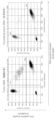
- FIG. 2 shows the results of quantification of positive/negative droplets in mutation-positive patients.
- Ch1 indicates Mut droplets
- Ch2 indicates WT droplets
- the lower left frame is WT and Mut negative droplets
- the lower right frame is WT positive droplets
- the upper left frame is Mut positive droplets
- the upper right frame is WT. and Mut-positive droplets.
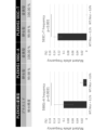
- FIG. 1 shows detection accuracy (upper) and mutation rate (lower) of validation plasmids (wild-type (WT) and mutant-type (Mut)).
- FIG. 2 shows the results of quantification of positive/
- FIG. 3 shows changes over time in MAF: Mutation Allele Frequency after initial treatment (transurethral bladder tumor resection) of mutation-positive patients.
- the arrows in the figure indicate mutations (or indicates the specific stage of the sample containing it).
- the left figure shows the results of patient number 03 (Pt03), and the right figure shows the results of patient number 21 (Pt21).
- Pt03 the results of patient number 03
- Pt21 patient number 21
- FIG. 4 illustrates the coverage of bladder cancer patients by the primers/probes of the present invention.
- a primer set for detecting the presence of bladder cancer in a specimen comprising: a pair of primers, which are oligonucleotides with a length of 16 bases or more and 21 bases or less, targeting mutations in the PLEKHS1 promoter; one or more pairs of primers that target mutations in the TERT promoter;
- a primer set comprising:
- the specimen is not particularly limited as long as it can detect PLEKHS1 promoter mutation and TERT promoter mutation suggesting the presence of bladder cancer.
- Fluid specimens containing cfDNA are preferred, such as blood or other bodily fluids. More specifically, the specimen is preferably blood, serum, plasma, urine, stool, saliva, sputum, interstitial fluid, cerebrospinal fluid, bodily fluids such as swabs, or dilutions thereof, particularly plasma.
- Specimens are collected from subjects suspected of having bladder cancer.
- Bladder cancer may be non-muscle invasive or muscle invasive.
- the sample may be collected once or multiple times for purposes such as prognosis observation after bladder cancer treatment.
- ctDNA bladder cancer-derived DNA
- the PLEKHS1 promoter mutation is a G to A substitution (hereinafter also referred to as “590G>A”) at position 115,511,590 on chromosome 10, 115,511,593 on chromosome 10. It may be a C to T substitution at the position (hereinafter also referred to as “593C>T”) or both.
- a primer is an oligonucleotide with a length of 16 bases or more and no more than 21 bases. Among them, those having a low Tm value are preferable. Specifically, by setting the Tm value of the reverse primer to be 1 to 3° C. higher than that of the probe, the probe is annealed to the template DNA first, and then the reverse primer is annealed.
- the primers can be designed so that the amplicon size to be amplified is less than 200 bp.
- SEQ ID NO: 1 having a base length of 16 bases
- examples include the forward primer (gacctcttggcttcca) described above and the reverse primer (ctgcaaattttccatttcca) described in SEQ ID NO: 2, which has a base length of 21 bases. It is preferable to use a primer set consisting of the sequences set forth in SEQ ID NOS: 1 and 2. These primers may be combined with known primer sets that amplify the PLEKHS1 promoter region.
- the TERT promoter mutation is a C to T substitution (hereinafter also referred to as “228C>T”) at position 1,295,228 on chromosome 5, 1,295,250 on chromosome 5 It may be a C to T substitution at the position (hereinafter also referred to as “250C>T”) or both.
- 228C>T is known to be highly detectable in bladder cancer and 250C>T in non-muscle invasive bladder cancer.
- Primers that amplify sequences containing probes that detect both the 228C>T and 250C>T mutations should be within 113 bp upstream or downstream of each mutation, i.e., primers targeting the former mutation. and within the region of positions 1,295,115 to 1,295,341 for the probe and within the region of positions 1,295,237 to 1,295,363 for primers and probes targeting the latter mutation. can be designed.
- sequence shown in SEQ ID NO: 7 is obtained by converting the sequence into the sequence on the sense strand side and reflecting both the 228C>T and 250C>T mutations.
- primers and probes that detect 228C>T the 227 bp sequence from positions 1,295,341 to 1,295,115 corresponding to positions 1,295,115 to 1,295,341 in SEQ ID NO: 7
- Desired primers and probes can be appropriately designed from (SEQ ID NO: 8).
- primers and probes for detecting 250C>T which can be designed from the 127 bp sequence (SEQ ID NO: 9) from positions 1,295,363 to 1,295,237.
- a primer made by BioRad (TERT C228T_113: DHSAEXD72405942, TERT C250T_113: DHSAEXD466675715) (Ampuri Conto) (J Mol Diagn. 2019 MAR. 2019 MAR. See 21 (2) 274-285 ).
- Such primers may be combined with known primer sets that amplify the TERT promoter region.
- SEQ ID NO:6 GCCGGGGCCAGGGCTTCCCACGTGCAGCAGGACGCAGCGCTGCCTGAAACTCGCGCCGCGAGGAGAGGGCGGGGCCGCGGAAAGGAAGGGGAGGGGCTGGGAGGGCCCGGAGGGGGCTGGGCCGGGGACCCGGGAGGGGTCGGGACGGGGGGCGGGGTCCGCGCGGAGGAGGCGGAGCTGGAAGGTGAAGGGGCAGGACGGGTGCCCGGGTCCCCAGTCCCTCCGCCACGTGGGAAGCGCGGT CCTGGGMore SEQ ID NO: 7 CCCAGGACCGCGCTTCCCACGTGGCGGAGGGACTGGGGACCCCGGGCACCCGTCCTGCCCCTTCACCTTCCAGCTCCCGTCCCGGGTCCCCCCTCCGGGCCCTCCCTCCTCCGGGCCCTCCCTCCTCCGGGCCCTCCCTCCCCTCCGGGCCCTCCCTCCCCTCCGGGCCCTCCCTCCCCTCCGGGCCCTCCCTCCCTCCTCCGGGCCCTCCCTCCCTCCTCCGGGCCCTCC
- the nucleotides that make up the base sequences of primers and the like may be either ribonucleotides or deoxyribonucleotides. These oligonucleotides can be synthesized by known methods, for example, by any nucleic acid synthesis method such as solid-phase phosphoramidite method and triester method, according to the base sequence.
- a nucleic acid amplification reaction using the above primers makes it possible to amplify sequences containing hotspot regions associated with bladder cancer.
- Nucleic acid amplification methods include PCR methods, and in addition to normal PCR, digital PCR, multiplex PCR, LAMP (Loop-mediated isothermal amplification) method, ICAN (Isothermal and Chimeric primer-initiated amplification of nucleic acids) method, RCA ( Rolling Circle Amplification) method, LCR (Ligase Chain Reaction) method, SDA (Strand Displacement Amplification) method, and the like.
- the nucleic acid amplification method is preferably digital PCR.
- digital PCR it is possible to determine the ratio of mutant to normal types (MAF: Mutation Allele Frequency) for oncogenes.
- Digital PCR may be droplet digital PCR.
- a probe for detecting the presence of bladder cancer in a specimen is provided, the probe targeting mutations in the PLEKHS1 promoter.
- the sequence that constitutes the probe is not limited as long as it can detect mutations in the PLEKHS1 promoter.
- probes that can detect the 590G>A mutation or the 593C>T mutation are preferred.
- the probe that detects the 590G>A mutation has the sequence set forth in SEQ ID NO: 4 (tgcaattgtttaattgcaaaaagc).
- the probe consists of the sequence set forth in SEQ ID NO:4.
- the probe that detects the 593C>T mutation has the sequence set forth in SEQ ID NO:5.
- the probe preferably consists of the sequence (tgcaattattcaattgcaaaaagc) set forth in SEQ ID NO:5.
- the probe may be partially modified, examples of which include those whose ends are aminated and those whose bases are partially modified with linker bases.
- another base may be inserted into part of the base sequence, a part of the base sequence may be deleted, replaced with another base, or substituted with a substance other than a base. .
- the probe may be modified with a labeling substance or the like, and may have, for example, a fluorescent dye and its quencher.
- a fluorescent dye and its quencher Commercially available products can be used as such labeling substances.
- the quencher is not particularly limited as long as it can quench the fluorescence from the fluorescent dye.
- the nucleotide residues constituting the probe may themselves be modified, for example, substituted with artificially modified nucleotide residues.
- kits in a second aspect, includes a primer set for detecting the presence of bladder cancer in a specimen.
- the primer set described above can be used.
- the kit may further contain probes targeting mutations in the PLEKHS1 promoter.
- Probes for detecting the 590G>A and 593C>T mutations include those having the base sequences of SEQ ID NOS: 4 and 5, respectively.
- the kit may further include one or more pairs of primers and probes targeting mutations in the TERT promoter.
- Mutations in the TERT promoter include 228C>T and 250C>T. within the region of positions 1,295,115 to 1,295,341 for primers and probes targeting 228C>T; It can be designed within the region of 295,363 positions.
- a reaction solution containing the above primer set, probe, sample, and DNA polymerase is prepared. After being distributed to wells or droplets, the reaction solution is subjected to an amplification reaction.
- a labeling substance that can specifically recognize the amplification product may be used to detect the amplification product obtained in the nucleic acid amplification reaction.
- labeling substances include fluorescent dyes, biotin, digoxigenin and the like.
- fluorescence can be detected using a fluorescence microscope, a fluorescence plate reader, or the like.
- a substance that intercalates with the amplification product can also be used as a labeling substance.
- the intercalator is not particularly limited as long as it is a substance that intercalates with the double-stranded DNA and emits fluorescence.
- detection may be performed by known methods such as electrophoresis using polyacrylamide or agarose gel.
- electrophoresis the presence of an amplification product can be identified from the mobility of the amplification product relative to the mobility of a marker of known molecular weight.
- Detection method In a third aspect, a method for detecting the presence of bladder cancer in a specimen is provided, comprising detecting mutations in the PLEKHS1 promoter using the primer set or kit described above.
- the primer set includes primers that target mutations in the TERT promoter, and that mutations in the TERT promoter are also detected in the detection process.
- Other steps may be included before or after the detection step, for example, a specimen may be obtained from a subject suspected of having bladder cancer. The detection step may be performed multiple times, for example, specimens derived from the same subject may be collected periodically and subjected to the detection step each time they are collected.
- a mutation in the PLEKHS1 promoter is detected, it is determined that bladder cancer is present in the specimen. This can assist doctors in diagnosing bladder cancer and determining subsequent treatment methods.
- surgery such as transurethral bladder tumor resection, radical cystectomy, preoperative chemotherapy, postoperative chemotherapy, chemotherapy such as recurrent chemotherapy (anti-cancer drug therapy) and radiotherapy can be performed.
- transurethral bladder tumor resection it will be possible to diagnose muscle invasion from the results of transurethral bladder tumor resection. If the tumor does not invade the muscle layer, treatment is completed with only transurethral resection of the bladder tumor, followed by follow-up.
- preoperative chemotherapy will be started, and if recurrence or metastasis is found after completion of treatment, recurrent chemotherapy will be introduced.
- Preoperative chemotherapy can improve the prognosis, but it may not be given depending on the histology of the tumor and the patient's wishes.
- Postoperative chemotherapy is often added when intraoperative findings suggest residual disease. If radical cystectomy is considered difficult due to age or general condition, radiation may be performed instead.
- primer dimer evaluation is performed by PCR electrophoresis, PCR efficiency for each primer pair was confirmed by quantitative PCR. Since cfDNA was the target, the amplicon was within the range of 80 to 150 mers, and the Tm value calculated from the probe was taken into consideration for the sequence and number of bases of the primers. Taking into account PCR efficiency, primer dimers, and non-specific bands, the most efficient primer pair was determined, and correct amplification of the target region was confirmed by Sanger sequencing.
- tDNA tumor tissue-derived DNA
- gDNA tumor tissue-derived DNA
- gDNA tumor tissue-derived DNA
- gDNA tumor tissue-derived DNA
- gDNA tumor tissue-derived DNA
- plasmids for validation were prepared in the following order.
- the vector is introduced into Escherichia coli, and about 10 to 20 colonies are isolated and cultured in large quantities.
- Vectors were recovered by mini prep, and whether each vector was WT or Mutant was confirmed by Sanger sequencing.
- the primer/probe detection accuracy was confirmed using the validation plasmid in the following order.
- the forward primer for detecting wild-type PLEKHS1 and mutant PLEKHS1 consists of the sequence (gacctcttggcttcca) set forth in SEQ ID NO: 1, and the reverse primer consists of the sequence set forth in SEQ ID NO: 2 (ctgcaaaattttccatttcca). used something.
- a probe consisting of the sequence (tgcaattgttcaattgcaaaaagc) set forth in SEQ ID NO: 3 was used as a probe for detecting wild-type PLEKHS1.
- the 590G>A mutant probe consists of the sequence (tgcaattgtttaattgcaaaaagc) set forth in SEQ ID NO: 4, and the probe for detecting the 593C>T mutation consists of the sequence set forth in SEQ ID NO: 5 (tgcaattattcaattgcaaaaagc). It was used.
- (ii) Generation of 0.1% mutant plasmids at a ratio of wild-type:mutant 999:1.
- primers and probes exist within a range of 113 bp upstream or downstream from C228 or C250. It may be designed within the range of 50 to 200 bp upstream or downstream from the mutation site.
- Method 1. Prepare ⁇ 50 ng/well (up to 7 ⁇ L) DNA solution (15 ng can be added for 10-50 copies) 2. Thaw at room temperature, vortex, centrifuge, and protect from light. 3. Prepare Mix (restriction enzyme undiluted solution 10 U/ ⁇ L) 4. Vortex/centrifuge and leave at room temperature for 3 minutes5. Add 20 ⁇ L of mix to the sample well of the DG8® cartridge and 70 ⁇ L of Droplet Generation Oil to the oil well. 6. 7. Place in Droplet Generator to create droplets in solution. Put 40 ⁇ L of Droplet into PCR 96well and seal (aluminum foil) 8. Analysis with Droplet Reader
- dPCR was used to detect tumor DNA from 26 specimens, and the TERT promoter and PLEKHS1 promoter were positive in 50.0% (13/26) and 42.3% (11/26) of the patients, respectively. rice field.
- one patient with multiple PLEKHS1 promoter mutations was counted. With the TERT promoter alone, 50.0% of target patients could be observed, whereas the addition of the PLEKHS1 promoter improved to 57.7% (15/26). Also, multiple mutations could not be observed in the same patient with the TERT promoter alone, i.e., 0.0% (0/26), compared to 34.6% (9/26) with the addition of the PLEKHS1 promoter. Patients can now be observed with multiple mutations.
- the advantage of being able to analyze mutations by dPCR not only in the TERT promoter but also in the PLEKHS1 promoter is that the number of patients who can be analyzed with the same assay increases. Since tumor mutations in bladder cancer are diverse, the mainstream method is to analyze mutations that differ from patient to patient over time. Compared to this method, hotspot detection is advantageous in terms of reducing analysis costs. Second, by analyzing multiple MAFs, it is possible to more accurately assess the disease state of the patient. Multiple events in which MAF could not be detected were observed despite the diagnosis of systemic metastasis, suggesting the risk of being judged as false negative when follow-up is performed for a single mutation. be. It is believed that observing multiple MAFs can reduce the possibility of a false negative evaluation.
Landscapes
- Chemical & Material Sciences (AREA)
- Life Sciences & Earth Sciences (AREA)
- Health & Medical Sciences (AREA)
- Organic Chemistry (AREA)
- Engineering & Computer Science (AREA)
- Zoology (AREA)
- Genetics & Genomics (AREA)
- Wood Science & Technology (AREA)
- Proteomics, Peptides & Aminoacids (AREA)
- Biotechnology (AREA)
- General Engineering & Computer Science (AREA)
- Bioinformatics & Cheminformatics (AREA)
- Physics & Mathematics (AREA)
- General Health & Medical Sciences (AREA)
- Microbiology (AREA)
- Molecular Biology (AREA)
- Immunology (AREA)
- Biophysics (AREA)
- Analytical Chemistry (AREA)
- Biochemistry (AREA)
- Pathology (AREA)
- Biomedical Technology (AREA)
- Chemical Kinetics & Catalysis (AREA)
- Oncology (AREA)
- Hospice & Palliative Care (AREA)
- Plant Pathology (AREA)
- Measuring Or Testing Involving Enzymes Or Micro-Organisms (AREA)
Abstract
本発明は、検体における膀胱癌の存在を検出するためのプライマーセットであって、PLEKHS1プロモーターの変異を標的とする、16塩基長以上で且つ21塩基長以下のオリゴヌクレオチドである1対のプライマーと、TERTプロモーターの変異を標的とする、1又は複数の対のプライマーと、を含む、プライマーセット、を提供する。
Description
本発明は広く、膀胱癌の存在を検出するためのプライマー及びプローブ等に関する。
腫瘍由来の遺伝子変異を血中循環DNA(cell-free DNA:cfDNA)で検出するリキッドバイオプシーは低侵襲な検査であり、術後の経過観察や治療効果判定において有効であることが近年報告されている。
cfDNAの分析手法として、次世代シーケンサー(NGS)とデジタルPCRの利用が挙げられる。デジタルPCRはNGSよりも低いコストで分析できるという利点がある。
Clinical significance of hotspot mutation analysis of urinary cell-free DNA in urothelial bladder cancer(Frontiers in oncology, 2020)
膀胱癌等の多くの癌では、TERT(telomerase reverse transcriptase)のプロモーター領域に点変異が起きており、そのような点変異を検出するためのプライマーが知られている。
膀胱癌では、TERTのみならずPLEKHS1(pleckstrin homology domain-containing S1)のプロモーター領域の遺伝子変異がホットスポット、つまり複数の患者に共通で見られる変異である可能性があるが、cfDNAを検体としたとき、定量PCRでの検出は難しい。また、従来のプライマーやプローブではcfDNAを十分な精度で検出できないという問題もあった。
膀胱癌の存在を検出するために、TERTプロモーターの変異に加え、PLEKHS1プロモーターの変異を標的とするプライマー等を、通常とは異なる思想で設計した結果、従来のTERTプロモーター変異を対象とした検出では40%程度であった検出カバー率が増大した。
すなわち、本願は以下の発明を包含する。
(1)
検体における膀胱癌の存在を検出するためのプライマーセットであって、
PLEKHS1プロモーターの変異を標的とする、16塩基長以上で且つ21塩基長以下のオリゴヌクレオチドである1対のプライマーと、
TERTプロモーターの変異を標的とする、1又は複数の対のプライマーと、
を含む、プライマーセット。
(2)
PLEKHS1プロモーターの変異が、10番染色体の115,511,590位におけるGからAへの置換、10番染色体の115,511,593位におけるCからTへの置換又はその両方である、(1)に記載のプライマーセット。
(3)
PLEKHS1プロモーターの変異を標的とするプライマーが配列番号1及び/又は2に記載の塩基配列を有する、(1)又は(2)に記載のプライマーセット。
(4)
TERTプロモーターの変異を標的とするプライマーが、5番染色体の1,295,228位におけるCからTへの置換、5番染色体の1,295,250位におけるCからTへの置換又はその両方である、(1)~(3)のいずれかに記載のプライマーセット。
(5)
検体における膀胱癌の存在を検出するためのプローブであって、PLEKHS1プロモーターの変異を標的とする、プローブ。
(6)
プローブが配列番号4又は5に記載の塩基配列を有する、(5)に記載のプローブ。
(7)
(1)~(4)のいずれかに記載のプライマーセットを含む、キット。
(8)
(5)又は(6)に記載のプローブを更に含む、(7)に記載のキット。
(9)
検体における膀胱癌の存在を検出するための方法であって、(1)~(4)のいずれかに記載のプライマーセット、あるいは(5)又は(6)に記載のプローブを用いてPLEKHS1プロモーターの変異を検出する工程を含む、方法。
(10)
検出工程で使用される核酸増幅法がデジタルPCRである、(9)に記載の方法。
(11)
検体が液性検体である、(9)又は(10)に記載の方法。
(12)
検体が血中循環DNA(cfDNA)を含む、(9)~(11)のいずれかに記載の方法。
(1)
検体における膀胱癌の存在を検出するためのプライマーセットであって、
PLEKHS1プロモーターの変異を標的とする、16塩基長以上で且つ21塩基長以下のオリゴヌクレオチドである1対のプライマーと、
TERTプロモーターの変異を標的とする、1又は複数の対のプライマーと、
を含む、プライマーセット。
(2)
PLEKHS1プロモーターの変異が、10番染色体の115,511,590位におけるGからAへの置換、10番染色体の115,511,593位におけるCからTへの置換又はその両方である、(1)に記載のプライマーセット。
(3)
PLEKHS1プロモーターの変異を標的とするプライマーが配列番号1及び/又は2に記載の塩基配列を有する、(1)又は(2)に記載のプライマーセット。
(4)
TERTプロモーターの変異を標的とするプライマーが、5番染色体の1,295,228位におけるCからTへの置換、5番染色体の1,295,250位におけるCからTへの置換又はその両方である、(1)~(3)のいずれかに記載のプライマーセット。
(5)
検体における膀胱癌の存在を検出するためのプローブであって、PLEKHS1プロモーターの変異を標的とする、プローブ。
(6)
プローブが配列番号4又は5に記載の塩基配列を有する、(5)に記載のプローブ。
(7)
(1)~(4)のいずれかに記載のプライマーセットを含む、キット。
(8)
(5)又は(6)に記載のプローブを更に含む、(7)に記載のキット。
(9)
検体における膀胱癌の存在を検出するための方法であって、(1)~(4)のいずれかに記載のプライマーセット、あるいは(5)又は(6)に記載のプローブを用いてPLEKHS1プロモーターの変異を検出する工程を含む、方法。
(10)
検出工程で使用される核酸増幅法がデジタルPCRである、(9)に記載の方法。
(11)
検体が液性検体である、(9)又は(10)に記載の方法。
(12)
検体が血中循環DNA(cfDNA)を含む、(9)~(11)のいずれかに記載の方法。
PLEKHS1プロモーターの変異として、10番染色体の115,511,590位におけるGからAへの置換や10番染色体の115,511,593位におけるCからTへの置換がある。本発明者らは、プローブの設計にあたり、これらの変異に相当する位置がプローブの中心付近となるようにし、最もTm値が高くなるようにした。具体的には、プローブは通常20量体で設計され、短くても18量体であり、長くても25量体であるところ、本願発明ではTm値が最大限高値となるよう25量体とした。
プライマーも通常とは異なる思想で設計した。一般的に、プライマーの長さは20~25量体程度となるよう設計される。プライマーの長さを20量体未満とすると標的以外の塩基配列に対する非特異的に結合してしまう配列が増える可能性があるが、PLEKHS1プロモーターの変異を検出するフォワードプライマーは16量体のものを設計した。これは、野生型・変異体用のプローブよりTm値を下げる必要があることを考慮したものである。PLEKHS1プロモーター領域のホットスポットを含む領域がATリッチな配列となっているがプライマー設計領域はATリッチとはいえない配列であることからTm値が高くなってしまい、通常よりも配列を短くする必要がある。さらにGCを避けようとすると、例えば、プライマーの3’末端にAまたはTを設計すると、鋳型DNAに対する特異性が落ちる可能性がある。しかしながら、検証実験をすることで、一般的な設計よりもTm値が低く、短い長さで設計することを達成した。
なお、フォワードプライマーのTm値より変異体プローブのTm値の方が低い。その結果、アニーリングの順番は野生型プローブ(53.92)、フォワードプライマー(52.61)、変異体プローブ(52.28)、リバースプライマー(50.66)となる。
更に、cfDNAの断片は200bp未満とされていることから、増幅するアンプリコンのサイズが200bp未満になるように工夫して設計した。
TERTプロモーターの変異を標的とする従来のプライマーに加え、上記のとおり通常とは異なる思想のもと設計したPLEKHS1プロモーターの変異を標的とするプライマー等を用いることにより、膀胱癌の検出において従来カバーできなかった患者についてもカバーすることができるようになる。
以下、本発明の実施の形態(以下、「本実施形態」という。)について説明するが、本発明の範囲は以下の実施形態に限定して解釈されない。
(プライマーセット)
第一の態様において、検体における膀胱癌の存在を検出するためのプライマーセットであって、
PLEKHS1プロモーターの変異を標的とする、16塩基長以上で且つ21塩基長以下のオリゴヌクレオチドである1対のプライマーと、
TERTプロモーターの変異を標的とする、1又は複数の対のプライマーと、
を含む、プライマーセットが提供される。
第一の態様において、検体における膀胱癌の存在を検出するためのプライマーセットであって、
PLEKHS1プロモーターの変異を標的とする、16塩基長以上で且つ21塩基長以下のオリゴヌクレオチドである1対のプライマーと、
TERTプロモーターの変異を標的とする、1又は複数の対のプライマーと、
を含む、プライマーセットが提供される。
検体は、膀胱癌の存在を示唆するPLEKHS1プロモーターの変異とTERTプロモーターの変異を検出できるものであれば特に限定されないが、本発明のプライマーセット等はリキッドバイオプシーにおいて好適に使用されるため、検体はcfDNAを含む液性検体、例えば血液やその他の体液であることが好ましい。より具体的には、検体は血液、血清、血漿、尿、便、唾液、喀痰、組織液、髄液、ぬぐい液等の体液等又はその希釈物等、特に血漿であることが好ましい。
検体は、膀胱癌を有すると疑われる被験者から採取される。膀胱癌は筋層非浸潤性又は筋層浸潤性のいずれでもよい。採取回数は1回でもよいし、膀胱癌治療後の予後観察等の目的のために複数回であってもよい。
リキッドバイオプシーで得られるcfDNA中に含まれる膀胱癌由来のDNA(ctDNA)の割合は非常に少ないため、検体の種類によって、核酸増幅前に検体からctDNAを抽出する工程や、夾雑物等を除去する工程を実施してもよい。血液を例に説明すると、血漿分離交換法等の公知の手段を行うによりctDNAの回収率を増大させることができる。
一実施形態において、PLEKHS1プロモーターの変異は、10番染色体の115,511,590位におけるGからAへの置換(以下、「590G>A」ともいう。)、10番染色体の115,511,593位におけるCからTへの置換(以下、「593C>T」ともいう。)又はその両方であってもよい。
プライマーは16塩基長以上で且つ21塩基長以下のオリゴヌクレオチドである。中でも、Tm値が低いものが好ましい。具体的には、リバースプライマーのTm値をプローブより1~3℃高く設定することで、鋳型DNAに対して先にプローブがアニールし、その後、リバースプライマーがアニールするようになる。
更に、プライマーは増幅するアンプリコンのサイズが200bp未満になるように設計され得る。
上記のような設計思想に基づくプライマーであって、590G>Aと593C>Tの両方の変異を検出するプローブを含む配列を増幅するプライマーの例として、塩基長が16塩基である配列番号1に記載のフォワードプライマー(gacctcttggcttcca)、塩基長が21塩基である配列番号2に記載のリバースプライマー(ctgcaaaattttccatttcca)が挙げられる。プライマーセットは、配列番号1及び2に記載の配列から成るものを使用することが好ましい。これらのプライマーを、PLEKHS1プロモーター領域を増幅する公知のプライマーセットと組み合わせてもよい。
一実施形態において、TERTプロモーターの変異は、5番染色体の1,295,228位におけるCからTへの置換(以下、「228C>T」ともいう。)、5番染色体の1,295,250位におけるCからTへの置換(以下、「250C>T」ともいう。)又はその両方であってもよい。228C>Tは膀胱癌において、そして250C>Tは筋層非浸潤性膀胱癌において検出率が高いことが知られている。
228C>Tと250C>Tの両方の変異を検出するプローブを含む配列を増幅するプライマーは、、それぞれの変異を起点として、その上流もしくは下流に113bp内、つまり、前者の変異を標的とするプライマー及びプローブについては1,295,115位から1,295,341位の領域内、後者の変異を標的とするプライマー及びプローブについては1,295,237位から1,295,363位の領域内で設計され得る。GenBankに開示されているTERTプロモーター領域のコーディング鎖の5’上流にある配列(配列番号6)(ref|NC_000005.9|:1295115-1295374 Homo sapiens chromosome 5, GRCh37.p13 Primary Assembly)を参照して設計方法の例について説明すると、当該配列をセンス鎖側の配列に変換し、また、228C>Tと250C>Tの両方の変異を反映させると配列番号7に示す配列となる。228C>Tを検出するプライマー、プローブについては、配列番号7における、1,295,115位から1,295,341位に対応する1,295,341位~1,295,115位の227bpの配列(配列番号8)から適宜所望とするプライマー、プローブを設計することができる。250C>Tを検出するプライマー、プローブについても同様であり、1,295,363から1,295,237位の127bpの配列(配列番号9)から設計され得る。そのようなプライマーの例として、BioRad社製のプライマー(TERT C228T_113:dHsaEXD72405942,TERT C250T_113:dHsaEXD46675715)(アンプリコンサイズ:113bp)(J Mol Diagn. 2019 Mar;21(2)274-285も参照のこと。)が挙げられる。このようなプライマーを、TERTプロモーター領域を増幅する公知のプライマーセットと組み合わせてもよい。
配列番号6~9の配列を以下に示す。
配列番号6:
GCCGGGGCCAGGGCTTCCCACGTGCGCAGCAGGACGCAGCGCTGCCTGAAACTCGCGCCGCGAGGAGAGGGCGGGGCCGCGGAAAGGAAGGGGAGGGGCTGGGAGGGCCCGGAGGGGGCTGGGCCGGGGACCCGGGAGGGGTCGGGACGGGGCGGGGTCCGCGCGGAGGAGGCGGAGCTGGAAGGTGAAGGGGCAGGACGGGTGCCCGGGTCCCCAGTCCCTCCGCCACGTGGGAAGCGCGGTCCTGGG
配列番号:7
CCCAGGACCGCGCTTCCCACGTGGCGGAGGGACTGGGGACCCGGGCACCCGTCCTGCCCCTTCACCTTCCAGCTCCGCCTCCTCCGCGCGGACCCCGCCCCGTCCCGACCCCTCCCGGGTCCCCGGCCCAGCCCCCTCCGGGCCCTCCCAGCCCCTCCCCTTCCTTTCCGCGGCCCCGCCCTCTCCTCGCGGCGCGAGTTTCAGGCAGCGCTGCGTCCTGCTGCGCACGTGGGAAGCCCTGGCCCCGGC
配列番号8:
GGCGGAGGGACTGGGGACCCGGGCACCCGTCCTGCCCCTTCACCTTCCAGCTCCGCCTCCTCCGCGCGGACCCCGCCCCGTCCCGACCCCTCCCGGGTCCCCGGCCCAGCCCCCTCCGGGCCCTCCCAGCCCCTCCCCTTCCTTTCCGCGGCCCCGCCCTCTCCTCGCGGCGCGAGTTTCAGGCAGCGCTGCGTCCTGCTGCGCACGTGGGAAGCCCTGGCCCCGGC
配列番号9:
CCCAGGACCGCGCTTCCCACGTGGCGGAGGGACTGGGGACCCGGGCACCCGTCCTGCCCCTTCACCTTCCAGCTCCGCCTCCTCCGCGCGGACCCCGCCCCGTCCCGACCCCTC CCGGGTCCCCGGC
配列番号6:
GCCGGGGCCAGGGCTTCCCACGTGCGCAGCAGGACGCAGCGCTGCCTGAAACTCGCGCCGCGAGGAGAGGGCGGGGCCGCGGAAAGGAAGGGGAGGGGCTGGGAGGGCCCGGAGGGGGCTGGGCCGGGGACCCGGGAGGGGTCGGGACGGGGCGGGGTCCGCGCGGAGGAGGCGGAGCTGGAAGGTGAAGGGGCAGGACGGGTGCCCGGGTCCCCAGTCCCTCCGCCACGTGGGAAGCGCGGTCCTGGG
配列番号:7
CCCAGGACCGCGCTTCCCACGTGGCGGAGGGACTGGGGACCCGGGCACCCGTCCTGCCCCTTCACCTTCCAGCTCCGCCTCCTCCGCGCGGACCCCGCCCCGTCCCGACCCCTCCCGGGTCCCCGGCCCAGCCCCCTCCGGGCCCTCCCAGCCCCTCCCCTTCCTTTCCGCGGCCCCGCCCTCTCCTCGCGGCGCGAGTTTCAGGCAGCGCTGCGTCCTGCTGCGCACGTGGGAAGCCCTGGCCCCGGC
配列番号8:
GGCGGAGGGACTGGGGACCCGGGCACCCGTCCTGCCCCTTCACCTTCCAGCTCCGCCTCCTCCGCGCGGACCCCGCCCCGTCCCGACCCCTCCCGGGTCCCCGGCCCAGCCCCCTCCGGGCCCTCCCAGCCCCTCCCCTTCCTTTCCGCGGCCCCGCCCTCTCCTCGCGGCGCGAGTTTCAGGCAGCGCTGCGTCCTGCTGCGCACGTGGGAAGCCCTGGCCCCGGC
配列番号9:
CCCAGGACCGCGCTTCCCACGTGGCGGAGGGACTGGGGACCCGGGCACCCGTCCTGCCCCTTCACCTTCCAGCTCCGCCTCCTCCGCGCGGACCCCGCCCCGTCCCGACCCCTC CCGGGTCCCCGGC
プライマー等の塩基配列を構成するヌクレオチドは、リボヌクレオチド又はデオキシリボヌクレオチドのいずれでもよい。これらのオリゴヌクレオチドは、既知の方法で合成することができ、例えば、塩基配列に従って、固相ホスホルアミダイト法及びトリエステル法等の任意の核酸合成法により合成され得る。
上記プライマーを用いる核酸増幅反応により、膀胱癌に関連するホットスポット領域を含む配列を増幅することが可能になる。
核酸増幅法としてはPCR法があり、通常のPCRに加え、デジタルPCR、マルチプレックスPCR、LAMP(Loop-mediated isothermal AMPlification)法、ICAN(Isothermal and Chimeric primer-initiated Amplification of Nucleic acids)法、RCA(Rolling Circle Amplification)法、LCR(Ligase Chain Reaction)法、SDA(Strand Displacement Amplification)法等を挙げることができる。
cfDNA中の変異を対象とする場合、核酸増幅方法はデジタルPCRであることが好ましい。デジタルPCRを使用することで、癌遺伝子について変異型と正常型の割合(MAF:Mutation Allele Frequency)を決定することができる。デジタルPCRはドロップレットデジタルPCRであってもよい。
(プローブ)
第二の態様において、検体における膀胱癌の存在を検出するためのプローブであって、PLEKHS1プロモーターの変異を標的とする、プローブ、が提供される。
第二の態様において、検体における膀胱癌の存在を検出するためのプローブであって、PLEKHS1プロモーターの変異を標的とする、プローブ、が提供される。
プローブを構成する配列はPLEKHS1プロモーターの変異を検出できるものであれば限定されず、例えば590G>Aの変異又は593C>Tの変異を検出できるプローブが好ましい。
一実施形態において、590G>Aの変異を検出するプローブは配列番号4に記載の配列(tgcaattgtttaattgcaaaaaagc)を有する。プローブは配列番号4に記載の配列から成るものが好ましい。
一実施形態において、593C>Tの変異を検出するプローブは配列番号5に記載の配列を有する。プローブは配列番号5に記載の配列(tgcaattattcaattgcaaaaaagc)から成るものが好ましい。
プローブはその一部が修飾されていてもよく、その例として、末端がアミノ化されたものや、一部の塩基がリンカーとなる塩基で修飾されたもの等が挙げられる。また、塩基配列の一部へ別の塩基を挿入したり、その塩基配列の一部の塩基を欠失させたり、又は別の塩基に置換したり、あるいは塩基以外の物質に置換してもよい。
更に、プローブは標識物質等で修飾されていてもよく、例えば蛍光色素とそのクエンチャーを有してもよい。そのような標識物質は市販のものを使用することができる。クエンチャーは蛍光色素からの蛍光をクエンチできるものであれば特に制限されない。プローブを構成するヌクレオチド残基自体に修飾が施されていてもよく、例えば、人工的に修飾されたヌクレオチド残基に置換されていてもよい。
(キット)
第二の態様において、検体における膀胱癌の存在を検出するためのプライマーセットを含むキットが提供される。プライマーセットは上述したものを使用することができる。
第二の態様において、検体における膀胱癌の存在を検出するためのプライマーセットを含むキットが提供される。プライマーセットは上述したものを使用することができる。
キットはPLEKHS1プロモーターの変異を標的とするプローブを更に含んでもよい。590G>A及び593C>Tの変異を検出するプローブとして、それぞれ配列番号4及び5に記載の塩基配列を有するものが挙げられる。
キットは更に、TERTプロモーターの変異を標的とする、1又は複数の対のプライマーとプローブを含んでもよい。TERTプロモーターの変異としては228C>Tと250C>Tがある。228C>Tを標的とするプライマー及びプローブについては1,295,115位から1,295,341位の領域内、250C>Tを標的とするプライマー及びプローブについては1,295,237位から1,295,363位の領域内で設計され得る。
デジタルPCRで核酸を増幅する場合、上記プライマーセット、プローブ、検体、及びDNAポリメラーゼを含む反応溶液が調製される。反応溶液は、ウェルやドロップレットに分配された後、増幅反応にかけられる。
核酸増幅反応で得られる増幅産物の検出には、増幅産物を特異的に認識することができる標識物質を利用してもよい。そのような標識物質としては蛍光色素、ビオチン、ジゴキシゲニン等が挙げられる。標識体として蛍光を用いた場合には、その蛍光を蛍光顕微鏡、蛍光プレートリーダー等を用いて検出することができる。
標識物質として、増幅産物にインターカレートする物質を使用することもできる。インターカレーターは、二本鎖DNAにインターカレートして蛍光を発する物質であれば特に限定されない。
あるいは、ポリアクリルアミド又はアガロースゲルを用いた電気泳動法など、既知の方法で検出を行ってもよい。例えば、電気泳動では、分子量が既知のマーカーの移動度に対する増幅産物の移動度から、増幅産物の存在を同定することができる。
(検出方法)
第三の態様において、検体における膀胱癌の存在を検出するための方法であって、上記プライマーセット、あるいは上記キットを用いてPLEKHS1プロモーターの変異を検出する工程を含む、方法、が提供される。
第三の態様において、検体における膀胱癌の存在を検出するための方法であって、上記プライマーセット、あるいは上記キットを用いてPLEKHS1プロモーターの変異を検出する工程を含む、方法、が提供される。
プライマーセットにはTERTプロモーターの変異を標的とするプライマーを含め、検出工程においてTERTプロモーターの変異も併せて検出することが好ましい。検出工程の前後に他の工程を含んでもよく、例えば、検体は、膀胱癌を有すると疑われる被験者から採取され得る。検出工程は複数回行ってもよく、例えば同一の被験者に由来する検体を定期的に採取して、それらを採取の都度検出工程にかけることもできる。
PLEKHS1プロモーターの変異が検出された場合、検体において膀胱癌が存在することが決定される。これにより、医師等による膀胱癌の診断やその後の治療方法の決定を補助することができる。膀胱癌と診断された被験者に対しては、経尿道的膀胱腫瘍切除術、根治的膀胱摘出術等の手術(外科治療)、術前化学療法、術後化学療法、再発化学療法等の化学療法(抗癌剤治療)、放射線治療を行うことができる。
例えば、経尿道的膀胱腫瘍切除術の結果から筋層浸潤かどうかの診断が可能になる。筋層非浸潤であれば経尿道的膀胱腫瘍切除術のみで治療は終了し、その後経過観察となる。
経尿道的膀胱腫瘍切除術の手術結果より筋層浸潤と診断されると術前化学療法に進み、治療完遂後に再発転移を認められた場合、再発化学療法が導入される。
術前化学療法を行うことで予後が改善され得るが、腫瘍の組織型や患者の希望次第で行わない場合もある。術後化学療法は術中の所見から残存病変が疑われる際に追加される場合が多い。年齢や全身状態によって根治的膀胱摘出術が難しいと考えられる場合、代わりに放射線照射が行われる場合もある。
以下に実施例を挙げて本発明を更に具体的に説明するが、本発明はこれらに限定されるものではない。
1.1.プライマー/プローブの設計
1)プローブの認識部位の決定
PLEKHS1 115,511,590及び115,511,593周辺の塩基配列をNCBI等で取得し、変異を含む箇所に検出用プローブの認識部位を設定した。プローブの認識位置・塩基長は、GC含有率とTm値を考慮して決定した。
1)プローブの認識部位の決定
PLEKHS1 115,511,590及び115,511,593周辺の塩基配列をNCBI等で取得し、変異を含む箇所に検出用プローブの認識部位を設定した。プローブの認識位置・塩基長は、GC含有率とTm値を考慮して決定した。
2)プライマー設計
プローブを含む配列を増幅するようなプライマーペアを複数設計し、gDNA、cfDNAにおける非特異的なアンプリコン形成の有無、プライマーダイマー評価をPCRの電気泳動で、プライマーペアごとのPCR効率を定量PCRで確認した。cfDNAを対象とするため、アンプリコンは80~150量体の範囲内とし、プライマーの配列・塩基数はプローブから算出されるTm値を考慮した。PCR効率、プライマーダイマー、非特異的なバンドを考慮し最も効率的なプライマーペアを決定し、ターゲットとしている領域が正しく増幅されていることをサンガーシーケンシングによって確認した。
プローブを含む配列を増幅するようなプライマーペアを複数設計し、gDNA、cfDNAにおける非特異的なアンプリコン形成の有無、プライマーダイマー評価をPCRの電気泳動で、プライマーペアごとのPCR効率を定量PCRで確認した。cfDNAを対象とするため、アンプリコンは80~150量体の範囲内とし、プライマーの配列・塩基数はプローブから算出されるTm値を考慮した。PCR効率、プライマーダイマー、非特異的なバンドを考慮し最も効率的なプライマーペアを決定し、ターゲットとしている領域が正しく増幅されていることをサンガーシーケンシングによって確認した。
3)プローブ配列の決定
それぞれのプライマーのTm値とプローブのTm値を比較し、センス鎖側に結合させるかアンチセンス鎖側に結合させるかプローブの塩基配列を決定した。
それぞれのプライマーのTm値とプローブのTm値を比較し、センス鎖側に結合させるかアンチセンス鎖側に結合させるかプローブの塩基配列を決定した。
上記の手順で決定した配列を以下の表に示す。

2.検体の調製
膀胱癌(筋層非浸潤・筋層浸潤を問わない)患者における腫瘍組織・血液検体を対象検体として採取した(26検体)。なお、膀胱癌は治療既往のない初発のものを対象とした。筋層浸潤性膀胱癌と診断された患者においては治療後も血液検体を3ヶ月毎に採取した。
膀胱癌(筋層非浸潤・筋層浸潤を問わない)患者における腫瘍組織・血液検体を対象検体として採取した(26検体)。なお、膀胱癌は治療既往のない初発のものを対象とした。筋層浸潤性膀胱癌と診断された患者においては治療後も血液検体を3ヶ月毎に採取した。
血液検体のサンプリングは、通常採血(EDTA採血管)にて末梢白血球、血漿を回収することで行った。腫瘍組織は手術にて摘出された腫瘍組織を冷凍保存した。いずれのサンプルも抽出・解析まで-80℃で冷凍保存とした。
得られたサンプルについて、腫瘍組織由来のDNAをtDNA(tumor DNA)、体細胞(末梢白血球)由来のDNAをgDNA(genome DNA)、血漿中の血中循環DNAをcfDNAとしてそれぞれ解析を行なった。
3.実験方法
3.1.実験のフロー
以下の順序で設計したプライマーの妥当性をチェックした。
(i) PCRによる断片の増幅。
(ii) DNA精製。
(iii) 電気泳動による目的断片の確認とプライマーダイマーの確認。
(iv) 断片のサンガーシーケンシングによる配列確認。
3.1.実験のフロー
以下の順序で設計したプライマーの妥当性をチェックした。
(i) PCRによる断片の増幅。
(ii) DNA精製。
(iii) 電気泳動による目的断片の確認とプライマーダイマーの確認。
(iv) 断片のサンガーシーケンシングによる配列確認。
続いて、以下の順序でバリデーション用プラスミドを作製した。
(i) 患者の腫瘍サンプルからゲノムDNAの抽出。
(ii) 野生型PLEKHS1の115,511,590~115,511,593領域を含むDNA断片のPCRによる増幅。
(iii) ベクターへPLEKHS1断片の挿入。
(iv) ベクターを大腸菌に導入し、10~20程度のコロニーを分離し大量培養。
(v) ベクターをmini prepで回収し、それぞれのベクターがWTかMutantかサンガーシーケンシングで確認。
(vi) WTのみ回収された場合、野生型PLEKHS1導入プラスミドを鋳型にしたMutagenesisキットによる590G>A及び593C>T変異の導入。
(vii) 変異型PLEKHS1導入プラスミドの取得。
(i) 患者の腫瘍サンプルからゲノムDNAの抽出。
(ii) 野生型PLEKHS1の115,511,590~115,511,593領域を含むDNA断片のPCRによる増幅。
(iii) ベクターへPLEKHS1断片の挿入。
(iv) ベクターを大腸菌に導入し、10~20程度のコロニーを分離し大量培養。
(v) ベクターをmini prepで回収し、それぞれのベクターがWTかMutantかサンガーシーケンシングで確認。
(vi) WTのみ回収された場合、野生型PLEKHS1導入プラスミドを鋳型にしたMutagenesisキットによる590G>A及び593C>T変異の導入。
(vii) 変異型PLEKHS1導入プラスミドの取得。
以下の順序によりバリデーション用プラスミドを用いてプライマー/プローブの検出精度を確認した。
(i) 野生型PLEKHS1導入プラスミドと変異型PLEKHS1導入プラスミドそれぞれに対し、野生型プライマー/プローブセット、並びに変異型プライマー/プローブセットを用いたdPCRによる検出精度の確認。
なお、野生型PLEKHS1及び変異型PLEKHS1を検出するためのフォワードプライマーとして、配列番号1に記載の配列(gacctcttggcttcca)から成るものを、そして、リバースプライマーとして配列番号2に記載の配列(ctgcaaaattttccatttcca)から成るものを使用した。野生型PLEKHS1を検出するためのプローブとして、配列番号3に記載の配列(tgcaattgttcaattgcaaaaaagc)から成るものを使用した。590G>Aの変異型のプローブとしては配列番号4に記載の配列(tgcaattgtttaattgcaaaaaagc)から成るものを、そして、593C>Tの変異を検出するプローブは配列番号5に記載の配列(tgcaattattcaattgcaaaaaagc)から成るものを使用した。(ii) 野生型:変異型=999:1の割合での0.1%変異プラスミドの作製。
(iii) 0.1%変異プラスミドの検出精度の確認。
(i) 野生型PLEKHS1導入プラスミドと変異型PLEKHS1導入プラスミドそれぞれに対し、野生型プライマー/プローブセット、並びに変異型プライマー/プローブセットを用いたdPCRによる検出精度の確認。
なお、野生型PLEKHS1及び変異型PLEKHS1を検出するためのフォワードプライマーとして、配列番号1に記載の配列(gacctcttggcttcca)から成るものを、そして、リバースプライマーとして配列番号2に記載の配列(ctgcaaaattttccatttcca)から成るものを使用した。野生型PLEKHS1を検出するためのプローブとして、配列番号3に記載の配列(tgcaattgttcaattgcaaaaaagc)から成るものを使用した。590G>Aの変異型のプローブとしては配列番号4に記載の配列(tgcaattgtttaattgcaaaaaagc)から成るものを、そして、593C>Tの変異を検出するプローブは配列番号5に記載の配列(tgcaattattcaattgcaaaaaagc)から成るものを使用した。(ii) 野生型:変異型=999:1の割合での0.1%変異プラスミドの作製。
(iii) 0.1%変異プラスミドの検出精度の確認。
患者サンプルを用いて、dPCRによりTERT及びPLEKHS1変異を検出した。
(i) 検証済み野生型PLEKHS1プライマー/プローブ及び変異型PLEKHS1プライマー/プローブを用いた、tDNAおよびgDNA(各26検体)をサンプルとした変異検出確認。
(ii) 同様にTERTのプライマー/プローブを用いた、tDNAおよびgDNA(各26検体)をサンプルとした変異検出確認。なお、TERTのプライマー/プローブは市販品(BioRad社製のプライマー/プローブのセット(TERT C228T_113:dHsaEXD72405942,TERT C250T_113:dHsaEXD46675715)(アンプリコンサイズ:113bp))を使用した。それらの配列は不明であるが、アンプリコンサイズが2種とも113bpとなっており、また、一般的には変異箇所を中心にプライマーを設計し、変異箇所にプローブが当たるようにプローブを設計することから、C228もしくはC250を起点にして、上流もしくは下流の113bpの範囲内にプライマーとプローブがあると考えられる。上記変異箇所から上流もしくは下流に50~200bpの範囲内で設計を行ってもよい。
(iii) tDNAにおいて変異型陽性かつgDNAで変異型陰性であることが確認できた症例において、cfDNAをサンプルとした変異検出・MAFの算出。腫瘍において低頻度である場合、血中での検出が困難であることが予想されたため、tDNAにおいて変異率が10%以上のものを変異型陽性とした。
(i) 検証済み野生型PLEKHS1プライマー/プローブ及び変異型PLEKHS1プライマー/プローブを用いた、tDNAおよびgDNA(各26検体)をサンプルとした変異検出確認。
(ii) 同様にTERTのプライマー/プローブを用いた、tDNAおよびgDNA(各26検体)をサンプルとした変異検出確認。なお、TERTのプライマー/プローブは市販品(BioRad社製のプライマー/プローブのセット(TERT C228T_113:dHsaEXD72405942,TERT C250T_113:dHsaEXD46675715)(アンプリコンサイズ:113bp))を使用した。それらの配列は不明であるが、アンプリコンサイズが2種とも113bpとなっており、また、一般的には変異箇所を中心にプライマーを設計し、変異箇所にプローブが当たるようにプローブを設計することから、C228もしくはC250を起点にして、上流もしくは下流の113bpの範囲内にプライマーとプローブがあると考えられる。上記変異箇所から上流もしくは下流に50~200bpの範囲内で設計を行ってもよい。
(iii) tDNAにおいて変異型陽性かつgDNAで変異型陰性であることが確認できた症例において、cfDNAをサンプルとした変異検出・MAFの算出。腫瘍において低頻度である場合、血中での検出が困難であることが予想されたため、tDNAにおいて変異率が10%以上のものを変異型陽性とした。
デジタルPCRに用いた材料と方法を以下に記載する。
材料:
サンプルDNA(-20℃)
2x ddPCR Supermix for Probes(No dUTP) (4℃)
20x プライマー/プローブ:
制限酵素: HaeIII
材料:
サンプルDNA(-20℃)
2x ddPCR Supermix for Probes(No dUTP) (4℃)
20x プライマー/プローブ:
制限酵素: HaeIII
方法:
1.<50ng/ウェル(7μLまで)のDNA溶液を調整(10-50コピーの場合は15ng追加可)
2.室温で溶解し、ボルテックスし、遠心し、遮光しておく。
3.Mixを調製(制限酵素は原液10U/μL)
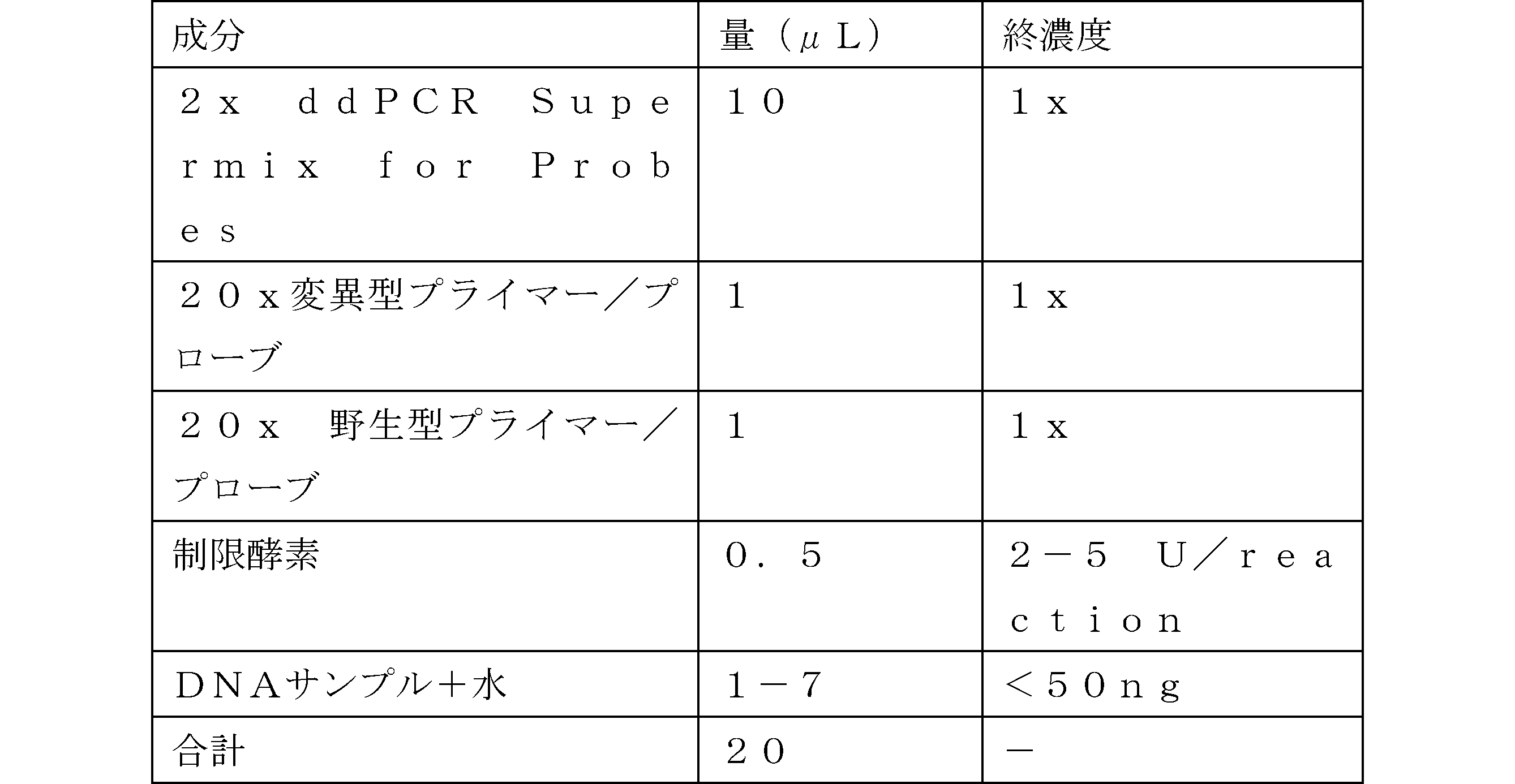
4.Vortex・遠心し室温で3分放置
5.DG8(登録商標)カートリッジのサンプルウェルに20μLのmixを、オイルウェルに70μLのDroplet Generation Oilを添加。
6.Droplet Generatorに入れて溶液中のドロップレットを作成する
7.Droplet 40μLをPCR 96wellに入れてシーリング(アルミホイル)
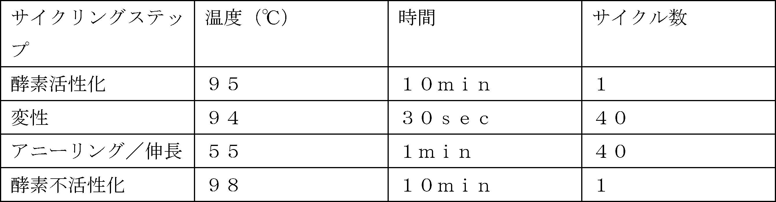
8.Droplet Readerで解析
1.<50ng/ウェル(7μLまで)のDNA溶液を調整(10-50コピーの場合は15ng追加可)
2.室温で溶解し、ボルテックスし、遠心し、遮光しておく。
3.Mixを調製(制限酵素は原液10U/μL)
5.DG8(登録商標)カートリッジのサンプルウェルに20μLのmixを、オイルウェルに70μLのDroplet Generation Oilを添加。
6.Droplet Generatorに入れて溶液中のドロップレットを作成する
7.Droplet 40μLをPCR 96wellに入れてシーリング(アルミホイル)
4.結果
バリデーション用のプラスミド(WT、Mut)を作成し検出精度を確認した。結果を図1に示す。PLEKHS1 590G>Aの検出に関してはWTとMutの検出精度はそれぞれ99.98%、100.00%だった。PLEKHS1 593C>Tの検出に関してはWTとMutの検出精度はそれぞれ99.99%、100.00%だった。また、WTでMutを1000倍希釈したDNA溶液を検出したところ有意に0.1%の変異を検出できた(590G>A:p=0.0031、593C>T:p<0.001)。
バリデーション用のプラスミド(WT、Mut)を作成し検出精度を確認した。結果を図1に示す。PLEKHS1 590G>Aの検出に関してはWTとMutの検出精度はそれぞれ99.98%、100.00%だった。PLEKHS1 593C>Tの検出に関してはWTとMutの検出精度はそれぞれ99.99%、100.00%だった。また、WTでMutを1000倍希釈したDNA溶液を検出したところ有意に0.1%の変異を検出できた(590G>A:p=0.0031、593C>T:p<0.001)。
変異型陽性患者サンプル(tDNA、cfDNA)を対象にdPCRを試行したところWT、Mutとも検出シグナルは良好であり、いずれのサンプルでも陽性ドロップレットと陰性ドロップレットを明確に分離できた(図2)。
図3に示すとおり、変異型陽性である患者のcfDNAのMAFを経時的に解析したところ、画像による転移診断より前にMAFの増加を検出できた。また、同じ検体をPLEKHS1以外にTERTプロモーターでも解析したところ変異によって異なる変動を見ることがあった。例えば、図3中の矢印は、再発している段階、つまり「ctDNAがあり他変異で陽性と判定できる」にもかかわらず、MAFが0.00%と検出されてしまっている変異(又はそれを含む試料)の特定の段階を示しており、この結果から、膀胱癌の術後を経過観察においては複数の変異の経過を観察する必要があることが分かる。
図4に示すとおり、26検体の腫瘍DNAをdPCRで検出したところ、TERTプロモーターとPLEKHS1プロモーターはそれぞれ50.0%(13/26)、42.3%(11/26)の患者が陽性であった。なお、PLEKHS1プロモーターの変異が重複している患者は1とカウントした。TERTプロモーターのみでは50.0%の対象患者を観察できるのに対し、PLEKHS1プロモーターの追加によって57.7%(15/26)に改善した。また、TERTプロモーターのみでは同一患者において複数の変異を観察できなかった、つまり0.0%(0/26)であったのに対し、PLEKHS1プロモーターの追加によって34.6%(9/26)の患者を複数の変異で観察できるようになった。
5.考察
従来の方法で検出できるTERTプロモーターと同様に、今回検出したPLEKHS1プロモーターの変異も複数の患者で陽性であり、ホットスポットとして有用であった。これをcfDNAのMAFとして検出することにより患者の病勢を評価できることが可能であると考えられる。PLEKHS1プロモーターの変異検出精度はWT、Mutとも高精度でありcfDNAのMAF解析のような低頻度の解析においても利用可能である。
従来の方法で検出できるTERTプロモーターと同様に、今回検出したPLEKHS1プロモーターの変異も複数の患者で陽性であり、ホットスポットとして有用であった。これをcfDNAのMAFとして検出することにより患者の病勢を評価できることが可能であると考えられる。PLEKHS1プロモーターの変異検出精度はWT、Mutとも高精度でありcfDNAのMAF解析のような低頻度の解析においても利用可能である。
WTで軽微なミスアニーリングが起こる原因としては、変異によってGC%が変動しプローブのTm値が変わることが原因であると考えられる。患者サンプルを用いた検討でもtDNAとcfDNAは類似したシグナル検出能でありcfDNAを対象とした解析にも有用であると判断できた。
TERTプロモーターだけでなくPLEKHS1プロモーターにおいてもdPCRでの変異解析ができる利点として、第一に同一アッセイで解析可能な患者が増えることが挙げられる。膀胱癌の腫瘍変異は多岐に富むため患者ごとに異なる変異を経時的に解析する方法が主流であり、この方法に比してホットスポットを検出することは解析コストの改善において有利である。第二に複数のMAFを解析することによって患者の病勢評価をより正確に行うことが可能となる。全身転移が診断されているにも関わらずMAFが検出できない事象を複数回認めたが、これは単一の変異で経過観察を行なった場合に偽陰性と判断される危険性を示唆するものである。複数のMAFを観察することで偽陰性と評価される可能性を減少させることができると考えられる。
複数検出による臨床的な意義としては、安価にモニタリングができるため病勢の早期評価に有用だが、NGSと異なり単一の変異をみるdPCRにおいては偽陰性による誤った病勢評価の影響を受けやすく、いずれの変異検出においても単一変異での評価は誤評価の危険性を伴うため、複数での検出評価が望ましい。
Claims (12)
- 検体における膀胱癌の存在を検出するためのプライマーセットであって、
PLEKHS1プロモーターの変異を標的とする、16塩基長以上で且つ21塩基長以下のオリゴヌクレオチドである1対のプライマーと、
TERTプロモーターの変異を標的とする、1又は複数の対のプライマーと、
を含む、プライマーセット。 - PLEKHS1プロモーターの変異が、10番染色体の115,511,590位におけるGからAへの置換、10番染色体の115,511,593位におけるCからTへの置換又はその両方である、請求項1に記載のプライマーセット。
- PLEKHS1プロモーターの変異を標的とするプライマーが配列番号1及び/又は2に記載の塩基配列を有する、請求項1又は2に記載のプライマーセット。
- TERTプロモーターの変異を標的とするプライマーが、5番染色体の1,295,228位におけるCからTへの置換、5番染色体の1,295,250位におけるCからTへの置換又はその両方である、請求項1~3のいずれか一項に記載のプライマーセット。
- 検体における膀胱癌の存在を検出するためのプローブであって、PLEKHS1プロモーターの変異を標的とする、プローブ。
- プローブが配列番号4又は5に記載の塩基配列を有する、請求項5に記載のプローブ。
- 請求項1~4のいずれか一項に記載のプライマーセットを含む、キット。
- 請求項5又は6に記載のプローブを更に含む、請求項7に記載のキット。
- 検体における膀胱癌の存在を検出するための方法であって、請求項1~4のいずれか一項に記載のプライマーセット、あるいは請求項5又は6に記載のプローブを用いてPLEKHS1プロモーターの変異を検出する工程を含む、方法。
- 検出工程で使用される核酸増幅法がデジタルPCRである、請求項9に記載の方法。
- 検体が液性検体である、請求項9又は10に記載の方法。
- 検体が血中循環DNA(cfDNA)を含む、請求項9~11のいずれか一項に記載の方法。
Priority Applications (1)
| Application Number | Priority Date | Filing Date | Title |
|---|---|---|---|
| JP2023576937A JPWO2023145754A1 (ja) | 2022-01-26 | 2023-01-25 |
Applications Claiming Priority (2)
| Application Number | Priority Date | Filing Date | Title |
|---|---|---|---|
| JP2022009850 | 2022-01-26 | ||
| JP2022-009850 | 2022-01-26 |
Publications (1)
| Publication Number | Publication Date |
|---|---|
| WO2023145754A1 true WO2023145754A1 (ja) | 2023-08-03 |
Family
ID=87471454
Family Applications (1)
| Application Number | Title | Priority Date | Filing Date |
|---|---|---|---|
| PCT/JP2023/002178 Ceased WO2023145754A1 (ja) | 2022-01-26 | 2023-01-25 | 膀胱癌の存在を検出するためのプライマー及びプローブ |
Country Status (3)
| Country | Link |
|---|---|
| JP (1) | JPWO2023145754A1 (ja) |
| TW (1) | TW202338101A (ja) |
| WO (1) | WO2023145754A1 (ja) |
-
2023
- 2023-01-25 JP JP2023576937A patent/JPWO2023145754A1/ja active Pending
- 2023-01-25 WO PCT/JP2023/002178 patent/WO2023145754A1/ja not_active Ceased
- 2023-01-30 TW TW112103135A patent/TW202338101A/zh unknown
Non-Patent Citations (2)
| Title |
|---|
| BAXTER L., GORDON N. S., OTT S., WANG J., PATEL P., GOEL A., PIECHOCKI K., SILCOCK L., SALE C., ZEEGERS M. P., CHENG K. K., JAMES : "Properties of non-coding mutation hotspots as urinary biomarkers for bladder cancer detection", SCIENTIFIC REPORTS, vol. 13, no. 1, XP093080486, DOI: 10.1038/s41598-023-27675-4 * |
| WU SONG, OU TONG, XING NIANZENG, LU JIANG, WAN SHENGQING, WANG CHANGXI, ZHANG XI, YANG FEIYA, HUANG YI, CAI ZHIMING: "Whole-genome sequencing identifies ADGRG6 enhancer mutations and FRS2 duplications as angiogenesis-related drivers in bladder cancer", NATURE COMMUNICATIONS, vol. 10, no. 1, XP093080485, DOI: 10.1038/s41467-019-08576-5 * |
Also Published As
| Publication number | Publication date |
|---|---|
| TW202338101A (zh) | 2023-10-01 |
| JPWO2023145754A1 (ja) | 2023-08-03 |
Similar Documents
| Publication | Publication Date | Title |
|---|---|---|
| CN101815789B (zh) | 靶序列的富集 | |
| Jakupciak et al. | Mitochondrial DNA as a cancer biomarker | |
| US20230366034A1 (en) | Compositions and methods for diagnosing lung cancers using gene expression profiles | |
| JP2012005500A (ja) | 食道癌、結腸癌、頭頸部癌、およびメラノーマにおけるマーカーの同定 | |
| JP2004533241A (ja) | マイクロサテライトの不安定性の検出および腫瘍診断におけるその使用 | |
| JP6438119B2 (ja) | ホットスポット変異の迅速かつ高感度の検出のための方法 | |
| JP3844996B2 (ja) | 反復性pcr産物の融解曲線解析方法 | |
| US20200263259A1 (en) | Ctnnb1 mutation detection kit, method, and use in hcc detection and management | |
| US20100184015A1 (en) | Method for detection of xmrv | |
| CN104805206A (zh) | 检测tert基因启动子突变的试剂盒及其检测方法 | |
| US11535897B2 (en) | Composite epigenetic biomarkers for accurate screening, diagnosis and prognosis of colorectal cancer | |
| KR101825117B1 (ko) | Pna 기반의 실시간 pcr 클램핑을 이용한 braf 돌연변이 검출 방법 및 키트 | |
| JP2024523655A (ja) | 核酸分析のための方法および組成物 | |
| US9074247B2 (en) | P53 assay for a urine test for HCC screening | |
| CN113930501A (zh) | 人egfr基因突变的数字pcr检测方法及应用 | |
| US9920376B2 (en) | Method for determining lymph node metastasis in cancer or risk thereof and rapid determination kit for the same | |
| CN110964833B (zh) | 一种一管检测血浆游离dna中kras和braf基因突变的试剂盒 | |
| TW202338101A (zh) | 用於檢測膀胱癌之存在之引子及探針 | |
| TW202442874A (zh) | 膀胱癌之生物標記 | |
| KR101860547B1 (ko) | 세포-유리형 dna의 암 질환의 진단 용도 | |
| WO2024204603A1 (ja) | 膀胱癌のバイオマーカー | |
| JP4359497B2 (ja) | 核酸増幅用プライマー、核酸増幅用プライマーセット及びこれを用いた癌の検査方法 | |
| KR102816628B1 (ko) | 대사증후군 특이적 후성유전 메틸화 마커 및 이의 용도 | |
| US20250236917A1 (en) | Rapid and accurate single-nucleotide polymorphism detection by fluorophore-nucleic acid interaction | |
| CN113913514B (zh) | 人ctnnb1基因突变的数字pcr检测方法及应用 |
Legal Events
| Date | Code | Title | Description |
|---|---|---|---|
| 121 | Ep: the epo has been informed by wipo that ep was designated in this application |
Ref document number: 23746965 Country of ref document: EP Kind code of ref document: A1 |
|
| WWE | Wipo information: entry into national phase |
Ref document number: 2023576937 Country of ref document: JP |
|
| NENP | Non-entry into the national phase |
Ref country code: DE |
|
| 122 | Ep: pct application non-entry in european phase |
Ref document number: 23746965 Country of ref document: EP Kind code of ref document: A1 |