WO2023058627A1 - 認知症の検査方法 - Google Patents
認知症の検査方法 Download PDFInfo
- Publication number
- WO2023058627A1 WO2023058627A1 PCT/JP2022/037054 JP2022037054W WO2023058627A1 WO 2023058627 A1 WO2023058627 A1 WO 2023058627A1 JP 2022037054 W JP2022037054 W JP 2022037054W WO 2023058627 A1 WO2023058627 A1 WO 2023058627A1
- Authority
- WO
- WIPO (PCT)
- Prior art keywords
- ecrg4
- antibody
- dementia
- polypeptide
- patients
- Prior art date
Links
- 206010012289 Dementia Diseases 0.000 title claims abstract description 137
- 238000012360 testing method Methods 0.000 title claims abstract description 26
- 101710115121 Augurin Proteins 0.000 claims abstract description 157
- 102100027393 Augurin Human genes 0.000 claims abstract description 157
- 210000004369 blood Anatomy 0.000 claims abstract description 38
- 239000008280 blood Substances 0.000 claims abstract description 38
- 108090000765 processed proteins & peptides Proteins 0.000 claims description 119
- 102000004196 processed proteins & peptides Human genes 0.000 claims description 111
- 238000000034 method Methods 0.000 claims description 108
- 229920001184 polypeptide Polymers 0.000 claims description 104
- 125000003275 alpha amino acid group Chemical group 0.000 claims description 70
- 238000001514 detection method Methods 0.000 claims description 29
- 230000001900 immune effect Effects 0.000 claims description 22
- 239000003814 drug Substances 0.000 claims description 13
- 229940079593 drug Drugs 0.000 claims description 11
- 239000003795 chemical substances by application Substances 0.000 claims description 9
- 102000004190 Enzymes Human genes 0.000 claims description 8
- 108090000790 Enzymes Proteins 0.000 claims description 8
- 239000003547 immunosorbent Substances 0.000 claims description 7
- 208000010877 cognitive disease Diseases 0.000 abstract description 37
- 208000027061 mild cognitive impairment Diseases 0.000 abstract description 35
- 210000002381 plasma Anatomy 0.000 abstract description 32
- 239000000090 biomarker Substances 0.000 abstract description 11
- 230000037058 blood plasma level Effects 0.000 abstract 1
- 208000024827 Alzheimer disease Diseases 0.000 description 31
- 108010064539 amyloid beta-protein (1-42) Proteins 0.000 description 25
- 108010064397 amyloid beta-protein (1-40) Proteins 0.000 description 23
- 210000001175 cerebrospinal fluid Anatomy 0.000 description 23
- 108090000623 proteins and genes Proteins 0.000 description 16
- 210000004027 cell Anatomy 0.000 description 13
- 102000004169 proteins and genes Human genes 0.000 description 13
- 239000000126 substance Substances 0.000 description 13
- 230000036470 plasma concentration Effects 0.000 description 12
- 235000018102 proteins Nutrition 0.000 description 12
- 238000002965 ELISA Methods 0.000 description 11
- 101150066783 Ecrg4 gene Proteins 0.000 description 10
- 241000283973 Oryctolagus cuniculus Species 0.000 description 10
- 235000001014 amino acid Nutrition 0.000 description 10
- 150000001413 amino acids Chemical class 0.000 description 10
- 238000003745 diagnosis Methods 0.000 description 10
- 238000010586 diagram Methods 0.000 description 10
- 238000010790 dilution Methods 0.000 description 10
- 239000012895 dilution Substances 0.000 description 10
- 101000936427 Homo sapiens Augurin Proteins 0.000 description 8
- 238000004458 analytical method Methods 0.000 description 8
- 102000051913 human ECRG4 Human genes 0.000 description 8
- 229940126619 mouse monoclonal antibody Drugs 0.000 description 8
- 201000003077 normal pressure hydrocephalus Diseases 0.000 description 8
- 238000005406 washing Methods 0.000 description 8
- 206010033892 Paraplegia Diseases 0.000 description 7
- 208000032930 Spastic paraplegia Diseases 0.000 description 7
- 238000002372 labelling Methods 0.000 description 7
- 208000033808 peripheral neuropathy Diseases 0.000 description 7
- 206010028570 Myelopathy Diseases 0.000 description 6
- 239000000872 buffer Substances 0.000 description 6
- 208000037265 diseases, disorders, signs and symptoms Diseases 0.000 description 6
- 239000012634 fragment Substances 0.000 description 6
- 201000001119 neuropathy Diseases 0.000 description 6
- 230000007823 neuropathy Effects 0.000 description 6
- 239000011534 wash buffer Substances 0.000 description 6
- FWMNVWWHGCHHJJ-SKKKGAJSSA-N 4-amino-1-[(2r)-6-amino-2-[[(2r)-2-[[(2r)-2-[[(2r)-2-amino-3-phenylpropanoyl]amino]-3-phenylpropanoyl]amino]-4-methylpentanoyl]amino]hexanoyl]piperidine-4-carboxylic acid Chemical compound C([C@H](C(=O)N[C@H](CC(C)C)C(=O)N[C@H](CCCCN)C(=O)N1CCC(N)(CC1)C(O)=O)NC(=O)[C@H](N)CC=1C=CC=CC=1)C1=CC=CC=C1 FWMNVWWHGCHHJJ-SKKKGAJSSA-N 0.000 description 5
- 101100272054 Mus musculus Ecrg4 gene Proteins 0.000 description 5
- 201000010099 disease Diseases 0.000 description 5
- 229940088598 enzyme Drugs 0.000 description 5
- 210000004408 hybridoma Anatomy 0.000 description 5
- 239000000243 solution Substances 0.000 description 5
- 208000024891 symptom Diseases 0.000 description 5
- 238000010998 test method Methods 0.000 description 5
- 206010067889 Dementia with Lewy bodies Diseases 0.000 description 4
- 238000012286 ELISA Assay Methods 0.000 description 4
- 201000002832 Lewy body dementia Diseases 0.000 description 4
- 241000699666 Mus <mouse, genus> Species 0.000 description 4
- 208000010291 Primary Progressive Nonfluent Aphasia Diseases 0.000 description 4
- 206010042928 Syringomyelia Diseases 0.000 description 4
- 201000004810 Vascular dementia Diseases 0.000 description 4
- 238000006243 chemical reaction Methods 0.000 description 4
- 239000003153 chemical reaction reagent Substances 0.000 description 4
- OSASVXMJTNOKOY-UHFFFAOYSA-N chlorobutanol Chemical compound CC(C)(O)C(Cl)(Cl)Cl OSASVXMJTNOKOY-UHFFFAOYSA-N 0.000 description 4
- 238000011161 development Methods 0.000 description 4
- 230000018109 developmental process Effects 0.000 description 4
- 238000003384 imaging method Methods 0.000 description 4
- 238000012744 immunostaining Methods 0.000 description 4
- 238000009169 immunotherapy Methods 0.000 description 4
- 230000003902 lesion Effects 0.000 description 4
- 208000010325 limbic encephalitis Diseases 0.000 description 4
- 208000021629 neuronal intranuclear inclusion disease Diseases 0.000 description 4
- 201000002212 progressive supranuclear palsy Diseases 0.000 description 4
- 239000000758 substrate Substances 0.000 description 4
- 238000001262 western blot Methods 0.000 description 4
- 108010090849 Amyloid beta-Peptides Proteins 0.000 description 3
- 102000013455 Amyloid beta-Peptides Human genes 0.000 description 3
- 208000014644 Brain disease Diseases 0.000 description 3
- 206010008025 Cerebellar ataxia Diseases 0.000 description 3
- 208000032274 Encephalopathy Diseases 0.000 description 3
- 208000002339 Frontotemporal Lobar Degeneration Diseases 0.000 description 3
- 201000011240 Frontotemporal dementia Diseases 0.000 description 3
- 208000021642 Muscular disease Diseases 0.000 description 3
- 208000003926 Myelitis Diseases 0.000 description 3
- 201000009623 Myopathy Diseases 0.000 description 3
- DNIAPMSPPWPWGF-UHFFFAOYSA-N Propylene glycol Chemical compound CC(O)CO DNIAPMSPPWPWGF-UHFFFAOYSA-N 0.000 description 3
- 238000002835 absorbance Methods 0.000 description 3
- 238000004220 aggregation Methods 0.000 description 3
- 230000002776 aggregation Effects 0.000 description 3
- 239000000427 antigen Substances 0.000 description 3
- 102000036639 antigens Human genes 0.000 description 3
- 108091007433 antigens Proteins 0.000 description 3
- 239000012131 assay buffer Substances 0.000 description 3
- 230000036765 blood level Effects 0.000 description 3
- 206010008118 cerebral infarction Diseases 0.000 description 3
- 239000000544 cholinesterase inhibitor Substances 0.000 description 3
- 239000002299 complementary DNA Substances 0.000 description 3
- 150000001875 compounds Chemical class 0.000 description 3
- 230000014509 gene expression Effects 0.000 description 3
- 239000003550 marker Substances 0.000 description 3
- 238000004949 mass spectrometry Methods 0.000 description 3
- 239000000463 material Substances 0.000 description 3
- 239000003755 preservative agent Substances 0.000 description 3
- 238000011002 quantification Methods 0.000 description 3
- 238000003118 sandwich ELISA Methods 0.000 description 3
- 201000000306 sarcoidosis Diseases 0.000 description 3
- 239000007790 solid phase Substances 0.000 description 3
- 238000007619 statistical method Methods 0.000 description 3
- 238000011282 treatment Methods 0.000 description 3
- 229940124648 γ-Secretase Modulator Drugs 0.000 description 3
- YBJHBAHKTGYVGT-ZKWXMUAHSA-N (+)-Biotin Chemical compound N1C(=O)N[C@@H]2[C@H](CCCCC(=O)O)SC[C@@H]21 YBJHBAHKTGYVGT-ZKWXMUAHSA-N 0.000 description 2
- VBICKXHEKHSIBG-UHFFFAOYSA-N 1-monostearoylglycerol Chemical compound CCCCCCCCCCCCCCCCCC(=O)OCC(O)CO VBICKXHEKHSIBG-UHFFFAOYSA-N 0.000 description 2
- 206010003062 Apraxia Diseases 0.000 description 2
- CIWBSHSKHKDKBQ-JLAZNSOCSA-N Ascorbic acid Chemical compound OC[C@H](O)[C@H]1OC(=O)C(O)=C1O CIWBSHSKHKDKBQ-JLAZNSOCSA-N 0.000 description 2
- 208000006740 Aseptic Meningitis Diseases 0.000 description 2
- VTYYLEPIZMXCLO-UHFFFAOYSA-L Calcium carbonate Chemical compound [Ca+2].[O-]C([O-])=O VTYYLEPIZMXCLO-UHFFFAOYSA-L 0.000 description 2
- BVKZGUZCCUSVTD-UHFFFAOYSA-L Carbonate Chemical compound [O-]C([O-])=O BVKZGUZCCUSVTD-UHFFFAOYSA-L 0.000 description 2
- 229920002134 Carboxymethyl cellulose Polymers 0.000 description 2
- 206010008027 Cerebellar atrophy Diseases 0.000 description 2
- FBPFZTCFMRRESA-KVTDHHQDSA-N D-Mannitol Chemical compound OC[C@@H](O)[C@@H](O)[C@H](O)[C@H](O)CO FBPFZTCFMRRESA-KVTDHHQDSA-N 0.000 description 2
- 208000014094 Dystonic disease Diseases 0.000 description 2
- KCXVZYZYPLLWCC-UHFFFAOYSA-N EDTA Chemical compound OC(=O)CN(CC(O)=O)CCN(CC(O)=O)CC(O)=O KCXVZYZYPLLWCC-UHFFFAOYSA-N 0.000 description 2
- 208000001640 Fibromyalgia Diseases 0.000 description 2
- WSFSSNUMVMOOMR-UHFFFAOYSA-N Formaldehyde Chemical compound O=C WSFSSNUMVMOOMR-UHFFFAOYSA-N 0.000 description 2
- 102100031181 Glyceraldehyde-3-phosphate dehydrogenase Human genes 0.000 description 2
- 208000004356 Hysteria Diseases 0.000 description 2
- 206010027201 Meningitis aseptic Diseases 0.000 description 2
- 208000019695 Migraine disease Diseases 0.000 description 2
- 208000001089 Multiple system atrophy Diseases 0.000 description 2
- 208000002033 Myoclonus Diseases 0.000 description 2
- 108091028043 Nucleic acid sequence Proteins 0.000 description 2
- 108091034117 Oligonucleotide Proteins 0.000 description 2
- ISWSIDIOOBJBQZ-UHFFFAOYSA-N Phenol Chemical compound OC1=CC=CC=C1 ISWSIDIOOBJBQZ-UHFFFAOYSA-N 0.000 description 2
- 241000276498 Pollachius virens Species 0.000 description 2
- 108020004511 Recombinant DNA Proteins 0.000 description 2
- PXIPVTKHYLBLMZ-UHFFFAOYSA-N Sodium azide Chemical compound [Na+].[N-]=[N+]=[N-] PXIPVTKHYLBLMZ-UHFFFAOYSA-N 0.000 description 2
- 229920002472 Starch Polymers 0.000 description 2
- 238000000692 Student's t-test Methods 0.000 description 2
- QAOWNCQODCNURD-UHFFFAOYSA-N Sulfuric acid Chemical compound OS(O)(=O)=O QAOWNCQODCNURD-UHFFFAOYSA-N 0.000 description 2
- 206010044074 Torticollis Diseases 0.000 description 2
- 101710120037 Toxin CcdB Proteins 0.000 description 2
- 230000032683 aging Effects 0.000 description 2
- 230000001476 alcoholic effect Effects 0.000 description 2
- 238000000540 analysis of variance Methods 0.000 description 2
- 238000003556 assay Methods 0.000 description 2
- 229960000686 benzalkonium chloride Drugs 0.000 description 2
- CADWTSSKOVRVJC-UHFFFAOYSA-N benzyl(dimethyl)azanium;chloride Chemical compound [Cl-].C[NH+](C)CC1=CC=CC=C1 CADWTSSKOVRVJC-UHFFFAOYSA-N 0.000 description 2
- 230000027455 binding Effects 0.000 description 2
- 210000000601 blood cell Anatomy 0.000 description 2
- 210000004556 brain Anatomy 0.000 description 2
- 239000001768 carboxy methyl cellulose Substances 0.000 description 2
- 235000010948 carboxy methyl cellulose Nutrition 0.000 description 2
- 239000008112 carboxymethyl-cellulose Substances 0.000 description 2
- 238000005119 centrifugation Methods 0.000 description 2
- 201000002866 cervical dystonia Diseases 0.000 description 2
- 238000012767 chemiluminescent enzyme immunoassay Methods 0.000 description 2
- 229960004926 chlorobutanol Drugs 0.000 description 2
- 238000007796 conventional method Methods 0.000 description 2
- 208000012839 conversion disease Diseases 0.000 description 2
- VFLDPWHFBUODDF-FCXRPNKRSA-N curcumin Chemical compound C1=C(O)C(OC)=CC(\C=C\C(=O)CC(=O)\C=C\C=2C=C(OC)C(O)=CC=2)=C1 VFLDPWHFBUODDF-FCXRPNKRSA-N 0.000 description 2
- 238000002059 diagnostic imaging Methods 0.000 description 2
- 239000003085 diluting agent Substances 0.000 description 2
- 239000007884 disintegrant Substances 0.000 description 2
- ADEBPBSSDYVVLD-UHFFFAOYSA-N donepezil Chemical compound O=C1C=2C=C(OC)C(OC)=CC=2CC1CC(CC1)CCN1CC1=CC=CC=C1 ADEBPBSSDYVVLD-UHFFFAOYSA-N 0.000 description 2
- 208000010118 dystonia Diseases 0.000 description 2
- 239000003995 emulsifying agent Substances 0.000 description 2
- 206010014599 encephalitis Diseases 0.000 description 2
- 206010015037 epilepsy Diseases 0.000 description 2
- -1 etc.) Substances 0.000 description 2
- 238000011156 evaluation Methods 0.000 description 2
- 239000000284 extract Substances 0.000 description 2
- ASUTZQLVASHGKV-JDFRZJQESA-N galanthamine Chemical compound O1C(=C23)C(OC)=CC=C2CN(C)CC[C@]23[C@@H]1C[C@@H](O)C=C2 ASUTZQLVASHGKV-JDFRZJQESA-N 0.000 description 2
- 108020004445 glyceraldehyde-3-phosphate dehydrogenase Proteins 0.000 description 2
- 230000003053 immunization Effects 0.000 description 2
- 201000008319 inclusion body myositis Diseases 0.000 description 2
- 239000004615 ingredient Substances 0.000 description 2
- 239000003112 inhibitor Substances 0.000 description 2
- 150000002500 ions Chemical class 0.000 description 2
- 239000004816 latex Substances 0.000 description 2
- 229920000126 latex Polymers 0.000 description 2
- 238000004811 liquid chromatography Methods 0.000 description 2
- 239000012528 membrane Substances 0.000 description 2
- LXCFILQKKLGQFO-UHFFFAOYSA-N methylparaben Chemical compound COC(=O)C1=CC=C(O)C=C1 LXCFILQKKLGQFO-UHFFFAOYSA-N 0.000 description 2
- 206010027599 migraine Diseases 0.000 description 2
- 238000002552 multiple reaction monitoring Methods 0.000 description 2
- 201000006417 multiple sclerosis Diseases 0.000 description 2
- 239000013642 negative control Substances 0.000 description 2
- 210000002569 neuron Anatomy 0.000 description 2
- 239000002245 particle Substances 0.000 description 2
- 239000000546 pharmaceutical excipient Substances 0.000 description 2
- 229920000642 polymer Polymers 0.000 description 2
- 208000005987 polymyositis Diseases 0.000 description 2
- 239000013641 positive control Substances 0.000 description 2
- 238000002360 preparation method Methods 0.000 description 2
- 230000009758 senescence Effects 0.000 description 2
- 210000002966 serum Anatomy 0.000 description 2
- 238000002553 single reaction monitoring Methods 0.000 description 2
- 238000002603 single-photon emission computed tomography Methods 0.000 description 2
- 235000020183 skimmed milk Nutrition 0.000 description 2
- 239000003381 stabilizer Substances 0.000 description 2
- 239000008107 starch Substances 0.000 description 2
- 235000019698 starch Nutrition 0.000 description 2
- 239000006228 supernatant Substances 0.000 description 2
- 239000000375 suspending agent Substances 0.000 description 2
- 238000012353 t test Methods 0.000 description 2
- 210000001519 tissue Anatomy 0.000 description 2
- 238000012546 transfer Methods 0.000 description 2
- 239000013598 vector Substances 0.000 description 2
- LNAZSHAWQACDHT-XIYTZBAFSA-N (2r,3r,4s,5r,6s)-4,5-dimethoxy-2-(methoxymethyl)-3-[(2s,3r,4s,5r,6r)-3,4,5-trimethoxy-6-(methoxymethyl)oxan-2-yl]oxy-6-[(2r,3r,4s,5r,6r)-4,5,6-trimethoxy-2-(methoxymethyl)oxan-3-yl]oxyoxane Chemical compound CO[C@@H]1[C@@H](OC)[C@H](OC)[C@@H](COC)O[C@H]1O[C@H]1[C@H](OC)[C@@H](OC)[C@H](O[C@H]2[C@@H]([C@@H](OC)[C@H](OC)O[C@@H]2COC)OC)O[C@@H]1COC LNAZSHAWQACDHT-XIYTZBAFSA-N 0.000 description 1
- IXPNQXFRVYWDDI-UHFFFAOYSA-N 1-methyl-2,4-dioxo-1,3-diazinane-5-carboximidamide Chemical compound CN1CC(C(N)=N)C(=O)NC1=O IXPNQXFRVYWDDI-UHFFFAOYSA-N 0.000 description 1
- PRDFBSVERLRRMY-UHFFFAOYSA-N 2'-(4-ethoxyphenyl)-5-(4-methylpiperazin-1-yl)-2,5'-bibenzimidazole Chemical compound C1=CC(OCC)=CC=C1C1=NC2=CC=C(C=3NC4=CC(=CC=C4N=3)N3CCN(C)CC3)C=C2N1 PRDFBSVERLRRMY-UHFFFAOYSA-N 0.000 description 1
- RBTBFTRPCNLSDE-UHFFFAOYSA-N 3,7-bis(dimethylamino)phenothiazin-5-ium Chemical compound C1=CC(N(C)C)=CC2=[S+]C3=CC(N(C)C)=CC=C3N=C21 RBTBFTRPCNLSDE-UHFFFAOYSA-N 0.000 description 1
- 244000215068 Acacia senegal Species 0.000 description 1
- QTBSBXVTEAMEQO-UHFFFAOYSA-M Acetate Chemical compound CC([O-])=O QTBSBXVTEAMEQO-UHFFFAOYSA-M 0.000 description 1
- 102100022900 Actin, cytoplasmic 1 Human genes 0.000 description 1
- 108010085238 Actins Proteins 0.000 description 1
- 208000006888 Agnosia Diseases 0.000 description 1
- 241001047040 Agnosia Species 0.000 description 1
- 206010001541 Akinesia Diseases 0.000 description 1
- 208000037259 Amyloid Plaque Diseases 0.000 description 1
- 206010003226 Arteriovenous fistula Diseases 0.000 description 1
- 206010003445 Ascites Diseases 0.000 description 1
- 241000416162 Astragalus gummifer Species 0.000 description 1
- 206010003694 Atrophy Diseases 0.000 description 1
- 102000015081 Blood Coagulation Factors Human genes 0.000 description 1
- 108010039209 Blood Coagulation Factors Proteins 0.000 description 1
- 201000006390 Brachial Plexus Neuritis Diseases 0.000 description 1
- 108010075254 C-Peptide Proteins 0.000 description 1
- 241000283707 Capra Species 0.000 description 1
- 208000000483 Central Nervous System Vascular Malformations Diseases 0.000 description 1
- KRKNYBCHXYNGOX-UHFFFAOYSA-K Citrate Chemical compound [O-]C(=O)CC(O)(CC([O-])=O)C([O-])=O KRKNYBCHXYNGOX-UHFFFAOYSA-K 0.000 description 1
- QCDFBFJGMNKBDO-UHFFFAOYSA-N Clioquinol Chemical compound C1=CN=C2C(O)=C(I)C=C(Cl)C2=C1 QCDFBFJGMNKBDO-UHFFFAOYSA-N 0.000 description 1
- 208000028698 Cognitive impairment Diseases 0.000 description 1
- 108020004635 Complementary DNA Proteins 0.000 description 1
- 208000011990 Corticobasal Degeneration Diseases 0.000 description 1
- 208000019736 Cranial nerve disease Diseases 0.000 description 1
- 108050006400 Cyclin Proteins 0.000 description 1
- 102000004127 Cytokines Human genes 0.000 description 1
- 108090000695 Cytokines Proteins 0.000 description 1
- FBPFZTCFMRRESA-FSIIMWSLSA-N D-Glucitol Natural products OC[C@H](O)[C@H](O)[C@@H](O)[C@H](O)CO FBPFZTCFMRRESA-FSIIMWSLSA-N 0.000 description 1
- IGXWBGJHJZYPQS-SSDOTTSWSA-N D-Luciferin Chemical compound OC(=O)[C@H]1CSC(C=2SC3=CC=C(O)C=C3N=2)=N1 IGXWBGJHJZYPQS-SSDOTTSWSA-N 0.000 description 1
- FBPFZTCFMRRESA-JGWLITMVSA-N D-glucitol Chemical compound OC[C@H](O)[C@@H](O)[C@H](O)[C@H](O)CO FBPFZTCFMRRESA-JGWLITMVSA-N 0.000 description 1
- 108020004414 DNA Proteins 0.000 description 1
- CYCGRDQQIOGCKX-UHFFFAOYSA-N Dehydro-luciferin Natural products OC(=O)C1=CSC(C=2SC3=CC(O)=CC=C3N=2)=N1 CYCGRDQQIOGCKX-UHFFFAOYSA-N 0.000 description 1
- 208000032131 Diabetic Neuropathies Diseases 0.000 description 1
- 238000008157 ELISA kit Methods 0.000 description 1
- 241000196324 Embryophyta Species 0.000 description 1
- 241000588724 Escherichia coli Species 0.000 description 1
- LFQSCWFLJHTTHZ-UHFFFAOYSA-N Ethanol Chemical compound CCO LFQSCWFLJHTTHZ-UHFFFAOYSA-N 0.000 description 1
- 238000004252 FT/ICR mass spectrometry Methods 0.000 description 1
- 238000000729 Fisher's exact test Methods 0.000 description 1
- BJGNCJDXODQBOB-UHFFFAOYSA-N Fivefly Luciferin Natural products OC(=O)C1CSC(C=2SC3=CC(O)=CC=C3N=2)=N1 BJGNCJDXODQBOB-UHFFFAOYSA-N 0.000 description 1
- WHUUTDBJXJRKMK-UHFFFAOYSA-N Glutamic acid Natural products OC(=O)C(N)CCC(O)=O WHUUTDBJXJRKMK-UHFFFAOYSA-N 0.000 description 1
- 229920000084 Gum arabic Polymers 0.000 description 1
- HTTJABKRGRZYRN-UHFFFAOYSA-N Heparin Chemical compound OC1C(NC(=O)C)C(O)OC(COS(O)(=O)=O)C1OC1C(OS(O)(=O)=O)C(O)C(OC2C(C(OS(O)(=O)=O)C(OC3C(C(O)C(O)C(O3)C(O)=O)OS(O)(=O)=O)C(CO)O2)NS(O)(=O)=O)C(C(O)=O)O1 HTTJABKRGRZYRN-UHFFFAOYSA-N 0.000 description 1
- 241000238631 Hexapoda Species 0.000 description 1
- 208000023105 Huntington disease Diseases 0.000 description 1
- 108060003951 Immunoglobulin Proteins 0.000 description 1
- 108010021625 Immunoglobulin Fragments Proteins 0.000 description 1
- 102000008394 Immunoglobulin Fragments Human genes 0.000 description 1
- 206010061218 Inflammation Diseases 0.000 description 1
- 238000012313 Kruskal-Wallis test Methods 0.000 description 1
- GUBGYTABKSRVRQ-QKKXKWKRSA-N Lactose Natural products OC[C@H]1O[C@@H](O[C@H]2[C@H](O)[C@@H](O)C(O)O[C@@H]2CO)[C@H](O)[C@@H](O)[C@H]1O GUBGYTABKSRVRQ-QKKXKWKRSA-N 0.000 description 1
- 102100026517 Lamin-B1 Human genes 0.000 description 1
- 206010072742 Late onset hypogonadism syndrome Diseases 0.000 description 1
- 208000034800 Leukoencephalopathies Diseases 0.000 description 1
- 108060001084 Luciferase Proteins 0.000 description 1
- 239000005089 Luciferase Substances 0.000 description 1
- DDWFXDSYGUXRAY-UHFFFAOYSA-N Luciferin Natural products CCc1c(C)c(CC2NC(=O)C(=C2C=C)C)[nH]c1Cc3[nH]c4C(=C5/NC(CC(=O)O)C(C)C5CC(=O)O)CC(=O)c4c3C DDWFXDSYGUXRAY-UHFFFAOYSA-N 0.000 description 1
- 238000000585 Mann–Whitney U test Methods 0.000 description 1
- 206010027259 Meningitis tuberculous Diseases 0.000 description 1
- 241001465754 Metazoa Species 0.000 description 1
- 208000026072 Motor neurone disease Diseases 0.000 description 1
- 241000699670 Mus sp. Species 0.000 description 1
- 208000007101 Muscle Cramp Diseases 0.000 description 1
- 102000004868 N-Methyl-D-Aspartate Receptors Human genes 0.000 description 1
- 108090001041 N-Methyl-D-Aspartate Receptors Proteins 0.000 description 1
- 208000012902 Nervous system disease Diseases 0.000 description 1
- 206010029229 Neuralgic amyotrophy Diseases 0.000 description 1
- 208000025966 Neurological disease Diseases 0.000 description 1
- 101100275485 Neurospora crassa (strain ATCC 24698 / 74-OR23-1A / CBS 708.71 / DSM 1257 / FGSC 987) cox-4 gene Proteins 0.000 description 1
- 239000000020 Nitrocellulose Substances 0.000 description 1
- 229910019142 PO4 Inorganic materials 0.000 description 1
- 206010053658 Pachymeningitis Diseases 0.000 description 1
- 206010072106 Paraneoplastic neurological syndrome Diseases 0.000 description 1
- 102000035195 Peptidases Human genes 0.000 description 1
- 108091005804 Peptidases Proteins 0.000 description 1
- 206010034620 Peripheral sensory neuropathy Diseases 0.000 description 1
- 102000003992 Peroxidases Human genes 0.000 description 1
- 206010034719 Personality change Diseases 0.000 description 1
- 241000233805 Phoenix Species 0.000 description 1
- 229920001213 Polysorbate 20 Polymers 0.000 description 1
- 108010013381 Porins Proteins 0.000 description 1
- 102000009339 Proliferating Cell Nuclear Antigen Human genes 0.000 description 1
- 239000004365 Protease Substances 0.000 description 1
- 101710156592 Putative TATA-binding protein pB263R Proteins 0.000 description 1
- 239000012083 RIPA buffer Substances 0.000 description 1
- 238000011579 SCID mouse model Methods 0.000 description 1
- 229920002684 Sepharose Polymers 0.000 description 1
- DBMJMQXJHONAFJ-UHFFFAOYSA-M Sodium laurylsulphate Chemical compound [Na+].CCCCCCCCCCCCOS([O-])(=O)=O DBMJMQXJHONAFJ-UHFFFAOYSA-M 0.000 description 1
- 208000005392 Spasm Diseases 0.000 description 1
- 206010041591 Spinal osteoarthritis Diseases 0.000 description 1
- 208000009415 Spinocerebellar Ataxias Diseases 0.000 description 1
- 108010090804 Streptavidin Proteins 0.000 description 1
- CZMRCDWAGMRECN-UGDNZRGBSA-N Sucrose Chemical compound O[C@H]1[C@H](O)[C@@H](CO)O[C@@]1(CO)O[C@@H]1[C@H](O)[C@@H](O)[C@H](O)[C@@H](CO)O1 CZMRCDWAGMRECN-UGDNZRGBSA-N 0.000 description 1
- 229930006000 Sucrose Natural products 0.000 description 1
- LSNNMFCWUKXFEE-UHFFFAOYSA-N Sulfurous acid Chemical compound OS(O)=O LSNNMFCWUKXFEE-UHFFFAOYSA-N 0.000 description 1
- 102100040296 TATA-box-binding protein Human genes 0.000 description 1
- 101710145783 TATA-box-binding protein Proteins 0.000 description 1
- 229920001615 Tragacanth Polymers 0.000 description 1
- 208000022971 Tuberculous meningitis Diseases 0.000 description 1
- 102000004243 Tubulin Human genes 0.000 description 1
- 108090000704 Tubulin Proteins 0.000 description 1
- 238000001793 Wilcoxon signed-rank test Methods 0.000 description 1
- 235000010489 acacia gum Nutrition 0.000 description 1
- 239000000205 acacia gum Substances 0.000 description 1
- 239000002253 acid Substances 0.000 description 1
- 229950008995 aducanumab Drugs 0.000 description 1
- 230000004520 agglutination Effects 0.000 description 1
- VREFGVBLTWBCJP-UHFFFAOYSA-N alprazolam Chemical compound C12=CC(Cl)=CC=C2N2C(C)=NN=C2CN=C1C1=CC=CC=C1 VREFGVBLTWBCJP-UHFFFAOYSA-N 0.000 description 1
- 230000006933 amyloid-beta aggregation Effects 0.000 description 1
- FEWOUVRMGWFWIH-ILZZQXMPSA-N amyloid-beta polypeptide 40 Chemical compound C([C@@H](C(=O)N[C@@H](C)C(=O)N[C@@H](CCC(O)=O)C(=O)N[C@@H](CC(O)=O)C(=O)N[C@H](C(=O)NCC(=O)N[C@@H](CO)C(=O)N[C@@H](CC(N)=O)C(=O)N[C@@H](CCCCN)C(=O)NCC(=O)N[C@@H](C)C(=O)N[C@H](C(=O)N[C@@H]([C@@H](C)CC)C(=O)NCC(=O)N[C@@H](CC(C)C)C(=O)N[C@@H](CCSC)C(=O)N[C@@H](C(C)C)C(=O)NCC(=O)NCC(=O)N[C@@H](C(C)C)C(=O)N[C@@H](C(C)C)C(O)=O)[C@@H](C)CC)C(C)C)NC(=O)[C@H](CC=1C=CC=CC=1)NC(=O)[C@@H](NC(=O)[C@H](CC(C)C)NC(=O)[C@H](CCCCN)NC(=O)[C@H](CCC(N)=O)NC(=O)[C@H](CC=1N=CNC=1)NC(=O)[C@H](CC=1N=CNC=1)NC(=O)[C@@H](NC(=O)[C@H](CCC(O)=O)NC(=O)[C@H](CC=1C=CC(O)=CC=1)NC(=O)CNC(=O)[C@H](CO)NC(=O)[C@H](CC(O)=O)NC(=O)[C@H](CC=1N=CNC=1)NC(=O)[C@H](CCCNC(N)=N)NC(=O)[C@H](CC=1C=CC=CC=1)NC(=O)[C@H](CCC(O)=O)NC(=O)[C@H](C)NC(=O)[C@@H](N)CC(O)=O)C(C)C)C(C)C)C1=CC=CC=C1 FEWOUVRMGWFWIH-ILZZQXMPSA-N 0.000 description 1
- DZHSAHHDTRWUTF-SIQRNXPUSA-N amyloid-beta polypeptide 42 Chemical compound C([C@@H](C(=O)N[C@@H](C)C(=O)N[C@@H](CCC(O)=O)C(=O)N[C@@H](CC(O)=O)C(=O)N[C@H](C(=O)NCC(=O)N[C@@H](CO)C(=O)N[C@@H](CC(N)=O)C(=O)N[C@@H](CCCCN)C(=O)NCC(=O)N[C@@H](C)C(=O)N[C@H](C(=O)N[C@@H]([C@@H](C)CC)C(=O)NCC(=O)N[C@@H](CC(C)C)C(=O)N[C@@H](CCSC)C(=O)N[C@@H](C(C)C)C(=O)NCC(=O)NCC(=O)N[C@@H](C(C)C)C(=O)N[C@@H](C(C)C)C(=O)N[C@@H]([C@@H](C)CC)C(=O)N[C@@H](C)C(O)=O)[C@@H](C)CC)C(C)C)NC(=O)[C@H](CC=1C=CC=CC=1)NC(=O)[C@@H](NC(=O)[C@H](CC(C)C)NC(=O)[C@H](CCCCN)NC(=O)[C@H](CCC(N)=O)NC(=O)[C@H](CC=1N=CNC=1)NC(=O)[C@H](CC=1N=CNC=1)NC(=O)[C@@H](NC(=O)[C@H](CCC(O)=O)NC(=O)[C@H](CC=1C=CC(O)=CC=1)NC(=O)CNC(=O)[C@H](CO)NC(=O)[C@H](CC(O)=O)NC(=O)[C@H](CC=1N=CNC=1)NC(=O)[C@H](CCCNC(N)=N)NC(=O)[C@H](CC=1C=CC=CC=1)NC(=O)[C@H](CCC(O)=O)NC(=O)[C@H](C)NC(=O)[C@@H](N)CC(O)=O)C(C)C)C(C)C)C1=CC=CC=C1 DZHSAHHDTRWUTF-SIQRNXPUSA-N 0.000 description 1
- 230000002421 anti-septic effect Effects 0.000 description 1
- 239000003146 anticoagulant agent Substances 0.000 description 1
- 229940127219 anticoagulant drug Drugs 0.000 description 1
- 239000000074 antisense oligonucleotide Substances 0.000 description 1
- 238000012230 antisense oligonucleotides Methods 0.000 description 1
- 229940064004 antiseptic throat preparations Drugs 0.000 description 1
- 201000007201 aphasia Diseases 0.000 description 1
- 238000003491 array Methods 0.000 description 1
- 235000010323 ascorbic acid Nutrition 0.000 description 1
- 229960005070 ascorbic acid Drugs 0.000 description 1
- 239000011668 ascorbic acid Substances 0.000 description 1
- 230000037444 atrophy Effects 0.000 description 1
- 210000003719 b-lymphocyte Anatomy 0.000 description 1
- 230000006399 behavior Effects 0.000 description 1
- 230000015572 biosynthetic process Effects 0.000 description 1
- 229960002685 biotin Drugs 0.000 description 1
- 235000020958 biotin Nutrition 0.000 description 1
- 239000011616 biotin Substances 0.000 description 1
- 230000000903 blocking effect Effects 0.000 description 1
- 239000003114 blood coagulation factor Substances 0.000 description 1
- 229940019700 blood coagulation factors Drugs 0.000 description 1
- 230000005978 brain dysfunction Effects 0.000 description 1
- 229910000019 calcium carbonate Inorganic materials 0.000 description 1
- 235000010216 calcium carbonate Nutrition 0.000 description 1
- 239000000969 carrier Substances 0.000 description 1
- 230000015556 catabolic process Effects 0.000 description 1
- 230000002490 cerebral effect Effects 0.000 description 1
- 208000026106 cerebrovascular disease Diseases 0.000 description 1
- 239000002738 chelating agent Substances 0.000 description 1
- 229960005228 clioquinol Drugs 0.000 description 1
- 230000015271 coagulation Effects 0.000 description 1
- 238000005345 coagulation Methods 0.000 description 1
- 230000019771 cognition Effects 0.000 description 1
- 238000004440 column chromatography Methods 0.000 description 1
- 239000004148 curcumin Substances 0.000 description 1
- 235000012754 curcumin Nutrition 0.000 description 1
- 229940109262 curcumin Drugs 0.000 description 1
- 238000004163 cytometry Methods 0.000 description 1
- 238000003066 decision tree Methods 0.000 description 1
- 230000003247 decreasing effect Effects 0.000 description 1
- 230000007850 degeneration Effects 0.000 description 1
- 230000006866 deterioration Effects 0.000 description 1
- 238000002405 diagnostic procedure Methods 0.000 description 1
- 238000000502 dialysis Methods 0.000 description 1
- VFLDPWHFBUODDF-UHFFFAOYSA-N diferuloylmethane Natural products C1=C(O)C(OC)=CC(C=CC(=O)CC(=O)C=CC=2C=C(OC)C(O)=CC=2)=C1 VFLDPWHFBUODDF-UHFFFAOYSA-N 0.000 description 1
- UGMCXQCYOVCMTB-UHFFFAOYSA-K dihydroxy(stearato)aluminium Chemical compound CCCCCCCCCCCCCCCCCC(=O)O[Al](O)O UGMCXQCYOVCMTB-UHFFFAOYSA-K 0.000 description 1
- 208000035475 disorder Diseases 0.000 description 1
- 238000004090 dissolution Methods 0.000 description 1
- 229960003530 donepezil Drugs 0.000 description 1
- 231100000673 dose–response relationship Toxicity 0.000 description 1
- 230000009977 dual effect Effects 0.000 description 1
- 239000000975 dye Substances 0.000 description 1
- 238000005516 engineering process Methods 0.000 description 1
- 201000004403 episodic ataxia Diseases 0.000 description 1
- 238000010195 expression analysis Methods 0.000 description 1
- 239000013604 expression vector Substances 0.000 description 1
- 208000004967 femoral neuropathy Diseases 0.000 description 1
- 238000000684 flow cytometry Methods 0.000 description 1
- MHMNJMPURVTYEJ-UHFFFAOYSA-N fluorescein-5-isothiocyanate Chemical compound O1C(=O)C2=CC(N=C=S)=CC=C2C21C1=CC=C(O)C=C1OC1=CC(O)=CC=C21 MHMNJMPURVTYEJ-UHFFFAOYSA-N 0.000 description 1
- 238000002073 fluorescence micrograph Methods 0.000 description 1
- 229960003980 galantamine Drugs 0.000 description 1
- ASUTZQLVASHGKV-UHFFFAOYSA-N galanthamine hydrochloride Natural products O1C(=C23)C(OC)=CC=C2CN(C)CCC23C1CC(O)C=C2 ASUTZQLVASHGKV-UHFFFAOYSA-N 0.000 description 1
- 238000001502 gel electrophoresis Methods 0.000 description 1
- 235000013922 glutamic acid Nutrition 0.000 description 1
- 239000004220 glutamic acid Substances 0.000 description 1
- 229940075507 glyceryl monostearate Drugs 0.000 description 1
- 229960002897 heparin Drugs 0.000 description 1
- 229920000669 heparin Polymers 0.000 description 1
- 208000014612 hereditary episodic ataxia Diseases 0.000 description 1
- 238000004128 high performance liquid chromatography Methods 0.000 description 1
- 210000001320 hippocampus Anatomy 0.000 description 1
- 229920003063 hydroxymethyl cellulose Polymers 0.000 description 1
- 229940031574 hydroxymethyl cellulose Drugs 0.000 description 1
- 230000002267 hypothalamic effect Effects 0.000 description 1
- 238000010191 image analysis Methods 0.000 description 1
- 238000002649 immunization Methods 0.000 description 1
- 238000003119 immunoblot Methods 0.000 description 1
- 238000003317 immunochromatography Methods 0.000 description 1
- 102000018358 immunoglobulin Human genes 0.000 description 1
- 230000016784 immunoglobulin production Effects 0.000 description 1
- 229940072221 immunoglobulins Drugs 0.000 description 1
- 238000011532 immunohistochemical staining Methods 0.000 description 1
- 238000001114 immunoprecipitation Methods 0.000 description 1
- 230000002757 inflammatory effect Effects 0.000 description 1
- 230000004054 inflammatory process Effects 0.000 description 1
- 238000007689 inspection Methods 0.000 description 1
- 238000005040 ion trap Methods 0.000 description 1
- 239000008101 lactose Substances 0.000 description 1
- 108010052263 lamin B1 Proteins 0.000 description 1
- 230000008449 language Effects 0.000 description 1
- JNODQFNWMXFMEV-UHFFFAOYSA-N latrepirdine Chemical compound C1N(C)CCC2=C1C1=CC(C)=CC=C1N2CCC1=CC=C(C)N=C1 JNODQFNWMXFMEV-UHFFFAOYSA-N 0.000 description 1
- 208000027905 limb weakness Diseases 0.000 description 1
- 231100000861 limb weakness Toxicity 0.000 description 1
- 230000007787 long-term memory Effects 0.000 description 1
- 210000003141 lower extremity Anatomy 0.000 description 1
- KNJDBYZZKAZQNG-UHFFFAOYSA-N lucigenin Chemical compound [O-][N+]([O-])=O.[O-][N+]([O-])=O.C12=CC=CC=C2[N+](C)=C(C=CC=C2)C2=C1C1=C(C=CC=C2)C2=[N+](C)C2=CC=CC=C12 KNJDBYZZKAZQNG-UHFFFAOYSA-N 0.000 description 1
- HWYHZTIRURJOHG-UHFFFAOYSA-N luminol Chemical compound O=C1NNC(=O)C2=C1C(N)=CC=C2 HWYHZTIRURJOHG-UHFFFAOYSA-N 0.000 description 1
- 210000002540 macrophage Anatomy 0.000 description 1
- 239000006249 magnetic particle Substances 0.000 description 1
- 210000004962 mammalian cell Anatomy 0.000 description 1
- 235000010355 mannitol Nutrition 0.000 description 1
- 238000004519 manufacturing process Methods 0.000 description 1
- 238000000816 matrix-assisted laser desorption--ionisation Methods 0.000 description 1
- 238000005259 measurement Methods 0.000 description 1
- 238000000691 measurement method Methods 0.000 description 1
- 230000001404 mediated effect Effects 0.000 description 1
- BUGYDGFZZOZRHP-UHFFFAOYSA-N memantine Chemical compound C1C(C2)CC3(C)CC1(C)CC2(N)C3 BUGYDGFZZOZRHP-UHFFFAOYSA-N 0.000 description 1
- 229960004640 memantine Drugs 0.000 description 1
- 206010027175 memory impairment Diseases 0.000 description 1
- 210000002418 meninge Anatomy 0.000 description 1
- 208000001223 meningeal tuberculosis Diseases 0.000 description 1
- 229920000609 methyl cellulose Polymers 0.000 description 1
- 235000010270 methyl p-hydroxybenzoate Nutrition 0.000 description 1
- 239000004292 methyl p-hydroxybenzoate Substances 0.000 description 1
- 239000001923 methylcellulose Substances 0.000 description 1
- 229960002216 methylparaben Drugs 0.000 description 1
- 229960000907 methylthioninium chloride Drugs 0.000 description 1
- 210000000274 microglia Anatomy 0.000 description 1
- 239000000203 mixture Substances 0.000 description 1
- 239000001788 mono and diglycerides of fatty acids Substances 0.000 description 1
- 208000005264 motor neuron disease Diseases 0.000 description 1
- 201000000987 multiple cranial nerve palsy Diseases 0.000 description 1
- 210000003205 muscle Anatomy 0.000 description 1
- 201000006938 muscular dystrophy Diseases 0.000 description 1
- 230000035772 mutation Effects 0.000 description 1
- 210000001178 neural stem cell Anatomy 0.000 description 1
- 230000004770 neurodegeneration Effects 0.000 description 1
- 208000015122 neurodegenerative disease Diseases 0.000 description 1
- 208000008795 neuromyelitis optica Diseases 0.000 description 1
- 239000004090 neuroprotective agent Substances 0.000 description 1
- 238000010855 neuropsychological testing Methods 0.000 description 1
- 208000000288 neurosarcoidosis Diseases 0.000 description 1
- 229920001220 nitrocellulos Polymers 0.000 description 1
- 229940021182 non-steroidal anti-inflammatory drug Drugs 0.000 description 1
- 239000002773 nucleotide Substances 0.000 description 1
- 125000003729 nucleotide group Chemical group 0.000 description 1
- 108040007629 peroxidase activity proteins Proteins 0.000 description 1
- 229960003742 phenol Drugs 0.000 description 1
- WVDDGKGOMKODPV-ZQBYOMGUSA-N phenyl(114C)methanol Chemical compound O[14CH2]C1=CC=CC=C1 WVDDGKGOMKODPV-ZQBYOMGUSA-N 0.000 description 1
- NBIIXXVUZAFLBC-UHFFFAOYSA-K phosphate Chemical compound [O-]P([O-])([O-])=O NBIIXXVUZAFLBC-UHFFFAOYSA-K 0.000 description 1
- 239000010452 phosphate Substances 0.000 description 1
- 239000002504 physiological saline solution Substances 0.000 description 1
- 239000004033 plastic Substances 0.000 description 1
- 239000000256 polyoxyethylene sorbitan monolaurate Substances 0.000 description 1
- 235000010486 polyoxyethylene sorbitan monolaurate Nutrition 0.000 description 1
- 102000007739 porin activity proteins Human genes 0.000 description 1
- 230000000750 progressive effect Effects 0.000 description 1
- 239000012460 protein solution Substances 0.000 description 1
- 230000001107 psychogenic effect Effects 0.000 description 1
- 238000005173 quadrupole mass spectroscopy Methods 0.000 description 1
- 230000002285 radioactive effect Effects 0.000 description 1
- 238000003127 radioimmunoassay Methods 0.000 description 1
- 238000011160 research Methods 0.000 description 1
- 238000005185 salting out Methods 0.000 description 1
- 239000012898 sample dilution Substances 0.000 description 1
- 238000012216 screening Methods 0.000 description 1
- 201000005572 sensory peripheral neuropathy Diseases 0.000 description 1
- 238000000926 separation method Methods 0.000 description 1
- 230000006403 short-term memory Effects 0.000 description 1
- 235000010413 sodium alginate Nutrition 0.000 description 1
- 239000000661 sodium alginate Substances 0.000 description 1
- 229940005550 sodium alginate Drugs 0.000 description 1
- 239000001509 sodium citrate Substances 0.000 description 1
- NLJMYIDDQXHKNR-UHFFFAOYSA-K sodium citrate Chemical compound O.O.[Na+].[Na+].[Na+].[O-]C(=O)CC(O)(CC([O-])=O)C([O-])=O NLJMYIDDQXHKNR-UHFFFAOYSA-K 0.000 description 1
- 235000019333 sodium laurylsulphate Nutrition 0.000 description 1
- 239000000600 sorbitol Substances 0.000 description 1
- 235000010356 sorbitol Nutrition 0.000 description 1
- 230000009870 specific binding Effects 0.000 description 1
- 208000005801 spondylosis Diseases 0.000 description 1
- 238000010561 standard procedure Methods 0.000 description 1
- 210000000130 stem cell Anatomy 0.000 description 1
- 239000008223 sterile water Substances 0.000 description 1
- 230000001629 suppression Effects 0.000 description 1
- 230000002194 synthesizing effect Effects 0.000 description 1
- 238000004885 tandem mass spectrometry Methods 0.000 description 1
- 108010026424 tau Proteins Proteins 0.000 description 1
- 210000003478 temporal lobe Anatomy 0.000 description 1
- 230000000542 thalamic effect Effects 0.000 description 1
- 229940124597 therapeutic agent Drugs 0.000 description 1
- 238000002560 therapeutic procedure Methods 0.000 description 1
- WGUNVTCKLNFADW-UHFFFAOYSA-N thieno[3,2-c]pyridazin-3-amine Chemical compound N1=NC(N)=CC2=C1C=CS2 WGUNVTCKLNFADW-UHFFFAOYSA-N 0.000 description 1
- ANRHNWWPFJCPAZ-UHFFFAOYSA-M thionine Chemical compound [Cl-].C1=CC(N)=CC2=[S+]C3=CC(N)=CC=C3N=C21 ANRHNWWPFJCPAZ-UHFFFAOYSA-M 0.000 description 1
- 210000000115 thoracic cavity Anatomy 0.000 description 1
- 235000010487 tragacanth Nutrition 0.000 description 1
- 239000000196 tragacanth Substances 0.000 description 1
- 229940116362 tragacanth Drugs 0.000 description 1
- 229960005486 vaccine Drugs 0.000 description 1
- 235000015112 vegetable and seed oil Nutrition 0.000 description 1
- 239000008158 vegetable oil Substances 0.000 description 1
- 210000003462 vein Anatomy 0.000 description 1
- XLYOFNOQVPJJNP-UHFFFAOYSA-N water Chemical compound O XLYOFNOQVPJJNP-UHFFFAOYSA-N 0.000 description 1
- 210000005253 yeast cell Anatomy 0.000 description 1
Images


Classifications
-
- C—CHEMISTRY; METALLURGY
- C07—ORGANIC CHEMISTRY
- C07K—PEPTIDES
- C07K14/00—Peptides having more than 20 amino acids; Gastrins; Somatostatins; Melanotropins; Derivatives thereof
- C07K14/435—Peptides having more than 20 amino acids; Gastrins; Somatostatins; Melanotropins; Derivatives thereof from animals; from humans
- C07K14/46—Peptides having more than 20 amino acids; Gastrins; Somatostatins; Melanotropins; Derivatives thereof from animals; from humans from vertebrates
-
- C—CHEMISTRY; METALLURGY
- C07—ORGANIC CHEMISTRY
- C07K—PEPTIDES
- C07K16/00—Immunoglobulins [IGs], e.g. monoclonal or polyclonal antibodies
- C07K16/18—Immunoglobulins [IGs], e.g. monoclonal or polyclonal antibodies against material from animals or humans
-
- G—PHYSICS
- G01—MEASURING; TESTING
- G01N—INVESTIGATING OR ANALYSING MATERIALS BY DETERMINING THEIR CHEMICAL OR PHYSICAL PROPERTIES
- G01N33/00—Investigating or analysing materials by specific methods not covered by groups G01N1/00 - G01N31/00
- G01N33/48—Biological material, e.g. blood, urine; Haemocytometers
- G01N33/50—Chemical analysis of biological material, e.g. blood, urine; Testing involving biospecific ligand binding methods; Immunological testing
- G01N33/53—Immunoassay; Biospecific binding assay; Materials therefor
- G01N33/531—Production of immunochemical test materials
-
- G—PHYSICS
- G01—MEASURING; TESTING
- G01N—INVESTIGATING OR ANALYSING MATERIALS BY DETERMINING THEIR CHEMICAL OR PHYSICAL PROPERTIES
- G01N33/00—Investigating or analysing materials by specific methods not covered by groups G01N1/00 - G01N31/00
- G01N33/48—Biological material, e.g. blood, urine; Haemocytometers
- G01N33/50—Chemical analysis of biological material, e.g. blood, urine; Testing involving biospecific ligand binding methods; Immunological testing
- G01N33/53—Immunoassay; Biospecific binding assay; Materials therefor
- G01N33/543—Immunoassay; Biospecific binding assay; Materials therefor with an insoluble carrier for immobilising immunochemicals
-
- G—PHYSICS
- G01—MEASURING; TESTING
- G01N—INVESTIGATING OR ANALYSING MATERIALS BY DETERMINING THEIR CHEMICAL OR PHYSICAL PROPERTIES
- G01N33/00—Investigating or analysing materials by specific methods not covered by groups G01N1/00 - G01N31/00
- G01N33/48—Biological material, e.g. blood, urine; Haemocytometers
- G01N33/50—Chemical analysis of biological material, e.g. blood, urine; Testing involving biospecific ligand binding methods; Immunological testing
- G01N33/68—Chemical analysis of biological material, e.g. blood, urine; Testing involving biospecific ligand binding methods; Immunological testing involving proteins, peptides or amino acids
Definitions
- the present invention relates to a method for testing dementia, and more particularly to a method for testing dementia using the blood level of ECRG4 as an index.
- the present invention also relates to methods for detecting ECRG4 polypeptide levels by immunological methods, systems for performing such detection, and the like.
- Dementia such as Alzheimer's disease (AD) is a progressive neurodegenerative disease that mainly occurs in presenile to old age. Its main symptoms are memory impairment, higher brain dysfunction (aphasia, apraxia, agnosia, structural apraxia), personality changes, and the like. Due to such symptoms, not only the quality of life of the patient himself/herself is lowered, but also the life of the family and others around the patient is greatly affected. Furthermore, the number of patients with dementia is steadily increasing with the aging of the population, and dementia has become a serious problem faced by modern society worldwide.
- AD Alzheimer's disease
- acetylcholinesterase inhibitors are actually used in clinical settings, and have achieved some results, and it has become possible to suppress the progression of symptoms to some extent.
- aducanumab anti-amyloid beta (A ⁇ ) antibody
- MCI mild cognitive impairment
- MCI The stage before the onset of dementia, that is, the stage when mild clinical symptoms appear, is called MCI, and it is defined as the intermediate stage between normal aging (healthy state) and dementia.
- a variety of neuropsychological tests are commonly used to test for MCI, such as orientation, short-term and long-term memory, behavior, language and comprehension, and scoring for cognitive impairment.
- MMSE Folstein's Mini-Mental State Examination
- interview method is only used for screening purposes and does not lead to a definitive diagnosis.
- Non-Patent Documents 1 to 3 There is a demand for discovery and development of inspection methods using it (Non-Patent Documents 1 to 3).
- the present invention has been made in view of the problems of the prior art, and identifies a blood biomarker molecule that makes it possible to distinguish between dementia patients and healthy subjects, and uses the amount of the molecule as an index.
- the purpose is to provide a test method for dementia that
- ECRG4 is a protein that has been revealed by the present inventors to be involved in neuronal cell senescence from a cell senescence experimental system using neural stem/progenitor cells (Non-Patent Document 4).
- Non-Patent Document 4 a report that the expression of ECRG4 is remarkably enhanced in comparison with healthy subjects in a comprehensive gene expression analysis of the hippocampus of AD patients.
- Ecrg4 full length 148 amino acids
- Non-Patent Documents 6-8) The region consisting of amino acids (C peptide region)) activates microglia / macrophages and induces inflammatory cytokine expression, reported by the present inventors and another group (Non-Patent Documents 6-8) . However, it has not been clarified what the blood level of the protein is.
- an ECRG4 detection system was constructed by an enzyme-linked immunosorbent method (ELISA method).
- a polyclonal antibody that recognizes the amino acid sequence consisting of positions 107-132 of ECRG4 and a monoclonal antibody that recognizes the amino acid sequence of positions 116-124 of ECRG4 or a monoclonal antibody that recognizes the amino acid sequence of positions 134-139 of ECRG4. It was found that ECRG4 can be detected with high quantification by the ELISA method used in combination.
- the positive rate and concentration of ECRG4 in the plasma of dementia patients including Alzheimer's disease, mild cognitive impairment (MCI) patients, non-dementia patients with neuropathy, etc. were detected.
- MCI mild cognitive impairment
- the plasma level of ECRG4 in dementia patients and MCI patients is much higher than that in non-dementia patients, and it was found that ECRG4 is extremely effective as a blood biomarker for dementia, and the present invention was completed. came to.
- the present invention relates to a method for testing dementia using the blood level of ECRG4 as an index, and more specifically to the following.
- a method of testing for dementia comprising the following steps (a) to (c): (a) the step of detecting the amount of ECRG4 polypeptide in a blood sample separated from a subject; (b) comparing the amount of polypeptide detected in step (a) to a reference amount; (c) determining that the subject is suffering from dementia if the result of the comparison in step (b) is that the amount of polypeptide in the subject is higher than the reference amount;
- ⁇ 2> The method according to ⁇ 1>, wherein the blood sample is plasma.
- ⁇ 3> The method according to ⁇ 1> or ⁇ 2>, wherein the amount of ECRG4 polypeptide is detected by an immunological method.
- the immunological method is a method that uses at least an antibody that recognizes the region consisting of the amino acid sequence of positions 71 to 132 of ECRG4. the method of.
- ⁇ 5> The method according to any one of ⁇ 1> to ⁇ 4>, wherein the immunological method is an enzyme-linked immunosorbent method.
- ⁇ 6> A drug for testing dementia by the method according to any one of ⁇ 1> to ⁇ 5>, which contains an antibody that recognizes an ECRG4 polypeptide.
- ⁇ 7> The agent according to ⁇ 6>, wherein the antibody recognizes a region consisting of the amino acid sequence of positions 71-132 of ECRG4.
- ⁇ 10> Including an antibody that recognizes the region consisting of the amino acid sequence of positions 107-132 of ECRG4 and an antibody that recognizes the region consisting of the amino acid sequence of positions 133-148 of ECRG4 or the amino acid sequence of positions 71-132 of ECRG4 , a system for detecting the amount of ECRG4 polypeptide in a sample by an immunological method.
- dementia it is possible to test for dementia.
- the present invention since blood samples are targeted, dementia can be tested with low invasiveness.
- an antibody that recognizes a region consisting of an amino acid sequence consisting of positions 107 to 132 of ECRG4, an antibody that recognizes a region consisting of an amino acid sequence of positions 133 to 148 of ECRG4, or an antibody that recognizes a region consisting of an amino acid sequence of positions 133 to 148 of ECRG4 or positions 71 to 132 of ECRG4 It is also possible to detect ECRG4 with high quantification by an immunological method in combination with an antibody that recognizes a region consisting of an amino acid sequence.
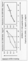
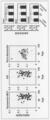
- FIG. 1 is a graph showing the results of detection of ECRG4 by an enzyme-linked immunosorbent assay (ELISA method).
- ELISA method enzyme-linked immunosorbent assay
- the horizontal axis indicates the addition concentration of the ECRG4 partial polypeptide (polypeptide consisting of the amino acid sequence of positions 71 to 148 of human ECRG4 (amino acid sequence set forth in SEQ ID NO: 2)) added to the detection system
- the vertical axis indicates The axis indicates relative signal (absorbance at 450 nm).
- a formula represents each approximation curve.
- error bars represent ⁇ standard deviation (SD). It is a graph which shows the result of having detected ECRG4 by ELISA method.
- an anti-ECRG4 mouse monoclonal antibody (2A8, addition concentration: 1 ⁇ g/ml (open circle) or 2 ⁇ g/ml (open triangle)
- an anti-ECRG4 rabbit polyclonal antibody (OB1264) was used as the detection antibody.
- addition concentration 1 ⁇ g/ml (left figure) or 2 ⁇ g/ml (right figure)).
- the horizontal axis indicates the addition concentration of the ECRG4 partial polypeptide (polypeptide consisting of the amino acid sequence of positions 71 to 132 of human ECRG4 (amino acid sequence set forth in SEQ ID NO: 2)) added to the detection system, and the vertical axis indicates The axis indicates relative signal (absorbance at 450 nm). Also, error bars represent ⁇ standard deviation (SD).
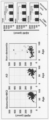
- FIG. 4 is a dot plot diagram and graph showing the results of analyzing plasma levels of ECRG4.
- “Dementia+MCI”, “AD” and “Non-dementia” indicate the analysis results for dementia patients, MCI patients, Alzheimer's disease patients and non-dementia patients, respectively.
- the horizontal axis of the dot plot diagram indicates the age of each patient, and the vertical axis indicates the ECRG4 concentration in each individual patient. Also, white circles indicate females, and black circles indicate males. The vertical axis of the graph indicates the average concentration of ECRG4 in each patient group, and the error bars represent the standard deviation. Statistical significance was determined by t-test, * P ⁇ 0.05, ** P ⁇ 0.01, *** P ⁇ 0.001, *** P ⁇ 0.0001 represents that FIG. 3 is a dot plot diagram showing the results of analysis of plasma levels of ECRG4, amyloid beta (A ⁇ ) 40 and A ⁇ 42 in healthy subjects (8 individuals).
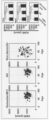
- FIG. 4 is a dot plot diagram and graph showing the results of analyzing the ratio of A ⁇ 42/A ⁇ 40 in plasma.
- the notation in the figure is the same as in FIG. 2A.
- FIG. 2 is a dot plot diagram and graph showing the results of analyzing plasma levels of A ⁇ 40.
- FIG. 2 is a dot plot diagram and graph showing the results of analyzing plasma levels of A ⁇ 42.
- FIG. 2A The notation in the figure is the same as in FIG. 2A.
- FIG. 4 is a dot plot diagram and graph showing the results of analyzing the ratio of A ⁇ 42/A ⁇ 40 in plasma.
- the notation in the figure is the same as in FIG. 2A.
- FIG. 2 is a dot plot diagram and graph showing the results of analyzing plasma levels of A ⁇ 42.
- FIG. The notation in the figure is the same as in FIG. 2A.
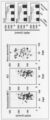
- FIG. 4 is a dot plot and graph showing the results of analyzing cerebrospinal fluid (CSF) levels of ECRG4.
- CSF cerebrospinal fluid
- FIG. 2 is a dot plot diagram and graph showing the results of analyzing CSF levels of A ⁇ 40.
- FIG. 2 is a dot plot diagram and graph showing the results of analyzing CSF levels of A ⁇ 42.
- FIG. 4 is a dot plot diagram and graph showing the results of analyzing the ratio of A ⁇ 42/A ⁇ 40 in CSF.
- the notation in the figure is the same as in FIG. 2A.
- the present invention has been completed based on the above findings, and provides a method for testing dementia, including the following steps (a) to (c). (a) detecting the amount of ECRG4 polypeptide in a blood sample separated from a subject; (b) comparing the amount of polypeptide detected in step (a) to a reference amount; (c) determining that the subject is suffering from dementia if the result of the comparison in step (b) is that the amount of polypeptide in the subject is higher than the reference amount;
- Dementia is a type of cognitive disorder, and means a state in which intelligence, once normally developed, is irreversibly reduced due to an acquired organic disorder of the brain.
- AD normal pressure hydrocephalus
- iNPH idiopathic normal pressure hydrocephalus
- PNFA progressive non-fluent aphasia
- PSP progressive supranuclear palsy
- DLB dementia with Lewy bodies
- FTLD frontotemporal lobar degeneration
- VaD vascular dementia
- NIID neuronal intranuclear inclusion disease
- dementia according to the present invention includes not only the so-called dementia described above, but also mild cognitive impairment (MCI), which is a pre-stage of dementia.
- MCI mild cognitive impairment
- the "subject” is not particularly limited as long as it is a human. include those who are determined to be suffering from the disease.
- the "blood sample” separated from such a subject may be any sample containing blood, and examples thereof include whole blood, serum, and plasma.
- Blood can be collected from a subject by methods known to those skilled in the art.
- blood whole blood
- Serum is a portion of whole blood from which blood cells and specific blood coagulation factors have been removed, and can be obtained, for example, as a supernatant after coagulation of whole blood.
- Plasma is the portion of whole blood from which blood cells have been removed, e.g., by centrifugation under conditions that do not clot whole blood (e.g., in the presence of chelating agents such as EDTA, anticoagulants such as sodium citrate and heparin). It can be obtained as a supernatant when subjected to.
- chelating agents such as EDTA, anticoagulants such as sodium citrate and heparin
- the blood sample according to the present invention when the blood sample according to the present invention is subjected to a method for detecting the amount of polypeptide described later, it may be in a form suitable for the method (e.g., protein solution extracted from blood sample, formalin fixation, alcohol fixation, etc.). processed, frozen, or paraffin-embedded tissue, etc.).
- a person skilled in the art can select and prepare a known technique in consideration of the type and condition of the blood sample.
- the "ECRG4 polypeptide" detected as a blood marker for dementia is a human-derived polypeptide also referred to as AUGURIN, typically the nucleotide set forth in SEQ ID NO: 1 A polypeptide encoded by the sequence (a polypeptide comprising the amino acid sequence set forth in SEQ ID NO:2). It should be noted that the DNA sequence of a gene encoding a polypeptide may mutate naturally (that is, non-artificially) due to its mutation or the like. Therefore, ECRG4 polypeptides to be detected in the present invention are not limited to the typical amino acid sequences described above, but also include natural variants of those amino acid sequences.
- ECRG4 polypeptides to be detected in the present invention include not only those consisting of full-length amino acid sequences, but also partial peptides thereof.
- a partial peptide is not particularly limited, but is preferably a polypeptide comprising the amino acid sequence of positions 31-148 set forth in SEQ ID NO:2, more preferably a polypeptide containing the amino acid sequence of positions 71-148 set forth in SEQ ID NO:2.
- a polypeptide comprising an amino acid sequence more preferably a polypeptide comprising the amino acid sequence of positions 71 to 132 set forth in SEQ ID NO: 2 (a polypeptide comprising the M region of human ECRG4), more preferably SEQ ID NO: 2
- the "amount of ECRG4 polypeptide" detected in the present invention may be not only an absolute amount but also a relative amount.
- Relative amounts include, for example, percentages of the total polypeptide amount.
- the relative amount includes a ratio of polypeptide amounts based on the measuring method or measuring device used for detection (a numerical value expressed in so-called arbitrary units (AU)).
- AU arbitrary units
- the relative amount for example, a value calculated based on the amount of the reference protein may be used.
- the "reference protein” according to the present invention may be any protein that is stably present in a blood sample and has a small difference in amount between different blood samples.
- an endogenous control (internal standard) protein and more specifically ⁇ -actin, ⁇ -tubulin, COX-4, GAPDH, Lamin B1, PCNA, TBP, VDCA1/Porin.
- Detection of the amount of polypeptide can be carried out by a person skilled in the art by appropriately adopting a known technique.
- known techniques include, for example, enzyme-linked immunosorbent assay (ELISA), CLEIA (chemiluminescent enzyme immunoassay), latex agglutination, antibody array, immunoblotting, immunochromatography, imaging cytometry, flow cytometry, Methods of detection using antibodies (immunological methods), such as radioimmunoassay, immunoprecipitation, immunohistochemical staining, and mass spectrometry.
- an antibody that recognizes an ECRG4 polypeptide is used, the antibody is brought into contact with the ECRG4 polypeptide, and the amount of the ECRG4 polypeptide is detected using the binding property of the antibody to the ECRG4 polypeptide as an index. be done.
- the ECRG4 polypeptide in the blood sample is brought into contact with an antibody that recognizes the Ecrg4 polypeptide (capture antibody) immobilized on a solid phase such as a substrate.
- an antibody that recognizes a labeled Ecrg4 polypeptide described below is allowed to act on the captured Ecrg4 polypeptide, and the label is chemically or optically detected to obtain Ecrg4
- the amount of polypeptide can be detected.
- the “capture antibody” and the “detection antibody” may be the same antibody or different antibodies as long as they recognize the ECRG4 polypeptide. From the viewpoint of being able to capture and detect, different antibodies are preferred. Examples of different antibodies include, for example, a combination in which the capture antibody is a polyclonal antibody that recognizes ECRG4 polypeptide and the detection antibody is a monoclonal antibody that recognizes ECRG4 polypeptide, and a combination in which the capture antibody is a monoclonal antibody that recognizes ECRG4 polypeptide.
- the detecting antibody is a polyclonal antibody that recognizes the ECRG4 polypeptide, or the capturing antibody is a monoclonal antibody that recognizes the ECRG4 polypeptide, and the detecting antibody is the capturing antibody in the ECRG4 polypeptide
- at least one of the capturing antibody and the detecting antibody is an antibody that recognizes a region consisting of the amino acid sequence of positions 71 to 148 of ECRG4, and more preferably, the capturing antibody and the detecting antibody.
- At least one of the antibodies for is an antibody that recognizes a region consisting of the amino acid sequence of positions 71 to 132 of ECRG4, more preferably, at least one of the capture antibody and the detection antibody is an antibody that recognizes positions 107 to 132 of ECRG4.
- An antibody that recognizes a region consisting of the amino acid sequence of, and more preferably, a combination of a capture antibody and a detection antibody An antibody (more preferably a monoclonal antibody) that recognizes a region consisting of an amino acid sequence at positions 71 to 132 (more preferably positions 107 to 132, more preferably positions 116 to 124) of ECRG4 and an amino acid sequence at positions 107 to 132 of ECRG4 Combination with an antibody (more preferably a polyclonal antibody) that recognizes a region consisting of; An antibody (more preferably a polyclonal antibody) that recognizes a region consisting of the amino acid sequence at positions 107 to 132 of ECRG4 and an amino acid sequence at positions 71 to 132 (more preferably positions 107 to 132, still more preferably positions 116 to 124) of ECRG4 combination with an antibody (more preferably a monoclonal antibody) that recognizes a region consisting of; An antibody (more preferably a monoclonal antibody
- a method using a secondary antibody conjugated with a labeling substance or a method of binding a secondary antibody and a labeling substance can be used. Indirect detection methods can also be used, such as those that utilize a polymer that has been exposed.
- the term "secondary antibody” refers to an antibody that exhibits specific binding to the antibody of the present invention.
- protein G, protein A, or the like bound with a labeling substance may be used instead of the secondary antibody.
- Mass spectrometry is a method in which a peptide sample (the aforementioned blood sample) is ionized using an ion source, and the peptide sample is ionized by moving it in a vacuum and using electromagnetic force or by a time-of-flight difference in the analysis unit.
- a measurement method using a mass spectrometer that can separate and detect according to the mass-to-charge ratio.
- Methods of ionization using an ion source include the EI method, the CI method, the FD method, the FAB method, the MALDI method, A method such as the ESI method can be selected as appropriate.
- a separation method such as a magnetic field deflection type, a quadrupole type, an ion trap type, a time of flight (TOF) type, a Fourier transform ion cyclotron resonance type, etc. is appropriately selected. be able to.
- tandem mass spectrometry combining two or more mass spectrometry methods and triple quadrupole mass spectrometry can be used.
- multiple biomarker molecules are measured simultaneously in a single measurement by selected reaction monitoring (SRM) or multiple reaction monitoring (MRM) with a triple quadrupole mass spectrometer. be able to.
- the mass spectrometer may be used alone, or by combining liquid chromatography (LC) and high performance liquid chromatography (HPLC), to separate and purify the peptides constituting the target polypeptide (ECRG4 polypeptide). can be used as a sample.
- LC liquid chromatography
- HPLC high performance liquid chromatography
- the amount of ECRG4 polypeptide thus detected is compared with a standard amount of the same polypeptide.
- a person skilled in the art can make such a comparison by appropriately selecting and setting a statistical analysis method suitable for the above-described detection method, for example.
- Statistical analysis methods include, for example, t-test, Mann-Whitney U-test, analysis of variance (ANOVA), Kruskal-Wallis test, Wilcoxon test, odds ratio, hazard ratio, Fisher's exact test, and receiver operating characteristic analysis. (ROC analysis), classification tree and decision tree analysis (CART analysis). Also, normalized or standardized and normalized data can be used in the comparison.
- the "reference amount of ECRG4 polypeptide" to be compared is not particularly limited. It can be set as a so-called cut-off value that can distinguish between a person without dementia (for example, a healthy person or a non-dementia patient (a person suffering from a disease other than dementia)) and a dementia sufferer.
- a reference amount for discriminating between dementia and non-dementia for example, a population not suffering from dementia (e.g., a healthy population, a population of non-dementia patients) ECRG4 poly detected in Median or mean values for peptides are included.
- a value determined by comparing the amount of ECRG4 polypeptide in a population not suffering from dementia and a population suffering from dementia e.g., a population not suffering from dementia and the amount of polypeptide in a population suffering from dementia
- a more specific example of such a reference amount is 0.1 ng/ml, preferably 0.2 ng/ml, and more preferably 0.4 ng/ml, as shown in Examples (FIG. 2A) described later.
- ml more preferably 0.6 ng/ml, more preferably 0.7 ng/ml, still more preferably 0.8 ng/ml, more preferably 0.9 ng/ml.
- the amount of polypeptide in the subject is higher than the reference amount can be appropriately determined, for example, by a person skilled in the art based on the statistical analysis method described above. Also, for example, the detected amount of polypeptide is higher than the reference amount, and the difference is statistically significant (eg, P ⁇ 0.05). Also, for example, the amount of the detected polypeptide is twice or more the corresponding reference amount (preferably, 3 times or more, 4 times or more, 5 times or more, 6 times or more, 7 times or more, 8 times or more, 9 times or more ) is also mentioned. Then, in the test method of the present invention, when the polypeptide amount in the subject is higher than the reference amount, the subject is determined to be suffering from dementia.
- the term “affected” includes not only having already developed dementia, but also a state where there is a risk of developing dementia.
- the method of the present invention includes a method of collecting data on the above-mentioned polypeptide amount for diagnosis by a doctor, a method of presenting the data to the doctor, and a method of comparing and analyzing the above-mentioned polypeptide amount and a reference amount. , a method for assisting diagnosis by a doctor.
- testing method of the present invention may be performed in combination with other testing methods for dementia.
- Such other examination methods are not particularly limited, but for example, electroencephalogram diagnosis, biochemical examination for blood/cerebrospinal fluid, CT, MRI, PET/SPECT, amyloid imaging (amyloid PET examination), etc. diagnostic imaging.
- dementia can be tested according to the present invention. Then, based on the result of such evaluation, it is possible to determine whether or not to treat dementia.
- the present invention can also provide a method for treating dementia, including the step of treating a subject determined to have dementia by the testing method of the present invention.
- treatments for dementia are not particularly limited, but include, for example, immunotherapy and methods of administering agents for suppressing lesions of dementia.
- Immunotherapies include, for example, active immunotherapy (vaccine therapy) using partial peptides of A ⁇ for suppression of aggregation of amyloid beta (A ⁇ ), and passive immunotherapy in which antibodies against A ⁇ are administered.
- drugs administered to suppress dementia lesions include, for example, drugs for suppressing A ⁇ production ( ⁇ secretase modulator (GSM), ⁇ secretase modulator inhibitor (GSI), non-steroidal anti-inflammatory drugs, etc.), drugs for suppressing A ⁇ aggregation (curcumin, polysulfate compounds, clioquinol, etc.), drugs for suppressing tau aggregation (aminothienopyridazine, cyanine dyes, methylene blue, etc.), neuroprotective drugs ( dimebon etc.), cholinesterase inhibitors (donepezil etc.), acetylcholinesterase inhibitors (galantamine etc.), glutamic acid NMDA receptor inhibitors (memantine etc.).
- GSM secretase modulator
- GSI ⁇ secretase modulator inhibitor
- non-steroidal anti-inflammatory drugs etc.
- drugs for suppressing A ⁇ aggregation curcumin, polysulfate compounds, clioquinol, etc.
- the presence or absence of dementia can be determined by detecting the amount of ECRG4 polypeptide using an antibody that recognizes the polypeptide.
- the present invention provides an agent for testing dementia by the method described above, the agent comprising an antibody that recognizes Ecrg4 polypeptide.
- the “antibody” contained in the agent of the present invention may be a polyclonal antibody, a monoclonal antibody, or a functional fragment of an antibody.
- Antibody includes all classes and subclasses of immunoglobulins.
- a “polyclonal antibody” is an antibody preparation containing different antibodies directed against different epitopes.
- a “monoclonal antibody” also means an antibody (including antibody fragments) obtained from a substantially homogeneous population of antibodies. In contrast to polyclonal antibodies, monoclonal antibodies recognize a single determinant on an antigen.
- the term "functional fragment” of an antibody means a portion (partial fragment) of an antibody that specifically recognizes a target protein. Specifically, Fab, Fab', F(ab')2, variable region fragment (Fv), disulfide bond Fv, single chain Fv (scFv), sc(Fv)2, diabodies, multispecific antibodies, and polymers thereof.
- the antibody according to the present invention is a polyclonal antibody, immunize an immunized animal with an antigen (ECRG4 polypeptide, cells expressing it, etc.), and remove the antiserum by conventional means (e.g., salting out, centrifugation) , dialysis, column chromatography, etc.).
- an antigen ECRG4 polypeptide, cells expressing it, etc.
- remove the antiserum by conventional means (e.g., salting out, centrifugation) , dialysis, column chromatography, etc.).
- monoclonal antibodies can be produced by the hybridoma method or recombinant DNA method.
- a representative example of the hybridoma method is Kohler and Milstein's method (Kohler & Milstein, Nature, 256:495 (1975)).
- the DNA encoding the antibody of the present invention is cloned from hybridomas, B cells, etc., incorporated into an appropriate vector, and transformed into host cells (e.g., mammalian cell lines, E. coli, yeast cells, insect cells, etc.).
- the antibody according to the present invention is not particularly limited as long as it can recognize the ECRG4 polypeptide.
- At least one selected from an antibody (more preferably a monoclonal antibody) that recognizes the region consisting of the amino acid sequence of and an antibody (more preferably a polyclonal antibody) that recognizes the region consisting of the amino acid sequence of positions 107 to 132 of ECRG4 is an antibody.
- Antibodies according to the present invention may also be provided in a solid-phased form for use in ELISA, antibody arrays, and the like.
- the solid phase include plates such as plastic plates, fibrous substances such as nitrocellulose, and particles such as magnetic particles and latex particles.
- the antibody may be labeled with a labeling substance in accordance with the above detection method.
- Labeling substances include, for example, enzymes such as HRP, ⁇ -D-glucosidase and luciferase; luminous substances such as luminol, luciferin and lucigenin; fluorescent substances such as FITC, FAM, DEAC, R6G, TexRed and Cy5 ; Examples include radioactive isotopes such as H, 14 C, 32 P, 35 S and 123 I, and affinity substances such as biotin and streptavidin.
- enzymes such as HRP, ⁇ -D-glucosidase and luciferase
- luminous substances such as luminol, luciferin and lucigenin
- fluorescent substances such as FITC, FAM, DEAC, R6G, TexRed and Cy5 ;
- radioactive isotopes such as H, 14 C, 32 P, 35 S and 123 I
- affinity substances such as biotin and streptavidin.
- the agent of the present invention can contain other ingredients that are acceptable as a composition, in addition to the antibody.
- Such other ingredients include, for example, pharmacologically acceptable carriers or diluents (sterile water, physiological saline, vegetable oils, excipients, disintegrants, buffers, emulsifiers, suspending agents, stabilizers, preservatives, preservatives, etc.).
- pharmacologically acceptable carriers or diluents sterile water, physiological saline, vegetable oils, excipients, disintegrants, buffers, emulsifiers, suspending agents, stabilizers, preservatives, preservatives, etc.
- excipients lactose, starch, sorbitol, D-mannitol, white sugar and the like can be used.
- Starch, carboxymethyl cellulose, calcium carbonate and the like can be used as the disintegrant.
- Phosphate, citrate, acetate and the like can be used as
- Gum arabic, sodium alginate, tragacanth and the like can be used as emulsifiers.
- suspending agents glyceryl monostearate, aluminum monostearate, methylcellulose, carboxymethylcellulose, hydroxymethylcellulose, sodium lauryl sulfate and the like can be used.
- Propylene glycol, dietyrin sulfite, ascorbic acid and the like can be used as stabilizers.
- Preservatives that can be used include phenol, benzalkonium chloride, benzyl alcohol, chlorobutanol, methylparaben, and the like.
- antiseptics sodium azide, benzalkonium chloride, paraoxybenzoic acid, chlorobutanol and the like can be used.
- substrates necessary for label detection solutions for dissolving proteins in samples (blood samples, etc.) (protein dissolution reagents), buffers used for sample dilution and washing Solution (diluent, washing solution), reagent for stopping the detection reaction of the label (reaction stopping agent), positive control (e.g., ECRG4 polypeptide, standard, blood sample derived from dementia patient), negative control (e.g. , a blood sample derived from a person not suffering from dementia), an isotype control antibody against the antibody according to the present invention, etc. can be combined, and a kit for testing dementia can be prepared.
- ECRG4 polypeptide e.g., ECRG4 polypeptide, standard, blood sample derived from dementia patient
- negative control e.g. , a blood sample derived from a person not suffering from dementia
- kits include, for example, at least one antibody that recognizes an ECRG4 polypeptide, and at least one article selected from an isotype control antibody against the antibody, a positive control, and a negative control for testing dementia. Kits are included. Moreover, when an unlabeled antibody is used as an antibody sample, it can be combined with a labeled substance (eg, secondary antibody, protein G, protein A, etc.) that binds to the antibody. Additionally, such kits can include instructions for use of the kit.
- a labeled substance eg, secondary antibody, protein G, protein A, etc.
- an antibody that recognizes a region consisting of an amino acid sequence consisting of positions 107 to 132 of ECRG4 and an antibody that recognizes a region consisting of an amino acid sequence of positions 134 to 139 of ECRG4 or 116 to 124 of ECRG4 It is possible to detect ECRG4 with high quantification by using an immunological method in combination with an antibody that recognizes the region consisting of the amino acid sequence (see Fig. 1, etc.).
- the present invention An antibody that recognizes a region consisting of the amino acid sequence of positions 107 to 132 of ECRG4 polypeptide and an antibody that recognizes a region consisting of the amino acid sequence of positions 133 to 143 of ECRG4 or a region consisting of the amino acid sequence of positions 71 to 132 of ECRG4.
- a method of detecting the amount of ECRG4 polypeptide in a sample by an immunological method using a recognizing antibody An antibody that recognizes a region consisting of the amino acid sequence of positions 107 to 132 of ECRG4 polypeptide and an antibody that recognizes a region consisting of the amino acid sequence of positions 133 to 143 of ECRG4 or a region consisting of the amino acid sequence of positions 71 to 132 of ECRG4.
- an agent for detecting the amount of ECRG4 polypeptide in a sample comprising a recognizing antibody;
- a system is provided for detecting the amount of ECRG4 polypeptide in a sample by an immunological method.
- the explanation of the methods, drugs and systems, and their components conforms to the explanations in ⁇ Method for testing dementia> and ⁇ Drugs for testing dementia> above.
- the "sample” targeted by these is not limited to the above blood sample, as long as it can contain the ECRG4 polypeptide. tissue, its cells, etc.).
- the system of the present invention may include a unit capable of performing an immunological method in addition to the antibody or drug containing the antibody.
- units include, for example, units for detecting a labeling substance directly or indirectly bound to an antibody and measuring the amount of antibody bound to an ECRG4 polypeptide in a sample (spectrophotometer, fluorescence spectrophotometer, etc.); units for injecting or removing samples, antibodies, washing solutions, reagents, etc.; units for handling the solid phases such as plates (eg, robot arms); and units for controlling these units. It is not limited.
- An anti-ECRG4 rabbit polyclonal antibody (hereinafter also referred to as "OB1264") is prepared by injecting a synthetic peptide (a polypeptide consisting of positions 107 to 132 of human ECRG4 (amino acid sequence set forth in SEQ ID NO: 2)) into a rabbit using a conventional method. It was prepared by immunization (produced by Medical and Biological Laboratories Co., Ltd.).
- Anti-ECRG4 mouse monoclonal antibodies were prepared by synthesizing various partial peptides of human ECRG4 using a conventional method and immunizing mice to prepare hybridomas. Then, 5 million of each selected hybridoma was intraperitoneally injected into 8-week-old NOD/SCID mice, and ascites was collected. As a result, monoclonal antibodies recognizing each of the partial peptides shown below could be obtained.
- an anti-ECRG4 mouse monoclonal antibody that recognizes positions 116-124 of human ECRG4 hereinafter also referred to as "2A8”
- An anti-ECRG4 mouse monoclonal antibody hereinafter also referred to as "1H4"
- 1H4 an anti-ECRG4 mouse monoclonal antibody that recognizes a polypeptide consisting of positions 134-139 of human ECRG4.
- the anti-ECRG4 rabbit polyclonal antibody and anti-ECRG4 mouse monoclonal antibody were purified by fast flow using Protein A Sepharose 4 (manufactured by GE Healthcare) according to the instructions for use.
- ECRG4 sandwich ELISA assay First, a 96-well plate (manufactured by Falcon) was treated overnight at 4° C. with 50 ⁇ l of mouse anti-ECRG4 antibody (1 ⁇ g/ml or 2 ⁇ g/ml)-containing carbonate buffer (0.05 M, pH 9.5). coated. After washing the plate five times with wash buffer (0.05% Tween-20/PBS), the plate was incubated with 150 ⁇ l of blocking buffer (5% skimmed milk in carbonate buffer) for 90 minutes at room temperature.
- wash buffer 0.05% Tween-20/PBS
- TMB Microwell Peroxidase Substrate System manufactured by KPL
- Amyloid beta (A ⁇ )40 and A ⁇ 42 ELISA assay The concentrations of A ⁇ 40 and A ⁇ 42 in plasma and CSF were measured using Human/Rat ⁇ Amyloid40 and ⁇ Amyloid42 ELISA Kits WAKO (manufactured by Fujifilm Wako Pure Chemical Industries, Ltd.) according to the contents of the instruction manual.
- MMSE Mini-Mental State Examination
- AD Alzheimer's disease
- NPH normal pressure hydrocephalus
- iNPH idiopathic normal pressure hydrocephalus
- PNFA progressive non-fluent aphasia
- PSP progressive supranuclear palsy
- DLB dementia with Lewy bodies
- FTLD frontal Temporal lobe degeneration
- VaD vascular dementia
- NIID neuronal intranuclear inclusion disease
- MCI mild cognitive impairment.
- the diseases shown in Table 2 are as follows. Myospasm: muscle spasm, Myoclonus: myoclonus, migraine: migraine, syringomyelia: syringomyelia, hysteria: hysteria, limbic encephalitis: limbic encephalitis, myopathy: myopathy, Neuromyelitis optic: neuromyelitis optica, epilepsy: epilepsy Aseptic meningitis, psychogenic reaction, episodic ataxia, sarcoidosis, neuro-Behcet, tuberculous meningitis, femoral neuropathy, multiple cerebral infarction: multiple cerebral infarction, multiple sclerosis: multiple sclerosis, postsuscitation encephalopathy: postresuscitative encephalopathy, myelopathy: myelopathy, fibromyalgia: fibromyalgia, dystonia: dystonia, spastic paraplegia: spastic paraplegia, musculopathy Atrophy, Huntington'
- Plasma and CSF samples treated with EDTA were used in patients diagnosed with dementia, including AD, normal pressure hydrocephalus (NPH) and mild cognitive impairment (MCI), as well as multiple system atrophy (MSA) and myopathy. were obtained using standard methods (Waybright et al, 2010, Menck et al, 2017) from a patient diagnosed as non-demented with neuropathy. Plasma samples were also obtained from 8 healthy individuals as controls (Fig. 2B). Informed consent was obtained from all subjects, and this study was approved by the Ethics Committee of Hokkaido University.
- a sense oligonucleotide and an antisense oligonucleotide encoding a polypeptide consisting of amino acids 133 to 148 of mouse Ecrg4 (amino acid sequence set forth in SEQ ID NO: 4) are annealed, pFUSE-mEcrg4(133-148)-hIgG1Fc2 ( 133-148) were inserted into pFUSE-hIgG1Fc2.
- the following primer set was used to synthesize a cDNA encoding a polypeptide consisting of amino acids 32-70 of mouse Ecrg4 (amino acid sequence shown in SEQ ID NO: 4).
- the following primer set was used to synthesize a cDNA encoding a polypeptide consisting of amino acids 71-132 of mouse Ecrg4.
- 5′ primer 5′-TGATATCGCAGCTGTGGGACCGTACG-3′ (SEQ ID NO: 7) and 3′ primer: 5′-AAGATCTGTGGGGACCAATGGCCGCA-3′ (SEQ ID NO: 8)
- the following primer set was used to synthesize a cDNA encoding a polypeptide consisting of amino acids 133-148 of mouse Ecrg4.
- 5′ primer 5′-ATCGAGCCGGAAAGCTTCAGGCATGGAGCCAGTGTCAACTATAATGACTATA-3′ (SEQ ID NO: 9) and 3′ primer: 5′-GATCTATAGTCATTATAGTTGACACTGGCTCCATGCCTGAAGCTTTCCCGGCTCGAT-3′ (SEQ ID NO: 10).
- the nucleotide sequences of the cDNAs cloned using these primer sets were confirmed using BigDye Terminator Kit version 3.1 (manufactured by Thermo Fisher Scientific) and ABI sequencer model 3130xl (manufactured by Thermo Fisher Scientific).
- Ecrg4 (addition concentration: 1 ⁇ g / ml, detection antibody: OB1264), Ecrg4 (addition concentration: 1 ⁇ g / ml, detection antibody: 2A8) or GAPDH (dilution: 1:1000, detection antibody: anti-mouse Ab, Chemicon) made).
- these primary antibodies are HRP-labeled anti-mouse IgG antibody (dilution: 1:5000, manufactured by Santa Cruz Biotechnology), HRP-labeled anti-rabbit IgG antibody (dilution: 1 :5000, Santa Cruz Biotechnology) and HRP-labeled anti-human IgG antibody (dilution: 1:500, Bethyl Laboratories).
- Immunostaining was performed according to the method described in Nishide et al, 2009, Kujuro et al, 2010. Briefly, first, various vectors were introduced into 293T cells as described above. Three days after the gene transfer, the cells were fixed and subjected to immunostaining with 2A8 (1 ⁇ g/ml) and OB1264 (1 ⁇ g/ml), respectively.
- Human Fc, mouse Fc and rabbit Fc are Alexa488-labeled anti-human IgG antibody (dilution: 1:1000, ThermoFisher Scientific), Alexa594-labeled anti-mouse IgG antibody (dilution: 1:1000, ThermoFisher Scientific), and Alexa594-labeled anti-rabbit IgG antibody (dilution ratio: 1:1000, ThermoFisher Scientific).
- Cells were counterstained with Hoechst 33342 (addition concentration: 1 ⁇ g/ml, ThermoFisher Scientific) to visualize nuclei. Fluorescence images were acquired using an AxioImager M1 microscope (manufactured by Carl Zeiss).
- the optimum concentrations of both OB1264 and 2A8 to be used are 1 ⁇ g/ml in detecting the ECRG4 polypeptide.
- the aforementioned Western blotting and immunostaining analyzes were performed to confirm that OB1264 and 2A8, respectively, can detect ECRG4 polypeptide, but not detect proteins other than Ecrg4.
- these antibodies do not detect Ecrg4 regions other than positions 71-132, while the ECRG4 polypeptide (positions 71-132) can be detected by OB1264 and 2A8, respectively.
- Plasma ECRG4 levels were analyzed using the ECRG4 ELISA assay in subjects with dementia, MCI, and non-dementia with neuropathy, as described above. As a result, as shown in Table 3 below and FIG. 80), ECRG4 was detected in each plasma.
- the ECRG4 positive rate (ECRG4 signal detection rate in plasma) in female dementia patients and MCI patients is 51% (46/91) and 35% (6/17), respectively, and those in male patients are 49% (33/68) and 9% (1/11) respectively.
- ECRG4 was detected in 47% (61/129) of AD patients and 60% (18/30) of non-AD dementia patients.
- the ECRG4 positive rate in AD patients was 44% (24/54) in males and 49% (37/75) in females.
- the positive rate of ECRG4 in plasma in non-dementia patients with neurological disease was 5% (4/80).
- the average ECRG4 level in plasma was 0.96 ng/ml in dementia and MCI patients and 1.02 ng/ml in AD patients, as shown in Figure 2A. On the other hand, it was 0.08 ng/ml in non-dementia patients. Furthermore, in contrast to these, ECRG4 could not be detected in the plasma of healthy subjects as shown in FIG. 2B.
- the MMSE score is 23 points or less and dementia is suspected
- the positive rate of ECRG4 in plasma was 34% (23/67), and the positive rate of ECRG4 in plasma was 13% ( 4/31).
- a ⁇ 40 and A ⁇ 42 are known to be involved in the formation of amyloid plaques in the brain parenchyma, resulting in decreased levels thereof in the plasma and CSF of AD patients.
- aggregation of A ⁇ 42 occurs earlier than A ⁇ 40 (Hampel et al, 2010), which has been shown to reduce the ratio of A ⁇ 42/A ⁇ 40 in the plasma and CSF of AD patients (Palmqvist et al, 2019).
- ECRG4 was not detected in 95% of non-dementia patients in plasma, although it was detected in 50% of dementia and MCI patients.
- ECRG4 levels in dementia patients and MCI patients were much higher than those in non-dementia patients, demonstrating that ECRG4 is extremely effective as a plasma biomarker for dementia.
- Palmqvist S Zinc S
- Werner S Zinc S
- Zetterberg H Brix B
- Eichenlaub U Dage JL
- Chai X Blennow K
- Mattsson N Hansson O.; (2019).
- the present invention it is possible to test for dementia.
- the test can be performed using a blood sample, it is less invasive and does not place a heavy burden on the subject. Therefore, the present invention is extremely useful in the medical field related to dementia.
Abstract
認知症患者と健常者とを判別することを可能とする血中バイオマーカー分子を同定し、当該分子の量を指標とする認知症の検査方法を提供することを目的とし、認知症患者、軽度認知障害患者及び非認知症患者等の血漿における、ECRG4の陽性率及び濃度を検出した。その結果、血漿においてECRG4は、認知症患者及び軽度認知障害の50%において検出されたにも関わらず、非認知症患者の95%において検出されなかった。さらに、認知症患者及びMCI患者におけるECRG4の血漿レベルは、非認知症患者におけるそれと比してはるかに高く、認知症の血中バイオマーカー分子としてECRG4が極めて有効であることを見出した。
Description
本発明は、認知症の検査方法に関し、より詳しくは、ECRG4の血中レベルを指標として認知症を検査する方法等に関する。本発明はまた、免疫学的方法によりECRG4ポリペプチド量を検出する方法、及び当該検出を行うためのシステム等に関する。
アルツハイマー病(AD)等の認知症は、主に初老期~老年期に生じる進行性の神経変性疾患である。その主な症状は、記憶障害、高次脳機能障害(失語、失行、失認、構成失行)、人格の変化等である。そして、このような症状故、患者本人の生活の質の低下のみならず、家族等の周囲の生活にも多大な影響を与えている。さらに、その患者数は人口の高齢化に伴い増加の一途を辿っており、認知症は世界的に現代社会の抱える深刻な問題となっている。
しかしながら、昨今、認知症の症状の進行を遅らせることは可能となりつつある。例えば、アセチルコリンエステラーゼ阻害剤が、実際に臨床の場で使用され、それなりの成果をあげ、症状の進行をある程度抑えることも可能になっている。また、アデュカヌマブ(抗アミロイドベータ(Aβ)抗体)が、米国においてAD治療薬として承認され、ADによる軽度認知障害(MCI)及び軽度ADの臨床状態の悪化を遅らせること等が期待されている。そのため、かかる状況を鑑み、認知症の治療においては、早期又は発症前診断法の開発が喫緊の課題となっている。
認知症が発症する前段階、すなわち軽度の臨床的症状が現れる段階は、MCIと称され、それは正常な老化(健常状態)と認知症との中間段階として定義されている。そして、MCIを検査する方法として、様々な神経心理学的検査が主に用いられており、例えば、見当識、短期及び長期の記憶、行動、言語及び理解について検査し、認知障害をスコア化する、フォルスタインのミニメンタルステート検査(MMSE)が用いられている。しかしながら、このような問診法はあくまでスクリーニング目的で用いられ、確定診断に至ることはない。
また、リン酸化タウ、Aβ40、Aβ42等が、ADバイオマーカーとして利用されているが、実際の臨床において、それらの有効性はまだ不十分な現状にあり、さらに感度や特異度の高いバイオマーカーの発見とそれを利用した検査方法の開発が求められている(非特許文献1~3)。
さらに、これらバイオマーカーを検出する上でも、認知症の検査方法として、CT、MRI、PET/SPECT等の画像診断等の開発が進められている。しかしながら、画像診断には特殊な設備を必要とするため、実施できる医療機関が限られる。また現状の画像診断技術により、認知症が発症する前段階と健常状態等との差を捉えることは依然として難しいため、画像を見る医師によって判断が異なることがあり、客観性に欠けるという欠点も有する。
このように、客観的かつ精度高く認知症患者と健常者とを判別できる方法は、確立していないのが現状である。そのため、血液のような容易に得られる被検体の試料を用い、客観的な診断を可能にするバイオマーカーの探索が、認知症の検査方法において重要視されている。
しかしながら、AD等やその前段階であるMCIに関しては、未だ有用な血中バイオマーカーとなり得る分子は見出されておらず、精度高く認知症を検査できる方法は未だ開発されていないのが現状である。
Krance et al,2019,J Neurosci, 39,7428-7437.
Manca et al,2019,J Neurol,266, 1685-1692.
Reimand et al,2019,Alzheimers Res Ther,11,100.
Kujuro et al,2010,PNAS,107,8259-8264.
Xu et al,2007,Mol Cell Neurosci,36,313-331.
Baird et al,2012,J Leukoc Biol,91,773-781.
Moriguchi et al,2016,OncoImmunolog,5,e1242547
Moriguchi et al,2018,Sci Rep,8,4048.
本発明は、前記従来技術の有する課題に鑑みてなされたものであり、認知症患者と健常者とを判別することを可能とする血中バイオマーカー分子を同定し、当該分子の量を指標とする認知症の検査方法を提供することを目的とする。
本発明者らは、前記目的を達成すべく鋭意研究を重ねた結果、ECRG4に着目した。ECRG4は、本発明者らによって、神経幹細胞/前駆細胞を用いた細胞老化実験系から、神経細胞の細胞老化に関与していることが明らかになったタンパク質である(非特許文献4)。また、ECRG4は、AD患者の海馬についての網羅的な遺伝子発現解析にて、健常者と比較して顕著に発現が亢進しているという報告もある(非特許文献5)。さらに、Ecrg4(全長148アミノ酸)は、様々なプロテアーゼによりペプチドに分解されることが知られているが、これらペプチド断片(71~132位のアミノ酸からなる領域(Mペプチド領域)、133~148位のアミノ酸からなる領域(Cペプチド領域))は、ミクログリア/マクロファージを活性化し炎症性サイトカイン発現を誘導することが、本発明者らや別のグループによって報告されている(非特許文献6~8)。しかしながら、当該タンパク質の血中レベルがどのようになっているかは明らかになっていなかった。
そこで、本発明者らは、先ず、ECRG4の107~132位からなるアミノ酸配列を認識するウサギポリクローナル抗体と、ECRG4の71~148位からなるアミノ酸配列を認識するマウスモノクローナル抗体等とを組み合わせて用いることにより、酵素結合免疫吸着法(ELISA法)によるECRG4検出系を構築した。
特に、ECRG4の107~132位からなるアミノ酸配列を認識するポリクローナル抗体と、ECRG4の116~124位のアミノ酸配列を認識するモノクローナル抗体又はECRG4の134~139位のアミノ酸配列を認識するモノクローナル抗体とを組み合わせて用いたELISA法によって、ECRG4を高い定量性をもって検出できることを見出した。
そして、かかる検出系を用い、アルツハイマー病等を含む認知症の罹患者、軽度認知障害(MCI)の患者、神経障害を伴う非認知症患者等の血漿における、ECRG4の陽性率及び濃度を検出した。その結果、血漿においてECRG4は、認知症患者及びMCI患者の50%において検出されたにも関わらず、非認知症患者の95%において検出されなかった。さらに、認知症患者及びMCI患者におけるECRG4の血漿レベルは、非認知症患者におけるそれと比してはるかに高く、認知症の血中バイオマーカーとしてECRG4が極めて有効であることを見出し、本発明を完成するに至った。
すなわち、本発明は、ECRG4の血中レベルを指標として認知症を検査する方法等に関し、より具体的には以下に関する。
<1> 下記(a)~(c)の工程を含む、認知症を検査する方法
(a)被検体から分離された血液試料について、ECRG4ポリペプチドの量を検出する工程、
(b)工程(a)で検出したポリペプチド量を基準量と比較する工程、
(c)工程(b)における比較の結果、前記被検体におけるポリペプチド量が基準量よりも高い場合、前記被検体は認知症を罹患していると判定する工程。
(a)被検体から分離された血液試料について、ECRG4ポリペプチドの量を検出する工程、
(b)工程(a)で検出したポリペプチド量を基準量と比較する工程、
(c)工程(b)における比較の結果、前記被検体におけるポリペプチド量が基準量よりも高い場合、前記被検体は認知症を罹患していると判定する工程。
<2> 前記血液試料が血漿である、<1>に記載の方法。
<3> 前記ECRG4ポリペプチドの量の検出を、免疫学的方法により行う<1>又は<2>に記載の方法。
<4> 前記免疫学的方法は、ECRG4の71~132位のアミノ酸配列からなる領域を認識する抗体を、少なくとも用いる方法である、<1>~<3>のうちのいずれか一項に記載の方法。
<5> 前記免疫学的方法は、酵素結合免疫吸着法である、<1>~<4>のうちのいずれか一項に記載の方法。
<6> ECRG4ポリペプチドを認識する抗体を含む、<1>~<5>のうちのいずれか一項に記載の方法によって認知症を検査するための薬剤。
<7> 前記抗体は、ECRG4の71~132位のアミノ酸配列からなる領域を認識する抗体である、<6>に記載の薬剤。
<8> ECRG4の107~132位のアミノ酸配列からなる領域を認識する抗体と、ECRG4の133~148位のアミノ酸配列からなる領域を認識する抗体又はECRG4の71~132位のアミノ酸配列からなる領域を認識する抗体とを用いる免疫学的方法により、試料中のECRG4ポリペプチド量を検出する方法。
<9> 前記免疫学的方法は、酵素結合免疫吸着法である、<8>に記載の方法。
<10> ECRG4の107~132位のアミノ酸配列からなる領域を認識する抗体と、ECRG4の133~148位のアミノ酸配列からなる領域又はECRG4の71~132位のアミノ酸配列を認識する抗体とを含む、試料中のECRG4ポリペプチド量を免疫学的方法により検出するためのシステム。
<11> 前記免疫学的方法は、酵素結合免疫吸着法である、<10>に記載のシステム。
本発明によれば、認知症を検査することが可能となる。特に、本発明においては、血液試料を対象とするため、侵襲性低く認知症を検査できる。
また、本発明において、ECRG4の107~132位からなるアミノ酸配列からなる領域を認識する抗体と、ECRG4の133~148位のアミノ酸配からなる領域列を認識する抗体又はECRG4の71~132位のアミノ酸配列からなる領域を認識する抗体とを組み合わせて用いる免疫学的方法によって、ECRG4を高い定量性をもって検出することも可能となる。
<認知症の検査方法>
後述の実施例に示すとおり、認知症患者、軽度認知障害(MCI)患者及び非認知症患者等の血漿における、ECRG4の陽性率及び濃度を検出した結果、血漿においてECRG4は、認知症患者及びMCI患者の50%において検出されたにも関わらず、非認知症患者の95%において検出されなかった。さらに、認知症患者及びMCI患者におけるECRG4の血漿レベルは、非認知症患者におけるそれと比してはるかに高く、認知症の血中バイオマーカー分子としてECRG4が極めて有効であることを見出した。
後述の実施例に示すとおり、認知症患者、軽度認知障害(MCI)患者及び非認知症患者等の血漿における、ECRG4の陽性率及び濃度を検出した結果、血漿においてECRG4は、認知症患者及びMCI患者の50%において検出されたにも関わらず、非認知症患者の95%において検出されなかった。さらに、認知症患者及びMCI患者におけるECRG4の血漿レベルは、非認知症患者におけるそれと比してはるかに高く、認知症の血中バイオマーカー分子としてECRG4が極めて有効であることを見出した。
本発明は、以上の知見に基づき完成されたものであり、下記(a)~(c)の工程を含む、認知症を検査する方法を提供する。
(a)被検体から分離された血液試料について、ECRG4ポリペプチドの量を検出する工程、
(b)工程(a)で検出したポリペプチド量を基準量と比較する工程、
(c)工程(b)における比較の結果、前記被検体におけるポリペプチド量が基準量よりも高い場合、前記被検体は認知症を罹患していると判定する工程。
(a)被検体から分離された血液試料について、ECRG4ポリペプチドの量を検出する工程、
(b)工程(a)で検出したポリペプチド量を基準量と比較する工程、
(c)工程(b)における比較の結果、前記被検体におけるポリペプチド量が基準量よりも高い場合、前記被検体は認知症を罹患していると判定する工程。
「認知症」は、認知障害の一種であり、後天的な脳の器質的障害により、いったん正常に発達した知能が不可逆的に低下した状態を意味し、例えば、アルツハイマー病(アルツハイマー型認知症、AD)、正常圧水頭症(NPH)、突発性正常圧水頭症(iNPH)、進行性非流暢性失語症(PNFA)、進行性核上性麻痺(PSP)、レビー小体型認知症(DLB)、前頭側頭葉変性症(FTLD)、血管性認知症(VaD)、神経核内封入体病(NIID)が挙げられるが、これらに限定されるものではない。また、本発明にかかる認知症には、前述の所謂認知症のみならず、その前段階である軽度認知障害(MCI)も含まれる。
本発明において、「被検体」とは、ヒトであれば特に制限はなく、例えば、認知機能の低下を感じている者、認知症を罹患しているおそれがある者、他の検査方法において認知症を罹患していると判断されている者が挙げられる。また、かかる被検体から分離される「血液試料」としては、血液を含む試料であればよく、例えば、全血、血清、血漿が挙げられる。血液は、当業者に公知の方法で被検体から採取することができる。例えば、血液(全血)は、注射器等を用いた採血によって採取することができる。血清は、全血から血球及び特定の血液凝固因子を除去した部分であり、例えば、全血を凝固させた後の上澄みとして得ることができる。血漿は、全血から血球を除去した部分であり、例えば、全血を凝固させない条件下(例えば、キレート剤(EDTA等)、クエン酸ナトリウム及びヘパリン等の抗凝固剤の存在下)で遠心分離に供した際の上澄みとして得ることができる。また、本発明にかかる血液試料は、後述のポリペプチド量を検出する方法に供する際には、更にその方法に適した形態(例えば、血液試料から抽出されたタンパク質溶液、ホルマリン固定処理、アルコール固定処理、凍結処理又はパラフィン包埋処理が施された組織等)に適宜調製されていてもよい。当業者であれば、血液試料の種類及び状態等を考慮し、公知の手法を選択して調製することが可能である。
本発明において、認知症の血中マーカーとして検出される「ECRG4ポリペプチド」は、ヒト由来であって、AUGURINとも称されるポリペプチドであり、典型的には、配列番号:1に記載のヌクレオチド配列がコードするポリペプチド(配列番号:2に記載のアミノ酸配列を含むポリペプチド)である。なお、ポリペプチドをコードする遺伝子のDNA配列は、その変異等により、自然界において(すなわち、非人工的に)変異しうる。したがって、本発明において検出対象となるECRG4ポリペプチドは、前記典型的なアミノ酸配列に特定されることなく、それらアミノ酸配列の天然の変異体も含まれる。
また、本発明において検出対象となるECRG4ポリペプチドは、全長のアミノ酸配列からなるもののみならず、その部分ペプチドも含まれる。かかる部分ペプチドとしては、特に制限はないが、好ましくは、配列番号:2に記載の31~148位のアミノ酸配列を含むポリペプチド、より好ましくは、配列番号:2に記載の71~148位のアミノ酸配列を含むポリペプチド、さらに好ましくは、配列番号:2に記載の71~132位のアミノ酸配列を含むポリペプチド(ヒトECRG4のM領域を含むポリペプチド)、より好ましくは、配列番号:2に記載の107~132位のアミノ酸配列を含むポリペプチド、さらに好ましくは、配列番号:2に記載の116~124位のアミノ酸配列を含むポリペプチドが挙げられる。
本発明において検出する「ECRG4ポリペプチドの量」とは、絶対量のみならず、相対量であってもよい。相対量としては、例えば、総ポリペプチド量に対する割合が挙げられる。また、相対量としては、検出に用いる測定方法又は測定装置に基づくポリペプチド量の比(所謂、任意単位(AU)で表される数値)が挙げられる。相対量としてはまた、例えば、参照タンパク質の量を基準として算出した値を用いてもよい。本発明にかかる「参照タンパク質」は、血液試料において安定して存在しており、また異なる血液試料間において、その量の差が小さいタンパク質であればよく、例えば、内在性コントロール(内部標準)タンパク質が挙げられ、より具体的には、β-アクチン、α-チューブリン、COX-4、GAPDH、ラミンB1、PCNA、TBP、VDCA1/Porinが挙げられる。
「ポリペプチドの量の検出」は、当業者であれば、適宜公知の手法を採用して実施することができる。かかる公知の手法としては、例えば、酵素結合免疫吸着法(ELISA法)、CLEIA(化学発光酵素免疫測定法)、ラテックス凝集法、抗体アレイ、イムノブロッティング、イムノクロマトグラフィー、イメージングサイトメトリー、フローサイトメトリー、ラジオイムノアッセイ、免疫沈降法、免疫組織化学的染色法等の抗体を用いて検出する方法(免疫学的方法)や、質量分析法が挙げられる。
「免疫学的方法」では、ECRG4ポリペプチドを認識する抗体が使用され、当該抗体をECRG4ポリペプチドに接触させ、当該抗体のECRG4ポリペプチドへの結合性を指標として、ECRG4ポリペプチドの量が検出される。
より具体的に、ELISA法(サンドイッチELISA法)においては、先ず、基板等の固相に固定した、Ecrg4ポリペプチドを認識する抗体(捕獲用抗体)に、前記血液試料中のECRG4ポリペプチドを接触させることにより、当該抗体が認識する該ポリペプチドは捕獲される。次いで、捕捉されたEcrg4ポリペプチドに対して、後述の標識がなされたEcrg4ポリペプチドを認識する抗体(検出用抗体)を作用させ、当該標識を化学的に又は光学的に検出することにより、Ecrg4ポリペプチドの量を検出することができる。
なお、「捕獲用抗体」と「検出用抗体」とは、ECRG4ポリペプチドを認識する限り、同一の抗体であってもよく、異なる抗体であってもよいが、ECRG4ポリペプチドを非競合的に捕獲、検出することができるという観点から、異なる抗体であることが好ましい。異なる抗体としては、例えば、捕獲用抗体がECRG4ポリペプチドを認識するポリクローナル抗体であり、検出用抗体がECRG4ポリペプチドを認識するモノクローナル抗体であるという組み合わせ、捕獲用抗体がECRG4ポリペプチドを認識するモノクローナル抗体であり、検出用抗体がECRG4ポリペプチドを認識するポリクローナル抗体であるという組み合わせ、または捕獲用抗体がECRG4ポリペプチドを認識するモノクローナル抗体であり、検出用抗体が、ECRG4ポリペプチドにおいて当該捕獲用抗体の認識する部位(エピトープ)とは異なる部位を認識するモノクローナル抗体であるという組み合わせが挙げられる。より具体的に、好ましくは、捕獲用抗体及び検出用抗体の少なくともいずれかが、ECRG4の71~148位のアミノ酸配列からなる領域を認識する抗体であり、さらに、好ましくは、捕獲用抗体及び検出用抗体の少なくともいずれかが、ECRG4の71~132位のアミノ酸配列からなる領域を認識する抗体であり、より好ましくは、捕獲用抗体及び検出用抗体の少なくともいずれかが、ECRG4の107~132位のアミノ酸配列からなる領域を認識する抗体であり、さらに好ましくは、捕獲用抗体と検出用抗体との組み合わせが、
ECRG4の71~132位(より好ましくは107~132位、さらに好ましくは116~124位)のアミノ酸配列からなる領域を認識する抗体(より好ましくはモノクローナル抗体)とECRG4の107~132位のアミノ酸配列からなる領域を認識する抗体(より好ましくはポリクローナル抗体)との組み合わせ;
ECRG4の107~132位のアミノ酸配列からなる領域を認識する抗体(より好ましくはポリクローナル抗体)とECRG4の71~132位(より好ましくは107~132位、さらに好ましくは116~124位)のアミノ酸配列からなる領域を認識する抗体(より好ましくはモノクローナル抗体)との組み合わせ;
ECRG4の133~148位(より好ましくは134~139位)のアミノ酸配列からなる領域を認識する抗体(より好ましくはモノクローナル抗体)とECRG4の107~132位のアミノ酸配列からなる領域を認識する抗体(より好ましくはポリクローナル抗体)との組み合わせ;又は
ECRG4の107~132位のアミノ酸配列からなる領域を認識する抗体(より好ましくはポリクローナル抗体)とECRG4の133~148位(より好ましくは134~139位)のアミノ酸配列からなる領域を認識する抗体(より好ましくはモノクローナル抗体)との組み合わせ
が挙げられる。
また、標識物質を結合させた抗体を用いてECRG4ポリペプチドに結合した抗体量を直接測定する方法以外に、標識物質を結合させた二次抗体を利用する方法や二次抗体と標識物質を結合させたポリマーを利用する方法などの間接的検出方法を利用することもできる。ここで「二次抗体」とは、本発明に係る抗体に特異的な結合性を示す抗体である。また、二次抗体に代えて、標識物質を結合させたプロテインGやプロテインA等を用いることも可能である。
「質量分析法」とは、ペプチド試料(前述の血液試料)を、イオン源を用いてイオン化し、分析部において、真空中で運動させ電磁気力を用いて、あるいは飛行時間差によりイオン化したペプチド試料を質量電荷比に応じて分離し、検出できる質量分析計を用いた測定方法のことをいい、イオン源を用いてイオン化する方法としては、EI法、CI法、FD法、FAB法、MALDI法、ESI法等の方法を適宜選択することができる。また、分析部において、イオン化したペプチド試料を分離する方法としては、磁場偏向型、四重極型、イオントラップ型、飛行時間(TOF)型、フーリエ変換イオンサイクロトロン共鳴型等分離方法を適宜選択することができる。また、2以上の質量分析法を組み合わせたタンデム型質量分析(MS/MS)やトリプル四重極型質量分析を利用することができる。特に、トリプル四重極型質量分析計による選択反応モニタリング(SRM:Selected reaction monitoring,SRM)又は多重反応モニタリング(MRM:Multiple reaction monitoring)によって、1回の測定で多種のバイオマーカー分子を同時に測定することができる。また、質量分析計は単独で用いられてもよいし、液体クロマトグラフィー(LC)や高速液体クロマトグラフィー(HPLC)を組み合わせることにより、対象ポリペプチド(ECRG4ポリペプチド)を構成するペプチドを分離・精製してサンプルとすることができる。
本発明の検査方法においては、このようにして検出されたECRG4ポリペプチドの量と、同ポリペプチドの基準量とを比較する。かかる比較は、当業者であれば、例えば、上記検出方法に合った統計学的解析方法を適宜選択し設定して行うことができる。統計学的解析方法としては、例えば、t検定、マン・ホイットニーのU検定、分散分析(ANOVA)、クラスカル・ウォリス検定、ウィルコクソン検定、オッズ比、ハザード比、フィッシャーの正確検定、受信者動作特性解析(ROC解析)、分類木と決定木解析(CART解析)が挙げられる。また、比較の際には、正規化された又は標準化かつ正規化されたデータを用いることもできる。
比較対象となる「ECRG4ポリペプチドの基準量」としては特に制限はなく、当業者であれば上記検出方法及び統計学的解析方法に合わせ、それを基準とすることにより、認知症を罹患していない者(例えば、健常者又は非認知症患者(認知症以外の疾患を罹患している者))と認知症罹患者とを判別することのできる、所謂カットオフ値として設定することができる。
認知症と非認知症とを判別するための基準量としては、例えば、認知症を罹患していない個体群(例えば、健常な個体群、非認知症患者の個体群)において検出されたECRG4ポリペプチドの中央値又は平均値が挙げられる。また、認知症を罹患していない個体群と認知症を罹患している個体群とにおいて、ECRG4ポリペプチドの量を比較することにより決定される値(例えば、認知症を罹患していない個体群のECRG4ポリペプチドの量と、認知症に罹患している個体群のポリペプチド量との、間の値)であってもよい。かかる基準量として、後述の実施例(図2A)に示すように、より具体的な例としては、0.1ng/mlが挙げられ、好ましくは0.2ng/ml、より好ましくは0.4ng/ml、さらに好ましくは0.6ng/ml、より好ましくは0.7ng/ml、さらに好ましくは0.8ng/ml、より好ましくは0.9ng/mlが挙げられる。
「前記被検体におけるポリペプチド量が基準量よりも高い」とは、例えば、当業者であれば上記統計学的解析方法に基づき適宜判断することができる。また、例えば、検出されたポリペプチド量が基準量より高く、その差が統計的に有意と認められること(例えば、P<0.05)が挙げられる。また、例えば、検出されたポリペプチド量が対応する基準量の2倍以上(好ましくは、3倍以上、4倍以上、5倍以上、6倍以上、7倍以上、8倍以上、9倍以上)であることも挙げられる。そして、本発明の検査方法において、前記被検体におけるポリペプチド量が基準量よりも高い場合、前記被検体は認知症を罹患していると判定される。なお、ここで「罹患している」とは、既に認知症を発症していることのみならず、その発症のおそれ(リスク)がある状態も含まれる。
また、認知症の検査は、通常、医師(医師の指示を受けた者も含む)によって行われるが、上述のポリペプチド量等に関するデータは、医師による治療のタイミング等の判断も含めた診断に役立つものである。よって、本発明の方法は、医師による診断のために上述のポリペプチド量に関するデータを収集する方法、当該データを医師に提示する方法、上述のポリペプチド量と基準量とを比較し分析する方法、医師による診断を補助するための方法とも表現し得る。
さらに、本発明の検査方法は、他の認知症の検査方法と組み合わせて行ってもよい。かかる他の検査方法としては、特に制限はないが、例えば、脳波診断、血液・脳脊髄液を対象とした生化学的検査、CT、MRI、PET/SPECT、アミロイドイメージング(アミロイドPET検査)等の画像診断が挙げられる。
以上の通り、本発明によれば、認知症を検査することができる。そして、かかる評価の結果により、認知症の治療を行うか否かを判断することもできる。
したがって、本発明は、本発明の検査方法により認知症を罹患していると判定された被検体に、認知症の治療を施す工程を含む、認知症の治療方法を提供することもできる。かかる認知症の治療としては特に制限はないが、例えば、免疫療法、認知症の病変を抑えるための薬剤を投与する方法が挙げられる。免疫療法としては、例えば、アミロイドベータ(Aβ)の凝集の抑制を対象とする、Aβの部分ペプチドを用いる能動免疫療法(ワクチン療法)、Aβに対する抗体を投与する受動免疫療法が挙げられる。また、認知症の病変を抑えるために投与される薬剤としては、例えば、Aβの産生を抑制するための薬剤(γセクレターゼモジュレーター(GSM)、γセクレターゼモジュレーター阻害剤(GSI)、非ステロイド性抗炎症薬等)、Aβの凝集を抑制するための薬剤(クルクミン、ポリ硫酸化合物、クリオキノール等)、tauの凝集を抑制するための薬剤(アミノチエノピリダジン、シアニン色素、メチレンブルー等)、神経保護薬(ジメボン等)、コリンエステラーゼ阻害薬(ドネぺジル等)、アセチルコリンエステラーゼ阻害薬(ガランタミン等)、グルタミン酸NMDA受容体阻害薬(メマンチン等)が挙げられる。
<認知症を検査するための薬剤>
上述の通り、本発明の検査方法においては、ECRG4ポリペプチドの量を、当該ポリペプチドを認識する抗体を用いて検出することにより、認知症罹患の有無を判定することができる。
上述の通り、本発明の検査方法においては、ECRG4ポリペプチドの量を、当該ポリペプチドを認識する抗体を用いて検出することにより、認知症罹患の有無を判定することができる。
したがって、本発明は、上述の方法により、認知症を検査するための薬剤であって、Ecrg4ポリペプチドを認識する抗体を含む薬剤を提供する。
本発明の薬剤に含まれる「抗体」は、ポリクローナル抗体であっても、モノクローナル抗体であってもよく、また、抗体の機能的断片であってもよい。「抗体」には、免疫グロブリンの全てのクラス及びサブクラスが含まれる。「ポリクローナル抗体」は、異なるエピトープに対する異なる抗体を含む抗体調製物である。また、「モノクローナル抗体」とは、実質的に均一な抗体の集団から得られる抗体(抗体断片を含む)を意味する。ポリクローナル抗体とは対照的に、モノクローナル抗体は、抗原上の単一の決定基を認識するものである。本発明において抗体の「機能的断片」とは、抗体の一部分(部分断片)であって、標的蛋白質を特異的に認識するものを意味する。具体的には、Fab、Fab’、F(ab’)2、可変領域断片(Fv)、ジスルフィド結合Fv、一本鎖Fv(scFv)、sc(Fv)2、ダイアボディー、多特異性抗体、及びこれらの重合体等が挙げられる。
本発明にかかる抗体は、ポリクローナル抗体であれば、抗原(ECRG4ポリペプチド、またはこれを発現する細胞等)で免疫動物を免疫し、その抗血清から、従来の手段(例えば、塩析、遠心分離、透析、カラムクロマトグラフィー等)によって、精製して取得することができる。
また、モノクローナル抗体は、ハイブリドーマ法や組換えDNA法によって作製することができる。ハイブリドーマ法としては、代表的には、コーラー及びミルスタインの方法(Kohler&Milstein,Nature,256:495(1975))が挙げられる。組換えDNA法は、上記本発明に係る抗体をコードするDNAをハイブリドーマやB細胞等からクローニングし、適当なベクターに組み込んで、これを宿主細胞(例えば哺乳類細胞株、大腸菌、酵母細胞、昆虫細胞、植物細胞等)に導入し、本発明に係る抗体を組換え抗体として産生させる手法である(例えば、P.J.Delves,Antibody Production:Essential Techniques,1997 WILEY、P.Shepherd and C.Dean Monoclonal Antibodies,2000 OXFORD UNIVERSITY PRESS、Vandamme A.M. et al.,Eur.J.Biochem.192:767-775(1990))。
本発明にかかる抗体としては、ECRG4ポリペプチドを認識し得る限り、特に制限はないが、好ましくは、ECRG4の71~148位のアミノ酸配列からなる領域を認識する抗体、ECRG4の71~132位(さらにより好ましくは107~132位、さらに好ましくは116~124位)のアミノ酸配列からなる領域を認識する抗体(より好ましくはモノクローナル抗体)、ECRG4の133~148位(さらに好ましくは134~139位)のアミノ酸配列からなる領域を認識する抗体(さらに好ましくはモノクローナル抗体)、及び、ECRG4の107~132位のアミノ酸配列からなる領域を認識する抗体(より好ましくはポリクローナル抗体)から選択される少なくとも1の抗体である。
本発明にかかる抗体はまた、ELISA法や抗体アレイ等に用いるべく、固相化された形態で提供されてもよい。固相としては、例えば、プラスチックプレート等のプレート、ニトロセルロース等の繊維状物質、磁性粒子やラテックス粒子等の粒子を用いることができる。また、上記検出方法に合わせ、抗体は標識用物質で標識されていてもよい。標識用物質としては、例えば、HRP、β-D-グルコシダ―ゼ、ルシフェラーゼ等の酵素、ルミノール、ルシフェリン、ルシゲニン等の発光物質、FITC、FAM、DEAC、R6G、TexRed、Cy5等の蛍光物質、3H、14C、32P、35S、123I等の放射性同位体、ビオチン、ストレプトアビジン等の親和性物質が挙げられる。
本発明の薬剤は、前記抗体の他、組成物として許容される他の成分を含むことができる。このような他の成分としては、例えば、薬理学上許容される担体又は希釈剤(滅菌水、生理食塩水、植物油、賦形剤、崩壊剤、緩衝剤、乳化剤、懸濁剤、安定剤、保存剤、防腐剤等)が挙げられる。賦形剤としては、乳糖、デンプン、ソルビトール、D-マンニトール、白糖等を用いることができる。崩壊剤としてはデンプン、カルボキシメチルセルロース、炭酸カルシウム等を用いることができる。緩衝剤としてはリン酸塩、クエン酸塩、酢酸塩等を用いることができる。乳化剤としてはアラビアゴム、アルギン酸ナトリウム、トラガント等を用いることができる。懸濁剤としてはモノステアリン酸グリセリン、モノステアリン酸アルミニウム、メチルセルロース、カルボキシメチルセルロース、ヒドロキシメチルセルロース、ラウリル硫酸ナトリウム等を用いることができる。安定剤としてはプロピレングリコール、ジエチリン亜硫酸塩、アスコルビン酸等を用いることができる。保存剤としてはフェノール、塩化ベンザルコニウム、ベンジルアルコール、クロロブタノール、メチルパラベン等を用いることができる。防腐剤としてはアジ化ナトリウム、塩化ベンザルコニウム、パラオキシ安息香酸、クロロブタノール等を用いることができる。
また、上記本発明にかかる抗体又は薬剤の他、標識の検出に必要な基質、試料(血液試料等)のタンパク質を溶解するための溶液(タンパク質溶解用試薬)、試料の希釈や洗浄に用いる緩衝液(希釈液、洗浄液)、標識の検出反応を停止するための試薬(反応停止薬)、陽性対照(例えば、ECRG4ポリペプチド、標品、認知症患者に由来する血液試料)、陰性対照(例えば、認知症を罹患していない者に由来する血液試料)、本発明にかかる抗体に対するアイソタイプコントロール抗体等を組み合わせることができ、認知症を検査するためのキットとすることもできる。かかるキットとしては、例えば、少なくとも1のECRG4ポリペプチドを認識する抗体と、前記抗体に対するアイソタイプコントロール抗体、陽性対照及び陰性対照から選択される少なくとも1の物品とを含む、認知症を検査するためのキットが挙げられる。また、標識されていない抗体を抗体標品とした場合には、当該抗体に結合する物質(例えば、二次抗体、プロテインG、プロテインA等)を標識化したものを組み合わせることができる。さらに、かかるキットには、当該キットの使用説明書を含めることができる。
以上、本発明の好適な実施形態について説明したが、本発明は上記実施形態に限定されるものではない。後述の実施例に示すとおり、ECRG4の107~132位からなるアミノ酸配列からなる領域を認識する抗体と、ECRG4の134~139位のアミノ酸配からなる領域列を認識する抗体又はECRG4の116~124位のアミノ酸配列からなる領域を認識する抗体とを組み合わせて用いる免疫学的方法によって、ECRG4を高い定量性をもって検出することが可能となる(図1等 参照)。
よって、本発明は、
ECRG4ポリペプチドの107~132位のアミノ酸配列からなる領域を認識する抗体と、ECRG4の133~143位のアミノ酸配列からなる領域を認識する抗体又はECRG4の71~132位のアミノ酸配列からなる領域を認識する抗体とを用いる免疫学的方法により、試料中のECRG4ポリペプチド量を検出する方法、
ECRG4ポリペプチドの107~132位のアミノ酸配列からなる領域を認識する抗体と、ECRG4の133~143位のアミノ酸配列からなる領域を認識する抗体又はECRG4の71~132位のアミノ酸配列からなる領域を認識する抗体とを含む、試料中のECRG4ポリペプチド量を検出するための薬剤、
ECRG4ポリペプチドの107~132位のアミノ酸配列からなる領域を認識する抗体と、ECRG4の133~143位のアミノ酸配列からなる領域又はECRG4の71~132位のアミノ酸配列を認識する抗体とを含む、試料中のECRG4ポリペプチド量を免疫学的方法により検出するためのシステム
を提供する。
なお、かかる方法、薬剤及びシステム、並びにこれらの構成要素(抗体等)についての説明は、上記<認知症の検査方法>及び<認知症を検査するための薬剤>における説明に準ずる。また、これらが対象とする「試料」としては、上記血液試料に限定されることなく、ECRG4ポリペプチドを含み得るものであればよく、例えば、上述の血液の他、脳脊髄液、脳(その組織、その細胞等)が挙げられる。
また、本発明のシステムにおいては、前記抗体又は当該抗体を含む薬剤の他、免疫学的方法を実施できるようなユニットを備えていてもよい。かかるユニットとして、例えば、抗体に直接又は間接的に結合した標識物質を検出し、試料中のECRG4ポリペプチドに結合した抗体量を測定するためのユニット(分光光度計、蛍光分光光度計等);試料、抗体、洗浄液、試薬等を注入又は除去するためのユニット;プレート等の上記固相をハンドリングするためのユニット(ロボットアーム等);これらユニットを制御するためのユニットが挙げられるが、これらに限定されるものではない。
以下、実施例に基づいて本発明をより具体的に説明するが、本発明は以下の実施例に限定されるものではない。本実施例は以下に示す材料及び方法を用いて行った。なお、本[実施例]の記載における文献の表記は、最後に列挙してある引用文献に対応する。
<材料及び方法>
(化合物及び試薬)
全ての化合物は、特に断りがない場合を除き、Nacalai Tesque社又はSigma-Aldrich社から購入して用いた。
(化合物及び試薬)
全ての化合物は、特に断りがない場合を除き、Nacalai Tesque社又はSigma-Aldrich社から購入して用いた。
(マウス及びウサギの抗ECRG4抗体)
抗ECRG4ウサギポリクローナル抗体(以下「OB1264」とも称する)は、合成ペプチド(ヒトECRG4(配列番号:2に記載のアミノ酸配列)の107~132位からなるポリペプチド)を、常法を用い、ウサギに免疫することにより調製した(株式会社医学生物学研究所にて作製)。
抗ECRG4ウサギポリクローナル抗体(以下「OB1264」とも称する)は、合成ペプチド(ヒトECRG4(配列番号:2に記載のアミノ酸配列)の107~132位からなるポリペプチド)を、常法を用い、ウサギに免疫することにより調製した(株式会社医学生物学研究所にて作製)。
抗ECRG4マウスモノクローナル抗体は、常法を用い、ヒトECRG4の部分ペプチドを種々合成し、マウスに免疫することにより、ハイブリドーマを調製した。そして、選択した各ハイブリドーマ500万個を、8週齢のNOD/SCIDマウスの腹腔内に注射し、腹水を採取した。結果、以下に示す各部分ペプチドを認識するモノクローナル抗体を取得することができた。
ヒトECRG4の116~124位を認識する抗ECRG4マウスモノクローナル抗体(以下「2A8」とも称する)、
ヒトECRG4の134~139位からなるポリペプチドを認識する抗ECRG4マウスモノクローナル抗体(以下「1H4」とも称する)。
ヒトECRG4の116~124位を認識する抗ECRG4マウスモノクローナル抗体(以下「2A8」とも称する)、
ヒトECRG4の134~139位からなるポリペプチドを認識する抗ECRG4マウスモノクローナル抗体(以下「1H4」とも称する)。
抗ECRG4ウサギポリクローナル抗体及び抗ECRG4マウスモノクローナル抗体は、プロテインAセファロース4(GEヘルスケア社製)を用い、その使用説明書の記載に沿って、fast flowにて精製した。
(ECRG4サンドイッチELISAアッセイ)
先ず、96ウェルプレート(Falcon社製)を、4℃で一晩中、50μlのマウス抗ECRG4抗体(1μg/ml又は2μg/ml)含有炭酸緩衝液(0.05M、pH9.5)にて、コーティングした。洗浄バッファー(0.05%Tween-20/PBS)にて、プレートを5回洗浄した後、プレートを室温で90分間、ブロッキングバッファー(炭酸緩衝液中の5%スキムミルク)150μlでインキュベートした。
先ず、96ウェルプレート(Falcon社製)を、4℃で一晩中、50μlのマウス抗ECRG4抗体(1μg/ml又は2μg/ml)含有炭酸緩衝液(0.05M、pH9.5)にて、コーティングした。洗浄バッファー(0.05%Tween-20/PBS)にて、プレートを5回洗浄した後、プレートを室温で90分間、ブロッキングバッファー(炭酸緩衝液中の5%スキムミルク)150μlでインキュベートした。
次に、洗浄バッファーにて前記プレートを3回洗浄した後、50μlの各試料を添加し、室温下90分間インキュベートした。添加した試料は、ECRG4ペプチド(71~148位からなるペプチド又は71~132位からなるペプチド(共に、Phoenix Pharmaceuticals社製))、血漿(2倍希釈)又は脳脊髄液(CSF、2倍希釈)であり、これら試料は、いずれもアッセイバッファー(2.5%スキムミルク含有洗浄バッファー)にて希釈して用いた。
次に、洗浄バッファーを用いてプレートを5回洗浄した後、アッセイバッファーで希釈したOB1264(1μg/ml)50μlをウェルに塗布し、プレートを室温下60分間インキュベートした。
次に、洗浄バッファーを用いてプレートを3回洗浄した後、アッセイバッファーで希釈した50μlのヤギ抗ウサギIgG-HRP(希釈率:1:1000、Bio-Rad社製)をウェルに塗布し、プレートを室温下60分間遮光してインキュベートした。
次に、洗浄バッファーを用いてプレートを5回洗浄した後、基質溶液(TMB Microwell Peroxidase Substrate System,KPL社製)50μlを各ウェルに加え、プレートを室温下30分間遮光してインキュベートした。
そして、2N硫酸 25μlを加えて反応を停止した後、ベンチマークマイクロプレートリーダー(Bio-Rad社製)を用いて450nmで吸光度を測定した。
(アミロイドベータ(Aβ)40及びAβ42 ELISAアッセイ)
血漿及びCSFにおけるAβ40及びAβ42の濃度は、Human/Rat βAmyloid40 and βAmyloid42 ELISA Kits WAKO(富士フィルム和光純薬株式会社製)を用い、その使用説明書の記載内容に沿って、測定した。
血漿及びCSFにおけるAβ40及びAβ42の濃度は、Human/Rat βAmyloid40 and βAmyloid42 ELISA Kits WAKO(富士フィルム和光純薬株式会社製)を用い、その使用説明書の記載内容に沿って、測定した。
(認知症、アルツハイマー病及び非認知症患者についての診断)
認知症患者は、認知症及びアルツハイマー病(AD)のガイドラインに従って判別した(McKhann et al 2011,7;263-269、Albert et al,2011)。また、ミニメンタルステート検査(MMSE)は認知症診断において部分的に用いた(Folstein et al,1975,Kaufer et al,2008,Saxton et al,2009)。認知症患者及び非認知症患者の内訳は、下記表1及び2に各々示すとおりである。
認知症患者は、認知症及びアルツハイマー病(AD)のガイドラインに従って判別した(McKhann et al 2011,7;263-269、Albert et al,2011)。また、ミニメンタルステート検査(MMSE)は認知症診断において部分的に用いた(Folstein et al,1975,Kaufer et al,2008,Saxton et al,2009)。認知症患者及び非認知症患者の内訳は、下記表1及び2に各々示すとおりである。

なお、表1に示す各略称が表す疾患は、以下のとおりである。
AD:アルツハイマー病、NPH:正常圧水頭症、iNPH:突発性正常圧水頭症、PNFA:進行性非流暢性失語症、PSP:進行性核上性麻痺、DLB:レビー小体型認知症、FTLD:前頭側頭葉変性症、VaD:血管性認知症、NIID:神経核内封入体病、MCI:軽度認知障害。
AD:アルツハイマー病、NPH:正常圧水頭症、iNPH:突発性正常圧水頭症、PNFA:進行性非流暢性失語症、PSP:進行性核上性麻痺、DLB:レビー小体型認知症、FTLD:前頭側頭葉変性症、VaD:血管性認知症、NIID:神経核内封入体病、MCI:軽度認知障害。

なお、表2に示す疾患は、以下のとおりである。
Myospasm:筋痙攣、Myoclonus:ミオクローヌス、migraine:片頭痛、syringomyelia:脊髄空洞症、hysteria:ヒステリー、limbic encephalitis:辺縁系脳炎、myopathy:ミオパチー、Neuromyelitis optica:視神経脊髄炎、epilepsy:てんかん、aseptic meningitis:無菌性髄膜炎、psychogenic reaction:心因反応、episodic ataxia:周期性失調症、sarcoidosis:サルコイドーシス、neuro‐Behcet:神経ベーチェット、tuberculous meningitis:結核性髄膜炎、femoral neuropathy:大腿神経障害、multiple cerebral infarction:多発脳梗塞、multiple sclerosis:多発性硬化症、postresuscitation encephalopathy:蘇生後脳症、myelopathy:脊髄症、fibromyalgia:線維筋痛症、dystonia:ジストニア、spastic paraplegia:痙性対麻痺、multiple system atrophy:多系統萎縮症、Huntington’s disease:ハンチントン病、encephalitis:脳炎、cervical dystonia:頚部ジストニア、Cervical cord sarcoidosis:頚髄サルコイドーシス、polymyositis:多発筋炎、diabetic peripheral neuropathy:糖尿病性末梢神経障害、brachial neuritis:腕神経叢炎、immune-mediated neuropathy:免疫介在性ニューロパチー、syringomyelia:脊髄空洞症、Cavernous vein sinus meninges:海綿静脈洞髄膜、hypothalamic lesion:視床病変、brachial plexus neuritis:腕神経叢炎、thoracic myelopathy:胸髄症、leukoencephalopathy:白質脳症、aseptic meningitis:無菌性髄膜炎、paraneoplastic neurological syndrome:傍腫瘍症候群、cerebellar ataxia:小脳失調症、peripheral neuropathy:末梢神経障害、muscular dystrophy:筋ジストロフィー、spastic paraplegia:痙性対麻痺、encephalopathy:脳症、cervical spondylotic myelopathy:頸椎症性脊髄症、hypertrophic pachymeningitis:肥厚性硬膜炎、multiple cranial nerve palsy:多発脳神経麻痺、myelitis:脊髄炎、Muscle sarcoidosis:筋サルコイドーシス、neurosarcoidosis:神経サルコイドーシス、Sequelae of myelitis:脊髄炎後遺症、limbic encephalitis:辺縁系脳炎、cerebellar ataxia:小脳失調症、myelopathy:脊髄症、spastic paraplegia:痙性対麻痺、alcoholic cerebellar atrophy:アルコール性小脳萎縮症、corticobasal degeneration:大脳皮質基底核変性症、cerebral infarction sequelae:脳梗塞後遺症、Left lower limb weakness:左下肢脱力、late-onset hypogonadism syndrome: LOH症候群、spondylosis deformans:変形性脊椎症、Pure akinesia:純粋無動症、Sensory neuropathy:感覚性ニューロパチー、dural arteriovenous fistula:硬膜動性脈ろう、inclusion body myositis:封入体筋炎、spastic paraplegia:痙性対麻痺、Multiple cranial nerve disorders:多発脳神経障害、motor neuron disease:運動ニューロン病、cranial polyneuritis:多発脳神経炎。
Myospasm:筋痙攣、Myoclonus:ミオクローヌス、migraine:片頭痛、syringomyelia:脊髄空洞症、hysteria:ヒステリー、limbic encephalitis:辺縁系脳炎、myopathy:ミオパチー、Neuromyelitis optica:視神経脊髄炎、epilepsy:てんかん、aseptic meningitis:無菌性髄膜炎、psychogenic reaction:心因反応、episodic ataxia:周期性失調症、sarcoidosis:サルコイドーシス、neuro‐Behcet:神経ベーチェット、tuberculous meningitis:結核性髄膜炎、femoral neuropathy:大腿神経障害、multiple cerebral infarction:多発脳梗塞、multiple sclerosis:多発性硬化症、postresuscitation encephalopathy:蘇生後脳症、myelopathy:脊髄症、fibromyalgia:線維筋痛症、dystonia:ジストニア、spastic paraplegia:痙性対麻痺、multiple system atrophy:多系統萎縮症、Huntington’s disease:ハンチントン病、encephalitis:脳炎、cervical dystonia:頚部ジストニア、Cervical cord sarcoidosis:頚髄サルコイドーシス、polymyositis:多発筋炎、diabetic peripheral neuropathy:糖尿病性末梢神経障害、brachial neuritis:腕神経叢炎、immune-mediated neuropathy:免疫介在性ニューロパチー、syringomyelia:脊髄空洞症、Cavernous vein sinus meninges:海綿静脈洞髄膜、hypothalamic lesion:視床病変、brachial plexus neuritis:腕神経叢炎、thoracic myelopathy:胸髄症、leukoencephalopathy:白質脳症、aseptic meningitis:無菌性髄膜炎、paraneoplastic neurological syndrome:傍腫瘍症候群、cerebellar ataxia:小脳失調症、peripheral neuropathy:末梢神経障害、muscular dystrophy:筋ジストロフィー、spastic paraplegia:痙性対麻痺、encephalopathy:脳症、cervical spondylotic myelopathy:頸椎症性脊髄症、hypertrophic pachymeningitis:肥厚性硬膜炎、multiple cranial nerve palsy:多発脳神経麻痺、myelitis:脊髄炎、Muscle sarcoidosis:筋サルコイドーシス、neurosarcoidosis:神経サルコイドーシス、Sequelae of myelitis:脊髄炎後遺症、limbic encephalitis:辺縁系脳炎、cerebellar ataxia:小脳失調症、myelopathy:脊髄症、spastic paraplegia:痙性対麻痺、alcoholic cerebellar atrophy:アルコール性小脳萎縮症、corticobasal degeneration:大脳皮質基底核変性症、cerebral infarction sequelae:脳梗塞後遺症、Left lower limb weakness:左下肢脱力、late-onset hypogonadism syndrome: LOH症候群、spondylosis deformans:変形性脊椎症、Pure akinesia:純粋無動症、Sensory neuropathy:感覚性ニューロパチー、dural arteriovenous fistula:硬膜動性脈ろう、inclusion body myositis:封入体筋炎、spastic paraplegia:痙性対麻痺、Multiple cranial nerve disorders:多発脳神経障害、motor neuron disease:運動ニューロン病、cranial polyneuritis:多発脳神経炎。
(ヒト血漿とCSFの調製)
EDTAにて処理した血漿及びCSFの試料は、AD、正常圧水頭症(NPH)及び軽度認知障害(MCI)を含む認知症と診断された患者、並びに、多発系萎縮症(MSA)及びミオパシー等の神経障害を伴う非認知症と診断された患者から、標準的な方法(Waybright et al,2010,Menck et al,2017)を用い、取得した。血漿試料については、コントロールとして健康な8名からも取得した(図2B)。なお、全ての被験者からインフォームド・コンセントを得ており、本研究は、北海道大学倫理委員会が承認したものである。
EDTAにて処理した血漿及びCSFの試料は、AD、正常圧水頭症(NPH)及び軽度認知障害(MCI)を含む認知症と診断された患者、並びに、多発系萎縮症(MSA)及びミオパシー等の神経障害を伴う非認知症と診断された患者から、標準的な方法(Waybright et al,2010,Menck et al,2017)を用い、取得した。血漿試料については、コントロールとして健康な8名からも取得した(図2B)。なお、全ての被験者からインフォームド・コンセントを得ており、本研究は、北海道大学倫理委員会が承認したものである。
(発現ベクター)
マウスEcrg4(配列番号:4に記載のアミノ酸配列)の32~70位のアミノ酸からなるポリペプチド及び71~132位のアミノ酸からなるポリペプチドを各々コードするフラグメントは、pCMS-EGFP-mEcrg4(Kujuro et al,2010)を鋳型とし、KOD plus-Ver.2ポリメラーゼ(TOYOBO株式会社製)を用い、その使用説明書の記載に沿って増幅した。次いで、pFUSE-mEcrg4(32-70)-hIgG1Fc2(32-70)及びpFUSE-mEcrg4(71-132)-hIgG1Fc2(71-132)を調製するため、これらフラグメントを各々pFUSE-hIgG1Fc2(InvivoGen社製)に挿入した。
マウスEcrg4(配列番号:4に記載のアミノ酸配列)の32~70位のアミノ酸からなるポリペプチド及び71~132位のアミノ酸からなるポリペプチドを各々コードするフラグメントは、pCMS-EGFP-mEcrg4(Kujuro et al,2010)を鋳型とし、KOD plus-Ver.2ポリメラーゼ(TOYOBO株式会社製)を用い、その使用説明書の記載に沿って増幅した。次いで、pFUSE-mEcrg4(32-70)-hIgG1Fc2(32-70)及びpFUSE-mEcrg4(71-132)-hIgG1Fc2(71-132)を調製するため、これらフラグメントを各々pFUSE-hIgG1Fc2(InvivoGen社製)に挿入した。
マウスEcrg4(配列番号:4に記載のアミノ酸配列)の133~148位のアミノ酸からなるポリペプチドをコードするセンスオリゴヌクレオチド及びアンチセンスオリゴヌクレオチドをアニールし、pFUSE-mEcrg4(133-148)-hIgG1Fc2(133-148)を調製するため、pFUSE-hIgG1Fc2に挿入した。
なお、マウスEcrg4(配列番号:4に記載のアミノ酸配列)の32~70位のアミノ酸からなるポリペプチドをコードするcDNAを合成するために、下記プライマーセットを用いた。
5’プライマー:5’-TGATATCGAACAAACTCAAGAAGATGCTCC-3’(配列番号:5)及び
3’プライマー:5’-AAGATCTTCGTTTGGCACGCTTCAGG-3’(配列番号:6)
マウスEcrg4の71~132位のアミノ酸からなるポリペプチドをコードするcDNAを合成するために、下記プライマーセットを用いた。
5’プライマー:5’-TGATATCGCAGCTGTGGGACCGTACG-3’(配列番号:7)及び
3’プライマー:5’-AAGATCTGTGGGGACCAATGGCCGCA-3’(配列番号:8)
マウスEcrg4の133~148位のアミノ酸からなるポリペプチドをコードするcDNAを合成するために、下記プライマーセットを用いた。
5’プライマー:5’-ATCGAGCCGGGAAAGCTTCAGGCATGGAGCCAGTGTCAACTATAATGACTATA-3’(配列番号:9)及び
3’プライマー:5’-GATCTATAGTCATTATAGTTGACACTGGCTCCATGCCTGAAGCTTTCCCGGCTCGAT-3’(配列番号:10)。
そして、これらプライマーセットを用いてクローニングしたcDNAのヌクレオチド配列は、BigDye Terminator Kit version 3.1(Thermo Fisher Scientific社製)及びABI sequencer model 3130xl(Thermo Fisher Scientific社製)を用いて確認した。
(ウェスタンブロッティング)
ウエスタンブロッティングは、Takanaga et al,2009に記載の方法に沿って行なった。簡潔に説明すると、pFUSE-hIgG1Fc2、pFUSE-mEcrg4(32-70)-hIgG1Fc2(32-70)、pFUSE-mEcrg4(71-132)-hIgG1Fc2(71-132)及びpFUSE-mEcrg4(133-148)-hIgG1Fc2(133-148)を、EndoFectin(GeneCopoeia社製)を用い、その使用説明書の記載に沿って、293T細胞に導入した。当該遺伝子導入の3日後に、前記細胞をRIPAバッファーにて処理し、細胞抽出物を調製した。次いで、それら抽出物を、ゲル電気泳動に供し、Hybond-Pメンブレン(Amersham社製)に転写した。
ウエスタンブロッティングは、Takanaga et al,2009に記載の方法に沿って行なった。簡潔に説明すると、pFUSE-hIgG1Fc2、pFUSE-mEcrg4(32-70)-hIgG1Fc2(32-70)、pFUSE-mEcrg4(71-132)-hIgG1Fc2(71-132)及びpFUSE-mEcrg4(133-148)-hIgG1Fc2(133-148)を、EndoFectin(GeneCopoeia社製)を用い、その使用説明書の記載に沿って、293T細胞に導入した。当該遺伝子導入の3日後に、前記細胞をRIPAバッファーにて処理し、細胞抽出物を調製した。次いで、それら抽出物を、ゲル電気泳動に供し、Hybond-Pメンブレン(Amersham社製)に転写した。
そして、当該メンブレンについて以下に示す抗原を、括弧内にて併記する一次抗体を用いて各々検出した。
Ecrg4(添加濃度:1μg/ml、検出用抗体:OB1264)、Ecrg4(添加濃度:1μg/ml、検出用抗体:2A8)又はGAPDH(希釈率:1:1000、検出抗体:抗マウスAb、Chemicon社製)。
Ecrg4(添加濃度:1μg/ml、検出用抗体:OB1264)、Ecrg4(添加濃度:1μg/ml、検出用抗体:2A8)又はGAPDH(希釈率:1:1000、検出抗体:抗マウスAb、Chemicon社製)。
さらに、これら一次抗体(マウスIgG、ウサギIgG及びヒトIgG)は各々、HRP標識抗マウスIgG抗体(希釈率:1:5000、Santa Cruz Biotechnology社製)、HRP標識抗ウサギIgG抗体(希釈率:1:5000、Santa Cruz Biotechnology社製)及びHRP標識抗ヒトIgG抗体(希釈率:1:500、Bethyl Laboratories社製)を用いて検出した。
なお、検出には、ECLシステム(Amersham製)を用い、またこれら抗体によるウェスタンブロット画像解析は、ChemiDoc MP Imaging Labシステム(Bio-Rad社製)を用いて取得した。
(免疫染色)
免疫染色は、Nishide et al,2009,Kujuro et al,2010に記載の方法に沿って行なった。簡潔に説明すると、先ず、前述のとおり、各種ベクターを293T細胞に導入した。当該遺伝子導入の3日後に、前記細胞を固定し、2A8(1μg/ml)及びOB1264(1μg/ml)を各々用いた免疫染色に供した。
免疫染色は、Nishide et al,2009,Kujuro et al,2010に記載の方法に沿って行なった。簡潔に説明すると、先ず、前述のとおり、各種ベクターを293T細胞に導入した。当該遺伝子導入の3日後に、前記細胞を固定し、2A8(1μg/ml)及びOB1264(1μg/ml)を各々用いた免疫染色に供した。
ヒトFc、マウスFc及びウサギFcは、Alexa488標識抗ヒトIgG抗体(希釈率:1:1000、ThermoFisher Scientific社製)、Alexa594標識抗マウスIgG抗体(希釈率:1:1000、ThermoFisher Scientific社製)、及びAlexa594標識抗ウサギIgG抗体(希釈率:1:1000、ThermoFisher Scientific社製)を各々用いて検出した。細胞は、核を可視化するために、Hoechst33342(添加濃度:1μg/ml、ThermoFisher Scientific社製)にて対比染色を行なった。蛍光画像は、AxioImager M1 顕微鏡(Carl Zeiss社製)を用いて取得した。
以上の材料及び方法を用いて得られた結果を、以下に示す。
<ヒトECRG4 ELISAアッセイシステムの開発>
ECRG4サンドイッチELISAアッセイシステムを確立するために、本発明者らは、上述の抗ECRG4ウサギポリクローナル抗体(OB1264)と抗ECRG4マウスモノクローナル抗体(1H4、2A8)とを組み合わせて用い、ECRG4ポリペプチド(71~148位のアミノ酸からなるポリペプチド、71~132位のアミノ酸からなるポリペプチド)を検出できるかどうかを調べた。
ECRG4サンドイッチELISAアッセイシステムを確立するために、本発明者らは、上述の抗ECRG4ウサギポリクローナル抗体(OB1264)と抗ECRG4マウスモノクローナル抗体(1H4、2A8)とを組み合わせて用い、ECRG4ポリペプチド(71~148位のアミノ酸からなるポリペプチド、71~132位のアミノ酸からなるポリペプチド)を検出できるかどうかを調べた。
その結果、図1A及び1Bに示すとおり、OB1264と1H4又は2A8との組み合わせを用いることによって、用量依存的に前記ECRG4ポリペプチドを検出できることが明らかになった。さらに、OB1264と2A8との組み合わせによるシグナルは、OB1264と1H4との組み合わせによるそれよりも高かった。よって、以降のECRG4 ELISAアッセイには、OB1264と2A8との組み合わせを用いることにした。
なお、図には示さないが、ECRG4ポリペプチドの検出において、OB1264と2A8の使用濃度は共に1μg/mlが最適であることを見出している。また、上述のウェスタンブロッティング及び免疫染色による分析を行ない、OB1264及び2A8各々によってECRG4ポリペプチドを検出できる一方で、Ecrg4以外のタンパク質を検出しないということを確認している。さらに、ECRG4ポリペプチド(71~132位)はOB1264及び2A8各々によって検出できる一方で、これらの抗体は71~132位以外のEcrg4領域を検出しないことも確認している。
<認知症患者由来の血漿におけるECRG4の増加>
上述のとおり、認知症、MCI及び神経障害を伴う非認知症の各々の罹患者を対象とし、前記ECRG4 ELISAアッセイを用い、血漿中のECRG4レベルを分析した。その結果、下記表3及び図2Aに示すとおり、認知症患者においては約50%(79/159)、MCI患者においては25%(7/28)、非認知症患者においては5%(4/80)の割合で、ECRG4が各々の血漿において検出された。
上述のとおり、認知症、MCI及び神経障害を伴う非認知症の各々の罹患者を対象とし、前記ECRG4 ELISAアッセイを用い、血漿中のECRG4レベルを分析した。その結果、下記表3及び図2Aに示すとおり、認知症患者においては約50%(79/159)、MCI患者においては25%(7/28)、非認知症患者においては5%(4/80)の割合で、ECRG4が各々の血漿において検出された。

また、女性の認知症患者及びMCI患者におけるECRG4の陽性率(血漿中のECRG4シグナル検出率)は各々、51%(46/91)及び35%(6/17)であり、男性患者におけるそれらは各々、49%(33/68)及び9%(1/11)であった。さらに、認知症患者のうち、ECRG4は、AD患者の47%(61/129)において検出され、AD以外の認知症患者の60%(18/30)において検出された。また、AD患者におけるECRG4陽性率は、男性において44%(24/54)、女性において49%(37/75)であった。一方、神経疾患を伴う非認知症患者における血漿中のECRG4の陽性率は、5%(4/80)であった。
また、血漿中の平均ECRG4レベルは、図2Aに示すとおり、認知症及びMCI患者において0.96ng/mlであり、AD患者において1.02ng/mlであった。一方、非認知症患者では0.08ng/mlであった。さらに、これらに対し、図2Bに示すとおり、健常者の血漿ではECRG4を検出することができなかった。
次に、MMSEテスト(Folstein et al,1975,Kaufer et al,2008,Saxton et al,2009)によって評価した結果、下記表4に示すとおり、MMSEスコアが23点以下であり認知症罹患が疑われる者における、血漿中のECRG4の陽性率は、34%(23/67)であり、MMSEスコアが27点以下でありMCI罹患が疑われる者における、血漿中のECRG4の陽性率は、13%(4/31)であった。

以上の結果から、ECRG4が認知症診断のための新しい血漿マーカーとして有効であることが明らかになった。
また、Aβ40及びAβ42は、脳実質におけるアミロイドプラークの形成に関与し、AD患者の血漿及びCSFにおけるそれらのレベル低下をもたらすことが知られている。さらに、Aβ42の凝集体はAβ40より早く生じるため(Hampel et al,2010)、AD患者の血漿及びCSFにおけるAβ42/Aβ40の比が低下することが示されている(Palmqvist et al,2019)。
そこで、上述の血漿試料についてAβ40レベル及びAβ42レベルを調べた結果、Aβ42/Aβ40の比については各試料間での変化が認められなかったにも関わらず(図3A)、認知症患者及びMCI患者、並びにAD患者における血漿中Aβ40及びAβ42の平均濃度は、非認知症患者のそれらと比して低下していた(図3B、認知症患者及びMCI患者におけるAβ40の血漿レベル(平均):82pmol、AD患者におけるAβ40の血漿レベル(平均):85pmol、非認知症患者におけるAβ40の血漿レベル(平均):105pmol、認知症患者及びMCI患者におけるAβ42の血漿レベル(平均):9.4pmol、AD患者におけるAβ42の血漿レベル(平均):9.7pmol、非認知症患者におけるAβ42の血漿レベル(平均):11.5pmol)。
また、上述のCSFについてもAβ40レベル及びAβ42レベルを調べた結果、認知症患者及びMCI患者、並びにAD患者におけるこれらの平均濃度は、非認知症患者のそれらと比して低下していた(図4B及び4C、認知症患者及びMCI患者におけるAβ40のCSFレベル(平均):909pmol、AD患者におけるAβ40のCSFレベル(平均):909pmol、非認知症患者におけるAβ40のCSFレベル(平均):1154pmol、認知症患者及びMCI患者におけるAβ42のCSFレベル(平均):108pmol、AD患者におけるAβ42のCSFレベル(平均):107pmol、非認知症患者におけるAβ42のCSFレベル(平均):152pmol)。
さらに、Palmqvistら、2019において示されているように、認知症患者及びMCI患者、並びにAD患者のCSFにおけるAβ42/Aβ40の比は、非認知症患者におけるそれらと比して、低下していることが認められた(図4D)。
なお、ECRG4レベルはCSFサンプル間で有意に変化しなかった(図4A)。
これらの結果から、CSF及び血漿中のAβ40レベル、Aβ42レベル及びAβ42/Aβ40の比に関しては、認知症患者及び非認知症患者間の差は非常に少なく、これらは認知症診断のための信頼できるマーカーではないことが示される。
これとは対照的に、血漿においてECRG4は、認知症患者及びMCI患者の50%において検出されたにも関わらず、非認知症患者の95%において検出されなかった。また、認知症患者及びMCI患者におけるECRG4レベルは、非認知症患者におけるそれと比してはるかに高く、認知症の血漿バイオマーカーとしてECRG4が極めて有効であることが明らかになった。
[引用文献]
Kujuro,Y.,Suzuki,N.,&Kondo,T.(2010).Proc.Natl.Acad.Sci.USA.107,8259-8264.
Nishide,K.,Nakatani,Y.,Kiyonari,H.,&Kondo,T.(2009).PLoS ONE 4,e6869.
Takanaga,H.,Tsuchida-Straten,N.,Nishide,K.,Watanabe,A.,Aburatani,H.,&Kondo,T.(2009).Stem Cells 27,165-174.
Waybright T.,Avellino A.M.,Ellenbogen R.G.,Hollinger B.J.,Veenstra T.D.,Morrison R.S.(2010).J.Proteomics 73,1156-1162.
Menck K,Bleckmann A,Schulz M,Ries L,Binder C.(2017).J Vis Exp.6,55057.
McKhann GM,Knopman DS,Chertkow H,Hyman BT,Jack CR Jr,Kawas CH,Klunk WE,Koroshetz WJ,Manly JJ,Mayeux R,Mohs RC,Morris JC,Rossor MN,Scheltens P,Carrillo MC,Thies B,Weintraub S,Phelps CH.(2011).Alzheimers Dement.7,263-269.
Albert MS,DeKosky ST,Dickson D,Dubois B,Feldman HH,Fox NC,Gamst A,Holtzman DM,Jagust WJ,Petersen RC,Snyder PJ,Carrillo MC,Thies B,Phelps CH.(2011).Alzheimers Dement.7,270-279.
Folstein MF,Folstein SE,McHugh PR.(1975).J Psychiatr Res.12,189-198.
Hampel H,Shen Y,Walsh DM,Aisen P,Shaw LM,Zetterberg H,Trojanowski JQ,Blennow K.(2010).Exp Neurol.223,334-346.
Kaufer DI,Williams CS,Braaten AJ,Gill K,Zimmerman S,Sloane PD.(2008).J Am Med Dir Assoc 9,586-593.
Saxton J,Morrow L,Eschman A,Archer G,Luther J,Zuccolotto A.(2009).Postgrad Med. 121,177-185.
Palmqvist S,Insel PS,Stomrud E,Janelidze S,Zetterberg H,Brix B,Eichenlaub U,Dage JL,Chai X,Blennow K,Mattsson N,Hansson O.(2019).EMBO Mol Med.11,e11170.
以上説明したように、本発明によれば、認知症を検査することが可能となる。特に、血液試料を用いて検査できるため、侵襲性低く、被検者に大きな負担をかけることがない。そのため、本発明は、認知症に関する医療分野において極めて有用である。
Claims (11)
- 下記(a)~(c)の工程を含む、認知症を検査する方法
(a)被検体から分離された血液試料について、ECRG4ポリペプチドの量を検出する工程、
(b)工程(a)で検出したポリペプチド量を基準量と比較する工程、
(c)工程(b)における比較の結果、前記被検体におけるポリペプチド量が基準量よりも高い場合、前記被検体は認知症を罹患していると判定する工程。 - 前記血液試料が血漿である、請求項1に記載の方法。
- 前記ECRG4ポリペプチドの量の検出を、免疫学的方法により行う、請求項1又は2に記載の方法。
- 前記免疫学的方法は、ECRG4の71~132位のアミノ酸配列からなる領域を認識する抗体を、少なくとも用いる方法である、請求項1~3のうちのいずれか一項に記載の方法。
- 前記免疫学的方法は、酵素結合免疫吸着法である、請求項1~4のうちのいずれか一項に記載の方法。
- ECRG4ポリペプチドを認識する抗体を含む、請求項1~5のうちのいずれか一項に記載の方法によって認知症を検査するための薬剤。
- 前記抗体は、ECRG4の71~132位のアミノ酸配列からなる領域を認識する抗体である、請求項6に記載の薬剤。
- ECRG4の107~132位のアミノ酸配列からなる領域を認識する抗体と、ECRG4の133~148位のアミノ酸配列からなる領域を認識する抗体又はECRG4の71~132位のアミノ酸配列からなる領域を認識する抗体とを用いる免疫学的方法により、試料中のECRG4ポリペプチド量を検出する方法。
- 前記免疫学的方法は、酵素結合免疫吸着法である、請求項8に記載の方法。
- ECRGの107~132位のアミノ酸配列からなる領域を認識する抗体と、ECRG4の133~148位のアミノ酸配列からなる領域又はECRG4の71~132位のアミノ酸配列を認識する抗体とを含む、試料中のECRG4ポリペプチド量を免疫学的方法により検出するためのシステム。
- 前記免疫学的方法は、酵素結合免疫吸着法である、請求項10に記載のシステム。
Applications Claiming Priority (2)
| Application Number | Priority Date | Filing Date | Title |
|---|---|---|---|
| JP2021-163639 | 2021-10-04 | ||
| JP2021163639 | 2021-10-04 |
Publications (1)
| Publication Number | Publication Date |
|---|---|
| WO2023058627A1 true WO2023058627A1 (ja) | 2023-04-13 |
Family
ID=85803467
Family Applications (1)
| Application Number | Title | Priority Date | Filing Date |
|---|---|---|---|
| PCT/JP2022/037054 WO2023058627A1 (ja) | 2021-10-04 | 2022-10-04 | 認知症の検査方法 |
Country Status (1)
| Country | Link |
|---|---|
| WO (1) | WO2023058627A1 (ja) |
Citations (4)
| Publication number | Priority date | Publication date | Assignee | Title |
|---|---|---|---|---|
| WO2018221212A1 (ja) * | 2017-05-31 | 2018-12-06 | 公立大学法人名古屋市立大学 | アルツハイマー病バイオマーカー |
| WO2020255938A1 (ja) * | 2019-06-18 | 2020-12-24 | 国立大学法人 東京医科歯科大学 | 軽度認知障害の診断マーカー |
| WO2021039941A1 (ja) * | 2019-08-30 | 2021-03-04 | ニプロ株式会社 | アルツハイマー型認知症又は軽度認知症を判別する方法 |
| WO2021107154A1 (ja) * | 2019-11-29 | 2021-06-03 | 富士フイルム和光純薬株式会社 | アルツハイマー型認知症又は軽度認知障害の診断を補助する方法、バイオマーカー、試薬キット及び装置 |
-
2022
- 2022-10-04 WO PCT/JP2022/037054 patent/WO2023058627A1/ja active Application Filing
Patent Citations (4)
| Publication number | Priority date | Publication date | Assignee | Title |
|---|---|---|---|---|
| WO2018221212A1 (ja) * | 2017-05-31 | 2018-12-06 | 公立大学法人名古屋市立大学 | アルツハイマー病バイオマーカー |
| WO2020255938A1 (ja) * | 2019-06-18 | 2020-12-24 | 国立大学法人 東京医科歯科大学 | 軽度認知障害の診断マーカー |
| WO2021039941A1 (ja) * | 2019-08-30 | 2021-03-04 | ニプロ株式会社 | アルツハイマー型認知症又は軽度認知症を判別する方法 |
| WO2021107154A1 (ja) * | 2019-11-29 | 2021-06-03 | 富士フイルム和光純薬株式会社 | アルツハイマー型認知症又は軽度認知障害の診断を補助する方法、バイオマーカー、試薬キット及び装置 |
Non-Patent Citations (2)
| Title |
|---|
| KUJURO YUKI, SUZUKI NORIHIRO, KONDO TORU: "Esophageal cancer-related gene 4 is a secreted inducer of cell senescence expressed by aged CNS precursor cells", PROCEEDINGS OF THE NATIONAL ACADEMY OF SCIENCES, NATIONAL ACADEMY OF SCIENCES, vol. 107, no. 18, 4 May 2010 (2010-05-04), pages 8259 - 8264, XP093055499, ISSN: 0027-8424, DOI: 10.1073/pnas.0911446107 * |
| PODVIN SONIA, MILES C. MILLER, RYAN ROSSI, JASMINE CHUKWUEKE, JOHN E. DONAHUE, CONRAD E. JOHANSON, ANDREW BAIRD, EDWARD G. STOPA: "The Orphan C2orf40 Gene is a Neuroimmune Factor in Alzheimer's Disease", HHS PUBLIC ACCES, vol. 3, no. 1, 1 January 2016 (2016-01-01), pages 1020, XP093055468 * |
Similar Documents
| Publication | Publication Date | Title |
|---|---|---|
| US10352949B2 (en) | Biomarker and methods for early diagnosis of Alzheimer's disease | |
| US20170184614A1 (en) | Methylated Peptides Derived from Tau Protein and their Antibodies for Diagnosis and Therapy of Alzheimer's Disease | |
| JP7457337B2 (ja) | アルツハイマー病バイオマーカー | |
| JP2015514206A (ja) | 抗ベータアミロイド抗体の検出に関する方法及びキット | |
| Bastrup et al. | Anti-Aβ antibody aducanumab regulates the proteome of senile plaques and closely surrounding tissue in a transgenic mouse model of Alzheimer’s disease | |
| EP2950102A1 (en) | Method for the diagnosis of alzheimer s disease and mild cognitive impairment | |
| JP2024054181A (ja) | アルツハイマー病の判定薬および判定方法 | |
| EP3457133A1 (en) | Diagnostic marker for dementia and method for identifying contraction of dementia using said marker | |
| Abe et al. | A new serum biomarker set to detect mild cognitive impairment and Alzheimer’s disease by peptidome technology | |
| WO2023058627A1 (ja) | 認知症の検査方法 | |
| JP6262727B2 (ja) | アルツハイマー病及び軽度認知障害関連トロポミオシンアイソフォーム | |
| US20160018416A1 (en) | New dual biomarker of neurodegeneration and of neuroregeneration | |
| US20230349924A1 (en) | Determination agent and determination method for tauopathy and dementia-related diseases | |
| KR20190048788A (ko) | 타우 단백질의 발현수준을 측정하는 제제를 포함하는 알츠하이머 중증도 진단용 조성물 및 이를 이용한 알츠하이머 중증도의 진단방법 | |
| JP7457300B2 (ja) | 神経変性疾患の診断用ペプチドマーカー | |
| US20220308073A1 (en) | Biomarker for alzheimer's disease | |
| US20220118068A1 (en) | Modulation of Proprotein Convertase Subtilisin/Kexin 9 Expression (PCSK9) with HSP 27 and/or HSP25 | |
| EP4170032A1 (en) | Method for determining disease caused by synaptic dysfunction or disease accompanied by synaptic dysfunction | |
| WO2020251024A1 (ja) | 特発性肺線維症の予後予測方法 | |
| Taha | Biomarkers in CNS-originating Extracellular Vesicles for Parkinson’s disease and Multiple System Atrophy | |
| WO2018212261A1 (ja) | 4リピートタウの質的違いを検出する特異的結合試薬、これを用いた検査方法、検査キット、及び医薬のスクリーニング方法 | |
| JP2024064487A (ja) | 認知症の診断マーカー | |
| Begcevic | Proteomic-based Signature of Brain-related Proteins as Novel Candidate Biomarkers for Alzheimer's Disease Diagnosis | |
| JP2020016453A (ja) | 敗血症に関連する疾患のペプチドマーカー | |
| JP5986440B2 (ja) | アルツハイマー病の発症を予測する方法 |
Legal Events
| Date | Code | Title | Description |
|---|---|---|---|
| 121 | Ep: the epo has been informed by wipo that ep was designated in this application |
Ref document number: 22878496 Country of ref document: EP Kind code of ref document: A1 |
|
| WWE | Wipo information: entry into national phase |
Ref document number: 2023552883 Country of ref document: JP |