WO2023054317A1 - 放射線による神経障害の処置に用いる細胞調製物 - Google Patents
放射線による神経障害の処置に用いる細胞調製物 Download PDFInfo
- Publication number
- WO2023054317A1 WO2023054317A1 PCT/JP2022/035846 JP2022035846W WO2023054317A1 WO 2023054317 A1 WO2023054317 A1 WO 2023054317A1 JP 2022035846 W JP2022035846 W JP 2022035846W WO 2023054317 A1 WO2023054317 A1 WO 2023054317A1
- Authority
- WO
- WIPO (PCT)
- Prior art keywords
- cells
- radiation
- cell preparation
- umbilical cord
- cell
- Prior art date
- Legal status (The legal status is an assumption and is not a legal conclusion. Google has not performed a legal analysis and makes no representation as to the accuracy of the status listed.)
- Ceased
Links
Images




Classifications
-
- A—HUMAN NECESSITIES
- A61—MEDICAL OR VETERINARY SCIENCE; HYGIENE
- A61K—PREPARATIONS FOR MEDICAL, DENTAL OR TOILETRY PURPOSES
- A61K35/00—Medicinal preparations containing materials or reaction products thereof with undetermined constitution
- A61K35/12—Materials from mammals; Compositions comprising non-specified tissues or cells; Compositions comprising non-embryonic stem cells; Genetically modified cells
- A61K35/48—Reproductive organs
- A61K35/51—Umbilical cord; Umbilical cord blood; Umbilical stem cells
-
- A—HUMAN NECESSITIES
- A61—MEDICAL OR VETERINARY SCIENCE; HYGIENE
- A61P—SPECIFIC THERAPEUTIC ACTIVITY OF CHEMICAL COMPOUNDS OR MEDICINAL PREPARATIONS
- A61P25/00—Drugs for disorders of the nervous system
-
- C—CHEMISTRY; METALLURGY
- C12—BIOCHEMISTRY; BEER; SPIRITS; WINE; VINEGAR; MICROBIOLOGY; ENZYMOLOGY; MUTATION OR GENETIC ENGINEERING
- C12N—MICROORGANISMS OR ENZYMES; COMPOSITIONS THEREOF; PROPAGATING, PRESERVING, OR MAINTAINING MICROORGANISMS; MUTATION OR GENETIC ENGINEERING; CULTURE MEDIA
- C12N5/00—Undifferentiated human, animal or plant cells, e.g. cell lines; Tissues; Cultivation or maintenance thereof; Culture media therefor
- C12N5/06—Animal cells or tissues; Human cells or tissues
Definitions
- the present disclosure relates to cell preparations for use in the treatment of radiation-induced neuropathy.
- Radioactive materials are used in various fields such as medicine, agriculture, industry, and energy generation.
- the operator may be exposed to radiation, resulting in health hazards. Therefore, establishment of an effective treatment method for radiation exposure is desired.
- Non-Patent Documents 1 and 2 Non-Patent Documents 1 and 2.
- Neuropathy is known as radiation injury.
- the symptoms can be alleviated by administering steroids, there is no therapeutic method that can be expected to have a sufficient therapeutic effect on the neuropathy.
- the present disclosure aims to provide a cell preparation that can treat radiation-induced neuropathy.
- the present disclosure is a cell preparation (hereinafter also referred to as “cell preparation”) used for treatment of radiation-induced neuropathy, wherein the cell preparation contains umbilical cord-derived cells.
- radiation-induced neuropathy can be treated.
- FIG. 1 is a graph showing the ratio of GAP43-positive cells in Example 1.
- FIG. 2 is a photograph showing an immunostained image of cortical neuron cells in Example 1.
- FIG. 3 is a graph showing the length of neurites in Example 1.
- FIG. 4 is a photograph showing an immunostained image of cortical neuron cells in Example 1.
- FIG. 5 is a photograph showing a fluorescent image and a bright field image of cortical neuron cells in Example 1.
- FIG. 6 is a graph showing the ratio of fluorescence-positive cells due to active oxygen in Example 1.
- FIG. 7 is a photograph showing a fluorescence image and a bright field image of necrosis in Example 1.
- FIG. 8 is a graph showing the ratio of viable cells, apoptotic cells and necrotic cells in Example 1.
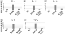
- FIG. 9 is a graph showing the expression level of each gene in Example 1.
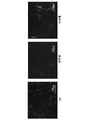
- FIG. FIG. 10 is a photograph showing a histologically stained image of the mouse brain tissue in Example 1, showing the thickness
- radiation neuropathy means a state in which normal nerve cells (neuron cells) and/or nerve tissue are damaged by radiation exposure such as radiation therapy.
- the neuropathy includes, for example, neuronal and/or neurological tissue disorders associated with demyelination, axonal degeneration, coagulative necrosis, and the like.
- the neuropathy caused by radiation includes, for example, radiation encephalopathy, radiation neuritis, radiation myelopathy, radiation brain necrosis, radiation neurosis, leukoencephalopathy, hypoglossal nerve palsy, facial nerve palsy, trigeminal neuropathy, and radiation-induced brachial plexopathy. etc.
- radiation means particle beams or electromagnetic waves emitted when an unstable atomic nucleus structure changes to a stable atomic nucleus structure.
- the radiation includes, for example, ionizing radiation or non-ionizing radiation.
- the ionizing radiation include particle beams and electromagnetic waves.
- the particle beam include ⁇ -rays, ⁇ -rays, proton beams, deuteron beams, triple proton beams, heavy ion beams, charged meson beams, uncharged meson beams, neutrinos, and neutron beams.
- the electromagnetic waves include X-rays and ⁇ -rays.
- the non-ionizing radiation include radio waves, microwaves, infrared rays, visible rays, and ultraviolet rays.
- nerve means a tissue that transmits information.
- the nerves include central nerves and peripheral nerves.
- the central nervous system includes the cerebrum; brain stems such as the diencephalon, midbrain, pons, and medulla oblongata; the cerebellum; the brain; and the spinal cord.
- the peripheral nerves include motor nerves, sensory nerves, and autonomic nerves.
- treatment means therapeutic treatment and/or prophylactic treatment.
- treatment means treating, curing, preventing, arresting, ameliorating, ameliorating a disease, condition, or disorder, or halting, arresting, reducing, or delaying the progression of a disease, condition, or disorder. do.
- prevention means reducing the likelihood of developing a disease or condition or delaying the onset of a disease or condition.
- the “treatment” may be, for example, treatment of a subject (patient) who develops the target disease, or treatment of a model animal of the target disease.
- cell preparation means a cell population containing desired cells or a composition containing desired cells.
- a cell preparation can also be referred to herein, for example, as a cell population or composition.
- the ratio of desired cells to all cells can be quantified, for example, as the ratio of cells expressing one or more markers that desired cells express.
- the purity is, for example, the percentage in viable cells.
- the purity can be measured, for example, by flow cytometry, immunohistochemistry, in situ hybridization, and the like.
- the purity of the cell preparation is, for example, 50% or higher, 55% or higher, 60% or higher, 65% or higher, 70% or higher, 75% or higher, 80% or higher, 85% or higher, 90% or higher , 91% or more, 92% or more, 93% or more, 94% or more, 95% or more, 96% or more, 97% or more, 98% or more, or 99% or more.
- mesenchymal cell means a cell that constitutes connective tissue derived from mesoderm or neural crest and/or a cell that has the potential to differentiate into said cell.
- the mesenchymal cells can also be called mesenchymal stem cells (MSCs).
- Said mesenchymal cells are usually present in blood vessels; in and around organs such as liver or pancreas; fat; bone marrow or umbilical cord;
- the mesenchymal stem cells are one of multipotent stem cells, and mean cells that have the potential to differentiate into adipocytes, osteocytes, chondrocytes, muscle cells, hepatocytes, tendon cells, and/or nerve cells. do.
- the mesenchymal stem cells are, for example, adipose tissue-derived mesenchymal stem cells, bone marrow-derived mesenchymal stem cells, placenta-derived mesenchymal stem cells, dental pulp-derived mesenchymal stem cells, and umbilical cord-derived mesenchymal stem cells. They are called stem cells.
- positive is defined as a negative control reaction using negative control cells that do not express the antigen or an antibody that does not react with the antigen by an analysis method such as flow cytometry that is detected using an antigen-antibody reaction. By comparison, it means that a higher signal or the like is detected.
- negative means that a signal or the like that is equal to or lower than that in a negative control reaction using a negative control cell that does not express the antigen or an antibody that does not react with the antigen is detected. means.
- under inflammatory conditions refers to conditions in which inflammatory cytokines such as interferon ⁇ are contacted or added.
- neuron cell or “neuron cell” means a cell composed of dendrites that receive information and axons (neurites) that transmit information.
- the neuron cells include mature neuron cells and immature neuron cells.
- the mature neuron cells are morphologically cells with well-developed dendrites.
- the mature neuron cells can be identified by expression of mature neuron marker genes such as MAP2.
- the immature neuronal cells are morphologically simple cells with dendrites compared to the mature neuronal cells.
- the immature neuron cells can be identified by expression of immature neuron marker genes such as GAP43.
- active oxygen species means atoms, molecules, radicals, compounds, etc. containing oxygen atoms in a chemically activated state.
- the radical means an atom, molecule or ion having an unpaired electron.
- Said active oxygen is, for example, non-radical species such as singlet oxygen ( 1 O 2 ), ozone (O 3 ), and hydrogen peroxide (H 2 O 2 ); ), peroxy radicals (LOO.), hydroperoxy radicals (HOO.), nitric oxide (NO.), nitrogen dioxide ( NO.sub.2 .), superoxide anions ( O.sub.2- ), radical species such as lipid radicals; is given.
- cell death means the death of cells that are constituents of living organisms.
- the cell death includes, for example, apoptosis and necrosis.
- Said apoptosis refers to programmed cell death controlled by molecular mechanisms.
- Said necrosis refers to passive cell death caused when cells are physically and/or chemically damaged.
- isolated or “isolated” means the state of being identified and separated, and/or the state of being recovered from components in their natural state. Said “isolation” or “isolated” can be carried out, for example, by going through at least one purification step.
- the "isolated” or “isolated” refers to a state in which the cells of interest are separated and/or purified from tissue. may mean. Examples of the tissue include organs such as blood vessels, liver, and pancreas; fat; bone marrow; umbilical cord; Examples of the cells include mesenchymal cells.
- the description provides a cell preparation for use in treating radiation-induced neuropathy.
- the cell preparation of the present disclosure as described above, is a cell preparation for use in the treatment of radiation-induced neuropathy, said cell preparation comprising umbilical cord-derived cells.
- the cell preparation of the present disclosure is characterized by containing umbilical cord-derived cells, and other configurations and conditions are not particularly limited.
- the therapeutic agent for radiation neuropathy of the present invention is characterized by containing the cell preparation of the present disclosure.
- the umbilical cord-derived cells suppress neurite retraction of mature neuron cells caused by irradiation, suppress cell death of immature neuron cells caused by irradiation, We have found that it suppresses the generation of reactive oxygen in neuronal cells caused by irradiation, suppresses necrosis of neuronal cells caused by irradiation, and suppresses inflammation in the nervous system caused by irradiation.
- the present inventors have found that demyelination in radiation-induced neuropathy patients can be suppressed by using umbilical cord-derived cells exhibiting one or more of these activities, and have established the present disclosure. .
- the umbilical cord-derived cells are presumed to suppress symptoms of radiation-induced neuropathy by suppressing damage to neuronal cells caused by radiation and/or promoting recovery from damage caused by radiation. Therefore, it can be said that the cell preparation of the present disclosure can suitably treat radiation-induced neuropathy by containing the umbilical cord-derived cells.
- umbilical cord is a white tubular tissue that connects the fetus and placenta, and means tissue that does not contain placenta and cord blood.
- the origin of the "umbilical cord” is not particularly limited. is the umbilical cord of a primate mammal, more preferably a human umbilical cord.
- the umbilical cord may be an umbilical cord collected from a subject for administration, treatment, or treatment (hereinafter collectively referred to as "administration subject"), or an umbilical cord collected from a subject other than the administration subject. There may be. It is preferable to use an umbilical cord collected from a person other than the subject of administration from the viewpoint of not being subject to restrictions during preparation.
- the umbilical cord-derived cells of the present disclosure may be umbilical cord-derived cells (autologous cells) collected from an administration subject, or umbilical cord-derived cells collected from a non-administration subject, as shown in Examples described later. (allogeneic cells). It has been confirmed that the umbilical cord-derived cells have therapeutic effects without being eliminated by immune rejection or the like, for example.
- the umbilical cord can be collected by appropriately removing the placenta from, for example, postpartum tissue containing the placenta and/or umbilical cord delivered by vaginal delivery or cesarean section.
- the umbilical cord may be one obtained by removing cord blood from the collected umbilical cord, and may be one subjected to aseptic or bacteriostatic treatment. Removal of the cord blood can be performed, for example, by rinsing or perfusion with a solution containing an anticoagulant such as heparin.
- Said aseptic or bactericidal treatment is not particularly limited, for example, application of disinfectants such as povidone-iodine; addition of antibiotics such as penicillin, streptomycin, amphotericin B, gentamicin, and/or nystatin, and/or antimycotics immersion in a medium or buffer;
- the umbilical cord may also selectively lyse red blood cells, for example, if desired.
- methods well known in the art can be used, such as incubation in hypertonic or hypotonic medium by lysis with ammonium chloride.
- the term "umbilical cord-derived cells” means a cell population prepared using umbilical cord as a raw material.
- the "umbilical cord-derived cells” may be composed of a single type of cell, or may be composed of a plurality of types of cells.
- the umbilical cord-derived cells preferably contain umbilical cord-derived mesenchymal cells.
- the umbilical cord-derived cells may be a cell population composed of multiple types of cells including the umbilical cord-derived mesenchymal cells.
- the umbilical cord-derived cells of the present disclosure may be, for example, a cell population having any one or more characteristics and/or markers of (a) to (c) below, preferably a cell population having all characteristics is.
- (a) shows adhesion (adherence) to plastic in culture in the presence of a medium;
- (b) CD105, CD73, CD90, CD44, HLA-classI, HLA-G5 and PD-L (Programmed cell death 1 ligand) 2 positive, CD45, CD34, CD11b, CD19 and HLA-ClassII negative ;
- IDO indoleamine 2,3-dioxygenase
- PGE 2 prostaglandin E2
- PD-L1 genes and/or proteins is induced under inflammatory conditions;
- HLA-class I means HLA-A, B, or C.
- HLA-Class II means HLA-DR, DQ, or DP.
- the umbilical cord-derived cells may be extracts and/or secretions of the umbilical cord-derived cells.
- the umbilical cord-derived cell extract can be obtained by, for example, subjecting the umbilical cord-derived cells to concentration treatment, centrifugation treatment, drying treatment, freeze-drying treatment, solvent treatment, surfactant treatment, protease, glycolytic enzyme, or the like. processed products obtained by enzymatic treatment, protein extraction treatment, ultrasonic treatment and/or grinding treatment, or processed products obtained by a combination of these treatments, and the like.
- the secretions of the umbilical cord-derived cells include exosomes and cell culture supernatants of umbilical cord-derived cells.
- the cell preparation preferably exhibits an inhibitory effect on neurite retraction of mature neuron cells caused by irradiation.
- the mature neuron cells are, for example, central nerve neuron cells, preferably cerebral mature neuron cells.
- the neurite retraction inhibitory action of the mature neuron cells is, for example, when the irradiated mature neuron cells and the subject coexist, compared to the absence of the subject, the mature neuron It can be evaluated by whether it can suppress retraction of neurites of cells. Specifically, the neurite retraction inhibitory action can be evaluated based on the rate of inhibition of neurite length retraction. The inhibition rate was calculated by comparing the neurite length (L nc ) of non-irradiated mature neuronal cells with that of irradiated mature neurons treated identically but cultured in the absence of the specimen.
- Length of neurites of mature neuron cells irradiated and cultured in the presence of the subject, with reference to the difference (L nc ⁇ L c ) from the neurite length (L c ) of the cells The ratio of the difference (L e ⁇ L c ) between the length (L e ) and the length (L c ) of the neurite ((L e ⁇ L c )/(L nc ⁇ L c ) ⁇ 100(%) ) can be calculated as
- the type and irradiation dose of radiation used to calculate the inhibition rate, mature neuron cells, and the length of each neurite can be measured according to Example 1 (7) described below, and more specifically, the radiation
- the mature neuron cells mouse fetal-derived mature neuron cells can be used for measurement.
- the suppression rate is 10% or more, 15% or more, 20% or more, 25% or more, 30% or more, 35% or more, 40% or more, 45% or more, 50% or more, 55% or more % or more, 60% or more, 65% or more, 70% or more, 75% or more, 80% or more, 85% or more, 90% or more, 95% or more, 96% or more, 97% or more, 98% or more, or 99%
- the test object can be evaluated as having an inhibitory effect on neurite retraction.
- the cell preparation preferably exhibits an inhibitory effect on cell death of immature neuronal cells caused by irradiation.
- the immature neuron cell is, for example, a central nervous immature neuron cell, preferably a brain immature neuron cell.
- the effect of suppressing cell death of the immature neuron cells is, for example, when the irradiated immature neuron cells and the test object coexist, compared to the absence of the test object, the immature neuron cell It can be evaluated by whether it can suppress cell death of mature neuron cells. Specifically, the cell death inhibitory action can be evaluated based on the cell death inhibition rate of the immature neuron cells. The inhibition rate was calculated by comparing the number of surviving immature neuronal cells (C nc ) that were not irradiated and the immature neurons that were irradiated and treated in the same manner except that they were cultured in the absence of the test substance.
- C nc surviving immature neuronal cells
- C e Surviving cell number (C e ) of immature neuron cells that have been irradiated and cultured in the presence of the subject, and the surviving cells, based on the difference from the surviving cell number (C c ) of the cells It can be calculated as the ratio of the difference (C e ⁇ C c ) from the number (C c ) ((C e ⁇ C c )/(C nc ⁇ C c ) ⁇ 100(%)).
- the viable cell count the ratio of the viable cell count to the total cell count may be used.
- the type and irradiation dose of radiation, the number of immature neuron cells, and the number of viable cells used for calculating the inhibition rate can be measured according to Example 1 (9) described later.
- the suppression rate is 10% or more, 15% or more, 20% or more, 25% or more, 30% or more, 35% or more, 40% or more, 45% or more, 50% or more, 55% or more % or more, 60% or more, 65% or more, 70% or more, 75% or more, 80% or more, 85% or more, 90% or more, 95% or more, 96% or more, 97% or more, 98% or more, or 99%
- the test substance can be evaluated as having an effect of suppressing cell death.
- the cell preparation exhibits an action of suppressing the generation of reactive oxygen in neuronal cells caused by irradiation.
- the neuron cells are, for example, central nerve neuron cells, preferably brain neuron cells.
- the effect of suppressing the generation of reactive oxygen in the neuron cells is, for example, when the irradiated neuron cells and the test object are allowed to coexist, compared to the absence of the test object, in the neuron cells It can be evaluated by whether or not generation of active oxygen can be suppressed. Specifically, the inhibitory effect on the generation of reactive oxygen can be evaluated based on the rate of inhibition of the generation of reactive oxygen in the neuron cells. The inhibition rate is calculated by comparing the number of reactive oxygen marker-positive cells (R nc ) of non-irradiated neuronal cells and neurons that have been irradiated and treated in the same manner except that they were cultured in the absence of the test substance.
- R nc reactive oxygen marker-positive cells
- the number of reactive oxygen marker-positive cells ( Re ) and the ratio of the difference (R e -R c ) between the number of reactive oxygen marker-positive cells (R c ) ((R e -R c )/(R nc -R c ) ⁇ 100 (%)) can be calculated.
- the ratio of the number of reactive oxygen marker-positive cells to the total number of cells may be used.
- the type and irradiation dose of radiation used to calculate the inhibition rate, neuron cells, the reactive oxygen marker to be used, and the number of cells positive for each reactive oxygen marker can be measured according to Example 1 (8) described below.
- gamma rays can be used as the radiation
- mouse fetal neuron cells can be used as the neuron cells.
- the suppression rate is 10% or more, 15% or more, 20% or more, 25% or more, 30% or more, 35% or more, 40% or more, 45% or more, 50% or more, 55% or more % or more, 60% or more, 65% or more, 70% or more, 75% or more, 80% or more, 85% or more, 90% or more, 95% or more, 96% or more, 97% or more, 98% or more, or 99%
- the test object can be evaluated as having an effect of suppressing the generation of active oxygen.
- the cell preparation preferably exhibits an inhibitory effect on necrosis of neuronal cells caused by irradiation.
- the neuron cells are, for example, central nerve neuron cells, preferably brain neuron cells.
- the effect of suppressing necrosis in neuronal cells is, for example, when the irradiated neuronal cells and the test substance are allowed to coexist, compared to the absence of the test substance, suppresses necrosis in the neuronal cells. It can be evaluated according to whether it can be done. Specifically, the necrosis-suppressing action can be evaluated based on the necrosis-suppressing rate of the neuronal cell. The inhibition rate was calculated by comparing the number of necrotic cells (N nc ) in non-irradiated neuronal cells and the number of irradiated neuronal cells treated in the same manner but cultured in the absence of the test substance.
- the number of necrotic cells (N e ) of irradiated neuronal cells cultured in the presence of the subject, and the necrotic cells, based on the difference from the number of necrotic cells (N c ) can be calculated as a ratio ((N e -N c )/(N nc -N c ) ⁇ 100(%)) of the difference (N e ⁇ N c ) from the number of cells (N c ) of the cell number (N c ).
- the ratio of the number of necrotic cells to the total number of cells may be used.
- the type and irradiation dose of radiation used to calculate the inhibition rate, the detection of neuronal cells, and each necrotic cell can be measured according to Example 1 (9) described below, and more specifically, the radiation It can be measured using ⁇ -rays and mouse embryo-derived neuron cells as the neuron cells.
- the suppression rate is 10% or more, 15% or more, 20% or more, 25% or more, 30% or more, 35% or more, 40% or more, 45% or more, 50% or more, 55% or more % or more, 60% or more, 65% or more, 70% or more, 75% or more, 80% or more, 85% or more, 90% or more, 95% or more, 96% or more, 97% or more, 98% or more, or 99%
- the test object can be evaluated as having an inhibitory effect on necrosis.
- the method for producing (preparing) umbilical cord-derived cells includes, for example, a step of isolating cells from the umbilical cord, and optionally a step of subculturing the isolated cells.
- the preparation method includes, for example, (1) cutting the umbilical cord, (2) culturing the umbilical cord segment, and (3) subculturing.
- the preparation method includes, for example, (A) a step of cutting the umbilical cord, a step of enzymatic treatment, or a step of dissociating the tissue by both, (B) a step of culturing the umbilical cord tissue, and ( C) including the step of passaging.
- the umbilical cord-derived cells may be a uniform cell population or a heterogeneous cell population.
- the umbilical cord-derived cells are prepared by the method including the steps (1) to (3) or the method including the steps (A) to (C), an example can be carried out as follows.
- the method for preparing the umbilical cord-derived cells is not limited to the following example.
- the method including the steps (1) to (3) will be described.
- the umbilical cord obtained by the above method is subjected to mechanical force (shredding force or shear force) in a state containing amniotic membrane, blood vessels, perivascular tissue and/or Walton's jelly. It can be implemented by cutting.
- the size of the umbilical cord segment obtained by cutting is not particularly limited, and examples thereof include 1 to 10 mm 3 , 1 to 5 mm 3 , 1 to 4 mm 3 , 1 to 3 mm 3 and 1 to 2 mm 3 .
- the cut umbilical cord segment is seeded in an incubator such as a Petri dish, dish, or flask, and cultured in a culture medium suitable for umbilical cord-derived cells. .
- the umbilical cord segment is not treated with a digestive enzyme.
- the "incubator” may be, for example, an incubator having a solid surface.
- an incubator used for culturing cells, tissues, and/or organs can be used.
- Said “solid surface” means, for example, any material capable of binding with said umbilical cord-derived cells.
- the material includes, for example, a plastic material that has been treated (eg, hydrophilicity-enhancing treatment) to promote binding of mammalian cells to its surface.
- the type of culture vessel having the solid surface is not particularly limited, and examples thereof include petri dishes, dishes, flasks, and the like.
- the "culture medium suitable for umbilical cord-derived cells” can be prepared, for example, by adding additives such as serum to the basal medium.
- additives are, for example, serum and/or one or more of albumin, transferrin, fatty acids, insulin, sodium selenite, cholesterol, collagen precursors, trace elements, 2-mercaptoethanol, 3′-thiolglycerol, etc. of serum replacement.
- the culture solution further contains lipids, amino acids, proteins, polysaccharides, vitamins, growth factors, low-molecular-weight compounds, antibiotics, antifungal agents, antioxidants, pyruvic acid, buffers, and inorganic salts. You may add substances, such as.
- the basal medium is not particularly limited.
- Ham's F10 Medium F10
- Ham's F-12 Medium F12
- Iscove's Modified Dulbecco's (IMDM) Medium Fischer's Medium
- Mesenchymal Stem Cell Growth Medium MSCGM
- DMEM/F12 RPMI 1640
- CELL-GRO-FREE CELL-GRO-FREE
- serum examples include animal-derived serum such as human serum, fetal bovine serum (FBS), bovine serum, calf serum, goat serum, horse serum, pig serum, sheep serum, rabbit serum and rat serum.
- the amount of serum added to the basal medium is, for example, 5 v/v % to 15 v/v %, preferably about 10 v/v %.
- the fatty acid is not particularly limited, and examples thereof include linoleic acid, oleic acid, linoleic acid, arachidonic acid, myristic acid, palmitoic acid, palmitic acid, and stearic acid.
- the lipid is not particularly limited, and examples thereof include phosphatidylserine, phosphatidylethanolamine, phosphatidylcholine and the like.
- the amino acid is not particularly limited, and examples thereof include amino acids such as L-alanine, L-arginine, L-aspartic acid, L-asparagine, L-cysteine, L-cystine, L-glutamic acid, L-glutamine, and L-glycine.
- the protein is not particularly limited, and examples thereof include ecotin, reduced glutathione, fibronectin, ⁇ 2-microglobulin and the like.
- the polysaccharide is not particularly limited, and examples thereof include glycosaminoglycans such as hyaluronic acid and heparan sulfate.
- the growth factor is not particularly limited, and examples include platelet-derived growth factor (PDGF), epidermal growth factor (EGF), fibroblast growth factor (FGF), vascular endothelial growth factor (VEGF), insulin-like growth factor-1 (IGF-1), leukocyte inhibitory factor (LIF), basic fibroblast growth factor (bFGF), transforming growth factor beta (TGF- ⁇ ), hepatocyte growth factor (HGF), connective tissue growth factor (CTGF) , erythropoietin (EPO) and the like.
- the antibiotic and/or antifungal agent is not particularly limited, and examples thereof include penicillin G, streptomycin sulfate, amphotericin B, gentamicin, nystatin, and mixtures thereof.
- step (2) in order to prevent the seeded umbilical cord segments from floating in the culture medium, it is preferable to hold down the umbilical cord segments using a plate or the like during the culture period.
- the plate include plates described in JP-A-2015-70824.
- culture conditions are not particularly limited, and for example, general culture conditions for cells, tissues, organs, etc. can be referred to.
- the CO 2 concentration in the step (2) is, for example, 0 to 5%.
- the O 2 concentration in the step (2) is, for example, 2 to 25%, preferably 5 to 20%.
- the culture temperature in the step (2) is, for example, 25 to 40°C, preferably about 37°C (35 to 39°C).
- the culture period is not particularly limited. It is preferable to culture until the cells become confluent.
- the cells are washed and treated with a solution containing a chelating agent such as EDTA, trypsin, collagenase, dispase, or the like. protease, sugar chain degrading enzyme such as hyaluronidase, or a stripping agent containing a mixture thereof.
- a chelating agent such as EDTA, trypsin, collagenase, dispase, or the like.
- protease sugar chain degrading enzyme such as hyaluronidase, or a stripping agent containing a mixture thereof.
- the step (2) for example, by filtering the detachment solution containing the cells and the umbilical cord segment using a cell strainer or the like, only cells can be obtained as umbilical cord-derived cells.
- the obtained umbilical cord-derived cells can be seeded, for example, in the culture vessel described above and cultured using the culture medium described above.
- the umbilical cord-derived cells can be appropriately grown to the required number by subculturing.
- the cells in the subculture, the cells may be detached with the detachment agent, seeded at an appropriate cell density in a separately prepared culture vessel, and culture continued.
- the cell density (seeding density) when seeding the cells is, for example, 1 ⁇ 10 2 to 1 ⁇ 10 5 cells/cm 2 , 5 ⁇ 10 2 to 5 ⁇ 10 4 cells/cm 2 , 1 ⁇ 10 3 1 ⁇ 10 4 cells/cm 2 , 2 ⁇ 10 3 to 1 ⁇ 10 4 cells/cm 2 and the like, preferably 2 ⁇ 10 3 to 1 ⁇ 10 4 cells/cm 2 .
- the seeding density is preferably adjusted, for example, so that the period to reach a suitable confluency is 3 to 7 days.
- the medium may be changed as appropriate, if necessary.
- the number of passages in the step (3) is not particularly limited, and may be performed, for example, until senescence, when cell division stops.
- it is preferably subcultured 3 to 25 times, more preferably subcultured 4 to 12 times.
- the umbilical cord obtained by the method described above is enzymatically treated in a state containing amniotic membrane, blood vessels, perivascular tissue and/or Walton's Jelly to dissociate the tissues.
- the enzyme used for the enzymatic treatment is not particularly limited, and examples thereof include proteases such as collagenase and dispase; glycolytic enzymes such as hyaluronidase; and the like.
- step of (B) culturing the umbilical cord tissue and the step of (C) subculturing are, for example, the same as the step of (2) culturing the umbilical cord tissue and the step of (3) subculturing, respectively. can be implemented as
- the umbilical cord-derived cells can be obtained after the step (3) or (C).
- the cells obtained by the method for preparing umbilical cord-derived cells are umbilical cord-derived cells
- surface antigens and the like may be analyzed by conventional methods using flow cytometry and the like.
- whether or not the cells obtained by the method for preparing umbilical cord-derived cells are umbilical cord-derived cells may be evaluated by measuring the amounts of various proteins produced from the cells.
- the cells obtained by the method for preparing umbilical cord-derived cells may be directly prepared for treatment or may be cryopreserved.
- the cryopreservation is performed, for example, by suspending the cells in a cryopreservation solution capable of preserving the umbilical cord-derived cells and storing the cells at -80°C to -180°C.
- the cryopreservation solution is not particularly limited, and examples thereof include an aqueous solution containing a cryoprotectant and glucose.
- the antifreeze agent include dimethyl sulfoxide (hereinafter also referred to as "DMSO"), dextran, glycerol, propylene glycol, and 1-methyl-2-pyrrolidone, preferably DMSO and/or propylene glycol.
- the cryoprotectant is contained, for example, in the cryopreservation solution at 1 to 15 v/v%, preferably 5 to 15 v/v%, more preferably 5 to 12v/v%, More preferably, it is contained in an amount of 8 to 11 v/v%.
- Glucose contained in the cryopreservation solution is, for example, 0.5 to 10 w/v%, preferably 1 to 10 w/v%, more preferably 2 to 10 w/v% in the cryopreservation solution. 8 w/v %, more preferably 2 to 5 w/v %.
- the cryopreservation solution may further contain other components.
- Other components include, for example, pH adjusters and thickeners.
- the pH adjuster include sodium hydrogen carbonate, HEPES, and phosphate buffer.
- BSS basic stock solution
- a phosphate buffer is preferably used as the pH adjuster.
- the pH adjuster is preferably used to adjust the pH in the cryopreservation solution to, for example, about 6.5-9, preferably 7-8.5.
- the "phosphate buffer” includes, for example, sodium chloride, monosodium phosphate (anhydrous), monopotassium phosphate (anhydrous), disodium phosphate (anhydrous), trisodium phosphate ( Anhydrous), potassium chloride, and potassium dihydrogen phosphate (anhydrous), etc., especially sodium chloride, monosodium phosphate (anhydrous), potassium chloride, or potassium dihydrogen phosphate (anhydrous).
- a buffer containing The pH adjuster is contained, for example, in the cryopreservation solution at 0.01 to 1 w/v%, preferably 0.05 to 0.5 w/v%.
- the cryopreservation solution may or may not contain natural animal-derived components.
- the natural animal-derived components include the aforementioned serum and basal medium.
- the cryopreservation solution does not contain natural animal-derived components.
- the cryopreservation solution that does not contain the natural animal-derived components does not cause quality differences between lots of natural animal-derived components, and components such as various cytokines, growth factors, and hormones contained in serum. It is possible to suppress the possibility of changes in the properties of the cells in the umbilical cord tissue caused by , and also to suppress the influence of components of unknown origin contained in the basal medium. Therefore, cryopreservation solutions free of said natural animal-derived components are very useful, especially in clinical use.
- the cryopreservation solution may further contain a thickening agent.
- the thickening agent is not particularly limited, and examples thereof include those capable of constituting a cryopreservation solution capable of sufficiently preserving the umbilical cord tissue.
- the thickener include carboxymethyl cellulose (hereinafter also referred to as "CMC"), carboxymethyl cellulose sodium (hereinafter also referred to as "CMC-Na”), organic acid polymers, propylene glycol alginate, sodium alginate and the like.
- CMC and CMC-Na are preferred, and CMC-Na is particularly preferred.
- the organic acid polymer is preferably sodium polyacrylate.
- the thickening agent is contained, for example, in the cryopreservation solution at 0.1 to 1 w/v%, preferably 0.1 to 0.5 w/v%, more preferably 0.2 Contains ⁇ 0.4 w/v%.
- the cryopreservation solution is preferably an aqueous solution.
- the osmotic pressure of the cryopreservation solution is, for example, preferably 1000 mOsm or more, more preferably 1000 to 2700 mOsm, in order to maintain performance as a preservation solution.
- the cryopreservation solution is preferably an aqueous solution that contains a thickener, a cryoprotectant, and glucose, and does not contain natural animal-derived components.
- the cryopreservation solution is more preferably an aqueous solution containing CMC-Na, DMSO, and glucose and free of natural animal-derived components.
- the cryopreservation solution more preferably contains 0.1 to 1 w/v% CMC-Na, 1 to 15 v/v% DMSO, 0.5 to 10 w/v% glucose, and natural It is an aqueous solution containing no animal-derived components.
- the cells obtained by the method for preparing umbilical cord-derived cells may be used as cell preparations for various applications, for example, by mixing with infusion preparations.
- the cryopreserved umbilical cord-derived cells may be suspended in the cryopreservation solution, and after thawing, may be used directly as a cell preparation for various purposes, or may be used after thawing. It may be mixed with an infusion formulation and the resulting mixture used as a cell preparation for various uses.
- the culture medium or cryopreservation solution in which the umbilical cord-derived cells are suspended may be mixed with the infusion preparation or the like, and the culture medium or cryopreservation solution may be separated by centrifugation or the like. After separating the cells from the solvent, the cells alone may be mixed with the infusion formulation.
- the preparation method for example, in order to avoid the complexity of the procedure, the step of culturing the frozen cells after thawing is not included, or the cryopreservation solution in which the thawed cells are suspended is directly used as an infusion preparation. Mixing is preferred.
- the above-mentioned "infusion preparation” includes, for example, solutions such as infusion solutions used for human treatment, and specific examples include physiological saline, 5% glucose solution, Ringer's solution, lactated Ringer's solution, and acetated Ringer's solution. , No. 1 liquid, No. 2 liquid, No. 3 liquid, No. 4 liquid, and the like.
- the cell preparation of the present disclosure may be a kit containing the infusion preparation in addition to the umbilical cord-derived cells.
- the cell preparation of the present disclosure may contain a pharmaceutically acceptable carrier in addition to or instead of the infusion preparation.
- the carrier includes suspending agents, solubilizers, stabilizers, tonicity agents, preservatives, antiadsorption agents, surfactants, diluents, vehicles, pH adjusters, Examples include soothing agents, buffering agents, sulfur-containing reducing agents, antioxidants, and the like, and can be added appropriately within a range that does not interfere with the effects of the present disclosure.
- the suspending agent is not particularly limited, and examples thereof include methylcellulose, polysorbate 80, hydroxyethylcellulose, gum arabic (gum arabic), tragacanth powder, carboxymethylcellulose sodium, polyoxyethylene sorbitan monolaurate, and the like.
- the solution adjuvant is not particularly limited, and examples thereof include polyoxyethylene hydrogenated castor oil, polysorbate 80, nicotinic acid amide, polyoxyethylene sorbitan monolaurate, macrogol, castor oil fatty acid ethyl ester, and the like.
- the stabilizer is not particularly limited, and examples include dextran 40, methylcellulose, gelatin, sodium sulfite, sodium metasulfate, and the like.
- the tonicity agent is not particularly limited, and examples thereof include D-mannitol and sorbitol.
- the preservative is not particularly limited, and examples thereof include methyl paraoxybenzoate, ethyl parahydroxybenzoate, sorbic acid, phenol, cresol, and chlorocresol.
- the antiadsorption agent is not particularly limited, and examples thereof include human serum albumin, lecithin, dextran, ethylene oxide propylene oxide copolymer, hydroxypropyl cellulose, methyl cellulose, hydrogenated castor oil, and polyethylene glycol.
- the sulfur-containing reducing agent is not particularly limited. Those having a sulfhydryl group such as sodium sulfate, glutathione, and thioalkanoic acids having 1 to 7 carbon atoms are included.
- the antioxidant is not particularly limited, and examples thereof include erythorbic acid, dibutylhydroxytoluene, butylhydroxyanisole, ⁇ -tocopherol, tocopherol acetate, L-ascorbic acid and its salts, L-ascorbic acid palmitate, L-ascorbic acid stear. sodium bisulfite, sodium sulfite, triamyl gallate, propyl gallate or sodium ethylenediaminetetraacetate (EDTA), sodium pyrophosphate, sodium metaphosphate and the like.
- erythorbic acid dibutylhydroxytoluene, butylhydroxyanisole, ⁇ -tocopherol, tocopherol acetate, L-ascorbic acid and its salts, L-ascorbic acid palmitate, L-ascorbic acid stear.
- Cell preparations of the present disclosure may further include inorganic salts such as sodium chloride, potassium chloride, calcium chloride, sodium phosphate, potassium phosphate, sodium bicarbonate; organic salts such as sodium citrate, potassium citrate, sodium acetate; Saccharides such as glucose; and other commonly added components may be added as appropriate.
- inorganic salts such as sodium chloride, potassium chloride, calcium chloride, sodium phosphate, potassium phosphate, sodium bicarbonate
- organic salts such as sodium citrate, potassium citrate, sodium acetate
- Saccharides such as glucose
- ACD-A solution composition composed of sodium citrate hydrate, citric acid hydrate, glucose, etc.
- Cellular preparations of the present disclosure may be mixed with, for example, for topical administration, organics such as biopolymers; inorganics such as hydroxyapatite; specific examples include collagen matrices, polylactic polymers or copolymers, polyethylene It may be mixed with glycol polymers or copolymers and chemical derivatives thereof.
- organics such as biopolymers
- inorganics such as hydroxyapatite
- specific examples include collagen matrices, polylactic polymers or copolymers, polyethylene It may be mixed with glycol polymers or copolymers and chemical derivatives thereof.
- a cell preparation of the present disclosure may be used, for example, in vitro or in vivo.
- the cell preparations of the present disclosure can be used, for example, as research reagents and can be used as pharmaceuticals.
- the subject of administration of the cell preparation of the present disclosure is not particularly limited.
- the administration subject includes, for example, humans or non-human animals other than humans.
- non-human animals include mammals such as mice, rats, rabbits, dogs, cats, cows, horses, pigs, monkeys, dolphins, and sea lions; birds; fish;
- the subject of administration includes, for example, cells, tissues, organs, and the like, and the cells include, for example, cells collected from living organisms, cultured cells, and the like.
- the tissue or organ includes, for example, a tissue (living tissue) or organ collected from a living body.
- the administration subject is, for example, a subject diagnosed as having neuropathy due to radiation, a subject suspected of having neuropathy due to radiation, or a subject having neuropathy due to radiation.
- the neuropathy includes, for example, radiation encephalopathy, radiation neuritis, radiation myelopathy, radiation brain necrosis, radiation neurosis, leukoencephalopathy, hypoglossal nerve palsy, facial nerve palsy, trigeminal neuropathy, and radiation-induced brachial plexopathy. is given.
- the administration subject may be a subject whose administration has been determined based on the radiation exposure dose.
- the exposure dose of the radiation to be administered may be the exposure dose that causes neuropathy or the exposure dose that is predicted to cause neuropathy.
- the lower limit of the exposure dose is, for example, 0.1 Gy or more, 1 Gy or more, 2 Gy or more, 3 Gy or more, 4 Gy or more, 5 Gy or more, 6 Gy or more, 7 Gy or more, 8 Gy or more, 9 Gy or more, or 10 Gy or more.
- the upper limit of the exposure dose is, for example, 100 Gy or less, 50 Gy or less, 40 Gy or less, 30 Gy or less, 20 Gy or less, or 15 Gy or less.
- the numerical range of the exposure dose can be, for example, any combination of the upper limit and the lower limit, and specific examples are 0.1 to 100 Gy, 1 to 100 Gy, 2 to 100 Gy, 3 to 50 Gy, 4 to 50 Gy, 6-40 Gy, 7-40 Gy, 8-30 Gy, 9-20 Gy, or 10-15 Gy.
- the exposure dose may be an actually measured value or an estimated value.
- the radiation exposure dose may be the total amount of radiation exposure dose in a predetermined period. In this case, the total radiation exposure dose may be a single radiation exposure dose or a total radiation exposure dose for a plurality of times.
- the predetermined period is preferably a period during which acute radiation-induced symptoms occur, for example, up to 6 weeks before the determination of administration (for example, Non-Patent Document 1).
- the radiation exposure may be, for example, radiation therapy exposure, occupational accident exposure, accident exposure, terrorism exposure, or the like.
- the site of radiation exposure in the administration subject may be a part of the body or the whole body.
- the part of the body is, for example, a part containing nerves, preferably the head containing the brain, the back containing the spinal cord, and the like.
- the usage conditions (administration conditions) of the cell preparation of the present disclosure are not particularly limited, and for example, the dosage form, administration period, dosage, etc. can be appropriately set according to the type of administration subject.
- Examples of administration methods of the cell preparation of the present disclosure include intracerebral administration, intrathecal administration, intramuscular administration, subcutaneous administration, and intravenous administration. Intravenous administration is preferred because it can be administered rapidly and stably.
- the dosage of the cell preparation of the present disclosure is such that when the cell preparation is administered to the administration subject, compared to when the cell preparation is not administered to the administration subject, the therapeutic effect of neuropathy, the nerve of mature neuron cells It is the amount of cells (therapeutically effective amount) capable of obtaining the effect of suppressing process regression, the effect of suppressing cell death of immature neuronal cells, the effect of suppressing the generation of reactive oxygen in neuronal cells, or the effect of suppressing necrosis of neuronal cells.
- the dosage can be appropriately determined according to, for example, the age, body weight, symptoms, etc. of the subject.
- the dose is, for example, 10 4 to 10 9 cells/kg body weight, 10 4 to 10 8 cells/kg body weight, 10 4 to 10 7 cells/kg as the number of umbilical cord-derived cells per administration.
- body weight preferably 10 4 to 10 8 cells/kg body weight, 10 4 to 10 7 cells/kg body weight.
- the number of administrations of the cell preparation of the present disclosure is one or more.
- the plurality of times is, for example, 2 times, 3 times, 4 times, 5 times or more.
- the frequency of administration may be determined as appropriate while confirming the effect of treatment on the subject.
- the administration interval can be determined appropriately while confirming the therapeutic effect of the subject, for example, once a day, once a week, once every two weeks, once a month, once every three months. , once every six months, etc.
- the cell preparations of the present disclosure may be used in combination with agents and/or methods used for other neurological disorders, for example.
- drugs used for the neuropathy include steroids such as dexamethasone; osmotic diuretics such as glyceol; anticonvulsants; Methods used for the neuropathy include, for example, hyperbaric oxygen therapy.
- the cell preparations of the present disclosure can treat radiation-induced neuropathy, as described above.
- the present disclosure may include methods of treating a subject with radiation-induced neuropathy.
- the present disclosure is a method of treating a subject with radiation-induced neuropathy using the cell preparation of the present disclosure in the subject.
- a method of treating a subject with radiation neuropathy of the present disclosure comprises, for example, administering to the subject the cell preparation of the present disclosure. The above description can be used for the administration conditions in the administration step.
- the present disclosure provides a cell preparation for use in inhibiting radiation-induced neurite retraction of mature neuronal cells or a method for inhibiting radiation-induced neurite retraction of mature neuronal cells.
- the present disclosure is a cell preparation for use in inhibiting radiation-induced neurite retraction of mature neuronal cells, wherein the cell preparation comprises umbilical cord-derived cells.
- the present disclosure also provides a method for suppressing neurite retraction of mature neuron cells caused by radiation, wherein a cell preparation used in the method for suppressing neurite retraction of mature neuron cells caused by radiation of the present disclosure is used as a subject. .
- the present disclosure can incorporate the description of the cell preparation of the present disclosure above.
- the present disclosure provides a cell preparation for use in inhibiting radiation-induced immature neuronal cell death or a method for inhibiting radiation-induced immature neuronal cell death.
- the disclosure is a cell preparation for use in inhibiting radiation-induced cell death of immature neuronal cells, said cell preparation comprising umbilical cord-derived cells.
- the present disclosure also provides a method for suppressing radiation-induced immature neuronal cell death, wherein a cell preparation used in the method for suppressing radiation-induced immature neuronal cell death of the present disclosure is used as a subject. .
- the present disclosure can incorporate the description of the cell preparation of the present disclosure above.
- the present disclosure provides a cell preparation for use in suppressing generation of reactive oxygen species in neuronal cells caused by radiation or a method for suppressing generation of reactive oxygen species in neuronal cells caused by radiation.
- the present disclosure is a cell preparation used for suppressing generation of reactive oxygen in neuronal cells caused by radiation, wherein the cell preparation contains umbilical cord-derived cells.
- the present disclosure also provides a method for suppressing the generation of reactive oxygen in neuronal cells caused by radiation, and uses a cell preparation used in the method for suppressing the generation of reactive oxygen in neuronal cells caused by radiation of the present disclosure as an object.
- the present disclosure can incorporate the description of the cell preparation of the present disclosure above.
- the present disclosure provides a cell preparation for use in inhibiting radiation-induced necrosis of neuronal cells or a method of inhibiting radiation-induced neuronal cell necrosis.
- the present disclosure is a cell preparation for use in inhibiting radiation-induced necrosis of neuronal cells, said cell preparation comprising umbilical cord-derived cells.
- the present disclosure also provides a method of inhibiting radiation-induced necrosis of neuronal cells, wherein the cell preparation used in the method of inhibiting radiation-induced necrosis of neuronal cells of the present disclosure is used as a subject.
- the present disclosure can incorporate the description of the cell preparation of the present disclosure above.
- the present disclosure provides cell preparations for use in suppressing radiation-induced nervous system inflammation or methods of suppressing radiation-induced nervous system inflammation.
- the disclosure is a cell preparation for use in suppressing radiation-induced nervous system inflammation, wherein the cell preparation comprises umbilical cord-derived cells.
- the present disclosure also provides a method of suppressing inflammation of the nervous system caused by radiation, wherein the cell preparation used in the method of suppressing inflammation of the nervous system caused by radiation of the present disclosure is used as a subject.
- the present disclosure can incorporate the description of the cell preparation of the present disclosure above.
- Each of the above-described aspects can be preferably applied to, for example, human nervous system or human central nervous system neuronal cells, immature neuronal cells, or mature neuronal cells.
- the present disclosure provides a treatable method of radiation neuropathy.
- the method of treating neuropathy by radiation according to the present disclosure includes an administration step of administering the cell preparation of the present disclosure to a subject (administration subject).
- the treatment method of the present disclosure is characterized by administering the cell preparation of the present disclosure, and other steps and conditions are not particularly limited. Since the treatment method of the present disclosure uses the cell preparation of the present disclosure, suppression of neurite retraction of mature neuron cells caused by radiation, suppression of cell death of immature neuron cells caused by radiation, and suppression of cell death of immature neuron cells caused by radiation and/or suppress necrosis of neuronal cells caused by radiation.
- the treatment method of the present disclosure can be suitably used for treatment of radiation-induced neuropathy.
- the method for treating radiation neuropathy of the present invention is a treatment method for prophylactic or therapeutic treatment of radiation neuropathy, comprising the step of administering the therapeutic agent for radiation neuropathy of the present invention to an administration subject.
- the administration subject may be an administration subject including humans or an administration subject other than humans.
- the present disclosure provides treatment of neuropathy by radiation, suppression of neurite retraction of mature neuron cells caused by radiation, suppression of cell death of immature neuron cells caused by radiation, suppression of generation of active oxygen in neuronal cells caused by radiation, and A cell preparation for use in inhibiting radiation-induced necrosis of neuronal cells, said cell preparation comprising umbilical cord-derived cells.
- the present disclosure provides treatment of neuropathy by radiation, suppression of neurite retraction of mature neuron cells caused by radiation, suppression of cell death of immature neuron cells caused by radiation, suppression of generation of active oxygen in neuronal cells caused by radiation, and and/or the use of umbilical cord-derived cells to manufacture cell preparations for use in inhibiting radiation-induced necrosis of neuronal cells.
- the present disclosure can incorporate the description of the cell preparation of the present disclosure above.
- a use of the present invention is also the use of a cell preparation of the present disclosure for use in treating radiation-induced neuropathy.
- Umbilical cord mesenchymal stem cells were collected by the method described in Cytotherapy, 18, 229-241, 2016. Specifically, with the approval of the ethics committee of the Institute of Medical Science, the University of Tokyo, all tissue elements of the umbilical cord (amniotic membrane, blood vessels, perivascular tissue, and Walton's jelly) collected with the consent of the donor. ) were chopped into 1-2 mm 3 pieces and seeded onto culture dishes.
- UC-MSCs were obtained by a modified explant method of culturing. The property of the obtained UC-MSCs cells is that they adhere to plastic.
- UC-MSCs highly express the HGF (Hepatic Growth Factor) gene under normal conditions, and the IDO (Indoleamine 2,3-dioxygenase) gene under inflammatory conditions (IFN- ⁇ 100 ng/ml). Induction of expression was confirmed by Realtime PCR. In particular, HGF expression was found to be higher in UC-MSCs than in bone marrow-derived mesenchymal stem cells. In addition, it was confirmed by ELISA that PGE2 secretion was induced by co-culturing UC-MSCs with MLR (mixed allogeneic lymphocyte reaction).
- HGF Hepatic Growth Factor
- IDO Indoleamine 2,3-dioxygenase
- Cortical neuron cells were prepared from an albino strain of B6 strain background mice (B6N-Tyr c-Brd /BrdCrCrl, manufactured by Charles River Japan). Specifically, a 17-day-embryonic fetal mouse was obtained from a pregnant mouse, and the brain and cerebral cortex of the fetal mouse were removed under a microscope (manufactured by Olympus). The removed brain and cortex were used for cortical neuron cell primary culture using a nerve cell dispersion (manufactured by Wako Pure Chemical Industries, Ltd.).
- cortical neuron cells After isolation of cortical neuron cells, the cortical neuron cells were resuspended in Neurobasal medium supplemented with 2% B27 and seeded in Poly-D-Lysine Culture Dishes (BioCoatTM, Corning). The cortical neuron cells were cultured at 37° C. and 5% CO 2 , and half of the medium was replaced with fresh medium on day 3-5 of culture.
- Cortical Neuron Cells for Radiation Injury were prepared using the cortical neuron cells obtained in Example 1(4). Specifically, the cortical neuron cells in the dish were irradiated with radiation. Using a ⁇ -ray irradiation apparatus (IBL 437C III, manufactured by Cis Bio International) using cesium 137 as a radiation source, the cortical neuron cells were irradiated with a set dose of 12 Gy.
- IBL 437C III manufactured by Cis Bio International
- the UC-MSCs were seeded at 1.5 ⁇ 10 4 cells/well in the upper chamber. After the seeding, the 24-well chambers were incubated at 37°C, 5% CO2 for 72 hours. Neurobasal medium was used as the medium for the co-culture.
- the cortical neuron cells were allowed to react with the primary antibody at 4° C. overnight, and stained with the secondary antibody at room temperature for 1 hour.
- 4',6-diamidino-2-phenylindole (DAPI, manufactured by Sigma-Aldrich) was used and added to the secondary antibody solution.
- DAPI 4',6-diamidino-2-phenylindole
- mouse anti-human MAP2 antibody 1000-fold dilution, Cat.No: MA5-12826, Thermo Fisher Scientific
- rabbit anti-GAP43 antibody 200-fold dilution, Cat.No: 8945S , Cell Signaling Technology
- the secondary antibodies included Alexa Fluor R 488-labeled donkey anti-mouse IgG (H + L) (1000-fold dilution, Abcam) and Alexa Fluor R 594-labeled donkey anti-rabbit IgG (H + L) antibody (1000-fold dilution). Dilution, Abcam) and were used.
- cortical neuron cells were observed using a fluorescence microscope (Nikon Eclipse Ti, manufactured by Nikon Solutions) and microscope image integrated software (NIS-Elements software version 4.10). Under observation with a fluorescence microscope set at a magnification of 200, the number of immature neuron cell marker-positive cells and all cells present in 10 randomly selected fields (view fields) were counted. Then, the ratio of the immature neuron cell marker-positive cells to the total number of cells was calculated. Neurite length was measured using ImageJ. MAP2-positive neurites were measured for length in at least 10 randomly selected fields. The negative control group (NC) was performed in the same manner, except that cortical neuron cells that were not irradiated were used.
- NNS-Elements software version 4.10 microscope image integrated software
- Figure 1 is a graph showing the percentage of GAP43-positive cells.
- the vertical axis indicates the percentage of GAP43-positive cells
- the horizontal axis indicates the type of sample.
- the percentage of immature neuronal cells in the radiation group was significantly decreased compared to the negative control group (NC).
- the co-culture group with UC-MSCs co-culture
- the percentage of immature neuron cells was significantly higher than in the radiation group. From these results, it was found that immature neuronal cells are damaged by irradiation, but co-culture with UC-MSCs can reduce radiation damage to immature neuronal cells.
- Figure 2 is a photograph showing an immunostained image of cortical neuron cells.
- the scale bar indicates 100 ⁇ m.
- the radiation group had reduced expression of immature neuronal cell markers.
- expression of immature neuron cell markers was observed as in the control group.
- Fig. 3 is a graph showing the length of neurites.
- the vertical axis indicates the length of neurites
- the horizontal axis indicates the type of sample.
- the neurite length of mature neuronal cells in the radiation group was significantly reduced compared to the negative control group.
- the neurite length of mature neuron cells was significantly longer in the co-culture group with UC-MSCs than in the radiation group. From these results, it was found that radiation injury to mature neuron cells is caused by irradiation, and that radiation injury to mature neuron cells can be alleviated by co-culturing with UC-MSCs.
- Fig. 4 is a photograph showing an immunostained image of cortical neuron cells.
- the upper photograph shows a stained image of MAP2 (MAP-2)
- the middle photograph shows a stained image of GAP43 (GAP43)
- the lower photograph shows a stained image of GAP43. and a stained image of MAP2 are superimposed (Merged).
- the photographs in the left column show the results of the negative control group (NC)
- the photographs in the middle row show the results of the radiation group (radiation)
- the photographs in the right column show The results of the co-culture group (co-culture) are shown.
- the scale bar indicates 100 ⁇ m.
- the radiation group had reduced expression of immature neuronal cell markers and mature neuronal cell markers.
- the co-culture group with UC-MSCs expression of immature neuron cell markers and mature neuron cell markers was observed as in the negative control group.
- DCFDA cell-permeable fluorescent probe 2',7'-Dichlorodihydrofluorescin diacetate, also referred to as "H2DCFDA", “DCFH-DA” or “DCFH”
- H2DCFDA cell-permeable fluorescent probe
- DCFH-DA cell-permeable fluorescent probe
- DCFH cell-permeable fluorescent probe
- 20 mmol/l DCFDA was diluted to a final concentration of 20 ⁇ mol/l with 1 ⁇ buffer from the kit.
- the cortical neuron cells obtained in Example 1(6) were cultured in the presence of the diluted DCFDA solution at 37° C. for 45 minutes in the dark. After the culture, the DCFDA solution was removed, and the 1 ⁇ buffer was added at 20 ⁇ l/well.
- the cortical neuron cells were washed two more times with the 1 ⁇ buffer. After the washing, cortical neuron cells were observed using the fluorescence microscope and the integrated software for microscopic images. The number of fluorescent probe-positive cells and the total number of cells were counted in 10 randomly selected fields (fields of view) under observation with a fluorescence microscope set at a magnification of 200 times. Then, the ratio of the fluorescent probe-positive cells to the total number of cells was calculated.
- the negative control group (NC) cortical neuron cells that were not irradiated were used in the same manner, and for the comparative example, the radiation group (radiation) was co-cultured with UC-MSCs after irradiation. The percentage of fluorescent probe-positive cells was calculated in the same manner, except that cortical neuron cells without cytotoxicity were used. These results are shown in FIGS. 5-6.
- FIG. 5 is a photograph showing a fluorescent image and a bright field image of cortical neuron cells.
- the upper photograph shows the fluorescence image
- the lower photograph shows the bright field image.
- the scale bar indicates 100 ⁇ m.
- FIG. 6 is a graph showing the ratio of fluorescent probe-positive cells, which is an index of active oxygen.
- the vertical axis indicates the percentage of fluorescent probe-positive cells
- the horizontal axis indicates the type of sample.
- the proportion of fluorescent probe-positive cells in the radiation group was significantly higher than that in the negative control group.
- the percentage of fluorescent probe-positive cells was significantly lower than in the radiation group. From these results, it was found that the generation of reactive oxygen increased in irradiated cortical neuron cells, whereas the generation of reactive oxygen could be suppressed by co-culturing with UC-MSCs.
- Necrotic and late apoptotic (secondary necrotic) cells of the cortical neuronal cells were distinguished by ethidium homodimer III staining. Hoechst33342 was used for nuclear staining.
- the 5x binding buffer of the kit was diluted with distilled water to 1x binding buffer, and the cortical neuron cells obtained in Example 1(6) were washed with the 1x binding buffer. After the washing, 5 ⁇ l of FITC-annexin V, 5 ⁇ l of ethidium homodimer III, and 100 ⁇ l of the 1 ⁇ Binding buffer were added to the cortical neuron cells. The cortical neuron cells were then incubated at room temperature for 15 minutes in the dark.
- cortical neuron cells were observed using the fluorescence microscope and the integrated software for microscopic images.
- the number of viable cells, the number of apoptotic cells, and the number of necrotic cells were counted in 10 randomly selected fields (fields of view) under observation with a fluorescence microscope set at 200x magnification.
- the percentages of viable, apoptotic, and necrotic cells were then calculated.
- the control group cortical neuron cells that were not irradiated were used in the same manner, and in the comparative radiation group (radiation), co-culture with UC-MSCs was not performed after irradiation. Percentages of viable, apoptotic, and necrotic cells were calculated in the same manner, except cortical neuronal cells were used.
- FIG. 7 is a photograph showing a fluorescent image and a bright field image of cortical neuron cells.
- the upper photographs show fluorescence images, and the lower photographs show bright field images.
- the scale bar indicates 100 ⁇ m. Arrows in the figure indicate necrotic cells.
- the number of fluorescent probe-positive cells which is an indicator of necrosis, was smaller than in the radiation group. Therefore, it was found that UC-MSCs can suppress cell death of cortical neuron cells.
- FIG. 8 is a graph showing the percentage of viable cells, apoptotic cells, and necrotic cells.
- the vertical axis indicates the type of sample, and the horizontal axis indicates the ratio of viable cells, apoptotic cells, and necrotic cells.
- the ratio of surviving cells was significantly lower in the radiation group and co-culture group than in the negative control group (NC).
- the percentage of viable cells was significantly higher in the co-culture group compared to the radiation group.
- the proportion of necrotic cells was significantly higher in the radiation group and co-culture group than in the negative control group.
- the percentage of viable cells was significantly lower in the co-culture group than in the radiation group. From these results, it was found that irradiated cortical neuron cells decreased viable cells due to necrosis, whereas co-culture with UC-MSCs could suppress necrosis and suppress the decrease in viable cells. .
- mice Upon injection of the mice, anesthesia used medetomidine in the amount of 0.75 mg/kg, midazolam in the amount of 4.0 mg/kg, and butorphanol in the amount of 5.0 mg/kg. The mice were used 3 weeks after injection of the UC-MSCs.
- Example 12 Evaluation of Cortical Neuron Cells by Gene Expression Whether administration of UC-MSCs to radiation-damaged mice alleviates radiation injury in brain tissue was examined by expression of inflammatory cytokine genes. Specifically, the brain tissue of each group of mice obtained in Example 1 (11) was collected, and TRI Reagent (manufactured by Invitrogen) and chloroform (manufactured by Wako Pure Chemical Industries, Ltd.) were used to treat the brain tissue. Total RNA was extracted. Using the obtained total RNA and SYBR (registered trademark) Green PCR Mix (manufactured by Takara Bio Inc.), cDNA was synthesized.
- SYBR registered trademark Green PCR Mix
- FIG. 9 is a graph showing the expression level of each gene.
- (A) shows the results of IFN- ⁇
- (B) shows the results of IL-12
- (C) shows the results of IL-1 ⁇
- (D) shows the results of IL- 6
- (E) shows the results for TNF ⁇ .
- the vertical axis indicates the expression level of each gene
- the horizontal axis indicates the type of sample.
- gene expression levels of inflammatory cytokines in the radiation group (radiation) were significantly increased compared to the negative control group (NC).
- the treated mouse group (radiation + MSCs) injected with UC-MSCs had significantly lower gene expression levels of inflammatory cytokines compared to the radiation group.
- Example 3 Evaluation of cortical neuron cells by tissue staining Examination of whether radiation injury of brain tissue is reduced by administering UC-MSCs to radiation-damaged mice, and the presence or absence of demyelination of myelin marrow by tissue staining with KB staining bottom.
- brain tissue was collected from each group of mice obtained in Example 1 (11) and fixed with 10% or 20% formalin. After the fixation, the brain tissue was embedded in paraffin to prepare 6 ⁇ m-thick paraffin-embedded sections. The sections were deparaffinized and hydrated with 95% alcohol. After the hydration, the sections were stained in Luxol fast blue solution (manufactured by CHROMA) at 75° C. for 5 minutes.
- FIG. 10 is a photograph showing a histological staining image of mouse brain tissue, showing the thickness of the myelin sheath (myelin sheath).
- the upper photograph shows a stained image at a magnification of 40 times
- the lower photograph shows a stained image at a magnification of 200 times.
- the radiation group had demyelinating myelin sheaths compared to the negative control group (NC).
- the umbilical cord-derived cells can reduce brain radiation damage such as demyelination.
- the umbilical cord-derived cells suppress neurite retraction of the neuronal cells, suppress generation of reactive oxygen in the neuronal cells, suppress cell death of the immature neuronal cells, and suppress the neuron cells after the irradiation. It was presumed that this was due to suppression of necrosis, etc.
- the present disclosure is in no way limited by the presumption.
- ⁇ Cell preparation used for treatment of radiation-induced neuropathy> (Appendix 1) A cell preparation for the treatment of radiation neuropathy, comprising: A cell preparation, wherein the cell preparation comprises umbilical cord-derived cells. (Appendix 2) The cell preparation of paragraph 1, wherein the umbilical cord-derived cells are umbilical cord-derived mesenchymal cells. (Appendix 3) The umbilical cord-derived cells are (i) positive for CD105, CD73, CD90, CD44, HLA-class I, HLA-G5 and PD-L2; 2. Cell preparation according to 2.
- the umbilical cord-derived cells are (iii) A cell preparation according to any one of Appendices 1 to 3, wherein the expression of the gene and/or protein of any one of IDO, PGE2, PD-L1 is induced under conditions of inflammation. (Appendix 5) 5. A cell preparation according to any one of Appendices 1 to 4, wherein the umbilical cord-derived cells are cells prepared from umbilical cord tissue comprising amnion, blood vessels, perivascular tissue and/or Walton's Jelly. (Appendix 6) 6.
- the cell preparation of Appendix 5, wherein the umbilical cord-derived cells are cells prepared from the umbilical cord tissue substantially free of proteases (e.g., collagenase, dispase, etc.) (without degradation by proteases).
- Appendix 7 The cell preparation according to appendix 5 or 6, wherein the umbilical cord-derived cells are adherent cells obtained by cutting the umbilical cord tissue into pieces and culturing the pieces.
- Appendix 8) 8.
- Appendix 10 10. A cell preparation according to any one of Appendices 1 to 9, which is for intravenous administration.
- Appendix 11 A cell preparation according to any one of Appendices 1 to 10, which inhibits neurite retraction of mature neuronal cells caused by irradiation.
- Appendix 12 12.
- Appendix 13 13.
- Appendix 14 14.
- Appendix 15 15.
- Appendix 16 16.
- the radiation-induced neuropathy consists of radiation encephalopathy, radiation neuritis, radiation myelopathy, radiation brain necrosis, radiation optic nerve, leukoencephalopathy, hypoglossal nerve palsy, facial nerve palsy, trigeminal neuropathy, and radiation-induced brachial plexopathy. 17.
- a cell preparation according to any one of appendices 1 to 16, selected from the group. ⁇ Application of cell preparation> (Appendix 18) A cell preparation for suppressing neurite retraction of mature neuronal cells caused by radiation, 18.
- a cell preparation comprising a cell preparation according to any one of Appendices 1 to 17 and/or an umbilical cord-derived cell according to any one of Appendices 1 to 17.
- (Appendix 19) A method for suppressing neurite retraction of mature neuron cells caused by radiation, comprising: 19. A method of using the cell preparation of Supplementary Note 18 in a subject. (Appendix 20) 20. The method of paragraph 19, comprising administering said cell preparation to said subject. (Appendix 21) 21. The method of paragraphs 19 or 20, wherein said cell preparation is used in vitro or in vivo. (Appendix 22) A cell preparation for use in suppressing radiation-induced cell death of immature neuronal cells, comprising: 18. A cell preparation comprising a cell preparation according to any one of Appendices 1 to 17 and/or an umbilical cord-derived cell according to any one of Appendices 1 to 17.
- (Appendix 23) A method for suppressing cell death of immature neuronal cells caused by radiation, comprising: A method of using the cell preparation of Supplementary Note 22 in a subject. (Appendix 24) 24. The method of Clause 23, comprising administering said cell preparation to said subject. (Appendix 25) 25. The method of paragraphs 23 or 24, wherein said cell preparation is used in vitro or in vivo. (Appendix 26) A cell preparation used to suppress the generation of reactive oxygen in neuronal cells caused by radiation, 18. A cell preparation comprising a cell preparation according to any one of Appendices 1 to 17 and/or an umbilical cord-derived cell according to any one of Appendices 1 to 17.
- (Appendix 27) A method for suppressing the generation of reactive oxygen in neuronal cells caused by radiation, comprising: A method of using the cell preparation of Supplementary Note 26 in a subject. (Appendix 28) 28. The method of Clause 27, comprising administering said cell preparation to said subject. (Appendix 29) 29. The method of paragraphs 27 or 28, wherein said cell preparation is used in vitro or in vivo.
- (Appendix 30) A cell preparation for use in inhibiting radiation-induced necrosis of neuronal cells, comprising: 18.
- a cell preparation comprising a cell preparation according to any one of Appendices 1 to 17 and/or an umbilical cord-derived cell according to any one of Appendices 1 to 17.
- (Appendix 31) A method for suppressing the occurrence of neuronal necrosis caused by radiation, comprising: A method of using the cell preparation of Supplementary Note 30 in a subject. (Appendix 32) 32. The method of paragraph 31, comprising administering said cell preparation to said subject. (Appendix 33) 33. The method of paragraphs 31 or 32, wherein said cell preparation is used in vitro or in vivo.
- (Appendix 34) A cell preparation for use in suppressing radiation-induced nervous system inflammation, comprising: 18.
- a cell preparation comprising a cell preparation according to any one of Appendices 1 to 17 and/or an umbilical cord-derived cell according to any one of Appendices 1 to 17.
- (Appendix 35) A method of inhibiting inflammation of the nervous system caused by radiation, comprising: A method of using the cell preparation of Supplementary Note 34 in a subject. (Appendix 36) 36. The method of paragraph 35, comprising administering said cell preparation to said subject. (Appendix 37) 37. The method of paragraphs 35 or 36, wherein said cell preparation is used in vitro or in vivo.
- ⁇ Treatment method> A method of treating neuropathy with radiation, comprising: 18.
- a method, wherein the subject uses a cell preparation according to any one of Appendices 1-17 and/or an umbilical cord-derived cell according to any one of Appendices 1-17. (Appendix 39) 385.
- the method of Clause 384 comprising administering said cell preparation and/or umbilical cord-derived cells to said subject.
- Appendix 40 40.
- ⁇ Use of cell preparation> (Appendix 41) A cell preparation for use in the treatment of radiation neuropathy, 18.
- a cell preparation comprising a cell preparation according to any one of Appendices 1 to 17 and/or an umbilical cord-derived cell according to any one of Appendices 1 to 17.
- Appendix 42 A cell preparation for use in inhibiting neurite retraction of mature neuronal cells caused by radiation, 18.
- a cell preparation comprising a cell preparation according to any one of Appendices 1 to 17 and/or an umbilical cord-derived cell according to any one of Appendices 1 to 17.
- Appendix 43 A cell preparation for use in suppressing radiation-induced cell death of immature neuronal cells, 18.
- a cell preparation comprising a cell preparation according to any one of Appendices 1 to 17 and/or an umbilical cord-derived cell according to any one of Appendices 1 to 17.
- Appendix 44 A cell preparation for use in suppressing generation of reactive oxygen species in neuronal cells caused by radiation, 18.
- a cell preparation comprising a cell preparation according to any one of Appendices 1 to 17 and/or an umbilical cord-derived cell according to any one of Appendices 1 to 17.
- Appendix 45 A cell preparation for use in inhibiting necrosis of neuronal cells caused by radiation, 18.
- a cell preparation comprising a cell preparation according to any one of Appendices 1 to 17 and/or an umbilical cord-derived cell according to any one of Appendices 1 to 17.
- Appendix 46 A cell preparation for use in suppressing radiation-induced nervous system inflammation, 18.
- a cell preparation comprising a cell preparation according to any one of Appendices 1 to 17 and/or an umbilical cord-derived cell according to any one of Appendices 1 to 17.
- the present invention it is possible to suppress neurite retraction of mature neuron cells caused by irradiation, suppress cell death of immature neuronal cells caused by irradiation, and reduce active oxygen in neuronal cells caused by irradiation. It can suppress development, suppress necrosis of neuronal cells caused by irradiation, and suppress inflammation of the nervous system caused by irradiation. Therefore, according to the present invention, it can be said that radiation-induced neuropathy can be treated. Therefore, the present invention is extremely useful in, for example, the medical field.
Landscapes
- Health & Medical Sciences (AREA)
- Life Sciences & Earth Sciences (AREA)
- Engineering & Computer Science (AREA)
- Biomedical Technology (AREA)
- Chemical & Material Sciences (AREA)
- Zoology (AREA)
- Cell Biology (AREA)
- Organic Chemistry (AREA)
- Bioinformatics & Cheminformatics (AREA)
- General Health & Medical Sciences (AREA)
- Biotechnology (AREA)
- Animal Behavior & Ethology (AREA)
- Veterinary Medicine (AREA)
- Medicinal Chemistry (AREA)
- Pharmacology & Pharmacy (AREA)
- Wood Science & Technology (AREA)
- Genetics & Genomics (AREA)
- Developmental Biology & Embryology (AREA)
- Public Health (AREA)
- Reproductive Health (AREA)
- Virology (AREA)
- Neurology (AREA)
- Neurosurgery (AREA)
- Chemical Kinetics & Catalysis (AREA)
- General Chemical & Material Sciences (AREA)
- Nuclear Medicine, Radiotherapy & Molecular Imaging (AREA)
- Hematology (AREA)
- Immunology (AREA)
- Microbiology (AREA)
- Epidemiology (AREA)
- Biochemistry (AREA)
- General Engineering & Computer Science (AREA)
- Medicines Containing Material From Animals Or Micro-Organisms (AREA)
Priority Applications (1)
| Application Number | Priority Date | Filing Date | Title |
|---|---|---|---|
| JP2023551503A JPWO2023054317A1 (https=) | 2021-09-30 | 2022-09-27 |
Applications Claiming Priority (2)
| Application Number | Priority Date | Filing Date | Title |
|---|---|---|---|
| JP2021162249 | 2021-09-30 | ||
| JP2021-162249 | 2021-09-30 |
Publications (1)
| Publication Number | Publication Date |
|---|---|
| WO2023054317A1 true WO2023054317A1 (ja) | 2023-04-06 |
Family
ID=85782706
Family Applications (1)
| Application Number | Title | Priority Date | Filing Date |
|---|---|---|---|
| PCT/JP2022/035846 Ceased WO2023054317A1 (ja) | 2021-09-30 | 2022-09-27 | 放射線による神経障害の処置に用いる細胞調製物 |
Country Status (2)
| Country | Link |
|---|---|
| JP (1) | JPWO2023054317A1 (https=) |
| WO (1) | WO2023054317A1 (https=) |
Cited By (1)
| Publication number | Priority date | Publication date | Assignee | Title |
|---|---|---|---|---|
| CN116819093A (zh) * | 2023-06-15 | 2023-09-29 | 中国人民解放军军事科学院军事医学研究院 | 电磁辐射敏感的神经细胞相关血清学标志物及其应用 |
Citations (2)
| Publication number | Priority date | Publication date | Assignee | Title |
|---|---|---|---|---|
| WO2017204231A1 (ja) * | 2016-05-24 | 2017-11-30 | 国立大学法人 東京大学 | 臍帯由来細胞を含む脳障害の治療剤 |
| JP2019535691A (ja) * | 2016-11-03 | 2019-12-12 | エグゾステム バイオテック リミテッド | 間葉系幹細胞集団、それらの生成物およびそれらの使用 |
-
2022
- 2022-09-27 JP JP2023551503A patent/JPWO2023054317A1/ja active Pending
- 2022-09-27 WO PCT/JP2022/035846 patent/WO2023054317A1/ja not_active Ceased
Patent Citations (2)
| Publication number | Priority date | Publication date | Assignee | Title |
|---|---|---|---|---|
| WO2017204231A1 (ja) * | 2016-05-24 | 2017-11-30 | 国立大学法人 東京大学 | 臍帯由来細胞を含む脳障害の治療剤 |
| JP2019535691A (ja) * | 2016-11-03 | 2019-12-12 | エグゾステム バイオテック リミテッド | 間葉系幹細胞集団、それらの生成物およびそれらの使用 |
Cited By (1)
| Publication number | Priority date | Publication date | Assignee | Title |
|---|---|---|---|---|
| CN116819093A (zh) * | 2023-06-15 | 2023-09-29 | 中国人民解放军军事科学院军事医学研究院 | 电磁辐射敏感的神经细胞相关血清学标志物及其应用 |
Also Published As
| Publication number | Publication date |
|---|---|
| JPWO2023054317A1 (https=) | 2023-04-06 |
Similar Documents
| Publication | Publication Date | Title |
|---|---|---|
| US12545889B2 (en) | Colony forming medium and use thereof | |
| JP7126703B2 (ja) | 臍帯由来細胞を含む脳障害の治療剤 | |
| Nicolay et al. | Mesenchymal stem cells–A new hope for radiotherapy-induced tissue damage? | |
| CA2642887C (en) | Mapc therapeutics without adjunctive immunosuppressive treatment | |
| Shams et al. | Mesenchymal stem cells pretreated with HGF and FGF4 can reduce liver fibrosis in mice | |
| JP5809068B2 (ja) | 免疫抑制−関連疾患の治療 | |
| JP6307531B2 (ja) | ニューロンの軸索退縮を予防するための幹細胞の使用 | |
| EP3502237A1 (en) | Composition for promoting growth of stem cells comprising phytosphingosine-1-phosphate or derivatives thereof, and composition for culturing media of stem cells comprising same | |
| JP2022528339A (ja) | 慢性肝不全の急性憎悪の治療のための成人肝前駆細胞 | |
| WO2023054317A1 (ja) | 放射線による神経障害の処置に用いる細胞調製物 | |
| US20250152633A1 (en) | Cells having improved proliferative capacity and reduced cellular senescence | |
| WO2024095996A1 (ja) | 子宮処理用組成物、子宮環境改善剤、着床補助剤、胚移植液、精子移植液及び着床率改善剤 | |
| JP7803543B2 (ja) | 筋肉量の低下抑制に用いる細胞調製物 | |
| KR20230127938A (ko) | 무혈청배지를 이용한 줄기세포 배양 방법 | |
| Lange-Consiglio et al. | In vitro studies of horse umbilical cord matrix-derived cells: from characterization to labeling for magnetic resonance imaging | |
| JP7165352B2 (ja) | 多能性幹細胞による慢性肺疾患の改善及び治療 | |
| JP2022523503A (ja) | 脳内出血を処置することにおける使用のための多分化能成体前駆細胞 | |
| HK40097609A (en) | Cell preparation to be used for preventing decrease in muscle mass | |
| WO2025100424A1 (ja) | 急性脳症治療剤及び急性脳症の治療方法 | |
| US20210130789A1 (en) | Methods of stromal cell expansion, uses and materials related thereto | |
| US20230016479A1 (en) | Method for preparing mesenchymal stem cells having improved viability through anti-cancer virus introduction | |
| WO2025015004A1 (en) | Therapeutic use of fibroblast modulation of microglia | |
| Capasso et al. | Effect of Low-Dose Ionizing Radiations on the Biology of Human Mesenchymal Stromal Cells | |
| WO2022180911A1 (ja) | 血球貪食症候群の治療に用いる細胞調製物 | |
| CA3220002A1 (en) | Methods and compositions for treating liver disease |
Legal Events
| Date | Code | Title | Description |
|---|---|---|---|
| 121 | Ep: the epo has been informed by wipo that ep was designated in this application |
Ref document number: 22876183 Country of ref document: EP Kind code of ref document: A1 |
|
| WWE | Wipo information: entry into national phase |
Ref document number: 2023551503 Country of ref document: JP |
|
| NENP | Non-entry into the national phase |
Ref country code: DE |
|
| 122 | Ep: pct application non-entry in european phase |
Ref document number: 22876183 Country of ref document: EP Kind code of ref document: A1 |