WO2023026528A1 - 手術システム、制御方法、およびプログラム - Google Patents
手術システム、制御方法、およびプログラム Download PDFInfo
- Publication number
- WO2023026528A1 WO2023026528A1 PCT/JP2022/009610 JP2022009610W WO2023026528A1 WO 2023026528 A1 WO2023026528 A1 WO 2023026528A1 JP 2022009610 W JP2022009610 W JP 2022009610W WO 2023026528 A1 WO2023026528 A1 WO 2023026528A1
- Authority
- WO
- WIPO (PCT)
- Prior art keywords
- segmentation
- attention area
- area
- surgical
- region
- Prior art date
Links
- 238000001356 surgical procedure Methods 0.000 title claims abstract description 59
- 238000000034 method Methods 0.000 title claims abstract description 39
- 230000011218 segmentation Effects 0.000 claims abstract description 162
- 238000012545 processing Methods 0.000 claims description 73
- 210000000056 organ Anatomy 0.000 claims description 55
- 230000008569 process Effects 0.000 claims description 16
- 238000005516 engineering process Methods 0.000 abstract description 14
- 238000012937 correction Methods 0.000 description 15
- 238000010586 diagram Methods 0.000 description 13
- 230000008859 change Effects 0.000 description 5
- 206010028980 Neoplasm Diseases 0.000 description 4
- 230000009471 action Effects 0.000 description 4
- 230000006870 function Effects 0.000 description 2
- 238000003709 image segmentation Methods 0.000 description 2
- 238000003384 imaging method Methods 0.000 description 2
- 230000006399 behavior Effects 0.000 description 1
- 230000005540 biological transmission Effects 0.000 description 1
- 210000004204 blood vessel Anatomy 0.000 description 1
- 239000003086 colorant Substances 0.000 description 1
- 238000004891 communication Methods 0.000 description 1
- 230000000694 effects Effects 0.000 description 1
- 238000002357 laparoscopic surgery Methods 0.000 description 1
- 210000002429 large intestine Anatomy 0.000 description 1
- 238000010801 machine learning Methods 0.000 description 1
- 230000007257 malfunction Effects 0.000 description 1
- 210000000713 mesentery Anatomy 0.000 description 1
- 239000000203 mixture Substances 0.000 description 1
- 238000012986 modification Methods 0.000 description 1
- 230000004048 modification Effects 0.000 description 1
- 230000003287 optical effect Effects 0.000 description 1
- 230000001151 other effect Effects 0.000 description 1
- 210000000664 rectum Anatomy 0.000 description 1
- 230000004044 response Effects 0.000 description 1
- 239000004065 semiconductor Substances 0.000 description 1
- 230000001954 sterilising effect Effects 0.000 description 1
- 238000004659 sterilization and disinfection Methods 0.000 description 1
- 210000003384 transverse colon Anatomy 0.000 description 1
Images


Classifications
-
- A—HUMAN NECESSITIES
- A61—MEDICAL OR VETERINARY SCIENCE; HYGIENE
- A61B—DIAGNOSIS; SURGERY; IDENTIFICATION
- A61B34/00—Computer-aided surgery; Manipulators or robots specially adapted for use in surgery
- A61B34/20—Surgical navigation systems; Devices for tracking or guiding surgical instruments, e.g. for frameless stereotaxis
Definitions
- the present technology relates to a surgical system, control method, and program, and more particularly to a surgical system, control method, and program that allow an operator to appropriately set a region of interest.
- Patent Document 1 discloses a technique for controlling the focus of a camera through non-contact input using the voice, gestures, line of sight, etc. of the operator.
- Patent Document 2 discloses a technique for controlling the focus and exposure of a camera by performing image segmentation.
- Non-contact input is generally more likely to be misrecognized than contact input. Misrecognition of inputs can lead to malfunction of the surgical system.
- the line of sight when used as a non-contact input, it is mistakenly recognized that the operator is looking at an organ next to the organ to be operated on, and the endoscope focuses on the organ next to the organ to be operated on. may be controlled in such a way that During surgery, the operator's line of sight is often directed to the edge of the organ to be operated on rather than to the center. There is something.
- This technology was created in view of this situation, and allows the operator to appropriately set the area of interest.
- a surgery system includes an image processing unit that performs segmentation on an image captured by a camera and sets a segmentation region in which each target is captured; An attention area candidate acquisition unit that acquires a candidate, and a control unit that sets the attention area based on a relationship between the segmentation area and the attention area candidate.
- an image captured by a camera is segmented, a segmentation region in which each target is captured is set, and an attention region candidate, which is a candidate region for an operator's attention region, is acquired,
- the attention area is set based on the relationship between the segmentation area and the attention area candidate.
- FIG. 4 is a diagram showing an example of an operating field image
- FIG. 4 is a diagram showing examples of attention area candidates and segmentation areas
- 2 is a block diagram showing a configuration example of a control device in FIG. 1
- FIG. FIG. 2 is a flow chart describing a series of processes of the control device of FIG. 1
- FIG. FIG. 7 is a flowchart for explaining processing of a control unit performed in step S3 of FIG. 6
- FIG. 4 is a diagram showing an example of division of a segmentation region
- FIG. 4 is a diagram showing an example of concatenation of segmentation regions
- It is a block diagram which shows the structural example of the hardware of a computer.
- Embodiments for implementing the present technology will be described below. The explanation is given in the following order.
- First Embodiment (Example of Method of Setting Attention Area) 2.
- Configuration of control device 3 Operation of the controller;4.
- Second embodiment setting of segmentation area) 5.
- Third Embodiment (Countermeasures when the segmentation area is small) 6.
- Fourth Embodiment (Countermeasures when Attention Area Candidates Have Errors) 7.
- Fifth embodiment weighting for segmentation regions) 8.
- Sixth Embodiment (Division of Segmentation Area Using Depth Information) 9.
- Seventh Embodiment (Concatenation of Segmentation Regions Using Depth Information) 10.
- Eighth embodiment (division of segmentation region using SLAM information) 11.
- FIG. 1 is a diagram illustrating a configuration example of a surgery system according to an embodiment of the present technology.
- the surgical system in FIG. 1 is composed of a control device 1, a surgical camera 11, a motion recognition camera 12, a display 13, an operating table 14, a line of sight recognition device 15, a microphone 16, and a foot switch 17.
- the surgical system is a system that is placed in an operating room or the like and used for treatment such as surgical operation with reference to images captured by the surgical camera 11 . Treatment is performed by the operator H who wears the line-of-sight recognition device 15 and the microphone 16 on the head.
- the surgical camera 11 is, for example, a camera used for photographing the surgical field in laparoscopic surgery.
- the surgical camera 11 photographs the surgical field of the patient lying on the operating table 14, and transmits the resulting image to the control device 1 as the surgical field image.
- a moving image or a still image is captured as the operative field image.
- the motion recognition camera 12 is a camera used for recognizing the motion of the operator H.
- the action recognition camera 12 is arranged above the display 13, for example.
- the action recognition camera 12 photographs the operator H and transmits the resulting image to the control device 1 as an operator image.
- the display 13 displays operative field images and the like according to the control by the control device 1 .
- the display 13 is installed with the display surface facing the operator H.
- the control device 1 receives the operator image transmitted from the action recognition camera 12 and recognizes the operator H's gesture.
- the control device 1 also receives information transmitted from the line-of-sight recognition device 15 and recognizes the position of the viewpoint on the screen of the display 13 .
- Information on the line of sight of the operator H is transmitted from the line of sight recognition device 15 .
- the control device 1 receives the voice transmitted from the microphone 16 and performs voice recognition.
- the control device 1 receives a signal transmitted from the foot switch 17 and recognizes the content of the operator H's operation on the foot switch 17 .
- the control device 1 controls the imaging of the surgical camera 11 and the display of the display 13 based on the recognized information.
- control device 1 controls the operation system based on at least one input from the operator H's voice, line of sight, touch, gesture, and operation of the operator H using the foot switch 17. It is a device.
- the microphone 16 acquires the voice of the operator H and transmits it to the control device 1.
- the foot switch 17 is placed at the feet of the operator H.
- the foot switch 17 transmits to the control device 1 an operation signal representing the content of the operation performed by the operator H using the foot.
- the operator H lays the patient on the operating table 14 and, while viewing the surgical field image and the like displayed on the display 13 via the line-of-sight recognition device 15, Treatment such as surgery is performed.
- the operator H when the operator H changes the imaging conditions, position and angle of the surgical camera 11, the display of the display 13, etc., the operator H performs input by voice, line of sight, touch, gesture, and foot switch operation.
- the operator H can perform non-contact input for operating the surgical camera 11 while holding a surgical tool (not shown) by using voice, line of sight, gestures, and the like.
- Any method can be adopted as the method of recognizing the line of sight of the operator H, the method of detecting gestures, and the method of acquiring voice.
- a region of interest which is the region that the operator H is supposed to be paying attention to, is set in the surgical field image, and according to the region of interest, Driving of the surgical camera 11 is controlled. For example, focus control for focusing on the attention area and exposure control for adjusting the brightness of the attention area are performed according to the attention area.
- Such an attention area used as a determination area for focus control and exposure control is set based on the relationship between an attention area candidate, which is a candidate for an attention area, and a segmentation area set by performing image segmentation. be done.
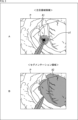
- FIG. 2 is a diagram showing an example of a surgical field image.
- the colored area on the right side is the area in which the organ to be operated is shown.
- Other organs are shown around the organ to be operated on.
- the vicinity of the distal end of the surgical tool T is shown in the area below the center of the surgical field image P, which is shaded.
- the control device When the operative field image P is being captured by the operative camera 11, the control device 1, for example, based on the information supplied from the line-of-sight recognition device 15, colors A in FIG. Such attention area candidate A1 is set.
- FIG. 3A a circular range with a certain distance centered on the viewpoint position p1 is set as the attention area candidate A1.
- the viewpoint position p1 is a position near the edge of the organ to be operated.
- segmentation is performed on the surgical field image P, so that a region in which the organ to be operated is shown is set as a segmentation region A2, as shown by adding color to B in FIG. be done.
- a region in which the organ to be operated is shown is set as a segmentation region A2
- the segmentation region A2 in which the organ to be operated is shown is used for setting the region of interest.
- the segmentation of the operative field image P is performed, for example, using an inference model generated in advance by machine learning using images showing each organ as learning data.
- an inference model generated in advance by machine learning using images showing each organ as learning data.
- FIG. 4 is a diagram showing an example of a method of setting an attention area.
- the control device 1 controls the surgical camera 11 by focusing on the attention area A3 and adjusting the exposure according to the brightness of the attention area A3.
- the attention area A3 is set based on the relationship between the attention area candidate A1 and the segmentation area A2.
- the attention area A3 set by excluding such an area in which a non-interesting object is shown is an area in line with the intention of the operator H who is paying attention to the organ to be operated.
- the position of operator H's viewpoint is always recognized as swaying. Therefore, if the attention area candidate A1 is set as the attention area based only on the viewpoint position, the surgical camera 11 is controlled according to the fluctuation of the viewpoint position, and the image of the surgical field image changes each time. end up By setting the attention area A3 using the attention area candidate A1 and the segmentation area A2, it is possible to suppress such changes in appearance.
- the attention area A3 is set based on the importance set for each position in the segmentation area A2.
- weighting is performed according to the distance from the viewpoint position, and the importance is set for each position within the segmentation area A2.
- a region of interest A3 is set so as to include positions for which the degree of importance equal to or greater than the threshold is set. Setting of the attention area A3 using the degree of importance will be described later.
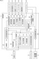
- FIG. 5 is a block diagram showing a configuration example of the control device 1 of FIG. Among the configurations shown in FIG. 5, the same components as those described with reference to FIG. 1 are denoted by the same reference numerals. Duplicate explanations will be omitted as appropriate.
- the control device 1 is composed of an attention area candidate acquisition unit 31, an image processing unit 32, a control unit 33, a surgical procedure information acquisition unit 34, a segmentation target provision unit 35, and an attention area correction information acquisition unit 36.
- Each functional unit as shown in FIG. 5 is realized by executing a predetermined program by a computer that constitutes the control device 1 .
- the attention area candidate acquisition unit 31 has a voice recognition unit 51 , a line-of-sight recognition unit 52 , a touch recognition unit 53 , a gesture recognition unit 54 and an operation recognition unit 55 .
- Information output from each of the input devices of the motion recognition camera 12 , line-of-sight recognition device 15 , microphone 16 , foot switch 17 , spatial touch panel 18 , and touch panel 19 is input to attention area candidate acquisition section 31 .
- the voice recognition unit 51 performs voice recognition based on the voice of the operator H supplied from the microphone 16.
- the line-of-sight recognition unit 52 recognizes the viewpoint position on the screen of the display 13 based on the line-of-sight information of the operator H supplied from the line-of-sight recognition device 15 .
- the touch recognition unit 53 recognizes the content of touch input by the operator H based on the operation signals supplied from the spatial touch panel 18 and the touch panel 19 .
- the spatial touch panel 18 is an input device that detects an input by the operator H to a predetermined space using a finger or hand.
- a spatial touch panel 18 is provided at a predetermined position of the surgical system.
- the touch panel 19 is provided over the display 13, for example.
- the gesture recognition unit 54 recognizes the content of gesture input by the operator H based on the operator image supplied from the action recognition camera 12 .
- the operation recognition unit 55 recognizes the content of the operator H's input based on the operation signal supplied from the foot switch 17 .
- the attention area candidate acquisition unit 31 acquires (sets) attention area candidates based on the speech recognition result, viewpoint position, touch input, gesture input, and foot switch input, which are the recognition results of each unit.
- the attention area candidate acquisition unit 31 outputs information on the attention area candidates to the control unit 33 .
- attention area candidates can be acquired based on information other than the viewpoint position. For example, when an utterance such as "near the surgical tool" is uttered, a region in the vicinity of the tip of the surgical tool is set as a target region candidate based on the result of voice recognition.
- An attention area candidate may be set based on two or more recognition results instead of being set based on one recognition result. Attention area candidates can be set based on at least one of voice recognition results, viewpoint positions, touch inputs, gesture inputs, and foot switch inputs.
- the image processing unit 32 is composed of a segmentation processing unit 61 and an attention area superimposition processing unit 62 .
- the segmentation processing unit 61 performs segmentation on the surgical field image supplied from the surgical camera 11 and outputs information about the segmentation result to the control unit 33 .
- Information supplied to the control unit 33 includes information on each segmentation region.
- the segmentation processing unit 61 has a segmentation weighting processing unit 71, a depth processing unit 72, and a SLAM processing unit 73. The function of each part of the segmentation processing part 61 will be described later.
- the control unit 33 sets the region of interest by appropriately using the information acquired by the segmentation weighting processing unit 71 , the depth processing unit 72 , and the SLAM processing unit 73 .
- the attention area superimposition processing unit 62 displays the attention area on the display 13 based on the information supplied from the attention area setting unit 81 of the control unit 33 .
- the region of interest is displayed so as to be superimposed on the operative field image.
- the control unit 33 has an attention area setting unit 81 .
- the attention area setting unit 81 determines the relationship between the attention area candidate represented by the information supplied from the attention area candidate acquisition unit 31 and the segmentation area represented by the information supplied from the segmentation processing unit 61 of the image processing unit 32. A region of interest is set based on gender.
- the attention area setting unit 81 outputs information on the attention area to the image processing unit 32 .
- control unit 33 controls driving of the surgical camera 11 based on the region of interest.
- the surgical procedure information acquisition unit 34 receives and acquires surgical procedure information supplied from the surgical procedure information providing device 2 .
- the surgical procedure information includes information such as details of surgery and organs to be operated.
- the surgical procedure information acquired by the surgical procedure information acquiring unit 34 is supplied to the segmentation target providing unit 35 . Acquisition of the surgical procedure information by the surgical procedure information acquisition unit 34 is appropriately performed based on the voice supplied from the microphone 16 .
- the segmentation target providing unit 35 identifies an area to be set as a segmentation area based on the surgical procedure information supplied from the surgical procedure information acquiring unit 34, and provides it to the segmentation processing unit 61 of the image processing unit 32. For example, a surgical target organ is specified based on the surgical procedure information, and information indicating that the surgical target organ is to be set as a segmentation region is provided to the segmentation processing unit 61 .
- the attention area correction information acquisition unit 36 generates correction information, which is information for instructing correction (change) of the attention area, based on the sound supplied from the microphone 16 and outputs it to the control unit 33 . For example, when the operator H utters a request to change the attention area, correction information is generated. Based on the correction information generated by the attention area correction information acquisition unit 36, the attention area is appropriately changed. Correction of the attention area may be instructed based on non-contact input other than voice input.
- step S1 the attention area candidate acquisition unit 31 acquires the operator H's attention area candidates.
- step S2 the image processing unit 32 performs segmentation of the operative field image, and sets a region in which the organ to be operated is shown as a segmentation region.
- step S3 the processing of the control unit 33 is performed.
- step S11 the control unit 33 determines whether or not it is possible to acquire the attention area candidates. For example, when information about the recognition result of the viewpoint position of the operator H is included in the information supplied from the attention area candidate acquisition unit 31, it is determined that the attention area candidate can be acquired.
- step S12 the control unit 33 determines whether or not it is possible to acquire the segmentation area. For example, when the segmentation of the operative field image is performed by the segmentation processing unit 61 and the information of the segmentation region is included in the information supplied from the segmentation processing unit 61, it is determined that the segmentation region can be acquired. be.
- step S13 the control unit 33 sets the attention area based on the relationship between the attention area candidate and the segmentation area. As described above, for example, the common area between the attention area candidate and the segmentation area is set as the attention area.
- step S14 the control unit 33 determines whether or not the surgical camera 11 needs to be controlled. For example, when there is a change in the attention area, it is determined that the surgical camera 11 needs to be controlled.
- step S15 the control unit 33 controls at least one of focus and exposure of the surgical camera 11 according to the state of the region of interest. .
- step S15 After the operation of the surgical camera 11 is controlled in step S15, the process proceeds to step S16. If it is determined that the region of interest candidate cannot be acquired in step S11, if it is determined that the segmentation region cannot be acquired in step S12, or if it is determined that the surgical camera 11 cannot be controlled in step S14 Similarly, when it is determined that , the process proceeds to step S16.
- step S16 the control unit 33 determines whether or not to turn off the power of the control device 1.
- step S16 If it is determined in step S16 that the power of the control device 1 should not be turned off, the process returns to step S11 and the above processing is repeated.
- step S16 If it is determined in step S16 that the power of the control device 1 is to be turned off, the process returns to step S3 of FIG. 6 and the processing of the control device 1 ends.
- control device 1 can appropriately set the attention area based on the relationship between the attention area candidate and the segmentation area.
- control device 1 can appropriately control the surgical camera 11 based on the attention area set in accordance with the operator H's intention.
- a plurality of segmentation regions may be set instead of setting one segmentation region for the entire region in which the organ to be operated is shown.
- each region showing the transverse colon, upper rectum, etc., and each narrower region showing the mesentery, blood vessels, etc. are used as the segmentation regions. set.
- the segmentation target providing unit 35 in FIG. 5 sets the granularity of the region to be set as the segmentation region based on the surgical procedure information acquired by the surgical procedure information acquisition unit 34. Based on the granularity set by the segmentation target providing unit 35, the segmentation processing unit 61 sets a region showing a part of one organ to be operated on as a segmentation region.
- the area showing the part with the tumor and the area showing the part without the tumor may be set as different segmentation areas.
- a common area between one attention area candidate and each of a plurality of segmentation areas may be set as the attention area.
- the segmentation processing unit 61 sets a plurality of segmentation regions for the operative field image.
- the attention area setting unit 81 sets a common area between the attention area candidate and each segmentation area as an attention area.
- the attention area is determined based on the positional relationship between the surgical tool and the organ to be operated. may be set.
- the surgical procedure is determined based on the positional relationship between the surgical tool and the organ to be operated on, with reference to the surgical procedure information.
- surgery using an endoscope since the locations to be treated are standardized according to the surgical technique, it is possible to determine the surgical process based on the positional relationship between the surgical tool and the organ.
- the segmentation weighting processing unit 71 identifies the cut-off part or cut part of the organ to be operated on, and sets a high degree of importance, for example, to the part showing the organ sandwiched by forceps.
- a region-of-interest setting unit 81 sets a region of interest based on the degree of importance so as to include a portion in which an organ sandwiched by forceps is shown. For example, the attention area is set so as to include a portion for which the degree of importance equal to or greater than the threshold is set.
- Each portion of the segmentation region may be weighted so that the region of interest is preferentially included in the region of interest.
- the segmentation weighting processing unit 71 identifies a region in which the tumor portion of the organ to be operated is displayed based on the surgical procedure information acquired by the surgical procedure information acquiring unit 34, and for the specified region, Set high importance. Further, the attention area setting unit 81 sets the area including the area where the tumor part is shown as the attention area based on the importance set for each area.
- Each region may be weighted so that a high-contrast region, such as a region where a surgical tool is shown, is included in the region of interest.
- Focus performance can be improved by performing focus control based on a region of interest including a high-contrast region.
- a segmentation region showing an organ to be operated may be divided into a plurality of segmentation regions based on depth information of the organ to be operated.
- the depth processing unit 72 performs depth estimation using the surgical field image captured by the surgical camera 11, and acquires depth information representing the distance to each part shown in the surgical field image.
- the depth estimation performed by the depth processing unit 72 is so-called monocular depth estimation.
- the segmentation processing unit 61 segment the entire region in which the organ is visible into multiple segmentation regions.
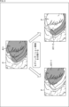
- FIG. 8 is a diagram showing an example of division of the segmentation area.
- the surgical target organ is shown in the surgical field image P, and a segmentation area A11 is set. If it is difficult to focus on the entire segmentation area A11, the segmentation area A11 is divided into a segmentation area A11-1 and a segmentation area A11-2 as indicated by arrows in FIG. For example, the division of the segmentation region is performed based on the depth information so that the distance to each position within the region is within a certain distance.
- the achievable depth of field is shallow due to the short distance to the subject.
- the pixel pitch of the image sensor used in the endoscope becomes narrower due to the higher resolution, which also reduces the achievable depth of field.
- the segmentation region is divided so that the distance to each position within the region is within a certain distance, so that the focus can be appropriately set in any region within the segmentation region. It is possible to match.
- a plurality of segmentation regions showing the organ to be operated may be connected to one segmentation region based on the depth information of the organ to be operated.
- the depth processing unit 72 performs depth estimation using the surgical field image captured by the surgical camera 11, and acquires depth information representing the distance to each part shown in the surgical field image.
- the segmentation processing unit 61 A plurality of regions in which the organs of are shown are combined into one segmentation region.
- FIG. 9 is a diagram showing an example of concatenation of segmentation regions.
- an organ to be operated is shown in the surgical field image P, and a segmentation area A21-1 and a segmentation area A21-2 are set.
- the segmentation region A21-1 and segmentation area A21-2 are connected to one segmentation area A21.
- a wide area is set as an attention area that serves as a reference for focusing. As a result, it is possible to capture an operating field image in which the entire organ in a wide area is in focus.
- SLAM information can be used to divide the segmentation region.
- the SLAM processing unit 73 performs SLAM processing using the surgical field image captured by the surgical camera 11 .
- the segmentation processing unit 61 identifies the distance to each part shown in the operative field image based on SLAM information representing the result of SLAM processing, and divides the segmentation region as described with reference to FIG.
- SLAM information can be used to concatenate segmentation regions.
- the SLAM processing unit 73 performs SLAM processing using the surgical field image captured by the surgical camera 11 .
- the segmentation processing unit 61 identifies the distance to each part shown in the operative field image based on the SLAM information representing the result of SLAM processing, and connects the segmentation regions as described with reference to FIG.
- This also makes it possible to capture an operative field image in which the entire organ in a wide area is in focus.
- the attention area superimposition processing unit 62 causes the display 13 to display information indicating which area the attention area is set to based on the information supplied from the attention area setting unit 81 . For example, an image of a predetermined color is displayed superimposed on the operative field image, and the operator H is presented with a region of interest.
- the operator H can appropriately grasp the behavior of the surgical system.
- the setting of the attention area may be changed according to the operator H's speech after the presentation of the information about the attention area.
- the attention area correction information acquisition unit 36 generates correction information, which is information for instructing correction of the attention area, based on the sound supplied from the microphone 16 . Correction information is generated in response to utterances such as "a little earlier", “a little later", and "no".
- the attention area setting unit 81 changes the attention area based on the correction information generated by the attention area correction information acquisition unit 36, and controls the surgical camera 11 according to the changed attention area.
- the surgical technique information is obtained from the surgical technique information providing device 2 that constitutes the HIS (Hospital Information System), but the surgical technique information may be obtained based on the speech at time-out.
- the time-out is the time for confirming the patient's name, surgical method, and surgical site. For example, a timeout period is secured before the start of surgery.
- the surgical procedure information acquisition unit 34 recognizes the speech detected by the microphone 16 at the time of timeout, and generates surgical procedure information by specifying the patient's name, surgical method, and surgical site. Based on the surgical procedure information generated by the surgical procedure information acquisition unit 34, the setting of the degree of importance and the like are performed. That is, the surgical procedure information acquisition unit 34 acquires surgical procedure information based on at least one of the information transmitted from the linked HIS and the recognition result of the speech of the operator H or the like before the start of surgery. Is possible.
- the setting of the attention area may be changed according to the display magnification of the surgical field image captured by the surgical camera 11 .
- the attention area setting unit 81 sets the attention area to a narrower area, and when the surgical field image is displayed in a reduced size on the display 13, the attention area setting unit 81 sets the attention area to Set to a wider area.
- the common area of the attention area candidate and the segmentation area is set as the attention area
- the common area may be set based on another relationship different from the common area. For example, when the distance between the attention area candidate and the segmentation area is shorter than the threshold distance, it is possible to set the attention area candidate and the entire segmentation area as the attention area.
- the attention area may be set based on various relationships including the positional relationship between the attention area candidate and the segmentation area.
- the series of processes described above can be executed by hardware or by software.
- a program that constitutes the software is installed from a program recording medium into a computer built into dedicated hardware or a general-purpose personal computer.
- FIG. 10 is a block diagram showing a hardware configuration example of a computer that executes the series of processes described above by a program.
- a CPU Central Processing Unit 101
- a ROM Read Only Memory
- RAM Random Access Memory
- An input/output interface 105 is further connected to the bus 104 .
- Input unit 106 , output unit 107 , storage unit 108 , communication unit 109 , and drive 110 are connected to input/output interface 105 .
- a drive 110 drives a removable medium 111 such as a magnetic disk, optical disk, magneto-optical disk, or semiconductor memory.
- the CPU 101 loads, for example, a program stored in the storage unit 108 into the RAM 103 via the input/output interface 105 and the bus 104 and executes the above-described series of programs. is processed.
- Programs executed by the CPU 101 are, for example, recorded on the removable media 111, or provided via a wired or wireless transmission medium such as a local area network, the Internet, or digital broadcasting, and installed in the storage unit 108.
- the program executed by the computer may be a program in which processing is performed in chronological order according to the order described in this specification, or in parallel or at a necessary timing such as when a call is made. It may be a program in which processing is performed.
- Embodiments of the present technology are not limited to the above-described embodiments, and various modifications are possible without departing from the gist of the present technology.
- this technology can take the configuration of cloud computing in which one function is shared by multiple devices via a network and processed jointly.
- each step described in the flowchart above can be executed by a single device, or can be shared by a plurality of devices.
- one step includes multiple processes
- the multiple processes included in the one step can be executed by one device or shared by multiple devices.
- a system means a set of multiple components (devices, modules (parts), etc.), and it does not matter whether all the components are in the same housing. Therefore, a plurality of devices housed in separate housings and connected via a network, and a single device housing a plurality of modules in one housing, are both systems. .
- the present technology can also take the following configurations.
- an image processing unit that performs segmentation of an image captured by a camera and sets a segmentation region in which each target is captured; a region-of-interest candidate acquisition unit that acquires a region-of-interest candidate that is a candidate for a region of interest of an operator; and a control unit that sets the attention area based on the relationship between the segmentation area and the attention area candidate.
- the control unit sets a common area of the segmentation area and the attention area candidate as the attention area.
- the control unit controls at least one of focus and exposure of the camera.
- the attention area candidate acquisition unit acquires the attention area candidate based on at least one input from the operator's voice, line of sight, touch, gesture, and foot switch operation.
- the image processing unit sets the segmentation region to a region in which an organ to be operated that is specified based on surgical procedure information is displayed.
- the control section determines a surgical process based on the positional relationship between the surgical tool and the organ to be operated, and sets the region of interest based on the determination result.
- the image processing unit sets the importance based on the surgical procedure information to each part of the segmentation region in which the organ to be operated is displayed,
- the surgical operation system according to (6) or (7), wherein the control unit sets the region of interest so as to include a portion of which the degree of importance is higher than a threshold.
- the image processing unit performs depth estimation based on the image captured by the camera, and divides the segmentation region or connects a plurality of the segmentation regions based on depth information representing the result of the depth estimation.
- the surgical system according to any one of (1) to (8) above.
- the image processing unit performs SLAM processing based on the image captured by the camera, and divides the segmentation region or connects a plurality of the segmentation regions based on SLAM information representing the result of the SLAM processing.
- the surgical system according to any one of (1) to (8) above.
- the control unit changes the attention area according to the operator's speech performed after the presentation of the information about the attention area.
- the surgical operation system according to any one of (1) to (13), wherein the control unit changes the attention area according to a display magnification of the image captured by the camera.
- the surgical system Segment the image captured by the camera, set the segmentation area where each target is shown, Obtaining an attention area candidate, which is an area that is a candidate for an operator's attention area, A control method of setting the attention area based on a relationship between the segmentation area and the attention area candidate.
Landscapes
- Health & Medical Sciences (AREA)
- Surgery (AREA)
- Engineering & Computer Science (AREA)
- Life Sciences & Earth Sciences (AREA)
- Biomedical Technology (AREA)
- Robotics (AREA)
- Nuclear Medicine, Radiotherapy & Molecular Imaging (AREA)
- Heart & Thoracic Surgery (AREA)
- Medical Informatics (AREA)
- Molecular Biology (AREA)
- Animal Behavior & Ethology (AREA)
- General Health & Medical Sciences (AREA)
- Public Health (AREA)
- Veterinary Medicine (AREA)
- Image Processing (AREA)
Abstract
本技術は、術者が注目する領域を適切に設定することができるようにする手術システム、制御方法、およびプログラムに関する。 本技術の一側面の手術システムは、カメラにより撮影された画像のセグメンテーションを行い、それぞれの対象が映るセグメンテーション領域を設定し、術者の注目領域の候補となる領域である注目領域候補を取得し、セグメンテーション領域と注目領域候補との関係性に基づいて、注目領域を設定するものである。本技術は、内視鏡を用いた手術システムに適用することができる。
Description
本技術は、手術システム、制御方法、およびプログラムに関し、特に、術者が注目する領域を適切に設定することができるようにした手術システム、制御方法、およびプログラムに関する。
内視鏡などを用いた手術システムでは、術者の滅菌対策が必要となる。そのため、内視鏡などの機器を非接触で操作できるようにした技術が各種提案されている。
特許文献1には、術者の音声、ジェスチャ、視線などを用いた非接触の入力により、カメラのフォーカスを制御する技術が開示されている。
また、特許文献2には、画像のセグメンテーションを行うことにより、カメラのフォーカスや露出を制御する技術が開示されている。
非接触の入力は、一般的に、接触による入力に比べて誤認識される可能性が高い。入力の誤認識により、手術システムの誤作動が発生することがある。
例えば、非接触による入力として視線を用いた場合、手術対象の臓器の隣にある臓器に術者が注目しているとして誤認識され、手術対象の臓器の隣にある臓器に内視鏡のフォーカスを合わせるような制御が行われてしまうことがある。手術中の術者の視線は、手術の対象となる臓器の中心ではなく端に向けられることが多いことから、手術対象の臓器の隣にある臓器に術者が注目しているとして誤認識されることがある。
本技術はこのような状況に鑑みてなされたものであり、術者が注目する領域を適切に設定することができるようにするものである。
本技術の一側面の手術システムは、カメラにより撮影された画像のセグメンテーションを行い、それぞれの対象が映るセグメンテーション領域を設定する画像処理部と、術者の注目領域の候補となる領域である注目領域候補を取得する注目領域候補取得部と、前記セグメンテーション領域と前記注目領域候補との関係性に基づいて、前記注目領域を設定する制御部とを備える。
本技術の一側面においては、カメラにより撮影された画像のセグメンテーションが行われ、それぞれの対象が映るセグメンテーション領域が設定され、術者の注目領域の候補となる領域である注目領域候補が取得され、前記セグメンテーション領域と前記注目領域候補との関係性に基づいて、前記注目領域が設定される。
以下、本技術を実施するための形態について説明する。説明は以下の順序で行う。
1.第1の実施の形態(注目領域の設定方法の例)
2.制御装置の構成
3.制御装置の動作
4.第2の実施の形態(セグメンテーション領域の設定)
5.第3の実施の形態(セグメンテーション領域が小さい場合の対策)
6.第4の実施の形態(注目領域候補に誤差がある場合の対策)
7.第5の実施の形態(セグメンテーション領域に対する重み付け)
8.第6の実施の形態(Depth情報を用いたセグメンテーション領域の分割)
9.第7の実施の形態(Depth情報を用いたセグメンテーション領域の連結)
10.第8の実施の形態(SLAM情報を用いたセグメンテーション領域の分割)
11.第9の実施の形態(SLAM情報を用いたセグメンテーション領域の連結)
12.第10の実施の形態(注目領域の表示)
13.第11の実施の形態(発話による注目領域の設定変更)
14.第12の実施の形態(術式情報の取得元の例)
15.第13の実施の形態(表示倍率に応じた注目領域の設定)
16.その他
<第1の実施の形態(注目領域の設定方法の例)>
・本技術を適用した手術システムの構成例
図1は、本技術の一実施形態に係る手術システムの構成例を示す図である。
・本技術を適用した手術システムの構成例
図1は、本技術の一実施形態に係る手術システムの構成例を示す図である。
図1の手術システムは、制御装置1、術用カメラ11、動作認識用カメラ12、ディスプレイ13、手術台14、視線認識デバイス15、マイクロフォン16、およびフットスイッチ17により構成される。手術システムは、手術室等に配置され、術用カメラ11により撮影される画像を参照した外科手術等の処置に用いられるシステムである。視線認識デバイス15とマイクロフォン16を頭部に装着した術者Hにより処置が行われる。
術用カメラ11は、例えば腹腔鏡手術における術野の撮影に用いられるカメラである。術用カメラ11は、手術台14に横たわる患者の術野等を撮影し、その結果得られる画像を術野画像として制御装置1に送信する。術野画像として、動画像または静止画像の撮影が行われる。
動作認識用カメラ12は、術者Hの動作の認識に用いられるカメラである。動作認識用カメラ12は例えばディスプレイ13の上に配置される。動作認識用カメラ12は、術者Hを撮影し、その結果得られる画像を術者画像として制御装置1に送信する。
ディスプレイ13は、制御装置1による制御にしたがって術野画像等を表示する。ディスプレイ13は、表示面を術者Hに向けて設置される。
制御装置1は、動作認識用カメラ12から送信される術者画像を受信し、術者Hのジェスチャを認識する。
また、制御装置1は、視線認識デバイス15から送信される情報を受信し、ディスプレイ13の画面上における視点の位置を認識する。視線認識デバイス15からは、術者Hの視線の情報が送信されてくる。
制御装置1は、マイクロフォン16から送信される音声を受信し、音声認識を行う。制御装置1は、フットスイッチ17から送信される信号を受信し、フットスイッチ17に対する術者Hの操作の内容を認識する。
制御装置1は、認識した情報に基づいて、術用カメラ11の撮影やディスプレイ13の表示を制御する。
このように、制御装置1は、術者Hの音声、視線、タッチ、ジェスチャ、およびフットスイッチ17を用いた術者Hの操作のうちの少なくとも1つの入力に基づいて、手術システムの制御を行う装置である。
マイクロフォン16は、術者Hの音声を取得し、制御装置1に送信する。
フットスイッチ17は、術者Hの足元に配置される。フットスイッチ17は、足を用いて行われる術者Hの操作の内容を表す操作信号を制御装置1に送信する。
以上のように構成される手術システムでは、術者Hは、患者を手術台14の上に横たわらせ、ディスプレイ13に表示される術野画像等を、視線認識デバイス15を介して見ながら外科手術等の処置を行う。
また、術者Hは、術用カメラ11の撮影条件、位置および角度、ディスプレイ13の表示等を変更する場合、音声、視線、タッチ、ジェスチャ、およびフットスイッチ操作による入力を行う。術者Hは、音声、視線、ジェスチャなどを用いることにより、図示せぬ術具を把持した状態で、術用カメラ11の操作のための入力を非接触で行うことができる。
なお、術者Hの視線の認識方法、ジェスチャの検出方法、および音声の取得方法として、任意の方法を採用することができる。
以上のような構成を有する手術システムを制御する制御装置1においては、術者Hが注目していると考えられる領域である注目領域が術野画像に対して設定され、注目領域に応じて、術用カメラ11の駆動が制御される。例えば、注目領域にフォーカスを合わせるフォーカス制御、注目領域の明るさに合わせた露出制御が注目領域に応じて行われる。
このような、フォーカス制御、露出制御の判定エリアとして用いられる注目領域は、注目領域の候補である注目領域候補と、画像のセグメンテーションを行うことにより設定されたセグメンテーション領域との関係性に基づいて設定される。
・注目領域の設定方法の例
図2は、術野画像の例を示す図である。
図2は、術野画像の例を示す図である。
ここでは、図2に示す術野画像Pを用いて、術者Hの注目領域の設定方法について説明する。術野画像Pのうち、色を付して示す右側の領域が、手術対象の臓器が映っている領域である。手術対象の臓器の周りには他の臓器が映っている。また、斜線を付して示す、術野画像Pの中央下方の領域には術具Tの先端付近が映っている。
術野画像Pの撮影が術用カメラ11により行われている場合、制御装置1においては、例えば、視線認識デバイス15から供給される情報に基づいて、図3のAに色を付して示すような注目領域候補A1が設定される。図3のAにおいては、視点位置p1を中心として一定の距離の円形の範囲が注目領域候補A1として設定されている。視点位置p1は、手術対象の臓器の縁の近傍の位置である。
また、制御装置1においては、術野画像Pを対象としたセグメンテーションが行われることにより、図3のBに色を付して示すような、手術対象の臓器が映る領域がセグメンテーション領域A2として設定される。例えば複数の臓器が術野画像Pに映っている場合、セグメンテーションが行われることによって複数のセグメンテーション領域が設定され、そのうちの手術対象の臓器が映るセグメンテーション領域A2が、注目領域の設定に用いられる。
術野画像Pのセグメンテーションは、例えば、各臓器が映る画像を学習データとした機械学習によってあらかじめ生成された推論モデルを用いて行われる。推論モデルに対して術野画像Pを入力することにより、各臓器が映っているセグメンテーション領域に関する情報が出力される。
図4は、注目領域の設定方法の例を示す図である。
以上のようにして注目領域候補A1とセグメンテーション領域A2が設定された場合、図4に示すように、例えば、注目領域候補A1とセグメンテーション領域A2の共通領域が、注目領域A3として設定される。制御装置1においては、注目領域A3にフォーカスを合わせたり、注目領域A3の明るさに合わせて露出を調整したりして、術用カメラ11の制御が行われる。
このように、制御装置1においては、注目領域候補A1とセグメンテーション領域A2との関係性に基づいて注目領域A3が設定される。
これにより、視点に近い位置に映っている非注目物を注目領域A3から除くことが可能となり、術者Hの意図に沿った領域を注目領域A3として設定することが可能となる。すなわち、視点位置に基づいて設定された注目領域候補A1のうちの、セグメンテーション領域A2外の領域は、手術対象の臓器に隣接する臓器が映っている領域であり、非注目物が映っている領域である。そのような、非注目物が映っている領域を除くようにして設定された注目領域A3は、手術対象の臓器に注目している術者Hの意図に沿った領域であるといえる。
また、注目領域A3に基づいて術用カメラ11を制御することにより、術者Hの意図に沿ったフォーカス制御、露出制御が可能となる。
通常、術者Hの視点位置は常に揺れている状態で認識される。そのため、視点位置だけに基づいて、注目領域候補A1を注目領域として設定するとした場合、視点位置が揺れることに応じて術用カメラ11の制御が行われ、術野画像の映りがその都度変化してしまう。注目領域候補A1とともにセグメンテーション領域A2を用いて注目領域A3の設定が行われることにより、そのような映りの変化を抑えることが可能となる。
注目領域候補A1とセグメンテーション領域A2の共通領域が注目領域A3として一律に設定されるのではなく、セグメンテーション領域A2内の各位置に設定された重要度に基づいて注目領域A3が設定されるようにしてもよい。この場合、例えば、視点位置からの距離に応じた重み付けが行われ、セグメンテーション領域A2内の各位置に対して重要度が設定される。また、閾値以上の重要度が設定された位置を含めるようにして注目領域A3が設定される。重要度を用いた注目領域A3の設定については後述する。
<制御装置の構成>
図5は、図1の制御装置1の構成例を示すブロック図である。図5に示す構成のうち、図1を参照して説明した構成と同じ構成には同じ符号を付してある。重複する説明については適宜省略する。
図5は、図1の制御装置1の構成例を示すブロック図である。図5に示す構成のうち、図1を参照して説明した構成と同じ構成には同じ符号を付してある。重複する説明については適宜省略する。
制御装置1は、注目領域候補取得部31、画像処理部32、制御部33、術式情報取得部34、セグメンテーション対象提供部35、および注目領域修正情報取得部36により構成される。図5に示すような各機能部が、制御装置1を構成するコンピュータにより所定のプログラムが実行されることによって実現される。
注目領域候補取得部31は、音声認識部51、視線認識部52、タッチ認識部53、ジェスチャ認識部54、および操作認識部55を有する。動作認識用カメラ12、視線認識デバイス15、マイクロフォン16、フットスイッチ17、空間タッチパネル18、タッチパネル19のそれぞれの入力デバイスから出力された情報が注目領域候補取得部31に入力される。
音声認識部51は、マイクロフォン16から供給される術者Hの音声に基づいて音声認識を行う。
視線認識部52は、視線認識デバイス15から供給される術者Hの視線の情報に基づいて、ディスプレイ13の画面上における視点位置を認識する。
タッチ認識部53は、空間タッチパネル18およびタッチパネル19から供給される操作信号に基づいて、術者Hのタッチ入力の内容を認識する。空間タッチパネル18は、指や手を用いて行われる、所定の空間に対する術者Hの入力を検出する入力デバイスである。空間タッチパネル18は手術システムの所定の位置に設けられる。タッチパネル19は例えばディスプレイ13に重ねて設けられる。
ジェスチャ認識部54は、動作認識用カメラ12から供給される術者画像に基づいて、術者Hのジェスチャ入力の内容を認識する。
操作認識部55は、フットスイッチ17から供給される操作信号に基づいて、術者Hの入力の内容を認識する。
注目領域候補取得部31は、各部における認識結果である、音声認識結果、視点位置、タッチ入力、ジェスチャ入力、フットスイッチ入力に基づいて、注目領域候補を取得する(設定する)。注目領域候補取得部31は、注目領域候補の情報を制御部33に出力する。
このように、注目領域候補が、視点位置以外の情報に基づいて取得されるようにすることが可能である。例えば、「術具の近く」といった発話が行われた場合、音声認識の結果に基づいて、術具の先端の近傍の領域が注目領域候補として設定される。
1つの認識結果に基づいて注目領域候補が設定されるのではなく、2つ以上の認識結果に基づいて注目領域候補が設定されるようにしてもよい。音声認識結果、視点位置、タッチ入力、ジェスチャ入力、フットスイッチ入力のうちの少なくともいずれかに基づいて注目領域候補の設定が行われるようにすることが可能である。
画像処理部32は、セグメンテーション処理部61と注目領域重畳処理部62により構成される。
セグメンテーション処理部61は、術用カメラ11から供給される術野画像を対象としてセグメンテーションを行い、セグメンテーションの結果に関する情報を制御部33に出力する。制御部33に供給される情報には、各セグメンテーション領域の情報が含まれる。
セグメンテーション処理部61は、セグメンテーション重み付け処理部71、Depth処理部72、SLAM処理部73を有する。セグメンテーション処理部61が有する各部の機能については後述する。セグメンテーション重み付け処理部71、Depth処理部72、SLAM処理部73の各部により取得された情報を適宜用いて、注目領域の設定が制御部33により行われる。
注目領域重畳処理部62は、制御部33の注目領域設定部81から供給された情報に基づいて、注目領域をディスプレイ13に表示させる。注目領域の表示は、術野画像に重畳させるようにして行われる。
制御部33は注目領域設定部81を有する。注目領域設定部81は、注目領域候補取得部31から供給された情報により表される注目領域候補と、画像処理部32のセグメンテーション処理部61から供給された情報により表されるセグメンテーション領域との関係性に基づいて注目領域を設定する。注目領域設定部81は、注目領域の情報を画像処理部32に出力する。
また、制御部33は、術用カメラ11の駆動を注目領域に基づいて制御する。
術式情報取得部34は、術式情報提供機器2から供給された術式情報を受信し、取得する。術式情報には、手術内容や手術対象の臓器などの情報が含まれる。術式情報取得部34により取得された術式情報は、セグメンテーション対象提供部35に供給される。術式情報取得部34による術式情報の取得は、適宜、マイクロフォン16から供給された音声に基づいて行われる。
セグメンテーション対象提供部35は、術式情報取得部34から供給された術式情報に基づいて、セグメンテーション領域として設定する領域を特定し、画像処理部32のセグメンテーション処理部61に提供する。例えば、手術対象の臓器が術式情報に基づいて特定され、手術対象の臓器をセグメンテーション領域として設定することを表す情報がセグメンテーション処理部61に対して提供される。
注目領域修正情報取得部36は、マイクロフォン16から供給された音声に基づいて、注目領域の修正(変更)を指示する情報である修正情報を生成し、制御部33に出力する。例えば、注目領域を変更することを要求する内容の発話が術者Hにより行われた場合、修正情報が生成される。注目領域修正情報取得部36により生成された修正情報に基づいて、適宜、注目領域が変更される。音声入力以外の非接触の入力に基づいて注目領域の修正が指示されるようにしてもよい。
<制御装置の動作>
ここで、以上のような構成を有する制御装置1の動作について説明する。
ここで、以上のような構成を有する制御装置1の動作について説明する。
はじめに、図6のフローチャートを参照して、制御装置1の一連の処理について説明する。
ステップS1において、注目領域候補取得部31は、術者Hの注目領域候補を取得する。
ステップS2において、画像処理部32は、術野画像のセグメンテーションを行い、手術対象の臓器が映る領域をセグメンテーション領域として設定する。
ステップS3において、制御部33の処理が行われる。
次に、図7のフローチャートを参照して、図6のステップS3において行われる制御部の処理について説明する。
ステップS11において、制御部33は、注目領域候補を取得することが可能であるか否かを判定する。例えば、術者Hの視点位置の認識結果に関する情報が注目領域候補取得部31から供給される情報に含まれている場合、注目領域候補を取得することが可能であると判定される。
注目領域候補を取得することが可能であるとステップS11において判定された場合、ステップS12において、制御部33は、セグメンテーション領域を取得することが可能であるか否かを判定する。例えば、術野画像のセグメンテーションがセグメンテーション処理部61により行われ、セグメンテーション領域の情報がセグメンテーション処理部61から供給される情報に含まれている場合、セグメンテーション領域を取得することが可能であると判定される。
セグメンテーション領域を取得することが可能であるとステップS12において判定された場合、ステップS13において、制御部33は、注目領域候補とセグメンテーション領域との関係性に基づいて注目領域を設定する。上述したように、例えば、注目領域候補とセグメンテーション領域との共通領域が注目領域として設定される。
ステップS14において、制御部33は、術用カメラ11の制御が必要であるか否かを判定する。例えば、注目領域に変更があった場合、術用カメラ11の制御が必要であると判定される。
術用カメラ11の制御が必要であるとステップS14において判定された場合、ステップS15において、制御部33は、注目領域の状況に応じて、術用カメラ11のフォーカスおよび露出の少なくとも一方を制御する。
ステップS15において術用カメラ11の駆動が制御された後、処理はステップS16に進む。ステップS11において注目領域候補を取得することができないと判定された場合、ステップS12においてセグメンテーション領域を取得することができないと判定された場合、または、ステップS14において術用カメラ11の制御が必要ではないと判定された場合も同様に、処理はステップS16に進む。
ステップS16において、制御部33は、制御装置1の電源をオフにするか否かを判定する。
制御装置1の電源をオフにしないとステップS16において判定された場合、ステップS11に戻り、以上の処理が繰り返される。
制御装置1の電源をオフにするとステップS16において判定された場合、図6のステップS3に戻り、制御装置1の処理は終了となる。
以上の処理により、制御装置1は、注目領域候補とセグメンテーション領域との関係性に基づいて、注目領域を適切に設定することができる。また、制御装置1は、術者Hの意図に沿うようにして設定した注目領域に基づいて、術用カメラ11を適切に制御することができる。
<第2の実施の形態(セグメンテーション領域の設定)>
手術対象の臓器が映っている領域全体に1つのセグメンテーション領域が設定されるのではなく、複数のセグメンテーション領域が設定されるようにしてもよい。
手術対象の臓器が映っている領域全体に1つのセグメンテーション領域が設定されるのではなく、複数のセグメンテーション領域が設定されるようにしてもよい。
例えば、手術対象の臓器が大腸である場合、横行結腸、上部直腸などの部位が映っているそれぞれの領域、腸間膜や血管などの部位が映っているより狭いそれぞれの領域が、セグメンテーション領域として設定される。
この場合、例えば、図5のセグメンテーション対象提供部35は、セグメンテーション領域として設定する領域の粒度を、術式情報取得部34により取得された術式情報に基づいて設定する。セグメンテーション処理部61は、セグメンテーション対象提供部35により設定された粒度に基づいて、手術対象の1つの臓器の一部分が映っている領域をセグメンテーション領域として設定する。
これにより、より狭い注目領域の設定が可能になる。
手術対象の臓器のうち、腫瘍がある部分が映っている領域と腫瘍がない部分が映っている領域がそれぞれ異なるセグメンテーション領域として設定されるようにしてもよい。
<第3の実施の形態(セグメンテーション領域が小さい場合の対策)>
1つの注目領域候補と、複数のセグメンテーション領域のそれぞれとの共通領域が注目領域として設定されるようにしてもよい。
1つの注目領域候補と、複数のセグメンテーション領域のそれぞれとの共通領域が注目領域として設定されるようにしてもよい。
この場合、セグメンテーション処理部61は、術野画像に対して複数のセグメンテーション領域を設定する。注目領域設定部81は、注目領域候補とそれぞれのセグメンテーション領域との共通領域を注目領域として設定する。
これにより、1つのセグメンテーション領域が狭い場合であっても、オートフォーカスの制御、露出制御の基準となる注目領域として一定の広さの領域を確保することが可能となる。
<第4の実施の形態(注目領域候補に誤差がある場合の対策)>
術者Hの視線が揺れるなどして各タイミングにおける注目領域候補に誤差があることから注目領域の設定が困難である場合、術具と手術対象の臓器との位置関係などにも基づいて注目領域が設定されるようにしてもよい。
術者Hの視線が揺れるなどして各タイミングにおける注目領域候補に誤差があることから注目領域の設定が困難である場合、術具と手術対象の臓器との位置関係などにも基づいて注目領域が設定されるようにしてもよい。
この場合、例えば、術式情報を参照し、術具と手術対象の臓器との位置関係に基づいて手術工程が判定される。内視鏡を用いた手術においては、処置を施す箇所が術式によって統一されているため、術具と臓器の位置関係に基づいて、手術工程を判定することが可能である。
セグメンテーション重み付け処理部71は、手術対象の臓器の切り離し部分や切断部分を特定し、例えば鉗子によって挟まれている臓器が映っている部分に対して、高い重要度を設定する。注目領域設定部81は、鉗子によって挟まれている臓器が映っている部分を含むように、重要度に基づいて注目領域を設定する。例えば、閾値以上の重要度が設定されている部分を含むようにして注目領域が設定される。
これにより、各タイミングにおける注目領域候補に誤差がある場合であっても、注目領域設定部81は、注目領域を適切に設定することが可能となる。
<第5の実施の形態(セグメンテーション領域に対する重み付け)>
腫瘍部分が映る領域が優先的に注目領域に含まれるように、セグメンテーション領域の各部に対する重み付けが行われるようにしてもよい。
腫瘍部分が映る領域が優先的に注目領域に含まれるように、セグメンテーション領域の各部に対する重み付けが行われるようにしてもよい。
この場合、例えば、セグメンテーション重み付け処理部71は、術式情報取得部34により取得された術式情報に基づいて、手術対象の臓器の腫瘍部分が映る領域を特定し、特定した領域に対して、高い重要度を設定する。また、注目領域設定部81は、各領域に設定された重要度に基づいて、腫瘍部分が映る領域を含む領域を注目領域として設定する。
これにより、術者Hの意図に沿ったフォーカス制御と露出制御が可能となる。
術具が映っている領域のような、コントラストが高い領域を注目領域に含めるように各領域に対する重み付けが行われるようにしてもよい。コントラストが高い領域を含む注目領域に基づいてフォーカス制御が行われることにより、フォーカス性能を向上させることが可能となる。
<第6の実施の形態(Depth情報を用いたセグメンテーション領域の分割)>
手術対象の臓器が映っているセグメンテーション領域が、手術対象の臓器のDepth情報に基づいて、複数のセグメンテーション領域に分割されるようにしてもよい。
手術対象の臓器が映っているセグメンテーション領域が、手術対象の臓器のDepth情報に基づいて、複数のセグメンテーション領域に分割されるようにしてもよい。
この場合、Depth処理部72は、術用カメラ11により撮影された術野画像を用いたDepth推定を行い、術野画像に映る各部までの距離を表すDepth情報を取得する。Depth処理部72により行われるDepth推定は、いわゆる単眼のDepth推定となる。
また、手術対象の臓器が、フォーカスを合わせるのに必要となる被写界深度が深い対象物である場合(手術対象の臓器の奥行き方向の幅が広い場合)、セグメンテーション処理部61は、手術対象の臓器が映っている領域全体を複数のセグメンテーション領域に分割する。
図8は、セグメンテーション領域の分割の例を示す図である。
図8の例においては、術野画像Pに手術対象の臓器が映り、セグメンテーション領域A11が設定されている。セグメンテーション領域A11全体にフォーカスを合わせることが困難である場合、図8の矢印に示すように、セグメンテーション領域A11がセグメンテーション領域A11-1とセグメンテーション領域A11-2に分割される。例えば、領域内の各位置までの距離が一定の距離に収まるように、セグメンテーション領域の分割がDepth情報に基づいて行われる。
これにより、同じような距離の位置にあるセグメンテーション領域A11-1とセグメンテーション領域A11-2のいずれかを注目領域の設定に用いることにより、フォーカスを適切に合わせることが可能となる。
内視鏡を用いた手術システムにおいては、被写体となる物体までの距離が近いために、実現可能な被写界深度が浅くなる。また、内視鏡に用いられるイメージセンサの画素ピッチが高解像度化によって狭くなり、これによっても、実現可能な被写界深度が浅くなる。上述したように、領域内の各位置までの距離が一定の距離に収まるようにセグメンテーション領域の分割が行われることにより、セグメンテーション領域内のどの領域に注目領域が設定された場合でもフォーカスを適切に合わせることが可能となる。
また、他の臓器を傷つけないようにするために、手術対象の臓器を鉗子で持ち上げた状態で切開や切除などの処置が行われる。この場合、臓器全体にフォーカス合わせるのに必要となる被写界深度が深くなるが、上述したように、領域内の各位置までの距離が一定の距離に収まるようにセグメンテーション領域の分割が行われることにより、フォーカスを適切に制御することが可能となる。
<第7の実施の形態(Depth情報を用いたセグメンテーション領域の連結)>
手術対象の臓器が映っている複数のセグメンテーション領域が、手術対象の臓器のDepth情報に基づいて、1つのセグメンテーション領域に連結されるようにしてもよい。
手術対象の臓器が映っている複数のセグメンテーション領域が、手術対象の臓器のDepth情報に基づいて、1つのセグメンテーション領域に連結されるようにしてもよい。
この場合、Depth処理部72は、術用カメラ11により撮影された術野画像を用いたDepth推定を行い、術野画像に映る各部までの距離を表すDepth情報を取得する。
また、手術対象の臓器が、フォーカスを合わせるのに必要となる被写界深度が浅い対象物である場合(手術対象の臓器の奥行き方向の幅が狭い場合)、セグメンテーション処理部61は、手術対象の臓器が映っている複数の領域を1つのセグメンテーション領域に連結する。
図9は、セグメンテーション領域の連結の例を示す図である。
図9の例においては、術野画像Pに手術対象の臓器が映り、セグメンテーション領域A21-1およびセグメンテーション領域A21-2が設定されている。セグメンテーション領域A21-1に映っている部分とセグメンテーション領域A21-2に映っている部分のそれぞれの部分までの距離が一定の範囲に収まる距離にある場合、図9の矢印に示すように、セグメンテーション領域A21-1とセグメンテーション領域A21-2が1つのセグメンテーション領域A21に連結される。
セグメンテーション領域A21を用いることにより、フォーカスを合わせる基準となる注目領域として広い領域が設定される。これにより、広い領域に映る臓器全体にフォーカスが合った状態の術野画像を撮影することが可能となる。
<第8の実施の形態(SLAM情報を用いたセグメンテーション領域の分割)>
SLAM情報がセグメンテーション領域の分割に用いられるようにすることが可能である。
SLAM情報がセグメンテーション領域の分割に用いられるようにすることが可能である。
この場合、SLAM処理部73は、術用カメラ11により撮影された術野画像を用いたSLAM処理を行う。セグメンテーション処理部61は、術野画像に映る各部までの距離をSLAM処理の結果を表すSLAM情報に基づいて特定し、図8を参照して説明したようにしてセグメンテーション領域を分割する。
これによっても、複数のセグメンテーション領域のそれぞれに適切にフォーカスを合わせることが可能となる。
<第9の実施の形態(SLAM情報を用いたセグメンテーション領域の連結)>
SLAM情報がセグメンテーション領域の連結に用いられるようにすることが可能である。
SLAM情報がセグメンテーション領域の連結に用いられるようにすることが可能である。
この場合、SLAM処理部73は、術用カメラ11により撮影された術野画像を用いたSLAM処理を行う。セグメンテーション処理部61は、術野画像に映る各部までの距離をSLAM処理の結果を表すSLAM情報に基づいて特定し、図9を参照して説明したようにしてセグメンテーション領域を連結する。
これによっても、広い領域に映る臓器全体にフォーカスが合った状態の術野画像を撮影することが可能となる。
<第10の実施の形態(注目領域の表示)>
術用カメラ11のフォーカスおよび露出の少なくとも一方の制御が行われているときに、注目領域に関する情報が術者Hにフィードバックされるようにしてもよい。
術用カメラ11のフォーカスおよび露出の少なくとも一方の制御が行われているときに、注目領域に関する情報が術者Hにフィードバックされるようにしてもよい。
この場合、注目領域重畳処理部62は、注目領域設定部81から供給された情報に基づいて、注目領域がどの領域に設定されているのかを表す情報をディスプレイ13に表示させる。例えば、所定の色の画像が術野画像に重ねて表示され、注目領域が術者Hに提示される。
ディスプレイ13に注目領域が表示されることで、術者Hは手術システムの挙動を適切に把握することができる。
<第11の実施の形態(発話による注目領域の設定変更)>
注目領域に関する情報の提示の後に行われた術者Hの発話に応じて、注目領域の設定が変更されるようにしてもよい。
注目領域に関する情報の提示の後に行われた術者Hの発話に応じて、注目領域の設定が変更されるようにしてもよい。
この場合、注目領域修正情報取得部36は、マイクロフォン16から供給された音声に基づいて、注目領域の修正を指示する情報である修正情報を生成する。「もう少し前」、「もう少し後ろ」、「違う」などの発話が行われることに応じて、修正情報が生成される。注目領域設定部81は、注目領域修正情報取得部36により生成された修正情報に基づいて、注目領域を変更し、変更後の注目領域に応じて術用カメラ11を制御する。
これにより、術者Hの意図に沿わない形で注目領域が設定されてしまった場合でも、注目領域を適切に修正することが可能となる。
<第12の実施の形態(術式情報の取得元の例)>
HIS(Hospital Information System)を構成する術式情報提供機器2から術式情報が取得されるものとしたが、タイムアウト時の発話に基づいて術式情報が取得されるようにしてもよい。タイムアウトは、患者の氏名、手術方法、手術部位を確認するための時間である。例えば、手術開始前などにタイムアウトの時間が確保される。
HIS(Hospital Information System)を構成する術式情報提供機器2から術式情報が取得されるものとしたが、タイムアウト時の発話に基づいて術式情報が取得されるようにしてもよい。タイムアウトは、患者の氏名、手術方法、手術部位を確認するための時間である。例えば、手術開始前などにタイムアウトの時間が確保される。
この場合、術式情報取得部34は、マイクロフォン16により検出されたタイムアウト時の発話を認識し、患者の氏名、手術方法、手術部位を特定することによって術式情報を生成する。術式情報取得部34により生成された術式情報に基づいて、重要度の設定などが行われる。すなわち、術式情報取得部34は、連携するHISから送信されてきた情報と、手術開始前における、術者Hなどの発話の認識結果とのうちの少なくとも一方に基づいて術式情報を取得することが可能である。
これにより、術式情報の取得元の選択肢を増やすことが可能となる。
<第13の実施の形態(表示倍率に応じた注目領域の設定)>
術用カメラ11によって撮影された術野画像の表示倍率に応じて、注目領域の設定が変更されるようにしてもよい。
術用カメラ11によって撮影された術野画像の表示倍率に応じて、注目領域の設定が変更されるようにしてもよい。
例えば、注目領域設定部81は、術野画像がディスプレイ13に拡大表示されている場合、注目領域をより狭い領域に設定し、術野画像がディスプレイ13に縮小表示されている場合、注目領域をより広い領域に設定する。
これにより、術野画像全体に表示されている範囲に応じた広さの注目領域を設定することが可能となる。
<その他>
注目領域候補とセグメンテーション領域の共通領域が注目領域として設定されるものとしたが、共通領域とは異なる他の関係性に基づいて共通領域が設定されるようにしてもよい。例えば、注目領域候補とセグメンテーション領域の距離が閾値となる距離より近い場合に、注目領域候補とセグメンテーション領域の全体が注目領域として設定されるようにすることが可能である。
注目領域候補とセグメンテーション領域の共通領域が注目領域として設定されるものとしたが、共通領域とは異なる他の関係性に基づいて共通領域が設定されるようにしてもよい。例えば、注目領域候補とセグメンテーション領域の距離が閾値となる距離より近い場合に、注目領域候補とセグメンテーション領域の全体が注目領域として設定されるようにすることが可能である。
このように、注目領域候補とセグメンテーション領域の位置の関係性を含む、各種の関係性に基づいて注目領域の設定が行われるようにしてもよい。
・プログラムについて
上述した一連の処理は、ハードウェアにより実行することもできるし、ソフトウェアにより実行することもできる。一連の処理をソフトウェアにより実行する場合には、そのソフトウェアを構成するプログラムが、専用のハードウェアに組み込まれているコンピュータ、または汎用のパーソナルコンピュータなどに、プログラム記録媒体からインストールされる。
上述した一連の処理は、ハードウェアにより実行することもできるし、ソフトウェアにより実行することもできる。一連の処理をソフトウェアにより実行する場合には、そのソフトウェアを構成するプログラムが、専用のハードウェアに組み込まれているコンピュータ、または汎用のパーソナルコンピュータなどに、プログラム記録媒体からインストールされる。
図10は、上述した一連の処理をプログラムにより実行するコンピュータのハードウェアの構成例を示すブロック図である。
CPU(Central Processing Unit)101、ROM(Read Only Memory)102、RAM(Random Access Memory)103は、バス104により相互に接続されている。
バス104には、さらに、入出力インタフェース105が接続されている。入出力インタフェース105には、入力部106、出力部107、記憶部108、通信部109、およびドライブ110が接続されている。ドライブ110は、磁気ディスク、光ディスク、光磁気ディスク、または半導体メモリなどのリムーバブルメディア111を駆動する。
以上のように構成されるコンピュータでは、CPU101が、例えば、記憶部108に記憶されているプログラムを、入出力インタフェース105およびバス104を介して、RAM103にロードして実行することにより、上述した一連の処理が行われる。
CPU101が実行するプログラムは、例えばリムーバブルメディア111に記録して、あるいは、ローカルエリアネットワーク、インターネット、デジタル放送といった、有線または無線の伝送媒体を介して提供され、記憶部108にインストールされる。
なお、コンピュータが実行するプログラムは、本明細書で説明する順序に沿って時系列に処理が行われるプログラムであってもよいし、並列に、あるいは呼び出しが行われたとき等の必要なタイミングで処理が行われるプログラムであってもよい。
なお、本明細書に記載された効果はあくまで例示であって限定されるものでは無く、また他の効果があってもよい。
本技術の実施の形態は、上述した実施の形態に限定されるものではなく、本技術の要旨を逸脱しない範囲において種々の変更が可能である。
例えば、本技術は、1つの機能をネットワークを介して複数の装置で分担、共同して処理するクラウドコンピューティングの構成をとることができる。
また、上述のフローチャートで説明した各ステップは、1つの装置で実行する他、複数の装置で分担して実行することができる。
さらに、1つのステップに複数の処理が含まれる場合には、その1つのステップに含まれる複数の処理は、1つの装置で実行する他、複数の装置で分担して実行することができる。
なお、本明細書において、システムとは、複数の構成要素(装置、モジュール(部品)等)の集合を意味し、すべての構成要素が同一筐体中にあるか否かは問わない。したがって、別個の筐体に収納され、ネットワークを介して接続されている複数の装置、および、1つの筐体の中に複数のモジュールが収納されている1つの装置は、いずれも、システムである。
・構成の組み合わせ例
本技術は、以下のような構成をとることもできる。
本技術は、以下のような構成をとることもできる。
(1)
カメラにより撮影された画像のセグメンテーションを行い、それぞれの対象が映るセグメンテーション領域を設定する画像処理部と、
術者の注目領域の候補となる領域である注目領域候補を取得する注目領域候補取得部と、
前記セグメンテーション領域と前記注目領域候補との関係性に基づいて、前記注目領域を設定する制御部と
を備える手術システム。
(2)
前記制御部は、前記セグメンテーション領域と前記注目領域候補の共通領域を前記注目領域として設定する
前記(1)に記載の手術システム。
(3)
前記制御部は、前記カメラのフォーカスおよび露出の少なくとも一方を制御する
前記(1)または(2)に記載の手術システム。
(4)
前記注目領域候補取得部は、前記術者の音声、視線、タッチ、ジェスチャ、およびフットスイッチ操作の少なくとも1つの入力に基づいて前記注目領域候補を取得する
前記(1)乃至(3)のいずれかに記載の手術システム。
(5)
前記制御部は、複数の前記セグメンテーション領域を用いて前記注目領域を設定する
前記(1)乃至(4)のいずれかに記載の手術システム。
(6)
前記画像処理部は、術式情報に基づいて特定した手術対象となる臓器が映る領域に前記セグメンテーション領域を設定する
前記(1)乃至(5)のいずれかに記載の手術システム。
(7)
前記制御部は、術具と手術対象となる前記臓器との位置関係に基づいて手術工程を判定し、判定した結果に基づいて前記注目領域を設定する
前記(6)に記載の手術システム。
(8)
前記画像処理部は、手術対象となる前記臓器が映る前記セグメンテーション領域の各部に前記術式情報に基づいて重要度を設定し、
前記制御部は、前記重要度が閾値より高い部分を含むように前記注目領域を設定する
前記(6)または(7)に記載の手術システム。
(9)
前記画像処理部は、前記カメラにより撮影された前記画像に基づいてDepth推定を行い、前記Depth推定の結果を表すDepth情報に基づいて、前記セグメンテーション領域の分割、または、複数の前記セグメンテーション領域の連結を行う
前記(1)乃至(8)のいずれかに記載の手術システム。
(10)
前記画像処理部は、前記カメラにより撮影された前記画像に基づいてSLAM処理を行い、前記SLAM処理の結果を表すSLAM情報に基づいて、前記セグメンテーション領域の分割、または、複数の前記セグメンテーション領域の連結を行う
前記(1)乃至(8)のいずれかに記載の手術システム。
(11)
前記画像処理部は、前記カメラの制御時に、前記注目領域に関する情報を前記術者に提示する
前記(1)乃至(3)のいずれかに記載の手術システム。
(12)
前記制御部は、前記注目領域に関する情報の提示後に行われた前記術者の発話に応じて前記注目領域を変更する
前記(11)に記載の手術システム。
(13)
連携するHISから送信されてきた情報と、手術開始前における発話の認識結果とのうちの少なくとも一方に基づいて前記術式情報を取得する術式情報取得部をさらに備える
前記(6)乃至(8)のいずれかに記載の手術システム。
(14)
前記制御部は、前記カメラにより撮影された前記画像の表示倍率に応じて前記注目領域を変更する
前記(1)乃至(13)のいずれかに記載の手術システム。
(15)
手術システムが、
カメラにより撮影された画像のセグメンテーションを行い、それぞれの対象が映るセグメンテーション領域を設定し、
術者の注目領域の候補となる領域である注目領域候補を取得し、
前記セグメンテーション領域と前記注目領域候補との関係性に基づいて、前記注目領域を設定する
制御方法。
(16)
コンピュータに、
カメラにより撮影された画像のセグメンテーションを行い、それぞれの対象が映るセグメンテーション領域を設定し、
術者の注目領域の候補となる領域である注目領域候補を取得し、
前記セグメンテーション領域と前記注目領域候補との関係性に基づいて、前記注目領域を設定する
処理を実行させるためのプログラム。
1 制御装置, 2 術式情報提供機器, 11 術用カメラ, 31 注目領域候補取得部, 32 画像処理部, 33 制御部, 34 術式情報取得部, 35 セグメンテーション対象提供部, 36 注目領域修正情報取得部, 61 セグメンテーション処理部, 62 注目領域重畳処理部, 71 セグメンテーション重み付け処理部, 72 Depth処理部, 73 SLAM処理部, 81 注目領域設定部
Claims (16)
- カメラにより撮影された画像のセグメンテーションを行い、それぞれの対象が映るセグメンテーション領域を設定する画像処理部と、
術者の注目領域の候補となる領域である注目領域候補を取得する注目領域候補取得部と、
前記セグメンテーション領域と前記注目領域候補との関係性に基づいて、前記注目領域を設定する制御部と
を備える手術システム。 - 前記制御部は、前記セグメンテーション領域と前記注目領域候補の共通領域を前記注目領域として設定する
請求項1に記載の手術システム。 - 前記制御部は、前記カメラのフォーカスおよび露出の少なくとも一方を制御する
請求項1に記載の手術システム。 - 前記注目領域候補取得部は、前記術者の音声、視線、タッチ、ジェスチャ、およびフットスイッチ操作の少なくとも1つの入力に基づいて前記注目領域候補を取得する
請求項1に記載の手術システム。 - 前記制御部は、複数の前記セグメンテーション領域を用いて前記注目領域を設定する
請求項1に記載の手術システム。 - 前記画像処理部は、術式情報に基づいて特定した手術対象となる臓器が映る領域に前記セグメンテーション領域を設定する
請求項1に記載の手術システム。 - 前記制御部は、術具と手術対象となる前記臓器との位置関係に基づいて手術工程を判定し、判定した結果に基づいて前記注目領域を設定する
請求項6に記載の手術システム。 - 前記画像処理部は、手術対象となる前記臓器が映る前記セグメンテーション領域の各部に前記術式情報に基づいて重要度を設定し、
前記制御部は、前記重要度が閾値より高い部分を含むように前記注目領域を設定する
請求項6に記載の手術システム。 - 前記画像処理部は、前記カメラにより撮影された前記画像に基づいてDepth推定を行い、前記Depth推定の結果を表すDepth情報に基づいて、前記セグメンテーション領域の分割、または、複数の前記セグメンテーション領域の連結を行う
請求項1に記載の手術システム。 - 前記画像処理部は、前記カメラにより撮影された前記画像に基づいてSLAM処理を行い、前記SLAM処理の結果を表すSLAM情報に基づいて、前記セグメンテーション領域の分割、または、複数の前記セグメンテーション領域の連結を行う
請求項1に記載の手術システム。 - 前記画像処理部は、前記カメラの制御時に、前記注目領域に関する情報を前記術者に提示する
請求項3に記載の手術システム。 - 前記制御部は、前記注目領域に関する情報の提示後に行われた前記術者の発話に応じて前記注目領域を変更する
請求項11に記載の手術システム。 - 連携するHISから送信されてきた情報と、手術開始前における発話の認識結果とのうちの少なくとも一方に基づいて前記術式情報を取得する術式情報取得部をさらに備える
請求項6に記載の手術システム。 - 前記制御部は、前記カメラにより撮影された前記画像の表示倍率に応じて前記注目領域を変更する
請求項1に記載の手術システム。 - 手術システムが、
カメラにより撮影された画像のセグメンテーションを行い、それぞれの対象が映るセグメンテーション領域を設定し、
術者の注目領域の候補となる領域である注目領域候補を取得し、
前記セグメンテーション領域と前記注目領域候補との関係性に基づいて、前記注目領域を設定する
制御方法。 - コンピュータに、
カメラにより撮影された画像のセグメンテーションを行い、それぞれの対象が映るセグメンテーション領域を設定し、
術者の注目領域の候補となる領域である注目領域候補を取得し、
前記セグメンテーション領域と前記注目領域候補との関係性に基づいて、前記注目領域を設定する
処理を実行させるためのプログラム。
Priority Applications (1)
| Application Number | Priority Date | Filing Date | Title |
|---|---|---|---|
| JP2023543651A JPWO2023026528A1 (ja) | 2021-08-26 | 2022-03-07 |
Applications Claiming Priority (2)
| Application Number | Priority Date | Filing Date | Title |
|---|---|---|---|
| JP2021-138108 | 2021-08-26 | ||
| JP2021138108 | 2021-08-26 |
Publications (1)
| Publication Number | Publication Date |
|---|---|
| WO2023026528A1 true WO2023026528A1 (ja) | 2023-03-02 |
Family
ID=85322612
Family Applications (1)
| Application Number | Title | Priority Date | Filing Date |
|---|---|---|---|
| PCT/JP2022/009610 WO2023026528A1 (ja) | 2021-08-26 | 2022-03-07 | 手術システム、制御方法、およびプログラム |
Country Status (2)
| Country | Link |
|---|---|
| JP (1) | JPWO2023026528A1 (ja) |
| WO (1) | WO2023026528A1 (ja) |
Citations (6)
| Publication number | Priority date | Publication date | Assignee | Title |
|---|---|---|---|---|
| JP2006525582A (ja) * | 2003-04-29 | 2006-11-09 | コーニンクレッカ フィリップス エレクトロニクス エヌ ヴィ | 領域分割の微調整 |
| JP2010528762A (ja) * | 2007-06-06 | 2010-08-26 | カール シュトルツ ゲゼルシャフト ミット ベシュレンクテル ハフツング ウント コンパニー コマンディートゲゼルシャフト | 身体上の対象を見るためのビデオシステム |
| JP2017512554A (ja) * | 2014-03-19 | 2017-05-25 | インテュイティブ サージカル オペレーションズ, インコーポレイテッド | 視線追跡を使用する医療装置、システム、及び方法 |
| JP2017153968A (ja) * | 2015-07-13 | 2017-09-07 | ソニー株式会社 | 医療用観察装置、医療用観察装置の作動方法、内視鏡システム、及び手術用顕微鏡システム |
| JP2020157108A (ja) * | 2014-07-28 | 2020-10-01 | インテュイティブ サージカル オペレーションズ, インコーポレイテッド | 手術中のセグメンテーションについてのシステム及び方法 |
| US20200357170A1 (en) * | 2017-06-29 | 2020-11-12 | Open Space Labs, Inc. | Automated spatial indexing of images based on floorplan features |
-
2022
- 2022-03-07 JP JP2023543651A patent/JPWO2023026528A1/ja active Pending
- 2022-03-07 WO PCT/JP2022/009610 patent/WO2023026528A1/ja active Application Filing
Patent Citations (6)
| Publication number | Priority date | Publication date | Assignee | Title |
|---|---|---|---|---|
| JP2006525582A (ja) * | 2003-04-29 | 2006-11-09 | コーニンクレッカ フィリップス エレクトロニクス エヌ ヴィ | 領域分割の微調整 |
| JP2010528762A (ja) * | 2007-06-06 | 2010-08-26 | カール シュトルツ ゲゼルシャフト ミット ベシュレンクテル ハフツング ウント コンパニー コマンディートゲゼルシャフト | 身体上の対象を見るためのビデオシステム |
| JP2017512554A (ja) * | 2014-03-19 | 2017-05-25 | インテュイティブ サージカル オペレーションズ, インコーポレイテッド | 視線追跡を使用する医療装置、システム、及び方法 |
| JP2020157108A (ja) * | 2014-07-28 | 2020-10-01 | インテュイティブ サージカル オペレーションズ, インコーポレイテッド | 手術中のセグメンテーションについてのシステム及び方法 |
| JP2017153968A (ja) * | 2015-07-13 | 2017-09-07 | ソニー株式会社 | 医療用観察装置、医療用観察装置の作動方法、内視鏡システム、及び手術用顕微鏡システム |
| US20200357170A1 (en) * | 2017-06-29 | 2020-11-12 | Open Space Labs, Inc. | Automated spatial indexing of images based on floorplan features |
Also Published As
| Publication number | Publication date |
|---|---|
| JPWO2023026528A1 (ja) | 2023-03-02 |
Similar Documents
| Publication | Publication Date | Title |
|---|---|---|
| US20210401512A1 (en) | Surgical suite integration and optimization | |
| US20220331049A1 (en) | Systems and methods for controlling surgical data overlay | |
| JP6904254B2 (ja) | 手術用制御装置、手術用制御方法、およびプログラム | |
| US11642004B2 (en) | Image processing device, image processing method and recording medium | |
| US20190069957A1 (en) | Surgical recognition system | |
| US11503201B2 (en) | Focus detection device and method | |
| WO2018211969A1 (ja) | 入力制御装置、入力制御方法、および手術システム | |
| JP2017070636A (ja) | 手術システム、並びに、手術用制御装置および手術用制御方法 | |
| US20190361252A1 (en) | Branching optical system, imaging apparatus, and imaging system | |
| US20170164829A1 (en) | Registration Using a Microscope Insert | |
| US20230069839A1 (en) | Information processing device, information processing method, program, screen, and information drawing system | |
| US20180049840A1 (en) | Methods and systems for registration using a microscope insert | |
| WO2023026528A1 (ja) | 手術システム、制御方法、およびプログラム | |
| US11883120B2 (en) | Medical observation system, medical signal processing device, and medical signal processing device driving method | |
| US20210177284A1 (en) | Medical observation system, medical observation apparatus, and method for driving medical observation apparatus | |
| US11523729B2 (en) | Surgical controlling device, control method, and surgical system | |
| JP7480783B2 (ja) | 内視鏡システム、制御装置、および制御方法 | |
| US20190117052A1 (en) | Surgical system, surgical control method, and program | |
| US20230218143A1 (en) | Medical observation system, image processing method, and program | |
| JP2021018281A (ja) | 医療機器制御システム、制御装置、および制御方法 | |
| TWI805248B (zh) | 基於頭部追蹤控制內視鏡手術機器人的控制系統與控制方法 | |
| WO2023162479A1 (ja) | 情報処理装置、情報処理方法、及びプログラム | |
| WO2023017651A1 (ja) | 医療用観察システム、情報処理装置及び情報処理方法 | |
| US20210153959A1 (en) | Physical medical element affixation systems, methods, and materials | |
| EP4356396A1 (en) | Customization, troubleshooting, and wireless pairing techniques for surgical instruments |
Legal Events
| Date | Code | Title | Description |
|---|---|---|---|
| 121 | Ep: the epo has been informed by wipo that ep was designated in this application |
Ref document number: 22860826 Country of ref document: EP Kind code of ref document: A1 |
|
| WWE | Wipo information: entry into national phase |
Ref document number: 2023543651 Country of ref document: JP |
|
| NENP | Non-entry into the national phase |
Ref country code: DE |