WO2023007809A1 - コロナウイルス感染症の重症化リスクの評価方法、およびコロナウイルス感染症の重症化リスク評価キット - Google Patents
コロナウイルス感染症の重症化リスクの評価方法、およびコロナウイルス感染症の重症化リスク評価キット Download PDFInfo
- Publication number
- WO2023007809A1 WO2023007809A1 PCT/JP2022/009911 JP2022009911W WO2023007809A1 WO 2023007809 A1 WO2023007809 A1 WO 2023007809A1 JP 2022009911 W JP2022009911 W JP 2022009911W WO 2023007809 A1 WO2023007809 A1 WO 2023007809A1
- Authority
- WO
- WIPO (PCT)
- Prior art keywords
- coronavirus
- risk
- protein
- aggravation
- fragment
- Prior art date
- Legal status (The legal status is an assumption and is not a legal conclusion. Google has not performed a legal analysis and makes no representation as to the accuracy of the status listed.)
- Ceased
Links
Images


Classifications
-
- G—PHYSICS
- G01—MEASURING; TESTING
- G01N—INVESTIGATING OR ANALYSING MATERIALS BY DETERMINING THEIR CHEMICAL OR PHYSICAL PROPERTIES
- G01N33/00—Investigating or analysing materials by specific methods not covered by groups G01N1/00 - G01N31/00
- G01N33/48—Biological material, e.g. blood, urine; Haemocytometers
- G01N33/50—Chemical analysis of biological material, e.g. blood, urine; Testing involving biospecific ligand binding methods; Immunological testing
- G01N33/5005—Chemical analysis of biological material, e.g. blood, urine; Testing involving biospecific ligand binding methods; Immunological testing involving human or animal cells
- G01N33/5091—Chemical analysis of biological material, e.g. blood, urine; Testing involving biospecific ligand binding methods; Immunological testing involving human or animal cells for testing the pathological state of an organism
-
- C—CHEMISTRY; METALLURGY
- C07—ORGANIC CHEMISTRY
- C07K—PEPTIDES
- C07K14/00—Peptides having more than 20 amino acids; Gastrins; Somatostatins; Melanotropins; Derivatives thereof
- C07K14/005—Peptides having more than 20 amino acids; Gastrins; Somatostatins; Melanotropins; Derivatives thereof from viruses
- C07K14/08—RNA viruses
- C07K14/165—Coronaviridae, e.g. avian infectious bronchitis virus
-
- C—CHEMISTRY; METALLURGY
- C07—ORGANIC CHEMISTRY
- C07K—PEPTIDES
- C07K16/00—Immunoglobulins [IG], e.g. monoclonal or polyclonal antibodies
- C07K16/18—Immunoglobulins [IG], e.g. monoclonal or polyclonal antibodies against material from animals or humans
- C07K16/24—Immunoglobulins [IG], e.g. monoclonal or polyclonal antibodies against material from animals or humans against cytokines, lymphokines or interferons
-
- C—CHEMISTRY; METALLURGY
- C12—BIOCHEMISTRY; BEER; SPIRITS; WINE; VINEGAR; MICROBIOLOGY; ENZYMOLOGY; MUTATION OR GENETIC ENGINEERING
- C12Q—MEASURING OR TESTING PROCESSES INVOLVING ENZYMES, NUCLEIC ACIDS OR MICROORGANISMS; COMPOSITIONS OR TEST PAPERS THEREFOR; PROCESSES OF PREPARING SUCH COMPOSITIONS; CONDITION-RESPONSIVE CONTROL IN MICROBIOLOGICAL OR ENZYMOLOGICAL PROCESSES
- C12Q1/00—Measuring or testing processes involving enzymes, nucleic acids or microorganisms; Compositions therefor; Processes of preparing such compositions
- C12Q1/02—Measuring or testing processes involving enzymes, nucleic acids or microorganisms; Compositions therefor; Processes of preparing such compositions involving viable microorganisms
-
- G—PHYSICS
- G01—MEASURING; TESTING
- G01N—INVESTIGATING OR ANALYSING MATERIALS BY DETERMINING THEIR CHEMICAL OR PHYSICAL PROPERTIES
- G01N33/00—Investigating or analysing materials by specific methods not covered by groups G01N1/00 - G01N31/00
- G01N33/48—Biological material, e.g. blood, urine; Haemocytometers
- G01N33/50—Chemical analysis of biological material, e.g. blood, urine; Testing involving biospecific ligand binding methods; Immunological testing
-
- G—PHYSICS
- G01—MEASURING; TESTING
- G01N—INVESTIGATING OR ANALYSING MATERIALS BY DETERMINING THEIR CHEMICAL OR PHYSICAL PROPERTIES
- G01N33/00—Investigating or analysing materials by specific methods not covered by groups G01N1/00 - G01N31/00
- G01N33/48—Biological material, e.g. blood, urine; Haemocytometers
- G01N33/50—Chemical analysis of biological material, e.g. blood, urine; Testing involving biospecific ligand binding methods; Immunological testing
- G01N33/5005—Chemical analysis of biological material, e.g. blood, urine; Testing involving biospecific ligand binding methods; Immunological testing involving human or animal cells
- G01N33/5008—Chemical analysis of biological material, e.g. blood, urine; Testing involving biospecific ligand binding methods; Immunological testing involving human or animal cells for testing or evaluating the effect of chemical or biological compounds, e.g. drugs, cosmetics
- G01N33/5044—Chemical analysis of biological material, e.g. blood, urine; Testing involving biospecific ligand binding methods; Immunological testing involving human or animal cells for testing or evaluating the effect of chemical or biological compounds, e.g. drugs, cosmetics involving specific cell types
- G01N33/5047—Cells of the immune system
- G01N33/505—Cells of the immune system involving T-cells
-
- G—PHYSICS
- G01—MEASURING; TESTING
- G01N—INVESTIGATING OR ANALYSING MATERIALS BY DETERMINING THEIR CHEMICAL OR PHYSICAL PROPERTIES
- G01N33/00—Investigating or analysing materials by specific methods not covered by groups G01N1/00 - G01N31/00
- G01N33/48—Biological material, e.g. blood, urine; Haemocytometers
- G01N33/50—Chemical analysis of biological material, e.g. blood, urine; Testing involving biospecific ligand binding methods; Immunological testing
- G01N33/53—Immunoassay; Biospecific binding assay; Materials therefor
-
- G—PHYSICS
- G01—MEASURING; TESTING
- G01N—INVESTIGATING OR ANALYSING MATERIALS BY DETERMINING THEIR CHEMICAL OR PHYSICAL PROPERTIES
- G01N33/00—Investigating or analysing materials by specific methods not covered by groups G01N1/00 - G01N31/00
- G01N33/48—Biological material, e.g. blood, urine; Haemocytometers
- G01N33/50—Chemical analysis of biological material, e.g. blood, urine; Testing involving biospecific ligand binding methods; Immunological testing
- G01N33/53—Immunoassay; Biospecific binding assay; Materials therefor
- G01N33/569—Immunoassay; Biospecific binding assay; Materials therefor for microorganisms, e.g. protozoa, bacteria, viruses
-
- G—PHYSICS
- G01—MEASURING; TESTING
- G01N—INVESTIGATING OR ANALYSING MATERIALS BY DETERMINING THEIR CHEMICAL OR PHYSICAL PROPERTIES
- G01N33/00—Investigating or analysing materials by specific methods not covered by groups G01N1/00 - G01N31/00
- G01N33/48—Biological material, e.g. blood, urine; Haemocytometers
- G01N33/50—Chemical analysis of biological material, e.g. blood, urine; Testing involving biospecific ligand binding methods; Immunological testing
- G01N33/53—Immunoassay; Biospecific binding assay; Materials therefor
- G01N33/569—Immunoassay; Biospecific binding assay; Materials therefor for microorganisms, e.g. protozoa, bacteria, viruses
- G01N33/56983—Viruses
-
- G—PHYSICS
- G01—MEASURING; TESTING
- G01N—INVESTIGATING OR ANALYSING MATERIALS BY DETERMINING THEIR CHEMICAL OR PHYSICAL PROPERTIES
- G01N33/00—Investigating or analysing materials by specific methods not covered by groups G01N1/00 - G01N31/00
- G01N33/48—Biological material, e.g. blood, urine; Haemocytometers
- G01N33/50—Chemical analysis of biological material, e.g. blood, urine; Testing involving biospecific ligand binding methods; Immunological testing
- G01N33/68—Chemical analysis of biological material, e.g. blood, urine; Testing involving biospecific ligand binding methods; Immunological testing involving proteins, peptides or amino acids
-
- G—PHYSICS
- G01—MEASURING; TESTING
- G01N—INVESTIGATING OR ANALYSING MATERIALS BY DETERMINING THEIR CHEMICAL OR PHYSICAL PROPERTIES
- G01N2333/00—Assays involving biological materials from specific organisms or of a specific nature
- G01N2333/005—Assays involving biological materials from specific organisms or of a specific nature from viruses
- G01N2333/08—RNA viruses
- G01N2333/165—Coronaviridae, e.g. avian infectious bronchitis virus
-
- G—PHYSICS
- G01—MEASURING; TESTING
- G01N—INVESTIGATING OR ANALYSING MATERIALS BY DETERMINING THEIR CHEMICAL OR PHYSICAL PROPERTIES
- G01N2800/00—Detection or diagnosis of diseases
- G01N2800/50—Determining the risk of developing a disease
Definitions
- the present invention relates to a method for evaluating the risk of aggravation of coronavirus infection and a kit for evaluating the risk of aggravation of coronavirus infection.
- COVID-19 is a viral infection caused by SARS-CoV-2. Up to about 20% of COVID-19 cases are severe (14% moderate to severe, 5% severe, 2% death). Elderly people and those with underlying diseases such as hypertension, diabetes, cardiovascular disease, chronic respiratory disease, and cancer are at high risk of severe disease, and approximately 6-14% are said to develop severe disease. In COVID-19 epidemic areas, critically ill patients, the elderly, and patients with underlying diseases occupy ICUs and general beds, respectively, leading to medical collapse.
- Non-Patent Document 1 reports that CD4-positive T cells that react with SARS-CoV-2 were detected in about 40-60% of uninfected persons.
- the purpose of the present invention is to provide a method for evaluating the risk of aggravation of coronavirus infection and a kit for evaluating the risk of aggravation of coronavirus infection, which can easily determine the risk of aggravation of coronavirus infection.
- a method for evaluating the risk of aggravation of a coronavirus infection comprising: (a) subject-derived T cells to a different type of coronavirus that is a different type of coronavirus from the causative virus of the coronavirus infection; (b) assessing the risk of aggravation of the coronavirus infection based on the responsiveness of the T cells to the protein or fragment thereof; A method for evaluating the risk of exacerbation of viral infections.
- the protein or a fragment thereof is derived from a first heterospecies coronavirus that accepts a molecule different from the molecule that the causative virus accepts, and the step (b) includes the protein or The method for assessing the risk of aggravation of coronavirus infection according to [1], which is a step of assessing that the risk of aggravation of coronavirus infection is high when the responsiveness of the T cells to the fragment is low.
- the protein or a fragment thereof is derived from a second other coronavirus whose receptor is the same molecule as that of the causative virus, and the step (b) comprises the protein or its fragment
- the step (a) comprises (a1) a protein or a fragment thereof derived from a first heterospecies coronavirus that accepts a molecule different from the molecule that the causative virus accepts for the T cell.
- step of (b) comprises (b1) measuring a first responsiveness of the T cell to a protein or fragment thereof derived from the first heterogeneous coronavirus; and (b2) the (b3) measuring a second responsiveness of said T cell to a protein or fragment thereof derived from a second heterogeneous coronavirus; (Second responsiveness / first responsiveness), evaluating the risk of aggravation of the coronavirus infection according to [1], including the step of evaluating the risk of aggravation of the coronavirus infection. Evaluation method.
- [5] The coronavirus infection according to any one of [1] to [4], wherein the causative virus and the coronavirus from which the protein or fragment thereof is derived belong to the genus Betacoronavirus or Alphacoronavirus.
- [6] The method for evaluating the risk of aggravation of coronavirus infection according to any one of [1] to [5], wherein the causative virus is SARS-CoV-2.
- kits for evaluating the risk of aggravation of coronavirus infection wherein the coronavirus contains a protein or a fragment thereof derived from a different type of coronavirus that is a different type of coronavirus from the causative virus of the coronavirus infection.
- a kit for assessing the risk of aggravation of infectious diseases [8] The kit for assessing the risk of aggravation of coronavirus infection according to [7], further comprising a specific binding substance for cytokines secreted by T cells in response to antigenic stimulation.
- a method for evaluating the risk of aggravation of coronavirus infection and a kit for evaluating the risk of aggravation of coronavirus infection, which can easily determine the risk of aggravation of coronavirus infection, are provided.
- FIG. 2 shows the results of an ELISPOT assay using PBMCs isolated from the blood of COVID-19 convalescent patients and healthy subjects.
- FIG. 2 shows a graph quantifying the results of the ELISPOT assay using the HKU1-S protein in FIG. 1.
- FIG. 2 shows a graph quantifying the results of an ELISPOT assay using the OC43-S protein in FIG. 1.
- FIG. 2 shows a graph quantifying the results of an ELISPOT assay using the SARS-CoV-2-S protein in FIG. 1.
- FIG. The results of ELISPOT assay using HCoV-HKU1 S protein (HKU1-S) as a T lymphocyte stimulating antigen are shown.
- ELISPOT assay using HCoV-OC43 S protein (OC43-S) as a T lymphocyte stimulating antigen are shown.
- the results of ELISPOT assay using HCoV-NL63 S protein (NL63-S) as a T lymphocyte stimulating antigen are shown.
- the results of ELISPOT assay using HCoV-229E S protein (229E-S) as a T lymphocyte stimulating antigen are shown.
- the results of an ELISPOT assay using SARS-CoV-2 S protein (CoV2-S) as a T-lymphocyte-stimulating antigen are shown.
- FIG. 3B is a graph summarizing the results of moderate I, moderate II, and severe in FIG. 3A as “moderate or higher”.
- FIG. 3B is a graph summarizing the results of moderate I, moderate II, and severe in FIG. 3B as “moderate or higher”.
- FIG. 4B is a graph summarizing the results of moderate I, moderate II, and severe in FIG. 4A as “moderate or higher”.
- FIG. 4C is a graph summarizing the results of moderate I, moderate II, and severe in FIG. 4B as “moderate or higher”.
- FIG. 5B is a graph summarizing the results of moderate I, moderate II, and severe in FIG. 5A as “moderate or higher”.
- FIG. 5B is a graph summarizing the results of moderate I, moderate II, and severe in FIG. 5B as “moderate or higher”.
- 1 is a graph showing a severity risk value (Y) calculated by Equation (1) described later, classified into healthy subjects, mild cases, and moderate to severe cases.
- Y a severity risk value calculated by Equation (1) described later, classified into healthy subjects, mild cases, and moderate to severe cases.
- the results of ELISPOT assay using HKU1-S as a T-lymphocyte-stimulating antigen before (Pre) and after (Post) vaccination are shown.
- the results of ELISPOT assays performed before (Pre) and after (Post) vaccination using OC43-S as a T-lymphocyte-stimulating antigen are shown.
- the results of ELISPOT assays performed using HCoV-NL63 as a T-lymphocyte-stimulating antigen before (Pre) and after (Post) vaccination are shown.
- the results of ELISPOT assays performed before (Pre) and after (Post) vaccination using 229ES as a T-lymphocyte-stimulating antigen are shown.
- the results of ELISPOT assay using CoV2-S as a T-lymphocyte-stimulating antigen before (Pre) and after (Post) vaccination are shown.
- the results of ELISPOT assay using CoV2-all as a T lymphocyte stimulating antigen before (Pre) and after (Post) vaccination are shown.
- FIG. 3 is a scatter diagram showing the exacerbation risk value (Y) before vaccination (Pre) and after vaccination (Post).
- Fig. 2 shows changes in the severity risk value (Y) before vaccination (Pre) and after vaccination (Post).
- the result of analyzing the correlation between the aggravation risk value (Y) and age is shown.
- the result of analyzing the correlation between the aggravation risk value (Y) and BMI is shown.
- the result of analyzing the correlation between the severity risk value (Y) and the smoking index BI is shown.
- the result of analyzing the correlation between the severity risk value (Y) and smoking history is shown.
- 2 shows the results of analyzing the correlation between severity and age in the subjects of Example 2.
- FIG. 2 shows the results of analyzing the correlation between severity and BMI in the subjects of Example 2.
- FIG. 2 shows the results of analyzing the correlation between severity and smoking index BI in the subjects of Example 2.
- FIG. 2 shows the results of analyzing the correlation between severity and smoking history in the subjects of Example 2.
- the term “comprise” means that it may include elements other than the subject element.
- the term “consist of” means containing no elements other than the subject element.
- the term “consisting essentially of” means that it does not include constituent elements other than the subject constituent elements in a mode that exhibits a special function (such as a mode that completely loses the effect of the invention). means.
- the word “comprise” includes aspects that "consist of” and aspects that "consist essentially of.”
- Proteins, peptides, and cells can be isolated. "Isolated” means separated from the natural state or other components. “Isolated” can be substantially free of other components. “Substantially free of other components” means that the content of other components contained in the isolated component is negligible. The content of other components contained in the isolated component is, for example, 10% by mass or less, 5% by mass or less, 4% by mass or less, 3% by mass or less, 2% by mass or less, 1% by mass or less, 0.5% by mass or less. It may be 5% by mass or less, or 0.1% by mass or less.
- the proteins, peptides and cells described herein can be isolated proteins, isolated peptides and isolated cells.
- Coronavirus refers to a virus belonging to the Coronaviridae family. Viruses of the Coronaviridae family are single-stranded, positive-stranded RNA viruses with an envelope. Viruses are classified, for example, according to the classification of the International Committee on Taxonomy of Viruses (ICTV). Coronaviruses are taxonomically classified into the genus Nidovirus, family Coronaviridae. The Coronavirus family is further divided into the Coronavirinae (Orthocorovirinae) and the Torovirinae.
- ICTV International Committee on Taxonomy of Viruses
- coronavirus subfamily is subdivided into four genera: alpha ( ⁇ )coronavirus, beta ( ⁇ )coronavirus, gamma ( ⁇ )coronavirus, and delta ( ⁇ )coronavirus.
- SARS-CoV-1, MERS-CoV, SARS-CoV-2 are classified in the betacoronavirus genus.
- HCoV-HKU1 and HCoV-OC43 are classified in the betacoronavirus genus.
- the remaining human cold coronaviruses, HCoV-229E and HCoV-NL63 are classified in the genus Alphacoronavirus (El-Sayed A, et al.
- SARS-CoV-2 has a high homology of 75.7% with HCoV-HKU1 and 74.8% with HCoV-OC43, both belonging to the same betacoronavirus genus, in terms of amino acid sequence homology.
- SARS-CoV-2 has a low homology of 64.9% with HCoV-229E, which is an alphacoronavirus, and 56.3% with HCoV-NL63 (Cueno ME, et al. Front Med (Lausanne). 2021 Jan 14;7:594439.). High homology can be expected to increase cross-reactivity in cell-mediated immunological analysis.
- the coronavirus is a human coronavirus.
- Human coronavirus means a coronavirus that infects humans. Human coronaviruses include Alphacoronaviruses HCoV-229E and HCoV-NL63; and Betacoronaviruses HCoV-HKU1, HCoV-OC43, SARS-CoV, MERS-CoV, and SARS- CoV-2 and the like. SARS-CoV-2 exists in existing strains such as alpha, beta, gamma, delta, lambda, mu, epsilon, eta, iota, kappa, and omicron strains, as well as future strains. It includes possible mutant strains.
- Coronavirus infectious disease refers to a disease caused by infection with a coronavirus.
- HCoV-229E, HCoV-NL63, HCoV-HKU1, and HCoV-OC43 cause the common cold.
- SARS-CoV causes severe acute respiratory syndrome (SARS).
- MERS-CoV causes Middle East respiratory syndrome (MERS).
- SARS-CoV-2 causes the novel coronavirus disease (COVID-19).
- “Risk of aggravation of coronavirus infection” refers to the risk of progression of symptoms of coronavirus infection. For example, if the subject is a person infected with coronavirus, the risk of aggravation of coronavirus infection is qualitatively the possibility that the symptoms of the coronavirus will progress and the situation will require active medical intervention. can be an indicator. The risk of aggravation of coronavirus infection is, for example, if the subject is not infected with coronavirus, the symptoms of the coronavirus progress when the subject is infected with coronavirus, and active medical intervention is required. It can be a qualitative indicator of the likelihood of a necessary situation.
- the risk of aggravation of coronavirus infection may be, for example, the risk of developing various symptoms associated with the progression of infection when the subject is infected with coronavirus.
- Symptoms that develop with the progression of coronavirus infection include, for example, dyspnea, pneumonia, hypoxia, respiratory failure, acute respiratory distress syndrome, acute lung injury, sepsis, and multiple organ failure.
- coronavirus infection is COVID-19
- the risk of aggravation is moderate or higher (for example, new coronavirus infection (COVID-19) medical treatment guide, 5th edition (Ministry of Health, Labor and Welfare) (https://www .mhlw.go.jp/content/000785119.pdf), moderate I or higher, moderate II or higher, or severe or higher).
- COVID-19 Medical Care Guide, 5th Edition the severity of COVID-19 is classified as follows: Mild: Oxygen saturation (SpO2) ⁇ 96% with no respiratory symptoms or cough but no dyspnea. No evidence of pneumonia was observed in either case.
- Moderate I no respiratory failure
- Moderate II with respiratory failure
- Severe Requires admission to ICU or ventilator.
- the risk of aggravation is that if the subject is a cured person after suffering from COVID-19, due to the emergence of SARS-CoV-2 mutants, the future will be infected again with SARS-CoV-2 and develop COVID-19, It may be a risk of exacerbation.
- the risk of aggravation is that if the subject is a cured person after contracting COVID-19, due to the deterioration of the subject's immune memory, the risk of developing COVID-19 by being infected with SARS-CoV-2 again in the future and becoming severe may be If the subject is an asymptomatic person who is PCR-positive in the SARS-CoV-2 PCR test, it often follows a course of either aggravation or spontaneous recovery about 1 week after the onset of COVID-19. In this case, the risk of aggravation may be the risk of developing COVID-19 and leading to aggravation.
- the risk of severe disease is due to the emergence of vaccine-low-susceptibility SARS-CoV-2 mutant strains, which will infect SARS-CoV-2 again in the future and cause COVID-19. It may be a risk of developing and becoming severe. If the subject is a SARS-CoV-2 vaccine recipient, the risk of severe disease is that the subject will be infected with SARS-CoV-2 again in the future, develop COVID-19, and become severe, along with the weakening of the vaccine effect in the subject. can be a risk.
- antibody is meant an immunoglobulin that has antigen-binding activity. Antibodies are not limited to intact antibodies, and may be antigen-binding fragments as long as they have antigen-binding activity. As used herein, the term “antibody” includes antigen-binding fragments.
- An "antigen-binding fragment” is a polypeptide comprising a portion of an antibody that retains the antigen-binding properties of the original antibody. Antigen-binding fragments preferably include all six complementarity determining regions (CDRs) of the original antibody. That is, it preferably contains all of the CDR1, CDR2 and CDR3 of the heavy chain variable region and the CDR1, CDR2 and CDR3 of the light chain variable region.
- CDRs complementarity determining regions
- Antigen-binding fragments include, for example, Fab, Fab', F(ab') 2 , variable region fragments (Fv), disulfide-bonded Fv, single-chain Fv (scFv), sc(Fv) 2 and the like.
- Antibodies may be derived from any organism. Examples of organisms from which antibodies are derived include, but are not limited to, mammals (humans, mice, rats, rabbits, horses, cows, pigs, monkeys, dogs, etc.), birds (chickens, ostriches), and the like.
- Antibodies can be of any class and subclass of immunoglobulin.
- the antibody may be a monoclonal antibody or a polyclonal antibody, preferably a monoclonal antibody.
- Antibodies can be produced by known methods such as the immunization method, hybridoma method, and phage display method.
- Binding pair refers to two molecules (a pair of molecules) that bind to each other.
- a “first member” of a binding pair refers to one molecule of a pair of molecules that constitute the binding pair.
- a “second member” of a binding pair refers to the other molecule of the pair of molecules that constitute the binding pair. Examples of binding pairs include biotin or derivatives thereof and avidin or derivatives thereof (streptavidin, neutravidin, etc.).
- a "receptor” is a molecule that a virus binds to when it invades a cell. Receptors are usually present on the cell surface of host cells subject to viral infection. Coronaviruses are thought to infect host cells by binding to receptors present on the cell surface of host cells with spike proteins. Receptors for major species of alphacoronaviruses and betacoronaviruses are shown in Table 1. In Table 1, APN1 represents aminopeptidase N, ACE2 represents angiotensin covering enzyme 2, and DPP4 represents dipeptidyl peptidase-4.
- HCoC-NL63 and SARS-CoV have the same receptor as SARS-CoV-2 (ie ACE2).
- HCoC-229E, HCoC-OC43, HCoC-HKU1, and MERS-CoV have different receptors than SARS-CoV-2.
- a first aspect of the present disclosure is a method for evaluating the risk of aggravation of coronavirus infection.
- the method according to this aspect includes the following steps (a) and (b).
- the evaluation method according to this aspect can be implemented using an ELISPOT assay technique.
- a protein or a fragment thereof derived from a different type of coronavirus which is a different type of coronavirus from the coronavirus infection whose risk of aggravation is to be evaluated, can be used.
- the evaluation method according to this aspect is a method performed in vitro.
- step (a) subject-derived T cells are brought into contact with a protein or a fragment thereof derived from a coronavirus of a different species than the causative virus of coronavirus infection.
- Subject refers to an individual subject to assessment of the risk of severe coronavirus infection.
- the subject is not particularly limited as long as it is an animal suffering from coronavirus infection.
- Subjects include humans and mammals other than humans. Mammals other than humans include, for example, primates such as monkeys, chimpanzees, gorillas and marmosets; rodents such as mice, guinea pigs, hamsters and rats; carnivora such as dogs and cats; Not limited.
- the subject is human.
- the subject may be an infected person with the causative virus (hereinafter also simply referred to as the "causative virus") of the coronavirus infectious disease (hereinafter referred to as the "target coronavirus infectious disease”) subject to the risk assessment of aggravation, It may be a non-infected person.
- “Infected person” means a person who is in a state where the causative coronavirus can be detected.
- Non-infected person means a person in whom no causative coronavirus is detected.
- a coronavirus can be detected by a known method (PCR method, antigen method, etc.) using a biological sample (saliva, nasal swab, nasopharyngeal swab, etc.) collected from a subject.
- PCR method PCR method, antigen method, etc.
- a biological sample saliva, nasal swab, nasopharyngeal swab, etc.
- the subject When the subject is a causative coronavirus-infected person, it is preferably in the early stages of infection with the causative coronavirus. By assessing the risk of aggravation at the early stage of infection, it is possible to predict whether the subject will develop the target coronavirus infection and whether the symptoms will be aggravated. As a result, appropriate therapeutic intervention can be performed. By evaluating the risk of aggravation in non-infected people, it is possible to predict whether the symptoms of the target coronavirus infection will be aggravated when the target is infected with the causative virus. Appropriate protective interventions, such as vaccination, can then be taken.
- the subject may be a subject who has developed the subject coronavirus infection, or may be a non-developer.
- “Patient” means a person infected with a causative coronavirus and who develops one or more symptoms of the subject coronavirus infection.
- “Non-symptomatic” means a person who has been infected with the causative coronavirus and has not developed symptoms of the target coronavirus infection.
- the subject is a subject who has developed a coronavirus infection, it is preferably in the early stages of onset of the coronavirus infection. By evaluating the risk of aggravation at the early stage of onset, it is possible to predict whether the symptoms of the subject coronavirus infection will be aggravated in the subject.
- the target is a non-symptomatic person with the target coronavirus infection and is infected with the causative virus, by evaluating the risk of aggravation, the target will develop the target coronavirus infection and the symptoms will become severe. can predict whether As a result, appropriate therapeutic intervention can be performed.
- the subject may be an infected person with the subject coronavirus infection or may be an uninfected person.
- Pre-infected person means a person who has been infected with the causative virus at least once and who has been cured of the target coronavirus infection.
- Non-infected person means a person who has never been infected with the causative virus.
- the subject may be a person who has been vaccinated against the target coronavirus infection (a vaccinated person) or an unvaccinated person.
- a vaccinated person By evaluating the risk of aggravation of the vaccine recipient, it is possible to evaluate whether vaccination has acquired immunity to the extent that it is possible to avoid aggravation of the target coronavirus infection. Appropriate protective interventions, such as re-vaccination, can then be taken.
- protective interventions such as vaccination
- Appropriate protective interventions such as vaccination
- T cells A subject's T cells can be isolated from the subject's blood by known methods. For example, a peripheral blood mononuclear cell (PBMC) fraction containing T cells can be separated from blood by density gradient centrifugation. PBMCs include antigen-presenting cells such as dendritic cells, monocytes/macrophages, and B cells, in addition to T cells (CD8-positive T-cells, CD4-positive T-cells, etc.). In step (a), PBMCs may be used as the cell population containing T cells of interest. Alternatively, T cells may be isolated from PBMC using antibodies that specifically bind to T cell markers (CD8, CD4, etc.). In one embodiment, T cells comprise at least one selected from the group consisting of CD8-positive T-cells and CD4-positive T-cells. In one embodiment, T-cells include both CD8-positive T-cells and CD4-positive T-cells.
- PBMC peripheral blood mononuclear cell
- a “other coronavirus” is a different type of coronavirus from the causative virus.
- Other types of coronaviruses are viruses belonging to the coronavirus family, and are not particularly limited as long as they are different types of coronaviruses from the causative virus.
- the antigenic virus is a coronavirus belonging to a different species than the causative virus.
- the antigenic virus is a coronavirus belonging to the same genus as the causative virus and a different species from the causative virus.
- the antigenic virus may be a different species of the genus Betacoronavirus.
- the antigenic virus may be of the genus Alphacoronavirus.
- the antigenic virus is a coronavirus capable of infecting a subject.
- a virus belonging to the genus Betacoronavirus can be used as the antigenic virus.
- a human coronavirus belonging to the genus Betacoronavirus HCoV-OC43, HCoV-HKU1, etc.
- the antigenic virus may be a coronavirus that accepts a molecule different from that of the causative virus (hereinafter also referred to as "first antigenic virus” or “first other coronavirus”). good.
- first antigenic virus or “first other coronavirus”
- coronaviruses include HCoV-229E, HCoV-OC43, HCoV-HKU1, MERS-CoV, and the like.
- the antigenic virus may be a coronavirus whose receptor is the same molecule as that of the causative virus (hereinafter also referred to as "second antigenic virus” or “second other coronavirus”).
- second antigenic virus or “second other coronavirus”
- a coronavirus with ACE2 as a receptor can be used as an antigenic virus.
- Such coronaviruses include HCoV-NL63, SARS-CoV, and the like.
- antigenic virus only one type of antigenic virus may be used, or two or more types may be used.
- the antigenic virus it is preferable to use both the first antigenic virus and the second antigenic virus.
- the first antigen virus it is preferable to use one or more selected from the group consisting of HCoV-229E, HCoV-OC43, HCoV-HKU1, and MERS-CoV, more preferably two or more, It is more preferable to use three or more kinds. All of HCoV-229E, HCoV-OC43, and HCoV-HKU1 are preferably used as the first antigen virus.
- the second antigen virus it is preferable to use one or more selected from the group consisting of HCoV-NL63 and SARS-CoV. It is preferable to use HCoV-NL63 as the second antigenic virus.
- a protein derived from an antigen virus or a fragment thereof is used as a T cell stimulating antigen.
- a “protein derived from an antigenic virus” (hereinafter also referred to as “antigenic virus protein”) refers to a protein possessed by an antigenic virus or a protein expressed by a provirus of an antigenic virus.
- Antigen viral proteins include, for example, spike protein (S protein), nucleocapsid protein (N protein), membrane protein (M protein), envelope protein (E protein), RNA-dependent RNA polymerase (RdRp) and the like.
- proteins that bind to receptors are antigenic viral proteins.
- Immunological memory against an antigenic virus that has the same receptor as the causative virus induces an excessive immune response upon infection with the causative virus, which may lead to aggravation of coronavirus infection caused by the causative virus.
- Coronaviruses usually bind to their receptors via the spike protein (S protein). Therefore, it is preferable to use the S protein as the antigenic viral protein.
- the fragment of the antigenic viral protein is not particularly limited as long as it has a part of the amino acid sequence of the antigenic viral protein.
- the fragment of the antigenic viral protein preferably has a size that enables activation of T cells.
- a fragment of an antigenic viral protein may be a fragment of an antigenic viral protein of 8 amino acids or more, 9 amino acids or more, or 10 amino acids or more.
- a fragment of an antigenic viral protein comprises a T-cell epitope of the antigenic viral protein.
- the antigenic viral protein may be modified as long as it can induce cell-mediated immunity against the antigenic virus.
- the antigenic virus protein has one or several (eg, 2 to 20, 2 to 15, 2 to 10, 2 to 5, 2 to 4 1, 2 or 3) amino acids are substituted, deleted and/or added.
- the antigenic viral protein or fragment thereof may be synthesized based on the amino acid sequence of the antigenic viral protein. Alternatively, it may be synthesized by cellular or cell-free synthesis systems using polynucleotides encoding antigenic viral proteins or fragments thereof. Amino acid sequences or gene sequences of antigenic viral proteins can be obtained from sequence databases such as GenBank. For example, the amino acid sequence of the S protein of HCoV-OC43 is NCBI Reference Sequence: YP_009555241.1 (SEQ ID NO: 1) or GenBank No. The one registered under AMK59677.1 (SEQ ID NO: 2) can be used.
- the amino acid sequence of the S protein of HCoV-HKU1 is NCBI Reference Sequence: YP_173238.1 (SEQ ID NO: 3) or GenBank No.
- the one registered under AYN64561.1 (SEQ ID NO: 4) can be used.
- the HCoV-229E S protein amino acid sequence registered under NCBI Reference Sequence: NP — 073551.1 (SEQ ID NO: 5) can be used.
- the HCoV-NL63 S protein amino acid sequence registered under NCBI Reference Sequence: YP_003767.1 SEQ ID NO: 6
- antigen viral proteins or fragments thereof may be used.
- HcoV-OC43 S protein and HcoV-HKU1 S protein were obtained from Sino Biological Inc. etc. is commercially available.
- SARS-Cov-2 S protein and SARS-Cov-2 peptide pools are commercially available from mabtech AB and others.
- the antigenic viral protein or its fragment it is preferable to use the full-length antigenic viral protein.
- the full-length protein it is more likely to contain T-cell epitopes. In addition, it is easy to deal with mutant strains.
- the method of contacting the subject-derived T cells with the antigenic viral protein or fragment thereof is not particularly limited. For example, methods commonly used in ELISPOT assays can be used.
- the contacting of the T cell with the antigenic viral protein or fragment thereof occurs in the presence of an antigen presenting cell.
- Antigen-presenting cells include dendritic cells, B cells, macrophages, and the like.
- Antigen-presenting cells are preferably obtained from a subject.
- PBMCs isolated from a subject's blood contain T cells and antigen-presenting cells. As such, PBMC obtained from the subject's blood may be used for contact with antigenic viral proteins or fragments thereof.
- the antigenic viral protein or fragment thereof may be presented by antigen-presenting cells.
- the antigenic viral protein or its fragment is cultured with the antigen-presenting cell, the antigenic viral protein or its fragment is taken up by the antigen-presenting cell and presented to the antigen-presenting cell as an MHC/peptide complex.
- Antigen presenting cells presenting this MHC/peptide complex may be used to contact T cells from the subject.
- the amount of subject-derived T cells used in the reaction is not particularly limited.
- the amount of T cells includes, for example, 10 3 to 10 8 , 10 4 to 10 7 , 10 4 to 10 6 , or 10 3 to 10 8 .
- the number of PBMC cells used in the reaction is, for example, 10 3 to 10 8 , 10 4 to 10 7 , 10 4 to 10 6 , or 10 3 to 10 8 .
- the amount of the antigenic viral protein or fragment thereof used for the reaction is not particularly limited.
- examples of the amount of the antigenic viral protein or fragment thereof include 0.1 ⁇ g/mL or more, 0.5 ⁇ g/mL or more, 1 ⁇ g/mL or more, 2 ⁇ g/mL or more, 5 ⁇ g/mL or more, or 10 ⁇ g/mL or more. be done.
- the upper limit of the antigenic viral protein or fragment thereof is not particularly limited, but is mL or less, and the like. The above upper limit and lower limit can be arbitrarily combined.
- the reaction time is not particularly limited, as long as it is sufficient to induce an immune response of T cells by the antigenic viral protein or its fragment.
- Examples of the reaction time include 1 hour or longer, 2 hours or longer, 5 hours or longer, 7 hours or longer, 10 hours or longer, 15 hours or longer, or 20 hours or longer.
- the upper limit of the reaction time is not particularly limited, examples thereof include 50 hours or less, 40 hours or less, 30 hours or less, or 25 hours or less. The above upper limit and lower limit can be arbitrarily combined.
- reaction temperature examples include 20-40°C, 25-40°C, and 30-40°C.
- the reaction temperature is typically 37°C.
- the subject-derived T cells may be brought into contact with antigenic viral proteins or fragments thereof using a well plate for ELISPOT or the like.
- a commercially available ELISPOT plate can be used.
- the well plate for ELISPOT may be immobilized with capture antibodies that specifically bind to cytokines secreted by T cells upon antigen stimulation.
- the cytokines include interferon gamma (IFN-gamma), interleukin 2 (IL-2), IL-4, IL-5, IL-10, IL-13, perforin, granzyme B (GzB), tumor necrosis.
- Factor alpha (TNF- ⁇ ) and the like include, but are not limited to.
- One type of capturing antibody may be used, or two or more types may be used.
- the capturing antibodies are anti-IFN- ⁇ antibodies and anti-IL-2 antibodies.
- T cells responsive to antigenic viral proteins or fragments thereof secrete cytokines upon contact with antigenic viral proteins or fragments thereof.
- a capture antibody can bind to cytokines secreted from T cells and capture them on the well plate.
- the bottom surface of the wells of the ELISPOT plate may be formed of a polyvinylidene fluoride (PVDF) membrane to immobilize the capturing antibody.
- PVDF polyvinylidene fluoride
- T cells it is preferable to contact the subject-derived T cells with antigenic viral proteins or fragments thereof for each type of antigenic virus from which the antigenic proteins or fragments thereof are derived. That is, it is preferable to contact T cells with an antigenic viral protein or a fragment thereof derived from one type of antigenic virus. Thereby, the responsiveness of T cells can be measured for each type of antigen virus.
- a protein derived from HCoV-229E or a fragment thereof contacts T cells; contact of a protein derived from HCoV-HKU1 or a fragment thereof with T cells; contact of a protein derived from HCoV-HKU1 or a fragment thereof with T cells; and contact of a protein derived from HCoV-NL63 or a fragment thereof with T cells.
- HCoV-229E contacts T cells; contact of a protein derived from HCoV-HKU1 or a fragment thereof with T cells; contact of a protein derived from HCoV-HKU1 or a fragment thereof with T cells; and contact of a protein derived from HCoV-NL63 or a fragment thereof with T cells.
- antigenic viral proteins or fragments thereof multiple types of proteins or fragments thereof may be used for one type of antigenic virus, or only one type of protein or fragments thereof may be used.
- the protein or fragment thereof comprises the S protein or fragment thereof.
- step (b) the risk of aggravation of the subject's coronavirus infection is assessed based on the responsiveness of subject-derived T cells to antigenic viral proteins or fragments thereof.
- T cells proliferate and produce cytokines when activated by contact with antigenic viral proteins or fragments thereof.
- T cell responsiveness to antigenic viral proteins or fragments thereof may be measured based on T cell proliferation or cytokine production.
- T cell responsiveness can be measured based on the secretion of cytokines from T cells in response to stimulation by antigenic viral proteins or fragments thereof. For example, the greater the amount of cytokines secreted from T cells upon contact with antigenic viral proteins or fragments thereof, the higher the responsiveness can be evaluated.
- the amount of cytokine secretion can be measured, for example, by an immunological method using an antibody that specifically binds to the cytokine. Examples of such immunological techniques include, but are not limited to, ELISA, Western blotting, and the like.
- T cell responsiveness may be assessed based on the amount of cytokine mRNA expressed by the T cell population. Methods for measuring cytokine mRNA levels include, but are not limited to, RT-qPCR, Northern blotting, and the like.
- the responsiveness of T cells may be evaluated by the number of T cells that secrete cytokines upon contact with antigenic viral proteins or fragments thereof. For example, the greater the number of T cells that secrete cytokines upon contact with antigenic viral proteins or fragments thereof, the higher the responsiveness can be evaluated.
- the number of cytokine-secreting T cells can be measured, for example, by an immunological technique using an antibody that specifically binds to the cytokine. Examples of such immunological techniques include, but are not limited to, ELISPOT assays and the like.
- the responsiveness of subject-derived T cells to antigenic viral proteins or fragments thereof can be measured using the ELISPOT assay technique.
- An ELISPOT assay can be performed, for example, as follows. In the ELISPOT assay, T cells and T cell stimulating antigens (antigenic viral proteins or fragments thereof) are incubated in wells, and cytokines secreted by T cells are captured with well-immobilized capture antibodies. The wells are then washed to remove T cells before adding a labeled detection antibody to the wells. As the detection antibody, an antibody that specifically binds to the same cytokine as the capture antibody can be used. A detection antibody binds to the cytokine captured by the capture antibody.
- the cytokine can be detected by performing a color reaction according to the label of the detection antibody.
- a color reaction according to the label of the detection antibody.
- spots of chromogenic reaction occur where cytokine-secreting T cells are located. The number of spots can be counted by an ELISPOT reader or the like. It can be evaluated that the greater the number of spots, the higher the responsiveness of T cells to antigenic viral proteins or fragments thereof.
- the chromogenic reaction for detecting cytokines can be performed using an enzyme that catalyzes the chromogenic reaction (hereinafter referred to as "chromogenic enzyme") and a substrate for the enzyme.
- Chromogenic enzymes include, for example, peroxidase (eg, horseradish peroxidase; HRP), alkaline phosphatase (AP), and the like.
- HRP horseradish peroxidase
- AP alkaline phosphatase
- the chromogenic enzyme may be directly labeled to the detection antibody or may be conjugated to the detection antibody using a binding pair such as biotin-avidin binding.
- different chromogenic enzymes may be used for each cytokine.
- An antibody labeled with a fluorescent dye may be used as the detection antibody.
- fluorescent spots are generated at the locations of the T cells that have secreted cytokines.
- the fluorescent spots may be counted with a fluorescent spot detection reader or the like.
- Responsiveness of T cells from a subject to an antigenic viral protein or fragment thereof is associated with the risk of severe coronavirus infection in the subject. For example, if the responsiveness of T cells from a subject to an antigenic viral protein or fragment thereof is low, the subject can be assessed as having a high risk of severe disease for the subject coronavirus infection. For example, if subject-derived T cells are highly responsive to an antigenic viral protein or fragment thereof, the subject can be assessed as having a low risk of severe disease for the subject coronavirus infection.
- the high responsiveness of subject-derived T cells to antigenic viral proteins or fragments thereof may mean that the subject has experienced infection with the antigenic virus and maintains cell-mediated immunity against the antigenic virus. It is believed that T cells that are responsive to antigenic viruses also respond to causative viruses of target coronavirus infections through cross-reactivity. Therefore, it is thought that in subjects with cell-mediated immunity against the antigenic virus, the growth of the causative virus is suppressed, and aggravation of the subject's coronavirus infection can be avoided.
- the antigenic viral protein or fragment thereof is derived from the first antigenic virus
- the risk of aggravation of the subject coronavirus infection is assessed to be high when T cell responsiveness to the antigenic protein or fragment thereof is low. good too.
- the responsiveness to the first antigenic virus which has a different receptor from the causative virus, tends to show a negative correlation with the risk of aggravation of the target coronavirus infection.
- the causative virus is SARS-CoV-2
- the first antigen viruses include HCoV-OC43, HCoV-HKU1, HCoV-229E, and MERS-CoV.
- the antigenic viral protein or fragment thereof is derived from a second antigenic virus
- the risk of aggravation of the target coronavirus infection is assessed to be high when T-cell responsiveness to the antigenic protein or fragment thereof is high. good too.
- Responsiveness to a second antigenic virus having the same receptor as the causative virus tends to show a positive correlation with the risk of aggravation of the target coronavirus infection.
- the causative virus is SARS-CoV-2
- the second antigen viruses include HCoV-NL63 and SARS-CoV.
- the evaluation of the responsiveness of the target T cells may be performed based on a preset reference value. For example, when the responsiveness of T cells to antigenic viral proteins or fragments thereof is higher than a reference value, responsiveness may be evaluated as high. For example, when T cell responsiveness to antigenic viral proteins or fragments thereof is lower than a reference value, responsiveness may be evaluated as low.
- the reference value may be, for example, a numerical value calculated by performing statistical processing or the like on the responsiveness measured in a healthy subject group composed of an arbitrary number of people. Alternatively, it may be a numerical value calculated by statistical processing or the like from the responsiveness measured in a group of target coronavirus infected or symptomatic persons composed of an arbitrary number of people.
- the reference value may be, for example, a numerical value calculated from responsiveness measured in a group of subjects with mild symptoms of coronavirus infection. Alternatively, baseline values were calculated from responsiveness measured in moderate or higher patient groups for the subject coronavirus infection (e.g., moderate I, moderate II, and severe in the case of COVID-19) It may be a numerical value.
- T cells collected from a patient in the convalescent stage of the target coronavirus infection it is preferable to use T cells collected from a patient in the convalescent stage of the target coronavirus infection.
- the reference value is preferably set for each type of antigenic virus. For example, if the target coronavirus infection is COVID-19 and HCoV-229E, HCoV-OC43, HCoV-HKU1, and HCoV-NL63 are used as antigenic viruses, standard values are set for each of these four antigenic viruses. can do.
- step (a) may include the following steps (a1) and (a2). (a1) contacting a subject-derived T cell with a protein or a fragment thereof derived from a first heterospecies coronavirus (first antigenic virus) whose receptor is a molecule different from that of the causative virus; The process of making (a2) contacting a subject-derived T cell with a protein or a fragment thereof derived from a second heterospecies coronavirus (second antigenic virus) whose receptor is the same molecule as that of the causative virus; process.
- step (b) may include the following steps (b1) to (b3).
- (b1) measuring a first responsiveness of a subject-derived T cell to a protein or fragment thereof derived from a first heterospecies coronavirus (first antigenic virus);
- (b2) measuring a second responsiveness of T cells from the subject to a protein or fragment thereof from a second heterogeneous coronavirus;
- (b3) A step of evaluating the risk of aggravation of coronavirus infection based on the value of the ratio of the second responsiveness to the first responsiveness (second responsiveness/first responsiveness).
- Step (a1) may be carried out using a protein derived from the first antigen virus or a fragment thereof.
- step (a2) step (a) may be performed using a protein derived from the second antigen virus or a fragment thereof.
- step (b1) the responsiveness of the subject-derived T cells induced in step (a1) may be measured.
- the responsiveness obtained by the process (b1) be the 1st responsiveness.
- step (b2) the responsiveness of the subject-derived T cells induced in step (a2) may be measured.
- the responsiveness obtained by the process (b2) be the 2nd responsiveness.
- T cell responsiveness can be measured by the methods described above.
- T cell responsiveness is preferably measured by an ELISPOT assay.
- T cell responsiveness may be quantified, for example, as the number of valid spots by ELISPOT assay, or as the number of valid spots index.
- the number of effective spots is calculated as the number of spots obtained in the compartment (well) where T cells were incubated in the presence of the T cell stimulating antigen minus the number of spots in the negative control compartment (well).
- Negative control compartments are compartments in which T cells were incubated in the absence of T cell stimulating antigen.
- the active spots index is calculated as the number of active spots divided by the number of spots in the positive control wells.
- a positive control compartment is a compartment in which T cells were incubated with an anti-CD3 antibody to which all T cells reacted strongly as a T cell stimulating antigen.
- step (b3) the risk of aggravation of coronavirus infection is evaluated based on the ratio of the second responsiveness to the first responsiveness (second responsiveness/first responsiveness).
- the ratio value (second responsiveness/first responsiveness) may be multiplied by a suitable factor.
- the risk of aggravation can be evaluated by the aggravation risk value (Y) obtained by the following formula (1).
- Y k(B/A) (1)
- Y is a severity risk value
- k is a coefficient
- A is the first responsiveness
- B is the second responsiveness.
- the factor k can take a numerical value in the range 1-10, for example.
- each of the two or more first viruses is calculated by the above formula (1), and the risk value of aggravation (Y) is calculated and added. , may yield an overall severity risk value (Y).
- B in the formula (1) is the same value between the two or more first antigenic viruses. is preferably used.
- the severity risk value (Y) may be obtained by the following formula (2).
- Y k 1 (B/A 1 )+k 2 (B/A 2 )+ (2)
- Y is the risk value of aggravation
- k 1 and k 2 are coefficients
- a 1 is the first response by the first antigen virus of the first type
- B is second responsivity.
- the coefficients k 1 and k 2 can take numerical values ranging from 1 to 10, for example.
- addition can be performed in the same manner.
- the causative virus is SARS-CoV-2
- HCoV-NL63 it is preferable to use HCoV-NL63 as the second antigenic virus.
- the severity risk value (Y) may be obtained by the following formula (I).
- Y is a severity risk value
- k 1 , k 2 and k 3 are coefficients
- NL63 is T cell responsiveness to proteins or fragments thereof derived from HCoV-NL63
- OC43 is T cell responsiveness to proteins or fragments thereof derived from HCoV-OC43
- 229E is T cell responsiveness to proteins or fragments thereof derived from HCoV-229E.
- the coefficients k 1 , k 2 and k 3 can take numerical values ranging from 1 to 10, for example.
- the aggravation risk value (Y) obtained by the above formula (1), (2) or (I) shows a positive correlation with the aggravation risk of corona infection. Therefore, the risk of aggravation of the target corona infectious disease can be evaluated by the aggravation risk value (Y). For example, when the severity risk value (Y) obtained for a certain subject is higher than a preset reference value (cutoff value), the subject can be determined to have a high severity risk. When the severity risk value (Y) obtained for a certain subject is lower than a preset reference value (cutoff value), the subject can be determined to have a low severity risk.
- the reference value may be, for example, a numerical value obtained by statistically processing the aggravation risk value (Y) calculated for a group of infected or symptomatic persons composed of an arbitrary number of people.
- the risk of aggravation of coronavirus infection can be evaluated by a simple method. By assessing the risk of aggravation, it becomes possible to take socially and medically appropriate measures.
- a protein or a fragment thereof derived from a different type of coronavirus (antigenic virus) from the causative virus of the subject coronavirus infection is used as the T cell stimulating antigen. Therefore, the risk of aggravation of the target coronavirus infection should be evaluated in subjects who have not been infected with the causative virus, and in subjects who have been infected with the causative virus or who have been vaccinated but have lost cell-mediated immunity to the causative virus. be able to. According to the method according to this aspect, the risk of aggravation can be evaluated even for an unknown coronavirus infection.
- the method according to this aspect may include optional steps in addition to the steps described above.
- an optional step for example, (c) if the subject is a person infected with the causative virus, a step of determining a treatment policy for the subject coronavirus infection based on the evaluation result of the risk of aggravation of the subject coronavirus infection; (d) Observation of the symptoms of the target coronavirus infection and treatment of the target coronavirus infection if the subject is assessed as having a high risk of aggravation of the target coronavirus infection and is infected with the causative virus. a step of taking at least one measure selected from the group; and the like.
- the method according to this aspect may be a method for preventing aggravation of coronavirus infection.
- the method according to this aspect may be a method of treating coronavirus infection.
- the treatment policy for the subject coronavirus infection can be determined based on the risk of aggravation. Infected individuals assessed as having a low risk of severe disease may decide not to be aggressively treated but only observed. Patients with a high risk of severe illness may be able to avoid severe illness by carefully monitoring the progression of symptoms and providing aggressive treatment from the early stages of infection. Therefore, it may be decided to aggressively treat an infected person who is evaluated as having a high risk of severe disease from the early stage of infection.
- a therapeutic method for a target coronavirus infection can be selected according to the type of the target coronavirus. Treatment methods include administration of antiviral agents, neutralizing antibodies, and the like.
- antiviral drugs include molnupiravir (trade name: Lübrio Capsules), nirmatrelvir/ritonavir (trade name: Pakilovid Pack), remdesivir (trade name: Bellcry). intravenous drip infusion), dexamethasone, tocilizumab (trade name: Actemra), favipiravir (trade name: Avigan), and the like.
- neutralizing antibodies include antibody cocktail therapy of casilibimab and imdevimab (trade name: Lonaprive Injection Set).
- Step (d) Subject If the subject is assessed as having a high risk of aggravation of the coronavirus infection and is infected with the causative virus, careful follow-up of the symptoms from the early stage of infection and aggressive treatment should be taken to prevent aggravation. may be avoided. Therefore, those infected with a high risk of severe disease may be monitored for symptoms and/or treated for coronavirus infection. Treatment methods include those described above. In addition, admission to a hospital may be taken in preparation for follow-up observation and rapid response when the condition becomes severe.
- the step (d) may be a step of treating the causative virus-infected person who has been determined to have a high risk of aggravation to treat the corona infection. For example, step (d) may be a step of administering an antiviral drug or a neutralizing antibody against the causative virus to a causative virus-infected person determined to have a high risk of severe disease.
- Step (e) If a subject who has been assessed as having a high risk of aggravation of the subject coronavirus infection is not infected with the causative virus, there is a risk that the subject will become severe if infected with the causative virus. Therefore, it is conceivable to vaccinate such subjects against the targeted coronavirus infection. Acquiring immunity against the target coronavirus infection by vaccination can reduce the risk of aggravation of the target coronavirus in the subject. Step (e) may be a step of administering vaccination against the target coronavirus infectious disease to non-infected persons determined to have a high risk of aggravation.
- a second aspect of the present disclosure is a kit for evaluating the risk of aggravation of coronavirus infection.
- the kit according to this aspect contains a protein (antigenic virus protein) derived from a different type of coronavirus from the causative virus of the coronavirus infection, or a fragment thereof.
- Antigenic viral proteins or fragments thereof include those mentioned above.
- the antigenic virus is preferably a coronavirus belonging to the same genus as the causative virus and a species different from the causative virus.
- the antigenic virus may be a different species of the genus Betacoronavirus.
- the antigenic virus may be of the genus Alphacoronavirus.
- the antigenic virus is a coronavirus capable of infecting a subject.
- the causative virus is SARS-CoV-2
- a virus belonging to the genus Betacoronavirus can be used as the antigenic virus.
- a human coronavirus belonging to the genus Betacoronavirus (HCoV-OC43, HCoV-HKU1, etc.) can be used as an antigenic virus.
- the antigenic virus may be the first antigenic virus.
- the causative virus is SARS-CoV-2 and the subject is a human
- antigenic viral proteins or fragments thereof derived from HCoV-229E, HCoV-OC43, HCoV-HKU1, or MERS-CoV can be used. can be used.
- the antigenic virus may be a second antigenic virus.
- the causative virus is SARS-CoV-2 and the subject is a human
- antigenic viral proteins or fragments thereof derived from HCoV-NL63 or SARS-CoV can be used.
- the antigenic viral protein or fragment thereof may be of one type, or of two or more types.
- Antigenic viral proteins or fragments thereof preferably include both those derived from the first antigenic virus and those derived from the second antigenic virus.
- the kit according to this aspect is selected from the group consisting of HCoV-229E, HCoV-OC43, HCoV-HKU1, and MERS-CoV. It preferably contains antigenic viral proteins or fragments thereof derived from one or more species and antigenic viral proteins or fragments thereof derived from one or more species selected from the group consisting of HCoV-NL63 and SARS-CoV.
- the kit according to this aspect contains an antigenic viral protein or fragment thereof derived from HCoV-HKU1, an antigenic viral protein or fragment thereof derived from HCoV-OC43, an antigenic viral protein or fragment thereof derived from HCoV-229E, and HCoV - antigenic viral proteins or fragments thereof derived from NL63.
- Antigen viral proteins include S protein, N protein, M protein, and E protein, with S protein being preferred.
- the kit according to this aspect may contain any configuration in addition to the antigenic viral protein or fragment thereof.
- Optional components include, for example, specific binding substances for cytokines secreted by T cells in response to antigenic stimulation, solid phase carriers, chromogenic enzymes, chromogenic enzyme substrates, washing buffers, dilution buffers, instructions for use, and the like. .
- the kit according to this aspect may contain a specific binding substance for cytokines secreted by T cells in response to antigenic stimulation.
- a "specific binding substance” means a substance that has specific binding properties for a specific biomolecule. "Having specific binding properties” means having high binding affinity for a specific biomolecule, but very low binding affinity for other biomolecules.
- a specific binding substance preferably has high binding to a particular biomolecule, but little binding to other biomolecules.
- Specific binding substances for proteins include antibodies, aptamers, and the like.
- the specific binding agent is an antibody.
- Cytokines secreted by T cells in response to antigen stimulation include the same ones as above.
- Specific examples of specific binding substances for cytokines include anti-IFN- ⁇ antibody, anti-IL-2 antibody, anti-IL-4 antibody, anti-IL-5 antibody, anti-IL-10 antibody, anti-IL-13 antibody, Examples include, but are not limited to, anti-perforin antibodies, anti-granzyme B antibodies, anti-TNF- ⁇ antibodies, and the like.
- the specific binding substance may be used singly or in combination of two or more. When two or more cytokines are to be detected, specific binding substances that respectively bind to the two or more cytokines are used.
- a specific binding substance for cytokines may be bound to a solid-phase carrier as a capturing antibody. Binding of a specific binding substance to a solid phase carrier can be performed by a known method.
- Solid phase carriers include, for example, well plates. A specific binding substance may be attached to the well bottom of the well plate.
- the solid phase carrier may be, for example, an ELISPOT well plate.
- the solid phase carrier may have well bottoms composed of a PVDF membrane.
- a specific binding substance for cytokines may be labeled with a labeling substance as a detection antibody.
- the labeling substance may be a chromogenic enzyme or the first member of a binding pair for binding the chromogenic enzyme.
- a specific example of a specific binding substance labeled with the first member of a binding pair is a biotin-labeled antibody.
- the chromogenic enzyme include those similar to those described above.
- specific examples of specific binding substances include FITC-labeled anti-human IFN- ⁇ antibodies.
- specific examples of specific binding substances include biotin-labeled anti-human IL-2 antibodies.
- the detection antibody may be labeled with a fluorescent dye.
- a fluorescent dye Any known fluorescent dye can be used without particular limitation.
- fluorescent dyes include carboxyfluorescein (FAM), 6-carboxy-4',5'-dichloro2',7'-dimethoxyfluorescein (JOE), fluorescein isothiocyanate (FITC), tetrachlorofluorescein (TET), 5′-hexachloro-fluorescein-CE phosphoramidite (HEX), phycoerythrin (PE), Cy3, Cy5, Alexa568, Alexa647, etc., but not limited to these.
- FAM carboxyfluorescein
- JE 6-carboxy-4',5'-dichloro2',7'-dimethoxyfluorescein
- FITC fluorescein isothiocyanate
- TET tetrachlorofluorescein
- HEX 5′-hexachloro-fluorescein
- the chromogenic enzyme is not particularly limited, and known enzymes can be used. Chromogenic enzymes include HRP, AP, and the like.
- the chromogenic enzyme may be conjugated to a detection antibody. If the detection antibody is labeled with the first member of the binding pair (eg biotin), the chromogenic enzyme may be labeled with the second member of the binding pair (eg avidin or a derivative thereof). For example, if the detection antibody is a FITC-labeled anti-human IFN- ⁇ antibody, the chromogenic enzyme may be FITC-labeled HRP (FITC-HRP). For example, if the detection antibody is a biotin-labeled anti-human IL-2 antibody, the chromogenic enzyme may be streptavidin-labeled AP (Strep-Ap).
- the chromogenic enzyme substrate can be appropriately selected according to the type of chromogenic enzyme.
- chromogenic enzyme substrates include 3,3′-diaminobenzidine (DAB), 3,3′,5,5′-tetramethylbenzidine (TMB), 2,2′-azinobis[3 -ethylbenzothiazoline-6-sulfonic acid] (ABTS), o-phenylenediamine dihydrochloride (OPD), and the like.
- chromogenic enzyme is AP
- NBT nitroblue tetrazolium chloride
- BCIP 5-bromo-4-chloro-3-indolyl phosphate
- PNPP p-nitrophenyl phosphate
- a kit according to this aspect may be provided as an ELISPOT assay kit.
- the kit according to this aspect can be used to carry out the evaluation method according to the first aspect.
- the present disclosure provides the use of an antigenic viral protein or fragment thereof for manufacturing a kit for assessing the risk of severe coronavirus infection.
- the present disclosure provides the use of antigenic viral proteins or fragments thereof for assessing the risk of severe coronavirus infection.
- the present disclosure provides antigenic viral proteins or fragments thereof for assessing the risk of severe coronavirus infection. Examples of antigenic viral proteins include those mentioned above, and a combination of a protein derived from a first antigenic virus and a second antigenic viral protein is preferred.
- a method for determining the risk of aggravation of a coronavirus infection comprising: (a) subject-derived T cells are treated with a protein derived from a different type of coronavirus that is a different type of coronavirus from the causative virus of the coronavirus infection; or a fragment thereof; and (b) determining the risk of aggravation of the coronavirus infection based on the responsiveness of the T cell to the protein or fragment thereof.
- a method for selecting a T cell-containing sample derived from a subject at high risk of aggravation of coronavirus infection comprising: (a) identifying the T cells in the T cell-containing sample as a causative virus of the coronavirus infection; (b) determining the severity of said coronavirus infection based on the responsiveness of said T cells to said proteins or fragments thereof; and selecting said T cell-containing sample from a subject at high risk of aggravation of coronavirus infection.
- Example 1 Subjects were patients in the convalescent phase of COVID-19 infection who received treatment for COVID-19 at Tohoku University Hospital and related facilities (Kurihara Chuo Hospital, Kesennuma Municipal Hospital). Subjects were informed in advance and consent was obtained. During the recovery period after healing, about 10 mL of peripheral blood was collected in a dedicated container. Blood was collected in the same manner from a control group of healthy subjects with no history of COVID-19.
- COVID-19 The severity of COVID-19 in subjects was determined using the “New Coronavirus Infectious Disease (COVID-19) Medical Treatment Guide, 5th Edition (Ministry of Health, Labor and Welfare)” (https://www.mhlw.go.jp/content/000785119. pdf) were classified as follows.
- the subjects were 3 healthy subjects and 11 infected with COVID-19 (4 mild, 3 moderate I, 4 moderate II).
- PBMC peripheral blood mononuclear cells
- the ELISPOT assay was performed by partially modifying the method described in the literature (Czerkinsky C, et al. Immunol. Methods, 1988.). Specifically, a 96-well ELISPOT plate with a PVDF membrane on the bottom of the wells was coated with a human IFN- ⁇ /IL-2 capture antibody and incubated overnight at 4°C. After washing the plate with PBS, T lymphocyte stimulating antigen and co-stimulatory anti-CD28 antibody were added and incubated at 37° C. for 20 minutes.
- T-lymphocyte-stimulating antigens betacoronavirus (HCoV-OC43, HCoV-HKU1) spike (S) protein, SARS-CoV-2 S protein, SARS-CoV-2 S protein + nucleocapsid (N) protein
- S betacoronavirus
- SARS-CoV-2 S protein SARS-CoV-2 S protein + nucleocapsid
- N nucleocapsid
- M +membrane
- the concentration of the T lymphocyte stimulating antigen in the above incubation was 2 ⁇ g/mL (final concentration) for the SARS-CoV-2 S protein and peptide pool.
- the S protein of ⁇ -coronavirus (HCoV-OC43, HCoV-HKU1) was 12.5 ⁇ g/mL (final concentration).
- PBMCs collected from 3 healthy subjects and 11 COVID-19 infected subjects were collected at 300,000/well. added. It was then incubated at 37°C for 24 hours. After washing with PBS, it was washed with 0.05% Tween/PBS. Next, IFN- ⁇ /IL-2 detection antibodies (anti-human IFN gamma (FITC) Detection Ab and anti-human IL-2 (Biotin) Detection Ab) were added and incubated at room temperature for 2 hours.
- FITC anti-human IFN gamma
- Biotin biotin
- the number of reacted T lymphocytes was automatically analyzed as Spot-Forming Cells (SFC) using the Automated Immunospot Analyzer, CTL (Cellular Technologies, Cleveland, OH, USA).
- SFC Spot-Forming Cells
- CTL Cellular Technologies, Cleveland, OH, USA.
- a value obtained by subtracting the number of spots in the negative control group from the counted number of spots was evaluated according to the following criteria.
- Figure 1 shows the results of the ELISPOT assay.
- the upper figure is a well image in the ELISPOT assay.
- the figure below shows the evaluation results based on the above evaluation criteria. Healthy subjects did not respond to any of the S proteins of HCoV-OC43, HCoV-HKU1, and SARS-CoV-2. Among convalescent COVID patients, mild cases were moderate to strong responses to HCoV-OC43 and HCoV-HKU1. Moderate I was a weak responder. In moderate disease II, almost no responsiveness was observed.
- Figures 2A to 2C are graphs quantifying the results of the ELISPOT assay in Figure 1. Mild cases showed a higher number of active spots for HCoV-OC43 and HCoV-HKU1 compared to moderate I and moderate II. From this result, it was confirmed that the ELISPOT assay using HCoV-OC43 and/or HCoV-HKU1 antigen proteins can evaluate the risk of severe SARS-CoV-2.
- Example 2 ⁇ Subject> Subjects were 37 healthy individuals and 27 COVID-19 infected individuals (8 mild, 6 moderate I, 9 moderate II, 4 severe). As in Example 1, COVID-19 pre-infected subjects were treated for COVID-19 at Tohoku University Hospital and related facilities (Kurihara Central Hospital, Kesennuma Municipal Hospital). and
- PBMC peripheral blood mononuclear cells
- ELISPOT assay was performed using the T lymphocyte stimulating antigens shown in Table 3 in addition to the T lymphocyte stimulating antigens shown in Table 2.
- ELISPOT assay was performed by fluorescence method. Reactivity of T lymphocytes was assessed against secreted IFN- ⁇ and IL-2, TNF- ⁇ and GranzymeB. Human IFN- ⁇ /IL-2/TNF- ⁇ /GranzymeB Four-Color FluoroSPOT Kit: hT4000F-10, CTL (Cellular Technologies, Cleveland, OH, USA) was used for the ELISPOT assay. The ELISPOT assay was performed by partially modifying the method described in the literature (Czerkinsky C, et al. Immunol. Methods, 1988).
- a 96-well ELISPOT plate with a PVDF membrane on the well bottom was coated with a human IFN- ⁇ /IL-2/TNF- ⁇ /GranzymeB capture antibody and incubated overnight at 4°C. .
- T lymphocyte stimulating antigen and co-stimulatory anti-CD28 antibody were added and incubated at 37° C. for 20 minutes.
- T lymphocyte stimulating antigens in addition to the T lymphocyte stimulating antigens shown in Table 2, spike (S) proteins of alphacoronaviruses (HCoV-NL63, HCoV-229E) shown in Table 3 were used.
- Anti-CD3 antibody was added to positive control wells instead of the T-lymphocyte-stimulating antigen.
- T lymphocyte stimulating antigen was added to negative control wells.
- the T lymphocyte stimulating antigen concentration in the incubation was 2 ⁇ g/mL (final concentration) for the SARS-CoV-2 S protein and peptide pools.
- the S protein of human cold coronavirus (HCoV-OC43, HCoV-HKU1, HCoV-NL63, HCoV-229E) was 12.5 ⁇ g/mL (final concentration).
- PBMC collected from healthy subjects and COVID-19 infected subjects were added at 300,000 cells/well. It was then incubated at 37°C for 24 hours. After washing with PBS, it was washed with 0.05% Tween/PBS.
- IFN- ⁇ /IL-2/TNF- ⁇ /GranzymeB detection antibodies anti-human IFN- ⁇ (FITC) Detection Ab, Anti-human IL-2 (Hapten2) Detection Ab, Anti-human TNF- ⁇ ( Hapten1) Detection Ab and Anti-human Granzyme B (Biotin) Detection Ab
- FITC fluorescent-labelectron-labelectron-activated cytoplasmic cytoplasmin-associated cytoplasmin-associated cytoplasmin-associated cytoplasmin-associated cytoplasmin-associated fibroblasts, and anti-human TNF- ⁇ (Biotin) Detection Ab
- FITC anti-human IFN- ⁇
- Hapten2 Anti-human IL-2 (Hapten2) Detection Ab
- Anti-human TNF- ⁇ Hapten1 Detection Ab
- Anti-human Granzyme B Biotin
- T lymphocytes After washing with distilled water, it was naturally dried in a clean bench for 24 hours. Evaluation of the number of reacted T lymphocytes was automatically analyzed as Spot-Forming Cells (SFC) using an automated immunospot analyzer, CTL (Cellular Technologies, Cleveland, OH, USA).
- SFC Spot-Forming Cells
- HCoV-HKU1 S protein HKU1-S
- HCoV-OC43 S protein OC43-S
- HCoV-NL63 S protein NL63-S
- HCoV-229E S protein 229E-S
- SARS-CoV-2 S protein CoV2-S
- SARS-CoV-2 peptide pool CoV2 -all
- Figures 6A to 8B show the results when moderate I, moderate II, and severe are summarized as "moderate or higher". In both cases, the above tendency became more pronounced.
- NL63, HKU1, OC43, and 229E are T lymphocyte stimulating antigens, when ELISPOT assay is performed using NL63-S, HKU1-S, OC43-S, and 229E-S, respectively. is the effective number of spots index of .
- k 1 , k 2 , and k 3 are coefficients.
- FIG. 9 shows a graph showing the Y values obtained by the above formula (1) for groups of healthy subjects, mild cases, and moderate to severe cases.
- the Y value was significantly higher in moderate to severe cases than in mild cases.
- the Y value cutoff value (X) was set to 20, the following results were obtained.
- Example 3 Subjects were vaccinated individuals who received the Pfizer vaccine (BNT162b2). There were 28 subjects. Blood was collected before vaccination and after vaccination (generally 6 to 9 weeks later).
- PBMC peripheral blood mononuclear cells
- ELISPOT assay Preparation of PBMC and ELISPOT assay were performed in the same manner as in Example 2.
- FIG. 10A to FIG. 12B respectively show the results of ELISPOT assay using all (FIG. 12B). In the figure, "Pre” indicates before vaccination and "Post” indicates after vaccination.
- FIG. 14A to FIG. 15B show the results of analyzing the correlation between known COVID-19 exacerbation risk factors and exacerbation risk values (Y). Severity risk value (Y) was not significantly correlated with age (Figure 14A), BMI ( Figure 14B), smoking index BI (Figure 15A), or smoking history (Figure 15B).
- a method for evaluating the risk of aggravation of coronavirus infection and a kit for evaluating the risk of aggravation of coronavirus infection, which can easily determine the risk of aggravation of coronavirus infection are provided.
- the evaluation method and evaluation kit according to the present invention can be applied to the evaluation of the risk of aggravation of COVID-19. It is also applicable to future unknown coronavirus infections.
Landscapes
- Health & Medical Sciences (AREA)
- Life Sciences & Earth Sciences (AREA)
- Immunology (AREA)
- Engineering & Computer Science (AREA)
- Chemical & Material Sciences (AREA)
- Molecular Biology (AREA)
- Biomedical Technology (AREA)
- Hematology (AREA)
- Urology & Nephrology (AREA)
- Cell Biology (AREA)
- Biochemistry (AREA)
- General Health & Medical Sciences (AREA)
- Medicinal Chemistry (AREA)
- Analytical Chemistry (AREA)
- Biotechnology (AREA)
- Physics & Mathematics (AREA)
- Microbiology (AREA)
- Pathology (AREA)
- General Physics & Mathematics (AREA)
- Food Science & Technology (AREA)
- Organic Chemistry (AREA)
- Tropical Medicine & Parasitology (AREA)
- Proteomics, Peptides & Aminoacids (AREA)
- Virology (AREA)
- Bioinformatics & Cheminformatics (AREA)
- Genetics & Genomics (AREA)
- Biophysics (AREA)
- Toxicology (AREA)
- Physiology (AREA)
- Wood Science & Technology (AREA)
- Zoology (AREA)
- General Engineering & Computer Science (AREA)
- Communicable Diseases (AREA)
- Gastroenterology & Hepatology (AREA)
- Peptides Or Proteins (AREA)
Abstract
Description
本願は、2021年7月29日に、日本に出願された特願2021-124390号に基づき優先権を主張し、その内容をここに援用する。
[1]コロナウイルス感染症の重症化リスクの評価方法であって、(a)対象由来のT細胞を、前記コロナウイルス感染症の原因ウイルスとは異なる種類のコロナウイルスである他種コロナウイルスに由来するタンパク質またはその断片に接触させる工程と、(b)前記タンパク質またはその断片に対する前記T細胞の応答性に基づいて、前記コロナウイルス感染症の重症化リスクを評価する工程と、を含む、コロナウイルス感染症の重症化リスクの評価方法。
[2]前記タンパク質またはその断片が、前記原因ウイルスが受容体とする分子とは異なる分子を受容体とする第1の他種コロナウイルスに由来し、前記(b)の工程が、前記タンパク質またはその断片に対する前記T細胞の応答性が低い場合に、前記コロナウイルス感染症の重症化リスクが高いと評価する工程である、[1]に記載のコロナウイルス感染症の重症化リスクの評価方法。
[3]前記タンパク質またはその断片が、前記原因ウイルスが受容体とする分子と同じ分子を受容体とする第2の他種コロナウイルスに由来し、前記(b)の工程が、前記タンパク質またはその断片に対する前記T細胞の応答性が高い場合に、前記コロナウイルス感染症の重症化リスクが高いと評価する工程である、[1]に記載のコロナウイルス感染症の重症化リスクの評価方法。
[4]前記工程(a)が、(a1)前記T細胞を、前記原因ウイルスが受容体とする分子とは異なる分子を受容体とする第1の他種コロナウイルスに由来するタンパク質またはその断片に接触させる工程と、(a2)前記T細胞を、前記原因ウイルスが受容体とする分子と同じ分子を受容体とする第2の他種コロナウイルスに由来するタンパク質またはその断片に接触させる工程と、を含み、前記(b)の工程が、(b1)前記第1の他種コロナウイルスに由来するタンパク質またはその断片に対する前記T細胞の第1の応答性を測定する工程と、(b2)前記第2の他種コロナウイルスに由来するタンパク質またはその断片に対する前記T細胞の第2の応答性を測定する工程と、(b3)前記第1の応答性に対する前記第2の応答性の比の値(第2の応答性/第1の応答性)に基づいて、前記コロナウイルス感染症の重症化リスクを評価する工程と、を含む、[1]に記載のコロナウイルス感染症の重症化リスクの評価方法。
[5]前記原因ウイルスおよび前記タンパク質またはその断片が由来する前記コロナウイルスが、ベータコロナウイルス属またはアルファコロナウイルス属である、[1]~[4]のいずれか1つに記載のコロナウイルス感染症の重症化リスクの評価方法。
[6]前記原因ウイルスがSARS-CoV-2である、[1]~[5]のいずれか1つに記載のコロナウイルス感染症の重症化リスクの評価方法。
[7]コロナウイルス感染症の重症化リスク評価キットであって、前記コロナウイルス感染症の原因ウイルスとは異なる種類のコロナウイルスである他種コロナウイルスに由来するタンパク質またはその断片を含む、コロナウイルス感染症の重症化リスク評価キット。
[8]T細胞が抗原刺激に応答して分泌するサイトカインに対する特異的結合物質をさらに含む、[7]に記載のコロナウイルス感染症の重症化リスク評価キット。
[9]固相担体に結合された前記特異的結合物質と、標識物質で標識された前記特異的結合物質とを含む、[8]に記載のコロナウイルス感染症の重症化リスク評価キット。
[10]前記サイトカインがインターフェロンγ、インターロイキン2、及び腫瘍壊死因子アルファからなる群より選択される少なくとも1種である、[8]または[9]に記載のコロナウイルス感染症の重症化リスク評価キット。
[11]前記タンパク質またはその断片が、前記原因ウイルスが受容体とする分子とは異なる分子を受容体とする第1の他種コロナウイルスに由来するタンパク質またはその断片と、前記原因ウイルスが受容体とする分子と同じ分子を受容体とする第2の他種コロナウイルスに由来するタンパク質またはその断片と、を含む、[7]~[10]のいずれか1つに記載のコロナウイルス感染症の重症化リスク評価キット。
[12]前記原因ウイルスおよび前記タンパク質またはその断片が由来する前記コロナウイルスが、ベータコロナウイルス属またはアルファコロナウイルス属である、[7]~[11]のいずれか1つに記載のコロナウイルス感染症の重症化リスク評価キット。
[13]前記原因ウイルスがSARS-CoV-2である、[7]~[12]のいずれか1つに記載のコロナウイルス感染症の重症化リスク評価キット。
「を含む」(comprise)という用語は、対象となる構成要素以外の構成要素を含んでいてもよいことを意味する。「からなる」(consist of)という用語は、対象となる構成要素以外の構成要素を含まないことを意味する。「から本質的になる」(consist essentially of)という用語は、対象となる構成要素以外の構成要素を特別な機能を発揮する態様(発明の効果を完全に喪失させる態様など)では含まないことを意味する。本明細書において、「を含む」(comprise)と記載する場合、「からなる」(consist of)態様、及び「から本質的になる」(consist essentially of)態様を包含する。
軽症:酸素飽和度(SpO2)≧96%で、呼吸器症状なし、または、咳のみで呼吸困難なし。いずれの場合であっても肺炎所見を認めない。
中等症I(呼吸不全なし):93%<SpO2<96%で、呼吸困難,肺炎所見を認める。
中等症II(呼吸不全あり):SpO2≦93%で、酸素投与が必要。
重症:ICUに入室、または人工呼吸器が必要。
重症化リスクは、対象がCOVID-19罹患後の治癒者である場合、対象の免疫記憶の低下により、将来、再びSARS-CoV-2に感染してCOVID-19を発症し、重症化するリスクであってもよい。
対象がSARS-CoV-2のPCR検査におけるPCR陽性の無症状者である場合、COVID-19の発症約1週後に重症化に至るか自然治癒するかのどちらかの経過をたどることが多い。この場合、重症化リスクは、COVID-19を発症して重症化に至るリスクであってもよい。
重症化リスクは、対象がSARS-CoV-2のワクチン接種者である場合、ワクチン低感受性のSARS-CoV-2変異株の出現により、将来、再びSARS-CoV-2に感染してCOVID-19を発症し、重症化するリスクであってもよい。
重症化リスクは、対象がSARS-CoV-2のワクチン接種者である場合、対象におけるワクチン効果の減弱とともに、将来、再びSARS-CoV-2に感染してCOVID-19を発症し、重症化するリスクであってもよい。
抗体は、いずれの生物に由来するものであってもよい。抗体が由来する生物としては、例えば、哺乳類(ヒト、マウス、ラット、ウサギ、ウマ、ウシ、ブタ、サル、イヌ等)、鳥類(ニワトリ、ダチョウ)等が挙げられるが、これらに限定されない。
抗体は、免疫グロブリンのいずれのクラス及びサブクラスであってもよい。抗体は、モノクローナル抗体であってもよく、ポリクローナル抗体であってもよいが、モノクローナル抗体が好ましい。
抗体は、免疫法、ハイブリドーマ法、ファージディスプレイ法等の公知の方法により作製することができる。
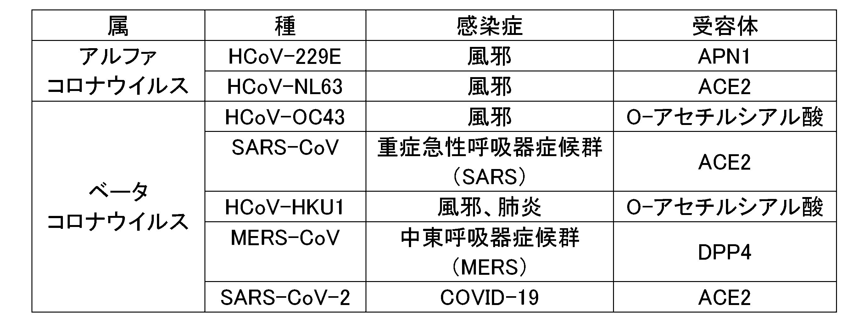
HCoC-229E、HCoC-OC43、HCoC-HKU1、及びMERS-CoVは、SARS-CoV-2とは異なる分子を受容体としている。
本開示の第1の態様は、コロナウイルス感染症の重症化リスクの評価方法である。本態様にかかる方法は、下記(a)及び(b)の工程を含む。
(a)対象由来のT細胞を、前記コロナウイルス感染症の原因ウイルスとは異なる種類のコロナウイルスである他種コロナウイルスに由来するタンパク質またはその断片に接触させる工程。
(b)前記タンパク質又はその断片に対する前記T細胞の応答性に基づいて、前記コロナウイルス感染症の重症化リスクを評価する工程。
工程(a)では、対象由来のT細胞を、コロナウイルス感染症の原因ウイルスとは異なる種類のコロナウイルスである他種コロナウイルスに由来するタンパク質又はその断片に接触させる。
「対象」とは、コロナウイルス感染症の重症化リスクの評価対象となる個体をいう。対象は、コロナウイルス感染症に罹患する動物であれば、特に限定されない。対象としては、ヒト、ヒト以外の哺乳動物が挙げられる。ヒト以外の哺乳動物としては、例えば、サル、チンパンジー、ゴリラ、マーモセット等の霊長類;マウス、モルモット、ハムスター、ラット等のげっ歯類;イヌ、ネコ等の食肉目等が挙げられるが、これらに限定されない。一実施形態において、対象は、ヒトである。
非感染者の重症化リスクを評価することにより、対象が原因ウイルスに感染した場合に、対象コロナウイルス感染症の症状が重症化するかを予測することができる。その結果、ワクチン接種等の適切な防御的介入を行うことができる。
対象が、対象コロナウイルス感染症発症者である場合、コロナウイルス感染症の発症初期であることが好ましい。発症初期に重症化リスクを評価することにより、対象において、対象コロナウイルス感染症の症状が重症化するかを予測することができる。その結果、適切な治療的介入を行うことができる。
対象が、対象コロナウイルス感染症の非発症者であって、原因ウイルス感染者である場合、重症化リスクを評価することにより、対象において、対象コロナウイルス感染症を発症し、その症状が重症化するかを予測することができる。その結果、適切な治療的介入を行うことができる。
既感染者の重症化リスクを評価することにより、対象が原因ウイルスに再度感染した場合に、対象コロナウイルス感染症を発症し、その症状が重症化するかを予測することができる。その結果、ワクチン接種等の適切な防御的介入を行うことができる。
未感染者の重症化リスクを評価することにより、対象が原因ウイルスに感染した場合に、対象コロナウイルス感染症を発症し、その症状が重症化するかを予測することができる。その結果、ワクチン接種等の適切な防御的介入を行うことができる。
ワクチン接種者の重症化リスクを評価することにより、ワクチン接種により、対象コロナウイルス感染症の重症化を回避できる程度の免疫が獲得できたかを評価することができる。その結果、再度のワクチン接種等の適切な防御的介入を行うことができる。
ワクチン未接種者の重症化リスクを評価することにより、対象が、ワクチン接種によらずに対象コロナウイルス感染症の重症化を回避できる程度の免疫を有しているかを評価することができる。その結果、ワクチン接種等の適切な防御的介入を行うことができる。
対象のT細胞は、対象の血液から、公知の方法により、単離することができる。例えば、密度勾配遠心法により、血液から、T細胞を含む末梢血単核球(Peripheral Blood Mononuclear Cells;PBMC)画分を分離することができる。PBMCは、T細胞(CD8陽性T細胞、CD4陽性T細胞等)のほか、樹状細胞、単球・マクロファージ、B細胞等の抗原提示細胞を含んでいる。工程(a)では、対象のT細胞を含む細胞集団として、PBMCを用いてもよい。
あるいは、T細胞マーカー(CD8、CD4等)に特異的に結合する抗体等を用いて、PBMCからT細胞を単離してもよい。一実施形態において、T細胞は、CD8陽性T細胞およびCD4陽性T細胞からなる群より選択される少なくとも1種を含む。一実施形態において、T細胞は、CD8陽性T細胞およびCD4陽性T細胞の両方を含む。
「他種コロナウイルス」は、原因ウイルスとは異なる種類のコロナウイルスである。他種コロナウイルス(以下、「抗原ウイルス」ともいう)は、コロナウイルス科に属するウイルスであって、原因ウイルスとは異なる種類のコロナウイルスであれば、特に限定されない。一実施形態において、抗原ウイルスは、原因ウイルスとは異なる種に属するコロナウイルスである。一実施形態において、抗原ウイルスは、原因ウイルスと同じ属に属し、且つ原因ウイルスと異なる種に属するコロナウイルスである。例えば、原因ウイルスがベータコロナウイルス属である場合、抗原ウイルスはベータコロナウイルス属の異なる種であってもよい。抗原ウイルスはアルファコロナウイルス属であってもよい。抗原ウイルスは、対象に感染可能なコロナウイルスであることが好ましい。
第1の抗原ウイルスとしては、HCoV-229E、HCoV-OC43、HCoV-HKU1、及びMERS-CoVからなる群より選択される1種以上を用いることが好ましく、2種以上を用いることがより好ましく、3種以上を用いることがさらに好ましい。第1の抗原ウイルスとしては、HCoV-229E、HCoV-OC43、及びHCoV-HKU1の全てを用いることが好ましい。
第2の抗原ウイルスとしては、HCoV-NL63、及びSARS-CoVからなる群より選択される1種以上を用いることが好ましい。第2の抗原ウイルスとしては、HCoV-NL63を用いることが好ましい。
対象由来のT細胞を、抗原ウイルスタンパク質またはその断片に接触させる方法は特に限定されない。例えば、ELISPOTアッセイで通常行われる方法を用いることができる。一実施形態において、T細胞と、抗原ウイルスタンパク質またはその断片との接触は、抗原提示細胞の存在下で行われる。抗原提示細胞としては、樹状細胞、B細胞、マクロファージ等が挙げられる。抗原提示細胞は、対象から採取されたものであることが好ましい。対象の血液から単離したPBMCは、T細胞及び抗原提示細胞を含む。そのため、抗原ウイルスタンパク質またはその断片との接触に、対象の血液から取得したPBMCを用いてもよい。
抗原ウイルスタンパク質またはその断片に対して応答性を有するT細胞は、抗原ウイルスタンパク質またはその断片との接触により、サイトカインを分泌する。捕捉用抗体は、T細胞から分泌されたサイトカインに結合し、ウェルプレート上に捕捉し得る。
捕捉用抗体を固定するために、ELISPOT用プレートのウェル底面はポリフッ化ビニリデン(PVDF)膜で形成されていてもよい。
工程(b)では、抗原ウイルスタンパク質またはその断片に対する対象由来のT細胞の応答性に基づいて、対象コロナウイルス感染症の重症化リスクを評価する。
例えば、抗原ウイルスタンパク質またはその断片に対する対象由来のT細胞の応答性が低い場合、前記対象は、対象コロナウイルス感染症に対する重症化リスクが高いと評価することができる。
例えば、抗原ウイルスタンパク質またはその断片に対する対象由来のT細胞の応答性が高い場合、前記対象は、対象コロナウイルス感染症に対する重症化リスクが低いと評価することができる。
(a1)対象由来のT細胞を、原因ウイルスが受容体とする分子とは異なる分子を受容体とする第1の他種コロナウイルス(第1の抗原ウイルス)に由来するタンパク質またはその断片に接触させる工程。
(a2)対象由来のT細胞を、原因ウイルスが受容体とする分子と同じ分子を受容体とする第2の他種コロナウイルス(第2の抗原ウイルス)に由来するタンパク質またはその断片に接触させる工程。
(b1)第1の他種コロナウイルス(第1の抗原ウイルス)に由来するタンパク質またはその断片に対する対象由来のT細胞の第1の応答性を測定する工程。
(b2)第2の他種コロナウイルスに由来するタンパク質またはその断片に対する対象由来のT細胞の第2の応答性を測定する工程。
(b3)第1の応答性に対する第2の応答性の比の値(第2の応答性/第1の応答性)に基づいて、コロナウイルス感染症の重症化リスクを評価する工程。
工程(a2)では、第2の抗原ウイルスに由来するタンパク質またはその断片を用いて、工程(a)を行えばよい。
工程(b2)では、工程(a2)により誘導された対象由来のT細胞の応答性を測定すればよい。工程(b2)により得られた応答性を第2の応答性とする。
T細胞の応答性は、上述の方法により測定することができる。T細胞の応答性は、ELISPOTアッセイにより測定することが好ましい。T細胞の応答性は、例えば、ELISPOTアッセイによる有効スポット数として定量化してもよいし、有効スポット数インデックスとして定量化してもよい。有効スポット数は、T細胞刺激抗原の存在下でT細胞をインキュベートした区画(ウェル)で得られたスポット数から、陰性対照区画(ウェル)のスポット数を差し引いた値として算出される。陰性対照区画は、T細胞刺激抗原の非存在下でT細胞をインキュベートした区画である。有効スポット数インデックスは、有効スポット数を陽性対照区画(ウェル)のスポット数で除した値として算出される。陽性対照区画は、T細胞刺激抗原としてすべてのT細胞が強く反応する抗CD3抗体を用いてT細胞をインキュベートした区画である。
式(1)中、Yは重症化リスク値;kは係数;Aは第1の応答性;Bは第2の応答性である。係数kは、例えば、1~10の範囲の数値を取り得る。
例えば、重症化リスク値(Y)は、下記式(2)により求めてもよい。
式(2)中、Yは重症化リスク値;k1およびk2は係数;A1は1種類目の第1の抗原ウイルスによる第1の応答性;A2は2種類目の第1の抗原ウイルスによる第1の応答性;Bは第2の応答性である。係数k1およびk2は、例えば、1~10の範囲の数値を取り得る。3種類目以降の第1の抗原ウイルスについても、同様に加算していけばよい。

本態様にかかる方法では、T細胞刺激抗原として、対象コロナウイルス感染症の原因ウイルスとは異なる種類のコロナウイルス(抗原ウイルス)に由来するタンパク質またはその断片を用いる。そのため、原因ウイルスの未感染者、および原因ウイルスの既感染者またはワクチン接種者であるが原因ウイルスに対する細胞性免疫が消失した対象であっても、対象コロナウイルス感染症の重症化リスクを評価することができる。
本態様にかかる方法によれば、未知のコロナウイルス感染症に対しても、重症化リスクを評価することができる。
本態様にかかる方法は、上記工程に加えて、任意の工程を含んでもよい。任意工程としては、例えば、(c)対象が原因ウイルス感染者である場合に、対象コロナウイルス感染症の重症化リスクの評価結果に基づいて、対象コロナウイルス感染症の治療方針を決定する工程、(d)対象コロナウイルス感染症の重症化リスクが高いと評価された対象が原因ウイルスの感染者である場合に、対象コロナウイルス感染症の症状の観察、および対象コロナウイルス感染症の治療からなる群より選択される少なくとも1種の措置を行う工程、ならびに(e)対象コロナウイルス感染症の重症化リスクが高いと評価された対象が原因ウイルスの非感染者である場合に、対象コロナウイルス感染症のワクチン接種を行う工程、等が挙げられる。工程(c)~(e)のいずれかを含む場合、本態様にかかる方法は、コロナウイルス感染症の重症化を予防する方法であってもよい。工程(c)及び工程(d)のいずれかを含む場合、本態様にかかる方法は、コロナウイルス感染症を治療する方法であってもよい。
対象が原因ウイルス感染者である場合、重症化リスクに基づいて、対象コロナウイルス感染症の治療方針を決定することができる。重症化リスクが低いと評価された感染者は、積極的な治療を行わず、経過観察のみを行うと決定してもよい。重症化リスクが高い感染者は、症状の進行を慎重に観察し、感染初期から積極的な治療を施すことで、重症化を回避できる可能性がある。そこで、重症化リスクが高いと評価された感染者に対しては、感染初期から積極的な治療を行うと決定してもよい。対象コロナウイルス感染症の治療方法は、対象コロナウイルスの種類に応じて選択可能である。治療方法としては、抗ウイルス薬または中和抗体等の投与が挙げられる。対象コロナウイルス感染症がCOVID-19である場合、抗ウイルス薬としては、モルヌピラビルル(商品名:ラゲブリオカプセル)、ニルマトレルビル/リトナビル(商品名:パキロビッドパック)、レムデシビル(商品名:ベルクリー点滴静注液)、デキサメサゾン、トシリズマブ(商品名:アクテムラ)、ファビピラビル(商品名:アビガン)等が挙げられる。中和抗体としては、カシリビマブとイムデビマブの抗体カクテル療法(商品名:ロナプリーブ注射液セット)が挙げられる。
対象コロナウイルス感染症の重症化リスクが高いと評価された対象が原因ウイルスの感染者である場合、感染初期から症状の慎重な経過観察を行い、積極的な治療を施すことで、重症化を回避できる可能性がある。そこで、重症化リスクが高い感染者に対しては、症状の観察、および/またはコロナウイルス感染症の治療を行ってもよい。治療方法としては、上記と同様のものが挙げられる。また、経過観察および重症化したときの迅速な対応に備えて、病院への入院措置を行ってもよい。工程(d)は、重症化リスクが高いと判定された原因ウイルス感染者に対し、コロナ感染症に対する治療を行う工程であってもよい。例えば、工程(d)は、重症化リスクが高いと判定された原因ウイルス感染者に対し、原因ウイルスに対する抗ウイルス薬又は中和抗体を投与する工程であってもよい。
対象コロナウイルス感染症の重症化リスクが高いと評価された対象が原因ウイルスの非感染者である場合、対象が原因ウイルスに感染すると重症化する恐れがある。そのため、そのような対象に対しては、対象コロナウイルス感染症のワクチン接種を行うことが考えられる。ワクチン接種を行って対象コロナウイルス感染症に対する免疫を獲得させることで、当該対象において、対象コロナウイルスに対する重症化リスクを低下させ得る。工程(e)は、重症化リスクが高いと判定された非感染者に対し、対象コロナウイルス感染症のワクチン接種を行う工程であってもよい。
本開示の第2の態様は、コロナウイルス感染症の重症化リスク評価キットである。本態様にかかるキットは、前記コロナウイルス感染症の原因ウイルスとは異なる種類のコロナウイルスに由来するタンパク質(抗原ウイルスタンパク質)又はその断片を含む。
抗原ウイルスタンパク質またはその断片としては、上記と同様のものが挙げられる。抗原ウイルスは、原因ウイルスと同じ属に属し、且つ原因ウイルスと異なる種に属するコロナウイルスであることが好ましい。例えば、原因ウイルスがベータコロナウイルス属である場合、抗原ウイルスはベータコロナウイルス属の異なる種であってもよい。抗原ウイルスは、アルファコロナウイルス属であってもよい。抗原ウイルスは、対象に感染可能なコロナウイルスであることが好ましい。
原因ウイルスが、SARS-CoV-2である場合、抗原ウイルスは、ベータコロナウイルス属のウイルスを用いることができる。原因ウイルスがSARS-CoV-2であり、対象がヒトである場合、抗原ウイルスとして、ベータコロナウイルス属のヒトコロナウイルス(HCoV-OC43、HCoV-HKU1等)を用いることができる。
本態様にかかるキットは、HCoV-HKU1に由来する抗原ウイルスタンパク質またはその断片と、HCoV-OC43に由来する抗原ウイルスタンパク質またはその断片と、HCoV-229Eに由来する抗原ウイルスタンパク質またはその断片と、HCoV-NL63に由来する抗原ウイルスタンパク質またはその断片とを含むことが好ましい。
本態様にかかるキットは、抗原ウイルスタンパク質またはその断片に加えて、任意の構成を含んでいてもよい。任意の構成としては、例えば、T細胞が抗原刺激に応答して分泌するサイトカインに対する特異的結合物質、固相担体、発色酵素、発色酵素基質、洗浄バッファー、希釈バッファー、使用説明書等が挙げられる。
本態様にかかるキットは、T細胞が抗原刺激に応答して分泌するサイトカインに対する特異的結合物質を含んでもよい。
「特異的結合物質」とは、特定の生体分子に対して、特異的結合性を有する物質を意味する。「特異的結合性を有する」とは、特定の生体分子に対して高い結合親和性を有するが、他の生体分子に対しては、極めて低い結合親和性しか有さないことを意味する。特異的結合物質は、好ましくは、特定の生体分子に高い結合性を有するが、他の生体分子にはほとんど結合性を有しない。タンパク質に対する特異的結合物質としては、抗体、およびアプタマー等が挙げられる。一実施形態において、特異的結合物質は、抗体である。
発色酵素は、特に限定されず、公知のものを用いることができる。発色酵素としては、HRP、およびAP等が挙げられる。発色酵素は、検出用抗体に結合されていてもよい。検出用抗体が、結合対の第1要素(例えば、ビオチン)で標識されている場合、発色酵素は結合対の第2要素(例えば、アビジン又はその誘導体)で標識されていてもよい。例えば、検出用抗体がFITC標識抗ヒトIFN-γ抗体である場合、発色酵素はFITC標識HRP(FITC-HRP)であってもよい。例えば、検出用抗体がビオチン標識抗ヒトIL-2抗体である場合、発色酵素はストレプトアビジン標識AP(Strep-Ap)であってもよい。
発色酵素基質は、発色酵素の種類に応じて、適宜選択することができる。
発色酵素がHRPである場合、発色酵素基質としては、3,3’-ジアミノベンジジン(DAB)、3,3’,5,5’-テトラメチルベンジジン(TMB)、2,2’-アジノビス[3-エチルベンゾチアゾリン-6-スルホン酸](ABTS)、o-フェニレンジアミン二塩酸塩(OPD)等が挙げられる。
発色酵素がAPである場合、発色酵素基質としては、ニトロブルーテトラゾリウムクロライド(NBT)、5-ブロモ-4-クロロ-3-インドリルホスフェート(BCIP)、p-ニトロフェニルリン酸(PNPP)等が挙げられる。
本態様にかかるキットは、前記第1の態様にかかる評価方法を実施するために使用することができる。
他の態様において、本開示は、コロナウイルス感染症の重症化リスクの評価キットを製造するための、抗原ウイルスタンパク質またはその断片の使用を提供する。他の態様において、本開示は、コロナウイルス感染症の重症化リスクの評価するための、抗原ウイルスタンパク質またはその断片の使用を提供する。他の態様において、本開示は、コロナウイルス感染症の重症化リスクの評価するための、抗原ウイルスタンパク質またはその断片を提供する。抗原ウイルスタンパク質としては、上記と同様のものが挙げられ、第1の抗原ウイルスに由来するタンパク質、及び第2の抗原ウイルスタンパク質の組合せが好ましい。
コロナウイルス感染症の重症化リスクの判定方法であって、(a)対象由来のT細胞を、前記コロナウイルス感染症の原因ウイルスとは異なる種類のコロナウイルスである他種コロナウイルスに由来するタンパク質またはその断片に接触させる工程と、(b)前記タンパク質またはその断片に対する前記T細胞の応答性に基づいて、前記コロナウイルス感染症の重症化リスクを判定する工程と、を含む、コロナウイルス感染症の重症化リスクの判定方法。
<被験者>
被験者は、東北大学病院ならびに関連施設(栗原中央病院、気仙沼市立病院)でCOVID-19の治療を受けたCOVID-19感染回復期にある患者とした。被験者には、事前に説明して同意を得た。治癒後の回復期に、末梢血10mL程度を専用の容器に採取した。COVID-19に罹患歴のない健常者を対照群として、同様に採血を行った。
中等症I(呼吸不全なし):93%<SpO2<96%で、呼吸困難,肺炎所見を認める。
中等症II(呼吸不全あり):SpO2≦93%で、酸素投与が必要。
重症:ICUに入室、または人工呼吸器が必要。
COVID-19感染後に6週以上経過して治癒期にある患者、および対照群である非感染者から、末梢血を採取した。BDバキュティナ(登録商標)CPT単核球分離用採血管(Becton,Dickinson and Company)を用いて、1500~1800G、室温で、15分間遠心分離して単核球層を分離および採取した。CTL-wash suppliment(Cellular Technologies,Cleveland,OH,USA)添加したRPMI-1640で、細胞を洗浄した。次いで、300G、室温で、15分間遠心分離した。この洗浄操作を2回繰り返した。その後、細胞保存液(CELLBANKER2 (日本全薬工業))を加えてPBMCを-80℃で保存した。
Tリンパ球の反応性は、分泌されるIFN-γとIL-2を対象として評価した。ELISPOTアッセイには、Human IFN-γ/IL-2 Double-Color ELISPOT Kit:hIFNgIL2-2M,CTL(Cellular Technologies,Cleveland,OH,USA)を用いた。
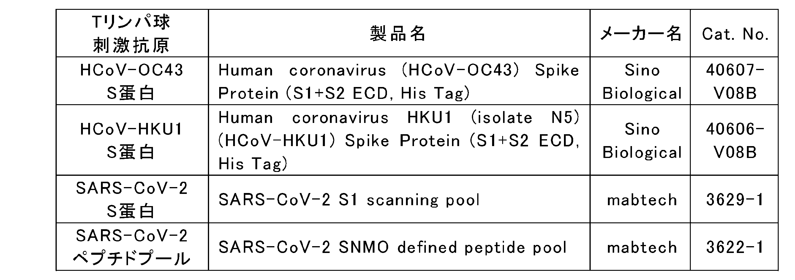
3+:50以上
2+:21以上50未満
+:6以上21未満
±:4以上6未満
-:4未満
図1に、ELISPOTアッセイの結果を示す。上図はELISPOTアッセイにおけるウェル画像である。下図は上記評価基準による評価結果である。健常者では、HCoV-OC43、HCoV-HKU1、およびSARS-CoV-2のいずれのS蛋白に対しても応答を認めなかった。
回復期のCOVID患者においては、軽症例では、HCoV-OC43およびHCoV-HKU1に対して中等度~強応答であった。中等症Iでは、弱応答であった。中等症IIでは、ほぼ応答性を認めなかった。
これらの結果は、ベータコロナウイルス属(HCoV-OC43、HCoV-HKU1)にあらかじめ細胞性免疫能を有すると、交差反応性により、COVID-19に対して保護的に作用して軽症で済むことを示す。一方、ベータコロナウイルス属に細胞性免疫能を有さないと、COVID-19の重症化を招くことを示す。
上記の結果により、SARS-CoV-2と季節性ベータコロナウイルスとの類似点に起因する細胞性免疫における交差反応性が、COVID-19の重症度を低下させることが示された。
<被験者>
被験者は、健常者が37例、COVID-19既感染者が27例(軽症が8例、中等症Iが6例、中等症IIが9例、重症が4)であった。COVID-19既感染者の被験者は、実施例1と同様に、東北大学病院ならびに関連施設(栗原中央病院、気仙沼市立病院)でCOVID-19の治療を受けたCOVID-19感染回復期にある患者とした。
実施例1と同様の方法で、PBMCを調製し、-80℃で保存した。
表2に示すTリンパ球刺激抗原に加えて、表3に示すTリンパ球刺激抗原を用いて、ELISPOTアッセイを行った。
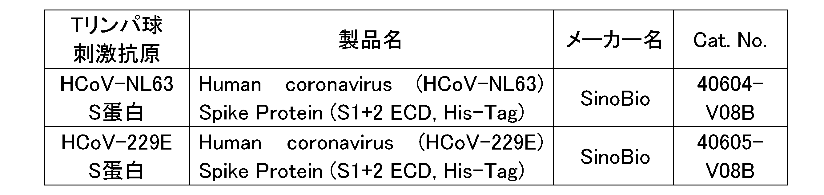
Tリンパ球の反応性は、分泌されるIFN-γとIL-2、TNF-α、GranzymeBを対象として評価した。ELISPOTアッセイには、Human IFN-γ/IL-2/TNF-α/GranzymeB Four-Color FluoroSPOT Kit:hT4000F-10,CTL(Cellular Technologies,Cleveland,OH,USA)を用いた。ELISPOTアッセイは、文献(Czerkinsky C, et al. Immunol. Methods,1988.)に記載の方法を一部変更して行った。具体的には、ウェル底面がPVDFメンブレンとなっている96ウェルのELISPOT用プレートに、ヒトIFN-γ/IL-2/TNF-α/GranzymeBキャプチャー抗体をコートして、4℃で一晩インキュベートした。PBSでプレートを洗浄した後、Tリンパ球刺激抗原および共刺激の抗CD28抗体を添加して、37℃で、20分間インキュベートした。Tリンパ球刺激抗原として、表2に示したTリンパ球刺激抗原に加えて、表3に示したアルファコロナウイルス属(HCoV-NL63、HCoV-229E)のスパイク(S)蛋白を用いた。ポジティブコントロールのウェルには、Tリンパ球刺激抗原に代えて抗CD3抗体を添加した。ネガティブコントロールのウェルには、Tリンパ球刺激抗原を添加しなかった。
上記インキュベートにおけるTリンパ球刺激抗原濃度は、SARS-CoV-2のS蛋白およびペプチドプールでは、2μg/mL(最終濃度)とした。ヒト風邪コロナウイルス(HCoV-OC43、HCoV-HKU1、HCoV-NL63、HCoV-229E)のS蛋白では、12.5μg/mL(最終濃度)とした。
上記インキュベート後、健常者、COVID-19既感染者から採取したPBMCを、300,000個/ウェルで添加した。次いで、37℃で、24時間インキュベートした。PBSで洗浄後、0.05% Tween/PBSで洗浄した。次いで、IFN-γ/IL-2/TNF-α/GranzymeB検出用抗体(anti-human IFN-γ(FITC) Detection Ab、Anti-human IL-2(Hapten2) Detection Ab、Anti-human TNF-α(Hapten1) Detection AbおよびAnti-human GranzymeB(Biotin) Detection Ab)を添加し、室温で、2時間インキュベートした。0.05% Tween/PBSで洗浄後、Anti-FITC Alexa Fluor488、Anti-Hapten2 CTL-Red、Anti-Hapten1 CTL-YellowおよびStreptavidin eFluor450の混合溶液をウェルに添加し、室温で、1時間インキュベートした。蒸留水で洗浄後、クリーンベンチ内で24時間自然乾燥させた。
反応したTリンパ球数の評価は、Spot-Forming Cells(SFC)として、自動免疫スポット分析器Automated Immunospot Analyzer,CTL(Cellular Technologies,Cleveland,OH,USA)で自動解析した。
Tリンパ球刺激抗原として、HCoV-HKU1のS蛋白(HKU1-S)(図3A)、HCoV-OC43のS蛋白(OC43-S)(図3B)、HCoV-NL63のS蛋白(NL63-S)(図4A)、HCoV-229EのS蛋白(229E-S)(図4B)、SARS-CoV-2のS蛋白(CoV2-S)(図5A)及び、SARS-CoV-2のペプチドプール(CoV2-all)(図5B)をそれぞれ用いて、ELISPOTアッセイを行った結果を図3A~図5Bにそれぞれ示す。
アルファコロナウイルス属であって、ACE2を受容体とするコロナウイルス(NL63)のS蛋白に対しては、軽症例と比較して、中等症以上の症例において反応が強い傾向があった(図4A)。
アルファコロナウイルス属であって、ACE2を受容体としないコロナウイルス(229E)のS蛋白に対しては、中等症以上の症例と比較して、軽症例において反応が強い傾向があった(図4A)。
SARS-CoV-2の蛋白に対しては、重症度が高くなるほど、反応が強い傾向があった(図5A、図5B)。
上記の結果から、以下の傾向が確認された。
(a)ACE2を受容体とするコロナウイルス(NL63)に対しては、軽症例よりも、中等症以上の症例において反応が強い。
(b)ACE2を受容体としないコロナウイルス(HKU1、OC43、229E)に対しては、中等症以上の症例よりも、軽症例において反応が強い。

前記式(I)中、k1、k2、及びk3は、係数である。
Y値のカットオフ値(X)を20に設定した場合、以下の結果となった。
X>20:100%の確率で中等症以上。
X≦20:8/15(53%)の確率で軽症。
<被験者>
被験者は、ファイザー製ワクチン(BNT162b2)を接種したワクチン接種者とした。被験者は、28名であった。ワクチン接種前とワクチン接種後(おおむね6週から9週後)に採血を行った。
PBMCの調製、及びELISPOTアッセイは、実施例2と同様の方法で行った。
Tリンパ球刺激抗原として、HKU1-S(図10A)、OC43-S(図10B)、NL63-S(図11A)、229E-S(図11B)、CoV2-S(図12A)及び、CoV2-all(図12B)をそれぞれ用いて、ELISPOTアッセイを行った結果を図10A~図12Bにそれぞれ示す。図中、「Pre」はワクチン接種前を示し、「Post」はワクチン接種後を示す。
ACE2を受容体とするコロナウイルス(NL63)のS蛋白に対する反応は、ワクチン接種前と比較して、ワクチン接種後で有意に上昇した(図11A)。
SARS-CoV-2の蛋白に対する反応は、ワクチン接種前と比較して、ワクチン接種後で有意に上昇した(図12A、図12B)。
ワクチン接種前とワクチン接種後とで、前記式(I)により、重症化リスク値(Y)を算出した。その結果を図13A及び図13Bに示す。
公知のCOVID-19重症化リスク因子と重症化リスク値(Y)との相関を解析した結果を図14A~図15Bに示す。重症化リスク値(Y)は、年齢(図14A)、BMI(図14B)、喫煙指数BI(図15A)、及び喫煙歴(図15B)のいずれとも有意な相関がなかった。
Claims (13)
- コロナウイルス感染症の重症化リスクの評価方法であって、
(a)対象由来のT細胞を、前記コロナウイルス感染症の原因ウイルスとは異なる種類のコロナウイルスである他種コロナウイルスに由来するタンパク質またはその断片に接触させる工程と、
(b)前記タンパク質またはその断片に対する前記T細胞の応答性に基づいて、前記コロナウイルス感染症の重症化リスクを評価する工程と、
を含む、コロナウイルス感染症の重症化リスクの評価方法。 - 前記タンパク質またはその断片が、前記原因ウイルスが受容体とする分子とは異なる分子を受容体とする第1の他種コロナウイルスに由来し、
前記(b)の工程が、前記タンパク質またはその断片に対する前記T細胞の応答性が低い場合に、前記コロナウイルス感染症の重症化リスクが高いと評価する工程である、請求項1に記載のコロナウイルス感染症の重症化リスクの評価方法。 - 前記タンパク質またはその断片が、前記原因ウイルスが受容体とする分子と同じ分子を受容体とする第2の他種コロナウイルスに由来し、
前記(b)の工程が、前記タンパク質またはその断片に対する前記T細胞の応答性が高い場合に、前記コロナウイルス感染症の重症化リスクが高いと評価する工程である、請求項1に記載のコロナウイルス感染症の重症化リスクの評価方法。 - 前記工程(a)が、
(a1)前記T細胞を、前記原因ウイルスが受容体とする分子とは異なる分子を受容体とする第1の他種コロナウイルスに由来するタンパク質またはその断片に接触させる工程と、
(a2)前記T細胞を、前記原因ウイルスが受容体とする分子と同じ分子を受容体とする第2の他種コロナウイルスに由来するタンパク質またはその断片に接触させる工程と、を含み、
前記(b)の工程が、
(b1)前記第1の他種コロナウイルスに由来するタンパク質またはその断片に対する前記T細胞の第1の応答性を測定する工程と、
(b2)前記第2の他種コロナウイルスに由来するタンパク質またはその断片に対する前記T細胞の第2の応答性を測定する工程と、
(b3)前記第1の応答性に対する前記第2の応答性の比の値(第2の応答性/第1の応答性)に基づいて、前記コロナウイルス感染症の重症化リスクを評価する工程と、
を含む、請求項1に記載のコロナウイルス感染症の重症化リスクの評価方法。 - 前記原因ウイルスおよび前記タンパク質またはその断片が由来する前記コロナウイルスが、ベータコロナウイルス属またはアルファコロナウイルス属である、請求項1~4のいずれか一項に記載のコロナウイルス感染症の重症化リスクの評価方法。
- 前記原因ウイルスがSARS-CoV-2である、請求項1~5のいずれか一項に記載のコロナウイルス感染症の重症化リスクの評価方法。
- コロナウイルス感染症の重症化リスク評価キットであって、
前記コロナウイルス感染症の原因ウイルスとは異なる種類のコロナウイルスである他種コロナウイルスに由来するタンパク質またはその断片を含む、
コロナウイルス感染症の重症化リスク評価キット。 - T細胞が抗原刺激に応答して分泌するサイトカインに対する特異的結合物質をさらに含む、請求項7に記載のコロナウイルス感染症の重症化リスク評価キット。
- 固相担体に結合された前記特異的結合物質と、標識物質で標識された前記特異的結合物質とを含む、請求項8に記載のコロナウイルス感染症の重症化リスク評価キット。
- 前記サイトカインがインターフェロンγ、インターロイキン2、及び腫瘍壊死因子アルファからなる群より選択される少なくとも1種である、請求項8または9に記載のコロナウイルス感染症の重症化リスク評価キット。
- 前記タンパク質またはその断片が、
前記原因ウイルスが受容体とする分子とは異なる分子を受容体とする第1の他種コロナウイルスに由来するタンパク質またはその断片と、
前記原因ウイルスが受容体とする分子と同じ分子を受容体とする第2の他種コロナウイルスに由来するタンパク質またはその断片と、
を含む、請求項7~10のいずれか一項に記載のコロナウイルス感染症の重症化リスク評価キット。 - 前記原因ウイルスおよび前記タンパク質またはその断片が由来する前記コロナウイルスが、ベータコロナウイルス属またはアルファコロナウイルス属である、請求項7~11のいずれか一項に記載のコロナウイルス感染症の重症化リスク評価キット。
- 前記原因ウイルスがSARS-CoV-2である、請求項7~12のいずれか一項に記載のコロナウイルス感染症の重症化リスク評価キット。
Priority Applications (3)
| Application Number | Priority Date | Filing Date | Title |
|---|---|---|---|
| CN202280052365.1A CN118119845A (zh) | 2021-07-29 | 2022-03-08 | 冠状病毒传染病的重症化风险的评估方法以及冠状病毒传染病的重症化风险评估试剂盒 |
| US18/291,704 US20250093335A1 (en) | 2021-07-29 | 2022-03-08 | Method for evaluating severity risk of coronavirus infectious disease, and kit for evaluating severity risk of coronavirus infection disease |
| JP2023538242A JPWO2023007809A1 (ja) | 2021-07-29 | 2022-03-08 |
Applications Claiming Priority (2)
| Application Number | Priority Date | Filing Date | Title |
|---|---|---|---|
| JP2021124390 | 2021-07-29 | ||
| JP2021-124390 | 2021-07-29 |
Publications (1)
| Publication Number | Publication Date |
|---|---|
| WO2023007809A1 true WO2023007809A1 (ja) | 2023-02-02 |
Family
ID=85087768
Family Applications (1)
| Application Number | Title | Priority Date | Filing Date |
|---|---|---|---|
| PCT/JP2022/009911 Ceased WO2023007809A1 (ja) | 2021-07-29 | 2022-03-08 | コロナウイルス感染症の重症化リスクの評価方法、およびコロナウイルス感染症の重症化リスク評価キット |
Country Status (4)
| Country | Link |
|---|---|
| US (1) | US20250093335A1 (ja) |
| JP (1) | JPWO2023007809A1 (ja) |
| CN (1) | CN118119845A (ja) |
| WO (1) | WO2023007809A1 (ja) |
Citations (6)
| Publication number | Priority date | Publication date | Assignee | Title |
|---|---|---|---|---|
| CN111999503A (zh) * | 2020-05-28 | 2020-11-27 | 首都医科大学附属北京地坛医院 | 一组用于预测急性病毒性呼吸道传染病重症化的标志物及其应用和试剂盒 |
| CN112034187A (zh) * | 2020-06-04 | 2020-12-04 | 北京臻知医学科技有限责任公司 | 一组预测2019冠状病毒病细胞因子和血栓风暴的标志物及应用和试剂盒 |
| JP6933834B1 (ja) * | 2020-06-18 | 2021-09-08 | 国立研究開発法人国立国際医療研究センター | 新型コロナウイルス感染者の重症化リスクの検査方法、その検査キット、コンパニオン診断薬及びその重症化リスクマーカー |
| WO2021251504A1 (ja) * | 2020-06-12 | 2021-12-16 | 国際スペースメディカル株式会社 | 血中rnaを利用したcovid-19重症化予測方法 |
| WO2022071601A1 (ja) * | 2020-10-02 | 2022-04-07 | 国際スペースメディカル株式会社 | 血中エクソソームのタンパク質マーカーを利用したcovid-19重症化予測方法 |
| JP2022068648A (ja) * | 2020-10-22 | 2022-05-10 | シスメックス株式会社 | 呼吸器感染症に関する情報の取得方法、バイオマーカーの測定値のモニタリング方法、試薬キット、呼吸器感染症に関する情報の取得装置及びコンピュータプログラム |
-
2022
- 2022-03-08 WO PCT/JP2022/009911 patent/WO2023007809A1/ja not_active Ceased
- 2022-03-08 CN CN202280052365.1A patent/CN118119845A/zh active Pending
- 2022-03-08 US US18/291,704 patent/US20250093335A1/en active Pending
- 2022-03-08 JP JP2023538242A patent/JPWO2023007809A1/ja active Pending
Patent Citations (6)
| Publication number | Priority date | Publication date | Assignee | Title |
|---|---|---|---|---|
| CN111999503A (zh) * | 2020-05-28 | 2020-11-27 | 首都医科大学附属北京地坛医院 | 一组用于预测急性病毒性呼吸道传染病重症化的标志物及其应用和试剂盒 |
| CN112034187A (zh) * | 2020-06-04 | 2020-12-04 | 北京臻知医学科技有限责任公司 | 一组预测2019冠状病毒病细胞因子和血栓风暴的标志物及应用和试剂盒 |
| WO2021251504A1 (ja) * | 2020-06-12 | 2021-12-16 | 国際スペースメディカル株式会社 | 血中rnaを利用したcovid-19重症化予測方法 |
| JP6933834B1 (ja) * | 2020-06-18 | 2021-09-08 | 国立研究開発法人国立国際医療研究センター | 新型コロナウイルス感染者の重症化リスクの検査方法、その検査キット、コンパニオン診断薬及びその重症化リスクマーカー |
| WO2022071601A1 (ja) * | 2020-10-02 | 2022-04-07 | 国際スペースメディカル株式会社 | 血中エクソソームのタンパク質マーカーを利用したcovid-19重症化予測方法 |
| JP2022068648A (ja) * | 2020-10-22 | 2022-05-10 | シスメックス株式会社 | 呼吸器感染症に関する情報の取得方法、バイオマーカーの測定値のモニタリング方法、試薬キット、呼吸器感染症に関する情報の取得装置及びコンピュータプログラム |
Non-Patent Citations (10)
| Title |
|---|
| ANONYMOUS: "Clinical Trials Conducted to Search for Aggravation Prediction Markers of COVID-19 Infection", CHIBA UNIVERSITY, PRESS RELEASE, 28 August 2020 (2020-08-28), XP093028888, Retrieved from the Internet <URL:https://www.chiba-u.ac.jp/general/publicity/press/files/2020/20200828covid19_rinsyou.pdf> [retrieved on 20230303] * |
| ANONYMOUS: "Kobe University and Sysmex Conduct Clinical Evaluation of ELISPOT Method as a New Blood Test for COVID-19: Report on T-Cell Immunity in Japanese People for SARS-CoV-2", SYSMEX, 24 December 2020 (2020-12-24), XP093028889, Retrieved from the Internet <URL:https://www.sysmex.co.jp/news/2020/pdf/201224.pdf> [retrieved on 20230303] * |
| BONIFACIUS AGNES, TISCHER-ZIMMERMANN SABINE, SANTAMORENA MARIA MICHELA, MAUSBERG PHILIP, SCHENK JOSEPHINE, KOCH STEPHANIE, BARNSTO: "Rapid Manufacturing of Highly Cytotoxic Clinical-Grade SARS-CoV-2-specific T Cell Products Covering SARS-CoV-2 and Its Variants for Adoptive T Cell Therapy", FRONTIERS IN BIOENGINEERING AND BIOTECHNOLOGY, vol. 10, 4 April 2022 (2022-04-04), pages 867042, XP093028879, DOI: 10.3389/fbioe.2022.867042 * |
| GRIFONI ALBA; WEISKOPF DANIELA; RAMIREZ SYDNEY I.; MATEUS JOSE; DAN JENNIFER M.; MODERBACHER CAROLYN RYDYZNSKI; RAWLINGS STEPHEN A: "Targets of T Cell Responses to SARS-CoV-2 Coronavirus in Humans with COVID-19 Disease and Unexposed Individuals", CELL, ELSEVIER, AMSTERDAM NL, vol. 181, no. 7, 20 May 2020 (2020-05-20), Amsterdam NL , pages 1489, XP086202865, ISSN: 0092-8674, DOI: 10.1016/j.cell.2020.05.015 * |
| NAKAYAMA TOSHINORI, HIRAHARA KIYOSHI, KIMURA MOTOKO Y, IWAMURA CHIAKI, KIUCHI MASAHIRO, KOKUBO KOTA, ONODERA ATSUSHI, HASHIMOTO KA: "CD4+ T cells in inflammatory diseases: pathogenic T-helper cells and the CD69–Myl9 system", DEPARTMENT OF MEDICAL IMMUNOLOGY, GRADUATE SCHOOL OF MEDICINE, CHIBA UNIVERSITY , 1-8-1 INOHANA CHUO-KU, CHIBA 260-8670 , JAPAN, vol. 33, no. 12, 25 November 2021 (2021-11-25), pages 699 - 704, XP093028881, DOI: 10.1093/intimm/dxab053 * |
| NILLES ERIC J., SIDDIQUI SAMEED M., FISCHINGER STEPHANIE, BARTSCH YANNIC C., DE ST. AUBIN MICHAEL, ZHOU GUOHAI, GLUCK MATTHEW J., : "Epidemiological and Immunological Features of Obesity and SARS-CoV-2", VIRUSES, vol. 13, no. 11, pages 2235, XP093028882, DOI: 10.3390/v13112235 * |
| TAMADA TSUTOMU: "Development and practical application of anti-coronavirus immunocompetence measurement technology", JST PROJECT DATABASE, 12 July 2021 (2021-07-12), XP093028887, Retrieved from the Internet <URL:https://projectdb.jst.go.jp/grant/JST-PROJECT-21447371/> [retrieved on 20230303] * |
| VASILEIOU SPYRIDOULA; KUVALEKAR MANIK; WORKINEH ASTER; WATANABE AYUMI; VELAZQUEZ YOVANA; LULLA SUHASINI; HESLOP HELEN E.; MOONEY K: "Allogeneic, Off-the-Shelf, Sars-Cov-2-Specific T Cells to Treat High-Risk Patients with COVID-19", TRANSPLANTATION AND CELLULAR THERAPY, ELSEVIER, AMSTERDAM, NL, vol. 27, no. 3, 31 January 2021 (2021-01-31), AMSTERDAM, NL , XP086478486, ISSN: 2666-6367, DOI: 10.1016/S2666-6367(21)00448-6 * |
| VIDLER JENNIFER, ALAGUTHURAI THANUSSUYAH, ABDUL-JAWAD SULTAN, VIRAMUTHU SIVALEKHA, BEATSON RICHARD, GRAHAM ROSALIND, IRSHAD SHEEBA: "Immunogenicity of Covid-19 Vaccination in Subjects with Myelodysplastic Syndromes", BLOOD, vol. 138, no. 1, 5 November 2021 (2021-11-05), pages 3696 - 3697, XP093028884, DOI: 10.1182/blood-2021-152401 * |
| YOSHIMURA AKIHIKO: "Why young people do not get severe symptoms", COVID -19 COMMENTARY FOR CARDIOLOGISTS, 4 February 2021 (2021-02-04), XP093028890, Retrieved from the Internet <URL:http://www.epi-c.jp/sp2020/article_05.html> [retrieved on 20230303] * |
Also Published As
| Publication number | Publication date |
|---|---|
| JPWO2023007809A1 (ja) | 2023-02-02 |
| US20250093335A1 (en) | 2025-03-20 |
| CN118119845A (zh) | 2024-05-31 |
Similar Documents
| Publication | Publication Date | Title |
|---|---|---|
| Shin et al. | Immune responses to Middle East respiratory syndrome coronavirus during the acute and convalescent phases of human infection | |
| Cevhertas et al. | Advances and recent developments in asthma in 2020 | |
| Oyaert et al. | Evaluation of humoral and cellular responses in SARS-CoV-2 mRNA vaccinated immunocompromised patients | |
| Kessing et al. | High number of activated CD8+ T cells targeting HIV antigens are present in cerebrospinal fluid in acute HIV infection | |
| Mustaffa-Kamal et al. | Characterization of antiviral T cell responses during primary and secondary challenge of laboratory cats with feline infectious peritonitis virus (FIPV) | |
| Fabris et al. | High T-cell response rate after COVID-19 vaccination in belimumab and rituximab recipients | |
| Lv et al. | Rapid SARS-CoV-2 antigen detection potentiates early diagnosis of COVID-19 disease | |
| Rümke et al. | Impaired SARS-CoV-2 specific T-cell response in patients with severe COVID-19 | |
| Havlin et al. | Respiratory syncytial virus prefusion F3 vaccine in lung transplant recipients elicits CD4+ T cell response in all vaccinees | |
| Cui et al. | Dynamic immune landscape in vaccinated-BA. 5-XBB. 1.9. 1 reinfections revealed a 5-month protection-duration against XBB infection and a shift in immune imprinting | |
| CA2544972C (en) | Use of interleukin-22 as a biomarker of resistance to hiv infections in humans and biological applications thereof | |
| WO2023007809A1 (ja) | コロナウイルス感染症の重症化リスクの評価方法、およびコロナウイルス感染症の重症化リスク評価キット | |
| Njarekkattuvalappil et al. | SEROSURVEILLANCE AMONG HEALTHCARE WORKERS VACCINATED WITH CHADOX1 NCOV-19 CORONA VACCINE IN A TERTIARY HOSPITAL OF KERALA, INDIA: PROSPECTIVE COHORT STUDY | |
| Rossignol et al. | Effective Anti–SARS-CoV-2 Immune Response in Patients With Clonal Mast Cell Disorders | |
| Yousif et al. | The clinical role of inflammatory chemokine RANTES (CCL5) in a sample of COVID-19 Baghdad province patients | |
| US20230349889A1 (en) | Methods of Determining Immune Response | |
| Aguareles et al. | Influence of anti-Spike protein antibody levels on tocilizumab efficacy in hospitalized patients with severe COVID-19 pneumonia: a post-hoc analysis of the COVACTA trial | |
| Satoh et al. | Development of monoclonal antibodies (MAbs) to feline interferon (fIFN)-γ as tools to evaluate cellular immune responses to feline infectious peritonitis virus (FIPV) | |
| Hussein | Correlation of CCL2, CCL5 and CXCL10 Chemokines with Disease Severity among Patients with COVID-19 Infection | |
| Rha et al. | Intramuscular COVID-19 vaccination elicits SARS-CoV-2 spike-specific CD8 tissue-resident memory T cells in the nasal tissue | |
| Selvavinayagam et al. | Factors associated with the decay of anti-SARS-CoV-2 neutralizing antibodies among recipients of an adenoviral vector-based AZD1222 and a whole-virion inactivated (BBV152) vaccine in Chennai, India: a prospective, longitudinal, cohort study | |
| Jongkees et al. | Immunogenicity of an additional mRNA-1273 SARS-CoV-2 vaccination in people living with HIV with hyporesponse after primary vaccination | |
| Malekzadeh et al. | Generated Immune Responses and Antibody-Mediated Immunity Against Corona Virus Disease-19: An Updated Review | |
| Rossignol et al. | Absence of severe COVID-19 In patients with clonal mast cells activation disorders: effective anti-SARS-Cov-2 immune response | |
| Hlagala | Developing a diagnostic assay for Pneumocystis jirovecii pneumonia and investigating the interaction of Pneumocystis murina and respiratory syncytial virus during co-infection in mice |
Legal Events
| Date | Code | Title | Description |
|---|---|---|---|
| 121 | Ep: the epo has been informed by wipo that ep was designated in this application |
Ref document number: 22848893 Country of ref document: EP Kind code of ref document: A1 |
|
| WWE | Wipo information: entry into national phase |
Ref document number: 2023538242 Country of ref document: JP |
|
| WWE | Wipo information: entry into national phase |
Ref document number: 18291704 Country of ref document: US |
|
| WWE | Wipo information: entry into national phase |
Ref document number: 202280052365.1 Country of ref document: CN |
|
| NENP | Non-entry into the national phase |
Ref country code: DE |
|
| 122 | Ep: pct application non-entry in european phase |
Ref document number: 22848893 Country of ref document: EP Kind code of ref document: A1 |
|
| WWP | Wipo information: published in national office |
Ref document number: 18291704 Country of ref document: US |