WO2022265081A1 - 心室圧波形推定装置、心室圧波形推定方法、心室圧波形推定プログラムおよび肺動脈圧波形推定装置 - Google Patents
心室圧波形推定装置、心室圧波形推定方法、心室圧波形推定プログラムおよび肺動脈圧波形推定装置 Download PDFInfo
- Publication number
- WO2022265081A1 WO2022265081A1 PCT/JP2022/024216 JP2022024216W WO2022265081A1 WO 2022265081 A1 WO2022265081 A1 WO 2022265081A1 JP 2022024216 W JP2022024216 W JP 2022024216W WO 2022265081 A1 WO2022265081 A1 WO 2022265081A1
- Authority
- WO
- WIPO (PCT)
- Prior art keywords
- pressure waveform
- ventricular pressure
- waveform
- pulmonary artery
- coefficients
- Prior art date
- Legal status (The legal status is an assumption and is not a legal conclusion. Google has not performed a legal analysis and makes no representation as to the accuracy of the status listed.)
- Ceased
Links
Images




Classifications
-
- A—HUMAN NECESSITIES
- A61—MEDICAL OR VETERINARY SCIENCE; HYGIENE
- A61B—DIAGNOSIS; SURGERY; IDENTIFICATION
- A61B5/00—Measuring for diagnostic purposes; Identification of persons
- A61B5/72—Signal processing specially adapted for physiological signals or for diagnostic purposes
- A61B5/7271—Specific aspects of physiological measurement analysis
- A61B5/7278—Artificial waveform generation or derivation, e.g. synthesizing signals from measured signals
-
- A—HUMAN NECESSITIES
- A61—MEDICAL OR VETERINARY SCIENCE; HYGIENE
- A61B—DIAGNOSIS; SURGERY; IDENTIFICATION
- A61B5/00—Measuring for diagnostic purposes; Identification of persons
- A61B5/02—Detecting, measuring or recording for evaluating the cardiovascular system, e.g. pulse, heart rate, blood pressure or blood flow
- A61B5/0205—Simultaneously evaluating both cardiovascular conditions and different types of body conditions, e.g. heart and respiratory condition
-
- A—HUMAN NECESSITIES
- A61—MEDICAL OR VETERINARY SCIENCE; HYGIENE
- A61B—DIAGNOSIS; SURGERY; IDENTIFICATION
- A61B5/00—Measuring for diagnostic purposes; Identification of persons
- A61B5/02—Detecting, measuring or recording for evaluating the cardiovascular system, e.g. pulse, heart rate, blood pressure or blood flow
- A61B5/021—Measuring pressure in heart or blood vessels
- A61B5/02108—Measuring pressure in heart or blood vessels from analysis of pulse wave characteristics
- A61B5/02116—Measuring pressure in heart or blood vessels from analysis of pulse wave characteristics of pulse wave amplitude
-
- A—HUMAN NECESSITIES
- A61—MEDICAL OR VETERINARY SCIENCE; HYGIENE
- A61B—DIAGNOSIS; SURGERY; IDENTIFICATION
- A61B5/00—Measuring for diagnostic purposes; Identification of persons
- A61B5/02—Detecting, measuring or recording for evaluating the cardiovascular system, e.g. pulse, heart rate, blood pressure or blood flow
- A61B5/024—Measuring pulse rate or heart rate
-
- A—HUMAN NECESSITIES
- A61—MEDICAL OR VETERINARY SCIENCE; HYGIENE
- A61B—DIAGNOSIS; SURGERY; IDENTIFICATION
- A61B5/00—Measuring for diagnostic purposes; Identification of persons
- A61B5/02—Detecting, measuring or recording for evaluating the cardiovascular system, e.g. pulse, heart rate, blood pressure or blood flow
- A61B5/024—Measuring pulse rate or heart rate
- A61B5/02416—Measuring pulse rate or heart rate using photoplethysmograph signals, e.g. generated by infrared radiation
-
- A—HUMAN NECESSITIES
- A61—MEDICAL OR VETERINARY SCIENCE; HYGIENE
- A61B—DIAGNOSIS; SURGERY; IDENTIFICATION
- A61B5/00—Measuring for diagnostic purposes; Identification of persons
- A61B5/02—Detecting, measuring or recording for evaluating the cardiovascular system, e.g. pulse, heart rate, blood pressure or blood flow
- A61B5/024—Measuring pulse rate or heart rate
- A61B5/0245—Measuring pulse rate or heart rate by using sensing means generating electric signals, i.e. ECG signals
-
- A—HUMAN NECESSITIES
- A61—MEDICAL OR VETERINARY SCIENCE; HYGIENE
- A61B—DIAGNOSIS; SURGERY; IDENTIFICATION
- A61B5/00—Measuring for diagnostic purposes; Identification of persons
- A61B5/72—Signal processing specially adapted for physiological signals or for diagnostic purposes
- A61B5/7235—Details of waveform analysis
-
- A—HUMAN NECESSITIES
- A61—MEDICAL OR VETERINARY SCIENCE; HYGIENE
- A61B—DIAGNOSIS; SURGERY; IDENTIFICATION
- A61B5/00—Measuring for diagnostic purposes; Identification of persons
- A61B5/72—Signal processing specially adapted for physiological signals or for diagnostic purposes
- A61B5/7235—Details of waveform analysis
- A61B5/7264—Classification of physiological signals or data, e.g. using neural networks, statistical classifiers, expert systems or fuzzy systems
- A61B5/7267—Classification of physiological signals or data, e.g. using neural networks, statistical classifiers, expert systems or fuzzy systems involving training the classification device
Definitions
- the present disclosure relates to a ventricular pressure waveform estimation device, a ventricular pressure waveform estimation method, a ventricular pressure waveform estimation program, and a pulmonary artery pressure waveform estimation device.
- LVEDP left ventricular end-diastolic pressure
- PCWP pulmonary capillary wedge pressure
- PAP pulmonary artery pressure
- LVEDP is left ventricular end diastolic pressure.
- PCWP is pulmonary artery wedge pressure.
- Pulmonary artery wedge pressure is also referred to as pulmonary arterial wedge pressure or abbreviated “PAWP”, pulmonary wedge pressure or abbreviated “PWP”, or pulmonary artery occlusion pressure or abbreviated “PAOP”.
- PAP is pulmonary artery pressure.
- Patent Literature 1 discloses a technique of estimating an arterial pressure waveform from heart sounds, upper arm cuff pressure, and K sound and calculating LVEDP. "K” is an abbreviation for Korotkoff.
- waveform shape of left and right ventricular pressures including information on contractility of the left and right hearts, and information on the waveform shape of pulmonary artery pressure are important.
- waveform shapes of left and right ventricular pressures and pulmonary artery pressures are measured by invasive methods.
- Patent Document 1 in order to estimate the left heart pressure, a cuff is wrapped around the upper arm and the time interval from the Q wave to the K sound of the electrocardiogram waveform is measured while the cuff pressure is slowly lowered. Additionally, some curve, such as a polynomial curve or a cosine curve, is fitted between the measured time interval and the cuff pressure. In Patent Document 1, LVEDP is estimated using this curve. However, this curve is an estimate of the arterial pressure waveform in the periphery and does not accurately reflect the left ventricular pressure waveform.
- an object of the present disclosure made by focusing on these points is to estimate waveform shapes of left ventricular pressure, right ventricular pressure and/or pulmonary artery pressure by a non-invasive or minimally invasive method.
- a ventricular pressure waveform estimating device uses values of one or more parameters related to heart rate or arterial pressure, and includes a plurality of coefficients indicating a left ventricular pressure waveform or a right ventricular pressure waveform.
- a computing unit for estimating at least part of the left ventricular pressure waveform or the right ventricular pressure waveform by estimating the respective coefficients of the model formula.
- the computing unit uses the one or more parameters as explanatory variables of a machine learning model to obtain the Estimate each coefficient of a plurality of coefficients.
- the machine learning model acquires at least a part of the left ventricular pressure waveform or the right ventricular pressure waveform, and the model formula to the measured waveform to calculate the values of the plurality of coefficients, at least one of the one or more parameters when the measured waveform is acquired as an explanatory variable, and each of the plurality of coefficients as an objective variable It is constructed by generating a data set for machine learning and executing machine learning using the data set for machine learning.
- the one or more parameters are one or more parameters related to heartbeat, One or more arterial pressure related parameters.
- the model formula is and a, b, c and k are the coefficients.
- the one or more parameters are diastolic blood pressure, systolic blood pressure, pulse pressure waveform rise at least one of the following: maximum velocity, blood pressure value difference between the rising starting point of the peripheral pulse pressure waveform and the double notch, pulse wave augmentation factor, heart rate, isovolumetric systolic time, pulse wave velocity, and systolic time including
- the computing unit based on at least part of the estimated left ventricular pressure waveform, , left ventricular end diastolic pressure (LVEDP).
- LVEDP left ventricular end diastolic pressure
- the computing unit based on at least part of the estimated right ventricular pressure waveform, , pulmonary artery wedge pressure (PCWP) values.
- PCWP pulmonary artery wedge pressure
- the computing unit is configured to calculate the estimated left ventricular pressure waveform or the estimated right ventricular pressure waveform. Based on the inclination, it is configured to be able to calculate an index of contractility of the left ventricle or the right ventricle.
- a first detector that detects one or more first biomedical signals related to heartbeat; one or more second detectors for detecting one or more second biosignals related to arterial pressure, wherein the value of the parameter is determined based on at least one of the first biosignal and the second biosignal. be done.
- the first detection unit and the second detection unit non-invasively detect the first biological signal and the second biological signal, respectively. method.
- the first detection unit and the second detection unit non-invasively detect the first biological signal and the second biological signal, respectively. or detected by minimally invasive methods.
- a method for estimating a ventricular pressure waveform as one aspect of the present disclosure is a method for estimating at least part of a left ventricular pressure waveform or a right ventricular pressure waveform by a computer, wherein one or more at least a portion of the left ventricular pressure waveform or the right ventricular pressure waveform by estimating each coefficient of a model formula including a plurality of coefficients indicative of the left ventricular pressure waveform or the right ventricular pressure waveform using the parameter values of presume.
- a ventricular pressure waveform estimation program uses values of one or more parameters related to heart rate or arterial pressure, and includes a plurality of coefficients indicating a left ventricular pressure waveform or a right ventricular pressure waveform.
- a computer is caused to perform processing for estimating at least a portion of a left ventricular pressure waveform or a right ventricular pressure waveform by estimating the respective coefficients of the model formula.
- a pulmonary artery pressure waveform estimating device uses the values of one or more parameters related to heartbeat or arterial pressure, and A calculation unit is provided for estimating at least a portion of the pulmonary artery pressure waveform by estimating the coefficients.
- the computing unit uses the one or more parameters as explanatory variables of a machine learning model to obtain the Estimate each coefficient of a plurality of coefficients.
- the machine learning model obtains at least a part of the measured waveform of the pulmonary artery pressure waveform, and fits the model formula to the measured waveform.
- the machine learning model By calculating the values of the plurality of coefficients, generating a data set for machine learning in which the one or more parameters at the time of acquiring the measured waveform are used as explanatory variables and each of the plurality of coefficients is used as an objective variable , is constructed by performing machine learning using the machine learning data set.
- the one or more parameters are one or more parameters related to heartbeat, One or more arterial pressure related parameters.
- the model formula is and a, b, c and k are the coefficients.
- the one or more parameters are diastolic blood pressure, systolic blood pressure, pulse pressure waveform rise at least one of the following: maximum velocity, blood pressure value difference between the rising starting point of the peripheral pulse pressure waveform and the double notch, pulse wave augmentation factor, heart rate, isovolumetric systolic time, pulse wave velocity, and systolic time including
- the calculating unit based on at least part of the estimated pulmonary artery pressure waveform, It is configured to be able to calculate the value of the pulmonary artery pressure.
- the computing unit based on at least part of the estimated pulmonary artery pressure waveform, The left ventricular end diastolic pressure (LVEDP) value is configured to be estimable.
- LVEDP left ventricular end diastolic pressure
- the calculating unit based on at least part of the estimated pulmonary artery pressure waveform, configured to calculate a pulmonary artery wedge pressure (PCWP) value
- the computing unit calculates the left ventricular pressure waveform based on the estimated slope of the pulmonary artery pressure waveform. Alternatively, it is configured to be able to calculate an index of contractility of the right ventricle.
- a first detector that detects one or more first biomedical signals related to heartbeat; one or more second detectors for detecting one or more second biosignals related to arterial pressure, wherein the value of the parameter is determined based on at least one of the first biosignal and the second biosignal. be done.
- the first detection unit and the second detection unit non-invasively detect the first biological signal and the second biological signal, respectively. method.
- the first detection unit and the second detection unit detect the first biological signal and the second biological signal, respectively, noninvasively or Detect by minimally invasive methods.
- the waveform shape of left ventricular pressure or right ventricular pressure can be estimated by a non-invasive or minimally invasive method.
- FIG. 1 is a block diagram showing the configuration of a ventricular pressure waveform estimation device according to an embodiment of the present disclosure
- FIG. It is a figure which shows an example of a left ventricular pressure waveform.
- FIG. 4 is a diagram showing an equivalent circuit representing the relationship between cardiac output (I(t)) and aortic pressure (P(t)) in the Windkessel model. 4 is a flow chart showing a procedure for constructing a machine learning model for estimating each coefficient of a model formula; It is a figure explaining the procedure which builds a machine-learning model.
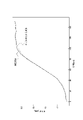
- FIG. 4 is a diagram showing an example of fitting to a measured waveform based on a model formula; FIG.
- 4 is a diagram illustrating estimation of coefficients of a model formula by a machine learning model; It is a figure explaining the example of a non-invasive parameter.
- 4 is a flow chart showing a procedure for estimating a right ventricular pressure waveform or a left ventricular pressure waveform;
- 1 is a block diagram showing the configuration of a pulmonary artery pressure waveform estimation device according to an embodiment of the present disclosure;
- FIG. 4 is a flow chart showing a procedure for estimating a pulmonary artery pressure waveform;
- a ventricular pressure waveform estimation device 10 estimates at least a left ventricular pressure waveform or a right ventricular pressure waveform based on one or more parameters related to heart rate or arterial pressure obtained by at least one of a noninvasive method and a minimally invasive method. Estimate some. In the present application, “non-invasive” means not damaging the human body. “Minimally invasive” means that it does not involve an incision around the heart of the human body or insertion of a device into the ventricle via a cardiac catheter.
- ventricular pressure waveform estimation device 10 includes acquisition section 11 , calculation section 12 , output section 13 , and storage section 14 .
- the ventricular pressure waveform estimation device 10 is a computer loaded with a program for estimating the ventricular pressure waveform.
- the ventricular pressure waveform estimation device 10 is, for example, a dedicated device, a general-purpose device such as a PC, or a server device belonging to a cloud computing system or other computing system.
- the acquisition unit 11 is configured to be able to acquire the value of one or more parameters related to heartbeat or arterial pressure.
- the acquisition unit 11 may receive one or more parameter values from outside the ventricular pressure waveform estimation device 10 as electrical signals.
- the acquisition unit 11 may read an electronic file containing one or more parameter values from a storage medium.
- the acquisition unit 11 may include an input terminal for receiving input from the outside, a communication interface with an external device, a reading device for reading information on a storage medium, and the like.
- the acquisition unit 11 obtains biosignals related to heartbeat or arterial pressure from a first detection unit 15 and a second detection unit 16 that detect biosignals from a human body 20 in a non-invasive or minimally invasive manner. You may get the value of the parameter that
- the ventricular pressure waveform estimation device 10 may include a first detector 15 and a second detector 16 . Parameters based on biological signals obtained by noninvasive methods are called noninvasive parameters. Parameters based on biological signals acquired by a minimally invasive method are called minimally invasive parameters.
- the first detection unit 15 may detect one or more first biomedical signals related to the heartbeat of the heart 21 only by non-invasive methods.
- the first detection unit 15 may include, for example, at least one of an electrocardiogram (ECG) that detects heartbeats, an optical sensor, an impedance sensor, and an acceleration sensor.
- ECG electrocardiogram
- the first detection unit 15 may include, for example, at least one of a microphone that detects heart sounds, an acceleration sensor, a vibration sensor, and a piezoelectric sensor.
- the second detection unit 16 may detect one or more second biomedical signals related to arterial pressure in a region distant from the heart 21, such as the arm 23, using only a non-invasive method.
- the second detection unit 16 uses, for example, a cuff pressure sensor, a photoplethysmography (PPG), a remote photoplethysmography (rPPG), and a tonometry method. can be done.
- Pulse pressure measurement using a cuff pressure sensor includes methods using K sound and oscillometric methods.
- the second detection unit 16 can detect the arterial pressure of a region including at least one of the carotid artery, brachial artery, radial artery, fingertip, and ear.
- the second detection unit 16 detects arterial pressure by a minimally invasive method, for example, an imaging catheter, arterial line (A line), intravascular implant (stent, artificial blood vessel, embedded sensor), subcutaneous implantation, etc. Invasive blood pressure measurement methods such as methods can be used.
- the second detection unit 16 can detect the arterial pressure of a region including at least one of the carotid artery, brachial artery, and radial artery.
- the first detection unit 15 may calculate values of parameters related to heartbeat from the first biosignal.
- the second detection unit 16 may calculate the value of the parameter related to arterial pressure from the second biosignal.
- the parameters related to heartbeat and the parameters related to arterial pressure may be calculated in the ventricular pressure waveform estimation device 10 from the first biomedical signal and the second biomedical signal instead of the first detection unit 15 and the second detection unit 16. good.
- the calculator 12 of the ventricular pressure waveform estimation device 10 may combine the first biosignal and the second biosignal to calculate the value of a parameter related to heartbeat or arterial pressure.
- the computing unit 12 includes at least one processor, and executes various computational processes required for the ventricular pressure waveform estimation device 10 while controlling each component of the ventricular pressure waveform estimation device 10 .
- the processor includes a general-purpose processor such as a CPU (central processing unit), or a dedicated processor specialized for specific processing.
- the arithmetic unit 12 includes an application specific integrated circuit (ASIC: Application Specific Integrated Circuit), a digital signal processor (DSP: Digital Signal Processor), a programmable logic device (PLD: Programmable Logic Device), a field programmable gate array (FPGA: Field -Programmable Gate Array), or any combination thereof.
- the computing unit 12 may include a memory built into the processor or a memory independent of the processor.
- the computing unit 12 estimates at least part of the left ventricular pressure waveform or the right ventricular pressure waveform using the value of one or more parameters related to heartbeat or arterial pressure acquired by the acquiring unit 11 .
- FIG. 2 shows a left ventricular pressure waveform 30 for one heartbeat.
- the calculation unit 12 estimates a partial waveform 31 that is a part of the left ventricular pressure waveform 30 surrounded by a dashed line.
- a partial waveform 31 is a systolic waveform of the left ventricle.
- the estimation target is, for example, a period of about 150 ms to 200 ms after the left ventricle starts to contract.
- the computing unit 12 has a model formula representing a left ventricular pressure waveform or a right ventricular pressure waveform.
- This model formula is represented by the following formula (1).
- the model formula includes four coefficients a, b, c and k.
- the calculation unit 12 estimates at least part of the left ventricular pressure waveform or the right ventricular pressure waveform by estimating the respective coefficients a, b, c, and k of the model formula. Pre-built machine learning models are used to estimate each coefficient. A method of estimating the left ventricular pressure waveform or the right ventricular pressure waveform executed by the calculation unit 12 will be described in more detail below.
- the output unit 13 includes at least one output interface.
- the output unit 13 outputs data of an output waveform 18 indicating temporal changes in internal pressure of the left ventricle or right ventricle obtained by the operation of the ventricular pressure waveform estimation device 10 .
- the output unit 13 may output the data of the output waveform 18 for processing by subsequent devices. Instead of outputting the output waveform 18 to another device, the ventricular pressure waveform estimating device 10 may perform further processing within itself based on this waveform data.
- the storage unit 14 includes at least one semiconductor memory, at least one magnetic memory, at least one optical memory, or any combination thereof.
- the semiconductor memory is, for example, RAM (random access memory) or ROM (read only memory).
- the RAM is, for example, SRAM (static random access memory) or DRAM (dynamic random access memory).
- the ROM is, for example, EEPROM (electrically erasable programmable read only memory).
- the storage unit 14 functions, for example, as a main memory device, an auxiliary memory device, or a cache memory.
- the storage unit 14 stores data used for the operation of the ventricular pressure waveform estimation device 10 and data obtained by the operation of the ventricular pressure waveform estimation device 10 .
- the storage unit 14 stores a machine learning model used for estimating the ventricular pressure waveform.
- the functions of the ventricular pressure waveform estimating device 10 are realized by executing a ventricular pressure waveform estimating program (hereinafter referred to as "program") according to the present embodiment by a processor as the computing unit 12. That is, the functions of the ventricular pressure waveform estimation device 10 are realized by software.
- the program causes the computer to function as the ventricular pressure waveform estimation device 10 by causing the computer to execute the operation of the ventricular pressure waveform estimation device 10 . That is, the computer functions as the ventricular pressure waveform estimation device 10 by executing the operation of the ventricular pressure waveform estimation device 10 according to the program.
- the program can be stored on a non-transitory computer-readable medium.
- a non-transitory computer-readable medium is, for example, a flash memory, a magnetic recording device, an optical disk, a magneto-optical recording medium, or a ROM.
- Distribution of the program includes the sale, transfer, or lending of portable media such as SD (Secure Digital) cards, DVDs (digital versatile discs), or CD-ROMs (compact disc read only memory) that store the programs.
- the program may be distributed by storing the program in the storage of the server and transferring the program from the server to another computer.
- a program may be provided as a program product.
- a computer for example, temporarily stores a program stored in a portable medium or a program transferred from a server in a main storage device. Then, the computer reads the program stored in the main storage device with the processor, and executes processing according to the read program with the processor.
- the computer may read the program directly from the portable medium and execute processing according to the program.
- the computer may execute processing according to the received program every time the program is transferred from the server to the computer.
- the processing may be executed by a so-called ASP (application service provider) type service that implements functions only by executing instructions and obtaining results without transferring the program from the server to the computer.
- a program includes information that is used for processing by a computer and that conforms to the program. For example, data that is not a direct instruction to a computer but that has the property of prescribing the processing of the computer corresponds to "things equivalent to a program.”
- a part or all of the functions of the ventricular pressure waveform estimating device 10 may be realized by a programmable circuit or a dedicated circuit as the arithmetic unit 12. That is, part or all of the functions of the ventricular pressure waveform estimation device 10 may be realized by hardware.
- the Windkessel model is known as a model for explaining blood pressure waveforms in arteries. According to the two-element model of the Windkessel model, fluctuations in the blood pressure pulse wave are shown in FIG. It can be expressed by replacing with an equivalent electric circuit.
- I(t) the volume of blood pumped from the heart
- Aortic blood pressure is also represented as potential P(t).
- Current I(t) and potential P(t) are functions of time t.
- Equation (2) the relationship between the current I(t) and the potential P(t) is represented by the differential equation shown in Equation (2) below.
- Equation (2) assumes that the current I(t), which represents the blood flow, is a sigmoid function, and can be solved for the potential P(t), which represents the aortic blood pressure.
- the sigmoid function is generally used as a model of the properties of nerve cells in living organisms. Since myocardial contraction is caused by action potentials on myocardial cells, it can be represented in principle in the same manner as nerve action potentials. Therefore, I(t) in equation (2) can be put in a sigmoidal function. It is also possible from experimental data to assume that I(t) follows a sigmoidal function.
- Equation (1) is rewritten as follows.
- the coefficients a, b, c and k in Equation (1) are arbitrary coefficients.
- Equation (1) can be read as representing the left ventricular pressure waveform.
- the pressure waveform of the right ventricle can also be represented by Equation (1) using a similar model, where P(t) is the blood pressure of the pulmonary artery and I(t) is the volume of blood pumped from the right ventricle. can.
- the ventricular pressure waveform estimating device 10 stores a machine learning model constructed in advance in the storage unit 14 .
- the machine learning model may be constructed using a machine learning computer different from the ventricular pressure waveform estimation device 10 .
- a procedure for constructing a machine learning model will be described with reference to the flow chart of FIG. 4 and FIG.
- At least one of a noninvasive parameter and a minimally invasive parameter related to the heartbeat or arterial pressure calculated from the measured waveform of the left ventricular pressure or the right ventricular pressure and the biosignal detected at the same time as the measured waveform is acquired.
- Non-invasive parameters and minimally invasive parameters are hereinafter simply referred to as parameters.
- a plurality of parameters are shown as parameters X, Y, and Z.
- the plurality of parameters can include at least one heart rate related parameter and at least one arterial pressure related parameter.
- the measured waveform of left ventricular pressure or right ventricular pressure may be measured by an invasive method using, for example, a pigtail catheter or balloon catheter.
- measured waveforms and parameters for example, data measured in normal and abnormal conditions of many heart failure patients can be used.
- Data of measured waveforms and parameters may be collected by an engineer or the like from measured data accumulated in a medical institution or the like and input to a computer for machine learning.
- the computer for machine learning fits the model formula (formula (1)) to the measured waveform for each measured waveform, thereby calculating the values of the coefficients a, b, c, and k of the model formula (step S102 ).
- a nonlinear least-squares method can be used for fitting the model formula.
- the nonlinear least squares method includes the steepest descent method, Gauss-Newton method, Levenberg-Marquardt method, Powell's least squares method, and so on.
- the coefficients of the model formula are determined so that the waveform of the model formula is closest to the measured waveform.
- the measured waveform in FIG. 6 corresponds to, for example, the partial waveform 31 of the left ventricular pressure waveform 30 shown in FIG.
- a computer for machine learning uses the parameters acquired in step S101 as explanatory variables, and generates a data set for machine learning in which the coefficients a, b, c, and k of the model formula calculated in step S102 are used as objective variables (step S103).
- the data set for machine learning may be generated by taking into consideration the patient's age group, gender, and other attributes and collecting data for each attribute.
- a computer for machine learning executes machine learning using the data set for machine learning generated in step S103, and builds a machine learning model for each coefficient (step S104).
- a constructed machine learning model can be rephrased as a trained model. If multiple data sets are generated for each patient attribute, multiple machine learning models may be built for each coefficient, depending on the patient attribute.
- a machine learning model is built for each coefficient a, b, c and k.
- Machine learning learning methods include gradient boosting, nonlinear multiple regression, and neural networks.
- Ventricular pressure waveform estimating apparatus 10 receives values of a plurality of parameters X, Y, and Z, and estimates four coefficients a, b, c, and k, respectively.
- the plurality of parameters X, Y, Z are non-invasive or minimally invasive parameters.
- the number of parameters of the plurality of parameters X, Y, Z is not limited to three.
- the number of parameters in the plurality of parameters X, Y, Z can be two, four or more.
- the combination of multiple parameters X, Y, Z may be different for each machine learning model 1-N for the four coefficients a, b, c and k.
- highly relevant parameters may be selected for each coefficient a, b, c and k in step S104.
- FIG. 8 illustrates examples of non-invasive parameters used in ventricular pressure waveform estimator 10 .
- FIG. 8 illustrates the aortic and peripheral pulse pressure waveforms along with the left ventricular pressure waveform.
- Left ventricular pressure waveforms are indicated by solid lines.
- Aortic and peripheral pulse pressure waveforms are indicated by dash-dotted and dashed lines, respectively.
- a peripheral pulse pressure waveform is a pulse pressure waveform observed at the arm, wrist, or the like of the human body 20 .
- Aortic pressure waveforms are acquired by an invasive method.
- a peripheral pulse pressure waveform can be obtained by a non-invasive method, for example, a non-invasive continuous blood pressure measurement method using a tonometer.
- diastolic blood pressure, systolic blood pressure, maximum velocity of pulse pressure waveform rising, blood pressure difference between the rising starting point of peripheral pulse pressure waveform and double notch, and pulse augmentation index (AI: augmentation index) can be obtained or calculated.
- Diastolic blood pressure is the pressure exerted on blood vessels when the heart expands. Diastolic blood pressure is also called diastolic blood pressure. Systolic blood pressure is the pressure exerted on blood vessels when the heart contracts. Systolic blood pressure is also called systolic blood pressure.
- the maximum speed of the pulse pressure waveform rise is the maximum value of the gradient dP/dt of the peripheral pulse pressure waveform due to contraction of the heart.
- the blood pressure value difference between the rising starting point of the peripheral pulse pressure waveform and the double notch indicates the difference between the diastolic blood pressure and the blood pressure value at the double notch, as shown in FIG.
- the double notch is an inflection point in the peripheral pulse pressure waveform caused by oscillations due to aortic valve closure.
- the pulse wave augmentation factor is the ratio of the reflected wave component to the blood pressure, and can be obtained by analyzing the peripheral pulse pressure waveform.
- parameters related to heartbeat such as heart rate, isovolumetric systolic time, pulse wave velocity, and systolic time, are derived from the outputs of the electrocardiograph and microphone, etc., and from these and peripheral pulse pressure waveforms. It can be obtained or calculated.
- Heart rate can be measured by an electrocardiograph.
- Isovolumetric systolic time means the time during which the mitral and aortic valves are closed. During the isovolumetric systolic time, the left ventricular volume remains unchanged and the left ventricular pressure increases. Isovolumetric systolic time is measured as the time from the Q wave measured by the electrocardiograph to the I sound of the heart sound acquired by the microphone. Sound I is the sound of the mitral valve closing.
- the pulse wave velocity is the speed at which the pulse wave, which is the pressure wave of the blood pumped from the heart, travels through the arteries to the peripheral blood vessels.
- the pulse wave velocity is measured as the time from the Q wave measured by an electrocardiograph to the rising start point of the peripheral pulse pressure waveform.
- Systolic time is the systolic time when the ventricle contracts.
- Systolic phase includes isovolumic systole and ejection phase.
- the ejection phase is the period during which the aortic valve opens and pushes blood from the left ventricle into the aorta.
- Systolic time is measured using the peripheral pulse pressure waveform as the time from the rising onset of the peripheral pulse pressure waveform to the double notch.
- the above parameters are examples.
- the ventricular pressure waveform estimation device 10 may use other parameters as explanatory variables.
- ventricular pressure waveform estimator 10 can use one or more parameters selected from the above parameters.
- Left and right ventricular pressure waveforms can be estimated using the same parameters.
- the calculation unit 12 calculates respective coefficients a, b, c and k from the non-invasive parameters or minimally invasive parameters calculated based on the biological signals detected by the first detection unit 15 and the second detection unit 16.
- An explanatory variable for doing is extracted (step S201).
- a biological signal is an electrocardiogram, a heart sound, a pulse wave, or the like.
- the non-invasive parameters or minimally invasive parameters may be calculated by the calculator 12 based on the biological signals detected by the first detector 15 and the second detector 16 .
- the calculation unit 12 applies the values of the coefficients a, b, c, and k estimated in step S202 to the model formula (1) to estimate the desired left ventricular pressure waveform or right ventricular pressure waveform (step S203). ).
- the computing unit 12 can use a machine learning model to estimate the patient's left ventricular pressure waveform or right ventricular pressure waveform based on noninvasively or minimally invasively detected biosignals. Therefore, according to the ventricular pressure waveform estimation device 10 of the present disclosure, it is possible to estimate the waveform shape of left ventricular pressure or right ventricular pressure by a non-invasive or minimally invasive method. As a result, medical professionals such as doctors can quickly identify signs of changes in symptoms of patients with heart failure using a simple method. exacerbation can be prevented.
- the calculation unit 12 can calculate the value of LVEDP or PCWP from the estimated left ventricular pressure waveform or right ventricular pressure waveform.
- LVEDP corresponds to the pressure at the rising time of the estimated left ventricular pressure waveform (that is, when the time is 0 ms).
- PCWP can be calculated from the estimated right ventricular pressure waveform as the pressure at a predetermined or arbitrary time. For example, it is possible to acquire or calculate the minimum value, maximum value, average value, etc. of PCWP from the estimated right ventricular pressure waveform.
- the computing unit 12 can further obtain an index of contractility of the left or right heart from the estimated slope (dP/dt) of the left ventricular pressure waveform or the right ventricular pressure waveform. If the slope of the left ventricular pressure waveform or the right ventricular pressure waveform is large, it can be determined that the contractility is high, and if it is small, the contractility is low. As a result, the ventricular pressure waveform estimating apparatus 10 can quickly and noninvasively or minimally invasively grasp the deterioration of the contractile function of the patient's heart.
- changes in ventricular pressure can be represented by the model formula of formula (1) according to the two-element model of the Windkessel model.
- the model formula has four coefficients a, b, c, and k.
- Other embodiments may adopt different model formulas based on different models. Different models can include the three-element and four-element models of the Windkessel model. When based on different models, the number of coefficients in the model formula can also be other than four.
- the value of one or more parameters related to heart rate or arterial pressure is used to determine each model formula including a plurality of coefficients representing the left ventricular pressure waveform or the right ventricular pressure waveform. At least a portion of the left or right ventricular pressure waveform was estimated by estimating the coefficients.
- the values of one or more parameters related to heart rate or arterial pressure are used to estimate respective coefficients of a model equation containing a plurality of coefficients indicative of the pulmonary artery pressure waveform, thereby at least A pulmonary artery pressure waveform estimator that estimates a portion can be provided. Therefore, in FIG.
- FIG. 10 shows the configuration of a device including a pulmonary artery pressure waveform estimation device 40 according to an embodiment of the present disclosure. 10 are the same as those in FIG. 1, the same reference numerals as those in FIG. 1 are used, and descriptions thereof are omitted.
- a machine learning learning model can be constructed according to the flowchart shown in FIG. 4, in which the "left ventricular pressure waveform or right ventricular pressure waveform" in step S101 is replaced with "pulmonary artery pressure waveform".
- the one or more parameters may include one or more heartbeat-related parameters and one or more arterial pressure-related parameters.
- One or more parameters include diastolic blood pressure, systolic blood pressure, maximum velocity of pulse pressure waveform rising, blood pressure value difference between the rising starting point of peripheral pulse pressure waveform and double notch, pulse wave augmentation coefficient, heart rate, etc. Volumetric systolic time, pulse wave velocity, and/or systolic time may be included.
- the calculator 12 of the pulmonary artery pressure waveform estimating device 40 may calculate the pulmonary artery pressure value and/or the pulmonary artery wedge pressure (PCWP) value based on at least part of the pulmonary artery pressure waveform.
- the calculation unit 12 of the pulmonary artery pressure waveform estimating device 40 may estimate the value of the left ventricular end diastolic pressure (LVEDP) based on at least part of the pulmonary artery pressure waveform.
- LEDP left ventricular end diastolic pressure
- the pulmonary artery pressure waveform estimating device 40 includes a first detection unit 15 that detects one or more first biological signals related to heartbeat, and one or more second biological signals related to arterial pressure. and one or more second detectors 16 for detecting signals.
- the values of the above parameters may be determined based on the first biosignal and/or the second biosignal.
- the first detection unit 15 and the second detection unit 16 may detect the first biosignal and the second biosignal, respectively, in a noninvasive or minimally invasive manner.
- FIG. 11 shows a flowchart showing the procedure for estimating the pulmonary artery pressure waveform.
- step S303 of the flowchart of FIG. 9 "left ventricular pressure waveform or right ventricular pressure waveform” in step S203 is replaced with "pulmonary artery pressure waveform".
- Other points are the same as the flow chart of FIG. 9, so the description is omitted.
- the ventricular pressure waveform estimating device, ventricular pressure waveform estimating method, and ventricular pressure waveform estimating program of the present disclosure can be applied to a monitoring device or the like for understanding the pathology of a heart failure patient or the like and evaluating the cardiac function. .
Landscapes
- Health & Medical Sciences (AREA)
- Life Sciences & Earth Sciences (AREA)
- Engineering & Computer Science (AREA)
- Cardiology (AREA)
- Physiology (AREA)
- Physics & Mathematics (AREA)
- Molecular Biology (AREA)
- General Health & Medical Sciences (AREA)
- Veterinary Medicine (AREA)
- Biophysics (AREA)
- Pathology (AREA)
- Biomedical Technology (AREA)
- Heart & Thoracic Surgery (AREA)
- Medical Informatics (AREA)
- Public Health (AREA)
- Surgery (AREA)
- Animal Behavior & Ethology (AREA)
- Artificial Intelligence (AREA)
- Signal Processing (AREA)
- Computer Vision & Pattern Recognition (AREA)
- Psychiatry (AREA)
- Pulmonology (AREA)
- Vascular Medicine (AREA)
- Evolutionary Computation (AREA)
- Fuzzy Systems (AREA)
- Mathematical Physics (AREA)
- Measuring Pulse, Heart Rate, Blood Pressure Or Blood Flow (AREA)
- Measuring And Recording Apparatus For Diagnosis (AREA)
Priority Applications (5)
| Application Number | Priority Date | Filing Date | Title |
|---|---|---|---|
| EP22825069.2A EP4356826A4 (en) | 2021-06-17 | 2022-06-16 | VENTRICULAR PRESSURE WAVEFORM ESTIMATION DEVICE, VENTRICULAR PRESSURE WAVEFORM ESTIMATION METHOD, VENTRICULAR PRESSURE WAVEFORM ESTIMATION PROGRAM, AND PULMONARY ARTERIAL PRESSURE WAVEFORM ESTIMATION DEVICE |
| AU2022294562A AU2022294562A1 (en) | 2021-06-17 | 2022-06-16 | Ventricular pressure waveform estimation device, ventricular pressure waveform estimation method, ventricular pressure waveform estimation program, and pulmonary artery pressure waveform estimation device |
| CN202280042930.6A CN117500432A (zh) | 2021-06-17 | 2022-06-16 | 心室压波形推定装置、心室压波形推定方法、心室压波形推定程序及肺动脉压波形推定装置 |
| JP2023530412A JPWO2022265081A1 (https=) | 2021-06-17 | 2022-06-16 | |
| US18/526,534 US20240108291A1 (en) | 2021-06-17 | 2023-12-01 | Ventricular pressure waveform estimation device, ventricular pressure waveform estimation method, ventricular pressure waveform estimation program, and pulmonary artery pressure waveform estimation device |
Applications Claiming Priority (2)
| Application Number | Priority Date | Filing Date | Title |
|---|---|---|---|
| JP2021-101120 | 2021-06-17 | ||
| JP2021101120 | 2021-06-17 |
Related Child Applications (1)
| Application Number | Title | Priority Date | Filing Date |
|---|---|---|---|
| US18/526,534 Continuation US20240108291A1 (en) | 2021-06-17 | 2023-12-01 | Ventricular pressure waveform estimation device, ventricular pressure waveform estimation method, ventricular pressure waveform estimation program, and pulmonary artery pressure waveform estimation device |
Publications (1)
| Publication Number | Publication Date |
|---|---|
| WO2022265081A1 true WO2022265081A1 (ja) | 2022-12-22 |
Family
ID=84527540
Family Applications (1)
| Application Number | Title | Priority Date | Filing Date |
|---|---|---|---|
| PCT/JP2022/024216 Ceased WO2022265081A1 (ja) | 2021-06-17 | 2022-06-16 | 心室圧波形推定装置、心室圧波形推定方法、心室圧波形推定プログラムおよび肺動脈圧波形推定装置 |
Country Status (6)
| Country | Link |
|---|---|
| US (1) | US20240108291A1 (https=) |
| EP (1) | EP4356826A4 (https=) |
| JP (1) | JPWO2022265081A1 (https=) |
| CN (1) | CN117500432A (https=) |
| AU (1) | AU2022294562A1 (https=) |
| WO (1) | WO2022265081A1 (https=) |
Cited By (2)
| Publication number | Priority date | Publication date | Assignee | Title |
|---|---|---|---|---|
| WO2024203141A1 (ja) * | 2023-03-28 | 2024-10-03 | テルモ株式会社 | プログラム、情報処理方法および情報処理装置 |
| WO2026070679A1 (ja) * | 2024-09-27 | 2026-04-02 | テルモ株式会社 | 情報処理方法、プログラム、及び情報処理装置 |
Families Citing this family (1)
| Publication number | Priority date | Publication date | Assignee | Title |
|---|---|---|---|---|
| CN120938381B (zh) * | 2025-10-15 | 2026-01-27 | 苏州心岭迈德医疗科技有限公司 | 左心室舒张末期压力的计算方法、装置、设备及存储介质 |
Citations (5)
| Publication number | Priority date | Publication date | Assignee | Title |
|---|---|---|---|---|
| JP2002513309A (ja) * | 1997-02-11 | 2002-05-08 | キネティック コンセプツ インコーポレイテッド | 1拍動毎の肺動脈楔入圧を推定する方法及び装置 |
| JP2005517469A (ja) * | 2002-02-14 | 2005-06-16 | ケビン エム. マッキンタイア | 左心室内圧の非侵襲性測定方法 |
| CN103315719A (zh) * | 2013-05-31 | 2013-09-25 | 山东省计量科学研究院 | 一种人体脉搏波波形信号产生装置 |
| US20150265163A1 (en) | 2012-10-11 | 2015-09-24 | Alon Marmor | Determination of ventricular pressure and related values |
| JP2016067490A (ja) * | 2014-09-29 | 2016-05-09 | フクダ電子株式会社 | 末梢血管抵抗推定方法 |
Family Cites Families (5)
| Publication number | Priority date | Publication date | Assignee | Title |
|---|---|---|---|---|
| CN100344257C (zh) * | 2004-06-17 | 2007-10-24 | 肖行贯 | 心血管动力学参数的检测方法 |
| US20160196384A1 (en) * | 2015-01-06 | 2016-07-07 | Siemens Aktiengesellschaft | Personalized whole-body circulation in medical imaging |
| WO2016179425A1 (en) * | 2015-05-05 | 2016-11-10 | The Johns Hopkins University | A device and method for non-invasive left ventricular end diastolic pressure (lvedp) measurement |
| WO2018053504A1 (en) * | 2016-09-19 | 2018-03-22 | Abiomed, Inc. | Cardiovascular assist system that quantifies heart function and facilitates heart recovery |
| CN107233087A (zh) * | 2017-04-28 | 2017-10-10 | 哈尔滨工业大学深圳研究生院 | 一种基于光电容积脉搏波特征的无创血压测量装置 |
-
2022
- 2022-06-16 EP EP22825069.2A patent/EP4356826A4/en not_active Withdrawn
- 2022-06-16 CN CN202280042930.6A patent/CN117500432A/zh active Pending
- 2022-06-16 WO PCT/JP2022/024216 patent/WO2022265081A1/ja not_active Ceased
- 2022-06-16 AU AU2022294562A patent/AU2022294562A1/en active Pending
- 2022-06-16 JP JP2023530412A patent/JPWO2022265081A1/ja active Pending
-
2023
- 2023-12-01 US US18/526,534 patent/US20240108291A1/en active Pending
Patent Citations (5)
| Publication number | Priority date | Publication date | Assignee | Title |
|---|---|---|---|---|
| JP2002513309A (ja) * | 1997-02-11 | 2002-05-08 | キネティック コンセプツ インコーポレイテッド | 1拍動毎の肺動脈楔入圧を推定する方法及び装置 |
| JP2005517469A (ja) * | 2002-02-14 | 2005-06-16 | ケビン エム. マッキンタイア | 左心室内圧の非侵襲性測定方法 |
| US20150265163A1 (en) | 2012-10-11 | 2015-09-24 | Alon Marmor | Determination of ventricular pressure and related values |
| CN103315719A (zh) * | 2013-05-31 | 2013-09-25 | 山东省计量科学研究院 | 一种人体脉搏波波形信号产生装置 |
| JP2016067490A (ja) * | 2014-09-29 | 2016-05-09 | フクダ電子株式会社 | 末梢血管抵抗推定方法 |
Non-Patent Citations (1)
| Title |
|---|
| See also references of EP4356826A4 |
Cited By (2)
| Publication number | Priority date | Publication date | Assignee | Title |
|---|---|---|---|---|
| WO2024203141A1 (ja) * | 2023-03-28 | 2024-10-03 | テルモ株式会社 | プログラム、情報処理方法および情報処理装置 |
| WO2026070679A1 (ja) * | 2024-09-27 | 2026-04-02 | テルモ株式会社 | 情報処理方法、プログラム、及び情報処理装置 |
Also Published As
| Publication number | Publication date |
|---|---|
| EP4356826A4 (en) | 2024-09-18 |
| AU2022294562A1 (en) | 2023-12-07 |
| EP4356826A1 (en) | 2024-04-24 |
| US20240108291A1 (en) | 2024-04-04 |
| CN117500432A (zh) | 2024-02-02 |
| JPWO2022265081A1 (https=) | 2022-12-22 |
Similar Documents
| Publication | Publication Date | Title |
|---|---|---|
| Pandit et al. | Cuffless blood pressure monitoring: promises and challenges | |
| JP4896125B2 (ja) | 心臓血管パラメータの連続的な評価のためのパルス輪郭法および装置 | |
| Guidoboni et al. | Cardiovascular function and ballistocardiogram: A relationship interpreted via mathematical modeling | |
| US11957438B2 (en) | Method and apparatus for left ventricular end diastolic pressure measurement | |
| Choudhury et al. | Estimating blood pressure using Windkessel model on photoplethysmogram | |
| US20240108291A1 (en) | Ventricular pressure waveform estimation device, ventricular pressure waveform estimation method, ventricular pressure waveform estimation program, and pulmonary artery pressure waveform estimation device | |
| US10251566B2 (en) | Methods and apparatus for determining a central aortic pressure waveform from a peripheral artery pressure waveform | |
| JP7160832B2 (ja) | 非侵襲的血圧測定 | |
| Sun et al. | Systolic blood pressure estimation using ECG and PPG in patients undergoing surgery | |
| US8551005B2 (en) | Monitoring respiratory variation of pulse pressure | |
| JP2012517291A (ja) | 心臓血管パラメータの計算 | |
| EP2967368A1 (en) | Apparatus and methods for computing cardiac output of a living subject via applanation tonometry | |
| CN116018087B (zh) | 用于估计心输出量测量结果的可靠性的方法和装置 | |
| CN115299900A (zh) | 基于集成学习的胸位血压估计方法和装置 | |
| US20230355116A1 (en) | Arterial-pressure estimation apparatus, arterial-pressure estimation system, and arterial-pressure estimation method | |
| Agham et al. | Prevalent approach of learning based cuffless blood pressure measurement system for continuous health-care monitoring | |
| CN113180614B (zh) | 无导丝ffr、无导丝imr和无导丝cfr的检测方法 | |
| JP2012521223A (ja) | 抹消遮断の監視 | |
| US20210219924A1 (en) | Noninvasive Diagnostics of Proximal Heart Health Biomarkers | |
| Maher et al. | Non-invasive calibration-free blood pressure estimation based on artificial neural network | |
| Wuerich et al. | Cuffless Beat-to-Beat Blood Pressure Estimation from Photoplethysmogram Signals | |
| JP2025505756A5 (https=) | ||
| Markulyova et al. | Hemodynamic Parameters Non-Invasive Hydro-Cuff Monitoring System | |
| Schutte | S-37-6: THE PROMISE OF CUFFLESS BLOOD PRESSURE MONITORING IN MANAGING HYPERTENSION | |
| Sharman | S-37-5: HOW TO CHECK WHETHER A BLOOD PRESSURE DEVICE HAS BEEN PROPERLY VALIDATED FOR ACCURACY |
Legal Events
| Date | Code | Title | Description |
|---|---|---|---|
| 121 | Ep: the epo has been informed by wipo that ep was designated in this application |
Ref document number: 22825069 Country of ref document: EP Kind code of ref document: A1 |
|
| ENP | Entry into the national phase |
Ref document number: 2023530412 Country of ref document: JP Kind code of ref document: A |
|
| WWE | Wipo information: entry into national phase |
Ref document number: 2022294562 Country of ref document: AU Ref document number: AU2022294562 Country of ref document: AU |
|
| ENP | Entry into the national phase |
Ref document number: 2022294562 Country of ref document: AU Date of ref document: 20220616 Kind code of ref document: A |
|
| WWE | Wipo information: entry into national phase |
Ref document number: 202280042930.6 Country of ref document: CN |
|
| WWE | Wipo information: entry into national phase |
Ref document number: 2022825069 Country of ref document: EP |
|
| NENP | Non-entry into the national phase |
Ref country code: DE |
|
| ENP | Entry into the national phase |
Ref document number: 2022825069 Country of ref document: EP Effective date: 20240117 |
|
| WWW | Wipo information: withdrawn in national office |
Ref document number: 2022825069 Country of ref document: EP |