WO2022196816A1 - 急性肺障害および肺線維症の急性増悪を改善するための医薬組成物 - Google Patents
急性肺障害および肺線維症の急性増悪を改善するための医薬組成物 Download PDFInfo
- Publication number
- WO2022196816A1 WO2022196816A1 PCT/JP2022/012823 JP2022012823W WO2022196816A1 WO 2022196816 A1 WO2022196816 A1 WO 2022196816A1 JP 2022012823 W JP2022012823 W JP 2022012823W WO 2022196816 A1 WO2022196816 A1 WO 2022196816A1
- Authority
- WO
- WIPO (PCT)
- Prior art keywords
- chain variable
- variable region
- amino acid
- acid sequence
- seq
- Prior art date
- Legal status (The legal status is an assumption and is not a legal conclusion. Google has not performed a legal analysis and makes no representation as to the accuracy of the status listed.)
- Ceased
Links
Images













Classifications
-
- C—CHEMISTRY; METALLURGY
- C07—ORGANIC CHEMISTRY
- C07K—PEPTIDES
- C07K14/00—Peptides having more than 20 amino acids; Gastrins; Somatostatins; Melanotropins; Derivatives thereof
- C07K14/195—Peptides having more than 20 amino acids; Gastrins; Somatostatins; Melanotropins; Derivatives thereof from bacteria
- C07K14/305—Peptides having more than 20 amino acids; Gastrins; Somatostatins; Melanotropins; Derivatives thereof from bacteria from Micrococcaceae (F)
- C07K14/31—Peptides having more than 20 amino acids; Gastrins; Somatostatins; Melanotropins; Derivatives thereof from bacteria from Micrococcaceae (F) from Staphylococcus (G)
-
- A—HUMAN NECESSITIES
- A61—MEDICAL OR VETERINARY SCIENCE; HYGIENE
- A61K—PREPARATIONS FOR MEDICAL, DENTAL OR TOILETRY PURPOSES
- A61K39/00—Medicinal preparations containing antigens or antibodies
-
- A—HUMAN NECESSITIES
- A61—MEDICAL OR VETERINARY SCIENCE; HYGIENE
- A61K—PREPARATIONS FOR MEDICAL, DENTAL OR TOILETRY PURPOSES
- A61K39/00—Medicinal preparations containing antigens or antibodies
- A61K39/02—Bacterial antigens
- A61K39/085—Staphylococcus
-
- A—HUMAN NECESSITIES
- A61—MEDICAL OR VETERINARY SCIENCE; HYGIENE
- A61P—SPECIFIC THERAPEUTIC ACTIVITY OF CHEMICAL COMPOUNDS OR MEDICINAL PREPARATIONS
- A61P1/00—Drugs for disorders of the alimentary tract or the digestive system
- A61P1/16—Drugs for disorders of the alimentary tract or the digestive system for liver or gallbladder disorders, e.g. hepatoprotective agents, cholagogues, litholytics
-
- A—HUMAN NECESSITIES
- A61—MEDICAL OR VETERINARY SCIENCE; HYGIENE
- A61P—SPECIFIC THERAPEUTIC ACTIVITY OF CHEMICAL COMPOUNDS OR MEDICINAL PREPARATIONS
- A61P11/00—Drugs for disorders of the respiratory system
-
- A—HUMAN NECESSITIES
- A61—MEDICAL OR VETERINARY SCIENCE; HYGIENE
- A61P—SPECIFIC THERAPEUTIC ACTIVITY OF CHEMICAL COMPOUNDS OR MEDICINAL PREPARATIONS
- A61P13/00—Drugs for disorders of the urinary system
- A61P13/12—Drugs for disorders of the urinary system of the kidneys
-
- A—HUMAN NECESSITIES
- A61—MEDICAL OR VETERINARY SCIENCE; HYGIENE
- A61P—SPECIFIC THERAPEUTIC ACTIVITY OF CHEMICAL COMPOUNDS OR MEDICINAL PREPARATIONS
- A61P43/00—Drugs for specific purposes, not provided for in groups A61P1/00-A61P41/00
-
- C—CHEMISTRY; METALLURGY
- C07—ORGANIC CHEMISTRY
- C07K—PEPTIDES
- C07K16/00—Immunoglobulins [IG], e.g. monoclonal or polyclonal antibodies
- C07K16/12—Immunoglobulins [IG], e.g. monoclonal or polyclonal antibodies against material from bacteria
- C07K16/1267—Gram-positive bacteria
- C07K16/1271—Micrococcaceae (F); Staphylococcaceae (F), e.g. Staphylococcus (G)
-
- G—PHYSICS
- G01—MEASURING; TESTING
- G01N—INVESTIGATING OR ANALYSING MATERIALS BY DETERMINING THEIR CHEMICAL OR PHYSICAL PROPERTIES
- G01N33/00—Investigating or analysing materials by specific methods not covered by groups G01N1/00 - G01N31/00
- G01N33/48—Biological material, e.g. blood, urine; Haemocytometers
- G01N33/50—Chemical analysis of biological material, e.g. blood, urine; Testing involving biospecific ligand binding methods; Immunological testing
- G01N33/53—Immunoassay; Biospecific binding assay; Materials therefor
- G01N33/569—Immunoassay; Biospecific binding assay; Materials therefor for microorganisms, e.g. protozoa, bacteria, viruses
- G01N33/56911—Bacteria
- G01N33/56938—Staphylococcus
-
- G—PHYSICS
- G01—MEASURING; TESTING
- G01N—INVESTIGATING OR ANALYSING MATERIALS BY DETERMINING THEIR CHEMICAL OR PHYSICAL PROPERTIES
- G01N33/00—Investigating or analysing materials by specific methods not covered by groups G01N1/00 - G01N31/00
- G01N33/48—Biological material, e.g. blood, urine; Haemocytometers
- G01N33/50—Chemical analysis of biological material, e.g. blood, urine; Testing involving biospecific ligand binding methods; Immunological testing
- G01N33/68—Chemical analysis of biological material, e.g. blood, urine; Testing involving biospecific ligand binding methods; Immunological testing involving proteins, peptides or amino acids
- G01N33/6884—Chemical analysis of biological material, e.g. blood, urine; Testing involving biospecific ligand binding methods; Immunological testing involving proteins, peptides or amino acids from lung
-
- A—HUMAN NECESSITIES
- A61—MEDICAL OR VETERINARY SCIENCE; HYGIENE
- A61K—PREPARATIONS FOR MEDICAL, DENTAL OR TOILETRY PURPOSES
- A61K39/00—Medicinal preparations containing antigens or antibodies
- A61K2039/505—Medicinal preparations containing antigens or antibodies comprising antibodies
-
- C—CHEMISTRY; METALLURGY
- C07—ORGANIC CHEMISTRY
- C07K—PEPTIDES
- C07K2317/00—Immunoglobulins specific features
- C07K2317/30—Immunoglobulins specific features characterized by aspects of specificity or valency
- C07K2317/34—Identification of a linear epitope shorter than 20 amino acid residues or of a conformational epitope defined by amino acid residues
-
- C—CHEMISTRY; METALLURGY
- C07—ORGANIC CHEMISTRY
- C07K—PEPTIDES
- C07K2317/00—Immunoglobulins specific features
- C07K2317/50—Immunoglobulins specific features characterized by immunoglobulin fragments
- C07K2317/56—Immunoglobulins specific features characterized by immunoglobulin fragments variable (Fv) region, i.e. VH and/or VL
- C07K2317/565—Complementarity determining region [CDR]
-
- C—CHEMISTRY; METALLURGY
- C07—ORGANIC CHEMISTRY
- C07K—PEPTIDES
- C07K2317/00—Immunoglobulins specific features
- C07K2317/70—Immunoglobulins specific features characterized by effect upon binding to a cell or to an antigen
- C07K2317/76—Antagonist effect on antigen, e.g. neutralization or inhibition of binding
-
- G—PHYSICS
- G01—MEASURING; TESTING
- G01N—INVESTIGATING OR ANALYSING MATERIALS BY DETERMINING THEIR CHEMICAL OR PHYSICAL PROPERTIES
- G01N2333/00—Assays involving biological materials from specific organisms or of a specific nature
- G01N2333/195—Assays involving biological materials from specific organisms or of a specific nature from bacteria
- G01N2333/305—Assays involving biological materials from specific organisms or of a specific nature from bacteria from Micrococcaceae (F)
- G01N2333/31—Assays involving biological materials from specific organisms or of a specific nature from bacteria from Micrococcaceae (F) from Staphylococcus (G)
-
- G—PHYSICS
- G01—MEASURING; TESTING
- G01N—INVESTIGATING OR ANALYSING MATERIALS BY DETERMINING THEIR CHEMICAL OR PHYSICAL PROPERTIES
- G01N2800/00—Detection or diagnosis of diseases
- G01N2800/12—Pulmonary diseases
-
- G—PHYSICS
- G01—MEASURING; TESTING
- G01N—INVESTIGATING OR ANALYSING MATERIALS BY DETERMINING THEIR CHEMICAL OR PHYSICAL PROPERTIES
- G01N2800/00—Detection or diagnosis of diseases
- G01N2800/56—Staging of a disease; Further complications associated with the disease
Definitions
- the present invention relates to a pharmaceutical composition for ameliorating acute exacerbation of pulmonary fibrosis and a method therefor, in particular, a composition for ameliorating acute exacerbation of pulmonary fibrosis containing an inhibitor of pulmonary flora-derived peptides. and methods therefor.
- Idiopathic pulmonary fibrosis is a chronic disease of unknown etiology and an incurable disease.
- IPF Idiopathic pulmonary fibrosis
- pirfenidone and nintedanib Two types of antifibrotic drugs, pirfenidone and nintedanib, which are currently used for the treatment of IPF, can be expected to survive for only about 2 to 3 years after diagnosis (Non-Patent Document 1).
- These antifibrotic drugs only slightly reduce the loss of lung function and do not improve patient survival or quality of life (2-4).
- Recent epidemiological studies indicate that there are more than 5 million IPF patients worldwide and that the number is increasing worldwide (Non-Patent Document 5).
- Non-Patent Document 1 Alveolar epithelial cell damage and injury resulting from increased secretion of pulmonary fibrogenic factors such as Transforming Growth Factor- ⁇ 1, excessive recruitment of myofibroblasts in the lung, and extracellular matrix deposition.
- Abnormal tissue repair plays a central role in the cause of IPF (Non-Patent Document 1). This progression ultimately leads to scarring of lung tissue, structural destruction of the lung, and respiratory failure (Non-Patent Document 5).
- the natural history of IPF is unpredictable and variable [6], and includes slow-progressing cases, rapidly-progressing cases, and abrupt clinical exacerbations called acute exacerbations (AEs). (Non-Patent Documents 7 and 8).
- Non-Patent Documents 9, 10 The most frequent cause of death in IPF is AE, which is seen in about 46% of IPF (Non-Patent Documents 9, 10). AE predicts high mortality during onset (up to 50%) and short-term survival after acute onset; once diagnosed with AE, patients survive only 3-4 months [8, 9]. ). Mortality increases by up to 90% in cases requiring mechanical ventilation (Non-Patent Document 9). Infection and diagnosis or surgery can trigger AE, but the exact mechanism is unknown (Non-Patent Document 8). Currently, there is no effective treatment for AE (Non-Patent Document 3).
- pulmonary microbiome lung microbiome
- IPF idiopathic pulmonary fibrosis
- an imbalanced or abundant pulmonary microbiota is associated with persistent alterations in the expression of genes involved in host defense, decreased immune responses, lung epithelial cell damage, abnormal fibroblast reactivity, It is associated with severity of pulmonary function abnormalities, worsening chest radiographic findings, disease progression and decreased survival (11-16).
- Altered pulmonary microbiota and bacterial burden are exacerbated in IPF with acute exacerbation (17, 18).
- the mechanisms associated with pulmonary microbiota with intrapulmonary fibrosis remain unclear.
- Colicins are peptides that are conserved in a variety of staphylococci and various pathogenic bacterial strains (19).
- Non-Patent Document 19 The amino acid sequence of colicin corresponds to a bacterial transglycosylase fragment. Intrapulmonary administration of colicin or colicin-bearing bacteria to mice with established pulmonary fibrosis causes apoptosis in alveolar epithelial cells, resulting in acute exacerbation (AE) (Non-Patent Document 19). Also, IPF patients with indolent disease progression had increased lung levels of colicin compared to healthy controls, and patients with AE had significantly higher levels of lung colicin compared to stable subjects (Non-Patent Literature 19).
- Idiopathic pulmonary fibrosis Diagnosis, epidemiologyand natural history. Respirology21, 427-437, doi: 10.1111/resp.l2683 (2016). Kreuter, M. et al. Acute examination of idiopathic pulmonary fibrosis: international survey and call for harmonisation. Eur Respir J 55, doi: 10.1183/13993003.01760-2019(2020). Collard, H. R. et al. Acute Exacerbation of Idiopathic Pulmonary Fibrosis. Natsuizaka, M. et al. Epidemiologic survey of Japanese patients with idiopathic pulmonary fibrosis and investigation of ethnic differences.
- the present invention includes the following aspects.
- ⁇ Monoclonal antibody molecule> An isolated monoclonal antibody molecule or derivative thereof that specifically binds to colicin.
- [2] The monoclonal antibody molecule or derivative thereof of [1], wherein the colicin is IVMPESSGNPNAVNPAGYR (SEQ ID NO: 1).
- [3] The monoclonal antibody molecule or derivative thereof according to [1] or [2], which has PESSGNP (SEQ ID NO: 68) or NPAGY (SEQ ID NO: 69) as an epitope.
- IC a L chain variable region comprising the L chain variable region amino acid sequence of SEQ ID NO: 40, or an amino acid sequence that is at least 80% identical thereto, or in the L chain variable region amino acid sequence of SEQ ID NO: 40, compared to 1 or a light chain variable region comprising at least one framework region comprising an amino acid sequence having substitutions, deletions, insertions or additions of several amino acid residues
- IIC a L chain variable region comprising the L chain variable region amino acid sequence of SEQ ID NO: 51, or an amino acid sequence that is at least 80% identical thereto, or in the L chain variable region amino acid sequence of SEQ ID NO: 51 compared to 1 or a light chain variable region comprising at least one framework region comprising an amino acid sequence having substitutions, deletions, insertions or additions of several amino acid residues
- IIIC a L chain variable region comprising the L chain variable region amino acid sequence of
- the H chain variable region of (IB) according to [5] is an H chain variable region comprising an amino acid sequence that is at least 80% identical to the H chain variable region amino acid sequence of SEQ ID NO: 36, and ( IC) is an L chain variable region comprising an amino acid sequence that is at least 80% identical to the L chain variable region amino acid sequence of SEQ ID NO: 40; or the H chain variable region of (IIB) according to [5].
- the region is an H chain variable region comprising an amino acid sequence that is at least 80% identical to the H chain variable region amino acid sequence of SEQ ID NO: 48, and the L chain variable region of (IIC) described in [6] is SEQ ID NO: 51 or the H chain variable region of (IVB) according to [5] comprises the H chain variable region amino acid sequence of SEQ ID NO: 71
- An H chain variable region comprising an amino acid sequence that is at least 80% identical to the sequence, and wherein the L chain variable region of (IVC) described in [6] is at least 80% identical to the H chain variable region amino acid sequence of SEQ ID NO:75.
- H chain variable region of (IB) described above has one or several amino acid residue substitutions, deletions, insertions or additions in the H chain variable region amino acid sequence of SEQ ID NO:36.
- the L chain variable region of (IC) described in [6] is the L chain variable region amino acid sequence of SEQ ID NO: 40 , an L chain variable region comprising at least one framework (FW) region comprising an amino acid sequence having one or several amino acid residue substitutions, deletions, insertions or additions compared to it; or [5 ]
- the amino acid sequence in which the H chain variable region of (IIB) described above has one or several amino acid residue substitutions, deletions, insertions or additions in comparison with the H chain variable region amino acid sequence of SEQ ID NO: 48 and the L chain variable region of (IIC) described in [6] comprises, in the L chain variable region amino acid sequence of SEQ ID NO: 51, and an L chain variable region comprising at least one framework (FW) region comprising an amino acid sequence having a substitution, deletion, insertion or addition of one or several amino acid residues in comparison; or (IVB) comprises an amino acid sequence having one or
- [12] A derivative of the antibody molecule of any one of [1] to [11], which is Fab, F(ab') 2 , Fv or single-chain Fv fragment (scFv). [13] The antibody molecule or derivative thereof according to any one of [1] to [12]], which neutralizes and/or inhibits colicin-induced apoptosis.
- ⁇ Pharmaceutical composition A pharmaceutical composition for treating or preventing acute lung injury or fibrosis, containing an effective amount of the monoclonal antibody molecule or derivative thereof according to any one of [1] to [13].
- the pharmaceutical composition according to [14] wherein the acute lung injury causes acute respiratory distress syndrome (ARDS), which is fatal pneumonia requiring respiratory control due to decreased lung function.
- ARDS acute respiratory distress syndrome
- the pharmaceutical composition of [14], wherein the fibrosis is selected from the group consisting of idiopathic pulmonary fibrosis (IPF), liver cirrhosis, renal fibrosis, cystic fibrosis, myelofibrosis, and mammary fibrosis.
- Acute lung injury comprising as an active ingredient a colicin mutant having one or several mutations in the colicin sequence having the amino acid sequence IVMPESSGNPNAVNPAGYR (SEQ ID NO: 1) and not exhibiting apoptotic activity or a vaccine composition for preventing fibrosis.
- the present inventors further investigated the effects of anti-colicin monoclonal antibodies in a common model of acute lung injury and obtained results on the potential effects of colicin on such diseases.
- the present invention further elucidates the role of colicin in the etiology and progression of pulmonary fibrosis and possibly other acute lung injury-related pulmonary diseases, as well as new approaches to treat this incurable disease: new drugs, therapeutic methods. etc.
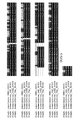
- Figure 1 shows the homology of the putative transglycosylase containing colicin peptide of Staphylococcus nepalensis strain CNDG (CNDG) and three strains of Staphylococcus haemolyticus isolated from the same lung fibrous tissue in mice. Alignment. Colicin peptides are shown with bars in the fourth column. Outside the colicin peptide sequence, conserved amino acids are shaded in black and similar amino acids are shaded in gray.
- Staphylococcus haemolyticus is Staphylococcus haemolyticus, and 1bp_00353, 12bp_00350, 7bp_00350 are S. haemolyticus strain 1 protein number 353, S.
- haemolyticus strain 12 protein number 350 and S. haemolyticus strain 7 protein number 350, respectively.
- the protein of S. neparensis strain CNDG is encoded at position 351 of the open reading frame (ORF) of the genome counting from the gene encoding DnaA (ORF00001). Amino acids with similar properties are grouped into LIMV, AG, YWF, DEQN, KRH and ST. Alignments were performed using multiple sequence comparisons by Log-Expectation (MUSCLE (https://www.ebi.ac.uk/Tools/msa/muscle/)).
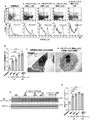
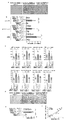
- Figure 2 shows that culture supernatants from three strains of Staphylococcus haemolyticus induce apoptosis of alveolar epithelial cells.
- Figure 2a Flow cytometry results of A549 alveolar epithelial cells cultured in the presence of culture supernatants (1/10 dilution) from Staphylococcus haemolyticus strains 1, 7 and 12 for 48 hours. Fluorescein-labeled annexin V and propidium iodide staining are used as indicators. A549 cells treated with the culture supernatant of Staphylococcus neparensis strain CNDG served as a positive control, and cells treated with saline served as a negative control.
- Figure 2c A549 cells cultured in the presence of S. haemolyticus culture supernatant for 48 hours and evaluated by transmission electron microscopy.
- Figure 2d Western blotting results of caspase-3 cleavage in A549 cells cultured in the presence of culture supernatants of three S. haemolyticus strains 1, 7 and 12 and S. neparensis.
- Figure 2e Graph showing the results of caspase-3 cleavage in A549 cells cultured in the presence of culture supernatants of three S. haemolyticus strains 1, 7 and 12 and S. neparensis. Data are expressed as mean ⁇ SD. Statistical analysis was performed using the unpaired t-test. **p ⁇ 0.001, ***p ⁇ 0.0001.
- Figure 3 shows that synthetic colicin-like peptides of transglycosylases from several pathogens induce apoptosis of alveolar epithelial cells.
- Figures 3a and 3b are the results of flow cytometry showing the results of evaluating apoptosis after culturing A549 alveolar epithelial cells in the presence of 10 ⁇ M colicin-like peptide. A549 cells treated with synthetic colicin sequences or scrambled peptides served as positive and negative controls, respectively.
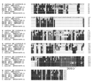
- Figure 4 shows an alignment of colicin-containing putative transglycosylases from various bacterial pathogens.
- Organism names are Wconf (Weissella confusa, Genbank protein accession number WP_112464134.1), CNDG-00351 (S. nepalensis strain CNDG protein 00351), L. monocyt-2 (Listeria monocytogenes, Genbank Protein Accession No. HAB0417320.1), Myabs-2 (Mycobacteroides abscessus, GenBank Protein Accession No. SKR69498.1), Myabs-1 (Mycobacteroides abscessus, GenBank Protein Accession No.
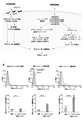
- Figure 5 shows apoptotic pathways induced by S. haemolyticus culture supernatant.
- Figure 5a Apoptosis occurs by extrinsic and intrinsic pathways. The extrinsic pathway is initiated by ligand binding to membrane-bound death receptors, resulting in activation of intracellular signaling and cleavage of procaspase-8 to caspase-8.
- the intrinsic pathway is regulated by mitochondria. In the presence of stimuli, perturbation of the mitochondrial membrane increases its permeability, causing release of the apoptotic repeat-containing (BIRC) protein and suppressors of the baculovirus inhibitor of pro-apoptotic factors, which converts procaspase-9 to caspase- Contributes to apoptosome formation that cleaves to 9.
- BIRC apoptotic repeat-containing
- FIG. 7 shows degradation of colicin-containing transglycosylase by the culture supernatant of Staphylococcus neparensis strain CNDG and preparation of the peptidase fraction.
- Figure 7a Amino acid sequences of the transglycosylase and colicin moieties of Staphylococcus neparensis strain CNDG.
- Fig. 7b Procedures for concentrating the Staphylococcus neparensis culture supernatant to specific fractions.
- Figure 7c Data and procedure to obtain fractions with high digestive activity.
- Figure 7d Data and procedure to obtain fractions with high digestive activity against recombinant transglycosylase 351 for fractions with high absorbance at OD (280 nm).
- FIG. 8 shows that bacterial secretion products cleave transglycosylases to induce apoptosis of lung epithelial cells.
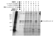
- Figure 8a Photograph showing the results of a sodium dodecyl sulfate-polyacrylamide gel after incubation of the recombinant transglycosylase for 2 hours in the presence of peptidase fractions prepared from various amounts of Staphylococcus neparensis culture supernatant. be.
- Figure 8b Photograph showing the results of a sodium dodecyl sulfate-polyacrylamide gel after incubation of the recombinant transglycosylase in the presence of a constant amount of the peptidase fraction for various time intervals.
- Figure 8c Flow cytometric analysis assessing apoptosis of A549 alveolar epithelial cells after treatment with the indicated reaction mixtures.
- FIG. 9 is a photograph of a sodium dodecyl sulfate-polyacrylamide gel showing that the culture supernatant of Staphylococcus haemolyticus degrades recombinant transglycosylase. The supernatant of Staphylococcus nepalensis served as a control.
- Figure 10 shows that putative serine proteases from bacteria cleave transglycosylases.
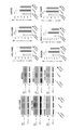
- Figure 10a,b Photographs and graphs showing the effect of the serine protease inhibitor Pefabloc SC (a,b) on transglycosylase degradation.
- Figure 10c,d Photographs and graphs showing the effect of diisopropylfluorophosphate (DFP) on transglycosylase degradation.
- Figure 10e,f Photographs and graphs showing the effect of the chelating agent ethylenediaminetetraacetic acid (EDTA) on transglycosylase degradation.
- Figure 10g, h Photographs and graphs showing the effect of the cysteine protease inhibitor E-64 on transglycosylase degradation.
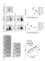
- FIG. 11 shows that different classes of anti-colicin monoclonal antibodies bind to colicin peptides and colicin sequences on transglycosylase 351.
- FIG. Figure 11a Shows the isotype of each monoclonal antibody.
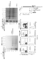
- FIG. 11b Western blotting results showing the binding of each monoclonal antibody to transglycosylase 351.
- FIG. Figure 11c A graph evaluating the binding of each clone of the anti-colicin monoclonal antibody. Data are expressed as mean ⁇ SD. The small band below the 26 kDa molecular weight marker is a degradation product of transglycosylase 351, which is known to degrade during purification of recombinant proteins.
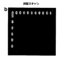
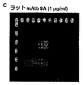
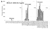
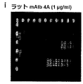
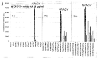
- FIG. 12 shows that the rat anti-colicin monoclonal antibody binds to the colicin consensus motifs PESSGNP or NPAGY.
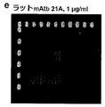
- Figure 12a,b Results of staining of peptide microarray copies verifying overall peptide microarray integrity and assay quality.
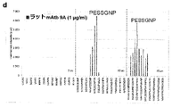
- Figure 12c, d Scan intensity of rat monoclonal (mAtb) antibody 9A.
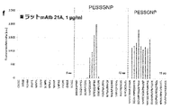
- Figure 12e, f Scan intensity of rat monoclonal (mAtb) antibody 21A.
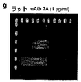
- Figure 12g,h Scan intensity of rat monoclonal (mAtb) antibody 2A.
- Figure 12i,j Scan intensity of rat monoclonal (mAtb) antibody 4A.
- Figure 13 shows the sequences of the variable framework and complementarity determining regions of anti-colicin monoclonal antibody clones.
- Lower case letters indicate framework regions (FR1, FR2, FR3, FR4) and bold upper case letters indicate CDR regions (CDR1, CDR2, CDR3) of antibody variable regions.
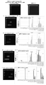
- Figure 14 includes graphs showing that anti-colicin monoclonal antibody clone 21A has potent neutralizing activity against colicin-induced apoptosis of human alveolar epithelial cells.
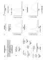
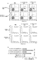
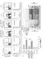
- Figure 14a Flow cytometric analysis showing the inhibitory effect of anti-colicin monoclonal antibody on the pro-apoptotic activity of colicin on cultured alveolar epithelial cells. The points in the upper part of a, excluding the third quadrant, are apoptotic cells. The bumps on the right side of the X-axis in the lower diagram of a indicate apoptotic cells.
- Figure 15 shows that anti-colicin monoclonal antibody clone 21A neutralizes apoptotic activity of culture supernatant from Staphylococcus haemolyticus against human alveolar epithelial cells.
- Figure 15a Flow cytometric analysis showing the inhibitory effect of anti-colicin monoclonal antibody clone 21A on Staphylococcus haemolyticus colicin pro-apoptotic activity.
- the points in the upper part of a, excluding the third quadrant, are apoptotic cells.
- the bumps on the right side of the X-axis in the lower diagram of a indicate apoptotic cells.
- FIG. 16 shows that anti-colicin-neutralizing monoclonal antibodies inhibit lung cell apoptosis induced by transglycosylase degradation products.
- Figure 16a Flow cytometric analysis showing the inhibitory effect of anti-colicin-neutralizing monoclonal antibody on pulmonary cell apoptosis induced by transglycosylase degradation products. The dots in the upper part of a, excluding the third quadrant, are apoptotic cells. The bumps on the right side of the X-axis in the lower diagram of a indicate apoptotic cells.
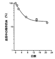
- Figure 17 is a graph showing the mean half-life of anti-colicin neutralizing antibodies in plasma.
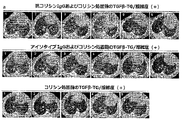
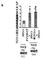
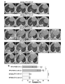
- FIG. 18 shows the matched score of pulmonary fibrosis between TGF ⁇ 1 TG mice treated with anti-colicin antibody and TGF ⁇ 1 TG mice treated with isotype antibody prior to intratracheal instillation of colicin.
- Figure 18a Results of computed tomography (CT) performed before starting treatment with anti-colicin monoclonal antibody (mAtb) or isotype antibody (Atb).
- CT computed tomography
- mAtb anti-colicin monoclonal antibody
- Atb isotype antibody
- N 6 in the TGF ⁇ 1-TG/fibrosis (+)/anti-colicin antibody/colicin and TGF ⁇ 1-TG/fibrosis (+)/isotype antibody/colicin groups
- n in the TGF ⁇ 1-T/fibrosis (-)/colicin group 5.
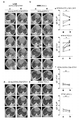
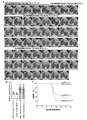
- FIG. 19 shows that monoclonal anti-colicin antibodies prevent exacerbation of lung CT findings in TGF ⁇ 1 TG mice.
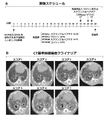
- Figure 19a Schedule for obtaining CT findings in TGF ⁇ 1 TG mice with pulmonary fibrosis. Grading of CT findings for pulmonary fibrosis was blinded and scored by an expert.
- Figure 19b, c Graphs showing CT findings and their scores before and after intratracheal instillation of colicin in the TGF ⁇ 1-TG/fibrosis (-)/colicin group. Statistical analysis was performed by paired Student's t-test. ns, not significant.
- Figure 19d Graphs showing CT findings and their scores before and after intratracheal instillation of colicin in the TGF ⁇ 1-TG/fibrosis (+)/colicin/isotype group. Bars indicate average scores.
- Statistical analysis was performed by paired t-test. *p ⁇ 0.05.
- TGF ⁇ 1 transforming growth factor ⁇ 1, TG: transgenic.
- Figure 19f,g CT findings and their scores before and after intratracheal instillation of colicin in the TGF ⁇ 1-TG/fibrosis(+)/colicin/anti-colicin group. Bars indicate average scores.
- Statistical analysis was performed by paired t-test. ** p ⁇ 0.01.
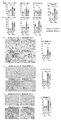
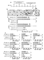
- FIG. 20 shows that monoclonal anti-colicin antibodies suppress acute exacerbation (AE) of pulmonary fibrosis in TGF ⁇ 1 TG mice.
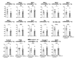
- Figure 20a Giemsa staining results for cell fraction numbers. Scale bar indicates 50 ⁇ m.
- Figure 20b Graph showing the total number of BALF cells and neutrophils. Data are mean ⁇ SD. Statistical analysis was performed by one-way ANOVA with Neuman-Keuls test. *p ⁇ 0.05; **p ⁇ 0.01; ***p ⁇ 0.0001.
- Figure 20c Graph showing the levels of surfactant protein D (SP-D), MUC5B protein, matrix metalloproteinase-1 (MMP-1), decorin and MUC-1 as measured by commercially available enzyme immunoassay kits. be.
- SP-D surfactant protein D
- MUC5B protein matrix metalloproteinase-1
- MMP-1 matrix metalloproteinase-1
- Hydroxyproline levels were measured using a commercially available colorimetric kit according to the manufacturer's instructions. Data are mean ⁇ SD. Statistical analysis was performed by one-way ANOVA with Neuman-Keuls test. *p ⁇ 0.05; **p ⁇ 0.01; ***p ⁇ 0.0001.
- Figure 20d, e Photographs and graphs showing collagen deposition in lung tissue of each group by Masson's staining. Scale bar indicates 200 ⁇ m. Data are mean ⁇ SD. Statistical analysis was performed by one-way ANOVA with Neuman-Keuls test. *p ⁇ 0.05; ***p ⁇ 0.0001.
- Figure 20f Graph showing correlation between fibrosis area and total number of inflammatory cells as described in Figure 20b. Statistical analysis with Spearman's correlation coefficient.
- Figure 21 shows the experimental schedule for induction of acute exacerbation in mice with bleomycin-induced pulmonary fibrosis and CT criteria for pulmonary fibrosis.
- Figure 21a Experimental schedule for induction of acute exacerbations in mice with bleomycin-induced pulmonary fibrosis.
- Figure 21b Shows CT criteria for pulmonary fibrosis. The following CT scoring system was used to determine the grade of pulmonary fibrosis. Score 1: normal findings. Score 2: slightly increased lung density. Score 3: speckled ground-glass opacity and septal thickening. Score 4: Moderate ground-glass opacity and septal thickening. Score 5: Subpleural thickening, diffuse ground-glass opacification, and reticular septal thickening.
- Score 6 subpleural thickening, diffuse ground-glass opacification, reticular septal thickening, and traction bronchiectasis.
- Score 7 Subpleural thickening, severe ground-glass opacification, reticular septal thickening, and diffuse traction bronchiectasis.
- Figure 22 shows deterioration of lung CT findings in mice with bleomycin-induced pulmonary fibrosis after intratracheal instillation of colicin.
- Figure 23 shows acute exacerbation of pulmonary fibrosis in mice with bleomycin-induced pulmonary fibrosis after intratracheal instillation of colicin.
- Figure 23a Shows representative photomicrographs of BALF cell staining for each treatment group in BLM-induced pulmonary fibrosis mice treated with colicin.
- Figure 23b Graph showing staining BALF cell numbers for each treatment group in mice with BLM-induced pulmonary fibrosis receiving colicin. Scale bar indicates 20 ⁇ m. Data are mean ⁇ SD statistical analysis by t-test. *p ⁇ 0.05; *p ⁇ 0.01.
- FIG. 23c Levels of serum amyloid P component (SAP), MUC-1, monocyte chemoattractant protein-1 (MCP-1), periostin, collagen I, and osteopontin in colicin-treated BLM-induced pulmonary fibrosis mice. It is a graph showing. Data are mean ⁇ SD statistical analysis by unpaired t-test. * p ⁇ 0.05; * p ⁇ 0.01; *** p ⁇ 0.001.
- Figure 23d Western blotting evaluation and quantification of caspase-3 cleavage in colicin-treated BLM-induced pulmonary fibrosis mice.
- Figure 23e Shows a graph of the ratio of cleaved caspase-3 to ⁇ -actin in BLM-induced pulmonary fibrosis mice treated with colicin. Bars represent mean ⁇ SD statistical analysis by unpaired t-test. *p ⁇ 0.05.
- Figure 23f Shows the results of quantifying collagen deposition.
- Figure 23g Shows a graph of the results of quantifying collagen deposition. Data are mean ⁇ SD statistical analysis by unpaired t-test. *p ⁇ 0.05.
- Figure 23h Graphical representation of lung tissue content measurements of hydroxyproline. Bars represent mean ⁇ SD statistical analysis by unpaired t-test. *p ⁇ 0.05, **p ⁇ 0.001.
- Figure 24 shows improved lung CT findings and reduced lung inflammatory cell infiltration in mice with bleomycin-induced pulmonary fibrosis treated with anti-colicin monoclonal antibody.
- Figure 24a Shows the schedule for investigating the anti-pulmonary fibrosis effect of anti-colicin monoclonal antibodies.
- Figure 24b,c:b shows lung CT findings in mice with bleomycin-induced pulmonary fibrosis treated with anti-colicin monoclonal antibody.
- FIG. 25 shows amelioration of acute tissue injury, parenchymal cell apoptosis and tissue fibrosis in lungs of pulmonary fibrosis mice treated with anti-colicin monoclonal antibody.
- Figure 25a Graph showing levels of osteopontin, MUC-1 and MUC5B in lungs of pulmonary fibrosis mice treated with anti-colicin monoclonal antibody.
- N 4 for WT/SAL/isotype and WT/SAL/anti-colicin groups
- n 9 for WT/BLM/isotype and WT/BLM/anti-colicin groups. Bars show mean ⁇ SD statistical analysis by one-way ANOVA with Tukey's test. *p ⁇ 0.05, **p ⁇ 0.01, ***p ⁇ 0.0001.
- Figure 25b Photograph showing DNA fragmentation in lungs of pulmonary fibrosis mice treated with anti-colicin monoclonal antibody. Arrows indicate apoptotic cells.
- Figure 25f,g f is a Masson's trichrome stained photograph of lung collagen deposition and g is a graph of it quantified using WindRoof image software.
- Figure 26 shows day 9 lung CT abnormalities and reduction in lung inflammatory cells in mice with bleomycin-induced pulmonary fibrosis treated with anti-colicin monoclonal antibody during the acute phase of disease.
- Figure 26a Shows the schedule for examining the amelioration of BLM-induced lung injury pulmonary fibrosis with anti-colicin monoclonal antibody treated in the acute phase.
- Figure 26b, c b is a photograph showing CT lung opacity in BLM-induced lung injury pulmonary fibrosis in acute treatment with anti-colicin monoclonal antibody, c is a graph of CT scores reflecting the results of b. be.
- N 7 in mice treated with anti-colicin monoclonal antibody (WT/BLM/anti-colicin) or isotype antibody (WT/BLM/isotype). Data are mean ⁇ SD. Statistical analysis was performed by unpaired t-test. *p ⁇ 0.05.
- N 7 in mice treated with anti-colicin monoclonal antibody (WT/BLM/anti-colicin) or isotype antibody (WT/BLM/isotype). Scale bar indicates 20 ⁇ m. Data are mean ⁇ SD statistical analysis by unpaired t-test. ** p ⁇ 0.01.
- FIG. 27 shows day 22 lung CT abnormalities and reduction in lung inflammatory cells in mice with bleomycin-induced pulmonary fibrosis treated with anti-colicin monoclonal antibody during the acute phase of disease.
- Figure 27c,d c Cell differentiation after Giemsa staining in bronchoalveolar lavage fluid (BALF) 22 days after BLM pump in mice with bleomycin-induced pulmonary fibrosis treated with anti-colicin monoclonal antibody during the acute phase of disease.
- Figure 28 shows improvement in lung injury, lung parenchymal apoptosis and collagen deposition on day 22 in mice with bleomycin-induced pulmonary fibrosis treated with anti-colicin monoclonal antibody during the acute phase of disease.
- Figure 28a Graph showing levels of MUC-1, surfactant protein C (SP-C), SP-D, collagen I, periostin, osteopontin and total TGF ⁇ 1 in BALF and plasma taken 22 days after BLM pump infusion. Data are mean ⁇ SD. Statistical analysis was performed by unpaired t-test. *p ⁇ 0.05, **p ⁇ 0.01, ***p ⁇ 0.001.
- Figure 28b, c b shows the results of Western blotting of cleaved caspase-3 and ⁇ -actin, c shows the graph of the cleaved caspase-3 to ⁇ -actin ratio resulting from b.
- Figure 28d,e Seven experts blindedly scored the grade of pulmonary fibrosis using Ashcroft's score. Scale bar indicates 200 ⁇ m.
- N 7 in both WT/BLM/isotype and WT/BLML/anti-colicin groups.
- Figure 29 shows that anti-colicin monoclonal antibody prolongs survival in mice with acute exacerbation of pulmonary fibrosis.
- Figure 29a,b a shows computed tomography (CT) results in mice with acute exacerbation of pulmonary fibrosis and b is a graph of CT scores reflecting the results of b.
- N 22 in both WT/BLM/isotype and WT/BLM/anti-colicin groups
- n 5 in both WT/SAL/isotype and WT/SAL/anti-colicin groups. Bars indicate mean ⁇ SD Statistical analysis was performed by one-way ANOVA with Tukey's test. *p ⁇ 0.05.
- Figure 30 shows that anti-colicin monoclonal antibodies attenuate severe lipopolysaccharide-induced acute lung injury.
- Figure 30a Shows the schedule for testing that anti-colicin monoclonal antibodies ameliorate severe lipopolysaccharide-induced acute lung injury.
- Figure 30b Shows representative photomicrographs of BALF cell staining for each treatment group.
- Figure 31 shows that treatment with anti-colicin monoclonal antibody improves CT findings in mice with severe lipopolysaccharide-induced acute lung injury.
- Figure 31b Graph showing CT opacity from apical to basal regions of the lung quantified using WindRoof imaging software. Data are mean ⁇ SD Statistical analysis by one-way ANOVA with Neuman-Keuls' test. *p ⁇ 0.05, ***p ⁇ 0.001.
- Figure 32 shows that anti-colicin monoclonals attenuate moderate lipopolysaccharide-induced acute lung injury.
- Figure 32a Schedule for testing whether anti-colicin monoclonals attenuate moderate lipopolysaccharide-induced acute lung injury.
- Figure 32b Shows representative photomicrographs of BALF cell staining for each treatment group.
- FIG. 4 is a graph showing plasma BALF and plasma levels of A (LDHA), MUC-5B and matrix metalloproteinase-1 (MMP-1).
- Data are mean ⁇ SD statistical analysis by analysis of variance with the Newman-Keuls method. *p ⁇ 0.05, **p ⁇ 0.01, ***p ⁇ 0.001.
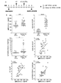
- Figure 33 shows that elevated circulating levels of colicin correlate with markers of pulmonary fibrosis.
- Figure 33b Graph showing Ashcroft scores for pulmonary fibrosis and measurements of plasma colicin and lung hydroxyproline in TGF ⁇ 1TG and WT mice. Statistical differences between groups were analyzed by unpaired t-tests and strength of relationships by Pearson-moment correlation. *p ⁇ 0.05, **p ⁇ 0.01 ***p ⁇ 0.001.
- FIG. 34 shows increased CT scores for pulmonary fibrosis in TGF ⁇ 1TG mice.
- Figure 34a CT score results for pulmonary fibrosis in TGF ⁇ 1TG mice.
- Figure 34b Graph showing CT opacity from apical to basal regions of the lung quantified using WindRoof imaging software.
- WT wild type
- TGF ⁇ 1TG transforming growth factor ⁇ 1
- CT computed tomography. Data are mean ⁇ SD. Statistical analysis was performed by unpaired Student's t-test. ***p ⁇ 0.001.
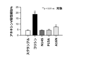
- Figure 35 is a graph showing apoptotic activity in colicin peptides with specific amino acid mutations.
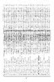
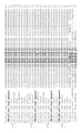
- Figure 36 shows a multiple sequence alignment of the conserved sequences of the pro-apoptotic segments of transglycosylases in several species of staphylococci and streptococci.
- Figure 37 shows a multiple sequence alignment of the conserved sequences of the pro-apoptotic segments of transglycosylases in several species of staphylococci and streptococci.
- Figure 38 shows a multiple sequence alignment of the conserved sequences of the pro-apoptotic segments of transglycosylases in several species of staphylococci and streptococci.
- Examples of colicins shown in Figures 36, 37 and 38C include IVMPESGGNPNAVNPAGYR (SEQ ID NO: 58), IIMPESGGNPNIVNPYGYS (SEQ ID NO: 59), IVMPESGGNPNAVNPYGYR (SEQ ID NO: 60), IVLPESSGNPNAVNPAGYR (SEQ ID NO: 61), IVLPESSGNPNAVNELGYR (SEQ ID NO: 62), IVMPESGGNPNAVNELGYR (SEQ ID NO: 63), IVMPESSGNPNAVNELGYR (SEQ ID NO: 64), IVMPESSGNPDAVNELGYR (SEQ ID NO: 65), IAQRESGGDLKAVNPSSGA (SEQ ID NO: 66), or IAERESGGDLKAVNPSSGA (SEQ ID NO: 67), which are one of the present invention. Or it can be used in multiple aspects.
- FIG. 40 is a graph comparing serum colicin concentrations in healthy subjects, stable idiopathic pulmonary fibrosis (IPF) patients, and idiopathic pulmonary fibrosis (IPF) patients with acute exacerbation.
- Figure 41c A graph measuring the concentration of native colicin in undiluted culture supernatants of Staphylococcus neparensis and Staphylococcus haemolyticus. Bars indicate mean ⁇ SD.
- Figure 41d A549 alveolar epithelial cells were isolated from bronchoalveolar lavage fluid ( BALF) (1:2 dilution), anti-colicin neutralizing antibody (clone 21A) (Anticorisin in the figure) or control lgG (Control IgG in the figure). This is the result of the examination of the metric. Bars indicate mean ⁇ SD.
- Figure 41g A549 alveolar epithelial cells were cultured, synthetic colicin was added to the cell culture medium at a final concentration of 10 ⁇ g/ml, and the medium was harvested after 0, 1, 3, 6, 12, and 24 hours. It is the graph which measured the colicin density
- the present invention in one aspect, relates to an isolated monoclonal antibody molecule or derivative thereof that specifically binds colicin.
- colicin generally refers to a staphylococcal pro-apoptotic peptide that has been found to induce acute exacerbations of pulmonary fibrosis.
- Colicins are, for example, IVMPESSGNPNAVNPAGYR (SEQ ID NO: 1), IVMPESGGNPNAVNPAGYR (SEQ ID NO: 58), IIMPESGGNPNIVNPYGYS (SEQ ID NO: 59), IVMPESGGNPNAVNPYGYR (SEQ ID NO: 60), IVLPESSGNPNAVNPAGYR (SEQ ID NO: 61), IVLPESSGNPNAVNELGYR (SEQ ID NO: 62), IVMPESGGNPNAVNELGYR (SEQ ID NO: 62), No.
- colicin is a peptide having the amino acid sequence IVMPESSGNPNAVNPAGYR (SEQ ID NO: 1).
- SEQ ID NO: 1 may originate from Staphylococcus neparensis. Referring to Figures 36, 37 and 38, note the origin of the sequences of other SEQ ID NOs: ⁇ SEQ ID NO: 58: wp022791177 (top 236) 1 ⁇ SEQ ID NO: 59: wp049409534 wp103371985 (top 248) 2 ⁇ SEQ ID NO: 60: wp103328722 wp126565453 (top 239, 244) 2 ⁇ SEQ ID NO: 61: wp069827045 9 wp069827045 (top row 246) 2 ⁇ SEQ ID NO: 62: wp061853755 (top row 244) 1 ⁇ SEQ ID NO: 63: suk04795 wp105995336 (top row 116) 2 ⁇ SEQ ID NO: 64: wp112369066 (top row 252) 1 ⁇ SEQ ID NO: 65: wp0990
- staphylococci share greater than 98% identity with the corresponding colicin regions of transglycosylases from other members of the IsaA-1 and IsaA-2 clusters and 60% identity with the corresponding regions of members of the SceD cluster. shared identity ( Figures 36, 37, 38). Genomic constructs of gene clustering around transglycosylases (synteny) tended to be conserved in Staphylococcus cornii and Staphylococcus neparensis.
- the present invention relates to the monoclonal antibody molecule of the present invention or a derivative thereof whose epitope is an amino acid having the amino acid sequence of PESSGNP (SEQ ID NO: 68) or NPAGY (SEQ ID NO: 69).
- various anti-colicin monoclonal antibodies having binding properties to the colicin molecule were prepared, and epitope mapping was performed to identify the binding site thereof, thereby obtaining the amino acid sequence of PESSGNP (SEQ ID NO: 68) or NPAGY (SEQ ID NO: 69).
- an epitope with A monoclonal antibody molecule or derivative thereof targeting this epitope would have comparable binding properties to the colicin and comparable functionality, eg, apoptosis-inhibiting and/or neutralizing activity. Accordingly, preferred monoclonal antibody molecules or derivatives thereof of the present invention have neutralizing activity against colicin. This includes neutralizing antibodies.
- (IA) (i) an H chain variable region comprising the H chain variable region CDR1 amino acid sequence of SEQ ID NO:37, the H chain variable region CDR2 amino acid sequence of SEQ ID NO:38 and the H chain variable region CDR3 amino acid sequence of SEQ ID NO:39, and (ii) a sequence L chain variable region comprising the L chain variable region CDR1 amino acid sequence of number 41, the L chain variable region CDR2 amino acid sequence of SEQ ID NO: 42 and the L chain variable region CDR3 amino acid sequence of SEQ ID NO: 43; (IIA); (i) an H chain variable region comprising the H chain variable region CDR1 amino acid sequence of SEQ ID NO: 70, the H chain variable region CDR2 amino acid sequence of SEQ ID NO: 49 and the H chain variable region CDR3 amino acid sequence of SEQ ID NO: 50, and (ii) a sequence L chain variable region comprising the L chain variable region CDR1 amino acid sequence of SEQ ID NO: 52, the L chain
- a "monoclonal antibody molecule” is a tetrapeptide chain structure in which two identical H chains (heavy chains) and two identical L chains (light chains) are interconnected by disulfide bonds.
- the variable region comprises three hypervariable regions (HVR) and four relatively conserved framework regions (FR). Three hypervariable regions determine the specificity of an antibody and are also known as complementarity determining regions (CDRs).
- Each light chain variable region (LCVR) and each heavy chain variable region (HCVR) has three CDR regions and It consists of four FR regions.
- the three light chain CDRs refer to LCDR1, LCDR2 and LCDR3 and the three heavy chain CDRs refer to HCDR1, HCDR2 and HCDR3.
- Antibody molecules include non-human antibody molecules, humanized antibody molecules and human antibody molecules.
- isolated means a sample separated from its original natural environment.
- a naturally occurring polynucleotide or polypeptide present in a living animal is not isolated, but the same polynucleotide or polypeptide that has been artificially separated from some or all of the coexisting samples in the natural system.
- the peptide has been isolated.
- “derivatives of monoclonal antibody molecules” means diabodies and single-chain molecules, as well as “antigen-binding fragments.”
- Antigen-binding fragment refers to Fab fragments, Fab' fragments, F(ab')2 fragments, and Fv fragments and scFv fragments that bind to colicin, which have antigen-binding activity. It can contain more than one CDR region.
- An Fv fragment is the minimum antibody fragment that contains the H chain variable region, the L chain variable region, and all the antigen-binding sites without the constant region.
- Fv antibodies can further comprise a polypeptide linker between the H and L chain variable region domains to form the necessary structure for antigen binding.
- the "antigen-binding site" of the invention refers to a discrete three-dimensional site on an antigen that is recognized by an antibody or antigen-binding fragment of the invention.
- “specifically recognizing colicin” means having the ability to bind to colicin, and is synonymous with “antibody that binds to colicin” or “antibody against colicin”. .
- an H chain variable region comprising the H chain variable region amino acid sequence of SEQ ID NO:36, or an amino acid sequence that is at least 80% identical thereto, or in the H chain variable region amino acid sequence of SEQ ID NO:36 compared to 1 or an H chain variable region comprising at least one framework (FW) region comprising an amino acid sequence having substitutions, deletions, insertions or additions of several amino acid residues;
- an H chain variable region comprising the H chain variable region amino acid sequence of SEQ ID NO:48, or an amino acid sequence that is at least 80% identical thereto, or in the H chain variable region amino acid sequence of SEQ ID NO:48 compared to 1 or an H chain variable region comprising at least one framework (FW) region comprising an amino acid sequence with substitutions, deletions, insertions or additions of several amino acid residues; or
- the H chain variable region of SEQ ID NO:71 an amino acid sequence, or an H chain variable region comprising an amino acid sequence that
- (IC) a L chain variable region comprising the L chain variable region amino acid sequence of SEQ ID NO: 40, or an amino acid sequence that is at least 80% identical thereto, or in the L chain variable region amino acid sequence of SEQ ID NO: 40, compared to 1 or a light chain variable region comprising at least one framework region comprising an amino acid sequence having substitutions, deletions, insertions or additions of several amino acid residues;
- (IIC) a L chain variable region comprising the L chain variable region amino acid sequence of SEQ ID NO: 51, or an amino acid sequence that is at least 80% identical thereto, or in the L chain variable region amino acid sequence of SEQ ID NO: 51 compared to 1 or a light chain variable region comprising at least one framework region comprising an amino acid sequence having substitutions, deletions, insertions or additions of several amino acid residues;
- IIIC a L chain variable region comprising the L chain variable region amino acid sequence of SEQ ID NO:44, or an amino acid sequence that is at least
- a light chain variable region comprising at least one framework region comprising amino acid sequences with additions; C-series monoclonal antibody molecules or derivatives thereof that specify the monoclonal antibodies of the present invention by their light chain variable regions and variants having In the present invention, a mutated monoclonal antibody molecule or derivative thereof has binding properties and functions equivalent to those of the original monoclonal antibody molecule without mutation.
- H and L chain variable region combinations are those of isolated monoclonal antibody clones 4A, 9A, 2A and 21A, respectively. Specifically, the following combinations are used.
- Anti-colicin Monoclonal Antibody Clone 9A Monoclonal Antibody (IIIC) Related: Light chain (9A-LC.7402): SEQ ID NO:44 divmtqspssqavsagekvtmsckssQNLLYSEDKKNYlawyqqkpgqspklliyWAStresgvpdrfigsgsgtdftltvtsvqaedlavyycQYYNFPRTfgggtklelk
- Anti-Colicin Monoclonal Antibody Clone 2A Monoclonal Antibodies (IIA), (IIB), (IIC) Related: H chain (2A-HC.7352): SEQ ID NO:48 evklvesggglvqpgnsltlscvasGFTFTNYGmhwirqapkkglewiamIYYDSSKMsyadtvkgrftisrdnskntlylemnslrsedtamyycAAEGFGTPFPYwgqgtlvtvss, and Light chain (2A-LC.7413): SEQ ID NO:51 divmtqtpssravsagekvtmsckssQSLLYSENEKNYlawyqqrpgqspklliyWAStresgvpdrfigtgsgtdftltissvqaedlavyycQYYHFPRTfgggtrlelk
- H chain (KK1410-2_VH_pep): SEQ ID NO:71 evklvesggglvqpgnsltlscgasgftftNYGMHwirqapkkglewigMIYYDSSKMSYADTVKGrftisrdnsknilylemnslrsedtamyycaaEGFGTPFPYwgqgtlvtvss, and L chain (KK1410-2_VL_pep): SEQ ID NO:75 divmtqtpssqavsagekvtmscKSSQSLLYREDTKNYLAwyqqrpgqspklliyWASTRESgvpdrfigsgsgtdftltissvqaedlavyycQYYHFPRTfgggtrlelk.
- the monoclonal antibody molecules of the present invention can have any H chain variable region and/or any L chain variable region.
- H and L chain variable regions have a high frequency, termed 'complementarity determining regions' (CDRs), interspersed with regions that are more conserved, termed 'framework regions' (FR or FW). Variants from the ability to subdivide into regions of variability.
- the light or heavy chain variable framework of the monoclonal antibody molecule against colicin is at least 80%, 85%, 87%, 90% of the light or heavy chain variable framework residues derived from a human consensus sequence. , 93%, 95%, 97%, 99%.
- a mutant monoclonal antibody of the present invention comprising any of the mutant H chain variable regions of the present invention and any of the mutant L chain variable regions of the present invention It relates to molecules or derivatives thereof.
- a preferred variant H chain and variant L chain variable region combination is that of isolated monoclonal antibody clones 4A, 9A, 2A and 21A, respectively. Specifically, the following combinations are used.
- H chain SEQ ID NO:36 a heavy chain variable region comprising an amino acid sequence that is at least 80% identical to the heavy chain variable region amino acid sequence of evqvlesggglvqpgnslklscatsGFTFSTAWmywyrqfpekrlewvarIKAKSNSYATdytesvkgrftisrddskgsiylrmnnlkeedtaiyycASTDAFYFSHSYwgqgvlvtvss; and Light chain: SEQ ID NO:40 divmtqtpssqavsagekvtmrcrssQSLLYSENKKNYlawyqqkpgrspklliyWASTgesgvpdrfigsgsgtdftltissvqaedlavyycQYYNF
- H chain SEQ ID NO:48 an heavy chain variable region comprising an amino acid sequence that is at least 80% identical to the heavy chain variable region amino acid sequence of
- Light chain SEQ ID NO:51 divmtqtpssravsagekvtmsckssQSLLYSENEKNYlawyqqrpgqspklliyWAStresgvpdrfigtgsgtdftltissvqaedlavyycQYYHFPRTfgggtrlelk
- Anti-Colicin Monoclonal Antibody Clone 21A Monoclonal Antibodies (IVA), (IVB), (IVC) Related: H chain: SEQ ID NO:71 a heavy chain variable region comprising an amino acid sequence that is at least 80% identical to the heavy chain variable region amino acid sequence of Light chain: SEQ ID NO:75 A heavy chain variable region comprising an amino acid sequence that is at least 80% identical to the heavy chain variable region amino acid sequence of divmtqtpssqavsagekvtmscKSSQSLLYREDTKNYLAwyqqrpgqspklliyWASTRESgvpdrfigsgsgtdftltissvqaedlavyycQYYHFPRTfgggtrlelk.
- a mutant monoclonal antibody of the present invention comprising any of the mutant H chain variable regions of the present invention and any of the mutant L chain variable regions of the present invention It relates to molecules or derivatives thereof.
- a preferred variant H chain and variant L chain variable region combination is that of isolated monoclonal antibody clones 4A, 9A, 2A and 21A, respectively. Specifically, the following combinations are used.
- H chain SEQ ID NO:36 evqvlesggglvqpgnslklscatsGFTFSTAWmywyrqfpekrlewvarIKAKSNSYATdytesvkgrftisrddskgsiylrmnnlkeedtaiyycASTDAFYFSHSYwgqgvlvtvss in the H chain variable region amino acid sequence of at least one framework (FW) region comprising an amino acid sequence having one or several amino acid residue substitutions, deletions, insertions or additions compared to it chain variable regions, and Light chain: SEQ ID NO:40 divmtqtpssqavsagekvtmrcrssQSLLYSENKKNYlawyqqkpgrspklliyWASTgesgvpdrfigsgsgt
- H chain SEQ ID NO:48 evklvesggglvqpgnsltlscvasGFTFTNYGmhwirqapkkglewiamIYYDSSKMsyadtvkgrftisrdnskntlylemnslrsedtamyycAAEGFGTPFPYwgqgtlvtvss H containing at least one framework (FW) region comprising an amino acid sequence having one or several amino acid residue substitutions, deletions, insertions or additions compared to the H chain variable region amino acid sequence of chain variable regions, and Light chain: SEQ ID NO:51 divmtqtpssravsagekvtmsckssQSLLYSENEKNYlawyqqrpgqspklliyWAStresgvpdrfigtgsg
- Anti-Colicin Monoclonal Antibody Clone 21A Monoclonal Antibodies (IVA), (IVB), (IVC) Related: H chain: SEQ ID NO:71 evklvesggglvqpgnsltlscgasgftftNYGMHwirqapkkglewigMIYYDSSKMSYADTVKGrftisrdnsknilylemnslrsedtamyycaaEGFGTPFPYwgqgtlvtvss in the H chain variable region amino acid sequence of at least one framework (FW) region comprising an amino acid sequence having one or several amino acid residue substitutions, deletions, insertions or additions compared to it chain variable regions, and Light chain: SEQ ID NO:75 divmtqtpssqavsagekvtmscKSSQSLLYREDTKNYLAwyqqrpgqspklliyWASTRESgvpdrfigsgsgtdftltis
- the monoclonal antibody molecules of the present invention can be antibodies produced by hybridomas or genetically recombinant antibodies produced by transformed cells transformed with an expression vector containing antibody genes.
- Monoclonal antibody molecules of the invention, monoclonal antibody molecules of the invention having any heavy chain variable region of the invention and any light chain variable region of the invention, and variant monoclonal antibody molecules are known in the art.
- the vectors can be introduced together into the host cell.
- Vectors and host cells containing nucleic acids encoding such antibody molecules may be cultured under conditions that allow protein expression.
- the expressed antibody molecules can then be obtained from the medium in which the cells were cultured, or from the cells themselves, and purified by any of a number of suitable means known in the art.
- colicin-induced apoptosis occurs through activation of the extrinsic or intrinsic pathway (reference document 25).
- the extrinsic pathway is mediated by a membrane-bound death receptor that activates the cell through the intracellular death domain after ligand binding and procaspase-8 cleavage to caspase-8. transduces internal death signals (Fig. 5a) (ref. 25).
- the intrinsic pathway is regulated by mitochondria.
- Pro-apoptotic stimuli cause mitochondrial perturbations that increase mitochondrial membrane permeability, release suppressors of the baculovirus inhibitor of apoptotic repeat-containing (BIRC) proteins and pro-apoptotic factors, which contribute to apoptosis formation and procaspase- Cleaves 9 to caspase-9 (refs 26, 27).
- the truncated forms of caspase-8 and caspase-9 activate caspase-3, an apoptotic agent (Fig. 5a).
- apoptotic activity can be examined by various well-known methods.
- the antibody molecule or derivative thereof of the present invention is preferably a colicin-neutralizing antibody molecule or derivative thereof, more preferably an antibody molecule or derivative thereof that neutralizes and/or inhibits colicin-induced apoptosis.
- the present invention in one aspect, relates to a pharmaceutical composition for treating or preventing acute lung injury or fibrosis, containing an effective amount of a monoclonal antibody molecule or derivative thereof of the present invention.
- acute lung injury is fatal pneumonia that causes acute respiratory distress syndrome (ARDS), reduces lung function, and requires respiratory management.
- Important human lung disorders including COVID-19 and severe acute respiratory syndrome (SARS), are characterized by acute lung injury with sequelae of pulmonary fibrosis.
- fibrosis includes idiopathic pulmonary fibrosis (IPF), liver cirrhosis, renal fibrosis, cystic fibrosis, myelofibrosis and/or mammary fibrosis.
- idiopathic pulmonary fibrosis is an incurable disease of unknown etiology. Increased apoptosis of alveolar epithelial cells and subsequent abnormal lung tissue repair are central to its progression.
- the inventors discovered the present invention by identifying a peptide, colicin, that induces apoptosis of alveolar epithelial cells and an acute exacerbation of pulmonary fibrosis.
- a monoclonal antibody molecule or derivative thereof of the invention can be a neutralizing antibody, eg, an antibody that blocks or inhibits the negative effects of colicin in the lungs or other tissues of patients suffering from fibrosis.
- treatment means (1) delaying or preventing the onset of a disease or condition; (2) slowing or halting the progression, exacerbation, or deterioration of symptoms of a disease or condition; (3) a disease or condition. or (4) any method or process intended to cure a disease or condition. Treatment may be administered prior to the onset of the disease or condition as a prophylactic measure, or alternatively treatment may be administered after the onset of the disease.
- prevention means preventing the development of inflammatory and/or fibrotic conditions.
- a pharmaceutical composition usually means an agent for treatment, treatment or prevention of a disease or pathological condition, or examination/diagnosis.
- Subject or “patient” as used herein means an animal, which is the subject of treatment, observation or testing.
- the subject may be a mammal, including humans.
- Antibodies against colicin, preferably for humans, may be provided alone or as a pharmaceutical composition.
- the compounds may be manufactured into various pharmaceutical dosage forms according to prophylactic or therapeutic purposes. Examples of pharmaceutical dosage forms are oral formulations, injections, suppositories, ointments, salves and the like. Such dosage forms are well known to those skilled in the art and can be formulated in a conventional manner.
- a "pharmaceutical composition” is one or more of Contains antibodies against colicin.
- the pharmaceutical composition facilitates administration of antibodies against colicin to an organism.
- Several techniques of administering antibodies against colicin are known, including oral or parenteral administration such as topical, intravenous, aerosol, and eye drops.
- dilute refers to a compound used to dilute the desired agent (eg, antibody against colicin) prior to delivery. Diluents can also be used for stabilizers as they can provide a more stable environment. Salts dissolved in buffers capable of controlling or maintaining pH are useful diluents, including phosphate-buffered saline.
- Effective amount or “therapeutically effective amount” means a sufficient amount of an antibody to colicin of the present invention administered to alleviate one or more of the symptoms of the disease, disorder or condition being treated. .
- an “effective amount” in therapeutic applications is the amount of antibody to colicin required to provide the desired pharmacological effect, therapeutic improvement, or clinically significant reduction in disease symptoms without undue side effects.
- the "effective amount” varies from subject to subject due to differences in antibody metabolism, genetics, combination, or subject's age, weight, general condition, condition being treated, severity of the condition being treated, and judgment of the prescribing physician. can differ from
- an “effective amount” includes a “prophylactically effective amount,” which is an antibody to colicin of the present invention applied to a patient to alleviate one or more of the symptoms of the disease, condition or disorder being treated. means quantity. In such prophylactic applications, such amounts may vary with the subject's state of health, weight, and the like.
- each unit dosage form may vary depending on the health condition of the subject or type of formulation. Preferred amounts per unit dosage form are about 1-1000 mg for oral administration and about 0.1-500 mg for injection.
- the daily dose varies according to the patient's symptoms, weight, age, sex and other factors, and the usual range is about 0.1-5000 mg, preferably about 1-1000 mg per day for adults.
- the formulation is preferably administered in a single dose or in 2-4 divided doses.
- excipients and, if necessary, binders, disintegrants, lubricants, colorants, correctives, flavoring agents, etc. are added to the anti-PD-1 formulation to provide the formulation.
- Such additives are well known in the art and useful examples include excipients such as lactose, sucrose, sodium chloride, starch, calcium carbonate, kaolin, microcrystalline cellulose and silicic acid, water, ethanol propanol, simple Syrup, glucose solution, starch solution, gelatin solution, binders such as carboxymethylcellulose, hydroxypropylcellulose, hydroxypropyl starch, methylcellulose, ethylcellulose, shellac, calcium phosphate and polyvinylpyrrolidone, dry starch, sodium alginate, agar powder, sodium bicarbonate, disintegrating agents such as calcium carbonate, sodium lauryl sulfate, monoglyceride stearate and lactose; lubricating agents such as purified talc, stearates, borax and polyethylene glycol; corrective agents such as sucrose, orange peel, citric acid and tartaric acid.
- excipients such as lactose, sucrose, sodium chloride, starch
- correctives, buffers, stabilizers, flavoring agents, etc. are added to the compounds, and the mixture is formulated into oral liquid formulations, syrups, elixirs, etc. by conventional methods.
- useful corrective agents are described above.
- buffering agents are sodium citrate and the like.
- stabilizers are tragacanth, gum arabic, gelatin and the like.
- a syrup may be prepared in a conventional manner using the ingredients in the proportions indicated below.
- Injections are prepared by conventional methods as subcutaneous, intramuscular or intravenous injections by adding pH adjusters, buffers, stabilizers, isotonic agents, local anesthetics, etc. to colicin antibodies.
- pH adjusters and buffers are sodium citrate, sodium acetate, sodium phosphate, and the like.
- stabilizers are sodium pyrosulfite, EDTA, thioglycolic acid, and the like.
- local anesthetics are procalin hydrochloride, lidocaine hydrochloride, and the like.
- isotonic agents are sodium chloride, glucose and the like.
- an injectable formulation can be prepared in a conventional manner using the ingredients in the proportions indicated below.
- Another aspect of this aspect of the present invention is a method for treating or preventing acute lung injury or fibrosis, comprising administering the monoclonal antibody molecule or derivative thereof of the present invention to a subject in need of such treatment or the like. preferably comprising administering to such subject an effective amount of a monoclonal antibody molecule of the invention or derivative thereof.
- the acute lung injury is life-threatening pneumonia that causes acute respiratory distress syndrome (ARDS) and compromises lung function and requires respiratory management
- the fibrosis is idiopathic pulmonary fibrosis (IPF). ), liver cirrhosis, renal fibrosis, cystic fibrosis, myelofibrosis, and mammary fibrosis.
- another aspect of the invention relates to a monoclonal antibody molecule or derivative thereof of the invention for treating or preventing acute lung injury or fibrosis.
- the acute lung injury is life-threatening pneumonia that causes acute respiratory distress syndrome (ARDS) and compromises lung function and requires respiratory management, or the fibrosis is idiopathic pulmonary fibrosis (IPF). ), cirrhosis of the liver, renal fibrosis, cystic fibrosis, myelofibrosis, and mammary fibrosis, a monoclonal antibody molecule of the invention or derivative thereof.
- the invention relates to the use of the monoclonal antibody molecule of the invention or derivative thereof for the manufacture of a medicament for treating or preventing acute lung injury or fibrosis.
- the acute lung injury is life-threatening pneumonia that causes acute respiratory distress syndrome (ARDS) and compromises lung function and requires respiratory management, or the fibrosis is idiopathic pulmonary fibrosis (IPF). ), cirrhosis of the liver, renal fibrosis, cystic fibrosis, myelofibrosis, and mammary fibrosis, a monoclonal antibody molecule of the invention or derivative thereof.
- ⁇ Vaccine> Another embodiment of the present invention provides a colicin mutant having one or several mutations in the colicin sequence having the amino acid sequence: IVMPESSGNPNAVNPAGYR (SEQ ID NO: 1), wherein the mutant does not exhibit apoptotic activity. to prevent acute lung injury or fibrosis, comprising as an active ingredient a vaccine composition.
- Preferred colicin mutants are colicin N14S: IVMPESSGNPNAVSPAGYR (SEQ ID NO:55) and colicin P15A: IVMPESSGNPNAVNAAGYR (SEQ ID NO:56).
- vaccine composition means a biological preparation containing an antigen that induces a specific immune response (specific antibody production).
- vaccines may contain peptides that may be modified.
- the colicin mutant contained in the vaccine composition of the present invention has antigenicity and weakens the apoptosis-promoting activity of colicin due to specific mutations or changes in amino acid positions. That is, peptides that change amino acids in different positions of the colicin peptide and that do not induce apoptosis, but are sufficiently similar to colicin to induce antibodies that neutralize the pro-apoptotic colicin peptide.
- mutation includes "substitution of amino acid residue”, “deletion of amino acid residue”, “insertion of amino acid residue” or “addition of amino acid residue”.
- substitution of amino acid residues includes, for example, non-conservative amino acid substitutions.
- a non-conservative amino acid substitution refers to replacing a specific amino acid with an amino acid having a side chain with different properties from the side chain of that amino acid. Specifically, in non-conservative amino acid substitutions, a particular amino acid is replaced by another amino acid belonging to a different group than that amino acid.
- Groups of amino acids with side chains of different nature are known in the art. Examples of such include amino acids with basic side chains (e.g.
- amino acids with acidic side chains e.g. aspartic acid, glutamic acid
- amino acids with neutral side chains e.g. glycine , asparagine, glutamine, serine, threonine, tyrosine, cysteine, alanine, valine, leucine, isoleucine, proline, phenylalanine, methionine, tryptophan).
- Amino acids with neutral side chains also include amino acids with polar side chains (e.g. asparagine, glutamine, serine, threonine, tyrosine, cysteine) and amino acids with non-polar side chains (e.g. glycine, alanine, valine, leucine, isoleucine, proline, phenylalanine, methionine, tryptophan), again selecting groups of amino acids with different properties.
- polar side chains e.g. asparagine, glutamine, serine, threonine, tyrosine, cysteine
- non-polar side chains e.g. glycine, alanine, valine, leucine, isoleucine, proline, phenylalanine, methionine, tryptophan
- amino acids with aromatic side chains e.g., phenylalanine, tryptophan, tyrosine
- amino acids with side chains containing hydroxyl groups alcoholic hydroxyl groups, phenolic hydroxyl groups
- serine, threonine, Tyrosine etc.
- “Deletion of an amino acid residue” includes, for example, selecting and deleting an arbitrary amino acid residue from a partial sequence of a colicin peptide. "Insertion of an amino acid residue” or “addition of an amino acid residue” includes, for example, inserting an amino acid residue inside a partial sequence of a colicin peptide, or at the N-terminal side or C-terminal side of the partial sequence, For example, adding an amino acid residue.
- Another aspect of this aspect of the invention is a vaccine therapy for preventing acute lung injury or fibrosis, comprising one or several within the sequence of colicin having the amino acid sequence: IVMPESSGNPNAVNPAGYR (SEQ ID NO: 1)
- a therapy comprising administering a colicin mutant having a mutation that does not exhibit apoptotic activity to a subject in need of such treatment, preferably the monoclonal antibody molecule of the present invention or its It relates to therapy comprising administering to such a subject an effective amount of the derivative.
- Preferred colicin mutants are colicin N14S: IVMPESSGNPNAVSPAGYR (SEQ ID NO:55) and colicin P15A: IVMPESSGNPNAVNAAGYR (SEQ ID NO:56).
- another aspect of the present invention is one or several mutations in the sequence of colicin having the amino acid sequence IVMPESSGNPNAVNPAGYR (SEQ ID NO: 1) for use in vaccine therapy to prevent acute lung injury or fibrosis. and which do not exhibit apoptotic activity.
- Another aspect of the invention relates to the use of colicin as a biomarker for determining the progression of acute lung injury or fibrosis.
- the use of colicin as a biomarker also encompasses evaluating or diagnosing a subject suspected of having or developing fibrosis.
- a biological sample is obtained from a subject and the amount of colicin present in the biological sample is detected.
- a detected amount of colicin in a biological sample may be compared to one or more predetermined threshold values.
- the predetermined threshold can be set based on, for example, the level of colicin normally present, which is typical for healthy individuals.
- a biological sample can be, for example, sputum, bronchial secretions, pleural effusion, bronchoalveolar lavage fluid (BALF), and tissue taken from the bronchi or lungs.
- a biological sample may be blood.
- colicin is, for example, IVMPESSGNPNAVNPAGYR (SEQ ID NO: 1), IVMPESGGNPNAVNPAGYR (SEQ ID NO: 58), IIMPESGGNPNIVNPYGYS (SEQ ID NO: 59), IVMPESGGNPNAVNPYGYR (SEQ ID NO: 60), IVLPESSGNPNAVNPAGYR (SEQ ID NO: 61), IVLPESSGNPNAVNELGYR (SEQ ID NO: 62), It may have one of the amino acid sequences IVMPESGGNPNAVNELGYR (SEQ ID NO: 63), IVMPESSGNPNAVNELGYR (SEQ ID NO: 64), IVMPESSGNPDAVNELGYR (SEQ ID NO: 65),
- IVMPESSGNPNAVNPAGYR (SEQ ID NO: 1), IVMPESGGNPNAVNPAGYR (SEQ ID NO: 58), IIMPESGGNPNIVNPYGYS (SEQ ID NO: 59), IVMPESGGNPNAVNPYGYR (SEQ ID NO: 60) ), IVLPESSGNPNAVNPAGYR (SEQ ID NO: 61), IVLPESSGNPNAVNELGYR (SEQ ID NO: 62), IVMPESGGNPNAVNELGYR (SEQ ID NO: 63), IVMPESSGNPNAVNELGYR (SEQ ID NO: 64), IVMPESSGNPDAVNELGYR (SEQ ID NO: 65), IAQRESGGDLKAVNPSSGA (SEQ ID NO: 66), or IAERESGDLKAVNPSSGA (SEQ ID NO: 67) ) can be detected
- IVMPESSGNPNAVNPAGYR (SEQ ID NO: 1), IVMPESGGNPNAVNPAGYR (SEQ ID NO
- the present invention provides a method for determining the degree of progression of fibrosis, comprising: (a10) measuring the amount of colicin in the subject (the amount of the biomarker to be tested); (b10) comparing the test biomarker amount and the reference amount of colicin (control biomarker amount), and (c10) if the test biomarker amount is greater than the control biomarker amount, the subject as advanced acute lung injury or fibrosis.
- the reference amount of colicin may be the amount of colicin measured in the same subject before step (a10), or may be the amount of colicin in myofibroblasts of a healthy subject.
- colicin is a fibrosis-specific marker molecule and that it is associated with fibrotic diseases, particularly severe fibrotic diseases.
- the use of the methods, biomarkers and colicin of the present invention allows prediction of the progression of acute lung injury or fibrosis and is beneficial for prevention or treatment of acute lung injury or fibrosis.
- Reference result 1 A broad spectrum of pathogens with pro-apoptotic colicin-like peptides. demonstrated that culture supernatants from a mixture of isolated Staphylococcus spp. induced apoptosis of alveolar epithelial cells (Non-Patent Document 19). also reported that a transglycosylase fragment termed colicin released by Staphylococcus neparensis is responsible for pulmonary epithelial cell apoptosis in established transgenic mice, causing acute exacerbation of chronic pulmonary fibrosis ( Non-Patent Document 19).
- Non-Patent Document 19 was a mixture of pro-apoptotic and non-apoptotic bacteria.
- Whole genome sequencing revealed that these three apoptotic bacterial isolates were distinct strains of Staphylococcus haemolyticus.
- A549 alveolar epithelial cells were cultured in the presence of the bacterial culture supernatant of each bacterial strain to evaluate apoptosis.
- the culture supernatant of each strain induced apoptosis of alveolar epithelial cells (Fig. 2a,b).
- Transmission electron microscopy confirmed an increase in apoptotic cells after treatment with culture supernatant of Staphylococcus haemolyticus strain 12 (Fig. 2c).
- A549 alveolar epithelial cells cultured in the presence of culture supernatants from each Staphylococcus haemolyticus strain also showed a significant increase in caspase-3 cleavage (Fig. 2d,e).
- Staphylococcus haemolyticus is a commensal bacterium, but it is also a well-known opportunistic and multidrug-resistant pathogen (ref. 20). This bacterium grows more optimally under conditions of oxygen and salt (10% NaCl) (ref.
- FIG. 5a shows the extrinsic or intrinsic pathways of apoptosis.
- A549 alveolar epithelial cells were cultured in the presence of culture supernatant of S. haemolyticus strain 12 (1/10 dilution) for 24 hours.
- the percentage of cells positive for cleaved caspase-8, cleaved caspase-9, and cleaved caspase-3 was determined by flow cytometry and quantified.
- S. haemolyticus culture supernatant significantly increased the number of alveolar epithelial cells with activated caspase-8 and activated caspase-3 compared to control stimulation (Fig. 5b).
- colicin significantly decreased the mRNA expression of anti-apoptotic factors and significantly reduced the mRNA expression of pro-apoptotic factors such as Bax and activating apoptotic protease-1 (APAF-1) compared to the scrambled peptide. It was also found to increase (Fig. 6, Table 1). These results indicate that bacterial-derived peptides selectively activate the intrinsic pathway of apoptosis.
- BIRC baculovirus inhibitor of apoptotic repeat-containing (BIRC) protein
- GAPDH glyceraldehyde 3-phosphate dehydrogenase
- Bcl-2 B-cell lymphoma 2
- Bax Bcl-2-associated X protein
- BclXL B cell lymphoma - excess large
- APAF-1 apoptotic protease activator 1
- CCND1 cyclin D1
- PCNA proliferating cell nuclear antigen
- MPKi-67 proliferation marker Ki-67
- neparensis were serially concentrated using >50 kDa filters and then flow-through ( ⁇ 50 kDa) fractions were concentrated using >10 kDa filters (Fig. 7b).
- Fig. 7b After loading the concentrated fractions onto a Sepracryl S-300 column and separating the products by gel electrophoresis, the digestive activity of the harvested fractions against the colicin-containing recombinant transglycosylase was measured (Fig. 7c). Fractions with high digestive activity were pooled, concentrated and loaded a second time onto a Sepracryl S-300 column to separate fractions with high digestive activity (Fig. 7d).
- reaction mixtures containing digestion buffer, recombinant transglycosylase, and varying amounts of peptidase fractions (>10 kDa) prepared from Staphylococcus neparensis culture supernatants were then incubated at 37°C for 2 hours followed by dodecyl sulfate Sodium-polyacrylamide gel electrophoresis was performed and then stained with silver stain. Also, reaction mixtures containing digestion buffer, recombinant transglycosylase, and doses of peptidase fractions (>10 kDa) prepared from Staphylococcus nepalensis culture supernatant were incubated at 37° C.
- the reaction mixture containing digestion buffer, recombinant transglycosylase and culture supernatant of Staphylococcus haemolyticus strain 12 was incubated at 37°C for 14 hours before sodium dodecyl sulfate-polyacrylamide gel electrophoresis followed by silver staining. dyed. The results showed that the culture supernatant of Staphylococcus haemolyticus also cleaved the recombinant transglycosylase (Fig. 9).
- A549 alveolar epithelial cells were then cultured to subconfluence and the cultured cells were treated with degradation products prepared by incubating the recombinant transglycosylase in the bacterial supernatant of Staphylococcus neparensis for 2 h.
- flow cytometry analysis performed after 48 hours of cell culture revealed significantly increased apoptosis of epithelial cells cultured in the presence of degradation products compared to controls (Fig. 8c). , d).
- bacterial secreted factors such as proteases cleave transglycosylases, thereby yielding colicin peptides that induce apoptosis of lung epithelial cells.
- Reference result 4 Bacterial Secreted Factors that Cleave Transglycosylases: Putative Serine Proteases To determine whether the transglycosylase cleaving factor is a protease, recombinant transglycosylase (2 mg/ml), peptidase fraction prepared from culture supernatant of Staphylococcus neparensis The serine protease inhibitor Pefabloc SC or diisopropylfluorophosphate (DFP), the cysteine protease inhibitor E-64, or the chelator ethylenediaminetetraacetic acid (EDTA) were added to the reaction mixture containing minutes (>10kDa).
- DFP diisopropylfluorophosphate
- E-64 cysteine protease inhibitor
- EDTA chelator ethylenediaminetetraacetic acid
- reagent Human lung epithelial cell line A549 and supersalted medium (ATCC media 1097, 2168) were from the American Type Culture Collection (Manassas, VA, USA), Dulbecco's Modified Eagle Medium (DMEM) was from Sigma-Aldrich (St. Louis, MO). Fetal bovine serum (FBS) was purchased from Bio Whittaker (Walkersville, Md. (USA)). L-glutamine, penicillin and streptomycin were purchased from Invitrogen (Carlsbad, Calif.).
- ATCC media 1097, 2168 were from the American Type Culture Collection (Manassas, VA, USA)
- DMEM Dulbecco's Modified Eagle Medium
- FBS Fetal bovine serum
- L-glutamine, penicillin and streptomycin were purchased from Invitrogen (Carlsbad, Calif.).
- Synthetic colicin and the corresponding synthetic scrambled peptide: NRVYNGPAASPVSEGMPIN were prepared and provided by Peptide Institute Incorporation (Osaka, Japan) and ThermoFisher Scientific (Waltham, Mass.).
- Bleomycin (BLM) was purchased from Nippon Kayaku (Tokyo, Japan) and Alzet osmotic minipumps (model 2001) from Alza Corporation (Palo Alto, Calif.).
- the scrambled peptide is a peptide whose amino acid sequence is disjointed to lose its function. Specifically, it is a peptide whose function is lost due to its amino acid sequence being disjointed.
- mice As experimental animals, female WT C 57 BL/6 J mice were purchased from Japan SLC (Hamamatsu City, Japan) and lung injury, lung fibrosis, and acute exacerbation were induced (reference 29). WT mice weighing 20-22 g and 8-9 weeks old were used for BLM-induced lung fibrosis experiments. We also used female and male transforming ⁇ 1 (TGF ⁇ 1) TG mice and their WT littermates on the C57BL/6 J background, which spontaneously develop fatal pulmonary fibrosis (19, ref. References 31 and 61). TG mice and WT littermates weighed 20-24 g and were 8-10 weeks old.
- TGF ⁇ 1 transforming ⁇ 1
- mice were bred in a specific pathogen-free environment at 21°C with a 12-hour light-dark cycle at the Laboratory Animal Facility of Mie University. Mouse cages were supplied with wood-wool nesting material and mice had free access to water and food. TG mice were genotyped by standard PCR analysis using DNA isolated from mouse tails and specific primer pairs (Non-Patent Document 19).
- Degradation Assay for Recombinant Transglycosylase To 50 ⁇ l of the reaction mixture was added 12.5 ⁇ l of recombinant transglycosylase (2 ⁇ g/mL), 5 ⁇ l of 10 ⁇ digestion buffer (TrisCl, 1.4 NaCl, pH 7.5), 25 ⁇ l of Staphylococcus neparensis culture supernatant and 7.5 ⁇ l of distilled water. The reaction mixture was incubated overnight at 37° C., then 10 ⁇ l of the reaction mixture was subjected to sodium dodecyl sulfate-polyacrylamide gel electrophoresis (SDS-PAGE) and products visualized by silver staining.
- SDS-PAGE sodium dodecyl sulfate-polyacrylamide gel electrophoresis
- Assay for transglycosylase degradation inhibition Digestion buffer containing recombinant transglycosylase (2 ⁇ g/mL), Staphylococcus nepalensis culture supernatant, various concentrations (0.1, 0.3, 1 mM) of serine protease inhibitors (Pefabloc SC or diisopropylfluorophosphate [ DFP]), a cysteine protease inhibitor (E-64) or a chelating agent (ethylenediaminetetraacetic acid [EDTA]) was prepared. The mixture was then incubated for 0.5, 1, and 2 hours before SDS-PAGE and silver staining.
- Apoptosis assay A549 cells (4 x 10⁵ cells/well) were cultured to subconfluency in 12-well plates and serum-starved for 24 hours followed by colicin or colicin-like peptides or colicin sequence-based scrambles to assess apoptosis after 48 hours. treated with peptides. Apoptosis was assessed by flow cytometry (FACScan, BD Biosciences, Oxford, UK) after fluorescein-labeled annexin V and propidium iodide (FITC Annexin V Apoptosis Detection Kit with PI, Bioelgend, San Diego, Calif.) staining. The percentage of cells positive for cleaved caspase-8, cleaved caspase-9 and cleaved caspase-3 was assessed by flow cytometry using antibodies from Cell signaling Technology (Beverly, MA, USA).
- A549 cells (10 ⁇ 10 4 cells/ml) were plated onto collagen-coated 8-well chamber slides (BD Bioscience, San Jose, Calif.) and cultured to semi-confluence. Cells were serum starved for 6 hours and stimulated with pro-apoptotic peptide (5 ⁇ M) for 16 hours. Cells were fixed with 2% fresh formaldehyde and 2.5% glutaraldehyde in 0.1 M sodium cacodylate buffer (pH 7.4) for 2 hours at room temperature.
- transglycosylase degradation products A549 alveolar epithelial cells (2 ⁇ 10 5 cells/well) were cultured in 12-well plates until confluent. After overnight serum starvation, a reaction mixture containing 5 ⁇ M recombinant transglycosylase 351, 1/10 volume of Staphylococcus neparensis supernatant, and 5 ⁇ M anti-colicin monoclonal or isotype antibody was added to the cell cultures and 48 incubated for hours. Apoptosis was assessed as described above.
- Genomic DNA sequencing and genome annotation Genome sequencing was performed with a combination of Oxford Nanopore sequencing and Illumina Miseq nanosequencing as previously reported (Non-Patent Document 19). Briefly, genomic DNA from each bacterial strain (400 ng) was converted into a nanopore library using the Rapid Barcoding library kit SQK-RAD 004, and the resulting library was analyzed using a GridION sequencer using the SpotON R 9.4.1 Sequencing on a FLOMIN-MIN 106 flow cell for 48 hours. Most of the reads were 6 kb to 30 kb in length, but a 94 kb long read was also obtained. Illumina Miseq sequencing was performed by preparing a shotgun genomic library using the Hyper Library construction kit from Kapa Biosystems (Roche). Libraries were quantified by qPCR and sequenced over 251 cycles on one MiSeq Nano flow cell. Fastq files were generated and separated with bcl2 fastq v 2.20 conversion software (Illumina).
- Example 1 Inhibition of pulmonary cell apoptosis induced by transglycosylase degradation products by anti-colicin-neutralizing monoclonal antibody 1-1: Preparation of monoclonal antibody against apoptosis-promoting peptide IVMPESSGNPNAVNPAGYR (SEQ ID NO: 1) Antibodies were expected to inhibit the pro-apoptotic activity of transglycosylase degradation products derived from the proteolytic activity of putative bacterial serine proteases. To test this prediction, we first prepared and characterized a monoclonal antibody against the pro-apoptotic peptide IVMPESSGNPNAVNPAGYR as an anti-colicin-neutralizing monoclonal antibody.
- the apoptosis-promoting peptide IVMPESSGNPNAVNPAGYR is the colicin-like sequence of Staphylococcus neparensis shown in Figures 3 and 4 and was prepared by the manufacturer (ThermoFisher Scientific).
- a monoclonal antibody was prepared at Eurofins Genomics Inc. (Tokyo, Japan). Briefly, the peptide used for immunization (NH2-C+IVMPESSGNPNAVNPAGYR-COOH) was synthesized, purified by high-performance liquid chromatography, and then purified by the maleimide method using the commonly used carrier protein keyhole limpet. It was conjugated to hemocyanin (Refs 72, 73). For experimental animal immunization, two Wistar rats received one subcutaneous injection of the immunogen at weeks 0, 2, 4, and 6, followed by one intravenous injection of the same peptide eight weeks after the first immunization. rice field.
- Splenocytes from one rat were used for cell fusion with myeloma cells (P3U1 and Sp2).
- Cell fusion was performed by the polyethylene glycol method using hypoxanthine-aminopterin-thymidine medium (ref. 74).
- Cloning of monoclonal antibody-producing hybridomas was performed by limiting dilution (reference 74). The resulting hybridomas were stored in liquid nitrogen for future use.
- Isotyping was performed using a Rapid RAT Monoclonal Antibody Isotyping XpressCard purchased from Antagen Pharmaceuticals, Inc. (Boston, MA, USA).
- aprotinin 5 ⁇ g/mL aprotinin, 5 mM sodium fluoride
- RIPA radioimmunoprecipitation assay
- the recombinant transglycosylase 351 used here was expressed as follows. To prepare recombinant transglycosylase 351, the gene encoding the protein was synthesized using Escherichia coli optimized codons, added with terminal A and amplified into the TA cloning vector pGEM-T Easy (Promega, Madison, Wisconsin (USA)). The gene was then excised and cloned into a modified pET 28a vector, transformed into E. coli BL 21 DE 3 cells, expressed as a 6-histidine-tagged (His-tagged) protein and purified (Non-Patent Document 19).
- hybridoma clones 2A, 3Aa, 4A, 9A and 21A which produce immunoglobulin (Ig) IgG2b ⁇ , IgM ⁇ , IgG2a ⁇ , IgG2a ⁇ , IgG2a ⁇ and IgG2b ⁇ , respectively, were established (Fig. 11a). Isotyping of each monoclonal antibody was performed using the Rapid Monoclonal Antibody Isotyping Kit.
- the peptide IVMPESSGNPNAVNPAGYR was first extended using neutral GSGSG linkers at the C-terminus and N-terminus to avoid becoming a truncated peptide.
- the extended peptide sequence was translated into 5, 10 and 15 amino acid peptides with 4, 9 and 14 amino acid peptide-peptide overlaps.
- the resulting peptide microarray contained 72 different linear peptides in duplicate (144 spots) and an additional hemagglutinin (YPYDVPDYAG, 17 spots) control peptide.
- Prestaining of peptide microarray copies was performed in incubation buffer (washing buffer [phosphate buffered saline, pH 7.4, 0.05% Tween 20, 10% Rockland blocking buffer MB-070)] followed by secondary (goat anti-rat IgG [H+L] DyLight 680) and control (mouse monoclonal anti-HA [12 CA 5] DyLight 800) antibodies to determine background interactions with 72 different peptides on the microarray.
- Other peptide microarray copies were then incubated with rat monoclonal antibodies 9A, 21A, 2A and 4A at a concentration of 1 ⁇ g/ml in the incubation buffer, then stained with secondary and control antibodies and labeled with the LI-COR Odyssey.
- the imaging system was used to read at a scan intensity of 7/7 (middle spot/marginal spot (top and left marginal spots)). Additional hemagglutinin peptides comprising the peptide microarray were co-stained as internal quality controls to confirm assay quality and peptide microarray integrity (Fig. 12a,b).
- Quantification of spot intensities and peptide annotations was based on 24-bit grayscale tiff files at a scan intensity of 7/7 showing a higher dynamic range than 16-bit colorized tiff files.
- Microarray image analysis was performed using the PepSlide® Analyzer and the results are described in Supplementary Microarray Data.
- a software algorithm decomposes the fluorescence intensity of each spot into raw, foreground and background signals and calculates the median mean foreground intensity and the spot-to-spot deviation of spot overlap.
- Intensity maps were generated based on median mean foreground intensities and intensity color codes highlighted interactions in the peptide map with medium spots for high spot intensities and white for low spot intensities. The maximum deviation from spot to spot was 40%.
- variable frameworks and complementarity determining regions of anti-colicin monoclonal antibody clones The sequences of the variable regions of anti-colicin monoclonal antibody clones 4A, 9A and 2A were obtained from syb labs Incorporation (Hopkinton, MA (USA)). decided by Using cDNA synthesized using total RNA derived from each hybridoma, 5' rapid amplification reaction of cDNA ends was performed. Positive bands obtained by PCR analysis were cloned and sequenced. The complementarity determining regions (CDRs) of each clone were sequenced. CDR regions were defined using IMGT/V-QUEST (http://www.imgt.org).
- variable region of anti-colicin monoclonal antibody clone 21A was determined in the same manner. CDR identification was based on the Kabat system.
- Figure 13 shows the variable framework and complementarity determining regions of monoclonal antibodies.
- Anti-colicin monoclonal antibody clone 21A H chain (KK1410-2_VH_pep): variable region evklvesggglvqpgnsltlscgasgftftNYGMHwirqapkkglewigMIYYDSSKMSYADTVKGrftisrdnsknilylemnslrsedtamyycaaEGFGTPFPYwgqgtlvtvss (SEQ ID NO: 71) H chain variable region CDR1 amino acid sequence: NYGMH (SEQ ID NO: 72) H chain variable region CDR2 amino acid sequence: MIYYDSSKMSYADTVKG (SEQ ID NO: 73) H chain variable region CDR3 amino acid sequence: EFGGFPFPY (SEQ ID NO: 74) H chain (KK1410-2_VH_pep): full length MDFRLNLVFLVFILKGVWCevklvesggglvqpgnsltlscgasgft
- A549 alveolar epithelial cells were pretreated with each clone of anti-colicin monoclonal antibody (1:100 dilution of hybridoma supernatant) and then cultured in the presence of colicin or scrambled peptide.
- A549 cells pretreated with rat anti-colicin serum were used as positive controls and cells without pretreatment were used as negative controls.
- Flow cytometric analysis revealed that only clone 21A monoclonal antibody significantly neutralized colicin-induced apoptosis of alveolar epithelial cells in vitro (Fig. 14a,b).
- A549 alveolar epithelial cells were pretreated with anti-colicin monoclonal antibody clone A21 hybridoma supernatant (1:100 dilution) and then cultured in the presence of Staphylococcus haemolyticus strain 12 culture supernatant.
- A549 cells pretreated with monoclonal antibody A21 or isotype IgG only were used as negative controls, and cells pretreated with supernatant of Staphylococcus neparensis were used as positive controls.
- the clone 21A neutralizing monoclonal antibody also significantly inhibited the apoptotic activity of bacterial supernatants of both Staphylococcus neparensis and Staphylococcus haemolyticus on A549 alveolar epithelial cells (Fig. 15a,b).
- clone 21A monoclonal antibody was used to evaluate whether anti-colicin monoclonal antibodies could inhibit the pro-apoptotic activity of the breakdown products of Staphylococcus neparensiscolysin-containing transglycosylase.
- Reaction mixtures containing recombinant transglycosylase (2 mg/ml), peptidase fraction (>10 kDa) from Staphylococcus nepalensis culture supernatant, and 20 ⁇ g/ml anti-colicin monoclonal antibody clone A21 or isotype antibody were incubated for 2 h. , it was added to the A549 cell culture.
- apoptosis was assessed by flow cytometric analysis.
- Cells treated with colicin and anti-colicin antibody, or isotype antibody were used as positive controls, and cells treated with anti-colicin antibody or isotype antibody alone were used as negative controls.
- flow cytometric analysis showed that the anti-colicin monoclonal antibody clone A21 significantly inhibited the apoptotic activity of transglycosylase degradation products on alveolar epithelial cells compared to the isotype antibody control (Fig. 16a, b).
- Example 2 Inhibitory effect of colicin-induced AEs of pulmonary fibrosis in anti-colicin-neutralizing antibodies
- colicin induces AEs of pulmonary fibrosis in TGF ⁇ 1 TG mice.
- anti-colicin-neutralizing antibodies suppress AEs in fibrotic diseases.
- changes in circulating levels of anti-colicin monoclonal antibody clone 21A after intraperitoneal administration were evaluated and its half-life was calculated.
- TGF ⁇ 1 transgenic mice with pulmonary fibrosis caused by lung-specific overexpression of the gene encoding full-length human TGF ⁇ 1 (TGF ⁇ 1 TG mice) were injected intraperitoneally with 20 mg/kg anti-colicin antibody clone A21. Blood was collected after 3, 6, 24, 48 168, 336, and 504 hours. Plasma was separated after centrifugation and anti-colicin antibody levels were measured using an enzyme immunoassay as described in Materials and Methods. As a result, the average plasma half-life of the anti-colicin-neutralizing monoclonal antibody was 3.8 ⁇ 2.5 days (FIG. 17).
- TGF ⁇ 1 TG mice with pulmonary fibrosis were then assigned to two groups with corresponding computed tomography (CT) scores (Fig. 18a,b).
- CT examination was performed as follows. Lung CT was performed with a microCT Latheta LCT-200 purchased from Hitachi Aloka Medical (Tokyo, Japan). Mice were anesthetized by inhalation of isoflurane and then placed in the prone position to collect data (Non-Patent Document 19, Reference 61).
- Non-Patent Document 19 The same criteria are used to score pulmonary inflammation and fibrosis in a BLM-induced pulmonary fibrosis mouse model ( Figure 21b).
- fibrosis-free TGF ⁇ 1 TG mice administered intratracheal colicin was used as a control (Fig. 18a, b).
- One group with pulmonary fibrosis received anti-colicin monoclonal antibody clone 21A, and the other group with pulmonary fibrosis received 3 doses during 2 weeks prior to intratracheal instillation of colicin to induce AEs.
- An irrelevant isotype antibody was injected intraperitoneally once daily (Fig. 19a).
- a group of TGF ⁇ 1-TG mice without fibrosis (n 5) received intratracheal colicin alone. For comparison, CT was performed before and after intratracheal instillation of colicin or saline.
- TGF ⁇ 1 TG mice treated with saline, isotype antibody or anti-colicin antibody showed no changes (Fig. 18b).
- TGF ⁇ 1TG mice were randomly assigned to two grade-matched groups of pulmonary fibrosis and one group without pulmonary fibrosis.
- a group of TGF ⁇ 1-TG mice without fibrosis (n 5) received intratracheal colicin only.
- bronchoalveolar lavage fluid BALF was collected from each group of mice, the BALF fluid was centrifuged, and the pellet was used to assess total cell counts and cell fractions between treatment groups. .
- TGF ⁇ 1 TG mice treated with anti-colicin monoclonal antibody had significantly reduced total and lymphocyte counts in BALF compared to control and isotype antibody-treated mice (Fig. 20a,b).
- Markers of lung injury such as surfactant protein-D (SP-D), MUC5B, matrix metalloproteinase-1 (MMP-1), decorin, and bronchoalveolar lavage fluid (BALF) of SP-D, MUC5B, MUC-1 Levels were significantly reduced in anti-colicin monoclonal antibody-treated mice compared to control and isotype antibody-treated mice (FIG. 20c).
- Example 3 AE-induced effects of bleomycin-induced pulmonary fibrosis in colicin Bleomycin (BLM)-induced pulmonary fibrosis in mice or rats is the most characterized and commonly used preclinical model for IPF studies (ref. 28). ). It is unclear whether colicin exacerbates disease in BLM-induced pulmonary fibrosis. To address this question, an experimental model was developed in wild-type (WT) mice by infusing BLM via osmotic minipumps and administering colicin via intratracheal instillation (Fig. 21a).
- bleomycin BLM
- SAL saline
- Bleomycin was administered to wild-type (WT) mice by osmotic minipump on day 0, and colicin or scrambled peptide (300 ⁇ g/mouse) was intratracheally instilled (75 ⁇ l) on day 20 after pump implantation.
- CT was performed as described in Example 2. As a result, CT scores of pulmonary fibrosis mice were significantly exacerbated after intratracheal colicin administration (Fig. 22a,b,c).
- Wild-type mice were then administered saline (SAL) by osmotic minipump on day 0 and colicin or scrambled peptide (300 ⁇ g/mouse) by intratracheal instillation (300 ⁇ g/mouse) on day 20 after pump implantation. 75 ⁇ l).
- SAL saline
- CT scores were unchanged in mice with pulmonary fibrosis that received intratracheal instillation of scrambled peptide or in mice without pulmonary fibrosis that received either intratracheal colicin or scrambled peptide (Fig. 22d,e,f). ).
- Wild-type mice were administered bleomycin (BLM) or saline (SAL) by osmotic minipump on day 0 and colicin or scrambled peptide (300 ⁇ g/mouse) on day 20 after pump implantation.
- Intraductal injection 75 ⁇ l was performed.
- Total cell numbers of BALF were determined using a nucleocounter and cell fraction counts were performed after Giemsa staining using WindRoof image software.
- SAP serum amyloid P component
- MUC-1 monocyte chemoattractant protein-1
- MCP-1 monocyte chemoattractant protein-1
- periostin collagen I and osteopontin
- caspase-3 cleavage was evaluated and quantified by Western blotting. Caspase-3 cleavage was significantly increased in mice treated with intratracheal colicin compared to counterparts in mice treated with the scrambled peptide (Fig. 23d,e). Collagen deposition was assessed by Masson's trichrome staining and quantified using WindRoof imaging software. Lung tissue content of hydroxyproline was determined by colorimetry using a commercial kit according to the manufacturer's instructions. As a result, collagen deposition and hydroxyproline content in the lungs were also increased in colicin-treated mice compared to mice treated with scrambled peptide (Fig. 23f, g, h). Mice administered saline via osmotic minipumps and colicin or scrambled showed no significant changes. These findings indicated that colicin exacerbated disease also in BLM-induced pulmonary fibrosis.
- Example 4 Suppressive effect of anti-colicin monoclonal antibody on BLM-induced pulmonary fibrosis. Since colicin exacerbates BLM-induced pulmonary fibrosis, we expected this lethal peptide to play a role in this experimental model. To test this prediction, mice were infused with BLM via osmotic minipumps and treated with anti-colicin-neutralizing monoclonal or isotype antibodies by intraperitoneal route 3 times weekly for 3 weeks (Fig. 24a).
- Wild-type (WT) mice were administered bleomycin (BLM) by osmotic minipump and treated with anti-colicin monoclonal antibody (WT/BLM/anti-colicin) or isotype antibody (WT/BLM/isotype) by intraperitoneal route three times a week. Treated for 3 weeks.
- Saline solution (SAL) was administered by osmotic minipumps and treated with anti-colicin monoclonal antibody (WT/SAL/anti-colicin) or isotype antibody (WT/SAL/isotype) by intraperitoneal route 3 times a week for 3 weeks.
- WT mice served as control mice.
- Computed tomography was performed 1 day before mouse euthanasia, and CT findings were recorded by nine experts blinded to treatment group. Mice were euthanized 21 days after BLM or saline (SAL) pump implantation. Mice administered saline (SAL) via osmotic minipumps were control mice prepared to rule out antibody secondary effects.
- SAL saline
- WT Wild-type mice were administered bleomycin (BLM) by osmotic minipump and treated weekly with anti-colicin monoclonal antibody (WT/BLM/anti-colicin) or isotype antibody (WT/BLM/isotype) by intraperitoneal route. Treated 3 times for 3 weeks.
- Lung collagen deposition was assessed by Masson's trichrome staining and quantified using WindRoof image software. Lung tissue content of hydroxyproline was determined by colorimetry using a commercial kit according to the manufacturer's instructions.
- mice with BLM-induced pulmonary fibrosis treated with anti-colicin monoclonal antibodies had lower plasma and BALF levels of osteopontin, MUC-1 and MUC5B, decreased numbers of apoptotic lung epithelial cells, and reduced numbers of apoptotic lung epithelial cells compared to mice treated with isotype antibodies. and decreased Ashcroft score and collagen deposition and hydroxyproline in the lungs (Fig. 25a,b,c,d,e,f,g,h).
- Example 5 Improvement of BLM-induced lung injury pulmonary fibrosis by anti-colicin monoclonal antibody for acute treatment Lung injury induced by BLM administered subcutaneously via osmotic minipumps is characterized by an acute phase of pulmonary inflammation that peaks on day 11, followed by a chronic phase of pulmonary fibrosis (ref. 29).
- Bleomycin was injected via an osmotic minipump subcutaneously implanted on the back of the mouse.
- mice On days 2, 4, 6, 9 and 11 after injection, mice were treated intraperitoneally with isotype antibody (WT/BLM/isotype) or anti-colicin monoclonal antibody (WT/BLM/anti-colicin) at a dose of 20 mg/kg.
- WT/BLM/isotype isotype antibody
- WT/BLM/anti-colicin anti-colicin monoclonal antibody
- Mice were not treated during the chronic phase from day 12 until euthanasia on day 22.
- Blood sampling was performed on day 9 and computed tomography (CT) was performed on day 9 and before mouse euthanasia. Mice were euthanized 22 days after BLM pump implantation.
- CT lung opacity was measured in each group using WindRoof imaging software. Blood was collected 9 days after implantation of the BLM pumps and total cell numbers in the blood were determined using
- Wild-type (WT) mice were then administered bleomycin (BLM) by osmotic minipump and anti-colicin monoclonal antibody (WT/BLM) by intraperitoneal route on days 2, 4, 6, 9 and 11 after bleomycin pump infusion. /anti-colicin) or isotype antibody (WT/BLM/isotype).
- CT was performed 21 days after BLM pump implantation (chronic phase).
- Bronchoalveolar lavage fluid (BALF) was also collected 22 days after the BLM pump and total cell counts in BALF were determined using a nucleocounter and differential cell counts were performed after Giemsa staining using WindRoof image software.
- mice treated with anti-colicin monoclonal antibody had significantly higher CT fibrosis scores and lymphocyte counts in BALF during the chronic phase of disease (day 22 after initiation of BLM infusion) compared to mice that received control antibody. (Fig. 27a, b, c, d).
- mice were administered bleomycin (BLM) via osmotic minipumps and treated with anti-colicin monoclonal antibody (WT/BLM/anti colicin) or isotype antibody (WT/BLM/isotype).
- BLM bleomycin
- BALF Bronchoalveolar lavage fluid
- Fig. 28a Western blotting of cleaved caspase-3 and ⁇ -actin was also performed (Fig. 28b,c). Lung collagen deposition was quantified using Masson's trichrome-stained photographs and WindRoof image software (Fig. 28f,g). Lung hydroxyproline content was measured by a commercial colorimetric assay according to the manufacturer's instructions (Fig. 28h).
- Plasma levels of MUC-1, SP-C, SP-D, collagen I, periostin, osteopontin, and BALF levels of MUC-1 and total TGF ⁇ 1 at day 22 were higher than anti-colicin monoclonal in mice treated with isotype antibody. significantly lower in antibody-treated mice (Fig. 28a). Mice treated with anti-colicin monoclonal antibodies had significantly reduced Ashcroft score, collagen deposition, hydroxyproline content, and reduced caspase-3 cleavage in the lung compared to isotype antibody-treated mice ( Figure 28b, c, d, e, f, g). These observations indicate that colicin-associated acute lung injury in the acute phase is a key determinant of pulmonary fibrosis in the chronic phase of disease.
- Example 6 Prolonging effect of anti-colicin monoclonal antibodies on survival of pulmonary fibrosis AE mice
- Computed tomography was performed in human transforming growth factor ⁇ 1 (TGF ⁇ 1) transgenic (TG) mice. Seven experts blindly recorded the grade of pulmonary fibrosis, and the average of the seven expert scores on each individual mouse was taken as the CT score of the mouse.
- TGF ⁇ 1TG mice with pulmonary fibrosis were divided into two groups with matched CT fibrosis scores and administered bleomycin (BLM) via osmotic minipumps.
- BBM bleomycin
- TGF ⁇ 1-TG/BLM/anti-colicin an anti-colicin monoclonal antibody
- TGF ⁇ 1-TG/BLM/anti-colicin an anti-colicin monoclonal antibody
- TGF ⁇ 1-TG/BLM/isotype an isotype antibody
- TGF ⁇ 1-TG/BLM/isotype did. They were treated with anti-colicin monoclonal antibody or isotype antibody 3 times a week for 3 weeks.
- Fig. 29a, b Survival rates in mice treated in this manner are shown in Figure 29c.
- TGF ⁇ 1 TG mice with pulmonary fibrosis and BLM-induced AE treated with anti-colicin monoclonal antibody showed a significantly longer survival rate than mice treated with isotype antibody (Fig. 29c).
- Fig. 29c mice treated with isotype antibody
- Example 7 Suppress lipopolysaccharide-induced acute lung injury in anti-colicin monoclonal antibody. Based on these results, we investigated whether colicin also affects the development of acute lung injury caused by other substances such as lipopolysaccharide (LPS). Wild-type (WT) mice were treated with isotype antibody or anti-colicin monoclonal antibody at a dose of 20 mg/kg once a day every other day for 3 times by intraperitoneal route. Mice were intratracheally (IT) instilled with a high dose (150 ⁇ g) of lipopolysaccharide (LPS) or saline (SAL) 2 days after the last treatment with antibody and euthanized 2 days after LPS infusion.
- IT intratracheally
- SAL saline
- mice administered intratracheal saline (SAL) and treated with isotypic antibodies (WT/SAL/isotype) or anti-colicin antibodies (WT/Sal/anti-colicin) served as control mice (FIG. 30a).
- Bronchoalveolar lavage fluid (BALF) was collected from mice euthanized 2 days after intratracheal LPS injection, the resulting BALF cells were counted using a nucleocounter, and the cell fraction was stained with Giemsa. , counted (Fig. 30b,c).
- lactate dehydrogenase A LDHA
- SP-D surfactant protein D
- MMP-1 matrix metalloproteinase-1
- TNF ⁇ tumor necrosis factor alpha
- Control mice were administered intratracheal saline (SAL) and treated with isotypic antibodies (WT/SAL/isotype) or anti-colicin antibodies (WT/Sal/anti-colicin).
- Computed tomography was performed 1 day after intratracheal instillation of lipopolysaccharide (LPS) (Fig. 31a).
- CT opacity from apical to basal regions of the lung was quantified using WindRoof imaging software as described in Materials and Methods (Fig. 31b).
- mice treated with anti-colicin monoclonal antibody had significantly reduced lung CT opacity compared to mice pretreated with isotype control (Fig. 31a,b), and the number of lung-infiltrating neutrophils was and significantly decreased plasma and BALF levels of lactate dehydrogenase A (LDHA), SP-D and MMP-1, and BALF levels of MUC-1 and tumor necrosis factor- ⁇ (TNF ⁇ ), indicating that indicate that colicin is involved in the acute inflammatory response to LPS (Fig. 30c, d).
- LDHA lactate dehydrogenase A
- SP-D and MMP-1 SP-D and MMP-1
- TNF ⁇ tumor necrosis factor- ⁇
- mice were treated with isotypic antibody or anti-colicin monoclonal antibody at a dose of 20 mg/kg once a day every other day for 3 times by intraperitoneal route.
- Mice were IT injected with a low dose (75 ⁇ g) of LPS 2 days after the last treatment with antibody and euthanized 2 days after LPS injection.
- BALF was then harvested and BALF cells were counted using a nucleocounter and stained with Giemsa for cell fraction counting as described in Materials and Methods (Fig. 32b,c).
- BALF of surfactant protein D (SP-D), monocyte chemoattractant protein-1 (MCP-1), TNF ⁇ , lactate dehydrogenase A (LDHA), MUC-5B and matrix metalloproteinase-1 (MMP-1) and plasma levels were measured using a commercial immunoassay kit according to the manufacturer's instructions (Fig. 32d).
- mice pretreated with anti-colicin monoclonal antibodies were significantly less likely to be treated with isotype than controls pretreated with isotype.
- the number of lung-infiltrating neutrophils was significantly reduced, and plasma and BALF levels of SP-D, monocyte chemoattractant protein-1 (MCP-1) and TNF ⁇ were significantly reduced, and LDHA BALF levels and plasma levels of MUC5B and MMP-1 were significantly decreased (Fig. 32a,b,c,d).
- Example 8 Plasma Levels of Colicin as a Potential Biomarker for Pulmonary Fibrosis
- the incidence and frequency of AEs in IPF patients predict accelerated pulmonary fibrosis and unfavorable clinical outcome (Non-Patent Document 1).
- CT computed tomography
- Ashcroft score of fibrosis and lung hydroxyproline content in TGF ⁇ 1 TG mice were significantly increased compared to WT mice (Fig. 32b; Fig. 34a; b).
- TGF ⁇ 1 TG mice had significantly elevated plasma concentrations of colicin compared with WT mice, and plasma concentrations of colicin were significantly correlated with CT fibrosis score, Ashcroft score and lung hydroxyproline content, which correlated with lung Supporting the potential application of circulating colicin as a biomarker of disease progression in fibrosis (Fig. 31b).
- Example 9 Vaccine Preparation For several transglycosylases of the same family as the colicin-containing transglycosylases of S. nepalensis (i.e., S. nepalensis strain CNDG protein 00351), the amino acid sequences at the colicin positions were modified as shown in the alignment of FIG. Examined. It is very clear that certain bacteria have amino acid changes at specific positions.
- the Weissella confusa colicin peptide showed two amino acid changes, Asp14Ser (or N14S) and Ala16Asp (or A16N). All designations of mutations/changes are based on the S. nepalensis colicin number.
- position 1 is isoleucine (I) and position 19 is arginine (R).
- the W.confusa peptide (Shem_00350 or 1b or 7b or 12b) was synthesized (accession number WP_112464134.1) and its apoptotic activity was reduced except for the two changes shown in the Wconf peptide. were compared to S. haemolyticus colicin peptides with the same sequence (ie, N14S and A16N-peptide accession number WP_112464134.1).
- nepalensis peptide ie, Gly 7 Ser or G 7 S in the S. nepalensis peptide. Unlike the previous examples, it always showed a slightly higher apoptosis-inducing activity compared to the Sraphylococcus haemolyticus peptide.
- colicin N14S IVMPESSGNPNAVSPAGYR (SEQ ID NO: 55)
- colicin P15A IVMPESSGNPNAVNAAGYR (SEQ ID NO: 56)
- Colicin A16N IVMPESSGNPNAVNPNGYR (SEQ ID NO: 57)
- Example 10 Additional Activities of Colicin The Caki-2 cell line from the proximal tubular epithelium, the keratinocyte HaCaT cell line from the adult skin, the ARPE-19 cell line from the retinal pigment epithelium, the Caco-2 cell line from the colon cancer, and A cell line of HIEC-6 derived from normal human small intestinal epithelial cells was obtained from the American Type Culture Collection (Manassas, VA).
- RPMI 1640 medium was purchased from Sigma-Aldrich (St. Louis, Mo.) and fetal bovine serum (FBS) was purchased from Bio Whittaker (Walkersville, Md.). L-glutamine, penicillin, and streptomycin were from Invitrogen (Carlsbad, Calif.).
- Caki-2, Caco-2, and ihPOD cells were cultured in RPMI 1640 medium, HaCaT cells in DMEM medium, ARPE-19 cells in DMEM/F12 medium, and HIEC-6 cells in DMEM
- each cell line was cultured to subconfluence. Cells were then washed and cultured overnight in medium without FCS. Cells were stimulated with 10 ⁇ g/ml scrambled peptide, colicin, or colicin-like peptide and cultured for 24 hours prior to Western blotting.
- FIG. 39 The results obtained are shown in Fig. 39.
- Examination of caspase-3 activity showed that cleavage of caspase-3 was significantly increased in all cells treated with colicin and colicin-like peptides compared to cells treated with scrambled peptides, indicating a significant increase in caspase-3 cleavage in the apoptotic pathway. indicating activation.
- Figure 39 shows that colicin induces apoptosis in large intestine mucosal epithelial cells, small intestinal mucosal epithelial cells, skin keratinocytes, retinal epithelial cells, kidney podocytes, and ureteral epithelial cells.
- the anti-colicin monoclonal antibody of the present invention can be used to treat diseases other than pulmonary fibrosis caused by these cells, such as ulcerative colitis, Crohn's disease, retinopathy, nephritis/renal fibrosis, chronic dermatitis, A therapeutic effect on scleroderma, liver cirrhosis, etc. can be expected.
- Example 11 Comparison of Serum Colicin Concentration An enzyme immunoassay was developed using a rabbit polyclonal anti-transglycosylase polyclonal antibody and a biotinylated anti-colicin 9A monoclonal antibody. Serum colicin levels were compared between healthy subjects, patients with stable idiopathic pulmonary fibrosis (IPF), and IPF patients with acute exacerbation. The results obtained are shown in FIG. Figure 40 shows that serum colicin levels are significantly higher in patients with stable IPF compared to healthy subjects and significantly increased in patients with acute exacerbation of IPF compared to patients with stable IPF. . These results demonstrate the usefulness of measuring colicin as a biomarker for IPF patients.
- IPPF idiopathic pulmonary fibrosis
- Example 12 Natural colicin exhibits stronger apoptotic activity than synthetic colicin
- synthetic colicin was shown to have apoptotic activity at a concentration of 10 ⁇ g/ml.
- the apoptotic activity of the bacterial culture supernatant was examined. A 10-fold (1/10) diluted culture supernatant of Staphylococcus neparensis was added to the medium of A59 alveolar epithelial cells in the presence or absence of an anti-colicin monoclonal antibody (clone A21) (20 ⁇ g/ml). . After culturing A549 cells for 48 hours, apoptosis was examined by flow cytometry.
- Figure 41a shows that apoptosis of A549 cells by 10-fold diluted bacterial supernatant is significantly and almost completely inhibited by anti-colicin monoclonal antibody.
- FIGS 41b and c show that the mean concentration of native colicin in undiluted culture supernatant of Staphylococcus neparensis was 1042 ⁇ 174.6 pg/ml and the mean concentration of native colicin in Staphylococcus haemolyticus undiluted culture supernatant was 1042 ⁇ 174.6 pg/ml. It shows a concentration of 802.4 ⁇ 57.7 pg/ml.
- Anti-colicin monoclonal antibody (clone A21) also inhibits the apoptotic activity of natural colicin in human body fluid (bronchoalveolar lavage fluid)
- BALF bronchoalveolar lavage fluid
- Fig. 41d shows the results obtained for each IPF patient or each healthy subject.
- Figure 41e shows the results obtained in all IPF patients or all healthy subjects.
- Figure 41e shows that the anti-colicin monoclonal antibody of the present invention significantly inhibits the apoptotic activity of BALF in IPF patients.
- a 1:2 dilution of BALF was used in the apoptosis induction experiment in A549 cells, indicating that native colicin in human body fluids also has potent biological activity.
- Fig. 41g shows the half-life of colicin in vitro and in the cell culture supernatant.
- A549 alveolar epithelial cells were cultured in DMEM supplemented with 10% fetal bovine serum until confluent. Colicin was then inoculated into the cell culture medium at a final concentration of 10 microgram/ml and cell supernatants were harvested after 0, 1, 3, 6, 12 and 24 hours. The concentration of colicin was measured by enzyme immunoassay, and the half-life of colicin was examined.
- Pulmonary and extrapulmonary acute lung injury inflammatory and ultrastructural analyses. J Appl Physiol (1985) 98, 1777-1783, doi: 10.1152/japplphysiol.01182.2004 (2005). 49. Chiba, H., Otsuka, M. & Takahashi, H. Significance of molecular biomarkers in idiopathic pulmonary fibrosis: A mini review. Respir Investig 56, 384-391, doi: 10.1016/j.resinv.2018.06.001 (2018 ).
Landscapes
- Health & Medical Sciences (AREA)
- Life Sciences & Earth Sciences (AREA)
- Chemical & Material Sciences (AREA)
- General Health & Medical Sciences (AREA)
- Medicinal Chemistry (AREA)
- Immunology (AREA)
- Organic Chemistry (AREA)
- Engineering & Computer Science (AREA)
- Animal Behavior & Ethology (AREA)
- Pharmacology & Pharmacy (AREA)
- Public Health (AREA)
- Veterinary Medicine (AREA)
- Molecular Biology (AREA)
- Microbiology (AREA)
- Biochemistry (AREA)
- Mycology (AREA)
- Epidemiology (AREA)
- General Chemical & Material Sciences (AREA)
- Chemical Kinetics & Catalysis (AREA)
- Bioinformatics & Cheminformatics (AREA)
- Nuclear Medicine, Radiotherapy & Molecular Imaging (AREA)
- Proteomics, Peptides & Aminoacids (AREA)
- Urology & Nephrology (AREA)
- Hematology (AREA)
- Biomedical Technology (AREA)
- Biophysics (AREA)
- Genetics & Genomics (AREA)
- Physics & Mathematics (AREA)
- General Physics & Mathematics (AREA)
- Cell Biology (AREA)
- Gastroenterology & Hepatology (AREA)
- Food Science & Technology (AREA)
- Pulmonology (AREA)
- Analytical Chemistry (AREA)
- Biotechnology (AREA)
- Pathology (AREA)
- Tropical Medicine & Parasitology (AREA)
- Virology (AREA)
- Peptides Or Proteins (AREA)
- Medicines Containing Antibodies Or Antigens For Use As Internal Diagnostic Agents (AREA)
Priority Applications (8)
| Application Number | Priority Date | Filing Date | Title |
|---|---|---|---|
| EP22771559.6A EP4310099A4 (en) | 2021-03-19 | 2022-03-18 | PHARMACEUTICAL COMPOSITION FOR THE RELIEF OF ACUTE LUNG INJURY AND ACUTE AGGRAVATION OF PULMONARY FIBROSIS |
| AU2022239051A AU2022239051A1 (en) | 2021-03-19 | 2022-03-18 | Pharmaceutical composition for ameliorating acute lung injury and acute worsening of pulmonary fibrosis |
| CN202280035592.3A CN117425672A (zh) | 2021-03-19 | 2022-03-18 | 用于改善急性肺损伤和肺纤维化急性加重的药物组合物 |
| JP2023507207A JPWO2022196816A1 (cg-RX-API-DMAC7.html) | 2021-03-19 | 2022-03-18 | |
| US18/282,975 US20260116957A1 (en) | 2021-03-19 | 2022-03-18 | Pharmaceutical composition for ameliorating acute lung injury and acute worsening of pulmonary fibrosis |
| MX2023010992A MX2023010992A (es) | 2021-03-19 | 2022-03-18 | Composicion farmaceutica para mejorar la lesion pulmonar aguda y el empeoramiento agudo de la fibrosis pulmonar. |
| KR1020237035872A KR20230171941A (ko) | 2021-03-19 | 2022-03-18 | 급성 폐손상 및 폐섬유증의 급성 악화를 개선하기 위한 의약 조성물 |
| CA3214262A CA3214262A1 (en) | 2021-03-19 | 2022-03-18 | Pharmaceutical composition for ameliorating acute lung injury and acute worsening of pulmonary fibrosis |
Applications Claiming Priority (4)
| Application Number | Priority Date | Filing Date | Title |
|---|---|---|---|
| JP2021046301 | 2021-03-19 | ||
| JP2021-046301 | 2021-03-19 | ||
| JP2021173381 | 2021-10-22 | ||
| JP2021-173381 | 2021-10-22 |
Publications (1)
| Publication Number | Publication Date |
|---|---|
| WO2022196816A1 true WO2022196816A1 (ja) | 2022-09-22 |
Family
ID=83320552
Family Applications (1)
| Application Number | Title | Priority Date | Filing Date |
|---|---|---|---|
| PCT/JP2022/012823 Ceased WO2022196816A1 (ja) | 2021-03-19 | 2022-03-18 | 急性肺障害および肺線維症の急性増悪を改善するための医薬組成物 |
Country Status (8)
| Country | Link |
|---|---|
| US (1) | US20260116957A1 (cg-RX-API-DMAC7.html) |
| EP (1) | EP4310099A4 (cg-RX-API-DMAC7.html) |
| JP (1) | JPWO2022196816A1 (cg-RX-API-DMAC7.html) |
| KR (1) | KR20230171941A (cg-RX-API-DMAC7.html) |
| AU (1) | AU2022239051A1 (cg-RX-API-DMAC7.html) |
| CA (1) | CA3214262A1 (cg-RX-API-DMAC7.html) |
| MX (1) | MX2023010992A (cg-RX-API-DMAC7.html) |
| WO (1) | WO2022196816A1 (cg-RX-API-DMAC7.html) |
Cited By (3)
| Publication number | Priority date | Publication date | Assignee | Title |
|---|---|---|---|---|
| CN116570716A (zh) * | 2023-04-28 | 2023-08-11 | 水木星辰生物制药(深圳)有限公司 | lncRNA LOC646762表达的抑制剂的应用和药物 |
| CN117327147A (zh) * | 2023-09-26 | 2024-01-02 | 广东海洋大学 | 一种抗急性肺损伤多肽及其应用 |
| WO2026134246A1 (ja) * | 2024-12-18 | 2026-06-25 | 国立大学法人三重大学 | Corisin発現非ヒト動物 |
Citations (5)
| Publication number | Priority date | Publication date | Assignee | Title |
|---|---|---|---|---|
| JP2005247868A (ja) * | 1996-10-31 | 2005-09-15 | Chiron Srl | 免疫原性の解毒変異体E.coliLT−A毒素 |
| WO2020157222A1 (en) * | 2019-01-30 | 2020-08-06 | Ceva Sante Animale S.A. | Inactivated apxia, apxiia and apxiiia toxins |
| JP2021046301A (ja) | 2019-09-19 | 2021-03-25 | 株式会社Screenホールディングス | フィルムの剥離装置及び、フィルムの剥離方法 |
| WO2021126957A1 (en) | 2019-12-17 | 2021-06-24 | Mie University | Methods and compositions for evaluating and treating fibrosis |
| JP2021173381A (ja) | 2020-04-28 | 2021-11-01 | トヨタ自動車株式会社 | ファンカップリング装置の制御装置 |
Family Cites Families (9)
| Publication number | Priority date | Publication date | Assignee | Title |
|---|---|---|---|---|
| WO2014089209A2 (en) * | 2012-12-04 | 2014-06-12 | Abbvie, Inc. | Blood-brain barrier (bbb) penetrating dual specific binding proteins |
| LT3071597T (lt) * | 2013-11-21 | 2020-10-12 | F. Hoffmann-La Roche Ag | Antikūnai prieš alfa-sunukleiną ir jų naudojimo būdai |
| JP6394441B2 (ja) * | 2014-04-07 | 2018-09-26 | 株式会社デンソー | 内燃機関の冷却装置 |
| US10280223B2 (en) * | 2014-07-09 | 2019-05-07 | Nippon Zenyaku Kogyo Co., Ltd. | Anti-canine PD-1 antibody or anti-canine PD-L1 antibody |
| EP3380521A1 (en) * | 2015-11-24 | 2018-10-03 | Eli Lilly and Company | Combination therapy for cancer |
| WO2017214456A1 (en) * | 2016-06-08 | 2017-12-14 | Abbvie Inc. | Anti-cd98 antibodies and antibody drug conjugates |
| EP3678717B1 (en) * | 2017-09-04 | 2026-07-08 | Wockhardt Limited | Autoinjector with a needle cover |
| EP3629024A1 (en) * | 2018-09-26 | 2020-04-01 | Assistance Publique - Hôpitaux de Marseille | Cd146 and uses thereof as a biomarker and as a therapeutic target in the diagnosis and treatment of fibrosis |
| US11723926B2 (en) * | 2019-09-27 | 2023-08-15 | Starkage Therapeutics | Senescent cell-associated antigen-binding domains, antibodies and chimeric antigen receptors comprising the same, and uses thereof |
-
2022
- 2022-03-18 JP JP2023507207A patent/JPWO2022196816A1/ja active Pending
- 2022-03-18 KR KR1020237035872A patent/KR20230171941A/ko active Pending
- 2022-03-18 WO PCT/JP2022/012823 patent/WO2022196816A1/ja not_active Ceased
- 2022-03-18 CA CA3214262A patent/CA3214262A1/en active Pending
- 2022-03-18 US US18/282,975 patent/US20260116957A1/en active Pending
- 2022-03-18 AU AU2022239051A patent/AU2022239051A1/en active Pending
- 2022-03-18 MX MX2023010992A patent/MX2023010992A/es unknown
- 2022-03-18 EP EP22771559.6A patent/EP4310099A4/en active Pending
Patent Citations (5)
| Publication number | Priority date | Publication date | Assignee | Title |
|---|---|---|---|---|
| JP2005247868A (ja) * | 1996-10-31 | 2005-09-15 | Chiron Srl | 免疫原性の解毒変異体E.coliLT−A毒素 |
| WO2020157222A1 (en) * | 2019-01-30 | 2020-08-06 | Ceva Sante Animale S.A. | Inactivated apxia, apxiia and apxiiia toxins |
| JP2021046301A (ja) | 2019-09-19 | 2021-03-25 | 株式会社Screenホールディングス | フィルムの剥離装置及び、フィルムの剥離方法 |
| WO2021126957A1 (en) | 2019-12-17 | 2021-06-24 | Mie University | Methods and compositions for evaluating and treating fibrosis |
| JP2021173381A (ja) | 2020-04-28 | 2021-11-01 | トヨタ自動車株式会社 | ファンカップリング装置の制御装置 |
Non-Patent Citations (83)
| Title |
|---|
| "Genbank", Database accession no. 99287.1 |
| ARGEMI, X.HANSMANN, Y.PROLA, K.PREVOST, G.: "Coagulase-Negative Staphylococci Pathogenomics", INT JMOL SCI20, 2019 |
| ASHCROFT, T.SIMPSON, J. M.TIMBRELL, V.: "Simple method of estimating severity of pulmonary fibrosis on a numerical scale", J CLIN PATHOL, vol. 41, 1988, pages 467 - 470 |
| BANKEVICH, A. ET AL.: "SPAdes: a new genome assembly algorithm and its applications to single-cell sequencing", JCOMPUTBIOL, vol. 19, 2012, pages 455 - 477, XP002764171, DOI: 10.1089/cmb.2012.0021 |
| BARBAS-FILHO, J. V. ET AL.: "Evidence of type II pneumocyte apoptosis in the pathogenesis of idiopathic pulmonary fibrosis (IFP)/usual interstitial pneumonia (UIP", J CLIN PATHOL54, 2001, pages 132 - 138 |
| BATANDIER, C.LEVERVE, X.FONTAINE, E.: "Opening of the mitochondrial permeability transition pore induces reactive oxygen species production at the level of the respiratory chain complex I", J BIOL CHEM, vol. 279, 2004, pages 17197 - 17204 |
| BEDOUI, S.HEROLD, M. J.STRASSER, A.: "Emerging connectivity of programmed cell death pathways and its physiological implications", NAT REV MOL CELL BIOL, vol. 21, 2020, pages 678 - 695, XP037274454, DOI: 10.1038/s41580-020-0270-8 |
| BELLANI, G. ET AL.: "Epidemiology, Patterns of Care, and Mortality for Patients With Acute Respiratory Distress Syndrome in Intensive Care Units in 50 Countries", JAMA, vol. 315, 2016, pages 788 - 800 |
| CHIBA, H.OTSUKA, M.TAKAHASHI, H.: "Significance of molecular biomarkers in idiopathic pulmonary fibrosis: A mini review", RESPIR INVESTIG, vol. 56, 2018, pages 384 - 391 |
| COLLARD, H. R. ET AL.: "Acute Exacerbation of Idiopathic Pulmonary Fibrosis. An International Working Group Report", AM J RESPIR CRIT CARE MED, vol. 194, 2016, pages 265 - 275 |
| COLLARD, H. R. ET AL.: "Plasma biomarker profiles in acute exacerbation of idiopathic pulmonary fibrosis", AM J PHYSIOL LUNG CELL MOL PHYSIOL, vol. 299, 2010, pages L3 |
| COLLARD, H. R. ET AL.: "Suspected acute exacerbation of idiopathic pulmonary fibrosis as an outcome measure in clinical trials", RESPIR RES, vol. 14, 2013, pages 73, XP021156083, DOI: 10.1186/1465-9921-14-73 |
| CZEKAJ, T.CISZEWSKI, M.SZEWCZYK, E. M.: "Staphylococcus haemolyticus - an emerging threat in the twilight of the antibiotics age", MICROBIOLOGY (READING, vol. 161, 2015, pages 2061 - 2068 |
| D’ALESSANDRO-GABAZZA CORINA N., KOBAYASHI TETSU, YASUMA TARO, TODA MASAAKI, KIM HEEJIN, FUJIMOTO HAJIME, HATAJI OSAMU, TAKES: "A Staphylococcus pro-apoptotic peptide induces acute exacerbation of pulmonary fibrosis", NATURE COMMUNICATIONS, vol. 11, no. 1, 1 December 2020 (2020-12-01), XP055778258, DOI: 10.1038/s41467-020-15344-3 * |
| D'ALESSANDRO-GABAZZA, C. N. ET AL.: "A Staphylococcus pro-apoptotic peptide induces acute exacerbation of pulmonary fibrosis", NAT COMMUN, vol. 11, 2020, pages 1539, XP055778258, DOI: 10.1038/s41467-020-15344-3 |
| D'ALESSANDRO-GABAZZA, C. N. ET AL.: "Development and preclinical efficacy of novel transforming growth factor-betal short interfering RNAs for pulmonary fibrosis", AM J RESPIR CELL MOL BIOL46, 2012, pages 397 - 406 |
| DAS, KM ET AL.: "Follow-up chest radiographic findings in patients with MERS-CoV after recovery", INDIAN J RADIOL IMAGING, vol. 27, 2017, pages 342 - 349 |
| DELCHER, A. L.SALZBERG, S. L.PHILLIPPY, A. M.: "Using MUMmer to identify similar regions in large sequence sets", CURR PROTOC BIOINFORMATICS, vol. 10, 2003 |
| DICKSON, R. P. ET AL.: "Radiographic Honeycombing and Altered Lung Microbiota in Patients with Idiopathic Pulmonary Fibrosis", AM J RESPIR CRIT CARE MED200, 2019, pages 1544 - 1547 |
| DOMSCHEIT, H.HEGEMAN, M. A.CARVALHO, N.SPIETH, P. M.: "Molecular Dynamics of Lipopolysaccharide-Induced Lung Injury in Rodents", FRONT PHYSIOL, vol. 11, 2020, pages 36 |
| DRAKOPANAGIOTAKIS, F.XIFTERI, A.POLYCHRONOPOULOS, V.BOUROS, D.: "Apoptosis in lung injury and fibrosis", EUR RESPIR J32, 2008, pages 1631 - 1638 |
| DU, C.FANG, M.LI, Y.LI, L.WANG, X.: "Smac, a mitochondrial protein that promotes cytochrome c-dependent caspase activation by eliminating IAP inhibition", CELL, vol. 102, 2000, pages 33 - 42, XP002175396, DOI: 10.1016/S0092-8674(00)00008-8 |
| FONTANA M. R., ET AL.: "CONSTRUCTION OF NONTOXIC DERIVATIVES OF CHOLERA TOXIN AND CHARACTERIZATION OF THE IMMUNOLOGICAL RESPONSE AGAINST THE A SUBUNIT.", INFECTION AND IMMUNITY, AMERICAN SOCIETY FOR MICROBIOLOGY, US, vol. 63., no. 06., 1 June 1995 (1995-06-01), US , pages 2356 - 2360., XP002019078, ISSN: 0019-9567 * |
| FORCE, A. D. T. ET AL.: "Acute respiratory distress syndrome: the Berlin Definition", JAMA, vol. 307, 2012, pages 2526 - 2533 |
| FRANCIS, M. B.CARRICO, I. S.: "New frontiers in protein bioconjugation", CURR OPIN CHEM BIOLU, 2010, pages 771 - 773, XP027545764 |
| FUJIWARA, K. ET AL.: "Inhibition of Cell Apoptosis and Amelioration of Pulmonary Fibrosis by Thrombomodulin", AM JPATHOIM, 2017, pages 2312 - 2322 |
| FUSCO, V. ET AL.: "The genus Weissella: taxonomy, ecology and biotechnological potential", FRONT MICROBIOL, vol. 6, 2015, pages 155 |
| GEORGE, P. M.WELLS, A. U.JENKINS, R. G.: "Pulmonary fibrosis and COVID-19: the potential role for antifibrotic therapy", LANCETRESPIRMED8, 2020, pages 807 - 815, XP055826297, DOI: 10.1016/S2213-2600(20)30225-3 |
| GUREVICH, A.SAVELIEV, V.VYAHHI, N.TESLER, G.: "QUAST: quality assessment tool for genome assemblies", BIOINFORMATICS, vol. 29, 2013, pages 1072 - 1075 |
| HAN, M. K. ET AL.: "Lung microbiome and disease progression in idiopathic pulmonary fibrosis: an analysis of the COMET study", LANCET RESPIR MED, vol. 2, 2014, pages 548 - 556 |
| HUANG, Y. ET AL.: "Microbes Are Associated with Host Innate Immune Response in Idiopathic Pulmonary Fibrosis", AM J RESPIR CRIT CARE MED, vol. 196, 2017, pages 208 - 219 |
| HUNT, M. ET AL.: "Circlator: automated circularization of genome assemblies using long sequencing reads", GENOME BIOL, vol. 16, 2015, pages 294 |
| JENKINS, R. G.: " An Official American Thoracic Society Workshop Report: Use of Animal Models for the Preclinical Assessment of Potential Therapies for Pulmonary Fibrosis.", AM J RESPIR CELL MOL BIOL, vol. 56, 2017, pages 667 - 679 |
| JEON, K. ET AL.: "Prognostic factors and causes of death in Korean patients with idiopathic pulmonary fibrosis", RESPIRMEDLOO, 2006, pages 451 - 457, XP005285314, DOI: 10.1016/j.rmed.2005.06.013 |
| JOHANSEN, M. D.HERRMANN, J. L.KREMER, L.: "Non-tuberculous mycobacteria and the rise of Mycobacterium abscessus", NAT REV MICROBIOL, vol. 18, 2020, pages 392 - 407 |
| KING, T. E., JR. ET AL.: "A phase 3 trial of pirfenidone in patients with idiopathic pulmonary fibrosis", N ENGL J MED, vol. 370, 2014, pages 2083 - 2092 |
| KING, T. E., JR.NOBLE, P. W.BRADFORD, W. Z.: "Treatments for idiopathic pulmonary fibrosis", N ENGL J MED, vol. 371, 2014, pages 783 - 784 |
| KING, T. E., JR.PARDO, A.SELMAN, M.: "Idiopathic pulmonary fibrosis", LANCET, vol. 378, 2011, pages 1949 - 1961, XP055227094, DOI: 10.1016/S0140-6736(11)60052-4 |
| KOEDIJK DENNIS G. A. M., PASTRANA FRANCISCO ROMERO, HOEKSTRA HEDZER, BERG SANNE VAN DEN, BACK JAAP WILLEM, KERSTHOLT CAROLIEN, PRI: "Differential epitope recognition in the immunodominant staphylococcal antigen A of Staphylococcus aureus by mouse versus human IgG antibodies", SCIENTIFIC REPORTS, vol. 7, no. 1, 1 December 2017 (2017-12-01), XP055779130, DOI: 10.1038/s41598-017-08182-9 * |
| KONDOH, Y. ET AL.: "Risk factors of acute exacerbation of idiopathic pulmonary fibrosis", SARCOIDOSIS VASE DIFFUSE LUNG DIS, vol. 27, 2010, pages 103 - 110 |
| KONIEV, O.WAGNER, A.: "Developments and recent advancements in the field of endogenous amino acid selective bond forming reactions for bioconjugation", CHEM SOC REV, vol. 44, 2015, pages 5495 - 5551, XP055699661, DOI: 10.1039/C5CS00048C |
| KOREN, S. ET AL.: "Canu: scalable and accurate long-read assembly via adaptive k-mer weighting and repeat separation", GENOME RES, vol. 21, 2017, pages 722 - 736 |
| KREUTER, M. ET AL.: "Acute exacerbation of idiopathic pulmonary fibrosis: international survey and call for harmonisation", EUR RESPIR J, vol. 55, 2020 |
| KREUTER, M.MAHER, T. M.: "Treatment of Acute Exacerbation of Idiopathic Pulmonary Fibrosis", A CALL TO ARMS. AM J RESPIR CRIT CARE MED, vol. 201, 2020, pages 1030 - 1032 |
| LEDERER, D. J.MARTINEZ, F. J.: "Idiopathic Pulmonary Fibrosis", N ENGL J MED, vol. 378, 2018, pages 1811 - 1823 |
| LEY, B.COLLARD, H. R.KING, T. E., JR.: "Clinical course and prediction of survival in idiopathic pulmonary fibrosis", AM J RESPIR CRIT CARE MED, vol. 183, 2011, pages 431 - 440, XP055963952, DOI: 10.1164/rccm.201006-0894CI |
| LOMAN, N. J.QUICK, J.SIMPSON, J. T.: "A complete bacterial genome assembled de novo using only nanopore sequencing data", NAT METHODS, vol. 12, 2015, pages 733 - 735, XP055609143, DOI: 10.1038/nmeth.3444 |
| MACARDLE, P. J.BAILEY, S.: "Cell Biology: A Laboratory Handbooksol.", vol. 57, 2006, ELSEVIER, pages: 475 - 490 |
| MARANVILLE, J. C.DI RIENZO, A.: "Combining genetic and nongenetic biomarkers to realize the promise of pharmacogenomics for inflammatory diseases", PHARMACOGENOMICS, vol. 15, 2014, pages 1931 - 1940 |
| MARCHIONI, A. ET AL.: "Acute exacerbation of idiopathic pulmonary fibrosis: lessons learned from acute respiratory distress syndrome?", CRIT CARE, vol. 22, 2018, pages 80 |
| MATTHAY, M. A. ET AL.: "Acute respiratory distress syndrome", NAT REV DIS PRIMERS, vol. 5, 2019, pages 18 |
| MENEZES, S. L.: "Pulmonary and extrapulmonary acute lung injury: inflammatory and ultrastructural analyses. ", J APPL PHYSIOL, vol. 98, 1985, pages 1777 - 1783 |
| MOLYNEAUX, P. L. ET AL.: "Changes in the respiratory microbiome during acute exacerbations of idiopathic pulmonary fibrosis", RESPIR RES, vol. 18, 2017, pages 29 |
| MOLYNEAUX, P. L. ET AL.: "Host-Microbial Interactions in Idiopathic Pulmonary Fibrosis", AM J RESPIR CRIT CARE MED, vol. 195, 2017, pages 1640 - 1650 |
| MOODLEY, Y. P. ET AL.: "Analysis by proteomics reveals unique circulatory proteins in idiopathic pulmonary fibrosis", RESPIROLOGY, vol. 24, 2019, pages 1111 - 1114 |
| NATSUIZAKA, M. ET AL.: "Epidemiologic survey of Japanese patients with idiopathic pulmonary fibrosis and investigation of ethnic differences", AM J RESPIR CRIT CARE MED, vol. 190, 2014, pages 773 - 779 |
| O'DWYER, D. N. ET AL.: "The peripheral blood proteome signature of idiopathic pulmonary fibrosis is distinct from normal and is associated with novel immunological processes", SCI REPL, 2017, pages 46560 |
| RAGHU, G. ET AL.: "Diagnosis of Idiopathic Pulmonary Fibrosis. An Official ATS/ERS/JRS/ALAT Clinical Practice Guideline", AM J RESPIR CRIT CARE MED, vol. 198, 2018, pages e44 - e68 |
| RICHELDI, L. ET AL.: "Efficacy and safety of nintedanib in idiopathic pulmonary fibrosis", N ENGL J MED370, 2014, pages 2071 - 2082, XP055753710, DOI: 10.1056/NEJMoa1402584 |
| ROSAS, I. O. ET AL.: "MMP7 as potential peripheral blood biomarkers in idiopathic pulmonary fibrosis", PLOS MED5, 2008, pages e93 |
| RYERSON, C. J.COTTIN, V.BROWN, K. K.COLLARD, H. R.: "Acute exacerbation of idiopathic pulmonary fibrosis: shifting the paradigm", EUR RESPIR J, vol. 46, 2015, pages 512 - 520 |
| SAULER, M.BAZAN, I. S.LEE, P. J.: "Cell Death in the Lung: The Apoptosis-Necroptosis Axis", ANNU REV PHYSIOL, vol. 81, 2019, pages 375 - 402 |
| See also references of EP4310099A4 |
| SEEMANN, T.: "Prokka: rapid prokaryotic genome annotation", BIOINFORMATICS, vol. 30, 2014, pages 2068 - 2069, XP093034914, DOI: 10.1093/bioinformatics/btu153 |
| SGALLA, G.BIFFI, A.RICHELDI, L.: "Idiopathic pulmonary fibrosis: Diagnosis, epidemiology and natural history", RESPIROLOGY, vol. 21, 2016, pages 427 - 437 |
| TAKAHASHI, Y. ET AL.: "Impaired diversity of the lung microbiome predicts progression of idiopathic pulmonary fibrosis", RESPIR RESL9, vol. 34, 2018 |
| TODD, J. L. ET AL.: "Peripheral blood proteomic profiling of idiopathic pulmonary fibrosis biomarkers in the multicentre IPF-PRO Registry", RESPIR RES, vol. 20, 2019, pages 227 |
| TOMARU, A. ET AL.: "Oligonucleotide-targeting periostin ameliorates pulmonary fibrosis", GENE THERLA, 2017, pages 706 - 716, XP055836213, DOI: 10.1038/gt.2017.80 |
| UDO LORENZ, BIRGIT LORENZ, TIM SCHMITTER, KARIN STREKER, CHRISTIAN ERCK, JÜRGEN WEHLAND, JOACHIM NICKEL, BASTIAN ZIMMERMANN5 AND K: "Functional Antibodies Targeting IsaA of Staphylococcus aureus Augment Host Immune Response and Open New Perspectives for Antibacterial Therapy", ANTIMICROBIAL AGENTS AND CHEMOTHERAPY, AMERICAN SOCIETY FOR MICROBIOLOGY, US, vol. 55, no. 1, 1 January 2011 (2011-01-01), US , pages 165 - 173, XP002687630, ISSN: 0066-4804, DOI: 10.1128/AAC.01144-10 * |
| UHAL, B. D. ET AL.: "Fibroblasts isolated after fibrotic lung injury induce apoptosis of alveolar epithelial cells in vitro", AM J PHYSIOL, vol. 269, 1995, pages L819 - 828 |
| UPAGUPTA, C.SHIMBORI, C.ALSILMI, R.KOLB, M.: "Matrix abnormalities in pulmonary fibrosis", EUR RESPIR REV, vol. 27, 2018 |
| URAWA, M. ET AL.: "Protein S is protective in pulmonary fibrosis.", THROMB HAEMOST, vol. 14, 2016, pages 1588 - 1599 |
| WALKER, B.J. ET AL.: "Pilon: an integrated tool for comprehensive microbial variant detection and genome assembly improvement.", PLOS ONE, vol. 9, 2014, pages 12963 |
| WANG, J. ET AL.: "Lung microbiome and host immune tone in subjects with idiopathic pulmonary fibrosis treated with inhaled interferon-gamma", ERJ OPEN RES, vol. 3, 2017 |
| WANG, J. ET AL.: "Research advances in the mechanism of pulmonary fibrosis induced by coronavirus disease 2019 and the corresponding therapeutic measures", ZHONGHUA SHAO SHANGZAZHI36, 2020, pages 691 - 697 |
| WARE, L. B.MATTHAY, M. A.: "The acute respiratory distress syndrome", NENGLJ MED342, 2000, pages 1334 - 1349 |
| WATERHOUSE, R. M. ET AL.: "BUSCO applications from quality assessments to gene prediction and phylogenomics", MOLBIOLEVOL35, 2017, pages 543 - 548 |
| WENG, D. ET AL.: "The Role of Infection in Acute Exacerbation of Idiopathic Pulmonary Fibrosis", MEDIATORS INFLAMM, 2019, pages 5160694 |
| WHITE, E. S. ET AL.: "Plasma Surfactant Protein-D, Matrix Metalloproteinase-7, and Osteopontin Index Distinguishes Idiopathic Pulmonary Fibrosis from Other Idiopathic Interstitial Pneumonias", AM J RESPIR CRIT CARE MED, vol. 194, 2016, pages 1242 - 1251 |
| WICK, R. R.JUDD, L. M.GORRIE, C. L.HOLT, K. E.: "Unicycler: Resolving bacterial genome assemblies from short and long sequencing reads", PLOS COMPUT BIOL, vol. 13, 2017, pages el005595 |
| YANG, I. V. ET AL.: "The peripheral blood transcriptome identifies the presence and extent of disease in idiopathic pulmonary fibrosis", PLOS ONE, vol. 7, 2012, pages e37708 |
| YASUI, H. ET AL.: "Intratracheal administration of activated protein C inhibits bleomycin-induced lung fibrosis in the mouse", AM J RESPIR CRIT CARE MED, vol. 163, 2001, pages 1660 - 1668, XP002426167 |
| YIN, Y. ET AL.: "A hybrid sub-lineage of Listeria monocytogenes comprising hypervirulent isolates", NAT COMMUN, vol. 10, 2019, pages 4283 |
Cited By (4)
| Publication number | Priority date | Publication date | Assignee | Title |
|---|---|---|---|---|
| CN116570716A (zh) * | 2023-04-28 | 2023-08-11 | 水木星辰生物制药(深圳)有限公司 | lncRNA LOC646762表达的抑制剂的应用和药物 |
| CN117327147A (zh) * | 2023-09-26 | 2024-01-02 | 广东海洋大学 | 一种抗急性肺损伤多肽及其应用 |
| CN117327147B (zh) * | 2023-09-26 | 2024-05-10 | 广东海洋大学 | 一种抗急性肺损伤多肽及其应用 |
| WO2026134246A1 (ja) * | 2024-12-18 | 2026-06-25 | 国立大学法人三重大学 | Corisin発現非ヒト動物 |
Also Published As
| Publication number | Publication date |
|---|---|
| US20260116957A1 (en) | 2026-04-30 |
| EP4310099A1 (en) | 2024-01-24 |
| JPWO2022196816A1 (cg-RX-API-DMAC7.html) | 2022-09-22 |
| AU2022239051A9 (en) | 2023-10-26 |
| MX2023010992A (es) | 2023-11-14 |
| AU2022239051A1 (en) | 2023-10-12 |
| KR20230171941A (ko) | 2023-12-21 |
| CA3214262A1 (en) | 2022-09-22 |
| EP4310099A4 (en) | 2025-09-17 |
Similar Documents
| Publication | Publication Date | Title |
|---|---|---|
| Koyama et al. | Staphylococcus aureus cell envelope antigen is a new candidate for the induction of IgA nephropathy | |
| US20260116957A1 (en) | Pharmaceutical composition for ameliorating acute lung injury and acute worsening of pulmonary fibrosis | |
| DK2580240T3 (en) | S100A4 ANTIBODIES AND THERAPEUTIC APPLICATIONS THEREOF | |
| AU2016353552B2 (en) | N-terminal epitopes in Amyloid beta and conformationally-selective antibodies thereto | |
| WO2017079832A1 (en) | C-terminal epitopes in amyloid beta and conformationally-selective antibodies thereto | |
| CA2711863C (en) | Antibody directed against pcrv | |
| EP3374383A1 (en) | Amyloid beta epitopes and antibodies thereto | |
| Tan et al. | VlsE, the nexus for antigenic variation of the Lyme disease spirochete, also mediates early bacterial attachment to the host microvasculature under shear force | |
| WO2017064258A1 (en) | Bactericidal monoclonal antibody targeting klebsiella pneumoniae | |
| US11485776B2 (en) | Antibodies against infectious diseases | |
| US20240050389A1 (en) | Treatment of idiopathic pulmonary fibrosis | |
| US9868784B2 (en) | Antibodies against the S100P protein for the treatment and diagnosis of cancer | |
| CN117425672A (zh) | 用于改善急性肺损伤和肺纤维化急性加重的药物组合物 | |
| JP7750490B2 (ja) | 線維症の評価および処置のための方法および組成物 | |
| EP3497125A1 (en) | Anti-galactan ii monoclonal antibodies targeting klebsiella pneumoniae | |
| WO2023156186A1 (en) | Markers for high-affinity sars-cov-2 spike protein antibodies | |
| Cann et al. | Inhibition of lung microbiota-derived proapoptotic peptides ameliorates acute exacerbation of pulmonary fibrosis |
Legal Events
| Date | Code | Title | Description |
|---|---|---|---|
| 121 | Ep: the epo has been informed by wipo that ep was designated in this application |
Ref document number: 22771559 Country of ref document: EP Kind code of ref document: A1 |
|
| WWE | Wipo information: entry into national phase |
Ref document number: 2023507207 Country of ref document: JP |
|
| WWE | Wipo information: entry into national phase |
Ref document number: MX/A/2023/010992 Country of ref document: MX |
|
| WWE | Wipo information: entry into national phase |
Ref document number: 3214262 Country of ref document: CA |
|
| WWE | Wipo information: entry into national phase |
Ref document number: 2022239051 Country of ref document: AU Ref document number: AU2022239051 Country of ref document: AU |
|
| ENP | Entry into the national phase |
Ref document number: 2022239051 Country of ref document: AU Date of ref document: 20220318 Kind code of ref document: A |
|
| WWE | Wipo information: entry into national phase |
Ref document number: 202317070624 Country of ref document: IN |
|
| WWE | Wipo information: entry into national phase |
Ref document number: 2022771559 Country of ref document: EP |
|
| NENP | Non-entry into the national phase |
Ref country code: DE |
|
| ENP | Entry into the national phase |
Ref document number: 2022771559 Country of ref document: EP Effective date: 20231019 |
|
| WWE | Wipo information: entry into national phase |
Ref document number: 202280035592.3 Country of ref document: CN |