WO2022186349A1 - リンパ管新生促進因子発現線維芽細胞及びそれを含む医薬組成物 - Google Patents
リンパ管新生促進因子発現線維芽細胞及びそれを含む医薬組成物 Download PDFInfo
- Publication number
- WO2022186349A1 WO2022186349A1 PCT/JP2022/009205 JP2022009205W WO2022186349A1 WO 2022186349 A1 WO2022186349 A1 WO 2022186349A1 JP 2022009205 W JP2022009205 W JP 2022009205W WO 2022186349 A1 WO2022186349 A1 WO 2022186349A1
- Authority
- WO
- WIPO (PCT)
- Prior art keywords
- fibroblasts
- cells
- pharmaceutical composition
- adm
- cell
- Prior art date
- Legal status (The legal status is an assumption and is not a legal conclusion. Google has not performed a legal analysis and makes no representation as to the accuracy of the status listed.)
- Ceased
Links
Images




Classifications
-
- A—HUMAN NECESSITIES
- A61—MEDICAL OR VETERINARY SCIENCE; HYGIENE
- A61K—PREPARATIONS FOR MEDICAL, DENTAL OR TOILETRY PURPOSES
- A61K35/00—Medicinal preparations containing materials or reaction products thereof with undetermined constitution
- A61K35/12—Materials from mammals; Compositions comprising non-specified tissues or cells; Compositions comprising non-embryonic stem cells; Genetically modified cells
- A61K35/33—Fibroblasts
-
- A—HUMAN NECESSITIES
- A61—MEDICAL OR VETERINARY SCIENCE; HYGIENE
- A61K—PREPARATIONS FOR MEDICAL, DENTAL OR TOILETRY PURPOSES
- A61K35/00—Medicinal preparations containing materials or reaction products thereof with undetermined constitution
- A61K35/12—Materials from mammals; Compositions comprising non-specified tissues or cells; Compositions comprising non-embryonic stem cells; Genetically modified cells
- A61K35/42—Respiratory system, e.g. lungs, bronchi or lung cells
-
- A—HUMAN NECESSITIES
- A61—MEDICAL OR VETERINARY SCIENCE; HYGIENE
- A61P—SPECIFIC THERAPEUTIC ACTIVITY OF CHEMICAL COMPOUNDS OR MEDICINAL PREPARATIONS
- A61P11/00—Drugs for disorders of the respiratory system
-
- A—HUMAN NECESSITIES
- A61—MEDICAL OR VETERINARY SCIENCE; HYGIENE
- A61P—SPECIFIC THERAPEUTIC ACTIVITY OF CHEMICAL COMPOUNDS OR MEDICINAL PREPARATIONS
- A61P37/00—Drugs for immunological or allergic disorders
- A61P37/02—Immunomodulators
- A61P37/04—Immunostimulants
-
- A—HUMAN NECESSITIES
- A61—MEDICAL OR VETERINARY SCIENCE; HYGIENE
- A61P—SPECIFIC THERAPEUTIC ACTIVITY OF CHEMICAL COMPOUNDS OR MEDICINAL PREPARATIONS
- A61P41/00—Drugs used in surgical methods, e.g. surgery adjuvants for preventing adhesion or for vitreum substitution
-
- A—HUMAN NECESSITIES
- A61—MEDICAL OR VETERINARY SCIENCE; HYGIENE
- A61P—SPECIFIC THERAPEUTIC ACTIVITY OF CHEMICAL COMPOUNDS OR MEDICINAL PREPARATIONS
- A61P43/00—Drugs for specific purposes, not provided for in groups A61P1/00-A61P41/00
-
- A—HUMAN NECESSITIES
- A61—MEDICAL OR VETERINARY SCIENCE; HYGIENE
- A61P—SPECIFIC THERAPEUTIC ACTIVITY OF CHEMICAL COMPOUNDS OR MEDICINAL PREPARATIONS
- A61P7/00—Drugs for disorders of the blood or the extracellular fluid
- A61P7/10—Antioedematous agents; Diuretics
-
- C—CHEMISTRY; METALLURGY
- C12—BIOCHEMISTRY; BEER; SPIRITS; WINE; VINEGAR; MICROBIOLOGY; ENZYMOLOGY; MUTATION OR GENETIC ENGINEERING
- C12N—MICROORGANISMS OR ENZYMES; COMPOSITIONS THEREOF; PROPAGATING, PRESERVING, OR MAINTAINING MICROORGANISMS; MUTATION OR GENETIC ENGINEERING; CULTURE MEDIA
- C12N5/00—Undifferentiated human, animal or plant cells, e.g. cell lines; Tissues; Cultivation or maintenance thereof; Culture media therefor
- C12N5/06—Animal cells or tissues; Human cells or tissues
- C12N5/0602—Vertebrate cells
- C12N5/0652—Cells of skeletal and connective tissues; Mesenchyme
- C12N5/0656—Adult fibroblasts
-
- C—CHEMISTRY; METALLURGY
- C12—BIOCHEMISTRY; BEER; SPIRITS; WINE; VINEGAR; MICROBIOLOGY; ENZYMOLOGY; MUTATION OR GENETIC ENGINEERING
- C12N—MICROORGANISMS OR ENZYMES; COMPOSITIONS THEREOF; PROPAGATING, PRESERVING, OR MAINTAINING MICROORGANISMS; MUTATION OR GENETIC ENGINEERING; CULTURE MEDIA
- C12N2501/00—Active agents used in cell culture processes, e.g. differentation
- C12N2501/20—Cytokines; Chemokines
- C12N2501/23—Interleukins [IL]
- C12N2501/2304—Interleukin-4 (IL-4)
-
- C—CHEMISTRY; METALLURGY
- C12—BIOCHEMISTRY; BEER; SPIRITS; WINE; VINEGAR; MICROBIOLOGY; ENZYMOLOGY; MUTATION OR GENETIC ENGINEERING
- C12N—MICROORGANISMS OR ENZYMES; COMPOSITIONS THEREOF; PROPAGATING, PRESERVING, OR MAINTAINING MICROORGANISMS; MUTATION OR GENETIC ENGINEERING; CULTURE MEDIA
- C12N2501/00—Active agents used in cell culture processes, e.g. differentation
- C12N2501/20—Cytokines; Chemokines
- C12N2501/25—Tumour necrosing factors [TNF]
Definitions
- the present invention relates to lymphangiogenesis-promoting factor-expressing fibroblasts, particularly lymphatic vessels expressing specific genes and proteins (adrenomedullin (hereinafter also referred to as ADM) and/or hematopoietically-expressed homeobox protein (hereinafter also referred to as HHEX)).
- ADM adrenomedullin
- HHEX hematopoietically-expressed homeobox protein
- the present invention relates to neogenesis-promoting factor-expressing fibroblasts and pharmaceutical compositions containing the same.
- Lymphatic vessels are a network of ducts that run parallel to the vascular system and are distributed throughout the body. It plays an important role in the treatment and improvement of fibrosis (Non-Patent Document 1), maintenance of tissue fluid homeostasis (Non-Patent Document 2), transportation of lipids and vitamins (Non-Patent Document 3), and regulation of immune surveillance. (Non-Patent Document 4). Also in the heart, lymphatic vessels maintain fluid balance in order to maintain normal cardiac output. In recent years, it has been reported that the effect of promoting formation of lymphatic vessels (hereinafter referred to as lymphangiogenesis) is useful as a therapeutic target for recovery of cardiac output after myocardial ischemia and myocardial infarction. It is also attracting attention as a therapeutic target for alleviating
- ADM is a cardioprotective peptide that plays an important role in the development of cardiovascular and lymphatic systems. It has been reported that ADM stabilizes the barrier of lymphatic endothelial cells and promotes lymphangiogenesis (Non-Patent Document 5). Clinically, it has been reported that intravenous administration of ADM to patients with acute myocardial infarction results in significant cardiovascular improvement (Non-Patent Document 6).
- HHEX is known as a direct transcriptional regulator of VEGFA, VEGFR1 and VEGFR2, and has also been reported to regulate lymphangiogenesis via VEGFC/FLT4/PROX1 signaling (Non-Patent Document 7).
- interstitial pneumonia is caused by viral infections such as the new coronavirus infection (COVID-19), risk factors such as aging and smoking, and diseases such as connective tissue disease and sarcoidosis.
- COVID-19 new coronavirus infection
- risk factors such as aging and smoking
- diseases such as connective tissue disease and sarcoidosis.
- cytokine storm which refers to a pathological condition in which the gas exchange function of the lung is impaired due to the progression of fibrosis in the alveoli
- Non-Patent Document 8 Non-Patent Document 8
- IIPs idiopathic interstitial pneumonia
- this disease is designated as an "intractable disease (specific disease)" in Japan.
- Idiopathic pulmonary fibrosis which affects 80-90% of patients with IIPs, is particularly resistant to current treatments, with a median survival of 3-5 years after diagnosis and a median survival after acute exacerbation. is known as a disease with a poorer prognosis than most cancers, within 2 months.
- therapeutic methods for interstitial pneumonia and pulmonary fibrosis, including IPF have been developed, and several drugs have been clinically applied.
- Pirfenidone has been shown to slow the decline in forced ventilation (FVC) and 6-minute walking distance and may improve mortality in certain patients. Nintedanib was also shown to tend to slow the decline of FVC and reduce mortality.
- Non-Patent Document 9 It has been suggested that structural and functional changes in lymphatic vessels are involved in the induction of fibrosis by such an excessive inflammatory reaction (Non-Patent Document 10).
- lymphatic vessels are composed of a single lymphatic endothelial cell (LEC) wall with a discontinuous basement membrane, which transports antigens and antigen-presenting cells from peripheral tissues to the lymph nodes and carries interstitial fluid. It works as a clearance. Lymphatic vessel damage occurs in fibrosis. For example, in a radiation-induced IPF animal model, it has been reported that a significant decrease in lymphatic vessel density correlates with the induction of fibrosis (Non-Patent Document 11).
- Non-Patent Document 12 platelet-derived growth factor receptor (PDGFR)- ⁇ -positive parietal cells induce pulmonary lymphangiopathy by inhibiting efflux of hyaluronan, a key molecule in lung injury and repair. It has been reported that it does (Non-Patent Document 12).
- human IPF has an increased lymphatic vessel density in the lung tissue, but the formed lymphatic vessels have structural and functional defects, and localization of PDGFR-positive parietal cells and intermittent formation of disrupted lymphatic vessels is observed (Non-Patent Document 13).
- TGF Transforming growth factor
- Non-Patent Document 15 cardiac-derived vascular cell adhesion molecule-1 (VCAM-1, CD106), which can efficiently induce cardiac lymphangiogenesis and greatly improve cardiac contractility in heart failure.
- Non-Patent Document 15 positive fibroblasts
- Non-Patent Document 1 a method for producing adult heart-derived VCAM-1-positive fibroblasts
- An object of the present invention is to provide fibroblasts with lymphangiogenic potential and pharmaceutical compositions containing the same.
- the present inventors conducted studies to solve the above problems, and found that fibroblasts contacted with TNF- ⁇ and IL-4 significantly improved the gene expression level of ADM and / or HHEX. perfected the invention.
- the present invention includes the following first aspect (Invention A).
- a pharmaceutical composition comprising fibroblasts with high ADM and/or HHEX expression.
- A2) The pharmaceutical composition according to (A1), wherein the fibroblasts are adult-derived fibroblasts.
- A3) The pharmaceutical composition according to (A1) or (A2), wherein the gene expression level of ADM and/or HHEX in the cells is 1.20 times or more higher than that of control fibroblasts. thing.
- A4 The pharmaceutical composition according to any one of (A1) to (A3) for treating fibrosis.
- A5) The pharmaceutical composition according to any one of (A1) to (A4) for ameliorating a state of reduced lymphangiogenic potential.
- (A7) The pharmaceutical composition according to any one of (A1) to (A6) for alleviating edema.
- A8) The pharmaceutical composition according to any one of (A1) to (A7), for improving impaired lipid and/or vitamin transport ability.
- (A9) The pharmaceutical composition according to any one of (A1) to (A8) for activating immune surveillance.
- A11 The pharmaceutical composition according to any one of (A1) to (A9), wherein the cells are constructed as planar or three-dimensional cell tissue.
- (A12) The pharmaceutical composition according to any one of (A1) to (A11), wherein the fibroblasts are heart-derived fibroblasts.
- the present invention also includes the following second aspect (invention B).
- (B1) A method of treating fibrosis in a subject, comprising administering or transplanting to the subject a pharmaceutical composition comprising fibroblasts with high ADM and/or HHEX expression.
- (B2) A method for improving a state of reduced lymphangiogenic potential in a subject, comprising administering or transplanting a pharmaceutical composition containing ADM and/or HHEX-highly expressing fibroblasts to the subject.
- a method including (B3) A method of ameliorating impaired tissue fluid homeostasis in a subject, comprising administering or transplanting to the subject a pharmaceutical composition comprising fibroblasts with high ADM and/or HHEX expression. .
- (B4) A method of reducing edema in a subject, comprising administering or transplanting to the subject a pharmaceutical composition comprising fibroblasts with high ADM and/or HHEX expression.
- (B5) A method for improving lipid and/or vitamin transport impairment in a subject, comprising administering or transplanting a pharmaceutical composition containing ADM and/or HHEX-highly expressing fibroblasts to the subject.
- a method, including (B6) A method of activating immune surveillance in a subject, comprising administering or transplanting to the subject a pharmaceutical composition comprising fibroblasts with high ADM and/or HHEX expression.
- (B7) A method of treating organ failure in a subject, comprising administering or transplanting to the subject a pharmaceutical composition comprising fibroblasts with high ADM and/or HHEX expression.
- the present invention also includes the following third aspect (Invention C).
- C1 A pharmaceutical composition containing fibroblasts highly expressing ADM and/or HHEX as an injection for administration to incompetent organ tissue and/or its surrounding area, or fibrotic tissue and/or its surrounding area use.
- C2 As a transplant material for transplanting a pharmaceutical composition containing ADM and/or HHEX-highly expressing fibroblasts into incompetent organ tissue and/or its surrounding area, or fibrotic tissue and/or its surrounding area use.
- the present invention includes the following fourth aspect (Invention D).
- D1 A method for producing fibroblasts highly expressing ADM, comprising: contacting fibroblasts with TNF- ⁇ and IL-4, and culturing the contacted fibroblasts under conditions that allow high expression of ADM;
- a method for producing fibroblasts with high ADM expression comprising: (D2) The method according to (D1), wherein the fibroblasts highly express VEGF-C.
- D3 The method according to (D1) or (D2), wherein the fibroblasts are CD106-positive.
- D4) The method according to (D3), which comprises selecting or enriching fibroblasts with high ADM expression using an anti-CD106 antibody.
- (D5) The method according to any one of (D1) to (D4), wherein the fibroblasts are lung-derived fibroblasts.
- (D6) Fibroblasts highly expressing ADM obtained by the production method according to any one of (D1) to (D5).
- (D7) A fibroblast population comprising the cells described in (D6).
- the present invention includes the following fifth aspect (Invention E).
- (E1) Fibroblasts with high ADM expression.
- (E2) The fibroblast according to (E1), which highly expresses VEGF-C.
- (E3) The fibroblast according to (E1) or (E2), which is CD106-positive.
- (E4) Any of (E1) to (E3), wherein the gene expression levels of VEGF-C and/or ADM in the cells are 1.20 times or more higher than those in control fibroblasts.
- a cell according to (E5) The cell according to any one of (E1) to (E4), wherein the fibroblasts are lung-derived fibroblasts.
- (E6) A fibroblast population comprising the cell according to any one of (E1) to (E5).
- E7 A cell population comprising lung-derived fibroblasts that are CD106-positive, A cell population, wherein the ratio of CD106-positive lung-derived fibroblasts (based on cell count) is more than 18.01% of all fibroblasts contained in the cell population.
- the present invention includes the following sixth aspect (Invention F).
- (F1) A pharmaceutical composition comprising the cell according to any one of (D6) and (E1) to (E5) or the cell population according to any one of (D7) and (E6) to (E7).
- (F2) The pharmaceutical composition of (F1) for treating pulmonary diseases.
- (F3) The pharmaceutical composition of (F2), wherein the pulmonary disease is pulmonary fibrosis or interstitial pneumonia.
- the pharmaceutical composition according to (F1) for ameliorating a state of reduced lymphangiogenic potential.
- (F5) The pharmaceutical composition according to any one of (F1) to (F4), which is an injectable composition.
- (F6) The pharmaceutical composition according to any one of (F1) to (F4), wherein the cell or cell population is constructed as a planar or three-dimensional cell tissue.
- the present invention includes the following seventh aspect (invention G).
- (G1) A method of treating lung disease in a subject, comprising: the cell of any of (D6) and (E1)-(E5); any of (D7) and (E6)-(E7) A method comprising administering or transplanting the cell population described or the pharmaceutical composition described in (F1) to the subject.
- (G2) The method of (G1), wherein the pulmonary disease is pulmonary fibrosis or interstitial pneumonia.
- (G3) A method for ameliorating a state of reduced lymphangiogenic potential in a subject, comprising: the cell according to any one of (D6) and (E1) to (E5), (D7) and (E6) A method comprising administering or transplanting the cell population according to any one of (E7) to (E7) or the pharmaceutical composition according to (F1) to the subject.
- the present invention includes the following eighth aspect (invention H).
- H1 The cell according to any one of (D6) and (E1) to (E5), the cell population according to any one of (D7) and (E6) to (E7), or the pharmaceutical composition according to (F1) as an injectable agent for administration to pulmonary tissue and/or surrounding areas.
- H2 The cell according to any one of (D6) and (E1) to (E5), the cell population according to any one of (D7) and (E6) to (E7), or the pharmaceutical composition according to (F1) as a graft material for implantation into pulmonary tissue and/or the surrounding area.
- a pharmaceutical composition containing fibroblasts having lymphangiogenic potential that is, a pharmaceutical composition containing ADM and/or HHEX highly expressing cells, and to treat fibrosis using the pharmaceutical composition.
- means for treating lymphangiogenesis-impaired conditions means for regulating tissue fluid homeostasis, transport of lipids and/or vitamins, immunosurveillance, and treating organ failure.
- FIG. 3 is a diagram showing the characteristics of adult-derived cardiac fibroblasts in each culture condition.
- A is a photomicrograph of cultured cardiac fibroblasts (scale bar: 500 ⁇ m).
- No treatment (NT) represents untreated cardiac fibroblasts without cytokine addition
- +CKs represents cardiac fibroblasts with TNF- ⁇ and IL-4 addition.
- B is an analysis diagram of flow cytometry. In the scatter diagram, the horizontal axis indicates the CD90 expression level, and the vertical axis indicates the CD106 expression level. Histograms show CD90 and CD106 expression levels, respectively.
- FIG. 3 is a diagram showing expression levels of lymphangiogenesis genes in adult-derived cardiac fibroblasts under various culture conditions.
- A is the analysis result of real-time quantitative RT-PCR (RT-qPCR) showing the mRNA expression level of ADM.
- the ADM gene expression level of fibroblasts in all groups was normalized by the ⁇ -actin gene expression level, and the difference in gene expression level under each condition was expressed as fold increase.
- NT represents untreated cardiac fibroblasts
- +CKs represents cardiac fibroblasts supplemented with TNF- ⁇ and IL-4.
- B is the result of RT-qPCR analysis showing the expression level of HHEX mRNA. As with ADM, HHEX gene expression levels in all groups were normalized with ⁇ -Actin and expressed as fold increase (**P ⁇ 0.01 vs. NT).
- FIG. 3 is a diagram showing the lymphangiogenic effect of human adult-derived cardiac fibroblasts.
- A is a photograph showing the angiogenesis effect of various human cardiac fibroblasts on cardiac lymphatic vessels by fluorescence microscopy.
- Capillary-like structures indicate vascular endothelial (VE)-Cadherin-positive cells and refer to vessels formed by lymphatic endothelial cells collected from heart-derived cells. Other cell regions show various cardiac fibroblasts with Vimentin.
- Nuclei were shown with Hoechst 33258 (10 ⁇ magnification).
- NT represents untreated cardiac fibroblasts
- +CKs represents cardiac fibroblasts supplemented with TNF- ⁇ and IL-4.
- B represents the quantitative evaluation of the fluorescence microscopy image.
- FIG. 4 is a diagram showing flow cytometry analysis diagrams of various fibroblasts.
- the upper row shows human adult-derived cardiac fibroblasts (A-HCF), adult-derived cardiac fibroblasts (uA-HCF) cultured for 72 hours after adding TNF- ⁇ and IL-4 to adult-derived cardiac fibroblasts, and TNF- ⁇ and IL-4 were added to adult-derived cardiac fibroblasts, cultured for 72 hours, and then CD90-positive adult-derived cardiac fibroblasts (uA90-HCF) obtained by cell sorting using CD90 as an index.
- A-HCF human adult-derived cardiac fibroblasts
- uA-HCF adult-derived cardiac fibroblasts
- uA90-HCF CD90-positive adult-derived cardiac fibroblasts
- FIG. 2 is a diagram showing the fibrosis therapeutic effect of human adult-derived cardiac fibroblasts.
- A is a micrograph showing Sirius red (SR) stained images of chronic heart failure rat heart tissue to which various adult-derived cardiac fibroblasts (+A-HCFs, +uA-HCFs, +uA90-HCFs) were administered.
- SR Sirius red
- C is a photomicrograph showing a Sirius red-stained image of heart tissue from chronic heart failure rats to which fetal-derived cardiac fibroblasts (+F-VCF) were administered.
- FIG. 2 is a diagram showing the fibrosis therapeutic effect of human adult-derived cardiac fibroblasts.
- A is a micrograph showing hematoxylin and eosin (HE) staining images of chronic heart failure rat heart tissue to which various adult-derived cardiac fibroblasts (+A-HCFs, +uA-HCFs, +uA90-HCFs) were administered.
- B is a magnified photomicrograph of parts a to l described in A.
- FIG. C is a table showing pathological qualitative scoring. The degree of pathological change was qualitatively evaluated as no change (score 0), mild (score 1), moderate (score 2), severe (score 3).
- Fig. 2 shows the lymphangiogenesis effect in the myocardium by human adult-derived cardiac fibroblasts.
- Fig. 3 is a micrograph showing Lyve-1 stained images of heart tissue of chronic heart failure rats to which adult-derived cardiac fibroblasts (+A-HCFs, +uA-HCFs, +uA90-HCFs) were administered (in situ hybridization). Each panel in the right column is an enlarged view of the square frame in each panel in the left column.
- FIG. 3 shows the localization of lymphatic endothelial cells in rat heart.
- FIG. 10 is a micrograph showing an immunohistochemical staining image of chronic heart failure model rat heart tissue to which adult-derived cardiac fibroblasts (+A-HCFs, +uA-HCFs, +uA90-HCFs) were administered. +uA90-HCFs show low and high magnification pictures.
- Myocardial structures were indicated by cTnT, central luminal structures by Prox-1 or VEGFR3, and nuclei by DAPI.
- FIG. 2 shows the morphology and lymphangiogenic potential of human adult-derived pulmonary fibroblasts.
- A is a photomicrograph showing a bright-field image of human lung fibroblasts (5 ⁇ magnification).
- FIG. 2 shows the morphology of lymphatic endothelial cells recovered from human lung-derived cells and the lymphangiogenic effect of human adult-derived pulmonary fibroblasts.
- A is a photomicrograph showing a bright-field image of human microvascular endothelial cells (5 ⁇ magnification).
- B shows the rate of Vascular endothelial (VE)-Cadherin and Podoplanin (PDPN) positive cells of human microvascular endothelial cells by FCM analysis (in each graph, the peak on the left indicates the isotype control staining sample, and the peak on the right indicates various shows an antibody-stained sample).
- C is a photograph showing the angiogenic effect of various human pulmonary fibroblasts on pulmonary lymph vessels by fluorescence microscopy. Capillary-like structures indicate VE-Cadherin-positive cells and refer to vessels formed by lymphatic endothelial cells harvested from lung-derived cells. Other cell regions show various pulmonary fibroblasts with Vimentin. Nuclei were shown with Hoechst 33258 (10 ⁇ magnification).
- FIG. 2 shows the morphology and lymphangiogenic potential of mouse adult-derived pulmonary fibroblasts.
- A is a photomicrograph showing a bright-field observation image of mouse lung fibroblasts (5 ⁇ magnification).
- B shows the VCAM-1-positive cell rate of mouse lung fibroblasts by FCM analysis (in each graph, the left peak indicates the isotype control-stained sample, and the right peak indicates the various antibody-stained samples).
- FIG. 1 is a photomicrograph showing a bright-field observation image of mouse lung fibroblasts (5 ⁇ magnification).
- B shows the VCAM-1-positive cell rate of mouse lung fibroblasts by FCM analysis (in each graph, the left peak indicates the isotype control-stained sample, and the right peak
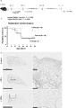
- FIG. 2 shows the therapeutic effects of adult-derived pulmonary fibroblasts (VPF) with high ADM expression and improved VCAM-1 expression on interstitial pneumonia and pulmonary fibrosis.
- A shows the animal study design. Seven days after administration of the bleomycin solution, the cells were administered to pathological model mice, and four weeks later, the mice were sacrificed and lungs were collected.
- B shows survival analysis diagrams by the Kaplan-meier method for each group. Dots refer to cutoff points.
- C is a photomicrograph showing Sirius red-stained images of each group.
- FIG. 2 shows the therapeutic effects of various fibroblasts two weeks after administration. A shows a survival analysis diagram.
- P-values were calculated by the log-rank (Mantel-Cox) test.
- B is a photomicrograph showing fibrotic regions and alveolar structures in the lungs of each group. Lung tissue sampled 14 days after cell administration was stained with Masson's Trichrome. BLM indicates the lung tissue on day 0 of cell administration, and Ctrl indicates the lung tissue of the saline-administered group (scale bar: 500 ⁇ m).
- FIG. 2 shows cell characteristics of primary recovered rPF and rVPF.
- A is a photomicrograph showing the state of rPF at each passage number. PN refers to Passage number.
- B is a photograph showing bright-field observation images of rPF and rVPF.
- C shows a flow cytometry analysis diagram.
- FIG. 2 shows the therapeutic effects of various fibroblasts two weeks after administration.
- hPF fibroblast marker
- CD106 VCAM-1 positive cell rates of various human lung fibroblasts by flow cytometry (FCM) analysis
- N 5, ****P ⁇ 0.0001, ns indicates no significant difference, calculated by 2-way ANOVA with Tukey's multiple comparisons test.
- hPF refers to human lung fibroblasts
- hPF+CKs refers to human lung fibroblasts cultured with TNF- ⁇ (50 ng/mL) and IL-4 (2 ng/mL) for 72 hours.
- hVPF refers to hPF+CKs obtained by cell sorting using CD106 as an index.
- One embodiment of the first invention is a pharmaceutical composition comprising fibroblasts with high ADM and/or HHEX expression.
- fibroblasts there is no limit to the origin of fibroblasts, and pluripotent stem cells such as embryonic stem cells (ES cells), induced pluripotent stem cells (iPS cells) and Muse cells, and adult stem cells such as mesenchymal stem cells can be differentiated. may be used. Also, primary cells collected from animals (including humans) may be used, or established cells may be used. Examples of fibroblast-derived organs include heart-derived, kidney-derived, skin-derived, and lung-derived. Fibroblasts are not particularly limited, but are preferably of adult origin. Fibroblasts derived from fibroblasts with high ADM and/or HHEX expression may originally express a certain amount of ADM and/or HHEX, or may not express ADM and/or HHEX at all. In addition, fibroblasts in the present embodiment may be cells that express cell surface markers that are expressed by normal fibroblasts, and further by the method for producing cells of the present invention described below. , cells that additionally express other markers, or cells that do not express some
- ADM mRNA expression level in fibroblasts in the product is 1.20 times or more, 1.40 times or more, 1.50 times or more, 1.60 times or more, 1.70 times or more, 1.80 times or more, 8. 1.90 times or more, 2.00 times or more, 2.30 times or more, 3.00 times or more, 4.00 times or more, 5.00 times or more, 6.00 times or more, 7.00 times or more;
- the increase is preferably 00 times or more, 9.00 times or more, 10.0 times or more, or more.
- the mRNA expression level of fibroblasts used as a control may be the mRNA expression level in untreated fibroblasts, or may be the average mRNA expression level of the entire cells after treatment.
- the percentage of ADM-positive (hereinafter also referred to as ADM+) fibroblasts may be 0.1% or more based on the number of cells, and 1% or more. well, may be 2% or more, may be 4% or more, may be 5% or more, may be 10% or more, may be 20% or more, may be 30% or more, 40% or more, 50% or more, 60% or more, 70% or more, 80% or more, 90% or more, 95% or more, may be 98% or more, or may be 99% or more.
- High expression of HHEX is not particularly limited.
- HHEX mRNA expression level in fibroblasts in the product is 1.20 times or more, 1.40 times or more, 1.50 times or more, 1.60 times or more, 1.70 times or more, 1.80 times or more, 8. 1.90 times or more, 2.00 times or more, 2.58 times or more, 3.00 times or more, 4.00 times or more, 5.00 times or more, 6.00 times or more, 7.00 times or more;
- the increase is preferably 00 times or more, 9.00 times or more, 10.0 times or more, or more.
- the mRNA expression level in fibroblasts used as a control may be the mRNA expression level in untreated fibroblasts, or may be the average mRNA expression level of all cells after treatment.
- the percentage of HHEX-positive (hereinafter also referred to as HHEX+) fibroblasts may be 0.03% or more based on the cell number, and 0.1% or more. may be, may be 1% or more, may be 2% or more, may be 4% or more, may be 5% or more, may be 10% or more, may be 20% or more well, may be 30% or more, may be 40% or more, may be 50% or more, may be 60% or more, may be 70% or more, may be 80% or more, It may be 90% or more, it may be 95% or more, it may be 98% or more, or it may be 99% or more.
- the lymphangiogenesis-promoting factor is preferably ADM or HHEX gene or protein.
- ADM is a gene having the nucleotide sequence represented by SEQ ID NO: 1, or 90% or more, 95% or more, or 98% of that nucleotide sequence.
- a gene having a nucleotide sequence identical to SEQ ID NO: 1, which hybridizes under stringent conditions with a sequence complementary to all or part of the nucleotide sequence represented by SEQ ID NO: 1, and has lymphangiogenic potential HHEX is a gene having a nucleotide sequence represented by SEQ ID NO: 2, or a gene having a nucleotide sequence that is 90% or more, 95% or more, or 98% or more identical to that nucleotide sequence It may be a gene that encodes a protein that hybridizes under stringent conditions with a sequence complementary to all or part of the base sequence represented by SEQ ID NO: 2 and that has lymphangiogenesis ability.
- the fibroblasts in the pharmaceutical composition of this embodiment may express CD106 as a cell surface marker.
- the fibroblasts in the pharmaceutical composition of this embodiment may be a fibroblast population including the above-described ADM and/or HHEX-highly expressed fibroblasts.
- the ratio of CD106-positive cells in a fibroblast population containing fibroblasts produced by the method for producing fibroblasts highly expressing ADM and/or HHEX described below may be high, but vice versa. does not necessarily hold true.
- fibroblasts In preparation of fibroblasts, the origin of fibroblasts is not particularly limited, as already described. On the other hand, they may be autologous cells, for example, cardiac fibroblasts isolated from the heart tissue of a patient suffering from heart disease, or kidney fibroblasts isolated by differentiating the patient's adult (somatic) stem cells. Prepare the blast cells. Fibroblasts collected by differentiating pluripotent stem cells such as iPS cells derived from autologous cells may also be used. In addition, cells other than autologous cells may be used, for example, cardiac fibroblasts isolated from cardiac tissue derived from a donor who provides cardiac cells, cardiac tissue prepared using an animal or the like, and donor adult (body) cells. Sex) Prepare cardiac fibroblasts isolated by differentiating stem cells. Fibroblasts collected by differentiating donor-derived pluripotent stem cells such as iPS cells and ES cells may also be used.
- autologous cells for example, cardiac fibroblasts isolated from the heart tissue of a patient suffering
- Prepared fibroblasts can be separated, typically by treatment with enzymes such as dispase, collagenase. Separation can be carried out by enzymes such as dispase, collagenase, or other treatments, such as mechanical treatments, if the required separation is possible.
- the method includes contacting fibroblasts with TNF- ⁇ and IL-4, and culturing the contacted fibroblasts under conditions that allow high ADM and/or HHEX expression.
- the method of contacting fibroblasts with TNF- ⁇ and IL-4 is not particularly limited as long as the induction of ADM and/or HHEX-highly expressing fibroblasts is not significantly impaired, and typically includes fibroblasts.
- a non-limiting example is the addition of TNF- ⁇ and IL-4 to the medium. Only one type of TNF- ⁇ and IL-4 may be added, or a plurality of types may be added. In the case of multiple types, each may be contacted with fibroblasts separately or simultaneously.
- TNF- ⁇ and IL-4 may be dissolved in water, media, serum, or dimethylsulfoxide (DMSO) prior to addition to media.
- DMSO dimethylsulfoxide
- fibroblasts can be cultured in a medium supplemented with TNF- ⁇ and IL-4, fibroblasts can be cultured while maintaining the expression level of ADM and/or HHEX in fibroblasts. Therefore, fibroblasts with high ADM and/or HHEX expression levels can be produced.
- the amount of TNF- ⁇ added is not particularly limited, but is usually 0.1 ng/mL or more, and may be 0.5 ng/mL or more, It may be 1 ng/mL or greater, may be 10 ng/mL or greater, or may be 50 ng/mL or greater.
- the upper limit is not particularly limited, and is usually 500 ng/mL or less, and may be 100 ng/mL or less.
- the amount of IL-4 added is not particularly limited, but is usually 0.1 ng/mL or more, may be 0.5 ng/mL or more, may be 1 ng/mL or more, or may be 2 ng/mL or more.
- the upper limit is not particularly limited, and is usually 10 ng/mL or less, may be 5 ng/mL or less, or may be 1 ng/mL or less.
- the ratio (weight ratio) of TNF- ⁇ and IL-4 to be added is TNF- ⁇ :IL-4, usually 10000:1 to 1:1, and may be 50000:1 to 10:1. Further, it is 1:1 to 1:10000, and may be 1:10 to 1:50000.
- fibroblasts contacted with TNF- ⁇ and IL-4 can produce ADM and/or HHEX+ fibroblasts. Cultivation of fibroblasts is not particularly limited as long as the fibroblasts can become ADM and/or HHEX positive, and may be performed by known cell culture methods.
- the medium used for culture can be appropriately set depending on the type of cells to be cultured, and for example, DMEM, ⁇ -MEM, RPMI-1640, HFDM-1(+) and the like can be used. Nutrients such as FCS and FBS, growth factors, cytokines, antibiotics and the like may be added to the medium. Furthermore, it may be cultured in a medium containing TNF- ⁇ and IL-4. As for the culture period, the culture time and culture days can be appropriately set according to the purpose such as until the cultured cells reach a desired number of cells and/or until the cultured cells have desired functions.
- the culture temperature can be appropriately set according to the type of cells to be cultured. °C or less, 50 °C or less, 45 °C or less, 40 °C or less. Culturing may be performed multiple times. For example, the purity of the desired fibroblasts can be improved by selection or enrichment, and culture can be performed each time.
- the production of fibroblasts may further include using an anti-CD106 antibody to select or enrich fibroblasts that highly express ADM and/or HHEX.
- the method of selection or enrichment is not particularly limited, and can be performed by a method known to those skilled in the art. (Miltenyi Biotec) for secondary immunostaining, and the stained cells are collected by autoMACS (Miltenyi Biotec). Moreover, selection or concentration may be performed at any timing.
- a known anti-CD106 antibody can be used, and a commercially available product can be obtained and used. Selection or enrichment with an anti-CD106 antibody can increase the percentage of CD106 positive fibroblasts in the fibroblast population, and can also increase the percentage of ADM and/or HHEX+ fibroblasts. .
- the cultured fibroblasts may be collected by detaching the cells with a protease such as trypsin, or by using a temperature-responsive culture dish to detach the cells by changing the temperature.
- the cells may be detached and collected using an instrument such as a cell scraper.
- Lymphangiogenesis can be promoted in vivo by administering or transplanting the pharmaceutical composition of this embodiment in vivo. Therefore, although not particularly limited, for example, it is possible to improve a state in which lymphangiogenesis is reduced due to organ tissue damage, defect, or malfunction, or to improve tissue fluid homeostasis or lipid and / or vitamin levels. It can ameliorate impaired transportation and activate the immune surveillance mechanism. It may also be administered to a patient with organ failure, thereby treating the organ failure. Organ failure refers to the condition in which an organ fails to perform its expected function and/or the degree of organ dysfunction to the extent that normal homeostasis cannot be maintained without clinical intervention.
- Specific organs include, but are not limited to, heart, kidney, adrenal gland, thyroid, parathyroid, vocal cord, thymus, cerebrum, cerebellum, brainstem, spinal cord, peripheral nerve, skin, muscle, eyeball, tongue, esophagus, stomach.
- organ failure in the heart or kidney may be a state in which the function of the organ is reduced compared to normal, particularly a state in which the function of the organ is reduced to 30% or less compared to normal. It may be about Fibrosis can be treated or ameliorated by in vivo administration or transplantation of the pharmaceutical composition of this embodiment.
- the onset site of fibrosis is not particularly limited, and examples thereof include the heart, lungs, and kidneys.
- the method of administering or implanting the pharmaceutical composition of this embodiment into a living body is not particularly limited, it can be administered, for example, by injection.
- a failing organ or its peri-infarct region e.g., subcapsular region
- a damaged region, fibrotic region, or its surrounding region in an organ that develops fibrosis states improvement and treatment of organ failure.
- the pharmaceutical composition may also be administered intravenously, arterially, in a lymph node, in a lymphatic vessel, in the bone marrow, subcutaneously, in the corpus cavernosum, intraperitoneally, in muscle, intrathecally, or in a joint cavity.
- Administration to veins, arteries, and lymph vessels may be by intravascular injection, injection through a catheter, or other known techniques.
- the injection method is not particularly limited, and known injection techniques such as needle injection and needle-free injection can be applied, and known catheter methods can be applied for injection, and are not particularly limited.
- the pharmaceutical composition of the present embodiment is not particularly limited, but may be applied to a living body as an artificial organ material. Anything is fine.
- This planar or three-dimensional cell tissue may be obtained by culturing fibroblasts alone, or co-cultivating fibroblasts and other cells, such as renal cells, and adding planar or three-dimensional cell tissue. It may be Examples of flat or three-dimensional cell tissue include, but are not limited to, cell sheets, cell fibers, and tissue formed by a 3D printer.
- the size and shape of the planar or three-dimensional cell tissue is not particularly limited as long as it can be applied to necrotic, damaged or fibrotic organ tissue and/or its surrounding area and/or lesion area.
- planar shape may be either a single layer or multiple layers.
- Conditions such as medium composition, temperature, and humidity in cell culture are not particularly limited as long as ADM and/or HHEX+ fibroblasts can be cultured and planar or three-dimensional cell tissue can be constructed.
- planar or three-dimensional cell tissue By applying such planar or three-dimensional cell tissue to necrotic, damaged or fibrotic organ tissue and/or its surrounding area, or as an artificial organ, necrotic, damaged or fibrotic organ tissue and/or its By exchanging with the surrounding area, improving the state of reduced lymphangiogenesis ability, improving homeostasis of interstitial fluid, improving the ability to transport lipids and / or vitamins, and immunosurveillance mechanism can be activated, organ failure can be treated, and fibrosis can be treated or ameliorated.
- the ratio of ADM and/or HHEX+ fibroblasts contained in the pharmaceutical composition is appropriately set based on the mode of administration or transplantation to a patient, etc., and a therapeutically effective amount of ADM and/or HHEX+ fibroblasts is used as an active ingredient. should be included.
- the ratio of ADM and / or HHEX + fibroblasts to the total amount of fibroblasts contained in the pharmaceutical composition is 0.03% or more based on the number of cells may be, may be 0.1% or more, may be 1% or more, may be 2% or more, may be 4% or more, may be 5% or more, may be 10% or more may be, may be 20% or more, may be 30% or more, may be 40% or more, may be 50% or more, may be 60% or more, may be 70% or more It may be 80% or more, it may be 90% or more, it may be 95% or more, it may be 98% or more, or it may be 99% or more.
- ADM and / or HHEX expression level of the control fibroblasts is 1.20 times or more, 1.40 times or more, 1.50 times or more, 1.60 times or more, 1.70 times or more, 1.80 times or more, 9. 1.90 times or more, 2.00 times or more, 3.00 times or more, 4.00 times or more, 5.00 times or more, 6.00 times or more, 7.00 times or more, 8.00 times or more; It may be 00 times or more, or 10.0 times or more.
- the number of ADM and/or HHEX+ fibroblasts contained in the pharmaceutical composition is, for example, 1.0 ⁇ 10 2 cells/mL or more, 1.0 ⁇ 10 3 cells/mL or more, 1.0 ⁇ 10 4 cells/mL or more, mL or more, 1.0 ⁇ 10 5 cells/mL or more, 1.0 ⁇ 10 6 cells/mL or more, 5.0 ⁇ 10 6 cells/mL or more, 1.0 ⁇ 10 7 cells/mL or more can.
- the number of CD106-positive fibroblasts contained in the injectable composition may be further increased or decreased depending on the severity of the disease.
- the method for confirming the effect of the above-mentioned treatment and/or improvement by the pharmaceutical composition is not particularly limited. recovery of organ functionality after administration of the composition or transplantation compared to before administration of the composition or transplantation; exemplified by reducing the degree of fragility.
- the pharmaceutical composition may contain other ingredients that are physiologically acceptable as a pharmaceutical composition.
- Such other components include physiological saline, cell preservation medium, cell culture medium, hydrogel, extracellular matrix, cryopreservation medium, and the like.
- fibrosis is treated or treated by administering or transplanting a pharmaceutical composition containing ADM and/or HHEX highly expressing cells to a fibrotic tissue and/or its surrounding area. It can be a way to improve.
- lymphangiogenesis is achieved by administering or transplanting the pharmaceutical composition containing the above-mentioned ADM and/or HHEX high-expressing cells to the incompetent organ tissue and/or its peripheral region. improving impaired function, improving homeostasis of interstitial fluid, reducing edema, improving impaired transport of lipids and/or vitamins, and activating the immune surveillance mechanism. It can also be a method of treating organ failure.
- the composition containing ADM and/or HHEX+ cells is administered to incompetent organ tissue and/or its surrounding area, or fibrotic tissue and/or its surrounding area, or It can be an injection for implantation or use as an implant material.
- An embodiment of the second invention is fibroblasts with high ADM expression.
- the fibroblasts of this embodiment are preferably CD106 positive.
- VEGF-C may be highly expressed.
- fibroblasts there is no limit to the origin of fibroblasts, and pluripotent stem cells such as embryonic stem cells (ES cells), induced pluripotent stem cells (iPS cells) and Muse cells, and adult stem cells such as mesenchymal stem cells can be differentiated. may be used. Also, primary cells collected from animals (including humans) may be used, or established cells may be used. Examples of fibroblast-derived organs include lung-derived, heart-derived, kidney-derived, and skin-derived. Fibroblasts are not particularly limited, but are preferably of adult origin. Fibroblasts from which the cells of the present embodiment are derived may originally express a certain amount of VEGF-C or ADM, or may not express them at all. In addition, fibroblasts in the present embodiment may be cells that express cell surface markers that are expressed by normal fibroblasts, and further by the method for producing cells of the present invention described below. , cells that additionally express other markers, or cells that do not express some markers.
- ES cells embryonic stem cells
- VEGF-C mRNA expression level in cells is 1.20-fold or more, 1.40-fold or more, 1.50-fold or more, 1.60-fold or more, 1.70-fold or more, 1.80-fold or more, 1.90-fold times or more, 2.00 times or more, 2.60 times or more, 2.99 times or more, 3.00 times or more, 3.38 times or more, 3.52 times or more, 4.00 times or more, 4.11 times or more , 4.70 times or more, 5.00 times or more, 6.00 times or more, 7.00 times or more, 8.00 times or more, 9.00 times or more, 10.0 times or more, or more preferably.
- the mRNA expression level of fibroblasts used as a control may be the mRNA expression level in untreated fibroblasts, or may be the average mRNA expression level of the entire cells after treatment.
- the percentage of VEGF-C positive (hereinafter also referred to as VEGF-C+) fibroblasts may be 0.03% or more based on the number of cells, respectively.
- ADM mRNA expression level is 1.20 times or more, 1.40 times or more, 1.43 times or more, 1.50 times or more, 1.60 times or more, 1.70 times or more, 1.80 times or more, 1 .90 times or more, 1.91 times or more, 1.97 times or more, 2.00 times or more, 2.46 times or more, 3.00 times or more, 3.15 times or more, 4.00 times or more, 5.00 times It is preferable that the increase is a multiple of times or more, 6.00 times or more, 7.00 times or more, 8.00 times or more, 9.00 times or more, 10.0 times or more, or more.
- the mRNA expression level of fibroblasts used as a control may be the mRNA expression level in untreated fibroblasts, or may be the average mRNA expression level of the entire cells after treatment. Further, when the protein expression level is measured by flow cytometry, the percentage of ADM-positive (hereinafter also referred to as ADM+) fibroblasts may be 0.1% or more based on the number of cells, and 1% or more.
- ком ⁇ онент may be 2% or more, may be 4% or more, may be 5% or more, may be 10% or more, may be 20% or more, may be 30% or more, 40% or more, 50% or more, 60% or more, 70% or more, 80% or more, 90% or more, 95% or more, may be 98% or more, or may be 99% or more.
- the nucleotide sequences and amino acid sequences of VEGF-C and ADM are not particularly limited.
- ADM is a gene having the nucleotide sequence represented by SEQ ID NO: 1 or 15, or 90% or more, 95% or more of the nucleotide sequence % or more or 98% or more of the same base sequence, hybridizes under stringent conditions with a complementary sequence to all or part of the base sequence represented by SEQ ID NO: 1 or 15, and has ADM It may be a gene encoding a protein having activity.
- the cells of this embodiment preferably express CD106 as a cell surface marker.
- This embodiment may be a fibroblast population containing the above-described fibroblasts with high ADM expression.
- fibroblasts In preparation of fibroblasts, the origin of fibroblasts is not particularly limited, as already described. On the other hand, they may be autologous cells, for example lung fibroblasts isolated from lung tissue of a patient suffering from lung disease, or lung fibroblasts isolated by differentiating the patient's adult (somatic) stem cells. Blast cells may be prepared. Fibroblasts collected by differentiating pluripotent stem cells such as iPS cells derived from autologous cells may also be used.
- cells other than autologous cells may be used, for example, lung fibroblasts isolated from lung tissue derived from donors who provide lung cells, lung fibroblasts isolated from lung tissue prepared using animals, etc., donor adult (body) sex) stem cells may be differentiated to provide isolated pulmonary fibroblasts. Fibroblasts collected by differentiating donor-derived pluripotent stem cells such as iPS cells and ES cells may also be used.
- Prepared fibroblasts can be separated, typically by treatment with enzymes such as dispase, collagenase. Separation can be carried out by enzymes such as dispase, collagenase, or other treatments, such as mechanical treatments, if the required separation is possible.
- Fibroblast culture Fibroblasts may be contacted with TNF- ⁇ and IL-4 by the method described below, and cultured until the desired number of cells is obtained and/or the desired function is provided. good too.
- Culture conditions are not limited, and known cell culture methods may be used.
- the medium used for culture can be appropriately set depending on the type of cells to be cultured, and for example, HFDM-1(+), DMEM, ⁇ -MEM, RPMI-1640 and the like can be used.
- Nutrients such as NBCS, FCS and FBS, growth factors, cytokines, antibiotics and the like may be added to the medium. Furthermore, it may be cultured in a medium containing TNF- ⁇ and IL-4.
- the culture time and the number of culture days can be appropriately set according to the purpose such as until the cultured cells reach the desired number of cells and/or until the cultured cells have desired functions. For example, 1 hour, 2 hours, 3 hours, 4 hours, 5 hours, 6 hours, 7 hours, 8 hours, 9 hours, 10 hours, 11 hours, 12 hours, 15 hours, 18 hours, 21 hours, 1 day, 2 days, 3 days, 4 days, 5 days, 6 days, 7 days, 8 days, 9 days, 10 days, 2 weeks, 1 month, 2 months, 3 months, 6 months, etc. be done.
- the culture temperature can be appropriately set according to the type of cells to be cultured. °C or less, 50 °C or less, 45 °C or less, 40 °C or less. Culturing may be performed multiple times. For example, the purity of the desired fibroblasts can be improved by selection or enrichment, and culture can be performed each time.
- the cultured fibroblasts may be collected by detaching the cells with a protease such as trypsin.
- An instrument such as a scraper may be used to dislodge and collect the cells.
- Another embodiment of the second invention is a method for producing fibroblasts highly expressing ADM, comprising contacting fibroblasts with TNF- ⁇ and IL-4, and contacting fibroblasts
- a method for producing fibroblasts highly expressing ADM comprising culturing under conditions that allow high expression of ADM.
- the fibroblasts of this embodiment are preferably CD106 positive.
- VEGF-C may be highly expressed.
- the method of contacting fibroblasts with TNF- ⁇ and IL-4 is induction of fibroblasts with high ADM expression, or induction of high ADM expression and high VEGF-C expression and / or CD106 positive fibroblasts.
- TNF- ⁇ and IL-4 may be contacted with fibroblasts separately or simultaneously.
- TNF- ⁇ and IL-4 may be dissolved in water, media, serum, or dimethylsulfoxide (DMSO) prior to addition to media.
- fibroblasts can be cultured in a medium supplemented with TNF- ⁇ and IL-4, the expression level of ADM in fibroblasts is maintained, and the expression level of VEGF-C is maintained. , can culture fibroblasts. Therefore, it is possible to produce fibroblasts with high ADM expression level and further high VEGF-C expression level.
- the amount of TNF- ⁇ added is not particularly limited, but is usually 0.1 ng/mL or more, and may be 0.5 ng/mL or more, It may be 1 ng/mL or more, 10 ng/mL or more, or 50 ng/mL or more.
- the upper limit is not particularly limited, and is usually 500 ng/mL or less, and may be 100 ng/mL or less.
- the amount of IL-4 added is not particularly limited, but is usually 0.1 ng/mL or more, may be 0.5 ng/mL or more, may be 1 ng/mL or more, or may be 2 ng/mL or more.
- the upper limit is not particularly limited, and is usually 10 ng/mL or less, may be 5 ng/mL or less, or may be 2 ng/mL or less.
- the ratio (weight ratio) of TNF- ⁇ and IL-4 to be added is TNF- ⁇ :IL-4, usually 10000:1 to 1:1, and may be 50000:1 to 10:1. Further, it is 1:1 to 1:10000, and may be 1:10 to 1:50000.
- fibroblasts with high ADM expression can be produced.
- the fibroblasts may further be highly VEGF-C-expressing. Cultivation of fibroblasts is not particularly limited as long as the conditions allow fibroblasts to express ADM at a high level, and may be performed by known cell culture methods. Also, the conditions may be such that high VEGF-C expression can occur. It is preferable to be under conditions that can become CD106 positive. The method described in the section (Culture of fibroblasts) in the method for producing fibroblasts is exemplified.
- anti-CD106 antibodies when fibroblasts are CD106-positive, anti-CD106 antibodies can be used to select or enrich CD106-positive fibroblasts with high ADM expression.
- the fibroblasts may further be highly VEGF-C-expressing.
- the method of selection or enrichment is not particularly limited, and can be performed by a method known to those skilled in the art. (Miltenyi Biotec) for secondary immunostaining, and the stained cells are collected by autoMACS (Miltenyi Biotec). Moreover, selection or concentration may be performed at any timing.
- a known anti-CD106 antibody can be used, and a commercially available product can be obtained and used. Selection or enrichment with an anti-CD106 antibody can increase the proportion of high ADM-expressing fibroblasts in the fibroblast population.
- fibroblasts highly expressing ADM are obtained, and the fibroblasts or a fibroblast population containing the cells is another embodiment of the second invention.
- the fibroblasts may further have high VEGF-C expression and may be CD106 positive.
- Fibroblasts are stimulated by TNF- ⁇ and IL-4 to increase the expression of ADM and VEGF-C, so fibroblasts with high ADM expression and fibroblasts with high VEGF-C expression can be suitably used as a therapeutic composition for diseases in which lymphangiogenic potential is reduced and pulmonary diseases such as pulmonary fibrosis and interstitial pneumonia.
- fibroblasts are not particularly limited in origin, but are preferably lung-derived.
- Another embodiment may be a fibroblast population containing cells obtained from the aforementioned ADM-highly expressing fibroblasts.
- Another embodiment of the second invention is a cell population comprising CD106-positive lung-derived fibroblasts.
- the fibroblasts of this embodiment may have high ADM expression or high VEGF-C expression.
- fibroblasts there is no limit to the origin of fibroblasts, and pluripotent stem cells such as embryonic stem cells (ES cells), induced pluripotent stem cells (iPS cells) and Muse cells, and adult stem cells such as mesenchymal stem cells can be differentiated. may be used. Also, primary cells collected from animals (including humans) may be used, or established cells may be used. Fibroblasts are not particularly limited, but are preferably of adult origin. Fibroblasts from which the cells of the present embodiment are derived may originally express a certain amount of VEGF-C or ADM, or may not express them at all. In addition, fibroblasts in the present embodiment may be cells that express cell surface markers that are expressed by normal fibroblasts, and further by the method for producing cells of the present invention described below. , cells that additionally express other markers, or cells that do not express some markers.
- ES cells embryonic stem cells
- iPS cells induced pluripotent stem cells
- Muse cells induced pluripotent stem cells
- the ratio of CD106-positive fibroblasts to all fibroblasts is not particularly limited, but may be greater than 18.01%, may be greater than 27.97%, based on the number of cells, may be greater than 30%, may be greater than 31.38%, may be greater than 34.79%, may be greater than 40%, may be greater than 50%, may be greater than 60% Well, it may be greater than 70%, it may be greater than 80%, it may be greater than 90%, it may be greater than 95%, it may be greater than 97.10%.
- the number of cells it may be 18.01% or more, 27.97% or more, 30% or more, 31.38% or more, 34.79% or more may be 40% or more, may be 50% or more, may be 60% or more, may be 70% or more, may be 80% or more, may be 90% or more may be 95% or more, and may be 97.10% or more.
- the fibroblast population of this embodiment may further be a CD90-positive fibroblast population.
- the ratio of CD106-positive and CD90-positive fibroblasts to all fibroblasts is not particularly limited, but may be more than 17.97%, 27.91% may be greater than, may be greater than 30%, may be greater than 31.21%, may be greater than 34.51%, may be greater than 40%, may be greater than 50%, It may be greater than 60%, it may be greater than 70%, it may be greater than 80%, it may be greater than 90%, it may be greater than 95%, it may be greater than 96.29%.
- the number of cells it may be 17.97% or more, 27.91% or more, 30% or more, 31.21% or more, 34.51% or more may be 40% or more, may be 50% or more, may be 60% or more, may be 70% or more, may be 80% or more, may be 90% or more may be 95% or more, and may be 96.29% or more.
- fibroblasts In preparation of fibroblasts, the origin of fibroblasts is not particularly limited, as already described. On the other hand, they may be autologous cells, for example lung fibroblasts isolated from lung tissue of a patient suffering from lung disease, or lung fibroblasts isolated by differentiating the patient's adult (somatic) stem cells. Blast cells may be prepared. Fibroblasts collected by differentiating pluripotent stem cells such as iPS cells derived from autologous cells may also be used.
- cells other than autologous cells may be used, for example, lung fibroblasts isolated from lung tissue derived from donors who provide lung cells, lung fibroblasts isolated from lung tissue prepared using animals, etc., donor adult (body) sex) stem cells may be differentiated to provide isolated pulmonary fibroblasts. Fibroblasts collected by differentiating donor-derived pluripotent stem cells such as iPS cells and ES cells may also be used.
- Prepared fibroblasts can be separated, typically by treatment with enzymes such as dispase, collagenase. Separation can be carried out by enzymes such as dispase, collagenase, or other treatments, such as mechanical treatments, if the required separation is possible.
- Fibroblast culture Fibroblasts may be contacted with TNF- ⁇ and IL-4 by the method described below, and cultured until the desired number of cells is obtained and/or the desired function is provided. good too.
- Culture conditions are not limited, and known cell culture methods may be used.
- the medium used for culture can be appropriately set depending on the type of cells to be cultured, and for example, HFDM-1(+), DMEM, ⁇ -MEM, RPMI-1640 and the like can be used.
- Nutrients such as NBCS, FCS and FBS, growth factors, cytokines, antibiotics and the like may be added to the medium. Furthermore, it may be cultured in a medium containing TNF- ⁇ and IL-4.
- the culture time and the number of culture days can be appropriately set according to the purpose such as until the cultured cells reach the desired number of cells and/or until the cultured cells have desired functions. For example, 1 hour, 2 hours, 3 hours, 4 hours, 5 hours, 6 hours, 7 hours, 8 hours, 9 hours, 10 hours, 11 hours, 12 hours, 15 hours, 18 hours, 21 hours, 1 day, 2 days, 3 days, 4 days, 5 days, 6 days, 7 days, 8 days, 9 days, 10 days, 2 weeks, 1 month, 2 months, 3 months, 6 months, etc. be done.
- the culture temperature can be appropriately set according to the type of cells to be cultured. °C or less, 50 °C or less, 45 °C or less, 40 °C or less. Culturing may be performed multiple times. For example, the purity of the desired fibroblasts can be improved by selection or enrichment, and culture can be performed each time.
- the cultured fibroblasts may be collected by detaching the cells with a protease such as trypsin.
- An instrument such as a scraper may be used to dislodge and collect the cells.
- CD106-positive fibroblasts Cell sorting of CD106-positive fibroblasts
- the fibroblasts may be subjected to selection or enrichment for CD106 positive fibroblasts. Selection or enrichment may be omitted if CD106 positive fibroblasts are available without selection or enrichment. Selection or enrichment of CD106-positive fibroblasts is exemplified by flow cytometry, magnetic bead method, affinity column method, panning method, etc. using anti-CD106 antibody (anti-VCAM-1 antibody). Specifically, a magnetic cell sorting method (MACS), a fluorescent labeled cell sorting method (FACS), or the like can be used. A commercially available anti-CD106 antibody may be used, or one prepared by a known method may be used.
- CD106-positive fibroblasts may be obtained by introducing a drug resistance gene and excluding CD106-negative fibroblasts.
- a fluorescent protein-encoding gene may be introduced and fluorescent protein-positive cells may be isolated using FACS or the like.
- techniques such as real-time PCR, next-generation sequencing, in situ hybridization, and the like may be used to confirm the expression of the CD106 gene in cells and isolate the CD106 gene expression group.
- Another embodiment of the second invention is a pharmaceutical composition comprising the above-mentioned ADM-highly expressing fibroblasts or cell population thereof, or a pharmaceutical composition comprising the above-mentioned CD106-positive lung-derived fibroblast cell population.
- the fibroblasts of this embodiment are preferably CD106 positive.
- VEGF-C may be highly expressed.
- lymphangiogenesis can occur in an organ such as the lung. It can improve the state of declining ability. Therefore, although not particularly limited, for example, in organs such as lungs, it is possible to improve a state in which lymphangiogenic potential is reduced. It is particularly useful in treating pulmonary diseases such as pulmonary fibrosis or interstitial pneumonitis.
- fibroblasts are not particularly limited in origin, but are preferably lung-derived.
- the method of administering or implanting the pharmaceutical composition of this embodiment into a living body is not particularly limited, it can be administered, for example, by injection.
- the pharmaceutical composition may also be administered intravenously, arterially, in a lymph node, in a lymphatic vessel, in the bone marrow, subcutaneously, in the corpus cavernosum, intraperitoneally, in muscle, intrathecally, or in a joint cavity.
- Administration to veins, arteries, and lymph vessels may be by intravascular injection, injection through a catheter, or other known techniques.
- the injection method is not particularly limited, and known injection techniques such as needle injection and needle-free injection can be applied, and known catheter methods can be applied for injection, and are not particularly limited.
- the pharmaceutical composition of the present embodiment is not particularly limited, but may be applied to a living body as an artificial organ material. Anything is fine.
- This planar or three-dimensional cell tissue may be obtained by culturing fibroblasts alone, or co-cultivating fibroblasts and other cells, such as lung cells, and adding planar or three-dimensional cell tissue. It may be Examples of flat or three-dimensional cell tissue include, but are not limited to, cell sheets, cell fibers, and tissue formed by a 3D printer.
- the size and shape of the planar or three-dimensional cell tissue are not particularly limited as long as it can be applied to damaged or fibrotic lung tissue and/or its peripheral region and/or lesion region.
- the planar shape may be either a single layer or multiple layers.
- conditions such as medium composition, temperature, and humidity in cell culture can culture VEGF-C and / or CD106 positive fibroblasts with high ADM expression, and as long as planar or three-dimensional cell tissue can be constructed.
- the culture conditions described in the section (Fibroblast culture) and/or (Fibroblast collection) in the method for producing fibroblasts can be exemplified.
- the ratio of high-ADM-expressing fibroblasts contained in the pharmaceutical composition is appropriately set based on the mode of administration or transplantation to a patient, and a therapeutically effective amount of high-ADM-expressing fibroblasts is included as an active ingredient. as long as it is When other fibroblasts are contained in the pharmaceutical composition, for example, the ratio of fibroblasts highly expressing ADM to the total amount of fibroblasts contained in the pharmaceutical composition is 0.03% or more based on the number of cells.
- the ratio of CD106-positive lung-derived fibroblasts contained in the pharmaceutical composition is appropriately set based on the mode of administration or transplantation to a patient, and a therapeutically effective amount of ADM-highly expressed fibroblasts is used as an active ingredient. should be included. Specifically, the ratio of CD106-positive lung-derived fibroblasts described in the section ⁇ Cell population containing CD106-positive lung-derived fibroblasts> can be used.
- the number of fibroblasts with high ADM expression or the number of CD106-positive lung-derived fibroblasts contained in the pharmaceutical composition is, for example, 1.0 ⁇ 10 2 cells/mL or more, or 1.0 ⁇ 10 3 cells/mL or more. , 1.0 ⁇ 10 4 cells/mL or more, 1.0 ⁇ 10 5 cells/mL or more, 1.0 ⁇ 10 6 cells/mL or more, 5.0 ⁇ 10 6 cells/mL or more, 1.0 ⁇ 10 It can be 7 cells/mL or more.
- the number of cells contained in the injectable composition may be further increased or decreased depending on the severity of the disease.
- the method for confirming the effect of the above-mentioned treatment and/or improvement by the pharmaceutical composition is not particularly limited.
- Recovery of functionality of an organ such as lung after composition administration or transplantation compared to before composition administration or transplantation, and organ such as lung after composition administration or transplantation compared to before composition administration or transplantation For example, the degree of damage and the degree of fibrosis are reduced.
- the pharmaceutical composition may contain other ingredients that are physiologically acceptable as a pharmaceutical composition.
- Such other components include physiological saline, cell preservation medium, cell culture medium, hydrogel, extracellular matrix, cryopreservation medium, and the like.
- the above-mentioned high ADM-expressing fibroblasts, cell populations thereof, CD106-positive lung-derived fibroblast cell populations, or pharmaceutical compositions containing these are used in organs such as lungs. It can be a method of treating pulmonary diseases such as pulmonary fibrosis or interstitial pneumonitis by administering or implanting the tissue and/or the surrounding area.
- the fibroblasts of this embodiment are preferably CD106 positive.
- VEGF-C may be highly expressed.
- the above-mentioned high ADM-expressing fibroblasts, cell populations thereof, CD106-positive lung-derived fibroblast cell populations, or pharmaceutical compositions containing these are used in lungs, etc. administration or transplantation to the organ tissue and/or its peripheral region, thereby improving the state of reduced lymphangiogenic potential.
- the fibroblasts of this embodiment are preferably CD106 positive.
- VEGF-C may be highly expressed.
- the fibroblasts with high ADM expression, cell populations thereof, or compositions containing these are administered or transplanted into organ tissues such as lungs and/or their peripheral regions. It can be used as an injectable or implantable material for The fibroblasts of this embodiment are preferably CD106 positive. Furthermore, VEGF-C may be highly expressed.
- Adult human hearts were purchased from Science Care, and adult human cardiac fibroblasts were isolated after enzymatic treatment with Liberase MNP-S (Roche).
- the cells were added with 1% newborn calf serum (NBCS, Hyclone) and recombinant human epidermal growth factor (rh-EGF)-containing serum-free culture medium for fibroblasts (HFDM-1 (+), Cell After 5 passages, the cells were cultured in a cell culture dish, and the untreated group (NT), TNF- ⁇ (50 ng/mL) (Peprotech) and IL-4 ( 2 ng/mL) (Wako) was treated with a mixture of two drugs (+CKs).All groups were treated with HFDM-1 (+) supplemented with 1% NBCS as a basal medium for 1 day (24 hours). cultured.
- NBCS newborn calf serum
- rh-EGF human epidermal growth factor
- Fibroblasts were immunofluorescently stained with BV421 mouse anti-human CD106 (VCAM1) antibody (BD Biosciences) and human CD90-PE (Miltenyi Biotec, Bergisch Gladbach, Germany).
- Isotype controls were BV421 mouse IgG1; kappa isotype control (BD Biosciences) and REA control (S)-PE (Miltenyi Biotec).
- the viable cells were allowed to react with the antibody for 30 minutes at 4°C.
- the immunofluorescence-stained cells were analyzed with a flow cytometer (MACSQuant Analyzer 10, Miltenyi Biotec). After recognizing the cell area with FSC-A and SSC-A, the positive cell rate (%, in terms of cell number) for CD106 protein was evaluated.
- ⁇ Increased expression of ADM and/or HHEX gene in human adult-derived cardiac fibroblasts Human adult-derived cardiac fibroblasts were cultured in cell culture dishes, untreated (NT), TNF- ⁇ (50 ng/mL) (Peprotech) and IL-4 (2 ng/mL) (Wako ) was treated with a two-drug mixture (+CKs). All groups were cultured for 1 day (24 hours) with HFDM-1(+) supplemented with 1% NBCS as the basal medium.
- the primers used were human mRNA sequences published in NCBI (ADM, NM_001124.3 (SEQ ID NO: 1); HHEX, NM_002729.5 (SEQ ID NO: 2); ACTB ( ⁇ -Actin), NM_001101.5 (SEQ ID NO: Based on 3)), the sequences were designed so that the annealing temperature calculated by the nearest neighbor base pair method was around 60° C. and one or more introns were sandwiched (SEQ ID NOs: 4 to 9).
- the 5' nuclease probe sequence is also designed based on the published human mRNA sequence data, the annealing temperature of this probe is 5 ° C.
- the binding position of this probe is on the gene to be measured, the forward primer and the binding position of the reverse primer.
- the 5′ end of the probe was bound to the fluorescent dye 6-FAM
- the quencher dye ZEN was bound to the 9 bases 3′ side of the probe
- the second quencher dye Iowa Black FQ was bound to the 3′ end.
- a dual quencher probe is used to reduce background signals in RT-PCR analysis (SEQ ID NOS: 10-12). Synthesis of all the above primers and probes was entrusted to Integrated DNA Technologies (Table 1).
- RNA extraction> Extraction of total RNA from cardiac fibroblasts was performed using the Qiagen RNeasy Plus Mini Kit (QIAGEN). At that time, the cell lysate was prepared using a QIAshredder (QIAGEN), and other operations including DNase treatment followed the protocol recommended by the kit manufacturer. The concentration of the obtained total RNA was determined by measuring absorbance at a wavelength of 260 nm using a Nanodrop One/One spectrophotometer (Thermo Fisher Scientific). At the same time, the A260/A280 value indicating the degree of protein contamination was calculated and confirmed to be A260/A280>2.0.
- Reverse transcription reaction and RT-PCR were performed together in the same tube using One Step PrimeScript 3 RT-qPCR Mix, with UNG Kit (TaKaRa).
- the reaction was performed with a reaction solution (total volume of 12.5 ⁇ L) containing 1 ⁇ One Step PrimeScript 3 RT-qPCR Mix, with UNG, 0.2 ⁇ M Forward & Reverse primers, 0.2 ⁇ M specific probe, and 10 ng template RNA solution.
- the reverse transcription reaction was first performed by heating at 52° C. for 5 minutes, followed by heating at 95° C. for 10 seconds to inactivate the reverse transcriptase. Subsequently, a cycle of heating at 95° C. for 5 seconds and 60° C.
- the calibration curve for each gene had a correlation coefficient of 0.98 or more and an amplification efficiency of 90% or more, and was considered sufficient as a calibration curve.
- the content was calculated as a relative value. This relative value was normalized by the value of ⁇ -Actin.
- lymphatic angiogenesis assay human lymphatic endothelial cells collected from heart-derived cells were co-cultured with various human cardiac fibroblasts by a method known to those skilled in the art, and cardiac lymphatic vessels were formed by various cardiac fibroblasts. Evaluation of the regenerative effect was performed (Non-Patent Document 15). Specifically, human lymphatic endothelial cells collected from human heart-derived cells and various human adult-derived cardiac fibroblasts were seeded at a concentration of 2.45 ⁇ 10 5 cells/cm 2 at a ratio of 2:8, followed by EGM. -2MV Microvascular Endothelial Cell Growth Medium-2 BulletKit (Lonza, Basel, Switzerland) for 3 days.
- the cells were fixed with 4% paraformaldehyde, permeabilized with 0.1% TritonX, and then subjected to immunofluorescence staining.
- Anti-VE-Cadherin rabbit polyclonal antibody (abcam, Cambridge, UK) and anti-Vimentin mouse polyclonal antibody (abcam) were used as primary antibodies. Secondary antibodies used were goat anti-rabbit IgG H&L (Alexa Fluor 488) (abcam) and goat anti-mouse IgG H&L (Alexa Fluor 647) (abcam). Nuclear staining was performed with Hoechst 33258 solution (Dojindo Laboratories, Kumamoto, Japan).
- an anisotropic PDE-based filter (Icy plug-in: “Membrane Filter”, v.0.1.0.1) is applied to correct the image, enhancing the contrast of the filtered image, normalizing it, and removing the background ROI was defined by subtracting Finally, the resulting images were analyzed with the Angiogenesis Analyzer macro. A detailed protocol can be found in dx. doi. org/10.17504/protocols. io. Published online on bhtaj6ie. Quantitative data are graphical representations of the total length of mature lymphatic vessels and the number of junctions, and expressed as fold increase, with the mean NT being 1.
- Cells were purchased from the following vendors: Human Adult Cardiac Fibroblasts (Lonza); Human Fetal Cardiac Fibroblasts (Cell Applications, San Diego, Calif.). The cells were expanded and cultured in a fibroblast culture medium (HFDM-1(+) medium (Cell Science Laboratory Co., Ltd., Miyagi, Japan) and used for transplantation experiments.
- HFDM-1(+) medium Cell Science Laboratory Co., Ltd., Miyagi, Japan
- Fibroblasts were primary immunostained with human CD106 (VCAM-1)-biotin (Miltenyi Biotec) or human CD90-biotin (Miltenyi Biotec) and secondary immunostained with anti-biotin microbeads (Miltenyi Biotec). .
- the stained cells were autoMACS (Miltenyi Biotec) and CD106 and CD90 positive cells were collected.
- LVEF left ventricular ejection fraction
- fibroblasts administered to chronic heart failure model rats.
- the various fibroblasts that had been cryopreserved were thawed, diluted with high-glucose DMEM+10% NBCS, and the viable cell count was 2.0 ⁇ 10 6 cells/50 ⁇ L for each individual. dosed.
- the animals were kept anesthetized by the same method as the model preparation, and under artificial respiration management, the infarcted lesion was divided into 2 sites, and 50 ⁇ L of the total volume of cell suspension was administered using a catheter with a 30G needle.
- SR Sirius red
- HE hematoxylin and eosin
- the adult-derived cardiac fibroblast administration group was imaged with a virtual slide scanner (NanoZoomer S210, Hamamatsu Photonics, Shizuoka, Japan). The embryo-derived cardiac fibroblast-administered group was imaged with an Aperio ScanScope slide scanner.
- HE-stained images were qualitatively scored for the following items: Inflammatory cell aggregation, proliferation of fibroblasts, fibrosis, lime deposition, thinning of the left ventricular. The degree of pathological change was qualitatively evaluated as no change (score 0), mild (score 1), moderate (score 2), severe (score 3).
- RNAscope Target Probe Rn-Lyve-1-C2 Bio-Techne, Minneapolis, Minn.
- the probes were treated with a HybEZ Oven (Bio-Techne).
- Cryosections were labeled with anti-cardiac troponin T (cTnT) antibody (Abcam, Cambridge, UK), anti-prospero-related homeobox 1 (Prox-1) antibody (Proteintech, Rosemont, Ill.), and anti-Vascular endothelial growth Factor 3 (Vector Rec. ) immunohistochemical staining was performed with an antibody (Bioss Antibodies, Woburn, Mass.), and imaging was performed with an FV1200 confocal laser microscope (Olympus, Tokyo, Japan).
- Example A1 Improvement of CD106-positive cell rate of adult-derived cardiac fibroblasts by contact with TNF- ⁇ and IL-4>
- Human adult-derived cardiac fibroblasts were cultured in a cell culture dish to provide an NT group and a +CKs group.
- Human adult-derived cardiac fibroblasts in all groups had a flat, spindle-shaped shape with a typical fibroblast-like shape (Fig. 1A).
- the NT group had 48.88% (CD90), 0.20% (CD106), 0.14% (both positive, DP), while the +CKs group was 57.55% (CD90), 50.78% (CD106), 34.76% (DP), respectively, CD90 positive cell rate, CD106 positive cell rate and both positive cell rate (%) were greatly improved (Fig. 1B). These results indicated that contacting fibroblasts with TNF- ⁇ and IL-4 improved the CD90, CD106, and both-positive cell ratios (%) of fibroblasts.
- Example A2 Contact with TNF- ⁇ and IL-4 enhanced the expression levels of ADM and/or HHEX in adult-derived cardiac fibroblasts>
- the gene expression levels of ADM and HHEX in fibroblasts in all groups were normalized by the gene expression level of ⁇ -Actin, and the gene expression level of ADM and/or HHEX in the NT group (average value) was set to 1.00, It is expressed in Fold increase. As a result, it was shown that the ADM expression level was improved 2.30-fold in the +CKs group, confirming a significant difference in mRNA expression level (Fig. 2A). On the other hand, the expression level of HHEX was shown to be improved 2.58-fold in the +CKs group, confirming a significant difference in the expression level of mRNA between all groups (Fig. 2B). These results indicated that contacting fibroblasts with TNF- ⁇ and IL-4 improved the gene expression levels of ADM and/or HHEX in fibroblasts.
- Example A3 Adult-derived cardiac fibroblasts in contact with TNF- ⁇ and IL-4 have a high neoplastic effect on cardiac lymphatic vessels>
- human cardiovascular endothelial cells containing lymphatic endothelial cells were expanded.
- a test was performed using lymphatic endothelial cells that are both positive for VE-cadherin and PDPN, which are endothelial cell markers.
- Human lymphatic endothelial cells collected from heart-derived cells and various human adult-derived cardiac fibroblasts were co-cultured, and lymphatic vessels were observed by immunofluorescence staining.
- Example A4 Adult-derived cardiac fibroblasts that are both CD90- and CD106-positive by contact with TNF- ⁇ and IL-4 have a therapeutic effect on fibrosis>
- a number of previous studies have reported that lymphangiogenesis can treat scarring (fibrosis) that forms with organ tissue damage.
- the following adult-derived cardiac fibroblasts were first prepared (Fig. 4): untreated adult-derived cardiac fibroblasts (A-HCF); TNF- ⁇ (50 ng/mL) and IL-4 (2 ng / mL) was added and cultured for 72 hours.
- FIG. 4 shows the results of evaluating the characteristics of each fibroblast by FCM. The following cells were administered to chronic heart failure model rats, and fibrosis regions were observed by Sirius red staining (Fig. 5).
- Test 1 A-HCF administration group; uA-HCF administration group; uA90-HCF administration group; Control group (group administered only medium); Microscopic observations were performed on groups (Figs. 5A and B).
- Test 2 The F-VCF-administered group and the control group (group to which only the medium was administered) were microscopically observed (FIGS. 5C and 5D).
- fibroblasts to which TNF- ⁇ (50 ng/mL) and IL-4 (2 ng/mL) were added had a smaller area of myocardial fibrotic regions, and in particular, the effects of TNF- ⁇ and IL-4 reduced CD90 and CD106 In the group (uA90-HCF) to which only the cell population that became both positive was administered, the fibrotic area was remarkably small. Interestingly, as a result of administering the naturally occurring CD90 and CD106 positive cardiac fibroblasts (F-VCF) administration group, no therapeutic effect on the fibrosis area was observed.
- F-VCF naturally occurring CD90 and CD106 positive cardiac fibroblasts
- TNF- ⁇ 50 ng/mL
- IL-4 2 ng/mL
- functional differences in fibrosis of artificially produced lymphangiogenic fibroblasts could be found. rice field.
- TNF- ⁇ 50 ng/mL
- IL-4 2 ng /mL
- the scores of Proliferation of fibroblast, Fibrosis, and Lime deposition tended to decrease, and only the cell population that became both CD90 and CD106 positive due to the effects of TNF- ⁇ and IL-4 was administered.
- lymphatic vessel endothelial cells recruited to myocardial infarction lesions were observed by staining lymphatic vessel endothelial receptor 1 (Lyve-1), which is a lymphatic vessel endothelial marker, by in situ hybridization.
- Lyve-1 lymphatic vessel endothelial receptor 1
- fibroblasts supplemented with TNF- ⁇ (50 ng/mL) and IL-4 (2 ng/mL) had a lymphatic endothelial cell recruitment effect (lymphangiogenesis effect).
- TNF- ⁇ 50 ng/mL
- IL-4 2 ng/mL
- Example A5 In vivo effect of lymphangiogenesis by both CD90- and CD106-positive cardiac fibroblasts> Fibroblasts (A-HCF, uA-HCF, or uA90-HCF) or Vehicle Control (medium only) were administered transepicardial and intramyocardial to chronic heart failure model rats, and lymphangiogenesis in vivo in each group was confirmed. observed (Fig. 8). As a result of immunohistochemical observation, the localization of lymphatic endothelial cell (LEC) markers Prox-1 and VEGFR3 could be confirmed only in the uA90-HCF administration group, but the localization of LEC could not be confirmed in the other groups. No (Fig. 8).
- LEC lymphatic endothelial cell
- Example B Example concerning fibroblasts with high ADM expression> ⁇ Culture of Human Lung Fibroblasts and Preparation of Fibroblasts with High ADM Expression and Improved VCAM-1 Expression>
- Human adult lung fibroblasts were purchased from PromoCell (Heidelberg, Germany) and added to fibroblast culture medium (HFDM-1(+) medium (Cell Co., Ltd.) supplemented with 5% (v/v) NBCS (Newborn Calf Serum). Science Institute, Miyagi, Japan)
- Human pulmonary microvascular endothelial cells were purchased from Lonza (Basel, Switzerland) and expanded in EBM-2 medium (Lonza). Leica GmbH, Wetzlar, Germany).
- human lung fibroblasts were expanded in a cell culture dish and cultured without treatment (Control), and TNF- ⁇ (50 ng /mL) (Peprotech, Cranbury, NJ) and IL-4 (2 ng/mL) (Fujifilm Wako Pure Chemical Industries, Ltd., Osaka, Japan) were treated with a two-drug combination (+Factor-X). All groups were cultured for 1 day (24 hours) with HFDM-1(+) supplemented with 5% NBCS as the basal medium.
- Human lung fibroblasts were immunofluorescently stained with human CD90-PE (Miltenyi Biotec, Bergisch Gladbach, Germany) and BV421 mouse anti-human CD106 (BD Biosciences, San Jose, Calif.). Isotype controls used were REA control (S)-PE (Miltenyi Biotec) and BV421 mouse IgG1 kappa isotype control (BD Biosciences). Meanwhile, mouse lung fibroblasts were immunofluorescently stained with mouse CD90.2-PE (Miltenyi Biotec) and mouse CD106 (VCAM-1)-APC (Miltenyi Biotec).
- Rat IgG2b-PE (Miltenyi Biotec) and rat IgG2a-APC (Miltenyi Biotec) were used as isotype controls.
- the viable cells were allowed to react with the antibody for 30 minutes at 4°C.
- the immunofluorescence-stained cells were analyzed with a flow cytometer (MACSQuant Analyzer 10, Miltenyi Biotec). After recognizing cell regions with FSC-A and SSC-A, the positive cell rate (%, in terms of cell number) for CD90 and CD106 proteins was evaluated.
- VEGF-C vascular endothelial growth factor-C
- ADM NM_001124.3
- ACTB ⁇ -Actin
- NM_001101.5 SEQ ID NO: 3
- mouse mRNA sequences VEGF-C, NM_009506.2 (SEQ ID NO: 14
- ADM NM_009627.2 (SEQ ID NO: 15)
- GAPDH NM_001289726.1
- SEQ ID NOS: 17-18 human VEGF-C primers
- SEQ ID NOS: 4-5 human ADM primers
- SEQ ID NOs:8-9 human ACTB primers
- SEQ ID NOs:19-24 mouse VEGF-C primers, mouse ADM primers, mouse GAPDH primers
- the 5' nuclease probe sequence is also designed based on the published human and mouse mRNA sequence data, the annealing temperature of this probe is 5 ° C. or more higher than that of the primer, and the binding position of this probe is on the gene to be measured, It was designed to bind to the base between the binding position of the forward primer and the binding position of the reverse primer.
- the 5′ end of the probe was bound to the fluorescent dye 6-FAM
- the quencher dye ZEN was bound to the 9 bases 3′ side of the probe
- the second quencher dye Iowa Black FQ was bound to the 3′ end.
- Dual quencher probes to reduce background signals in RT-PCR analysis SEQ ID NO: 25 (human VEGF-C probe), SEQ ID NO: 10 (human ADM probe), SEQ ID NO: 12 (human ACTB probe), sequence Numbers 26-28 (mouse VEGF-C probe, mouse ADM probe, mouse GAPDH probe)). Synthesis of all the above primers and probes was entrusted to Integrated DNA Technologies (Table 2).
- RNA from lung fibroblasts was performed using the Qiagen RNeasy Plus Mini Kit (QIAGEN). At that time, the cell lysate was disrupted using a QIAshredder (QIAGEN), and other operations including DNase treatment followed the protocol recommended by the kit manufacturer. The concentration of the obtained total RNA was determined by measuring absorbance at a wavelength of 260 nm using a Nanodrop One/One spectrophotometer (Thermo Fisher Scientific). At the same time, the A260/A280 value indicating the degree of protein contamination was calculated and confirmed to be A260/A280>2.0.
- Reactions were collectively performed in the same tube using One Step PrimeScript 3 RT-qPCR Mix, with UNG Kit (TaKaRa). Reactions consisted of 1 ⁇ One Step PrimeScript 3 RT-qPCR Mix, with UNG, 0.2 ⁇ M forward & reverse primers, 0.2 ⁇ M specific probe, 10 ng (human lung fibroblasts) or 100 ng (mouse lung fibroblasts) template. A reaction mixture containing RNA solution (total volume 15 ⁇ L) was used. The reverse transcription reaction was first performed by heating at 52° C. for 5 minutes, followed by heating at 95° C. for 10 seconds to inactivate the reverse transcriptase.
- a fluorescence intensity baseline and a threshold value were set for each target gene for the resulting amplification curve, and the cycle number (Ct value) at which the fluorescence signal reached the threshold value was determined for each sample.
- the calibration curve for each gene had a correlation coefficient of 0.99 or more and an amplification efficiency of 85% or more, and was considered sufficient as a calibration curve.
- the content was calculated as a relative value. This relative value was standardized with the value of the housekeeping gene ( ⁇ -Actin or GAPDH) calculated in the same manner.
- the gene expression level of fibroblasts was expressed as fold increase, with the control gene expression level normalized by the housekeeping gene expression level being 1.
- Lymphatic angiogenesis assay was carried out by co-culturing lymphatic endothelial cells collected from human lung-derived cells and various human lung fibroblasts by a method known to those skilled in the art, and pulmonary lymphangiogenesis by various lung fibroblasts. The effect was evaluated (Non-Patent Document 15). Specifically, lymphatic endothelial cells collected from human lung-derived cells and various human adult-derived lung fibroblasts were seeded at a concentration of 2.5 ⁇ 10 5 cells/cm 2 at a ratio of 1:9. 2 culture medium for 3 days. After co-cultivation, cells were fixed with 4% paraformaldehyde and subjected to immunofluorescence staining.
- Anti-VE-Cadherin rabbit polyclonal antibody (abcam, Cambridge, UK) and anti-Vimentin mouse polyclonal antibody (abcam) were used as primary antibodies. Secondary antibodies used were goat anti-rabbit IgG H&L (Alexa Fluor 488) (abcam) and goat anti-mouse IgG H&L (Alexa Fluor 647) (abcam). Nuclear staining was performed with Hoechst 33258 solution (Dojindo Laboratories, Kumamoto, Japan). Stained samples were photographed with an IN Cell Analyzer 2200 (Cytiva, Marlborough, Mass.).
- the collected adult mouse lung tissue (C57BL/6 mouse, 10 weeks old) was minced into 1 mm squares with a scalpel and added with 0.14 Wunsch units/mL Liberase TM (Sigma Aldrich, St. Louis, MO). was added, enzyme treatment was performed in a 37° C. incubator, and primary lung fibroblasts were isolated and cultured. DMEM/F12 medium supplemented with 15% FBS was used as the culture medium. Brightfield observation was performed with a Leica DMi1 (Leica GmbH, Wetzlar, Germany).
- adult-derived pulmonary fibroblasts (VPF) with high ADM expression and improved VCAM-1 expression
- adult-derived mouse pulmonary fibroblasts were expanded and cultured in a cell culture dish and cultured without treatment (Control ) and a group (+Factor-X) treated with a two-drug combination of recombinant mouse TNF- ⁇ (50 ng/mL) (Peprotech) and recombinant mouse IL-4 (2 ng/mL) (Peprotech). All groups were cultured for 3 days (72 hours) using DMEM/F12 medium supplemented with 15% FBS as a basal medium, thereby producing adult-derived pulmonary fibroblasts (VPF) with high ADM expression and improved VCAM-1 expression. Created.
- VPF VPF was used in Examples B3 and B4 below.
- lungs were collected from C57BL/6 mice (female, 10 weeks old) purchased from Charles River Japan (Kanagawa, Japan), and lung fibroblasts (PF) were expanded by primary culture.
- PF lung fibroblasts
- ADM high expression and VCAM1 expression improved PF (VPF) was produced.
- This VPF was used in Example B5 described later.
- mouse-derived PF may be referred to as mPF
- rat-derived PF may be referred to as rPF.
- Mouse-derived VPF is sometimes called mVPF
- rat-derived VPF is sometimes called rVPF.
- bleomycin solution (1 U/ml in 0.9% saline) was filled into a liquid MicroSprayer (PennCentury, Wyndmoor, PA), and mice (male, 9 weeks old) were injected at a concentration of 2 mg/kg in 50 mL. 7 days after administration of the bleomycin solution into the lungs of 10 years old) under deep anesthesia, and the mice were regarded as an interstitial pneumonia/pulmonary fibrosis model.
- Fibroblasts were administered intravenously via the tail vein to interstitial pneumonia/pulmonary fibrosis model mice.
- Various fibroblasts were administered at 1.0 ⁇ 10 5 cells/100 ⁇ L physiological saline/body.
- Each mouse received mPF, mVPF or saline as a control (Ctrl).
- the therapeutic effect of each cell was determined by survival analysis and pathological evaluation by the Kaplan-meier method.
- animals were sacrificed and lungs were collected at the end of the study, 4 weeks (28 days) after cell administration. The sampled lungs were fixed with 4% paraformaldehyde and then embedded in paraffin.
- Paraffin sections were stained with Sirius Red (SR) and imaged with a virtual slide scanner (NanoZoomer S210, Hamamatsu Photonics, Shizuoka, Japan).
- SR Sirius Red
- Example B5 the animals were sacrificed 2 weeks (14 days) after cell administration, which is the end of the test, and lungs were collected. The sampled lungs were fixed with 4% paraformaldehyde and then embedded in paraffin.
- Paraffin sections were imaged with BZ-X710 (Keyence, Osaka, Japan) after Masson's Trichrome staining. The imaged image was subjected to quantification of the fibrotic area using ImageJ software.
- Lungs were taken from WKY/NCrlCrlj rats (female, 6 weeks old) purchased from Charles River Japan (Kanagawa, Japan), and rat pulmonary fibroblasts (rPF) were expanded by primary culture. Specifically, the rat lung tissue collected by the primary was minced into 1 mm squares with a scalpel, 0.14 Wunsch units/mL Liberase TM (Sigma Aldrich, St. Louis, MO) was added and placed in a 37°C incubator. Enzymatic treatment was performed, and expansion culture was performed with DMEM (Low Glucose).
- DMEM Low Glucose
- ADM-high expression and VCAM-1 expression-improved rPF was produced by the same method as that for producing mVPF described above. Specifically, rat recombinant TNF- ⁇ and IL-4 were added to rPF (final concentrations: 50 ng/mL TNF- ⁇ , 2 ng/mL IL-4) and cultured for 72 hours. The percentage of VCAM-1 positive cells in rVPF was assessed by flow cytometry on a MACS Quant Analyzer (Miltenyi Biotec, Bergisch Gladbach, Germany).
- ⁇ Preparation of rat interstitial pneumonia/pulmonary fibrosis model> The development of disease model rats was outsourced to Charles River Japan (Kanagawa, Japan). BN/CrlCrlj rats (male, 6 weeks old) were intratracheally sprayed with a single bleomycin solution (5 mg/kg physiological saline) using a liquid MicroSprayer to prepare interstitial pneumonia/pulmonary fibrosis model rats.
- This rat strain was selected for the following reasons: (1) BN rats and WKY rats are inbred strains and are immunologically divergent, and the selection of both strains is due to the effectiveness of allogeneic cell medicines. (2) Interstitial pneumonia/pulmonary fibrosis model preparation in this strain has been reported (Non-Patent Document 17).
- 2 weeks (14 days) after cell administration the animals were sacrificed and lungs were collected. The sampled lung was fixed with 4% paraformaldehyde and then embedded in paraffin. Paraffin sections were imaged with BZ-X710 (Keyence, Osaka, Japan) after Masson's Trichrome staining. The imaged image was subjected to quantification of the fibrotic area using ImageJ software.
- Human adult lung fibroblasts were purchased from PromoCell (Heidelberg, Germany) and cultured in fibroblast culture medium (HFDM-1(+) medium (Cell Co., Ltd.) supplemented with 5% (v/v) NBCS (Newborn Calf Serum). Kagaku Kenkyusho, Miyagi, Japan).
- VCAM-1 expression-enhanced fibroblasts human lung fibroblasts were expanded and cultured in cell culture dishes, and an untreated group (hPF) and TNF- ⁇ (50 ng/mL) ( Peprotech, Cranbury, NJ) and IL-4 (2 ng/mL) (Fujifilm Wako Pure Chemical Industries, Ltd., Osaka, Japan) were treated with a two-drug treatment group (hPF+CKs). All groups were cultured for 3 days (72 hours) with HFDM-1(+) supplemented with 5% NBCS as the basal medium.
- hPF untreated group
- TNF- ⁇ 50 ng/mL
- IL-4 2 ng/mL
- hPF+CKs two-drug treatment group
- hPF + CKs were immunostained with anti-CD106 (VCAM-1)-Biotin, human antibody (Miltenyi Biotec, Bergisch Gladbach, Germany) and Anti-Biotin microBeads (Miltenyi Biotec), and autoMACS proMitory cell sorting (Separate). Recovered.
- Example B1 Ability of fibroblasts with high ADM expression and improved VCAM-1 expression>Lymphangiogenesis> Human adult-derived pulmonary fibroblasts were expanded by adhesion culture in a cell culture dish. Bright-field observation showed that the cultured human adult-derived lung fibroblasts had a spindle-shaped, fibroblast-like morphology (Fig. 9A). TNF- ⁇ (50 ng / mL) and IL-4 (2 ng / mL) were added to the cultured human adult-derived lung fibroblasts in a two-agent mixture (Factor-X) and cultured for 24 hours (Fig. 9B).
- the percentage of positive cells for fibroblast markers CD90 and VCAM-1 was evaluated by cytometry (FCM) (FIGS. 9C and 9D). Almost all human adult-derived lung fibroblasts were positive for CD90, and the CD90-positive cell rate of untreated human adult-derived lung fibroblasts (Control) was 96.00 ⁇ 4.33%. In contrast, the CD90-positive cell rate of human adult lung fibroblasts (+Factor-X) added with TNF- ⁇ and IL-4 was 95.56 ⁇ 4.51%. Also, the CD106-positive cell rate for Control was 6.03 ⁇ 1.91%, while the CD106-positive cell rate for +Factor-X was 31.96 ⁇ 5.78%.
- Example B2 Lymphangiogenesis effect of fibroblasts with high ADM expression and improved VCAM-1 expression> Human pulmonary microvessels containing lymphatic endothelial cells collected from human lung-derived cells to evaluate the lymphangiogenic effect of human-derived adult-derived pulmonary fibroblasts (VPF) with high ADM expression and improved VCAM-1 expression Endothelial cells were expanded. By bright-field observation, the cultured cells showed a cobblestone-like morphology (Fig.
- lymphatic endothelial cells that are both positive for VE-Cadherin, which is an endothelial cell marker, and PDPN, which is a lymphatic endothelial cell marker. Lymphatic endothelial cells collected from human lung-derived cells and various human adult-derived lung fibroblasts were co-cultured, and lymphatic vessels were observed by immunofluorescence staining.
- Example B3 Effect of treating interstitial pneumonia and pulmonary fibrosis by fibroblasts with high ADM expression and improved VCAM-1 expression>
- a syngeneic transplantation experiment was designed to evaluate the efficacy of adult-derived pulmonary fibroblasts treated with TNF- ⁇ and IL-4, which have strong lymphangiogenic effects, in treating pulmonary interstitial lung disease and pulmonary fibrosis.
- Lung-derived fibroblasts were harvested from C57BL/6 adult mice and expanded. Brightfield observation showed that the cultured primary adult-derived lung fibroblasts had a spindle-shaped, fibroblast-like morphology (FIG. 11A).
- TNF- ⁇ 50 ng / mL
- IL-4 2 ng / mL
- the CD106 positive cell rate of each mouse adult lung fibroblast was evaluated by FCM (Fig. 11B).
- Example B4 Effect of treating interstitial pneumonia and pulmonary fibrosis by fibroblasts with high ADM expression and improved VCAM-1 expression>
- Kaplan-meier survival analysis evaluated the effect on life prognosis in each group, and the survival rate on Day 28 was 40.00% (Control), 37.50% (+PFs), and 80.00% (+VPFs), respectively.
- Fig. 12B the median survival time was 14.5 days (Control), 14 days (+PFs), and undefined due to 80% individual survival (+VPFs), indicating an improvement in life prognosis by VPF administration.
- the lungs were collected, the lung tissue of each group was stained with Sirius red, and the degree of progression of alveolar structure and fibrosis was evaluated by pathological observation. No clear compartment was observed, and a fibrotic area was confirmed mainly around the bronchi, whereas in the +VPFs group, a clear alveolar compartment was observed, and no fibrotic area was observed (Fig. 12C).
- Example B5 Administration of mVPF to mouse interstitial pneumonia/pulmonary fibrosis model and evaluation of therapeutic effect 2 weeks after administration> Furthermore, mPF, mVPF or physiological saline (Ctrl) was administered to mouse interstitial pneumonia / pulmonary fibrosis model mice, respectively, and the survival rate of 2 weeks after administration was comparatively evaluated. Individual deaths were confirmed and the survival rate was 63.64% (7/11 cases) (Fig. 13A). On the other hand, in the mPF-administered group, 1 death was confirmed on Day 7 and 1 on Day 9, and the survival rate was 71.43% (5/7 cases). Furthermore, in the mVPF-administered group, not a single death was confirmed, and the survival rate was 100% (5/5 cases).
- fibrosis area was smaller in the mVPF-administered group than in the mPF-administered group. From these results, it was found that syngeneic transplantation and administration of mVPF increased the survival rate of the bleomycin-administered mouse interstitial pneumonia/pulmonary fibrosis model and suppressed the progression of fibrosis.
- Example B6 Isolation of rat lung fibroblasts and preparation of VCAM-1 positive rat lung fibroblasts> Lung fibroblasts (rPF) were isolated from WKY/NCrlCrlj rats and expanded in DMEM (Low Glucose) medium (Fig. 14A). Next, rat recombinant TNF- ⁇ (final concentration: 50 ng/mL) and IL-4 (final concentration: 2 ng/mL) were added to rPF to try to prepare rVPF (Fig. 14B).
- rPF Lung fibroblasts
- rPF had a VCAM-1 (CD106) positive cell rate of 25.18%, whereas rVPF prepared by adding TNF- ⁇ and IL-4 had a positive cell rate of 80.02%. (Fig. 14C). Therefore, rPF showed responsiveness to TNF- ⁇ and IL-4 in the same manner as mPF, and it was found that rVPF could be produced.
- Example B7 Administration of rVPF to rat interstitial pneumonia/pulmonary fibrosis model and evaluation of therapeutic effect 2 weeks after administration> Rat interstitial pneumonia / pulmonary fibrosis model mice were administered rPF, rVPF or physiological saline (Ctrl), respectively, and the survival rate of 2 weeks after administration was comparatively evaluated. Two weeks after administration of physiological saline, no dead or moribund animals were observed in any group, and therefore no difference in survival rate between groups was observed. On the other hand, Masson's Trichrome staining of the lung tissue sampled 14 days after the cell administration was performed, and pathological observation was performed. As a result, a large difference was confirmed between the groups.
- Example B8 Cellular characterization of human lung fibroblasts with enhanced VCAM-1 expression>
- Human adult-derived pulmonary fibroblasts (hPF) were expanded by adhesion culture in a cell culture dish.
- TNF- ⁇ (50 ng/mL) and IL-4 (2 ng/mL) were added to the cultured hPF in a two-agent mixture (Factor-X) and cultured for 72 hours to prepare hPF+CKs.
- hPF+CKs were subjected to cell sorting using VCAM-1 (CD106) as an indicator to prepare hVPF.
- the positive cell rate of fibroblast markers CD90 and VCAM-1 (CD106) was evaluated by flow cytometry (FCM) (Fig. 16A).
- hPF + CKs has a significantly higher CD106 positive cell rate than hPF
- hVPF has a significantly higher positive cell rate than hPF + CKs. was found to be high (Fig. 16B).
- hVPF + CKs had a significantly higher DP than hPF
- hVPF had a significantly higher DP than hPF + CKs (Fig. 16B).
- fibroblasts treated with TNF- ⁇ and IL-4 improved the ADM high expression and VCAM-1 positive cell rate of fibroblasts, and also improved the ADM high expression and VCAM-1 expression.
- Fibroblasts have been found to promote continuous lymphatic angiogenesis due to their enhanced lymphangiogenic potential.
- Syngeneic intravenous administration of the fibroblasts, especially the lung-derived fibroblasts significantly improved the survival prognosis of interstitial pneumonia and pulmonary fibrosis in mice, indicating that these cells are effective in improving fibrosis.
- adult-derived fibroblasts with high ADM expression and enhanced VCAM-1 expression, especially VPF were suggested to have ameliorating and preventive effects on pulmonary fibrosis both as autologous and allogeneic cell medicines. Therefore, intravenous administration of adult-derived fibroblasts with high ADM expression and improved VCAM-1 expression, particularly VPF, is expected as a novel therapeutic agent for pulmonary fibrosis including IPF, which is known as a disease with poor prognosis.
Landscapes
- Health & Medical Sciences (AREA)
- Life Sciences & Earth Sciences (AREA)
- Engineering & Computer Science (AREA)
- Chemical & Material Sciences (AREA)
- General Health & Medical Sciences (AREA)
- Animal Behavior & Ethology (AREA)
- Medicinal Chemistry (AREA)
- Pharmacology & Pharmacy (AREA)
- Public Health (AREA)
- Veterinary Medicine (AREA)
- Organic Chemistry (AREA)
- Bioinformatics & Cheminformatics (AREA)
- Biomedical Technology (AREA)
- General Chemical & Material Sciences (AREA)
- Nuclear Medicine, Radiotherapy & Molecular Imaging (AREA)
- Chemical Kinetics & Catalysis (AREA)
- Biotechnology (AREA)
- Zoology (AREA)
- Immunology (AREA)
- Cell Biology (AREA)
- Pulmonology (AREA)
- Developmental Biology & Embryology (AREA)
- Virology (AREA)
- Epidemiology (AREA)
- Genetics & Genomics (AREA)
- Wood Science & Technology (AREA)
- Biochemistry (AREA)
- Rheumatology (AREA)
- Microbiology (AREA)
- General Engineering & Computer Science (AREA)
- Hematology (AREA)
- Diabetes (AREA)
- Physiology (AREA)
- Surgery (AREA)
- Medicines Containing Material From Animals Or Micro-Organisms (AREA)
- Micro-Organisms Or Cultivation Processes Thereof (AREA)
- Medicines That Contain Protein Lipid Enzymes And Other Medicines (AREA)
Priority Applications (9)
| Application Number | Priority Date | Filing Date | Title |
|---|---|---|---|
| EP22763401.1A EP4302771A4 (en) | 2021-03-04 | 2022-03-03 | FIBROBLAST FOR PROMOTING LYMPHANGIOGENESIS PROMOTER FACTOR AND PHARMACEUTICAL COMPOSITION THEREOF |
| CA3211113A CA3211113A1 (en) | 2021-03-04 | 2022-03-03 | Lymphangiogenesis promoting factor-expressing fibroblast, and pharmaceutical composition containing same |
| CN202280014652.3A CN117120063A (zh) | 2021-03-04 | 2022-03-03 | 淋巴管新生促进因子表达成纤维细胞以及含有该细胞的医药组合物 |
| IL305069A IL305069A (en) | 2021-03-04 | 2022-03-03 | Fibroblast beta-factor promotes lymphangiogenesis, and a pharmaceutical preparation containing it |
| KR1020237027718A KR20230154177A (ko) | 2021-03-04 | 2022-03-03 | 림프관 신생촉진인자 발현 섬유아세포 및 이를 포함하는의약 조성물 |
| US18/548,803 US20240148795A1 (en) | 2021-03-04 | 2022-03-03 | Lymphangiogenesis promoting factor-expressing fibroblast, and pharmaceutical composition containing same |
| JP2022539639A JP7233147B2 (ja) | 2021-03-04 | 2022-03-03 | リンパ管新生促進因子発現線維芽細胞及びそれを含む医薬組成物 |
| AU2022231584A AU2022231584A1 (en) | 2021-03-04 | 2022-03-03 | Lymphangiogenesis promoting factor-expressing fibroblast, and pharmaceutical composition containing same |
| JP2023020860A JP2023058660A (ja) | 2021-03-04 | 2023-02-14 | リンパ管新生促進因子発現線維芽細胞及びそれを含む医薬組成物 |
Applications Claiming Priority (4)
| Application Number | Priority Date | Filing Date | Title |
|---|---|---|---|
| JP2021034016 | 2021-03-04 | ||
| JP2021034018 | 2021-03-04 | ||
| JP2021-034018 | 2021-03-04 | ||
| JP2021-034016 | 2021-03-04 |
Publications (1)
| Publication Number | Publication Date |
|---|---|
| WO2022186349A1 true WO2022186349A1 (ja) | 2022-09-09 |
Family
ID=83155368
Family Applications (1)
| Application Number | Title | Priority Date | Filing Date |
|---|---|---|---|
| PCT/JP2022/009205 Ceased WO2022186349A1 (ja) | 2021-03-04 | 2022-03-03 | リンパ管新生促進因子発現線維芽細胞及びそれを含む医薬組成物 |
Country Status (8)
| Country | Link |
|---|---|
| US (1) | US20240148795A1 (https=) |
| EP (1) | EP4302771A4 (https=) |
| JP (2) | JP7233147B2 (https=) |
| KR (1) | KR20230154177A (https=) |
| AU (1) | AU2022231584A1 (https=) |
| CA (1) | CA3211113A1 (https=) |
| IL (1) | IL305069A (https=) |
| WO (1) | WO2022186349A1 (https=) |
Cited By (1)
| Publication number | Priority date | Publication date | Assignee | Title |
|---|---|---|---|---|
| EP4349349A4 (en) * | 2021-09-08 | 2025-05-21 | Lymphogenix Limited | FIBROBLASTS-CONTAINING PHARMACEUTICAL COMPOSITION FOR THE TREATMENT OF KIDNEY DISEASES |
Families Citing this family (1)
| Publication number | Priority date | Publication date | Assignee | Title |
|---|---|---|---|---|
| EP4510225A4 (en) | 2022-11-11 | 2025-09-10 | Lg Energy Solution Ltd | NEGATIVE ELECTRODE ACTIVE MATERIAL, NEGATIVE ELECTRODE COMPRISING SAME, SECONDARY BATTERY COMPRISING SAME, AND METHOD FOR PRODUCING NEGATIVE ELECTRODE ACTIVE MATERIAL |
Citations (1)
| Publication number | Priority date | Publication date | Assignee | Title |
|---|---|---|---|---|
| WO2020045547A1 (ja) | 2018-08-29 | 2020-03-05 | 株式会社メトセラ | 線維芽細胞の製造方法及びg-csf陽性線維芽細胞集団 |
Family Cites Families (2)
| Publication number | Priority date | Publication date | Assignee | Title |
|---|---|---|---|---|
| KR102407387B1 (ko) * | 2017-02-24 | 2022-06-10 | 가부시키가이샤 메토세라 | 섬유아세포를 포함하고, 심장 질환들의 치료를 위하여 사용될 수 있는 주사용 조성물, 및 치료 용도를 위하여 섬유아세포를 생산하는 방법 |
| JPWO2023037868A1 (https=) * | 2021-09-08 | 2023-03-16 |
-
2022
- 2022-03-03 CA CA3211113A patent/CA3211113A1/en active Pending
- 2022-03-03 WO PCT/JP2022/009205 patent/WO2022186349A1/ja not_active Ceased
- 2022-03-03 AU AU2022231584A patent/AU2022231584A1/en active Pending
- 2022-03-03 IL IL305069A patent/IL305069A/en unknown
- 2022-03-03 US US18/548,803 patent/US20240148795A1/en active Pending
- 2022-03-03 KR KR1020237027718A patent/KR20230154177A/ko active Pending
- 2022-03-03 EP EP22763401.1A patent/EP4302771A4/en active Pending
- 2022-03-03 JP JP2022539639A patent/JP7233147B2/ja active Active
-
2023
- 2023-02-14 JP JP2023020860A patent/JP2023058660A/ja not_active Withdrawn
Patent Citations (1)
| Publication number | Priority date | Publication date | Assignee | Title |
|---|---|---|---|---|
| WO2020045547A1 (ja) | 2018-08-29 | 2020-03-05 | 株式会社メトセラ | 線維芽細胞の製造方法及びg-csf陽性線維芽細胞集団 |
Non-Patent Citations (21)
| Title |
|---|
| AM J PHYSIOL - HEART CIRC PHYSIOL., vol. 295, 2008, pages 2113 - 2127 |
| AM J PHYSIOL ENDOCRINOL METAB, vol. 296, 2009, pages 1183 - 1194 |
| ANN INTERN MED., vol. 172, no. 9, 2020, pages 629 - 632 |
| BLOOD, vol. 119, 2012, pages 5931 - 5942 |
| BULL N Y ACAD MED., vol. 14, 1938, pages 231 - 251 |
| CIRCULATION, vol. 133, 2016, pages 1484 - 1497 |
| DUNWORTH, W.P. ; FRITZ-SIX, K.L. ; CARON, K.M.: "Adrenomedullin stabilizes the lymphatic endothelial barrier in vitro and in vivo", PEPTIDES, ELSEVIER, AMSTERDAM, NL, vol. 29, no. 12, 1 December 2008 (2008-12-01), AMSTERDAM, NL , pages 2243 - 2249, XP025673190, ISSN: 0196-9781, DOI: 10.1016/j.peptides.2008.09.009 * |
| FRONT IMMUNOL., vol. 7, 2016, pages 613 |
| J CARDIOVASC PHARMACOL., vol. 56, 2010, pages 413 - 419 |
| KACH JACOB, SANDBO NATHAN, SETHAKORN NAN, WILLIAMS JESSE, REED ELEANOR B., LA JENNIFER, TIAN XINYONG, BRAIN SUSAN D., RAJENDRAN KA: "Regulation of myofibroblast differentiation and bleomycin-induced pulmonary fibrosis by adrenomedullin", AMERICAN JOURNAL OF PHYSIOLOGY - LUNG CELLULAR AND MOLECULAR PHYSIOLOGY, AMERICAN PHYSIOLOGICAL SOCIETY, US, vol. 304, no. 11, 1 June 2013 (2013-06-01), US , pages 757 - 764, XP055964355, ISSN: 1040-0605, DOI: 10.1152/ajplung.00262.2012 * |
| KONG, F. ET AL.: "Exosomal adrenomedullin derived from cancer-associated fibroblasts promotes lipolysis in adipose tissue", GUT MICROBIOTA, BRITISH MEDICAL ASSOCIATION , LONDON, UK, vol. 67, no. 12, 1 December 2018 (2018-12-01), UK , pages 2226 - 2227, XP009539396, ISSN: 0017-5749, DOI: 10.1136/gutjnl-2017-315778 * |
| LYMPHAT RES BIOL., vol. 12, 2014, pages 238 - 250 |
| LYMPHAT RES BIOL., vol. 7, 2009, pages 197 - 203 |
| LYMPHAT RES BIOL., vol. 8, 2010, pages 199 - 207 |
| NAT COMMUN., vol. 9, 2018, pages 2704 |
| PEPTIDES, vol. 29, 2008, pages 2243 - 2249 |
| PLOS ONE., vol. 15, 2020, pages e0237810 |
| PULM PHARMACOL THER., vol. 25, August 2012 (2012-08-01), pages 281 - 285 |
| See also references of EP4302771A4 |
| SEMIN RESPIR CRIT CARE MED., vol. 27, 2006, pages 600 - 612 |
| STEM CELLS TRANSL MED., vol. 6, no. 10, 2017, pages 1905 - 1916 |
Cited By (1)
| Publication number | Priority date | Publication date | Assignee | Title |
|---|---|---|---|---|
| EP4349349A4 (en) * | 2021-09-08 | 2025-05-21 | Lymphogenix Limited | FIBROBLASTS-CONTAINING PHARMACEUTICAL COMPOSITION FOR THE TREATMENT OF KIDNEY DISEASES |
Also Published As
| Publication number | Publication date |
|---|---|
| US20240148795A1 (en) | 2024-05-09 |
| JPWO2022186349A1 (https=) | 2022-09-09 |
| EP4302771A1 (en) | 2024-01-10 |
| JP7233147B2 (ja) | 2023-03-06 |
| AU2022231584A1 (en) | 2023-09-07 |
| CA3211113A1 (en) | 2022-09-09 |
| IL305069A (en) | 2023-10-01 |
| KR20230154177A (ko) | 2023-11-07 |
| EP4302771A4 (en) | 2025-04-02 |
| JP2023058660A (ja) | 2023-04-25 |
Similar Documents
| Publication | Publication Date | Title |
|---|---|---|
| Rieder et al. | Inflammation-induced endothelial-to-mesenchymal transition: a novel mechanism of intestinal fibrosis | |
| Chen et al. | Kidney-derived mesenchymal stem cells contribute to vasculogenesis, angiogenesis and endothelial repair | |
| US20120093885A1 (en) | Therapeutic vesicles | |
| JP7428389B2 (ja) | 線維芽細胞の製造方法及びg-csf陽性線維芽細胞集団 | |
| JP2007143554A (ja) | 細胞移植方法及び試薬 | |
| JP7233147B2 (ja) | リンパ管新生促進因子発現線維芽細胞及びそれを含む医薬組成物 | |
| JP2013542178A (ja) | 心臓修復のための骨髄由来cd271前駆細胞 | |
| Maruya et al. | Autologous adipose-derived stem cell sheets enhance the strength of intestinal anastomosis | |
| JP2017530977A (ja) | 肺高血圧症の治療剤としての心筋球由来細胞(cdc) | |
| Zou et al. | Activation of bone marrow–derived cells and resident Aortic cells during aortic injury | |
| Guo et al. | Young human PRP promotes the rejuvenation of aged bone marrow mesenchymal stem cells and the therapeutic effect on ischemic heart disease | |
| WO2018025976A1 (ja) | 多能性幹細胞による虚血再灌流肺障害の軽減及び治療 | |
| Suzuki et al. | The reduction of hemodynamic loading assists self-regeneration of the injured heart by increasing cell proliferation, inhibiting cell apoptosis, and inducing stem-cell recruitment | |
| CN117120063A (zh) | 淋巴管新生促进因子表达成纤维细胞以及含有该细胞的医药组合物 | |
| JP2019533703A (ja) | 非間葉系ヒト肺幹細胞及び呼吸器疾患を治療するためのそれらの使用方法 | |
| US20150196604A1 (en) | Agent for Treating Urinary Incontinence Including Stem Cells Derived from Amniotic Fluid | |
| US20080317720A1 (en) | Fat-Derived Progenitor Cell and Use Thereof | |
| US20200011855A1 (en) | COMPOSITION FOR PREVENTING OR TREATING LIVER FIBROSIS OR CIRRHOSIS, COMPRISING EXPRESSION OR ACTIVITY ENHANCER OF TIF1y AS ACTIVE INGREDIENT | |
| Xing et al. | Localized COUP‐TFII pDNA Delivery Modulates Stem/Progenitor Cell Differentiation to Enhance Endothelialization and Inhibit Calcification of Decellularized Allografts | |
| WO2025086234A1 (zh) | Cd133阳性内皮样干细胞在制备促进内皮细胞分化和血管再生的药物的应用 | |
| Altrieth | Endothelial and Stromal Cell Contributions to Salivary Gland Injury and Repair | |
| Nie et al. | MiR-93-5p in BM-MSCs-derived exosomes amelio-rates renal fibrosis by affecting macrophage polar-ization | |
| Hunter | Investigation of Injectable Therapeutics to Treat Right Ventricular Heart Failure | |
| WO2022138955A1 (ja) | 動脈解離の治療剤 | |
| Torre-Pérez et al. | Migration and differentiation of human umbilical cord stem cells after heart injury in chicken embryos |
Legal Events
| Date | Code | Title | Description |
|---|---|---|---|
| ENP | Entry into the national phase |
Ref document number: 2022539639 Country of ref document: JP Kind code of ref document: A |
|
| 121 | Ep: the epo has been informed by wipo that ep was designated in this application |
Ref document number: 22763401 Country of ref document: EP Kind code of ref document: A1 |
|
| WWE | Wipo information: entry into national phase |
Ref document number: 305069 Country of ref document: IL |
|
| ENP | Entry into the national phase |
Ref document number: 3211113 Country of ref document: CA |
|
| WWE | Wipo information: entry into national phase |
Ref document number: 2022231584 Country of ref document: AU |
|
| WWE | Wipo information: entry into national phase |
Ref document number: 18548803 Country of ref document: US |
|
| ENP | Entry into the national phase |
Ref document number: 2022231584 Country of ref document: AU Date of ref document: 20220303 Kind code of ref document: A |
|
| WWE | Wipo information: entry into national phase |
Ref document number: 202347064525 Country of ref document: IN |
|
| WWE | Wipo information: entry into national phase |
Ref document number: 2022763401 Country of ref document: EP |
|
| NENP | Non-entry into the national phase |
Ref country code: DE |
|
| ENP | Entry into the national phase |
Ref document number: 2022763401 Country of ref document: EP Effective date: 20231004 |
|
| WWE | Wipo information: entry into national phase |
Ref document number: 11202306433V Country of ref document: SG |