WO2022137964A1 - 軟骨・骨・関節疾患の予防または治療用医薬組成物および軟骨・骨・関節疾患の予防または治療用薬剤のスクリーニング方法 - Google Patents
軟骨・骨・関節疾患の予防または治療用医薬組成物および軟骨・骨・関節疾患の予防または治療用薬剤のスクリーニング方法 Download PDFInfo
- Publication number
- WO2022137964A1 WO2022137964A1 PCT/JP2021/043187 JP2021043187W WO2022137964A1 WO 2022137964 A1 WO2022137964 A1 WO 2022137964A1 JP 2021043187 W JP2021043187 W JP 2021043187W WO 2022137964 A1 WO2022137964 A1 WO 2022137964A1
- Authority
- WO
- WIPO (PCT)
- Prior art keywords
- plexin
- cells
- substance
- test substance
- semaphorin
- Prior art date
- Legal status (The legal status is an assumption and is not a legal conclusion. Google has not performed a legal analysis and makes no representation as to the accuracy of the status listed.)
- Ceased
Links
Images
















Classifications
-
- A—HUMAN NECESSITIES
- A61—MEDICAL OR VETERINARY SCIENCE; HYGIENE
- A61K—PREPARATIONS FOR MEDICAL, DENTAL OR TOILETRY PURPOSES
- A61K31/00—Medicinal preparations containing organic active ingredients
- A61K31/70—Carbohydrates; Sugars; Derivatives thereof
- A61K31/7088—Compounds having three or more nucleosides or nucleotides
-
- A—HUMAN NECESSITIES
- A61—MEDICAL OR VETERINARY SCIENCE; HYGIENE
- A61K—PREPARATIONS FOR MEDICAL, DENTAL OR TOILETRY PURPOSES
- A61K39/00—Medicinal preparations containing antigens or antibodies
- A61K39/395—Antibodies; Immunoglobulins; Immune serum, e.g. antilymphocytic serum
-
- A—HUMAN NECESSITIES
- A61—MEDICAL OR VETERINARY SCIENCE; HYGIENE
- A61K—PREPARATIONS FOR MEDICAL, DENTAL OR TOILETRY PURPOSES
- A61K45/00—Medicinal preparations containing active ingredients not provided for in groups A61K31/00 - A61K41/00
-
- A—HUMAN NECESSITIES
- A61—MEDICAL OR VETERINARY SCIENCE; HYGIENE
- A61K—PREPARATIONS FOR MEDICAL, DENTAL OR TOILETRY PURPOSES
- A61K48/00—Medicinal preparations containing genetic material which is inserted into cells of the living body to treat genetic diseases; Gene therapy
-
- A—HUMAN NECESSITIES
- A61—MEDICAL OR VETERINARY SCIENCE; HYGIENE
- A61P—SPECIFIC THERAPEUTIC ACTIVITY OF CHEMICAL COMPOUNDS OR MEDICINAL PREPARATIONS
- A61P19/00—Drugs for skeletal disorders
- A61P19/02—Drugs for skeletal disorders for joint disorders, e.g. arthritis, arthrosis
-
- A—HUMAN NECESSITIES
- A61—MEDICAL OR VETERINARY SCIENCE; HYGIENE
- A61P—SPECIFIC THERAPEUTIC ACTIVITY OF CHEMICAL COMPOUNDS OR MEDICINAL PREPARATIONS
- A61P19/00—Drugs for skeletal disorders
- A61P19/08—Drugs for skeletal disorders for bone diseases, e.g. rachitism, Paget's disease
-
- A—HUMAN NECESSITIES
- A61—MEDICAL OR VETERINARY SCIENCE; HYGIENE
- A61P—SPECIFIC THERAPEUTIC ACTIVITY OF CHEMICAL COMPOUNDS OR MEDICINAL PREPARATIONS
- A61P19/00—Drugs for skeletal disorders
- A61P19/08—Drugs for skeletal disorders for bone diseases, e.g. rachitism, Paget's disease
- A61P19/10—Drugs for skeletal disorders for bone diseases, e.g. rachitism, Paget's disease for osteoporosis
-
- A—HUMAN NECESSITIES
- A61—MEDICAL OR VETERINARY SCIENCE; HYGIENE
- A61P—SPECIFIC THERAPEUTIC ACTIVITY OF CHEMICAL COMPOUNDS OR MEDICINAL PREPARATIONS
- A61P29/00—Non-central analgesic, antipyretic or antiinflammatory agents, e.g. antirheumatic agents; Non-steroidal antiinflammatory drugs [NSAID]
-
- A—HUMAN NECESSITIES
- A61—MEDICAL OR VETERINARY SCIENCE; HYGIENE
- A61P—SPECIFIC THERAPEUTIC ACTIVITY OF CHEMICAL COMPOUNDS OR MEDICINAL PREPARATIONS
- A61P43/00—Drugs for specific purposes, not provided for in groups A61P1/00-A61P41/00
-
- G—PHYSICS
- G01—MEASURING; TESTING
- G01N—INVESTIGATING OR ANALYSING MATERIALS BY DETERMINING THEIR CHEMICAL OR PHYSICAL PROPERTIES
- G01N33/00—Investigating or analysing materials by specific methods not covered by groups G01N1/00 - G01N31/00
- G01N33/15—Medicinal preparations ; Physical properties thereof, e.g. dissolubility
-
- G—PHYSICS
- G01—MEASURING; TESTING
- G01N—INVESTIGATING OR ANALYSING MATERIALS BY DETERMINING THEIR CHEMICAL OR PHYSICAL PROPERTIES
- G01N33/00—Investigating or analysing materials by specific methods not covered by groups G01N1/00 - G01N31/00
- G01N33/48—Biological material, e.g. blood, urine; Haemocytometers
- G01N33/50—Chemical analysis of biological material, e.g. blood, urine; Testing involving biospecific ligand binding methods; Immunological testing
-
- G—PHYSICS
- G01—MEASURING; TESTING
- G01N—INVESTIGATING OR ANALYSING MATERIALS BY DETERMINING THEIR CHEMICAL OR PHYSICAL PROPERTIES
- G01N33/00—Investigating or analysing materials by specific methods not covered by groups G01N1/00 - G01N31/00
- G01N33/48—Biological material, e.g. blood, urine; Haemocytometers
- G01N33/50—Chemical analysis of biological material, e.g. blood, urine; Testing involving biospecific ligand binding methods; Immunological testing
- G01N33/53—Immunoassay; Biospecific binding assay; Materials therefor
-
- C—CHEMISTRY; METALLURGY
- C07—ORGANIC CHEMISTRY
- C07K—PEPTIDES
- C07K14/00—Peptides having more than 20 amino acids; Gastrins; Somatostatins; Melanotropins; Derivatives thereof
- C07K14/435—Peptides having more than 20 amino acids; Gastrins; Somatostatins; Melanotropins; Derivatives thereof from animals; from humans
- C07K14/705—Receptors; Cell surface antigens; Cell surface determinants
-
- C—CHEMISTRY; METALLURGY
- C12—BIOCHEMISTRY; BEER; SPIRITS; WINE; VINEGAR; MICROBIOLOGY; ENZYMOLOGY; MUTATION OR GENETIC ENGINEERING
- C12N—MICROORGANISMS OR ENZYMES; COMPOSITIONS THEREOF; PROPAGATING, PRESERVING, OR MAINTAINING MICROORGANISMS; MUTATION OR GENETIC ENGINEERING; CULTURE MEDIA
- C12N15/00—Mutation or genetic engineering; DNA or RNA concerning genetic engineering, vectors, e.g. plasmids, or their isolation, preparation or purification; Use of hosts therefor
- C12N15/09—Recombinant DNA-technology
- C12N15/11—DNA or RNA fragments; Modified forms thereof; Non-coding nucleic acids having a biological activity
-
- C—CHEMISTRY; METALLURGY
- C12—BIOCHEMISTRY; BEER; SPIRITS; WINE; VINEGAR; MICROBIOLOGY; ENZYMOLOGY; MUTATION OR GENETIC ENGINEERING
- C12N—MICROORGANISMS OR ENZYMES; COMPOSITIONS THEREOF; PROPAGATING, PRESERVING, OR MAINTAINING MICROORGANISMS; MUTATION OR GENETIC ENGINEERING; CULTURE MEDIA
- C12N15/00—Mutation or genetic engineering; DNA or RNA concerning genetic engineering, vectors, e.g. plasmids, or their isolation, preparation or purification; Use of hosts therefor
- C12N15/09—Recombinant DNA-technology
- C12N15/11—DNA or RNA fragments; Modified forms thereof; Non-coding nucleic acids having a biological activity
- C12N15/115—Aptamers, i.e. nucleic acids binding a target molecule specifically and with high affinity without hybridising therewith ; Nucleic acids binding to non-nucleic acids, e.g. aptamers
Definitions
- the present invention relates to a pharmaceutical composition for preventing or treating cartilage / bone / joint disease and a method for screening a drug for preventing or treating cartilage / bone / joint disease.
- articular cartilage Destruction of articular cartilage is the most common feature of chronic cartilage, bone and joint diseases such as rheumatoid arthritis (RA) and osteoarthritis (OA).
- RA rheumatoid arthritis
- OA osteoarthritis
- Inflammatory-inducing cytokines include tumor necrosis factor- ⁇ (TNF ⁇ ), interleukin-1 ⁇ (IL-1 ⁇ ), interleukin 6 (IL-6), etc., which are matrix metalloproteinases (MMPs) and aggrecanases. Promotes expression directly and / or indirectly.
- TNF ⁇ tumor necrosis factor- ⁇
- IL-1 ⁇ interleukin-1 ⁇
- IL-6 interleukin 6
- MMPs matrix metalloproteinases
- Patent Document 1 describes a prophylactic / therapeutic agent for bone diseases containing a binding inhibitor of semaphorin 4D and plexin B1 as an active ingredient, and bone diseases include bone fracture, bone defect, osteoporosis, and bone softening. , Osteoporosis, lumbar backache, bone paget disease, rigid spondylitis, rheumatoid arthritis and osteoarthritis. However, Patent Document 1 does not refer to signal transduction of semaphorin 4D and plexin B2.
- the present invention has found an inflammatory cytokine that causes destruction of articular cartilage, a pharmaceutical composition for preventing or treating cartilage / bone / joint disease targeting the inflammatory cytokine, and a cartilage / bone targeting the inflammatory cytokine.
- -It is an object to provide a screening method for a drug for preventing or treating joint diseases.
- a pharmaceutical composition for preventing or treating cartilage, bone, and joint diseases which contains a substance that inhibits signal transduction of semaphorin 4D-plexin B2 as an active ingredient.
- the substance is a substance that inhibits the binding between plexin B2 and semaphorin 4D, a substance that inhibits the binding between c-Met and semaphorin 4D, a substance that inhibits the binding between plexin B2 and Traf2, or a substance that inhibits the binding between plexin B2 and c-.
- the pharmaceutical composition according to the above [1] which is a substance that inhibits the binding of Met.
- a method for screening a drug for preventing or treating cartilage, bone, or joint disease which comprises a step of selecting a test substance that inhibits signal transduction of semaphorin 4D-plexin B2.
- the step of contacting semaphorin 4D with the test substance and the expression level of the reporter gene are measured by contacting the cells of a cultured cell or a genetically modified animal in which a reporter gene is inserted downstream of the gene whose expression is induced by semaphorin 4D.
- the screening method according to [7] above comprising the step of selecting a test substance that reduces the expression level of the reporter gene as compared with a control that has not been contacted with the test substance.
- [9] A step of contacting a cultured cell or a gene-modified animal cell in which a reporter gene is inserted downstream of an expression control region recognized by I ⁇ B ⁇ or CEBP ⁇ with a test substance, and measuring the expression level of the reporter gene.
- the step of contacting plexin B2 and Traf2 with the test substance, the step of measuring the binding between plexin B2 and Traf2, and the step of measuring the binding between plexin B2 and Traf2 are reduced as compared with the control to which the test substance is not added.
- the screening method according to the above [7] which comprises a step of selecting a test substance that reduces the expression level of CEBP ⁇ .
- the present invention it is possible to provide a pharmaceutical composition for preventing or treating cartilage, bone and joint diseases containing a substance that inhibits signal transduction of semaphorin 4D-plexin B2 as an active ingredient.
- the present invention also provides a method for screening a drug for preventing or treating cartilage, bone, and joint diseases, which comprises a step of selecting a test substance that inhibits signal transduction of semaphorin 4D-plexin B2.
- (B) is the result of MEK inhibitor U0126 and c-Met inhibitor SU11274,
- (C) is the result of Rho kinase inhibitor RKI-1447 and ErbB2 inhibitor AG825, and
- (D) is the result of Rho kinase inhibitor Y-27632.
- the result of the Akt inhibitor MK-2206. It is a figure which shows the result of having confirmed the knockdown effect of c-Met in the articular cartilage superficial cell treated with semaphorin 4D, (A) is the result of the expression level of c-Met, and (B) is the result of the expression level of Mmp13. Is.
- Anti-c-Met antibody in cytolytic fluid of articular cartilage surface cells treated with semaphorin 4D or hepatocellular proliferation factor (HGF) Anti-phosphorylated tyrosine antibody for immunoprecipitates, anti-phosphorylated Erkl / 2 antibody for cytolytic fluid
- HGF hepatocellular proliferation factor
- Cytolysis of articular cartilage surface cells treated with semaphorin 4D or interleukin-1 ⁇ (positive control) was fractionated into cytoplasmic and nuclear fractions, each fraction being anti-NF- ⁇ B (p65) antibody and anti. It is a figure which shows the result of the analysis by the immune blotting using the histone H3 antibody. Articular cartilage surface cells treated with semaphorin 4D or interleukin-1 ⁇ (positive control) were immunofluorescently stained with anti-NF- ⁇ B (p65) antibody and fluorescently labeled secondary antibody, and the results observed with a fluorescence microscope are shown. It is a figure.
- the present invention provides a pharmaceutical composition for the prevention or treatment of cartilage, bone and joint diseases (hereinafter referred to as "the composition of the present invention") containing a substance that inhibits signal transduction of semaphorin 4D-plexin B2 as an active ingredient.
- the present inventors have stated that macrophages that induce inflammation are involved in the destruction of articular cartilage, and that soluble semaphorin 4D secreted from macrophages that induce inflammation acts on chondrocytes to form a matrix metalloproteinase.
- the invention is based on the finding that it induces the expression of (MMP) and that the expression of MMP in chondrocytes is induced by signal transduction by binding of soluble semaphorin 4D and chondrocyte plexin-B2. be. Furthermore, we found that semaphorin 4D-plexin B2 binding activates both the TRAF2-NF- ⁇ B signaling pathway and the c-Met-Ras-Erk l / 2 signaling pathway and is a transcription factor. It was found that it exerts its action by inducing I ⁇ B ⁇ and CEBP ⁇ . We also revealed that semaphorin 4D acts cooperatively with IL-6.
- the cartilage / bone / joint disease which is the target disease of the therapeutic pharmaceutical composition of the present invention is not particularly limited as long as it is a disease accompanied by destruction of articular cartilage.
- articular cartilage Specifically, for example, fracture, bone defect, osteoporosis, osteomalacia, bone loss, lumbar backache, Paget's disease of bone, rigid spondylitis, rheumatoid arthritis, osteoarthritis, osteodysplasia. , Osteomalacia and the like. Rheumatoid arthritis, osteoarthritis or osteoporosis are preferred.
- the substance that inhibits the signal transduction of semaphorin 4D-plexin B2 may be a substance that inhibits the binding between semaphorin B2 and semaphorin 4D.
- the substance that inhibits the binding between plexin B2 and semaphorin 4D may be, for example, an antibody that specifically binds to plexin B2 and inhibits the binding between plexin B2 and semaphorin 4D.
- it may be a small molecule compound or peptide that inhibits the binding of plexin B2 to semaphorin 4D.
- the substance that inhibits the signal transduction of semaphorin 4D-plexin B2 may be a substance that inhibits the binding between c-Met and semaphorin 4D.
- the substance that inhibits the binding between c-Met and semaphorin 4D may be, for example, an antibody that specifically binds to c-Met and inhibits the binding between c-Met and semaphorin 4D. Alternatively, it may be a small molecule compound or peptide that inhibits the binding of c-Met to semaphorin 4D.
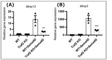
- the substance that inhibits the signal transduction of semaphorin 4D-plexin B2 may be a substance that inhibits the binding between plexin B2 and Traf2.
- the substance that inhibits the binding between plexin B2 and Traf2 may be, for example, an antibody that specifically binds to plexin B2 and inhibits the binding between plexin B2 and Traf2.
- it may be an antibody that specifically binds to Traf2 and inhibits the binding of plexin B2 to Traf2.
- it may be a small molecule compound or peptide that inhibits the binding between plexin B2 and Traf2.
- the substance that inhibits the signal transduction of semaphorin 4D-plexin B2 may be a substance that inhibits the binding between plexin B2 and c-Met.
- the substance that inhibits the binding between plexin B2 and c-Met may be, for example, an antibody that specifically binds to plexin B2 and inhibits the binding between plexin B2 and c-Met.
- it may be an antibody that specifically binds to c-Met and inhibits the binding of plexin B2 to c-Met.
- it may be a small molecule compound or peptide that inhibits the binding of plexin B2 to c-Met.
- Each antibody exemplified above may be a polyclonal antibody or a monoclonal antibody.
- the antibody may be a low molecular weight antibody to which an antibody fragment having an antigen-binding ability (for example, Fab, Fab', F (ab') 2, Fv, scFv, diabody, etc.) or a variable portion of the antibody is bound. good.
- Polyclonal antibody and monoclonal antibody can be produced by a known method. Polyclonal antibody immunizes mammals (mouse, rat, rabbit, goat, horse, etc.) using, for example, an antigen dissolved in PBS and optionally mixed with an appropriate amount of a usual adjuvant (for example, Freund's complete adjuvant) as an immunogen.
- a usual adjuvant for example, Freund's complete adjuvant
- the immunization method is not particularly limited, but for example, a method of subcutaneous injection or intraperitoneal injection once or multiple times at appropriate intervals is preferable.
- the monoclonal antibody can be produced, for example, by fusing immune cells (for example, splenocytes) obtained from the immunized mammal and myeloma cells to obtain a hybridoma, and collecting the antibody from the culture of the hybridoma. can.
- an antibody gene from a hybridoma incorporate it into an appropriate vector, introduce it into a host cell, and use gene recombination technology to produce a recombinant monoclonal antibody.
- monoclonal antibodies can also be prepared using the phage display method.
- the antibody may be a human chimeric antibody or a humanized antibody.
- the substance that inhibits the signal transduction of semaphorin 4D-plexin B2 may be a nucleic acid that inhibits the expression of plexin B2.
- the nucleic acid that inhibits the expression of plexin B2 include siRNA (short interfering RNA), shRNA (short hairpin RNA), antisense oligonucleotide, and antisense mRNA of the plexin B2 gene.
- the nucleotide sequence of the plexin B2 gene of the administration target animal can be obtained from a known database (NCBI, etc.).
- a siRNA is a double-stranded RNA having a length of about 20 bases (for example, about 21 to 23 bases) or less, and by introducing such siRNA into a cell, the gene targeted by the siRNA (book). In the invention, the expression of plexin B2 gene) can be suppressed.
- shRNA is a single-stranded RNA containing a partially palindromic base sequence, so that it has a double-stranded structure within the molecule and consists of a short hairpin structure with a protrusion at the 3'end of about 20 base pairs. Refers to the above molecules.
- shRNA After being introduced into a cell, such shRNA is decomposed into a length of about 20 bases (typically, for example, 21 bases, 22 bases, and 23 bases) in the cell and becomes a target like siRNA.
- the expression of a gene can be suppressed.
- the siRNA and shRNA may be in any form as long as they can suppress the expression of plexin B2.
- the siRNA or shRNA can be designed by a known method based on the base sequence of the target gene.
- siRNA or shRNA can be artificially synthesized.
- Antisense and sense RNA can also be synthesized in vitro from template DNA using, for example, T7 RNA polymerase and T7 promoter.
- the antisense oligonucleotide may be either DNA or RNA, as long as it is a nucleotide that is complementary or hybridizes to the consecutive 5 to 100 base sequences in the DNA sequence of the plexin B2 gene. Further, it may be modified as long as it does not interfere with the function.
- the antisense oligonucleotide can be synthesized by a conventional method, and can be easily synthesized by, for example, a commercially available DNA synthesizer.
- the antisense mRNA may be an RNA that is complementary to or hybridizes to the base sequence of the mRNA of plexin B2. Antisense mRNA can be artificially chemically synthesized.
- Antisense and sense RNA can also be synthesized in vitro from template DNA using, for example, T7 RNA polymerase and T7 promoter. During synthesis, pseudouridine is used instead of uridine to impart a cap structure and poly-A signal.
- the substance that inhibits the signal transduction of semaphorin 4D-plexin B2 may be a substance that inhibits the function of plexin B2.
- Functions of plexin B2 include, but are not limited to, the ability to bind to semaphorin 4D, the ability to bind to Traf2, and the ability to bind to c-Met. Substances that inhibit the function of plexin B2, which will be discovered in the future, are also included in the substances that inhibit the signal transduction of semaphorin 4D-plexin B2.
- the substance that inhibits the signal transduction of semaphorin 4D-plexin B2 may be a substance that inhibits the expression of I ⁇ B ⁇ or CEBP ⁇ .
- examples of the substance that inhibits the expression of I ⁇ B ⁇ or CEBP ⁇ include siRNA, shRNA, antisense oligonucleotide, and antisense mRNA of the I ⁇ B ⁇ gene or CEBP ⁇ gene.
- the nucleotide sequence of the I ⁇ B ⁇ gene and the nucleotide sequence of the CEBP ⁇ gene of the administration target animal can be obtained from a known database (NCBI, etc.).
- the substance that inhibits the signal transduction of semaphorin 4D-plexin B2 may be a substance that inhibits the function of I ⁇ B ⁇ or CEBP ⁇ .
- Functions of I ⁇ B ⁇ or CEBP ⁇ include, for example, DNA binding ability, transcriptional activation ability, and ability to bind to other transcription factors. Therefore, substances that inhibit the function of I ⁇ B ⁇ or CEBP ⁇ include, for example, substances that inhibit binding to DNA recognized by I ⁇ B ⁇ or CEBP ⁇ , substances that reduce the expression of genes involved in the expression control region recognized by I ⁇ B ⁇ or CEBP ⁇ , and the like. Can be mentioned.
- the substance that inhibits semaphorin 4D-plexin B2 signal transduction may be a c-Met inhibitor.
- the present inventors cultivated mouse femoral head cartilage, added semaphorin 4D or semaphorin 4D and the c-Met inhibitor crizotinib to the medium, and then added the c-Met inhibitor. It has been confirmed that the amount of aggrecan released into the medium is significantly suppressed, that is, cartilage destruction is suppressed (see Example 8). Therefore, c-Met inhibitors are useful as active ingredients in pharmaceutical compositions for the prevention or treatment of cartilage, bone and joint diseases.
- the c-Met inhibitor is not particularly limited, and examples thereof include crizotinib, tepotinib, and capmatinib.
- the pharmaceutical composition of the present invention contains a substance that inhibits signal transduction of semaphorin 4D-plexin B2 as an active ingredient, and can be formulated according to conventional means.
- formulations for oral administration include solid or liquid dosage forms, specifically tablets (including sugar-coated tablets and film-coated tablets), pills, granules, powders, capsules (including soft capsules), and the like. Examples include syrups, emulsions and suspending agents. These formulations are manufactured by known methods and contain carriers, diluents or excipients commonly used in the pharmaceutical field. For example, lactose, starch, sucrose, magnesium stearate and the like are used as carriers and excipients for tablets.
- injections, suppositories, etc. are used as preparations for parenteral administration, and the injections are intravenous injection, subcutaneous injection, intradermal injection, intramuscular injection, drip injection, intra-articular injection. Including dosage forms such as.
- Such injections are prepared according to known methods, for example, by dissolving, suspending or emulsifying the active ingredient in a sterile aqueous or oily solution normally used for injections.
- aqueous solution for injection for example, a physiological saline solution, an isotonic solution containing glucose and other auxiliary agents, and the like are used, and appropriate solubilizing agents such as alcohol (for example, ethanol etc.) and polyalcohol (for example) are used.
- suppositories used for rectal administration are prepared by mixing the active ingredient with a conventional suppository base.
- the parenteral route of administration eg, intravenous, intramuscular, intradermal, as an injection or infusion formulated with a pharmaceutically acceptable carrier.
- a pharmaceutically acceptable carrier Preferably administered intraperitoneally, subcutaneously or topically.
- the active ingredient of the pharmaceutical composition of the invention is nucleic acid
- it can be administered in the form of a non-viral vector or a viral vector.
- a method of introducing a nucleic acid molecule using a liposome liposome method, HVJ-liposome method, cationic liposome method, lipofection method, lipofectamine method, etc.
- microinjection method microinjection method
- gene gun GeneGun
- siRNA or shRNA When siRNA or shRNA is administered in the form of a viral vector, detoxified retrovirus, lentivirus, adenovirus, adeno-associated virus, herpesvirus, vaccinia virus, poxvirus, poliovirus, Sindobis virus, Sendai virus, SV40
- a DNA expressing siRNA or shRNA into a DNA virus or RNA virus such as, and infecting cells or tissues with this recombinant virus, siRNA or shRNA can be introduced into cells or tissues.
- the formulations thus obtained are safe and low toxicity, and therefore, for example, orally to humans and mammals (eg, rats, mice, rabbits, sheep, pigs, cows, cats, dogs, monkeys, etc.). Alternatively, it can be administered parenterally.
- the pharmaceutical composition of the present invention can contain 0.001 to 50% by mass, preferably 0.01 to 10% by mass, and more preferably 0.1 to 1% by mass of the active ingredient.
- the dose of the pharmaceutical composition of the present invention is appropriately set in consideration of the severity of the disease, the age, weight, sex, medical history, type of active ingredient, and the like of the patient. For an average human having a body weight of about 65 to 70 kg, about 0.02 mg to 4000 mg per day is preferable, and about 0.1 mg to 200 mg is more preferable.
- the total daily dose may be a single dose or a divided dose.
- Screening method Provided is a method for screening a drug for preventing or treating cartilage, bone, or joint disease (hereinafter referred to as "the screening method of the present invention"), which comprises a step of selecting a test substance that inhibits signal transduction of semaphorin 4D-plexin B2. do.
- the test substance used in the screening method of the present invention is not particularly limited, and for example, a single substance such as a natural compound, an organic compound, an inorganic compound, a nucleic acid, a nucleic acid oligo, a protein, or a peptide, a compound library, or a nucleic acid oligolive.
- a single substance such as a natural compound, an organic compound, an inorganic compound, a nucleic acid, a nucleic acid oligo, a protein, or a peptide, a compound library, or a nucleic acid oligolive.
- peptide library, gene library expression product, cell extract, cell culture supernatant, fermented microbial product, marine biological extract, plant extract, prokaryotic cell extract, eukaryotic single cell extract, animal cell extract Things can be mentioned.
- the test substance may be a novel substance or a known substance. These test substances may form salts.
- the first embodiment of the screening method of the present invention may include the following steps (1) to (3).
- (2) Step of measuring the binding between plexin B2 and semaphorin 4D and (3) selecting a test substance that reduces the binding between plexin B2 and semaphorin 4D as compared with a control to which no test substance is added.
- step (1) plexin B2, semaphorin 4D, and the test substance are brought into contact with each other.
- the system in which plexin B2 and semaphorin 4D are brought into contact with the test substance include a solution system or a culture system.
- a solution system for example, a system in which plexin B2, semaphorin 4D and a test substance are brought into contact with each other in a buffer solution suitable for a protein solution can be used.
- a control is provided that is not in contact with the test substance.
- buffer solution suitable for the protein solution examples include Tris buffer solution having a pH of 6 to 9, a phosphate buffer solution, an acetate buffer solution, a borate buffer solution, and a citrate buffer solution.
- Plexin B2 and semaphorin 4D can be produced as recombinant proteins using known gene recombination techniques and recombinant protein expression techniques. Semaphorin 4D may be soluble semaphorin 4D.
- the amino acid sequence information of plexin B2, the base sequence information of the gene encoding plexin B2, the amino acid sequence information of semaphorin 4D, and the base sequence information of the gene encoding semaphorin 4D can be obtained from a known database (NCBI, etc.). can.
- accession numbers of the human and mouse base sequences and amino acid sequences are as follows.
- NP_001268809.1 amino acid sequence
- a culture system for example, a system in which semaphorin 4D or soluble semaphorin 4D and a test substance are added to a medium in which plexin B2-expressing cells are cultured, a system in which plexin B2-expressing cultured cells and soluble semaphorin 4D are added. It is possible to use a system in which the test substance is added to the medium in which the cells secreting the cells are co-cultured. Usually, a control without the addition of the test substance is provided.
- plexin B2-expressing cells include articular cartilage surface cells, cultured cells into which a plexin B2 expression vector has been introduced, chondrocytes, osteoblasts, and limb blast cells.
- cells that secrete semaphorin 4D include macrophages, lymphocytes, synovial cells, osteoclasts, etc. treated with LPS.
- step (2) the binding between plexin B2 and semaphorin 4D is measured.
- the method for measuring the binding between plexin B2 and semaphorin 4D is not particularly limited, and a known method can be appropriately selected and used.
- a known method can be appropriately selected and used.
- an ELISA method, a fluorescence polarization method, an immunostaining method, a mass spectrometry method, a structural analysis, a screening method using an HTS system using cells, or the like can be preferably used.
- either one of the recombinant protein of soluble semaphorin 4D and the recombinant protein of the extracellular region of plexin B2 is immobilized, and the other and the test substance are added thereto to react with plexin B2.
- the binding level of semaphorin 4D can be detected using the appropriate primary and secondary antibodies.
- a test substance that reduces the binding between plexin B2 and semaphorin 4D as compared with the control to which the test substance is not added is selected. Criteria for selection are not particularly limited, but for example, the binding level is 50% or less, 40% or less, 30% or less, 20% or less, 10% or less as compared with the binding level of the control to which the test substance is not added.
- the test substance to be lowered may be selected.
- the second embodiment of the screening method of the present invention may include the following steps (1) to (3).
- Step of contacting plexin B2 and Traf2 with a test substance (2) A step of measuring the binding between plexin B2 and Traf2, and (3) a step of selecting a test substance that reduces the binding between plexin B2 and Traf2 as compared with a control to which no test substance is added.
- step (1) plexin B2 and Traf2 are brought into contact with the test substance.
- the system in which plexin B2 and Traf2 are brought into contact with the test substance include a solution system and a culture system.
- a solution system for example, a system in which plexin B2 and Traf2 are brought into contact with a test substance in a buffer solution suitable for a protein solution can be used.
- a culture system for example, a system in which Traf2 and a test substance are added to a medium in which plexin B2-expressing cells are cultured can be used.
- a control is provided that is not in contact with the test substance.
- Plexins B2 and Traf2 can be produced as recombinant proteins using known gene recombination techniques and recombinant protein expression techniques.
- the sequence information of plexin B2 is as described above, and the amino acid sequence information of Traf2 and the base sequence information of the gene encoding Traf2 can be obtained from a known database (NCBI or the like).
- NCBI National Center for Biotechnology Information
- accession numbers of the human and mouse base sequences and amino acid sequences are as follows.
- plexin B2-expressing cell Human: NM_021138.4 (base sequence), NP_066961.2 (amino acid sequence) Mouse: NM_001290413.1 (base sequence), NP_001277342.1 (amino acid sequence)
- the cell exemplified in the first embodiment can be used.
- the steps (2) and (3) of the second embodiment of the screening method of the present invention are the steps of the first embodiment except that Traf2 is used instead of semaphorin 4D in the first embodiment. It can be performed in the same manner as in 2) and (3).
- the third embodiment of the screening method of the present invention may include the following steps (1) to (3).
- (2) Step of measuring the binding between plexin B2 and c-Met may be used.
- step (1) plexin B2, c-Met and the test substance are brought into contact with each other.
- the system in which the plexin B2 and c-Met are brought into contact with the test substance include a solution system and a culture system.
- a solution system for example, a system in which plexin B2, c-Met and a test substance are brought into contact with each other in a buffer solution suitable for a protein solution can be used.
- a culture system for example, a system in which c-Met and a test substance are added to a medium in which plexin B2-expressing cells are cultured, and a system in which plexin B2 and a test substance are added to a medium in which c-Met-expressing cells are cultured.
- a system in which a test substance is added to a medium in which plexin B2-expressing cells and c-Met-expressing cells are co-cultured can be used.
- a control is provided that is not in contact with the test substance.
- Semaphorins 4D and c-Met can be produced as recombinant proteins using known gene recombination techniques and recombinant protein expression techniques.
- the sequence information of plexin B2 is as described above, and the amino acid sequence information of c-Met and the base sequence information of the gene encoding c-Met can be obtained from a known database (NCBI or the like).
- NCBI National Center for Biotechnology Information
- accession numbers of the human and mouse base sequences and amino acid sequences are as follows.
- the cell exemplified in the first embodiment can be used.
- the c-Met expressing cell include articular cartilage surface layer cells, cultured cells into which a c-Met expression vector has been introduced, chondrocytes, osteoblasts, and limb blast cells.
- steps (2) and (3) of the third embodiment of the screening method of the present invention are the same as that of the first embodiment, except that c-Met is used instead of semaphorin 4D in the first embodiment. It can be carried out in the same manner as in steps (2) and (3).
- the fourth embodiment of the screening method of the present invention may include the following steps (1) to (3). (1) A step of contacting a test substance with a cell expressing I ⁇ B ⁇ and / or CEBP ⁇ , (2) A step of measuring the expression level of I ⁇ B ⁇ and / or CEBP ⁇ , and (3) a step of selecting a test substance that reduces the expression level of I ⁇ B ⁇ and / or CEBP ⁇ as compared with a control not contacted with the test substance.
- the test substance is brought into contact with cells expressing I ⁇ B ⁇ and / or CEBP ⁇ .
- the test substance can be brought into contact with the test substance by adding the test substance to the medium of the cultured cells expressing I ⁇ B ⁇ and / or CEBP ⁇ .
- a control is provided that is not in contact with the test substance.
- cells expressing I ⁇ B ⁇ and / or CEBP ⁇ include articular cartilage superficial cells, chondrocytes, osteoblasts, limb blast cells and the like.
- the expression levels of I ⁇ B ⁇ and / or CEBP ⁇ are measured.
- the expression level may be measured by measuring the amount of protein or mRNA.
- the protein can be extracted from the cell by a known method and quantified by using a known method for measuring the amount of protein.
- Known methods for measuring the amount of protein include, for example, Western blotting, EIA method, ELISA method, RIA method, and a method using a protein measuring reagent.
- RNA can be extracted from cells by a known method and quantified using a known method for measuring the amount of mRNA.
- Known methods for measuring the amount of mRNA include Northern blotting, RT-PCR, quantitative RT-PCR, RNase protection assay and the like.
- step (3) a test substance that reduces the expression level of I ⁇ B ⁇ and / or CEBP ⁇ as compared with a control that has not been contacted with the test substance is selected.
- the criteria for selection are not particularly limited, but for example, the expression level is 50% or less, 40% or less, 30% or less, 20% or less, 10% or less as compared with the expression level of the control to which the test substance is not added.
- the test substance to be lowered may be selected.
- the fifth embodiment of the screening method of the present invention may include the following steps (1) to (3).
- I ⁇ B ⁇ or CEBP ⁇ and the test substance are brought into contact with cultured cells or genetically modified animal cells in which a reporter gene is inserted downstream of the expression control region recognized by I ⁇ B ⁇ or CEBP ⁇ .
- cultured cells used in the step (1) include established cultured cells, articular superficial chondrocytes, chondrocytes, osteoblasts, and limb blasts.
- genetically modified animal used in the step (1) include mice, rats, rabbits, sheep, pigs, cows, cats, dogs, monkeys and the like.
- the reporter gene containing these gene expression control regions is introduced by a known transfection method, virus transfer method, or the like. Can be made. Genetically modified animals in which a reporter gene is inserted downstream of the expression control region recognized by I ⁇ B ⁇ or CEBP ⁇ should be produced by knocking in (inserting) the reporter gene into these gene expression regions by genome editing, homologous recombination, etc. Can be done.
- the reporter gene is not particularly limited as long as it is generally used, but a reporter gene that is stable and whose activity can be easily quantified is preferable.
- genes encoding luciferase, ⁇ -galactosidase (lacZ), ⁇ -glucuronidase (GUS), chloramphenicol acetyltransferase (CAT), alkaline phosphatase (ALP), peroxidase (POD), green fluorescent protein (GFP), etc. can be mentioned.
- the cultured cells When the cultured cells are used in the step (1), for example, by adding I ⁇ B ⁇ or CEBP ⁇ and the test substance to the medium of the cultured cells, the cells can be brought into contact with the I ⁇ B ⁇ or CEBP ⁇ and the test substance.
- a genetically modified animal for example, by administering I ⁇ B ⁇ or CEBP ⁇ and the test substance to the genetically modified animal, the cells of the genetically modified animal can be brought into contact with I ⁇ B ⁇ or CEBP ⁇ and the test substance.
- the route of administration is not particularly limited, and examples thereof include systemic administration such as oral administration, intravenous administration, and intraperitoneal administration, and local administration to a target organ or target tissue. Usually, a control is provided that is not in contact with the test substance.
- the expression level of the reporter gene is measured.
- the expression level of the reporter gene can be measured by appropriately selecting a known measuring method according to the reporter gene to be used.
- the reporter gene is a CAT gene
- the expression level of the reporter gene can be measured by detecting the acetylation of chloramphenicol by the gene product.
- the reporter gene is the lacZ gene
- the color development of the dye compound due to the catalytic action of the gene expression product is detected
- the reporter gene is the luciferase gene
- the luminescence of luciferin due to the catalytic action of the gene product is emitted.
- the expression level of the reporter gene can be measured.
- step (3) a test substance that reduces the expression level of the reporter gene as compared with a control that has not been contacted with the test substance is selected.
- the criteria for selection are not particularly limited, but for example, the expression level is 50% or less, 40% or less, 30% or less, 20% or less, 10% or less as compared with the expression level of the control to which the test substance is not added.
- the test substance to be lowered may be selected.
- the sixth embodiment of the screening method of the present invention may include the following steps (1) to (3).
- step (1) of the sixth embodiment semaphorin 4D and the test substance are brought into contact with cultured cells or genetically modified animal cells in which a reporter gene is inserted downstream of a gene whose expression is induced by semaphorin 4D.
- Examples of the cultured cells and genetically modified animals used in the step (1) include the cells and animals exemplified in the fifth embodiment.
- Examples of the gene whose expression is induced by semaphorin 4D include Mmp13, Mmp3, IL-6, Nfikbz, C / ebpd and the like.
- a known transfection method or virus transfer method is used to construct a construct in which a reporter gene is linked downstream of a gene whose expression is induced by semaphorin 4D. It can be produced by introducing a gene using such as.
- a gene-modified animal in which a reporter gene is inserted downstream of a gene whose expression is induced by semaphorin 4D knocks in (inserts) a reporter gene downstream of a gene whose expression is induced by semaphorin 4D by genome editing, homologous recombination, etc. ) Can be produced.
- the sixth embodiment of the screening method of the present invention is carried out in the same manner as the fifth embodiment, except that the cultured cells into which the reporter gene used in the step (1) is inserted or the cells of the genetically modified animal are different. be able to.
- the present invention includes the following inventions.
- a method for preventing or treating cartilage / bone / joint disease which comprises a step of administering a substance that inhibits signal transduction of semaphorin 4D-plexin B2 to a patient with cartilage / bone / joint disease.
- a substance that inhibits signal transduction of semaphorin 4D-plexin B2 for use in the prevention or treatment of cartilage, bone and joint diseases.
- Use of substances that inhibit semaphorin 4D-plexin B2 signaling to produce pharmaceutical compositions for the prevention or treatment of cartilage, bone and joint diseases.
- Nfkbiz KO mice Nfkbiz flox / flox mice were mated with CAG-CRE transgenic mice to obtain Nfkbiz +/- mice.
- Nfkbiz +/- mice were mated with Nfkbiz +/- mice to generate Nfkbiz -/- mice.
- Sema4d knockout mice (hereinafter referred to as "Sema4d KO mice") were produced using the TAKE method (Technique for Animal Knockout system by Electroporation) based on the CRISPR / Cas9 system (SciRep 4, 6382, doi: 10.1038 / srep06382 (2014). )).
- Guide RNA target sequence: 5'-AGGATGTGCGCCCCGTTAG-3', SEQ ID NO: 1 targeting around the start codon of the Sema4d gene is transcribed in vitro using Guide-it sgRNA In Vitro Transcription and Screening System (Takara Bio).
- Sema4d KO mice Cas9 protein and mRNA that introduces a stop codon, were introduced into fertilized egg cells of C57BL / 6J mice by electroporation. Genomic DNA sequence analysis of the Sema4d gene was performed to confirm appropriate genome editing in Sema4d KO mice.
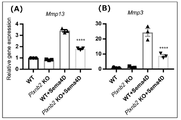
- Plxnb2 flox / flox mice were generated using the two-step TAKE method.
- a guide RNA targeting downstream of exon 22 encoding Plxnb2 is transcribed in vitro, and Cas9 protein, mRNA and single-stranded oligodeoxy are transcribed.
- a guide RNA targeting the upstream of exon 21 encoding the Plxnb2 gene is transcribed in vitro, and Cas9 protein, mRNA and ssODN (5') are transcribed.
- --ACAAGCTCATCCACGCCCGGGTAAGAAGACCCCTACATAACTTCGTATAGCATACATTATACGAAGTTATGGCTGGCATGGGGTCCCGGGTCGGGCGGGG -3', SEQ ID NO: 5 was introduced into fertilized egg cells of Plxnb2 LoxP transgenic mice by the electroporation method. The introduction of two LoxP sites was confirmed by DNA sequence analysis.
- Plxnb2 flox / flox mice were mated with CAG-CRE transgenic mice to obtain Plxnb2 +/- mice.
- Plxnb2 +/- mice were mated with Plxnb2 +/- mice to produce Plxnb2 knockout mice (hereinafter referred to as "Plxnb2 KO mice").
- DNA sequence analysis confirmed that these exons were excised by mating with CAG-CRE transgenic mice and that the transmembrane domain of plexin-B2 was deleted.
- the paws of each mouse were evaluated histologically.
- the feet were fixed with 4% PFA / PBS and then decalcified with 10% EDTA for 4 weeks.
- the cartilage was embedded in paraffin to prepare a section having a thickness of 5 ⁇ m. Sections were deparaffinized and stained with hematoxylin and eosin (HE). Joint lesions were evaluated and quantified according to the description of Maeda et al. (Nature Medicine 18, 405-U166, doi: 10.1038 / nm.2653 (2012)).
- HEK293 cells were cultured in DMEM medium (Fujifilm Wako Pure Chemical Industries) supplemented with 10% FCS and penicillin / streptomycin / glutamine (Fujifilm Wako Pure Chemical Industries, Ltd.). Bone marrow-derived by differentiating tibial and femoral bone marrow progenitor cells in RPMI (Fujifilm Wako Pure Drug) supplemented with 10% FCS, 25% L929 acclimatized medium, sodium pyruvate, HEPES, 2ME and penicillin-streptomycin. Macrophages (bone marrow derived macrophages, hereinafter referred to as "BMDM”) were obtained.
- BMDM bone marrow derived macrophages
- SFZ cells Articular cartilage surface layer (SFZ: superficial zone) cells
- LPS 10 ng / ml
- SFZ cells Articular cartilage surface layer cells
- SFZ cells were isolated by the method described in Yasuhara et al. (Lab Invest 91, 1739-1752, doi: 10.1038 / labinvest.2011.144 (2011)). .. That is, the proximal end of the femur and the distal end of the tibia were cut from the knee joint of a 17.5 day embryo for the production of 4-day-old mice or Plxnb2 KO mice, and ligaments and tendons were carefully removed from their attachment sites.
- Tissues were incubated with Hanks Balanced Salt Solution (HBSS) containing 0.25% trypsin for 1 hour, followed by digestion with DMEM containing 0.25% collagenase (Wako Pure Chemical Industries, Ltd.) for 1.5 hours.
- Dissociated cells were seeded on BioCoat TM fibronectin plates (Sigma-Aldrich) and incubated for 20 minutes.
- Non-adherent cells were rinsed twice with phosphate buffered saline (PBS) and adherent cells were used as SFZ cells.
- SFZ cells were maintained in DMEM medium containing 10% FBS and penicillin / streptomycin / glutamine.
- Deep zone (DZ: deep zone) cells were separated by additional digestion of residual cartilage tissue with collagenase.
- DZ cells were maintained in DMEM medium containing 10% FBS and penicillin / streptomycin / glutamine.
- Primary osteoblasts were obtained by continuous digestion of the calvaria of 4-day-old mice with collagenase. Osteoblasts were collected from the dispersion and cultured in ⁇ -MEM medium supplemented with 10% FBS and penicillin / streptomycin / glutamine.
- Costal chondrocytes were isolated by digesting the ribs of 4-day-old mice with collagenase (Wako Pure Chemical Industries, Ltd.).
- Costal chondrocytes were collected from the dispersion and cultured in DMEM medium containing 10% FBS and penicillin / streptomycin / glutamine. For the stimulus assay, medium was replaced with serum-free DMEM 2 hours prior to the assay and treated with each stimulant.
- the VSV plasmid was purchased from Addgene.
- Expression vectors for mouse I ⁇ B ⁇ and C / EBP ⁇ were purchased from GenScript. I ⁇ B ⁇ and C / EBP ⁇ were subcloned into pLVSIN-CMVPur Vector (Takara Bio). Each plasmid was introduced into cells using the transfection reagent X-tremeGENE 9 (Sigma-Aldrich).
- Plxnb1 targeting shRNAs (Plxnb1 shRNA, TRCN0000078917, TRCN0000078916), Plxnb2 targeting shRNAs (Plxnb2 shRNA, TRCN0000078853, TRCN0000078857), and c-Met targeting shRNAs (c-Met shRNA, TRCN0000226122).
- TRCN0000235 was cloned into the lentivirus pLKO.1 plasmid (Sigma-Aldrich). An empty vector was used as a negative control. Lentivirus particles were prepared and infected according to the manufacturer's (Sigma-Aldrich) protocol.
- Lentiviruses expressing controls or shRNA were infected with SFZ cells and selected in media containing puromycin (2 ⁇ g / ml).
- puromycin 2 ⁇ g / ml
- pLVSIN-I ⁇ B ⁇ or pLVSIN-C / EBP ⁇ was introduced into Lenti-X 293T cells (Takara Bio) together with Lentiviral High Titer Packaging Mix (Takara Bio). Lentivirus particles were prepared and infected according to the manufacturer's (Takara Bio) protocol.
- c-Met inhibitors As various inhibitors, c-Met inhibitors: SU11274 (Fujifilm Wako Pure Drug, 1 ⁇ M), K252a (abcam, 1 ⁇ M) and crizotinib (Sigma, 0.5 ⁇ M), MEK inhibitors: PD98059 (Cell Signaling Technology, 10 ⁇ M) and U0126 (Cell Signaling Technology, 10 ⁇ M), RAS Inhibitor: Salilacib (abcam, 10 ⁇ M), Rho Kinase Inhibitor: RKI-1447 (Sigma-Aldrich, 5 ⁇ M) and Y-27632 (Sigma-Aldrich, 10 ⁇ M), ErbB2 Inhibitor : AG825 (abcam, 1 ⁇ M), Akt inhibitor: MK-2206 (Selleck, 1 ⁇ M), Translation inhibitor: Cycloheximide (Fujifilm Wako Pure Drug, 100 ⁇ g / ml) and NF
- the membrane was immunoblotted with a primary antibody, and anti-mouse IgG antibody (Jackson, 1: 5,000) to which Western wasabi peroxidase was bound, anti-rabbit antibody IgG antibody (Jackson, 1: 5,000), anti-goat antibody IgG antibody (Jackson, 1: 5,000). Visualization was performed using 5000) and an anti-sheep IgG antibody (Jackson, 1: 5000) and the peroxidase luminescent substrate Immuno Star LD (Fujifilm Wako Junyaku).
- Anti-MMP13 antibody (ab39012, 1: 2000) and anti-Myc antibody (ab9132, 1: 2000) were purchased from Abcam as primary antibodies, and anti-c-Met antibody (82202, 1: 1000) and anti-NF- ⁇ B (p65).
- cells washed with cold PBS are lysed buffer (10 mM HEPES, pH 7.5, 1 mM MgCl 2 , 10 mM KCl, 0.1% TritonX-100, 20) on ice for 10 minutes. % Glycerol, phosphatase inhibitors, protease inhibitors).
- cytolytic solution was centrifuged at 3,000 g for 10 minutes at 4 ° C., and the supernatant (cytoplasmic fraction) was collected.
- Organ culture Organ culture of articular cartilage was performed according to the description of Stanton et al. (Nat Protoc 6, 388-404, doi: 10.1038 / nprot.2010.179 (2011)). That is, the femoral head was isolated from 3-week-old ICR mice and cultured for 3 days in serum-free DMEM medium containing Sema4D (20 ⁇ g / ml) or IL-1 ⁇ (10 ng / ml), or serum-free DMEM medium containing neither. did. The femoral head cartilage was fixed with 4% paraformaldehyde and then decalcified with 10% EDTA for 2 weeks. The cartilage was embedded in paraffin to prepare a section having a thickness of 5 ⁇ m.
- the sections were deparaffinized, washed with PBS and distilled water, and then stained with 0.2% fast green for 10 minutes and with 0.1% safranin O for 20 minutes. Aggrecan release into the medium was analyzed by the dimethylmethylene blue assay.
- CM conditioned media
- the supernatant is divided into anti-FLAG antibody (M185-3, 1: 400), anti-Plexin-B2 antibody (PA547880, 1:50) or anti-c-Met antibody (1:50) (3127, Cell Signaling) and 4 After incubating overnight at ° C, it was incubated with Dynabeads Protein G at 4 ° C for 1 hour. The beads were washed 5 times with NP-40 buffer. The bound protein was eluted with SDS sample buffer and then immunoblotting was performed. To identify the plexin-B2-binding protein, the bound protein was purified by immunoprecipitation using an anti-plexin-B2 antibody (1: 100) (abcam, ab193355). The bound protein was eluted with SDS sample buffer and then mass spectrometric analysis was performed.
- RNA-Seq RNA-Seqing
- Microarray Analysis Total RNA was isolated from SFZ cells using Nucleospin RNA Plus for RNA-Seq.
- the library was prepared using the TruSeq stranded mRNA sample prep kit (Illumina) and according to the manufacturer's instructions. The sequence was run in single-ended mode with 101 bases on the Illumina NovaSeq 6000 platform. I used Illumina Casava 1.8.2 software for the base call. Sequence reads were mapped to the mouse reference genome sequence (mm10) using TopHat v2.0.13 in combination with Bowtie2 ver.2.2.3 and SAMtools ver.0.1.19.
- FPKMs fragments per kilobase of exon per million mapped fragments
- Cufflinks version 2.2.1 Bioinformatics analysis using iDEP (http://bioinformatics.sdstate.edu/idep/) was performed to evaluate the screened genes.
- Luciferase Reporter Assay A luciferase reporter plasmid (Clontech) containing NF- ⁇ B responsible elements was introduced into SFZ cells using the transfection reagent X-tremeGENE 9 (Sigma-Aldrich). Twenty-four hours after transfection, cells were treated with Sema4D (2 ⁇ g / ml) for 12 hours. Cells treated with IL-1 ⁇ (10 ng / ml) were designated as positive controls, and untreated cells were designated as negative controls. Luciferase activity was measured using the Luciferase Assay System (Promega) according to the manufacturer's protocol.
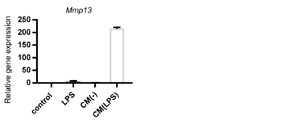
- SFZ cells cultured with LPS-treated BMDM CM did not express Mmp13
- SFZ cells cultured with LPS-treated BMDM CM did not express Mmp13.
- Remarkably induced Mmp13 expression Remarkably induced Mmp13 expression.
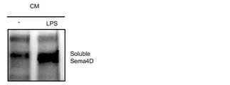
- Example 1 Identification of Mmp13 expression-inducing factor present in CM of BMDM treated with LPS
- Example 2 Shotgun Mass Spectrometry
- shotgun mass spectrometry of proteins present in CM was performed, and 32 types of candidate proteins shown in Table 2 below were detected. ..
- LPS-treated BMDM CM was fractionated by 50 kDa, and SFZ cells were cultured for 16 hours in CM ⁇ 50 kDa fraction, CM> 50 kDa fraction, and unfractionated CM. Then, the expression level of Mmp13 was analyzed by RT-qPCR. In addition, LPS-treated BMDM CM was fractionated by 100 kDa, SFZ cells were cultured for 16 hours in CM ⁇ 100 kDa fraction, CM> 100 kDa fraction, and unfractionated CM, and the expression level of Mmp13 was RT. -analyzed by PCR (two independent experiments).
- Fig. 2 The results shown in Fig. 2 are the results when (A) is fractionated by 50 kDa and (B) is the result when fractionated by 100 kDa.
- (A) Mmp13 was expressed in the> 50 kDa fraction, and Mmp13 was hardly expressed in the ⁇ 50 kDa fraction.
- (B) the expression level of Mmp13 was significantly higher in the ⁇ 100 kDa fraction than in the> 100 kDa fraction. From this result, we focused on semaphorin 4D (Sema4D) among 32 types of candidate proteins by mass spectrometry.
- Sema4D semaphorin 4D
- Example 2 Effect of Sema4D on chondrocytes
- a4D Induction of Mmp13 expression in SFZ cells by Sema4D
- (A) is the result of RT-qPCR when culturing at three stages of Sema4D concentration
- (B) is the result of RT-qPCR when culturing at three stages of culture time
- (C) is the result of immune blotting. ..
- Sema4D induced dose- and time-dependent expression of Mmp13 in SFZ cells (** P ⁇ 0.01, *** P ⁇ 0.001). Sema4D also significantly increased the MMP13 protein.
- Sema4D induced the expression of Mmp13 not only in SFZ cells but also in costal cartilage cells and deep articular cartilage cells.
- the induction of Mmp13 expression on osteoblasts was weak, and the expression of Mmp13 was not induced on macrophages.
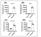
- the results of analysis of the expression level of cartilage matrix degrading enzyme are shown in Fig. 8.
- (A) is the result of Mmp13
- (B) is the result of Mmp3
- (C) is the result of Adamts4
- (D) is the result of Adamts5.
- Sema4D has been shown to increase the expression level of cartilage matrix degrading enzymes in SFZ cells (*** P ⁇ 0.001, **** P ⁇ 0.0001).
- Example 3 Effect of Sema4D on cartilage tissue
- 3-1 Organ culture of mouse articular cartilage Tissue sections of mouse femoral head cartilage organ-cultured by the method described in Experimental Method (8) were stained with safranin O and fast green and observed under a microscope.
- IL-1 ⁇ is a positive control.
- the results are shown in Fig. 9.
- (A) is the result of control (negative control)
- (B) is the result of culturing in the medium containing Sema4D
- (C) is the result of culturing in the medium containing IL-1 ⁇ .
- the scale bar is 200 ⁇ m.
- Sema4D reduced proteoglycans stained with safranin O in the superficial and intermediate layers of cartilage.
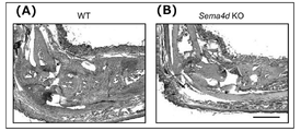
- Sema4d KO mice scored significantly lower than wild-type mice (* P ⁇ 0.05, ** P ⁇ 0.01). The results of the erosion score in the tissue specimen are shown in Fig. 13. Sema4d KO mice scored significantly lower than wild-type mice (* P ⁇ 0.05).
- Example 4 Search for molecules involved in Sema4D-dependent Mmp13 expression induction
- 4-1 Expression level of Sema4D receptor in SFZ cells
- the expression level of Sema4D receptor in SFZ cells was evaluated by RNA-Seq. The results are shown in FIG. It was shown that the expression level of Plexin-B2 (Plxnb2 in the figure) was higher than that of Plexin-B1 (Plxnb1 in the figure), Plexin-B3 (Plxnb3 in the figure) and CD72 (Cd72 in the figure) in SFZ cells. ..
- Plexin-B2 knockdown SFZ cells are transfected with a Plexin-B1 targeting shRNA (Plxnb1 shRNA) expression vector, a Plexin-B2 targeting shRNA (Plxnb2 shRNA) expression vector, or an empty vector (Control). did.
- a group treated with Sema4D and a group not treated with Sema4D were provided, respectively.
- SFZ cells were treated with Sema4D (2 ⁇ g / ml) or Sema4D and various inhibitors for 12 hours. Untreated SFZ cells were used as controls.
- the expression level of Mmp13 (n 2 each) was analyzed by RT-qPCR. The results are shown in FIGS. 17 (A) to 17 (D).
- c-Met inhibitors, RAS inhibitors and MEK inhibitors suppressed Sema4D-dependent Mmp13 expression induction.
- Rho-kinase inhibitors, ErbB2 inhibitors and Akt inhibitors did not suppress Sema4D-dependent Mmp13 expression induction.
- c-Met knockdown SFZ cells were transfected with a c-Met-targeted shRNA (c-Met shRNA) expression vector or an empty vector (Control).
- c-Met shRNA c-Met-targeted shRNA
- Control a c-Met-targeted shRNA
- Two clones (shcMet1 and shcMet2) from SFZ cells transfected with the c-Met shRNA expression vector were used in the experiment.
- Analyzed by -qPCR The results are shown in FIG. (A) is the result of the expression level of c-Met, and (B) is the result of the expression level of Mmp13. Knockdown of c-Met markedly inhibited Sema4D-dependent induction
- Immunoprecipitated SFZ cells with anti-c-Met antibody were treated with Sema4D or HGF (hepatocyte growth factor) for 1 or 5 minutes. Untreated SFZ cells were used as controls. Cytolysis was prepared, anti-c-Met antibody was added, and the immunoprecipitate was recovered. Phosphorylated c-Met in immunoprecipitated proteins and phosphorylated Erk l / 2 in cytolysis were analyzed by immunobrotting with anti-phosphorylated tyrosine and anti-phosphorylated Erk l / 2 antibodies. The results are shown in FIG. Sema4D has been shown to stimulate c-Met phosphorylation and Erk l / 2 phosphorylation downstream of c-Met in SFZ cells.
- HGF hepatocyte growth factor
- Semc4D transmits the effect on SFZ cells via both the plexin-B2 pathway and the c-Met-Ras-Erk l / 2 pathway.
- Example 5 Search for molecules that mediate semaphorin 4D / plexin-B2 signaling in SFZ cells.
- the semaphorin-plexin system is known to regulate cell morphology and migration by regulating the rearrangement of the actin cytoskeleton, primarily via RhoA, a member of the Rho family (Perrot et al). ., J Biol Chem 277, 43115-43120 (2002)).
- Rho-kinase inhibitors did not suppress Sema4D-dependent Mmp13 expression induction (see FIGS. 17 (c) and 17 (d)). This result suggests that other signaling pathways mediate the signal of plexin-B2 in SFZ cells to regulate Mmp13 expression.
- Shotgun mass spectrometry of plexin-B2-binding protein in 5-1 SFZ cells Shotgun mass spectrometry of plexin-B2-binding protein was performed by the methods described in Experimental Methods (10) and (11). The results are shown in Table 3. We found that TNF receptor-related factor 2 (TRAF2), a regulator of the NF- ⁇ B signaling pathway, binds to plexin-B2.
- TNF receptor-related factor 2 a regulator of the NF- ⁇ B signaling pathway
- HEK293T cells were introduced with either or both of the Plexin-B2 expression vector and the FLAG-TRAF2 expression vector.
- HEK293T cells into which an expression vector had not been introduced were used as controls.
- a cytolytic solution was prepared.
- Anti-Plexin-B2 antibody or anti-FLAG antibody was added to the cytolytic solution to recover the immunoprecipitate.
- Immunoprecipitated protein with anti-Plexin-B2 antibody, immunoprecipitated protein with anti-FLAG antibody, and post-cytolytic solution were subjected to immunoblotting and analyzed with anti-Plexin-B2 antibody and anti-FLAG antibody.
- the results are shown in FIG. It was confirmed that Plexin-B2 and TRAF2 were associated in cells into which both the Plexin-B2 expression vector and the FLAG-TRAF2 expression vector were introduced.
- each of the above cells was immobilized, immunofluorescently stained with an anti-NF- ⁇ B (p65) antibody as the primary antibody, and an anti-rabbit IgG antibody Alexa Fluor 568 as the secondary antibody, and observed with a fluorescence microscope.
- the results of immune blotting are shown in FIG.
- the results of immunofluorescent staining are shown in FIG. In Figure 22, DAPI shows the nucleus and the scale bar is 50 ⁇ m. From these results, it was clarified that Sema4D induces the translocation of NF- ⁇ B from the cytoplasm to the nucleus in SFZ cells.
- Sema4D was shown to activate the TRAF2-NF- ⁇ B and c-Met-Ras-Erk l / 2 signaling pathways, it was activated by Sema4D using a MEK inhibitor.
- a MEK inhibitor was investigated the role of the c-Met-Ras-Erk l / 2 signaling pathway in the NF- ⁇ B pathway. 2 ⁇ g / ml of recombinant Sema4D, Sema4D and MEK inhibitor PD98059 (10 ⁇ M) or Sema4D and MEK inhibitor U0126 (10 ⁇ M) were added to the medium of SFZ cells and cultured for 12 hours. Untreated SFZ cells were used as controls.
- Each of the above cells was immobilized, immunofluorescently stained with anti-NF- ⁇ B (p65) antibody as the primary antibody and anti-rabbit IgG antibody Alexa Fluor 568 as the secondary antibody, and observed with a fluorescence microscope.
- the results are shown in FIG. DAPI shows the nucleus and the scale bar is 50 ⁇ m.
- Sema4D has been shown to induce the translocation of NF- ⁇ B to the nucleus by a MEK inhibitor. This result suggests that c-Met-Ras-Erk l / 2 signaling is required for NF- ⁇ B activation by Sema4D.
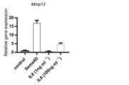
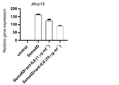
- Example 6 Search for known inflammatory cytokines involved in Sema4D-dependent Mmp13 expression induction. Since Sema4D activated NF- ⁇ B signaling in SFZ cells and up-regulated inflammatory responsive genes, the following to determine whether Sema4D has a direct or indirect inflammatory effect on SFZ cells: Experiment was conducted.
- Sema4D (1 ⁇ g / ml), IL-6 (1 ⁇ g / ml) or Sema4D and IL-6 were added to the medium of SFZ cells and cultured for 12 hours. Untreated SFZ cells were used as controls.
- (A) is the result of the expression level of Mmp13
- (B) is the result of the expression level of Mmp3.
- Example 7 Search for transcription factors involved in Sema4D-dependent Mmp13 expression induction
- RNA-Seq analysis of transcription factor genes highly expressed in Sema4D-treated SFZ cells
- the results of the de novo protein synthesis inhibition experiment in Example 6 show that specific transcription factors are involved in the induction of Sema4D-dependent Mmp13 expression. It suggests that it is. Therefore, RNA-Seq was used to analyze transcription factor genes that were 10-fold or more highly expressed in Sema4D-treated SFZ cells as compared to untreated SFZ cells.
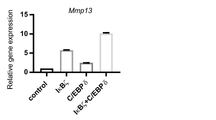
- Table 4 Sema4D has been shown to increase the expression of Nfkbiz, which encodes I ⁇ B ⁇ , and Cebpd, which encodes C / EBP ⁇ , in SFZ cells.
- Example 8 Suppression of agrecan release from articular cartilage by a c-Met inhibitor
- Mouse femoral head cartilage was organ-cultured by the method described in Experimental Method (8). That is, the femoral head was isolated from 3-week-old ICR mice, Sema4D (20 ⁇ g / ml) or Sema4D and the c-Met inhibitor crizotinib (0.5 ⁇ M) were added to serum-free DMEM medium, cultured for 3 days, and then cultured into the medium. Aggrecan release was analyzed by dimethylmethylene blue assay. Controls were cultured in serum-free DMEM medium containing neither for 3 days. The results are shown in Figure 39.
- the c-Met inhibitor significantly suppressed the aggrecan release increased by Sema4D treatment (* P ⁇ 0.05, **** P ⁇ 0.0001). This result indicates that c-Met inhibitors are effective in the prevention or treatment of cartilage destruction caused by Sema4D.
- Mouse femoral head cartilage was organ-cultured by the method described in Experimental Method (8). That is, the femoral head was isolated from 3-week-old ICR mice, and Sema4D (20 ⁇ g / ml) or Sema4D and the NF- ⁇ B inhibitor BAY11-7082 (10 ⁇ M) were added to serum-free DMEM medium and cultured for 3 days. Controls were cultured in serum-free DMEM medium containing neither for 3 days. The femoral head cartilage was fixed with 4% paraformaldehyde and then decalcified with 10% EDTA for 2 weeks.
- the cartilage was embedded in paraffin to prepare a section having a thickness of 5 ⁇ m.
- the sections were deparaffinized, washed with PBS and distilled water, and then stained with 0.2% fast green for 10 minutes and with 0.1% safranin O for 20 minutes. Proteoglycan release into the medium was analyzed by the dimethylmethylene blue assay.
- Fig. 40 The observation results of the articular cartilage section are shown in Fig. 40.
- (A) is the result of control
- (B) is the result of culturing in the medium containing Sema4D
- (C) is the result of culturing in the medium containing Sema4D and BAY11-7082.
- the scale bar is 200 ⁇ m. It was observed that Sema4D reduced the proteoglycan stained by safranin O in the superficial and intermediate layers of cartilage, but the addition of BAY11-7082 suppressed the reduction of proteoglycan.
- the results of the dimethylmethylene blue assay are shown in Figure 41.
- the NF- ⁇ B inhibitor significantly suppressed the aggrecan release increased by Sema4D treatment (** P ⁇ 0.01).
Landscapes
- Health & Medical Sciences (AREA)
- Life Sciences & Earth Sciences (AREA)
- Chemical & Material Sciences (AREA)
- Engineering & Computer Science (AREA)
- Medicinal Chemistry (AREA)
- General Health & Medical Sciences (AREA)
- Pharmacology & Pharmacy (AREA)
- Animal Behavior & Ethology (AREA)
- Public Health (AREA)
- Veterinary Medicine (AREA)
- Immunology (AREA)
- Molecular Biology (AREA)
- Physical Education & Sports Medicine (AREA)
- Bioinformatics & Cheminformatics (AREA)
- Organic Chemistry (AREA)
- Rheumatology (AREA)
- General Chemical & Material Sciences (AREA)
- Nuclear Medicine, Radiotherapy & Molecular Imaging (AREA)
- Chemical Kinetics & Catalysis (AREA)
- Biomedical Technology (AREA)
- Hematology (AREA)
- Orthopedic Medicine & Surgery (AREA)
- Urology & Nephrology (AREA)
- Epidemiology (AREA)
- Biotechnology (AREA)
- Food Science & Technology (AREA)
- Microbiology (AREA)
- Physics & Mathematics (AREA)
- Analytical Chemistry (AREA)
- Pathology (AREA)
- General Physics & Mathematics (AREA)
- Biochemistry (AREA)
- Cell Biology (AREA)
- Genetics & Genomics (AREA)
- Mycology (AREA)
- Biophysics (AREA)
- Pain & Pain Management (AREA)
- Medicines That Contain Protein Lipid Enzymes And Other Medicines (AREA)
- Pharmaceuticals Containing Other Organic And Inorganic Compounds (AREA)
Priority Applications (1)
| Application Number | Priority Date | Filing Date | Title |
|---|---|---|---|
| JP2022571994A JPWO2022137964A1 (https=) | 2020-12-23 | 2021-11-25 |
Applications Claiming Priority (2)
| Application Number | Priority Date | Filing Date | Title |
|---|---|---|---|
| JP2020213230 | 2020-12-23 | ||
| JP2020-213230 | 2020-12-23 |
Publications (1)
| Publication Number | Publication Date |
|---|---|
| WO2022137964A1 true WO2022137964A1 (ja) | 2022-06-30 |
Family
ID=82157661
Family Applications (1)
| Application Number | Title | Priority Date | Filing Date |
|---|---|---|---|
| PCT/JP2021/043187 Ceased WO2022137964A1 (ja) | 2020-12-23 | 2021-11-25 | 軟骨・骨・関節疾患の予防または治療用医薬組成物および軟骨・骨・関節疾患の予防または治療用薬剤のスクリーニング方法 |
Country Status (2)
| Country | Link |
|---|---|
| JP (1) | JPWO2022137964A1 (https=) |
| WO (1) | WO2022137964A1 (https=) |
Cited By (1)
| Publication number | Priority date | Publication date | Assignee | Title |
|---|---|---|---|---|
| CN116949097A (zh) * | 2023-09-20 | 2023-10-27 | 江苏集萃药康生物科技股份有限公司 | 一种sema4d人源化小鼠模型的构建方法及其应用 |
Citations (7)
| Publication number | Priority date | Publication date | Assignee | Title |
|---|---|---|---|---|
| JP2010507618A (ja) * | 2006-10-27 | 2010-03-11 | グラクソ グループ リミテッド | c−Metキナーゼ阻害薬としての7−アザインドール誘導体 |
| WO2012157237A1 (ja) * | 2011-05-13 | 2012-11-22 | 国立大学法人東京医科歯科大学 | 骨形成促進剤 |
| CN103030654A (zh) * | 2011-10-09 | 2013-04-10 | 济南赛文医药技术有限公司 | 一种小分子c-Met蛋白激酶抑制剂 |
| WO2015170683A1 (ja) * | 2014-05-09 | 2015-11-12 | サントリーホールディングス株式会社 | メトキシフラボンを含むNOX阻害剤及びNFκB阻害剤 |
| JP2017502920A (ja) * | 2013-10-21 | 2017-01-26 | バクシネックス インコーポレーティッド | 神経変性障害の処置のためのセマフォリン−4d結合分子の使用 |
| WO2017217545A1 (ja) * | 2016-06-16 | 2017-12-21 | 国立大学法人東京大学 | プレキシンの結合調節剤 |
| WO2020165094A1 (en) * | 2019-02-11 | 2020-08-20 | MAX-PLANCK-Gesellschaft zur Förderung der Wissenschaften e.V. | Dual inhibition of plexin-b1 and plexin-b2 |
-
2021
- 2021-11-25 JP JP2022571994A patent/JPWO2022137964A1/ja active Pending
- 2021-11-25 WO PCT/JP2021/043187 patent/WO2022137964A1/ja not_active Ceased
Patent Citations (7)
| Publication number | Priority date | Publication date | Assignee | Title |
|---|---|---|---|---|
| JP2010507618A (ja) * | 2006-10-27 | 2010-03-11 | グラクソ グループ リミテッド | c−Metキナーゼ阻害薬としての7−アザインドール誘導体 |
| WO2012157237A1 (ja) * | 2011-05-13 | 2012-11-22 | 国立大学法人東京医科歯科大学 | 骨形成促進剤 |
| CN103030654A (zh) * | 2011-10-09 | 2013-04-10 | 济南赛文医药技术有限公司 | 一种小分子c-Met蛋白激酶抑制剂 |
| JP2017502920A (ja) * | 2013-10-21 | 2017-01-26 | バクシネックス インコーポレーティッド | 神経変性障害の処置のためのセマフォリン−4d結合分子の使用 |
| WO2015170683A1 (ja) * | 2014-05-09 | 2015-11-12 | サントリーホールディングス株式会社 | メトキシフラボンを含むNOX阻害剤及びNFκB阻害剤 |
| WO2017217545A1 (ja) * | 2016-06-16 | 2017-12-21 | 国立大学法人東京大学 | プレキシンの結合調節剤 |
| WO2020165094A1 (en) * | 2019-02-11 | 2020-08-20 | MAX-PLANCK-Gesellschaft zur Förderung der Wissenschaften e.V. | Dual inhibition of plexin-b1 and plexin-b2 |
Non-Patent Citations (1)
| Title |
|---|
| ZHANG YING, SHEN SHENG, LI PEIFENG, FAN YANAN, ZHANG LEILEI, LI WUYIN, LIU YOUWEN: "PLEXIN-B2 promotes the osteogenic differentiation of human bone marrow mesenchymal stem cells via activation of the RhoA signaling pathway", CELLULAR SIGNALLING., ELSEVIER SCIENCE LTD., GB, vol. 62, 1 October 2019 (2019-10-01), GB , pages 109343, XP055945999, ISSN: 0898-6568, DOI: 10.1016/j.cellsig.2019.06.008 * |
Cited By (2)
| Publication number | Priority date | Publication date | Assignee | Title |
|---|---|---|---|---|
| CN116949097A (zh) * | 2023-09-20 | 2023-10-27 | 江苏集萃药康生物科技股份有限公司 | 一种sema4d人源化小鼠模型的构建方法及其应用 |
| CN116949097B (zh) * | 2023-09-20 | 2023-12-12 | 江苏集萃药康生物科技股份有限公司 | 一种sema4d人源化小鼠模型的构建方法及其应用 |
Also Published As
| Publication number | Publication date |
|---|---|
| JPWO2022137964A1 (https=) | 2022-06-30 |
Similar Documents
| Publication | Publication Date | Title |
|---|---|---|
| Wehmeyer et al. | Sclerostin inhibition promotes TNF-dependent inflammatory joint destruction | |
| Wang et al. | E2F2 directly regulates the STAT1 and PI3K/AKT/NF-κB pathways to exacerbate the inflammatory phenotype in rheumatoid arthritis synovial fibroblasts and mouse embryonic fibroblasts | |
| Sakic et al. | Neutralization of S100A4 induces stabilization of atherosclerotic plaques: role of smooth muscle cells | |
| US20160025745A1 (en) | Methods of diagnosing and treating fibrosis | |
| Han et al. | IRE1α dissociates with BiP and inhibits ER stress-mediated apoptosis in cartilage development | |
| Liu et al. | NLRP3 inflammasome induced liver graft injury through activation of telomere‐independent RAP1/KC axis | |
| Fu et al. | Role of DHX33 in c-Myc-induced cancers | |
| Liu et al. | MicroRNA let-7 induces M2 macrophage polarization in COPD emphysema through the IL-6/STAT3 pathway | |
| CN119074926B (zh) | 一种靶向fap的car-t细胞在制备治疗心肌炎慢性期心脏纤维化的药物中的应用 | |
| Xu et al. | Inhibition of the cardiac fibroblast-enriched histone methyltransferase Dot1L prevents cardiac fibrosis and cardiac dysfunction | |
| Wilson et al. | Sine oculis homeobox homolog 1 plays a critical role in pulmonary fibrosis | |
| Zhou et al. | Hepatocyte nuclear factor 4α negatively regulates connective tissue growth factor during liver regeneration | |
| Bai et al. | Enhanced expression of SAM‐pointed domain–containing E ts‐like factor in Chronic rhinosinusitis with nasal polyps | |
| Li et al. | Downregulation of miR-98 contributes to hypoxic pulmonary hypertension by targeting ALK1 | |
| US20220105122A1 (en) | Therapeutic and prophylactic agent for glioma, brain tumor malignancy marker, brain tumor prognostic marker, method for determining malignancy and prognosis of brain tumor and antibody inhibiting tumor proliferation | |
| WO2022137964A1 (ja) | 軟骨・骨・関節疾患の予防または治療用医薬組成物および軟骨・骨・関節疾患の予防または治療用薬剤のスクリーニング方法 | |
| Huang et al. | Alternative polyadenylation contributes to fibroblast senescence in pulmonary fibrosis | |
| Mia et al. | YAP/TAZ activation in fibroblasts coordinates fibrotic remodeling, fibroinflammation, and epithelial dysfunction in pulmonary fibrosis | |
| CN116615460A (zh) | β-血影蛋白(SPTBN1)缺乏保护小鼠免于高脂肪饮食诱导的肝病和癌症发展 | |
| Peng et al. | TGF-β1/SMAD3-mediated non-canonical hedgehog signaling promotes pancreatic stellate cell activation and fibrosis in chronic pancreatitis | |
| Cuevas et al. | Non-canonical Telomerase Reverse Transcriptase Controls Osteogenic Reprogramming of Aortic Valve Cells Through STAT5 | |
| EP4454663A1 (en) | Therapeutic or prophylactic agent for heart attack, heart fibrosis, or heart failure, where htra3 is therapeutic target | |
| CN116699135B (zh) | 癌症免疫治疗靶标和诊断、预后预测生物标志物的应用 | |
| KR102279751B1 (ko) | 근원성 인자 또는 nogo-a 측정 제제를 포함하는 근육병 진단용 조성물과 이를 이용한 근육병 진단 방법 | |
| Tassey et al. | GP130 Y814 SIGNALING IS REQUIRED FOR THE DYNAMIN-MEDIATED ENDOCYTOSIS, MAPK/P38 ACTIVATION AND PERSISTENCE OF CHRONIC SYSTEMIC INFLAMMATION INDUCED BY HIGH FAT DIET |
Legal Events
| Date | Code | Title | Description |
|---|---|---|---|
| 121 | Ep: the epo has been informed by wipo that ep was designated in this application |
Ref document number: 21910117 Country of ref document: EP Kind code of ref document: A1 |
|
| ENP | Entry into the national phase |
Ref document number: 2022571994 Country of ref document: JP Kind code of ref document: A |
|
| NENP | Non-entry into the national phase |
Ref country code: DE |
|
| 122 | Ep: pct application non-entry in european phase |
Ref document number: 21910117 Country of ref document: EP Kind code of ref document: A1 |