WO2021177312A1 - 情報保存装置、方法およびプログラム、並びに解析記録生成装置、方法およびプログラム - Google Patents
情報保存装置、方法およびプログラム、並びに解析記録生成装置、方法およびプログラム Download PDFInfo
- Publication number
- WO2021177312A1 WO2021177312A1 PCT/JP2021/007977 JP2021007977W WO2021177312A1 WO 2021177312 A1 WO2021177312 A1 WO 2021177312A1 JP 2021007977 W JP2021007977 W JP 2021007977W WO 2021177312 A1 WO2021177312 A1 WO 2021177312A1
- Authority
- WO
- WIPO (PCT)
- Prior art keywords
- image
- property information
- analysis record
- information
- medical
- Prior art date
- Legal status (The legal status is an assumption and is not a legal conclusion. Google has not performed a legal analysis and makes no representation as to the accuracy of the status listed.)
- Ceased
Links
Images


Classifications
-
- G—PHYSICS
- G06—COMPUTING OR CALCULATING; COUNTING
- G06F—ELECTRIC DIGITAL DATA PROCESSING
- G06F40/00—Handling natural language data
- G06F40/40—Processing or translation of natural language
-
- G—PHYSICS
- G06—COMPUTING OR CALCULATING; COUNTING
- G06F—ELECTRIC DIGITAL DATA PROCESSING
- G06F40/00—Handling natural language data
- G06F40/40—Processing or translation of natural language
- G06F40/55—Rule-based translation
- G06F40/56—Natural language generation
-
- A—HUMAN NECESSITIES
- A61—MEDICAL OR VETERINARY SCIENCE; HYGIENE
- A61B—DIAGNOSIS; SURGERY; IDENTIFICATION
- A61B6/00—Apparatus or devices for radiation diagnosis; Apparatus or devices for radiation diagnosis combined with radiation therapy equipment
- A61B6/02—Arrangements for diagnosis sequentially in different planes; Stereoscopic radiation diagnosis
- A61B6/03—Computed tomography [CT]
-
- G—PHYSICS
- G06—COMPUTING OR CALCULATING; COUNTING
- G06F—ELECTRIC DIGITAL DATA PROCESSING
- G06F40/00—Handling natural language data
- G06F40/10—Text processing
- G06F40/166—Editing, e.g. inserting or deleting
- G06F40/169—Annotation, e.g. comment data or footnotes
-
- G—PHYSICS
- G06—COMPUTING OR CALCULATING; COUNTING
- G06F—ELECTRIC DIGITAL DATA PROCESSING
- G06F40/00—Handling natural language data
- G06F40/40—Processing or translation of natural language
- G06F40/42—Data-driven translation
- G06F40/44—Statistical methods, e.g. probability models
-
- G—PHYSICS
- G06—COMPUTING OR CALCULATING; COUNTING
- G06T—IMAGE DATA PROCESSING OR GENERATION, IN GENERAL
- G06T11/00—Two-dimensional [2D] image generation
- G06T11/60—Creating or editing images; Combining images with text
-
- G—PHYSICS
- G16—INFORMATION AND COMMUNICATION TECHNOLOGY [ICT] SPECIALLY ADAPTED FOR SPECIFIC APPLICATION FIELDS
- G16H—HEALTHCARE INFORMATICS, i.e. INFORMATION AND COMMUNICATION TECHNOLOGY [ICT] SPECIALLY ADAPTED FOR THE HANDLING OR PROCESSING OF MEDICAL OR HEALTHCARE DATA
- G16H10/00—ICT specially adapted for the handling or processing of patient-related medical or healthcare data
- G16H10/60—ICT specially adapted for the handling or processing of patient-related medical or healthcare data for patient-specific data, e.g. for electronic patient records
-
- G—PHYSICS
- G16—INFORMATION AND COMMUNICATION TECHNOLOGY [ICT] SPECIALLY ADAPTED FOR SPECIFIC APPLICATION FIELDS
- G16H—HEALTHCARE INFORMATICS, i.e. INFORMATION AND COMMUNICATION TECHNOLOGY [ICT] SPECIALLY ADAPTED FOR THE HANDLING OR PROCESSING OF MEDICAL OR HEALTHCARE DATA
- G16H15/00—ICT specially adapted for medical reports, e.g. generation or transmission thereof
-
- G—PHYSICS
- G16—INFORMATION AND COMMUNICATION TECHNOLOGY [ICT] SPECIALLY ADAPTED FOR SPECIFIC APPLICATION FIELDS
- G16H—HEALTHCARE INFORMATICS, i.e. INFORMATION AND COMMUNICATION TECHNOLOGY [ICT] SPECIALLY ADAPTED FOR THE HANDLING OR PROCESSING OF MEDICAL OR HEALTHCARE DATA
- G16H30/00—ICT specially adapted for the handling or processing of medical images
- G16H30/40—ICT specially adapted for the handling or processing of medical images for processing medical images, e.g. editing
-
- G—PHYSICS
- G16—INFORMATION AND COMMUNICATION TECHNOLOGY [ICT] SPECIALLY ADAPTED FOR SPECIFIC APPLICATION FIELDS
- G16H—HEALTHCARE INFORMATICS, i.e. INFORMATION AND COMMUNICATION TECHNOLOGY [ICT] SPECIALLY ADAPTED FOR THE HANDLING OR PROCESSING OF MEDICAL OR HEALTHCARE DATA
- G16H50/00—ICT specially adapted for medical diagnosis, medical simulation or medical data mining; ICT specially adapted for detecting, monitoring or modelling epidemics or pandemics
- G16H50/20—ICT specially adapted for medical diagnosis, medical simulation or medical data mining; ICT specially adapted for detecting, monitoring or modelling epidemics or pandemics for computer-aided diagnosis, e.g. based on medical expert systems
-
- G—PHYSICS
- G06—COMPUTING OR CALCULATING; COUNTING
- G06T—IMAGE DATA PROCESSING OR GENERATION, IN GENERAL
- G06T2200/00—Indexing scheme for image data processing or generation, in general
- G06T2200/24—Indexing scheme for image data processing or generation, in general involving graphical user interfaces [GUIs]
-
- G—PHYSICS
- G06—COMPUTING OR CALCULATING; COUNTING
- G06T—IMAGE DATA PROCESSING OR GENERATION, IN GENERAL
- G06T2211/00—Image generation
- G06T2211/40—Computed tomography
- G06T2211/441—AI-based methods, deep learning or artificial neural networks
Definitions
- This disclosure relates to an information storage device, a method and a program, and an analysis record generation device, a method and a program.
- CT Computer Tomography
- MRI Magnetic Resonance Imaging
- medical images are analyzed by CAD (Computer-Aided Diagnosis) using a learning model that has been machine-learned by deep learning, etc., and the shape, density, and position of structures of interest such as abnormal shadow candidates included in the medical images. It is also practiced to discriminate properties such as size and size, and obtain these as analysis results.
- the analysis result acquired by CAD is associated with the test information such as the patient name, gender, age, and the modality from which the medical image was acquired, and is stored in the database.
- the medical image and the analysis result are transmitted to the terminal of the image interpreting doctor who interprets the medical image.
- the image interpreting doctor interprets the medical image by referring to the transmitted medical image and the analysis result on his / her terminal, and creates an image interpretation report.
- JP-A-2019-153250 a learning model in which machine learning such as a recurrent neural network is trained so as to generate a sentence from characters representing input property information is used.
- Medical texts (hereinafter referred to as medical texts) are created.
- medical texts By automatically generating medical texts as in the method described in JP-A-2019-153250, it is possible to reduce the burden on the interpretation doctor when creating a medical document such as an interpretation report.
- the automatically generated interpretation report may be modified by the interpretation doctor.
- the past medical image interpretation report is often referred to. Therefore, a method of extracting the corrected part of the corrected image interpretation report (see Japanese Patent Application Laid-Open No. 2011-125402) and a method of extracting the difference between the past medical image interpretation report and the latest medical image interpretation report (see Japanese Patent Application Laid-Open No. 2011-125402). JP-A-2007-122679) has been proposed.
- a learning model that generates an image analysis record such as a sentence from an image
- a learning model that generates an interpretation report from a medical image there is a user's preference for the content and expression of the analysis record, and that preference. It is desired to build a learning model that reflects the above.
- the user's preference for example, regarding the property information analyzed from the image, which property information should be reflected in the final analysis record and the like can be mentioned.
- the generated image analysis record may not match the user's preference. In such a case, the user needs to modify the generated image analysis record.
- the image analysis record is a sentence
- the sentences before and after the modification are compared using the methods described in JP-A-2011-125402 and JP-A-2007-122679, any part of the sentence can be compared. Can be recognized as having been modified.
- the present disclosure has been made in view of the above circumstances, and when the image analysis record generated from the image is modified, it is possible to recognize which property information derived by analyzing the image is modified.
- the purpose is.
- the information storage device includes at least one processor.
- the processor derives a plurality of property information representing the properties of the structure of interest contained in the image.
- Generate an image analysis record that contains at least a portion of multiple property information Accepts user corrections to property information It is configured to distinguish and store the derived property information and the modified property information.
- the processor may be configured to display the image analysis record on the display.
- the processor is configured to accept at least one of the deletion of the property information included in the displayed image analysis record and the addition of the property information not included in the image analysis record as a modification. It may be what is done.
- the processor displays all or a part of the derived property information on the display. It may be configured to accept corrections based on the user's selection of the displayed property information.
- the information storage device may further include a learning model that has been trained to output an image analysis record when property information is input.
- the processor may generate a sentence including at least a part of the property information as an image analysis record.
- the image may be a medical image
- the text may be a medical text relating to the structure of interest included in the medical image.
- the analysis record generator comprises at least one processor.
- the processor derives a plurality of property information representing the properties of the structure of interest contained in the target image to be analyzed. It is configured to generate a target image analysis record containing at least a part of the property information by referring to the information stored by the information storage device according to the present disclosure.
- the processor identifies stored information including property information that matches the property information derived from the target image, and performs image analysis associated with the specified stored information.
- the recording may be generated as a target image analysis record.
- the processor is further configured to generate other target image analysis records including at least a part of the derived property information without reference to the stored information. It may be one.
- Unreferenced ... Generate another target image analysis record means to generate another target image analysis record without referring to the saved information.
- the processor may be configured to display the target image analysis record and other target image analysis records on the display.
- the processor may be configured to accept the selection of either the displayed target image analysis record or another target image analysis record.
- the processor may be configured to generate a sentence including at least a part of the property information as a target image analysis record.
- the image may be a medical image
- the text may be a medical text relating to a structure of interest included in the medical image.
- the information storage method derives a plurality of property information representing the properties of the structure of interest contained in the image by analyzing the image. Generate an image analysis record that contains at least a portion of multiple property information Accepts user corrections to property information The derived property information and the modified property information are stored separately.
- the analysis record generation method derives a plurality of property information representing the properties of the structure of interest contained in the target image to be analyzed. With reference to the information stored by the information storage device according to the present disclosure, a target image analysis record including at least a part of the property information is generated.
- the information storage method and the analysis record generation method according to the present disclosure may be provided as a program for executing the computer.
- the image analysis record generated from the image is modified, it is possible to recognize which property information derived by analyzing the image is modified.
- Functional configuration diagram of the information storage device and analysis record generation device according to this embodiment Diagram showing an example of teacher data for learning the first learning model Diagram for explaining the property information derived by the analysis department The figure which shows the schematic structure of the recurrent neural network Diagram showing an example of a medical text display screen Diagram showing an example of a display screen for modified medical text Diagram for explaining saved information showing the saved result of property information Diagram showing an example of the target medical text and the display screen of another plan Flowchart showing information storage processing performed in this embodiment Flowchart of analysis record generation processing performed in this embodiment Diagram showing an example of a medical text and collation result display screen
- FIG. 1 is a diagram showing a schematic configuration of the medical information system 1.
- the medical information system 1 shown in FIG. 1 is based on an inspection order from a doctor in a clinical department using a known ordering system, photographs of a part to be inspected of a subject, storage of a medical image acquired by the imaging, and an image interpreter. It is a system for interpreting medical images and creating an interpretation report, and for viewing the interpretation report by the doctor of the requesting clinical department and observing the details of the medical image to be interpreted.
- the medical information system 1 includes a plurality of imaging devices 2, a plurality of image interpretation WS (WorkStation) 3 which are image interpretation terminals, a medical care WS 4, an image server 5, and an image database (hereinafter, image DB (DataBase)).
- the report server 7 and the report database (hereinafter referred to as the report DB) 8 are connected to each other via a wired or wireless network 10 so as to be able to communicate with each other.
- Each device is a computer on which an application program for functioning as a component of the medical information system 1 is installed.
- the application program is recorded and distributed on a recording medium such as a DVD (Digital Versatile Disc) and a CD-ROM (Compact Disc Read Only Memory), and is installed on a computer from the recording medium.
- a recording medium such as a DVD (Digital Versatile Disc) and a CD-ROM (Compact Disc Read Only Memory)
- it is stored in the storage device of the server computer connected to the network 10 or in the network storage in a state of being accessible from the outside, and is downloaded and installed in the computer upon request.
- the photographing device 2 is a device (modality) that generates a medical image representing the diagnosis target part by photographing the part to be diagnosed of the subject. Specifically, it is a simple X-ray imaging apparatus, a CT apparatus, an MRI apparatus, a PET (Positron Emission Tomography) apparatus, and the like.
- the medical image generated by the imaging device 2 is transmitted to the image server 5 and stored in the image DB 6.
- the image interpretation WS3 is a computer used by, for example, an image interpretation doctor in a radiology department to interpret a medical image and create an image interpretation report, and is represented by an information storage device and an analysis record generation device (hereinafter referred to as an information storage device) according to the present embodiment. ) 20 is included.
- an information storage device an analysis record generation device (hereinafter referred to as an information storage device) according to the present embodiment. ) 20 is included.
- an information storage device an analysis record generation device
- the medical care WS4 is a computer used by doctors in the clinical department for detailed observation of images, viewing of interpretation reports, creation of electronic medical records, etc., and is a processing device, a display device such as a display, and an input device such as a keyboard and a mouse. Consists of.
- an image viewing request is made to the image server 5
- an image received from the image server 5 is displayed
- an image interpretation report viewing request is made to the report server 7
- an image interpretation report received from the report server 7 is displayed.
- the image server 5 is a general-purpose computer in which a software program that provides a database management system (DataBase Management System: DBMS) function is installed. Further, the image server 5 includes a storage in which the image DB 6 is configured. This storage may be a hard disk device connected by the image server 5 and the data bus, or a disk device connected to NAS (Network Attached Storage) and SAN (Storage Area Network) connected to the network 10. It may be.
- NAS Network Attached Storage
- SAN Storage Area Network
- the image data and incidental information of the medical image acquired by the imaging device 2 are registered in the image DB 6.
- the incidental information includes, for example, an image ID (identification) for identifying an individual medical image, a patient ID for identifying a subject, an examination ID for identifying an examination, and a unique ID assigned to each medical image ( UID: unique identification), examination date when the medical image was generated, examination time, type of imaging device used in the examination to acquire the medical image, patient information such as patient name, age, gender, examination site (imaging) Includes information such as site), imaging information (imaging protocol, imaging sequence, imaging method, imaging conditions, use of contrast medium, etc.), series number or collection number when multiple medical images are acquired in one examination. ..
- the image server 5 when the image server 5 receives the viewing request from the image interpretation WS3 and the medical examination WS4 via the network 10, the image server 5 searches for the medical image registered in the image DB 6, and uses the searched medical image as the requesting image interpretation WS3 and the medical examination. Send to WS4.
- the report server 7 incorporates a software program that provides the functions of a database management system to a general-purpose computer.
- the report server 7 receives the image interpretation report registration request from the image interpretation WS3, the report server 7 prepares the image interpretation report in a database format and registers the image interpretation report in the report DB 8.
- the image interpretation report includes, for example, a medical image to be interpreted, an image ID for identifying the medical image, an image interpretation doctor ID for identifying the image interpretation doctor who performed the image interpretation, a lesion name, a lesion position information, and a medical image including a specific area. It may include information for access and information such as property information.
- the report server 7 when the report server 7 receives a viewing request for the interpretation report from the interpretation WS3 and the medical treatment WS4 via the network 10, the report server 7 searches for the interpretation report registered in the report DB 8 and uses the searched interpretation report as the requester's interpretation. It is transmitted to WS3 and medical treatment WS4.
- the medical image is a three-dimensional CT image composed of a plurality of tomographic images with the diagnosis target as the lung, and the CT image is interpreted by the interpretation WS3 to obtain an abnormal shadow contained in the lung.
- An interpretation report shall be prepared as a medical document.
- the medical image is not limited to the CT image, and any medical image such as an MRI image and a two-dimensional image acquired by a simple X-ray imaging device can be used.
- Network 10 is a wired or wireless local area network that connects various devices in the hospital.
- the network 10 may be configured such that the local area networks of each hospital are connected to each other by the Internet or a dedicated line.
- FIG. 2 describes the hardware configuration of the information storage device and the analysis record generation device according to the present embodiment.
- the information storage device 20 represents the information storage device and the analysis record generation device.
- the information storage device 20 includes a CPU (Central Processing Unit) 11, a non-volatile storage 13, and a memory 16 as a temporary storage area.
- the information storage device 20 includes a display 14 such as a liquid crystal display, an input device 15 such as a keyboard and a mouse, and a network I / F (InterFace) 17 connected to the network 10.
- the CPU 11, the storage 13, the display 14, the input device 15, the memory 16, and the network I / F 17 are connected to the bus 18.
- the CPU 11 is an example of the processor in the present disclosure.
- the storage 13 is realized by an HDD (Hard Disk Drive), an SSD (Solid State Drive), a flash memory, or the like.
- the information storage program 12A and the analysis record generation program 12B are stored in the storage 13 as a storage medium.
- the CPU 11 reads the information storage program 12A and the analysis record generation program 12B from the storage 13 and then expands the information storage program 12A and the analysis record generation program 12B into the memory 16 to execute the expanded information storage program 12A and the analysis record generation program 12B.
- FIG. 3 is a diagram showing a functional configuration of the information storage device and the analysis record generation device according to the present embodiment.
- the information storage device (and analysis record generation device) 20 includes an image acquisition unit 21, an analysis unit 22, an analysis record generation unit 23, a display control unit 24, a correction unit 25, a storage control unit 26, and communication.
- a unit 27 is provided.
- the CPU 11 executes the information storage program 12A and the analysis record generation program 12B, the CPU 11 has the image acquisition unit 21, the analysis unit 22, the analysis record generation unit 23, the display control unit 24, the correction unit 25, and the storage control. It functions as a unit 26 and a communication unit 27.
- the image acquisition unit 21, the analysis unit 22, the analysis record generation unit 23, the display control unit 24, and the communication unit 27 have a common configuration in the information storage program 12A and the analysis record generation program 12B.
- the image acquisition unit 21 acquires a medical image for creating an image interpretation report from the image server 5 in response to an instruction from the input device 15 by the image interpretation doctor who is the operator.
- the medical image also includes a target medical image to be analyzed, which will be described later.
- the analysis unit 22 analyzes the medical image to derive property information representing the properties of the structure of interest such as an abnormal shadow candidate included in the medical image.
- the analysis unit 22 has a first learning model 22A in which machine learning is performed so as to discriminate the abnormal shadow candidates in the medical image and discriminate the properties of the discriminated abnormal shadow candidates.
- the first learning model 22A determines whether or not each pixel (voxel) in the medical image represents an abnormal shadow candidate, and if it is an abnormal shadow candidate, the property thereof is determined. It consists of a convolutional neural network (CNN (Convolutional Neural Network)) in which deep learning is performed using teacher data so as to discriminate.
- CNN Convolutional Neural Network
- FIG. 4 is a diagram showing an example of teacher data for learning the first learning model.
- the teacher data 30 includes a medical image 32 including the abnormal shadow 31 and property information 33 about the abnormal shadow.
- the abnormal shadow 31 is a lung nodule
- the property information 33 represents a plurality of properties of the lung nodule.
- the property information 33 includes the location of the abnormal shadow, the size of the abnormal shadow, the shape of the boundary (clear and irregular), the type of absorption value (solid and suriglass type), the presence or absence of spicula, the mass or nodule, and the pleura.
- the presence or absence of contact, the presence or absence of pleural invasion, the presence or absence of pleural infiltration, the presence or absence of cavities, the presence or absence of calcification, etc. are used.
- the property information 33 shows that the location of the abnormal shadow is under the left pulmonary pleura, the size of the abnormal shadow is 4.2 cm in diameter, and the boundary is defined as shown in FIG. Irregular shape, full absorption, with spicula, mass, with pleural contact, with pleural infiltration, no pleural infiltration, no cavities, and no calcification.
- + is given when “yes” and ⁇ is given when there is no.
- the first learning model 22A is constructed by learning a neural network using a large number of teacher data as shown in FIG. For example, by using the teacher data 30 shown in FIG. 4, the first learning model 22A determines the abnormal shadow 31 included in the medical image 32 when the medical image 32 shown in FIG. 4 is input, and determines the abnormal shadow 31. With respect to 31, learning is performed so as to output the property information 33 shown in FIG.
- any learning model such as a support vector machine (SVM (Support Vector Machine)) can be used.
- SVM Support Vector Machine
- FIG. 5 is a diagram for explaining the property information derived by the analysis unit 22.
- the property information 35 derived by the analysis unit 22 is "left pulmonary subpleural", “4.2 cm”, “irregular”, “enriched”, “no spicula”, “tumor”, “mass”. It shall be "with pleural contact”, “with pleural invagination”, “without pleural infiltration”, “without cavities” and “without calcification”.
- the analysis record generation unit 23 generates an image analysis record using the property information derived by the analysis unit 22.
- the medical text is generated as an image analysis record.
- the analysis record generation unit 23 includes a second learning model 23A that has been trained to generate a sentence from the input information.
- a recurrent neural network can be used as the second learning model 23A.
- FIG. 6 is a diagram showing a schematic configuration of a recurrent neural network. As shown in FIG. 6, the recurrent neural network 40 includes an encoder 41 and a decoder 42. The property information derived by the analysis unit 22 is input to the encoder 41.
- property information of "left pulmonary subpleuralis”, “4.2 cm”, “Spicula +” and “mass” is input to the encoder 41.
- the decoder 42 is learned so as to document the character information, and generates a sentence from the input property information. Specifically, from the above-mentioned property information of "left pulmonary subpleura”, “4.2 cm”, “spicula +” and “mass”, "a 4.2 cm diameter mass having spicula under the left pulmonary pleura is recognized. Will be generated. " In FIG. 6, "EOS” indicates the end of the sentence (End Of Sentence).
- the recurrent neural network 40 learns the encoder 41 and the decoder 42 using a large amount of teacher data composed of a combination of the property information and the medical text. Be built.
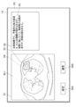
- FIG. 7 is a diagram showing an example of a medical text display screen according to the present embodiment.
- the display screen 50 includes an image display area 51 and a text display area 52.
- the slice image SL1 that is most likely to identify the abnormal shadow candidate detected by the analysis unit 22 is displayed.
- the slice image SL1 includes an abnormal shadow candidate 53, and the abnormal shadow candidate 53 is surrounded by a rectangular region 54.
- the medical text 55 generated by the analysis record generation unit 23 is displayed in the text display area 52.
- Medical text 55 states, "Under the left lung pleura, an irregular tumor with a maximum lateral diameter of 4.2 cm is found. It is in contact with the chest wall, and pleural infiltration is observed, but infiltration is not observed.” be.
- the property information used in the medical text 55 is "left pulmonary subpleural”, “irregular”, “4.2 cm”, “tumor”, and “chest wall” among the property information derived by the analysis unit 22. "With contact”, “With pleural infiltration” and “Without pleural infiltration”.
- a correction button 58A and a confirmation button 58B are displayed below the image display area 51.
- the image interpreting doctor interprets the abnormal shadow candidate 53 in the slice image SL1 displayed in the image display area 51, and determines the suitability of the medical sentence 55 displayed in the sentence display area 52.
- the correction unit 25 receives corrections by the image interpreting doctor for the property information. That is, the medical text 55 displayed in the text display area 52 can be manually corrected by input from the input device 15. Further, by selecting the confirmation button 58B, the medical sentence 55 displayed in the sentence display area 52 can be confirmed with its contents. In this case, the medical text 55 is transcribed into the interpretation report, and the interpretation report to which the medical text 55 is transcribed is transmitted to the report server 7 together with the slice image SL1 and stored.
- the interpreter When the interpreter selects the correction button 58A to correct the medical sentence 55, the interpreter is included in the abnormal shadow 31, but if there is a missing property in the medical sentence 55, the missing property is added. Modify medical text 55 to do so.
- the interpreter inputs the missing properties using the input device 15.
- the interpreting doctor inputs the property information of "Spicula” using the input device 15.
- the medical text 55 is modified so that the correction unit 25 adds the property information of "Spicula".
- the image interpreter modifies the medical text 55 to remove unwanted properties. For example, in the present embodiment, when the property of being in contact with the chest wall is unnecessary, the image interpreter deletes the property information of "being in contact with the chest wall" using the input device 15. As a result, the medical sentence 55 is modified so that the correction unit 25 deletes the property information "in contact with the chest wall".
- FIG. 8 is a diagram showing an example of a modified medical text display screen. As shown in FIG. 8, in the sentence display area 72, the modified medical sentence 59 obtained by modifying the medical sentence 55 is displayed.
- the modified medical text 59 states, "Under the left pleura, a tumor with an irregular shape and a maximum lateral diameter of 4.2 cm is observed. Pleural infiltration is observed, but infiltration is not observed.”
- the medical sentence 55 displayed in the sentence display area 52 can be confirmed with the contents.
- the medical text 55 is transcribed into the interpretation report, and the interpretation report to which the medical text 55 is transcribed is transmitted to the report server 7 by the communication unit 27 together with the slice image SL1 and stored.
- the interpretation doctor selects the confirmation button 58B after the correction, the correction medical sentence 59 can be confirmed with the content thereof.
- the modified medical text 59 is transcribed in the interpretation report, and the interpretation report to which the modified medical text 59 is transcribed is transmitted to the report server 7 by the communication unit 27 together with the slice image SL1 and the stored information 45 described later. Will be stored.
- the interpretation report and the stored information 45 are associated and stored.
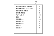
- FIG. 9 is a diagram for explaining the stored information showing the stored result of the property information.
- the stored information 45 is corrected from "without spicula” to "with spicula” and “with chest wall contact” to “without chest wall contact” in the property information derived by the analysis unit 22 shown in FIG. It has been done.
- the property information corrected from "none” to "yes” has a +1 flag
- the property information corrected from "yes” to "no” has a -1 flag, but there is no correction.
- a flag of 0 is added to each of the property information.
- the stored information 45 the property information derived by the analysis unit 22 and the modified property information can be distinguished by a flag.
- the stored information 45 stored in the storage 13 is transmitted to the report server 7 together with the interpretation report and stored as described above.
- the analysis unit 22 analyzes the target medical image to characterize the target medical image. Derive information. Further, the analysis record generation unit 23 generates the target medical text from the property information of the target medical image with the stored information 45 stored in the report server 7 as a non-reference, that is, without referring to the stored information 45. Specifically, the analysis record generation unit 23 generates a target medical sentence by using only the property information derived by the analysis unit 22. The target medical text without reference to the stored information 45 corresponds to the other target image analysis records of the present disclosure. Further, the analysis record generation unit 23 refers to the stored information 45 stored in the report server 7 and generates a target medical sentence as an alternative from the property information of the target medical image.
- the analysis record generation unit 23 identifies the stored information 45 including the property information that matches the property information of the target medical image derived by the analysis unit 22 by searching the report server 7.
- the property information and the stored information 45 include the location and size of the abnormal shadow, but specify the stored information 45 in which the property items excluding the location and size of the abnormal shadow match. For example, when the property information of the target medical image is "tumor", "pleural infiltration” and “no infiltration", the analysis record generation unit 23 determines "tumor", "pleural infiltration” and "no infiltration”.
- the stored information 45 including the property information of "" is searched. Then, the analysis record generation unit 23 acquires an interpretation report associated with the stored information 45 that matches the property information of the target medical image from the report server 7.
- the fact that the property information matches is not limited to the case where all the property information except for the location and size of the abnormal shadow among the plurality of property information matches, and the majority of the plurality of property information, for example, a plurality. It also includes the case where 80% or more and even 90% or more of the property information of the above matches.
- the stored information 45 that match the property information of the target medical image can be selected according to the criteria such as the one with a new creation date or the one created by the interpretation doctor who is operating the interpretation WS3.
- the number of interpretation reports to be acquired may be limited to search for stored information that matches the property information.
- the analysis record generation unit 23 rewrites the location and size of the abnormal shadow in the acquired interpretation report to the location and size of the abnormal shadow included in the property information of the target medical image, and generates another plan.
- the stored information 45 that matches the property information of the target medical image is not stored in the report server 7. In such a case, no alternative plan shall be generated in this embodiment.
- FIG. 10 is a diagram showing a target medical text and an alternative display screen. Note that FIG. 10 shows only one alternative. As shown in FIG. 10, the display screen 70 includes an image display area 71 and a text display area 72. In the image display area 71, the slice image SL2, which is the easiest to identify the abnormal shadow candidate detected by the analysis unit 22 from the target medical image, is displayed. The slice image SL2 includes an abnormal shadow candidate 73, and the abnormal shadow candidate 73 is surrounded by a rectangular region 74.
- the target medical text 75 and the alternative plan 76 generated by the analysis record generation unit 23 are displayed.
- the target medical text 75 is "Under the left pulmonary pleura, a tumor with an irregular shape and a maximum lateral diameter of 4.2 cm is observed. Pleural infiltration is observed, but infiltration is not observed.”
- Alternative 76 says, "Under the left lung pleura, an irregular tumor with a maximum lateral diameter of 4.2 cm is found. It is in contact with the chest wall, and pleural infiltration is observed, but infiltration is not observed.” be.
- a correction button 78A and a confirmation button 78B are displayed below the image display area 71.
- the image interpreting doctor interprets the abnormal shadow candidate 73 in the slice image SL2 displayed in the image display area 71, and determines the suitability of the target medical sentence 75 and the alternative plan 76 displayed in the sentence display area 72.
- the correction button 78A is selected using the input device 15.
- the medical text 75 displayed in the text display area 72 can be corrected in the same manner as described above by inputting from the input device 15.
- the interpreter when the interpreter adopts either the target medical text 75 or the alternative 76, the interpreter selects either the target medical text 75 or the alternative 76 using the input device 15 and presses the confirm button. By selecting 78B, either the target medical text 75 or the alternative 76 can be determined by its contents. In this case, any of the selected target medical text 75 and the alternative 76 is transcribed in the interpretation report, and the interpretation report to which the text is transcribed is transmitted to the report server 7 together with the slice image SL2 and stored.
- the communication unit 27 exchanges information between the information storage device 20 and the external device via the network I / F17.
- FIG. 11 is a flowchart of the information storage process performed in the present embodiment. It is assumed that the medical image to be read is acquired from the image server 5 by the image acquisition unit 21 and stored in the storage 13. The process is started when an instruction to create an image interpretation report is given by the image interpretation doctor, and the analysis unit 22 analyzes the medical image to represent the properties of the structure of interest such as an abnormal shadow candidate included in the medical image. Derivation of information (step ST1). Next, the analysis record generation unit 23 generates a medical sentence related to the medical image as an image analysis record based on the property information (step ST2). Subsequently, the display control unit 24 displays the medical text generated by the analysis record generation unit 23 on the text display area 52 of the display screen 50 displayed on the display 14 (step ST3).
- step ST4 determines whether or not the correction button 58A displayed on the display screen 50 is selected.
- step ST4 affirmed, the correction unit 25 accepts correction using the input device 15 for the property information included in the medical text displayed in the text display area 52 (step ST5).
- step ST6 determines whether or not the confirmation button 58B has been selected (step ST6). If step ST6 is denied, the process returns to step ST5 and the correction is continuously accepted.
- step ST6 is affirmed, the storage control unit 26 distinguishes between the derived property information and the modified property information and stores the modified property information in the storage 13 (step ST7).
- the display control unit 24 transfers the corrected medical text to the interpretation report, and the communication unit 27 transmits the interpretation report to which the corrected medical text is transcribed to the report server 7 together with the slice image SL1 (transmission of the interpretation report: Step ST8), the process is terminated.
- step ST4 determines whether or not the confirmation button 58B has been selected (step ST9). If step ST9 is denied, the process returns to step ST4. When step ST9 is affirmed, the process proceeds to step ST8, the display control unit 24 transfers the medical text to the interpretation report, and the communication unit 27 transfers the medical text transcribed interpretation report together with the slice image SL1 to the report server 7. And finish the process.
- FIG. 12 is a flowchart of the analysis record generation process performed in the present embodiment. It is assumed that the medical image to be read is acquired from the image server 5 by the image acquisition unit 21 and stored in the storage 13. Further, it is assumed that the storage information 45, which is stored separately from the derived property information and the modified property information, is also stored in the storage 13.
- the process is started when an instruction to create an image interpretation report is given by the image interpretation doctor, and the analysis unit 22 analyzes the medical image to represent the properties of the structure of interest such as an abnormal shadow candidate included in the medical image. Derivation of information (step ST11).
- the analysis record generation unit 23 generates the target medical text related to the target medical image as an image analysis record based on the property information derived by the analysis unit 22 without referring to the stored information 45, that is, without referring to the stored information 45. (Step ST12). Further, the analysis record generation unit 23 refers to the stored information 45 stored in the report server 7 and generates a target medical text regarding the target medical image as an alternative plan (step ST13).
- the display control unit 24 displays the target medical text 75 and the alternative 76 generated by the analysis record generation unit 23 in the text display area 52 of the display screen 50 displayed on the display 14 (medical text display: step ST14). ).
- the display control unit 24 accepts the selection of either the target medical text 75 or the alternative 76 (step ST15). Further, the display control unit 24 determines whether or not the correction button 78A displayed on the display screen is selected (step ST16). When step ST16 is affirmed, the correction unit 25 accepts correction of the property information contained in the selected medical text using the input device 15 (step ST17). Subsequently, the correction unit 25 determines whether or not the confirmation button 78B has been selected (step ST18). If step ST18 is denied, the process returns to step ST17 and the correction is continuously accepted. When step ST18 is affirmed, the storage control unit 26 distinguishes between the derived property information and the modified property information and stores the modified property information in the storage 13 (step ST19).
- Step ST20 Interpretation report transmission: Step ST20
- step ST16 determines whether or not the confirmation button 78B has been selected (step ST21). If step ST21 is denied, the process returns to step ST16. When step ST21 is affirmed, the process proceeds to step ST20, the display control unit 24 transfers the selected medical text to the interpretation report, and the communication unit 27 combines the interpretation report in which the medical text is transcribed with the slice image SL1. Is sent to the report server 7 to end the process.
- a plurality of property information representing the properties of the structure of interest included in the image is derived, an image analysis record including at least a part of the plurality of property information is generated, and the user for the property information Accepted the correction by, and saved the derived property information and the corrected property information separately. Therefore, by referring to the stored stored information, when the image analysis record generated from the image is modified, it is possible to recognize which property information derived by analyzing the image is modified.
- the interpretation report is displayed by displaying an alternative plan referring to the stored information in addition to the target medical text generated based on the property information derived from the target medical image without referring to the stored information 45. You can increase the choices of sentences to be posted to. Therefore, the image interpreter can transfer the medical text describing the desired property information to the image interpretation report.
- the target medical text is generated using only the property information derived by the analysis unit 22 for the target medical image, but the present invention is not limited to this.
- An alternative plan generated by referring only to the stored information 45 may be used as the target medical text without generating the target medical text using only the property information derived by the analysis unit 22 for the target medical image.
- FIG. 13 is a diagram showing another example of the medical text display screen in the present embodiment.
- the display screen 80 includes an image display area 81, a property information display area 82, and a text display area 83.
- the slice image SL3 which is the easiest to identify the abnormal shadow candidate detected by the analysis unit 22, is displayed.
- the slice image SL3 includes an abnormal shadow candidate 84, and the abnormal shadow candidate 84 is surrounded by a rectangular region 85.
- the shape of the boundary (clear and irregular), the type of absorption value (solid type and suriglass type), the presence or absence of spicula, the presence or absence of a mass or nodule, the presence or absence of pleural contact, the presence or absence of pleural infiltration, Buttons 82A-82I are displayed to specify the presence / absence of pleural infiltration, the presence / absence of cavities, and the presence / absence of calcification, respectively.
- the medical text 86 generated by the analysis record generation unit 23 is displayed in the text display area 83.
- the medical text 86 states, "Under the left lung pleura, an irregular tumor with a maximum lateral diameter of 4.2 cm is found. It is in contact with the chest wall, and pleural infiltration is observed, but infiltration is not observed.” be.
- the property information used in the medical text 86 is "left pulmonary subpleural”, “irregular”, “4.2 cm”, “tumor”, and “chest wall” among the property information derived by the analysis unit 22. "With contact”, “With pleural infiltration” and "Without pleural infiltration”.
- a correction button 88A and a confirmation button 88B are displayed below the image display area 81. Since the functions of the correction button 88A and the confirmation button 88B are the same as those of the correction buttons 58A and 78A and the confirmation buttons 58B and 78B described above, detailed description thereof will be omitted here.
- the image interpreter can modify the medical text 86 by selecting the desired button for the property information displayed in the property information display area 82. For example, by selecting the button 82C, it is possible to correct the absence of spicula to the presence of spicula. Further, by selecting the button 82E, it is possible to correct the presence of pleural contact to the absence of pleural contact. As a result, in the text display area 83, "A tumor with an irregular shape and a maximum lateral diameter of 4.2 cm is observed under the left pulmonary pleura. Pleural infiltration is observed, but infiltration is not observed. "Corrected medical text will be displayed.
- medical texts are generated using medical images whose diagnosis target is the lungs to support the creation of medical documents such as interpretation reports, but the diagnosis target is limited to the lungs. It's not something.
- diagnosis target is limited to the lungs. It's not something.
- any part of the human body such as the heart, liver, brain, and limbs can be diagnosed.
- each learning model of the analysis unit 22 and the analysis record generation unit 23 is prepared to perform analysis processing and analysis record processing according to the diagnosis target, and analysis processing and analysis record generation processing according to the diagnosis target.
- the learning model to perform is selected, and the analysis record generation process is executed.
- the technique of the present disclosure is applied when creating an interpretation report as an analysis record, but when a medical document other than the interpretation report such as an electronic medical record and a diagnostic report is created as an analysis record.
- a medical document other than the interpretation report such as an electronic medical record and a diagnostic report is created as an analysis record.
- the technology of the present disclosure can also be applied.
- the image analysis record is generated using the medical image, but the present invention is not limited to this. It goes without saying that the technique of the present disclosure can also be applied when generating an image analysis record for an arbitrary image other than a medical image. For example, when analyzing an image of the chemical formula of a compound, deriving the type of cyclic hydrocarbon and the type of functional group as property information, and generating the name of the compound as an image analysis record from the derived property information, this book is also available. The disclosed technology can be applied.
- a process of executing various processes such as an image acquisition unit 21, an analysis unit 22, an analysis record generation unit 23, a display control unit 24, a correction unit 25, a storage control unit 26, and a communication unit 27.
- various processors processors shown below can be used.
- the various processors include a CPU, which is a general-purpose processor that executes software (program) and functions as various processing units, and a circuit after manufacturing an FPGA (Field Programmable Gate Array) or the like.
- Dedicated electricity which is a processor with a circuit configuration specially designed to execute specific processing such as programmable logic device (PLD), ASIC (Application Specific Integrated Circuit), which is a processor whose configuration can be changed. Circuits and the like are included.
- One processing unit may be composed of one of these various processors, or a combination of two or more processors of the same type or different types (for example, a combination of a plurality of FPGAs or a combination of a CPU and an FPGA). ) May be configured. Further, a plurality of processing units may be configured by one processor.
- one processor is configured by combining one or more CPUs and software. There is a form in which this processor functions as a plurality of processing units.
- SoC System On Chip
- the various processing units are configured by using one or more of the above-mentioned various processors as a hardware structure.
- circuitry in which circuit elements such as semiconductor elements are combined can be used.
Landscapes
- Engineering & Computer Science (AREA)
- Health & Medical Sciences (AREA)
- General Health & Medical Sciences (AREA)
- Theoretical Computer Science (AREA)
- Medical Informatics (AREA)
- Public Health (AREA)
- Physics & Mathematics (AREA)
- Epidemiology (AREA)
- Primary Health Care (AREA)
- General Physics & Mathematics (AREA)
- Computational Linguistics (AREA)
- General Engineering & Computer Science (AREA)
- Audiology, Speech & Language Pathology (AREA)
- Artificial Intelligence (AREA)
- Nuclear Medicine, Radiotherapy & Molecular Imaging (AREA)
- Radiology & Medical Imaging (AREA)
- Life Sciences & Earth Sciences (AREA)
- Biomedical Technology (AREA)
- Pathology (AREA)
- High Energy & Nuclear Physics (AREA)
- Optics & Photonics (AREA)
- Biophysics (AREA)
- Heart & Thoracic Surgery (AREA)
- Molecular Biology (AREA)
- Surgery (AREA)
- Animal Behavior & Ethology (AREA)
- Veterinary Medicine (AREA)
- Probability & Statistics with Applications (AREA)
- Data Mining & Analysis (AREA)
- Databases & Information Systems (AREA)
- Medical Treatment And Welfare Office Work (AREA)
- Measuring And Recording Apparatus For Diagnosis (AREA)
Priority Applications (3)
| Application Number | Priority Date | Filing Date | Title |
|---|---|---|---|
| JP2022504406A JP7436628B2 (ja) | 2020-03-03 | 2021-03-02 | 情報保存装置、方法およびプログラム、並びに解析記録生成装置、方法およびプログラム |
| US17/890,270 US12387054B2 (en) | 2020-03-03 | 2022-08-18 | Information saving apparatus, method, and program and analysis record generation apparatus, method, and program for recognizing correction made in image analysis record |
| JP2023195440A JP7684374B2 (ja) | 2020-03-03 | 2023-11-16 | 情報保存装置、方法およびプログラム、並びに解析記録生成装置、方法およびプログラム |
Applications Claiming Priority (2)
| Application Number | Priority Date | Filing Date | Title |
|---|---|---|---|
| JP2020035800 | 2020-03-03 | ||
| JP2020-035800 | 2020-03-03 |
Related Child Applications (1)
| Application Number | Title | Priority Date | Filing Date |
|---|---|---|---|
| US17/890,270 Continuation US12387054B2 (en) | 2020-03-03 | 2022-08-18 | Information saving apparatus, method, and program and analysis record generation apparatus, method, and program for recognizing correction made in image analysis record |
Publications (1)
| Publication Number | Publication Date |
|---|---|
| WO2021177312A1 true WO2021177312A1 (ja) | 2021-09-10 |
Family
ID=77612638
Family Applications (1)
| Application Number | Title | Priority Date | Filing Date |
|---|---|---|---|
| PCT/JP2021/007977 Ceased WO2021177312A1 (ja) | 2020-03-03 | 2021-03-02 | 情報保存装置、方法およびプログラム、並びに解析記録生成装置、方法およびプログラム |
Country Status (3)
| Country | Link |
|---|---|
| US (1) | US12387054B2 (https=) |
| JP (2) | JP7436628B2 (https=) |
| WO (1) | WO2021177312A1 (https=) |
Families Citing this family (4)
| Publication number | Priority date | Publication date | Assignee | Title |
|---|---|---|---|---|
| JP7537905B2 (ja) * | 2020-04-17 | 2024-08-21 | キヤノンメディカルシステムズ株式会社 | 読影レポート作成装置 |
| JP7706987B2 (ja) * | 2021-08-17 | 2025-07-14 | 富士フイルム株式会社 | 学習装置、方法およびプログラム、並びに情報処理装置、方法およびプログラム |
| JP7718915B2 (ja) * | 2021-08-30 | 2025-08-05 | 富士フイルム株式会社 | 学習装置、方法およびプログラム、並びに情報処理装置、方法およびプログラム |
| JP7799735B2 (ja) * | 2024-03-27 | 2026-01-15 | キヤノン株式会社 | 文章生成システム、文章生成方法およびプログラム |
Citations (6)
| Publication number | Priority date | Publication date | Assignee | Title |
|---|---|---|---|---|
| JPH0731591A (ja) * | 1993-07-19 | 1995-02-03 | Toshiba Corp | 読影レポート作成支援装置 |
| JP2009082443A (ja) * | 2007-09-28 | 2009-04-23 | Canon Inc | 診断支援装置及びその制御方法 |
| JP2009259000A (ja) * | 2008-04-16 | 2009-11-05 | Fujifilm Corp | 文書作成支援装置、文書作成支援方法、並びに文書作成支援プログラム |
| JP2016040688A (ja) * | 2014-08-12 | 2016-03-24 | 株式会社東芝 | 読影レポート作成支援装置 |
| JP2019149130A (ja) * | 2018-02-28 | 2019-09-05 | 富士フイルム株式会社 | 医用画像表示装置、方法およびプログラム |
| JP2019153250A (ja) * | 2018-03-06 | 2019-09-12 | 富士フイルム株式会社 | 医療文書作成支援装置、方法およびプログラム |
Family Cites Families (9)
| Publication number | Priority date | Publication date | Assignee | Title |
|---|---|---|---|---|
| EP0973116A1 (en) * | 1993-03-01 | 2000-01-19 | Kabushiki Kaisha Toshiba | Medical information processing system for supporting diagnosis |
| JP2005148990A (ja) * | 2003-11-13 | 2005-06-09 | Konica Minolta Medical & Graphic Inc | 医用画像読影システム及び読影レポート作成方法 |
| JP2007122679A (ja) | 2005-09-27 | 2007-05-17 | Fujifilm Corp | 読影支援システム |
| JP5566674B2 (ja) | 2009-12-15 | 2014-08-06 | 株式会社東芝 | 読影レポート作成支援システム |
| WO2012105281A1 (ja) * | 2011-01-31 | 2012-08-09 | 日本電気株式会社 | 情報処理システム、情報処理方法、情報処理装置及びその制御方法とその制御プログラムを格納した記憶媒体 |
| JP6532371B2 (ja) | 2015-10-02 | 2019-06-19 | オリンパス株式会社 | 医療レポート作成支援システム |
| US10803581B2 (en) * | 2017-11-06 | 2020-10-13 | Beijing Keya Medical Technology Co., Ltd. | System and method for generating and editing diagnosis reports based on medical images |
| JP6625155B2 (ja) | 2018-03-29 | 2019-12-25 | キヤノン株式会社 | 情報処理装置、情報処理装置の作動方法及びプログラム |
| US10892056B2 (en) * | 2018-11-16 | 2021-01-12 | International Business Machines Corporation | Artificial intelligence based alert system |
-
2021
- 2021-03-02 WO PCT/JP2021/007977 patent/WO2021177312A1/ja not_active Ceased
- 2021-03-02 JP JP2022504406A patent/JP7436628B2/ja active Active
-
2022
- 2022-08-18 US US17/890,270 patent/US12387054B2/en active Active
-
2023
- 2023-11-16 JP JP2023195440A patent/JP7684374B2/ja active Active
Patent Citations (6)
| Publication number | Priority date | Publication date | Assignee | Title |
|---|---|---|---|---|
| JPH0731591A (ja) * | 1993-07-19 | 1995-02-03 | Toshiba Corp | 読影レポート作成支援装置 |
| JP2009082443A (ja) * | 2007-09-28 | 2009-04-23 | Canon Inc | 診断支援装置及びその制御方法 |
| JP2009259000A (ja) * | 2008-04-16 | 2009-11-05 | Fujifilm Corp | 文書作成支援装置、文書作成支援方法、並びに文書作成支援プログラム |
| JP2016040688A (ja) * | 2014-08-12 | 2016-03-24 | 株式会社東芝 | 読影レポート作成支援装置 |
| JP2019149130A (ja) * | 2018-02-28 | 2019-09-05 | 富士フイルム株式会社 | 医用画像表示装置、方法およびプログラム |
| JP2019153250A (ja) * | 2018-03-06 | 2019-09-12 | 富士フイルム株式会社 | 医療文書作成支援装置、方法およびプログラム |
Also Published As
| Publication number | Publication date |
|---|---|
| US12387054B2 (en) | 2025-08-12 |
| JPWO2021177312A1 (https=) | 2021-09-10 |
| JP7684374B2 (ja) | 2025-05-27 |
| US20220391599A1 (en) | 2022-12-08 |
| JP2024012644A (ja) | 2024-01-30 |
| JP7436628B2 (ja) | 2024-02-21 |
Similar Documents
| Publication | Publication Date | Title |
|---|---|---|
| JP7618003B2 (ja) | 文書作成支援装置、方法およびプログラム | |
| JP2019153250A (ja) | 医療文書作成支援装置、方法およびプログラム | |
| JP7684374B2 (ja) | 情報保存装置、方法およびプログラム、並びに解析記録生成装置、方法およびプログラム | |
| US20230197253A1 (en) | Medical image processing apparatus, method, and program | |
| US12406755B2 (en) | Document creation support apparatus, method, and program | |
| US12288611B2 (en) | Information processing apparatus, method, and program | |
| WO2022215530A1 (ja) | 医用画像装置、医用画像方法、及び医用画像プログラム | |
| JP7102509B2 (ja) | 医療文書作成支援装置、医療文書作成支援方法、及び医療文書作成支援プログラム | |
| US20230281810A1 (en) | Image display apparatus, method, and program | |
| US20220366151A1 (en) | Document creation support apparatus, method, and program | |
| WO2019193983A1 (ja) | 医療文書表示制御装置、医療文書表示制御方法、及び医療文書表示制御プログラム | |
| US12211600B2 (en) | Information processing apparatus, information processing method, and information processing program | |
| WO2021107098A1 (ja) | 文書作成支援装置、文書作成支援方法及び文書作成支援プログラム | |
| WO2021177357A1 (ja) | 情報処理装置、情報処理方法及び情報処理プログラム | |
| JP7007469B2 (ja) | 医療文書作成支援装置、方法およびプログラム、学習済みモデル、並びに学習装置、方法およびプログラム | |
| WO2022196106A1 (ja) | 文書作成装置、方法およびプログラム | |
| WO2021193548A1 (ja) | 文書作成支援装置、方法およびプログラム | |
| US12387825B2 (en) | Information processing apparatus, information processing method, and information processing program | |
| WO2022230641A1 (ja) | 文書作成支援装置、文書作成支援方法、及び文書作成支援プログラム | |
| US20220277134A1 (en) | Document creation support apparatus, method, and program | |
| WO2022153702A1 (ja) | 医用画像表示装置、方法およびプログラム | |
| JP7368592B2 (ja) | 文書作成支援装置、方法およびプログラム | |
| JP7376715B2 (ja) | 経過予測装置、経過予測装置の作動方法および経過予測プログラム |
Legal Events
| Date | Code | Title | Description |
|---|---|---|---|
| 121 | Ep: the epo has been informed by wipo that ep was designated in this application |
Ref document number: 21764028 Country of ref document: EP Kind code of ref document: A1 |
|
| ENP | Entry into the national phase |
Ref document number: 2022504406 Country of ref document: JP Kind code of ref document: A |
|
| NENP | Non-entry into the national phase |
Ref country code: DE |
|
| 122 | Ep: pct application non-entry in european phase |
Ref document number: 21764028 Country of ref document: EP Kind code of ref document: A1 |