WO2021009906A1 - Cell image analysis method and cell image analysis device - Google Patents
Cell image analysis method and cell image analysis device Download PDFInfo
- Publication number
- WO2021009906A1 WO2021009906A1 PCT/JP2019/028268 JP2019028268W WO2021009906A1 WO 2021009906 A1 WO2021009906 A1 WO 2021009906A1 JP 2019028268 W JP2019028268 W JP 2019028268W WO 2021009906 A1 WO2021009906 A1 WO 2021009906A1
- Authority
- WO
- WIPO (PCT)
- Prior art keywords
- image
- colony
- cell
- region
- cells
- Prior art date
Links
Images


Classifications
-
- G—PHYSICS
- G06—COMPUTING; CALCULATING OR COUNTING
- G06V—IMAGE OR VIDEO RECOGNITION OR UNDERSTANDING
- G06V10/00—Arrangements for image or video recognition or understanding
- G06V10/20—Image preprocessing
- G06V10/22—Image preprocessing by selection of a specific region containing or referencing a pattern; Locating or processing of specific regions to guide the detection or recognition
- G06V10/235—Image preprocessing by selection of a specific region containing or referencing a pattern; Locating or processing of specific regions to guide the detection or recognition based on user input or interaction
-
- G—PHYSICS
- G06—COMPUTING; CALCULATING OR COUNTING
- G06V—IMAGE OR VIDEO RECOGNITION OR UNDERSTANDING
- G06V10/00—Arrangements for image or video recognition or understanding
- G06V10/70—Arrangements for image or video recognition or understanding using pattern recognition or machine learning
-
- C—CHEMISTRY; METALLURGY
- C12—BIOCHEMISTRY; BEER; SPIRITS; WINE; VINEGAR; MICROBIOLOGY; ENZYMOLOGY; MUTATION OR GENETIC ENGINEERING
- C12M—APPARATUS FOR ENZYMOLOGY OR MICROBIOLOGY; APPARATUS FOR CULTURING MICROORGANISMS FOR PRODUCING BIOMASS, FOR GROWING CELLS OR FOR OBTAINING FERMENTATION OR METABOLIC PRODUCTS, i.e. BIOREACTORS OR FERMENTERS
- C12M1/00—Apparatus for enzymology or microbiology
- C12M1/34—Measuring or testing with condition measuring or sensing means, e.g. colony counters
-
- C—CHEMISTRY; METALLURGY
- C12—BIOCHEMISTRY; BEER; SPIRITS; WINE; VINEGAR; MICROBIOLOGY; ENZYMOLOGY; MUTATION OR GENETIC ENGINEERING
- C12Q—MEASURING OR TESTING PROCESSES INVOLVING ENZYMES, NUCLEIC ACIDS OR MICROORGANISMS; COMPOSITIONS OR TEST PAPERS THEREFOR; PROCESSES OF PREPARING SUCH COMPOSITIONS; CONDITION-RESPONSIVE CONTROL IN MICROBIOLOGICAL OR ENZYMOLOGICAL PROCESSES
- C12Q1/00—Measuring or testing processes involving enzymes, nucleic acids or microorganisms; Compositions therefor; Processes of preparing such compositions
- C12Q1/02—Measuring or testing processes involving enzymes, nucleic acids or microorganisms; Compositions therefor; Processes of preparing such compositions involving viable microorganisms
- C12Q1/04—Determining presence or kind of microorganism; Use of selective media for testing antibiotics or bacteriocides; Compositions containing a chemical indicator therefor
-
- G—PHYSICS
- G06—COMPUTING; CALCULATING OR COUNTING
- G06T—IMAGE DATA PROCESSING OR GENERATION, IN GENERAL
- G06T7/00—Image analysis
-
- G—PHYSICS
- G06—COMPUTING; CALCULATING OR COUNTING
- G06V—IMAGE OR VIDEO RECOGNITION OR UNDERSTANDING
- G06V10/00—Arrangements for image or video recognition or understanding
- G06V10/40—Extraction of image or video features
- G06V10/46—Descriptors for shape, contour or point-related descriptors, e.g. scale invariant feature transform [SIFT] or bags of words [BoW]; Salient regional features
-
- G—PHYSICS
- G06—COMPUTING; CALCULATING OR COUNTING
- G06V—IMAGE OR VIDEO RECOGNITION OR UNDERSTANDING
- G06V10/00—Arrangements for image or video recognition or understanding
- G06V10/70—Arrangements for image or video recognition or understanding using pattern recognition or machine learning
- G06V10/77—Processing image or video features in feature spaces; using data integration or data reduction, e.g. principal component analysis [PCA] or independent component analysis [ICA] or self-organising maps [SOM]; Blind source separation
- G06V10/774—Generating sets of training patterns; Bootstrap methods, e.g. bagging or boosting
-
- G—PHYSICS
- G06—COMPUTING; CALCULATING OR COUNTING
- G06V—IMAGE OR VIDEO RECOGNITION OR UNDERSTANDING
- G06V20/00—Scenes; Scene-specific elements
- G06V20/60—Type of objects
- G06V20/69—Microscopic objects, e.g. biological cells or cellular parts
-
- G—PHYSICS
- G06—COMPUTING; CALCULATING OR COUNTING
- G06V—IMAGE OR VIDEO RECOGNITION OR UNDERSTANDING
- G06V20/00—Scenes; Scene-specific elements
- G06V20/60—Type of objects
- G06V20/69—Microscopic objects, e.g. biological cells or cellular parts
- G06V20/695—Preprocessing, e.g. image segmentation
Definitions
- the present invention relates to a cell image analysis method and a cell image analysis device.
- iPS cells induced pluripotent stem cells
- the cells being cultured are extracted from the culture vessel and transferred to another culture vessel, and subculture is performed to obtain the next generation cell line. This extraction of cells is called picking. Since there are cells unsuitable for passage such as cells that have already started differentiation (cells that have lost pluripotency) in the culture vessel, the cells suitable for passage are the workers (users) at the time of passage. Sorted by.
- the worker (user) searches for cells suitable for passage from the culture vessel by checking the cell colonies in the culture vessel one by one with a microscope. Next, the worker (user) marks a position where the cells suitable for passage are located, and sucks the cell colony at the marked position with a pipetter or the like. In addition, at the initial stage of culturing, cells that have already started differentiation (cells that have lost pluripotency), dead cells, and the like are extracted from the culture vessel and removed. Similar to the passage, such removal work is also performed by searching for the removal target using a microscope and picking the found removal target.
- Non-Patent Document 1 a device that automatically performs a cell picking operation by a pipetta by imaging a microscopic image of cells in a culture vessel, determining a picking target from the image, and then operating a controller. It is disclosed.
- Non-Patent Document 1 Since the cell search work needs to be performed by the user himself, it is desired to reduce the workload of the user for the cell search work.
- the cells to be picked differ depending on the purpose of cell culture performed by the user, the type of cells to be cultured, and the like. Therefore, it is necessary for the user to independently determine which cell region is suitable for the picking target from among the large number of cell regions observed from the microscope image. As a result, conventionally, when culturing pluripotent cells, it has been difficult to reduce the load on the user associated with the work of searching for a picking target.
- the present invention has been made to solve the above-mentioned problems, and one object of the present invention is to effectively reduce the load on the user associated with the work of searching for cells to be picked in cell culture. It is to provide a cell image analysis method and a cell image analysis apparatus which can be performed.
- the cell image analysis method in the first aspect of the present invention includes a step of acquiring a first image of a cell colony containing cells having differentiation potential, and a cell colony in the first image.
- the step of converting the first image into a label image by performing a segmentation process for identifying the colony region of the cell that has already started differentiation and the colony region of the undifferentiated cell, and the shape feature amount of the cell colony from the label image.
- a step of receiving an input regarding a colony area to be searched from a user using a computer a step of setting a judgment standard of a shape feature amount based on the user's input, and a shape feature amount and a judgment standard.
- each cell colony includes a step of determining whether or not a colony region as a candidate to be searched is included.
- the cell image analyzer in the second aspect of the present invention includes a storage unit in which a microscopic image of a cell colony containing cells capable of differentiating is input, and a cell in the microscopic image that has already started differentiation.
- a segmentation processing unit that performs segmentation processing to identify the colony region of the undifferentiated cells and converts the first image into a label image, an input unit that accepts input regarding the colony region to be searched, and a microscope image.
- Each cell colony contained in the cell colony is provided with a determination processing unit for determining whether or not a colony region as a candidate for search is included, and the determination processing unit acquires the shape feature amount of the cell colony from the label image. It is configured to determine the colony region based on the shape feature amount and the determination standard of the shape feature amount set based on the input of the user.
- the cell image analysis method in the third aspect of the present invention includes a step of creating a trained model for machine-learned determination and a step of acquiring a first image of a cell colony containing cells having differentiation potential. , A step of performing a segmentation process for identifying a colony region of cells that have already started differentiation and a colony region of undifferentiated cells in the cell colonies in the first image, and converting the first image into a label image.
- the step of creating a trained model includes a step of determining whether or not a colony region that is a candidate for a search target is included for each cell colony by inputting a label image of one image into the trained model.
- the step of accepting the input of selection information as to whether or not the cell colony in the second image acquired in advance includes a desired colony region, and the label image obtained by segmenting the second image as input data, and the selection information as teacher data. Includes steps to create a trained model by machine learning.
- the colony region of cells that have started differentiation by the segmentation treatment and the colony region of undifferentiated cells are picked according to the purpose of culturing and the like.
- the colony area that can be the target of the above can be identified from the image.
- each cell colony in the image becomes a candidate for search target based on the shape feature amount and the determination criterion of the shape feature amount set based on the user's input. It is possible to obtain a determination result as to whether or not the colony region is included.
- the colony region that is a candidate for the search target is included for each cell colony in the image by the trained model machine-learned using the selection information of whether or not the desired colony region is included. It is possible to obtain the determination result of whether or not.
- the cell image analysis method and the cell image analysis apparatus 100 according to the present embodiment will be described with reference to FIGS. 1 to 14.
- Cell image analysis method In the cell image analysis method shown in FIG. 1, in a cell picking operation in cell culture, a microscopic image of a cell colony 10 is acquired, and the cell colony 10 shown in the microscopic image is a colony region that is a candidate for a search target. It is a method of determining whether or not it is included.
- the cell colony 10 is a cell mass (a collection of a large number of cells) derived from a single cell.
- the colony region is a region of cell colonies composed of specific cells.
- Cell picking is to extract cells or cell clusters to be picked from the cell culture vessel 90.
- the picking of cells is performed by sucking the picking target using an instrument such as a pipetter.
- the cells to be picked in this embodiment are cells having differentiation potential.
- Cells having differentiation potential are, for example, iPS cells, ES cells (embryonic stem cells) and the like. These cells have pluripotency (potency) to differentiate into cells that make up various tissues and organs.
- "undifferentiated cells” which are cells that maintain pluripotency and "undifferentiated deviant cells” that have deviated from the undifferentiated state and have already started to differentiate are generated. Therefore, a colony region of undifferentiated cells and a colony region of undifferentiated deviant cells are formed in the cell colony.
- a cell colony may consist of either a colony region of undifferentiated cells or a colony region of undifferentiated deviant cells, or may include a colony region of undifferentiated cells and a colony region of undifferentiated deviant cells. ..
- undifferentiated cells are picked and transferred to another culture vessel, and cultured as the next generation cell line.
- the work of picking and removing from the culture vessel 90 is performed.
- the cell used for passage is not limited to any undifferentiated cell.
- picking a colony region of undifferentiated cells according to the purpose of the user performing the culturing operation is searched by the user and selected as a target for picking.
- the cell image analysis method distinguishes between the colony region 11 of undifferentiated cells and the colony region 12 of undifferentiated deviant cells when culturing cells having such differentiation potential. Then, the cell image analysis method determines the cell colony 10 including the colony region that is a candidate for the search target for picking according to the purpose of the user who performs the culture work, and thereby the cell colony 10 to be picked. Support the search work of.
- the colony region 12 of undifferentiated deviant cells is an example of the “colony region of cells that have already started differentiation” in the claims.
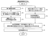
- the cell image analysis method includes at least the following steps 71 to 76.
- Step 71 Obtain a first image 21 in which a cell colony 10 containing cells having differentiation potential is imaged.
- Step 72 In the cell colony 10 in the first image 21, a segmentation process for identifying the colony region 12 of the cells that have already started differentiation (undifferentiated deviation cells) and the colony region 11 of the undifferentiated cells is performed.
- the first image 21 is converted into the label image 21A.
- the shape feature amount 25 of the cell colony 10 is acquired from the label image 21A.
- Step 74 The computer is used to receive input from the user regarding the colony area to be searched.
- Step 75 The determination criterion 30 of the shape feature amount 25 is set based on the input of the user.
- Step 76 Based on the shape feature amount 25 and the determination criterion 30, it is determined whether or not each cell colony 10 includes a colony region that is a candidate for a search target.
- the first image 21 may be a microscopic image of a cell colony 10 containing cells having differentiation potential.
- the microscope image is an image acquired by an optical microscope such as a transmission observation type microscope or a phase contrast microscope.
- the first image 21 can be a holographic microscope image.
- the first image 21 is not particularly limited as long as it is an image in which the shape of the cell colony 10 can be captured nondestructively at an observable magnification.
- the first image 21 is an image obtained by capturing at least one cell colony 10.
- the first image 21 is acquired by imaging the cell colonies 10 in the culture vessel 90 with the imaging device 200, for example, as shown in FIG.
- the culture container 90 is a transparent flat plate-shaped culture dish such as a so-called petri dish, a microplate (well plate) in which a plurality of wells are formed, or the like.
- a segmentation process is performed on the acquired first image 21.
- the "segmentation process” is a process of dividing an image into a plurality of areas, and by assigning a label indicating the detection target to the area in which the detection target appears, the input image is divided into a plurality of label areas. It is a process to classify into.
- the label area is an area (a part of an image) composed of a group of pixels with a common label in the image.
- the segmentation process is realized by image processing using a computer.
- the label is information indicating the meaning indicated by the image portion constituting the label area. Segmentation is performed by assigning a label to each pixel in the image.
- the label may be given in units of a group of a plurality of pixels (pixel group).
- the type of label is called a class.
- the segmentation process for the first image 21 generates a label image 21A in which the first image 21 is divided into a plurality of label areas.
- the segmentation process in step 72 divides the first image 21 into at least the label region of the colony region 12 of the cells that have started differentiation (undifferentiated deviant cells) and the label region of the colony region 11 of the undifferentiated cells. It is a process to do. That is, at least two classes are classified. As a result, the label image 21A generated includes at least two label regions, a colony region 12 of the undifferentiated deviant cells and a colony region 11 of the undifferentiated cells.
- the label image 21A includes, for example, three (three classes) label regions: a colony region 12 of undifferentiated deviant cells, a colony region 11 of undifferentiated cells, and a background region 13 other than the colony region.
- each pixel in the same label area is represented by the same pixel value or the same color. Different label areas are represented by different pixel values or different colors.
- each pixel in the first image 21 is a ternary pixel corresponding to three classes of labels (colony region of undifferentiated cells, colony region of undifferentiated cells, background region). It is a ternary image classified by any of the values. As a result, image information irrelevant to the shape of the cell colony, such as the pattern of the colony region and the gradation of light and darkness appearing in the first image 21, is removed, and only the shape of each label region can be accurately extracted.
- the shape feature amount 25 of the cell colony 10 is acquired from the label image 21A.
- the shape feature amount 25 is information that numerically expresses the shape of the cell colony 10 included in the label image 21A.
- the shape feature amount 25 can be an amount (numerical value) indicating the size, contour shape, width, length, etc. of the label region indicating the cell colony 10.
- the shape feature amount 25 of the cell colony 10 may be a shape feature amount relating to the entire region of the cell colony 10, a colony region 11 of undifferentiated cells which is a part of the cell colony 10, or a colony region of undifferentiated deviation cells. It may be a shape feature amount with respect to 12. The specific contents of the shape feature amount 25 will be described later.
- step 74 the input regarding the colony area to be searched is received via the input device provided in the computer. Then, in step 75, the determination criterion 30 of the shape feature amount 25 is set by the computer based on the input information.
- the information about the colony area to be searched is the information for setting the determination criterion 30 of the shape feature amount 25.
- the information regarding the colony region to be searched may be the determination criterion 30 itself.
- the information about the colony area to be searched may be information indicating the user's tendency as to what kind of colony area the user determines as the search target. More simply, the information about the colony region to be searched for can be information that represents the user's preference for the cell colony 10 as to what colony region the user wants. Therefore, the determination criterion 30 of the shape feature amount 25 according to the user's own determination criterion is set based on the input regarding the colony region to be searched.
- the determination criterion 30 of the shape feature amount 25 set based on the user input may be, for example, the threshold value of the shape feature amount 25.
- the determination criterion 30 may include a weight for each shape feature quantity 25 used for the determination.
- the user inputs and sets the threshold value and the weight of the shape feature amount 25 according to the characteristics of the cell colony 10 to be picked.
- a sample microscope image is prepared in advance, and the user himself / herself selects (teaches) whether or not the individual cell colonies 10 appearing in the microscope image include a colony region as a candidate for search. As a result, the determination criterion 30 may be set.
- step 76 it is determined for each cell colony 10 appearing in the image whether or not each cell colony 10 in the first image 21 (label image 21A) includes a colony region that is a candidate for search.
- the determination process is realized by image processing using a computer.
- the determination process of whether or not the colony region as a candidate for the search target is included depends on whether or not the shape feature amount 25 acquired for the cell colony 10 to be determined matches the determination criterion 30 of the shape feature amount 25. Will be implemented.
- a determination result 35 is generated for each cell colony 10 in the first image 21 (label image 21A) as to whether or not the colony region as a candidate for the search target is included.
- the determination result 35 may be binary information of "including a colony region as a candidate for a search target (positive example)" and "not including a colony region as a candidate for a search target (negative example)".
- the determination result 35 may be information indicating the degree of high possibility of "including the colony region as a candidate for the search target”.
- the determination result 35 is output. Based on the determination result 35, it can be determined by the user himself / herself or automatically whether or not the individual cell colonies 10 in the first image 21 (label image 21A) are to be picked. For example, the user selects the cell colony 10 according to the purpose from the cell colonies 10 determined to be a positive example in consideration of the determination result 35 of the individual cell colonies 10 as the picking target. Further, for example, from the cell colonies 10 determined to be positive examples, any cell colony 10 is selected as a picking target by an automatic process such as a threshold value process. Therefore, the user can determine the picking target without requiring a search operation such as confirming the morphology of each cell colony 10 in the culture vessel 90 in detail in the picking operation.
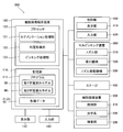
- the cell image analysis device 100 is a device that executes the cell image analysis method shown in FIGS. 1 and 2.
- the cell image analysis device 100 shown in FIG. 3 includes a storage unit 110, a segmentation processing unit 121, a determination processing unit 122, and an input unit 140.
- the cell image analysis device 100 can acquire a microscope image from the cell image pickup device 200 by inputting / outputting a signal, data communication, or transferring data via a recording medium.
- a microscopic image of a cell colony 10 containing cells having differentiation potential is input to the storage unit 110.
- the microscope image is the first image 21 shown in FIG.
- the cell image analysis device 100 is configured to store the image data of the first image 21 acquired from the cell imaging device 200 in the storage unit 110. As a result, step 71 of FIG. 2 is carried out.
- the segmentation processing unit 121 performs a segmentation process for identifying the colony region 12 of cells that have already started differentiation and the colony region 11 of undifferentiated cells in the cell colony 10 in the microscopic image (first image 21). That is, the segmentation processing unit 121 carries out step 72 of FIG.
- the segmentation processing unit 121 converts the first image 21 into a label image 21A (see FIG. 1) by performing the segmentation processing on the first image 21.
- the determination processing unit 122 determines whether or not each cell colony 10 included in the microscope image includes a colony region that is a candidate for a search target.
- the determination processing unit 122 acquires the shape feature amount 25 (see FIG. 1) of the cell colony 10 from the label image 21A, and determines the colony region based on the shape feature amount 25 and the determination criterion 30 of the shape feature amount 25. Is configured to do. That is, the determination processing unit 122 executes steps 73 and 76 of FIG.
- the determination criterion 30 of the shape feature amount 25 is set in the cell image analysis device 100 based on the input of the user using the input unit 140 in steps 74 and 75 prior to the determination process (step 76), and is stored in the storage unit 110. Will be done.
- the determination processing unit 122 outputs the determination result 35 in step 77 of FIG. Based on the determination result 35, it can be determined by the user or automatically whether or not the individual cell colonies 10 in the first image 21 (label image 21A) are to be picked.
- the cell image analysis apparatus 100 stores data with a processor 120 that performs arithmetic processing such as a CPU (Central Processing Unit), a GPU (Graphics Processing Unit), and an FPGA (field-programmable gate array). It is composed of a personal computer (PC) including a storage unit 110 and an input unit 140.
- a processor 120 that performs arithmetic processing such as a CPU (Central Processing Unit), a GPU (Graphics Processing Unit), and an FPGA (field-programmable gate array). It is composed of a personal computer (PC) including a storage unit 110 and an input unit 140.
- PC personal computer
- the processor 120 functions as the segmentation processing unit 121 and the determination processing unit 122 by executing the program 111 stored in the storage unit 110. That is, in the example of FIG. 3, the segmentation processing unit 121 and the determination processing unit 122 are realized as functional blocks of the processor 120. The segmentation processing unit 121 and the determination processing unit 122 may be configured as separate hardware.
- the individual hardware includes that the segmentation processing unit 121 and the determination processing unit 122 are configured by separate processors.
- the individual hardware means that the cell image analysis apparatus 100 includes a plurality of computers (PCs), and a computer (PC) for performing segmentation processing and a computer (PC) for performing determination processing are separately provided. Including.
- the storage unit 110 may be composed of a volatile and / or non-volatile storage device.
- the storage unit 110 includes a hard disk drive or a solid state drive.
- the program 111 of the cell image analyzer 100 is stored in the storage unit 110.
- the storage unit 110 stores various data such as the acquired microscope image (first image 21).
- the input unit 140 is a device that receives operation input from the user.
- the input unit 140 includes an input device such as a mouse or a keyboard.
- the input unit 140 is configured to receive input regarding the colony region to be searched.
- the cell image analysis device 100 is connected to a display unit 130 that displays an image.
- the display unit 130 includes a monitor of a liquid crystal system or an organic / inorganic EL system.
- the input unit 140 may be a touch panel integrated with the display unit 130.
- the cell image analysis device 100 is configured to be able to set the determination criterion 30 by receiving the user's operation input via the input unit 140.
- the cell image analysis apparatus 100 can display a microscope image (first image 21), a label image 21A, a determination result 35, and the like on the display unit 130.
- the cell image analysis device 100 receives, for example, an input of a selection operation for the label image 21A displayed on the display unit 130.
- the cell image analysis device 100 allows the user to identify the picking target (cell colony 10 including the colony region that is a candidate for the search target).
- the determination criterion 30 can be automatically set from the input information by accepting the operation input from the user.
- the step 74 of accepting the input regarding the colony region to be searched is a step of accepting the input of the selection information 40 of whether or not the cell colony 10 in the second image 22 acquired in advance includes the desired colony region from the user. Including. Then, in step 75 of setting the determination criterion 30, the determination criterion 30 is set based on the received selection information 40.
- the second image 22 is a microscope image acquired in advance as a sample for setting the determination criterion 30.
- the second image 22 is an image of a cell colony 10 containing cells having differentiation potential, similar to the first image 21 (see FIG. 1).
- the selection information 40 is information selected by the user as to whether the cell colony 10 in the second image 22 is a positive example (including a desired colony region) or a negative example (not including a desired colony region). Is. That is, the selection information 40 is one-to-one associated with one cell colony 10 in the second image 22, and is information indicating whether the associated cell colony 10 is a positive example or a negative example.
- the selection information 40 is binary information indicating either a positive example (for example, “1”) or a negative example (for example, “0”).
- the selection information 40 is acquired, for example, by accepting a user's selection via the input unit 140 with respect to the image displayed on the display unit 130.
- the step of accepting the input of the selection information 40 enables the user to specify the colony area in the label image 22A of the second image 22 or the second image 22, or the user can specify the colony area. Includes enabling picking.
- the selection information 40 can be input by the user actually designating the colony area or by the user actually picking the colony area.
- FIG. 6 shows an example of operation input for the label image 22A of the second image 22.
- the label image 22A and the schematic diagram for explanation are shown side by side.
- the user performs an operation input 41 for selecting a cell colony 10 as a positive example via the input unit 140 (see FIG. 3).
- the user actually picks the cell colony 10 as an example in the state where the label image 22A of the second image 22 or the second image 22 is displayed.
- the cell colony 10 is removed from the image, so that the selection information 40 that the removed cell colony 10 is a positive example can be obtained.
- the cell colony 10 for which no input has been made is given selection information 40 as a negative example.
- an operation input of being a negative example may be performed.
- Selection information 40 is generated for an appropriate number of cell colonies 10 based on the user's operation input. From the generated plurality of selection information 40, it is possible to grasp the user's tendency (that is, preference information) as to what kind of cell colony 10 is determined as a positive example. As a result, the determination criterion 30 is set based on the received selection information 40.
- the determination criterion 30 can be acquired by machine learning using the selection information 40. That is, in step 75 for setting the determination criterion 30, as shown in FIG. 4, machine learning using the shape feature amount 25 acquired from the label image 22A of the second image 22 as input data and the selection information 40 as teacher data. This includes creating a first trained model 50 that has acquired the determination criterion 30 for the shape feature amount 25. Then, in step 76 (see FIG. 2) of determining whether or not the colony region as a candidate for the search target is included, the first trained model 50 uses the shape feature amount 25 acquired from the label image 21A of the first image 21. Is included in the generation of the determination result 35.
- the label image 22A of the second image 22 is acquired by a segmentation process on the second image 22, similarly to the label image 21A of the first image 21 shown in FIG.
- the label image 22A of the second image 22 is divided into a plurality of regions in the same manner as the label image 21A of the first image 21. That is, the label image 22A (see FIG. 6) of the second image 22 includes at least the colony region 12 of the undifferentiated deviant cells and the colony region 11 of the undifferentiated cells.
- the shape feature amount 25 is calculated for each cell colony 10.
- the shape feature amount 25 of the cell colony 10 and the selection information 40 about the cell colony 10 are acquired.
- the shape feature amount 25 of the cell colony 10 of interest is given by machine learning using the shape feature amount 25 as input data and the selection information 40 as teacher data
- the cell colony 10 is a positive example (a candidate to be searched). It is learned to determine whether or not the colony region is included. That is, the first trained model 50 that has acquired the determination criterion 30 of the shape feature amount 25 is generated by machine learning.
- the first trained model 50 (learning model for determination processing) is, for example, an SVM (support vector machine).
- the first trained model 50 is a non-linear SVM.
- the first trained model 50 takes the shape feature amount 25 of the unknown cell colony 10 reflected in the label image 21A of the first image 21 as an input, and determines whether or not the cell colony 10 corresponds to a positive example 35. To generate.
- the degree of certainty that the cell colony 10 of interest is highly likely to correspond to a positive example is in the range of 0 (%) to 100 (%). It is configured to generate with.
- FIG. 5 shows the creation process of the first trained model 50.
- the first trained model 50 can be created by, for example, the cell image analyzer 100 shown in FIG.
- step 81 the second image 22 is acquired from the cell imaging device 200 and input to the storage unit 110.
- step 82 the segmentation processing unit 121 performs the segmentation processing on the second image 22.
- the label image 22A of the second image 22 is generated by the segmentation process.
- step 83 the input of the selection information 40 for the cell colony 10 in the label image 22A is accepted by the input unit 140.
- This step 83 is an example of the “step of accepting input of selection information from the user” in the cell image analysis method of the present embodiment.
- step 84 the determination processing unit 122 calculates the shape feature amount 25 of the cell colony 10 into which the selection information 40 is input.
- step 85 the determination processing unit 122 performs machine learning using the shape feature amount 25 as input data and the selection information 40 as teacher data.
- Step 85 is an example of the “step of setting the criterion” in the cell image analysis method of the present embodiment.
- step 86 it is determined whether or not the predetermined number of learnings has been completed. If the learning is not completed a predetermined number of times, the process returns to step 81, and the determination processing unit 122 learns the next cell colony 10. When a predetermined number of learnings are completed, machine learning is completed. In step 87, the created first trained model 50 is stored in the storage unit 110.
- the process of creating the first trained model 50 that has acquired the determination criterion 30 of the shape feature amount 25 is performed.
- the determination processing unit 122 calculates the shape feature amount 25 from the label image 21A of the first image 21, and the first trained model. Enter in 50.
- the determination processing unit 122 generates the determination result 35 by the first trained model 50.
- Machine learning and determination processing using such a shape feature amount 25 requires a smaller amount of input data than, for example, when the entire image is used as input data, so that the processing load is small and high-speed processing is possible. .. Therefore, the machine learning of the first trained model 50 in which the user is involved can be completed in a short time, and the process until the determination result 35 is output during the picking operation can be performed quickly.
- Shape feature amount Next, the shape feature amount 25 will be described.
- the shape feature amount 25 is applied to i) the entire region of the cell colony 10 contained in the label image 21A, ii) the colony region 11 of the undifferentiated cells contained in the cell colony 10, and iii) the cell colony 10. It contains a shape feature amount for at least one of the colony regions 12 of the included cells that have initiated differentiation (undifferentiated deviant cells).
- the shape feature amount 25A of the entire region of the cell colony 10 is referred to as “colony feature amount”
- the shape feature amount 25B of the colony region 11 of the undifferentiated cell is referred to as “undifferentiated region feature amount”.
- the shape feature amount 25C of the colony region 12 of the undifferentiated deviating cells is referred to as “deviation region feature amount”.
- the first image 21 (second image 22) is divided into three classes of labels, a colony region 11 of undifferentiated cells, a colony region 12 of undifferentiated deviant cells, and a background region 13. Will be done. Therefore, as shown in FIG. 8, the entire region of the cell colony 10 is the sum of the colony region 11 of the undifferentiated cells and the colony region 12 of the undifferentiated deviant cells.
- FIG. 8 shows a cell colony 10 including a colony region 11 and a colony region 12, a cell colony 10 containing only the colony region 11 of undifferentiated cells and a cell colony containing only the colony region 12 of undifferentiated deviant cells. There are also ten.
- the shape feature amount 25 specifically includes i) the area of the region, ii) the contour length of the region, iii) the circularity of the region, and iv) the aspect ratio of the minimum circumscribing rectangle of the region. And v) include at least one of the area ratios of the colony area 11 to the total area of the cell colony 10.
- the "region" referred to here is any one of the entire region of the cell colony 10, the colony region 11 or the colony region 12.
- the "area of the area” corresponds to the number of pixels included in the area of interest.
- the "regional contour length” corresponds to the number of pixels constituting the contour 91 of the region of interest.
- the "aspect ratio of the minimum circumscribing rectangle of the region” is represented by (short side / long side) of the minimum circumscribing rectangle 92 of the region of interest.
- the minimum circumscribing rectangle 92 is a rectangle having the smallest area among the rectangles surrounding the region of interest.
- the "area ratio of the colony region 11 to the entire region of the cell colony 10" is the ratio occupied by the colony region 11 of the undifferentiated cells in the cell colony 10. The area ratio is expressed as (the area of the colony region 11 of undifferentiated cells / the area of the entire cell colony 10).
- FIG. 7 shows an example of the shape feature amount 25 that can be adopted when picking the colony region 11 of undifferentiated cells at the time of passage.
- the shape feature amount 25A (colony feature amount) for the entire region of the cell colony 10 includes the area of the entire region of the cell colony 10, the contour length of the entire region of the cell colony 10, the circularity of the entire region of the cell colony 10, and the cells. The aspect ratio of the entire area of the colony 10 can be adopted.
- the shape feature amount 25B (undifferentiated area feature amount) relating to the colony area 11 of the undifferentiated cell includes the area of the colony area 11 of the undifferentiated cell, the contour length of the colony area 11 of the undifferentiated cell, and the colony area of the undifferentiated cell. A circularity of 11 and an aspect ratio of the colony region 11 of undifferentiated cells can be adopted.
- the shape feature amount 25C (deviation area feature amount) relating to the colony region 12 of the undifferentiated deviant cells
- the area of the colony region 12 of the undifferentiated deviant cells and the area ratio of the colony region 11 to the entire region of the cell colony 10 can be adopted.
- the area ratio indicates that the larger the value, the smaller the colony region 12 of the undifferentiated deviant cells, and when picking the colony region 11 of the undifferentiated cells, it is used as a measure of the small proportion of the undifferentiated deviant cells. Become.
- the shape feature amount 25 used in the determination process includes one or more of the above feature amounts. Since the feature points of the cell colonies 10 that are actually used as a material for determining whether or not the user is to be picked are not single, it is preferable to use a plurality of shape feature amounts 25 for the determination process. For example, in the determination process, at least the shape feature 25A for the entire region of the cell colony 10, the shape feature 25B for the colony region 11 of the undifferentiated cell, and the shape feature 25C for the colony region 12 of the undifferentiated deviant cell are used. Use one by one. As the number of shape feature amounts 25 increases, it becomes more difficult to set an appropriate determination criterion 30. By using the first trained model 50 by machine learning shown in FIG. 4, it is not necessary to directly obtain an appropriate threshold value for each shape feature amount 25 even when a plurality of shape feature amounts 25 are used, and it is comprehensive. It is preferable because the criterion 30 can be obtained.
- the segmentation process shown in step 72 of FIG. 2 can be performed by the second trained model 60 (see FIG. 9) in which the segmentation process is machine-learned. That is, in step 72 of converting the first image 21 into the label image 21A, as shown in FIG. 9, the second trained model 60 that uses the first image 21 as input data and labels the colony region 11 with the segmentation result. Includes generating the label image 21A.
- the second trained model 60 performs segmentation processing on the input images (first image 21, second image 22), and divides the input image into a plurality of label areas (label image 21A, label image 22A). Is output.
- the machine learning method any method such as a full-layer convolutional neural network (FCN), a neural network, a support vector machine (SVM), and boosting can be used.
- FCN full-layer convolutional neural network
- SVM support vector machine
- boosting boosting
- the second trained model 60 of the present embodiment from the viewpoint of the identification performance of the label region, it is preferable to use a convolutional neural network frequently used for semantic segmentation, and more preferably a full-layer convolutional neural network.
- Such a second trained model 60 is configured to include an input layer, a convolution layer, and an output layer into which an image is input.
- FIG. 10 shows an input image 23, a teacher label image 24 as teacher data, and a label image 23A as a result of performing segmentation processing on the input image 23 by the second trained model 60 after machine learning.
- the teacher label image 24 and the label image 23A are labeled in three classes: a colony region 11 of undifferentiated cells, a colony region 12 of undifferentiated deviant cells, and a background region 13.
- Each image is actually a color image, and the label image is an image color-coded into three colors.
- a schematic diagram showing the cell colonies 10 is shown side by side in each image for the sake of explanation.
- the learning data 61 used for machine learning includes an input image 23 and a teacher label image 24 for the same cell colony 10.
- the input image 23 is an original image before the segmentation process is performed, and is an image in which the cell colony 10 similar to the first image 21 and the second image 22 is captured.
- the teacher label image 24 is created as a correct image to be generated as a result of the segmentation process on the input image 23. That is, the teacher label image 24 is a label image obtained by dividing the input image 23 into a plurality of label areas.
- the teacher label image 24 is created by the creator of the learning image for executing machine learning. For example, for the cell colony 10, a cell membrane-stained image in which the cell region was stained with a staining agent and a nuclear-stained image in which the nuclear-stained region of undifferentiated cells was stained with an undifferentiated marker were acquired, and the cell membrane-stained image and the nuclear-stained image were obtained.
- the teacher label image 24 is created by obtaining the difference between the two images after binarizing each of the two images by the threshold processing.
- the conversion process (segmentation process) from the input image 23 to the correct teacher label image 24 is learned for the segmentation process.
- the label image 21A and the label image 22A subjected to the segmentation processing are generated. Will be done.
- the first trained model 50 for the determination processing and the second trained model 60 for the segmentation processing as described above are stored in the storage unit 110 as, for example, a part of the program 111 executed by the processor 120 of FIG. Will be done. Then, when the processor 120 functions as the determination processing unit 122, the determination processing is performed using the first trained model 50. When the processor 120 functions as the segmentation processing unit 121, the segmentation processing is performed using the second trained model 60.
- Cell picking system 500 including the cell image analysis device 100, the cell imaging device 200, and the cell picking device 300 of the present embodiment will be described with reference to FIG.
- the cell picking system 500 images the cell colony 10 (see FIG. 1) in the culture vessel 90 with the cell imaging device 200, performs segmentation processing and determination processing on the obtained first image 21, and obtains a determination result 35. Based on this, it is configured to be able to perform picking work automatically or semi-automatically.
- the cell picking system 500 includes a control unit 400 for controlling the picking operation.
- FIG. 12 shows a configuration example of the cell picking system 500 in which the cell imaging device 200, the cell picking device 300, and the control unit 400 are combined.
- the cell imaging device 200 images the cell colonies 10 in the culture vessel 90 and acquires microscopic images (first image 21, second image 22).
- the cell imaging device 200 is configured to output a microscope image to the control unit 400.
- the cell imaging device 200 includes an illumination unit 210, an optical system 220, and an imaging unit 230.
- the illumination unit 210 irradiates the optical system 220 with illumination light via the culture container 90 placed on the stage 450.
- the illumination unit 210 includes a light source such as an LED that generates illumination light in the visible wavelength region.
- the optical system 220 includes a lens group such as an objective lens, and sends the incident light transmitted through the culture vessel 90 to the imaging unit 230 to form an image having a desired magnification.
- the image pickup unit 230 includes an image sensor and converts the light received via the optical system 220 into an electric signal to generate a microscope image. Examples of the image sensor include a CCD (Charge-Coupled Device) image sensor, a CMOS (Complementary MOS) image sensor, and the like.
- CCD Charge-Coupled Device
- CMOS Complementary MOS
- FIG. 12 shows a configuration example in which the imaging unit 230 is attached to an inverted optical microscope (phase contrast microscope) to form a cell imaging device 200.
- the user can see the cell colony 10 with the naked eye instead of the image through the eyepiece 250 of the optical microscope.
- the control unit 400 is a computer including a processor and a storage unit (not shown), and the processor functions as a control unit by executing a program stored in the storage unit.
- the control unit 400 is communicably connected to the cell image analysis device 100, the cell imaging device 200, and the cell picking device 300.
- the control unit 400 is a tablet terminal, and includes a display unit 410 and a touch panel type input unit 420.
- the input unit 420 can be used by the user to input the selection information 40.
- the control unit 400 acquires the imaged microscope images (first image 21, second image 22) from the cell imaging device 200 and displays them on the display unit 410.
- the control unit 400 receives a user's input operation via the input unit 420 on the displayed image.
- the control unit 400 transmits the acquired microscope images (first image 21, second image 22) to the cell image analysis device 100, and receives the selection information 40 received from the user via the input unit 420 to the cell image analysis device 100. Send to.
- the control unit 400 controls the picking operation of the cell picking device 300 by transmitting coordinate information to the cell picking device 300.
- the cell image analysis device 100 may function as the control unit 400 without providing the control unit 400.
- the cell picking device 300 is configured to pick cells in the culture vessel 90 placed on the stage 450.
- the cell picking device 300 includes a nozzle portion 310, a suction mechanism 320, and a nozzle moving mechanism 330.
- the nozzle unit 310 is configured to access the inside of the culture vessel 90 and suck cells.
- the nozzle portion 310 is configured so that, for example, a disposable pipette tip 340 can be attached and detached.
- the nozzle portion 310 sucks cells from the tip of the attached pipette tip 340.
- the suction mechanism 320 is fluidly connected to the nozzle portion 310 and is configured to apply a suction force to the nozzle portion 310.
- the nozzle moving mechanism 330 is configured to move the nozzle portion 310.
- the nozzle moving mechanism 330 is a robot mechanism having a drive source such as a motor.
- the tip of the nozzle portion 310 is moved to the picking coordinates in the culture container 90, and after suction, the tip of the nozzle portion 310 is moved to the outside of the culture container 90. Evacuate to.
- the cell picking device 300 is controlled by the control unit 400 to start and end the picking operation, and receives input of picking coordinates from the control unit 400.
- the stage 450 is, for example, an electric stage.
- the control unit 400 can control the stage 450 so as to sequentially acquire microscopic images of a predetermined range in the culture vessel 90.
- the stage 450 does not have to be an electric stage.
- the user in the picking operation, can, for example, grasp and move the culture vessel 90 on the stage 450 to sequentially acquire a plurality of microscope images at each imaging position in the culture vessel 90.
- the processor 120 of the cell image analysis apparatus 100 includes a segmentation processing unit 121, a determination processing unit 122, and a picking processing unit 123 as functional blocks.
- the segmentation processing unit 121 performs segmentation processing on the microscope images (first image 21, second image 22) using the second trained model 60 stored in the storage unit 110.
- the determination processing unit 122 is configured to acquire the second image 22 and the selection information 40 via the control unit 400, create the first trained model 50, and store it in the storage unit 110.
- the determination processing unit 122 performs determination processing on the cell colony 10 in the first image 21 by using the first learned model 50 stored in the storage unit 110.
- the picking processing unit 123 is configured to determine whether or not the cell colony 10 corresponds to the picking target, and set the picking coordinates 26 (see FIG. 8) for the cell colony 10 corresponding to the picking target. ..
- the storage unit 110 stores the second trained model 60 in advance. Since the segmentation process does not depend on the user's preference (the tendency of which cell colony 10 is judged as the picking target), the second trained model 60 created by executing machine learning in advance should be stored. Is possible. Image data including the first image 21, the second image 22, selection information 40, and the like are input to and stored in the storage unit 110.
- the process of creating the first trained model 50 shown in FIG. 5 is executed as a preparatory work before the first image 21 is acquired and the determination process and the picking process are executed. That is, the first trained model 50 is created in advance by the second image 22 as a sample, and is stored in the storage unit 110.
- the user prepares a culture container 90 containing a sample cultured cell and places it on the stage 450.
- the second image 22 is imaged by the cell imaging device 200 and transmitted to the cell image analysis device 100 via the control unit 400 (step 81 in FIG. 5).
- the segmentation processing unit 121 executes the segmentation processing by the second trained model 60 to generate the label image 22A of the second image 22 (step 82 in FIG. 5). As shown in FIG. 6, the control unit 400 displays the second image 22 or the generated label image 22A on the display unit 410, and accepts the input of the selection information 40 from the user via the input unit 420 (FIG. 5). Step 83). The image display and input reception can also be performed by using the display unit 130 and the input unit 140 (see FIG. 11) included in the cell image analysis apparatus 100.
- the control unit 400 transmits the selection information 40 to the cell imaging device 200.
- the determination processing unit 122 calculates the shape feature amount 25 of the cell colony 10 from the label image 22A (step 84 in FIG. 5).
- the determination processing unit 122 calculates one or a plurality of preset shape feature amounts 25 from the various shape feature amounts shown in FIG. 7.
- the determination processing unit 122 performs machine learning using the shape feature amount 25 of the cell colony 10 and the selection information 40 for the cell colony 10 (step 85 in FIG. 5).
- the user repeats the above operations to execute machine learning a predetermined number of times required to create the first trained model 50 (step 86 in FIG. 5).
- the creation of the first trained model 50 as a preparatory work is completed.
- the created first trained model 50 is stored in the storage unit 110 (step 87 in FIG. 5).
- the picking work may actually be performed in the process of creating the first trained model 50. That is, when the user taps the cell colony 10 in the displayed image, the control unit 400 acquires the tapped position as the picking coordinates 26 (see FIG. 6). The control unit 400 transmits the acquired picking coordinates 26 and a picking operation start command to the cell picking device 300. As a result, the cell picking device 300 picks the colony area 11 at the designated picking coordinates.
- the user can perform the preparation process (machine learning) of the first trained model 50, which is the preparatory work, only by actually executing the picking work such as the passage by manual input.
- the user causes the cell picking system 500 to manually input the picking work for the first culture container 90. Since a large number of cell colonies 10 to be determined are contained in the culture container 90, for example, when the picking operation for the first culture container 90 is completed, the process of creating the first trained model 50 by machine learning is performed. Is also completed. Then, the second and subsequent picking operations for the culture vessel 90 can be automatically or semi-automatically executed using the created first trained model 50, as will be described later.
- step 151 in FIG. 13 corresponds to step 71 of the cell image analysis method of the present embodiment shown in FIG.
- Step 152 in FIG. 13 corresponds to step 72 shown in FIG.
- Step 153 in FIG. 13 corresponds to step 73 shown in FIG.
- Step 154 of FIG. 13 corresponds to step 76 shown in FIG.
- a cell removal method using a cell image analysis method is shown. That is, the cell removal method of the present embodiment includes steps 71 to 76 of FIG. 2, and obtains a determination result 35 as to whether or not a colony region as a candidate for a search target is included.
- the cell removal method includes step 156 of setting the picking coordinates 26 of the colony region 11 determined to be desired by the user based on the determination result 35 of whether or not the colony region is included as a candidate for the search target, and picking. It further comprises a step 157 of picking the cells at coordinate 26 from the culture vessel 90.
- step 151 the cell imaging device 200 images the cell colonies 10 in the culture vessel 90 on the stage 450, and generates a first image 21 showing the cell colonies 10 included in the imaging field of view.
- the segmentation processing unit 121 of the cell image analysis apparatus 100 acquires the first image 21 via the control unit 400.
- step 152 the segmentation processing unit 121 executes the segmentation processing on the acquired first image 21. That is, the segmentation processing unit 121 generates the label image 21A of the first image 21 by inputting the first image 21 into the second trained model 60.
- step 153 the determination processing unit 122 acquires the shape feature amount 25 of the cell colony 10 from the generated label image 21A. That is, the determination processing unit 122 calculates one or a plurality of preset shape feature amounts 25 from the examples shown in FIG. 7.
- the determination processing unit 122 determines whether or not each cell colony 10 includes a colony region that is a candidate for search. That is, the determination processing unit 122 inputs the shape feature amount 25 for each cell colony 10 into the first trained model 50, so that each cell colony 10 is a positive example (including a colony region as a candidate for search target). The determination result 35 of whether or not is output. The determination result 35 is output as a certainty (numerical value) from 0 (%) to 100 (%).
- step 155 the picking processing unit 123 of the cell image analysis device 100 determines whether or not the cell colony 10 for which the determination result 35 is output corresponds to the picking target. When it is determined that the cell colony 10 for which the determination result 35 is output does not correspond to the picking target, the picking processing unit 123 proceeds to the process in step 158.
- the picking processing unit 123 determines that the cell colony 10 for which the determination result 35 is output corresponds to the picking target, the picking coordinate 26 of the colony region 11 determined to be desired by the user is set in step 156.
- the position coordinates in the colony region 11 of the undifferentiated cells among the cell colonies 10 determined to correspond to the picking target are set to the picking coordinates 26.
- the picking processing unit 123 calculates the geometric center of gravity in the colony region 11 of the undifferentiated cells and sets the calculated center of gravity position at the picking coordinates 26, as illustrated in FIG. Further, the picking processing unit 123 acquires the contour shape of the region actually picked by the cell picking device 300 in advance, and sets the picking coordinates 26 as a position where the picked region fits within the colony region 11 of the undifferentiated cells. To do.
- the picking processing unit 123 outputs the set picking coordinates 26 to the control unit 400.
- step 157 the cell picking device 300 picks the cells at the picking coordinate 26 from the culture vessel 90 under the control of the control unit 400.
- the cell picking device 300 extracts the cells (colony region) of the picking coordinate 26 into the pipette tip by moving and sucking the tip of the nozzle unit 310 of the picking coordinate 26 transmitted from the control unit 400.
- the cell picking device 300 retracts the tip of the nozzle unit 310 to the outside of the culture container 90, and then discharges the extracted cells into the cell container at a predetermined position.
- the process proceeds to step 158.
- step 158 the control unit 400 determines whether or not to end the picking work.
- the process returns to step 151 to acquire the next first image 21.
- the control unit 400 may perform the control unit 400. , Judge that the picking work is finished.
- FIG. 13 shows an example in which a determination process is performed each time the first image 21 is acquired at each imaging position in the culture vessel 90 (hereinafter, referred to as sequential process).
- a plurality of first images 21 at each imaging position in the culture vessel 90 are imaged in advance, and for each cell colony 10 reflected in each of the imaged first images 21.
- the determination processing may be performed collectively (hereinafter referred to as batch processing).
- batch processing after the determination result 35 for each cell colony 10 imaged in advance is obtained, it is determined whether or not each cell colony 10 is to be picked based on each determination result 35. To.
- FIG. 14 shows an example of the criteria for determining whether or not the cell colony 10 corresponds to the picking target in step 155.
- the picking processing unit 123 determines that the picking target corresponds to, for example, when the value (confidence) of the determination result 35 is larger than the preset threshold value.
- the threshold is pre-populated by the user. The user sets a relatively high threshold value when he / she wants to carefully select the cell colonies 10 to be picked, and sets a relatively low threshold value when he / she wants to pick as many cell colonies 10 as possible.
- the first criterion can be applied to both sequential processing and batch processing.
- the picking processing unit 123 sets the picking target when the rank of the value (confidence) of the determination result 35 of the plurality of cell colonies 10 is higher than the threshold of the preset rank.
- the second criterion can be applied in the case of batch processing. That is, after performing the determination processing for the plurality of (all) cell colonies 10 appearing in the plurality of first images 21 by the batch processing first, each cell colony 10 is ranked according to the determination result 35, and the determination result 35.
- the top N cell colonies 10 having a high value of are set as picking targets.
- the rank threshold N is input in advance by the user.
- the picking process can be performed fully automatically by setting the threshold value in advance.
- the picking processing unit 123 determines that the picking target corresponds to the picking target when the input operation indicating that the picking target is to be received is received. For example, the value (certainty) of the determination result 35 is displayed on the display unit 410 together with the image of the cell colony 10, and the user is made to input whether or not to select the displayed cell colony 10 as the picking target via the input unit 420. .. The user can determine whether or not to set the cell colony 10 as the picking target after confirming the cell colony 10 by himself / herself with reference to the numerical value of the determination result 35.
- step 76 for determining whether or not each cell colony 10 includes a colony region as a candidate to be searched based on the shape feature
- the cell image analysis device 100 of the present embodiment has a storage unit 110 into which a microscope image (first image 21) of an image of a cell colony 10 containing cells having differentiation ability is input and a microscope image as described above.
- a segmentation process for identifying the colony region 12 of the cells that have already started differentiation and the colony region 11 of the undifferentiated cells is performed, and the microscope image (first image 21) is converted into the label image 21A.
- the determination processing unit 122 acquires the shape feature amount 25 of the cell colony 10 from the label image 21A, and determines the shape feature amount 25 and the shape feature amount 25 set based on the input of the user. It is configured to determine the colony region based on the criterion 30.
- the colony region that can be the target of picking is specified from the image according to the culture purpose and the like. It can. Then, based on the shape feature amount 25 and the determination criterion 30 of the shape feature amount 25 set based on the user's input, does each cell colony 10 in the image include a colony region as a candidate for search target? A determination result of 35 can be obtained.
- the cell colony 10 including the colony region that is likely to be determined by the user as a picking target can be determined and shown to the user. Therefore, a large number of cell regions in the microscope image can be displayed. It is not necessary for the user to observe and judge one by one. As a result, the load on the user associated with the search work for cells to be picked in the cell culture can be effectively reduced.
- the cell colony 10 in the second image 22 acquired in advance receives the input of the selection information 40 as to whether or not the desired colony region is included from the user, and the step 83.
- a step 85 of setting a determination criterion 30 based on the selection information 40 is included.
- the user inputs the user's own determination result for the cell colony 10 in the second image 22 as a sample, thereby determining whether or not the "colony region as a candidate for search target" is included. Criteria 30 can be set.
- step 85 of setting the determination criterion 30 the shape feature amount 25 acquired from the label image 21A of the second image 22 is used as input data, and the selection information 40 is used as teacher data.
- step 76 of determining whether or not the colony region as a candidate for the search target is included the shape feature amount 25 acquired from the label image 21A of the first image 21 is input to the first trained model 50 and determined. Includes producing result 35.
- the first trained model 50 that has acquired the determination criterion 30 for the shape feature amount 25 can be obtained by machine learning simply by inputting the selection information 40 by the user.
- the determination result 35 generated by the first trained model 50 can be provided to the user.
- the colony region shown in the first image 21 has various forms, and the user's preference (selection tendency of the picking target) is also various. Therefore, in practice, it is difficult to construct the determination criterion 30 that generates the determination result 35 according to the user's preference by a rule-based method that does not rely on machine learning. On the other hand, if the machine learning method is used, even the construction of the determination criterion 30 in which a plurality of shape feature quantities 25 are combined can be easily performed as compared with the rule-based method. By learning using the selection information 40 which is the result of the user's own judgment, it is possible to easily provide the judgment result 35 with high accuracy according to the user's preference.
- the step 83 of accepting the input of the selection information 40 enables the user to specify the colony region in the label image 21A of the second image 22 or the second image 22. , Or to allow the user to pick the colony area.
- the user can simply specify the colony area for determining the picking target for the cell picking device 300, and input the selection information 40 as a sample for setting the determination criterion 30. Therefore, since it is not necessary to perform a special input operation for inputting the selection information 40, the workload of the user for setting the determination criterion 30 can be reduced.
- the shape feature amount 25 is i) the entire region of the cell colony 10 contained in the label image 21A, ii) the colony region 11 of the undifferentiated cells contained in the cell colony 10, iii. ) Includes a shape feature 25 for at least one of the colony regions 12 of undifferentiated deviating cells contained in the cell colony 10.
- the shape feature amount 25 is i) the area of the region, ii) the contour length of the region, iii) the circularity of the region, and the aspect ratio of the minimum circumscribing rectangle of the iv) region.
- v) include at least one of the area ratios of the colony area 11 to the total area of the cell colony 10.
- the microscopic image (input image 23) obtained by imaging the cell colony 10 is used as input data, and the colony region (11, 12) includes generating a label image 21A by a second trained model 60 that labels the segmentation results.
- the second trained model 60 that has learned the morphology of various cell colonies 10.
- the colony region 11 shown in the microscope image has various morphologies, and the colony region 11 (undifferentiated region, deviant region) having various morphologies can be obtained by a rule-based method that does not rely on machine learning.
- the colony region 11 determined to be desired by the user is determined based on the determination result 35 of whether or not the colony region as a candidate for the search target obtained in steps 71 to 76 is included.
- the step 156 for setting the picking coordinates 26 of the above and the step 157 for picking the cells at the picking coordinates 26 from the culture vessel 90 are further provided.
- the shape feature amount 25 of the cell colony 10 is acquired from the segmentation-processed label image 21A and the determination process is performed based on the shape feature amount 25, but the modifications shown in FIGS. 15 and 16 are shown.
- the determination process is performed without using the shape feature amount 25 of the cell colony 10 is shown. Since the cell image analysis method based on the modified example can be carried out with the same hardware configuration as that of the above embodiment, the description of the hardware will be omitted.
- the cell image analysis method includes step 161 of creating a trained model 55 for machine-learned determination and step 162 of acquiring a first image 21 of an image of a cell colony 10 containing cells having differentiation potential. Then, in the cell colony 10 in the first image 21, a segmentation process was performed to identify the colony region 12 of the cells that had already started differentiation and the colony region 11 of the undifferentiated cells, and the first image 21 was labeled as the label image 21A. By inputting the label image 21A of the first image 21 into the trained model 55 in step 163 of conversion to, and step 164 of determining whether or not each cell colony 10 includes a colony region as a candidate to be searched. And.
- Step 161 for creating the learned model 55 for determination is a step 167 for accepting input of selection information 40 as to whether or not the cell colony 10 in the second image 22 acquired in advance includes a desired colony region.
- step 165 the second image 22 is acquired.
- step 166 the input of the selection information 40 is accepted.
- step 167 a segmentation process for the second image 22 is performed. Steps 165 to 167 are the same as steps 81 to 83 of FIG.
- the shape feature amount 25 is not calculated.
- the label image 22A of the second image 22 is used as the input data instead of the shape feature amount 25.
- the selection information 40 is used as teacher data. Therefore, in step 168, when the label image 21A of the cell colony 10 of interest is given by machine learning using the label image 21A as input data and the selection information 40 as teacher data, the cell colony 10 is used. Learn to determine (guess) whether or not it is a positive example (including a colony region that is a candidate for search).
- any method such as convolutional neural network, neural network, SVM, boosting, etc. can be used. From the viewpoint of the identification performance of the label region, it is preferable to use a convolutional neural network for the trained model 55 for determination based on the modified example.
- the label image 22A obtained by the segmentation process is used as the input image as in this modification, the input image becomes an image divided by label areas of 3 classes (at least 2 classes), for example, the texture (pattern) of the cell surface. ) And information such as variations in brightness in the image due to illumination light and extraneous light are removed. That is, it can be said that the label image 22A is an image in which only the information on the shape of the cell colony 10 is selectively extracted.
- the first image 21 is acquired in step 162, and in step 163, the first image 21 is subjected to the segmentation process on the first image 21.
- Label image 21A is generated. Steps 162 and 163 are similar to steps 71 and 72 shown in FIG.
- step 164 by inputting the label image 21A of the generated first image 21 into the trained model 55 for determination, whether or not each cell colony 10 includes a colony region as a candidate to be searched.
- the determination result 35 is generated.
- step 169 the generated determination result 35 is output.
- the colony region 12 of the cell that has started differentiation by the segmentation treatment and the colony region 11 of the undifferentiated cell the colony region that can be the target of picking according to the culture purpose or the like is selected from the image. Can be identified. Then, whether or not each cell colony 10 in the image includes a colony region as a candidate to be searched by the trained model 55 machine-learned using the selection information 40 of whether or not the desired colony region is included. The determination result 35 of the above can be obtained.
- the cell colony 10 including the colony region that is likely to be determined by the user as a picking target can be determined and shown to the user. Therefore, a large number of cell regions in the microscope image can be displayed. It is not necessary for the user to observe and judge one by one. As a result, the load on the user associated with the search work for cells to be picked in the cell culture can be effectively reduced.
- the present invention is limited to this. Absent.
- the cell colony 10 including the colony region 12 of the undifferentiated deviant cell may be determined as the picking target. That is, the determination process may be performed assuming that the colony region 12 of the undifferentiated deviant cell is a colony region that is a candidate for the search target.
- the criterion 30 is set separately according to the purpose of picking.
- the first trained model 50 (learned model 55 for determination) is created separately because the selection information 40 input as teacher data is different from the case of the colony region 11 of undifferentiated cells.
- the storage unit 110 shown in FIG. 3 separately stores the first trained model 50 for substituting and the first trained model 50 for removing undifferentiated deviant cells, depending on the purpose of the user. You may switch the trained model to be used.
- a segmentation process for classifying two classes of live cells and dead cells may be further performed.
- a segmentation process for classifying the cells adhering to the well and the cells exfoliating into two classes may be further performed.
- the cell colonies 10 including the colony regions that are candidates for the search target can be determined, or "adhered” and From the cell colonies 10 including the colony region of "undifferentiated cells", the cell colony 10 including the colony region as a candidate to be searched can be determined.
- a class of "foreign matter” other than cells may be added so that the foreign matter mixed in the culture vessel 90 can be found and removed.
- the present invention is not limited to this.
- a part or all of the processing executed by the cell image analyzer 100 may be executed via the network.
- the cell image analysis device 100 at a remote location is configured as a server device connected to the cell imaging device 200 via a network 600.
- the user uses the cell imaging device 200 to image the first image 21 and the second image 22, and inputs the selection information 40.
- the cell imaging device 200 transmits the first image 21, the second image 22, and the selection information 40 to the cell image analysis device 100 via the network 600.
- the cell image analyzer 100 performs a segmentation process to generate label images 21A and 22A.
- the cell image analysis apparatus 100 creates the first trained model 50 by using the generated label image 22A and the received selection information 40.
- the cell image analysis device 100 inputs the label image 21A of the first image 21 into the first trained model 50, executes the determination process, and transmits the determination result 35 to the cell imaging device 200.
- the user can acquire the determination result 35 for each cell colony 10 included in the first image 21 and execute the picking operation using the determination result 35.
- the present invention is not limited to this.
- a part of the processing executed by the cell image analysis apparatus 100 may be executed by another apparatus.
- the data processing device 610 which is a server device at a remote location, is communicably connected to the determination processing device 620 via the network 600.
- the data processing device 610 receives a microscope image (first image 21 or second image 22) from the determination processing device 620, which is a PC used by the user, and performs segmentation processing.
- the data processing device 610 performs segmentation processing using, for example, the second trained model 60, and transmits the generated label images (21A, 22A) to the determination processing device 620.
- the determination processing device 620 creates the first trained model 50 by machine learning using the label image 22A of the second image 22 received from the data processing device 610 and the selection information 40 received from the user.
- the determination processing device 620 calculates the shape feature amount 25 of the cell colony 10 from the label image 21A of the first image 21 received from the data processing device 610, and performs the determination process using the first trained model 50.
- the determination processing device 620 acquires the determination result 35 by inputting the shape feature amount 25 into the first trained model 50.
- the user can acquire the determination result 35 for each cell colony 10 included in the first image 21 and execute the picking operation using the determination result 35.
- the cell image analysis method of the present invention may be implemented in the form of a so-called cloud service or the like by the cooperation of a plurality of computers connected to the network.
- the present invention is limited to this. Absent. In the present invention, an amount other than the above may be used as the shape feature amount 25.
- the determination process may be executed by using the rule-based method based on the determination criteria 30 such as the threshold value and the weight set according to the user's preference without using the machine learning method.
- the segmentation process may be executed by using a rule-based method without using the machine learning method by using, for example, threshold processing or feature extraction processing for an image.
- the cell imaging device 200 is configured by providing an imaging unit 230 in an optical microscope is shown, but the present invention is not limited to this.
- the cell imaging device 200 may be a device dedicated to capturing microscopic images of cells, and may not have a function as an optical microscope.
- the stage 450 is used as an electric stage is shown, but the cell imaging device 200 is a movable imaging with respect to the fixed stage 450.
- the unit 230 may be provided.
- the step of accepting the input regarding the colony region to be searched includes the step of accepting the input of selection information from the user as to whether or not the cell colony in the second image acquired in advance includes the desired colony region.
- the cell image analysis method wherein in the step of setting the determination criteria, the determination criteria are set based on the received selection information.
- the determination criterion for the shape feature amount is set by machine learning using the shape feature amount acquired from the label image of the second image as input data and the selection information as teacher data. Including creating the acquired first trained model In the step of determining whether or not the colony region that is a candidate for the search target is included, the shape feature amount acquired from the label image of the first image is input to the first trained model, and the determination result is obtained.
- the cell image analysis method which comprises producing.
- the step of accepting the input of the selection information enables the user to specify the colony area in the second image or the label image of the second image, or allows the user to pick the colony area.
- the cell image analysis method according to item 2 which comprises. (Item 5)
- the shape feature amounts were i) the entire region of the cell colony contained in the label image, ii) the colony region of the undifferentiated cell contained in the cell colony, and iii) the differentiation contained in the cell colony started.
- the cell image analysis method according to item 1 which comprises the shape feature amount for at least one of the cell colony regions.
- the shape feature amounts are i) the area of the region, ii) the contour length of the region, iii) the circularity of the region, iv) the aspect ratio of the minimum circumscribing rectangle of the region, and v) the colony region with respect to the entire region of the cell colony.
- the label image is generated by a second trained model in which a microscope image obtained by imaging the cell colony is used as input data and a label of a segmentation result is given to the colony region.
- the cell image analysis method according to item 1 which comprises the above.
- a cell removal method using the cell image analysis method according to item 1. A step of setting the picking coordinates of the colony region determined to be desired by the user based on the determination result of whether or not the colony region is included as a candidate for the search target, and A cell removal method further comprising a step of picking cells at the picking coordinates from a culture vessel.
- An input unit that accepts input related to the colony area to be searched, Each of the cell colonies included in the microscopic image is provided with a determination processing unit for determining whether or not the colony region as a candidate for the search target is included.
- the determination processing unit acquires the shape feature amount of the cell colony from the label image, and based on the shape feature amount and the determination criterion of the shape feature amount set based on the input of the user, the said A cell image analyzer configured to determine a colony region.
- Step 10 Steps to create a trained model for machine-learned decisions, A step of acquiring a first image of a cell colony containing cells having differentiation potential, and A step of performing a segmentation process for identifying a colony region of cells that have already started differentiation and a colony region of undifferentiated cells in the cell colony in the first image, and converting the first image into a label image.
- each cell colony includes a step of determining whether or not the colony region as a candidate for the search target is included.
- the step of creating the trained model is A step of accepting input of selection information as to whether or not the cell colony in the second image acquired in advance includes the desired colony region, and A cell image analysis method including a step of creating the trained model by machine learning using the label image obtained by segmenting the second image as input data and the selection information as teacher data.
Abstract
This cell image analysis method comprises a step for performing segmentation processing on a first image (21) to specify regions (12) of cells that have already begun differentiating and regions (11) of undifferentiated cells and converting to a labelled image (21A), a step for acquiring shape feature quantities (25) from the labelled image, and a step for determining, on the basis of the shape feature quantities (25) and determination criteria (30), whether there are colony regions that could be the target of a search.
Description
本発明は、細胞画像解析方法および細胞画像解析装置に関する。
The present invention relates to a cell image analysis method and a cell image analysis device.
従来、iPS細胞(induced pluripotent stem cells)などの多能性を有する細胞を培養することが知られている。
Conventionally, it is known to culture cells having pluripotency such as iPS cells (induced pluripotent stem cells).
細胞培養に際しては、培養容器内から培養中の細胞を抽出して別の培養容器に移し替えて、次代の細胞株とする継代が行われる。この細胞の抽出は、ピッキングと呼ばれる。培養容器中には、既に分化を開始した細胞(多能性を喪失した細胞)などの継代に不適な細胞が存在するため、継代に際して、継代に適した細胞が作業者(ユーザ)によって選別される。
When culturing cells, the cells being cultured are extracted from the culture vessel and transferred to another culture vessel, and subculture is performed to obtain the next generation cell line. This extraction of cells is called picking. Since there are cells unsuitable for passage such as cells that have already started differentiation (cells that have lost pluripotency) in the culture vessel, the cells suitable for passage are the workers (users) at the time of passage. Sorted by.
具体的には、作業者(ユーザ)が、顕微鏡で培養容器内の細胞コロニーを1つずつ確認することにより、培養容器内から継代に適した細胞を探索する。次に、作業者(ユーザ)は、継代に適した細胞がある位置にマーキングを行い、マーキングした位置の細胞コロニーをピペッタなどにより吸引する。また、培養初期などには、培養容器内から、既に分化を開始した細胞(多能性を喪失した細胞)や死細胞などを抽出して、除去する作業も行われる。このような除去作業も、継代と同様に顕微鏡を用いた除去対象の探索と、発見された除去対象のピッキングとにより行われる。
Specifically, the worker (user) searches for cells suitable for passage from the culture vessel by checking the cell colonies in the culture vessel one by one with a microscope. Next, the worker (user) marks a position where the cells suitable for passage are located, and sucks the cell colony at the marked position with a pipetter or the like. In addition, at the initial stage of culturing, cells that have already started differentiation (cells that have lost pluripotency), dead cells, and the like are extracted from the culture vessel and removed. Similar to the passage, such removal work is also performed by searching for the removal target using a microscope and picking the found removal target.
このような細胞の探索作業やピッキング作業は、繊細でユーザの負荷が大きいため、作業の一部を自動化する装置が提案されている(たとえば、非特許文献1参照)。非特許文献1では、培養容器内の細胞の顕微鏡画像を撮像し、ユーザが画像中からピッキング対象を決定した後、コントローラを操作することにより、ピペッタによる細胞のピッキング作業を自動的に行う装置が開示されている。
Since such cell search work and picking work are delicate and place a heavy load on the user, a device that automates a part of the work has been proposed (see, for example, Non-Patent Document 1). In Non-Patent Document 1, a device that automatically performs a cell picking operation by a pipetta by imaging a microscopic image of cells in a culture vessel, determining a picking target from the image, and then operating a controller. It is disclosed.
上記非特許文献1に開示された装置によっても、細胞の探索作業はユーザ自身が行う必要があるため、細胞の探索作業についてのユーザの作業負荷を軽減することが望まれる。
Even with the device disclosed in Non-Patent Document 1, since the cell search work needs to be performed by the user himself, it is desired to reduce the workload of the user for the cell search work.
しかしながら、ピッキングの対象となる細胞は、ユーザが実施する細胞培養の目的、培養する細胞の種類、などによっても異なる。そのため、顕微鏡画像から観察される多数の細胞領域のうちから、どの細胞領域がピッキング対象に適しているかは、ユーザが主体的に判断する必要がある。その結果、従来、多能性を有する細胞の培養に際して、ピッキング対象の探索作業に伴うユーザの負荷を軽減することが困難であった。
However, the cells to be picked differ depending on the purpose of cell culture performed by the user, the type of cells to be cultured, and the like. Therefore, it is necessary for the user to independently determine which cell region is suitable for the picking target from among the large number of cell regions observed from the microscope image. As a result, conventionally, when culturing pluripotent cells, it has been difficult to reduce the load on the user associated with the work of searching for a picking target.
この発明は、上記のような課題を解決するためになされたものであり、この発明の1つの目的は、細胞培養におけるピッキング対象となる細胞の探索作業に伴うユーザの負荷を、効果的に軽減することが可能な細胞画像解析方法および細胞画像解析装置を提供することである。
The present invention has been made to solve the above-mentioned problems, and one object of the present invention is to effectively reduce the load on the user associated with the work of searching for cells to be picked in cell culture. It is to provide a cell image analysis method and a cell image analysis apparatus which can be performed.
上記目的を達成するために、この発明の第1の局面における細胞画像解析方法は、分化能を有する細胞を含む細胞コロニーを撮像した第1画像を取得するステップと、第1画像中の細胞コロニーにおける、既に分化を開始した細胞のコロニー領域と、未分化細胞のコロニー領域とを特定するセグメンテーション処理を実施し、第1画像をラベル画像に変換するステップと、ラベル画像から細胞コロニーの形状特徴量を取得するステップと、コンピュータを用いて、ユーザから、探索対象のコロニー領域に関する入力を受け付けるステップと、ユーザの入力に基づいて形状特徴量の判定基準を設定するステップと、形状特徴量と判定基準とに基づいて、細胞コロニー毎に、探索対象の候補となるコロニー領域を含むか否かを判定するステップと、を備える。
In order to achieve the above object, the cell image analysis method in the first aspect of the present invention includes a step of acquiring a first image of a cell colony containing cells having differentiation potential, and a cell colony in the first image. In the step of converting the first image into a label image by performing a segmentation process for identifying the colony region of the cell that has already started differentiation and the colony region of the undifferentiated cell, and the shape feature amount of the cell colony from the label image. A step of receiving an input regarding a colony area to be searched from a user using a computer, a step of setting a judgment standard of a shape feature amount based on the user's input, and a shape feature amount and a judgment standard. Based on the above, each cell colony includes a step of determining whether or not a colony region as a candidate to be searched is included.
この発明の第2の局面における細胞画像解析装置は、分化能を有する細胞を含む細胞コロニーを撮像した顕微鏡画像が入力される記憶部と、顕微鏡画像中の細胞コロニーにおける、既に分化を開始した細胞のコロニー領域と、未分化細胞のコロニー領域とを特定するセグメンテーション処理を実施し、第1画像をラベル画像に変換するセグメンテーション処理部と、探索対象のコロニー領域に関する入力を受け付ける入力部と、顕微鏡画像に含まれる細胞コロニー毎に、探索対象の候補となるコロニー領域を含むか否かを判定する判定処理部と、を備え、判定処理部は、ラベル画像から細胞コロニーの形状特徴量を取得し、形状特徴量と、ユーザの入力に基づいて設定された形状特徴量の判定基準とに基づいて、コロニー領域の判定を行うように構成されている。
The cell image analyzer in the second aspect of the present invention includes a storage unit in which a microscopic image of a cell colony containing cells capable of differentiating is input, and a cell in the microscopic image that has already started differentiation. A segmentation processing unit that performs segmentation processing to identify the colony region of the undifferentiated cells and converts the first image into a label image, an input unit that accepts input regarding the colony region to be searched, and a microscope image. Each cell colony contained in the cell colony is provided with a determination processing unit for determining whether or not a colony region as a candidate for search is included, and the determination processing unit acquires the shape feature amount of the cell colony from the label image. It is configured to determine the colony region based on the shape feature amount and the determination standard of the shape feature amount set based on the input of the user.
この発明の第3の局面における細胞画像解析方法は、機械学習された判定用の学習済みモデルを作成するステップと、分化能を有する細胞を含む細胞コロニーを撮像した第1画像を取得するステップと、第1画像中の細胞コロニーにおける、既に分化を開始した細胞のコロニー領域と、未分化細胞のコロニー領域とを特定するセグメンテーション処理を実施し、第1画像をラベル画像に変換するステップと、第1画像のラベル画像を学習済みモデルに入力することにより、細胞コロニー毎に、探索対象の候補となるコロニー領域を含むか否かを判定するステップと、を備え、学習済みモデルを作成するステップは、予め取得された第2画像中の細胞コロニーについて、所望のコロニー領域を含む否かの選択情報の入力を受け付けるステップと、第2画像をセグメンテーションしたラベル画像を入力データとし、選択情報を教師データとする機械学習により、学習済みモデルを作成するステップと、を含む。
The cell image analysis method in the third aspect of the present invention includes a step of creating a trained model for machine-learned determination and a step of acquiring a first image of a cell colony containing cells having differentiation potential. , A step of performing a segmentation process for identifying a colony region of cells that have already started differentiation and a colony region of undifferentiated cells in the cell colonies in the first image, and converting the first image into a label image. The step of creating a trained model includes a step of determining whether or not a colony region that is a candidate for a search target is included for each cell colony by inputting a label image of one image into the trained model. , The step of accepting the input of selection information as to whether or not the cell colony in the second image acquired in advance includes a desired colony region, and the label image obtained by segmenting the second image as input data, and the selection information as teacher data. Includes steps to create a trained model by machine learning.
本発明の第1~第3の局面によれば、上記のように、セグメンテーション処理によって、分化を開始した細胞のコロニー領域と、未分化細胞のコロニー領域とのうち、培養目的等に応じてピッキングの対象となり得るコロニー領域を画像中から特定できる。そして、第1および第2の局面では、形状特徴量と、ユーザの入力に基づいて設定された形状特徴量の判定基準とに基づいて、画像中の細胞コロニー毎に、探索対象の候補となるコロニー領域を含むか否かの判定結果を得ることができる。また、第3の局面では、所望のコロニー領域を含むか否かの選択情報を用いて機械学習させた学習済みモデルによって、画像中の細胞コロニー毎に、探索対象の候補となるコロニー領域を含むか否かの判定結果を得ることができる。これらの結果、本発明の第1~第3の局面によれば、ユーザがピッキング対象として判断する可能性の高いコロニー領域を含む細胞コロニーを判別してユーザに示すことが可能となるため、多数の細胞領域をユーザが1つ1つ観察して判断する必要がなくなる。これにより、細胞培養におけるピッキング対象となる細胞の探索作業に伴うユーザの負荷を、効果的に軽減することができる。
According to the first to third aspects of the present invention, as described above, the colony region of cells that have started differentiation by the segmentation treatment and the colony region of undifferentiated cells are picked according to the purpose of culturing and the like. The colony area that can be the target of the above can be identified from the image. Then, in the first and second aspects, each cell colony in the image becomes a candidate for search target based on the shape feature amount and the determination criterion of the shape feature amount set based on the user's input. It is possible to obtain a determination result as to whether or not the colony region is included. Further, in the third aspect, the colony region that is a candidate for the search target is included for each cell colony in the image by the trained model machine-learned using the selection information of whether or not the desired colony region is included. It is possible to obtain the determination result of whether or not. As a result, according to the first to third aspects of the present invention, it is possible to discriminate and show the cell colony including the colony region that the user is likely to determine as a picking target to the user. It is not necessary for the user to observe and judge the cell regions of the cells one by one. As a result, the load on the user associated with the search work for cells to be picked in the cell culture can be effectively reduced.
以下、本発明を具体化した実施形態を図面に基づいて説明する。
Hereinafter, embodiments embodying the present invention will be described with reference to the drawings.
図1~図14を参照して、本実施形態による細胞画像解析方法および細胞画像解析装置100について説明する。
The cell image analysis method and the cell image analysis apparatus 100 according to the present embodiment will be described with reference to FIGS. 1 to 14.
(細胞画像解析方法)
図1に示す細胞画像解析方法は、細胞培養における細胞のピッキング作業において、細胞コロニー10を撮像した顕微鏡画像を取得し、顕微鏡画像中に写る細胞コロニー10が、探索対象の候補となるコロニー領域を含むか否かを判定する方法である。 (Cell image analysis method)
In the cell image analysis method shown in FIG. 1, in a cell picking operation in cell culture, a microscopic image of acell colony 10 is acquired, and the cell colony 10 shown in the microscopic image is a colony region that is a candidate for a search target. It is a method of determining whether or not it is included.
図1に示す細胞画像解析方法は、細胞培養における細胞のピッキング作業において、細胞コロニー10を撮像した顕微鏡画像を取得し、顕微鏡画像中に写る細胞コロニー10が、探索対象の候補となるコロニー領域を含むか否かを判定する方法である。 (Cell image analysis method)
In the cell image analysis method shown in FIG. 1, in a cell picking operation in cell culture, a microscopic image of a
細胞コロニー10とは、単一細胞に由来する細胞塊(多数の細胞の集合)のことである。コロニー領域とは、細胞コロニーのうちで、特定の細胞により構成された領域である。
The cell colony 10 is a cell mass (a collection of a large number of cells) derived from a single cell. The colony region is a region of cell colonies composed of specific cells.
細胞のピッキングとは、細胞の培養容器90中から、ピッキング対象となる細胞または細胞塊を抽出することである。細胞のピッキングは、たとえばピペッタなどの器具を用いてピッキング対象を吸引することにより行われる。
Cell picking is to extract cells or cell clusters to be picked from the cell culture vessel 90. The picking of cells is performed by sucking the picking target using an instrument such as a pipetter.
本実施形態におけるピッキング対象となる細胞は、分化能を有する細胞である。分化能を有する細胞は、たとえばiPS細胞、ES細胞(胚性幹細胞)などである。これらの細胞は、様々な組織や臓器を構成する細胞に分化する分化多能性(分化能)を有する。このような細胞の培養では、多能性を維持した細胞である「未分化細胞」と、未分化状態を逸脱し既に分化を開始した細胞である「未分化逸脱細胞」と、が生じる。そのため、細胞コロニーには、未分化細胞のコロニー領域や未分化逸脱細胞のコロニー領域が形成される。細胞コロニーは、未分化細胞のコロニー領域または未分化逸脱細胞のコロニー領域のいずれかだけで構成される場合もあり、未分化細胞のコロニー領域と未分化逸脱細胞のコロニー領域とを含む場合もある。
The cells to be picked in this embodiment are cells having differentiation potential. Cells having differentiation potential are, for example, iPS cells, ES cells (embryonic stem cells) and the like. These cells have pluripotency (potency) to differentiate into cells that make up various tissues and organs. In such cell culture, "undifferentiated cells" which are cells that maintain pluripotency and "undifferentiated deviant cells" that have deviated from the undifferentiated state and have already started to differentiate are generated. Therefore, a colony region of undifferentiated cells and a colony region of undifferentiated deviant cells are formed in the cell colony. A cell colony may consist of either a colony region of undifferentiated cells or a colony region of undifferentiated deviant cells, or may include a colony region of undifferentiated cells and a colony region of undifferentiated deviant cells. ..
多能性を維持した未分化細胞を増殖させるために、未分化細胞をピッキングして別の培養容器に移し替え、次代の細胞株として培養を行う継代と呼ばれる作業や、未分化逸脱細胞をピッキングして培養容器90中から除去する作業が行われる。
In order to proliferate undifferentiated cells that maintain pluripotency, undifferentiated cells are picked and transferred to another culture vessel, and cultured as the next generation cell line. The work of picking and removing from the culture vessel 90 is performed.
たとえば継代に用いられる細胞は、未分化細胞であればどのような細胞でもよいという訳ではない。ピッキングを行う際には、培養作業を行うユーザの目的に応じた未分化細胞のコロニー領域が、ユーザによって探索され、ピッキングの対象として選別される。
For example, the cell used for passage is not limited to any undifferentiated cell. When picking, a colony region of undifferentiated cells according to the purpose of the user performing the culturing operation is searched by the user and selected as a target for picking.
本実施形態による細胞画像解析方法は、このような分化能を有する細胞の培養に際して、未分化細胞のコロニー領域11と未分化逸脱細胞のコロニー領域12とを区別する。そして、細胞画像解析方法は、培養作業を行うユーザの目的に応じて、ピッキングのために探索対象の候補となるコロニー領域を含んだ細胞コロニー10を判別することにより、ピッキング対象となる細胞コロニー10の探索作業を支援する。未分化逸脱細胞のコロニー領域12は、請求の範囲の「既に分化を開始した細胞のコロニー領域」の一例である。
The cell image analysis method according to the present embodiment distinguishes between the colony region 11 of undifferentiated cells and the colony region 12 of undifferentiated deviant cells when culturing cells having such differentiation potential. Then, the cell image analysis method determines the cell colony 10 including the colony region that is a candidate for the search target for picking according to the purpose of the user who performs the culture work, and thereby the cell colony 10 to be picked. Support the search work of. The colony region 12 of undifferentiated deviant cells is an example of the “colony region of cells that have already started differentiation” in the claims.
図1および図2に示すように、本実施形態による細胞画像解析方法は、少なくとも、下記のステップ71~76を備える。
(ステップ71)分化能を有する細胞を含む細胞コロニー10を撮像した第1画像21を取得する。
(ステップ72)第1画像21中の細胞コロニー10における、既に分化を開始した細胞(未分化逸脱細胞)のコロニー領域12と、未分化細胞のコロニー領域11とを特定するセグメンテーション処理を実施し、第1画像21をラベル画像21Aに変換する。
(ステップ73)ラベル画像21Aから細胞コロニー10の形状特徴量25を取得する。
(ステップ74)コンピュータを用いて、ユーザから、探索対象のコロニー領域に関する入力を受け付ける。
(ステップ75)ユーザの入力に基づいて形状特徴量25の判定基準30を設定する。
(ステップ76)形状特徴量25と判定基準30とに基づいて、細胞コロニー10毎に、探索対象の候補となるコロニー領域を含むか否かを判定する。 As shown in FIGS. 1 and 2, the cell image analysis method according to the present embodiment includes at least the followingsteps 71 to 76.
(Step 71) Obtain afirst image 21 in which a cell colony 10 containing cells having differentiation potential is imaged.
(Step 72) In thecell colony 10 in the first image 21, a segmentation process for identifying the colony region 12 of the cells that have already started differentiation (undifferentiated deviation cells) and the colony region 11 of the undifferentiated cells is performed. The first image 21 is converted into the label image 21A.
(Step 73) Theshape feature amount 25 of the cell colony 10 is acquired from the label image 21A.
(Step 74) The computer is used to receive input from the user regarding the colony area to be searched.
(Step 75) Thedetermination criterion 30 of the shape feature amount 25 is set based on the input of the user.
(Step 76) Based on theshape feature amount 25 and the determination criterion 30, it is determined whether or not each cell colony 10 includes a colony region that is a candidate for a search target.
(ステップ71)分化能を有する細胞を含む細胞コロニー10を撮像した第1画像21を取得する。
(ステップ72)第1画像21中の細胞コロニー10における、既に分化を開始した細胞(未分化逸脱細胞)のコロニー領域12と、未分化細胞のコロニー領域11とを特定するセグメンテーション処理を実施し、第1画像21をラベル画像21Aに変換する。
(ステップ73)ラベル画像21Aから細胞コロニー10の形状特徴量25を取得する。
(ステップ74)コンピュータを用いて、ユーザから、探索対象のコロニー領域に関する入力を受け付ける。
(ステップ75)ユーザの入力に基づいて形状特徴量25の判定基準30を設定する。
(ステップ76)形状特徴量25と判定基準30とに基づいて、細胞コロニー10毎に、探索対象の候補となるコロニー領域を含むか否かを判定する。 As shown in FIGS. 1 and 2, the cell image analysis method according to the present embodiment includes at least the following
(Step 71) Obtain a
(Step 72) In the
(Step 73) The
(Step 74) The computer is used to receive input from the user regarding the colony area to be searched.
(Step 75) The
(Step 76) Based on the
ステップ71において、第1画像21は、分化能を有する細胞を含む細胞コロニー10を撮像した顕微鏡画像でありうる。顕微鏡画像は、たとえば透過観察型顕微鏡、位相差顕微鏡などの光学顕微鏡により取得される画像である。第1画像21は、ホログラフィック顕微鏡画像であり得る。第1画像21は、細胞コロニー10の形状を観察可能な倍率で非破壊的に撮像可能な画像であれば、特に限定されない。
In step 71, the first image 21 may be a microscopic image of a cell colony 10 containing cells having differentiation potential. The microscope image is an image acquired by an optical microscope such as a transmission observation type microscope or a phase contrast microscope. The first image 21 can be a holographic microscope image. The first image 21 is not particularly limited as long as it is an image in which the shape of the cell colony 10 can be captured nondestructively at an observable magnification.
第1画像21は、少なくとも1つの細胞コロニー10を撮像した画像である。第1画像21は、たとえば図1に示すように、培養容器90中の細胞コロニー10を、撮像装置200によって撮像することにより取得される。培養容器90は、いわゆるシャーレなどの透明な平皿状の培養皿、複数のウェルが形成されたマイクロプレート(ウェルプレート)などである。
The first image 21 is an image obtained by capturing at least one cell colony 10. The first image 21 is acquired by imaging the cell colonies 10 in the culture vessel 90 with the imaging device 200, for example, as shown in FIG. The culture container 90 is a transparent flat plate-shaped culture dish such as a so-called petri dish, a microplate (well plate) in which a plurality of wells are formed, or the like.
図2のステップ72において、取得された第1画像21に対してセグメンテーション処理が実施される。本明細書において、「セグメンテーション処理」とは、画像を複数の領域に分割する処理であって、検出対象が写る領域にその検出対象を示すラベルを付与することにより、入力画像を複数のラベル領域に区分する処理である。ラベル領域とは、画像中で共通のラベルが付与された画素群によって構成される領域(画像の一部分)である。セグメンテーション処理は、コンピュータを用いた画像処理により実現される。
In step 72 of FIG. 2, a segmentation process is performed on the acquired first image 21. In the present specification, the "segmentation process" is a process of dividing an image into a plurality of areas, and by assigning a label indicating the detection target to the area in which the detection target appears, the input image is divided into a plurality of label areas. It is a process to classify into. The label area is an area (a part of an image) composed of a group of pixels with a common label in the image. The segmentation process is realized by image processing using a computer.
ラベルは、ラベル領域を構成する画像部分が示す意味を表す情報である。セグメンテーションは、画像中の1画素毎にラベルを付与することにより行われる。ラベルは、複数画素のまとまり(画素群)を単位として付与されてもよい。ラベルの種類のことを、クラスという。
The label is information indicating the meaning indicated by the image portion constituting the label area. Segmentation is performed by assigning a label to each pixel in the image. The label may be given in units of a group of a plurality of pixels (pixel group). The type of label is called a class.
図1に示したように、第1画像21に対するセグメンテーション処理により、第1画像21が、複数のラベル領域に分割されたラベル画像21Aが生成される。
As shown in FIG. 1, the segmentation process for the first image 21 generates a label image 21A in which the first image 21 is divided into a plurality of label areas.
ステップ72におけるセグメンテーション処理は、第1画像21を、少なくとも、分化を開始した細胞(未分化逸脱細胞)のコロニー領域12のラベル領域と、未分化細胞のコロニー領域11のラベル領域と、に領域分割する処理である。つまり、少なくとも2クラスの分類が行われる。これにより、生成されるラベル画像21Aは、未分化逸脱細胞のコロニー領域12と、未分化細胞のコロニー領域11と、の少なくとも2つのラベル領域を含む。ラベル画像21Aは、たとえば、未分化逸脱細胞のコロニー領域12と、未分化細胞のコロニー領域11と、コロニー領域以外の背景領域13と、の3つ(3クラス)のラベル領域を含む。
The segmentation process in step 72 divides the first image 21 into at least the label region of the colony region 12 of the cells that have started differentiation (undifferentiated deviant cells) and the label region of the colony region 11 of the undifferentiated cells. It is a process to do. That is, at least two classes are classified. As a result, the label image 21A generated includes at least two label regions, a colony region 12 of the undifferentiated deviant cells and a colony region 11 of the undifferentiated cells. The label image 21A includes, for example, three (three classes) label regions: a colony region 12 of undifferentiated deviant cells, a colony region 11 of undifferentiated cells, and a background region 13 other than the colony region.
ラベル画像21Aにおいて、同一のラベル領域内の各画素は、同一の画素値または同一の色で表現される。異なるラベル領域は、異なる画素値または異なる色で表現される。図1の例によるラベル画像21Aは、第1画像21中の各画素が、3クラスのラベル(未分化逸脱細胞のコロニー領域、未分化細胞のコロニー領域、背景領域)に対応する3値の画素値のいずれかで区分された、3値化画像である。これにより、第1画像21中に現われるコロニー領域の模様や明暗のグラデーションなどの、細胞コロニーの形状とは無関係の画像情報が除去され、各ラベル領域の形状だけを正確に抽出可能となる。
In the label image 21A, each pixel in the same label area is represented by the same pixel value or the same color. Different label areas are represented by different pixel values or different colors. In the label image 21A according to the example of FIG. 1, each pixel in the first image 21 is a ternary pixel corresponding to three classes of labels (colony region of undifferentiated cells, colony region of undifferentiated cells, background region). It is a ternary image classified by any of the values. As a result, image information irrelevant to the shape of the cell colony, such as the pattern of the colony region and the gradation of light and darkness appearing in the first image 21, is removed, and only the shape of each label region can be accurately extracted.
ステップ73において、ラベル画像21Aから細胞コロニー10の形状特徴量25が取得される。形状特徴量25は、ラベル画像21Aに含まれる細胞コロニー10の形状を数値によって表現した情報である。形状特徴量25は、細胞コロニー10を示すラベル領域の大きさ、輪郭形状、幅や長さなどを示す量(数値)でありうる。細胞コロニー10の形状特徴量25は、細胞コロニー10の全体領域に関する形状特徴量であってもよいし、細胞コロニー10の一部である未分化細胞のコロニー領域11や未分化逸脱細胞のコロニー領域12に関する形状特徴量であってもよい。形状特徴量25の具体的な内容については、後述する。
In step 73, the shape feature amount 25 of the cell colony 10 is acquired from the label image 21A. The shape feature amount 25 is information that numerically expresses the shape of the cell colony 10 included in the label image 21A. The shape feature amount 25 can be an amount (numerical value) indicating the size, contour shape, width, length, etc. of the label region indicating the cell colony 10. The shape feature amount 25 of the cell colony 10 may be a shape feature amount relating to the entire region of the cell colony 10, a colony region 11 of undifferentiated cells which is a part of the cell colony 10, or a colony region of undifferentiated deviation cells. It may be a shape feature amount with respect to 12. The specific contents of the shape feature amount 25 will be described later.
ステップ74では、コンピュータが備える入力デバイスを介して、探索対象のコロニー領域に関する入力が受け付けられる。そして、ステップ75において、コンピュータにより、入力された情報に基づいて形状特徴量25の判定基準30が設定される。
In step 74, the input regarding the colony area to be searched is received via the input device provided in the computer. Then, in step 75, the determination criterion 30 of the shape feature amount 25 is set by the computer based on the input information.
探索対象のコロニー領域に関する情報は、形状特徴量25の判定基準30を設定するための情報である。探索対象のコロニー領域に関する情報は、判定基準30そのものであってもよい。探索対象のコロニー領域に関する情報は、ユーザがどのようなコロニー領域を探索対象として判定するかというユーザの傾向を表す情報であってもよい。より簡単に言えば、探索対象のコロニー領域に関する情報は、ユーザがどのようなコロニー領域を所望するかという、細胞コロニー10に関するユーザの好みを表す情報でありうる。そのため、探索対象のコロニー領域に関する入力に基づいて、ユーザ自身の判断基準に沿った形状特徴量25の判定基準30が設定される。
The information about the colony area to be searched is the information for setting the determination criterion 30 of the shape feature amount 25. The information regarding the colony region to be searched may be the determination criterion 30 itself. The information about the colony area to be searched may be information indicating the user's tendency as to what kind of colony area the user determines as the search target. More simply, the information about the colony region to be searched for can be information that represents the user's preference for the cell colony 10 as to what colony region the user wants. Therefore, the determination criterion 30 of the shape feature amount 25 according to the user's own determination criterion is set based on the input regarding the colony region to be searched.
ユーザの入力に基づいて設定される形状特徴量25の判定基準30は、たとえば、形状特徴量25の閾値でありうる。複数の形状特徴量25を用いて判定が行われる場合、判定基準30は、判定に用いられる個々の形状特徴量25に対する重みを含みうる。ユーザは、たとえば、形状特徴量25の閾値や重みを、ピッキング対象にしたい細胞コロニー10の特徴に合わせて入力し、設定させる。予めサンプルとなる顕微鏡画像を準備しておいて、顕微鏡画像中に写る個々の細胞コロニー10について、探索対象の候補となるコロニー領域を含むか否かをユーザ自身が選択して入力(教示)することにより、判定基準30が設定されてもよい。
The determination criterion 30 of the shape feature amount 25 set based on the user input may be, for example, the threshold value of the shape feature amount 25. When the determination is made using a plurality of shape feature quantities 25, the determination criterion 30 may include a weight for each shape feature quantity 25 used for the determination. For example, the user inputs and sets the threshold value and the weight of the shape feature amount 25 according to the characteristics of the cell colony 10 to be picked. A sample microscope image is prepared in advance, and the user himself / herself selects (teaches) whether or not the individual cell colonies 10 appearing in the microscope image include a colony region as a candidate for search. As a result, the determination criterion 30 may be set.
ステップ76では、第1画像21(ラベル画像21A)中の個々の細胞コロニー10が、探索対象の候補となるコロニー領域を含むか否かが、画像中に写る細胞コロニー10毎に判定される。判定処理は、コンピュータを用いた画像処理により実現される。
In step 76, it is determined for each cell colony 10 appearing in the image whether or not each cell colony 10 in the first image 21 (label image 21A) includes a colony region that is a candidate for search. The determination process is realized by image processing using a computer.
探索対象の候補となるコロニー領域を含むか否かの判定処理は、判定対象となる細胞コロニー10について取得された形状特徴量25が、形状特徴量25の判定基準30に合致するか否かにより実施される。
The determination process of whether or not the colony region as a candidate for the search target is included depends on whether or not the shape feature amount 25 acquired for the cell colony 10 to be determined matches the determination criterion 30 of the shape feature amount 25. Will be implemented.
ステップ76の結果、第1画像21(ラベル画像21A)中の個々の細胞コロニー10について、探索対象の候補となるコロニー領域を含むか否かの判定結果35が生成される。判定結果35は、「探索対象の候補となるコロニー領域を含む(正例)」、「探索対象の候補となるコロニー領域を含まない(負例)」の2値情報でありうる。後述するように、判定結果35は、「探索対象の候補となるコロニー領域を含む」ことの可能性の高さの度合いを示す情報でありうる。
As a result of step 76, a determination result 35 is generated for each cell colony 10 in the first image 21 (label image 21A) as to whether or not the colony region as a candidate for the search target is included. The determination result 35 may be binary information of "including a colony region as a candidate for a search target (positive example)" and "not including a colony region as a candidate for a search target (negative example)". As will be described later, the determination result 35 may be information indicating the degree of high possibility of "including the colony region as a candidate for the search target".
ステップ77において、判定結果35が出力される。判定結果35に基づいて、ユーザ自身により、または自動で、第1画像21(ラベル画像21A)中の個々の細胞コロニー10をピッキング対象とするか否かが判断できる。たとえば、ユーザは、個々の細胞コロニー10の判定結果35を考慮して、正例であると判定された細胞コロニー10のうちから、目的に沿った細胞コロニー10をピッキング対象として選択する。また、たとえば、正例であると判定された細胞コロニー10のうちから、閾値処理などの自動的な処理により、いずれかの細胞コロニー10がピッキング対象として選択される。このため、ユーザは、ピッキング作業において培養容器90中の1つ1つの細胞コロニー10の形態を詳細に確認するなどの探索作業を要することなく、ピッキング対象を決定できる。
In step 77, the determination result 35 is output. Based on the determination result 35, it can be determined by the user himself / herself or automatically whether or not the individual cell colonies 10 in the first image 21 (label image 21A) are to be picked. For example, the user selects the cell colony 10 according to the purpose from the cell colonies 10 determined to be a positive example in consideration of the determination result 35 of the individual cell colonies 10 as the picking target. Further, for example, from the cell colonies 10 determined to be positive examples, any cell colony 10 is selected as a picking target by an automatic process such as a threshold value process. Therefore, the user can determine the picking target without requiring a search operation such as confirming the morphology of each cell colony 10 in the culture vessel 90 in detail in the picking operation.
(細胞画像解析装置)
図3を参照して、本実施形態の細胞画像解析装置100の概要について説明する。細胞画像解析装置100は、図1および図2に示した細胞画像解析方法を実行する装置である。 (Cell image analyzer)
The outline of the cellimage analysis apparatus 100 of this embodiment will be described with reference to FIG. The cell image analysis device 100 is a device that executes the cell image analysis method shown in FIGS. 1 and 2.
図3を参照して、本実施形態の細胞画像解析装置100の概要について説明する。細胞画像解析装置100は、図1および図2に示した細胞画像解析方法を実行する装置である。 (Cell image analyzer)
The outline of the cell
図3に示す細胞画像解析装置100は、記憶部110と、セグメンテーション処理部121と、判定処理部122と、入力部140と、を備える。細胞画像解析装置100は、信号の入出力、データ通信、または記録媒体を介したデータの移し替えにより、細胞撮像装置200から顕微鏡画像を取得可能である。
The cell image analysis device 100 shown in FIG. 3 includes a storage unit 110, a segmentation processing unit 121, a determination processing unit 122, and an input unit 140. The cell image analysis device 100 can acquire a microscope image from the cell image pickup device 200 by inputting / outputting a signal, data communication, or transferring data via a recording medium.
記憶部110には、分化能を有する細胞を含む細胞コロニー10を撮像した顕微鏡画像が入力される。顕微鏡画像は、図1に示した第1画像21である。細胞画像解析装置100は、細胞撮像装置200から取得した第1画像21の画像データを記憶部110に記憶させるように構成されている。これにより、図2のステップ71が実施される。
A microscopic image of a cell colony 10 containing cells having differentiation potential is input to the storage unit 110. The microscope image is the first image 21 shown in FIG. The cell image analysis device 100 is configured to store the image data of the first image 21 acquired from the cell imaging device 200 in the storage unit 110. As a result, step 71 of FIG. 2 is carried out.
セグメンテーション処理部121は、顕微鏡画像(第1画像21)中の細胞コロニー10における、既に分化を開始した細胞のコロニー領域12と、未分化細胞のコロニー領域11とを特定するセグメンテーション処理を実施する。すなわち、セグメンテーション処理部121は、図2のステップ72を実施する。セグメンテーション処理部121は、第1画像21に対するセグメンテーション処理により、第1画像21をラベル画像21A(図1参照)に変換する。
The segmentation processing unit 121 performs a segmentation process for identifying the colony region 12 of cells that have already started differentiation and the colony region 11 of undifferentiated cells in the cell colony 10 in the microscopic image (first image 21). That is, the segmentation processing unit 121 carries out step 72 of FIG. The segmentation processing unit 121 converts the first image 21 into a label image 21A (see FIG. 1) by performing the segmentation processing on the first image 21.
判定処理部122は、顕微鏡画像に含まれる細胞コロニー10毎に、探索対象の候補となるコロニー領域を含むか否かを判定する。
The determination processing unit 122 determines whether or not each cell colony 10 included in the microscope image includes a colony region that is a candidate for a search target.
判定処理部122は、ラベル画像21Aから細胞コロニー10の形状特徴量25(図1参照)を取得し、形状特徴量25と、形状特徴量25の判定基準30とに基づいて、コロニー領域の判定を行うように構成されている。すなわち、判定処理部122は、図2のステップ73および76を実行する。形状特徴量25の判定基準30は、判定処理(ステップ76)に先立つステップ74および75において、入力部140を用いたユーザの入力に基づいて細胞画像解析装置100に設定され、記憶部110に記憶される。
The determination processing unit 122 acquires the shape feature amount 25 (see FIG. 1) of the cell colony 10 from the label image 21A, and determines the colony region based on the shape feature amount 25 and the determination criterion 30 of the shape feature amount 25. Is configured to do. That is, the determination processing unit 122 executes steps 73 and 76 of FIG. The determination criterion 30 of the shape feature amount 25 is set in the cell image analysis device 100 based on the input of the user using the input unit 140 in steps 74 and 75 prior to the determination process (step 76), and is stored in the storage unit 110. Will be done.
判定処理部122は、図2のステップ77において、判定結果35を出力する。判定結果35に基づいて、ユーザにより、または自動で、第1画像21(ラベル画像21A)中の個々の細胞コロニー10をピッキング対象とするか否かが判断できる。
The determination processing unit 122 outputs the determination result 35 in step 77 of FIG. Based on the determination result 35, it can be determined by the user or automatically whether or not the individual cell colonies 10 in the first image 21 (label image 21A) are to be picked.
図3に示した例では、細胞画像解析装置100は、CPU(Central Processing Unit)、GPU(Graphics Processing Unit)、FPGA(field-programmable gate array)などの演算処理を行うプロセッサ120と、データを記憶する記憶部110と、入力部140とを備えたパーソナルコンピュータ(PC)により構成される。
In the example shown in FIG. 3, the cell image analysis apparatus 100 stores data with a processor 120 that performs arithmetic processing such as a CPU (Central Processing Unit), a GPU (Graphics Processing Unit), and an FPGA (field-programmable gate array). It is composed of a personal computer (PC) including a storage unit 110 and an input unit 140.
図3の例では、プロセッサ120が、記憶部110に記憶されたプログラム111を実行することにより、セグメンテーション処理部121および判定処理部122として機能する。すなわち、図3の例では、セグメンテーション処理部121および判定処理部122がプロセッサ120の機能ブロックとして実現されている。セグメンテーション処理部121および判定処理部122が個別のハードウェアとして構成されていてもよい。
In the example of FIG. 3, the processor 120 functions as the segmentation processing unit 121 and the determination processing unit 122 by executing the program 111 stored in the storage unit 110. That is, in the example of FIG. 3, the segmentation processing unit 121 and the determination processing unit 122 are realized as functional blocks of the processor 120. The segmentation processing unit 121 and the determination processing unit 122 may be configured as separate hardware.
個別のハードウェアとは、セグメンテーション処理部121および判定処理部122が別々のプロセッサによって構成されることを含む。個別のハードウェアとは、細胞画像解析装置100が複数台のコンピュータ(PC)を含み、セグメンテーション処理を行うコンピュータ(PC)と、判定処理を行うコンピュータ(PC)とが、別々に設けられることを含む。
The individual hardware includes that the segmentation processing unit 121 and the determination processing unit 122 are configured by separate processors. The individual hardware means that the cell image analysis apparatus 100 includes a plurality of computers (PCs), and a computer (PC) for performing segmentation processing and a computer (PC) for performing determination processing are separately provided. Including.
記憶部110は、揮発性および/または不揮発性の記憶装置により構成されうる。たとえば記憶部110は、ハードディスクドライブまたはソリッドステートドライブを含む。記憶部110には、細胞画像解析装置100のプログラム111が記憶されている。記憶部110は、取得された顕微鏡画像(第1画像21)などの各種データを記憶する。
The storage unit 110 may be composed of a volatile and / or non-volatile storage device. For example, the storage unit 110 includes a hard disk drive or a solid state drive. The program 111 of the cell image analyzer 100 is stored in the storage unit 110. The storage unit 110 stores various data such as the acquired microscope image (first image 21).
入力部140は、ユーザからの操作入力を受け付ける装置である。入力部140は、たとえばマウスやキーボードなどの入力デバイスを含む。これにより、入力部140は、探索対象のコロニー領域に関する入力を受け付けるように構成されている。また、細胞画像解析装置100は、画像表示を行う表示部130に接続されている。表示部130は、液晶方式や有機/無機EL方式などのモニタを含む。入力部140が表示部130と一体化されたタッチパネルであってもよい。
The input unit 140 is a device that receives operation input from the user. The input unit 140 includes an input device such as a mouse or a keyboard. As a result, the input unit 140 is configured to receive input regarding the colony region to be searched. Further, the cell image analysis device 100 is connected to a display unit 130 that displays an image. The display unit 130 includes a monitor of a liquid crystal system or an organic / inorganic EL system. The input unit 140 may be a touch panel integrated with the display unit 130.
細胞画像解析装置100は、入力部140を介してユーザの操作入力を受け付けることにより、判定基準30を設定可能に構成されている。細胞画像解析装置100は、顕微鏡画像(第1画像21)、ラベル画像21A、判定結果35などを表示部130に表示させることができる。細胞画像解析装置100は、たとえば、表示部130に表示させたラベル画像21Aに対する選択操作の入力を受け付ける。これにより、細胞画像解析装置100は、ユーザがピッキング対象(探索対象の候補となるコロニー領域を含んだ細胞コロニー10)を特定することができる。
The cell image analysis device 100 is configured to be able to set the determination criterion 30 by receiving the user's operation input via the input unit 140. The cell image analysis apparatus 100 can display a microscope image (first image 21), a label image 21A, a determination result 35, and the like on the display unit 130. The cell image analysis device 100 receives, for example, an input of a selection operation for the label image 21A displayed on the display unit 130. As a result, the cell image analysis device 100 allows the user to identify the picking target (cell colony 10 including the colony region that is a candidate for the search target).
(判定基準の設定)
次に、判定基準30の設定方法の一例について説明する。判定基準30は、ユーザから操作入力を受け付けることにより、入力された情報から自動的に設定することができる。 (Setting of judgment criteria)
Next, an example of the setting method of thedetermination criterion 30 will be described. The determination criterion 30 can be automatically set from the input information by accepting the operation input from the user.
次に、判定基準30の設定方法の一例について説明する。判定基準30は、ユーザから操作入力を受け付けることにより、入力された情報から自動的に設定することができる。 (Setting of judgment criteria)
Next, an example of the setting method of the
たとえば、探索対象のコロニー領域に関する入力を受け付けるステップ74は、予め取得された第2画像22中の細胞コロニー10について、ユーザから所望のコロニー領域を含む否かの選択情報40の入力を受け付けるステップを含む。そして、判定基準30を設定するステップ75では、受け付けた選択情報40に基づいて、判定基準30を設定する。
For example, the step 74 of accepting the input regarding the colony region to be searched is a step of accepting the input of the selection information 40 of whether or not the cell colony 10 in the second image 22 acquired in advance includes the desired colony region from the user. Including. Then, in step 75 of setting the determination criterion 30, the determination criterion 30 is set based on the received selection information 40.
第2画像22は、判定基準30を設定するためのサンプルとして、予め取得される顕微鏡画像である。第2画像22は、第1画像21(図1参照)と同様、分化能を有する細胞を含む細胞コロニー10を撮像した画像である。
The second image 22 is a microscope image acquired in advance as a sample for setting the determination criterion 30. The second image 22 is an image of a cell colony 10 containing cells having differentiation potential, similar to the first image 21 (see FIG. 1).
選択情報40は、第2画像22中の細胞コロニー10について、正例(所望のコロニー領域を含む)であるか、負例(所望のコロニー領域を含まない)であるかをユーザが選択した情報である。つまり、選択情報40は、第2画像22中の1つの細胞コロニー10と一対一で紐付けられ、紐付けられた細胞コロニー10が正例であるか負例であるかを示す情報である。選択情報40は、正例(たとえば「1」)または負例(たとえば「0」)のいずれかを示す2値情報である。
The selection information 40 is information selected by the user as to whether the cell colony 10 in the second image 22 is a positive example (including a desired colony region) or a negative example (not including a desired colony region). Is. That is, the selection information 40 is one-to-one associated with one cell colony 10 in the second image 22, and is information indicating whether the associated cell colony 10 is a positive example or a negative example. The selection information 40 is binary information indicating either a positive example (for example, “1”) or a negative example (for example, “0”).
選択情報40は、たとえば表示部130に表示させた画像に対して、入力部140を介してユーザの選択を受け付けることにより、取得される。本実施形態では、選択情報40の入力を受け付けるステップは、第2画像22または第2画像22のラベル画像22A中で、ユーザによるコロニー領域の指定を可能にすること、または、ユーザによるコロニー領域のピッキングを可能にすること、を含む。これにより、ユーザが実際にコロニー領域の指定し、または、ユーザが実際にコロニー領域をピッキングすることにより、選択情報40が入力できる。
The selection information 40 is acquired, for example, by accepting a user's selection via the input unit 140 with respect to the image displayed on the display unit 130. In the present embodiment, the step of accepting the input of the selection information 40 enables the user to specify the colony area in the label image 22A of the second image 22 or the second image 22, or the user can specify the colony area. Includes enabling picking. As a result, the selection information 40 can be input by the user actually designating the colony area or by the user actually picking the colony area.
図6は、第2画像22のラベル画像22Aに対する操作入力の例を示す。図6において、ラベル画像22Aと、説明用の模式図とを並べて図示している。ユーザは、第2画像22または第2画像22のラベル画像22A中で、入力部140(図3参照)を介して、正例となる細胞コロニー10を選択する操作入力41を行う。または、ユーザは、第2画像22または第2画像22のラベル画像22Aを表示させた状態で、正例となる細胞コロニー10を実際にピッキングする。ピッキングの結果、画像中から細胞コロニー10が除去されることにより、除去された細胞コロニー10が正例であるとの選択情報40を取得できる。入力が行われなかった細胞コロニー10は、負例であるとの選択情報40が付与される。ユーザが負例と判断する細胞コロニー10について、負例であることの操作入力が行われてもよい。
FIG. 6 shows an example of operation input for the label image 22A of the second image 22. In FIG. 6, the label image 22A and the schematic diagram for explanation are shown side by side. In the label image 22A of the second image 22 or the second image 22, the user performs an operation input 41 for selecting a cell colony 10 as a positive example via the input unit 140 (see FIG. 3). Alternatively, the user actually picks the cell colony 10 as an example in the state where the label image 22A of the second image 22 or the second image 22 is displayed. As a result of picking, the cell colony 10 is removed from the image, so that the selection information 40 that the removed cell colony 10 is a positive example can be obtained. The cell colony 10 for which no input has been made is given selection information 40 as a negative example. For the cell colony 10 that the user determines to be a negative example, an operation input of being a negative example may be performed.
適切な数の細胞コロニー10について、ユーザの操作入力に基づいて選択情報40が生成される。生成された複数の選択情報40から、どのような細胞コロニー10を正例として判定するかというユーザの傾向(すなわち、好み情報)を把握できる。その結果、受け付けた選択情報40に基づいて判定基準30が設定される。
Selection information 40 is generated for an appropriate number of cell colonies 10 based on the user's operation input. From the generated plurality of selection information 40, it is possible to grasp the user's tendency (that is, preference information) as to what kind of cell colony 10 is determined as a positive example. As a result, the determination criterion 30 is set based on the received selection information 40.
〈第1学習済みモデル〉
本実施形態では、判定基準30は、選択情報40を用いた機械学習によって獲得されうる。すなわち、判定基準30を設定するステップ75は、図4に示すように、第2画像22のラベル画像22Aから取得された形状特徴量25を入力データとし、選択情報40を教師データとする機械学習により、形状特徴量25に対する判定基準30を獲得した第1学習済みモデル50を作成することを含む。そして、探索対象の候補となるコロニー領域を含むか否かを判定するステップ76(図2参照)は、第1画像21のラベル画像21Aから取得された形状特徴量25を第1学習済みモデル50に入力して判定結果35を生成することを含む。 <1st trained model>
In the present embodiment, thedetermination criterion 30 can be acquired by machine learning using the selection information 40. That is, in step 75 for setting the determination criterion 30, as shown in FIG. 4, machine learning using the shape feature amount 25 acquired from the label image 22A of the second image 22 as input data and the selection information 40 as teacher data. This includes creating a first trained model 50 that has acquired the determination criterion 30 for the shape feature amount 25. Then, in step 76 (see FIG. 2) of determining whether or not the colony region as a candidate for the search target is included, the first trained model 50 uses the shape feature amount 25 acquired from the label image 21A of the first image 21. Is included in the generation of the determination result 35.
本実施形態では、判定基準30は、選択情報40を用いた機械学習によって獲得されうる。すなわち、判定基準30を設定するステップ75は、図4に示すように、第2画像22のラベル画像22Aから取得された形状特徴量25を入力データとし、選択情報40を教師データとする機械学習により、形状特徴量25に対する判定基準30を獲得した第1学習済みモデル50を作成することを含む。そして、探索対象の候補となるコロニー領域を含むか否かを判定するステップ76(図2参照)は、第1画像21のラベル画像21Aから取得された形状特徴量25を第1学習済みモデル50に入力して判定結果35を生成することを含む。 <1st trained model>
In the present embodiment, the
第2画像22のラベル画像22Aは、図1に示した第1画像21のラベル画像21Aと同様に、第2画像22に対するセグメンテーション処理によって取得される。第2画像22のラベル画像22Aは、第1画像21のラベル画像21Aと同じように複数領域に分割される。つまり、第2画像22のラベル画像22A(図6参照)は、少なくとも未分化逸脱細胞のコロニー領域12と、未分化細胞のコロニー領域11とを含む。
The label image 22A of the second image 22 is acquired by a segmentation process on the second image 22, similarly to the label image 21A of the first image 21 shown in FIG. The label image 22A of the second image 22 is divided into a plurality of regions in the same manner as the label image 21A of the first image 21. That is, the label image 22A (see FIG. 6) of the second image 22 includes at least the colony region 12 of the undifferentiated deviant cells and the colony region 11 of the undifferentiated cells.
第2画像22のラベル画像22Aから、細胞コロニー10毎に、形状特徴量25が算出される。第2画像22中に写る個々の細胞コロニー10について、その細胞コロニー10の形状特徴量25と、その細胞コロニー10についての選択情報40とが取得される。形状特徴量25を入力データとし、選択情報40を教師データとする機械学習により、着目する細胞コロニー10の形状特徴量25が与えられた時に、その細胞コロニー10が正例(探索対象の候補となるコロニー領域を含む)であるか否かを判定することが学習される。すなわち、機械学習によって、形状特徴量25の判定基準30を獲得した第1学習済みモデル50が生成される。
From the label image 22A of the second image 22, the shape feature amount 25 is calculated for each cell colony 10. For each cell colony 10 shown in the second image 22, the shape feature amount 25 of the cell colony 10 and the selection information 40 about the cell colony 10 are acquired. When the shape feature amount 25 of the cell colony 10 of interest is given by machine learning using the shape feature amount 25 as input data and the selection information 40 as teacher data, the cell colony 10 is a positive example (a candidate to be searched). It is learned to determine whether or not the colony region is included. That is, the first trained model 50 that has acquired the determination criterion 30 of the shape feature amount 25 is generated by machine learning.
第1学習済みモデル50(判定処理用の学習モデル)は、たとえばSVM(サポートベクターマシン)である。好ましくは、第1学習済みモデル50は、非線形SVMである。第1学習済みモデル50は、第1画像21のラベル画像21Aに写る未知の細胞コロニー10についての形状特徴量25を入力として、その細胞コロニー10が正例に該当するか否かの判定結果35を生成する。第1学習済みモデル50は、たとえば、判定結果35として、着目する細胞コロニー10が正例に該当する可能性の高さの度合いである確信度を、0(%)~100(%)の範囲で生成するように構成されている。
The first trained model 50 (learning model for determination processing) is, for example, an SVM (support vector machine). Preferably, the first trained model 50 is a non-linear SVM. The first trained model 50 takes the shape feature amount 25 of the unknown cell colony 10 reflected in the label image 21A of the first image 21 as an input, and determines whether or not the cell colony 10 corresponds to a positive example 35. To generate. In the first trained model 50, for example, as the determination result 35, the degree of certainty that the cell colony 10 of interest is highly likely to correspond to a positive example is in the range of 0 (%) to 100 (%). It is configured to generate with.
図5に、第1学習済みモデル50の作成処理を示す。第1学習済みモデル50の作成は、たとえば図3に示した細胞画像解析装置100により実施できる。
FIG. 5 shows the creation process of the first trained model 50. The first trained model 50 can be created by, for example, the cell image analyzer 100 shown in FIG.
ステップ81において、第2画像22が細胞撮像装置200から取得され、記憶部110に入力される。
In step 81, the second image 22 is acquired from the cell imaging device 200 and input to the storage unit 110.
ステップ82において、セグメンテーション処理部121により、第2画像22に対するセグメンテーション処理が行われる。セグメンテーション処理により、第2画像22のラベル画像22Aが生成される。
In step 82, the segmentation processing unit 121 performs the segmentation processing on the second image 22. The label image 22A of the second image 22 is generated by the segmentation process.
ステップ83において、ラベル画像22A中の細胞コロニー10に対する選択情報40の入力が、入力部140により受け付けられる。このステップ83は、本実施形態の細胞画像解析方法における「選択情報の入力をユーザから受け付けるステップ」の一例である。また、ステップ84において、判定処理部122により、選択情報40が入力された細胞コロニー10の形状特徴量25が算出される。
In step 83, the input of the selection information 40 for the cell colony 10 in the label image 22A is accepted by the input unit 140. This step 83 is an example of the “step of accepting input of selection information from the user” in the cell image analysis method of the present embodiment. Further, in step 84, the determination processing unit 122 calculates the shape feature amount 25 of the cell colony 10 into which the selection information 40 is input.
ステップ85において、判定処理部122により、形状特徴量25を入力データとし、選択情報40を教師データとする機械学習が行われる。ステップ85が、本実施形態の細胞画像解析方法における「判定基準を設定するステップ」の一例である。
In step 85, the determination processing unit 122 performs machine learning using the shape feature amount 25 as input data and the selection information 40 as teacher data. Step 85 is an example of the “step of setting the criterion” in the cell image analysis method of the present embodiment.
ステップ86において、所定回数の学習が完了したか否かが判断される。所定回数の学習が完了しない場合、ステップ81に戻り、判定処理部122により、次の細胞コロニー10について学習が行われる。所定回数の学習が完了した場合、機械学習が完了する。ステップ87において、作成された第1学習済みモデル50が記憶部110に記憶される。
In step 86, it is determined whether or not the predetermined number of learnings has been completed. If the learning is not completed a predetermined number of times, the process returns to step 81, and the determination processing unit 122 learns the next cell colony 10. When a predetermined number of learnings are completed, machine learning is completed. In step 87, the created first trained model 50 is stored in the storage unit 110.
以上により、形状特徴量25の判定基準30を獲得した第1学習済みモデル50の作成処理が行われる。これにより、図4に示したように、判定処理(図2のステップ76)では、判定処理部122が、第1画像21のラベル画像21Aから形状特徴量25を算出して第1学習済みモデル50に入力する。その結果、判定処理部122は、第1学習済みモデル50により判定結果35を生成する。このような形状特徴量25を用いた機械学習および判定処理は、たとえば画像全体を入力データとする場合と比べて、入力データ量が小さくて済むため、処理負荷が小さく高速な処理が可能である。そのため、ユーザが関与する第1学習済みモデル50の機械学習を短時間で完了させることができ、ピッキング作業の際に判定結果35が出力されるまでの処理も速やかに行える。
As described above, the process of creating the first trained model 50 that has acquired the determination criterion 30 of the shape feature amount 25 is performed. As a result, as shown in FIG. 4, in the determination process (step 76 in FIG. 2), the determination processing unit 122 calculates the shape feature amount 25 from the label image 21A of the first image 21, and the first trained model. Enter in 50. As a result, the determination processing unit 122 generates the determination result 35 by the first trained model 50. Machine learning and determination processing using such a shape feature amount 25 requires a smaller amount of input data than, for example, when the entire image is used as input data, so that the processing load is small and high-speed processing is possible. .. Therefore, the machine learning of the first trained model 50 in which the user is involved can be completed in a short time, and the process until the determination result 35 is output during the picking operation can be performed quickly.
(形状特徴量)
次に、形状特徴量25について説明する。 (Shape feature amount)
Next, theshape feature amount 25 will be described.
次に、形状特徴量25について説明する。 (Shape feature amount)
Next, the
図7に示すように、形状特徴量25は、i)ラベル画像21Aに含まれる細胞コロニー10の全体領域、ii)細胞コロニー10に含まれる未分化細胞のコロニー領域11、iii)細胞コロニー10に含まれる分化を開始した細胞(未分化逸脱細胞)のコロニー領域12、のうち少なくとも1つに関する形状特徴量を含む。図7では、便宜的に、細胞コロニー10の全体領域の形状特徴量25Aを「コロニー特徴量」と表記し、未分化細胞のコロニー領域11の形状特徴量25Bを「未分化領域特徴量」と表記し、未分化逸脱細胞のコロニー領域12の形状特徴量25Cを「逸脱領域特徴量」と表記している。
As shown in FIG. 7, the shape feature amount 25 is applied to i) the entire region of the cell colony 10 contained in the label image 21A, ii) the colony region 11 of the undifferentiated cells contained in the cell colony 10, and iii) the cell colony 10. It contains a shape feature amount for at least one of the colony regions 12 of the included cells that have initiated differentiation (undifferentiated deviant cells). In FIG. 7, for convenience, the shape feature amount 25A of the entire region of the cell colony 10 is referred to as “colony feature amount”, and the shape feature amount 25B of the colony region 11 of the undifferentiated cell is referred to as “undifferentiated region feature amount”. The shape feature amount 25C of the colony region 12 of the undifferentiated deviating cells is referred to as “deviation region feature amount”.
本実施形態では、第1画像21(第2画像22)は、未分化細胞のコロニー領域11と、未分化逸脱細胞のコロニー領域12と、背景領域13と、の3クラスの各ラベルに領域分割される。そのため、図8に示すように、細胞コロニー10の全体領域は、未分化細胞のコロニー領域11と未分化逸脱細胞のコロニー領域12とを合計した領域である。なお、図8は、コロニー領域11およびコロニー領域12を含む細胞コロニー10を示しているが、未分化細胞のコロニー領域11のみの細胞コロニー10や、未分化逸脱細胞のコロニー領域12のみの細胞コロニー10もある。
In the present embodiment, the first image 21 (second image 22) is divided into three classes of labels, a colony region 11 of undifferentiated cells, a colony region 12 of undifferentiated deviant cells, and a background region 13. Will be done. Therefore, as shown in FIG. 8, the entire region of the cell colony 10 is the sum of the colony region 11 of the undifferentiated cells and the colony region 12 of the undifferentiated deviant cells. Although FIG. 8 shows a cell colony 10 including a colony region 11 and a colony region 12, a cell colony 10 containing only the colony region 11 of undifferentiated cells and a cell colony containing only the colony region 12 of undifferentiated deviant cells. There are also ten.
図7に示すように、形状特徴量25は、具体的には、i)領域の面積、ii)領域の輪郭長さ、iii)領域の円形度、iv)領域の最小外接矩形のアスペクト比、およびv)細胞コロニー10の全体領域に対するコロニー領域11の面積比、のうち少なくとも1つを含む。ここでいう「領域」とは、細胞コロニー10の全体領域、コロニー領域11またはコロニー領域12のうち、いずれかのことである。
As shown in FIG. 7, the shape feature amount 25 specifically includes i) the area of the region, ii) the contour length of the region, iii) the circularity of the region, and iv) the aspect ratio of the minimum circumscribing rectangle of the region. And v) include at least one of the area ratios of the colony area 11 to the total area of the cell colony 10. The "region" referred to here is any one of the entire region of the cell colony 10, the colony region 11 or the colony region 12.
「領域の面積」は、着目する領域に含まれる画素の数に相当する。「領域の輪郭長さ」は、着目する領域の輪郭91を構成する画素の数に相当する。「領域の円形度」は、着目する領域の輪郭91が円形に近いほど1に近い値となる特徴量である。着目する領域の面積をS、着目する領域の輪郭長さをCとすると、領域の円形度RはR=4π×(S/C2)で表される。
The "area of the area" corresponds to the number of pixels included in the area of interest. The "regional contour length" corresponds to the number of pixels constituting the contour 91 of the region of interest. The "circularity of the region" is a feature amount in which the closer the contour 91 of the region of interest is to a circle, the closer the value is to 1. Assuming that the area of interest is S and the contour length of the region of interest is C, the circularity R of the region is represented by R = 4π × (S / C 2 ).
「領域の最小外接矩形のアスペクト比」は、着目する領域の最小外接矩形92の(短辺/長辺)によって表される。最小外接矩形92は、着目する領域を囲む矩形の中で最も面積が小さくなる矩形である。「細胞コロニー10の全体領域に対するコロニー領域11の面積比」は、細胞コロニー10中の未分化細胞のコロニー領域11が占める割合である。面積比は、(未分化細胞のコロニー領域11の面積/細胞コロニー10全体の面積)で表される。
The "aspect ratio of the minimum circumscribing rectangle of the region" is represented by (short side / long side) of the minimum circumscribing rectangle 92 of the region of interest. The minimum circumscribing rectangle 92 is a rectangle having the smallest area among the rectangles surrounding the region of interest. The "area ratio of the colony region 11 to the entire region of the cell colony 10" is the ratio occupied by the colony region 11 of the undifferentiated cells in the cell colony 10. The area ratio is expressed as (the area of the colony region 11 of undifferentiated cells / the area of the entire cell colony 10).
図7は、継代の際に、未分化細胞のコロニー領域11をピッキングする場合に採用されうる形状特徴量25の例を示したものである。細胞コロニー10の全体領域に関する形状特徴量25A(コロニー特徴量)としては、細胞コロニー10の全体領域の面積、細胞コロニー10の全体領域の輪郭長さ、細胞コロニー10の全体領域の円形度、細胞コロニー10の全体領域のアスペクト比が採用できる。
FIG. 7 shows an example of the shape feature amount 25 that can be adopted when picking the colony region 11 of undifferentiated cells at the time of passage. The shape feature amount 25A (colony feature amount) for the entire region of the cell colony 10 includes the area of the entire region of the cell colony 10, the contour length of the entire region of the cell colony 10, the circularity of the entire region of the cell colony 10, and the cells. The aspect ratio of the entire area of the colony 10 can be adopted.
未分化細胞のコロニー領域11に関する形状特徴量25B(未分化領域特徴量)としては、未分化細胞のコロニー領域11の面積、未分化細胞のコロニー領域11の輪郭長さ、未分化細胞のコロニー領域11の円形度、未分化細胞のコロニー領域11のアスペクト比が採用できる。
The shape feature amount 25B (undifferentiated area feature amount) relating to the colony area 11 of the undifferentiated cell includes the area of the colony area 11 of the undifferentiated cell, the contour length of the colony area 11 of the undifferentiated cell, and the colony area of the undifferentiated cell. A circularity of 11 and an aspect ratio of the colony region 11 of undifferentiated cells can be adopted.
未分化逸脱細胞のコロニー領域12に関する形状特徴量25C(逸脱領域特徴量)としては、未分化逸脱細胞のコロニー領域12の面積、細胞コロニー10の全体領域に対するコロニー領域11の面積比が採用できる。なお、面積比は、値が大きいほど未分化逸脱細胞のコロニー領域12が小さいことを表し、未分化細胞のコロニー領域11をピッキングする場合には、未分化逸脱細胞の割合の少なさの尺度となる。
As the shape feature amount 25C (deviation area feature amount) relating to the colony region 12 of the undifferentiated deviant cells, the area of the colony region 12 of the undifferentiated deviant cells and the area ratio of the colony region 11 to the entire region of the cell colony 10 can be adopted. The area ratio indicates that the larger the value, the smaller the colony region 12 of the undifferentiated deviant cells, and when picking the colony region 11 of the undifferentiated cells, it is used as a measure of the small proportion of the undifferentiated deviant cells. Become.
判定処理に用いられる形状特徴量25は、以上の特徴量のうち1つまたは複数を含む。実際にユーザがピッキング対象とするか否かの判断材料としている細胞コロニー10の特徴点は、単一ではないため、判定処理には、複数の形状特徴量25を用いることが好ましい。たとえば、判定処理には、細胞コロニー10の全体領域に関する形状特徴量25A、未分化細胞のコロニー領域11に関する形状特徴量25B、および、未分化逸脱細胞のコロニー領域12に関する形状特徴量25Cを、少なくとも1つずつ用いる。形状特徴量25の数が多くなるほど、適切な判定基準30を設定するのが難しくなる。図4に示した機械学習による第1学習済みモデル50を用いることは、複数の形状特徴量25を用いる場合でも、各形状特徴量25についての適切な閾値を直接求める必要がなく、総合的な判定基準30を獲得できるため好ましい。
The shape feature amount 25 used in the determination process includes one or more of the above feature amounts. Since the feature points of the cell colonies 10 that are actually used as a material for determining whether or not the user is to be picked are not single, it is preferable to use a plurality of shape feature amounts 25 for the determination process. For example, in the determination process, at least the shape feature 25A for the entire region of the cell colony 10, the shape feature 25B for the colony region 11 of the undifferentiated cell, and the shape feature 25C for the colony region 12 of the undifferentiated deviant cell are used. Use one by one. As the number of shape feature amounts 25 increases, it becomes more difficult to set an appropriate determination criterion 30. By using the first trained model 50 by machine learning shown in FIG. 4, it is not necessary to directly obtain an appropriate threshold value for each shape feature amount 25 even when a plurality of shape feature amounts 25 are used, and it is comprehensive. It is preferable because the criterion 30 can be obtained.
(第2学習済みモデル)
本実施形態では、図2のステップ72に示したセグメンテーション処理は、セグメンテーション処理を機械学習させた第2学習済みモデル60(図9参照)によって実施されうる。すなわち、第1画像21をラベル画像21Aに変換するステップ72は、図9に示すように、第1画像21を入力データとし、コロニー領域11にセグメンテーション結果のラベルを付与する第2学習済みモデル60により、ラベル画像21Aを生成することを含む。 (2nd trained model)
In the present embodiment, the segmentation process shown instep 72 of FIG. 2 can be performed by the second trained model 60 (see FIG. 9) in which the segmentation process is machine-learned. That is, in step 72 of converting the first image 21 into the label image 21A, as shown in FIG. 9, the second trained model 60 that uses the first image 21 as input data and labels the colony region 11 with the segmentation result. Includes generating the label image 21A.
本実施形態では、図2のステップ72に示したセグメンテーション処理は、セグメンテーション処理を機械学習させた第2学習済みモデル60(図9参照)によって実施されうる。すなわち、第1画像21をラベル画像21Aに変換するステップ72は、図9に示すように、第1画像21を入力データとし、コロニー領域11にセグメンテーション結果のラベルを付与する第2学習済みモデル60により、ラベル画像21Aを生成することを含む。 (2nd trained model)
In the present embodiment, the segmentation process shown in
第2学習済みモデル60は、入力された画像(第1画像21、第2画像22)に対して、セグメンテーション処理を行い、複数のラベル領域に分割したラベル画像(ラベル画像21A、ラベル画像22A)を出力する。機械学習法として、全層畳み込みニューラルネットワーク(FullyConvolutionalNetworks;FCN)、ニューラルネットワーク、サポートベクターマシン(SVM)、ブースティング等の任意の手法を用いることができる。本実施形態の第2学習済みモデル60には、ラベル領域の識別性能の点から、好ましくは、セマンティックセグメンテーションに頻用される畳み込みニューラルネットワーク、より好ましくは、全層畳み込みニューラルネットワークを用いるとよい。そのような第2学習済みモデル60は、画像が入力される入力層、畳み込み層、出力層を含んで構成される。
The second trained model 60 performs segmentation processing on the input images (first image 21, second image 22), and divides the input image into a plurality of label areas (label image 21A, label image 22A). Is output. As the machine learning method, any method such as a full-layer convolutional neural network (FCN), a neural network, a support vector machine (SVM), and boosting can be used. For the second trained model 60 of the present embodiment, from the viewpoint of the identification performance of the label region, it is preferable to use a convolutional neural network frequently used for semantic segmentation, and more preferably a full-layer convolutional neural network. Such a second trained model 60 is configured to include an input layer, a convolution layer, and an output layer into which an image is input.
第2学習済みモデル60を作成するためには、複数の学習データ61を含んだ学習用データセットを用いた機械学習が行われる。図10では、入力画像23と、教師データとしての教師ラベル画像24と、機械学習後の第2学習済みモデル60により入力画像23にセグメンテーション処理を実施した結果のラベル画像23Aと、を示す。教師ラベル画像24およびラベル画像23Aについては、未分化細胞のコロニー領域11と、未分化逸脱細胞のコロニー領域12と、背景領域13と、の3クラスでのラベル付与が行われている。各画像は実際にはカラー画像であり、ラベル画像は3色に色分けされた画像である。しかし、便宜的にグレースケール化しているため、説明のために細胞コロニー10を示した模式図を各画像に並べて示している。
In order to create the second trained model 60, machine learning is performed using a training data set including a plurality of training data 61. FIG. 10 shows an input image 23, a teacher label image 24 as teacher data, and a label image 23A as a result of performing segmentation processing on the input image 23 by the second trained model 60 after machine learning. The teacher label image 24 and the label image 23A are labeled in three classes: a colony region 11 of undifferentiated cells, a colony region 12 of undifferentiated deviant cells, and a background region 13. Each image is actually a color image, and the label image is an image color-coded into three colors. However, since it is grayscaled for convenience, a schematic diagram showing the cell colonies 10 is shown side by side in each image for the sake of explanation.
機械学習に用いる学習データ61は、同一の細胞コロニー10についての入力画像23と教師ラベル画像24とを含む。入力画像23は、セグメンテーション処理が行われる前の元画像であり、第1画像21および第2画像22と同様の細胞コロニー10が写る画像である。教師ラベル画像24は、入力画像23に対するセグメンテーション処理の結果として生成されるべき正解の画像として作成される。つまり、教師ラベル画像24は、入力画像23を複数のラベル領域に分割したラベル画像である。
The learning data 61 used for machine learning includes an input image 23 and a teacher label image 24 for the same cell colony 10. The input image 23 is an original image before the segmentation process is performed, and is an image in which the cell colony 10 similar to the first image 21 and the second image 22 is captured. The teacher label image 24 is created as a correct image to be generated as a result of the segmentation process on the input image 23. That is, the teacher label image 24 is a label image obtained by dividing the input image 23 into a plurality of label areas.
教師ラベル画像24は、機械学習を実行するための学習用画像の作成者によって作成される。たとえば、細胞コロニー10について、染色剤により細胞領域を染色した細胞膜染色画像と、未分化細胞の核染色領域を未分化マーカーにより染色した核染色画像とを取得し、細胞膜染色画像と核染色画像とをそれぞれ閾値処理により2値化した上で両画像の差分を取得することにより、教師ラベル画像24が作成される。
The teacher label image 24 is created by the creator of the learning image for executing machine learning. For example, for the cell colony 10, a cell membrane-stained image in which the cell region was stained with a staining agent and a nuclear-stained image in which the nuclear-stained region of undifferentiated cells was stained with an undifferentiated marker were acquired, and the cell membrane-stained image and the nuclear-stained image were obtained. The teacher label image 24 is created by obtaining the difference between the two images after binarizing each of the two images by the threshold processing.
図9および図10に示したように、第2学習済みモデル60を作成する際には、入力画像23から、正解である教師ラベル画像24への変換処理(セグメンテーション処理)をセグメンテーション処理用の学習モデルに学習させる。機械学習の結果、作成された第2学習済みモデル60に、処理対象となる第1画像21や第2画像22を入力することにより、セグメンテーション処理が行われたラベル画像21Aやラベル画像22Aが生成される。
As shown in FIGS. 9 and 10, when the second trained model 60 is created, the conversion process (segmentation process) from the input image 23 to the correct teacher label image 24 is learned for the segmentation process. Train the model. By inputting the first image 21 and the second image 22 to be processed into the second trained model 60 created as a result of machine learning, the label image 21A and the label image 22A subjected to the segmentation processing are generated. Will be done.
以上のような判定処理用の第1学習済みモデル50と、セグメンテーション処理用の第2学習済みモデル60とは、たとえば図3のプロセッサ120が実行するプログラム111の一部として、記憶部110に記憶される。そして、プロセッサ120が判定処理部122として機能する場合に、第1学習済みモデル50を用いて判定処理が行われる。プロセッサ120がセグメンテーション処理部121として機能する場合に、第2学習済みモデル60を用いてセグメンテーション処理が行われる。
The first trained model 50 for the determination processing and the second trained model 60 for the segmentation processing as described above are stored in the storage unit 110 as, for example, a part of the program 111 executed by the processor 120 of FIG. Will be done. Then, when the processor 120 functions as the determination processing unit 122, the determination processing is performed using the first trained model 50. When the processor 120 functions as the segmentation processing unit 121, the segmentation processing is performed using the second trained model 60.
(セルピッキングシステム)
より具体的な構成例として、図11を参照して、本実施形態の細胞画像解析装置100と、細胞撮像装置200と、セルピッキング装置300とを備えたセルピッキングシステム500について説明する。 (Cell picking system)
As a more specific configuration example, thecell picking system 500 including the cell image analysis device 100, the cell imaging device 200, and the cell picking device 300 of the present embodiment will be described with reference to FIG.
より具体的な構成例として、図11を参照して、本実施形態の細胞画像解析装置100と、細胞撮像装置200と、セルピッキング装置300とを備えたセルピッキングシステム500について説明する。 (Cell picking system)
As a more specific configuration example, the
セルピッキングシステム500は、培養容器90中の細胞コロニー10(図1参照)を細胞撮像装置200により撮像し、得られた第1画像21に対してセグメンテーション処理および判定処理を行い、判定結果35に基づいて自動的または半自動的にピッキング作業を実行可能に構成されている。セルピッキングシステム500は、ピッキング動作を制御するための制御部400を備える。セルピッキングシステム500のうち、細胞撮像装置200、セルピッキング装置300および制御部400を組み合わせた構成例を、図12に示す。
The cell picking system 500 images the cell colony 10 (see FIG. 1) in the culture vessel 90 with the cell imaging device 200, performs segmentation processing and determination processing on the obtained first image 21, and obtains a determination result 35. Based on this, it is configured to be able to perform picking work automatically or semi-automatically. The cell picking system 500 includes a control unit 400 for controlling the picking operation. FIG. 12 shows a configuration example of the cell picking system 500 in which the cell imaging device 200, the cell picking device 300, and the control unit 400 are combined.
図11および図12に示すように、細胞撮像装置200は、培養容器90中の細胞コロニー10を撮像して、顕微鏡画像(第1画像21、第2画像22)を取得する。細胞撮像装置200は、顕微鏡画像を制御部400に出力するように構成されている。細胞撮像装置200は、照明部210と、光学系220と、撮像部230とを備える。
As shown in FIGS. 11 and 12, the cell imaging device 200 images the cell colonies 10 in the culture vessel 90 and acquires microscopic images (first image 21, second image 22). The cell imaging device 200 is configured to output a microscope image to the control unit 400. The cell imaging device 200 includes an illumination unit 210, an optical system 220, and an imaging unit 230.
照明部210は、ステージ450に載置された培養容器90を介して光学系220へ照明光を照射する。照明部210は、可視波長領域の照明光を発生するLEDなどの光源を含む。光学系220は、対物レンズなどのレンズ群を含み、培養容器90を透過して入射した光を撮像部230へ送り、所望の倍率の像を形成する。撮像部230は、イメージセンサを含み、光学系220を介して受光した光を電気信号に変換して顕微鏡画像を生成する。イメージセンサとしては、たとえばCCD(Charge-Coupled Device)イメージセンサ、CMOS(Complementary MOS)イメージセンサなどがある。
The illumination unit 210 irradiates the optical system 220 with illumination light via the culture container 90 placed on the stage 450. The illumination unit 210 includes a light source such as an LED that generates illumination light in the visible wavelength region. The optical system 220 includes a lens group such as an objective lens, and sends the incident light transmitted through the culture vessel 90 to the imaging unit 230 to form an image having a desired magnification. The image pickup unit 230 includes an image sensor and converts the light received via the optical system 220 into an electric signal to generate a microscope image. Examples of the image sensor include a CCD (Charge-Coupled Device) image sensor, a CMOS (Complementary MOS) image sensor, and the like.
図12は、倒立型の光学顕微鏡(位相差顕微鏡)に撮像部230を取り付けて細胞撮像装置200とした構成例を示している。ユーザは光学顕微鏡の接眼レンズ250を通して、画像ではなく肉眼でも細胞コロニー10を確認できる。
FIG. 12 shows a configuration example in which the imaging unit 230 is attached to an inverted optical microscope (phase contrast microscope) to form a cell imaging device 200. The user can see the cell colony 10 with the naked eye instead of the image through the eyepiece 250 of the optical microscope.
制御部400は、図示しないプロセッサおよび記憶部を備えたコンピュータであり、記憶部に記憶されたプログラムを実行することにより、プロセッサが制御部として機能する。制御部400は、細胞画像解析装置100、細胞撮像装置200、およびセルピッキング装置300と通信可能に接続されている。図12の例では、制御部400は、タブレット端末であり、表示部410およびタッチパネル式の入力部420を備えている。入力部420は、ユーザが選択情報40を入力するために用いることができる。
The control unit 400 is a computer including a processor and a storage unit (not shown), and the processor functions as a control unit by executing a program stored in the storage unit. The control unit 400 is communicably connected to the cell image analysis device 100, the cell imaging device 200, and the cell picking device 300. In the example of FIG. 12, the control unit 400 is a tablet terminal, and includes a display unit 410 and a touch panel type input unit 420. The input unit 420 can be used by the user to input the selection information 40.
制御部400は、細胞撮像装置200から、撮像された顕微鏡画像(第1画像21、第2画像22)を取得して、表示部410に表示させる。制御部400は、表示させた画像上で、入力部420を介してユーザの入力操作を受け付ける。制御部400は、取得した顕微鏡画像(第1画像21、第2画像22)を細胞画像解析装置100に送信するとともに、入力部420を介してユーザから受け付けた選択情報40を細胞画像解析装置100に送信する。制御部400は、セルピッキング装置300に座標情報を送信することにより、セルピッキング装置300のピッキング動作を制御する。なお、制御部400を設けずに、細胞画像解析装置100を制御部400として機能させてもよい。
The control unit 400 acquires the imaged microscope images (first image 21, second image 22) from the cell imaging device 200 and displays them on the display unit 410. The control unit 400 receives a user's input operation via the input unit 420 on the displayed image. The control unit 400 transmits the acquired microscope images (first image 21, second image 22) to the cell image analysis device 100, and receives the selection information 40 received from the user via the input unit 420 to the cell image analysis device 100. Send to. The control unit 400 controls the picking operation of the cell picking device 300 by transmitting coordinate information to the cell picking device 300. The cell image analysis device 100 may function as the control unit 400 without providing the control unit 400.
セルピッキング装置300は、ステージ450上に載置された培養容器90内の細胞をピッキングするように構成されている。図12の例では、セルピッキング装置300は、ノズル部310と、吸引機構320と、ノズル移動機構330とを備える。
The cell picking device 300 is configured to pick cells in the culture vessel 90 placed on the stage 450. In the example of FIG. 12, the cell picking device 300 includes a nozzle portion 310, a suction mechanism 320, and a nozzle moving mechanism 330.
ノズル部310は、培養容器90内にアクセスして細胞を吸引するように構成されている。ノズル部310は、たとえば使い捨てのピペットチップ340を着脱可能に構成されている。ノズル部310は、装着されたピペットチップ340の先端から細胞を吸引する。吸引機構320は、ノズル部310と流体的に接続され、ノズル部310に吸引力を付与するように構成されている。ノズル移動機構330は、ノズル部310を移動させるように構成されている。ノズル移動機構330は、モータなどの駆動源を有するロボット機構であり、ピッキング時にノズル部310の先端を培養容器90内のピッキング座標へ移動させ、吸引後にノズル部310の先端を培養容器90の外部まで退避させる。セルピッキング装置300は、制御部400によって、ピッキング動作の開始、終了が制御されるとともに、制御部400からピッキング座標の入力を受け付ける。
The nozzle unit 310 is configured to access the inside of the culture vessel 90 and suck cells. The nozzle portion 310 is configured so that, for example, a disposable pipette tip 340 can be attached and detached. The nozzle portion 310 sucks cells from the tip of the attached pipette tip 340. The suction mechanism 320 is fluidly connected to the nozzle portion 310 and is configured to apply a suction force to the nozzle portion 310. The nozzle moving mechanism 330 is configured to move the nozzle portion 310. The nozzle moving mechanism 330 is a robot mechanism having a drive source such as a motor. At the time of picking, the tip of the nozzle portion 310 is moved to the picking coordinates in the culture container 90, and after suction, the tip of the nozzle portion 310 is moved to the outside of the culture container 90. Evacuate to. The cell picking device 300 is controlled by the control unit 400 to start and end the picking operation, and receives input of picking coordinates from the control unit 400.
ステージ450は、たとえば電動ステージである。この場合、制御部400は、培養容器90中の所定範囲の顕微鏡画像を順次取得するように、ステージ450を制御することができる。ステージ450は、電動ステージでなくてもよい。この場合、ピッキング作業において、ユーザは、たとえばステージ450上の培養容器90を把持して移動させて、培養容器90中の各撮像位置における複数の顕微鏡画像を順次取得させることができる。
The stage 450 is, for example, an electric stage. In this case, the control unit 400 can control the stage 450 so as to sequentially acquire microscopic images of a predetermined range in the culture vessel 90. The stage 450 does not have to be an electric stage. In this case, in the picking operation, the user can, for example, grasp and move the culture vessel 90 on the stage 450 to sequentially acquire a plurality of microscope images at each imaging position in the culture vessel 90.
図11の例では、細胞画像解析装置100のプロセッサ120が、セグメンテーション処理部121と、判定処理部122と、ピッキング処理部123とを機能ブロックとして含む。セグメンテーション処理部121は、記憶部110に記憶された第2学習済みモデル60を用いて、顕微鏡画像(第1画像21、第2画像22)に対するセグメンテーション処理を行う。判定処理部122は、制御部400を介して第2画像22と選択情報40とを取得して、第1学習済みモデル50を作成し、記憶部110に記憶するように構成されている。判定処理部122は、記憶部110に記憶した第1学習済みモデル50を用いて、第1画像21中の細胞コロニー10に対する判定処理を行う。ピッキング処理部123は、細胞コロニー10がピッキング対象に該当するか否かを判断し、ピッキング対象に該当する細胞コロニー10に対してピッキング座標26(図8参照)を設定するように構成されている。
In the example of FIG. 11, the processor 120 of the cell image analysis apparatus 100 includes a segmentation processing unit 121, a determination processing unit 122, and a picking processing unit 123 as functional blocks. The segmentation processing unit 121 performs segmentation processing on the microscope images (first image 21, second image 22) using the second trained model 60 stored in the storage unit 110. The determination processing unit 122 is configured to acquire the second image 22 and the selection information 40 via the control unit 400, create the first trained model 50, and store it in the storage unit 110. The determination processing unit 122 performs determination processing on the cell colony 10 in the first image 21 by using the first learned model 50 stored in the storage unit 110. The picking processing unit 123 is configured to determine whether or not the cell colony 10 corresponds to the picking target, and set the picking coordinates 26 (see FIG. 8) for the cell colony 10 corresponding to the picking target. ..
記憶部110は、第2学習済みモデル60を予め記憶している。セグメンテーション処理は、ユーザの好み(どの細胞コロニー10をピッキング対象として判定するかの傾向)に左右されないため、事前に機械学習を実行して作成された第2学習済みモデル60を記憶しておくことが可能である。記憶部110には、第1画像21、第2画像22を含む画像データ、選択情報40などが入力され、記憶される。
The storage unit 110 stores the second trained model 60 in advance. Since the segmentation process does not depend on the user's preference (the tendency of which cell colony 10 is judged as the picking target), the second trained model 60 created by executing machine learning in advance should be stored. Is possible. Image data including the first image 21, the second image 22, selection information 40, and the like are input to and stored in the storage unit 110.
細胞画像解析装置100において、第1画像21を取得して判定処理およびピッキング処理を実行する前段階の、準備作業として、図5に示した第1学習済みモデル50の作成処理が実行される。すなわち、サンプルとなる第2画像22により第1学習済みモデル50が予め作成され、記憶部110に記憶される。
In the cell image analysis apparatus 100, the process of creating the first trained model 50 shown in FIG. 5 is executed as a preparatory work before the first image 21 is acquired and the determination process and the picking process are executed. That is, the first trained model 50 is created in advance by the second image 22 as a sample, and is stored in the storage unit 110.
まず、ユーザは、サンプルとなる培養細胞を収容した培養容器90を用意して、ステージ450上に載置する。細胞撮像装置200により、第2画像22が撮像され、制御部400を介して細胞画像解析装置100に送信される(図5のステップ81)。
First, the user prepares a culture container 90 containing a sample cultured cell and places it on the stage 450. The second image 22 is imaged by the cell imaging device 200 and transmitted to the cell image analysis device 100 via the control unit 400 (step 81 in FIG. 5).
セグメンテーション処理部121が、第2学習済みモデル60により、セグメンテーション処理を実行し、第2画像22のラベル画像22Aを生成する(図5のステップ82)。制御部400は、図6に示したように第2画像22または生成されたラベル画像22Aを表示部410に表示させ、入力部420を介したユーザからの選択情報40の入力を受け付ける(図5のステップ83)。なお、画像表示および入力受け付けは、細胞画像解析装置100が備える表示部130および入力部140(図11参照)を用いても行うことができる。
The segmentation processing unit 121 executes the segmentation processing by the second trained model 60 to generate the label image 22A of the second image 22 (step 82 in FIG. 5). As shown in FIG. 6, the control unit 400 displays the second image 22 or the generated label image 22A on the display unit 410, and accepts the input of the selection information 40 from the user via the input unit 420 (FIG. 5). Step 83). The image display and input reception can also be performed by using the display unit 130 and the input unit 140 (see FIG. 11) included in the cell image analysis apparatus 100.
選択情報40の入力に際して、ユーザは、図6に示したように、表示された画像中に所望するコロニー領域を含む細胞コロニー10が存在する場合に、その細胞コロニー10の画像を画面上でタップすることにより、正例である選択情報40を入力する。画像をタップせずに次の画像を表示させた場合、その細胞コロニー10には負例である選択情報40が付される。制御部400は、選択情報40を細胞撮像装置200に送信する。
When inputting the selection information 40, as shown in FIG. 6, when the cell colony 10 containing the desired colony region is present in the displayed image, the user taps the image of the cell colony 10 on the screen. By doing so, the selection information 40 which is a positive example is input. When the next image is displayed without tapping the image, the selection information 40, which is a negative example, is attached to the cell colony 10. The control unit 400 transmits the selection information 40 to the cell imaging device 200.
選択情報40が取得されると、判定処理部122がラベル画像22Aから細胞コロニー10の形状特徴量25を算出する(図5のステップ84)。判定処理部122は、図7に示した各種の形状特徴量のうちから、予め設定された1つまたは複数の形状特徴量25を算出する。
When the selection information 40 is acquired, the determination processing unit 122 calculates the shape feature amount 25 of the cell colony 10 from the label image 22A (step 84 in FIG. 5). The determination processing unit 122 calculates one or a plurality of preset shape feature amounts 25 from the various shape feature amounts shown in FIG. 7.
判定処理部122は、細胞コロニー10の形状特徴量25と、その細胞コロニー10に対する選択情報40と、を用いて機械学習を行う(図5のステップ85)。ユーザは、以上の作業を繰り返して、第1学習済みモデル50の作成に必要な所定回数の機械学習を実行させる(図5のステップ86)。これにより、準備作業としての第1学習済みモデル50の作成が完了する。作成された第1学習済みモデル50は、記憶部110に記憶される(図5のステップ87)。
The determination processing unit 122 performs machine learning using the shape feature amount 25 of the cell colony 10 and the selection information 40 for the cell colony 10 (step 85 in FIG. 5). The user repeats the above operations to execute machine learning a predetermined number of times required to create the first trained model 50 (step 86 in FIG. 5). As a result, the creation of the first trained model 50 as a preparatory work is completed. The created first trained model 50 is stored in the storage unit 110 (step 87 in FIG. 5).
なお、この第1学習済みモデル50の作成処理において、ピッキング作業を実際に行ってもよい。すなわち、表示された画像中の細胞コロニー10をユーザがタップした場合、制御部400は、タップされた位置をピッキング座標26(図6参照)として取得する。制御部400は、取得したピッキング座標26およびピッキング動作の開始命令をセルピッキング装置300に送信する。これにより、セルピッキング装置300は、指定されたピッキング座標のコロニー領域11のピッキングを行う。
Note that the picking work may actually be performed in the process of creating the first trained model 50. That is, when the user taps the cell colony 10 in the displayed image, the control unit 400 acquires the tapped position as the picking coordinates 26 (see FIG. 6). The control unit 400 transmits the acquired picking coordinates 26 and a picking operation start command to the cell picking device 300. As a result, the cell picking device 300 picks the colony area 11 at the designated picking coordinates.
この構成によれば、ユーザは継代などのピッキング作業を手動入力で実際に実行するだけで、準備作業である第1学習済みモデル50の作成処理(機械学習)を行える。たとえばピッキング作業を行うべき培養容器90が複数ある場合に、1つ目の培養容器90に対しては、ユーザがセルピッキングシステム500に手動入力でピッキング作業を実施させる。培養容器90中には、判定対象となる多数の細胞コロニー10が収容されているため、たとえば1つ目の培養容器90に対するピッキング作業が完了すると、機械学習による第1学習済みモデル50の作成処理も完了する。そして、2つ目以降の培養容器90に対するピッキング作業は、後述するように、作成された第1学習済みモデル50を用いて自動または半自動的に実行可能となる。
According to this configuration, the user can perform the preparation process (machine learning) of the first trained model 50, which is the preparatory work, only by actually executing the picking work such as the passage by manual input. For example, when there are a plurality of culture containers 90 to be picked, the user causes the cell picking system 500 to manually input the picking work for the first culture container 90. Since a large number of cell colonies 10 to be determined are contained in the culture container 90, for example, when the picking operation for the first culture container 90 is completed, the process of creating the first trained model 50 by machine learning is performed. Is also completed. Then, the second and subsequent picking operations for the culture vessel 90 can be automatically or semi-automatically executed using the created first trained model 50, as will be described later.
(画像解析処理およびピッキング処理)
次に、図13を参照して、事前準備で作成された第1学習済みモデル50を用いた、セルピッキングシステム500による画像解析処理およびピッキング処理について説明する。 (Image analysis processing and picking processing)
Next, with reference to FIG. 13, an image analysis process and a picking process by thecell picking system 500 using the first trained model 50 created in advance will be described.
次に、図13を参照して、事前準備で作成された第1学習済みモデル50を用いた、セルピッキングシステム500による画像解析処理およびピッキング処理について説明する。 (Image analysis processing and picking processing)
Next, with reference to FIG. 13, an image analysis process and a picking process by the
なお、図13のステップ151が、図2に示した本実施形態の細胞画像解析方法のステップ71に対応する。図13のステップ152が、図2に示したステップ72に対応する。図13のステップ153は、図2に示したステップ73に対応する。図13のステップ154は、図2に示したステップ76に対応する。また、図13の例では、細胞画像解析方法を用いた細胞除去方法を示す。すなわち、本実施形態の細胞除去方法は、図2のステップ71~76を備え、探索対象の候補となるコロニー領域を含むか否かの判定結果35を取得する。そして、細胞除去方法は、探索対象の候補となるコロニー領域を含むか否かの判定結果35に基づいて、ユーザが所望すると判定されたコロニー領域11のピッキング座標26を設定するステップ156と、ピッキング座標26の細胞を培養容器90からピッキングするステップ157とをさらに備える。
Note that step 151 in FIG. 13 corresponds to step 71 of the cell image analysis method of the present embodiment shown in FIG. Step 152 in FIG. 13 corresponds to step 72 shown in FIG. Step 153 in FIG. 13 corresponds to step 73 shown in FIG. Step 154 of FIG. 13 corresponds to step 76 shown in FIG. Further, in the example of FIG. 13, a cell removal method using a cell image analysis method is shown. That is, the cell removal method of the present embodiment includes steps 71 to 76 of FIG. 2, and obtains a determination result 35 as to whether or not a colony region as a candidate for a search target is included. Then, the cell removal method includes step 156 of setting the picking coordinates 26 of the colony region 11 determined to be desired by the user based on the determination result 35 of whether or not the colony region is included as a candidate for the search target, and picking. It further comprises a step 157 of picking the cells at coordinate 26 from the culture vessel 90.
ステップ151において、細胞撮像装置200が、ステージ450上の培養容器90中の細胞コロニー10を撮像し、撮像視野内に含まれる細胞コロニー10を写した第1画像21を生成する。細胞画像解析装置100のセグメンテーション処理部121は、制御部400を介して第1画像21を取得する。
In step 151, the cell imaging device 200 images the cell colonies 10 in the culture vessel 90 on the stage 450, and generates a first image 21 showing the cell colonies 10 included in the imaging field of view. The segmentation processing unit 121 of the cell image analysis apparatus 100 acquires the first image 21 via the control unit 400.
ステップ152において、セグメンテーション処理部121は、取得した第1画像21に対してセグメンテーション処理を実行する。すなわち、セグメンテーション処理部121は、第1画像21を第2学習済みモデル60に入力することにより、第1画像21のラベル画像21Aを生成する。
In step 152, the segmentation processing unit 121 executes the segmentation processing on the acquired first image 21. That is, the segmentation processing unit 121 generates the label image 21A of the first image 21 by inputting the first image 21 into the second trained model 60.
ステップ153において、判定処理部122は、生成されたラベル画像21Aから細胞コロニー10の形状特徴量25を取得する。すなわち、判定処理部122は、図7に示した例のうちから、予め設定された1つまたは複数の形状特徴量25を算出する。
In step 153, the determination processing unit 122 acquires the shape feature amount 25 of the cell colony 10 from the generated label image 21A. That is, the determination processing unit 122 calculates one or a plurality of preset shape feature amounts 25 from the examples shown in FIG. 7.
ステップ154において、判定処理部122は、細胞コロニー10毎に、探索対象の候補となるコロニー領域を含むか否かを判定する。すなわち、判定処理部122は、細胞コロニー10毎の形状特徴量25を第1学習済みモデル50に入力することにより、それぞれの細胞コロニー10が正例(探索対象の候補となるコロニー領域を含む)か否かの判定結果35を出力する。判定結果35は、0(%)~100(%)までの確信度(数値)として出力される。
In step 154, the determination processing unit 122 determines whether or not each cell colony 10 includes a colony region that is a candidate for search. That is, the determination processing unit 122 inputs the shape feature amount 25 for each cell colony 10 into the first trained model 50, so that each cell colony 10 is a positive example (including a colony region as a candidate for search target). The determination result 35 of whether or not is output. The determination result 35 is output as a certainty (numerical value) from 0 (%) to 100 (%).
ステップ155において、細胞画像解析装置100のピッキング処理部123は、判定結果35が出力された細胞コロニー10がピッキング対象に該当するか否かを判断する。ピッキング処理部123は、判定結果35が出力された細胞コロニー10がピッキング対象に該当しないと判断された場合、ステップ158に処理を進める。
In step 155, the picking processing unit 123 of the cell image analysis device 100 determines whether or not the cell colony 10 for which the determination result 35 is output corresponds to the picking target. When it is determined that the cell colony 10 for which the determination result 35 is output does not correspond to the picking target, the picking processing unit 123 proceeds to the process in step 158.
ピッキング処理部123は、判定結果35が出力された細胞コロニー10がピッキング対象に該当すると判断した場合、ステップ156において、ユーザが所望すると判定されたコロニー領域11のピッキング座標26を設定する。
When the picking processing unit 123 determines that the cell colony 10 for which the determination result 35 is output corresponds to the picking target, the picking coordinate 26 of the colony region 11 determined to be desired by the user is set in step 156.
継代を行う例では、ピッキング対象に該当すると判断された細胞コロニー10のうちで、未分化細胞のコロニー領域11中の位置座標が、ピッキング座標26に設定される。たとえばピッキング処理部123は、図8に例示したように、未分化細胞のコロニー領域11中の幾何学的重心を算出し、算出した重心位置を、ピッキング座標26に設定する。また、ピッキング処理部123は、セルピッキング装置300により実際にピッキングされる領域の輪郭形状を予め取得し、ピッキングされる領域が未分化細胞のコロニー領域11内に収まる位置として、ピッキング座標26を設定する。ピッキング処理部123は、設定したピッキング座標26を制御部400に出力する。
In the example of subculture, the position coordinates in the colony region 11 of the undifferentiated cells among the cell colonies 10 determined to correspond to the picking target are set to the picking coordinates 26. For example, the picking processing unit 123 calculates the geometric center of gravity in the colony region 11 of the undifferentiated cells and sets the calculated center of gravity position at the picking coordinates 26, as illustrated in FIG. Further, the picking processing unit 123 acquires the contour shape of the region actually picked by the cell picking device 300 in advance, and sets the picking coordinates 26 as a position where the picked region fits within the colony region 11 of the undifferentiated cells. To do. The picking processing unit 123 outputs the set picking coordinates 26 to the control unit 400.
ステップ157において、セルピッキング装置300が、制御部400の制御の下、ピッキング座標26の細胞を培養容器90からピッキングする。セルピッキング装置300は、制御部400から送信されたピッキング座標26のノズル部310の先端を移動させて吸引させることにより、ピッキング座標26の細胞(コロニー領域)をピペットチップ内に抽出する。セルピッキング装置300は、ノズル部310の先端を培養容器90の外部に退避させた後、抽出した細胞を所定位置の細胞容器へ吐出させる。ピッキングが終了すると、処理がステップ158に進む。
In step 157, the cell picking device 300 picks the cells at the picking coordinate 26 from the culture vessel 90 under the control of the control unit 400. The cell picking device 300 extracts the cells (colony region) of the picking coordinate 26 into the pipette tip by moving and sucking the tip of the nozzle unit 310 of the picking coordinate 26 transmitted from the control unit 400. The cell picking device 300 retracts the tip of the nozzle unit 310 to the outside of the culture container 90, and then discharges the extracted cells into the cell container at a predetermined position. When the picking is completed, the process proceeds to step 158.
ステップ158において、制御部400は、ピッキング作業を終了するか否かを判断する。制御部400は、ピッキング作業を終了しないと判断した場合、処理をステップ151に戻して、次の第1画像21を取得させる。たとえばユーザからピッキング作業を終了する旨の操作入力を受け付けた場合、所定数の細胞コロニー10がピッキングされた場合、全ての細胞コロニー10について上記の処理が実施された場合などに、制御部400は、ピッキング作業を終了すると判断する。
In step 158, the control unit 400 determines whether or not to end the picking work. When the control unit 400 determines that the picking operation is not completed, the process returns to step 151 to acquire the next first image 21. For example, when an operation input to end the picking operation is received from the user, a predetermined number of cell colonies 10 are picked, or the above processing is performed on all the cell colonies 10, the control unit 400 may perform the control unit 400. , Judge that the picking work is finished.
以上で、第1学習済みモデル50を用いた、画像解析処理およびピッキング処理が終了する。
This completes the image analysis process and picking process using the first trained model 50.
図13では、培養容器90内の各撮像位置において、第1画像21を取得する度に判定処理を行う(以下、逐次処理という)の例を示した。本実施形態では、これとは異なり、培養容器90内の各撮像位置における複数枚の第1画像21を予め撮像しておいて、撮像されたそれぞれの第1画像21に写る細胞コロニー10毎に判定処理をまとめて行ってもよい(以下、バッチ処理という)。バッチ処理の場合は、予め撮像された各細胞コロニー10についての判定結果35が得られた後で、それぞれの判定結果35に基づいて、各細胞コロニー10をピッキング対象とするか否かが判断される。
FIG. 13 shows an example in which a determination process is performed each time the first image 21 is acquired at each imaging position in the culture vessel 90 (hereinafter, referred to as sequential process). In the present embodiment, unlike this, a plurality of first images 21 at each imaging position in the culture vessel 90 are imaged in advance, and for each cell colony 10 reflected in each of the imaged first images 21. The determination processing may be performed collectively (hereinafter referred to as batch processing). In the case of batch processing, after the determination result 35 for each cell colony 10 imaged in advance is obtained, it is determined whether or not each cell colony 10 is to be picked based on each determination result 35. To.
図14は、ステップ155における、細胞コロニー10がピッキング対象に該当するか否かの判断基準の例を示す。第1の判断基準として、ピッキング処理部123は、たとえば、判定結果35の値(確信度)が、予め設定された閾値よりも大きい場合に、ピッキング対象に該当すると判断する。閾値は、ユーザにより予め入力される。ユーザは、ピッキング対象となる細胞コロニー10を厳選したい場合に、相対的に高い閾値を設定し、なるべく多くの細胞コロニー10をピッキングしたい場合に、相対的に低い閾値を設定する。第1の判断基準は、逐次処理およびバッチ処理のいずれの場合も適用できる。
FIG. 14 shows an example of the criteria for determining whether or not the cell colony 10 corresponds to the picking target in step 155. As the first determination criterion, the picking processing unit 123 determines that the picking target corresponds to, for example, when the value (confidence) of the determination result 35 is larger than the preset threshold value. The threshold is pre-populated by the user. The user sets a relatively high threshold value when he / she wants to carefully select the cell colonies 10 to be picked, and sets a relatively low threshold value when he / she wants to pick as many cell colonies 10 as possible. The first criterion can be applied to both sequential processing and batch processing.
第2の判断基準として、ピッキング処理部123は、複数の細胞コロニー10の判定結果35の値(確信度)の順位が、予め設定された順位の閾値よりも上位である場合に、ピッキング対象に該当すると判断する。第2の判断基準は、バッチ処理の場合に適用できる。つまり、バッチ処理により、複数の第1画像21に写る複数の(全ての)細胞コロニー10に対する判定処理を先に実行した後で、判定結果35によって各細胞コロニー10を順位付けし、判定結果35の値が高い上位N個の細胞コロニー10をピッキング対象として設定する。順位の閾値Nは、ユーザにより予め入力される。
As a second determination criterion, the picking processing unit 123 sets the picking target when the rank of the value (confidence) of the determination result 35 of the plurality of cell colonies 10 is higher than the threshold of the preset rank. Judge as applicable. The second criterion can be applied in the case of batch processing. That is, after performing the determination processing for the plurality of (all) cell colonies 10 appearing in the plurality of first images 21 by the batch processing first, each cell colony 10 is ranked according to the determination result 35, and the determination result 35. The top N cell colonies 10 having a high value of are set as picking targets. The rank threshold N is input in advance by the user.
第1の判断基準および第2の判断基準では、予め閾値を設定しておくことで、ピッキング処理を全自動で行うことが可能である。この他、第3の判断基準として、ピッキング処理部123は、ピッキング対象とする旨の入力操作を受け付けた場合に、ピッキング対象に該当すると判断する。たとえば判定結果35の値(確信度)を細胞コロニー10の画像とともに表示部410に表示させ、表示させた細胞コロニー10をピッキング対象として選択するか否かを入力部420を介してユーザに入力させる。ユーザは、判定結果35の数値を参考に、細胞コロニー10を自分で確認した上でピッキング対象に設定するかどうかを決定できる。
In the first judgment standard and the second judgment standard, the picking process can be performed fully automatically by setting the threshold value in advance. In addition, as a third determination criterion, the picking processing unit 123 determines that the picking target corresponds to the picking target when the input operation indicating that the picking target is to be received is received. For example, the value (certainty) of the determination result 35 is displayed on the display unit 410 together with the image of the cell colony 10, and the user is made to input whether or not to select the displayed cell colony 10 as the picking target via the input unit 420. .. The user can determine whether or not to set the cell colony 10 as the picking target after confirming the cell colony 10 by himself / herself with reference to the numerical value of the determination result 35.
(本実施形態の効果)
本実施形態では、以下のような効果を得ることができる。 (Effect of this embodiment)
In this embodiment, the following effects can be obtained.
本実施形態では、以下のような効果を得ることができる。 (Effect of this embodiment)
In this embodiment, the following effects can be obtained.
本実施形態の細胞画像解析方法は、上記のように、分化能を有する細胞を含む細胞コロニー10を撮像した第1画像21を取得するステップ71と、第1画像21中の細胞コロニー10における、既に分化を開始した細胞(未分化逸脱細胞)のコロニー領域12と、未分化細胞のコロニー領域11とを特定するセグメンテーション処理を実施し、第1画像21をラベル画像21Aに変換するステップ72と、ラベル画像21Aから細胞コロニー10の形状特徴量25を取得するステップ73と、コンピュータを用いて、ユーザから、探索対象のコロニー領域に関する入力を受け付けるステップ74と、ユーザの入力に基づいて形状特徴量25の判定基準30を設定するステップ75と、形状特徴量25と判定基準30とに基づいて、細胞コロニー10毎に、探索対象の候補となるコロニー領域を含むか否かを判定するステップ76と、を備える。
In the cell image analysis method of the present embodiment, as described above, in step 71 of acquiring the first image 21 in which the cell colony 10 containing the cells having differentiation potential is imaged, and in the cell colony 10 in the first image 21. Step 72 of converting the first image 21 into the label image 21A by performing a segmentation process for identifying the colony region 12 of the cells that have already started differentiation (undifferentiated deviation cells) and the colony region 11 of the undifferentiated cells. Step 73 to acquire the shape feature amount 25 of the cell colony 10 from the label image 21A, step 74 to receive input regarding the colony region to be searched from the user using a computer, and the shape feature amount 25 based on the user's input. Step 75 for setting the determination criterion 30, and step 76 for determining whether or not each cell colony 10 includes a colony region as a candidate to be searched based on the shape feature amount 25 and the determination criterion 30. To be equipped.
また、本実施形態の細胞画像解析装置100は、上記のように、分化能を有する細胞を含む細胞コロニー10を撮像した顕微鏡画像(第1画像21)が入力される記憶部110と、顕微鏡画像中の細胞コロニー10における、既に分化を開始した細胞のコロニー領域12と、未分化細胞のコロニー領域11とを特定するセグメンテーション処理を実施し、顕微鏡画像(第1画像21)をラベル画像21Aに変換するセグメンテーション処理部121と、探索対象のコロニー領域に関する入力を受け付ける入力部140と、顕微鏡画像に含まれる細胞コロニー10毎に、探索対象の候補となるコロニー領域を含むか否かを判定する判定処理部122と、を備え、判定処理部122は、ラベル画像21Aから細胞コロニー10の形状特徴量25を取得し、形状特徴量25と、ユーザの入力に基づいて設定された形状特徴量25の判定基準30とに基づいて、コロニー領域の判定を行うように構成されている。
Further, the cell image analysis device 100 of the present embodiment has a storage unit 110 into which a microscope image (first image 21) of an image of a cell colony 10 containing cells having differentiation ability is input and a microscope image as described above. In the cell colony 10 inside, a segmentation process for identifying the colony region 12 of the cells that have already started differentiation and the colony region 11 of the undifferentiated cells is performed, and the microscope image (first image 21) is converted into the label image 21A. Segmentation processing unit 121, an input unit 140 that receives input regarding a colony region to be searched, and a determination process for determining whether or not each cell colony 10 included in the microscope image includes a colony region that is a candidate for the search target. The determination processing unit 122 acquires the shape feature amount 25 of the cell colony 10 from the label image 21A, and determines the shape feature amount 25 and the shape feature amount 25 set based on the input of the user. It is configured to determine the colony region based on the criterion 30.
上記構成によれば、セグメンテーション処理によって、分化を開始した細胞のコロニー領域12と、未分化細胞のコロニー領域11とのうち、培養目的等に応じてピッキングの対象となり得るコロニー領域を画像中から特定できる。そして、形状特徴量25と、ユーザの入力に基づいて設定された形状特徴量25の判定基準30とに基づいて、画像中の細胞コロニー10毎に、探索対象の候補となるコロニー領域を含むか否かの判定結果35を得ることができる。これらの結果、本実施形態では、ユーザがピッキング対象として判断する可能性の高いコロニー領域を含む細胞コロニー10を判定してユーザに示すことが可能となるため、顕微鏡画像中の多数の細胞領域をユーザが1つ1つ観察して判断する必要がなくなる。これにより、細胞培養におけるピッキング対象となる細胞の探索作業に伴うユーザの負荷を、効果的に軽減することができる。
According to the above configuration, of the colony region 12 of the cell that has started differentiation and the colony region 11 of the undifferentiated cell by the segmentation treatment, the colony region that can be the target of picking is specified from the image according to the culture purpose and the like. it can. Then, based on the shape feature amount 25 and the determination criterion 30 of the shape feature amount 25 set based on the user's input, does each cell colony 10 in the image include a colony region as a candidate for search target? A determination result of 35 can be obtained. As a result, in the present embodiment, the cell colony 10 including the colony region that is likely to be determined by the user as a picking target can be determined and shown to the user. Therefore, a large number of cell regions in the microscope image can be displayed. It is not necessary for the user to observe and judge one by one. As a result, the load on the user associated with the search work for cells to be picked in the cell culture can be effectively reduced.
また、上記実施形態の例では、以下のように構成したことによって、更なる効果が得られる。
Further, in the example of the above embodiment, a further effect can be obtained by configuring as follows.
すなわち、上記実施形態の細胞画像解析方法では、予め取得された第2画像22中の細胞コロニー10について、所望のコロニー領域を含む否かの選択情報40の入力をユーザから受け付けるステップ83と、受け付けた選択情報40に基づいて、判定基準30を設定するステップ85と、を含む。このように構成すれば、サンプルとなる第2画像22中の細胞コロニー10に対するユーザ自身の判定結果をユーザが入力することによって、「探索対象の候補となるコロニー領域」を含むか否かの判定基準30を設定することができる。この場合、判定基準30となる形状特徴量25の閾値や複数の形状特徴量25の重みなどの最適値をユーザ自身が見出す必要がないため、判定基準30を設定するためのユーザの作業負荷を軽減できる。
That is, in the cell image analysis method of the above embodiment, the cell colony 10 in the second image 22 acquired in advance receives the input of the selection information 40 as to whether or not the desired colony region is included from the user, and the step 83. A step 85 of setting a determination criterion 30 based on the selection information 40 is included. With this configuration, the user inputs the user's own determination result for the cell colony 10 in the second image 22 as a sample, thereby determining whether or not the "colony region as a candidate for search target" is included. Criteria 30 can be set. In this case, since it is not necessary for the user to find the optimum values such as the threshold value of the shape feature amount 25 which is the judgment standard 30 and the weights of the plurality of shape feature amounts 25, the user's workload for setting the judgment standard 30 is increased. Can be reduced.
また、上記実施形態の細胞画像解析方法では、判定基準30を設定するステップ85は、第2画像22のラベル画像21Aから取得された形状特徴量25を入力データとし、選択情報40を教師データとする機械学習により、形状特徴量25に対する判定基準30を獲得した第1学習済みモデル50を作成することを含む。そして、探索対象の候補となるコロニー領域を含むか否かを判定するステップ76は、第1画像21のラベル画像21Aから取得された形状特徴量25を第1学習済みモデル50に入力して判定結果35を生成することを含む。このように構成すれば、ユーザが選択情報40を入力するだけで、機械学習によって、形状特徴量25に対する判定基準30を獲得した第1学習済みモデル50を得ることができる。その結果、第1学習済みモデル50によって生成された判定結果35を、ユーザに提供できる。ここで、第1画像21中に写るコロニー領域は、多様な形態を有しており、ユーザの好み(ピッキング対象の選択傾向)も多様である。そのため、実際上は、機械学習によらないルールベースの手法によって、ユーザの好みに沿った判定結果35を生成する判定基準30を構築することは難度が高い。これに対して、機械学習の手法を用いれば、複数の形状特徴量25を組み合わせた判定基準30の構築でも、ルールベースの手法と比べて容易に行える。ユーザが自ら判定した結果である選択情報40を用いて学習することによって、ユーザの好みに沿った精度の高い判定結果35を容易に提供することができる。
Further, in the cell image analysis method of the above embodiment, in step 85 of setting the determination criterion 30, the shape feature amount 25 acquired from the label image 21A of the second image 22 is used as input data, and the selection information 40 is used as teacher data. This includes creating a first trained model 50 that has acquired the determination criterion 30 for the shape feature amount 25 by machine learning. Then, in step 76 of determining whether or not the colony region as a candidate for the search target is included, the shape feature amount 25 acquired from the label image 21A of the first image 21 is input to the first trained model 50 and determined. Includes producing result 35. With this configuration, the first trained model 50 that has acquired the determination criterion 30 for the shape feature amount 25 can be obtained by machine learning simply by inputting the selection information 40 by the user. As a result, the determination result 35 generated by the first trained model 50 can be provided to the user. Here, the colony region shown in the first image 21 has various forms, and the user's preference (selection tendency of the picking target) is also various. Therefore, in practice, it is difficult to construct the determination criterion 30 that generates the determination result 35 according to the user's preference by a rule-based method that does not rely on machine learning. On the other hand, if the machine learning method is used, even the construction of the determination criterion 30 in which a plurality of shape feature quantities 25 are combined can be easily performed as compared with the rule-based method. By learning using the selection information 40 which is the result of the user's own judgment, it is possible to easily provide the judgment result 35 with high accuracy according to the user's preference.
また、上記実施形態の細胞画像解析方法では、選択情報40の入力を受け付けるステップ83は、第2画像22または第2画像22のラベル画像21A中で、ユーザによるコロニー領域の指定を可能にすること、または、ユーザによるコロニー領域のピッキングを可能にすること、を含む。このように構成すれば、セルピッキング装置300に対するピッキング対象の決定のためにユーザがコロニー領域を指定するだけで、判定基準30を設定するためのサンプルとなる選択情報40を入力できる。そのため、選択情報40を入力するための特別な入力作業を行う必要がないため、判定基準30を設定するためのユーザの作業負荷を軽減できる。
Further, in the cell image analysis method of the above embodiment, the step 83 of accepting the input of the selection information 40 enables the user to specify the colony region in the label image 21A of the second image 22 or the second image 22. , Or to allow the user to pick the colony area. With this configuration, the user can simply specify the colony area for determining the picking target for the cell picking device 300, and input the selection information 40 as a sample for setting the determination criterion 30. Therefore, since it is not necessary to perform a special input operation for inputting the selection information 40, the workload of the user for setting the determination criterion 30 can be reduced.
また、上記実施形態の細胞画像解析方法では、形状特徴量25は、i)ラベル画像21Aに含まれる細胞コロニー10の全体領域、ii)細胞コロニー10に含まれる未分化細胞のコロニー領域11、iii)細胞コロニー10に含まれる分化を開始した未分化逸脱細胞のコロニー領域12、のうち少なくとも1つに関する形状特徴量25を含む。ここで、本願発明者が鋭意検討した結果、ユーザ自身が顕微鏡画像からピッキング対象を判定する場合にも、細胞コロニー10の全体の形状、未分化細胞のコロニー領域11の形状、未分化逸脱細胞のコロニー領域12の形状が、ユーザの判断に大きく影響するという知見が得られた。そこで、このような形状特徴量25を用いることによって、探索対象の候補となるコロニー領域を含むか否かの判定を容易かつ精度良く行える。
Further, in the cell image analysis method of the above embodiment, the shape feature amount 25 is i) the entire region of the cell colony 10 contained in the label image 21A, ii) the colony region 11 of the undifferentiated cells contained in the cell colony 10, iii. ) Includes a shape feature 25 for at least one of the colony regions 12 of undifferentiated deviating cells contained in the cell colony 10. Here, as a result of diligent studies by the inventor of the present application, even when the user himself / herself determines the picking target from the microscopic image, the overall shape of the cell colony 10, the shape of the colony region 11 of the undifferentiated cell, and the undifferentiated deviant cell It was found that the shape of the colony region 12 has a great influence on the judgment of the user. Therefore, by using such a shape feature amount 25, it is possible to easily and accurately determine whether or not a colony region that is a candidate for a search target is included.
また、上記実施形態の細胞画像解析方法では、形状特徴量25は、i)領域の面積、ii)領域の輪郭長さ、iii)領域の円形度、iv)領域の最小外接矩形のアスペクト比、およびv)細胞コロニー10の全体領域に対するコロニー領域11の面積比、のうち少なくとも1つを含む。このように構成すれば、探索対象の候補となるコロニー領域を含むか否かを判定するために、細胞コロニー10全体やコロニー領域を特徴付ける有用な特徴量を得ることができる。
Further, in the cell image analysis method of the above embodiment, the shape feature amount 25 is i) the area of the region, ii) the contour length of the region, iii) the circularity of the region, and the aspect ratio of the minimum circumscribing rectangle of the iv) region. And v) include at least one of the area ratios of the colony area 11 to the total area of the cell colony 10. With this configuration, it is possible to obtain a useful feature amount that characterizes the entire cell colony 10 or the colony region in order to determine whether or not the colony region that is a candidate for the search target is included.
また、上記実施形態の細胞画像解析方法では、第1画像21をラベル画像21Aに変換するステップ72は、細胞コロニー10を撮像した顕微鏡画像(入力画像23)を入力データとし、コロニー領域(11、12)にセグメンテーション結果のラベルを付与する第2学習済みモデル60により、ラベル画像21Aを生成することを含む。このように構成すれば、様々な細胞コロニー10の形態を学習した第2学習済みモデル60により、精度の高いセグメンテーション処理を行うことができる。上記の通り、顕微鏡画像中に写るコロニー領域11は、多様な形態を有しており、機械学習によらないルールベースの手法によって、多様な形態のコロニー領域11(未分化領域、逸脱領域)を精度良く特定可能なセグメンテーション処理を実現することは難度が高い。これに対して、機械学習の手法を用いれば、ルール化し難い形態的特徴でも学習モデルに学習させることが可能であり、精度の高いセグメンテーション処理を実現することができる。
Further, in the cell image analysis method of the above embodiment, in step 72 of converting the first image 21 into the label image 21A, the microscopic image (input image 23) obtained by imaging the cell colony 10 is used as input data, and the colony region (11, 12) includes generating a label image 21A by a second trained model 60 that labels the segmentation results. With this configuration, highly accurate segmentation processing can be performed by the second trained model 60 that has learned the morphology of various cell colonies 10. As described above, the colony region 11 shown in the microscope image has various morphologies, and the colony region 11 (undifferentiated region, deviant region) having various morphologies can be obtained by a rule-based method that does not rely on machine learning. It is difficult to realize a segmentation process that can be identified with high accuracy. On the other hand, if the machine learning method is used, it is possible to train the learning model even for morphological features that are difficult to make rules, and it is possible to realize highly accurate segmentation processing.
また、上記実施形態の細胞除去方法では、ステップ71~76により得られた探索対象の候補となるコロニー領域を含むか否かの判定結果35に基づいて、ユーザが所望すると判定されたコロニー領域11のピッキング座標26を設定するステップ156と、ピッキング座標26の細胞を培養容器90からピッキングするステップ157とをさらに備える。このように構成すれば、探索対象の候補となるコロニー領域を含むか否かの判定結果35をユーザに提供するだけでなく、判定結果35に基づいてピッキング対象と判断されるコロニー領域に対して、自動的なピッキング作業を行うことができる。この結果、すなわち、ピッキング対象の探索作業のみならず、探索後のピッキング作業までの自動化または半自動化を図ることができるので、細胞培養に関わるピッキング作業全体におけるユーザの作業負荷を効果的に軽減できる。
Further, in the cell removal method of the above embodiment, the colony region 11 determined to be desired by the user is determined based on the determination result 35 of whether or not the colony region as a candidate for the search target obtained in steps 71 to 76 is included. The step 156 for setting the picking coordinates 26 of the above and the step 157 for picking the cells at the picking coordinates 26 from the culture vessel 90 are further provided. With this configuration, not only is the determination result 35 of whether or not the colony area is included as a candidate for the search target is provided to the user, but also the colony area determined to be the picking target based on the determination result 35 is , Can perform automatic picking work. As a result, that is, not only the picking target search work but also the picking work after the search can be automated or semi-automated, so that the user's workload in the entire picking work related to cell culture can be effectively reduced. ..
(変形例)
図15および図16を参照して、細胞画像解析方法の変形例について説明する。 (Modification example)
A modified example of the cell image analysis method will be described with reference to FIGS. 15 and 16.
図15および図16を参照して、細胞画像解析方法の変形例について説明する。 (Modification example)
A modified example of the cell image analysis method will be described with reference to FIGS. 15 and 16.
上記実施形態では、セグメンテーション処理されたラベル画像21Aから細胞コロニー10の形状特徴量25を取得し、形状特徴量25に基づいて判定処理を行う例を示したが、図15および図16に示す変形例では、細胞コロニー10の形状特徴量25を用いることなく判定処理を行う例を示す。なお、変形例による細胞画像解析方法は、上記実施形態と同様のハードウェア構成によって実施できるため、ハードウェアの説明は省略する。
In the above embodiment, an example is shown in which the shape feature amount 25 of the cell colony 10 is acquired from the segmentation-processed label image 21A and the determination process is performed based on the shape feature amount 25, but the modifications shown in FIGS. 15 and 16 are shown. In the example, an example in which the determination process is performed without using the shape feature amount 25 of the cell colony 10 is shown. Since the cell image analysis method based on the modified example can be carried out with the same hardware configuration as that of the above embodiment, the description of the hardware will be omitted.
この変形例による細胞画像解析方法は、機械学習された判定用の学習済みモデル55を作成するステップ161と、分化能を有する細胞を含む細胞コロニー10を撮像した第1画像21を取得するステップ162と、第1画像21中の細胞コロニー10における、既に分化を開始した細胞のコロニー領域12と、未分化細胞のコロニー領域11とを特定するセグメンテーション処理を実施し、第1画像21をラベル画像21Aに変換するステップ163と、第1画像21のラベル画像21Aを学習済みモデル55に入力することにより、細胞コロニー10毎に、探索対象の候補となるコロニー領域を含むか否かを判定するステップ164と、を備える。
The cell image analysis method according to this modified example includes step 161 of creating a trained model 55 for machine-learned determination and step 162 of acquiring a first image 21 of an image of a cell colony 10 containing cells having differentiation potential. Then, in the cell colony 10 in the first image 21, a segmentation process was performed to identify the colony region 12 of the cells that had already started differentiation and the colony region 11 of the undifferentiated cells, and the first image 21 was labeled as the label image 21A. By inputting the label image 21A of the first image 21 into the trained model 55 in step 163 of conversion to, and step 164 of determining whether or not each cell colony 10 includes a colony region as a candidate to be searched. And.
判定用の学習済みモデル55を作成するステップ161は、予め取得された第2画像22中の細胞コロニー10について、所望のコロニー領域を含むか否かの選択情報40の入力を受け付けるステップ167と、第2画像22をセグメンテーションしたラベル画像22Aを入力データとし、選択情報40を教師データとする機械学習により、学習済みモデル55を作成するステップ168と、を含む。
Step 161 for creating the learned model 55 for determination is a step 167 for accepting input of selection information 40 as to whether or not the cell colony 10 in the second image 22 acquired in advance includes a desired colony region. This includes step 168 of creating a trained model 55 by machine learning using the label image 22A segmented from the second image 22 as input data and the selection information 40 as teacher data.
このように、判定用の学習済みモデル55の作成には、サンプルとなる第2画像22を用いる。まず、ステップ165において、第2画像22が取得される。ステップ166において、選択情報40の入力が受け付けられる。また、ステップ167において、第2画像22に対するセグメンテーション処理が実施される。ステップ165~ステップ167は、図5のステップ81~83と同様である。
In this way, the second image 22 as a sample is used to create the trained model 55 for determination. First, in step 165, the second image 22 is acquired. In step 166, the input of the selection information 40 is accepted. Further, in step 167, a segmentation process for the second image 22 is performed. Steps 165 to 167 are the same as steps 81 to 83 of FIG.
変形例では、図5の例と異なり、形状特徴量25が算出されない。変形例では、図15に示すように、形状特徴量25に代えて、第2画像22のラベル画像22Aが入力データとされる。選択情報40が教師データとされる。したがって、ステップ168において、学習モデルは、ラベル画像21Aを入力データとし、選択情報40を教師データとする機械学習により、着目する細胞コロニー10のラベル画像21Aが与えられた時に、その細胞コロニー10が正例(探索対象の候補となるコロニー領域を含む)であるか否かを判別(推測)することを学習する。
In the modified example, unlike the example of FIG. 5, the shape feature amount 25 is not calculated. In the modified example, as shown in FIG. 15, the label image 22A of the second image 22 is used as the input data instead of the shape feature amount 25. The selection information 40 is used as teacher data. Therefore, in step 168, when the label image 21A of the cell colony 10 of interest is given by machine learning using the label image 21A as input data and the selection information 40 as teacher data, the cell colony 10 is used. Learn to determine (guess) whether or not it is a positive example (including a colony region that is a candidate for search).
機械学習法として、畳み込みニューラルネットワーク、ニューラルネットワーク、SVM、ブースティング等の任意の手法を用いることができる。変形例による判定用の学習済みモデル55には、ラベル領域の識別性能の点から、好ましくは、畳み込みニューラルネットワークを用いるとよい。
As a machine learning method, any method such as convolutional neural network, neural network, SVM, boosting, etc. can be used. From the viewpoint of the identification performance of the label region, it is preferable to use a convolutional neural network for the trained model 55 for determination based on the modified example.
細胞画像から、探索対象の候補となるコロニー領域を含む細胞コロニー10か否かを判定しようとする場合、画像に含まれる情報が多様になり過ぎて、判定結果35が収束しにくい可能性がある。しかし、この変形例のように、セグメンテーション処理によるラベル画像22Aを入力画像とすれば、入力画像は3クラス(少なくとも2クラス)のラベル領域で区分された画像となり、たとえば細胞の表面のテクスチャ(模様)や照明光や外来光による画像中の輝度のばらつきなどの情報が除去される。つまり、ラベル画像22Aは、細胞コロニー10の形状の情報だけを選択的に抽出した画像と言える。その結果、ラベル画像22Aを入力画像とする機械学習では、上記実施形態における形状特徴量25と同様に、画像中の細胞コロニー10の形状に基づいて、その細胞コロニー10が正例であるか否かを判別することを、効果的に学習させることが可能となる。
When trying to determine whether or not the cell colony 10 includes a colony region that is a candidate for search from the cell image, the information contained in the image may become too diverse and the determination result 35 may be difficult to converge. .. However, if the label image 22A obtained by the segmentation process is used as the input image as in this modification, the input image becomes an image divided by label areas of 3 classes (at least 2 classes), for example, the texture (pattern) of the cell surface. ) And information such as variations in brightness in the image due to illumination light and extraneous light are removed. That is, it can be said that the label image 22A is an image in which only the information on the shape of the cell colony 10 is selectively extracted. As a result, in machine learning using the label image 22A as an input image, whether or not the cell colony 10 is a positive example based on the shape of the cell colony 10 in the image, as in the shape feature amount 25 in the above embodiment. It is possible to effectively learn to determine whether or not.
第2画像22を用いた機械学習により判定用の学習済みモデル55が作成されると、第1画像21に対する細胞画像解析の準備が完了する。
When the trained model 55 for determination is created by machine learning using the second image 22, the preparation for cell image analysis for the first image 21 is completed.
図16に示すように、学習済みモデル55を用いた細胞画像解析では、まず、ステップ162において、第1画像21が取得され、ステップ163において、第1画像21に対するセグメンテーション処理により、第1画像21のラベル画像21Aが生成される。ステップ162およびステップ163は、図2に示したステップ71および72と同様である。
As shown in FIG. 16, in the cell image analysis using the trained model 55, first, the first image 21 is acquired in step 162, and in step 163, the first image 21 is subjected to the segmentation process on the first image 21. Label image 21A is generated. Steps 162 and 163 are similar to steps 71 and 72 shown in FIG.
ステップ164において、生成された第1画像21のラベル画像21Aを、判定用の学習済みモデル55に入力することにより、細胞コロニー10毎に、探索対象の候補となるコロニー領域を含むか否かの判定結果35が生成される。ステップ169において、生成された判定結果35が出力される。
In step 164, by inputting the label image 21A of the generated first image 21 into the trained model 55 for determination, whether or not each cell colony 10 includes a colony region as a candidate to be searched. The determination result 35 is generated. In step 169, the generated determination result 35 is output.
(変形例による細胞画像解析方法の効果)
この変形例によれば、セグメンテーション処理によって、分化を開始した細胞のコロニー領域12と、未分化細胞のコロニー領域11とのうち、培養目的等に応じてピッキングの対象となり得るコロニー領域を画像中から特定できる。そして、所望のコロニー領域を含むか否かの選択情報40を用いて機械学習させた学習済みモデル55によって、画像中の細胞コロニー10毎に、探索対象の候補となるコロニー領域を含むか否かの判定結果35を得ることができる。この結果、変形例によれば、ユーザがピッキング対象として判断する可能性の高いコロニー領域を含む細胞コロニー10を判定してユーザに示すことが可能となるため、顕微鏡画像中の多数の細胞領域をユーザが1つ1つ観察して判断する必要がなくなる。これにより、細胞培養におけるピッキング対象となる細胞の探索作業に伴うユーザの負荷を、効果的に軽減することができる。 (Effect of cell image analysis method by modification)
According to this modification, of thecolony region 12 of the cell that has started differentiation by the segmentation treatment and the colony region 11 of the undifferentiated cell, the colony region that can be the target of picking according to the culture purpose or the like is selected from the image. Can be identified. Then, whether or not each cell colony 10 in the image includes a colony region as a candidate to be searched by the trained model 55 machine-learned using the selection information 40 of whether or not the desired colony region is included. The determination result 35 of the above can be obtained. As a result, according to the modified example, the cell colony 10 including the colony region that is likely to be determined by the user as a picking target can be determined and shown to the user. Therefore, a large number of cell regions in the microscope image can be displayed. It is not necessary for the user to observe and judge one by one. As a result, the load on the user associated with the search work for cells to be picked in the cell culture can be effectively reduced.
この変形例によれば、セグメンテーション処理によって、分化を開始した細胞のコロニー領域12と、未分化細胞のコロニー領域11とのうち、培養目的等に応じてピッキングの対象となり得るコロニー領域を画像中から特定できる。そして、所望のコロニー領域を含むか否かの選択情報40を用いて機械学習させた学習済みモデル55によって、画像中の細胞コロニー10毎に、探索対象の候補となるコロニー領域を含むか否かの判定結果35を得ることができる。この結果、変形例によれば、ユーザがピッキング対象として判断する可能性の高いコロニー領域を含む細胞コロニー10を判定してユーザに示すことが可能となるため、顕微鏡画像中の多数の細胞領域をユーザが1つ1つ観察して判断する必要がなくなる。これにより、細胞培養におけるピッキング対象となる細胞の探索作業に伴うユーザの負荷を、効果的に軽減することができる。 (Effect of cell image analysis method by modification)
According to this modification, of the
(他の変形例)
なお、今回開示された実施形態は、すべての点で例示であって制限的なものではないと考えられるべきである。本発明の範囲は、上記した実施形態の説明ではなく、特許請求の範囲によって示され、さらに特許請求の範囲と均等の意味および範囲内でのすべての変更(変形例)が含まれる。 (Other variants)
It should be noted that the embodiments disclosed this time are exemplary in all respects and are not considered to be restrictive. The scope of the present invention is shown by the scope of claims, not the description of the above-described embodiment, and further includes all modifications (modifications) within the meaning and scope equivalent to the scope of claims.
たとえば、上記実施形態では、細胞培養における継代のピッキング作業のために、未分化細胞のコロニー領域11を含む細胞コロニー10をピッキング対象として判定する例について示したが、本発明はこれに限られない。上記したように、未分化逸脱細胞のコロニー領域12を含む細胞コロニー10をピッキング対象として判定してもよい。つまり、未分化逸脱細胞のコロニー領域12を探索対象の候補となるコロニー領域であるとして、判定処理を行ってもよい。未分化細胞のコロニー領域11と、未分化逸脱細胞のコロニー領域12とでは、判定基準30が異なるため、判定基準30は、ピッキングの目的に応じて別々に設定される。第1学習済みモデル50(判定用の学習済みモデル55)は、教師データとして入力する選択情報40が未分化細胞のコロニー領域11の場合とは異なるため、別々に作成される。図3に示した記憶部110に、継代用の第1学習済みモデル50と、未分化逸脱細胞の除去用の第1学習済みモデル50と、を別々に記憶させておき、ユーザの目的に応じて使用する学習済みモデルを切り替えるようにしてもよい。
For example, in the above embodiment, an example of determining a cell colony 10 including a colony region 11 of undifferentiated cells as a picking target for a subculture picking operation in cell culture has been shown, but the present invention is limited to this. Absent. As described above, the cell colony 10 including the colony region 12 of the undifferentiated deviant cell may be determined as the picking target. That is, the determination process may be performed assuming that the colony region 12 of the undifferentiated deviant cell is a colony region that is a candidate for the search target. Since the criterion 30 is different between the colony region 11 of the undifferentiated cell and the colony region 12 of the undifferentiated deviant cell, the criterion 30 is set separately according to the purpose of picking. The first trained model 50 (learned model 55 for determination) is created separately because the selection information 40 input as teacher data is different from the case of the colony region 11 of undifferentiated cells. The storage unit 110 shown in FIG. 3 separately stores the first trained model 50 for substituting and the first trained model 50 for removing undifferentiated deviant cells, depending on the purpose of the user. You may switch the trained model to be used.
また、上記実施形態では、未分化細胞と、未分化逸脱細胞と、背景と、の3クラスのセグメンテーション処理を行う例を示したが、本発明はこれに限られない。本発明では、たとえば生細胞と死細胞との2クラス分類を行うセグメンテーション処理をさらに行ってもよい。また、ウェルに接着している細胞と剥離している細胞との2クラス分類を行うセグメンテーション処理をさらに行ってもよい。これによれば、たとえば「生細胞」かつ「未分化細胞」のコロニー領域を含む細胞コロニー10のうちから、探索対象の候補となるコロニー領域を含む細胞コロニー10を判定したり、「接着」かつ「未分化細胞」のコロニー領域を含む細胞コロニー10のうちから、探索対象の候補となるコロニー領域を含む細胞コロニー10を判定したりすることができる。さらに別の例として、細胞以外の「異物」のクラスを追加して、培養容器90に混入した異物を発見して除去作業を行えるようにしてもよい。
Further, in the above embodiment, an example of performing three classes of segmentation treatment of undifferentiated cells, undifferentiated deviant cells, and background is shown, but the present invention is not limited to this. In the present invention, for example, a segmentation process for classifying two classes of live cells and dead cells may be further performed. Further, a segmentation process for classifying the cells adhering to the well and the cells exfoliating into two classes may be further performed. According to this, for example, from among the cell colonies 10 including the colony regions of "live cells" and "undifferentiated cells", the cell colonies 10 including the colony regions that are candidates for the search target can be determined, or "adhered" and From the cell colonies 10 including the colony region of "undifferentiated cells", the cell colony 10 including the colony region as a candidate to be searched can be determined. As yet another example, a class of "foreign matter" other than cells may be added so that the foreign matter mixed in the culture vessel 90 can be found and removed.
また、上記実施形態では、細胞画像解析装置100が細胞撮像装置200と通信可能に接続される例を示したが、本発明はこれに限られない。本発明では、細胞画像解析装置100が実行する処理の一部または全部を、ネットワークを介して実行してもよい。
Further, in the above embodiment, an example in which the cell image analysis device 100 is communicably connected to the cell image pickup device 200 is shown, but the present invention is not limited to this. In the present invention, a part or all of the processing executed by the cell image analyzer 100 may be executed via the network.
たとえば図17に示す変形例では、遠隔地にある細胞画像解析装置100が、細胞撮像装置200とネットワーク600を介して接続されたサーバ装置として構成されている。ユーザは、細胞撮像装置200を使用して、第1画像21、第2画像22を撮像し、選択情報40を入力する。細胞撮像装置200は、ネットワーク600を介して、細胞画像解析装置100に第1画像21、第2画像22、および選択情報40を送信する。細胞画像解析装置100は、セグメンテーション処理を行って、ラベル画像21Aおよび22Aを生成する。細胞画像解析装置100は、生成したラベル画像22Aと、受信した選択情報40とを用いて、第1学習済みモデル50を作成する。細胞画像解析装置100は、第1画像21のラベル画像21Aを第1学習済みモデル50に入力して、判定処理を実行し、判定結果35を細胞撮像装置200へ送信する。これにより、ユーザは、第1画像21に含まれる細胞コロニー10毎の判定結果35を取得し、判定結果35を利用してピッキング作業を実行できる。
For example, in the modified example shown in FIG. 17, the cell image analysis device 100 at a remote location is configured as a server device connected to the cell imaging device 200 via a network 600. The user uses the cell imaging device 200 to image the first image 21 and the second image 22, and inputs the selection information 40. The cell imaging device 200 transmits the first image 21, the second image 22, and the selection information 40 to the cell image analysis device 100 via the network 600. The cell image analyzer 100 performs a segmentation process to generate label images 21A and 22A. The cell image analysis apparatus 100 creates the first trained model 50 by using the generated label image 22A and the received selection information 40. The cell image analysis device 100 inputs the label image 21A of the first image 21 into the first trained model 50, executes the determination process, and transmits the determination result 35 to the cell imaging device 200. As a result, the user can acquire the determination result 35 for each cell colony 10 included in the first image 21 and execute the picking operation using the determination result 35.
また、上記実施形態では、細胞画像解析装置100がセグメンテーション処理、第1学習済みモデル50の作成、および判定処理を行う例を示したが、本発明はこれに限られない。本発明では、細胞画像解析装置100が実行する処理の一部を、別の装置によって実行してもよい。
Further, in the above embodiment, an example in which the cell image analysis apparatus 100 performs the segmentation process, the creation of the first trained model 50, and the determination process is shown, but the present invention is not limited to this. In the present invention, a part of the processing executed by the cell image analysis apparatus 100 may be executed by another apparatus.
たとえば図18に示す変形例では、遠隔地にあるサーバ装置であるデータ処理装置610が、ネットワーク600を介して判定処理装置620と通信可能に接続されている。データ処理装置610は、ユーザが使用するPCである判定処理装置620から顕微鏡画像(第1画像21または第2画像22)を受信して、セグメンテーション処理を行う。データ処理装置610は、たとえば第2学習済みモデル60を用いてセグメンテーション処理を行い、生成したラベル画像(21A、22A)を判定処理装置620に送信する。判定処理装置620は、データ処理装置610から受信した第2画像22のラベル画像22Aと、ユーザから受け付けた選択情報40とを用いた機械学習により、第1学習済みモデル50を作成する。判定処理装置620は、データ処理装置610から受信した第1画像21のラベル画像21Aから細胞コロニー10の形状特徴量25を算出し、第1学習済みモデル50を用いて判定処理を行う。判定処理装置620は、第1学習済みモデル50に形状特徴量25を入力することにより、判定結果35を取得する。ユーザは、第1画像21に含まれる細胞コロニー10毎の判定結果35を取得し、判定結果35を利用してピッキング作業を実行できる。
For example, in the modified example shown in FIG. 18, the data processing device 610, which is a server device at a remote location, is communicably connected to the determination processing device 620 via the network 600. The data processing device 610 receives a microscope image (first image 21 or second image 22) from the determination processing device 620, which is a PC used by the user, and performs segmentation processing. The data processing device 610 performs segmentation processing using, for example, the second trained model 60, and transmits the generated label images (21A, 22A) to the determination processing device 620. The determination processing device 620 creates the first trained model 50 by machine learning using the label image 22A of the second image 22 received from the data processing device 610 and the selection information 40 received from the user. The determination processing device 620 calculates the shape feature amount 25 of the cell colony 10 from the label image 21A of the first image 21 received from the data processing device 610, and performs the determination process using the first trained model 50. The determination processing device 620 acquires the determination result 35 by inputting the shape feature amount 25 into the first trained model 50. The user can acquire the determination result 35 for each cell colony 10 included in the first image 21 and execute the picking operation using the determination result 35.
このように、本発明の細胞画像解析方法は、いわゆるクラウドサービスなどの形態で、ネットワーク接続された複数のコンピュータの協働により、実施されてもよい。
As described above, the cell image analysis method of the present invention may be implemented in the form of a so-called cloud service or the like by the cooperation of a plurality of computers connected to the network.
また、上記実施形態では、着目する領域の面積、輪郭長さ、円形度、アスペクト比、細胞コロニー全体に対する面積比などを形状特徴量25として用いる例を示したが、本発明はこれに限られない。本発明では、上記した以外の量を形状特徴量25として用いてもよい。
Further, in the above embodiment, an example is shown in which the area, contour length, circularity, aspect ratio, area ratio to the entire cell colony, etc. of the region of interest are used as the shape feature amount 25, but the present invention is limited to this. Absent. In the present invention, an amount other than the above may be used as the shape feature amount 25.
また、上記実施形態では、第1学習済みモデル50を用いて判定処理を行う例を示したが、本発明はこれに限られない。上記の通り、機械学習の手法を用いることなく、ユーザの好みに応じて設定された閾値や重みなどの判定基準30に基づいて、ルールベースの手法を用いて判定処理を実行してもよい。
Further, in the above embodiment, an example in which the determination process is performed using the first trained model 50 is shown, but the present invention is not limited to this. As described above, the determination process may be executed by using the rule-based method based on the determination criteria 30 such as the threshold value and the weight set according to the user's preference without using the machine learning method.
また、上記実施形態では、第2学習済みモデル60を用いてセグメンテーション処理を行う例を示したが、本発明はこれに限られない。本発明では、たとえば画像に対する閾値処理や特徴抽出処理などを用いて、機械学習の手法を用いることなく、ルールベースの手法を用いてセグメンテーション処理を実行してもよい。
Further, in the above embodiment, an example in which the segmentation process is performed using the second trained model 60 is shown, but the present invention is not limited to this. In the present invention, the segmentation process may be executed by using a rule-based method without using the machine learning method by using, for example, threshold processing or feature extraction processing for an image.
また、図12に示した例では、細胞撮像装置200を、光学顕微鏡に撮像部230を設けた構成とした例を示したが、本発明はこれに限られない。本発明では、細胞撮像装置200は、細胞の顕微鏡画像の撮像専用の装置であってもよく、光学顕微鏡としての機能を有さなくてもよい。図12に示した例では、細胞撮像装置200が光学顕微鏡として機能するため、ステージ450を電動ステージとした例を示したが、細胞撮像装置200は、固定のステージ450に対して移動可能な撮像部230を備えていてもよい。
Further, in the example shown in FIG. 12, an example in which the cell imaging device 200 is configured by providing an imaging unit 230 in an optical microscope is shown, but the present invention is not limited to this. In the present invention, the cell imaging device 200 may be a device dedicated to capturing microscopic images of cells, and may not have a function as an optical microscope. In the example shown in FIG. 12, since the cell imaging device 200 functions as an optical microscope, an example in which the stage 450 is used as an electric stage is shown, but the cell imaging device 200 is a movable imaging with respect to the fixed stage 450. The unit 230 may be provided.
[態様]
上記した例示的な実施形態は、以下の態様の具体例であることが当業者により理解される。
(項目1)
分化能を有する細胞を含む細胞コロニーを撮像した第1画像を取得するステップと、
前記第1画像中の前記細胞コロニーにおける、既に分化を開始した細胞のコロニー領域と、未分化細胞のコロニー領域とを特定するセグメンテーション処理を実施し、前記第1画像をラベル画像に変換するステップと、
前記ラベル画像から前記細胞コロニーの形状特徴量を取得するステップと、
コンピュータを用いて、ユーザから、探索対象のコロニー領域に関する入力を受け付けるステップと、
前記ユーザの入力に基づいて前記形状特徴量の判定基準を設定するステップと、
前記形状特徴量と前記判定基準とに基づいて、前記細胞コロニー毎に、前記探索対象の候補となる前記コロニー領域を含むか否かを判定するステップと、を備える、細胞画像解析方法。
(項目2)
探索対象のコロニー領域に関する入力を受け付けるステップは、予め取得された第2画像中の前記細胞コロニーについて、ユーザから所望の前記コロニー領域を含む否かの選択情報の入力を受け付けるステップを含み、
前記判定基準を設定するステップでは、受け付けた前記選択情報に基づいて、前記判定基準を設定する、項目1に記載の細胞画像解析方法。
(項目3)
前記判定基準を設定するステップは、前記第2画像のラベル画像から取得された前記形状特徴量を入力データとし、前記選択情報を教師データとする機械学習により、前記形状特徴量に対する前記判定基準を獲得した第1学習済みモデルを作成することを含み、
前記探索対象の候補となる前記コロニー領域を含むか否かを判定するステップは、前記第1画像のラベル画像から取得された前記形状特徴量を前記第1学習済みモデルに入力して判定結果を生成することを含む、項目2に記載の細胞画像解析方法。
(項目4)
前記選択情報の入力を受け付けるステップは、前記第2画像または前記第2画像のラベル画像中で、ユーザによる前記コロニー領域の指定を可能にすること、または、ユーザによる前記コロニー領域のピッキングを可能にすること、を含む、項目2に記載の細胞画像解析方法。
(項目5)
前記形状特徴量は、i)前記ラベル画像に含まれる前記細胞コロニーの全体領域、ii)前記細胞コロニーに含まれる前記未分化細胞のコロニー領域、iii)前記細胞コロニーに含まれる前記分化を開始した細胞のコロニー領域、のうち少なくとも1つに関する前記形状特徴量を含む、項目1に記載の細胞画像解析方法。
(項目6)
前記形状特徴量は、i)領域の面積、ii)領域の輪郭長さ、iii)領域の円形度、iv)領域の最小外接矩形のアスペクト比、およびv)前記細胞コロニーの全体領域に対するコロニー領域の面積比、のうち少なくとも1つを含む、項目5に記載の細胞画像解析方法。
(項目7)
前記第1画像をラベル画像に変換するステップは、前記細胞コロニーを撮像した顕微鏡画像を入力データとし、前記コロニー領域にセグメンテーション結果のラベルを付与する第2学習済みモデルにより、前記ラベル画像を生成することを含む、項目1に記載の細胞画像解析方法。
(項目8)
項目1に記載の細胞画像解析方法を用いた細胞除去方法であって、
前記探索対象の候補となる前記コロニー領域を含むか否かの判定結果に基づいて、ユーザが所望すると判定された前記コロニー領域のピッキング座標を設定するステップと、
前記ピッキング座標の細胞を培養容器からピッキングするステップとをさらに備える、細胞除去方法。
(項目9)
分化能を有する細胞を含む細胞コロニーを撮像した顕微鏡画像が入力される記憶部と、
前記顕微鏡画像中の前記細胞コロニーにおける、既に分化を開始した細胞のコロニー領域と、未分化細胞のコロニー領域とを特定するセグメンテーション処理を実施し、前記顕微鏡画像をラベル画像に変換するセグメンテーション処理部と、
探索対象のコロニー領域に関する入力を受け付ける入力部と、
前記顕微鏡画像に含まれる前記細胞コロニー毎に、前記探索対象の候補となる前記コロニー領域を含むか否かを判定する判定処理部と、を備え、
前記判定処理部は、前記ラベル画像から前記細胞コロニーの形状特徴量を取得し、前記形状特徴量と、前記ユーザの入力に基づいて設定された前記形状特徴量の判定基準とに基づいて、前記コロニー領域の判定を行うように構成されている、細胞画像解析装置。
(項目10)
機械学習された判定用の学習済みモデルを作成するステップと、
分化能を有する細胞を含む細胞コロニーを撮像した第1画像を取得するステップと、
前記第1画像中の前記細胞コロニーにおける、既に分化を開始した細胞のコロニー領域と、未分化細胞のコロニー領域とを特定するセグメンテーション処理を実施し、前記第1画像をラベル画像に変換するステップと、
前記第1画像のラベル画像を前記学習済みモデルに入力することにより、前記細胞コロニー毎に、前記探索対象の候補となる前記コロニー領域を含むか否かを判定するステップと、を備え、
前記学習済みモデルを作成するステップは、
予め取得された第2画像中の前記細胞コロニーについて、所望の前記コロニー領域を含む否かの選択情報の入力を受け付けるステップと、
前記第2画像をセグメンテーションしたラベル画像を入力データとし、前記選択情報を教師データとする機械学習により、前記学習済みモデルを作成するステップと、を含む、細胞画像解析方法。 [Aspect]
It will be understood by those skilled in the art that the above exemplary embodiments are specific examples of the following embodiments.
(Item 1)
A step of acquiring a first image of a cell colony containing cells having differentiation potential, and
A step of performing a segmentation process for identifying a colony region of cells that have already started differentiation and a colony region of undifferentiated cells in the cell colony in the first image, and converting the first image into a label image. ,
The step of acquiring the shape feature amount of the cell colony from the label image, and
A step of accepting input from the user regarding the colony area to be searched using a computer,
A step of setting a criterion for the shape feature amount based on the user's input, and
A cell image analysis method comprising a step of determining whether or not each cell colony includes the colony region that is a candidate for a search target, based on the shape feature amount and the determination criterion.
(Item 2)
The step of accepting the input regarding the colony region to be searched includes the step of accepting the input of selection information from the user as to whether or not the cell colony in the second image acquired in advance includes the desired colony region.
The cell image analysis method according toitem 1, wherein in the step of setting the determination criteria, the determination criteria are set based on the received selection information.
(Item 3)
In the step of setting the determination criterion, the determination criterion for the shape feature amount is set by machine learning using the shape feature amount acquired from the label image of the second image as input data and the selection information as teacher data. Including creating the acquired first trained model
In the step of determining whether or not the colony region that is a candidate for the search target is included, the shape feature amount acquired from the label image of the first image is input to the first trained model, and the determination result is obtained. The cell image analysis method according to item 2, which comprises producing.
(Item 4)
The step of accepting the input of the selection information enables the user to specify the colony area in the second image or the label image of the second image, or allows the user to pick the colony area. The cell image analysis method according to item 2, which comprises.
(Item 5)
The shape feature amounts were i) the entire region of the cell colony contained in the label image, ii) the colony region of the undifferentiated cell contained in the cell colony, and iii) the differentiation contained in the cell colony started. The cell image analysis method according toitem 1, which comprises the shape feature amount for at least one of the cell colony regions.
(Item 6)
The shape feature amounts are i) the area of the region, ii) the contour length of the region, iii) the circularity of the region, iv) the aspect ratio of the minimum circumscribing rectangle of the region, and v) the colony region with respect to the entire region of the cell colony. The cell image analysis method according to item 5, which comprises at least one of the area ratios of.
(Item 7)
In the step of converting the first image into a label image, the label image is generated by a second trained model in which a microscope image obtained by imaging the cell colony is used as input data and a label of a segmentation result is given to the colony region. The cell image analysis method according toitem 1, which comprises the above.
(Item 8)
A cell removal method using the cell image analysis method according toitem 1.
A step of setting the picking coordinates of the colony region determined to be desired by the user based on the determination result of whether or not the colony region is included as a candidate for the search target, and
A cell removal method further comprising a step of picking cells at the picking coordinates from a culture vessel.
(Item 9)
A storage unit in which microscopic images of cell colonies containing cells capable of differentiation are input, and
A segmentation processing unit that performs a segmentation process for identifying a colony region of cells that have already started differentiation and a colony region of undifferentiated cells in the cell colony in the microscopic image, and converts the microscopic image into a label image. ,
An input unit that accepts input related to the colony area to be searched,
Each of the cell colonies included in the microscopic image is provided with a determination processing unit for determining whether or not the colony region as a candidate for the search target is included.
The determination processing unit acquires the shape feature amount of the cell colony from the label image, and based on the shape feature amount and the determination criterion of the shape feature amount set based on the input of the user, the said A cell image analyzer configured to determine a colony region.
(Item 10)
Steps to create a trained model for machine-learned decisions,
A step of acquiring a first image of a cell colony containing cells having differentiation potential, and
A step of performing a segmentation process for identifying a colony region of cells that have already started differentiation and a colony region of undifferentiated cells in the cell colony in the first image, and converting the first image into a label image. ,
By inputting the label image of the first image into the trained model, each cell colony includes a step of determining whether or not the colony region as a candidate for the search target is included.
The step of creating the trained model is
A step of accepting input of selection information as to whether or not the cell colony in the second image acquired in advance includes the desired colony region, and
A cell image analysis method including a step of creating the trained model by machine learning using the label image obtained by segmenting the second image as input data and the selection information as teacher data.
上記した例示的な実施形態は、以下の態様の具体例であることが当業者により理解される。
(項目1)
分化能を有する細胞を含む細胞コロニーを撮像した第1画像を取得するステップと、
前記第1画像中の前記細胞コロニーにおける、既に分化を開始した細胞のコロニー領域と、未分化細胞のコロニー領域とを特定するセグメンテーション処理を実施し、前記第1画像をラベル画像に変換するステップと、
前記ラベル画像から前記細胞コロニーの形状特徴量を取得するステップと、
コンピュータを用いて、ユーザから、探索対象のコロニー領域に関する入力を受け付けるステップと、
前記ユーザの入力に基づいて前記形状特徴量の判定基準を設定するステップと、
前記形状特徴量と前記判定基準とに基づいて、前記細胞コロニー毎に、前記探索対象の候補となる前記コロニー領域を含むか否かを判定するステップと、を備える、細胞画像解析方法。
(項目2)
探索対象のコロニー領域に関する入力を受け付けるステップは、予め取得された第2画像中の前記細胞コロニーについて、ユーザから所望の前記コロニー領域を含む否かの選択情報の入力を受け付けるステップを含み、
前記判定基準を設定するステップでは、受け付けた前記選択情報に基づいて、前記判定基準を設定する、項目1に記載の細胞画像解析方法。
(項目3)
前記判定基準を設定するステップは、前記第2画像のラベル画像から取得された前記形状特徴量を入力データとし、前記選択情報を教師データとする機械学習により、前記形状特徴量に対する前記判定基準を獲得した第1学習済みモデルを作成することを含み、
前記探索対象の候補となる前記コロニー領域を含むか否かを判定するステップは、前記第1画像のラベル画像から取得された前記形状特徴量を前記第1学習済みモデルに入力して判定結果を生成することを含む、項目2に記載の細胞画像解析方法。
(項目4)
前記選択情報の入力を受け付けるステップは、前記第2画像または前記第2画像のラベル画像中で、ユーザによる前記コロニー領域の指定を可能にすること、または、ユーザによる前記コロニー領域のピッキングを可能にすること、を含む、項目2に記載の細胞画像解析方法。
(項目5)
前記形状特徴量は、i)前記ラベル画像に含まれる前記細胞コロニーの全体領域、ii)前記細胞コロニーに含まれる前記未分化細胞のコロニー領域、iii)前記細胞コロニーに含まれる前記分化を開始した細胞のコロニー領域、のうち少なくとも1つに関する前記形状特徴量を含む、項目1に記載の細胞画像解析方法。
(項目6)
前記形状特徴量は、i)領域の面積、ii)領域の輪郭長さ、iii)領域の円形度、iv)領域の最小外接矩形のアスペクト比、およびv)前記細胞コロニーの全体領域に対するコロニー領域の面積比、のうち少なくとも1つを含む、項目5に記載の細胞画像解析方法。
(項目7)
前記第1画像をラベル画像に変換するステップは、前記細胞コロニーを撮像した顕微鏡画像を入力データとし、前記コロニー領域にセグメンテーション結果のラベルを付与する第2学習済みモデルにより、前記ラベル画像を生成することを含む、項目1に記載の細胞画像解析方法。
(項目8)
項目1に記載の細胞画像解析方法を用いた細胞除去方法であって、
前記探索対象の候補となる前記コロニー領域を含むか否かの判定結果に基づいて、ユーザが所望すると判定された前記コロニー領域のピッキング座標を設定するステップと、
前記ピッキング座標の細胞を培養容器からピッキングするステップとをさらに備える、細胞除去方法。
(項目9)
分化能を有する細胞を含む細胞コロニーを撮像した顕微鏡画像が入力される記憶部と、
前記顕微鏡画像中の前記細胞コロニーにおける、既に分化を開始した細胞のコロニー領域と、未分化細胞のコロニー領域とを特定するセグメンテーション処理を実施し、前記顕微鏡画像をラベル画像に変換するセグメンテーション処理部と、
探索対象のコロニー領域に関する入力を受け付ける入力部と、
前記顕微鏡画像に含まれる前記細胞コロニー毎に、前記探索対象の候補となる前記コロニー領域を含むか否かを判定する判定処理部と、を備え、
前記判定処理部は、前記ラベル画像から前記細胞コロニーの形状特徴量を取得し、前記形状特徴量と、前記ユーザの入力に基づいて設定された前記形状特徴量の判定基準とに基づいて、前記コロニー領域の判定を行うように構成されている、細胞画像解析装置。
(項目10)
機械学習された判定用の学習済みモデルを作成するステップと、
分化能を有する細胞を含む細胞コロニーを撮像した第1画像を取得するステップと、
前記第1画像中の前記細胞コロニーにおける、既に分化を開始した細胞のコロニー領域と、未分化細胞のコロニー領域とを特定するセグメンテーション処理を実施し、前記第1画像をラベル画像に変換するステップと、
前記第1画像のラベル画像を前記学習済みモデルに入力することにより、前記細胞コロニー毎に、前記探索対象の候補となる前記コロニー領域を含むか否かを判定するステップと、を備え、
前記学習済みモデルを作成するステップは、
予め取得された第2画像中の前記細胞コロニーについて、所望の前記コロニー領域を含む否かの選択情報の入力を受け付けるステップと、
前記第2画像をセグメンテーションしたラベル画像を入力データとし、前記選択情報を教師データとする機械学習により、前記学習済みモデルを作成するステップと、を含む、細胞画像解析方法。 [Aspect]
It will be understood by those skilled in the art that the above exemplary embodiments are specific examples of the following embodiments.
(Item 1)
A step of acquiring a first image of a cell colony containing cells having differentiation potential, and
A step of performing a segmentation process for identifying a colony region of cells that have already started differentiation and a colony region of undifferentiated cells in the cell colony in the first image, and converting the first image into a label image. ,
The step of acquiring the shape feature amount of the cell colony from the label image, and
A step of accepting input from the user regarding the colony area to be searched using a computer,
A step of setting a criterion for the shape feature amount based on the user's input, and
A cell image analysis method comprising a step of determining whether or not each cell colony includes the colony region that is a candidate for a search target, based on the shape feature amount and the determination criterion.
(Item 2)
The step of accepting the input regarding the colony region to be searched includes the step of accepting the input of selection information from the user as to whether or not the cell colony in the second image acquired in advance includes the desired colony region.
The cell image analysis method according to
(Item 3)
In the step of setting the determination criterion, the determination criterion for the shape feature amount is set by machine learning using the shape feature amount acquired from the label image of the second image as input data and the selection information as teacher data. Including creating the acquired first trained model
In the step of determining whether or not the colony region that is a candidate for the search target is included, the shape feature amount acquired from the label image of the first image is input to the first trained model, and the determination result is obtained. The cell image analysis method according to item 2, which comprises producing.
(Item 4)
The step of accepting the input of the selection information enables the user to specify the colony area in the second image or the label image of the second image, or allows the user to pick the colony area. The cell image analysis method according to item 2, which comprises.
(Item 5)
The shape feature amounts were i) the entire region of the cell colony contained in the label image, ii) the colony region of the undifferentiated cell contained in the cell colony, and iii) the differentiation contained in the cell colony started. The cell image analysis method according to
(Item 6)
The shape feature amounts are i) the area of the region, ii) the contour length of the region, iii) the circularity of the region, iv) the aspect ratio of the minimum circumscribing rectangle of the region, and v) the colony region with respect to the entire region of the cell colony. The cell image analysis method according to item 5, which comprises at least one of the area ratios of.
(Item 7)
In the step of converting the first image into a label image, the label image is generated by a second trained model in which a microscope image obtained by imaging the cell colony is used as input data and a label of a segmentation result is given to the colony region. The cell image analysis method according to
(Item 8)
A cell removal method using the cell image analysis method according to
A step of setting the picking coordinates of the colony region determined to be desired by the user based on the determination result of whether or not the colony region is included as a candidate for the search target, and
A cell removal method further comprising a step of picking cells at the picking coordinates from a culture vessel.
(Item 9)
A storage unit in which microscopic images of cell colonies containing cells capable of differentiation are input, and
A segmentation processing unit that performs a segmentation process for identifying a colony region of cells that have already started differentiation and a colony region of undifferentiated cells in the cell colony in the microscopic image, and converts the microscopic image into a label image. ,
An input unit that accepts input related to the colony area to be searched,
Each of the cell colonies included in the microscopic image is provided with a determination processing unit for determining whether or not the colony region as a candidate for the search target is included.
The determination processing unit acquires the shape feature amount of the cell colony from the label image, and based on the shape feature amount and the determination criterion of the shape feature amount set based on the input of the user, the said A cell image analyzer configured to determine a colony region.
(Item 10)
Steps to create a trained model for machine-learned decisions,
A step of acquiring a first image of a cell colony containing cells having differentiation potential, and
A step of performing a segmentation process for identifying a colony region of cells that have already started differentiation and a colony region of undifferentiated cells in the cell colony in the first image, and converting the first image into a label image. ,
By inputting the label image of the first image into the trained model, each cell colony includes a step of determining whether or not the colony region as a candidate for the search target is included.
The step of creating the trained model is
A step of accepting input of selection information as to whether or not the cell colony in the second image acquired in advance includes the desired colony region, and
A cell image analysis method including a step of creating the trained model by machine learning using the label image obtained by segmenting the second image as input data and the selection information as teacher data.
10 細胞コロニー
11 コロニー領域(未分化細胞のコロニー領域)
12 コロニー領域(既に分化を開始した細胞のコロニー領域)
21 第1画像
21A 第1画像のラベル画像
22 第2画像
22A 第2画像のラベル画像
25 形状特徴量
26 ピッキング座標
35 判定結果
40 選択情報
50 第1学習済みモデル
55 学習済みモデル
60 第2学習済みモデル
90 培養容器
100 細胞画像解析装置
110 記憶部
121 セグメンテーション処理部
122 判定処理部 10cell colony 11 colony area (colony area of undifferentiated cells)
12 colony area (colony area of cells that have already started differentiation)
211st image 21A 1st image label image 22 2nd image 22A 2nd image label image 25 Shape feature amount 26 Picking coordinates 35 Judgment result 40 Selection information 50 1st trained model 55 Trained model 60 2nd trained Model 90 Culture vessel 100 Cell image analyzer 110 Storage unit 121 Segmentation processing unit 122 Judgment processing unit
11 コロニー領域(未分化細胞のコロニー領域)
12 コロニー領域(既に分化を開始した細胞のコロニー領域)
21 第1画像
21A 第1画像のラベル画像
22 第2画像
22A 第2画像のラベル画像
25 形状特徴量
26 ピッキング座標
35 判定結果
40 選択情報
50 第1学習済みモデル
55 学習済みモデル
60 第2学習済みモデル
90 培養容器
100 細胞画像解析装置
110 記憶部
121 セグメンテーション処理部
122 判定処理部 10
12 colony area (colony area of cells that have already started differentiation)
21
Claims (10)
- 分化能を有する細胞を含む細胞コロニーを撮像した第1画像を取得するステップと、
前記第1画像中の前記細胞コロニーにおける、既に分化を開始した細胞のコロニー領域と、未分化細胞のコロニー領域とを特定するセグメンテーション処理を実施し、前記第1画像をラベル画像に変換するステップと、
前記ラベル画像から前記細胞コロニーの形状特徴量を取得するステップと、
コンピュータを用いて、ユーザから、探索対象のコロニー領域に関する入力を受け付けるステップと、
前記ユーザの入力に基づいて前記形状特徴量の判定基準を設定するステップと、
前記形状特徴量と前記判定基準とに基づいて、前記細胞コロニー毎に、前記探索対象の候補となる前記コロニー領域を含むか否かを判定するステップと、を備える、細胞画像解析方法。 A step of acquiring a first image of a cell colony containing cells having differentiation potential, and
A step of performing a segmentation process for identifying a colony region of cells that have already started differentiation and a colony region of undifferentiated cells in the cell colony in the first image, and converting the first image into a label image. ,
The step of acquiring the shape feature amount of the cell colony from the label image, and
A step of accepting input from the user regarding the colony area to be searched using a computer,
A step of setting a criterion for the shape feature amount based on the user's input, and
A cell image analysis method comprising a step of determining whether or not each cell colony includes the colony region that is a candidate for a search target, based on the shape feature amount and the determination criterion. - 探索対象のコロニー領域に関する入力を受け付けるステップは、予め取得された第2画像中の前記細胞コロニーについて、ユーザから所望の前記コロニー領域を含む否かの選択情報の入力を受け付けるステップを含み、
前記判定基準を設定するステップでは、受け付けた前記選択情報に基づいて、前記判定基準を設定する、請求項1に記載の細胞画像解析方法。 The step of accepting the input regarding the colony region to be searched includes the step of accepting the input of selection information from the user as to whether or not the cell colony in the second image acquired in advance includes the desired colony region.
The cell image analysis method according to claim 1, wherein in the step of setting the determination criteria, the determination criteria are set based on the received selection information. - 前記判定基準を設定するステップは、前記第2画像のラベル画像から取得された前記形状特徴量を入力データとし、前記選択情報を教師データとする機械学習により、前記形状特徴量に対する前記判定基準を獲得した第1学習済みモデルを作成することを含み、
前記探索対象の候補となる前記コロニー領域を含むか否かを判定するステップは、前記第1画像のラベル画像から取得された前記形状特徴量を前記第1学習済みモデルに入力して判定結果を生成することを含む、請求項2に記載の細胞画像解析方法。 In the step of setting the determination criterion, the determination criterion for the shape feature amount is set by machine learning using the shape feature amount acquired from the label image of the second image as input data and the selection information as teacher data. Including creating the acquired first trained model
In the step of determining whether or not the colony region that is a candidate for the search target is included, the shape feature amount acquired from the label image of the first image is input to the first trained model, and the determination result is obtained. The cell image analysis method according to claim 2, which comprises producing. - 前記選択情報の入力を受け付けるステップは、前記第2画像または前記第2画像のラベル画像中で、ユーザによる前記コロニー領域の指定を可能にすること、または、ユーザによる前記コロニー領域のピッキングを可能にすること、を含む、請求項2に記載の細胞画像解析方法。 The step of accepting the input of the selection information enables the user to specify the colony area in the second image or the label image of the second image, or allows the user to pick the colony area. The cell image analysis method according to claim 2, which comprises.
- 前記形状特徴量は、i)前記ラベル画像に含まれる前記細胞コロニーの全体領域、ii)前記細胞コロニーに含まれる前記未分化細胞のコロニー領域、iii)前記細胞コロニーに含まれる前記分化を開始した細胞のコロニー領域、のうち少なくとも1つに関する前記形状特徴量を含む、請求項1に記載の細胞画像解析方法。 The shape feature amounts were i) the entire region of the cell colony contained in the label image, ii) the colony region of the undifferentiated cell contained in the cell colony, and iii) the differentiation contained in the cell colony started. The cell image analysis method according to claim 1, which comprises the shape feature amount for at least one of the colony regions of cells.
- 前記形状特徴量は、i)領域の面積、ii)領域の輪郭長さ、iii)領域の円形度、iv)領域の最小外接矩形のアスペクト比、およびv)前記細胞コロニーの全体領域に対するコロニー領域の面積比、のうち少なくとも1つを含む、請求項5に記載の細胞画像解析方法。 The shape feature amounts are i) the area of the region, ii) the contour length of the region, iii) the circularity of the region, iv) the aspect ratio of the minimum circumscribing rectangle of the region, and v) the colony region with respect to the entire region of the cell colony. The cell image analysis method according to claim 5, which comprises at least one of the area ratios of.
- 前記第1画像をラベル画像に変換するステップは、前記細胞コロニーを撮像した顕微鏡画像を入力データとし、前記コロニー領域にセグメンテーション結果のラベルを付与する第2学習済みモデルにより、前記ラベル画像を生成することを含む、請求項1に記載の細胞画像解析方法。 In the step of converting the first image into a label image, the label image is generated by a second trained model in which a microscope image obtained by imaging the cell colony is used as input data and a label of a segmentation result is given to the colony region. The cell image analysis method according to claim 1, which comprises the above.
- 請求項1に記載の細胞画像解析方法を用いた細胞除去方法であって、
前記探索対象の候補となる前記コロニー領域を含むか否かの判定結果に基づいて、ユーザが所望すると判定された前記コロニー領域のピッキング座標を設定するステップと、
前記ピッキング座標の細胞を培養容器からピッキングするステップとをさらに備える、細胞除去方法。 A cell removal method using the cell image analysis method according to claim 1.
A step of setting the picking coordinates of the colony region determined to be desired by the user based on the determination result of whether or not the colony region is included as a candidate for the search target, and
A cell removal method further comprising a step of picking cells at the picking coordinates from a culture vessel. - 分化能を有する細胞を含む細胞コロニーを撮像した顕微鏡画像が入力される記憶部と、
前記顕微鏡画像中の前記細胞コロニーにおける、既に分化を開始した細胞のコロニー領域と、未分化細胞のコロニー領域とを特定するセグメンテーション処理を実施し、前記顕微鏡画像をラベル画像に変換するセグメンテーション処理部と、
探索対象のコロニー領域に関する入力を受け付ける入力部と、
前記顕微鏡画像に含まれる前記細胞コロニー毎に、前記探索対象の候補となる前記コロニー領域を含むか否かを判定する判定処理部と、を備え、
前記判定処理部は、前記ラベル画像から前記細胞コロニーの形状特徴量を取得し、前記形状特徴量と、前記ユーザの入力に基づいて設定された前記形状特徴量の判定基準とに基づいて、前記コロニー領域の判定を行うように構成されている、細胞画像解析装置。 A storage unit in which microscopic images of cell colonies containing cells capable of differentiation are input, and
A segmentation processing unit that performs a segmentation process for identifying a colony region of cells that have already started differentiation and a colony region of undifferentiated cells in the cell colony in the microscopic image, and converts the microscopic image into a label image. ,
An input unit that accepts input related to the colony area to be searched,
Each of the cell colonies included in the microscopic image is provided with a determination processing unit for determining whether or not the colony region as a candidate for the search target is included.
The determination processing unit acquires the shape feature amount of the cell colony from the label image, and based on the shape feature amount and the determination criterion of the shape feature amount set based on the input of the user, the said A cell image analyzer configured to determine a colony region. - 機械学習された判定用の学習済みモデルを作成するステップと、
分化能を有する細胞を含む細胞コロニーを撮像した第1画像を取得するステップと、
前記第1画像中の前記細胞コロニーにおける、既に分化を開始した細胞のコロニー領域と、未分化細胞のコロニー領域とを特定するセグメンテーション処理を実施し、前記第1画像をラベル画像に変換するステップと、
前記第1画像のラベル画像を前記学習済みモデルに入力することにより、前記細胞コロニー毎に、前記探索対象の候補となる前記コロニー領域を含むか否かを判定するステップと、を備え、
前記学習済みモデルを作成するステップは、
予め取得された第2画像中の前記細胞コロニーについて、所望の前記コロニー領域を含む否かの選択情報の入力を受け付けるステップと、
前記第2画像をセグメンテーションしたラベル画像を入力データとし、前記選択情報を教師データとする機械学習により、前記学習済みモデルを作成するステップと、を含む、細胞画像解析方法。 Steps to create a trained model for machine-learned decisions,
A step of acquiring a first image of a cell colony containing cells having differentiation potential, and
A step of performing a segmentation process for identifying a colony region of cells that have already started differentiation and a colony region of undifferentiated cells in the cell colony in the first image, and converting the first image into a label image. ,
By inputting the label image of the first image into the trained model, each cell colony includes a step of determining whether or not the colony region as a candidate for the search target is included.
The step of creating the trained model is
A step of accepting input of selection information as to whether or not the cell colony in the second image acquired in advance includes the desired colony region, and
A cell image analysis method including a step of creating the trained model by machine learning using the label image obtained by segmenting the second image as input data and the selection information as teacher data.
Priority Applications (4)
| Application Number | Priority Date | Filing Date | Title |
|---|---|---|---|
| PCT/JP2019/028268 WO2021009906A1 (en) | 2019-07-18 | 2019-07-18 | Cell image analysis method and cell image analysis device |
| CN201980098569.7A CN114127307A (en) | 2019-07-18 | 2019-07-18 | Cell image analysis method and cell image analysis device |
| JP2021532644A JP7342950B2 (en) | 2019-07-18 | 2019-07-18 | Cell image analysis method and cell image analysis device |
| US17/578,179 US20220148282A1 (en) | 2019-07-18 | 2022-01-18 | Cell Image Analysis Method and Cell Image Analyzer |
Applications Claiming Priority (1)
| Application Number | Priority Date | Filing Date | Title |
|---|---|---|---|
| PCT/JP2019/028268 WO2021009906A1 (en) | 2019-07-18 | 2019-07-18 | Cell image analysis method and cell image analysis device |
Related Child Applications (1)
| Application Number | Title | Priority Date | Filing Date |
|---|---|---|---|
| US17/578,179 Continuation US20220148282A1 (en) | 2019-07-18 | 2022-01-18 | Cell Image Analysis Method and Cell Image Analyzer |
Publications (1)
| Publication Number | Publication Date |
|---|---|
| WO2021009906A1 true WO2021009906A1 (en) | 2021-01-21 |
Family
ID=74210370
Family Applications (1)
| Application Number | Title | Priority Date | Filing Date |
|---|---|---|---|
| PCT/JP2019/028268 WO2021009906A1 (en) | 2019-07-18 | 2019-07-18 | Cell image analysis method and cell image analysis device |
Country Status (4)
| Country | Link |
|---|---|
| US (1) | US20220148282A1 (en) |
| JP (1) | JP7342950B2 (en) |
| CN (1) | CN114127307A (en) |
| WO (1) | WO2021009906A1 (en) |
Cited By (1)
| Publication number | Priority date | Publication date | Assignee | Title |
|---|---|---|---|---|
| WO2023008428A1 (en) * | 2021-07-29 | 2023-02-02 | 株式会社島津製作所 | Cell image analysis method |
Families Citing this family (3)
| Publication number | Priority date | Publication date | Assignee | Title |
|---|---|---|---|---|
| CN113469180A (en) * | 2020-03-31 | 2021-10-01 | 阿里巴巴集团控股有限公司 | Medical image processing method and system and data processing method |
| CN113470037A (en) | 2020-03-31 | 2021-10-01 | 阿里巴巴集团控股有限公司 | Data processing method, device and system |
| CN115700821B (en) * | 2022-11-24 | 2023-06-06 | 广东美赛尔细胞生物科技有限公司 | Cell identification method and system based on image processing |
Citations (8)
| Publication number | Priority date | Publication date | Assignee | Title |
|---|---|---|---|---|
| JP2013201909A (en) * | 2012-03-27 | 2013-10-07 | National Institute Of Advanced Industrial Science & Technology | Apparatus, method, and program for cell image determination |
| WO2014192184A1 (en) * | 2013-05-31 | 2014-12-04 | 富士ゼロックス株式会社 | Image processing device, image processing method, program, and storage medium |
| WO2015025506A1 (en) * | 2013-08-22 | 2015-02-26 | 富士フイルム株式会社 | Stem cell differentiation assessment device, method and program |
| WO2015141275A1 (en) * | 2014-03-17 | 2015-09-24 | 富士フイルム株式会社 | Cell region display control device, method and program |
| JP2016154482A (en) * | 2015-02-24 | 2016-09-01 | シンフォニアテクノロジー株式会社 | Removal region setting method, removal region setting device, and program |
| WO2017056600A1 (en) * | 2015-09-28 | 2017-04-06 | 株式会社Screenホールディングス | Image processing method and control program |
| WO2018105432A1 (en) * | 2016-12-06 | 2018-06-14 | 富士フイルム株式会社 | Cell image evaluation device and cell image evaluation control program |
| WO2019044424A1 (en) * | 2017-08-29 | 2019-03-07 | 富士フイルム株式会社 | Imaging control device, method, and program |
Family Cites Families (7)
| Publication number | Priority date | Publication date | Assignee | Title |
|---|---|---|---|---|
| US9619881B2 (en) * | 2013-09-26 | 2017-04-11 | Cellogy, Inc. | Method and system for characterizing cell populations |
| JP6595156B2 (en) * | 2014-03-04 | 2019-10-23 | 富士フイルム株式会社 | Cell image acquisition apparatus and method, and program |
| JP7250793B2 (en) * | 2017-12-07 | 2023-04-03 | ベンタナ メディカル システムズ, インコーポレイテッド | Deep Learning Systems and Methods for Joint Cell and Region Classification in Biological Images |
| JP7163955B2 (en) * | 2018-03-08 | 2022-11-01 | 株式会社島津製作所 | Cell image analysis method, cell image analysis device, and learning model creation method |
| AU2019236297A1 (en) * | 2018-03-16 | 2020-10-08 | The United States Of America, As Represented By The Secretary, Department Of Health & Human Services | Using machine learning and/or neural networks to validate stem cells and their derivatives for use in cell therapy, drug discovery, and diagnostics |
| KR102565074B1 (en) * | 2018-08-06 | 2023-08-08 | 가부시키가이샤 시마쓰세사쿠쇼 | Teacher label image correction method, learning completion model creation method, and image analysis device |
| JP7049974B2 (en) * | 2018-10-29 | 2022-04-07 | 富士フイルム株式会社 | Information processing equipment, information processing methods, and programs |
-
2019
- 2019-07-18 CN CN201980098569.7A patent/CN114127307A/en active Pending
- 2019-07-18 JP JP2021532644A patent/JP7342950B2/en active Active
- 2019-07-18 WO PCT/JP2019/028268 patent/WO2021009906A1/en active Application Filing
-
2022
- 2022-01-18 US US17/578,179 patent/US20220148282A1/en active Pending
Patent Citations (8)
| Publication number | Priority date | Publication date | Assignee | Title |
|---|---|---|---|---|
| JP2013201909A (en) * | 2012-03-27 | 2013-10-07 | National Institute Of Advanced Industrial Science & Technology | Apparatus, method, and program for cell image determination |
| WO2014192184A1 (en) * | 2013-05-31 | 2014-12-04 | 富士ゼロックス株式会社 | Image processing device, image processing method, program, and storage medium |
| WO2015025506A1 (en) * | 2013-08-22 | 2015-02-26 | 富士フイルム株式会社 | Stem cell differentiation assessment device, method and program |
| WO2015141275A1 (en) * | 2014-03-17 | 2015-09-24 | 富士フイルム株式会社 | Cell region display control device, method and program |
| JP2016154482A (en) * | 2015-02-24 | 2016-09-01 | シンフォニアテクノロジー株式会社 | Removal region setting method, removal region setting device, and program |
| WO2017056600A1 (en) * | 2015-09-28 | 2017-04-06 | 株式会社Screenホールディングス | Image processing method and control program |
| WO2018105432A1 (en) * | 2016-12-06 | 2018-06-14 | 富士フイルム株式会社 | Cell image evaluation device and cell image evaluation control program |
| WO2019044424A1 (en) * | 2017-08-29 | 2019-03-07 | 富士フイルム株式会社 | Imaging control device, method, and program |
Non-Patent Citations (1)
| Title |
|---|
| TAKAHASHI, WATARU : "Development of segmentation technology for recognition of undifferentiated region of iPS cells using deep learning", MEDICAL SCIENCE DIGEST- MEDIKARU SAIENSU DAIJESUTO, vol. 44, no. 9, 1 January 2018 (2018-01-01), JP, pages 483 - 486, XP009526237, ISSN: 1347-4340 * |
Cited By (1)
| Publication number | Priority date | Publication date | Assignee | Title |
|---|---|---|---|---|
| WO2023008428A1 (en) * | 2021-07-29 | 2023-02-02 | 株式会社島津製作所 | Cell image analysis method |
Also Published As
| Publication number | Publication date |
|---|---|
| JPWO2021009906A1 (en) | 2021-01-21 |
| CN114127307A (en) | 2022-03-01 |
| US20220148282A1 (en) | 2022-05-12 |
| JP7342950B2 (en) | 2023-09-12 |
Similar Documents
| Publication | Publication Date | Title |
|---|---|---|
| WO2021009906A1 (en) | Cell image analysis method and cell image analysis device | |
| JP6981533B2 (en) | Cell image analysis device, cell image analysis system, training data generation method, training model generation method, training data generation program, and training data manufacturing method | |
| JP5707399B2 (en) | Microorganism detection method, microorganism detection apparatus, and program | |
| JP5244801B2 (en) | Method and apparatus for automated removal of cells and / or cell colonies | |
| US9189677B2 (en) | Recording medium having observation program recorded therein and observation apparatus | |
| WO2011016189A1 (en) | Technique for classifying cells, image processing program and image processing device using the technique, and method for producing cell mass | |
| JP6791245B2 (en) | Image processing device, image processing method and image processing program | |
| US8588504B2 (en) | Technique for determining the state of a cell aggregation image processing program and image processing device using the technique, and method for producing a cell aggregation | |
| US20030179916A1 (en) | High-throughput cell identification and isolation method and apparatus | |
| EP4130843A1 (en) | Microscope system, projection unit, and sperm sorting assistance method | |
| WO2017150194A1 (en) | Image processing device, image processing method, and program | |
| Yang et al. | A CNN-based active learning framework to identify mycobacteria in digitized Ziehl-Neelsen stained human tissues | |
| US20120106822A1 (en) | Method for determining the state of a cell aggregation, image processing program and image processing device using the method, and method for producing a cell aggregation | |
| JP6130801B2 (en) | Cell region display control apparatus and method, and program | |
| WO2011004568A1 (en) | Image processing method for observation of fertilized eggs, image processing program, image processing device, and method for producing fertilized eggs | |
| JP2017140006A (en) | Culture state-analyzing system and culture state-analyzing method, and program | |
| JP2012039929A (en) | Image processing method, program and apparatus for observing fertilized egg, and method for producing fertilized egg | |
| JP2011004638A (en) | Mthod, program, and apparatus for processing images of observed fertilized egg | |
| CN114585722A (en) | Image analysis device, cell culture observation device, image analysis method, program, and information processing system | |
| WO2022202368A1 (en) | Cell counting method, construction method of machine learning model for cell counting, computer program, and storage medium | |
| JP6567244B2 (en) | Cell motion observation method, image processing program, and image processing apparatus | |
| EP4125065A1 (en) | Image processing method and classification model construction method | |
| JP2022174740A (en) | Learning support system and learning support method | |
| JP2023117361A (en) | Cell image analysis method and program | |
| CN101799427A (en) | Online observing method and system of plant callus reproductive output based on image processing |
Legal Events
| Date | Code | Title | Description |
|---|---|---|---|
| 121 | Ep: the epo has been informed by wipo that ep was designated in this application |
Ref document number: 19937503 Country of ref document: EP Kind code of ref document: A1 |
|
| ENP | Entry into the national phase |
Ref document number: 2021532644 Country of ref document: JP Kind code of ref document: A |
|
| NENP | Non-entry into the national phase |
Ref country code: DE |
|
| 122 | Ep: pct application non-entry in european phase |
Ref document number: 19937503 Country of ref document: EP Kind code of ref document: A1 |