WO2020203651A1 - 皮膚疾患解析プログラム、皮膚疾患解析方法、皮膚疾患解析装置及び皮膚疾患解析システム - Google Patents
皮膚疾患解析プログラム、皮膚疾患解析方法、皮膚疾患解析装置及び皮膚疾患解析システム Download PDFInfo
- Publication number
- WO2020203651A1 WO2020203651A1 PCT/JP2020/013684 JP2020013684W WO2020203651A1 WO 2020203651 A1 WO2020203651 A1 WO 2020203651A1 JP 2020013684 W JP2020013684 W JP 2020013684W WO 2020203651 A1 WO2020203651 A1 WO 2020203651A1
- Authority
- WO
- WIPO (PCT)
- Prior art keywords
- skin
- image
- tumor
- skin disease
- nevus
- Prior art date
- Legal status (The legal status is an assumption and is not a legal conclusion. Google has not performed a legal analysis and makes no representation as to the accuracy of the status listed.)
- Ceased
Links
Images









Classifications
-
- G—PHYSICS
- G16—INFORMATION AND COMMUNICATION TECHNOLOGY [ICT] SPECIALLY ADAPTED FOR SPECIFIC APPLICATION FIELDS
- G16H—HEALTHCARE INFORMATICS, i.e. INFORMATION AND COMMUNICATION TECHNOLOGY [ICT] SPECIALLY ADAPTED FOR THE HANDLING OR PROCESSING OF MEDICAL OR HEALTHCARE DATA
- G16H50/00—ICT specially adapted for medical diagnosis, medical simulation or medical data mining; ICT specially adapted for detecting, monitoring or modelling epidemics or pandemics
- G16H50/20—ICT specially adapted for medical diagnosis, medical simulation or medical data mining; ICT specially adapted for detecting, monitoring or modelling epidemics or pandemics for computer-aided diagnosis, e.g. based on medical expert systems
-
- G—PHYSICS
- G06—COMPUTING OR CALCULATING; COUNTING
- G06T—IMAGE DATA PROCESSING OR GENERATION, IN GENERAL
- G06T7/00—Image analysis
- G06T7/0002—Inspection of images, e.g. flaw detection
- G06T7/0012—Biomedical image inspection
-
- A—HUMAN NECESSITIES
- A61—MEDICAL OR VETERINARY SCIENCE; HYGIENE
- A61B—DIAGNOSIS; SURGERY; IDENTIFICATION
- A61B5/00—Measuring for diagnostic purposes; Identification of persons
- A61B5/0059—Measuring for diagnostic purposes; Identification of persons using light, e.g. diagnosis by transillumination, diascopy, fluorescence
- A61B5/0077—Devices for viewing the surface of the body, e.g. camera, magnifying lens
-
- A—HUMAN NECESSITIES
- A61—MEDICAL OR VETERINARY SCIENCE; HYGIENE
- A61B—DIAGNOSIS; SURGERY; IDENTIFICATION
- A61B5/00—Measuring for diagnostic purposes; Identification of persons
- A61B5/44—Detecting, measuring or recording for evaluating the integumentary system, e.g. skin, hair or nails
- A61B5/441—Skin evaluation, e.g. for skin disorder diagnosis
- A61B5/444—Evaluating skin marks, e.g. mole, nevi, tumour, scar
-
- A—HUMAN NECESSITIES
- A61—MEDICAL OR VETERINARY SCIENCE; HYGIENE
- A61B—DIAGNOSIS; SURGERY; IDENTIFICATION
- A61B5/00—Measuring for diagnostic purposes; Identification of persons
- A61B5/72—Signal processing specially adapted for physiological signals or for diagnostic purposes
- A61B5/7235—Details of waveform analysis
- A61B5/7264—Classification of physiological signals or data, e.g. using neural networks, statistical classifiers, expert systems or fuzzy systems
- A61B5/7267—Classification of physiological signals or data, e.g. using neural networks, statistical classifiers, expert systems or fuzzy systems involving training the classification device
-
- G—PHYSICS
- G16—INFORMATION AND COMMUNICATION TECHNOLOGY [ICT] SPECIALLY ADAPTED FOR SPECIFIC APPLICATION FIELDS
- G16H—HEALTHCARE INFORMATICS, i.e. INFORMATION AND COMMUNICATION TECHNOLOGY [ICT] SPECIALLY ADAPTED FOR THE HANDLING OR PROCESSING OF MEDICAL OR HEALTHCARE DATA
- G16H30/00—ICT specially adapted for the handling or processing of medical images
- G16H30/40—ICT specially adapted for the handling or processing of medical images for processing medical images, e.g. editing
-
- G—PHYSICS
- G06—COMPUTING OR CALCULATING; COUNTING
- G06T—IMAGE DATA PROCESSING OR GENERATION, IN GENERAL
- G06T2207/00—Indexing scheme for image analysis or image enhancement
- G06T2207/20—Special algorithmic details
- G06T2207/20081—Training; Learning
-
- G—PHYSICS
- G06—COMPUTING OR CALCULATING; COUNTING
- G06T—IMAGE DATA PROCESSING OR GENERATION, IN GENERAL
- G06T2207/00—Indexing scheme for image analysis or image enhancement
- G06T2207/20—Special algorithmic details
- G06T2207/20084—Artificial neural networks [ANN]
-
- G—PHYSICS
- G06—COMPUTING OR CALCULATING; COUNTING
- G06T—IMAGE DATA PROCESSING OR GENERATION, IN GENERAL
- G06T2207/00—Indexing scheme for image analysis or image enhancement
- G06T2207/30—Subject of image; Context of image processing
- G06T2207/30004—Biomedical image processing
- G06T2207/30088—Skin; Dermal
-
- G—PHYSICS
- G06—COMPUTING OR CALCULATING; COUNTING
- G06T—IMAGE DATA PROCESSING OR GENERATION, IN GENERAL
- G06T2207/00—Indexing scheme for image analysis or image enhancement
- G06T2207/30—Subject of image; Context of image processing
- G06T2207/30004—Biomedical image processing
- G06T2207/30096—Tumor; Lesion
Definitions
- the present invention relates to a skin disease analysis program, a skin disease analysis method, a skin disease analysis device, and a skin disease analysis system.
- a magnifying glass with a light source called a dermoscope has been used, and doctors have become able to more accurately identify disease names by observing small parts of the skin.
- dermoscopes are not installed in all medical institutions, require a certain degree of skill to use, and require specialized knowledge of dermatology. Also, the doctor using the dermoscope and the patient must be in physical proximity.
- Patent Document 1 the image sent from the user terminal to the skin disease analysis center device is subjected to target analysis with the data for skin disease analysis accumulated inside the center device to predict the skin disease.
- a system for presenting the predicted disease name is disclosed.
- Patent Document 1 does not specifically indicate which disease can be determined, and does not disclose specific accuracy, so that it is considered difficult to put it into practical use.
- the present invention has been made to solve the above problems, and is a skin disease analysis program, a skin disease analysis method, a skin disease analysis device, and a skin disease analysis system that can analyze skin diseases with higher accuracy.
- the purpose is to provide.
- the present invention is a skin disease analysis program executed by a computer, and the computer is used with images of affected areas of various skin diseases in advance with respect to an image to be analyzed, which is an image of a skin tumor.
- the second step of predicting the type of skin tumor by the first trained model trained by the machine, and / or both of the first step and the third step are executed, and the first step is changed to the second step.
- the skin disease determination engine is used to determine whether or not the image to be analyzed is an image of a skin tumor.
- the determination results of the second step are likely to be erroneously determined by each other.
- the type of skin tumor with respect to the image to be analyzed by the second trained model which was machine-learned from the image of the affected part of a specific skin disease including the skin tumor that is easily misjudged from each other when it was one type of tumor. It is a step of re-predicting.
- the present invention is a skin disease analysis program executed by a computer, and whether or not the image to be analyzed is an image of a skin tumor by the skin disease determination engine on the computer.
- the analysis target image is determined to be an image of a skin tumor by the first step and the first step, the analysis target image is machine-learned in advance from images of affected areas of various skin diseases. It is characterized in that a second step of predicting the type of skin tumor by a trained model is performed.
- the present invention is a skin disease analysis program executed by a computer, and the analysis target image is based on a first trained model in which the computer is machine-learned from an image of an affected part of a skin disease by a skin disease determination engine.
- the second step of predicting the type of skin tumor and the second step determine that the image to be analyzed is one of the skin tumors that are likely to be misidentified as malignant melanoma, the skin that is likely to be misdetermined. It is characterized in that a third step of repredicting the type of skin tumor by a second trained model machine-trained by images of an affected area including a tumor and malignant melanoma is performed.
- the present invention it is possible to provide a skin disease analysis program, a skin disease analysis method, a skin disease analysis device, and a skin disease analysis system that can analyze skin diseases with higher accuracy.
- the present invention provides a system that analyzes an input image by machine learning using, for example, a deep neural network, and determines a different type of skin tumor from the image.
- the present invention is not limited to the input image being only an image of a skin tumor, for example, an image of poor image quality, an image of a disease other than a skin tumor such as an inflammatory disease, or even skin. Even if you mistakenly enter an image that has nothing to do with the tumor, you can handle it.
- skin cancer is mainly caused by ultraviolet rays in sunlight, and is more common in whites, less common in yellows, and extremely rare in blacks.
- the incidence of malignant melanoma is 1500 to 2000 per year in Japan, while it is about 91,000 per year in the United States. In Japan, the number of cases is small, so the awareness of the disease is low, and it is often difficult to distinguish between malignant melanoma and moles.
- the affected area does not show a black tone, the patient may judge that he / she has acne or boil and do not perform treatment (do not go to the hospital).
- an accurate diagnosis may not be made. As a result, the disease may progress and the prognosis may worsen.
- the present invention can be easily handled by a family doctor such as a general physician or a general practitioner, and can support the diagnosis of a skin tumor, a skin disease analysis program, a skin disease analysis method, a skin disease analysis device, and a skin disease analysis device. Provide a skin disease analysis system.
- Skin diseases include those showing solitary lesions such as melanoma and those showing lesions such as atopic dermatitis spreading in a planar manner.
- skin tumors are generally solitary, but rarely, such as mycosis fungoides (MF), are multiple or have a planar appearance.
- MF mycosis fungoides
- inflammatory skin diseases are generally planar, but some are solitary.
- a “skin tumor” is a single lesion (including multiple lesions in which individual lesions are surrounded by normal skin and can be recognized independently of other lesions).
- a skin disease. Specific examples of these "skin tumors" include the following 24 classes.
- Malignant tumors include actinic keratosis (AK), actinic keratosis (AKhorn), Bowen's disease (Bown), spinous cell carcinoma (SCC), basal cell carcinoma (BCC), and non-pigmented basal cell carcinoma (BCC).
- BCCamela BCCamela), extramammary Paget's disease (EMPD), malignant melanoma (MM, hereinafter also referred to as "malignant melanomanoma"), non-pigmented malignant melanoma (MMamela), angiosarcoma (AS).
- Benign tumors include sweat pore tumor (Poroma), melanocytic nevus (Poromamela), seborrheic nevus (SebaceousN), seborrheic keratosis (SK), and non-pigmented seborrheic keratosis (SKamela).
- Blue nevus Blue nevus
- CongenitalN congenital pigmented nevus
- NCN nevus cell nevus
- NCNamela non-pigmented nevus cell nevus
- Spitz mother spot Spitz
- non-pigmented spitz mother examples thereof include spots (Spitzamela), moles (Lentigo), flat nevus (Spils), and purulent granulomas (PG).
- mycosis fungoides is classified as a skin tumor, it is a hematological tumor and may not necessarily form a neoplastic lesion or may show multiple lesions. It shall not be included in the skin tumor.
- the camera for photographing the affected part of the skin tumor can be used regardless of the model as long as it has a certain resolution or higher. It is preferable to use a smartphone or tablet with a built-in camera because it is convenient to take a photograph of the affected area as it is or to process it and input it to the skin disease analysis device even remotely. Further, in order to improve the accuracy of the determination in the skin disease analysis device, it is preferable that the photographic image of the affected area does not show a part other than the skin.
- This trimming application is a program for fitting an image of an affected area into a predetermined format in order to facilitate analysis by a skin disease determination engine and a trained model.
- FIG. 1 is a block diagram showing a hardware configuration of a skin disease analysis system 1 according to an embodiment of the present invention.
- the skin disease analysis system 1 includes one or more user client terminals 100, a skin disease analysis device 200 which is a skin disease analysis server, and an administrator client terminal 300.
- the user client terminal 100 and the administrator client terminal 300 are connected to the skin disease analysis device 200 via the network 2.
- the network 2 is a wired and wireless communication system such as the Internet.
- the skin disease analysis program itself and the data necessary for executing the program according to the present embodiment are stored in the storage devices of the user client terminal 100, the skin disease analysis device 200, and the administrator client terminal 300.
- the storage device of the skin disease analysis device 200 may be, for example, a file server 205.
- the skin disease analyzer 200 may be any computer such as a personal computer, a workstation, a general-purpose computer, or a combination thereof.
- the skin disease analyzer 200 may be one computer or a plurality of computers.
- the skin disease analysis device 200 includes a Web server 201, a management server 202, an application server 203, an AI server 204, and a file server 205.
- Each server of the skin disease analysis device 200 functions as a control unit that controls the operation of the skin disease analysis device 200 by executing the skin disease analysis program.
- the Web server 201 communicates via the network 2.
- the management server 202 manages the account of the skin disease analysis system 1.
- the application server 203 provides various applications such as a user interface with a user who operates the user client terminal 100 and the administrator client terminal 300, and trimming of image data.
- the AI server 204 executes skin disease analysis on the image input from the user client terminal 100.
- the file server 205 stores and manages various data.
- the Web server 201, the management server 202, the application server 203, the AI server 204, and the file server 205 are connected to each other to send and receive various data.
- the file server 205 of the skin disease analyzer 200 contains facility information that is information about medical facilities and information about accounts of operators (doctors, medical personnel, patients, system administrators, etc.) of the user client terminal 100. A certain account information, various data input from the user client terminal 100, and programs and data necessary for the operation of the skin disease analysis system 1 are stored.
- the user client terminal 100 may be a mobile terminal such as a smartphone, a feature phone, a PDA (Personal Digital Assistant), or a tablet computer, or may be a desktop computer or the like. It is desirable that the user client terminal 100 can be operated with a touch panel.
- the user client terminal 100 is preferably operated by a doctor, but may be operated by a medical person other than the doctor or the patient himself / herself.
- the user client terminal 100 manages accounts of operators (doctors, examinees, medical facility managers, etc.) of the user client terminal 100, takes a picture of the affected area, transmits the photographed image to the skin disease analysis device 200, and skin. Display of various reports such as analysis results by the disease analysis device 200 is executed.
- the photograph of the affected area may be taken by an image pickup device such as a camera other than the user client terminal 100, and the image data taken by the image pickup device may be input to the user client terminal 100.
- the administrator client terminal 300 may be a mobile terminal such as a smartphone, a feature phone, a PDA (Personal Digital Assistant), or a tablet computer, or may be a desktop computer or the like.
- the administrator client terminal 300 may have the same configuration as the user client terminal 100, or may have a different configuration.
- the administrator client terminal 300 executes account management of an operator (system administrator, etc.) of the administrator client terminal 300, and displays various reports such as the operation status of the skin disease analysis system 1.
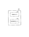
- FIG. 2 is a block diagram showing a functional configuration of the AI server 204 shown in FIG.
- the AI server 204 has a trained model 210 and a skin disease determination engine 211.
- the trained model 210 is a trained model for predicting the type of skin disease, which is machine-learned from images of affected areas of various skin diseases in advance.
- the skin disease determination engine 211 determines whether the skin disease is a non-skin disease or a skin tumor and a non-skin tumor.
- the AI server 204 analyzes the skin disease on the image data transmitted from the user client terminal 100, as will be described in detail later.
- FIG. 3 is a diagram illustrating an outline of the operation of the skin disease analysis system 1 shown in FIG.
- FIG. 3 shows an example in which the user client terminal 100 is a smartphone.
- the affected area is photographed by a camera separate from the user client terminal 100 or a camera built in the user client terminal 100.
- the captured image data may be sent to the user client terminal 100 via a detachable storage medium, or the image data may be sent to the user client terminal 100 by communication. Good.
- trimming is performed so as to exclude the part other than the skin from the captured image data.
- This trimming may be performed by an application program executed by the user client terminal 100.
- the user client terminal 100 executes a browser (web browser) to connect to the Web server 201 of the skin disease analysis device 200, and trims with an application program executed by the application server 203 of the skin disease analysis device 200. May be good. It is preferable that the application for performing this trimming crops the image so that the part other than the skin is not reflected and the lesion is substantially in the center.
- the user client terminal 100 uploads the trimmed image data to the skin disease analysis device 200.
- the user client terminal 100 uploads the image data before trimming to the skin disease analysis device 200. It is sufficient to upload one or more images for one case, but there is a possibility that an erroneous judgment may be made in order to avoid re-shooting when the image is unclear, or depending on the image conditions such as shooting angle, distance, and brightness. Therefore, it is preferable to upload two or more images and use them for the judgment. If there are too many images to be uploaded, it will be complicated, so it is preferable to upload 2 to 10 images, and it is particularly preferable to upload 3 to 5 images.
- the AI server 204 performs exclusion determination and classification as described in detail later.
- the analysis result by the AI server 204 is transmitted to the user client terminal 100 via the Web server 201, and the user client terminal 100 receiving the analysis display the analysis result on the browser.
- the name of the disease and the probability of the disease are displayed up to the third candidate in descending order of probability.
- the coping method for the disease may be displayed. Coping methods include follow-up and excision of the affected area.
- FIG. 4 is a flowchart showing an example of processing of a skin disease analysis program executed by the skin disease analysis system 1.
- the process of analyzing the skin disease is executed by the user client terminal 100 and the skin disease analysis device 200.
- the user client terminal 100 connects to the skin disease analysis device 200 according to the user's operation, and transmits a request for use of the skin disease analysis system 1 to the skin disease analysis device 200 (step S401).
- the skin disease analysis device 200 receives a usage request from the user client terminal 100 (step S451).
- This usage request includes an ID registered in advance for the user of the user client terminal 100 and a password associated with the ID.
- the ID and password are stored and registered in advance in, for example, the file server 205.
- the management server 202 collates the ID and password stored in the file server 205 with the ID and password included in the usage request from the user client terminal 100, and the user who has sent the usage request is valid. Confirm that you are a user (step S452).
- the skin disease analysis device 200 when it is determined in step S452 that the user who has sent the usage request is not a legitimate user, the skin disease analysis device 200 transmits that fact to the user client terminal 100 for use. Reject.
- the skin disease analysis device 200 makes a usage purpose inquiry inquiring the user client terminal 100 for the purpose of use (step S453). ..
- the purpose of use inquiry may be to request the user to input, for example, "diagnosis purpose", "research purpose", and the like.
- the user who operates the user client terminal 100 transmits an answer of the purpose of use to the skin disease analysis device 200 (step S402), and the skin disease analysis device 200 receives the purpose of use (step S454).
- the skin disease analysis device 200 transmits a request for transmitting the affected part photograph / affected part information to the user client terminal 100 (step S455).
- the user client terminal 100 Upon receiving the request for transmitting the affected part photograph and the affected part information, the user client terminal 100 takes a photograph of the affected part and transmits the image data to the skin disease analysis device 200 (step S403).
- the user client terminal 100 displays a transmission completion screen for the user (step S404).
- the photograph of the affected area may be taken before the request for use in step S401.
- the affected part information described in steps S455 and S403 includes the image data obtained by performing the above-mentioned trimming from the image data of the affected part photograph. As described above, the trimming may be performed by the user client terminal 100 or the skin disease analysis device 200. When the trimming is performed by the skin disease analyzer 200, the affected area information does not include the trimmed image data.
- the skin disease analyzer 200 makes an exclusion determination on the received photograph of the affected area (S456). This exclusion determination will be described with reference to FIG.
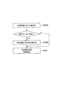
- FIG. 5 is a flowchart showing an exclusion determination process executed by the skin disease analyzer 200.
- the skin disease analysis device 200 determines whether the received image data of the affected portion photograph is an application target image or an excluded image (step S501).
- a skin image can be an application target image. Whether or not it is a skin image may be determined by learning a past skin image. If the image is to be excluded, the process proceeds to step S457 as a “warning display”. If it is an application target image, the process proceeds to step S502. In step S502, the skin disease analyzer 200 determines whether the image data is a tumor image.
- step S457 the process proceeds to step S457 as a “warning display”. If it is a tumor image, it is regarded as "classifiable” and the process proceeds to step S458. The processing of this exclusion determination will be described in detail later.
- the skin disease analysis device 200 transmits a warning / retransmission request to the user client terminal 100 (S457).
- This "warning display” means that the photograph of the affected area received from the user client terminal 100 is determined not to be the target tumor image by the skin disease analysis system 1, and in step S457, the user client terminal 100 is displayed. It warns against it and requests the correct re-transmission of the affected area photo. Upon receiving this, the user client terminal 100 displays a warning to the user (step S405).
- the user decides whether to resend the image data by taking a picture of the affected area again or forcibly execute skin disease analysis (classification) of the image data displayed with the warning.
- a selection is made, and the selected instruction is input to the user client terminal 100.
- the user client terminal 100 returns to step S403 and retransmits the image data of the affected portion photograph to the skin disease analysis device 200.
- an instruction for compulsory execution of the skin disease analysis (classification) is transmitted to the skin disease analysis device 200 (step S406). ).
- the skin disease analyzer 200 receives an instruction to forcibly determine the classification, the skin disease analyzer 200 proceeds to step S458 to continue the process.
- step S456 In the case of a system capable of transmitting a plurality of images of the affected area from the user client terminal 100, the exclusion determination of step S456 is performed for each transmitted image, and for example, it is determined that all the images are not "tumor images". When this is done, a warning / retransmission request is transmitted to the user client terminal 100 (S457). Further, in a system capable of transmitting three or more affected area images from the user client terminal 100, a warning / re-warning / re-warning to the user client terminal 100 when it is determined in step S456 that the number of images is not a tumor image. It is possible to appropriately set whether to transmit the transmission request (S457).
- the skin disease analysis device 200 determines the classification of skin tumors in step S458. Details of this classification will be described later.
- the determination result of step S458 includes the determination result of benign or malignant tumor, the disease name, and the probability of being a disease of the disease name.
- the skin disease analysis device 200 transmits the determination result of step S458 to the user client terminal 100 and ends the process.
- the user client terminal 100 that has received the determination result in step S458 displays the determination result toward the user (S407), and ends the process.
- a display example of step S407 is shown in FIGS. 6 and 7.
- FIG. 6 is a diagram showing a display example when the result of skin disease analysis by the skin disease analysis system 1 is a malignant tumor.
- FIG. 7 is a diagram showing a display example when the result of skin disease analysis by the skin disease analysis system 1 is a benign tumor.
- both the disease having the highest probability and the disease having the next highest probability are malignant tumors, and it is indicated that there is a possibility of malignant tumors.
- the disease having the highest probability is a benign tumor and the possibility of a benign tumor is high.
- the history of the determination result in step S458 may be stored in the file server 205 so that the user who operates the user client terminal 100 or the operator of the administrator terminal 300 can view it. Further, the history of the determination result may be associated with the user who operates the user client terminal 100, or may be associated with the patient of the determined image and managed.
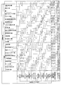
- FIG. 8 is a table showing an example of the determination result by the skin disease analysis system 1 of the present embodiment.
- the diagnosis results by a specialist are listed horizontally, and the results (output according to the present invention) of determining the image of the affected area diagnosed by the skin disease analysis system 1 are listed vertically.
- the leftmost column of the disease names lined up side by side is an image of a patient diagnosed with "actinic keratosis", and the total number is 21.
- the judgment result by the skin disease analysis system 1 7 out of 21 cases were judged as "actinic keratosis", 2 cases were judged as "cutaneous horn actinic keratosis", and 5 cases were "Bowen's disease”.
- the program executed by the AI server 204 of the present embodiment is a first step (step S456 in FIG. 4) for determining to exclude an image other than the skin tumor image from the image data (analysis target image), and the skin tumor image.
- the image if any, has a second step (step S458 in FIG. 4) of determining which type of skin tumor the image was not excluded in the first step.
- the skin disease determination engine 211 determines whether or not the image data of the affected area image is a skin tumor image.
- the skin disease determination engine 211 predicts the type of skin tumor in the image data of the affected area image determined as the skin tumor image by the first step by the trained model 210.
- the first step aims to narrow down the input image to skin tumors.
- the classifier used in the second step is to determine whether it is a benign tumor or a malignant tumor, a malignant epithelial cell lineage tumor, a malignant melanosite lineage cell tumor, a benign epithelial cell lineage cell tumor, and a benign tumor. Judgment of any of the 4 classes of melanocytotic cell tumors, and any of the 6 classes including malignant vascular component tumors and benign vascular component tumors, and further classified 14 to 24 It determines the type of skin tumor of class or higher, but assumes that the image input to this classifier is an image of a skin tumor.
- the benign or malignant determination and the determination of any of the 4 to 24 classes are performed. That is, for example, even an image of an inflammatory skin disease is determined to be a skin tumor, and an image in which the original tumor lesion is not shown because the image is unclear or an image that has nothing to do with the skin is erroneously input. Even in this case, it is determined as a skin tumor, which contributes to misidentification by the user. Therefore, in the present embodiment, it is preferable to provide a first step, which is a step of excluding images other than skin diseases.
- the method of this first step (sometimes referred to as "exclusion determination step") is not particularly limited as long as the image input in the second step can be narrowed down to the skin tumor image, but for example, (1) A method of judging skin tumor images and other images in one step, (2) A method in which images other than the skin are first excluded, and the remaining skin images are judged as a skin tumor image and other images in two stages. (3) First, a method in which images other than skin diseases are excluded, and from the remaining skin disease images, a skin tumor image and other images (including inflammatory skin disease images) are judged in two stages. (4) A method in which images other than the skin are first excluded from the remaining images based on the color distribution, and the skin tumor image and other images are judged in two stages from the remaining images. And so on.
- a message is transmitted to the effect that the image transmitted to the user may not be the image of the skin tumor and is excluded (corresponding to step S405). Specifically, for example, "We have detected that the sent image may not be a skin tumor image. Again to trouble you, but please reconfirm the sent image. After confirmation, resend the same image. Please send me a retake or a retake. ”A message asking the user to confirm.
- the second step is a step of determining the type of skin tumor by a classifier.
- a method for classifying skin tumors into 24 classes will be specifically described.
- the 24 classes of skin tumors include, as malignant tumors, actinic keratosis (AK), actinic keratosis (AKhorn), Bowen's disease (Bown), spinous cell carcinoma (SCC), and basal cell carcinoma.
- BCC non-pigmented basal cell carcinoma
- EMPD extramammary Paget's disease
- MM malignant melanoma
- MMamela non-pigmented malignant melanoma
- AS angiosarcoma
- benign tumors include Sweat pore tumor (Poroma), non-pigmented sweat pore tumor (Poromamela), sebaceous nevus (SebaceousN), seborrheic keratosis (SK), non-pigmented seborrheic keratosis (SKamella), blue mother spot (BlueN) ), Congenital pigmented nevus (CongenitalN), nevus cell nevus (NCN), non-pigmented nevus cell nevus (NCNamela), spitz mother spot (Spitz), non-pigmented spitz mother spot (Spitzamela), kuroko ( Lentigo), melanocytic nevus (Spilus), and pur
- the type (class) of the tumor estimated in the second step is usually 2 to 100 classes, preferably 4 to 50 classes, and more preferably 10 to 30 classes.
- the classifier for example, a trained model in which the skin tumor image obtained by photographing the affected part of each disease is machine-learned is used for the model in which the initial value of the weight is determined from the data of ImageNet.
- the model used in the present invention include, but are not limited to, ResNet50, DenseNet169, DenseNet201, InceptionResNetV2, VGG16, VGG19, MobileNet, DenseNet121, Expression, and InceptionV3.
- the order of certainty of the skin tumor judged by the classifier and its probability are generally output.
- the output value can be converted into an appropriate numerical value or word for the user according to the purpose and displayed.
- the user client terminal 100 may display only benign or malignant to the user as the determination result.
- a dermatologist it is considered necessary for a dermatologist to determine the type of skin tumor and determine the treatment policy according to the type. Therefore, when a dermatologist uses the skin disease analysis system 1 of the present embodiment as a user, for example, the skin of the top 3 to the top 5 determined by the classifier to be highly probable. Displaying the type of tumor can be mentioned.
- the skin disease analysis system 1 manages the user type such as whether the user is a family doctor (a doctor other than a dermatologist) or the user is a dermatologist on the management server 202, for example. It is also possible to perform a display for the user and perform an operation in which the display content in the step (S407) is different depending on the user type.
- the dermatologist refers to the types of skin tumors that are likely to be output by the skin disease analysis system 1 of the present embodiment, and also refers to the surface shape, unevenness, size, age of the patient, odor of the affected area, etc. , Various parameters that are difficult to measure with a plan image are captured, and a comprehensive judgment is made to diagnose a skin tumor.
- the AI server 204 may be provided with a skin disease reprediction engine, and the skin disease analysis program may cause a computer to perform a third step of reprediction by the skin disease reprediction engine.
- the skin disease reprediction engine (second learned model) repredicts the type of skin disease from the affected area information and the type of skin tumor predicted by the second step. This reprediction will be described with reference to FIG.
- FIG. 28 is an image of an affected part of a specific skin disease including a skin tumor that is easily misdetermined when it is classified into a disease class that is easily misdetermined as another disease as a result of performing a skin tumor classification determination (S458A). It is shown that the re-judgment is performed (S458B) by using the classifier (second trained model) created by machine learning by. For example, since a classifier (first trained model) that performs 24-class classification learns images of various types of diseases, it may erroneously determine diseases having similar shapes.
- a classifier created by learning only images of a small number of specific diseases (rejudgment engine: specifically, for example, basal cell carcinoma, malignant melanoma, seborrheic keratosis, melanocytic nevus) It is possible to more accurately classify diseases that are prone to erroneous judgment by the re-judgment engine of the four tumor classes.
- the affected area information includes one or more of the age of the patient, the size of the affected area, the odor of the affected area, the site of occurrence of the disease, and the three-dimensional shape of the affected area.
- This affected area information corresponds to the affected area information described in steps S455 and S403 of FIG. 4, and can be transmitted by the user to the skin disease analysis device 200.
- the result of the re-prediction by the third step can be displayed on the user client terminal 100 in the same manner as the result of the prediction by the second step.
- the configuration of the classifier in the second step it is possible to determine 14 to 24 class skin tumors in one step, and it may have two or more steps.
- the accuracy is determined by a method of re-determining a specific disease that is likely to be erroneously determined as the third step.
- the classifier used in the third step is one in which some specific disease classes are selected and trained from the disease classes learned by the classifier used in the second step. Therefore, the number of disease classes learned by the classifier used in the third step is smaller than the number of disease classes used in the second step.
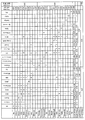
- the combination of disease classes that are likely to be misjudged from each other is a case where approximately 2% or more of the case images of a specific disease confirmed by a specialist are judged to be another specific disease by a classifier, or there is. Approximately 2% or more of the case images of a specific disease determined by the disease classifier have been diagnosed as another specific disease by an actual specialist, and these are in two or more disease classes. A combination that is recognized by each other. Specifically, for example, in FIG. 29, the actual number of MMs as a diagnosis result is 646, of which 43 (6.66%) are determined to be NCN by a classifier (24-class determiner). ..
- the number of images judged to be NCN by the classifier is 1305, but the number of images actually diagnosed as MM is 43 (3.30%), so MM and NCN. Is a combination of disease classes that are easily misjudged from each other.
- the classifier that obtained the judgment result shown in FIG. 29 in addition to MM (malignant) and NCN (benign), SK (benign) and BCC (malignant) are also a combination of disease classes that are easily misjudged from each other. Is.
- MM malignant melanoma
- NCN nevus cell nevus
- a model in which only the image of malignant melanoma and the image of the nevus cell nevus are trained in the third step is used. It is used again to determine whether it is malignant melanoma or nevus cell nevus. In this case, if it is determined to be malignant melanoma in the second step, the determination result of the second step is adopted as it is.
- tumors similar to malignant melanoma and nevus cell nevus may be added to the determination in the third step. Specifically, since it is considered that the four types of malignant melanoma, basal cell carcinoma, seborrheic keratosis, and nevus cell nevus are similar in shape, these four tumors were determined in the second step. In some cases, again in the third step, using a model trained only with images of malignant melanoma, basal cell carcinoma, seborrheic keratosis, and melanocytic nevus, these four Redetermine which of the tumors it is.
- the second trained model used in the third step is usually selected from 14 to 24 classes, usually 2 to 10 classes, preferably 3 to 8. Tumors of a class, more preferably 4 to 6 classes, are identified and trained.
- the determination result of the tumor class determination step is one type of skin tumor that is likely to be misdetermined from each other. If this is the case, a specific tumor class re-judgment step (third step) is performed in which re-judgment is performed by a second trained model machine-learned from images of specific affected areas including skin tumors that are easily misjudged from each other. ..
- the determination result of the tumor class determination step is one of benign tumors, and the benign tumor is said to be benign.
- the specific tumor class re-determination step (third step) for determining is executed.
- one of the skin tumors that are easily misjudged from each other contains malignant melanoma, that is, a tumor class determination step (second step) for predicting the type of skin tumor is performed.
- the analysis result of the tumor class determination step is one of the skin tumors that are easily misidentified as malignant melanoma
- the machine is based on the image of the skin tumor that is easily misidentified as malignant melanoma and the affected area including malignant melanoma.
- the specific tumor class re-judgment step (third step) in which re-judgment is performed by the trained second trained model is executed.
- the trained model it may be effective to perform re-judgment other than the combination including the above-mentioned malignant melanoma.
- combinations including non-pigmented basal cell carcinoma, non-pigmented seborrheic keratosis, and non-pigmented nevus cell nevus are likely to be misjudged from each other.
- the image of the affected area may be trimmed by a doctor who is a user of the skin disease analysis system 1 by operating an application for trimming.
- the doctor can visually recognize the affected area image, place the tumor part in the center of the screen, place the normal skin part on the periphery of the screen, and operate to exclude the non-skin part.
- .. -The affected area image is taken by the patient himself, and the photographed image is transmitted to the doctor, so that the doctor who is the user of the skin disease analysis system 1 operates the user client terminal 100 to obtain the image data of the affected area image for the skin disease. It may be transmitted to the analyzer 200.
- Embodiment 1 -First step: Classify into (1) skin tumors and (2) other groups.
- -Second step (1) Determine the type of skin tumor in the skin tumor-Third step: Redetermine the type of a specific skin tumor depending on the result of the second step.
- Embodiment 2 -First step A: Exclude images other than skin.
- -Second step (1) In the group of skin tumors, the type of skin tumor is determined.
- -Third step Depending on the result of the second step, the type of a specific skin tumor is redetermined.
- Embodiment 3 -First step A: Classify into groups other than (1) skin diseases (including skin tumors and inflammatory skin diseases) and (2) skin diseases.
- Embodiment 4 -First step: Classify into (1) skin tumors and (2) other groups.
- -Second step A (1) Skin tumors are classified into (3) pigmented tumors and (4) non-pigmented tumors.
- -Second step B The type of skin tumor is determined in each of (3) pigmented tumor and (4) non-pigmented tumor.
- -Third step Depending on the result of the second step, the type of a specific skin tumor is redetermined.
- Embodiment 5 ⁇ First step: (1) Separate into skin tumors and (2) other groups ⁇ Second step: Determine the type of skin tumor in skin tumors ⁇ Third step (one of the following steps) ) Third step 1: When the determination in the second step is actinic keratosis, four types of actinic keratosis, cutaneous horn actinic keratosis, Bowen's disease, and non-pigmented seborrheic keratosis Re-enter into the determination machine to determine if it is actinic keratosis, cutaneous horn actinic keratosis, Bowen's disease, or non-pigmented seborrheic keratosis.
- Third step 2 When the determination in the second step is basal cell carcinoma, re-enter into the determination machine for two types of basal cell carcinoma and unpigmented basal cell carcinoma, and basal cell carcinoma and unpigmented basal cell. Determine if it is cancer.
- Third step 3 When the determination in the second step is non-pigmented basal cell carcinoma, four types of non-pigmented basal cell carcinoma, actinic keratosis, spinous cell carcinoma, and non-pigmented seborrheic keratosis Is re-entered into the determination machine to determine whether it is unpigmented basal cell carcinoma, actinic keratosis, spinous cell carcinoma, or non-pigmented seborrheic keratosis.
- Third step 4 When the judgment in the second step is Bowen's disease, re-enter the judgment machine for three types of Bowen's disease, actinic keratosis, and non-pigmented Spitz nevus, and Bowen's disease and actinic keratosis. Determine whether it is keratosis or non-pigmented Spitz nevus.
- Third step 5 When the determination in the second step is non-pigmented sweat spore tumor, it is re-entered into the determination machine for three types of melanoma, malignant melanoma, and melanocytic nevus cell nevus.
- Third step 6 When the determination in the second step is non-pigmented sweat spore tumor, it is re-entered into the determination machine for three types of non-pigmented sweat spore tumor, malignant melanoma, and basal cell carcinoma, and the non-pigmented sweat pore is Determine if it is a tumor, malignant melanoma, or basal cell carcinoma.
- Third step 7 When the judgment of the second step is spinous cell carcinoma, five types of spinous cell carcinoma, actinic keratosis, non-pigmented basal cell carcinoma, Bowen's disease, and non-pigmented malignant melanoma Re-enter into the determination machine to determine whether it is spinous cell carcinoma, actinic keratosis, non-pigmented basal cell carcinoma, Bowen's disease, or non-pigmented malignant melanoma.
- Third step 8 If the determination in the second step is any of basal cell carcinoma, malignant melanoma, seborrheic keratosis, and melanocytic nevus, basal cell carcinoma, malignant melanoma, and seborrheic horn.
- nevus and nevus cell nevus determine whether it is basal cell carcinoma, malignant melanoma, seborrheic keratosis, or mammoth cell nevus.
- Third step 9 If the determination in the second step is either seborrheic keratosis or melanocytic nevus, basal cell carcinoma, malignant melanoma, seborrheic keratosis, and melanocytic nevus.
- Third step 10 When the determination in the second step is malignant melanoma or nevus cell nevus, re-enter into the determination machine for two types of malignant melanoma and nevus cell nevus, and malignant melanoma and mother Determine which is the melanocytic nevus.
- Third step 11 When the determination in the second step is a nevus cell nevus, re-enter into the determination machine for two types of malignant melanoma and nevus cell nevus, and malignant melanoma and nevus cell mother. Determine which of the spots it is.
- Third step 12 When the determination in the second step is melanocytic nevus cell nevus, there are three types: melanocytic basal cell carcinoma, melanocytic seborrheic keratosis, and melanocytic nevus cell nevus.
- Third step 13 If the determination in the second step is melanocytic nevus, the determination machine for four types of basal cell carcinoma, malignant melanoma, seborrheic keratosis, and nevus cell nevus is used again. Enter to determine if it is basal cell carcinoma, malignant melanoma, seborrheic keratosis, or melanocytic nevus.
- any one of the third steps 1 to 13 can be appropriately selected and executed. Further, when performing the second step and the third step, the first step may be omitted.
- a preferred embodiment of the present invention is that when a tumor class determination step for predicting the type of skin tumor is performed, the analysis result of the tumor class determination step is one of the skin tumors that are easily misdetermined from each other.
- the specific tumor class re-determination step of re-determining by the second trained model machine-learned by the image of the affected part of the specific skin disease including the skin tumors that are easily misjudged with each other is executed.
- the disease classes shown in steps 1 to 12 are examples of combinations of disease classes that are easily misjudged from each other.
- the non-pigmented sweat melanoma determined in the second step is not a candidate for the disease in the third step, but the non-pigmented sweat melanoma has a variety of forms and rare cases. Since the trained model may not have acquired sufficient learning depending on the sex, it is decided to make a benign judgment (false negative) for basal cell carcinoma and malignant melanoma, which are malignant tumors that are easily misidentified as achromatic sweat pore tumor. This is an example that can be conveniently used for the purpose of avoiding as much as possible.
- a classifier two types are selected from known image classification models having weights learned by ImageNet, and each teacher image (7532 images of the above 24 classes of skin tumors in total) is used. It was trained using (one example of the first trained model). The breakdown of the images was 278 for sunlight keratosis (AK), 95 for cutaneous sunlight keratosis (AKhorn), and Bowen as malignant tumors.
- AK sunlight keratosis
- AKhorn cutaneous sunlight keratosis
- Bowen as malignant tumors.
- the overall correct answer rate was 68% for model 1 and 69% for model 2.
- Model 2 of 110 images of spinous cell carcinoma, 80 were correctly determined to be spinous cell carcinoma by the classifier, and 4 were determined to be actinic keratosis. 4 were judged to have Bowen's disease, 5 were judged to be basal cell carcinoma, 7 were judged to be non-pigmented basal cell carcinoma, 2 were judged to be extramammary Paget's disease, and 2 were judged to be malignant melanoma.
- the 109 affected area images determined by the classifier to be spinous cell carcinoma 80 were actually diseased with spinous cell carcinoma, 1 was actually actinic keratosis, and skin. 4 with actinic keratosis, 4 with Bowen's disease, 5 with basal cell carcinoma, 5 with non-pigmented basal cell carcinoma, 1 with malignant melanoma
- the classifier determines the top three types of skin tumors that are likely. At this time, the judgment result (correct answer rate) when the case where there is a correct answer in any of the three types judged by the classifier is defined as the 24-class classification TOP3 correct answer is 91% in model 1 and 89% in model 2. Met.
- the classifier determines the top three types of skin tumors that are likely. At this time, if the actual tumor is malignant and there is a malignant tumor in any of the three types indicated by the classifier, the correct answer for malignant determination is made. In addition, when the actual tumor is benign, while all three types indicated by the classifier are benign tumors, the correct answer for benign judgment is made. The judgment result (correct answer rate) when this was defined as the two-class classification TOP3 correct answer was 99% in model 1 and 98% in model 2.
- the classifier determines the most likely type of skin tumor.
- the sensitivity (Recall) of the judgment result is 94% for malignant, 79% for benign, and the model in model 1. In 2, 94% were malignant and 83% were benign.
- a classifier select two types from known image classification models with weights learned by ImageNet, and select three classes of teacher images (from the above 24 classes, extramammary paget disease, angiosarcoma, and pyogenic granuloma). A total of 7011 images of 21 classes of skin tumors excluded) were used for training. The performance of the learning machine was evaluated by dividing the image into 10 parts, using 9/10 as a teacher image and 1/10 as a test image, and using a cross-validation method.
- the overall correct answer rate was 70% for model 1 and 74% for model 2.
- Model 2 of 72 images of malignant melanoma, 64 were correctly judged as malignant melanoma by the classifier, 1 was judged as spinous cell carcinoma, and basal cell carcinoma. 4 sheets were judged to be non-pigmented malignant melanoma, 2 pieces were judged to be non-pigmented malignant melanoma, and 2 pieces were judged to be seborrheic keratosis. That is, the ratio (sensitivity: Report) of correctly determining malignant melanoma in the image of malignant melanoma was 83%.
- the 86 affected area images determined by the classifier to be malignant melanoma 64 were the actual disease being malignant melanoma, 1 was the actual disease being sunlight keratosis, and spinous cell carcinoma. 2 pieces of basal cell carcinoma, 1 piece of non-pigmented malignant melanoma, 3 pieces of melanocytic nevus, and 3 pieces of seborrheic keratosis There were 3 melanomas, 4 blue nevus, 2 congenital melanocytic nevus, 2 nevus cell nevus, and 2 spitz nevus. .. That is, 74% of the images determined to be malignant melanoma had malignant melanoma as the actual disease (positive predictive value: Precision).
- 21 class classification TOP3 correct answer rate In determining 21 classes of disease, the classifier determines the top three types of skin tumors that are likely. At this time, the judgment result (correct answer rate) when there is a correct answer in any of the three types judged by the classifier is defined as 21 class classification TOP3 correct answer is 93% in model 1 and 93% in model 2. Met.
- the classifier determines the top three types of skin tumors that are likely. At this time, if the actual tumor is malignant and there is a malignant tumor in any of the three types indicated by the classifier, the correct answer for malignant determination is made. In addition, when the actual tumor is benign, while all three types indicated by the classifier are benign tumors, the correct answer for benign judgment is made. The judgment result (correct answer rate) when this was defined as the two-class classification TOP3 correct answer was 99% in model 1 and 98% in model 2.
- the classifier determines the most likely type of skin tumor.
- the sensitivity (Recall) of the judgment result is 98% malignant, 84% benign, and the model in model 1. In 2, 97% was malignant and 88% was benign.
- Model experiment of exclusion judgment device (first step) 1: For example, an exclusion judgment device (exclusion judgment model) was created by utilizing a deep neural network as a feature extractor and performing an exclusion judgment using the features. Six images that were not skin tumors were input to the exclusion determination device. As a result, all 6 sheets (car, cat, earth, arm, dog, ship) were excluded as not skin tumors. At this time, it was determined that about 5% of the images of the skin tumor were excluded from the skin tumor. In addition, when 6 images that were judged to be excluded from skin tumors were input to the classifier of Model 1, the most probable diseases were all malignant melanomas. This indicates that the classifier determines to any class of skin tumors, even if the images are completely unrelated to skin tumors.
- Model experiment of exclusion judgment device (first step) 2 As a classifier, one of the known image classification models with weights learned by ImageNet is selected, and a teacher image (a total of 7011 images of the same 21-class skin tumor as in Example 2 above, which is inflammatory.
- the images were trained using a total of 1044 images of three classes of skin diseases such as atopic dermatitis, psoriasis, and mycosis fungoides.
- the learning was performed by dividing the image into 10 images, 9/10 as a teacher image, and 1/10 as a teacher image.
- the generalization accuracy was calculated using the test data as a test image.
- As test images 691 images of skin tumors and 74 images of inflammatory skin diseases were used.
- the image of the skin tumor was judged to be a skin tumor by the classifier in 689 sheets, and the image of the skin tumor was judged to be an inflammatory skin disease by the classifier in 2 sheets.
- 66 images of inflammatory skin disease were judged by the classifier as inflammatory skin disease
- 8 images of inflammatory skin disease were judged by the classifier as skin tumor. That is, from the viewpoint of skin tumors, the sensitivity (Recall) was 99.7% and the positive predictive value (Precision) was 98.9%. This makes it possible to distinguish between a skin tumor and an inflammatory skin disease when an image of a skin disease is input. Therefore, according to this exclusion determination device, it is possible to exclude an inflammatory skin disease, determine only a skin tumor, and use it in the next second step by inputting an image that is certain to be some kind of skin disease. It becomes.
- Model experiment of exclusion judgment device (first step) 3 As a classifier, one type was selected from known image classification models having weights learned by ImageNet. As the image of the skin tumor, a total of 7011 images of the same 21-class skin tumor as in Example 2 were used. As images other than skin tumors, 7,000 images randomly selected from the confirmation data of Google Open Image V4 were used. Of these, 3476 images of skin tumors and 3500 images other than skin tumors were used as teacher data to be trained by deep learning. On the other hand, with respect to the remaining 3575 images of the skin tumor and 3500 images other than the skin tumor, it was determined whether the tumor was a skin tumor or a non-skin tumor by the trained model.
- a classifier one type was selected from known image classification models having weights learned by ImageNet.
- the teacher image and the test image a total of 7532 images of the above 24 classes of skin tumors were used as in Example 1.
- the performance of the learning machine was evaluated by dividing the image into 10 parts, training 9/10 as a teacher image, using 1/10 as a test image, and using a cross-validation method.
- the number of images judged as test images was 2,247 in total three times.
- the determination result is shown in FIG. According to this, out of 2247 images, the hit rate correctly determined as the disease for each disease was 70%.
- the accuracy rate for correctly determining whether the disease was malignant or benign was 89%.
- FIG. 9 and FIG. 29 described later, four tumors of basal cell carcinoma, malignant melanoma, seborrheic keratosis, and melanocytic nevus, which are easily misjudged from each other, are shown.
- a classifier was created by training a model using only images (an example of the second trained model). As the classifier used here, one kind was selected from the known image classification models having weights learned by ImageNet. The images used were 1100 for basal cell carcinoma, 625 for malignant melanoma, 520 for seborrheic keratosis, and 1165 for melanocytic nevus.
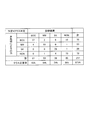
- the images of the above 4 tumor classes (basal cell carcinoma 1100, malignant melanoma 625, seborrheic keratosis 520, melanocytic nevus 1165) were divided into 10 and 9/10 was a teacher image, 1 /. 10 was used as a test image and evaluated using the cross-validation method. This evaluation was performed 10 times, and the result of making the most average judgment among them is shown in FIG. In addition, FIG. 11 shows the determination results as malignant (basal cell carcinoma and malignant melanoma) and benign (seborrheic keratosis and melanocytic nevus).
- the images of the above 4 tumor classes were divided into 10 images, 9/10 was used as a teacher image, 1/10 was used as a test image, and evaluation was performed using a cross-validation method.
- the images showing basal cell carcinoma and malignant melanoma are similar to other diseases (for example, those judged to be other disease classes by 24 class judgment and those of the same case. ) was extracted, and 292 images of basal cell carcinoma and 149 images of malignant melanoma were selected.
- Example 7 (FIGS. 10 and 11) and Example 8 (FIGS. 12 and 13) were compared, the correct answer rate was higher in Example 8.
- Example 8 by increasing the number of images that are easily mistaken for other disease classes, as a result of learning many images that are easily mistaken for other disease classes, it is possible to more accurately determine images that are similar to each other. It is thought that it was.
- the image determined by the 24-class classifier used in Example 6 was basal cell carcinoma, malignant melanoma, seborrheic keratosis, or melanocytic nevus. This is a simulation of the result when re-judgment is performed by the 4-class classifier used in. The procedure is as follows. Of the images used in Example 6, images in which the actual diagnosis result was basal cell carcinoma, malignant melanoma, seborrheic keratosis, or melanocytic nevus were selected from the 24 class classifier according to Example 6.
- Example 8 Extract those whose output results were basal cell carcinoma, malignant melanoma, seborrheic keratosis, or melanocytic nevus (both correct and incorrect). From the extracted images, the image used in Example 8 is extracted. That is, the images used for both are extracted. Then, for each extracted image, first, a 24-class classifier is used to determine whether the output result is basal cell carcinoma, malignant melanoma, seborrheic keratosis, or melanocytic nevus. 4. Obtain the result of re-judgment by a 4-class classifier (used in Example 8). The results are shown in FIG. In addition, FIG. 15 shows the determination results as malignant (basal cell carcinoma and malignant melanoma) and benign (seborrheic keratosis and melanocytic nevus).
- Example 8 the four-class classifier used in Example 8 was used for an image in which the result determined by the 24-class classifier used in Example 6 was seborrheic keratosis or melanocytic nevus. This is a simulation of the result when the judgment is made again by. The procedure is as follows. Of the images used in Example 6, images in which the actual diagnosis result was basal cell carcinoma, malignant melanoma, seborrheic keratosis, or melanocytic nevus were selected from the 24 class classifier according to Example 6. Extract those whose output results were basal cell carcinoma, malignant melanoma, seborrheic keratosis, or melanocytic nevus (both correct and incorrect).
- the image used in Example 8 is extracted. That is, the images used for both are extracted. Then, for each extracted image, first, a 24-class classifier is used to determine whether the output result is basal cell carcinoma, malignant melanoma, seborrheic keratosis, or melanocytic nevus. , The result of re-judgment of the output result of seborrheic keratosis or melanocytic nevus with a 4-class classifier (used in Example 8) is obtained. At this time, for images determined to be basal cell carcinoma or malignant melanoma by the 24-class classifier, the results are retained and re-judgment by the 4-class classifier is not performed.
- FIG. 17 shows the determination results as malignant (basal cell carcinoma and malignant melanoma) and benign (seborrheic keratosis and melanocytic nevus).
- malignant basic cell carcinoma and malignant melanoma
- benign benign (seborrheic keratosis and melanocytic nevus).
- FIG. 17 by re-judging a benign tumor that is easily misjudged as a specific malignant tumor, the number of cases in which benign is misjudged as malignant increases slightly, but malignant is misjudged as benign. It is useful from the viewpoint that malignant tumors are not overlooked.
- the output of the 24-class classifier was basal cell carcinoma, malignant melanoma, seborrheic keratosis, or melanocytic nevus, and the actual diagnosis was these four tumors.
- Example 6 This is a simulation of the result of re-judgment by a 4-class judge for non-classes.
- a 24-class classifier according to Example 6
- the output result of basal cell carcinoma, malignant melanoma, seborrheic keratosis, or melanocytic nevus is extracted.
- the image used in Example 8 is extracted. That is, the images used for both are extracted.
- the output result is basal cell carcinoma or malignant melanoma (malignant tumor), seborrheic keratosis or melanocytic nevus (benign) by a 24-class classifier. To determine if it is a tumor). As a result, the determination of malignancy and benign is shown in FIG. Next, the results of re-judgment of each extracted image by a 4-class classifier (used in Example 8) were obtained. As a result, the determination of malignancy and benign is shown in FIG.
- the output of the 24-class classifier was seborrheic keratosis or melanocytic nevus, and the actual diagnostic results were these basal cell carcinoma, malignant melanoma, and seborrheic horn. It is a simulation of the result of re-judgment by a 4-class determiner for non-four tumor classes of melanoma or nevus cell nevus.
- images used in Example 6 images in which the actual diagnosis result was other than basal cell carcinoma, malignant melanoma, seborrheic keratosis, or melanocytic nevus were classified into 24 classes according to Example 6.
- the image used in Example 8 is extracted. That is, the images used for both are extracted.
- the output result is basal cell carcinoma or malignant melanoma (malignant tumor), seborrheic keratosis or melanocytic nevus (benign tumor) by a 24-class classifier. ) Is determined.
- a result of re-judgment using a 4-class classifier (used in Example 8) is obtained for those whose judgment result is seborrheic keratosis or melanocytic nevus.
- FIG. 21 shows three cases having a plurality of image data for a case diagnosed with malignant melanoma and having different determination results (AI determination first candidate).
- the 24 class classifier used in Example 6 makes a judgment, it outputs a numerical value such that the certainty (confidence) judged by the classifier for each of the 24 class tumors is added and becomes 1.
- FIG. 21 shows the top two tumors with the highest confidence. Usually, the tumor with the highest degree of certainty is used as the judgment result of the classifier.
- the certainty may be different even in the same case, and the tumor having the highest certainty (that is, the judgment result) may be different.
- the determination results obtained in Example 6 it was found that the correct answer rate differs depending on the degree of certainty at that time (corresponding to the degree of certainty 1 in FIG. 21). That is, as shown in FIG. 22, the result was obtained that the higher the conviction, the higher the correct answer rate. This suggests that the correct answer rate can be improved by determining a plurality of images for one case, determining each image, and then adopting the one with the highest degree of certainty.
- FIG. 23 shows an example in which a plurality of images were determined for the same case in Example 6 for malignant melanoma (MM) and nevus cell nevus (NCN).
- Multiple correct answers means that when multiple images were judged for the same case, all were correct (for example, when the diagnosis result was MM, the judgment result by AI was all MM). Is.
- the "plurality of all incorrect answers” means that when a plurality of images were judged for the same case, all of them were incorrect.
- “Correct answer when selecting maximum confidence” means that when multiple images are judged for the same case, correct and incorrect answers are mixed, and when the one with the highest confidence is selected, that is the correct answer. It is an image.
- “Incorrect answer when selecting maximum certainty” means that when multiple images are judged for the same case, correct and incorrect answers are mixed, and if the one with the highest certainty is selected, it is not correct. It was the correct answer.
- the classifier one was selected from the known image classification models having weights learned by ImageNet.
- the teacher image and the test image a total of 7532 images of the above 24 classes of skin tumors were used as in Example 1.
- FIG. 23 in both cases of malignant melanoma and nevus cell nevus, "correct answer when selecting maximum certainty" is considerably more than "incorrect answer when selecting maximum certainty", that is, It was shown that the accuracy rate can be expected to improve by taking multiple shots.
- a classifier 10 types of known image classification models (ResNet50, DenseNet169, DenseNet201, InceptionResNetV2, VGG16, VGG19, MobileNet, DenseVet121, Xeption, Image) with weights learned by ImageNet, respectively.
- a total of 7532 images of the above 24 classes of skin tumors were used for learning and testing. About 90% of the images were used as training data and about 10% as test data, and the training data and the test data were judged.
- re-learning was performed so that the number of correct answers in the training data increased, but re-learning was repeated until the correct answer rate of the test data did not increase.
- the learning curve up to the time when the re-learning is completed is shown in FIGS. 24 and 25.
- the vertical axis represents the correct answer rate
- the horizontal axis represents the number of re-learning.
- FIG. 26 shows the correct answer rate for the test data having the highest correct answer rate at the time when the re-learning is completed.
- three tests are performed, and the average and standard deviation of the correct answer rate are shown for the 24 class judgment and the benign / malignant judgment. It can be seen that a certain good accuracy rate can be obtained in any of the trained models.
- FIG. 27 shows an example of a confusion matrix of test data when DenseNet201 is used as a model. In this case, 752 pieces were used as test data. From the viewpoint of distinguishing between malignant and benign, the overall correct answer rate was 89%, the correct answer rate for malignant tumors was 94%, and the correct answer rate for benign tumors was 83%.
- recall 17Class is the correct answer rate when 24 classes are grouped into 17 classes.
- BCC basic cell carcinoma
- BC Camella non-pigmented basal cell carcinoma
- the recall 6Class is a collection of 24 classes into 6 classes.
- AK, AKhorn, bowen, SCC, BCC, BCCamela, and EMPD which are classified as malignant epithelial cell lineage tumors
- EMPD which are classified as malignant epithelial cell lineage tumors
- the optimum model can be appropriately selected in consideration of the tendency of tumors to be mistaken, the susceptibility to overfitting, and what kind of judgment result is to be output.
- a classifier one type was selected from known image classification models having weights learned by ImageNet.
- the teacher image and the test image a total of 7532 images of the above 24 classes of skin tumors were used as in Example 1.
- the performance of the learning machine was evaluated by randomly extracting approximately 1/10 of the image each time to obtain a test image and learning the remaining approximately 9/10 as a learning image using a cross-validation method.
- the number of images judged as test images was 7472 in total 10 times.
- the determination result is shown in FIG. From the viewpoint of predicting malignant and benign, the overall correct answer rate was 90.0%, the correct answer rate for malignant tumors was 92.7%, and the correct answer rate for benign tumors was 85.8%.
- Example 13 48 sheets were determined to be sweat pore tumor (Poromamela). This was re-determined by the 4-class determiner (which determines whether it is BCC, MM, SK, or NCN) used in Example 8. The result is shown in FIG. Of those judged to be Poromamela, 17 were malignant tumors, but 15 of them were judged to be malignant (correct answer) by 4-class re-judgment. On the other hand, out of 31 sheets judged to be benign, 14 sheets were judged to be malignant (incorrect answer) by 4 class re-judgment. The overall correct answer rate by the 4-class re-judgment was 67%, which was almost the same as the 24-class judgment (65%). From this, it is considered that a specific tumor class re-determination is effective from the viewpoint of not overlooking malignancy.
- the 4-class determiner which determines whether it is BCC, MM, SK, or NCN
- an exclusion judgment engine (first step), a 24-class judgment device (second step), and a four-class re-judgment device (third step) are implemented, and the maximum is per case. It was judged using four images.
- an iPod registered trademark
- the client terminal authenticates the user's login, logs in, takes up to four pictures using the camera function of the iPad touch called from the Web browser, and arranges the tumor in the center of the picture at the time of taking.
- a guide frame is provided (the affected area is cut out), and an application with each function of sending the photographed photograph to the server is installed.
- the browser used was Google Chrome.
- Wifi was used for communication between the client terminal and the server, such as sending photos to the server.
- the server with the following specifications was used.
- CPU Intel (registered trademark) Core i7-8700K, 3.70 GHz
- GPU GeForce GTX 1080 Ti
- memory 32 GB.
- the server first receives the photo sent from the client terminal by the application server, and for each photo on the AI server, (1) a trained model that determines whether it is a tumor image (exclusion determination), or (2) 24 disease classes. Each is determined by a trained model that determines whether or not, and (3) a trained model that determines any of the four disease classes of BCC, MM, SK, and NCN. At the time of determination, the determination result is indicated by the name of the disease class and the degree of certainty (the total is 1) as to which disease class it corresponds to. In addition, the encrypted photo and the judgment result are saved in the file server.
- the judgment result can be referred from the administrator client terminal.
- the time from the transmission of the four photographs to the completion of the determination was approximately 60 to 120 seconds.
- 6 cases in which the diagnosis by the doctor was BCC were determined by the following procedure. (1) For each case, four photographs were taken with the user terminal, sent to the server, and three classifiers were executed. (2) Images judged not to be skin tumors by the exclusion judgment engine were excluded from the judgment candidates. In FIG. 31, there is one excluded image (case number 6, photo number 1), but the entire image was shining and the tumor was unclear. Therefore, it seems that it was not recognized as a skin tumor.
- Each process shown in FIG. 4 may be realized by a logic circuit (hardware) formed in an integrated circuit (IC chip) or the like, or may be realized by software using a CPU (Central Processing Unit). ..
- the user client terminal 100, the skin disease analysis device 200, and the administrator client terminal 300 are a CPU that executes instructions of a program that is software that realizes each function, and the above program. Also equipped with a ROM (Read Only Memory) or storage device (these are referred to as "recording media") in which various data are readablely recorded by a computer (or CPU), a RAM (Random Access Memory) for developing the above program, and the like. ing. Then, the object of the present invention is achieved when the computer (or CPU) reads the program from the recording medium and executes it.
- a "non-temporary tangible medium" for example, a tape, a disk, a card, a semiconductor memory, a programmable logic circuit, or the like can be used.
- the program may be supplied to the computer via an arbitrary transmission medium (communication network, broadcast wave, etc.) capable of transmitting the program.
- a transmission medium communication network, broadcast wave, etc.
- one aspect of the present invention can also be realized in the form of a data signal embedded in a carrier wave, in which the program is embodied by electronic transmission.
- the present invention is not limited to the above-described embodiment, and various modifications can be made within the disclosed range, and the present invention also relates to an embodiment obtained by appropriately combining the technical means disclosed in each of the different embodiments. Included in the technical scope of.
- 1 Skin disease analysis system, 2 ... Network, 100 ... User client terminal, 200 ... Skin disease analysis device, 300 ... Administrator client terminal.
Landscapes
- Health & Medical Sciences (AREA)
- Engineering & Computer Science (AREA)
- Life Sciences & Earth Sciences (AREA)
- Medical Informatics (AREA)
- General Health & Medical Sciences (AREA)
- Public Health (AREA)
- Physics & Mathematics (AREA)
- Biomedical Technology (AREA)
- Pathology (AREA)
- Veterinary Medicine (AREA)
- Biophysics (AREA)
- Heart & Thoracic Surgery (AREA)
- Molecular Biology (AREA)
- Surgery (AREA)
- Animal Behavior & Ethology (AREA)
- Artificial Intelligence (AREA)
- Nuclear Medicine, Radiotherapy & Molecular Imaging (AREA)
- Radiology & Medical Imaging (AREA)
- Computer Vision & Pattern Recognition (AREA)
- Epidemiology (AREA)
- Primary Health Care (AREA)
- General Physics & Mathematics (AREA)
- Quality & Reliability (AREA)
- Theoretical Computer Science (AREA)
- Physiology (AREA)
- Signal Processing (AREA)
- Psychiatry (AREA)
- Mathematical Physics (AREA)
- Dermatology (AREA)
- Fuzzy Systems (AREA)
- Evolutionary Computation (AREA)
- Data Mining & Analysis (AREA)
- Databases & Information Systems (AREA)
- Measuring And Recording Apparatus For Diagnosis (AREA)
- Measurement Of The Respiration, Hearing Ability, Form, And Blood Characteristics Of Living Organisms (AREA)
- Image Analysis (AREA)
Priority Applications (4)
| Application Number | Priority Date | Filing Date | Title |
|---|---|---|---|
| EP20784555.3A EP3951794A1 (en) | 2019-03-29 | 2020-03-26 | Skin disease analyzing program, skin disease analyzing method, skin disease analyzing device, and skin disease analyzing system |
| US17/599,281 US20220156932A1 (en) | 2019-03-29 | 2020-03-26 | Skin disease analyzing program, skin disease analyzing method, skin disease analyzing device, and skin disease analyzing system |
| JP2021511926A JPWO2020203651A1 (https=) | 2019-03-29 | 2020-03-26 | |
| AU2020255759A AU2020255759A1 (en) | 2019-03-29 | 2020-03-26 | Skin disease analyzing program, skin disease analyzing method, skin disease analyzing device, and skin disease analyzing system |
Applications Claiming Priority (4)
| Application Number | Priority Date | Filing Date | Title |
|---|---|---|---|
| JP2019067229 | 2019-03-29 | ||
| JP2019-067229 | 2019-03-29 | ||
| JP2019-206765 | 2019-11-15 | ||
| JP2019206765 | 2019-11-15 |
Publications (1)
| Publication Number | Publication Date |
|---|---|
| WO2020203651A1 true WO2020203651A1 (ja) | 2020-10-08 |
Family
ID=72668858
Family Applications (1)
| Application Number | Title | Priority Date | Filing Date |
|---|---|---|---|
| PCT/JP2020/013684 Ceased WO2020203651A1 (ja) | 2019-03-29 | 2020-03-26 | 皮膚疾患解析プログラム、皮膚疾患解析方法、皮膚疾患解析装置及び皮膚疾患解析システム |
Country Status (5)
| Country | Link |
|---|---|
| US (1) | US20220156932A1 (https=) |
| EP (1) | EP3951794A1 (https=) |
| JP (1) | JPWO2020203651A1 (https=) |
| AU (1) | AU2020255759A1 (https=) |
| WO (1) | WO2020203651A1 (https=) |
Cited By (1)
| Publication number | Priority date | Publication date | Assignee | Title |
|---|---|---|---|---|
| WO2022091530A1 (ja) * | 2020-11-02 | 2022-05-05 | Soinn株式会社 | 推定装置、推定方法及びプログラムが格納された非一時的なコンピュータ可読媒体 |
Families Citing this family (3)
| Publication number | Priority date | Publication date | Assignee | Title |
|---|---|---|---|---|
| TWI740647B (zh) * | 2020-09-15 | 2021-09-21 | 宏碁股份有限公司 | 疾病分類方法及疾病分類裝置 |
| CN116917998A (zh) * | 2021-02-01 | 2023-10-20 | 肤源有限公司 | 用于皮肤异常干预的支持机器学习的系统 |
| US12217422B1 (en) * | 2024-02-02 | 2025-02-04 | BelleTorus Corporation | Compute system with skin disease identification mechanism and method of operation thereof |
Citations (9)
| Publication number | Priority date | Publication date | Assignee | Title |
|---|---|---|---|---|
| JP2004005262A (ja) * | 2002-05-31 | 2004-01-08 | Alliance Group Inc | 医療情報提供サービスシステム、方法及びプログラム |
| JP2005058398A (ja) | 2003-08-11 | 2005-03-10 | Nec Fielding Ltd | 皮膚疾患解析システムと皮膚疾患解析センタ装置および皮膚疾患解析方法 |
| JP2005192944A (ja) * | 2004-01-09 | 2005-07-21 | Keio Gijuku | 色素沈着部位の遠隔診療システム |
| JP2008234627A (ja) * | 2007-02-19 | 2008-10-02 | Seiko Epson Corp | カテゴリー識別装置、及び、カテゴリー識別方法 |
| JP2015158738A (ja) * | 2014-02-21 | 2015-09-03 | 株式会社東芝 | 医療情報処理システムおよび医療情報処理装置 |
| JP2017510792A (ja) * | 2014-01-28 | 2017-04-13 | ベンタナ メディカル システムズ, インコーポレイテッド | 全スライド組織セグメント化のための適応分類 |
| JP2018147405A (ja) * | 2017-03-08 | 2018-09-20 | カシオ計算機株式会社 | 識別装置、識別方法及びプログラム |
| JP2019067229A (ja) | 2017-10-03 | 2019-04-25 | ソニーセミコンダクタソリューションズ株式会社 | 情報処理装置及び情報処理方法 |
| JP2019206765A (ja) | 2018-05-28 | 2019-12-05 | 株式会社Restyle | 手の指支持具 |
-
2020
- 2020-03-26 AU AU2020255759A patent/AU2020255759A1/en not_active Abandoned
- 2020-03-26 JP JP2021511926A patent/JPWO2020203651A1/ja active Pending
- 2020-03-26 US US17/599,281 patent/US20220156932A1/en not_active Abandoned
- 2020-03-26 EP EP20784555.3A patent/EP3951794A1/en not_active Withdrawn
- 2020-03-26 WO PCT/JP2020/013684 patent/WO2020203651A1/ja not_active Ceased
Patent Citations (9)
| Publication number | Priority date | Publication date | Assignee | Title |
|---|---|---|---|---|
| JP2004005262A (ja) * | 2002-05-31 | 2004-01-08 | Alliance Group Inc | 医療情報提供サービスシステム、方法及びプログラム |
| JP2005058398A (ja) | 2003-08-11 | 2005-03-10 | Nec Fielding Ltd | 皮膚疾患解析システムと皮膚疾患解析センタ装置および皮膚疾患解析方法 |
| JP2005192944A (ja) * | 2004-01-09 | 2005-07-21 | Keio Gijuku | 色素沈着部位の遠隔診療システム |
| JP2008234627A (ja) * | 2007-02-19 | 2008-10-02 | Seiko Epson Corp | カテゴリー識別装置、及び、カテゴリー識別方法 |
| JP2017510792A (ja) * | 2014-01-28 | 2017-04-13 | ベンタナ メディカル システムズ, インコーポレイテッド | 全スライド組織セグメント化のための適応分類 |
| JP2015158738A (ja) * | 2014-02-21 | 2015-09-03 | 株式会社東芝 | 医療情報処理システムおよび医療情報処理装置 |
| JP2018147405A (ja) * | 2017-03-08 | 2018-09-20 | カシオ計算機株式会社 | 識別装置、識別方法及びプログラム |
| JP2019067229A (ja) | 2017-10-03 | 2019-04-25 | ソニーセミコンダクタソリューションズ株式会社 | 情報処理装置及び情報処理方法 |
| JP2019206765A (ja) | 2018-05-28 | 2019-12-05 | 株式会社Restyle | 手の指支持具 |
Cited By (1)
| Publication number | Priority date | Publication date | Assignee | Title |
|---|---|---|---|---|
| WO2022091530A1 (ja) * | 2020-11-02 | 2022-05-05 | Soinn株式会社 | 推定装置、推定方法及びプログラムが格納された非一時的なコンピュータ可読媒体 |
Also Published As
| Publication number | Publication date |
|---|---|
| EP3951794A1 (en) | 2022-02-09 |
| AU2020255759A1 (en) | 2021-11-25 |
| EP3951794A8 (en) | 2022-03-23 |
| US20220156932A1 (en) | 2022-05-19 |
| JPWO2020203651A1 (https=) | 2020-10-08 |
Similar Documents
| Publication | Publication Date | Title |
|---|---|---|
| US11602302B1 (en) | Machine learning based non-invasive diagnosis of thyroid disease | |
| CN110349156B (zh) | 眼底图片中病变特征的识别方法和装置、存储介质 | |
| Raman et al. | Fundus photograph-based deep learning algorithms in detecting diabetic retinopathy | |
| Ilhan et al. | Improving oral cancer outcomes with imaging and artificial intelligence | |
| US9990472B2 (en) | System and method for segmentation and automated measurement of chronic wound images | |
| WO2020203651A1 (ja) | 皮膚疾患解析プログラム、皮膚疾患解析方法、皮膚疾患解析装置及び皮膚疾患解析システム | |
| CN103200861B (zh) | 类似病例检索装置以及类似病例检索方法 | |
| Li et al. | Artificial intelligence to detect malignant eyelid tumors from photographic images | |
| KR102739821B1 (ko) | 분류 및 분할을 이용한 근골격계 의료 영상 분석 방법, 장치 및 컴퓨터 프로그램 | |
| CN117836870A (zh) | 用于实时处理医学图像的系统和方法 | |
| CN111226287B (zh) | 用于分析医学成像数据集的方法、系统、程序产品和介质 | |
| CN108464840A (zh) | 一种乳腺肿块自动检测方法及系统 | |
| US11721023B1 (en) | Distinguishing a disease state from a non-disease state in an image | |
| KR101656582B1 (ko) | 갑상선암 자동 판별 방법 및 시스템 | |
| NK et al. | Early detection of glaucoma: feature visualization with a deep convolutional network | |
| Samant et al. | Comparative analysis of classification based algorithms for diabetes diagnosis using iris images | |
| Willingham Jr et al. | The potential of using artificial intelligence to improve skin cancer diagnoses in Hawai ‘i’s multiethnic population | |
| Lami et al. | Enhancing interstitial lung disease diagnoses through multimodal AI integration of histopathological and CT image data | |
| JP7346600B2 (ja) | 子宮頸がん自動診断システム | |
| JP2012011109A (ja) | 医用画像処理装置及びプログラム | |
| Oloruntoba et al. | Assessment of image quality on the diagnostic performance of clinicians and deep learning models: Cross‐sectional comparative reader study | |
| CN112750530A (zh) | 一种模型的训练方法、终端设备和存储介质 | |
| TWI825643B (zh) | 醫療輔助資訊產生方法與醫療輔助資訊產生系統 | |
| US20250131536A1 (en) | Method for harmonizing medical images and device for harmonizing medical images using the same | |
| CN112862822B (zh) | 一种超声乳腺肿瘤检测与分类方法、装置与介质 |
Legal Events
| Date | Code | Title | Description |
|---|---|---|---|
| 121 | Ep: the epo has been informed by wipo that ep was designated in this application |
Ref document number: 20784555 Country of ref document: EP Kind code of ref document: A1 |
|
| ENP | Entry into the national phase |
Ref document number: 2021511926 Country of ref document: JP Kind code of ref document: A |
|
| NENP | Non-entry into the national phase |
Ref country code: DE |
|
| ENP | Entry into the national phase |
Ref document number: 2020784555 Country of ref document: EP Effective date: 20211029 |
|
| ENP | Entry into the national phase |
Ref document number: 2020255759 Country of ref document: AU Date of ref document: 20200326 Kind code of ref document: A |