WO2020067541A1 - Antibody composition - Google Patents
Antibody composition Download PDFInfo
- Publication number
- WO2020067541A1 WO2020067541A1 PCT/JP2019/038421 JP2019038421W WO2020067541A1 WO 2020067541 A1 WO2020067541 A1 WO 2020067541A1 JP 2019038421 W JP2019038421 W JP 2019038421W WO 2020067541 A1 WO2020067541 A1 WO 2020067541A1
- Authority
- WO
- WIPO (PCT)
- Prior art keywords
- cd16a
- igg half
- igg
- amino acid
- antigen
- Prior art date
Links
- 239000000203 mixture Substances 0.000 title claims abstract description 151
- 239000000427 antigen Substances 0.000 claims abstract description 168
- 108091007433 antigens Proteins 0.000 claims abstract description 168
- 102000036639 antigens Human genes 0.000 claims abstract description 168
- 239000012636 effector Substances 0.000 claims abstract description 33
- 230000006378 damage Effects 0.000 claims abstract description 9
- 230000027455 binding Effects 0.000 claims description 310
- 101000917858 Homo sapiens Low affinity immunoglobulin gamma Fc region receptor III-A Proteins 0.000 claims description 256
- 102100029193 Low affinity immunoglobulin gamma Fc region receptor III-A Human genes 0.000 claims description 255
- 238000009739 binding Methods 0.000 claims description 241
- 125000000539 amino acid group Chemical group 0.000 claims description 137
- 230000004048 modification Effects 0.000 claims description 135
- 238000012986 modification Methods 0.000 claims description 135
- 238000000034 method Methods 0.000 claims description 119
- 125000003275 alpha amino acid group Chemical group 0.000 claims description 106
- 235000000346 sugar Nutrition 0.000 claims description 102
- 238000006467 substitution reaction Methods 0.000 claims description 89
- SHZGCJCMOBCMKK-UHFFFAOYSA-N D-mannomethylose Natural products CC1OC(O)C(O)C(O)C1O SHZGCJCMOBCMKK-UHFFFAOYSA-N 0.000 claims description 54
- SHZGCJCMOBCMKK-DHVFOXMCSA-N L-fucopyranose Chemical compound C[C@@H]1OC(O)[C@@H](O)[C@H](O)[C@@H]1O SHZGCJCMOBCMKK-DHVFOXMCSA-N 0.000 claims description 53
- PNNNRSAQSRJVSB-SLPGGIOYSA-N Fucose Natural products C[C@H](O)[C@@H](O)[C@H](O)[C@H](O)C=O PNNNRSAQSRJVSB-SLPGGIOYSA-N 0.000 claims description 52
- 108020004414 DNA Proteins 0.000 claims description 47
- 239000013598 vector Substances 0.000 claims description 36
- 230000002829 reductive effect Effects 0.000 claims description 29
- OVRNDRQMDRJTHS-UHFFFAOYSA-N N-acelyl-D-glucosamine Natural products CC(=O)NC1C(O)OC(CO)C(O)C1O OVRNDRQMDRJTHS-UHFFFAOYSA-N 0.000 claims description 20
- MBLBDJOUHNCFQT-LXGUWJNJSA-N N-acetylglucosamine Natural products CC(=O)N[C@@H](C=O)[C@@H](O)[C@H](O)[C@H](O)CO MBLBDJOUHNCFQT-LXGUWJNJSA-N 0.000 claims description 20
- 230000004075 alteration Effects 0.000 claims description 18
- 238000004519 manufacturing process Methods 0.000 claims description 17
- 230000003993 interaction Effects 0.000 claims description 16
- 229930182474 N-glycoside Natural products 0.000 claims description 15
- 229950006780 n-acetylglucosamine Drugs 0.000 claims description 15
- OVRNDRQMDRJTHS-FMDGEEDCSA-N N-acetyl-beta-D-glucosamine Chemical compound CC(=O)N[C@H]1[C@H](O)O[C@H](CO)[C@@H](O)[C@@H]1O OVRNDRQMDRJTHS-FMDGEEDCSA-N 0.000 claims description 14
- 108060003951 Immunoglobulin Proteins 0.000 claims description 13
- 230000002238 attenuated effect Effects 0.000 claims description 13
- 238000012217 deletion Methods 0.000 claims description 13
- 230000037430 deletion Effects 0.000 claims description 13
- 102000018358 immunoglobulin Human genes 0.000 claims description 13
- 230000036961 partial effect Effects 0.000 claims description 13
- 238000007792 addition Methods 0.000 claims description 5
- 230000001939 inductive effect Effects 0.000 claims description 3
- 102000006496 Immunoglobulin Heavy Chains Human genes 0.000 claims description 2
- 108010019476 Immunoglobulin Heavy Chains Proteins 0.000 claims description 2
- 102000013463 Immunoglobulin Light Chains Human genes 0.000 claims description 2
- 108010065825 Immunoglobulin Light Chains Proteins 0.000 claims description 2
- 241000282414 Homo sapiens Species 0.000 description 182
- 210000004027 cell Anatomy 0.000 description 172
- 230000000694 effects Effects 0.000 description 133
- 230000010056 antibody-dependent cellular cytotoxicity Effects 0.000 description 74
- 108090000623 proteins and genes Proteins 0.000 description 47
- 101000934356 Homo sapiens CD70 antigen Proteins 0.000 description 45
- 101000716102 Homo sapiens T-cell surface glycoprotein CD4 Proteins 0.000 description 45
- 102100036011 T-cell surface glycoprotein CD4 Human genes 0.000 description 45
- 239000002773 nucleotide Substances 0.000 description 43
- 125000003729 nucleotide group Chemical group 0.000 description 43
- 102100025221 CD70 antigen Human genes 0.000 description 41
- 235000001014 amino acid Nutrition 0.000 description 38
- 229940024606 amino acid Drugs 0.000 description 37
- 150000001413 amino acids Chemical class 0.000 description 35
- 102000004856 Lectins Human genes 0.000 description 31
- 108090001090 Lectins Proteins 0.000 description 27
- 239000002523 lectin Substances 0.000 description 27
- 239000002299 complementary DNA Substances 0.000 description 26
- 238000010586 diagram Methods 0.000 description 23
- 108090000790 Enzymes Proteins 0.000 description 22
- 241001465754 Metazoa Species 0.000 description 21
- 102000005962 receptors Human genes 0.000 description 21
- 108020003175 receptors Proteins 0.000 description 21
- 238000006243 chemical reaction Methods 0.000 description 20
- 230000004540 complement-dependent cytotoxicity Effects 0.000 description 20
- 235000018102 proteins Nutrition 0.000 description 20
- 102000004169 proteins and genes Human genes 0.000 description 20
- 239000000243 solution Substances 0.000 description 20
- 102000004190 Enzymes Human genes 0.000 description 19
- 229940088598 enzyme Drugs 0.000 description 19
- 239000013604 expression vector Substances 0.000 description 19
- 230000006870 function Effects 0.000 description 17
- 239000013612 plasmid Substances 0.000 description 17
- 238000004458 analytical method Methods 0.000 description 15
- 210000004102 animal cell Anatomy 0.000 description 15
- 230000014509 gene expression Effects 0.000 description 14
- -1 -Methionine Chemical compound 0.000 description 13
- 210000004408 hybridoma Anatomy 0.000 description 13
- 238000000746 purification Methods 0.000 description 13
- 239000012228 culture supernatant Substances 0.000 description 12
- 239000002609 medium Substances 0.000 description 12
- 238000002360 preparation method Methods 0.000 description 12
- 241000196324 Embryophyta Species 0.000 description 11
- WHUUTDBJXJRKMK-UHFFFAOYSA-N Glutamic acid Natural products OC(=O)C(N)CCC(O)=O WHUUTDBJXJRKMK-UHFFFAOYSA-N 0.000 description 11
- 206010028980 Neoplasm Diseases 0.000 description 11
- 241000024188 Andala Species 0.000 description 10
- 102100029185 Low affinity immunoglobulin gamma Fc region receptor III-B Human genes 0.000 description 10
- KDXKERNSBIXSRK-UHFFFAOYSA-N Lysine Natural products NCCCCC(N)C(O)=O KDXKERNSBIXSRK-UHFFFAOYSA-N 0.000 description 10
- 241000699666 Mus <mouse, genus> Species 0.000 description 10
- KRKNYBCHXYNGOX-UHFFFAOYSA-N citric acid Chemical compound OC(=O)CC(O)(C(O)=O)CC(O)=O KRKNYBCHXYNGOX-UHFFFAOYSA-N 0.000 description 9
- 230000001965 increasing effect Effects 0.000 description 9
- 239000000126 substance Substances 0.000 description 9
- 230000009261 transgenic effect Effects 0.000 description 9
- 230000015572 biosynthetic process Effects 0.000 description 8
- 201000011510 cancer Diseases 0.000 description 8
- 102000006815 folate receptor Human genes 0.000 description 8
- 108020005243 folate receptor Proteins 0.000 description 8
- 239000000523 sample Substances 0.000 description 8
- MTCFGRXMJLQNBG-REOHCLBHSA-N (2S)-2-Amino-3-hydroxypropansäure Chemical group OC[C@H](N)C(O)=O MTCFGRXMJLQNBG-REOHCLBHSA-N 0.000 description 7
- 101000917839 Homo sapiens Low affinity immunoglobulin gamma Fc region receptor III-B Proteins 0.000 description 7
- 229940125644 antibody drug Drugs 0.000 description 7
- 238000010367 cloning Methods 0.000 description 7
- 235000021310 complex sugar Nutrition 0.000 description 7
- 230000001419 dependent effect Effects 0.000 description 7
- 239000012634 fragment Substances 0.000 description 7
- 238000010369 molecular cloning Methods 0.000 description 7
- 210000000822 natural killer cell Anatomy 0.000 description 7
- 108090000765 processed proteins & peptides Proteins 0.000 description 7
- 238000002415 sodium dodecyl sulfate polyacrylamide gel electrophoresis Methods 0.000 description 7
- 102000017420 CD3 protein, epsilon/gamma/delta subunit Human genes 0.000 description 6
- 108050005493 CD3 protein, epsilon/gamma/delta subunit Proteins 0.000 description 6
- 101000716068 Homo sapiens C-C chemokine receptor type 6 Proteins 0.000 description 6
- 108091008606 PDGF receptors Proteins 0.000 description 6
- 206010035226 Plasma cell myeloma Diseases 0.000 description 6
- 102000011653 Platelet-Derived Growth Factor Receptors Human genes 0.000 description 6
- 238000002835 absorbance Methods 0.000 description 6
- 210000004978 chinese hamster ovary cell Anatomy 0.000 description 6
- 238000011156 evaluation Methods 0.000 description 6
- 230000036541 health Effects 0.000 description 6
- 230000001900 immune effect Effects 0.000 description 6
- 201000000050 myeloid neoplasm Diseases 0.000 description 6
- 238000003786 synthesis reaction Methods 0.000 description 6
- WQZGKKKJIJFFOK-QTVWNMPRSA-N D-mannopyranose Chemical compound OC[C@H]1OC(O)[C@@H](O)[C@@H](O)[C@@H]1O WQZGKKKJIJFFOK-QTVWNMPRSA-N 0.000 description 5
- 102000009109 Fc receptors Human genes 0.000 description 5
- 108010087819 Fc receptors Proteins 0.000 description 5
- 101000738771 Homo sapiens Receptor-type tyrosine-protein phosphatase C Proteins 0.000 description 5
- DCXYFEDJOCDNAF-REOHCLBHSA-N L-asparagine Chemical compound OC(=O)[C@@H](N)CC(N)=O DCXYFEDJOCDNAF-REOHCLBHSA-N 0.000 description 5
- WHUUTDBJXJRKMK-VKHMYHEASA-N L-glutamic acid Chemical compound OC(=O)[C@@H](N)CCC(O)=O WHUUTDBJXJRKMK-VKHMYHEASA-N 0.000 description 5
- 101710099301 Low affinity immunoglobulin gamma Fc region receptor III-A Proteins 0.000 description 5
- OVRNDRQMDRJTHS-RTRLPJTCSA-N N-acetyl-D-glucosamine Chemical compound CC(=O)N[C@H]1C(O)O[C@H](CO)[C@@H](O)[C@@H]1O OVRNDRQMDRJTHS-RTRLPJTCSA-N 0.000 description 5
- 102100037422 Receptor-type tyrosine-protein phosphatase C Human genes 0.000 description 5
- 108060008682 Tumor Necrosis Factor Proteins 0.000 description 5
- 239000000872 buffer Substances 0.000 description 5
- 230000002950 deficient Effects 0.000 description 5
- 239000000539 dimer Substances 0.000 description 5
- 239000003814 drug Substances 0.000 description 5
- 238000005516 engineering process Methods 0.000 description 5
- 238000002474 experimental method Methods 0.000 description 5
- 102000044238 human CCR6 Human genes 0.000 description 5
- 229950010245 ibalizumab Drugs 0.000 description 5
- 239000007924 injection Substances 0.000 description 5
- 238000002347 injection Methods 0.000 description 5
- 239000003446 ligand Substances 0.000 description 5
- 108020004999 messenger RNA Proteins 0.000 description 5
- 229920001184 polypeptide Polymers 0.000 description 5
- 239000000843 powder Substances 0.000 description 5
- 102000004196 processed proteins & peptides Human genes 0.000 description 5
- 239000011347 resin Substances 0.000 description 5
- 229920005989 resin Polymers 0.000 description 5
- 102000003390 tumor necrosis factor Human genes 0.000 description 5
- ICSNLGPSRYBMBD-UHFFFAOYSA-N 2-aminopyridine Chemical compound NC1=CC=CC=N1 ICSNLGPSRYBMBD-UHFFFAOYSA-N 0.000 description 4
- 102100029822 B- and T-lymphocyte attenuator Human genes 0.000 description 4
- 238000002965 ELISA Methods 0.000 description 4
- LQEBEXMHBLQMDB-UHFFFAOYSA-N GDP-L-fucose Natural products OC1C(O)C(O)C(C)OC1OP(O)(=O)OP(O)(=O)OCC1C(O)C(O)C(N2C3=C(C(N=C(N)N3)=O)N=C2)O1 LQEBEXMHBLQMDB-UHFFFAOYSA-N 0.000 description 4
- LQEBEXMHBLQMDB-JGQUBWHWSA-N GDP-beta-L-fucose Chemical compound O[C@H]1[C@H](O)[C@H](O)[C@H](C)O[C@@H]1OP(O)(=O)OP(O)(=O)OC[C@@H]1[C@@H](O)[C@@H](O)[C@H](N2C3=C(C(NC(N)=N3)=O)N=C2)O1 LQEBEXMHBLQMDB-JGQUBWHWSA-N 0.000 description 4
- 108010062427 GDP-mannose 4,6-dehydratase Proteins 0.000 description 4
- 102000002312 GDPmannose 4,6-dehydratase Human genes 0.000 description 4
- PEDCQBHIVMGVHV-UHFFFAOYSA-N Glycerine Chemical compound OCC(O)CO PEDCQBHIVMGVHV-UHFFFAOYSA-N 0.000 description 4
- 101000864344 Homo sapiens B- and T-lymphocyte attenuator Proteins 0.000 description 4
- AYFVYJQAPQTCCC-GBXIJSLDSA-N L-threonine Chemical compound C[C@@H](O)[C@H](N)C(O)=O AYFVYJQAPQTCCC-GBXIJSLDSA-N 0.000 description 4
- OUYCCCASQSFEME-QMMMGPOBSA-N L-tyrosine Chemical compound OC(=O)[C@@H](N)CC1=CC=C(O)C=C1 OUYCCCASQSFEME-QMMMGPOBSA-N 0.000 description 4
- 102100034925 P-selectin glycoprotein ligand 1 Human genes 0.000 description 4
- 240000004808 Saccharomyces cerevisiae Species 0.000 description 4
- 235000014680 Saccharomyces cerevisiae Nutrition 0.000 description 4
- FAPWRFPIFSIZLT-UHFFFAOYSA-M Sodium chloride Chemical compound [Na+].[Cl-] FAPWRFPIFSIZLT-UHFFFAOYSA-M 0.000 description 4
- 102100022153 Tumor necrosis factor receptor superfamily member 4 Human genes 0.000 description 4
- 239000000654 additive Substances 0.000 description 4
- 210000003719 b-lymphocyte Anatomy 0.000 description 4
- 238000004587 chromatography analysis Methods 0.000 description 4
- 238000012258 culturing Methods 0.000 description 4
- 238000001962 electrophoresis Methods 0.000 description 4
- 230000002708 enhancing effect Effects 0.000 description 4
- 239000003102 growth factor Substances 0.000 description 4
- 102000053826 human CD70 Human genes 0.000 description 4
- 210000001161 mammalian embryo Anatomy 0.000 description 4
- 239000000047 product Substances 0.000 description 4
- 239000012521 purified sample Substances 0.000 description 4
- 102100038078 CD276 antigen Human genes 0.000 description 3
- 101150013553 CD40 gene Proteins 0.000 description 3
- 102000014914 Carrier Proteins Human genes 0.000 description 3
- 108020004635 Complementary DNA Proteins 0.000 description 3
- 241000701022 Cytomegalovirus Species 0.000 description 3
- 102100039498 Cytotoxic T-lymphocyte protein 4 Human genes 0.000 description 3
- 102000016928 DNA-directed DNA polymerase Human genes 0.000 description 3
- 108010014303 DNA-directed DNA polymerase Proteins 0.000 description 3
- 102000001301 EGF receptor Human genes 0.000 description 3
- 108060006698 EGF receptor Proteins 0.000 description 3
- 241000588724 Escherichia coli Species 0.000 description 3
- 241000620209 Escherichia coli DH5[alpha] Species 0.000 description 3
- 108010017080 Granulocyte Colony-Stimulating Factor Proteins 0.000 description 3
- 102100039619 Granulocyte colony-stimulating factor Human genes 0.000 description 3
- 102100039622 Granulocyte colony-stimulating factor receptor Human genes 0.000 description 3
- 101100005713 Homo sapiens CD4 gene Proteins 0.000 description 3
- 101000746364 Homo sapiens Granulocyte colony-stimulating factor receptor Proteins 0.000 description 3
- 101001012157 Homo sapiens Receptor tyrosine-protein kinase erbB-2 Proteins 0.000 description 3
- 101000914484 Homo sapiens T-lymphocyte activation antigen CD80 Proteins 0.000 description 3
- 101000801234 Homo sapiens Tumor necrosis factor receptor superfamily member 18 Proteins 0.000 description 3
- 101000679921 Homo sapiens Tumor necrosis factor receptor superfamily member 21 Proteins 0.000 description 3
- 102000008394 Immunoglobulin Fragments Human genes 0.000 description 3
- 108010021625 Immunoglobulin Fragments Proteins 0.000 description 3
- 102000003746 Insulin Receptor Human genes 0.000 description 3
- 108010001127 Insulin Receptor Proteins 0.000 description 3
- QNAYBMKLOCPYGJ-REOHCLBHSA-N L-alanine Chemical compound C[C@H](N)C(O)=O QNAYBMKLOCPYGJ-REOHCLBHSA-N 0.000 description 3
- ODKSFYDXXFIFQN-BYPYZUCNSA-N L-arginine Chemical group OC(=O)[C@@H](N)CCCN=C(N)N ODKSFYDXXFIFQN-BYPYZUCNSA-N 0.000 description 3
- CKLJMWTZIZZHCS-REOHCLBHSA-N L-aspartic acid Chemical compound OC(=O)[C@@H](N)CC(O)=O CKLJMWTZIZZHCS-REOHCLBHSA-N 0.000 description 3
- AGPKZVBTJJNPAG-WHFBIAKZSA-N L-isoleucine Chemical group CC[C@H](C)[C@H](N)C(O)=O AGPKZVBTJJNPAG-WHFBIAKZSA-N 0.000 description 3
- ROHFNLRQFUQHCH-YFKPBYRVSA-N L-leucine Chemical compound CC(C)C[C@H](N)C(O)=O ROHFNLRQFUQHCH-YFKPBYRVSA-N 0.000 description 3
- KZSNJWFQEVHDMF-BYPYZUCNSA-N L-valine Chemical compound CC(C)[C@H](N)C(O)=O KZSNJWFQEVHDMF-BYPYZUCNSA-N 0.000 description 3
- 102100029204 Low affinity immunoglobulin gamma Fc region receptor II-a Human genes 0.000 description 3
- 108010046938 Macrophage Colony-Stimulating Factor Proteins 0.000 description 3
- 102000007651 Macrophage Colony-Stimulating Factor Human genes 0.000 description 3
- 102100022682 NKG2-A/NKG2-B type II integral membrane protein Human genes 0.000 description 3
- 108010054395 P-selectin ligand protein Proteins 0.000 description 3
- 240000004713 Pisum sativum Species 0.000 description 3
- 102100024216 Programmed cell death 1 ligand 1 Human genes 0.000 description 3
- 102100040678 Programmed cell death protein 1 Human genes 0.000 description 3
- DNIAPMSPPWPWGF-UHFFFAOYSA-N Propylene glycol Chemical compound CC(O)CO DNIAPMSPPWPWGF-UHFFFAOYSA-N 0.000 description 3
- 108010076504 Protein Sorting Signals Proteins 0.000 description 3
- 241000700159 Rattus Species 0.000 description 3
- 102100030086 Receptor tyrosine-protein kinase erbB-2 Human genes 0.000 description 3
- 102100027222 T-lymphocyte activation antigen CD80 Human genes 0.000 description 3
- 102100033117 Toll-like receptor 9 Human genes 0.000 description 3
- 101710120037 Toxin CcdB Proteins 0.000 description 3
- 108090000992 Transferases Proteins 0.000 description 3
- 102100029690 Tumor necrosis factor receptor superfamily member 13C Human genes 0.000 description 3
- 102100033728 Tumor necrosis factor receptor superfamily member 18 Human genes 0.000 description 3
- 102100040245 Tumor necrosis factor receptor superfamily member 5 Human genes 0.000 description 3
- 102100036856 Tumor necrosis factor receptor superfamily member 9 Human genes 0.000 description 3
- KZSNJWFQEVHDMF-UHFFFAOYSA-N Valine Chemical compound CC(C)C(N)C(O)=O KZSNJWFQEVHDMF-UHFFFAOYSA-N 0.000 description 3
- 241000700605 Viruses Species 0.000 description 3
- 238000001042 affinity chromatography Methods 0.000 description 3
- 229960003767 alanine Drugs 0.000 description 3
- 230000005888 antibody-dependent cellular phagocytosis Effects 0.000 description 3
- 238000013459 approach Methods 0.000 description 3
- 229960001230 asparagine Drugs 0.000 description 3
- 230000008840 asymmetric binding Effects 0.000 description 3
- 108091008324 binding proteins Proteins 0.000 description 3
- 210000004369 blood Anatomy 0.000 description 3
- 239000008280 blood Substances 0.000 description 3
- 239000002775 capsule Substances 0.000 description 3
- 238000005119 centrifugation Methods 0.000 description 3
- 239000003153 chemical reaction reagent Substances 0.000 description 3
- 230000000295 complement effect Effects 0.000 description 3
- 238000010276 construction Methods 0.000 description 3
- 210000004748 cultured cell Anatomy 0.000 description 3
- YPHMISFOHDHNIV-FSZOTQKASA-N cycloheximide Chemical compound C1[C@@H](C)C[C@H](C)C(=O)[C@@H]1[C@H](O)CC1CC(=O)NC(=O)C1 YPHMISFOHDHNIV-FSZOTQKASA-N 0.000 description 3
- 230000001472 cytotoxic effect Effects 0.000 description 3
- 230000003013 cytotoxicity Effects 0.000 description 3
- 231100000135 cytotoxicity Toxicity 0.000 description 3
- 230000003247 decreasing effect Effects 0.000 description 3
- 239000000839 emulsion Substances 0.000 description 3
- 239000012091 fetal bovine serum Substances 0.000 description 3
- 238000009472 formulation Methods 0.000 description 3
- 125000000404 glutamine group Chemical group N[C@@H](CCC(N)=O)C(=O)* 0.000 description 3
- 150000002337 glycosamines Chemical class 0.000 description 3
- 239000008187 granular material Substances 0.000 description 3
- 210000002865 immune cell Anatomy 0.000 description 3
- 230000003053 immunization Effects 0.000 description 3
- 238000002955 isolation Methods 0.000 description 3
- 108010034897 lentil lectin Proteins 0.000 description 3
- 210000004698 lymphocyte Anatomy 0.000 description 3
- 238000002156 mixing Methods 0.000 description 3
- 230000007935 neutral effect Effects 0.000 description 3
- 108020004707 nucleic acids Proteins 0.000 description 3
- 102000039446 nucleic acids Human genes 0.000 description 3
- 150000007523 nucleic acids Chemical class 0.000 description 3
- 230000001717 pathogenic effect Effects 0.000 description 3
- 238000011002 quantification Methods 0.000 description 3
- 238000011160 research Methods 0.000 description 3
- 108091008146 restriction endonucleases Proteins 0.000 description 3
- 238000004366 reverse phase liquid chromatography Methods 0.000 description 3
- 229960004641 rituximab Drugs 0.000 description 3
- 230000003248 secreting effect Effects 0.000 description 3
- 239000007921 spray Substances 0.000 description 3
- 239000000829 suppository Substances 0.000 description 3
- 239000006188 syrup Substances 0.000 description 3
- 235000020357 syrup Nutrition 0.000 description 3
- 239000003826 tablet Substances 0.000 description 3
- 230000032258 transport Effects 0.000 description 3
- 229960000575 trastuzumab Drugs 0.000 description 3
- 108091032973 (ribonucleotides)n+m Proteins 0.000 description 2
- OGNSCSPNOLGXSM-UHFFFAOYSA-N 2,4-diaminobutyric acid Chemical compound NCCC(N)C(O)=O OGNSCSPNOLGXSM-UHFFFAOYSA-N 0.000 description 2
- BGFTWECWAICPDG-UHFFFAOYSA-N 2-[bis(4-chlorophenyl)methyl]-4-n-[3-[bis(4-chlorophenyl)methyl]-4-(dimethylamino)phenyl]-1-n,1-n-dimethylbenzene-1,4-diamine Chemical compound C1=C(C(C=2C=CC(Cl)=CC=2)C=2C=CC(Cl)=CC=2)C(N(C)C)=CC=C1NC(C=1)=CC=C(N(C)C)C=1C(C=1C=CC(Cl)=CC=1)C1=CC=C(Cl)C=C1 BGFTWECWAICPDG-UHFFFAOYSA-N 0.000 description 2
- OYIFNHCXNCRBQI-UHFFFAOYSA-N 2-aminoadipic acid Chemical compound OC(=O)C(N)CCCC(O)=O OYIFNHCXNCRBQI-UHFFFAOYSA-N 0.000 description 2
- FJKROLUGYXJWQN-UHFFFAOYSA-N 4-hydroxybenzoic acid Chemical compound OC(=O)C1=CC=C(O)C=C1 FJKROLUGYXJWQN-UHFFFAOYSA-N 0.000 description 2
- GUBGYTABKSRVRQ-XLOQQCSPSA-N Alpha-Lactose Chemical compound O[C@@H]1[C@@H](O)[C@@H](O)[C@@H](CO)O[C@H]1O[C@@H]1[C@@H](CO)O[C@H](O)[C@H](O)[C@H]1O GUBGYTABKSRVRQ-XLOQQCSPSA-N 0.000 description 2
- 102000009840 Angiopoietins Human genes 0.000 description 2
- 108010009906 Angiopoietins Proteins 0.000 description 2
- DCXYFEDJOCDNAF-UHFFFAOYSA-N Asparagine Natural products OC(=O)C(N)CC(N)=O DCXYFEDJOCDNAF-UHFFFAOYSA-N 0.000 description 2
- 208000023275 Autoimmune disease Diseases 0.000 description 2
- 108010008014 B-Cell Maturation Antigen Proteins 0.000 description 2
- 102000006942 B-Cell Maturation Antigen Human genes 0.000 description 2
- 102100024222 B-lymphocyte antigen CD19 Human genes 0.000 description 2
- 102100022005 B-lymphocyte antigen CD20 Human genes 0.000 description 2
- 108010006836 Bauhinia purpurea lectin Proteins 0.000 description 2
- 102100037086 Bone marrow stromal antigen 2 Human genes 0.000 description 2
- 102100028668 C-type lectin domain family 4 member C Human genes 0.000 description 2
- 102100028681 C-type lectin domain family 4 member K Human genes 0.000 description 2
- 102100040840 C-type lectin domain family 7 member A Human genes 0.000 description 2
- 102100039521 C-type lectin domain family 9 member A Human genes 0.000 description 2
- 102100038077 CD226 antigen Human genes 0.000 description 2
- 102100027207 CD27 antigen Human genes 0.000 description 2
- 102100025238 CD302 antigen Human genes 0.000 description 2
- 108010021064 CTLA-4 Antigen Proteins 0.000 description 2
- 229940045513 CTLA4 antagonist Drugs 0.000 description 2
- 102000000905 Cadherin Human genes 0.000 description 2
- 108050007957 Cadherin Proteins 0.000 description 2
- 102100024155 Cadherin-11 Human genes 0.000 description 2
- 102100024533 Carcinoembryonic antigen-related cell adhesion molecule 1 Human genes 0.000 description 2
- 102100025466 Carcinoembryonic antigen-related cell adhesion molecule 3 Human genes 0.000 description 2
- 102100025475 Carcinoembryonic antigen-related cell adhesion molecule 5 Human genes 0.000 description 2
- 108010078791 Carrier Proteins Proteins 0.000 description 2
- 108010076119 Caseins Proteins 0.000 description 2
- 102000016289 Cell Adhesion Molecules Human genes 0.000 description 2
- 108010067225 Cell Adhesion Molecules Proteins 0.000 description 2
- 102100023126 Cell surface glycoprotein MUC18 Human genes 0.000 description 2
- 102000009410 Chemokine receptor Human genes 0.000 description 2
- 108050000299 Chemokine receptor Proteins 0.000 description 2
- 108010003384 Colony-Stimulating Factor Receptors Proteins 0.000 description 2
- 102000004626 Colony-Stimulating Factor Receptors Human genes 0.000 description 2
- 102100038497 Cytokine receptor-like factor 2 Human genes 0.000 description 2
- 102000004127 Cytokines Human genes 0.000 description 2
- 108090000695 Cytokines Proteins 0.000 description 2
- 101150074155 DHFR gene Proteins 0.000 description 2
- 229920002307 Dextran Polymers 0.000 description 2
- 108010055196 EphA2 Receptor Proteins 0.000 description 2
- 108010055191 EphA3 Receptor Proteins 0.000 description 2
- 108010066687 Epithelial Cell Adhesion Molecule Proteins 0.000 description 2
- 102000018651 Epithelial Cell Adhesion Molecule Human genes 0.000 description 2
- 102000003951 Erythropoietin Human genes 0.000 description 2
- 108090000394 Erythropoietin Proteins 0.000 description 2
- 108010073757 Evonymous europa lectin Proteins 0.000 description 2
- 108050007372 Fibroblast Growth Factor Proteins 0.000 description 2
- 102000018233 Fibroblast Growth Factor Human genes 0.000 description 2
- 102100023593 Fibroblast growth factor receptor 1 Human genes 0.000 description 2
- 101710182386 Fibroblast growth factor receptor 1 Proteins 0.000 description 2
- 102100023600 Fibroblast growth factor receptor 2 Human genes 0.000 description 2
- 101710182389 Fibroblast growth factor receptor 2 Proteins 0.000 description 2
- 102100027842 Fibroblast growth factor receptor 3 Human genes 0.000 description 2
- 101710182396 Fibroblast growth factor receptor 3 Proteins 0.000 description 2
- 102100027844 Fibroblast growth factor receptor 4 Human genes 0.000 description 2
- 101710182387 Fibroblast growth factor receptor 4 Proteins 0.000 description 2
- 108010045674 Fucose-1-phosphate guanylyltransferase Proteins 0.000 description 2
- 102000003688 G-Protein-Coupled Receptors Human genes 0.000 description 2
- 108090000045 G-Protein-Coupled Receptors Proteins 0.000 description 2
- 108010010803 Gelatin Proteins 0.000 description 2
- 108010086246 Glucagon-Like Peptide-1 Receptor Proteins 0.000 description 2
- 102100032882 Glucagon-like peptide 1 receptor Human genes 0.000 description 2
- WQZGKKKJIJFFOK-GASJEMHNSA-N Glucose Natural products OC[C@H]1OC(O)[C@H](O)[C@@H](O)[C@@H]1O WQZGKKKJIJFFOK-GASJEMHNSA-N 0.000 description 2
- DHMQDGOQFOQNFH-UHFFFAOYSA-N Glycine Chemical compound NCC(O)=O DHMQDGOQFOQNFH-UHFFFAOYSA-N 0.000 description 2
- 102000009465 Growth Factor Receptors Human genes 0.000 description 2
- 108010009202 Growth Factor Receptors Proteins 0.000 description 2
- 108010058611 Helix lectin Proteins 0.000 description 2
- 102100034459 Hepatitis A virus cellular receptor 1 Human genes 0.000 description 2
- 102100034458 Hepatitis A virus cellular receptor 2 Human genes 0.000 description 2
- 241000238631 Hexapoda Species 0.000 description 2
- 102100026122 High affinity immunoglobulin gamma Fc receptor I Human genes 0.000 description 2
- 101000980825 Homo sapiens B-lymphocyte antigen CD19 Proteins 0.000 description 2
- 101000897405 Homo sapiens B-lymphocyte antigen CD20 Proteins 0.000 description 2
- 101000740785 Homo sapiens Bone marrow stromal antigen 2 Proteins 0.000 description 2
- 101000766907 Homo sapiens C-type lectin domain family 4 member C Proteins 0.000 description 2
- 101000888548 Homo sapiens C-type lectin domain family 9 member A Proteins 0.000 description 2
- 101000884298 Homo sapiens CD226 antigen Proteins 0.000 description 2
- 101000914511 Homo sapiens CD27 antigen Proteins 0.000 description 2
- 101000884279 Homo sapiens CD276 antigen Proteins 0.000 description 2
- 101000914337 Homo sapiens Carcinoembryonic antigen-related cell adhesion molecule 3 Proteins 0.000 description 2
- 101000914324 Homo sapiens Carcinoembryonic antigen-related cell adhesion molecule 5 Proteins 0.000 description 2
- 101000623903 Homo sapiens Cell surface glycoprotein MUC18 Proteins 0.000 description 2
- 101000956427 Homo sapiens Cytokine receptor-like factor 2 Proteins 0.000 description 2
- 101000938354 Homo sapiens Ephrin type-A receptor 1 Proteins 0.000 description 2
- 101000913074 Homo sapiens High affinity immunoglobulin gamma Fc receptor I Proteins 0.000 description 2
- 101001035232 Homo sapiens Integrin alpha-9 Proteins 0.000 description 2
- 101001001420 Homo sapiens Interferon gamma receptor 1 Proteins 0.000 description 2
- 101001057504 Homo sapiens Interferon-stimulated gene 20 kDa protein Proteins 0.000 description 2
- 101001055144 Homo sapiens Interleukin-2 receptor subunit alpha Proteins 0.000 description 2
- 101000945371 Homo sapiens Killer cell immunoglobulin-like receptor 2DL2 Proteins 0.000 description 2
- 101000945351 Homo sapiens Killer cell immunoglobulin-like receptor 3DL1 Proteins 0.000 description 2
- 101000984197 Homo sapiens Leukocyte immunoglobulin-like receptor subfamily A member 2 Proteins 0.000 description 2
- 101000984199 Homo sapiens Leukocyte immunoglobulin-like receptor subfamily A member 4 Proteins 0.000 description 2
- 101000984196 Homo sapiens Leukocyte immunoglobulin-like receptor subfamily A member 5 Proteins 0.000 description 2
- 101000984206 Homo sapiens Leukocyte immunoglobulin-like receptor subfamily A member 6 Proteins 0.000 description 2
- 101000984189 Homo sapiens Leukocyte immunoglobulin-like receptor subfamily B member 2 Proteins 0.000 description 2
- 101000984192 Homo sapiens Leukocyte immunoglobulin-like receptor subfamily B member 3 Proteins 0.000 description 2
- 101000984186 Homo sapiens Leukocyte immunoglobulin-like receptor subfamily B member 4 Proteins 0.000 description 2
- 101000984185 Homo sapiens Leukocyte immunoglobulin-like receptor subfamily B member 5 Proteins 0.000 description 2
- 101000917826 Homo sapiens Low affinity immunoglobulin gamma Fc region receptor II-a Proteins 0.000 description 2
- 101000917824 Homo sapiens Low affinity immunoglobulin gamma Fc region receptor II-b Proteins 0.000 description 2
- 101001137987 Homo sapiens Lymphocyte activation gene 3 protein Proteins 0.000 description 2
- 101001109508 Homo sapiens NKG2-A/NKG2-B type II integral membrane protein Proteins 0.000 description 2
- 101001109503 Homo sapiens NKG2-C type II integral membrane protein Proteins 0.000 description 2
- 101000586618 Homo sapiens Poliovirus receptor Proteins 0.000 description 2
- 101001117317 Homo sapiens Programmed cell death 1 ligand 1 Proteins 0.000 description 2
- 101000932478 Homo sapiens Receptor-type tyrosine-protein kinase FLT3 Proteins 0.000 description 2
- 101000633778 Homo sapiens SLAM family member 5 Proteins 0.000 description 2
- 101000868472 Homo sapiens Sialoadhesin Proteins 0.000 description 2
- 101000914514 Homo sapiens T-cell-specific surface glycoprotein CD28 Proteins 0.000 description 2
- 101000763579 Homo sapiens Toll-like receptor 1 Proteins 0.000 description 2
- 101000831567 Homo sapiens Toll-like receptor 2 Proteins 0.000 description 2
- 101000831496 Homo sapiens Toll-like receptor 3 Proteins 0.000 description 2
- 101000669447 Homo sapiens Toll-like receptor 4 Proteins 0.000 description 2
- 101000669406 Homo sapiens Toll-like receptor 6 Proteins 0.000 description 2
- 101000800483 Homo sapiens Toll-like receptor 8 Proteins 0.000 description 2
- 101000610605 Homo sapiens Tumor necrosis factor receptor superfamily member 10A Proteins 0.000 description 2
- 101000610604 Homo sapiens Tumor necrosis factor receptor superfamily member 10B Proteins 0.000 description 2
- 101000795167 Homo sapiens Tumor necrosis factor receptor superfamily member 13B Proteins 0.000 description 2
- 101000795169 Homo sapiens Tumor necrosis factor receptor superfamily member 13C Proteins 0.000 description 2
- 101000801227 Homo sapiens Tumor necrosis factor receptor superfamily member 19 Proteins 0.000 description 2
- 101000679851 Homo sapiens Tumor necrosis factor receptor superfamily member 4 Proteins 0.000 description 2
- 101000851376 Homo sapiens Tumor necrosis factor receptor superfamily member 8 Proteins 0.000 description 2
- OAKJQQAXSVQMHS-UHFFFAOYSA-N Hydrazine Chemical compound NN OAKJQQAXSVQMHS-UHFFFAOYSA-N 0.000 description 2
- 108010073807 IgG Receptors Proteins 0.000 description 2
- 102100039903 Integrin alpha-9 Human genes 0.000 description 2
- 102000012334 Integrin beta4 Human genes 0.000 description 2
- 108010022238 Integrin beta4 Proteins 0.000 description 2
- 102100035678 Interferon gamma receptor 1 Human genes 0.000 description 2
- 102100027268 Interferon-stimulated gene 20 kDa protein Human genes 0.000 description 2
- 108050006617 Interleukin-1 receptor Proteins 0.000 description 2
- 102000019223 Interleukin-1 receptor Human genes 0.000 description 2
- 102100020790 Interleukin-12 receptor subunit beta-1 Human genes 0.000 description 2
- 102100020789 Interleukin-15 receptor subunit alpha Human genes 0.000 description 2
- 102100033599 Killer cell immunoglobulin-like receptor 2DL2 Human genes 0.000 description 2
- 102100033627 Killer cell immunoglobulin-like receptor 3DL1 Human genes 0.000 description 2
- 102100023678 Killer cell lectin-like receptor subfamily B member 1 Human genes 0.000 description 2
- XUJNEKJLAYXESH-REOHCLBHSA-N L-Cysteine Chemical compound SC[C@H](N)C(O)=O XUJNEKJLAYXESH-REOHCLBHSA-N 0.000 description 2
- ONIBWKKTOPOVIA-BYPYZUCNSA-N L-Proline Chemical compound OC(=O)[C@@H]1CCCN1 ONIBWKKTOPOVIA-BYPYZUCNSA-N 0.000 description 2
- 229930064664 L-arginine Natural products 0.000 description 2
- 235000014852 L-arginine Nutrition 0.000 description 2
- ZDXPYRJPNDTMRX-VKHMYHEASA-N L-glutamine Chemical compound OC(=O)[C@@H](N)CCC(N)=O ZDXPYRJPNDTMRX-VKHMYHEASA-N 0.000 description 2
- HNDVDQJCIGZPNO-YFKPBYRVSA-N L-histidine Chemical compound OC(=O)[C@@H](N)CC1=CN=CN1 HNDVDQJCIGZPNO-YFKPBYRVSA-N 0.000 description 2
- FBOZXECLQNJBKD-ZDUSSCGKSA-N L-methotrexate Chemical compound C=1N=C2N=C(N)N=C(N)C2=NC=1CN(C)C1=CC=C(C(=O)N[C@@H](CCC(O)=O)C(O)=O)C=C1 FBOZXECLQNJBKD-ZDUSSCGKSA-N 0.000 description 2
- 102000017578 LAG3 Human genes 0.000 description 2
- GUBGYTABKSRVRQ-QKKXKWKRSA-N Lactose Natural products OC[C@H]1O[C@@H](O[C@H]2[C@H](O)[C@@H](O)C(O)O[C@@H]2CO)[C@H](O)[C@@H](O)[C@H]1O GUBGYTABKSRVRQ-QKKXKWKRSA-N 0.000 description 2
- 240000004322 Lens culinaris Species 0.000 description 2
- 102100025586 Leukocyte immunoglobulin-like receptor subfamily A member 2 Human genes 0.000 description 2
- 102100025555 Leukocyte immunoglobulin-like receptor subfamily A member 4 Human genes 0.000 description 2
- 102100025574 Leukocyte immunoglobulin-like receptor subfamily A member 5 Human genes 0.000 description 2
- 102100025553 Leukocyte immunoglobulin-like receptor subfamily A member 6 Human genes 0.000 description 2
- 102100025584 Leukocyte immunoglobulin-like receptor subfamily B member 1 Human genes 0.000 description 2
- 102100025583 Leukocyte immunoglobulin-like receptor subfamily B member 2 Human genes 0.000 description 2
- 102100025582 Leukocyte immunoglobulin-like receptor subfamily B member 3 Human genes 0.000 description 2
- 102100025578 Leukocyte immunoglobulin-like receptor subfamily B member 4 Human genes 0.000 description 2
- 102100025577 Leukocyte immunoglobulin-like receptor subfamily B member 5 Human genes 0.000 description 2
- 102100035133 Lysosome-associated membrane glycoprotein 1 Human genes 0.000 description 2
- 102100025354 Macrophage mannose receptor 1 Human genes 0.000 description 2
- 241000699673 Mesocricetus auratus Species 0.000 description 2
- 102100022683 NKG2-C type II integral membrane protein Human genes 0.000 description 2
- 108010046016 Peanut Agglutinin Proteins 0.000 description 2
- 244000046052 Phaseolus vulgaris Species 0.000 description 2
- ISWSIDIOOBJBQZ-UHFFFAOYSA-N Phenol Chemical compound OC1=CC=CC=C1 ISWSIDIOOBJBQZ-UHFFFAOYSA-N 0.000 description 2
- NBIIXXVUZAFLBC-UHFFFAOYSA-N Phosphoric acid Chemical compound OP(O)(O)=O NBIIXXVUZAFLBC-UHFFFAOYSA-N 0.000 description 2
- 235000010582 Pisum sativum Nutrition 0.000 description 2
- 102100029740 Poliovirus receptor Human genes 0.000 description 2
- 102100024218 Prostaglandin D2 receptor 2 Human genes 0.000 description 2
- 239000012980 RPMI-1640 medium Substances 0.000 description 2
- 102100029986 Receptor tyrosine-protein kinase erbB-3 Human genes 0.000 description 2
- 102100020718 Receptor-type tyrosine-protein kinase FLT3 Human genes 0.000 description 2
- 102000007056 Recombinant Fusion Proteins Human genes 0.000 description 2
- 108010008281 Recombinant Fusion Proteins Proteins 0.000 description 2
- 102100029216 SLAM family member 5 Human genes 0.000 description 2
- 229920002684 Sepharose Polymers 0.000 description 2
- 102100032855 Sialoadhesin Human genes 0.000 description 2
- 229930006000 Sucrose Natural products 0.000 description 2
- CZMRCDWAGMRECN-UGDNZRGBSA-N Sucrose Chemical compound O[C@H]1[C@H](O)[C@@H](CO)O[C@@]1(CO)O[C@@H]1[C@H](O)[C@@H](O)[C@H](O)[C@@H](CO)O1 CZMRCDWAGMRECN-UGDNZRGBSA-N 0.000 description 2
- 102100035721 Syndecan-1 Human genes 0.000 description 2
- 102100027213 T-cell-specific surface glycoprotein CD28 Human genes 0.000 description 2
- 210000001744 T-lymphocyte Anatomy 0.000 description 2
- 102100033447 T-lymphocyte surface antigen Ly-9 Human genes 0.000 description 2
- 239000004473 Threonine Substances 0.000 description 2
- 102100027010 Toll-like receptor 1 Human genes 0.000 description 2
- 102100024333 Toll-like receptor 2 Human genes 0.000 description 2
- 102100024324 Toll-like receptor 3 Human genes 0.000 description 2
- 102100039360 Toll-like receptor 4 Human genes 0.000 description 2
- 102100039387 Toll-like receptor 6 Human genes 0.000 description 2
- 102100033110 Toll-like receptor 8 Human genes 0.000 description 2
- 102000004357 Transferases Human genes 0.000 description 2
- 108010009583 Transforming Growth Factors Proteins 0.000 description 2
- 102000009618 Transforming Growth Factors Human genes 0.000 description 2
- DTQVDTLACAAQTR-UHFFFAOYSA-N Trifluoroacetic acid Chemical compound OC(=O)C(F)(F)F DTQVDTLACAAQTR-UHFFFAOYSA-N 0.000 description 2
- 102100026890 Tumor necrosis factor ligand superfamily member 4 Human genes 0.000 description 2
- 102100031988 Tumor necrosis factor ligand superfamily member 6 Human genes 0.000 description 2
- 102100040113 Tumor necrosis factor receptor superfamily member 10A Human genes 0.000 description 2
- 102100040112 Tumor necrosis factor receptor superfamily member 10B Human genes 0.000 description 2
- 102100028787 Tumor necrosis factor receptor superfamily member 11A Human genes 0.000 description 2
- 101710178436 Tumor necrosis factor receptor superfamily member 11A Proteins 0.000 description 2
- 102100029675 Tumor necrosis factor receptor superfamily member 13B Human genes 0.000 description 2
- 102100033760 Tumor necrosis factor receptor superfamily member 19 Human genes 0.000 description 2
- 101710165473 Tumor necrosis factor receptor superfamily member 4 Proteins 0.000 description 2
- 102100036857 Tumor necrosis factor receptor superfamily member 8 Human genes 0.000 description 2
- 108010073929 Vascular Endothelial Growth Factor A Proteins 0.000 description 2
- 108010053099 Vascular Endothelial Growth Factor Receptor-2 Proteins 0.000 description 2
- 102000005789 Vascular Endothelial Growth Factors Human genes 0.000 description 2
- 108010019530 Vascular Endothelial Growth Factors Proteins 0.000 description 2
- 102100033177 Vascular endothelial growth factor receptor 2 Human genes 0.000 description 2
- 235000010749 Vicia faba Nutrition 0.000 description 2
- 240000006677 Vicia faba Species 0.000 description 2
- 235000002098 Vicia faba var. major Nutrition 0.000 description 2
- AJTUNRJTNFESSC-DPBDWXOKSA-N [(3s)-1-[[(2s,3r,4r,5s,6r)-6-[[(2r,3r,4r,5s,6r)-3-[[(3s)-3-dodecanoyloxytetradecanoyl]amino]-6-(hydroxymethyl)-5-phosphonooxy-4-[(3s)-3-tetradecanoyloxytetradecanoyl]oxyoxan-2-yl]oxymethyl]-2,5-dihydroxy-4-[(3s)-3-hydroxytetradecanoyl]oxyoxan-3-yl]amino]- Chemical compound O[C@H]1[C@H](OC(=O)C[C@@H](O)CCCCCCCCCCC)[C@@H](NC(=O)C[C@H](CCCCCCCCCCC)OC(=O)CCCCCCCCCCCCCCC)[C@@H](O)O[C@@H]1CO[C@H]1[C@H](NC(=O)C[C@H](CCCCCCCCCCC)OC(=O)CCCCCCCCCCC)[C@@H](OC(=O)C[C@H](CCCCCCCCCCC)OC(=O)CCCCCCCCCCCCC)[C@H](OP(O)(O)=O)[C@@H](CO)O1 AJTUNRJTNFESSC-DPBDWXOKSA-N 0.000 description 2
- 238000005903 acid hydrolysis reaction Methods 0.000 description 2
- 230000009471 action Effects 0.000 description 2
- 230000004913 activation Effects 0.000 description 2
- 239000004480 active ingredient Substances 0.000 description 2
- 239000011543 agarose gel Substances 0.000 description 2
- 235000004279 alanine Nutrition 0.000 description 2
- QWCKQJZIFLGMSD-UHFFFAOYSA-N alpha-aminobutyric acid Chemical compound CCC(N)C(O)=O QWCKQJZIFLGMSD-UHFFFAOYSA-N 0.000 description 2
- 230000000259 anti-tumor effect Effects 0.000 description 2
- 210000000628 antibody-producing cell Anatomy 0.000 description 2
- 230000000890 antigenic effect Effects 0.000 description 2
- 235000009582 asparagine Nutrition 0.000 description 2
- 229960005261 aspartic acid Drugs 0.000 description 2
- SQVRNKJHWKZAKO-UHFFFAOYSA-N beta-N-Acetyl-D-neuraminic acid Natural products CC(=O)NC1C(O)CC(O)(C(O)=O)OC1C(O)C(O)CO SQVRNKJHWKZAKO-UHFFFAOYSA-N 0.000 description 2
- 239000000969 carrier Substances 0.000 description 2
- 239000006143 cell culture medium Substances 0.000 description 2
- 230000001413 cellular effect Effects 0.000 description 2
- 230000008859 change Effects 0.000 description 2
- 239000000470 constituent Substances 0.000 description 2
- 238000007796 conventional method Methods 0.000 description 2
- 239000013078 crystal Substances 0.000 description 2
- 229960002433 cysteine Drugs 0.000 description 2
- 102000003675 cytokine receptors Human genes 0.000 description 2
- 108010057085 cytokine receptors Proteins 0.000 description 2
- 210000004443 dendritic cell Anatomy 0.000 description 2
- 238000001514 detection method Methods 0.000 description 2
- PMMYEEVYMWASQN-UHFFFAOYSA-N dl-hydroxyproline Natural products OC1C[NH2+]C(C([O-])=O)C1 PMMYEEVYMWASQN-UHFFFAOYSA-N 0.000 description 2
- 108010039433 dolichos biflorus agglutinin Proteins 0.000 description 2
- 229940079593 drug Drugs 0.000 description 2
- 239000000975 dye Substances 0.000 description 2
- 235000013601 eggs Nutrition 0.000 description 2
- 238000004520 electroporation Methods 0.000 description 2
- 238000010828 elution Methods 0.000 description 2
- 239000012149 elution buffer Substances 0.000 description 2
- 239000003623 enhancer Substances 0.000 description 2
- 108010053791 erythrina lectin Proteins 0.000 description 2
- 229940105423 erythropoietin Drugs 0.000 description 2
- 238000000605 extraction Methods 0.000 description 2
- 229940126864 fibroblast growth factor Drugs 0.000 description 2
- 239000000796 flavoring agent Substances 0.000 description 2
- 235000019634 flavors Nutrition 0.000 description 2
- 239000007850 fluorescent dye Substances 0.000 description 2
- 239000000499 gel Substances 0.000 description 2
- 239000008273 gelatin Substances 0.000 description 2
- 229920000159 gelatin Polymers 0.000 description 2
- 235000019322 gelatine Nutrition 0.000 description 2
- 235000011852 gelatine desserts Nutrition 0.000 description 2
- 238000010353 genetic engineering Methods 0.000 description 2
- 239000008103 glucose Substances 0.000 description 2
- 229960002989 glutamic acid Drugs 0.000 description 2
- 235000011187 glycerol Nutrition 0.000 description 2
- 210000002288 golgi apparatus Anatomy 0.000 description 2
- 210000003714 granulocyte Anatomy 0.000 description 2
- 102000057266 human FCGR3A Human genes 0.000 description 2
- 238000009396 hybridization Methods 0.000 description 2
- 230000002401 inhibitory effect Effects 0.000 description 2
- 230000002608 insulinlike Effects 0.000 description 2
- 238000001990 intravenous administration Methods 0.000 description 2
- 229960000310 isoleucine Drugs 0.000 description 2
- 239000008101 lactose Substances 0.000 description 2
- 229960003136 leucine Drugs 0.000 description 2
- 238000001638 lipofection Methods 0.000 description 2
- 108010003491 maclurin Proteins 0.000 description 2
- HQKMJHAJHXVSDF-UHFFFAOYSA-L magnesium stearate Chemical compound [Mg+2].CCCCCCCCCCCCCCCCCC([O-])=O.CCCCCCCCCCCCCCCCCC([O-])=O HQKMJHAJHXVSDF-UHFFFAOYSA-L 0.000 description 2
- 230000014759 maintenance of location Effects 0.000 description 2
- 238000005259 measurement Methods 0.000 description 2
- 230000007246 mechanism Effects 0.000 description 2
- 239000000178 monomer Substances 0.000 description 2
- 230000035772 mutation Effects 0.000 description 2
- 238000006386 neutralization reaction Methods 0.000 description 2
- 235000016709 nutrition Nutrition 0.000 description 2
- 230000035764 nutrition Effects 0.000 description 2
- 108010000953 osteoblast cadherin Proteins 0.000 description 2
- 238000007911 parenteral administration Methods 0.000 description 2
- 108091000699 pea lectin Proteins 0.000 description 2
- 239000000825 pharmaceutical preparation Substances 0.000 description 2
- 230000001766 physiological effect Effects 0.000 description 2
- 239000013641 positive control Substances 0.000 description 2
- OXCMYAYHXIHQOA-UHFFFAOYSA-N potassium;[2-butyl-5-chloro-3-[[4-[2-(1,2,4-triaza-3-azanidacyclopenta-1,4-dien-5-yl)phenyl]phenyl]methyl]imidazol-4-yl]methanol Chemical compound [K+].CCCCC1=NC(Cl)=C(CO)N1CC1=CC=C(C=2C(=CC=CC=2)C2=N[N-]N=N2)C=C1 OXCMYAYHXIHQOA-UHFFFAOYSA-N 0.000 description 2
- 239000003755 preservative agent Substances 0.000 description 2
- 229960002429 proline Drugs 0.000 description 2
- 238000003127 radioimmunoassay Methods 0.000 description 2
- 102000027426 receptor tyrosine kinases Human genes 0.000 description 2
- 108091008598 receptor tyrosine kinases Proteins 0.000 description 2
- 238000012163 sequencing technique Methods 0.000 description 2
- 229960001153 serine Drugs 0.000 description 2
- SQVRNKJHWKZAKO-OQPLDHBCSA-N sialic acid Chemical compound CC(=O)N[C@@H]1[C@@H](O)C[C@@](O)(C(O)=O)OC1[C@H](O)[C@H](O)CO SQVRNKJHWKZAKO-OQPLDHBCSA-N 0.000 description 2
- 108010076805 snowdrop lectin Proteins 0.000 description 2
- 239000011780 sodium chloride Substances 0.000 description 2
- 108010002645 sophora japonica agglutinin Proteins 0.000 description 2
- 230000010473 stable expression Effects 0.000 description 2
- 239000012089 stop solution Substances 0.000 description 2
- 239000000758 substrate Substances 0.000 description 2
- 239000005720 sucrose Substances 0.000 description 2
- 239000006228 supernatant Substances 0.000 description 2
- 239000013589 supplement Substances 0.000 description 2
- 239000004094 surface-active agent Substances 0.000 description 2
- 230000002194 synthesizing effect Effects 0.000 description 2
- 229960002898 threonine Drugs 0.000 description 2
- 210000001519 tissue Anatomy 0.000 description 2
- 230000010474 transient expression Effects 0.000 description 2
- 229960004441 tyrosine Drugs 0.000 description 2
- 102000027425 tyrosine-kinase associated receptors Human genes 0.000 description 2
- 108091008595 tyrosine-kinase associated receptors Proteins 0.000 description 2
- 229960004295 valine Drugs 0.000 description 2
- 238000005406 washing Methods 0.000 description 2
- XLYOFNOQVPJJNP-UHFFFAOYSA-N water Substances O XLYOFNOQVPJJNP-UHFFFAOYSA-N 0.000 description 2
- DIGQNXIGRZPYDK-WKSCXVIASA-N (2R)-6-amino-2-[[2-[[(2S)-2-[[2-[[(2R)-2-[[(2S)-2-[[(2R,3S)-2-[[2-[[(2S)-2-[[2-[[(2S)-2-[[(2S)-2-[[(2R)-2-[[(2S,3S)-2-[[(2R)-2-[[(2S)-2-[[(2S)-2-[[(2S)-2-[[2-[[(2S)-2-[[(2R)-2-[[2-[[2-[[2-[(2-amino-1-hydroxyethylidene)amino]-3-carboxy-1-hydroxypropylidene]amino]-1-hydroxy-3-sulfanylpropylidene]amino]-1-hydroxyethylidene]amino]-1-hydroxy-3-sulfanylpropylidene]amino]-1,3-dihydroxypropylidene]amino]-1-hydroxyethylidene]amino]-1-hydroxypropylidene]amino]-1,3-dihydroxypropylidene]amino]-1,3-dihydroxypropylidene]amino]-1-hydroxy-3-sulfanylpropylidene]amino]-1,3-dihydroxybutylidene]amino]-1-hydroxy-3-sulfanylpropylidene]amino]-1-hydroxypropylidene]amino]-1,3-dihydroxypropylidene]amino]-1-hydroxyethylidene]amino]-1,5-dihydroxy-5-iminopentylidene]amino]-1-hydroxy-3-sulfanylpropylidene]amino]-1,3-dihydroxybutylidene]amino]-1-hydroxy-3-sulfanylpropylidene]amino]-1,3-dihydroxypropylidene]amino]-1-hydroxyethylidene]amino]-1-hydroxy-3-sulfanylpropylidene]amino]-1-hydroxyethylidene]amino]hexanoic acid Chemical compound C[C@@H]([C@@H](C(=N[C@@H](CS)C(=N[C@@H](C)C(=N[C@@H](CO)C(=NCC(=N[C@@H](CCC(=N)O)C(=NC(CS)C(=N[C@H]([C@H](C)O)C(=N[C@H](CS)C(=N[C@H](CO)C(=NCC(=N[C@H](CS)C(=NCC(=N[C@H](CCCCN)C(=O)O)O)O)O)O)O)O)O)O)O)O)O)O)O)N=C([C@H](CS)N=C([C@H](CO)N=C([C@H](CO)N=C([C@H](C)N=C(CN=C([C@H](CO)N=C([C@H](CS)N=C(CN=C(C(CS)N=C(C(CC(=O)O)N=C(CN)O)O)O)O)O)O)O)O)O)O)O)O DIGQNXIGRZPYDK-WKSCXVIASA-N 0.000 description 1
- BJBUEDPLEOHJGE-UHFFFAOYSA-N (2R,3S)-3-Hydroxy-2-pyrolidinecarboxylic acid Natural products OC1CCNC1C(O)=O BJBUEDPLEOHJGE-UHFFFAOYSA-N 0.000 description 1
- YOFPFYYTUIARDI-ZCFIWIBFSA-N (2r)-2-aminooctanedioic acid Chemical group OC(=O)[C@H](N)CCCCCC(O)=O YOFPFYYTUIARDI-ZCFIWIBFSA-N 0.000 description 1
- UHEPSJJJMTWUCP-DHDYTCSHSA-N (2r,3r,4r,5r)-2-[(1s,2s,3r,4s,6r)-4,6-diamino-3-[(2s,3r,4r,5s,6r)-3-amino-4,5-dihydroxy-6-[(1r)-1-hydroxyethyl]oxan-2-yl]oxy-2-hydroxycyclohexyl]oxy-5-methyl-4-(methylamino)oxane-3,5-diol;sulfuric acid Chemical compound OS(O)(=O)=O.OS(O)(=O)=O.O1C[C@@](O)(C)[C@H](NC)[C@@H](O)[C@H]1O[C@@H]1[C@@H](O)[C@H](O[C@@H]2[C@@H]([C@@H](O)[C@H](O)[C@@H]([C@@H](C)O)O2)N)[C@@H](N)C[C@H]1N UHEPSJJJMTWUCP-DHDYTCSHSA-N 0.000 description 1
- BVAUMRCGVHUWOZ-ZETCQYMHSA-N (2s)-2-(cyclohexylazaniumyl)propanoate Chemical compound OC(=O)[C@H](C)NC1CCCCC1 BVAUMRCGVHUWOZ-ZETCQYMHSA-N 0.000 description 1
- MRTPISKDZDHEQI-YFKPBYRVSA-N (2s)-2-(tert-butylamino)propanoic acid Chemical compound OC(=O)[C@H](C)NC(C)(C)C MRTPISKDZDHEQI-YFKPBYRVSA-N 0.000 description 1
- NPDBDJFLKKQMCM-SCSAIBSYSA-N (2s)-2-amino-3,3-dimethylbutanoic acid Chemical compound CC(C)(C)[C@H](N)C(O)=O NPDBDJFLKKQMCM-SCSAIBSYSA-N 0.000 description 1
- JPSHPWJJSVEEAX-OWPBQMJCSA-N (2s)-2-amino-4-fluoranylpentanedioic acid Chemical compound OC(=O)[C@@H](N)CC([18F])C(O)=O JPSHPWJJSVEEAX-OWPBQMJCSA-N 0.000 description 1
- UKAUYVFTDYCKQA-UHFFFAOYSA-N -2-Amino-4-hydroxybutanoic acid Natural products OC(=O)C(N)CCO UKAUYVFTDYCKQA-UHFFFAOYSA-N 0.000 description 1
- HNSDLXPSAYFUHK-UHFFFAOYSA-N 1,4-bis(2-ethylhexyl) sulfosuccinate Chemical compound CCCCC(CC)COC(=O)CC(S(O)(=O)=O)C(=O)OCC(CC)CCCC HNSDLXPSAYFUHK-UHFFFAOYSA-N 0.000 description 1
- IXPNQXFRVYWDDI-UHFFFAOYSA-N 1-methyl-2,4-dioxo-1,3-diazinane-5-carboximidamide Chemical compound CN1CC(C(N)=N)C(=O)NC1=O IXPNQXFRVYWDDI-UHFFFAOYSA-N 0.000 description 1
- QKNYBSVHEMOAJP-UHFFFAOYSA-N 2-amino-2-(hydroxymethyl)propane-1,3-diol;hydron;chloride Chemical compound Cl.OCC(N)(CO)CO QKNYBSVHEMOAJP-UHFFFAOYSA-N 0.000 description 1
- QDGAVODICPCDMU-UHFFFAOYSA-N 2-amino-3-[3-[bis(2-chloroethyl)amino]phenyl]propanoic acid Chemical compound OC(=O)C(N)CC1=CC=CC(N(CCCl)CCCl)=C1 QDGAVODICPCDMU-UHFFFAOYSA-N 0.000 description 1
- PECYZEOJVXMISF-UHFFFAOYSA-N 3-aminoalanine Chemical group [NH3+]CC(N)C([O-])=O PECYZEOJVXMISF-UHFFFAOYSA-N 0.000 description 1
- 101150033839 4 gene Proteins 0.000 description 1
- FWMNVWWHGCHHJJ-SKKKGAJSSA-N 4-amino-1-[(2r)-6-amino-2-[[(2r)-2-[[(2r)-2-[[(2r)-2-amino-3-phenylpropanoyl]amino]-3-phenylpropanoyl]amino]-4-methylpentanoyl]amino]hexanoyl]piperidine-4-carboxylic acid Chemical compound C([C@H](C(=O)N[C@H](CC(C)C)C(=O)N[C@H](CCCCN)C(=O)N1CCC(N)(CC1)C(O)=O)NC(=O)[C@H](N)CC=1C=CC=CC=1)C1=CC=CC=C1 FWMNVWWHGCHHJJ-SKKKGAJSSA-N 0.000 description 1
- 229940090248 4-hydroxybenzoic acid Drugs 0.000 description 1
- UZOVYGYOLBIAJR-UHFFFAOYSA-N 4-isocyanato-4'-methyldiphenylmethane Chemical compound C1=CC(C)=CC=C1CC1=CC=C(N=C=O)C=C1 UZOVYGYOLBIAJR-UHFFFAOYSA-N 0.000 description 1
- 102100033400 4F2 cell-surface antigen heavy chain Human genes 0.000 description 1
- 102100022464 5'-nucleotidase Human genes 0.000 description 1
- 102100031585 ADP-ribosyl cyclase/cyclic ADP-ribose hydrolase 1 Human genes 0.000 description 1
- 102000007471 Adenosine A2A receptor Human genes 0.000 description 1
- 108010085277 Adenosine A2A receptor Proteins 0.000 description 1
- 102000007470 Adenosine A2B Receptor Human genes 0.000 description 1
- 108010085273 Adenosine A2B receptor Proteins 0.000 description 1
- 102000009346 Adenosine receptors Human genes 0.000 description 1
- 108050000203 Adenosine receptors Proteins 0.000 description 1
- 101710186708 Agglutinin Proteins 0.000 description 1
- 241000221702 Aleuria Species 0.000 description 1
- 241000221688 Aleuria aurantia Species 0.000 description 1
- 239000004475 Arginine Substances 0.000 description 1
- 102100026292 Asialoglycoprotein receptor 1 Human genes 0.000 description 1
- 102100022718 Atypical chemokine receptor 2 Human genes 0.000 description 1
- 241000271566 Aves Species 0.000 description 1
- 108091008875 B cell receptors Proteins 0.000 description 1
- 102100025218 B-cell differentiation antigen CD72 Human genes 0.000 description 1
- 102100038080 B-cell receptor CD22 Human genes 0.000 description 1
- 108010074708 B7-H1 Antigen Proteins 0.000 description 1
- 102100032412 Basigin Human genes 0.000 description 1
- 102100035606 Beta-casein Human genes 0.000 description 1
- 108091003079 Bovine Serum Albumin Proteins 0.000 description 1
- 206010006187 Breast cancer Diseases 0.000 description 1
- 208000026310 Breast neoplasm Diseases 0.000 description 1
- 239000012619 Butyl Sepharose® Substances 0.000 description 1
- 102100031151 C-C chemokine receptor type 2 Human genes 0.000 description 1
- 101710149815 C-C chemokine receptor type 2 Proteins 0.000 description 1
- 102100035875 C-C chemokine receptor type 5 Human genes 0.000 description 1
- 102100023698 C-C motif chemokine 17 Human genes 0.000 description 1
- 102100021936 C-C motif chemokine 27 Human genes 0.000 description 1
- 101710112538 C-C motif chemokine 27 Proteins 0.000 description 1
- 102100036166 C-X-C chemokine receptor type 1 Human genes 0.000 description 1
- 102100031650 C-X-C chemokine receptor type 4 Human genes 0.000 description 1
- 102100028667 C-type lectin domain family 4 member A Human genes 0.000 description 1
- 101710183165 C-type lectin domain family 4 member K Proteins 0.000 description 1
- 102100024217 CAMPATH-1 antigen Human genes 0.000 description 1
- 101710134031 CCAAT/enhancer-binding protein beta Proteins 0.000 description 1
- 102100021992 CD209 antigen Human genes 0.000 description 1
- 101710185679 CD276 antigen Proteins 0.000 description 1
- 108010029697 CD40 Ligand Proteins 0.000 description 1
- 102100032937 CD40 ligand Human genes 0.000 description 1
- 102100032912 CD44 antigen Human genes 0.000 description 1
- 108010065524 CD52 Antigen Proteins 0.000 description 1
- 102100022002 CD59 glycoprotein Human genes 0.000 description 1
- 108010062802 CD66 antigens Proteins 0.000 description 1
- 102100027221 CD81 antigen Human genes 0.000 description 1
- 102100027217 CD82 antigen Human genes 0.000 description 1
- 102100035793 CD83 antigen Human genes 0.000 description 1
- 108090000835 CX3C Chemokine Receptor 1 Proteins 0.000 description 1
- 102100039196 CX3C chemokine receptor 1 Human genes 0.000 description 1
- 102100025805 Cadherin-1 Human genes 0.000 description 1
- 108010052500 Calgranulin A Proteins 0.000 description 1
- 108010052495 Calgranulin B Proteins 0.000 description 1
- 244000045232 Canavalia ensiformis Species 0.000 description 1
- 102100025473 Carcinoembryonic antigen-related cell adhesion molecule 6 Human genes 0.000 description 1
- 208000024172 Cardiovascular disease Diseases 0.000 description 1
- 102000011632 Caseins Human genes 0.000 description 1
- 108010072135 Cell Adhesion Molecule-1 Proteins 0.000 description 1
- 102100024649 Cell adhesion molecule 1 Human genes 0.000 description 1
- 102100021396 Cell surface glycoprotein CD200 receptor 1 Human genes 0.000 description 1
- 102100023441 Centromere protein J Human genes 0.000 description 1
- 241000282693 Cercopithecidae Species 0.000 description 1
- 102100035294 Chemokine XC receptor 1 Human genes 0.000 description 1
- 108010012236 Chemokines Proteins 0.000 description 1
- 102000019034 Chemokines Human genes 0.000 description 1
- 108020004705 Codon Proteins 0.000 description 1
- 108700010070 Codon Usage Proteins 0.000 description 1
- 102100025680 Complement decay-accelerating factor Human genes 0.000 description 1
- 102100032768 Complement receptor type 2 Human genes 0.000 description 1
- 108010047041 Complementarity Determining Regions Proteins 0.000 description 1
- 241000699800 Cricetinae Species 0.000 description 1
- 241000699802 Cricetulus griseus Species 0.000 description 1
- 101710093674 Cyclic nucleotide-gated cation channel beta-1 Proteins 0.000 description 1
- 102100026846 Cytidine deaminase Human genes 0.000 description 1
- 101710199286 Cytosol aminopeptidase Proteins 0.000 description 1
- FBPFZTCFMRRESA-FSIIMWSLSA-N D-Glucitol Natural products OC[C@H](O)[C@H](O)[C@@H](O)[C@H](O)CO FBPFZTCFMRRESA-FSIIMWSLSA-N 0.000 description 1
- FBPFZTCFMRRESA-KVTDHHQDSA-N D-Mannitol Chemical compound OC[C@@H](O)[C@@H](O)[C@H](O)[C@H](O)CO FBPFZTCFMRRESA-KVTDHHQDSA-N 0.000 description 1
- CKLJMWTZIZZHCS-UHFFFAOYSA-N D-OH-Asp Natural products OC(=O)C(N)CC(O)=O CKLJMWTZIZZHCS-UHFFFAOYSA-N 0.000 description 1
- QNAYBMKLOCPYGJ-UHFFFAOYSA-N D-alpha-Ala Natural products CC([NH3+])C([O-])=O QNAYBMKLOCPYGJ-UHFFFAOYSA-N 0.000 description 1
- FBPFZTCFMRRESA-JGWLITMVSA-N D-glucitol Chemical compound OC[C@H](O)[C@@H](O)[C@H](O)[C@H](O)CO FBPFZTCFMRRESA-JGWLITMVSA-N 0.000 description 1
- 108010037897 DC-specific ICAM-3 grabbing nonintegrin Proteins 0.000 description 1
- 229920002271 DEAE-Sepharose Polymers 0.000 description 1
- 230000004544 DNA amplification Effects 0.000 description 1
- 102100025012 Dipeptidyl peptidase 4 Human genes 0.000 description 1
- 102100023471 E-selectin Human genes 0.000 description 1
- 101710197780 E3 ubiquitin-protein ligase LAP Proteins 0.000 description 1
- 102100025137 Early activation antigen CD69 Human genes 0.000 description 1
- 102100029722 Ectonucleoside triphosphate diphosphohydrolase 1 Human genes 0.000 description 1
- 102000012181 Edar Receptor Human genes 0.000 description 1
- 108010036626 Edar Receptor Proteins 0.000 description 1
- 101710181478 Envelope glycoprotein GP350 Proteins 0.000 description 1
- 102000050554 Eph Family Receptors Human genes 0.000 description 1
- 108091008815 Eph receptors Proteins 0.000 description 1
- 102100030322 Ephrin type-A receptor 1 Human genes 0.000 description 1
- 102100030340 Ephrin type-A receptor 2 Human genes 0.000 description 1
- 102100030324 Ephrin type-A receptor 3 Human genes 0.000 description 1
- 108091008794 FGF receptors Proteins 0.000 description 1
- 108010039471 Fas Ligand Protein Proteins 0.000 description 1
- 108010021468 Fc gamma receptor IIA Proteins 0.000 description 1
- 108010021472 Fc gamma receptor IIB Proteins 0.000 description 1
- 108010021470 Fc gamma receptor IIC Proteins 0.000 description 1
- 102000044168 Fibroblast Growth Factor Receptor Human genes 0.000 description 1
- 235000016623 Fragaria vesca Nutrition 0.000 description 1
- 240000009088 Fragaria x ananassa Species 0.000 description 1
- 235000011363 Fragaria x ananassa Nutrition 0.000 description 1
- 229930091371 Fructose Natural products 0.000 description 1
- 239000005715 Fructose Substances 0.000 description 1
- RFSUNEUAIZKAJO-ARQDHWQXSA-N Fructose Chemical compound OC[C@H]1O[C@](O)(CO)[C@@H](O)[C@@H]1O RFSUNEUAIZKAJO-ARQDHWQXSA-N 0.000 description 1
- 101710203794 GDP-fucose transporter Proteins 0.000 description 1
- 102100021260 Galactosylgalactosylxylosylprotein 3-beta-glucuronosyltransferase 1 Human genes 0.000 description 1
- 108010063919 Glucagon Receptors Proteins 0.000 description 1
- 102100040890 Glucagon receptor Human genes 0.000 description 1
- 239000004471 Glycine Substances 0.000 description 1
- 244000068988 Glycine max Species 0.000 description 1
- 235000010469 Glycine max Nutrition 0.000 description 1
- 102000003886 Glycoproteins Human genes 0.000 description 1
- 108090000288 Glycoproteins Proteins 0.000 description 1
- 108010017213 Granulocyte-Macrophage Colony-Stimulating Factor Proteins 0.000 description 1
- 102100039620 Granulocyte-macrophage colony-stimulating factor Human genes 0.000 description 1
- 102100028113 Granulocyte-macrophage colony-stimulating factor receptor subunit alpha Human genes 0.000 description 1
- 102000001398 Granzyme Human genes 0.000 description 1
- 108060005986 Granzyme Proteins 0.000 description 1
- 241000219726 Griffonia simplicifolia Species 0.000 description 1
- 102100030595 HLA class II histocompatibility antigen gamma chain Human genes 0.000 description 1
- 102100031573 Hematopoietic progenitor cell antigen CD34 Human genes 0.000 description 1
- 108010007712 Hepatitis A Virus Cellular Receptor 1 Proteins 0.000 description 1
- 101710083479 Hepatitis A virus cellular receptor 2 homolog Proteins 0.000 description 1
- 102100038006 High affinity immunoglobulin epsilon receptor subunit alpha Human genes 0.000 description 1
- 241000234473 Hippeastrum Species 0.000 description 1
- 102100031671 Homeobox protein CDX-2 Human genes 0.000 description 1
- 241000282412 Homo Species 0.000 description 1
- 101000800023 Homo sapiens 4F2 cell-surface antigen heavy chain Proteins 0.000 description 1
- 101000678236 Homo sapiens 5'-nucleotidase Proteins 0.000 description 1
- 101000777636 Homo sapiens ADP-ribosyl cyclase/cyclic ADP-ribose hydrolase 1 Proteins 0.000 description 1
- 101000785944 Homo sapiens Asialoglycoprotein receptor 1 Proteins 0.000 description 1
- 101000678892 Homo sapiens Atypical chemokine receptor 2 Proteins 0.000 description 1
- 101000934359 Homo sapiens B-cell differentiation antigen CD72 Proteins 0.000 description 1
- 101000884305 Homo sapiens B-cell receptor CD22 Proteins 0.000 description 1
- 101000777558 Homo sapiens C-C chemokine receptor type 10 Proteins 0.000 description 1
- 101000978362 Homo sapiens C-C motif chemokine 17 Proteins 0.000 description 1
- 101000947174 Homo sapiens C-X-C chemokine receptor type 1 Proteins 0.000 description 1
- 101000922348 Homo sapiens C-X-C chemokine receptor type 4 Proteins 0.000 description 1
- 101000766908 Homo sapiens C-type lectin domain family 4 member A Proteins 0.000 description 1
- 101100273718 Homo sapiens CD302 gene Proteins 0.000 description 1
- 101000868273 Homo sapiens CD44 antigen Proteins 0.000 description 1
- 101000897400 Homo sapiens CD59 glycoprotein Proteins 0.000 description 1
- 101000914479 Homo sapiens CD81 antigen Proteins 0.000 description 1
- 101000914469 Homo sapiens CD82 antigen Proteins 0.000 description 1
- 101000946856 Homo sapiens CD83 antigen Proteins 0.000 description 1
- 101000981093 Homo sapiens Carcinoembryonic antigen-related cell adhesion molecule 1 Proteins 0.000 description 1
- 101000914326 Homo sapiens Carcinoembryonic antigen-related cell adhesion molecule 6 Proteins 0.000 description 1
- 101000969553 Homo sapiens Cell surface glycoprotein CD200 receptor 1 Proteins 0.000 description 1
- 101000804783 Homo sapiens Chemokine XC receptor 1 Proteins 0.000 description 1
- 101000856022 Homo sapiens Complement decay-accelerating factor Proteins 0.000 description 1
- 101000941929 Homo sapiens Complement receptor type 2 Proteins 0.000 description 1
- 101000908391 Homo sapiens Dipeptidyl peptidase 4 Proteins 0.000 description 1
- 101000622123 Homo sapiens E-selectin Proteins 0.000 description 1
- 101000934374 Homo sapiens Early activation antigen CD69 Proteins 0.000 description 1
- 101001012447 Homo sapiens Ectonucleoside triphosphate diphosphohydrolase 1 Proteins 0.000 description 1
- 101000894906 Homo sapiens Galactosylgalactosylxylosylprotein 3-beta-glucuronosyltransferase 1 Proteins 0.000 description 1
- 101001082627 Homo sapiens HLA class II histocompatibility antigen gamma chain Proteins 0.000 description 1
- 101000777663 Homo sapiens Hematopoietic progenitor cell antigen CD34 Proteins 0.000 description 1
- 101000878611 Homo sapiens High affinity immunoglobulin epsilon receptor subunit alpha Proteins 0.000 description 1
- 101000777812 Homo sapiens Homeobox protein CDX-2 Proteins 0.000 description 1
- 101000878602 Homo sapiens Immunoglobulin alpha Fc receptor Proteins 0.000 description 1
- 101001046687 Homo sapiens Integrin alpha-E Proteins 0.000 description 1
- 101001046686 Homo sapiens Integrin alpha-M Proteins 0.000 description 1
- 101001046677 Homo sapiens Integrin alpha-V Proteins 0.000 description 1
- 101000935040 Homo sapiens Integrin beta-2 Proteins 0.000 description 1
- 101000599852 Homo sapiens Intercellular adhesion molecule 1 Proteins 0.000 description 1
- 101000852965 Homo sapiens Interleukin-1 receptor-like 2 Proteins 0.000 description 1
- 101001003135 Homo sapiens Interleukin-13 receptor subunit alpha-1 Proteins 0.000 description 1
- 101001003132 Homo sapiens Interleukin-13 receptor subunit alpha-2 Proteins 0.000 description 1
- 101000961065 Homo sapiens Interleukin-18 receptor 1 Proteins 0.000 description 1
- 101001019615 Homo sapiens Interleukin-18 receptor accessory protein Proteins 0.000 description 1
- 101100181107 Homo sapiens KLRB1 gene Proteins 0.000 description 1
- 101001027081 Homo sapiens Killer cell immunoglobulin-like receptor 2DL1 Proteins 0.000 description 1
- 101000945333 Homo sapiens Killer cell immunoglobulin-like receptor 2DL3 Proteins 0.000 description 1
- 101000945331 Homo sapiens Killer cell immunoglobulin-like receptor 2DL4 Proteins 0.000 description 1
- 101000945337 Homo sapiens Killer cell immunoglobulin-like receptor 2DL5A Proteins 0.000 description 1
- 101000945335 Homo sapiens Killer cell immunoglobulin-like receptor 2DL5B Proteins 0.000 description 1
- 101000945339 Homo sapiens Killer cell immunoglobulin-like receptor 2DS2 Proteins 0.000 description 1
- 101000945346 Homo sapiens Killer cell immunoglobulin-like receptor 2DS5 Proteins 0.000 description 1
- 101000945490 Homo sapiens Killer cell immunoglobulin-like receptor 3DL2 Proteins 0.000 description 1
- 101000945492 Homo sapiens Killer cell immunoglobulin-like receptor 3DS1 Proteins 0.000 description 1
- 101001018097 Homo sapiens L-selectin Proteins 0.000 description 1
- 101000605020 Homo sapiens Large neutral amino acids transporter small subunit 1 Proteins 0.000 description 1
- 101000777628 Homo sapiens Leukocyte antigen CD37 Proteins 0.000 description 1
- 101000984198 Homo sapiens Leukocyte immunoglobulin-like receptor subfamily A member 1 Proteins 0.000 description 1
- 101000984190 Homo sapiens Leukocyte immunoglobulin-like receptor subfamily B member 1 Proteins 0.000 description 1
- 101000868279 Homo sapiens Leukocyte surface antigen CD47 Proteins 0.000 description 1
- 101000980823 Homo sapiens Leukocyte surface antigen CD53 Proteins 0.000 description 1
- 101000608935 Homo sapiens Leukosialin Proteins 0.000 description 1
- 101000878605 Homo sapiens Low affinity immunoglobulin epsilon Fc receptor Proteins 0.000 description 1
- 101001023379 Homo sapiens Lysosome-associated membrane glycoprotein 1 Proteins 0.000 description 1
- 101000576894 Homo sapiens Macrophage mannose receptor 1 Proteins 0.000 description 1
- 101000934372 Homo sapiens Macrosialin Proteins 0.000 description 1
- 101000946889 Homo sapiens Monocyte differentiation antigen CD14 Proteins 0.000 description 1
- 101001109501 Homo sapiens NKG2-D type II integral membrane protein Proteins 0.000 description 1
- 101000589305 Homo sapiens Natural cytotoxicity triggering receptor 2 Proteins 0.000 description 1
- 101000971513 Homo sapiens Natural killer cells antigen CD94 Proteins 0.000 description 1
- 101000581981 Homo sapiens Neural cell adhesion molecule 1 Proteins 0.000 description 1
- 101000973177 Homo sapiens Nuclear factor interleukin-3-regulated protein Proteins 0.000 description 1
- 101000622137 Homo sapiens P-selectin Proteins 0.000 description 1
- 101000617708 Homo sapiens Pregnancy-specific beta-1-glycoprotein 1 Proteins 0.000 description 1
- 101001123448 Homo sapiens Prolactin receptor Proteins 0.000 description 1
- 101001010819 Homo sapiens Receptor tyrosine-protein kinase erbB-3 Proteins 0.000 description 1
- 101000633786 Homo sapiens SLAM family member 6 Proteins 0.000 description 1
- 101000709472 Homo sapiens Sialic acid-binding Ig-like lectin 15 Proteins 0.000 description 1
- 101000863880 Homo sapiens Sialic acid-binding Ig-like lectin 6 Proteins 0.000 description 1
- 101000863884 Homo sapiens Sialic acid-binding Ig-like lectin 8 Proteins 0.000 description 1
- 101000884271 Homo sapiens Signal transducer CD24 Proteins 0.000 description 1
- 101000651021 Homo sapiens Splicing factor, arginine/serine-rich 19 Proteins 0.000 description 1
- 101000831007 Homo sapiens T-cell immunoreceptor with Ig and ITIM domains Proteins 0.000 description 1
- 101100537522 Homo sapiens TNFSF13B gene Proteins 0.000 description 1
- 101000669402 Homo sapiens Toll-like receptor 7 Proteins 0.000 description 1
- 101000610602 Homo sapiens Tumor necrosis factor receptor superfamily member 10C Proteins 0.000 description 1
- 101000610609 Homo sapiens Tumor necrosis factor receptor superfamily member 10D Proteins 0.000 description 1
- 101000679903 Homo sapiens Tumor necrosis factor receptor superfamily member 25 Proteins 0.000 description 1
- 101000597785 Homo sapiens Tumor necrosis factor receptor superfamily member 6B Proteins 0.000 description 1
- 101000807561 Homo sapiens Tyrosine-protein kinase receptor UFO Proteins 0.000 description 1
- 101000863873 Homo sapiens Tyrosine-protein phosphatase non-receptor type substrate 1 Proteins 0.000 description 1
- 101000760337 Homo sapiens Urokinase plasminogen activator surface receptor Proteins 0.000 description 1
- 101710146024 Horcolin Proteins 0.000 description 1
- PMMYEEVYMWASQN-DMTCNVIQSA-N Hydroxyproline Chemical compound O[C@H]1CN[C@H](C(O)=O)C1 PMMYEEVYMWASQN-DMTCNVIQSA-N 0.000 description 1
- 102100034980 ICOS ligand Human genes 0.000 description 1
- 101710093458 ICOS ligand Proteins 0.000 description 1
- 101150102264 IE gene Proteins 0.000 description 1
- 102000038455 IGF Type 1 Receptor Human genes 0.000 description 1
- 108010031794 IGF Type 1 Receptor Proteins 0.000 description 1
- 229940099539 IL-36 receptor antagonist Drugs 0.000 description 1
- 108010073816 IgE Receptors Proteins 0.000 description 1
- 102000009438 IgE Receptors Human genes 0.000 description 1
- 102000018071 Immunoglobulin Fc Fragments Human genes 0.000 description 1
- 108010091135 Immunoglobulin Fc Fragments Proteins 0.000 description 1
- 108700005091 Immunoglobulin Genes Proteins 0.000 description 1
- 102100038005 Immunoglobulin alpha Fc receptor Human genes 0.000 description 1
- 238000012404 In vitro experiment Methods 0.000 description 1
- 102100021317 Inducible T-cell costimulator Human genes 0.000 description 1
- 102100022341 Integrin alpha-E Human genes 0.000 description 1
- 102100022338 Integrin alpha-M Human genes 0.000 description 1
- 102100022337 Integrin alpha-V Human genes 0.000 description 1
- 102100022297 Integrin alpha-X Human genes 0.000 description 1
- 102100025390 Integrin beta-2 Human genes 0.000 description 1
- 102100037877 Intercellular adhesion molecule 1 Human genes 0.000 description 1
- 102100026720 Interferon beta Human genes 0.000 description 1
- 102100037850 Interferon gamma Human genes 0.000 description 1
- 108090000467 Interferon-beta Proteins 0.000 description 1
- 108010074328 Interferon-gamma Proteins 0.000 description 1
- 108010050904 Interferons Proteins 0.000 description 1
- 102000014150 Interferons Human genes 0.000 description 1
- 102100036697 Interleukin-1 receptor-like 2 Human genes 0.000 description 1
- 108090000174 Interleukin-10 Proteins 0.000 description 1
- 102000003814 Interleukin-10 Human genes 0.000 description 1
- 108010065805 Interleukin-12 Proteins 0.000 description 1
- 101710103841 Interleukin-12 receptor subunit beta-1 Proteins 0.000 description 1
- 102100020792 Interleukin-12 receptor subunit beta-2 Human genes 0.000 description 1
- 101710103840 Interleukin-12 receptor subunit beta-2 Proteins 0.000 description 1
- 108090000176 Interleukin-13 Proteins 0.000 description 1
- 102100020791 Interleukin-13 receptor subunit alpha-1 Human genes 0.000 description 1
- 102100020793 Interleukin-13 receptor subunit alpha-2 Human genes 0.000 description 1
- 108090000172 Interleukin-15 Proteins 0.000 description 1
- 101710107699 Interleukin-15 receptor subunit alpha Proteins 0.000 description 1
- 108050003558 Interleukin-17 Proteins 0.000 description 1
- 102000013691 Interleukin-17 Human genes 0.000 description 1
- 102100035014 Interleukin-17 receptor B Human genes 0.000 description 1
- 101710186071 Interleukin-17 receptor B Proteins 0.000 description 1
- 108090000171 Interleukin-18 Proteins 0.000 description 1
- 102100039340 Interleukin-18 receptor 1 Human genes 0.000 description 1
- 102100035010 Interleukin-18 receptor accessory protein Human genes 0.000 description 1
- 102100026879 Interleukin-2 receptor subunit beta Human genes 0.000 description 1
- 102100030699 Interleukin-21 receptor Human genes 0.000 description 1
- 102100036672 Interleukin-23 receptor Human genes 0.000 description 1
- 108010002386 Interleukin-3 Proteins 0.000 description 1
- 108090000978 Interleukin-4 Proteins 0.000 description 1
- 102100039078 Interleukin-4 receptor subunit alpha Human genes 0.000 description 1
- 108010002616 Interleukin-5 Proteins 0.000 description 1
- 102100039881 Interleukin-5 receptor subunit alpha Human genes 0.000 description 1
- 108090001005 Interleukin-6 Proteins 0.000 description 1
- 102100037792 Interleukin-6 receptor subunit alpha Human genes 0.000 description 1
- 108010002586 Interleukin-7 Proteins 0.000 description 1
- 102100021593 Interleukin-7 receptor subunit alpha Human genes 0.000 description 1
- 108090001007 Interleukin-8 Proteins 0.000 description 1
- 108010002335 Interleukin-9 Proteins 0.000 description 1
- 102000015696 Interleukins Human genes 0.000 description 1
- 108010063738 Interleukins Proteins 0.000 description 1
- 101150069255 KLRC1 gene Proteins 0.000 description 1
- 102100037363 Killer cell immunoglobulin-like receptor 2DL1 Human genes 0.000 description 1
- 102100033634 Killer cell immunoglobulin-like receptor 2DL3 Human genes 0.000 description 1
- 102100033633 Killer cell immunoglobulin-like receptor 2DL4 Human genes 0.000 description 1
- 102100033628 Killer cell immunoglobulin-like receptor 2DL5B Human genes 0.000 description 1
- 102100033630 Killer cell immunoglobulin-like receptor 2DS2 Human genes 0.000 description 1
- 102100033626 Killer cell immunoglobulin-like receptor 2DS5 Human genes 0.000 description 1
- 102100034840 Killer cell immunoglobulin-like receptor 3DL2 Human genes 0.000 description 1
- 102100034833 Killer cell immunoglobulin-like receptor 3DS1 Human genes 0.000 description 1
- 101150033443 Klrb1a gene Proteins 0.000 description 1
- 241000235649 Kluyveromyces Species 0.000 description 1
- SNDPXSYFESPGGJ-BYPYZUCNSA-N L-2-aminopentanoic acid Chemical compound CCC[C@H](N)C(O)=O SNDPXSYFESPGGJ-BYPYZUCNSA-N 0.000 description 1
- QNAYBMKLOCPYGJ-UWTATZPHSA-N L-Alanine Natural products C[C@@H](N)C(O)=O QNAYBMKLOCPYGJ-UWTATZPHSA-N 0.000 description 1
- CKLJMWTZIZZHCS-UWTATZPHSA-N L-Aspartic acid Natural products OC(=O)[C@H](N)CC(O)=O CKLJMWTZIZZHCS-UWTATZPHSA-N 0.000 description 1
- AHLPHDHHMVZTML-BYPYZUCNSA-N L-Ornithine Chemical compound NCCC[C@H](N)C(O)=O AHLPHDHHMVZTML-BYPYZUCNSA-N 0.000 description 1
- ODKSFYDXXFIFQN-BYPYZUCNSA-P L-argininium(2+) Chemical compound NC(=[NH2+])NCCC[C@H]([NH3+])C(O)=O ODKSFYDXXFIFQN-BYPYZUCNSA-P 0.000 description 1
- 235000013878 L-cysteine Nutrition 0.000 description 1
- 239000004201 L-cysteine Substances 0.000 description 1
- 102100040648 L-fucose kinase Human genes 0.000 description 1
- 229930182816 L-glutamine Natural products 0.000 description 1
- UKAUYVFTDYCKQA-VKHMYHEASA-N L-homoserine Chemical compound OC(=O)[C@@H](N)CCO UKAUYVFTDYCKQA-VKHMYHEASA-N 0.000 description 1
- 229930182844 L-isoleucine Natural products 0.000 description 1
- 239000004395 L-leucine Substances 0.000 description 1
- 235000019454 L-leucine Nutrition 0.000 description 1
- KDXKERNSBIXSRK-YFKPBYRVSA-N L-lysine Chemical compound NCCCC[C@H](N)C(O)=O KDXKERNSBIXSRK-YFKPBYRVSA-N 0.000 description 1
- FFEARJCKVFRZRR-BYPYZUCNSA-N L-methionine Chemical compound CSCC[C@H](N)C(O)=O FFEARJCKVFRZRR-BYPYZUCNSA-N 0.000 description 1
- SNDPXSYFESPGGJ-UHFFFAOYSA-N L-norVal-OH Natural products CCCC(N)C(O)=O SNDPXSYFESPGGJ-UHFFFAOYSA-N 0.000 description 1
- LRQKBLKVPFOOQJ-YFKPBYRVSA-N L-norleucine Chemical compound CCCC[C@H]([NH3+])C([O-])=O LRQKBLKVPFOOQJ-YFKPBYRVSA-N 0.000 description 1
- COLNVLDHVKWLRT-QMMMGPOBSA-N L-phenylalanine Chemical compound OC(=O)[C@@H](N)CC1=CC=CC=C1 COLNVLDHVKWLRT-QMMMGPOBSA-N 0.000 description 1
- 229930182821 L-proline Natural products 0.000 description 1
- 102100033467 L-selectin Human genes 0.000 description 1
- 102000008192 Lactoglobulins Human genes 0.000 description 1
- 108010060630 Lactoglobulins Proteins 0.000 description 1
- 101710189395 Lectin Proteins 0.000 description 1
- 235000010666 Lens esculenta Nutrition 0.000 description 1
- ROHFNLRQFUQHCH-UHFFFAOYSA-N Leucine Natural products CC(C)CC(N)C(O)=O ROHFNLRQFUQHCH-UHFFFAOYSA-N 0.000 description 1
- 108010017736 Leukocyte Immunoglobulin-like Receptor B1 Proteins 0.000 description 1
- 102100031586 Leukocyte antigen CD37 Human genes 0.000 description 1
- 102100025587 Leukocyte immunoglobulin-like receptor subfamily A member 1 Human genes 0.000 description 1
- 102100032913 Leukocyte surface antigen CD47 Human genes 0.000 description 1
- 102100024221 Leukocyte surface antigen CD53 Human genes 0.000 description 1
- 102100039564 Leukosialin Human genes 0.000 description 1
- DUKURNFHYQXCJG-UHFFFAOYSA-N Lewis A pentasaccharide Natural products OC1C(O)C(O)C(C)OC1OC1C(OC2C(C(O)C(O)C(CO)O2)O)C(NC(C)=O)C(OC2C(C(OC3C(OC(O)C(O)C3O)CO)OC(CO)C2O)O)OC1CO DUKURNFHYQXCJG-UHFFFAOYSA-N 0.000 description 1
- 102100038007 Low affinity immunoglobulin epsilon Fc receptor Human genes 0.000 description 1
- 102100029205 Low affinity immunoglobulin gamma Fc region receptor II-b Human genes 0.000 description 1
- 102100029206 Low affinity immunoglobulin gamma Fc region receptor II-c Human genes 0.000 description 1
- 206010025323 Lymphomas Diseases 0.000 description 1
- 108010091221 Lymphotoxin beta Receptor Proteins 0.000 description 1
- 102000018170 Lymphotoxin beta Receptor Human genes 0.000 description 1
- 239000004472 Lysine Substances 0.000 description 1
- 101001018085 Lysobacter enzymogenes Lysyl endopeptidase Proteins 0.000 description 1
- 101710204480 Lysosomal acid phosphatase Proteins 0.000 description 1
- 108010009254 Lysosomal-Associated Membrane Protein 1 Proteins 0.000 description 1
- 241001521394 Maackia amurensis Species 0.000 description 1
- 239000012515 MabSelect SuRe Substances 0.000 description 1
- 101100404845 Macaca mulatta NKG2A gene Proteins 0.000 description 1
- 108010058398 Macrophage Colony-Stimulating Factor Receptor Proteins 0.000 description 1
- 102100028198 Macrophage colony-stimulating factor 1 receptor Human genes 0.000 description 1
- 102100025136 Macrosialin Human genes 0.000 description 1
- 241000124008 Mammalia Species 0.000 description 1
- 229930195725 Mannitol Natural products 0.000 description 1
- 101710179758 Mannose-specific lectin Proteins 0.000 description 1
- 101710150763 Mannose-specific lectin 1 Proteins 0.000 description 1
- 101710150745 Mannose-specific lectin 2 Proteins 0.000 description 1
- 102100027754 Mast/stem cell growth factor receptor Kit Human genes 0.000 description 1
- 108010061593 Member 14 Tumor Necrosis Factor Receptors Proteins 0.000 description 1
- 244000246386 Mentha pulegium Species 0.000 description 1
- 235000016257 Mentha pulegium Nutrition 0.000 description 1
- 235000004357 Mentha x piperita Nutrition 0.000 description 1
- 108090000015 Mesothelin Proteins 0.000 description 1
- 102000003735 Mesothelin Human genes 0.000 description 1
- 102000003792 Metallothionein Human genes 0.000 description 1
- 108090000157 Metallothionein Proteins 0.000 description 1
- 241000713869 Moloney murine leukemia virus Species 0.000 description 1
- 102100035877 Monocyte differentiation antigen CD14 Human genes 0.000 description 1
- 101100496109 Mus musculus Clec2i gene Proteins 0.000 description 1
- 101100407308 Mus musculus Pdcd1lg2 gene Proteins 0.000 description 1
- 241000699670 Mus sp. Species 0.000 description 1
- 125000001429 N-terminal alpha-amino-acid group Chemical group 0.000 description 1
- 108091008604 NGF receptors Proteins 0.000 description 1
- 102000027581 NK cell receptors Human genes 0.000 description 1
- 108091008877 NK cell receptors Proteins 0.000 description 1
- 102100022680 NKG2-D type II integral membrane protein Human genes 0.000 description 1
- 241001532689 Narcissus pseudonarcissus Species 0.000 description 1
- 108010004217 Natural Cytotoxicity Triggering Receptor 1 Proteins 0.000 description 1
- 108010004222 Natural Cytotoxicity Triggering Receptor 3 Proteins 0.000 description 1
- 102100032870 Natural cytotoxicity triggering receptor 1 Human genes 0.000 description 1
- 102100032851 Natural cytotoxicity triggering receptor 2 Human genes 0.000 description 1
- 102100032852 Natural cytotoxicity triggering receptor 3 Human genes 0.000 description 1
- 102100021462 Natural killer cells antigen CD94 Human genes 0.000 description 1
- 108090000028 Neprilysin Proteins 0.000 description 1
- 102000003729 Neprilysin Human genes 0.000 description 1
- 102000007339 Nerve Growth Factor Receptors Human genes 0.000 description 1
- 102100027347 Neural cell adhesion molecule 1 Human genes 0.000 description 1
- 102100028762 Neuropilin-1 Human genes 0.000 description 1
- 102100022163 Nuclear factor interleukin-3-regulated protein Human genes 0.000 description 1
- KNTFCRCCPLEUQZ-VKHMYHEASA-N O-methylserine Chemical compound COC[C@H](N)C(O)=O KNTFCRCCPLEUQZ-VKHMYHEASA-N 0.000 description 1
- 102100037589 OX-2 membrane glycoprotein Human genes 0.000 description 1
- 108010042215 OX40 Ligand Proteins 0.000 description 1
- 108700020796 Oncogene Proteins 0.000 description 1
- AHLPHDHHMVZTML-UHFFFAOYSA-N Orn-delta-NH2 Natural products NCCCC(N)C(O)=O AHLPHDHHMVZTML-UHFFFAOYSA-N 0.000 description 1
- UTJLXEIPEHZYQJ-UHFFFAOYSA-N Ornithine Natural products OC(=O)C(C)CCCN UTJLXEIPEHZYQJ-UHFFFAOYSA-N 0.000 description 1
- 241000283973 Oryctolagus cuniculus Species 0.000 description 1
- 108090000854 Oxidoreductases Proteins 0.000 description 1
- 102100023472 P-selectin Human genes 0.000 description 1
- 229910019142 PO4 Inorganic materials 0.000 description 1
- 102000057297 Pepsin A Human genes 0.000 description 1
- 108090000284 Pepsin A Proteins 0.000 description 1
- 108091005804 Peptidases Proteins 0.000 description 1
- KHGNFPUMBJSZSM-UHFFFAOYSA-N Perforine Natural products COC1=C2CCC(O)C(CCC(C)(C)O)(OC)C2=NC2=C1C=CO2 KHGNFPUMBJSZSM-UHFFFAOYSA-N 0.000 description 1
- 206010057249 Phagocytosis Diseases 0.000 description 1
- 235000010627 Phaseolus vulgaris Nutrition 0.000 description 1
- 239000002202 Polyethylene glycol Substances 0.000 description 1
- 239000004372 Polyvinyl alcohol Substances 0.000 description 1
- 102100022024 Pregnancy-specific beta-1-glycoprotein 1 Human genes 0.000 description 1
- 101710089118 Probable cytosol aminopeptidase Proteins 0.000 description 1
- 108700030875 Programmed Cell Death 1 Ligand 2 Proteins 0.000 description 1
- 102100024213 Programmed cell death 1 ligand 2 Human genes 0.000 description 1
- 102100029000 Prolactin receptor Human genes 0.000 description 1
- ONIBWKKTOPOVIA-UHFFFAOYSA-N Proline Natural products OC(=O)C1CCCN1 ONIBWKKTOPOVIA-UHFFFAOYSA-N 0.000 description 1
- 108050000258 Prostaglandin D receptors Proteins 0.000 description 1
- 239000004365 Protease Substances 0.000 description 1
- 101710118538 Protease Proteins 0.000 description 1
- 102100032442 Protein S100-A8 Human genes 0.000 description 1
- 102100032420 Protein S100-A9 Human genes 0.000 description 1
- 102000008022 Proto-Oncogene Proteins c-met Human genes 0.000 description 1
- 108010089836 Proto-Oncogene Proteins c-met Proteins 0.000 description 1
- 244000046095 Psophocarpus tetragonolobus Species 0.000 description 1
- 235000010580 Psophocarpus tetragonolobus Nutrition 0.000 description 1
- 239000012979 RPMI medium Substances 0.000 description 1
- 101710100969 Receptor tyrosine-protein kinase erbB-3 Proteins 0.000 description 1
- 102100029981 Receptor tyrosine-protein kinase erbB-4 Human genes 0.000 description 1
- 101710100963 Receptor tyrosine-protein kinase erbB-4 Proteins 0.000 description 1
- 102100037486 Reverse transcriptase/ribonuclease H Human genes 0.000 description 1
- 240000000528 Ricinus communis Species 0.000 description 1
- 108010017507 Ricinus communis agglutinin-1 Proteins 0.000 description 1
- 102100029197 SLAM family member 6 Human genes 0.000 description 1
- 241000235070 Saccharomyces Species 0.000 description 1
- 244000151637 Sambucus canadensis Species 0.000 description 1
- 235000018735 Sambucus canadensis Nutrition 0.000 description 1
- 241000235346 Schizosaccharomyces Species 0.000 description 1
- 238000012300 Sequence Analysis Methods 0.000 description 1
- MTCFGRXMJLQNBG-UHFFFAOYSA-N Serine Natural products OCC(N)C(O)=O MTCFGRXMJLQNBG-UHFFFAOYSA-N 0.000 description 1
- 108010022999 Serine Proteases Proteins 0.000 description 1
- 102000012479 Serine Proteases Human genes 0.000 description 1
- 102100034361 Sialic acid-binding Ig-like lectin 15 Human genes 0.000 description 1
- 102100029947 Sialic acid-binding Ig-like lectin 6 Human genes 0.000 description 1
- 102100029964 Sialic acid-binding Ig-like lectin 8 Human genes 0.000 description 1
- 102100038081 Signal transducer CD24 Human genes 0.000 description 1
- 102000008115 Signaling Lymphocytic Activation Molecule Family Member 1 Human genes 0.000 description 1
- 108010074687 Signaling Lymphocytic Activation Molecule Family Member 1 Proteins 0.000 description 1
- 235000002595 Solanum tuberosum Nutrition 0.000 description 1
- 244000061456 Solanum tuberosum Species 0.000 description 1
- 102100027779 Splicing factor, arginine/serine-rich 19 Human genes 0.000 description 1
- 241000191967 Staphylococcus aureus Species 0.000 description 1
- 229920002472 Starch Polymers 0.000 description 1
- 102100021669 Stromal cell-derived factor 1 Human genes 0.000 description 1
- 101710088580 Stromal cell-derived factor 1 Proteins 0.000 description 1
- 239000012505 Superdex™ Substances 0.000 description 1
- 108090000058 Syndecan-1 Proteins 0.000 description 1
- 108091008874 T cell receptors Proteins 0.000 description 1
- 230000006043 T cell recruitment Effects 0.000 description 1
- 102000016266 T-Cell Antigen Receptors Human genes 0.000 description 1
- 229940126547 T-cell immunoglobulin mucin-3 Drugs 0.000 description 1
- 102100024834 T-cell immunoreceptor with Ig and ITIM domains Human genes 0.000 description 1
- 101710114141 T-lymphocyte surface antigen Ly-9 Proteins 0.000 description 1
- 101150117918 Tacstd2 gene Proteins 0.000 description 1
- AYFVYJQAPQTCCC-UHFFFAOYSA-N Threonine Natural products CC(O)C(N)C(O)=O AYFVYJQAPQTCCC-UHFFFAOYSA-N 0.000 description 1
- 240000002657 Thymus vulgaris Species 0.000 description 1
- 108010060818 Toll-Like Receptor 9 Proteins 0.000 description 1
- 102100039390 Toll-like receptor 7 Human genes 0.000 description 1
- 102000004887 Transforming Growth Factor beta Human genes 0.000 description 1
- 108090001012 Transforming Growth Factor beta Proteins 0.000 description 1
- 102100025946 Transforming growth factor beta activator LRRC32 Human genes 0.000 description 1
- 101710169732 Transforming growth factor beta activator LRRC32 Proteins 0.000 description 1
- 241000223230 Trichosporon Species 0.000 description 1
- 239000007983 Tris buffer Substances 0.000 description 1
- 102000004142 Trypsin Human genes 0.000 description 1
- 108090000631 Trypsin Proteins 0.000 description 1
- 102100036922 Tumor necrosis factor ligand superfamily member 13B Human genes 0.000 description 1
- 102100040115 Tumor necrosis factor receptor superfamily member 10C Human genes 0.000 description 1
- 102100040110 Tumor necrosis factor receptor superfamily member 10D Human genes 0.000 description 1
- 101710178300 Tumor necrosis factor receptor superfamily member 13C Proteins 0.000 description 1
- 102100028785 Tumor necrosis factor receptor superfamily member 14 Human genes 0.000 description 1
- 102100033726 Tumor necrosis factor receptor superfamily member 17 Human genes 0.000 description 1
- 101710187743 Tumor necrosis factor receptor superfamily member 1A Proteins 0.000 description 1
- 102100033732 Tumor necrosis factor receptor superfamily member 1A Human genes 0.000 description 1
- 102100033733 Tumor necrosis factor receptor superfamily member 1B Human genes 0.000 description 1
- 101710187830 Tumor necrosis factor receptor superfamily member 1B Proteins 0.000 description 1
- 102100022203 Tumor necrosis factor receptor superfamily member 25 Human genes 0.000 description 1
- 102100035284 Tumor necrosis factor receptor superfamily member 6B Human genes 0.000 description 1
- 102100027212 Tumor-associated calcium signal transducer 2 Human genes 0.000 description 1
- 102100037236 Tyrosine-protein kinase receptor UFO Human genes 0.000 description 1
- 102100029948 Tyrosine-protein phosphatase non-receptor type substrate 1 Human genes 0.000 description 1
- 102100024689 Urokinase plasminogen activator surface receptor Human genes 0.000 description 1
- 108010079206 V-Set Domain-Containing T-Cell Activation Inhibitor 1 Proteins 0.000 description 1
- 102100038929 V-set domain-containing T-cell activation inhibitor 1 Human genes 0.000 description 1
- 108091008605 VEGF receptors Proteins 0.000 description 1
- 102000009484 Vascular Endothelial Growth Factor Receptors Human genes 0.000 description 1
- 241000219873 Vicia Species 0.000 description 1
- 101710087237 Whey acidic protein Proteins 0.000 description 1
- MZVQCMJNVPIDEA-UHFFFAOYSA-N [CH2]CN(CC)CC Chemical group [CH2]CN(CC)CC MZVQCMJNVPIDEA-UHFFFAOYSA-N 0.000 description 1
- 238000010521 absorption reaction Methods 0.000 description 1
- 238000009825 accumulation Methods 0.000 description 1
- 239000002253 acid Substances 0.000 description 1
- 230000003213 activating effect Effects 0.000 description 1
- 208000021841 acute erythroid leukemia Diseases 0.000 description 1
- 230000000996 additive effect Effects 0.000 description 1
- 239000000853 adhesive Substances 0.000 description 1
- 230000001070 adhesive effect Effects 0.000 description 1
- 239000000443 aerosol Substances 0.000 description 1
- 238000001261 affinity purification Methods 0.000 description 1
- 239000000910 agglutinin Substances 0.000 description 1
- 239000000556 agonist Substances 0.000 description 1
- 208000026935 allergic disease Diseases 0.000 description 1
- 229910000147 aluminium phosphate Inorganic materials 0.000 description 1
- 125000003277 amino group Chemical group 0.000 description 1
- BFNBIHQBYMNNAN-UHFFFAOYSA-N ammonium sulfate Chemical compound N.N.OS(O)(=O)=O BFNBIHQBYMNNAN-UHFFFAOYSA-N 0.000 description 1
- 229910052921 ammonium sulfate Inorganic materials 0.000 description 1
- 235000011130 ammonium sulphate Nutrition 0.000 description 1
- 230000003321 amplification Effects 0.000 description 1
- 238000010171 animal model Methods 0.000 description 1
- 238000005571 anion exchange chromatography Methods 0.000 description 1
- 150000001450 anions Chemical class 0.000 description 1
- 239000012062 aqueous buffer Substances 0.000 description 1
- ODKSFYDXXFIFQN-UHFFFAOYSA-N arginine Natural products OC(=O)C(N)CCCNC(N)=N ODKSFYDXXFIFQN-UHFFFAOYSA-N 0.000 description 1
- 235000009697 arginine Nutrition 0.000 description 1
- 125000000613 asparagine group Chemical group N[C@@H](CC(N)=O)C(=O)* 0.000 description 1
- 235000003704 aspartic acid Nutrition 0.000 description 1
- WQZGKKKJIJFFOK-VFUOTHLCSA-N beta-D-glucose Chemical compound OC[C@H]1O[C@@H](O)[C@H](O)[C@@H](O)[C@@H]1O WQZGKKKJIJFFOK-VFUOTHLCSA-N 0.000 description 1
- OQFSQFPPLPISGP-UHFFFAOYSA-N beta-carboxyaspartic acid Natural products OC(=O)C(N)C(C(O)=O)C(O)=O OQFSQFPPLPISGP-UHFFFAOYSA-N 0.000 description 1
- 239000011230 binding agent Substances 0.000 description 1
- 239000012472 biological sample Substances 0.000 description 1
- 239000000090 biomarker Substances 0.000 description 1
- 230000000903 blocking effect Effects 0.000 description 1
- 235000007123 blue elder Nutrition 0.000 description 1
- 230000037396 body weight Effects 0.000 description 1
- 210000000481 breast Anatomy 0.000 description 1
- 239000001506 calcium phosphate Substances 0.000 description 1
- 229910000389 calcium phosphate Inorganic materials 0.000 description 1
- 235000011010 calcium phosphates Nutrition 0.000 description 1
- 125000003178 carboxy group Chemical group [H]OC(*)=O 0.000 description 1
- 150000001735 carboxylic acids Chemical class 0.000 description 1
- 210000004413 cardiac myocyte Anatomy 0.000 description 1
- 238000005277 cation exchange chromatography Methods 0.000 description 1
- 230000005779 cell damage Effects 0.000 description 1
- 208000037887 cell injury Diseases 0.000 description 1
- 239000006285 cell suspension Substances 0.000 description 1
- 239000001913 cellulose Substances 0.000 description 1
- 229920002678 cellulose Polymers 0.000 description 1
- NFLRWRCXIJABMY-UHFFFAOYSA-M cesium diaminomethylideneazanium 2,2,2-trifluoroacetate thiocyanate Chemical compound [Cs+].[S-]C#N.NC(N)=N.OC(=O)C(F)(F)F NFLRWRCXIJABMY-UHFFFAOYSA-M 0.000 description 1
- 238000011098 chromatofocusing Methods 0.000 description 1
- 238000013375 chromatographic separation Methods 0.000 description 1
- 210000000349 chromosome Anatomy 0.000 description 1
- BJBUEDPLEOHJGE-IUYQGCFVSA-N cis-3-hydroxy-D-proline zwitterion Chemical compound O[C@H]1CCN[C@H]1C(O)=O BJBUEDPLEOHJGE-IUYQGCFVSA-N 0.000 description 1
- 239000007979 citrate buffer Substances 0.000 description 1
- 229940110456 cocoa butter Drugs 0.000 description 1
- 235000019868 cocoa butter Nutrition 0.000 description 1
- 230000024203 complement activation Effects 0.000 description 1
- 238000005094 computer simulation Methods 0.000 description 1
- 201000004440 congenital dyserythropoietic anemia Diseases 0.000 description 1
- 235000018417 cysteine Nutrition 0.000 description 1
- XUJNEKJLAYXESH-UHFFFAOYSA-N cysteine Natural products SCC(N)C(O)=O XUJNEKJLAYXESH-UHFFFAOYSA-N 0.000 description 1
- 125000000151 cysteine group Chemical group N[C@@H](CS)C(=O)* 0.000 description 1
- 238000002784 cytotoxicity assay Methods 0.000 description 1
- 231100000263 cytotoxicity test Toxicity 0.000 description 1
- 101150012655 dcl1 gene Proteins 0.000 description 1
- 108010025838 dectin 1 Proteins 0.000 description 1
- 239000003398 denaturant Substances 0.000 description 1
- 238000011033 desalting Methods 0.000 description 1
- 235000014113 dietary fatty acids Nutrition 0.000 description 1
- 230000004069 differentiation Effects 0.000 description 1
- 238000007865 diluting Methods 0.000 description 1
- 239000007884 disintegrant Substances 0.000 description 1
- 238000010494 dissociation reaction Methods 0.000 description 1
- 230000005593 dissociations Effects 0.000 description 1
- 235000007124 elderberry Nutrition 0.000 description 1
- 210000001671 embryonic stem cell Anatomy 0.000 description 1
- 230000006862 enzymatic digestion Effects 0.000 description 1
- 108010014507 erythroagglutinating phytohemagglutinin Proteins 0.000 description 1
- 150000002148 esters Chemical class 0.000 description 1
- 235000008995 european elder Nutrition 0.000 description 1
- 230000001747 exhibiting effect Effects 0.000 description 1
- 239000003925 fat Substances 0.000 description 1
- 235000019197 fats Nutrition 0.000 description 1
- 229930195729 fatty acid Natural products 0.000 description 1
- 239000000194 fatty acid Substances 0.000 description 1
- 238000001914 filtration Methods 0.000 description 1
- 239000010419 fine particle Substances 0.000 description 1
- 238000003234 fluorescent labeling method Methods 0.000 description 1
- 238000001215 fluorescent labelling Methods 0.000 description 1
- 229940014144 folate Drugs 0.000 description 1
- OVBPIULPVIDEAO-LBPRGKRZSA-N folic acid Chemical compound C=1N=C2NC(N)=NC(=O)C2=NC=1CNC1=CC=C(C(=O)N[C@@H](CCC(O)=O)C(O)=O)C=C1 OVBPIULPVIDEAO-LBPRGKRZSA-N 0.000 description 1
- 235000019152 folic acid Nutrition 0.000 description 1
- 239000011724 folic acid Substances 0.000 description 1
- 230000037406 food intake Effects 0.000 description 1
- 238000004108 freeze drying Methods 0.000 description 1
- 108010083136 fucokinase Proteins 0.000 description 1
- 230000004927 fusion Effects 0.000 description 1
- 101150023212 fut8 gene Proteins 0.000 description 1
- 229930182830 galactose Natural products 0.000 description 1
- 150000002270 gangliosides Chemical class 0.000 description 1
- 238000001641 gel filtration chromatography Methods 0.000 description 1
- 238000002523 gelfiltration Methods 0.000 description 1
- 238000003881 globally optimized alternating phase rectangular pulse Methods 0.000 description 1
- 235000013922 glutamic acid Nutrition 0.000 description 1
- 239000004220 glutamic acid Substances 0.000 description 1
- 229960002743 glutamine Drugs 0.000 description 1
- 150000004676 glycans Chemical class 0.000 description 1
- 229960002449 glycine Drugs 0.000 description 1
- 150000002334 glycols Chemical class 0.000 description 1
- 125000005842 heteroatom Chemical group 0.000 description 1
- 238000004128 high performance liquid chromatography Methods 0.000 description 1
- 229960002885 histidine Drugs 0.000 description 1
- 235000001050 hortel pimenta Nutrition 0.000 description 1
- 210000005104 human peripheral blood lymphocyte Anatomy 0.000 description 1
- 238000006698 hydrazinolysis reaction Methods 0.000 description 1
- 230000002209 hydrophobic effect Effects 0.000 description 1
- 229960002591 hydroxyproline Drugs 0.000 description 1
- 230000036737 immune function Effects 0.000 description 1
- 208000026278 immune system disease Diseases 0.000 description 1
- 238000002649 immunization Methods 0.000 description 1
- 238000003018 immunoassay Methods 0.000 description 1
- 230000001976 improved effect Effects 0.000 description 1
- 238000001727 in vivo Methods 0.000 description 1
- 208000015181 infectious disease Diseases 0.000 description 1
- 102000006495 integrins Human genes 0.000 description 1
- 108010044426 integrins Proteins 0.000 description 1
- 229940079322 interferon Drugs 0.000 description 1
- 102000053460 interleukin-17 receptor activity proteins Human genes 0.000 description 1
- 108040001304 interleukin-17 receptor activity proteins Proteins 0.000 description 1
- 108010074108 interleukin-21 Proteins 0.000 description 1
- 102000008640 interleukin-21 receptor activity proteins Human genes 0.000 description 1
- 108040002099 interleukin-21 receptor activity proteins Proteins 0.000 description 1
- 108040001844 interleukin-23 receptor activity proteins Proteins 0.000 description 1
- 238000007918 intramuscular administration Methods 0.000 description 1
- 238000001155 isoelectric focusing Methods 0.000 description 1
- BBJIPMIXTXKYLZ-UHFFFAOYSA-N isoglutamic acid Chemical compound OC(=O)CC(N)CC(O)=O BBJIPMIXTXKYLZ-UHFFFAOYSA-N 0.000 description 1
- AGPKZVBTJJNPAG-UHFFFAOYSA-N isoleucine Natural products CCC(C)C(N)C(O)=O AGPKZVBTJJNPAG-UHFFFAOYSA-N 0.000 description 1
- 108010084553 jacalin Proteins 0.000 description 1
- 210000003734 kidney Anatomy 0.000 description 1
- 238000002372 labelling Methods 0.000 description 1
- 208000032839 leukemia Diseases 0.000 description 1
- 150000002632 lipids Chemical class 0.000 description 1
- 239000007788 liquid Substances 0.000 description 1
- XIXADJRWDQXREU-UHFFFAOYSA-M lithium acetate Chemical compound [Li+].CC([O-])=O XIXADJRWDQXREU-UHFFFAOYSA-M 0.000 description 1
- 238000011068 loading method Methods 0.000 description 1
- 239000000314 lubricant Substances 0.000 description 1
- 210000003563 lymphoid tissue Anatomy 0.000 description 1
- 235000018977 lysine Nutrition 0.000 description 1
- 238000010841 mRNA extraction Methods 0.000 description 1
- 210000002540 macrophage Anatomy 0.000 description 1
- 235000019359 magnesium stearate Nutrition 0.000 description 1
- 239000000594 mannitol Substances 0.000 description 1
- 235000010355 mannitol Nutrition 0.000 description 1
- 108010009689 mannosyl-oligosaccharide 1,2-alpha-mannosidase Proteins 0.000 description 1
- 108010083819 mannosyl-oligosaccharide 1,3 - 1,6-alpha-mannosidase Proteins 0.000 description 1
- 238000013507 mapping Methods 0.000 description 1
- 239000003550 marker Substances 0.000 description 1
- 239000000463 material Substances 0.000 description 1
- 238000001840 matrix-assisted laser desorption--ionisation time-of-flight mass spectrometry Methods 0.000 description 1
- 230000010534 mechanism of action Effects 0.000 description 1
- 239000012528 membrane Substances 0.000 description 1
- 229930182817 methionine Natural products 0.000 description 1
- 229960004452 methionine Drugs 0.000 description 1
- 229960000485 methotrexate Drugs 0.000 description 1
- 229920012128 methyl methacrylate acrylonitrile butadiene styrene Polymers 0.000 description 1
- 244000005700 microbiome Species 0.000 description 1
- 230000005012 migration Effects 0.000 description 1
- 238000013508 migration Methods 0.000 description 1
- 235000013336 milk Nutrition 0.000 description 1
- 239000008267 milk Substances 0.000 description 1
- 210000004080 milk Anatomy 0.000 description 1
- 229950007699 mogamulizumab Drugs 0.000 description 1
- 239000002808 molecular sieve Substances 0.000 description 1
- 210000001616 monocyte Anatomy 0.000 description 1
- 210000000214 mouth Anatomy 0.000 description 1
- 210000002200 mouth mucosa Anatomy 0.000 description 1
- DAZSWUUAFHBCGE-KRWDZBQOSA-N n-[(2s)-3-methyl-1-oxo-1-pyrrolidin-1-ylbutan-2-yl]-3-phenylpropanamide Chemical compound N([C@@H](C(C)C)C(=O)N1CCCC1)C(=O)CCC1=CC=CC=C1 DAZSWUUAFHBCGE-KRWDZBQOSA-N 0.000 description 1
- 230000003472 neutralizing effect Effects 0.000 description 1
- 210000000440 neutrophil Anatomy 0.000 description 1
- 238000003199 nucleic acid amplification method Methods 0.000 description 1
- 229960003347 obinutuzumab Drugs 0.000 description 1
- 239000003921 oil Substances 0.000 description 1
- 235000019198 oils Nutrition 0.000 description 1
- 239000002674 ointment Substances 0.000 description 1
- 239000004006 olive oil Substances 0.000 description 1
- 235000008390 olive oil Nutrition 0.000 description 1
- 239000003960 organic solvent Substances 0.000 description 1
- 229960003104 ornithine Drugs 0.000 description 1
- 239000002245 particle Substances 0.000 description 1
- 230000008506 pathogenesis Effects 0.000 description 1
- 230000037361 pathway Effects 0.000 description 1
- 229940111202 pepsin Drugs 0.000 description 1
- 229930192851 perforin Natural products 0.000 description 1
- 210000005259 peripheral blood Anatomy 0.000 description 1
- 239000011886 peripheral blood Substances 0.000 description 1
- 210000001539 phagocyte Anatomy 0.000 description 1
- 230000008782 phagocytosis Effects 0.000 description 1
- 239000008194 pharmaceutical composition Substances 0.000 description 1
- 239000000546 pharmaceutical excipient Substances 0.000 description 1
- 229940127557 pharmaceutical product Drugs 0.000 description 1
- 125000001997 phenyl group Chemical group [H]C1=C([H])C([H])=C(*)C([H])=C1[H] 0.000 description 1
- 229960005190 phenylalanine Drugs 0.000 description 1
- COLNVLDHVKWLRT-UHFFFAOYSA-N phenylalanine Natural products OC(=O)C(N)CC1=CC=CC=C1 COLNVLDHVKWLRT-UHFFFAOYSA-N 0.000 description 1
- 239000002504 physiological saline solution Substances 0.000 description 1
- 239000013600 plasmid vector Substances 0.000 description 1
- 239000004014 plasticizer Substances 0.000 description 1
- 229920002401 polyacrylamide Polymers 0.000 description 1
- 238000002264 polyacrylamide gel electrophoresis Methods 0.000 description 1
- 229920001223 polyethylene glycol Polymers 0.000 description 1
- 238000003752 polymerase chain reaction Methods 0.000 description 1
- 229920002451 polyvinyl alcohol Polymers 0.000 description 1
- 239000011148 porous material Substances 0.000 description 1
- 239000002244 precipitate Substances 0.000 description 1
- 238000001556 precipitation Methods 0.000 description 1
- 230000002335 preservative effect Effects 0.000 description 1
- 125000002924 primary amino group Chemical group [H]N([H])* 0.000 description 1
- 229940124272 protein stabilizer Drugs 0.000 description 1
- 108091006084 receptor activators Proteins 0.000 description 1
- 238000005215 recombination Methods 0.000 description 1
- 230000006798 recombination Effects 0.000 description 1
- 238000011084 recovery Methods 0.000 description 1
- 210000000664 rectum Anatomy 0.000 description 1
- 210000001533 respiratory mucosa Anatomy 0.000 description 1
- 210000002345 respiratory system Anatomy 0.000 description 1
- 230000002441 reversible effect Effects 0.000 description 1
- 239000012266 salt solution Substances 0.000 description 1
- 238000005185 salting out Methods 0.000 description 1
- 238000000926 separation method Methods 0.000 description 1
- 210000002966 serum Anatomy 0.000 description 1
- 239000008159 sesame oil Substances 0.000 description 1
- 235000011803 sesame oil Nutrition 0.000 description 1
- 230000035939 shock Effects 0.000 description 1
- IFGCUJZIWBUILZ-UHFFFAOYSA-N sodium 2-[[2-[[hydroxy-(3,4,5-trihydroxy-6-methyloxan-2-yl)oxyphosphoryl]amino]-4-methylpentanoyl]amino]-3-(1H-indol-3-yl)propanoic acid Chemical compound [Na+].C=1NC2=CC=CC=C2C=1CC(C(O)=O)NC(=O)C(CC(C)C)NP(O)(=O)OC1OC(C)C(O)C(O)C1O IFGCUJZIWBUILZ-UHFFFAOYSA-N 0.000 description 1
- 239000000661 sodium alginate Substances 0.000 description 1
- 235000010413 sodium alginate Nutrition 0.000 description 1
- 229940005550 sodium alginate Drugs 0.000 description 1
- URGAHOPLAPQHLN-UHFFFAOYSA-N sodium aluminosilicate Chemical compound [Na+].[Al+3].[O-][Si]([O-])=O.[O-][Si]([O-])=O URGAHOPLAPQHLN-UHFFFAOYSA-N 0.000 description 1
- 239000002904 solvent Substances 0.000 description 1
- 238000000638 solvent extraction Methods 0.000 description 1
- 230000000392 somatic effect Effects 0.000 description 1
- 239000000600 sorbitol Substances 0.000 description 1
- 239000003549 soybean oil Substances 0.000 description 1
- 235000012424 soybean oil Nutrition 0.000 description 1
- 239000008107 starch Substances 0.000 description 1
- 235000019698 starch Nutrition 0.000 description 1
- 230000001954 sterilising effect Effects 0.000 description 1
- 238000012916 structural analysis Methods 0.000 description 1
- 238000003696 structure analysis method Methods 0.000 description 1
- 238000007920 subcutaneous administration Methods 0.000 description 1
- WPLOVIFNBMNBPD-ATHMIXSHSA-N subtilin Chemical compound CC1SCC(NC2=O)C(=O)NC(CC(N)=O)C(=O)NC(C(=O)NC(CCCCN)C(=O)NC(C(C)CC)C(=O)NC(=C)C(=O)NC(CCCCN)C(O)=O)CSC(C)C2NC(=O)C(CC(C)C)NC(=O)C1NC(=O)C(CCC(N)=O)NC(=O)C(CC(C)C)NC(=O)C(NC(=O)C1NC(=O)C(=C/C)/NC(=O)C(CCC(N)=O)NC(=O)C(CC(C)C)NC(=O)C(C)NC(=O)CNC(=O)C(NC(=O)C(NC(=O)C2NC(=O)CNC(=O)C3CCCN3C(=O)C(NC(=O)C3NC(=O)C(CC(C)C)NC(=O)C(=C)NC(=O)C(CCC(O)=O)NC(=O)C(NC(=O)C(CCCCN)NC(=O)C(N)CC=4C5=CC=CC=C5NC=4)CSC3)C(C)SC2)C(C)C)C(C)SC1)CC1=CC=CC=C1 WPLOVIFNBMNBPD-ATHMIXSHSA-N 0.000 description 1
- 150000008163 sugars Chemical class 0.000 description 1
- 239000000725 suspension Substances 0.000 description 1
- 239000000454 talc Substances 0.000 description 1
- 229910052623 talc Inorganic materials 0.000 description 1
- 239000013076 target substance Substances 0.000 description 1
- 230000008685 targeting Effects 0.000 description 1
- 238000012360 testing method Methods 0.000 description 1
- 229940124597 therapeutic agent Drugs 0.000 description 1
- 230000001225 therapeutic effect Effects 0.000 description 1
- 239000003053 toxin Substances 0.000 description 1
- 231100000765 toxin Toxicity 0.000 description 1
- 238000013518 transcription Methods 0.000 description 1
- 230000035897 transcription Effects 0.000 description 1
- 238000013519 translation Methods 0.000 description 1
- 238000002054 transplantation Methods 0.000 description 1
- QORWJWZARLRLPR-UHFFFAOYSA-H tricalcium bis(phosphate) Chemical compound [Ca+2].[Ca+2].[Ca+2].[O-]P([O-])([O-])=O.[O-]P([O-])([O-])=O QORWJWZARLRLPR-UHFFFAOYSA-H 0.000 description 1
- LENZDBCJOHFCAS-UHFFFAOYSA-N tris Chemical compound OCC(N)(CO)CO LENZDBCJOHFCAS-UHFFFAOYSA-N 0.000 description 1
- 239000012588 trypsin Substances 0.000 description 1
- 210000004881 tumor cell Anatomy 0.000 description 1
- OUYCCCASQSFEME-UHFFFAOYSA-N tyrosine Natural products OC(=O)C(N)CC1=CC=C(O)C=C1 OUYCCCASQSFEME-UHFFFAOYSA-N 0.000 description 1
- 238000000108 ultra-filtration Methods 0.000 description 1
- 241000701447 unidentified baculovirus Species 0.000 description 1
- 241001430294 unidentified retrovirus Species 0.000 description 1
- VBEQCZHXXJYVRD-GACYYNSASA-N uroanthelone Chemical compound C([C@@H](C(=O)N[C@H](C(=O)N[C@@H](CS)C(=O)N[C@@H](CC(N)=O)C(=O)N[C@@H](CS)C(=O)N[C@H](C(=O)N[C@@H]([C@@H](C)CC)C(=O)NCC(=O)N[C@@H](CC=1C=CC(O)=CC=1)C(=O)N[C@@H](CO)C(=O)NCC(=O)N[C@@H](CC(O)=O)C(=O)N[C@@H](CCCNC(N)=N)C(=O)N[C@@H](CS)C(=O)N[C@@H](CCC(N)=O)C(=O)N[C@@H]([C@@H](C)O)C(=O)N[C@@H](CCCNC(N)=N)C(=O)N[C@@H](CC(O)=O)C(=O)N[C@@H](CC(C)C)C(=O)N[C@@H](CCCNC(N)=N)C(=O)N[C@@H](CC=1C2=CC=CC=C2NC=1)C(=O)N[C@@H](CC=1C2=CC=CC=C2NC=1)C(=O)N[C@@H](CCC(O)=O)C(=O)N[C@@H](CC(C)C)C(=O)N[C@@H](CCCNC(N)=N)C(O)=O)C(C)C)[C@@H](C)O)NC(=O)[C@H](CO)NC(=O)[C@H](CC(O)=O)NC(=O)[C@H](CC(C)C)NC(=O)[C@H](CO)NC(=O)[C@H](CCC(O)=O)NC(=O)[C@@H](NC(=O)[C@H](CC=1NC=NC=1)NC(=O)[C@H](CCSC)NC(=O)[C@H](CS)NC(=O)[C@@H](NC(=O)CNC(=O)CNC(=O)[C@H](CC(N)=O)NC(=O)[C@H](CC(C)C)NC(=O)[C@H](CS)NC(=O)[C@H](CC=1C=CC(O)=CC=1)NC(=O)CNC(=O)[C@H](CC(O)=O)NC(=O)[C@H](CC=1C=CC(O)=CC=1)NC(=O)[C@H](CO)NC(=O)[C@H](CO)NC(=O)[C@H]1N(CCC1)C(=O)[C@H](CS)NC(=O)CNC(=O)[C@H]1N(CCC1)C(=O)[C@H](CC=1C=CC(O)=CC=1)NC(=O)[C@H](CO)NC(=O)[C@@H](N)CC(N)=O)C(C)C)[C@@H](C)CC)C1=CC=C(O)C=C1 VBEQCZHXXJYVRD-GACYYNSASA-N 0.000 description 1
- 239000004474 valine Substances 0.000 description 1
- 230000002792 vascular Effects 0.000 description 1
- 235000015112 vegetable and seed oil Nutrition 0.000 description 1
- 239000008158 vegetable oil Substances 0.000 description 1
- 230000003612 virological effect Effects 0.000 description 1
- 238000001262 western blot Methods 0.000 description 1
- 238000002424 x-ray crystallography Methods 0.000 description 1
- 235000021249 α-casein Nutrition 0.000 description 1
- 235000021247 β-casein Nutrition 0.000 description 1
Images


Classifications
-
- C—CHEMISTRY; METALLURGY
- C07—ORGANIC CHEMISTRY
- C07K—PEPTIDES
- C07K16/00—Immunoglobulins [IGs], e.g. monoclonal or polyclonal antibodies
- C07K16/18—Immunoglobulins [IGs], e.g. monoclonal or polyclonal antibodies against material from animals or humans
- C07K16/28—Immunoglobulins [IGs], e.g. monoclonal or polyclonal antibodies against material from animals or humans against receptors, cell surface antigens or cell surface determinants
- C07K16/2866—Immunoglobulins [IGs], e.g. monoclonal or polyclonal antibodies against material from animals or humans against receptors, cell surface antigens or cell surface determinants against receptors for cytokines, lymphokines, interferons
-
- C—CHEMISTRY; METALLURGY
- C07—ORGANIC CHEMISTRY
- C07K—PEPTIDES
- C07K16/00—Immunoglobulins [IGs], e.g. monoclonal or polyclonal antibodies
- C07K16/18—Immunoglobulins [IGs], e.g. monoclonal or polyclonal antibodies against material from animals or humans
- C07K16/28—Immunoglobulins [IGs], e.g. monoclonal or polyclonal antibodies against material from animals or humans against receptors, cell surface antigens or cell surface determinants
- C07K16/2803—Immunoglobulins [IGs], e.g. monoclonal or polyclonal antibodies against material from animals or humans against receptors, cell surface antigens or cell surface determinants against the immunoglobulin superfamily
- C07K16/283—Immunoglobulins [IGs], e.g. monoclonal or polyclonal antibodies against material from animals or humans against receptors, cell surface antigens or cell surface determinants against the immunoglobulin superfamily against Fc-receptors, e.g. CD16, CD32, CD64
-
- A—HUMAN NECESSITIES
- A61—MEDICAL OR VETERINARY SCIENCE; HYGIENE
- A61P—SPECIFIC THERAPEUTIC ACTIVITY OF CHEMICAL COMPOUNDS OR MEDICINAL PREPARATIONS
- A61P43/00—Drugs for specific purposes, not provided for in groups A61P1/00-A61P41/00
-
- C—CHEMISTRY; METALLURGY
- C07—ORGANIC CHEMISTRY
- C07K—PEPTIDES
- C07K16/00—Immunoglobulins [IGs], e.g. monoclonal or polyclonal antibodies
- C07K16/18—Immunoglobulins [IGs], e.g. monoclonal or polyclonal antibodies against material from animals or humans
- C07K16/28—Immunoglobulins [IGs], e.g. monoclonal or polyclonal antibodies against material from animals or humans against receptors, cell surface antigens or cell surface determinants
- C07K16/2803—Immunoglobulins [IGs], e.g. monoclonal or polyclonal antibodies against material from animals or humans against receptors, cell surface antigens or cell surface determinants against the immunoglobulin superfamily
- C07K16/2812—Immunoglobulins [IGs], e.g. monoclonal or polyclonal antibodies against material from animals or humans against receptors, cell surface antigens or cell surface determinants against the immunoglobulin superfamily against CD4
-
- C—CHEMISTRY; METALLURGY
- C07—ORGANIC CHEMISTRY
- C07K—PEPTIDES
- C07K16/00—Immunoglobulins [IGs], e.g. monoclonal or polyclonal antibodies
- C07K16/18—Immunoglobulins [IGs], e.g. monoclonal or polyclonal antibodies against material from animals or humans
- C07K16/28—Immunoglobulins [IGs], e.g. monoclonal or polyclonal antibodies against material from animals or humans against receptors, cell surface antigens or cell surface determinants
- C07K16/2875—Immunoglobulins [IGs], e.g. monoclonal or polyclonal antibodies against material from animals or humans against receptors, cell surface antigens or cell surface determinants against the NGF/TNF superfamily, e.g. CD70, CD95L, CD153, CD154
-
- C—CHEMISTRY; METALLURGY
- C07—ORGANIC CHEMISTRY
- C07K—PEPTIDES
- C07K16/00—Immunoglobulins [IGs], e.g. monoclonal or polyclonal antibodies
- C07K16/46—Hybrid immunoglobulins
- C07K16/468—Immunoglobulins having two or more different antigen binding sites, e.g. multifunctional antibodies
-
- C—CHEMISTRY; METALLURGY
- C07—ORGANIC CHEMISTRY
- C07K—PEPTIDES
- C07K2317/00—Immunoglobulins specific features
- C07K2317/30—Immunoglobulins specific features characterized by aspects of specificity or valency
- C07K2317/31—Immunoglobulins specific features characterized by aspects of specificity or valency multispecific
-
- C—CHEMISTRY; METALLURGY
- C07—ORGANIC CHEMISTRY
- C07K—PEPTIDES
- C07K2317/00—Immunoglobulins specific features
- C07K2317/40—Immunoglobulins specific features characterized by post-translational modification
-
- C—CHEMISTRY; METALLURGY
- C07—ORGANIC CHEMISTRY
- C07K—PEPTIDES
- C07K2317/00—Immunoglobulins specific features
- C07K2317/40—Immunoglobulins specific features characterized by post-translational modification
- C07K2317/41—Glycosylation, sialylation, or fucosylation
-
- C—CHEMISTRY; METALLURGY
- C07—ORGANIC CHEMISTRY
- C07K—PEPTIDES
- C07K2317/00—Immunoglobulins specific features
- C07K2317/50—Immunoglobulins specific features characterized by immunoglobulin fragments
- C07K2317/52—Constant or Fc region; Isotype
-
- C—CHEMISTRY; METALLURGY
- C07—ORGANIC CHEMISTRY
- C07K—PEPTIDES
- C07K2317/00—Immunoglobulins specific features
- C07K2317/50—Immunoglobulins specific features characterized by immunoglobulin fragments
- C07K2317/52—Constant or Fc region; Isotype
- C07K2317/524—CH2 domain
-
- C—CHEMISTRY; METALLURGY
- C07—ORGANIC CHEMISTRY
- C07K—PEPTIDES
- C07K2317/00—Immunoglobulins specific features
- C07K2317/50—Immunoglobulins specific features characterized by immunoglobulin fragments
- C07K2317/52—Constant or Fc region; Isotype
- C07K2317/526—CH3 domain
-
- C—CHEMISTRY; METALLURGY
- C07—ORGANIC CHEMISTRY
- C07K—PEPTIDES
- C07K2317/00—Immunoglobulins specific features
- C07K2317/50—Immunoglobulins specific features characterized by immunoglobulin fragments
- C07K2317/52—Constant or Fc region; Isotype
- C07K2317/53—Hinge
-
- C—CHEMISTRY; METALLURGY
- C07—ORGANIC CHEMISTRY
- C07K—PEPTIDES
- C07K2317/00—Immunoglobulins specific features
- C07K2317/60—Immunoglobulins specific features characterized by non-natural combinations of immunoglobulin fragments
- C07K2317/64—Immunoglobulins specific features characterized by non-natural combinations of immunoglobulin fragments comprising a combination of variable region and constant region components
-
- C—CHEMISTRY; METALLURGY
- C07—ORGANIC CHEMISTRY
- C07K—PEPTIDES
- C07K2317/00—Immunoglobulins specific features
- C07K2317/70—Immunoglobulins specific features characterized by effect upon binding to a cell or to an antigen
- C07K2317/73—Inducing cell death, e.g. apoptosis, necrosis or inhibition of cell proliferation
- C07K2317/732—Antibody-dependent cellular cytotoxicity [ADCC]
-
- C—CHEMISTRY; METALLURGY
- C07—ORGANIC CHEMISTRY
- C07K—PEPTIDES
- C07K2317/00—Immunoglobulins specific features
- C07K2317/70—Immunoglobulins specific features characterized by effect upon binding to a cell or to an antigen
- C07K2317/76—Antagonist effect on antigen, e.g. neutralization or inhibition of binding
Definitions
- the present invention relates to an antibody composition and a method for producing the same, an IgG half-mer, and a kit containing the IgG half-mer.
- Non-Patent Document 1 Typical examples thereof include a neutralizing activity for inhibiting the binding of a receptor such as a growth factor to a receptor, an agonist activity for activating the bound receptor, and an antibody-dependent cytotoxicity (antibody-dependant @ cellular @ cytotoxicity: the following). These include effector functions of IgG-class antibody molecules, such as ADCC) activity and complement-dependent cytotoxicity (CDC) activity.
- ADCC antibody-dependant @ cellular @ cytotoxicity
- CDC complement-dependent cytotoxicity
- Antibody is a tetramer protein of about 150 KDa composed of two polypeptide chains, two heavy chains (H chains) and two light chains (L chains).
- an antibody is directly involved in antigen binding and has a variable region including a complementarity determining (CDR) region in which the amino acid sequence differs from antibody clone to antibody clone, and controls the above-described effector functions and half-life in blood. It is divided into a constant region including an Fc region (hereinafter, also abbreviated as Fc).
- Fc contains CH1-CH3 domains and hinge domains.
- Human antibodies are classified into five classes, IgG, IgA, IgM, IgD, and IgE, whose functions differ depending on the sequence of the H chain constant region. Furthermore, the human IgG class is divided into four subclasses from IgG1 to IgG4.
- antibodies of the IgG1 subclass are known to have the highest ADCC activity and CDC activity (Non-Patent Document 4), and many antibody drugs including rituximab and trastuzumab are IgG1.
- the IgG4 subclass is called "Fab @ arm @ change” due to its low effector function, the unique amino acid sequence of the hinge region, and the weak interaction between the two CH3 domains as compared to the other subclasses. It is known that reversible binding and dissociation of two H chains occur in the body (Non-Patent Documents 5 and 6).
- the ADCC activity is an activity in which natural killer cells (NK cells) and the like recognize Fc of an IgG-type antibody bound to a membrane antigen on the surface of a cancer cell via one type of Fc receptor, Fc ⁇ RIIIA (hereinafter abbreviated as CD16a).
- NK cells natural killer cells
- Fc ⁇ RIIIA Fc ⁇ RIIIA
- Non-Patent Documents 7 and 8 X-ray crystal structure analysis reveals the binding mode between CD16a and human IgG1 (Non-Patent Documents 7 and 8). There are two CD16a binding regions on Fc of IgG1 due to the point symmetry of the structure of Fc, and Fc and IgG bind in a 1: 1 ratio (stoichiometry). The two CH2 domains constituting Fc are in contact with each other at different regions (hereinafter, CD16a binding regions).
- CD16a is composed of L235, G236, G237, P238, S239, D265, V266, S267, H268, E269, E294, Q295, Y296, N297, S298, T299, R301, on one CH2 domain of IgG1. Interacts with N325, A327, I332, and the like.
- CD16a simultaneously interacts with L235, G236, G237, K326, A327, L328, P329, A330, etc. on the other CH2 domain of IgG1 (the number indicates the position of the amino acid on the CH2 domain by EU numbering). (Representations) (Non-Patent Documents 7, 8, 9, 10).
- AD ADCC activity can be enhanced by artificially modifying the CH2 domain of an antibody drug to increase its ability to bind to CD16a.
- many examples of enhancing ADCC activity by modifying amino acids of the CH2 domain are known (Non-Patent Document 11).
- ADCC activity can be enhanced by modifying the sugar chain of the N-linked complex type sugar chain that binds to Fc (Non-Patent Document 12).
- Non-Patent Document 13 the technique of enhancing ADCC activity by sugar chain modification has been applied to approved antibody drugs such as mogamulizumab (Non-Patent Document 13) and obinutuzumab (Non-Patent Document 14).
- Bispecific antibodies unlike natural antibodies, are artificially modified antibody molecules that are capable of binding to two different types of antigens, and numerous molecular forms have been reported (Non-Patent Documents) 15).
- FIG. 2A shows a schematic diagram of the structure of the bispecific antibody.
- Examples of applications of bispecific antibodies to pharmaceuticals include, for example, cancer cell damage by binding to cancer cells and CD3 on the surface of T cells (hereinafter referred to as CD3 bispecific antibody), and two functions, Enhancement of drug efficacy by neutralization of molecules can be mentioned (Non-Patent Document 15).
- an antibody drug that exerts an effector function of an IgG type antibody and a T cell recruitment function of a CD3 bispecific antibody in order to remove cancerous cells and autoreactive lymphocytes of pathogenic cells. Is used.
- pathogenic cells are originally derived from normal cells, and it is rare that a single surface marker molecule can be accurately distinguished from normal cells in general. Therefore, removal of pathogenic cells using effector functions or the like often attacks even normal cells expressing the same antigen molecule, which may lead to side effects.
- CD20 a target antigen of rituximab used for treatment of lymphoma and various autoimmune diseases
- HER2 a target antigen of trastuzumab used for treatment of breast cancer, is expressed on cardiomyocytes. Therefore, there is a concern about side effects caused by the destruction of normal cells by these antibody drugs.
- an object of the present invention is to provide an antibody composition that exerts an effector function more selectively on target cells that co-express two different antigens and damages them.
- the present inventors have solved the above-mentioned problems by using an antibody composition having the following elements [1] to [3] different from a normal human IgG1 antibody in the constant region of an antibody molecule, as shown in FIG. I thought I could do it.
- [1] A mixture of antibody half-mers (first and second IgG half-mers) having antigen-binding sites for the first antigen (antigen molecule X) and the second antigen (antigen molecule Y) different from each other . That is, a mixture of “HL forms” in which there is no covalent bond by a disulfide bond between H chains between the first IgG half and the second IgG half.
- Each HL form for antigen molecules X and Y binds to the surface of target cells (both X / Y positive cells) expressing both antigen molecules X and Y and associates with each other to form the same as normal IgG.
- target cells both X / Y positive cells
- CD16a binding region To form an H2L2 body, constitute a CD16a binding region, and elicit antibody activity.
- the CD16a is not so induced as to elicit antibody activity against cells expressing only a single antigen molecule. Do not configure the binding area.
- the present inventors have found the following (1) and (2) and completed the present invention.
- An antibody composition comprising a first IgG half and a second IgG half, which are different from each other, against a first antigen and a second antigen,
- Each of the first IgG half and the second IgG half is composed of one immunoglobulin light chain (hereinafter abbreviated as L chain) and one immunoglobulin heavy chain (hereinafter abbreviated as H chain).
- the heavy chain comprises a heavy chain variable region, a hinge domain and a CH1-CH3 domain that are modified so that a disulfide bond is not formed by partial or total substitution or deletion, or modification, and first chains that differ from each other in a CH2 domain.
- the first IgG half-mer contains an antigen-binding domain that binds to a first antigen, and has a CD16a-binding activity in the first CD16a-binding region that is reduced by modification
- the second IgG half-mer contains an antigen-binding domain that binds to a second antigen, and has reduced CD16a-binding activity in the second CD16a-binding region by modification, No disulfide bond between H chains is formed between the first IgG half and the second IgG half.
- Antibody composition 2. 2. 2.
- the first CD16a binding region is represented by the EU index at positions 235, 236, 237, 238, 239, 265, 266, 267, 268, 269, 294, 295, 4.
- Modifications in the first CD16a binding region are indicated by EU index at positions 235, 236, 237, 238, 239, 265, 266, 267, 268, 269, 294, 295 5.
- the alteration in the first CD16a binding region is defined by the amino acid residues at positions 235, 238, 239, 265, 267, 269, 296, 298, 299, and 327 represented by the EU index. 6.
- Modifications in the first CD16a binding region are represented by EU index Leu at position 235, Pro at position 238, Ser at position 239, Asp at position 265, Ser at position 267, Glu at position 269, Tyr at position 296 7.
- the alteration in the first CD16a binding region is at least one amino acid residue selected from L235R, P238A, S239R, D265A, D265N, D265E, S267L, S267K, E269P, Y296P, S298E, T299A, A327I represented by the EU index. 8.
- the modification in the second CD16a binding region is at least one amino acid selected from the amino acid residues at positions 235, 236, 237, 326, 327, 328, 329, and 330 represented by the EU index 10.
- the modification in the second CD16a binding region is a substitution of at least one amino acid residue selected from at least one selected from amino acid residues at positions 326, 328, 329, and 330 represented by the EU index.
- the modification in the 11th, wherein the alteration in the second CD16a binding region is substitution of at least one amino acid residue selected from Lys at position 326, Leu at position 328, Pro at position 329, and Ala at 330 represented by the EU index
- the antibody composition according to the above 12, wherein the modification in the second CD16a binding region is at least one amino acid residue substitution selected from K326W, K326G, L328V, L328R, P329Y, P329K, P329W, and A330P represented by the EU index. Stuff. 14.
- the alteration in the first CD16a binding region is defined by the amino acid residues at positions 235, 238, 239, 265, 267, 269, 296, 298, 299, and 327 represented by the EU index.
- Modifications in the first CD16a binding region are represented by EU index Leu at position 235, Pro at position 238, Ser at position 239, Asp at position 265, Ser at position 267, Glu at position 269, Tyr at position 296 At least one amino acid residue selected from Ser at position 298, Thr at position 299, and Ala at position 327, wherein the modification in the second CD16a binding region is Lys at position 326 represented by the EU index, 15.
- the alteration in the first CD16a binding region is at least one amino acid selected from L235R, P238A, S239R, D265A, D265N, D265E, S267L, S267K, E269, P, Y296P, S298E, T299A, A327I represented by the EU index.
- the modification in the second CD16a binding region is a substitution of a residue, and the modification of at least one amino acid residue selected from K326W, K326G, L328V, L328R, P329Y, P329K, P329W, and A330P represented by the EU index. 16.
- the alteration in the first CD16a binding region is represented by the EU index at positions 238, 239, 265, 266, 267, 268, 269, 294, 295, 296, 297, 298 5.
- the antibody composition according to the above 21, wherein the modification in the second CD16a binding region is substitution of at least one amino acid residue selected from Lys at position 326, Leu at position 328, and Pro at position 329 represented by the EU index. Stuff. 23. 23.
- the antibody composition according to the above 22, wherein the alteration in the second CD16a binding region is at least one amino acid residue substitution selected from K326W, L328V, and P329Y represented by the EU index. 24.
- the first IgG half-mer and the second IgG half-mer may have any one of the above-mentioned 1 to 23, which comprise a hinge domain in which at least one amino acid residue at positions 226 and 229 represented by the EU index has been substituted.
- the immunoglobulin subclass of any of the first and second IgG hemimers, wherein the L chain, the H chain variable region of the H chain, and the immunoglobulin subclasses of the CH1 domain and the CH2 domain are IgG1.
- the antibody composition according to any one of 1 to 27 above which binds to CD16a via the second CD16a binding region in the first IgG half-mer and the first CD16a binding region in the second IgG half-mer. 29.
- the sugar chains in which fucose is not linked to N-acetylglucosamine at the reducing end of the sugar chain 29 are linked to any one of 1 to 28 above, wherein the proportion is 20% or more. 30. 30.
- a first IgG half-mer for use in combination with a second IgG half-mer wherein the first IgG half-mer and the second IgG half-mer consist of one L chain and one H chain, respectively, Is a first CD16a binding region comprising a heavy chain variable region, a hinge domain and a CH1 to CH3 domain which are modified so that a disulfide bond is not formed by partial or total substitution or deletion, or modification, and which are different from each other in a CH2 domain.
- the first IgG half-mer contains an antigen-binding domain that binds to a first antigen, and has a CD16a-binding activity in the first CD16a-binding region that is reduced by modification
- the second IgG half-mer contains an antigen-binding domain that binds to a second antigen different from the first antigen, and the CD16a-binding activity in the second CD16a-binding region is attenuated by modification,
- the first IgG half, wherein no disulfide bond between H chains is formed between the first IgG half and the second IgG half. 33.
- a first IgG half comprising a first IgG half and a second IgG half, for use in producing an antibody composition against a first antigen and a second antigen that are different from each other,
- Each of the first IgG half and the second IgG half is composed of one L chain and one H chain, and the H chain is obtained by substituting or deleting, or modifying, a heavy chain variable region, part or whole.
- the first IgG half-mer contains an antigen-binding domain that binds to a first antigen, and has a CD16a-binding activity in the first CD16a-binding region that is reduced by modification
- the second IgG half-mer contains an antigen-binding domain that binds to a second antigen, and has reduced CD16a-binding activity in the second CD16a-binding region by modification, No disulfide bond between H chains is formed between the first IgG half and the second IgG half.
- the first CD16a binding region is represented by the EU index at positions 235, 236, 237, 238, 239, 265, 266, 267, 268, 269, 294, 295,
- the first IgG half-mer according to the above item 32 or 33 comprising at least one selected from the amino acid residues at positions 296, 297, 298, 299, 301, 325, 327 and 332.
- Modifications in the first CD16a binding region are indicated by EU index at positions 235, 236, 237, 238, 239, 265, 266, 267, 268, 269, 294, 295 35.
- the first method according to 34 wherein the substitution is at least one amino acid residue selected from the amino acid residues at positions 296, 297, 298, 299, 301, 325, 327, and 332.
- IgG half. 36 The alteration in the first CD16a binding region is represented by the EU index at positions 238, 239, 265, 266, 267, 268, 269, 294, 295, 296, 297, 298 36.
- the alteration in the first CD16a binding region is defined by the amino acid residues at positions 235, 238, 239, 265, 267, 269, 296, 298, 299, and 327 represented by the EU index. 37.
- the first IgG half-mer according to the above 36 which is a substitution of at least one selected amino acid residue.
- Modifications in the first CD16a binding region are represented by EU index Leu at position 235, Pro at position 238, Ser at position 239, Asp at position 265, Ser at position 267, Glu at position 269, Tyr at position 296 38.
- the alteration in the first CD16a binding region is at least one amino acid residue selected from L235R, P238A, S239R, D265A, D265N, D265E, S267L, S267K, E269P, Y296P, S298E, T299A, A327I represented by the EU index.
- the first IgG halfmer of claim 38 which is a substitution. 40.
- a second IgG half-mer used in combination with the first IgG half-mer, wherein the first IgG half-mer and the second IgG half-mer consist of one L chain and one H chain, respectively, Is a first CD16a binding region comprising a heavy chain variable region, a hinge domain and a CH1 to CH3 domain which are modified so that a disulfide bond is not formed by partial or total substitution or deletion, or modification, and which are different from each other in a CH2 domain.
- the first IgG half-mer contains an antigen-binding domain that binds to a first antigen, and has a CD16a-binding activity in the first CD16a-binding region that is reduced by modification
- the second IgG half-mer contains an antigen-binding domain that binds to a second antigen different from the first antigen, and the CD16a-binding activity in the second CD16a-binding region is attenuated by modification
- a second IgG half, wherein no disulfide bond between H chains is formed between the first IgG half and the second IgG half. 41.
- a second IgG half comprising a first IgG half and a second IgG half, for use in producing an antibody composition against a first antigen and a second antigen that are different from each other,
- Each of the first IgG half and the second IgG half is composed of one L chain and one H chain, and the H chain is obtained by substituting or deleting, or modifying, a heavy chain variable region, part or whole.
- the first IgG half-mer contains an antigen-binding domain that binds to a first antigen, and has a CD16a-binding activity in the first CD16a-binding region that is reduced by modification
- the second IgG half-mer contains an antigen-binding domain that binds to a second antigen, and has reduced CD16a-binding activity in the second CD16a-binding region by modification, No disulfide bond between H chains is formed between the first IgG half and the second IgG half.
- Second IgG half. 42 The aforementioned 40, wherein the second CD16a binding region comprises at least one selected from the amino acid residues at positions 235, 236, 237, 326, 327, 328, 329 and 330 represented by the EU index. Or a second IgG half-mer according to 41. 43.
- the modification in the second CD16a binding region is at least one amino acid selected from the amino acid residues at positions 235, 236, 237, 326, 327, 328, 329, and 330 represented by the EU index 43.
- the second IgG half-mer according to the above-mentioned 42 which is a residue substitution. 44. 43.
- the method according to 45, wherein the modification in the second CD16a binding region is at least one amino acid residue substitution selected from K326W, K326G, L328V, L328R, P329Y, P329K, P329W, and A330P represented by the EU index.
- IgG half-mer. 47. DNA of the following a) or b). a) DNA encoding the amino acid sequence of the first IgG half-mer according to any one of the above items 32-39. b) DNA encoding the amino acid sequence of the second IgG half-mer according to any one of the above-mentioned 40 to 46. 48. 48. A recombinant vector comprising at least one of the DNAs of a) and b) according to 47.
- the transformant described in 49 above is cultured in a medium, and the first IgG half-mer according to any one of 32 to 39 and the second IgG according to any one of 40 to 46 described above are cultured in the culture.
- a method for producing an antibody composition comprising a step of accumulating at least one of IgG half-mers and collecting at least one of a first IgG half-mer and a second IgG half-mer from a culture. 51.
- a kit comprising a first IgG half and a second IgG half, Each of the first IgG half and the second IgG half is composed of one L chain and one H chain, and the H chain is obtained by substituting or deleting, or modifying, a heavy chain variable region, part or whole.
- the first IgG half-mer includes an antigen-binding domain that binds to a first antigen, and the CD16a-binding activity in the first CD16a-binding region is reduced by modification
- the second IgG half-mer contains an antigen-binding domain that binds to a second antigen, and the CD16a-binding activity in the second CD16a-binding region is attenuated by modification, No disulfide bond between H chains is formed between the first IgG half and the second IgG half. kit. 52.
- the antibody composition of the present invention is a first and a second IgG halfmers having two antigen-binding domains for two different antigens and having attenuated CD16a-binding activity in different CD16a-binding regions.
- an IgG half Therefore, even if the first IgG half-mer or the second IgG half-mer forms a homo-aggregate antibody structure, they cannot bind to CD16a and exhibit the activity of the antibody. Absent.
- the antibody structure of the hetero-aggregate is constituted by the first and second IgG hemimers, the second CD16a binding region in the first IgG half and the first CD16a in the second IgG hemimer It becomes possible to bind to CD16a via the binding region.
- the hinge domains in the first and second IgG hemmers have been modified so that no disulfide bonds are formed.
- no disulfide bond between the H chains is formed between the first IgG half and the second IgG half. Therefore, when the first and second IgG halfmers are mixed, it becomes possible for the first and second IgG halfmers to exist in an equilibrium state of an aggregate or a halfmer.
- the antibody composition of the present invention can exert an effector function on both positive cells more selectively than the single positive cells and can damage the cells.
- FIG. 1 is a schematic diagram showing the structure of an antibody, VHH-Fc and scFv-Fc.
- FIG. 2A shows a schematic diagram of the structure of a general bispecific antibody.
- FIG. 2B shows a schematic diagram of a binding mode using a general bispecific antibody.
- FIG. 3 shows a schematic diagram of one embodiment of the mode of binding by the antibody composition of the present invention.
- FIG. 4 shows a schematic diagram of one embodiment of the structure of the antibody composition of the present invention.
- FIG. 5 shows a schematic diagram of a normal binding mode between human IgG and CD16a.
- FIG. 6 shows a schematic diagram of the binding mode between the antibody composition of the present invention and CD16a.
- FIG. 1 is a schematic diagram showing the structure of an antibody, VHH-Fc and scFv-Fc.
- FIG. 2A shows a schematic diagram of the structure of a general bispecific antibody.
- FIG. 2B shows a schematic diagram of a binding mode using a
- FIG. 7 is a schematic diagram showing candidate sites for amino acid modification on a normal human IgG1 format.
- FIG. 8 shows the ADCC activity of a human IgG1 anti-CCR6 antibody that “disrupted” the CD16a binding region.
- FIG. 9 is a schematic diagram of a monovalent antibody for evaluating the ADCC activity by asymmetrically introducing each of the above modifications into only CH2-A and CH2-B.
- FIG. 10 is a diagram showing ADCC activity of a CD16a-binding asymmetrically modified monovalent antibody.
- FIG. 11 is a schematic diagram of IgG1114_AA_AAA_D265A (/ P329Y) type IgG halfmer used in Example 3.
- FIG. 12 shows the results of evaluation of the degree of purification of anti-CD4 and CD70 half-mers by SDS-PAGE.
- FIG. 13 shows the results of measuring the antigen expression of CD4 and CD70 in CD4 single positive cells, CD70 single positive cells, and both CD4 / CD70 positive cells.
- FIG. 14 is a diagram showing ADCC activities of anti-CD4, CD70 ⁇ IgG1 and a halfmer against CD4 single-positive cells, CD70 single-positive cells, and both CD4 / CD70 positive cells.
- FIG. 15A is a diagram showing ADCC activity when each halfmer is added to both CD4 / CD70 positive cells (TL-Om1), CD70 single positive cells (MT-1), and CD4 single positive cells (CD4 / EL4).
- FIG. 15B is a diagram showing ADCC activity of each CD4 / CD70-positive cell (TL-Om1), CD70 single-positive cell (MT-1), and CD4 single-positive cell (CD4 / EL4) when each halfmer is added.
- FIG. 15C is a diagram showing ADCC activity when each halfmer is added to both CD4 / CD70 positive cells (TL-Om1), CD70 single positive cells (MT-1), and CD4 single positive cells (CD4 / EL4).
- FIG. 15D is a diagram showing ADCC activity when each halfmer is added to both CD4 / CD70 positive cells (TL-Om1), CD70 single positive cells (MT-1), and CD4 single positive cells (CD4 / EL4).
- FIG. 15C is a diagram showing ADCC activity when each halfmer is added to both CD4 / CD70 positive cells (TL-Om1), CD70 single positive cells (MT-1), and CD4 single positive cells (CD4 / EL4).
- FIG. 15D is a
- FIG. 15E is a diagram showing the ADCC activity of each CD4 / CD70 positive cell (TL-Om1), CD70 single positive cell (MT-1), and CD4 single positive cell (CD4 / EL4) when each halfmer was added.
- FIG. 15F is a diagram showing ADCC activity when each halfmer is added to both CD4 / CD70 positive cells (TL-Om1), CD70 single positive cells (MT-1), and CD4 single positive cells (CD4 / EL4).
- FIG. 15G is a diagram showing ADCC activity when each halfmer is added to both CD4 / CD70 positive cells (TL-Om1), CD70 single positive cells (MT-1), and CD4 single positive cells (CD4 / EL4).
- FIG. 15G is a diagram showing ADCC activity when each halfmer is added to both CD4 / CD70 positive cells (TL-Om1), CD70 single positive cells (MT-1), and CD4 single positive cells (CD4 / EL4).
- 15H shows ADCC activity when adding each halfmer, anti-CD4 or CD70 IgG1 to CD4 / CD70 both positive cells (TL-Om1), CD70 single positive cells (MT-1), and CD4 single positive cells (CD4 / EL4).
- an antibody molecule is also referred to as an immunoglobulin (hereinafter, referred to as Ig), and a human antibody is classified into IgA1, IgA2, IgD, IgE, IgG1, IgG2, and IgG3 according to the difference in molecular structure.
- IgG4 and IgM isotypes.
- IgG1, IgG2, IgG3, and IgG4 having relatively high amino acid sequence homology are collectively referred to as IgG.
- Antibody molecules are composed of polypeptides called heavy chains (hereinafter referred to as H chains) and light chains (hereinafter referred to as L chains). Antibodies are tetrameric proteins consisting of two heavy chains and two light chains.
- the H chain is a variable region of the H chain (also denoted as VH) and a constant region of the H chain (also denoted as CH) from the N-terminal side
- the L chain is a variable region of the L chain (also denoted as VL) from the N-terminal side.
- L chain constant region also referred to as CL.
- CH ⁇ , ⁇ , ⁇ , ⁇ , and ⁇ chains are known in each subclass.
- CL ⁇ and ⁇ are known.
- Fc region refers to a functional structural unit constituting each polypeptide of an antibody molecule.
- Fc region refers to a partial sequence and a partial structure of an H chain constant region composed of a hinge domain, a CH2 domain and a CH3 domain.
- CH is further composed of a CH1 domain, a hinge domain, a CH2 domain, and a CH3 domain from the N-terminal side.
- the CH1 domain, hinge domain, CH2 domain, CH3 domain and Fc are determined from the N-terminus by the EU index [Kabat et al., Sequences of the Proteins of the Immunological Interest, US Dept. Health and Human Services (1991)]. It can be specified by the number of the amino acid residue.
- CH1 is an amino acid sequence at EU index positions 118 to 215
- a hinge is an amino acid sequence at EU index positions 216 to 230
- CH2 is an amino acid sequence at EU index positions 231 to 340
- CH3 is an amino acid sequence at EU index positions 341 to 447. Specified as the amino acid sequence respectively.
- ⁇ ⁇ ⁇ ⁇ IgG in the present invention also includes an artificial subspecies modified molecule having at least an antigen-binding domain and Fc and having a function similar to IgG. Specifically, it includes modified molecules in which amino acids of IgG have been substituted, deleted or added, or modified, as well as adducts of polypeptides and domains. Also included are those in which part or all of the antigen-binding site comprising IgG VH, VL, or Fab is replaced with another antigen-binding domain. Specifically, single-chain Fv (scFv) -Fc, VHH-Fc, and the like shown in FIG. 1 are also included (Brinkmann Uet et al., MABS 2017, 9: 182-212; Fernandes CFC etal., Frontiers in Immunology 2017, 8: 653).
- scFv single-chain Fv
- the antibody also includes a recombinant antibody produced by a gene recombination technique, in addition to a monoclonal antibody obtained from a hybridoma.
- the recombinant antibody includes a chimeric antibody in which a human antibody constant region is linked to a non-human antibody variable region, a humanized antibody, and a human antibody produced using a human antibody-producing animal.
- the chimeric antibody is obtained by obtaining cDNAs encoding VH and VL from a hybridoma derived from a non-human animal cell that produces a monoclonal antibody, and inserting the cDNAs into an animal cell expression vector having DNAs encoding the human antibody CH and CL.
- a human-type chimeric antibody expression vector can be constructed, expressed by introducing it into animal cells, and produced.
- the humanized antibody refers to a complementarity determining region (hereinafter, abbreviated as CDR) of the H chain and L chain of the non-human antibody variable region, and a framework region of the human antibody variable region (hereinafter abbreviated as FR). )).
- CDR complementarity determining region
- FR framework region of the human antibody variable region
- Humanized antibody (or CDR-grafted antibody) can be produced by the following method.
- a cDNA encoding the VL amino acid sequence consisting of the VL FR amino acid sequence of the antibody is constructed.
- Each of these cDNAs is inserted into an expression vector for animal cells having DNAs encoding CH and CL of a human antibody to construct a humanized antibody expression vector, and the humanized antibody is expressed by introducing the vector into animal cells.
- a human antibody originally refers to an antibody which can naturally exist in the human body or an antibody consisting of an amino acid sequence encoded by a human gene, but has been developed by recent genetic engineering, cell engineering, and developmental engineering techniques. Also included are a prepared human antibody phage library, cloning of immortalized human peripheral blood lymphocytes, or antibodies obtained from transgenic animals producing human antibodies.
- a human antibody can be obtained by immunizing a mouse (Tomizuka K.et.al., Proc Natl Acad Sci USA. 97, 722-7, 2000) carrying a human immunoglobulin gene with a desired antigen. Further, by using a phage @ display library obtained by amplifying an antibody gene from a human-derived B cell, a human antibody can be obtained without immunization by selecting a human antibody having a desired binding activity ( Winter G. et. Al., Annu Rev Immunol. 12: 433-55. 1994).
- Antibodies present in the human body for example, lymphocytes isolated from human peripheral blood, immortalized by infection with EB virus or the like, and then by cloning, it is possible to culture the antibody-producing lymphocytes, The antibody can be purified from the culture.
- the human antibody phage library is a phage library in which antibody fragments such as Fab or scFv are expressed on the surface by inserting an antibody gene prepared from human B cells into the phage gene. Phage expressing an antibody fragment having the desired antigen-binding activity can be recovered from the library using the binding activity to the substrate on which the antigen is immobilized as an index. The antibody fragment can be further converted into a human antibody molecule consisting of two complete H chains and two complete L chains by genetic engineering techniques.
- a human antibody-producing transgenic animal refers to an animal in which a human antibody gene has been integrated into the chromosome of a host animal.
- a human antibody-producing transgenic animal can be prepared by introducing a human antibody gene into mouse ES cells, transplanting the ES cells into an early embryo of another mouse, and then developing the embryo.
- a method for producing a human antibody from a human antibody-producing transgenic animal is to obtain a human antibody-producing hybridoma by a normal hybridoma production method performed in a mammal other than a human, and culture the human antibody in a culture. It can be produced and accumulated.
- the humanized antibody obtained by grafting the amino acid sequences of VH and VL of a human antibody, the amino acid sequences of VH and VL of a non-human animal antibody, and the CDRs of a non-human animal antibody into the framework of a human antibody And the amino acid sequences of VH and VL derived from human antibodies.
- examples include the amino acid sequences of VH and VL of non-human animal antibodies, humanized antibodies, and human antibodies produced by hybridomas or antibody-producing cells.
- the amino acid sequence of ⁇ CL may be either the amino acid sequence of a human antibody or the amino acid sequence of a non-human animal antibody, but is preferably the amino acid sequence of C ⁇ or C ⁇ of a human antibody.
- any CH may be used as long as it belongs to the immunoglobulin.
- any of the subclasses belonging to the human IgG class ⁇ 1 (IgG1), ⁇ 2 (IgG2), ⁇ 3 (IgG3), and ⁇ 4 (IgG4) is used. it can.
- the “antigen-binding domain” may be a binding protein that has been recombined using a binding domain of a known binding molecule such as an antibody, a ligand or a receptor.
- a recombinant protein containing the CDR of an antibody that binds to each antigen may be used.
- examples include proteins, antibody variable regions including CDRs, recombinant proteins including antibody variable regions and binding domains of ligands that bind to each antigen.
- the antigen-binding domain is preferably an antibody variable region.
- the antibody composition of the present invention is an antibody composition for a first antigen and a second antigen, which are composed of a first IgG half and a second IgG half, which are different from each other. ).
- the first and second IgG half-mers are each composed of one L chain and one H chain, and the H chain is composed of a heavy chain variable region, a partial or whole substitution or deletion, or a disulfide bond by modification.
- the first IgG half-mer contains an antigen-binding domain that binds to the first antigen, and the CD16a-binding activity in the first CD16a-binding region is attenuated by modification.
- the second IgG halfmer contains an antigen-binding domain that binds to the second antigen, and the CD16a-binding activity in the second CD16a-binding region is attenuated by modification.
- No disulfide bond between H chains is formed between the first IgG half and the second IgG half.
- An antibody composition against the first and second antigens different from each other refers to a composition containing an IgG half-mer for the first antigen and an IgG half-mer for the second antigen.
- an IgG half-mer in the antibody composition of the present invention examples include a half-mer and a dimer.
- the halfmer includes, for example, a first IgG halfmer and a second IgG halfmer.
- the dimer examples include a dimer of a first IgG halfmer, a dimer of a second IgG halfmer, and a dimer of a first IgG halfmer and a second IgG halfmer.
- antibody composition against the first and second antigens different from each other refers to an IgG half-mer for the first antigenic determinant (epitope) and the second antigenic determinant (epitope) for the same antigen. Also encompassed are antibody compositions comprising an IgG half.
- IgG half-mer is a dimeric protein consisting of one L chain and one H chain, and the H chain includes a heavy chain variable region and CH1 to CH3 domains. In the CH2 domain of the H chain of an IgG half-mer, there are two different CD16a binding regions (first and second CD16a binding regions).
- IgG half-mer also includes a half-molecule of an artificial subspecies modified molecule having an antigen-binding domain and Fc and similar in function to IgG. Specifically, it includes a half-mer of a modified molecule obtained by substitution, deletion or addition or modification of an amino acid of IgG, and a half-mer of an adduct of a polypeptide or a domain. In addition, a half-mer of IgG in which part or all of the antigen-binding site consisting of VH, VL, or Fab is replaced with another antigen-binding domain, specifically, single-chain ⁇ Fv (scFv ) -Fc and VHH-Fc.
- scFv single-chain ⁇ Fv
- CD16a binding region refers to a region that binds to CD16a present in IgG Fc. There are two CD16a binding regions on the Fc of IgG1 due to the point symmetry of the structure of Fc, and they are in contact with CD16a at different regions in the two CH2 domains constituting Fc.
- the immunoglobulin subclass of the CH2 domain is IgG1
- Leu at position 235, Gly at position 236, Gly at position 237, Gly at position 237, and Pro at position 238 represented by the EU index Ser, Asp at position 265, Val at position 266, Ser at position 267, His at position 268, Glu at position 269, Glu at position 294, Gln at position 295, Tyr at position 296, Asn at position 297, Asn at position 298, position 298 Ser, Thr at 299, Arg at position 301, Asn at position 325, Ala at position 327, a region containing at least one selected from the amino acid residues of Ile at position 332, and the like.
- the second CD16a binding region a region containing at least one selected from the amino acid residues at positions 235, 236, 237, 326, 327, 328, 329, and 330 represented by the EU index Is mentioned.
- the immunoglobulin subclass of the CH2 domain is IgG1
- the second CD16a binding region Leu at position 235, Gly at position 236, Gly at position 237, Gly at position 237, Lys at position 326, and Lys at position 327 represented by the EU index
- Examples include a region containing at least one selected from the amino acid residues of Ala, Leu at position 328, Pro at position 329, and Ala at position 330.
- CD16a binding activity refers to the activity of IgG Fc binding to CD16a.
- the CD16a-binding activity of an IgG half-mer can be confirmed by combining two molecules of IgG half-mer to form an IgG, producing a recombinant CD16a protein, and measuring the binding activity (US Patent Application Publication No. 2004/0259150). Further, it can also be confirmed by providing an IgG half-mer Fc with an effector activity (ADCC activity or the like) to be described later, forming an IgG by combining two molecules of the IgG half-mer, and measuring the effector activity of the IgG. .
- ADCC activity effector activity
- the CD16a binding activity in the CD16a binding region is attenuated is defined as an IgG effector obtained by combining two IgG halfmers modified to reduce the CD16a binding activity in the first or second CD16a binding region in Fc. It means that the activity (ADCC activity or the like) is reduced as compared with the effector activity of IgG obtained by combining two molecules of IgG halfmer before modification.
- CDCD16a binding activity in the CD16a binding region is attenuated by modification in the CD16a binding region.
- modification include substitution, deletion or addition, or modification of an amino acid residue.
- positions 235, 236, 237, 238, 239, 265, 266, 267, 268, 269 represented by the EU index
- substitution of at least one amino acid residue selected from the following amino acid residues is preferable: 235, 238, 239, 265, 267, 269
- Position 296, 298, 299, substitution is more preferably of at least one amino acid residue selected from the amino acid residue at position 327.
- combinations of amino acid residue substitution include, for example, a combination of positions 238 and 265 (hereinafter, such a combination is referred to as position 238/265, etc.), positions 238/267, 265/267, 238 Substitution of amino acid residues at positions / 265/267.
- modifications that attenuate the CD16a binding activity in the first CD16a binding region include Leu at position 235 represented by the EU index, Gly at position 236, Gly at position 237, Pro at 238, Ser at 239, Asp at 265, Val at 266, Ser at 267, His at 268, Glu at 294, Glu at 294, Gln at 295, Gyr at 296, Tyr at 296, Substitution of at least one amino acid residue selected from Asn at position 297, Ser at position 298, Thr at position 299, Arg at position 301, Asn at position 325, Ala at position 327, Ile at position 332, and 235 at Ile; Leu at position 238, Pro at position 238, Ser at position 239, Asp at position 265, V at position 266 1, Ser at position 267, His at position 268, Glu at position 269, Glu at position 294, Gln at position 295, Tyr at position 296, Asn at position 297, Ser
- Specific examples include at least one amino acid residue substitution selected from L235R, P238A, S239R, D265A, D265N, D265E, S267L, S267K, E269P, Y296P, S298E, T299A, and A327I.
- amino acid residues at positions 235, 236, 237, 326, 327, 328, 329, 330 and 330 represented by the EU index
- substitution of at least one amino acid residue selected from at least one selected from the group is preferred, and substitution of at least one amino acid residue selected from the amino acid residues at positions 326, 328, 329, and 330 is preferable, and position 326 is preferred.
- substitution of at least one amino acid residue selected from the amino acid residues at positions 328 and 329 is more preferred. Examples of combinations of amino acid residue substitution include substitution of amino acid residues at positions 326/328, 326/329, 328/329, 326/328/329.
- modifications that attenuate the CD16a binding activity in the second CD16a binding region include Leu at position 235 represented by the EU index, Gly at position 236, Gly at position 237, Substitution of at least one amino acid residue selected from Lys at position 326, Ala at position 327, Leu at position 328, Pro at position 329, and Ala at position 330, and Lys at position 326, Leu at position 328, and 329 Substitution of at least one amino acid residue selected from Pro and Ala at position 330 is preferable, and substitution of at least one amino acid residue selected from Lys at position 326, Leu at position 328 and Pro at position 329 is more preferable. . Specific examples include substitution of at least one amino acid residue selected from K326W, K326G, L328V, L328R, P329Y, P329K, P329W, and A330P.
- the combination of the modification that attenuates the CD16a-binding activity in the CD16a-binding region of the first IgG half-mer and the modification that attenuates the CD16a-binding activity in the CD16a-binding region of the second IgG half-mer is not particularly limited as long as the modification is asymmetric. There is no limitation, and the modifications described above can be appropriately combined. Specific examples include the following combinations of amino acid residue substitutions.
- a modification that reduces the CD16a binding activity in the CD16a binding region of the first IgG half-mer (first modification), and a modification that reduces the CD16a binding activity in the CD16a-binding region of the second IgG half-mer (second modification) The combination described in Table 1 below is preferable, and the combination in which the first modification and the second modification are S267K and P329Y, Y296P and P329Y, S298E and P329Y, D265A and P329Y, or S239R and P329Y is more preferable. preferable.
- amino acid residues after the above-mentioned substitution may be mutually substitutable amino acids.
- the following shows examples of amino acids that can be substituted for each other.
- Amino acids included in the same group can be substituted for each other.
- Group A leucine, isoleucine, norleucine, valine, norvaline, alanine, 2-aminobutanoic acid, methionine, O-methylserine, t-butylglycine, t-butylalanine, cyclohexylalanine
- B group aspartic acid, glutamic acid, isoaspartic acid, Isoglutamic acid, 2-aminoadipic acid, 2-aminosuberic acid group
- C asparagine
- glutamine group D lysine, arginine, ornithine, 2,4-diaminobutanoic acid, 2,3-diaminopropionic acid group
- E proline, 3 -Hyd
- the amino acids to be substituted may be natural or non-natural.
- natural amino acids include L-alanine, L-asparagine, L-aspartic acid, L-glutamine, L-glutamic acid, glycine, L-histidine, L-isoleucine, L-leucine, L-lysine, L-arginine, L-arginine, -Methionine, L-phenylalanine, L-proline, L-serine, L-threonine, L-tryptophan, L-tyrosine, L-valine, L-cysteine and the like.
- non-natural amino acid examples include various amino acids having an amino group and a carboxyl group, and preferably, derivatives of various natural amino acids.
- Many unnatural amino acids are available from reagent companies (Sigma-Aldrich, TCI). There are many disclosures of unnatural amino acids in the literature (Chem. Today 2003, 65. Curr Opin Chem Biol 2000, 645).
- the CD16a binding activity in the first CD16a binding region is attenuated by the modification
- the CD16a binding region activity in the second CD16a binding region is attenuated by the modification doing. Therefore, even if the first IgG half-mer or the second IgG half-mer forms a homo-aggregate antibody structure, they cannot bind to CD16a and exhibit the activity of the antibody. Absent.
- the antibody structure of the hetero-aggregate is constituted by the first and second IgG hemimers
- the second CD16a binding region in the first IgG half and the first CD16a in the second IgG hemimer It becomes possible to bind to CD16a via the binding region.
- the first and second IgG hemmers have different antigen binding domains. Therefore, by forming the antibody structure of the hetero-association composed of the first and second IgG half-mers, only when the two different antigens are bound to a target cell expressing the same cell on the same cell, , CD16a can be bound to selectively exhibit ADCC activity.
- the hinge domain in the first and second IgG hemmers has been modified such that partial or total substitution or deletion or modification does not form a disulfide bond.
- no disulfide bond between the H chains is formed between the first IgG half and the second IgG half. Therefore, the coexistence of the first and second IgG halfmers enables the first and second IgG halfmers to exist in an equilibrium state of the aggregate or the halfmer, and exhibits high selectivity for both positive cells. I can do it.
- position 226 represented by the EU index And 229 at least one amino acid residue substitution.
- the CH3 domain of the H chain preferably has weak interaction between CH3 domains.
- the CH3 domain of the H chain in the first and second IgG hemimers has weaker interaction between CH3 domains than the interaction between CH3 domains of the IgG1 subclass.
- the CH3 domain in which the interaction between CH3 domains is weaker than the interaction between CH3 domains of the IgG1 subclass includes the CH3 domain interaction by amino acid modification (amino acid substitution, deletion or addition, or modification, etc.).
- the CH3 domain of IgG1 and the CH3 domain of IgG4 whose action is attenuated are exemplified.
- the antibody composition of the present invention can also impart Fc-dependent effector activity in the first and second IgG hemmers. Effector activity of an antibody composition can be controlled by various methods.
- the first and second IgG hemmers further enhance CD16a binding activity by including at least one amino acid residue substitution in the CH2 domain that further enhances CD16a binding activity.
- the effector activity of the antibody composition can be enhanced.
- the amino acid residue substitution for enhancing the CD16a binding activity of the antibody composition is an amino acid residue substitution in a region different from the region modified to reduce CD16a binding activity in the first and second IgG half-mers. Preferably, there is.
- the effector activity refers to an antibody-dependent activity caused through Fc of an antibody, and is dependent on ADCC activity, CDC activity, or antibody-dependent phagocytosis by phagocytic cells such as macrophages or dendritic cells (Antibody-Dependent Cellular Phagycytosis). activity; ADCP activity) and the like.
- the ADCC activity and the CDC activity can be measured using a known measuring method [Cancer Immunol. ⁇ Immunother., ⁇ 36, ⁇ 373 ⁇ (1993)].
- ADCC activity means that an antibody bound to an antigen on a target cell activates an immune cell (such as a natural killer cell) by binding to an Fc receptor of an immune cell via Fc of the antibody, thereby damaging the target cell. Refers to activity.
- Fc receptor (hereinafter sometimes referred to as FcR) is a receptor that binds to Fc of an antibody, and induces various effector activities by binding of the antibody.
- FcR corresponds to the antibody subclass, and IgG, IgE, IgA, and IgM specifically bind to Fc ⁇ R, Fc ⁇ R, Fc ⁇ R, and Fc ⁇ R, respectively.
- Fc ⁇ R has subtypes of Fc ⁇ RI (CD64), Fc ⁇ RII (CD32) and Fc ⁇ RIII (CD16), and Fc ⁇ RIA, Fc ⁇ RIB, Fc ⁇ RIC, Fc ⁇ RIIA, Fc ⁇ RIIB, Fc ⁇ RIIC, Fc ⁇ RIIIA (CD16a) and Fc ⁇ RIIIB of Fc ⁇ RIIIA (CD16a) exist respectively. I do. These different Fc ⁇ Rs are present on different cells [Annu. Rev. Immunol. 9: 457-492 (1991)].
- Fc ⁇ RIIIB is specifically expressed on neutrophils, and Fc ⁇ RIIIA is expressed on monocytes, Natural Killer cells (NK cells) and some T cells. Binding of antibodies through Fc ⁇ RIIIA induces NK cell-dependent ADCC activity.
- CDC activity refers to an activity in which an antibody bound to an antigen on a target cell activates a series of cascades (complement activation pathway) composed of a group of complement-related proteins in blood to damage the target cell.
- cascades complement activation pathway
- migration and activation of immune cells can be induced by a protein fragment generated by activation of complement.
- the cascade of CDC activity begins when C1q, which has a binding domain to Fc of an antibody, binds to Fc and binds to two serine proteases, C1r and C1s, to form a C1 complex.
- Methods for controlling the effector activity of the antibody composition of the present invention include the following methods.
- an N-linked complex-type sugar chain that binds to Asn at EU index position 297 using the amino acid sequence of IgG1 subclass Fc (hereinafter, simply referred to as complex sugar chain) ), which controls the amount of fucose (also referred to as core fucose) that binds ⁇ -1,6 to N-acetylglucosamine (GlcNAc) present at the reducing end (WO 2005/035586, WO 2002/2002). 31140, WO 00/61739), or by substituting the amino acid residue of Fc of an antibody, the effector activity of the antibody composition can be controlled.
- fucose also referred to as core fucose
- GlcNAc N-acetylglucosamine
- a method for reducing the content of fucose added to the N-linked complex type sugar chain bound to Fc of IgG half-mer a method for reducing IgG content using CHO cells deficient in ⁇ 1,6-fucose transferase gene (FUT8) is used. By expressing the body, an IgG halfmer to which fucose is not bound can be obtained.
- An antibody composition comprising an IgG half having no fucose bound has a high ADCC activity.
- an IgG half-mer using a host cell into which an ⁇ 1,6-fucose transferase gene has been introduced By expressing, an IgG halfmer to which fucose is bound can be obtained.
- An antibody composition comprising an IgG half having fucose bound thereto has a lower ADCC activity than an antibody composition comprising an IgG half having no fucose bound.
- NN-linked sugar chains bind to Asn residues at EU index position 297 to Fc of IgG half-mer, but it is not known that sugar chains bind to other Asn residues of Fc. Therefore, two N-glycoside-linked sugar chains are usually bound per molecule of the antibody composition.
- N-linked sugar chains high mannose type, complex type and hybrid type are known. If any of the N-linked sugar chains is a sugar chain to which fucose is not linked, the sugar chain to which fucose is linked. Higher ADCC activity can be obtained.
- Gal-GlcNAc N-acetylglucosamine
- Gal-GlcNAc Gal-GlcNAc
- a complex type (complex type) sugar chain having sialic acid, bisecting N-acetylglucosamine (hereinafter referred to as bisecting GlcNAc) on the non-reducing terminal side of Gal-GlcNAc can be exemplified.
- core-fucose or ⁇ 1,6-fucose refers to the 6-position of N-acetylglucosamine (hereinafter, also referred to as GlcNAc) at the reducing end of an N-glycoside-linked complex type sugar chain and fucose ( Hereinafter, it may be referred to as Fuc).
- GlcNAc N-acetylglucosamine
- Fuc fucose
- the fact that fucose is not bound to N-acetylglucosamine at the reducing end of an N-glycoside-linked complex type sugar chain is simply referred to as a sugar chain having no fucose or no core fucose.
- the core structure or the tri-mannosyl core structure refers to a Man ⁇ 1-6 (Man ⁇ 1-3) Man ⁇ 1-4GlcNAc ⁇ 1-4GlcNAc structure.
- a double-stranded N-glycoside-linked complex sugar chain (also referred to as a biantennary complex sugar chain) as a sugar chain bound to an IgG halfmer is represented by the following chemical formula.
- the first and second IgG halfmers have Fc in which a complex type sugar chain is bound to Asn at position 297 of the EU index.
- it may have a single or plural different sugar chain structures.
- sugar chains in which fucose is not linked to N-acetylglucosamine at the reducing end of the sugar chain An antibody composition in which the ratio of sugar chains without core fucose is 20% or more.
- the ratio of sugar chains without core fucose includes any ratio as long as the ADCC activity of the antibody composition increases, but is preferably 20% or more, more preferably 51% to 100%, and still more preferably 80% to 80%. 100%, particularly preferably 90%, 91%, 92%, 93%, 94%, 95%, 96%, 97%, 98%, 99%, most preferably 100%.
- the percentage of sugar chains without core fucose is 50%, for example, when 100% of molecules in which fucose is not bound to one of the N-glycoside-linked sugar chains bound to the first and second IgG half-mers. % Of the N-glycoside-linked sugar chain bound to the first and second IgG half-mers, or 50% of the molecule in which fucose is not bound to the first and second N-glycoside-linked sugar chains. And an antibody composition containing 50% of a molecule in which fucose is bound to both sugar chains of the N-glycoside-linked sugar chain bound to the second IgG half-mer.
- the sugar chain without fucose has any structure of the sugar chain at the non-reducing end in the chemical formula shown above unless fucose is bonded to N-acetylglucosamine on the reducing end side. You may.
- the expression that fucose is not bound to N-acetylglucosamine at the reducing end of the sugar chain means that fucose is not substantially bound.
- an IgG halfmer to which fucose is not substantially bound refers to a case in which fucose is substantially undetectable in sugar chain analysis described below.
- the term “substantially undetectable” refers to a level below the detection limit of measurement.
- the antibody composition consisting of the first and second IgG hemmers without core fucose in all sugar chains has the highest ADCC activity.
- the ratio of IgG half having a fucose-free sugar chain to IgG half having Fc to which an N-glycoside-linked complex type sugar chain is bound can be determined by a known method such as hydrazinolysis or enzymatic digestion of the IgG half.
- Chemical Experimental Method 23-Glycoprotein Sugar Chain Research Method Society Publishing Center), edited by Reiko Takahashi (1989)]
- the released sugar chains are labeled with a fluorescent label or an isotope to label the sugar chains.
- the ratio of the IgG halfmer to which the sugar chain having no fucose is contained in the IgG halfmer containing Fc to which the complex sugar chain is bound is determined by the HPAED-PAD method (J. Liq. Chromatogr ., 6, 1577, 1983).
- the antibody composition of the present invention provides ADCC activity by conversion of the antibody subclass of Fc or amino acid residue substitution of Fc in the first IgG half and the second half.
- ADCP activity and CDC activity can be increased or decreased.
- an antibody of the IgG1 subclass has the highest ADCC activity and CDC activity among the IgG subclasses, and the immunoglobulin subclass of the CH2 domain is preferably IgG1.
- the CDC activity of the antibody can be increased by using the amino acid sequence of Fc described in US Patent Application Publication No. 2007/0148165, for example, as the amino acid residue substitution of Fc. Further, by performing amino acid residue substitution described in US Pat. No. 6,737,056, US Pat. No. 7,297,775 and US Pat. No. 7,317,091, an antibody The ADCC or CDC activity of the composition can be increased or decreased.
- Specific amino acid residue substitutions for enhancing ADCC activity include P247I, A339D, F243L, R292P, Y300L, P396L, T393A, H433P, S239D, S298A, A330L, I332E, E333A, and K334A.
- specific amino acid residue substitutions that decrease ADCC activity include L235E, P238A, N297A, K322A, and P331S.
- Specific amino acid residue substitutions to increase CDC activity include K326A, S267E, H268F, S324T, K274Q, N276K, Y296F, Y300F, K326W, K326Y, E333A, E333S, A339T, D356E, L358M, N384T, K392N. , T394Y, V397M and V422I.
- C CDC activity can be increased by combining any two or more amino acid residue substitutions, and the number of amino acid residues to be substituted can be increased according to the purpose.
- the amino acid residue substitution that increases CDC activity is preferably at least one amino acid residue substitution selected from N276K, A339T, T394F and T394Y, the amino acid residue substitution of N276K and A339T, and K274Q, N276K, Y296F, Y300F, A339T. , D356E, L358M, N384S, V397M and V422I amino acid residue substitutions.
- specific amino acid residue substitutions that reduce CDC activity include L235E, N297A, K322A, P329A, and P331S.
- a method for producing the antibody composition of the present invention includes a production method comprising the following steps 1 to 3.
- Step 1 a step of introducing a recombinant vector containing a DNA encoding the amino acid sequence of an IgG half-mer (hereinafter also abbreviated as a recombinant vector for expressing an IgG half-mer) into cells to obtain a transformant.
- Step 2 a step of culturing the transformant obtained in Step 1, accumulating an IgG half-mer in the culture, and collecting the IgG half-mer from the culture.
- Step 3 a step of obtaining an antibody composition consisting of the IgG halfmer collected in Step 2.
- each step will be described.
- Step 1 is a step of obtaining a transformant by introducing a recombinant vector containing DNA encoding the amino acid sequence of at least one of the first and second IgG halfmers into cells.
- Step (1) specifically includes the following steps (1-1) to (1-3).
- (1-1) a step of attenuating the CD16a binding activity in the first CD16a binding region of the first IgG half-mer by modification.
- (1-2) a step of attenuating the CD16a binding activity in the second CD16a binding region of the second IgG half-mer by modification.
- (1-3) A step of partially or wholly substituting, deleting, or modifying the hinge domains of the first and second IgG hemimers so as to prevent the formation of disulfide bonds between H chains.
- step (1-1) for example, in the production of the first recombinant vector for expressing IgG half-mers, positions 235, 236, 237, 238, 239, 265, At least one selected from the amino acid residues at positions 266, 267, 268, 269, 294, 295, 296, 297, 298, 299, 301, 325, 327, and 332;
- the step of substituting the amino acid residue is appropriately performed depending on the subclass of the CH2 domain.
- step (1-2) for example, in the production of the second recombinant vector for expressing IgG half-mers, positions 235, 236, 237, 326, 327, 328,
- the step of substituting at least one amino acid residue selected from the amino acid residues at positions 329 and 330 may be appropriately performed according to the subclass of the CH2 domain.
- the step of adding at least one amino acid residue at positions 226 and 229 represented by the EU index in the production of a recombinant vector for expressing an IgG half-mer is referred to as a hinge domain. May be performed as appropriate according to the subclass.
- IgG half-mers are described in Molecular Cloning Second Edition, Current Protocols in Molecular Biology, Antibodies, A Laboratory Manual, Cold Spring Harbor Laboratory, 1988, MonoclonallonAntibodies: principles and practice, AThird. , 1993, Antibody Engineering, A Practical Approach, IRL Press at Oxford University Press, 1996, etc., for example, by expressing in a transformant as follows.
- a recombinant vector for expressing an IgG half-mer is an animal cell into which a gene encoding the amino acid sequence of an IgG half-mer constituting the antibody composition of the present invention has been incorporated. It is an expression vector for use.
- a recombinant vector can be constructed by cloning a DNA encoding the amino acid sequence of an IgG half-mer into an expression vector for animal cells.
- DNA may be synthesized by total DNA or synthesized by polymerase chain reaction (PCR method) (Molecular Cloning 2nd edition). Furthermore, a gene encoding an IgG half-mer can be prepared by combining a plurality of these techniques.
- PCR method polymerase chain reaction
- any expression vector can be used as long as it can exert a function in the animal cell.
- pcDNAI, pCDM8 manufactured by Funakoshi
- pAGE107 Japanese Patent Application Laid-Open No. 3-22979; Cytotechnology, ⁇ 3, ⁇ 133 ⁇ (1990)]
- pAS3-3 Japanese Patent Application Laid-Open No. 2-2270075
- pCDM8 [ Nature, ⁇ 329, ⁇ 840 ⁇ (1987)]
- pcDNAI / Amp Invitrogen
- pcDNA3.1 Invitrogen
- pREP4 Invitrogen
- pAGE103 J.
- Any promoter can be used as long as it can exhibit a function in animal cells.
- Examples include the promoter of the immediate early (IE) gene of cytomegalovirus (CMV), the early promoter of SV40, the promoter of retrovirus, the metallothionein promoter, the heat shock promoter, the SR ⁇ promoter, and the promoter or enhancer of Moloney murine leukemia virus.
- an enhancer of the IE gene of human CMV may be used together with the promoter.
- the expression vector may be either of the type in which the antibody H chain and the L chain are present on separate vectors or the type in which the antibody is present on the same vector (hereinafter referred to as tandem type).
- CDNA is synthesized using, as a template, mRNA extracted from a hybridoma cell producing an arbitrary antibody.
- a cDNA library is prepared by inserting the synthesized cDNA into a vector such as a phage or a plasmid.
- a recombinant phage having a cDNA encoding VH or a recombinant plasmid and a recombinant phage having a cDNA encoding an L chain variable region The phage or recombinant plasmid is isolated, respectively.
- the entire nucleotide sequence of VH and VL of the target antibody on the recombinant phage or recombinant plasmid is determined, and the entire amino acid sequence of VH and VL is estimated from the nucleotide sequence.
- Hybridoma cells producing any non-human animal antibody can be immunized by immunizing a non-human animal with the antigen to which the antibody binds, using well-known methods [Molecular Cloning 2nd Edition, Current Protocols in Molecular Biology, Antibodies , A Laboratory manual, Cold Spring Harbor Laboratory, 1988, Monoclonal Antibodies: principles and practice, Third Edition, Acad. Press, 1993, Antibody Engineering, A Practical Approach, IRL Press at Oxford, 1996 A hybridoma is prepared from the antibody-producing cells and myeloma cells. Next, a single cell cloned hybridoma is selected, cultured, purified from the culture supernatant, and obtained.
- any animal such as mouse, rat, hamster or rabbit can be used as long as hybridoma cells can be produced.
- guanidine thiocyanate-cesium trifluoroacetate method for example, guanidine thiocyanate-cesium trifluoroacetate method [Methods in Enzymol., 154, ⁇ 3 ⁇ (1987)], RNeasy kit (manufactured by QIAGEN), and mRNA from total RNA
- RNeasy kit manufactured by QIAGEN
- mRNA from total RNA examples include an oligo (dT) -immobilized cellulose column method [Molecular cloning: A Laboratory Manual, Cold Spring Harbor Lab. Press New York, 1989] and the like. .
- kits for preparing mRNA from hybridoma cells include, for example, Fast Track mRNA Isolation Kit (manufactured by Invitrogen), Quick Prep mRNA Purification Kit (manufactured by Pharmacia), and the like.
- kits include, for example, SuperScript (registered trademark) Plasmid ⁇ System ⁇ for ⁇ cDNA ⁇ Synthesis ⁇ and ⁇ Plasmid ⁇ Cloning (GIBCO ⁇ BRL) or ZAP-cDNA ⁇ Synthesis Kit (Stratagene).
- any vector can be used as a vector for incorporating a cDNA synthesized using mRNA extracted from hybridoma cells as a template, as long as the vector can incorporate the cDNA.
- ZAP Express (Strategies, $ 5, $ 58, $ 1992), pBluescript II ⁇ SK (+) (Nucleic Acids Research, 17, 9494, 1989), ⁇ ZAP ⁇ II (Stratagene), ⁇ gt10 @ Actlon, , I, 49, 1985), Lambda eBlueMid (manufactured by Clontech), ⁇ ExCell, pT7T3 18U (manufactured by Pharmacia), pcD2 (Mol. Cell. Biol., 3, 280, 1983) and pUC18 (103, 33, Gene, 33, Gene, 33) 1985) can be used.
- any Escherichia coli can be used as long as the cDNA library can be introduced, expressed and maintained.
- a colony hybridization method or a plaque hybridization method (Molecular @ Cloning) using a probe labeled with isotope or fluorescence or the like is used. : A Laboratory Manual, Cold Spring Harbor Lab. Press New York, 1989.
- primers are prepared, and using cDNA or cDNA library as a template, VH and PCR are performed by PCR (Molecular Cloning: A Laboratory Laboratory Manual, Cold Spring Harbor Laboratory, Press New York, 1989; Current Protocols in Molecular Molecular Biology, Supplement 1-34).
- CDNA encoding VL can also be prepared.
- the cDNA selected by the above method is cleaved with an appropriate restriction enzyme or the like, it is cloned into a plasmid such as pBluescript ⁇ II ⁇ SK (-) (Stratagene), and a commonly used nucleotide sequence analysis method, for example, dideoxy by Sanger et al. Reaction (Proc. ⁇ Natl. ⁇ Acad. ⁇ Sci., ⁇ U. ⁇ S. ⁇ A., ⁇ 74, ⁇ 5463, ⁇ 1977) and the like, and a base sequence automatic analyzer, for example, ABI ⁇ PRISM377 ⁇ DNA sequencer (Applied Biosystems).
- the nucleotide sequence of the cDNA can be determined by analysis using a nucleotide sequence analyzer.
- amino acid sequence of the antibody variable region or the base sequence of the DNA encoding the variable region is already known, it can be produced by the following method.
- nucleotide sequence of the variable region is designed, Based on the base sequence of the designed DNA, several synthetic DNAs having a length of about 100 to 150 bases are synthesized, and PCR is performed using them, or the DNA is synthesized by synthesizing a full-length DNA. Obtainable.
- DNA can be obtained based on the information in the same manner as described above.
- cDNA encoding variable region of humanized antibody cDNA encoding VH and VL of the humanized antibody can be constructed as follows. First, the amino acid sequences of the framework regions (hereinafter, referred to as FR) of the VH and VL of the human antibody to which the CDRs of the VH and VL of the target non-human animal antibody are transplanted are selected. As the amino acid sequence of FRs of VH and VL of a human antibody, any amino acid sequence can be used as long as it is that of a human antibody.
- amino acid sequences of human antibody VH and VL FR and the common amino acid sequence of each subgroup of human antibody VH and VL FR registered in databases such as Protein ⁇ Data ⁇ Bank (Sequences ⁇ of ⁇ Proteins ⁇ of ⁇ Immunological ⁇ Interest, ⁇ US Dept. Health and Human Services, 1991).
- an amino acid sequence having as high a homology (at least 60% or more) as possible with the amino acid sequence of the FR of VH and VL of the target non-human animal antibody is selected. Is preferred.
- the amino acid sequences of the CDRs of the VH and VL of the target non-human animal antibody are transplanted into the amino acid sequences of the FRs of the selected human antibody VH and VL, and the amino acid sequences of the VH and VL of the humanized antibody are designed.
- the designed amino acid sequence is converted into a DNA base sequence in consideration of the frequency of codons found in the base sequence of the antibody gene (Sequences of Proteins of Immunology, US Dept. Health and Human Services, 1991) and humanized.
- the nucleotide sequence of the DNA encoding the VH and VL amino acid sequences of the antibody is designed. Completely synthesize the designed DNA base sequence.
- the DNA can be easily cloned into the recombinant vector for expressing an IgG half-mer constructed in the above 3 (1).
- the amplification product is cloned into a plasmid such as pBluescript ⁇ II ⁇ SK (-) (Stratagene), the nucleotide sequence is determined by the method described in 3 (2) above, and the desired humanized antibody VH and VL are determined.
- a plasmid having the nucleotide sequence of DNA encoding the amino acid sequence of
- the humanized antibody has an antigen-binding activity that is obtained by grafting only the CDRs of VH and VL of a non-human animal antibody to the FRs of VH and VL of a human antibody. It is known that it is lower than that of the original non-human animal antibody (BIO / TECHNOLOGY, 9, 266, 1991).
- the humanized antibody interacts with the amino acid residues of the FRs of the VH and VL of the human antibody that are directly involved in binding to the antigen or the amino acid residues of the CDR.
- identify amino acid residues involved in indirect binding to the antigen modify them to amino acid residues derived from the original non-human animal antibody, Increasing binding activity has been performed (BIO / TECHNOLOGY, 9, 266, 1991).
- the modification of the amino acid residues of FRs of VH and VL of a human antibody can be achieved by performing the PCR method described in 3 (4) using the synthetic DNA for modification.
- the nucleotide sequence of the amplified product after PCR is determined by the method described in 3 (2) to confirm that the desired modification has been performed.
- At least the first and second IgG half-mers are transiently or stably transfected by introducing the recombinant vector for expressing an IgG half-mer of 3 (1) above into an appropriate animal cell. A transformant producing one can be obtained.
- any cell can be used as long as it can express at least one of the first and second IgG half-mers.
- COS-7 cells American Type Culture Collection (ATCC) No .: CRL1651] (Methods in Nucleic Acids Res., CRC press, 283, 1991).
- a recombinant vector for expressing IgG half-mers into COS-7 cells can be carried out by the DEAE-dextran method (Methods in Nucleic Acids Res., CRC press, 1991) or the lipofection method (Proc. Natl. Acad. Sci. USA). , $ 84, $ 7413, $ 1987).
- the expression amount and antigen-binding activity of the IgG half-mer in the culture supernatant were determined by the enzyme immunoassay (Monoclonal Antibodies-Principles and practice, Third Edition, Academic Press (1996), Antibodies -A Laboratory Manual, Cold Spring Harbor Laboratory (1988), Monoclonal Antibody Experiment Manual, Kodansha Scientific (1987)].
- Any method can be used for introducing a recombinant vector into a host cell, as long as the method introduces DNA into the host cell.
- an electroporation method (Cytotechnology, # 3, # 133, # 1990), a calcium phosphate method (Japan) Japanese Unexamined Patent Publication (Kokai) No. 2-227075), lipofection method (Proc. Natl. Acad. Sci. U. S. A., 84, 7413, 1987), injection method [manipulating the mouse embryo a laboratory. Manual], a method using a particle gun (gene gun) (Japanese Patent No. 2606856, Japanese Patent No.
- any cell can be used as long as it can express at least one of the first and second IgG half-mers.
- human leukemia cells Namalwa cells, monkey COS cells, Chinese hamster CHO cells, HBT5637 (Japanese Patent Application Laid-Open No. 63-299), rat myeloma cells, mouse myeloma cells, Syrian hamster kidney Derived cells, embryonic stem cells, fertilized egg cells and the like can be mentioned.
- PER. C6 CHO-K1 (ATCC @ CCL-61), DUKXB11 (ATCC @ CCL-9096), Pro-5 (ATCC @ CCL-1781), CHO-S (Life @ Technologies, Cat # 11619), Lec13 cells, rat myeloma cells YB2 // 3HL. P2. G11.16Ag.
- mice 20 (ATCC number: CRL1662 or YB2 / 0), mouse myeloma cell NS0, mouse myeloma cell SP2 / 0-Ag14 (ATCC number: CRL1581), mouse P3X63-Ag8.653 cell (ATCC number: CRL1580), dihydro CHO cells (CHO / DG44) lacking the folate reductase gene (Dihydroformate Reduced, hereinafter referred to as dhfr) [Proc. Natl. Acad. Sci.
- a cell used for production of IgG half-mer a cell that reduces or lacks the amount of core fucose of a sugar chain binding to Asn of EU index 297 in Fc can also be used.
- a cell in which the enzyme involved in the synthesis of GDP-L-fucose or the transport to the Golgi apparatus or the enzyme involved in the binding of core fucose is reduced or deleted is selected, or obtained by various artificial techniques. Can also be used as host cells.
- a cell in which core fucose is controlled can be produced by a method of reducing or deficient enzyme activity related to sugar chain modification of core fucose or a method of increasing enzyme activity of cleaving core fucose.
- Examples of the enzyme involved in the modification of the core fucose sugar chain include an enzyme involved in the synthesis or transport of GDP-L-fucose or an enzyme involved in the binding of the core fucose of the N-glycoside-linked complex type sugar chain.
- GMD GDP-mannose 4,6-dehydratase
- Fx GDP-4-keto-6 -Deoxy-D-mannose-3,5-epimerase
- Fx GDP-beta-L-fucose-pyrophosphorylase
- fucokinase GDP-L-fucose transporter and the like.
- FUT8 ⁇ 1,6-fucosyltransferase
- one of the above-mentioned enzyme activities may be reduced or deleted, or a combination of a plurality of enzyme activities may be reduced or deleted.
- Examples of the method for reducing or deficient enzyme activity include the following: (a) a method of gene disruption targeting an enzyme gene; (b) a method of introducing a dominant negative body of an enzyme gene; (D) a technique for suppressing the transcription or translation of an enzyme gene; (e) an ⁇ -bond between the 6-position of N-acetylglucosamine at the reducing end of the N-glycoside-linked sugar chain and the 1-position of fucose. For selecting a strain that is resistant to a lectin that recognizes the sugar chain structure.
- lectins examples include lentil lectin LCA (Lens @ Agglutinin from Lens @ Culinaris), pea lectin PSA (Pea @ Lectin from Pisum @ sativum), fava bean lectin VFA (Agglutainica Ala. ) And lectins that bind to ⁇ 1,6 fucose.
- Specific cells include, for example, CHO cells deficient in the FUT8 gene (WO 2005/035586, WO 2002/31140, WO 2000/061739), and Lec13 (Somatic) which has acquired lectin resistance.
- Cell and Molecular genetics, 12, 55, 1986 cells lacking the GDP-fucose transporter gene (WO 2003/085102), cells lacking the GDP-mannose 4,6-dehydratase (GMD) gene (International Publication No. 2002/31140), WGA lectin-resistant cells and LCA lectin-resistant cells (International Publication No. 2002/31140).
- N-linked high-mannose type N-linked sugar chains were bound to reduce the amount of core fucose.
- An IgG halfmer can also be expressed.
- transformants that stably express an IgG half-mer include G418 sulfate (hereinafter abbreviated as G418), cycloheximide (hereinafter abbreviated as CHX), and methotrexate (hereinafter abbreviated as MTX).
- G418 sulfate hereinafter abbreviated as G418
- CHX cycloheximide
- MTX methotrexate
- animal cell culture medium examples include RPMI1640 medium (manufactured by Invitrogen), GIT medium (manufactured by Nippon Pharmaceutical), EX-CELL301 medium, EX-CELL302, EX-CELL325 medium (manufactured by JRH), IMDM medium (Invitrogen) ), A hybridoma-SFM medium (manufactured by Invitrogen), or a medium obtained by adding various additives such as fetal bovine serum (hereinafter abbreviated as FBS) to these mediums.
- FBS fetal bovine serum
- the obtained transformant is cultured in a medium to express and accumulate an IgG half-mer in the culture supernatant.
- the expression level of the IgG half-mer and the antigen-binding activity in the culture supernatant can be measured by ELISA or the like.
- the expression level of IgG half-mers produced by the transformant can be improved by using a dhfr gene amplification system (Japanese Patent Application Laid-Open No. 2-25791).
- yeast half, an insect cell, a plant cell, or an individual animal or a plant individual can also be used in a similar manner to an animal cell based on a known technique. Can be expressed.
- yeast When yeast is used as a host cell, microorganisms belonging to the genera Saccharomyces, Schizosaccharomyces, Kluyveromyces, Trichosporon, Schwaniomyces, and the like, such as Saccharomyces cerevisiae, Schizosaculomycospiespom, And the like.
- any method for introducing DNA into yeast can be used.
- an electroporation method Metals. Enzymol., 194, 182, 1990
- a spheroplast method Proc. Natl. Acad. Sci. U. S. A, 84, 1929, 1978
- lithium acetate method J. Bacteriology, 153, 163, 1983; Proc. Natl. Acad. Sci. U. S. A, 75, 1929
- 1978 J. Bacteriology, 153, 163, 1983
- Step 2 is a step of culturing the transformant obtained in Step 1, generating and accumulating IgG half-mers in the culture, collecting the IgG half-mer from the culture, and purifying it.
- the first and second IgG half-mers may be collected from the same transformant, or may be collected from a transformant expressing each alone. Usually, the first and second IgG hemmers are collected from a transformant that expresses each alone, purified, and then mixed to prepare an antibody composition.
- the host cell prepared in the step 1 has an ability to express an IgG half-mer
- the cell is cultured, and the desired IgG half-amount is obtained from the culture. Can collect body.
- transgenic non-human animal or individual plant (transgenic plant) into which the gene has been introduced is created, and IgG is produced using these individuals. Hemimers may be collected.
- the transformant When the transformant is an animal or plant individual, it is bred or cultivated according to a usual method to produce and accumulate an IgG halfmer, and the IgG halfmer can be collected from the animal or plant individual.
- an IgG half-mer using an animal individual for example, a known method (American Journal of Clinical Nutrition, 63, 639S, 1996; American Journal of Clinical Nutrition, 63, 627S, 1996; Bio / Technology, 9 830, 1991) to produce a desired IgG half-mer in an animal constructed by introducing a gene.
- a known method American Journal of Clinical Nutrition, 63, 639S, 1996; American Journal of Clinical Nutrition, 63, 627S, 1996; Bio / Technology, 9 830, 1991
- an IgG halfmer is produced and accumulated in the animal, and the IgG halfmer can be collected from the animal. .
- Examples of the place of production and accumulation in the animal include milk (eg, Japanese Patent Application Laid-Open No. 63-309192) or eggs of the animal. Any promoter can be used as long as it can be expressed in animals.
- ⁇ -casein promoter, ⁇ -casein promoter, ⁇ -lactoglobulin promoter, whey acidic protein promoter, etc. which are promoters specific to breast cells, are preferably used.
- a transgenic plant into which a DNA encoding the IgG half-mer has been introduced can be prepared by a known method [tissue culture, 20 (1994); tissue culture, 21 (1995); Trends ⁇ in ⁇ Biotechnology, ⁇ 15, 45 ⁇ (1997)], producing and accumulating an IgG half-mer in the plant, and collecting the IgG half-mer from the plant.
- IgG half can be purified as follows. For example, when an IgG half-mer is produced from a transformant into which a gene encoding an IgG half-mer has been introduced, if the IgG half-mer is expressed as a soluble protein in the cell, the cell is recovered by centrifugation after completion of the culture. Then, after suspending in an aqueous buffer, the cells are crushed by an ultrasonic crusher, a French press, a Mantongaulin homogenizer, a Dynomill or the like to obtain a cell-free extract.
- a normal enzyme isolation and purification method i.e., a solvent extraction method, a salting out method with ammonium sulfate, a desalting method, a precipitation method with an organic solvent, diethylamino Ethyl (DEAE) -Sepharose, anion exchange chromatography using a resin such as DIAION @ HPA-75 (manufactured by Mitsubishi Chemical), cation exchange chromatography using a resin such as S-Sepharose @ FF (Pharmacia), butyl sepharose , Using a method such as a hydrophobic chromatography method using a resin such as phenyl sepharose, a gel filtration method using a molecular sieve, an affinity chromatography method, a chromatofocusing method, an electrophoresis method such as isoelectric focusing, or a combination thereof, The IgG half can be purified.
- affinity chromatography using a CH conjugate or an Fc conjugate is used as the affinity chromatography (Monoclonal Antibodies-Principles and practice, Third edition, Academic Press, 1996; tiAntibodies-A Laboratory Manual, Cold SpringatoryHarbor) , 1988).
- the IgG halfmer when expressed by forming an insoluble body in the cell, the cells are similarly collected, crushed, and centrifuged to collect the IgG halfmer insoluble body as a precipitate fraction.
- the recovered insoluble IgG halfmer is solubilized with a protein denaturant. After diluting or dialyzing the solubilized solution to return the IgG half-mer to a normal three-dimensional structure, the IgG half-mer can be purified by the same isolation and purification method as described above.
- the IgG half-mer or its derivative can be collected in the culture supernatant. That is, a culture supernatant is obtained by treating the culture by a method such as centrifugation as described above, and an IgG halfmer is obtained from the culture supernatant by using the same isolation and purification method as described above. Can be purified.
- any protein or resin may be used as long as it binds to CH or Fc.
- an Fc binding protein an antibody H chain constant region (CH) And an antibody that binds to.
- CH H chain constant region
- Fc-binding proteins include Staphylococcus Aureus-derived ProteinroA, hemolyticyStreptococcus-derived Protein G, Fc receptors and their subclasses (Fc ⁇ RI, IIA, IIB, IIIA, IIIB) and binding partial fragments thereof.
- An antibody that binds to CH includes, for example, an antibody that binds to the CH1 domain, hinge domain, CH2 domain, or CH3 domain.
- CH conjugate examples include Protein A, Protein G, anti-CH1 antibody and the binding partial fragment.
- a method for purifying an IgG half-mer for example, after loading the culture supernatant cultured using the transformant described above in [Step 1] (6) onto a ProteinPA column or a Protein G column, the column is treated with phosphoric acid. Washing is performed using a buffer (phosphate @ buffer @ saline, hereinafter abbreviated as PBS).
- PBS phosphate @ buffer @ saline
- the IgG half-mer is eluted from the column with a low pH (pH 2.0 to 6.0) citrate buffer or the like, and the eluate is neutralized with an alkaline Tris buffer or the like.
- the neutralized eluate is dialyzed against a sufficient amount of PBS or the like to obtain a purified IgG half.
- the molecular weight of the purified IgG half was determined by polyacrylamide gel electrophoresis [Nature, 227, 680 (1970)], or Western blotting [Monoclonal Antibodies-Principles and practice, Third edition, Academic Press (1996); Antibodies-A Laboratory Manual, Cold Spring Harbor Laboratory (1988)].
- Step 3 is a step of mixing the first and second IgG hemmers collected and purified in step 2 to obtain an antibody composition.
- the mixing ratio of the first and second IgG half-mers is determined based on the binding activity to the antigen, the binding activity to the antigen-positive cultured cell line, the strength of the CD16a binding activity in each CD16a-binding region, the first IgG half-mer and the second IgG half-mer. It is preferable to set appropriately according to the interaction between CH3 and the IgG half-mer.
- the protein amount of the purified IgG half-mer, the FcR-binding activity, C1q-binding activity, antigen-binding activity, or cytotoxic activity such as ADCC activity or CDC activity of the antibody composition composed of the IgG half-mer As a measuring method, for example, Molecular Cloning 2nd Edition, Current Protocols in Molecular Biology; Antibodies, A Laboratory manual, Cold Spring Harbor Laboratory, 1988; Monoclonal Antibodies: principles and practice, Third Edition, Acad.Press, 1993; Antibody Engineering , A Practical Approach, IRL Press at Oxford University Press, 1996 and the like.
- the binding activity of the antibody composition to an antigen and the binding activity to an antigen-positive cultured cell line can be measured by an ELISA method and a fluorescent antibody method (Cancer Immunol. Immunolother, 36, 373, 1993). Cytotoxic activity against an antigen-positive cultured cell line can be evaluated by measuring CDC activity, ADCC activity, and the like (Cancer Immunol. Immunol, 36, 373, 1993; US Patent Application Publication No. 2004/0259150) book).
- the ability of the antibody composition to bind to CD16a can be confirmed by preparing a recombinant CD16a protein and measuring the binding activity (US Patent Application Publication No. 2004/0259150).
- ADCC activity for example, a target substance labeled with a radioisotope, a fluorescent substance or a dye, a contact substance with an antibody composition and an effector cell, and then a labeled substance released from the damaged target cell For measuring the activity of the enzyme or the physiological activity of the released enzyme.
- a target cell labeled with a radioisotope, a fluorescent substance or a dye, or the like is contacted with a biological sample such as a serum containing an antibody composition and a complement component, and then is injured.
- a biological sample such as a serum containing an antibody composition and a complement component
- sugar chain structure of an IgG halfmer expressed in various cells can be determined according to the analysis of the sugar chain structure of a normal glycoprotein.
- a sugar chain bound to an IgG half-mer is a neutral sugar such as galactose (Gal), mannose (Man) or fucose (Fuc), an amino sugar such as N-acetylglucosamine (GlcNAc), or sialic acid (Sial).
- a neutral sugar such as galactose (Gal), mannose (Man) or fucose (Fuc)
- an amino sugar such as N-acetylglucosamine (GlcNAc), or sialic acid (Sial).
- sialic acid Sial
- the sugar chain composition analysis of an IgG half-mer is carried out by performing acid hydrolysis of the sugar chain with trifluoroacetic acid or the like to release a neutral sugar or amino sugar.
- the composition ratio can be analyzed.
- a specific method is, for example, a method using a sugar composition analyzer manufactured by Dionex.
- BioLC is an apparatus for analyzing the sugar composition by HPAEC-PAD (high performance, anion, exchange, chromatography, pulsed, amperometric, detection) method (J. Liq., Chromatogr., 6, 1577, 1983).
- the composition ratio can be analyzed by a fluorescent labeling method using 2-aminopyridine. Specifically, a sample obtained by acid hydrolysis according to a known method [Agric. Biol. Chem., 55 (1), 283-284, 1991] is fluorescent-labeled by 2-aminopyridylation, and analyzed by HPLC for composition ratio. Can be calculated.
- an IgG half is degraded by hydrazine to release a sugar chain from the IgG half, and fluorescent labeling of the sugar chain with 2-aminopyridine (hereinafter abbreviated as PA) (J. @Biochem., # 95, 197, 1984), the sugar chain is separated from the excess PA-forming reagent and the like by gel filtration, and reverse phase chromatography is performed. Next, normal phase chromatography is performed on each peak of the separated sugar chain. Based on these results, the sugar chain structure was plotted on a two-dimensional sugar chain map, and the sugar chain structure was compared with the sugar chain standard (TaKaRa) and the literature (Anal. @Biochem., # 171, # 73, # 1988). Can be estimated.
- PA 2-aminopyridine
- mass analysis of each sugar chain such as MALDI-TOF-MS, can be performed to confirm the structure estimated by the two-dimensional sugar chain map method.
- Which part of the Fc of the IgG half is linked to the sugar chain can be determined by treating a reductively alkylated IgG half with an endoprotease such as trypsin, pepsin, Lys-C, or Asp-N. After separation by reverse phase chromatography (LC), it can be confirmed by analyzing with a mass spectrometer (MS) or the like.
- an endoprotease such as trypsin, pepsin, Lys-C, or Asp-N.
- the ratio of sugar chains without core fucose among the total N-glycoside-linked complex sugar chains that bind to Fc in the IgG half-mer is determined by the method described in 5. above. Can be identified by using the sugar chain structure analysis method described in (1). Alternatively, it can be identified by using an immunological quantification method using lectin.
- a lectin that recognizes the sugar chain structure of an IgG half-mer is labeled, and the labeled lectin is allowed to react with an antibody composition as a sample. Next, the amount of the complex of the labeled lectin and the antibody composition is measured.
- Examples of lectins used to identify the sugar chain structure of an IgG half-mer include WGA (wheat-germ @ agglutinin derived from T. vulgaris), ConA (concanavalin @ A derived from C. ensiformis), and RIC (toxin derived from R. communis). ), L-PHA (leukoagglutinin from P. vulgaris), LCA (lentil @ agglutinin from L. culinaris), PSA (Pea @ Lectin from P. sativum), AAL (Aleuria@aurantiaL.A.L.A.L.
- BPL Bactetin
- DSL Dataura stramonium Lectin
- DBA Dolichos biflorus Agglutinin
- EBL Elderberry Balk Lectin
- ECL Erythrina cristagalli Lectin
- EEL Euonymus europaeus Lectin
- GNL Galanthus nivalis Lectin
- GSL Griffonia simplicifolia Lectin
- HPA Helix pomatia Agglutinin
- HHL Hippeastrum ⁇ Hybrid ⁇ Lectin
- Jacalin LTL (Lotus @ tetragonolobus @ Lectin), LEL (Lycopersicon @ e) culentum Lectin), MAL (Maackia amurensis Lectin), MPL (Maclura pomifera Lectin), NPL (Narcissus pseudonarcissus Lectin), DBA (Do
- a lectin that specifically recognizes core fucose and specifically, for example, lentil lectin LCA (Lentil Agglutinin from Lens Culinaris), pea lectin PSA (Pea ⁇ Lectin from Pisum sativum), and broad bean lectin VFA (Vicia) Fablu-derived Agglutinin) and the Japanese locust lectin AAL (Aleuria aurantia-derived Lectin).
- the antibody composition of the present invention is composed of first and second IgG hemmers having antigen-binding domains for different antigens, thereby recognizing two different antigens. be able to.
- the antibody composition of the present invention can take a molecular form adapted to two different target antigens, an antibody having high selectivity for both positive cells expressing the two antigens
- the composition becomes a pharmaceutical product.
- the antigen to which the antibody composition of the present invention binds may be any antigen, preferably an antigen molecule relating to cancer, an immune disease, an allergic disease, a cardiovascular disease, or the like.
- antigen molecules preferably an antigen molecule relating to cancer, an immune disease, an allergic disease, a cardiovascular disease, or the like.
- examples include cytokines, chemokines, growth factors and their receptors and CD antigens.
- Examples of the cytokine or growth factor receptor include interferon (hereinafter, referred to as IFN) - ⁇ , IFN- ⁇ , IFN- ⁇ , interleukin (hereinafter, referred to as IL) -2, IL-3, and IL-4 , IL-5, IL-6, IL-7, IL-8, IL-9, IL-10, IL-12, IL-13, IL-15, IL-17, IL-18, IL-21, IL -23, IL-27, granulocyte colony stimulating factor (G-CSF), granulocyte / macrophage colony stimulating factor (GM-CSF), or a receptor for macrophage colony stimulating factor (M-CSF).
- IFN interferon
- IL interleukin
- IL interleukin -2, IL-3, and IL-4
- Chemokine receptors include, for example, receptors for SLC, ELC, I-309, TARC, MDC, MIP-3 ⁇ , CTACK.
- Examples of the receptor for the growth factor include Epidermal Growth Factor (EGF), vascular endothelial growth factor (VEGF), angiopoietin, fibroblast growth factor (FGF), heptofactor (fGF), hepto-g-factor (fGF) Insulin-like ⁇ growth factor (IGF), erythropoietin (EPO), TGF ⁇ , Iephrin, angiopoietin, Frizzled ⁇ ligand, receptor for SDF-1, and the like.
- EGF Epidermal Growth Factor
- VEGF vascular endothelial growth factor
- FGF fibroblast growth factor
- fGF heptofactor
- fGF hepto-g-factor
- IGF insulin-like ⁇ growth factor
- EPO erythropoietin
- TGF ⁇ Iephrin
- angiopoietin Frizzled ⁇ ligand
- CD antigens include CD1a, CD1c (BDCA1), CD1d, CD2, CD3, CD4, CD5, CD6, CD7, CD8, CD10, CD11a, CD11b, CD11c, CD14, CD16, CD18, CD19, CD20, CD21, CD22, CD23, CD24, CD25, CD26 (DPP-4), CD27, CD28, CD30, CD32, CD34, CD37, CD38, CD39, CD40, CD43, CD44, CD45, CD47, CD49.
- CD1a CD1c
- CD1d CD2, CD3, CD4, CD5, CD6, CD7, CD8, CD10, CD11a, CD11b, CD11c, CD14, CD16, CD18, CD19, CD20, CD21, CD22, CD23, CD24, CD25, CD26 (DPP-4), CD27, CD28, CD30, CD32, CD34, CD37, CD38, CD39, CD40, CD43, CD44, CD45, CD47, CD49.
- CD122 CD122 (IL-2Rb), CD123 (IL-3Ra), CD124 (IL-4Ra), CD125 (IL-5Ra), CD126 (IL-6Ra), CD127 (IL-7Ra), CD134 (OX40), CD135 (FLT3), CD137 (4-1BB), CD138 (Syndecan-1), CD140 (PDGFR), CD146 (MUC18), CD147 (EMMRRRIN), CD152 (CTLA-4), CD158a (KIR2DL1), CD158b1 (KIR2DL2), CD158b2 (KIR2DL3), CD158c (KIR2DS6), CD158d (KIR2DL4), CD158e1 (KIR3DL1), CD158e2 (KIR3DS1), CD158f (KIR2DL5), CD158g (KIR2DS5), CD15G8K1IR8, CD15g (15) (KIR2DS2), CD158k (KIR3DL2), CD159a (NKG2A), CD159c (NKG2C), CD
- CD185 CXCR5
- CD191 CD19 (CCR1), CD19 (CCR3), CD194 (CCR4), CD195 (CCR5), CD196 (CCR6), CD197 (CCR7), CD198 (CCR8), CD199 (CCR9), CD200 (OX2), CD206 (MMR), CD207 (Langerin), CD209 (DC-SIGN), CD212 (IL-12R ⁇ 1), CD213a1 (IL-13Ra1), CD213a2 (IL-13Ra2), CD215 (IL-15RA), CD217 (IL-17R), CD218a (IL-18Ra), CD218b ( IL-18R ⁇ ), CD223 (LAG3), CD226 (DNAM-1), CD229 (SLAMF3), CD252 (OX40L), CD269 (BCMA), CD272 (BTLA), CD274 (PD-L1) , CD276 (B7H3), CD278 (ICOS), CD279 (PD-1), CD281
- TLR9 TLR9
- CD294 CRTH2
- CD301 MCL
- CD302 DCL1
- CD303 CD303
- CD304 CD304
- CD317 BST2
- CD324 E-cadherin
- CD326 EpCAM
- CD357 GITR
- CD358 DR6
- CD360 IL-21R
- CD365 TIM-1
- CD366 TIM-3
- CD369 DECTIN-1
- CLEC9A human @ leukycy
- HLA e antigen
- antigens involved in tumor pathogenesis or antigens of antibodies that regulate immune functions include gangliosides GM1, GM2, GD2, GD3, Lewis X, Lewis Y, CD3, CD4, CD40, CD40 ligand, B7 family molecules.
- ligands for B7 family molecules eg, CD28, CTLA-4, ICOS, PD-1 or BTLA), OX -40, OX-40 ligand, CD137, tumor necrosis factor (TNF) receptor family molecules (eg, DR3, DR4, DR5, BAFFR, LIGHT, TNFR1 or TNFR2), TNF-related apoptosis-ind sing ligand receptor (TRAIL) family molecules, receptor families of TRAIL family molecules (eg, TRAIL-R1, TRAIL-R2, TRAIL-R3 or TRAIL-R4), receptor receptor activator ofnuclear factor Kappa NRA, Kappa NRA , CD25, folate receptor, Mesothelin, SIGLEC8, cytokine chemokine receptor [eg, IL-1RII, IL-12R ⁇ 2, IL-17
- Examples of the receptor tyrosine kinase include EGF receptor, insulin receptor, IGF-1 receptor, NGF receptor, PDGF receptor, M-CSF receptor, FGF receptor, VEGF receptor, and Eph receptor. No. Examples of the tyrosine kinase-associated receptor include a cytokine receptor and an Fc receptor. In addition, examples of the cell adhesion molecule include cadherin and integrin. Examples of the G protein-coupled receptor include an adenosine receptor and a glucagon receptor.
- EGFR Epidermal Growth Factor Receptor
- HER2 V-ERB-B2 Avian Erythroblastic Leukemia Viral Oncogene Homolog2
- HER3 V-ERB-B2 Avian Erythroblastic Leukemia Viral Oncogene Homlog3
- HER4 V-ERB-B2 Avian Erythroblastic Leukemia Viral Oncogene Homlog 4
- Insulin Receptor Insulin Receptor
- NGFR Insulin-like Growth Factor Receptor
- PDGFRA Platelet-derived Growth Factor Receptor
- Beta PDGFRB
- Colony-stimulating Factor Receptor CSF1R
- Colony-stimulating Factor2 Receptor Alpha
- CSF2RA Colony-stimulating Factor 3 Receptor
- Granulocyte CSF3R
- interleukin-1 receptor 1 IL-1 receptor 1
- IL-1RAP interleukin-1 receptor accessor protein
- c-Metometer gastrometer Platelet-derived growth factor receptor (PDGFR)
- PDGFR Platelet-derived growth factor receptor
- JAML junctional adhesion molecular-like
- nectin-like protein 5 Necl-5
- TNF-R2 tumor necrostor component eclosis factor receptor 2
- TNF-R2 TNF-related amortization-receptor
- TNF-related third-replacement-replacement-integrating relay DR6
- receptor activator of NF-kB RANK
- NGFR near growth factor receptor
- L ⁇ R lymphotoxin-beta receptor
- OX40 TNFRSF4 FN, FN (FN) SF12A), TACI (TNFRSF13B), BAFF-R (TNFRSF13C), HVEM (TNFRSF14), BCMA (TNFRSFSF4) FN, FN (
- lymphoid tissues CD3, CD27, CD30, CD40, Fc ⁇ RI, Fc ⁇ RIII and Fc ⁇ RI, Fc Fragment of IgG, Receptor Transporter, Alpha (FCGRT), and the like.
- cell adhesion molecules include Integrin, Alpha9 (ITGA9), P-selectin Glycoprotein Ligand-1 (PSGL-1), Cadherin11 (CDH11), Mucosal Vascular Address Cell Ingestion Agency, MD1A1A1M1A1M1A1M1A1M1A1M1A1M1A1M1A1M1A1M1A1M1A1M1A1M1A1M1A1M1A1M1A1M1A1M1A1M1A1M1A1M1A1A1M1A1M1A1M1A1M1A1M1A1M1A1M1A1M1A1M1A1M1A1M1A1M1A1M1A1M1A1M1A1M1A1M1A1M1A1M1A1M1A1M1A1M1A1M1A1M1A1M1A1M1A1M1A1M1
- Integrin, Beta4 (ITGB4), and G protein-coupled receptors include Adenosine A2A Receptor (ADORA2A), Adenosine A2B Receptor (ADORA2B), Repulsive Guidance, and GullucaGuleColgule GlucerCollegeGlucerCollegeMolecule.
- olactin Receptor PRLR
- Glucagon-like Peptide1 Receptor GLP1R
- the medicament containing the antibody composition of the present invention can be administered alone as a therapeutic agent, it is usually mixed with one or more pharmacologically acceptable carriers to form a pharmaceutical preparation. It is preferably provided as a pharmaceutical formulation produced by any method well known in the art.
- oral administration or parenteral administration such as oral cavity, respiratory tract, rectum, subcutaneous, intramuscular and intravenous administration can be mentioned, and in the case of an antibody composition preparation, preferably intravenous administration can be mentioned. .
- Examples of the administration form include sprays, capsules, tablets, granules, syrups, emulsions, suppositories, injections, ointments, tapes and the like.
- Suitable formulations for oral administration include, for example, emulsions, syrups, capsules, tablets, powders, granules and the like.
- Liquid preparations such as emulsions and syrups include water, sugars such as sucrose, sorbitol, fructose, glycols such as polyethylene glycol or propylene glycol, oils such as sesame oil or olive oil, soybean oil, p-hydroxybenzoic acid It can be produced using preservatives such as esters or flavors such as strawberry flavor or peppermint as additives.
- Capsules, tablets, powders, granules and the like are excipients such as lactose, glucose, sucrose or mannitol; disintegrants such as starch or sodium alginate; lubricants such as magnesium stearate or talc; polyvinyl alcohol; It can be produced using a binder such as propylcellulose or gelatin, a surfactant such as fatty acid ester, or a plasticizer such as glycerin as an additive.
- Suitable formulations for parenteral administration include, for example, injections, suppositories, sprays and the like.
- the injection is prepared using a carrier or the like consisting of a salt solution or a glucose solution or a mixture of both.
- a powder injection may be prepared by freeze-drying the antibody composition according to a conventional method and adding sodium chloride thereto.
- Suppositories are prepared using carriers such as cocoa butter, hydrogenated fats or carboxylic acids.
- Sprays are also prepared using the antibody composition itself or a carrier that does not irritate the oral and respiratory mucosa of the recipient and that disperses the antibody composition as fine particles to facilitate absorption.
- the carrier include lactose and glycerin.
- Formulations such as aerosols and dry powders are possible depending on the properties of the antibody composition and the carrier used.
- the components exemplified as additives for oral preparations can also be added.
- the dose or the number of times of administration varies depending on the intended therapeutic effect, administration method, treatment period, age, body weight, etc., but the amount of the active ingredient is usually 10 ⁇ g / kg to 20 mg / kg per adult per day.
- methods for examining the antitumor effect of the antibody composition on various tumor cells include CDC activity measuring methods and ADCC activity measuring methods as in vitro experiments, and in vivo experiments using experimental animals such as mice. Examples include antitumor experiments using tumor systems.
- kits containing the first and second IgG halfmers may optionally include other materials, including suitable containers (eg, bottles, vials, test tubes), labels with instructions, filters, needles, syringes, and instructions for use. Good.
- kits in addition to the IgG half as an active ingredient, for example, sterilized water, physiological saline, vegetable oil, surfactant, lipid, solubilizer, buffer, protein stabilizer (for example, BSA or gelatin, etc.) ), A preservative, a blocking solution, a reaction solution, a reaction stop solution, a reagent for treating a sample, and the like may be mixed as necessary.
- sterilized water for example, sterilized water, physiological saline, vegetable oil, surfactant, lipid, solubilizer, buffer, protein stabilizer (for example, BSA or gelatin, etc.)
- a preservative, a blocking solution, a reaction solution, a reaction stop solution, a reagent for treating a sample, and the like may be mixed as necessary.
- kits examples include (1) a method in which a first IgG half-mer and a second IgG half-mer are mixed and administered beforehand, and (2) a method in which the first IgG half-mer and the second IgG half-mer are mixed. There is a method of separately administering IgG half-mers. As the method of (2) separately administering the first IgG half-mer and the second IgG half-mer, for example, a method of simultaneously or sequentially administering the first IgG half-mer and the second IgG half-mer. Is mentioned. The amounts and ratios of the first IgG half and the second half can be appropriately adjusted.
- a first IgG half and a second IgG half which are used for producing an antibody composition against the first antigen and the second antigen which are different from each other, 1 IgG half-mer.
- a first IgG half and a second IgG half which are used for producing an antibody composition against a first antigen and a second antigen which are different from each other, 2 IgG half-mers.
- One embodiment of the present invention includes a first IgG halfmer used in combination with a second IgG halfmer.
- One embodiment of the present invention includes a second IgG halfmer used in combination with the first IgG halfmer.
- "to use together" the first IgG half-mer and the second IgG half-mer means that the first IgG half-mer and the second IgG half-mer are simultaneously or sequentially administered, and the first IgG half-mer and the second IgG half-mer are simultaneously administered.
- an antibody composition for the first antigen and the second antigen which are composed of an IgG half and a second IgG half.
- the constant region of the antibody molecule of the present invention requires the following elements different from ordinary human IgG1 antibodies.
- a mixture of half-mers of antibodies against different antigen molecules X and Y that is, an “HL form” in which there is no covalent bond due to a disulfide bond between H chains in the hinge region.
- each HL form for antigen molecules X and Y binds to the target cell surface expressing both X and Y and then associates to form an H2L2 form similar to normal IgG. , Constituting the CD16a binding region, and eliciting an antibody activity (eg, ADCC activity).
- the CD16a-binding region is maintained even when HL forms for X or Y associate with each other on the cell surface so as not to elicit antibody activity against cells expressing only a single antigen molecule. Do not configure.
- CD16a contacts two CH2 domains in Fc at different sites.
- the two CH2 domains are named CH2-A and CH2-B, and the interaction site with CD16a on CH2-A is region 1 and the interaction site with CD16a on CH2-B is region 2.
- L235, G236, G237, P238, S239, D265, V266, S267, H268, E269, E294, Q295, Y296, N297, S298, T299, R301, N325, A327, I332, etc. are known.
- L235, G236, G237, K326, A327, L328, P329, A330 and the like are known.
- the actual region consisting of region 2 of CH2-A and region 1 of CH2-B is located on the opposite side of the bonding region where the actual Fc and CD16a are bonded. Has another binding region not used for binding to CD16a.
- the CH2-A region 2 and the CH2-B region 1 which are not used for binding to CD16a are "disrupted" by introducing amino acid modifications.
- the HL forms (X) having such a modified CH2-A or the HL forms (Y) having the modified CH2-B are homo-associated to form an H2L2 form (XX, YY), , Only the region 1 or only the region 2 exists. Therefore, it is considered that these homoaggregates may not be able to bind CD16a with sufficient affinity.
- the H2L2 body (XY) in which the HL body having the modified CH2-A and the modified CH2-B as the respective constituent elements are hetero-associated may possibly satisfy the above conditions 2) and 3). .
- Example 1 Preparation of a Human IgG1 Anti-CCR6 Antibody with “Disrupted” CD16a Binding
- an antibody in which the above region 1 or region 2 was “disrupted” by amino acid modification was prepared. did. That is, as shown in FIG. 7, amino acid residues at candidate sites were modified on a normal human IgG1 format.
- the two H chains are encoded by the same gene on an expression vector, and there is no distinction between CH2-A and CH2-B. Since the two sites on the front and back of the binding site are simultaneously destroyed, a modified site where ADCC activity is reduced will be searched.
- FIGS. 5 and 6 are schematic diagrams of “region 1” and “region 2” on the CH2 domain.
- amino acids P238, D265, S267 that bind to CD16a at “region 1” on the CH2 domain, and CD16a at “region 2” on the CH2 domain An amino acid-modified human IgG1 anti-human CCR6 antibody (WO 2013/005649) expression vector was constructed by introducing amino acid modifications into the binding amino acid sites K326, L328 and P329. Table 2 shows the gene sequence and amino acid sequence of the antibody used in the construction.
- the PCR reaction was performed using GeneAmp ⁇ PCR ⁇ System ⁇ 9700 (Applied Biosystems) at 98 ° C for 1 minute, followed by 30 cycles of reaction at 98 ° C for 10 seconds, 58 ° C for 5 seconds, 58 ° C for 5 seconds and 72 ° C for 5 seconds. went.
- the reaction solution was subjected to electrophoresis using 0.8% agarose gel, and an amplified fragment was recovered using QIAquick ⁇ Gel ⁇ Extraction ⁇ Kit (Qiagen).
- a ligation reaction was performed with the plasmid pCI vector (Promega) using In-Fusion HD Cloning Kit (Clontech), and Escherichia coli DH5 ⁇ competent cells (Takara Bio) were transformed using the reaction solution.
- Each plasmid DNA was prepared from the clone of the obtained transformant, reacted with Big Dye Terminator Cycle Sequencing Kit v3.1 (Applied Biosystems) according to the attached instructions, and then reacted with the company's DNA sequencer ABI PRISM Al DNAnA using its DNA sequencer. The nucleotide sequence of the DNA inserted into the plasmid was analyzed.
- the culture volume was 200 mL, and the cells were cultured at 37 ° C., 5% CO 2 , and 125 rpm for 5 days. After the culture, the cell suspension was centrifuged, and the culture supernatant containing the modified antibody was collected through a 0.2 ⁇ m filter (ThermoScientific).
- Table 3 shows the names of the purified antibody samples for each antibody.
- concentration by ultrafiltration and substitution of buffer (10 mM citric acid, 150 mM NaCl, pH 6.0) were performed using Amicon Ultra-4 Centrifugal Filter Units (Millipore), and absorbance at 280 nm using Nanodrop 8000 (Thermo Scientific). (A280) was measured, and the concentration and preparation of the antibody solution were measured.
- human IgG1 anti-CCR6 antibody deficient in various CD16 bonds has an H chain of about 50 kDa (hereinafter referred to as kDa) and an L chain of about 25 kDa, as in the case of IgG1 type. A band was observed. In addition, under non-reducing conditions, a band was observed at about 150 kDa, confirming that the prepared anti-CCR6 domain exchange antibody was composed of the target H chain and L chain.
- the target IgG molecule composed of the H chain and the L chain was sufficient. It was confirmed that it was contained in a small proportion.
- target cells were dispensed at 50 ⁇ L / well.
- the effector cells were dispensed at 50 ⁇ L / well and centrifuged at 1800 rpm for 2 minutes to confirm that the cells were uniformly seeded.
- CytoTox96 (R) Non-Radioactive Cytotoxicity Assay (Promega) was used. ADCC activity (%) was calculated using the following equation.
- CD16a was obtained by introducing an amino acid modification of D265A or P238A / S267L in “region 1” on the CH2 domain, and introducing an amino acid modification of at least P329Y in “region 2” on the CH2 domain. It was revealed that the binding site could be destroyed.
- Example 2 Preparation of Human IgG1 Anti-CCR6 Monovalent Antibody Introducing CD16a Binding Asymmetric Modification
- CH2-A has D265A or “region 1” derived from “region 1”.
- P329Y derived from “region 2” was selected.
- FIG. 10 shows a schematic diagram of one embodiment of the monovalent antibody according to FIG. In FIG. 9, the hetero-aggregate is “asymmetric variant # 1”.
- the PCR reaction was performed using GeneAmp ⁇ PCR ⁇ System ⁇ 9700 (Applied Biosystems) at 98 ° C for 1 minute, followed by 30 cycles of reaction at 98 ° C for 10 seconds, 58 ° C for 5 seconds, 58 ° C for 5 seconds and 72 ° C for 5 seconds. went.
- reaction solution was subjected to electrophoresis using a 0.8% agarose gel, and an amplified fragment was recovered using QIAquick ⁇ Gel ⁇ Extraction ⁇ Kit (Qiagen).
- Qiagen QIAquick ⁇ Gel ⁇ Extraction ⁇ Kit
- a ligation reaction was performed with the plasmid pCI vector (Promega) using In-Fusion HD Cloning Kit (Clontech), and Escherichia coli DH5 ⁇ competent cells (Takara Bio) were transformed using the reaction solution.
- Each plasmid DNA was prepared from the clone of the obtained transformant, reacted with Big Dye Terminator Cycle Sequencing Kit v3.1 (Applied Biosystems) according to the attached instructions, and then reacted with the company's DNA sequencer ABI PRISM Al DNAnA using its DNA sequencer. The nucleotide sequence of the DNA inserted into the plasmid was analyzed.
- the antibody was purified in the same manner as in the third section of Example 1.
- the elution buffer used was 100 mM citric acid, pH 3.9. Thereafter, a monomer fraction was fractionated from the antibody solution using AKTA FPLC (GE Healthcare) and Superdex High-performance Column (GE Healthcare).
- the purified antibody was obtained by filtering and sterilizing with an AKTA system membrane filter (Millex-GV, Millipore) having a pore size of 0.22 ⁇ m.
- the absorbance at 280 nm (A280) was measured using Nanodrop 8000 (ThermoScientific).
- the purified sample of human IgG1 anti-CCR6 monovalent antibody into which various CD16a binding asymmetrical modifications obtained in the third section of the present example were introduced was composed of an H chain and an Fc fusion L chain, respectively. It was confirmed that the desired monovalent antibody molecule was contained in a sufficient ratio.
- ADCC activity of CD16a-binding asymmetrically modified monovalent antibody ADCC activity was measured in the same manner as in the fifth section of Example 1. The results are shown in FIG. As shown in FIG. 10, it was confirmed that monovalent antibodies (asymmetric variants # 1 to # 4) into which asymmetric modification of CD16a binding was introduced damage target cells in an antibody concentration-dependent manner. On the other hand, it was confirmed that the monovalent antibodies into which the symmetric modification of CD16a binding was introduced (symmetric variants # 1 to # 5) did not show ADCC activity on target cells.
- the monovalent antibody into which the asymmetric modification of CD16a binding was introduced maintains the binding property to human CD16a and can exert ADCC activity on target cells.
- Example 3 the modified CH2 capable of inducing ADCC activity for the first time after hetero-association obtained in Examples 1 and 2 was mounted on a half-mer (HL form) of two kinds of antibodies against different antigens, It was verified whether ADCC activity can be selectively induced in target cells that co-express.
- the H-chain constant region of the H-mer is a domain exchange antibody (hereinafter referred to as IgG1114_AA type) in which human IgG1 is used as a basic skeleton, only the CH3 domain is replaced with a human IgG4 sequence, and an amino acid modification (AA) is added to the hinge region.
- FIG. 11 shows a schematic diagram of an IgG1114_AA_AAA_D265A (/ P329Y) IgG halfmer.
- CD4 and CD70 were used as two kinds of model antigens.
- Anti-CD4 and CD70 having a heavy chain constant region comprising an amino acid sequence in which a known ADCC activity-enhancing amino acid modification (S298A / E333A / K334A: AAA) modification and a CD16a-binding asymmetric amino acid modification (D265A or P329Y) have been introduced into IgG1114_AA type.
- S298A / E333A / K334A: AAA ADCC activity-enhancing amino acid modification
- D265A or P329Y CD16a-binding asymmetric amino acid modification
- the antibodies are referred to as IgG1114_AA_AAA_D265A (/ P329Y) type anti-CD4 halfmer and IgG1114_AA_AAA_D265A (/ P329Y) type anti-CD70 halfmer, respectively.
- Table 5 shows the correspondence between the subclasses from which the respective domains of the designed anti-CD4 and CD70 hemimers are derived, and the amino acid sequences of the heavy chain constant regions.
- Table 6 shows the gene sequences and amino acid sequences of the antibodies against CD4 and CD70 used.
- Table 7 shows the names of the purified antibody samples.
- the purified antibodies showed bands at about 50 kDa for the H chain and about 25 kDa for the L chain under reducing conditions.
- the anti-CD4 IgG1 antibody and the anti-CD70 IgG1 antibody had bands around 150 kDa
- the anti-CD4 half-mer and the anti-CD70 half-mer had bands around 75 kDa.
- the prepared CD4 and CD70 IgG1 antibodies were Antibody structure composed of the target H chain and L chain, anti-CD4 half-mer and anti-CD70 half-mer are composed of the target H chain and L chain, and have an antibody structure that easily forms a half-mer. It was confirmed that.
- CD4 and CD70 single-positive cells ADCC activity in the same manner as in item 5 of Example 1 was measured.
- target cells CD4 / EL-4 transfectants were used as CD4 single positive cells
- MT-1 was used as CD70 single positive cells
- TL-Om1 was used as both CD4 / CD70 positive cells.
- FIG. 13 shows the results of measuring the expression levels of CD4 and CD70 in these cells using a flow cytometer.
- FIG. 14 shows the results of ADCC activity evaluation. Normally, IgG1-type anti-CD4 and CD70 antibodies were used as positive control antibodies.
- anti-CD4 antibody against CD4 / EL-4 transfectant As shown in FIG. 14, anti-CD4 antibody against CD4 / EL-4 transfectant, anti-CD70 antibody against MT-1, and anti-CD4 antibody against TL-Om1, as expected. And that the anti-CD70 antibody exerts ADCC activity.
- an antibody solution in which anti-CD4 antibody half-mer 1 and anti-CD70 antibody half-mer 2 are mixed, or an antibody solution in which anti-CD4 antibody half-mer 2 and anti-CD70 antibody half-mer 1 are mixed are CD4 and CD70 monomers. It was shown that ADCC activity was not exhibited for positive cells, and ADCC activity was specifically exhibited only for both CD4 / CD70 positive cells.
- the designed anti-CD4 half-mer and CD70 half-mer were prepared in the same manner as in Example 3, and the ADCC activity was evaluated.
- the antibody solution obtained by mixing the anti-CD4 half-mer and the anti-CD70 half-mer was added so as to be 1 ⁇ g / mL.
- a normal IgG1-type anti-CD4 antibody and an anti-CD70 antibody, which were used as positive control antibodies were also added at 1 ⁇ g / mL.
- Table 8 shows the correspondence between the designed subclasses of the respective domains of the anti-CD4 half-mer and CD70 half-mer, and the amino acid sequences of the heavy chain constant regions.
- an anti-CD4 halfmer carrying CH2 modified with the first CD16a binding region and an anti-CD70 halfmer carrying CH2 modified with the second CD16a binding region were mixed. It was shown that the antibody solution did not show ADCC activity on CD4 and CD70 single positive cells, but exhibited ADCC activity specifically on only CD4 / CD70 positive cells.
- SEQ ID NO: 1 nucleotide sequence of human CCR6 (KG1684)
- VL SEQ ID NO 2 amino acid sequence of human CCR6 (KG1684)
- VL SEQ ID NO 3 nucleotide sequence of human CCR6 (KG1684)
- VH SEQ ID NO 4 human CCR6 (KG1684)
- VH Amino acid sequence SEQ ID NO: 5 nucleotide sequence of human IgG1 CK
- SEQ ID NO: 6 amino acid sequence of human IgG1 CK
- SEQ ID NO: 7 nucleotide sequence of human IgG1 CH
- SEQ ID NO: 8 amino acid sequence of human IgG1 CH
- 9 human IgG1_P238A
- SEQ ID NO: 11 nucleotide sequence of human IgG1_D265A CH
- SEQ ID NO: 12 amino acid sequence of human IgG
Landscapes
- Health & Medical Sciences (AREA)
- Chemical & Material Sciences (AREA)
- Immunology (AREA)
- Organic Chemistry (AREA)
- Life Sciences & Earth Sciences (AREA)
- General Health & Medical Sciences (AREA)
- Medicinal Chemistry (AREA)
- Biochemistry (AREA)
- Biophysics (AREA)
- Genetics & Genomics (AREA)
- Molecular Biology (AREA)
- Proteomics, Peptides & Aminoacids (AREA)
- Engineering & Computer Science (AREA)
- Bioinformatics & Cheminformatics (AREA)
- Chemical Kinetics & Catalysis (AREA)
- General Chemical & Material Sciences (AREA)
- Nuclear Medicine, Radiotherapy & Molecular Imaging (AREA)
- Pharmacology & Pharmacy (AREA)
- Animal Behavior & Ethology (AREA)
- Public Health (AREA)
- Veterinary Medicine (AREA)
- Peptides Or Proteins (AREA)
- Medicines Containing Antibodies Or Antigens For Use As Internal Diagnostic Agents (AREA)
- Preparation Of Compounds By Using Micro-Organisms (AREA)
- Micro-Organisms Or Cultivation Processes Thereof (AREA)
Abstract
The purpose of the present invention is to provide an antibody composition which more selectively exhibits an effector function to damage target cells expressing two different types of antigen, and which is capable of retaining sufficiently high affinity for the individual target antigens. This invention is an antibody composition against a first antigen and a second antigen, which are different from each other, the composition being formed from a first IgG halfmer and a second IgG halfmer.
Description
本発明は、抗体組成物及びその製造方法、IgG半量体並びに該IgG半量体を含むキットに関する。
The present invention relates to an antibody composition and a method for producing the same, an IgG half-mer, and a kit containing the IgG half-mer.
今までに承認された抗体医薬は様々な作用メカニズムを有することが知られる(非特許文献1)。その代表的なものとしては、増殖因子などのリガンドと受容体の結合を阻害する中和活性、結合した受容体を活性化するアゴニスト活性、並びに抗体依存性細胞傷害(antibody-dependent cellular cytotoxicity:以下ADCC)活性及び補体依存性細胞傷害(complement-dependent cytotoxicity:以下CDC)活性など、IgGクラスの抗体分子が有するエフェクター機能などがある。これらの中でも、rituximab、trastuzumabの臨床試験のバイオマーカー解析から、ADCC活性は抗体医薬の臨床における重要な作用メカニズムであることが示唆されている(非特許文献2、3)。
抗体 It is known that antibody drugs approved so far have various action mechanisms (Non-Patent Document 1). Typical examples thereof include a neutralizing activity for inhibiting the binding of a receptor such as a growth factor to a receptor, an agonist activity for activating the bound receptor, and an antibody-dependent cytotoxicity (antibody-dependant @ cellular @ cytotoxicity: the following). These include effector functions of IgG-class antibody molecules, such as ADCC) activity and complement-dependent cytotoxicity (CDC) activity. Among these, biomarker analysis in clinical trials of rituximab and trastuzumab suggests that ADCC activity is an important mechanism of action of antibody drugs in clinical practice (Non-Patent Documents 2 and 3).
抗体は二分子の重鎖(H鎖)及び二分子の軽鎖(L鎖)、合計4分子のポリペプチド鎖からなる約150KDaの4量体タンパク質である。図1に示すように、抗体は、抗原結合に直接関与し抗体クローンごとにアミノ酸配列が異なる相補性決定(CDR)領域を含む可変領域と、上述のエフェクター機能や血中半減期の制御を司るFc領域(以下、Fcとも略す)を含む定常領域に分けられる。FcはCH1~CH3ドメイン及びヒンジドメインを含む。
Antibody is a tetramer protein of about 150 KDa composed of two polypeptide chains, two heavy chains (H chains) and two light chains (L chains). As shown in FIG. 1, an antibody is directly involved in antigen binding and has a variable region including a complementarity determining (CDR) region in which the amino acid sequence differs from antibody clone to antibody clone, and controls the above-described effector functions and half-life in blood. It is divided into a constant region including an Fc region (hereinafter, also abbreviated as Fc). Fc contains CH1-CH3 domains and hinge domains.
ヒト抗体は、H鎖定常領域の配列により機能の異なるIgG、IgA、IgM、IgD、IgEの5つのクラスに分類される。さらにヒ卜IgGクラスはIgG1からIgG4までの4種のサブクラスに分かれる。
Human antibodies are classified into five classes, IgG, IgA, IgM, IgD, and IgE, whose functions differ depending on the sequence of the H chain constant region. Furthermore, the human IgG class is divided into four subclasses from IgG1 to IgG4.
その中でもIgG1サブクラスの抗体が最も高いADCC活性、CDC活性を有することが知られ(非特許文献4)、rituximab、trastuzumabを含む、多くの抗体医薬がIgG1である。
中 で も Among them, antibodies of the IgG1 subclass are known to have the highest ADCC activity and CDC activity (Non-Patent Document 4), and many antibody drugs including rituximab and trastuzumab are IgG1.
一方でIgG4サブクラスはエフェクター機能が低く、また固有のヒンジ領域のアミノ酸配列を有することや、二つのCH3ドメイン間の相互作用が他のサブクラスと比較して弱いことによる、「Fab arm exchange」と呼ばれる、体内で二つのH鎖の可逆的な結合・乖離が起こることなどの特徴が知られる(非特許文献5、6)。
On the other hand, the IgG4 subclass is called "Fab @ arm @ change" due to its low effector function, the unique amino acid sequence of the hinge region, and the weak interaction between the two CH3 domains as compared to the other subclasses. It is known that reversible binding and dissociation of two H chains occur in the body (Non-Patent Documents 5 and 6).
ADCC活性はがん細胞表面の膜抗原に結合したIgG型の抗体のFcを、ナチュラルキラー細胞(以下NK細胞)などがFc受容体の一種FcγRIIIA(以下、CD16aとも略す)を介して認識し活性化され、その結果パーフォリンやグランザイム、Fasといった分子の発現を介して起こる細胞傷害のメカニズムである(非特許文献1)。
The ADCC activity is an activity in which natural killer cells (NK cells) and the like recognize Fc of an IgG-type antibody bound to a membrane antigen on the surface of a cancer cell via one type of Fc receptor, FcγRIIIA (hereinafter abbreviated as CD16a). This is a mechanism of cytotoxicity that occurs through the expression of molecules such as perforin, granzyme, and Fas (Non-patent Document 1).
X線結晶構造解析により、CD16aとヒトIgG1との結合様式は明らかとなっている(非特許文献7、8)。IgG1のFc上におけるCD16a結合領域は、Fcの構造の点対称性より二箇所存在し、実際にはFcとIgGは1:1の数比(stoichiometry)で結合する。そしてFcを構成する二つのCH2ドメインにおいては、それぞれ異なる領域(以下、CD16a結合領域)で接している。
X-ray crystal structure analysis reveals the binding mode between CD16a and human IgG1 (Non-Patent Documents 7 and 8). There are two CD16a binding regions on Fc of IgG1 due to the point symmetry of the structure of Fc, and Fc and IgG bind in a 1: 1 ratio (stoichiometry). The two CH2 domains constituting Fc are in contact with each other at different regions (hereinafter, CD16a binding regions).
具体的には、CD16aは、IgG1の一方のCH2ドメイン上におけるL235、G236、G237、P238、S239、D265、V266、S267、H268、E269、E294、Q295、Y296、N297、S298、T299、R301、N325、A327、I332等と相互作用する。一方で、CD16aは、同時にIgG1のもう一方のCH2ドメイン上におけるL235、G236、G237、K326、A327、L328、P329、A330等に相互作用する(番号はEUナンバリングによるCH2ドメイン上のアミノ酸の位置を表す)(非特許文献7、8、9、10)。
Specifically, CD16a is composed of L235, G236, G237, P238, S239, D265, V266, S267, H268, E269, E294, Q295, Y296, N297, S298, T299, R301, on one CH2 domain of IgG1. Interacts with N325, A327, I332, and the like. On the other hand, CD16a simultaneously interacts with L235, G236, G237, K326, A327, L328, P329, A330, etc. on the other CH2 domain of IgG1 (the number indicates the position of the amino acid on the CH2 domain by EU numbering). (Representations) (Non-Patent Documents 7, 8, 9, 10).
抗体医薬のCH2ドメインを人工的に改変し、CD16aとの結合能を上昇させることによってADCC活性を増強させることが可能である。実際にCH2ドメインのアミノ酸の改変によるADCC活性の増強例は数多く知られる(非特許文献11)。
AD ADCC activity can be enhanced by artificially modifying the CH2 domain of an antibody drug to increase its ability to bind to CD16a. In fact, many examples of enhancing ADCC activity by modifying amino acids of the CH2 domain are known (Non-Patent Document 11).
また、Fcに結合するN-結合型複合型糖鎖の糖鎖を改変することによってADCC活性を増強できることも知られている(非特許文献12)。特に糖鎖改変によるADCC活性の増強技術は、mogamulizumab(非特許文献13)、obinutuzumab(非特許文献14)といった承認済の抗体医薬に応用されている。
It is also known that ADCC activity can be enhanced by modifying the sugar chain of the N-linked complex type sugar chain that binds to Fc (Non-Patent Document 12). In particular, the technique of enhancing ADCC activity by sugar chain modification has been applied to approved antibody drugs such as mogamulizumab (Non-Patent Document 13) and obinutuzumab (Non-Patent Document 14).
バイスペシフィック抗体とは、天然の抗体とは異なり、二種類の異なる種類の抗原に結合することを可能にした人工的な改変抗体分子であり、数多くの分子形が報告されている(非特許文献15)。
Bispecific antibodies, unlike natural antibodies, are artificially modified antibody molecules that are capable of binding to two different types of antigens, and numerous molecular forms have been reported (Non-Patent Documents) 15).
図2Aにバイスペシフィック抗体の構造の模式図を示す。バイスペシフィック抗体の医薬への応用として、例えば、がん細胞とT細胞表面のCD3とに結合し(以下、CD3バイスペシフィック抗体)、両者を架橋することによるがん細胞の傷害、二種の機能分子の中和による薬効の増強が挙げられる(非特許文献15)。
FIG. 2A shows a schematic diagram of the structure of the bispecific antibody. Examples of applications of bispecific antibodies to pharmaceuticals include, for example, cancer cell damage by binding to cancer cells and CD3 on the surface of T cells (hereinafter referred to as CD3 bispecific antibody), and two functions, Enhancement of drug efficacy by neutralization of molecules can be mentioned (Non-Patent Document 15).
がんや自己免疫疾患の治療において、病因細胞のがん細胞や自己反応性のリンパ球を除去するために、IgG型抗体のエフェクター機能やCD3バイスペシフィック抗体のT細胞リクルート機能を発揮する抗体医薬が用いられている。
In the treatment of cancer and autoimmune diseases, an antibody drug that exerts an effector function of an IgG type antibody and a T cell recruitment function of a CD3 bispecific antibody in order to remove cancerous cells and autoreactive lymphocytes of pathogenic cells. Is used.
しかし、これらの病因細胞は本来正常な細胞由来であり、一般的には単一の表面マーカー分子では正確に正常細胞と区別できることはまれである。従ってエフェクター機能などを利用した病因細胞の除去は、多くの場合、同一の抗原分子を発現する正常細胞をも攻撃し副作用につながる恐れがある。
However, these pathogenic cells are originally derived from normal cells, and it is rare that a single surface marker molecule can be accurately distinguished from normal cells in general. Therefore, removal of pathogenic cells using effector functions or the like often attacks even normal cells expressing the same antigen molecule, which may lead to side effects.
例えば、リンパ腫や種々の自己免疫疾患の治療に用いられるrituximabの標的抗原であるCD20は正常B細胞に、乳がん治療に用いられるtrastuzumabの標的抗原であるHER2は心筋細胞にそれぞれ発現する。したがって、これらの抗体医薬により正常細胞が破壊されることによる副作用が懸念される。
For example, CD20, a target antigen of rituximab used for treatment of lymphoma and various autoimmune diseases, is expressed on normal B cells, and HER2, a target antigen of trastuzumab used for treatment of breast cancer, is expressed on cardiomyocytes. Therefore, there is a concern about side effects caused by the destruction of normal cells by these antibody drugs.
一方、Mazorらは個々の標的抗原に対する親和性を弱めたバイスペシフィック抗体が、両陽性細胞に対して相対的に強くエフェクター活性を発揮する例を報告している(非特許文献16)。
On the other hand, Mazor et al. Have reported an example in which a bispecific antibody having a reduced affinity for an individual target antigen exerts relatively strong effector activity on both positive cells (Non-Patent Document 16).
上記した副作用の解決法の一つとしては、より選択性の高い病因細胞の除去のため、二種類の異なる抗原を認識して初めてエフェクター機能を発揮する、バイスペシフィック抗体による技術が考えられる。
一 つ One of the solutions to the above-mentioned side effects is a bispecific antibody technique that can exhibit an effector function only after recognizing two different antigens in order to eliminate more selective pathogenic cells.
しかし、図2Bに示すように、通常のバイスペシフィック抗体によるエフェクター機能を用いて標的細胞を攻撃する場合、二種の抗原を共発現する標的細胞(以下、両陽性細胞とも略す)だけでなく、一種類の標的抗原のみを発現する細胞(以下、単陽性細胞とも略す)に対してもバイスペシフィック抗体が結合して攻撃する懸念がある。また、個々の標的抗原に対する親和性に関わらず、両陽性細胞に対して選択的にエフェクター機能を発揮し傷害する抗体技術は知られていない。
However, as shown in FIG. 2B, when a target cell is attacked using an effector function by a normal bispecific antibody, not only target cells co-expressing two antigens (hereinafter abbreviated as both positive cells) but also There is also a concern that bispecific antibodies will bind and attack cells that express only one type of target antigen (hereinafter also abbreviated as single positive cells). Further, there is no known antibody technology that selectively exerts an effector function on both positive cells regardless of their affinity for individual target antigens and damages them.
したがって、本発明者らは、互いに異なる二種の抗原を共発現する標的細胞に対してより選択的にエフェクター機能を発揮し傷害する抗体組成物を提供することを目的とする。
Accordingly, an object of the present invention is to provide an antibody composition that exerts an effector function more selectively on target cells that co-express two different antigens and damages them.
本発明者らは、抗体分子の定常領域において、図3に示すように、通常のヒトIgG1抗体と異なる以下に示す[1]~[3]の要素を有する抗体組成物により、上記課題を解決し得ることを着想した。
[1]互いに異なる第1の抗原(抗原分子X)及び第2の抗原(抗原分子Y)に対する抗原結合部位を有する抗体の半量体(第1及び第2のIgG半量体)の混合物であること。すなわち、第1のIgG半量体と第2のIgG半量体との間において、H鎖間のジスルフィド結合による共有結合が存在しない「HL体」の混合物であること。
[2]抗原分子X、Yに対するそれぞれのHL体が、抗原分子X、Yの両方を発現する標的細胞(X/Y両陽性細胞)の表面に結合した後に会合して通常のIgGと同様のH2L2体を形成し、CD16a結合領域を構成し、抗体の活性を惹起すること。
[3]単一の抗原分子のみを発現する細胞に対しては抗体の活性を惹起しないよう、抗原分子X又はYに対するHL体同士が細胞表面で会合しホモ会合体を形成しても、CD16a結合領域を構成しないこと。 The present inventors have solved the above-mentioned problems by using an antibody composition having the following elements [1] to [3] different from a normal human IgG1 antibody in the constant region of an antibody molecule, as shown in FIG. I thought I could do it.
[1] A mixture of antibody half-mers (first and second IgG half-mers) having antigen-binding sites for the first antigen (antigen molecule X) and the second antigen (antigen molecule Y) different from each other . That is, a mixture of “HL forms” in which there is no covalent bond by a disulfide bond between H chains between the first IgG half and the second IgG half.
[2] Each HL form for antigen molecules X and Y binds to the surface of target cells (both X / Y positive cells) expressing both antigen molecules X and Y and associates with each other to form the same as normal IgG. To form an H2L2 body, constitute a CD16a binding region, and elicit antibody activity.
[3] Even if the HL forms for the antigen molecule X or Y associate with each other on the cell surface to form a homoaggregate, the CD16a is not so induced as to elicit antibody activity against cells expressing only a single antigen molecule. Do not configure the binding area.
上記着想に基づき、本発明者らは、下記(1)及び(2)について見出し、本発明を完成させた。
(1)前記[1]については、図4に示すように、IgG半量体のヒンジドメインにおける一部又は全体を置換もしくは欠失、または修飾することにより、ヒンジドメインにおいてH鎖間にジスルフィド結合が形成されないよう改変する。
(2)図5に示すように、CD16aはFc中の二つのCH2ドメイン(図5中、CH2-A、CH2-B)に、それぞれ別の部位(CH2-Aの領域1、CH2-Bの領域2)で接する。
したがって結合に使われていないCH2-Aの領域2、及びCH2-Bの領域1を、それぞれアミノ酸の改変等により「破壊」して結合性を減弱させた場合、そのような改変CH2-Aを有するHL体同士のホモ会合体においてはCH2-Aの領域2のみ、あるいは改変CH2-Bを有するHL体同士のホモ会合体においてはCH2-Bの領域1のみが存在しないことになる。したがって、これらのホモ会合体にはCD16aは十分な親和性を持って結合することができない。
一方、そのような改変CH2-A、改変CH2-Bをそれぞれ構成要素として有するHL体の組み合わせ(ヘテロ会合体)によれば、上記[2]及び[3]の条件を満たす抗体組成物が得られる。 Based on the above idea, the present inventors have found the following (1) and (2) and completed the present invention.
(1) Regarding the above [1], as shown in FIG. 4, by partially or entirely substituting, deleting, or modifying the hinge domain of an IgG half-mer, a disulfide bond between H chains in the hinge domain is formed. Modify to prevent formation.
(2) As shown in FIG. 5, CD16a is located at two different CH2 domains in Fc (CH2-A and CH2-B in FIG. 5) at different sites (region 1 of CH2-A and CH2-B). Touch in area 2).
Accordingly, when the CH2-A region 2 and the CH2-B region 1 not used for binding are each "broken down" by amino acid modification or the like to reduce the binding property, such modified CH2-A is Only the CH2-A region 2 does not exist in the homo-association of the HL forms having the same, or only the CH2-B region 1 does not exist in the homo-association of the HL forms having the modified CH2-B. Therefore, CD16a cannot bind to these homoaggregates with sufficient affinity.
On the other hand, according to the combination (heteroaggregate) of HL forms each having such modified CH2-A and modified CH2-B as constituent elements, an antibody composition satisfying the above conditions [2] and [3] can be obtained. Can be
(1)前記[1]については、図4に示すように、IgG半量体のヒンジドメインにおける一部又は全体を置換もしくは欠失、または修飾することにより、ヒンジドメインにおいてH鎖間にジスルフィド結合が形成されないよう改変する。
(2)図5に示すように、CD16aはFc中の二つのCH2ドメイン(図5中、CH2-A、CH2-B)に、それぞれ別の部位(CH2-Aの領域1、CH2-Bの領域2)で接する。
したがって結合に使われていないCH2-Aの領域2、及びCH2-Bの領域1を、それぞれアミノ酸の改変等により「破壊」して結合性を減弱させた場合、そのような改変CH2-Aを有するHL体同士のホモ会合体においてはCH2-Aの領域2のみ、あるいは改変CH2-Bを有するHL体同士のホモ会合体においてはCH2-Bの領域1のみが存在しないことになる。したがって、これらのホモ会合体にはCD16aは十分な親和性を持って結合することができない。
一方、そのような改変CH2-A、改変CH2-Bをそれぞれ構成要素として有するHL体の組み合わせ(ヘテロ会合体)によれば、上記[2]及び[3]の条件を満たす抗体組成物が得られる。 Based on the above idea, the present inventors have found the following (1) and (2) and completed the present invention.
(1) Regarding the above [1], as shown in FIG. 4, by partially or entirely substituting, deleting, or modifying the hinge domain of an IgG half-mer, a disulfide bond between H chains in the hinge domain is formed. Modify to prevent formation.
(2) As shown in FIG. 5, CD16a is located at two different CH2 domains in Fc (CH2-A and CH2-B in FIG. 5) at different sites (
Accordingly, when the CH2-
On the other hand, according to the combination (heteroaggregate) of HL forms each having such modified CH2-A and modified CH2-B as constituent elements, an antibody composition satisfying the above conditions [2] and [3] can be obtained. Can be
すなわち、本発明は以下に関する。
1.第1のIgG半量体と第2のIgG半量体とからなる、互いに異なる第1の抗原及び第2の抗原に対する抗体組成物であって、
第1のIgG半量体及び第2のIgG半量体は、それぞれ、1つのイムノグロブリン軽鎖(以下、L鎖と略記する)及び1つのイムノグロブリン重鎖(以下、H鎖と略記する)からなり、H鎖はH鎖可変領域、一部または全体の置換もしくは欠失、または修飾によりジスルフィド結合が形成されないように改変されたヒンジドメイン及びCH1~CH3ドメインを含み、CH2ドメインにおいて互いに異なる第1のFcγ受容体IIIA(以下、CD16a)結合領域及び第2のCD16a結合領域のいずれかに改変を有し、
第1のIgG半量体は、第1の抗原に結合する抗原結合ドメインを含み、且つ第1のCD16a結合領域におけるCD16a結合活性が改変により減弱しており、
第2のIgG半量体は、第2の抗原に結合する抗原結合ドメインを含み、且つ第2のCD16a結合領域におけるCD16a結合活性が改変により減弱しており、
第1のIgG半量体と第2のIgG半量体との間において、H鎖間におけるジスルフィド結合が形成されない、
抗体組成物。
2.第1の抗原及び第2の抗原を共発現する標的細胞にのみエフェクター機能を発揮し傷害する、前記1に記載の抗体組成物。
3.第1のCD16a結合領域及び第2のCD16a結合領域における改変がアミノ酸の置換、欠失もしくは付加、または修飾である前記1または2に記載の抗体組成物。
4.第1のCD16a結合領域が、EUインデックスで表される235位、236位、237位、238位、239位、265位、266位、267位、268位、269位、294位、295位、296位、297位、298位、299位、301位、325位、327位、332位のアミノ酸残基から選ばれる少なくとも1を含む前記1~3のいずれか1に記載の抗体組成物。
5.第1のCD16a結合領域における改変が、EUインデックスで表される235位、236位、237位、238位、239位、265位、266位、267位、268位、269位、294位、295位、296位、297位、298位、299位、301位、325位、327位、332位のアミノ酸残基から選ばれる少なくとも1のアミノ酸残基の置換である前記4に記載の抗体組成物。
6.第1のCD16a結合領域における改変が、EUインデックスで表される235位、238位、239位、265位、267位、269位、296位、298位、299位、327位のアミノ酸残基から選ばれる少なくとも1のアミノ酸残基の置換である前記5に記載の抗体組成物。
7.第1のCD16a結合領域における改変が、EUインデックスで表される235位のLeu、238位のPro、239位のSer、265位のAsp、267位のSer、269位のGlu、296位のTyr、298位のSer、299位のThr、327位のAlaから選ばれる少なくとも1のアミノ酸残基の置換である前記6に記載の抗体組成物。
8.第1のCD16a結合領域における改変が、EUインデックスで表されるL235R、P238A、S239R、D265A、D265N、D265E、S267L、S267K、E269P、Y296P、S298E、T299A、A327Iから選ばれる少なくとも1のアミノ酸残基の置換である前記7に記載の抗体組成物。
9.第2のCD16a結合領域が、EUインデックスで表される235位、236位、237位、326位、327位、328位、329位、330位のアミノ酸残基から選ばれる少なくとも1を含む前記1~8のいずれか1に記載の抗体組成物。
10.第2のCD16a結合領域における改変が、EUインデックスで表される235位、236位、237位、326位、327位、328位、329位、330位のアミノ酸残基から選ばれる少なくとも1のアミノ酸残基の置換である前記9に記載の抗体組成物。
11.第2のCD16a結合領域における改変が、EUインデックスで表される326位、328位、329位、330位のアミノ酸残基から選ばれる少なくとも1から選ばれる少なくとも1のアミノ酸残基の置換である前記10に記載の抗体組成物。
12.第2のCD16a結合領域における改変が、EUインデックスで表される326位のLys、328位のLeu、329位のPro、330のAlaから選ばれる少なくとも1のアミノ酸残基の置換である前記11に記載の抗体組成物。
13.第2のCD16a結合領域における改変が、EUインデックスで表されるK326W、K326G、L328V、L328R、P329Y、P329K、P329W、A330Pから選ばれる少なくとも1のアミノ酸残基置換である前記12に記載の抗体組成物。
14.第1のCD16a結合領域における改変が、EUインデックスで表される235位、238位、239位、265位、267位、269位、296位、298位、299位、327位のアミノ酸残基から選ばれる少なくとも1のアミノ酸残基の置換であり、第2のCD16a結合領域における改変が、EUインデックスで表される326位、328位、329位、330位のアミノ酸残基から選ばれる少なくとも1のアミノ酸残基の置換である前記1~13のいずれか1に記載の抗体組成物。
15.第1のCD16a結合領域における改変が、EUインデックスで表される235位のLeu、238位のPro、239位のSer、265位のAsp、267位のSer、269位のGlu、296位のTyr、298位のSer、299位のThr、327位のAlaから選ばれる少なくとも1のアミノ酸残基の置換であり、第2のCD16a結合領域における改変が、EUインデックスで表される326位のLys、328位のLeu、329位のPro、330のAlaから選ばれる少なくとも1のアミノ酸残基の置換である前記14に記載の抗体組成物。
16.第1のCD16a結合領域における改変が、EUインデックスで表されるL235R、P238A、S239R、D265A、D265N、D265E、S267L、S267K、E269、P、Y296P、S298E、T299A、A327Iから選ばれる少なくとも1のアミノ酸残基の置換であり、第2のCD16a結合領域における改変が、EUインデックスで表されるK326W、K326G、L328V、L328R、P329Y、P329K、P329W、A330Pから選ばれる少なくとも1のアミノ酸残基の置換である前記15に記載の抗体組成物。
17.第1のCD16a結合領域における改変が、EUインデックスで表される238位、239位、265位、266位、267位、268位、269位、294位、295位、296位、297位、298位、299位、301位、325位、332位のアミノ酸残基から選ばれる少なくとも1のアミノ酸残基の置換である前記4に記載の抗体組成物。
18.第1のCD16a結合領域における改変が、EUインデックスで表される238位、265位、267位のアミノ酸残基から選ばれる少なくとも1のアミノ酸残基の置換である前記1~17のいずれか1に記載の抗体組成物。
19.第1のCD16a結合領域における改変が、EUインデックスで表される238位のPro、265位のAsp、267位のSerから選ばれる少なくとも1のアミノ酸残基の置換である前記18に記載の抗体組成物。
20.第1のCD16a結合領域における改変が、EUインデックスで表されるP238A、D265A、S267Lから選ばれる少なくとも1のアミノ酸残基置換である前記19に記載の抗体組成物。
21.第2のCD16a結合領域における改変が、EUインデックスで表される326位、328位、329位のアミノ酸残基から選ばれる少なくとも1のアミノ酸残基の置換である前記11~20のいずれか1に記載の抗体組成物。
22.第2のCD16a結合領域における改変が、EUインデックスで表される326位のLys、328位のLeu、329位のProから選ばれる少なくとも1のアミノ酸残基の置換である前記21に記載の抗体組成物。
23.第2のCD16a結合領域における改変が、EUインデックスで表されるK326W、L328V、P329Yから選ばれる少なくとも1のアミノ酸残基置換である前記22に記載の抗体組成物。
24.第1のIgG半量体及び第2のIgG半量体が、EUインデックスで表される226位及び229位の少なくとも一方のアミノ酸残基が置換されたヒンジドメインを含む前記1~23のいずれか1に記載の抗体組成物。
25.第1のIgG半量体及び第2のIgG半量体における、L鎖、H鎖のH鎖可変領域及びCH1ドメイン及びCH2ドメインのイムノグロブリンサブクラスがIgG1である、前記1~24のいずれか1に記載の抗体組成物。
26.第1のIgG半量体及び第2のIgG半量体における、H鎖のCH3ドメインが、IgG1サブクラスのCH3ドメインよりもCH3ドメイン間相互作用が弱い、前記25に記載の抗体組成物。
27.第1のIgG半量体及び第2のIgG半量体における、H鎖のCH3ドメインのイムノグロブリンサブクラスがIgG4である、前記26に記載の抗体組成物。
28.第1のIgG半量体における第2のCD16a結合領域、及び第2のIgG半量体における第1のCD16a結合領域を介してCD16aに結合する前記1~27のいずれか1に記載の抗体組成物。
29.第1のIgG半量体及び第2のIgG半量体における、Fc領域に結合する全N-グリコシド結合型糖鎖のうち、糖鎖還元末端のN-アセチルグルコサミンにフコースが結合していない糖鎖の割合が20%以上である、前記1~28のいずれか1に記載の抗体組成物。
30.第1のIgG半量体及び第2のIgG半量体が、CH2ドメインにおいて、さらにCD16a結合活性を増強させる少なくとも1のアミノ酸残基置換を含む前記1~29のいずれか1に記載の抗体組成物。
31.第1のIgG半量体及び第2のIgG半量体が、CH2ドメインにおいて、EUインデックスで表されるS298A、E333A、K334Aから選ばれる少なくとも1のアミノ酸残基置換を含む前記30に記載の抗体組成物。
32.第2のIgG半量体と併用する第1のIgG半量体であって、第1のIgG半量体及び第2のIgG半量体は、それぞれ、1つのL鎖及び1つのH鎖からなり、H鎖はH鎖可変領域、一部または全体の置換もしくは欠失、または修飾によりジスルフィド結合が形成されないように改変されたヒンジドメイン及びCH1~CH3ドメインを含み、CH2ドメインにおいて互いに異なる第1のCD16a結合領域及び第2のCD16a結合領域のいずれかに改変を有し、
第1のIgG半量体は、第1の抗原に結合する抗原結合ドメインを含み、且つ第1のCD16a結合領域におけるCD16a結合活性が改変により減弱しており、
第2のIgG半量体は、第1の抗原とは異なる第2の抗原に結合する抗原結合ドメインを含み、且つ第2のCD16a結合領域におけるCD16a結合活性が改変により減弱しており、
第1のIgG半量体と第2のIgG半量体との間において、H鎖間におけるジスルフィド結合が形成されない、第1のIgG半量体。
33.第1のIgG半量体と第2のIgG半量体とからなり、且つ互いに異なる第1の抗原及び第2の抗原に対する抗体組成物の製造に用いるための、第1のIgG半量体であって、
第1のIgG半量体及び第2のIgG半量体は、それぞれ、1つのL鎖及び1つのH鎖からなり、H鎖はH鎖可変領域、一部または全体の置換もしくは欠失、または修飾によりジスルフィド結合が形成されないように改変されたヒンジドメイン及びCH1~CH3ドメインを含み、CH2ドメインにおいて互いに異なる第1のCD16a結合領域及び第2のCD16a結合領域のいずれかに改変を有し、
第1のIgG半量体は、第1の抗原に結合する抗原結合ドメインを含み、且つ第1のCD16a結合領域におけるCD16a結合活性が改変により減弱しており、
第2のIgG半量体は、第2の抗原に結合する抗原結合ドメインを含み、且つ第2のCD16a結合領域におけるCD16a結合活性が改変により減弱しており、
第1のIgG半量体と第2のIgG半量体との間において、H鎖間におけるジスルフィド結合が形成されない、
第1のIgG半量体。
34.第1のCD16a結合領域が、EUインデックスで表される235位、236位、237位、238位、239位、265位、266位、267位、268位、269位、294位、295位、296位、297位、298位、299位、301位、325位、327位、332位のアミノ酸残基から選ばれる少なくとも1を含む前記32又は33に記載の第1のIgG半量体。
35.第1のCD16a結合領域における改変が、EUインデックスで表される235位、236位、237位、238位、239位、265位、266位、267位、268位、269位、294位、295位、296位、297位、298位、299位、301位、325位、327位、332位のアミノ酸残基から選ばれる少なくとも1のアミノ酸残基の置換である前記34に記載の第1のIgG半量体。
36.第1のCD16a結合領域における改変が、EUインデックスで表される238位、239位、265位、266位、267位、268位、269位、294位、295位、296位、297位、298位、299位、301位、325位、332位のアミノ酸残基から選ばれる少なくとも1のアミノ酸残基の置換である前記35に記載の第1のIgG半量体。
37.第1のCD16a結合領域における改変が、EUインデックスで表される235位、238位、239位、265位、267位、269位、296位、298位、299位、327位のアミノ酸残基から選ばれる少なくとも1のアミノ酸残基の置換である前記36に記載の第1のIgG半量体。
38.第1のCD16a結合領域における改変が、EUインデックスで表される235位のLeu、238位のPro、239位のSer、265位のAsp、267位のSer、269位のGlu、296位のTyr、298位のSer、299位のThr、327位のAlaから選ばれる少なくとも1のアミノ酸残基の置換である前記37に記載の第1のIgG半量体。
39.第1のCD16a結合領域における改変が、EUインデックスで表されるL235R、P238A、S239R、D265A、D265N、D265E、S267L、S267K、E269P、Y296P、S298E、T299A、A327Iから選ばれる少なくとも1のアミノ酸残基置換である前記38に記載の第1のIgG半量体。
40.第1のIgG半量体と併用する第2のIgG半量体であって、第1のIgG半量体及び第2のIgG半量体は、それぞれ、1つのL鎖及び1つのH鎖からなり、H鎖はH鎖可変領域、一部または全体の置換もしくは欠失、または修飾によりジスルフィド結合が形成されないように改変されたヒンジドメイン及びCH1~CH3ドメインを含み、CH2ドメインにおいて互いに異なる第1のCD16a結合領域及び第2のCD16a結合領域のいずれかに改変を有し、
第1のIgG半量体は、第1の抗原に結合する抗原結合ドメインを含み、且つ第1のCD16a結合領域におけるCD16a結合活性が改変により減弱しており、
第2のIgG半量体は、第1の抗原と異なる第2の抗原に結合する抗原結合ドメインを含み、且つ第2のCD16a結合領域におけるCD16a結合活性が改変により減弱しており、
第1のIgG半量体と第2のIgG半量体との間において、H鎖間におけるジスルフィド結合が形成されない、第2のIgG半量体。
41.第1のIgG半量体と第2のIgG半量体とからなり、且つ互いに異なる第1の抗原及び第2の抗原に対する抗体組成物の製造に用いるための、第2のIgG半量体であって、
第1のIgG半量体及び第2のIgG半量体は、それぞれ、1つのL鎖及び1つのH鎖からなり、H鎖はH鎖可変領域、一部または全体の置換もしくは欠失、または修飾によりジスルフィド結合が形成されないように改変されたヒンジドメイン及びCH1~CH3ドメインを含み、CH2ドメインにおいて互いに異なる第1のCD16a結合領域及び第2のCD16a結合領域のいずれかに改変を有し、
第1のIgG半量体は、第1の抗原に結合する抗原結合ドメインを含み、且つ第1のCD16a結合領域におけるCD16a結合活性が改変により減弱しており、
第2のIgG半量体は、第2の抗原に結合する抗原結合ドメインを含み、且つ第2のCD16a結合領域におけるCD16a結合活性が改変により減弱しており、
第1のIgG半量体と第2のIgG半量体との間において、H鎖間におけるジスルフィド結合が形成されない、
第2のIgG半量体。
42.第2のCD16a結合領域が、EUインデックスで表される235位、236位、237位、326位、327位、328位、329位、330位のアミノ酸残基から選ばれる少なくとも1を含む前記40または41に記載の第2のIgG半量体。
43.第2のCD16a結合領域における改変が、EUインデックスで表される235位、236位、237位、326位、327位、328位、329位、330位のアミノ酸残基から選ばれる少なくとも1のアミノ酸残基の置換である前記42に記載の第2のIgG半量体。
44.第2のCD16a結合領域における改変が、EUインデックスで表される326位、328位、329位、330位のアミノ酸残基から選ばれる少なくとも1のアミノ酸残基の置換である前記43に記載の第2のIgG半量体。
45.第2のCD16a結合領域における改変が、EUインデックスで表される326位のLys、328位のLeu、329位のPro、330のAlaから選ばれる少なくとも1のアミノ酸残基の置換である前記44に記載の第2のIgG半量体。
46.第2のCD16a結合領域における改変が、EUインデックスで表されるK326W、K326G、L328V、L328R、P329Y、P329K、P329W、A330Pから選ばれる少なくとも1のアミノ酸残基置換である前記45に記載の第2のIgG半量体。
47.以下のa)またはb)のDNA。
a)前記32~39のいずれか1に記載の第1のIgG半量体のアミノ酸配列をコードするDNA
b)前記40~46のいずれか1に記載の第2のIgG半量体のアミノ酸配列をコードするDNA
48.前記47に記載のa)及びb)の少なくとも一方のDNAを含む組換えベクター。
49.前記48に記載の組換えベクターが導入された形質転換体。
50.前記49に記載の形質転換体を培地に培養し、培養物中に前記32~39のいずれか1に記載の第1のIgG半量体及び前記40~46のいずれか1に記載の第2のIgG半量体の少なくとも一方を蓄積させ、培養物から第1のIgG半量体及び第2のIgG半量体の少なくとも一方を採取する工程を含む抗体組成物の製造方法。
51.第1のIgG半量体及び第2のIgG半量体を含むキットであって、
第1のIgG半量体及び第2のIgG半量体は、それぞれ、1つのL鎖及び1つのH鎖からなり、H鎖はH鎖可変領域、一部または全体の置換もしくは欠失、または修飾によりジスルフィド結合が形成されないように改変されたヒンジドメイン及びCH1~CH3ドメインを含み、CH2ドメインにおいて互いに異なる第1のCD16a結合領域及び第2のCD16a結合領域のいずれかに改変を有し、
第1のIgG半量体は、第1の抗原に結合する抗原結合ドメインを含み、且つ第1のCD16a結合領域におけるCD16a結合活性が改変により減弱しており、
第2のIgG半量体は、第2の抗原に結合する抗原結合ドメインを含み、且つ第2のCD16a結合領域におけるCD16a結合活性が改変により減弱しており、
第1のIgG半量体と第2のIgG半量体との間において、H鎖間におけるジスルフィド結合が形成されない、
キット。
52.前記1~31に記載の抗体組成物を用いる、第1の抗原及び第2の抗原を共発現する標的細胞にのみエフェクター機能を誘導する方法。 That is, the present invention relates to the following.
1. An antibody composition comprising a first IgG half and a second IgG half, which are different from each other, against a first antigen and a second antigen,
Each of the first IgG half and the second IgG half is composed of one immunoglobulin light chain (hereinafter abbreviated as L chain) and one immunoglobulin heavy chain (hereinafter abbreviated as H chain). The heavy chain comprises a heavy chain variable region, a hinge domain and a CH1-CH3 domain that are modified so that a disulfide bond is not formed by partial or total substitution or deletion, or modification, and first chains that differ from each other in a CH2 domain. Having an alteration in any of the Fcγ receptor IIIA (hereinafter, CD16a) binding region and the second CD16a binding region,
The first IgG half-mer contains an antigen-binding domain that binds to a first antigen, and has a CD16a-binding activity in the first CD16a-binding region that is reduced by modification,
The second IgG half-mer contains an antigen-binding domain that binds to a second antigen, and has reduced CD16a-binding activity in the second CD16a-binding region by modification,
No disulfide bond between H chains is formed between the first IgG half and the second IgG half.
Antibody composition.
2. 2. The antibody composition according to the above 1, wherein the antibody composition exerts an effector function only on target cells that co-express the first antigen and the second antigen and damages them.
3. 3. The antibody composition according to the above 1 or 2, wherein the alteration in the first CD16a binding region and the second CD16a binding region is amino acid substitution, deletion or addition, or modification.
4. The first CD16a binding region is represented by the EU index atpositions 235, 236, 237, 238, 239, 265, 266, 267, 268, 269, 294, 295, 4. The antibody composition according to any one of the above items 1 to 3, comprising at least one selected from the amino acid residues at positions 296, 297, 298, 299, 301, 325, 327, and 332.
5. Modifications in the first CD16a binding region are indicated by EU index at positions 235, 236, 237, 238, 239, 265, 266, 267, 268, 269, 294, 295 5. The antibody composition according to the above 4, wherein the substitution is at least one amino acid residue selected from the amino acid residues at positions 296, 297, 298, 299, 301, 325, 327, and 332. .
6. The alteration in the first CD16a binding region is defined by the amino acid residues at positions 235, 238, 239, 265, 267, 269, 296, 298, 299, and 327 represented by the EU index. 6. The antibody composition according to the above 5, wherein the substitution is at least one selected amino acid residue.
7. Modifications in the first CD16a binding region are represented by EU index Leu at position 235, Pro at position 238, Ser at position 239, Asp at position 265, Ser at position 267, Glu at position 269, Tyr at position 296 7. The antibody composition according to the above 6, wherein at least one amino acid residue selected from Ser at position 298, Thr at position 299, and Ala at position 327 is substituted.
8. The alteration in the first CD16a binding region is at least one amino acid residue selected from L235R, P238A, S239R, D265A, D265N, D265E, S267L, S267K, E269P, Y296P, S298E, T299A, A327I represented by the EU index. 8. The antibody composition according to the above 7, which is a substitution of
9. The first CD16a-binding region, wherein the second CD16a-binding region comprises at least one selected from the amino acid residues atpositions 235, 236, 237, 326, 327, 328, 329, and 330 represented by the EU index. 9. The antibody composition according to any one of items 8 to 8.
10. The modification in the second CD16a binding region is at least one amino acid selected from the amino acid residues atpositions 235, 236, 237, 326, 327, 328, 329, and 330 represented by the EU index 10. The antibody composition according to the above 9, wherein the substitution is a residue substitution.
11. The modification in the second CD16a binding region is a substitution of at least one amino acid residue selected from at least one selected from amino acid residues atpositions 326, 328, 329, and 330 represented by the EU index. 11. The antibody composition according to item 10.
12. The modification in the 11th, wherein the alteration in the second CD16a binding region is substitution of at least one amino acid residue selected from Lys at position 326, Leu at position 328, Pro at position 329, and Ala at 330 represented by the EU index The antibody composition of any of the preceding claims.
13. 13. The antibody composition according to the above 12, wherein the modification in the second CD16a binding region is at least one amino acid residue substitution selected from K326W, K326G, L328V, L328R, P329Y, P329K, P329W, and A330P represented by the EU index. Stuff.
14. The alteration in the first CD16a binding region is defined by the amino acid residues at positions 235, 238, 239, 265, 267, 269, 296, 298, 299, and 327 represented by the EU index. At least one amino acid residue selected from the amino acid residues atpositions 326, 328, 329, and 330 represented by the EU index. 14. The antibody composition according to any one of the above 1 to 13, wherein the antibody composition is a substitution of an amino acid residue.
15. Modifications in the first CD16a binding region are represented by EU index Leu at position 235, Pro at position 238, Ser at position 239, Asp at position 265, Ser at position 267, Glu at position 269, Tyr at position 296 At least one amino acid residue selected from Ser at position 298, Thr at position 299, and Ala at position 327, wherein the modification in the second CD16a binding region is Lys at position 326 represented by the EU index, 15. The antibody composition according to the above 14, wherein at least one amino acid residue selected from Leu at position 328, Pro at position 329, and Ala at 330 is substituted.
16. The alteration in the first CD16a binding region is at least one amino acid selected from L235R, P238A, S239R, D265A, D265N, D265E, S267L, S267K, E269, P, Y296P, S298E, T299A, A327I represented by the EU index. The modification in the second CD16a binding region is a substitution of a residue, and the modification of at least one amino acid residue selected from K326W, K326G, L328V, L328R, P329Y, P329K, P329W, and A330P represented by the EU index. 16. The antibody composition according to 15, above.
17. The alteration in the first CD16a binding region is represented by the EU index at positions 238, 239, 265, 266, 267, 268, 269, 294, 295, 296, 297, 298 5. The antibody composition according to 4, wherein the antibody composition is a substitution of at least one amino acid residue selected from the amino acid residues at positions 299, 301, 325, and 332.
18. The modification in the first CD16a-binding region, wherein the modification is at least one amino acid residue selected from the amino acid residues at positions 238, 265, and 267 represented by the EU index, The antibody composition of any of the preceding claims.
19. 19. The antibody composition according to the above 18, wherein the alteration in the first CD16a binding region is substitution of at least one amino acid residue selected from Pro at position 238, Asp at position 265, and Ser at position 267 represented by the EU index. Stuff.
20. 20. The antibody composition according to the above 19, wherein the modification in the first CD16a binding region is at least one amino acid residue substitution selected from P238A, D265A, and S267L represented by the EU index.
21. The modification in the second CD16a-binding region according to any one of 11 to 20, wherein the modification is at least one amino acid residue selected from the amino acid residues at positions 326, 328, and 329 represented by the EU index. The antibody composition of any of the preceding claims.
22. 22. The antibody composition according to the above 21, wherein the modification in the second CD16a binding region is substitution of at least one amino acid residue selected from Lys at position 326, Leu at position 328, and Pro at position 329 represented by the EU index. Stuff.
23. 23. The antibody composition according to the above 22, wherein the alteration in the second CD16a binding region is at least one amino acid residue substitution selected from K326W, L328V, and P329Y represented by the EU index.
24. The first IgG half-mer and the second IgG half-mer may have any one of the above-mentioned 1 to 23, which comprise a hinge domain in which at least one amino acid residue at positions 226 and 229 represented by the EU index has been substituted. The antibody composition of any of the preceding claims.
25. 25. The immunoglobulin subclass of any of the first and second IgG hemimers, wherein the L chain, the H chain variable region of the H chain, and the immunoglobulin subclasses of the CH1 domain and the CH2 domain are IgG1. Antibody composition.
26. 26. The antibody composition according to theabove item 25, wherein the CH3 domain of the H chain in the first IgG halfmer and the second IgG halfmer has a weaker interaction between CH3 domains than the CH3 domain of the IgG1 subclass.
27. 27. The antibody composition according to theabove item 26, wherein the immunoglobulin subclass of the CH3 domain of the H chain in the first IgG half and the second IgG half is IgG4.
28. 28. The antibody composition according to any one of 1 to 27 above, which binds to CD16a via the second CD16a binding region in the first IgG half-mer and the first CD16a binding region in the second IgG half-mer.
29. Of the total N-glycoside-linked sugar chains that bind to the Fc region in the first IgG half and the second IgG half, the sugar chains in which fucose is not linked to N-acetylglucosamine at the reducing end of the sugar chain 29. The antibody composition according to any one of 1 to 28 above, wherein the proportion is 20% or more.
30. 30. The antibody composition according to any one of theabove items 1 to 29, wherein the first IgG half-mer and the second IgG half-mer further comprise at least one amino acid residue substitution in the CH2 domain that further enhances CD16a binding activity.
31. 31. The antibody composition according to the above 30, wherein the first IgG half and the second IgG half contain at least one amino acid residue substitution selected from S298A, E333A, and K334A represented by the EU index in the CH2 domain. .
32. A first IgG half-mer for use in combination with a second IgG half-mer, wherein the first IgG half-mer and the second IgG half-mer consist of one L chain and one H chain, respectively, Is a first CD16a binding region comprising a heavy chain variable region, a hinge domain and a CH1 to CH3 domain which are modified so that a disulfide bond is not formed by partial or total substitution or deletion, or modification, and which are different from each other in a CH2 domain. And any of the second CD16a binding region has a modification,
The first IgG half-mer contains an antigen-binding domain that binds to a first antigen, and has a CD16a-binding activity in the first CD16a-binding region that is reduced by modification,
The second IgG half-mer contains an antigen-binding domain that binds to a second antigen different from the first antigen, and the CD16a-binding activity in the second CD16a-binding region is attenuated by modification,
The first IgG half, wherein no disulfide bond between H chains is formed between the first IgG half and the second IgG half.
33. A first IgG half, comprising a first IgG half and a second IgG half, for use in producing an antibody composition against a first antigen and a second antigen that are different from each other,
Each of the first IgG half and the second IgG half is composed of one L chain and one H chain, and the H chain is obtained by substituting or deleting, or modifying, a heavy chain variable region, part or whole. A hinge domain and a CH1 to CH3 domain that are modified so that a disulfide bond is not formed, wherein the CH2 domain has a modification in any of the first CD16a binding region and the second CD16a binding region that are different from each other;
The first IgG half-mer contains an antigen-binding domain that binds to a first antigen, and has a CD16a-binding activity in the first CD16a-binding region that is reduced by modification,
The second IgG half-mer contains an antigen-binding domain that binds to a second antigen, and has reduced CD16a-binding activity in the second CD16a-binding region by modification,
No disulfide bond between H chains is formed between the first IgG half and the second IgG half.
First IgG half.
34. The first CD16a binding region is represented by the EU index at positions 235, 236, 237, 238, 239, 265, 266, 267, 268, 269, 294, 295, The first IgG half-mer according to theabove item 32 or 33, comprising at least one selected from the amino acid residues at positions 296, 297, 298, 299, 301, 325, 327 and 332.
35. Modifications in the first CD16a binding region are indicated by EU index at positions 235, 236, 237, 238, 239, 265, 266, 267, 268, 269, 294, 295 35. The first method according to 34, wherein the substitution is at least one amino acid residue selected from the amino acid residues at positions 296, 297, 298, 299, 301, 325, 327, and 332. IgG half.
36. The alteration in the first CD16a binding region is represented by the EU index at positions 238, 239, 265, 266, 267, 268, 269, 294, 295, 296, 297, 298 36. The first IgG halfmer according to 35 above, wherein the substitution is at least one amino acid residue selected from the amino acid residues at positions 299, 301, 325 and 332.
37. The alteration in the first CD16a binding region is defined by the amino acid residues at positions 235, 238, 239, 265, 267, 269, 296, 298, 299, and 327 represented by the EU index. 37. The first IgG half-mer according to the above 36, which is a substitution of at least one selected amino acid residue.
38. Modifications in the first CD16a binding region are represented by EU index Leu at position 235, Pro at position 238, Ser at position 239, Asp at position 265, Ser at position 267, Glu at position 269, Tyr at position 296 38. The first IgG halfmer according to the above 37, wherein the substitution is at least one amino acid residue selected from Ser at position 298, Thr at position 299, and Ala at position 327.
39. The alteration in the first CD16a binding region is at least one amino acid residue selected from L235R, P238A, S239R, D265A, D265N, D265E, S267L, S267K, E269P, Y296P, S298E, T299A, A327I represented by the EU index. 39. The first IgG halfmer of claim 38, which is a substitution.
40. A second IgG half-mer used in combination with the first IgG half-mer, wherein the first IgG half-mer and the second IgG half-mer consist of one L chain and one H chain, respectively, Is a first CD16a binding region comprising a heavy chain variable region, a hinge domain and a CH1 to CH3 domain which are modified so that a disulfide bond is not formed by partial or total substitution or deletion, or modification, and which are different from each other in a CH2 domain. And any of the second CD16a binding region has a modification,
The first IgG half-mer contains an antigen-binding domain that binds to a first antigen, and has a CD16a-binding activity in the first CD16a-binding region that is reduced by modification,
The second IgG half-mer contains an antigen-binding domain that binds to a second antigen different from the first antigen, and the CD16a-binding activity in the second CD16a-binding region is attenuated by modification,
A second IgG half, wherein no disulfide bond between H chains is formed between the first IgG half and the second IgG half.
41. A second IgG half, comprising a first IgG half and a second IgG half, for use in producing an antibody composition against a first antigen and a second antigen that are different from each other,
Each of the first IgG half and the second IgG half is composed of one L chain and one H chain, and the H chain is obtained by substituting or deleting, or modifying, a heavy chain variable region, part or whole. A hinge domain and a CH1 to CH3 domain that are modified so that a disulfide bond is not formed, wherein the CH2 domain has a modification in any of the first CD16a binding region and the second CD16a binding region that are different from each other;
The first IgG half-mer contains an antigen-binding domain that binds to a first antigen, and has a CD16a-binding activity in the first CD16a-binding region that is reduced by modification,
The second IgG half-mer contains an antigen-binding domain that binds to a second antigen, and has reduced CD16a-binding activity in the second CD16a-binding region by modification,
No disulfide bond between H chains is formed between the first IgG half and the second IgG half.
Second IgG half.
42. The aforementioned 40, wherein the second CD16a binding region comprises at least one selected from the amino acid residues atpositions 235, 236, 237, 326, 327, 328, 329 and 330 represented by the EU index. Or a second IgG half-mer according to 41.
43. The modification in the second CD16a binding region is at least one amino acid selected from the amino acid residues atpositions 235, 236, 237, 326, 327, 328, 329, and 330 represented by the EU index 43. The second IgG half-mer according to the above-mentioned 42, which is a residue substitution.
44. 43. The modification according to the above-mentioned 43, wherein the modification in the second CD16a binding region is substitution of at least one amino acid residue selected from the amino acid residues atpositions 326, 328, 329 and 330 represented by the EU index. IgG half-mer of 2.
45. The above-mentioned 44, wherein the modification in the second CD16a binding region is substitution of at least one amino acid residue selected from Lys at position 326, Leu at position 328, Pro at position 329 and Ala at 330 represented by the EU index. The described second IgG half.
46. 45. The method according to 45, wherein the modification in the second CD16a binding region is at least one amino acid residue substitution selected from K326W, K326G, L328V, L328R, P329Y, P329K, P329W, and A330P represented by the EU index. IgG half-mer.
47. DNA of the following a) or b).
a) DNA encoding the amino acid sequence of the first IgG half-mer according to any one of the above items 32-39.
b) DNA encoding the amino acid sequence of the second IgG half-mer according to any one of the above-mentioned 40 to 46.
48. 48. A recombinant vector comprising at least one of the DNAs of a) and b) according to 47.
49. A transformant into which the recombinant vector according to 48 has been introduced.
50. The transformant described in 49 above is cultured in a medium, and the first IgG half-mer according to any one of 32 to 39 and the second IgG according to any one of 40 to 46 described above are cultured in the culture. A method for producing an antibody composition, comprising a step of accumulating at least one of IgG half-mers and collecting at least one of a first IgG half-mer and a second IgG half-mer from a culture.
51. A kit comprising a first IgG half and a second IgG half,
Each of the first IgG half and the second IgG half is composed of one L chain and one H chain, and the H chain is obtained by substituting or deleting, or modifying, a heavy chain variable region, part or whole. A hinge domain and a CH1 to CH3 domain that are modified so that a disulfide bond is not formed;
The first IgG half-mer includes an antigen-binding domain that binds to a first antigen, and the CD16a-binding activity in the first CD16a-binding region is reduced by modification,
The second IgG half-mer contains an antigen-binding domain that binds to a second antigen, and the CD16a-binding activity in the second CD16a-binding region is attenuated by modification,
No disulfide bond between H chains is formed between the first IgG half and the second IgG half.
kit.
52. A method for inducing effector functions only in target cells that co-express a first antigen and a second antigen, using the antibody composition according to any one of theabove items 1 to 31.
1.第1のIgG半量体と第2のIgG半量体とからなる、互いに異なる第1の抗原及び第2の抗原に対する抗体組成物であって、
第1のIgG半量体及び第2のIgG半量体は、それぞれ、1つのイムノグロブリン軽鎖(以下、L鎖と略記する)及び1つのイムノグロブリン重鎖(以下、H鎖と略記する)からなり、H鎖はH鎖可変領域、一部または全体の置換もしくは欠失、または修飾によりジスルフィド結合が形成されないように改変されたヒンジドメイン及びCH1~CH3ドメインを含み、CH2ドメインにおいて互いに異なる第1のFcγ受容体IIIA(以下、CD16a)結合領域及び第2のCD16a結合領域のいずれかに改変を有し、
第1のIgG半量体は、第1の抗原に結合する抗原結合ドメインを含み、且つ第1のCD16a結合領域におけるCD16a結合活性が改変により減弱しており、
第2のIgG半量体は、第2の抗原に結合する抗原結合ドメインを含み、且つ第2のCD16a結合領域におけるCD16a結合活性が改変により減弱しており、
第1のIgG半量体と第2のIgG半量体との間において、H鎖間におけるジスルフィド結合が形成されない、
抗体組成物。
2.第1の抗原及び第2の抗原を共発現する標的細胞にのみエフェクター機能を発揮し傷害する、前記1に記載の抗体組成物。
3.第1のCD16a結合領域及び第2のCD16a結合領域における改変がアミノ酸の置換、欠失もしくは付加、または修飾である前記1または2に記載の抗体組成物。
4.第1のCD16a結合領域が、EUインデックスで表される235位、236位、237位、238位、239位、265位、266位、267位、268位、269位、294位、295位、296位、297位、298位、299位、301位、325位、327位、332位のアミノ酸残基から選ばれる少なくとも1を含む前記1~3のいずれか1に記載の抗体組成物。
5.第1のCD16a結合領域における改変が、EUインデックスで表される235位、236位、237位、238位、239位、265位、266位、267位、268位、269位、294位、295位、296位、297位、298位、299位、301位、325位、327位、332位のアミノ酸残基から選ばれる少なくとも1のアミノ酸残基の置換である前記4に記載の抗体組成物。
6.第1のCD16a結合領域における改変が、EUインデックスで表される235位、238位、239位、265位、267位、269位、296位、298位、299位、327位のアミノ酸残基から選ばれる少なくとも1のアミノ酸残基の置換である前記5に記載の抗体組成物。
7.第1のCD16a結合領域における改変が、EUインデックスで表される235位のLeu、238位のPro、239位のSer、265位のAsp、267位のSer、269位のGlu、296位のTyr、298位のSer、299位のThr、327位のAlaから選ばれる少なくとも1のアミノ酸残基の置換である前記6に記載の抗体組成物。
8.第1のCD16a結合領域における改変が、EUインデックスで表されるL235R、P238A、S239R、D265A、D265N、D265E、S267L、S267K、E269P、Y296P、S298E、T299A、A327Iから選ばれる少なくとも1のアミノ酸残基の置換である前記7に記載の抗体組成物。
9.第2のCD16a結合領域が、EUインデックスで表される235位、236位、237位、326位、327位、328位、329位、330位のアミノ酸残基から選ばれる少なくとも1を含む前記1~8のいずれか1に記載の抗体組成物。
10.第2のCD16a結合領域における改変が、EUインデックスで表される235位、236位、237位、326位、327位、328位、329位、330位のアミノ酸残基から選ばれる少なくとも1のアミノ酸残基の置換である前記9に記載の抗体組成物。
11.第2のCD16a結合領域における改変が、EUインデックスで表される326位、328位、329位、330位のアミノ酸残基から選ばれる少なくとも1から選ばれる少なくとも1のアミノ酸残基の置換である前記10に記載の抗体組成物。
12.第2のCD16a結合領域における改変が、EUインデックスで表される326位のLys、328位のLeu、329位のPro、330のAlaから選ばれる少なくとも1のアミノ酸残基の置換である前記11に記載の抗体組成物。
13.第2のCD16a結合領域における改変が、EUインデックスで表されるK326W、K326G、L328V、L328R、P329Y、P329K、P329W、A330Pから選ばれる少なくとも1のアミノ酸残基置換である前記12に記載の抗体組成物。
14.第1のCD16a結合領域における改変が、EUインデックスで表される235位、238位、239位、265位、267位、269位、296位、298位、299位、327位のアミノ酸残基から選ばれる少なくとも1のアミノ酸残基の置換であり、第2のCD16a結合領域における改変が、EUインデックスで表される326位、328位、329位、330位のアミノ酸残基から選ばれる少なくとも1のアミノ酸残基の置換である前記1~13のいずれか1に記載の抗体組成物。
15.第1のCD16a結合領域における改変が、EUインデックスで表される235位のLeu、238位のPro、239位のSer、265位のAsp、267位のSer、269位のGlu、296位のTyr、298位のSer、299位のThr、327位のAlaから選ばれる少なくとも1のアミノ酸残基の置換であり、第2のCD16a結合領域における改変が、EUインデックスで表される326位のLys、328位のLeu、329位のPro、330のAlaから選ばれる少なくとも1のアミノ酸残基の置換である前記14に記載の抗体組成物。
16.第1のCD16a結合領域における改変が、EUインデックスで表されるL235R、P238A、S239R、D265A、D265N、D265E、S267L、S267K、E269、P、Y296P、S298E、T299A、A327Iから選ばれる少なくとも1のアミノ酸残基の置換であり、第2のCD16a結合領域における改変が、EUインデックスで表されるK326W、K326G、L328V、L328R、P329Y、P329K、P329W、A330Pから選ばれる少なくとも1のアミノ酸残基の置換である前記15に記載の抗体組成物。
17.第1のCD16a結合領域における改変が、EUインデックスで表される238位、239位、265位、266位、267位、268位、269位、294位、295位、296位、297位、298位、299位、301位、325位、332位のアミノ酸残基から選ばれる少なくとも1のアミノ酸残基の置換である前記4に記載の抗体組成物。
18.第1のCD16a結合領域における改変が、EUインデックスで表される238位、265位、267位のアミノ酸残基から選ばれる少なくとも1のアミノ酸残基の置換である前記1~17のいずれか1に記載の抗体組成物。
19.第1のCD16a結合領域における改変が、EUインデックスで表される238位のPro、265位のAsp、267位のSerから選ばれる少なくとも1のアミノ酸残基の置換である前記18に記載の抗体組成物。
20.第1のCD16a結合領域における改変が、EUインデックスで表されるP238A、D265A、S267Lから選ばれる少なくとも1のアミノ酸残基置換である前記19に記載の抗体組成物。
21.第2のCD16a結合領域における改変が、EUインデックスで表される326位、328位、329位のアミノ酸残基から選ばれる少なくとも1のアミノ酸残基の置換である前記11~20のいずれか1に記載の抗体組成物。
22.第2のCD16a結合領域における改変が、EUインデックスで表される326位のLys、328位のLeu、329位のProから選ばれる少なくとも1のアミノ酸残基の置換である前記21に記載の抗体組成物。
23.第2のCD16a結合領域における改変が、EUインデックスで表されるK326W、L328V、P329Yから選ばれる少なくとも1のアミノ酸残基置換である前記22に記載の抗体組成物。
24.第1のIgG半量体及び第2のIgG半量体が、EUインデックスで表される226位及び229位の少なくとも一方のアミノ酸残基が置換されたヒンジドメインを含む前記1~23のいずれか1に記載の抗体組成物。
25.第1のIgG半量体及び第2のIgG半量体における、L鎖、H鎖のH鎖可変領域及びCH1ドメイン及びCH2ドメインのイムノグロブリンサブクラスがIgG1である、前記1~24のいずれか1に記載の抗体組成物。
26.第1のIgG半量体及び第2のIgG半量体における、H鎖のCH3ドメインが、IgG1サブクラスのCH3ドメインよりもCH3ドメイン間相互作用が弱い、前記25に記載の抗体組成物。
27.第1のIgG半量体及び第2のIgG半量体における、H鎖のCH3ドメインのイムノグロブリンサブクラスがIgG4である、前記26に記載の抗体組成物。
28.第1のIgG半量体における第2のCD16a結合領域、及び第2のIgG半量体における第1のCD16a結合領域を介してCD16aに結合する前記1~27のいずれか1に記載の抗体組成物。
29.第1のIgG半量体及び第2のIgG半量体における、Fc領域に結合する全N-グリコシド結合型糖鎖のうち、糖鎖還元末端のN-アセチルグルコサミンにフコースが結合していない糖鎖の割合が20%以上である、前記1~28のいずれか1に記載の抗体組成物。
30.第1のIgG半量体及び第2のIgG半量体が、CH2ドメインにおいて、さらにCD16a結合活性を増強させる少なくとも1のアミノ酸残基置換を含む前記1~29のいずれか1に記載の抗体組成物。
31.第1のIgG半量体及び第2のIgG半量体が、CH2ドメインにおいて、EUインデックスで表されるS298A、E333A、K334Aから選ばれる少なくとも1のアミノ酸残基置換を含む前記30に記載の抗体組成物。
32.第2のIgG半量体と併用する第1のIgG半量体であって、第1のIgG半量体及び第2のIgG半量体は、それぞれ、1つのL鎖及び1つのH鎖からなり、H鎖はH鎖可変領域、一部または全体の置換もしくは欠失、または修飾によりジスルフィド結合が形成されないように改変されたヒンジドメイン及びCH1~CH3ドメインを含み、CH2ドメインにおいて互いに異なる第1のCD16a結合領域及び第2のCD16a結合領域のいずれかに改変を有し、
第1のIgG半量体は、第1の抗原に結合する抗原結合ドメインを含み、且つ第1のCD16a結合領域におけるCD16a結合活性が改変により減弱しており、
第2のIgG半量体は、第1の抗原とは異なる第2の抗原に結合する抗原結合ドメインを含み、且つ第2のCD16a結合領域におけるCD16a結合活性が改変により減弱しており、
第1のIgG半量体と第2のIgG半量体との間において、H鎖間におけるジスルフィド結合が形成されない、第1のIgG半量体。
33.第1のIgG半量体と第2のIgG半量体とからなり、且つ互いに異なる第1の抗原及び第2の抗原に対する抗体組成物の製造に用いるための、第1のIgG半量体であって、
第1のIgG半量体及び第2のIgG半量体は、それぞれ、1つのL鎖及び1つのH鎖からなり、H鎖はH鎖可変領域、一部または全体の置換もしくは欠失、または修飾によりジスルフィド結合が形成されないように改変されたヒンジドメイン及びCH1~CH3ドメインを含み、CH2ドメインにおいて互いに異なる第1のCD16a結合領域及び第2のCD16a結合領域のいずれかに改変を有し、
第1のIgG半量体は、第1の抗原に結合する抗原結合ドメインを含み、且つ第1のCD16a結合領域におけるCD16a結合活性が改変により減弱しており、
第2のIgG半量体は、第2の抗原に結合する抗原結合ドメインを含み、且つ第2のCD16a結合領域におけるCD16a結合活性が改変により減弱しており、
第1のIgG半量体と第2のIgG半量体との間において、H鎖間におけるジスルフィド結合が形成されない、
第1のIgG半量体。
34.第1のCD16a結合領域が、EUインデックスで表される235位、236位、237位、238位、239位、265位、266位、267位、268位、269位、294位、295位、296位、297位、298位、299位、301位、325位、327位、332位のアミノ酸残基から選ばれる少なくとも1を含む前記32又は33に記載の第1のIgG半量体。
35.第1のCD16a結合領域における改変が、EUインデックスで表される235位、236位、237位、238位、239位、265位、266位、267位、268位、269位、294位、295位、296位、297位、298位、299位、301位、325位、327位、332位のアミノ酸残基から選ばれる少なくとも1のアミノ酸残基の置換である前記34に記載の第1のIgG半量体。
36.第1のCD16a結合領域における改変が、EUインデックスで表される238位、239位、265位、266位、267位、268位、269位、294位、295位、296位、297位、298位、299位、301位、325位、332位のアミノ酸残基から選ばれる少なくとも1のアミノ酸残基の置換である前記35に記載の第1のIgG半量体。
37.第1のCD16a結合領域における改変が、EUインデックスで表される235位、238位、239位、265位、267位、269位、296位、298位、299位、327位のアミノ酸残基から選ばれる少なくとも1のアミノ酸残基の置換である前記36に記載の第1のIgG半量体。
38.第1のCD16a結合領域における改変が、EUインデックスで表される235位のLeu、238位のPro、239位のSer、265位のAsp、267位のSer、269位のGlu、296位のTyr、298位のSer、299位のThr、327位のAlaから選ばれる少なくとも1のアミノ酸残基の置換である前記37に記載の第1のIgG半量体。
39.第1のCD16a結合領域における改変が、EUインデックスで表されるL235R、P238A、S239R、D265A、D265N、D265E、S267L、S267K、E269P、Y296P、S298E、T299A、A327Iから選ばれる少なくとも1のアミノ酸残基置換である前記38に記載の第1のIgG半量体。
40.第1のIgG半量体と併用する第2のIgG半量体であって、第1のIgG半量体及び第2のIgG半量体は、それぞれ、1つのL鎖及び1つのH鎖からなり、H鎖はH鎖可変領域、一部または全体の置換もしくは欠失、または修飾によりジスルフィド結合が形成されないように改変されたヒンジドメイン及びCH1~CH3ドメインを含み、CH2ドメインにおいて互いに異なる第1のCD16a結合領域及び第2のCD16a結合領域のいずれかに改変を有し、
第1のIgG半量体は、第1の抗原に結合する抗原結合ドメインを含み、且つ第1のCD16a結合領域におけるCD16a結合活性が改変により減弱しており、
第2のIgG半量体は、第1の抗原と異なる第2の抗原に結合する抗原結合ドメインを含み、且つ第2のCD16a結合領域におけるCD16a結合活性が改変により減弱しており、
第1のIgG半量体と第2のIgG半量体との間において、H鎖間におけるジスルフィド結合が形成されない、第2のIgG半量体。
41.第1のIgG半量体と第2のIgG半量体とからなり、且つ互いに異なる第1の抗原及び第2の抗原に対する抗体組成物の製造に用いるための、第2のIgG半量体であって、
第1のIgG半量体及び第2のIgG半量体は、それぞれ、1つのL鎖及び1つのH鎖からなり、H鎖はH鎖可変領域、一部または全体の置換もしくは欠失、または修飾によりジスルフィド結合が形成されないように改変されたヒンジドメイン及びCH1~CH3ドメインを含み、CH2ドメインにおいて互いに異なる第1のCD16a結合領域及び第2のCD16a結合領域のいずれかに改変を有し、
第1のIgG半量体は、第1の抗原に結合する抗原結合ドメインを含み、且つ第1のCD16a結合領域におけるCD16a結合活性が改変により減弱しており、
第2のIgG半量体は、第2の抗原に結合する抗原結合ドメインを含み、且つ第2のCD16a結合領域におけるCD16a結合活性が改変により減弱しており、
第1のIgG半量体と第2のIgG半量体との間において、H鎖間におけるジスルフィド結合が形成されない、
第2のIgG半量体。
42.第2のCD16a結合領域が、EUインデックスで表される235位、236位、237位、326位、327位、328位、329位、330位のアミノ酸残基から選ばれる少なくとも1を含む前記40または41に記載の第2のIgG半量体。
43.第2のCD16a結合領域における改変が、EUインデックスで表される235位、236位、237位、326位、327位、328位、329位、330位のアミノ酸残基から選ばれる少なくとも1のアミノ酸残基の置換である前記42に記載の第2のIgG半量体。
44.第2のCD16a結合領域における改変が、EUインデックスで表される326位、328位、329位、330位のアミノ酸残基から選ばれる少なくとも1のアミノ酸残基の置換である前記43に記載の第2のIgG半量体。
45.第2のCD16a結合領域における改変が、EUインデックスで表される326位のLys、328位のLeu、329位のPro、330のAlaから選ばれる少なくとも1のアミノ酸残基の置換である前記44に記載の第2のIgG半量体。
46.第2のCD16a結合領域における改変が、EUインデックスで表されるK326W、K326G、L328V、L328R、P329Y、P329K、P329W、A330Pから選ばれる少なくとも1のアミノ酸残基置換である前記45に記載の第2のIgG半量体。
47.以下のa)またはb)のDNA。
a)前記32~39のいずれか1に記載の第1のIgG半量体のアミノ酸配列をコードするDNA
b)前記40~46のいずれか1に記載の第2のIgG半量体のアミノ酸配列をコードするDNA
48.前記47に記載のa)及びb)の少なくとも一方のDNAを含む組換えベクター。
49.前記48に記載の組換えベクターが導入された形質転換体。
50.前記49に記載の形質転換体を培地に培養し、培養物中に前記32~39のいずれか1に記載の第1のIgG半量体及び前記40~46のいずれか1に記載の第2のIgG半量体の少なくとも一方を蓄積させ、培養物から第1のIgG半量体及び第2のIgG半量体の少なくとも一方を採取する工程を含む抗体組成物の製造方法。
51.第1のIgG半量体及び第2のIgG半量体を含むキットであって、
第1のIgG半量体及び第2のIgG半量体は、それぞれ、1つのL鎖及び1つのH鎖からなり、H鎖はH鎖可変領域、一部または全体の置換もしくは欠失、または修飾によりジスルフィド結合が形成されないように改変されたヒンジドメイン及びCH1~CH3ドメインを含み、CH2ドメインにおいて互いに異なる第1のCD16a結合領域及び第2のCD16a結合領域のいずれかに改変を有し、
第1のIgG半量体は、第1の抗原に結合する抗原結合ドメインを含み、且つ第1のCD16a結合領域におけるCD16a結合活性が改変により減弱しており、
第2のIgG半量体は、第2の抗原に結合する抗原結合ドメインを含み、且つ第2のCD16a結合領域におけるCD16a結合活性が改変により減弱しており、
第1のIgG半量体と第2のIgG半量体との間において、H鎖間におけるジスルフィド結合が形成されない、
キット。
52.前記1~31に記載の抗体組成物を用いる、第1の抗原及び第2の抗原を共発現する標的細胞にのみエフェクター機能を誘導する方法。 That is, the present invention relates to the following.
1. An antibody composition comprising a first IgG half and a second IgG half, which are different from each other, against a first antigen and a second antigen,
Each of the first IgG half and the second IgG half is composed of one immunoglobulin light chain (hereinafter abbreviated as L chain) and one immunoglobulin heavy chain (hereinafter abbreviated as H chain). The heavy chain comprises a heavy chain variable region, a hinge domain and a CH1-CH3 domain that are modified so that a disulfide bond is not formed by partial or total substitution or deletion, or modification, and first chains that differ from each other in a CH2 domain. Having an alteration in any of the Fcγ receptor IIIA (hereinafter, CD16a) binding region and the second CD16a binding region,
The first IgG half-mer contains an antigen-binding domain that binds to a first antigen, and has a CD16a-binding activity in the first CD16a-binding region that is reduced by modification,
The second IgG half-mer contains an antigen-binding domain that binds to a second antigen, and has reduced CD16a-binding activity in the second CD16a-binding region by modification,
No disulfide bond between H chains is formed between the first IgG half and the second IgG half.
Antibody composition.
2. 2. The antibody composition according to the above 1, wherein the antibody composition exerts an effector function only on target cells that co-express the first antigen and the second antigen and damages them.
3. 3. The antibody composition according to the above 1 or 2, wherein the alteration in the first CD16a binding region and the second CD16a binding region is amino acid substitution, deletion or addition, or modification.
4. The first CD16a binding region is represented by the EU index at
5. Modifications in the first CD16a binding region are indicated by EU index at positions 235, 236, 237, 238, 239, 265, 266, 267, 268, 269, 294, 295 5. The antibody composition according to the above 4, wherein the substitution is at least one amino acid residue selected from the amino acid residues at positions 296, 297, 298, 299, 301, 325, 327, and 332. .
6. The alteration in the first CD16a binding region is defined by the amino acid residues at positions 235, 238, 239, 265, 267, 269, 296, 298, 299, and 327 represented by the EU index. 6. The antibody composition according to the above 5, wherein the substitution is at least one selected amino acid residue.
7. Modifications in the first CD16a binding region are represented by EU index Leu at position 235, Pro at position 238, Ser at position 239, Asp at position 265, Ser at position 267, Glu at position 269, Tyr at position 296 7. The antibody composition according to the above 6, wherein at least one amino acid residue selected from Ser at position 298, Thr at position 299, and Ala at position 327 is substituted.
8. The alteration in the first CD16a binding region is at least one amino acid residue selected from L235R, P238A, S239R, D265A, D265N, D265E, S267L, S267K, E269P, Y296P, S298E, T299A, A327I represented by the EU index. 8. The antibody composition according to the above 7, which is a substitution of
9. The first CD16a-binding region, wherein the second CD16a-binding region comprises at least one selected from the amino acid residues at
10. The modification in the second CD16a binding region is at least one amino acid selected from the amino acid residues at
11. The modification in the second CD16a binding region is a substitution of at least one amino acid residue selected from at least one selected from amino acid residues at
12. The modification in the 11th, wherein the alteration in the second CD16a binding region is substitution of at least one amino acid residue selected from Lys at position 326, Leu at position 328, Pro at position 329, and Ala at 330 represented by the EU index The antibody composition of any of the preceding claims.
13. 13. The antibody composition according to the above 12, wherein the modification in the second CD16a binding region is at least one amino acid residue substitution selected from K326W, K326G, L328V, L328R, P329Y, P329K, P329W, and A330P represented by the EU index. Stuff.
14. The alteration in the first CD16a binding region is defined by the amino acid residues at positions 235, 238, 239, 265, 267, 269, 296, 298, 299, and 327 represented by the EU index. At least one amino acid residue selected from the amino acid residues at
15. Modifications in the first CD16a binding region are represented by EU index Leu at position 235, Pro at position 238, Ser at position 239, Asp at position 265, Ser at position 267, Glu at position 269, Tyr at position 296 At least one amino acid residue selected from Ser at position 298, Thr at position 299, and Ala at position 327, wherein the modification in the second CD16a binding region is Lys at position 326 represented by the EU index, 15. The antibody composition according to the above 14, wherein at least one amino acid residue selected from Leu at position 328, Pro at position 329, and Ala at 330 is substituted.
16. The alteration in the first CD16a binding region is at least one amino acid selected from L235R, P238A, S239R, D265A, D265N, D265E, S267L, S267K, E269, P, Y296P, S298E, T299A, A327I represented by the EU index. The modification in the second CD16a binding region is a substitution of a residue, and the modification of at least one amino acid residue selected from K326W, K326G, L328V, L328R, P329Y, P329K, P329W, and A330P represented by the EU index. 16. The antibody composition according to 15, above.
17. The alteration in the first CD16a binding region is represented by the EU index at positions 238, 239, 265, 266, 267, 268, 269, 294, 295, 296, 297, 298 5. The antibody composition according to 4, wherein the antibody composition is a substitution of at least one amino acid residue selected from the amino acid residues at positions 299, 301, 325, and 332.
18. The modification in the first CD16a-binding region, wherein the modification is at least one amino acid residue selected from the amino acid residues at positions 238, 265, and 267 represented by the EU index, The antibody composition of any of the preceding claims.
19. 19. The antibody composition according to the above 18, wherein the alteration in the first CD16a binding region is substitution of at least one amino acid residue selected from Pro at position 238, Asp at position 265, and Ser at position 267 represented by the EU index. Stuff.
20. 20. The antibody composition according to the above 19, wherein the modification in the first CD16a binding region is at least one amino acid residue substitution selected from P238A, D265A, and S267L represented by the EU index.
21. The modification in the second CD16a-binding region according to any one of 11 to 20, wherein the modification is at least one amino acid residue selected from the amino acid residues at positions 326, 328, and 329 represented by the EU index. The antibody composition of any of the preceding claims.
22. 22. The antibody composition according to the above 21, wherein the modification in the second CD16a binding region is substitution of at least one amino acid residue selected from Lys at position 326, Leu at position 328, and Pro at position 329 represented by the EU index. Stuff.
23. 23. The antibody composition according to the above 22, wherein the alteration in the second CD16a binding region is at least one amino acid residue substitution selected from K326W, L328V, and P329Y represented by the EU index.
24. The first IgG half-mer and the second IgG half-mer may have any one of the above-mentioned 1 to 23, which comprise a hinge domain in which at least one amino acid residue at positions 226 and 229 represented by the EU index has been substituted. The antibody composition of any of the preceding claims.
25. 25. The immunoglobulin subclass of any of the first and second IgG hemimers, wherein the L chain, the H chain variable region of the H chain, and the immunoglobulin subclasses of the CH1 domain and the CH2 domain are IgG1. Antibody composition.
26. 26. The antibody composition according to the
27. 27. The antibody composition according to the
28. 28. The antibody composition according to any one of 1 to 27 above, which binds to CD16a via the second CD16a binding region in the first IgG half-mer and the first CD16a binding region in the second IgG half-mer.
29. Of the total N-glycoside-linked sugar chains that bind to the Fc region in the first IgG half and the second IgG half, the sugar chains in which fucose is not linked to N-acetylglucosamine at the reducing end of the sugar chain 29. The antibody composition according to any one of 1 to 28 above, wherein the proportion is 20% or more.
30. 30. The antibody composition according to any one of the
31. 31. The antibody composition according to the above 30, wherein the first IgG half and the second IgG half contain at least one amino acid residue substitution selected from S298A, E333A, and K334A represented by the EU index in the CH2 domain. .
32. A first IgG half-mer for use in combination with a second IgG half-mer, wherein the first IgG half-mer and the second IgG half-mer consist of one L chain and one H chain, respectively, Is a first CD16a binding region comprising a heavy chain variable region, a hinge domain and a CH1 to CH3 domain which are modified so that a disulfide bond is not formed by partial or total substitution or deletion, or modification, and which are different from each other in a CH2 domain. And any of the second CD16a binding region has a modification,
The first IgG half-mer contains an antigen-binding domain that binds to a first antigen, and has a CD16a-binding activity in the first CD16a-binding region that is reduced by modification,
The second IgG half-mer contains an antigen-binding domain that binds to a second antigen different from the first antigen, and the CD16a-binding activity in the second CD16a-binding region is attenuated by modification,
The first IgG half, wherein no disulfide bond between H chains is formed between the first IgG half and the second IgG half.
33. A first IgG half, comprising a first IgG half and a second IgG half, for use in producing an antibody composition against a first antigen and a second antigen that are different from each other,
Each of the first IgG half and the second IgG half is composed of one L chain and one H chain, and the H chain is obtained by substituting or deleting, or modifying, a heavy chain variable region, part or whole. A hinge domain and a CH1 to CH3 domain that are modified so that a disulfide bond is not formed, wherein the CH2 domain has a modification in any of the first CD16a binding region and the second CD16a binding region that are different from each other;
The first IgG half-mer contains an antigen-binding domain that binds to a first antigen, and has a CD16a-binding activity in the first CD16a-binding region that is reduced by modification,
The second IgG half-mer contains an antigen-binding domain that binds to a second antigen, and has reduced CD16a-binding activity in the second CD16a-binding region by modification,
No disulfide bond between H chains is formed between the first IgG half and the second IgG half.
First IgG half.
34. The first CD16a binding region is represented by the EU index at positions 235, 236, 237, 238, 239, 265, 266, 267, 268, 269, 294, 295, The first IgG half-mer according to the
35. Modifications in the first CD16a binding region are indicated by EU index at positions 235, 236, 237, 238, 239, 265, 266, 267, 268, 269, 294, 295 35. The first method according to 34, wherein the substitution is at least one amino acid residue selected from the amino acid residues at positions 296, 297, 298, 299, 301, 325, 327, and 332. IgG half.
36. The alteration in the first CD16a binding region is represented by the EU index at positions 238, 239, 265, 266, 267, 268, 269, 294, 295, 296, 297, 298 36. The first IgG halfmer according to 35 above, wherein the substitution is at least one amino acid residue selected from the amino acid residues at positions 299, 301, 325 and 332.
37. The alteration in the first CD16a binding region is defined by the amino acid residues at positions 235, 238, 239, 265, 267, 269, 296, 298, 299, and 327 represented by the EU index. 37. The first IgG half-mer according to the above 36, which is a substitution of at least one selected amino acid residue.
38. Modifications in the first CD16a binding region are represented by EU index Leu at position 235, Pro at position 238, Ser at position 239, Asp at position 265, Ser at position 267, Glu at position 269, Tyr at position 296 38. The first IgG halfmer according to the above 37, wherein the substitution is at least one amino acid residue selected from Ser at position 298, Thr at position 299, and Ala at position 327.
39. The alteration in the first CD16a binding region is at least one amino acid residue selected from L235R, P238A, S239R, D265A, D265N, D265E, S267L, S267K, E269P, Y296P, S298E, T299A, A327I represented by the EU index. 39. The first IgG halfmer of claim 38, which is a substitution.
40. A second IgG half-mer used in combination with the first IgG half-mer, wherein the first IgG half-mer and the second IgG half-mer consist of one L chain and one H chain, respectively, Is a first CD16a binding region comprising a heavy chain variable region, a hinge domain and a CH1 to CH3 domain which are modified so that a disulfide bond is not formed by partial or total substitution or deletion, or modification, and which are different from each other in a CH2 domain. And any of the second CD16a binding region has a modification,
The first IgG half-mer contains an antigen-binding domain that binds to a first antigen, and has a CD16a-binding activity in the first CD16a-binding region that is reduced by modification,
The second IgG half-mer contains an antigen-binding domain that binds to a second antigen different from the first antigen, and the CD16a-binding activity in the second CD16a-binding region is attenuated by modification,
A second IgG half, wherein no disulfide bond between H chains is formed between the first IgG half and the second IgG half.
41. A second IgG half, comprising a first IgG half and a second IgG half, for use in producing an antibody composition against a first antigen and a second antigen that are different from each other,
Each of the first IgG half and the second IgG half is composed of one L chain and one H chain, and the H chain is obtained by substituting or deleting, or modifying, a heavy chain variable region, part or whole. A hinge domain and a CH1 to CH3 domain that are modified so that a disulfide bond is not formed, wherein the CH2 domain has a modification in any of the first CD16a binding region and the second CD16a binding region that are different from each other;
The first IgG half-mer contains an antigen-binding domain that binds to a first antigen, and has a CD16a-binding activity in the first CD16a-binding region that is reduced by modification,
The second IgG half-mer contains an antigen-binding domain that binds to a second antigen, and has reduced CD16a-binding activity in the second CD16a-binding region by modification,
No disulfide bond between H chains is formed between the first IgG half and the second IgG half.
Second IgG half.
42. The aforementioned 40, wherein the second CD16a binding region comprises at least one selected from the amino acid residues at
43. The modification in the second CD16a binding region is at least one amino acid selected from the amino acid residues at
44. 43. The modification according to the above-mentioned 43, wherein the modification in the second CD16a binding region is substitution of at least one amino acid residue selected from the amino acid residues at
45. The above-mentioned 44, wherein the modification in the second CD16a binding region is substitution of at least one amino acid residue selected from Lys at position 326, Leu at position 328, Pro at position 329 and Ala at 330 represented by the EU index. The described second IgG half.
46. 45. The method according to 45, wherein the modification in the second CD16a binding region is at least one amino acid residue substitution selected from K326W, K326G, L328V, L328R, P329Y, P329K, P329W, and A330P represented by the EU index. IgG half-mer.
47. DNA of the following a) or b).
a) DNA encoding the amino acid sequence of the first IgG half-mer according to any one of the above items 32-39.
b) DNA encoding the amino acid sequence of the second IgG half-mer according to any one of the above-mentioned 40 to 46.
48. 48. A recombinant vector comprising at least one of the DNAs of a) and b) according to 47.
49. A transformant into which the recombinant vector according to 48 has been introduced.
50. The transformant described in 49 above is cultured in a medium, and the first IgG half-mer according to any one of 32 to 39 and the second IgG according to any one of 40 to 46 described above are cultured in the culture. A method for producing an antibody composition, comprising a step of accumulating at least one of IgG half-mers and collecting at least one of a first IgG half-mer and a second IgG half-mer from a culture.
51. A kit comprising a first IgG half and a second IgG half,
Each of the first IgG half and the second IgG half is composed of one L chain and one H chain, and the H chain is obtained by substituting or deleting, or modifying, a heavy chain variable region, part or whole. A hinge domain and a CH1 to CH3 domain that are modified so that a disulfide bond is not formed;
The first IgG half-mer includes an antigen-binding domain that binds to a first antigen, and the CD16a-binding activity in the first CD16a-binding region is reduced by modification,
The second IgG half-mer contains an antigen-binding domain that binds to a second antigen, and the CD16a-binding activity in the second CD16a-binding region is attenuated by modification,
No disulfide bond between H chains is formed between the first IgG half and the second IgG half.
kit.
52. A method for inducing effector functions only in target cells that co-express a first antigen and a second antigen, using the antibody composition according to any one of the
本発明の抗体組成物は、互いに異なる2種の抗原に対する抗原結合ドメインを有し、互いに異なるCD16a結合領域におけるCD16a結合活性が減弱している2種のIgG半量体である、第1及び第2のIgG半量体からなる。このことから、第1のIgG半量体同士、又は第2のIgG半量体同士によりホモ会合体の抗体構造を構成しても、CD16aと結合することはできず、抗体の有する活性を発揮し得ない。一方、第1及び第2のIgG半量体によりヘテロ会合体の抗体構造が構成されると、第1のIgG半量体における第2のCD16a結合領域と、第2のIgG半量体における第1のCD16a結合領域とを介してCD16aに対し結合可能となる。
The antibody composition of the present invention is a first and a second IgG halfmers having two antigen-binding domains for two different antigens and having attenuated CD16a-binding activity in different CD16a-binding regions. Of an IgG half. Therefore, even if the first IgG half-mer or the second IgG half-mer forms a homo-aggregate antibody structure, they cannot bind to CD16a and exhibit the activity of the antibody. Absent. On the other hand, when the antibody structure of the hetero-aggregate is constituted by the first and second IgG hemimers, the second CD16a binding region in the first IgG half and the first CD16a in the second IgG hemimer It becomes possible to bind to CD16a via the binding region.
また、第1及び第2のIgG半量体におけるヒンジドメインは、ジスルフィド結合が形成されないように改変されている。このことにより、第1のIgG半量体と第2のIgG半量体との間において、H鎖間におけるジスルフィド結合が形成されない。したがって、第1及び第2のIgG半量体を混合した場合には、第1及び第2のIgG半量体を会合体又は半量体の平衡状態で存在させることが可能となる。このことにより、本発明の抗体組成物は、単陽性細胞と比較して、両陽性細胞に対してより選択的にエフェクター機能を発揮し傷害し得る。
ヒ ン ジ Also, the hinge domains in the first and second IgG hemmers have been modified so that no disulfide bonds are formed. As a result, no disulfide bond between the H chains is formed between the first IgG half and the second IgG half. Therefore, when the first and second IgG halfmers are mixed, it becomes possible for the first and second IgG halfmers to exist in an equilibrium state of an aggregate or a halfmer. As a result, the antibody composition of the present invention can exert an effector function on both positive cells more selectively than the single positive cells and can damage the cells.
1.抗体組成物の構造
本発明において抗体分子はイムノグロブリン(以下、Igと表記する)とも称され、ヒト抗体は、分子構造の違いに応じて、IgA1、IgA2、IgD、IgE、IgG1、IgG2、IgG3、IgG4及びIgMのアイソタイプに分類される。アミノ酸配列の相同性が比較的高いIgG1、IgG2、IgG3及びIgG4を総称してIgGともいう。 1. Structure of Antibody Composition In the present invention, an antibody molecule is also referred to as an immunoglobulin (hereinafter, referred to as Ig), and a human antibody is classified into IgA1, IgA2, IgD, IgE, IgG1, IgG2, and IgG3 according to the difference in molecular structure. , IgG4 and IgM isotypes. IgG1, IgG2, IgG3, and IgG4 having relatively high amino acid sequence homology are collectively referred to as IgG.
本発明において抗体分子はイムノグロブリン(以下、Igと表記する)とも称され、ヒト抗体は、分子構造の違いに応じて、IgA1、IgA2、IgD、IgE、IgG1、IgG2、IgG3、IgG4及びIgMのアイソタイプに分類される。アミノ酸配列の相同性が比較的高いIgG1、IgG2、IgG3及びIgG4を総称してIgGともいう。 1. Structure of Antibody Composition In the present invention, an antibody molecule is also referred to as an immunoglobulin (hereinafter, referred to as Ig), and a human antibody is classified into IgA1, IgA2, IgD, IgE, IgG1, IgG2, and IgG3 according to the difference in molecular structure. , IgG4 and IgM isotypes. IgG1, IgG2, IgG3, and IgG4 having relatively high amino acid sequence homology are collectively referred to as IgG.
抗体分子は重鎖(heavy chain、以下H鎖と記す)及び軽鎖(light chain、以下L鎖と記す)と呼ばれるポリペプチドより構成される。抗体は2つのH鎖及び2つのL鎖からなる4量体タンパク質である。
Antibody molecules are composed of polypeptides called heavy chains (hereinafter referred to as H chains) and light chains (hereinafter referred to as L chains). Antibodies are tetrameric proteins consisting of two heavy chains and two light chains.
また、H鎖はN末端側よりH鎖可変領域(VHとも表記される)、H鎖定常領域(CHとも表記される)、L鎖はN末端側よりL鎖可変領域(VLとも表記される)、L鎖定常領域(CLとも表記される)の各領域により、それぞれ構成される。CHは各サブクラスにおいて、α、δ、ε、γ及びμ鎖がそれぞれ知られている。CLは、λ及びκが知られている。
The H chain is a variable region of the H chain (also denoted as VH) and a constant region of the H chain (also denoted as CH) from the N-terminal side, and the L chain is a variable region of the L chain (also denoted as VL) from the N-terminal side. ) And L chain constant region (also referred to as CL). As for CH, α, δ, ε, γ, and μ chains are known in each subclass. For CL, λ and κ are known.
ドメインとは、抗体分子の各ポリペプチドを構成する機能的な構造単位をいう。また、本発明におけるFc領域(Fc)とは、ヒンジドメイン、CH2ドメイン及びCH3ドメインから成るH鎖定常領域の部分配列及び部分構造を指す。
Domain refers to a functional structural unit constituting each polypeptide of an antibody molecule. Further, the Fc region (Fc) in the present invention refers to a partial sequence and a partial structure of an H chain constant region composed of a hinge domain, a CH2 domain and a CH3 domain.
CHはさらに、N末端側よりCH1ドメイン、ヒンジドメイン、CH2ドメイン及びCH3ドメインの各ドメインにより構成される。本発明におけるCH1ドメイン、ヒンジドメイン、CH2ドメイン、CH3ドメイン及びFcは、EUインデックス[Kabat et al.,Sequences of Proteins of Immunological Interest,US Dept.Health and Human Services(1991)]により、N末端からのアミノ酸残基の番号で特定することができる。
CH is further composed of a CH1 domain, a hinge domain, a CH2 domain, and a CH3 domain from the N-terminal side. In the present invention, the CH1 domain, hinge domain, CH2 domain, CH3 domain and Fc are determined from the N-terminus by the EU index [Kabat et al., Sequences of the Proteins of the Immunological Interest, US Dept. Health and Human Services (1991)]. It can be specified by the number of the amino acid residue.
具体的には、CH1はEUインデックス118~215位のアミノ酸配列、ヒンジはEUインデックス216~230位のアミノ酸配列、CH2はEUインデックス231~340位のアミノ酸配列、CH3はEUインデックス341~447位のアミノ酸配列とそれぞれ特定される。
Specifically, CH1 is an amino acid sequence at EU index positions 118 to 215, a hinge is an amino acid sequence at EU index positions 216 to 230, CH2 is an amino acid sequence at EU index positions 231 to 340, and CH3 is an amino acid sequence at EU index positions 341 to 447. Specified as the amino acid sequence respectively.
本発明におけるIgGとしては、少なくとも抗原結合ドメイン及びFcを有する、IgGに機能が類似する人工的な亜種の改変分子をも含む。具体的には、IgGのアミノ酸が置換、欠失もしくは付加、または修飾された改変分子、さらにはポリペプチドやドメインの付加体等を含む。また、IgGのVH、VL、あるいはFabからなる抗原結合部位の一部もしくは全部を、他の抗原結合ドメインに置き換えたものも含む。具体的には、図1に示されるsingle-chain Fv(scFv)-Fc、VHH-Fcなども含む(Brinkmann U et al., MABS 2017, 9: 182-212; Fernandes CFC et al., Frontiers in Immunology 2017, 8: 653)。
に お け る IgG in the present invention also includes an artificial subspecies modified molecule having at least an antigen-binding domain and Fc and having a function similar to IgG. Specifically, it includes modified molecules in which amino acids of IgG have been substituted, deleted or added, or modified, as well as adducts of polypeptides and domains. Also included are those in which part or all of the antigen-binding site comprising IgG VH, VL, or Fab is replaced with another antigen-binding domain. Specifically, single-chain Fv (scFv) -Fc, VHH-Fc, and the like shown in FIG. 1 are also included (Brinkmann Uet et al., MABS 2017, 9: 182-212; Fernandes CFC etal., Frontiers in Immunology 2017, 8: 653).
本発明において抗体としては、ハイブリドーマから取得されるモノクローナル抗体の他に、遺伝子組換え技術により作製された遺伝子組換え抗体も含まれる。遺伝子組換え抗体としては、ヒト抗体定常領域を非ヒト抗体可変領域に結合させたキメラ抗体、ヒト化抗体、ヒト抗体産生動物など用いて作製されるヒト抗体が包含される。
抗体 In the present invention, the antibody also includes a recombinant antibody produced by a gene recombination technique, in addition to a monoclonal antibody obtained from a hybridoma. The recombinant antibody includes a chimeric antibody in which a human antibody constant region is linked to a non-human antibody variable region, a humanized antibody, and a human antibody produced using a human antibody-producing animal.
キメラ抗体は、モノクローナル抗体を生産する非ヒト動物細胞由来のハイブリドーマより、VH及びVLをコードするcDNAを取得し、ヒト抗体のCH及びCLをコードするDNAを有する動物細胞用発現ベクターにそれぞれ挿入してヒト型キメラ抗体発現ベクターを構築し、動物細胞へ導入することにより発現させ、製造することができる。
The chimeric antibody is obtained by obtaining cDNAs encoding VH and VL from a hybridoma derived from a non-human animal cell that produces a monoclonal antibody, and inserting the cDNAs into an animal cell expression vector having DNAs encoding the human antibody CH and CL. Thus, a human-type chimeric antibody expression vector can be constructed, expressed by introducing it into animal cells, and produced.
ヒト化抗体とは、非ヒト抗体可変領域のH鎖及びL鎖の相補性決定領域(complementarity determining region、以下、CDRと略記する)をヒト抗体可変領域のフレームワーク領域(以下、FRと略記する)に挿入することで作製される抗体をいう。
The humanized antibody refers to a complementarity determining region (hereinafter, abbreviated as CDR) of the H chain and L chain of the non-human antibody variable region, and a framework region of the human antibody variable region (hereinafter abbreviated as FR). )).
ヒト化抗体(又は、CDR移植抗体)は、次の方法により製造できる。非ヒト動物抗体のVHのCDRのアミノ酸配列と任意のヒト抗体のVHのFRのアミノ酸配列からなるVHのアミノ酸配列をコードするcDNAと、非ヒト動物抗体のVLのCDRのアミノ酸配列と任意のヒト抗体のVLのFRのアミノ酸配列からなるVLのアミノ酸配列をコードするcDNAを構築する。ヒト抗体のCH及びCLをコードするDNAを有する動物細胞用発現ベクターにこれらのcDNAをそれぞれ挿入してヒト化抗体発現ベクターを構築し、動物細胞へ導入することにより発現させることによりヒト化抗体を製造できる。
Humanized antibody (or CDR-grafted antibody) can be produced by the following method. A cDNA encoding a VH amino acid sequence consisting of a VH CDR amino acid sequence of a non-human animal antibody and a VH FR amino acid sequence of any human antibody; a VL CDR amino acid sequence of a non-human animal antibody; A cDNA encoding the VL amino acid sequence consisting of the VL FR amino acid sequence of the antibody is constructed. Each of these cDNAs is inserted into an expression vector for animal cells having DNAs encoding CH and CL of a human antibody to construct a humanized antibody expression vector, and the humanized antibody is expressed by introducing the vector into animal cells. Can be manufactured.
ヒト抗体は、元来、ヒト体内に天然に存在し得る抗体又はヒト遺伝子によってコードされるアミノ酸配列からなる抗体をいうが、最近の遺伝子工学的、細胞工学的、発生工学的な技術の進歩により作製されたヒト抗体ファージライブラリー、不死化ヒト末梢血リンパ球のクローニング又はヒト抗体産生トランスジェニック動物から得られる抗体等も含まれる。
A human antibody originally refers to an antibody which can naturally exist in the human body or an antibody consisting of an amino acid sequence encoded by a human gene, but has been developed by recent genetic engineering, cell engineering, and developmental engineering techniques. Also included are a prepared human antibody phage library, cloning of immortalized human peripheral blood lymphocytes, or antibodies obtained from transgenic animals producing human antibodies.
ヒト抗体は、ヒトイムノグロブリン遺伝子を保持するマウス(Tomizuka K.et.al.,Proc Natl Acad Sci USA.97,722-7,2000)に所望の抗原を免疫することにより、取得することが出来る。また、ヒト由来のB細胞から抗体遺伝子を増幅したphage displayライブラリーを用いることにより、所望の結合活性を有するヒト抗体を選択することで、免疫を行わずにヒト抗体を取得することができる(Winter G. et. al., Annu Rev Immunol. 12: 433-55. 1994)。
A human antibody can be obtained by immunizing a mouse (Tomizuka K.et.al., Proc Natl Acad Sci USA. 97, 722-7, 2000) carrying a human immunoglobulin gene with a desired antigen. Further, by using a phage @ display library obtained by amplifying an antibody gene from a human-derived B cell, a human antibody can be obtained without immunization by selecting a human antibody having a desired binding activity ( Winter G. et. Al., Annu Rev Immunol. 12: 433-55. 1994).
さらに、EBウイルスを用いてヒトB細胞を不死化することにより、所望の結合活性を有するヒト抗体を生産する細胞を作製し、ヒト抗体を取得することができる(Rosen A. et. al., Nature 267, 52-54. 1977)。
Furthermore, by immortalizing human B cells with the EB virus, cells producing a human antibody having a desired binding activity can be prepared, and a human antibody can be obtained (Rosen A. et. Al., Nature 267, 52-54. 1977).
ヒト体内に存在する抗体は、例えば、ヒト末梢血から単離したリンパ球を、EBウイルス等を感染させることによって不死化した後、クローニングすることにより、該抗体を産生するリンパ球を培養でき、培養物中より該抗体を精製することができる。
Antibodies present in the human body, for example, lymphocytes isolated from human peripheral blood, immortalized by infection with EB virus or the like, and then by cloning, it is possible to culture the antibody-producing lymphocytes, The antibody can be purified from the culture.
ヒト抗体ファージライブラリーは、ヒトB細胞から調製した抗体遺伝子をファージ遺伝子に挿入することにより、Fab又はscFv等の抗体断片を表面に発現させたファージのライブラリーである。該ライブラリーより、抗原を固定化した基質に対する結合活性を指標として所望の抗原結合活性を有する抗体断片を発現しているファージを回収することができる。該抗体断片は、更に遺伝子工学的手法により、2本の完全なH鎖及び2本の完全なL鎖からなるヒト抗体分子へも変換することができる。
The human antibody phage library is a phage library in which antibody fragments such as Fab or scFv are expressed on the surface by inserting an antibody gene prepared from human B cells into the phage gene. Phage expressing an antibody fragment having the desired antigen-binding activity can be recovered from the library using the binding activity to the substrate on which the antigen is immobilized as an index. The antibody fragment can be further converted into a human antibody molecule consisting of two complete H chains and two complete L chains by genetic engineering techniques.
ヒト抗体産生トランスジェニック動物は、ヒト抗体遺伝子が宿主動物の染色体内に組込まれた動物をいう。具体的には、マウスES細胞へヒト抗体遺伝子を導入し、該ES細胞を他のマウスの初期胚へ移植後、発生させることによりヒト抗体産生トランスジェニック動物を作製することができる。
A human antibody-producing transgenic animal refers to an animal in which a human antibody gene has been integrated into the chromosome of a host animal. Specifically, a human antibody-producing transgenic animal can be prepared by introducing a human antibody gene into mouse ES cells, transplanting the ES cells into an early embryo of another mouse, and then developing the embryo.
ヒト抗体産生トランスジェニック動物からのヒト抗体の作製方法は、通常のヒト以外の哺乳動物で行われているハイブリドーマ作製方法によりヒト抗体産生ハイブリドーマを取得し、培養することで培養物中にヒト抗体を産生蓄積させることができる。
A method for producing a human antibody from a human antibody-producing transgenic animal is to obtain a human antibody-producing hybridoma by a normal hybridoma production method performed in a mammal other than a human, and culture the human antibody in a culture. It can be produced and accumulated.
VH及びVLのアミノ酸配列としては、ヒト抗体のVH及びVLのアミノ酸配列、非ヒト動物抗体のVH及びVLのアミノ酸配列、非ヒト動物抗体のCDRを、ヒト抗体のフレームワークに移植したヒト化抗体のアミノ酸配列並びにヒト抗体由来のVH及びVLのアミノ酸配列のいずれでもよい。
As the amino acid sequences of VH and VL, the humanized antibody obtained by grafting the amino acid sequences of VH and VL of a human antibody, the amino acid sequences of VH and VL of a non-human animal antibody, and the CDRs of a non-human animal antibody into the framework of a human antibody And the amino acid sequences of VH and VL derived from human antibodies.
具体的には、ハイブリドーマ又は抗体産生細胞が産生する非ヒト動物抗体、ヒト化抗体及びヒト抗体のVH及びVLのアミノ酸配列などが挙げられる。
Specifically, examples include the amino acid sequences of VH and VL of non-human animal antibodies, humanized antibodies, and human antibodies produced by hybridomas or antibody-producing cells.
CLのアミノ酸配列としては、ヒト抗体のアミノ酸配列又は非ヒト動物抗体のアミノ酸配列のいずれでもよいが、ヒト抗体のCκ又はCλのアミノ酸配列が好ましい。
The amino acid sequence of ΔCL may be either the amino acid sequence of a human antibody or the amino acid sequence of a non-human animal antibody, but is preferably the amino acid sequence of Cκ or Cλ of a human antibody.
CHとしては、イムノグロブリンに属すればいかなるものでもよいが、好ましくはヒトIgGクラスに属するサブクラス、γ1(IgG1)、γ2(IgG2)、γ3(IgG3)、γ4(IgG4)のいずれも用いることができる。
Any CH may be used as long as it belongs to the immunoglobulin. Preferably, any of the subclasses belonging to the human IgG class, γ1 (IgG1), γ2 (IgG2), γ3 (IgG3), and γ4 (IgG4) is used. it can.
「抗原結合ドメイン」としては、抗体、リガンド及び受容体など既知の結合分子の結合ドメインを利用して組換えた結合タンパク質でもよく、具体的には各抗原に結合する抗体のCDRを含む組換えタンパク質、CDRを含む抗体可変領域、抗体可変領域及び各抗原に結合するリガンドの結合ドメインを含む組換えタンパク質などが挙げられる。なかでも、本発明においては、抗原結合ドメインは抗体可変領域であることが好ましい。
The “antigen-binding domain” may be a binding protein that has been recombined using a binding domain of a known binding molecule such as an antibody, a ligand or a receptor. Specifically, a recombinant protein containing the CDR of an antibody that binds to each antigen may be used. Examples include proteins, antibody variable regions including CDRs, recombinant proteins including antibody variable regions and binding domains of ligands that bind to each antigen. Among them, in the present invention, the antigen-binding domain is preferably an antibody variable region.
本発明の抗体組成物は、第1のIgG半量体と第2のIgG半量体とからなる、互いに異なる第1の抗原及び第2の抗原に対する抗体組成物であって、以下の1)~4)の性質を有する。
1)第1及び第2のIgG半量体は、それぞれ、1つのL鎖及び1つのH鎖からなり、H鎖はH鎖可変領域、一部又は全体の置換もしくは欠失、または修飾によりジスルフィド結合が形成されないように改変されたヒンジドメイン及びCH1~CH3ドメインを含み、CH2ドメインにおいて互いに異なる第1及び第2のCD16a結合領域のいずれかに改変を有する。
2)第1のIgG半量体は、第1の抗原に結合する抗原結合ドメインを含み、且つ第1のCD16a結合領域におけるCD16a結合活性が改変により減弱している。
3)第2のIgG半量体は、第2の抗原に結合する抗原結合ドメインを含み、且つ第2のCD16a結合領域におけるCD16a結合活性が改変により減弱している。
4)第1のIgG半量体と第2のIgG半量体との間において、H鎖間におけるジスルフィド結合が形成されない。 The antibody composition of the present invention is an antibody composition for a first antigen and a second antigen, which are composed of a first IgG half and a second IgG half, which are different from each other. ).
1) The first and second IgG half-mers are each composed of one L chain and one H chain, and the H chain is composed of a heavy chain variable region, a partial or whole substitution or deletion, or a disulfide bond by modification. Has a modification in any of the first and second CD16a binding regions that are different from each other in the CH2 domain, including a hinge domain and a CH1 to CH3 domain that have been modified so as not to be formed.
2) The first IgG half-mer contains an antigen-binding domain that binds to the first antigen, and the CD16a-binding activity in the first CD16a-binding region is attenuated by modification.
3) The second IgG halfmer contains an antigen-binding domain that binds to the second antigen, and the CD16a-binding activity in the second CD16a-binding region is attenuated by modification.
4) No disulfide bond between H chains is formed between the first IgG half and the second IgG half.
「互いに異なる第1の抗原及び第2の抗原に対する抗体組成物」とは、第1の抗原に対するIgG半量体及び第2の抗原に対するIgG半量体を含む組成物を示す。
<< "An antibody composition against the first and second antigens different from each other" refers to a composition containing an IgG half-mer for the first antigen and an IgG half-mer for the second antigen.
本発明の抗体組成物におけるIgG半量体の取り得る態様としては、例えば半量体及び2量体が挙げられる。半量体としては、例えば、第1のIgG半量体、第2のIgG半量体が挙げられる。2量体としては、例えば、第1のIgG半量体の2量体、第2のIgG半量体の2量体、第1のIgG半量体と第2のIgG半量体との2量体が挙げられる。
態 様 Examples of possible forms of an IgG half-mer in the antibody composition of the present invention include a half-mer and a dimer. The halfmer includes, for example, a first IgG halfmer and a second IgG halfmer. Examples of the dimer include a dimer of a first IgG halfmer, a dimer of a second IgG halfmer, and a dimer of a first IgG halfmer and a second IgG halfmer. Can be
本発明の「互いに異なる第1の抗原及び第2の抗原に対する抗体組成物」は、同一抗原中の第1の抗原決定基(エピトープ)に対するIgG半量体及び第2の抗原決定基(エピトープ)に対するIgG半量体を含む抗体組成物をも包含する。
The “antibody composition against the first and second antigens different from each other” according to the present invention refers to an IgG half-mer for the first antigenic determinant (epitope) and the second antigenic determinant (epitope) for the same antigen. Also encompassed are antibody compositions comprising an IgG half.
「IgG半量体」とは、1つのL鎖及び1つのH鎖からなる2量体タンパク質であり、H鎖は、H鎖可変領域及びCH1~CH3ドメインを含む。IgG半量体のH鎖のCH2ドメインには、互いに異なる2箇所のCD16a結合領域(第1及び第2のCD16a結合領域)が存する。
“IgG half-mer” is a dimeric protein consisting of one L chain and one H chain, and the H chain includes a heavy chain variable region and CH1 to CH3 domains. In the CH2 domain of the H chain of an IgG half-mer, there are two different CD16a binding regions (first and second CD16a binding regions).
また、「IgG半量体」とは、抗原結合ドメイン及びFcを有する、IgGに機能が類似する人工的な亜種の改変分子の半量体をも含む。具体的には、IgGのアミノ酸の置換、欠失もしくは付加、または修飾された改変分子の半量体、さらにはポリペプチドやドメインの付加体の半量体などを含む。また、IgGのVH、VL、あるいはFabからなる抗原結合部位の一部もしくは全部を、他の抗原結合ドメインに置き換えたものの半量体、具体的には、図1に示されるsingle-chain Fv(scFv)-Fc、VHH-Fcなどの半量体も含む。
「" IgG half-mer "also includes a half-molecule of an artificial subspecies modified molecule having an antigen-binding domain and Fc and similar in function to IgG. Specifically, it includes a half-mer of a modified molecule obtained by substitution, deletion or addition or modification of an amino acid of IgG, and a half-mer of an adduct of a polypeptide or a domain. In addition, a half-mer of IgG in which part or all of the antigen-binding site consisting of VH, VL, or Fab is replaced with another antigen-binding domain, specifically, single-chain ΔFv (scFv ) -Fc and VHH-Fc.
「CD16a結合領域」とは、IgGのFcに存在するCD16aと結合する領域をいう。IgG1のFc上におけるCD16a結合領域はFcの構造の点対称性より二箇所存在し、Fcを構成する二つのCH2ドメインにおいてそれぞれ異なる領域でCD16aと接している。
"CD16a binding region" refers to a region that binds to CD16a present in IgG Fc. There are two CD16a binding regions on the Fc of IgG1 due to the point symmetry of the structure of Fc, and they are in contact with CD16a at different regions in the two CH2 domains constituting Fc.
第1のCD16a結合領域としては、EUインデックスで表される235位、236位、237位、238位、239位、265位、266位、267位、268位、269位、294位、295位、296位、297位、298位、299位、301位、325位、327位、332位のアミノ酸残基から選ばれる少なくとも1を含む領域が挙げられる。
As the first CD16a binding region, positions 235, 236, 237, 238, 239, 265, 266, 267, 268, 269, 294, 295 represented by the EU index And regions including at least one selected from the amino acid residues at positions 296, 297, 298, 299, 301, 325, 327, and 332.
CH2ドメインのイムノグロブリンサブクラスがIgG1である場合、第1のCD16a結合領域としては、EUインデックスで表される235位のLeu、236位のGly、237位のGly、238位のPro、239位のSer、265位のAsp、266位のVal、267位のSer、268位のHis、269位のGlu、294位のGlu、295位のGln、296位のTyr、297位のAsn、298位のSer、299のThr、301位のArg、325位のAsn、327位のAla、332位のIleのアミノ酸残基から選ばれる少なくとも1を含む領域が挙げられる。
When the immunoglobulin subclass of the CH2 domain is IgG1, as the first CD16a binding region, Leu at position 235, Gly at position 236, Gly at position 237, Gly at position 237, and Pro at position 238 represented by the EU index Ser, Asp at position 265, Val at position 266, Ser at position 267, His at position 268, Glu at position 269, Glu at position 294, Gln at position 295, Tyr at position 296, Asn at position 297, Asn at position 298, position 298 Ser, Thr at 299, Arg at position 301, Asn at position 325, Ala at position 327, a region containing at least one selected from the amino acid residues of Ile at position 332, and the like.
第2のCD16a結合領域としては、EUインデックスで表される235位、236位、237位、326位、327位、328位、329位、330位のアミノ酸残基から選ばれる少なくとも1を含む領域が挙げられる。
As the second CD16a binding region, a region containing at least one selected from the amino acid residues at positions 235, 236, 237, 326, 327, 328, 329, and 330 represented by the EU index Is mentioned.
CH2ドメインのイムノグロブリンサブクラスがIgG1である場合、第2のCD16a結合領域としては、EUインデックスで表される235位のLeu、236位のGly、237位のGly、326位のLys、327位のAla、328位のLeu、329位のPro、330位のAlaのアミノ酸残基から選ばれる少なくとも1を含む領域が挙げられる。
When the immunoglobulin subclass of the CH2 domain is IgG1, as the second CD16a binding region, Leu at position 235, Gly at position 236, Gly at position 237, Gly at position 237, Lys at position 326, and Lys at position 327 represented by the EU index Examples include a region containing at least one selected from the amino acid residues of Ala, Leu at position 328, Pro at position 329, and Ala at position 330.
「CD16a結合活性」とは、IgGのFcがCD16aに結合する活性をいう。IgG半量体のCD16a結合活性は、2分子のIgG半量体を組み合わせてIgGを構成し、遺伝子組み換えCD16aタンパク質を作製して、結合活性を測定することで確認することができる(米国特許出願公開第2004/0259150号明細書)。また、IgG半量体のFcにおいて後述するエフェクター活性(ADCC活性等)を備えさせ、該IgG半量体を2分子組み合わせたIgGを構成し、該IgGのエフェクター活性を測定することで確認することもできる。
"CD16a binding activity" refers to the activity of IgG Fc binding to CD16a. The CD16a-binding activity of an IgG half-mer can be confirmed by combining two molecules of IgG half-mer to form an IgG, producing a recombinant CD16a protein, and measuring the binding activity (US Patent Application Publication No. 2004/0259150). Further, it can also be confirmed by providing an IgG half-mer Fc with an effector activity (ADCC activity or the like) to be described later, forming an IgG by combining two molecules of the IgG half-mer, and measuring the effector activity of the IgG. .
「CD16a結合領域におけるCD16a結合活性が減弱している」とは、Fcにおいて第1又は第2のCD16a結合領域におけるCD16a結合活性を減弱させる改変を加えたIgG半量体を2分子組み合わせたIgGのエフェクター活性(ADCC活性等)が、改変する前のIgG半量体を2分子組み合わせたIgGのエフェクター活性と比較して、低下していることをいう。
“The CD16a binding activity in the CD16a binding region is attenuated” is defined as an IgG effector obtained by combining two IgG halfmers modified to reduce the CD16a binding activity in the first or second CD16a binding region in Fc. It means that the activity (ADCC activity or the like) is reduced as compared with the effector activity of IgG obtained by combining two molecules of IgG halfmer before modification.
CD16a結合領域におけるCD16a結合活性は、CD16a結合領域における改変により減弱させる。改変としては、例えば、アミノ酸残基の置換、欠失もしくは付加、または修飾が挙げられる。
CDCD16a binding activity in the CD16a binding region is attenuated by modification in the CD16a binding region. Examples of the modification include substitution, deletion or addition, or modification of an amino acid residue.
第1のCD16a結合領域におけるCD16a結合活性を減弱させる改変として、EUインデックスで表される235位、236位、237位、238位、239位、265位、266位、267位、268位、269位、294位、295位、296位、297位、298位、299位、301位、325位、327位、332位のアミノ酸残基から選ばれる少なくとも1のアミノ酸残基の置換が挙げられ、235位、238位、239位、265位、266位、267位、268位、269位、294位、295位、296位、297位、298位、299位、301位、325位、332位のアミノ酸残基から選ばれる少なくとも1のアミノ酸残基の置換が好ましく、235位、238位、239位、265位、267位、269位、296位、298位、299位、327位のアミノ酸残基から選ばれる少なくとも1のアミノ酸残基の置換がより好ましい。アミノ酸残基置換の組合せとしては、例えば、238位及び265位の組合せ(以下、このような組合せを238位/265位等と表記する)、238位/267位、265位/267位、238位/265位/267位のアミノ酸残基の置換が挙げられる。
As a modification for attenuating the CD16a binding activity in the first CD16a binding region, positions 235, 236, 237, 238, 239, 265, 266, 267, 268, 269 represented by the EU index At least one amino acid residue selected from the amino acid residues at positions 294, 295, 296, 297, 298, 299, 301, 325, 327, and 332; 235, 238, 239, 265, 266, 267, 268, 269, 294, 295, 296, 297, 298, 299, 301, 325, 332 Substitution of at least one amino acid residue selected from the following amino acid residues is preferable: 235, 238, 239, 265, 267, 269 Position 296, 298, 299, substitution is more preferably of at least one amino acid residue selected from the amino acid residue at position 327. Examples of combinations of amino acid residue substitution include, for example, a combination of positions 238 and 265 (hereinafter, such a combination is referred to as position 238/265, etc.), positions 238/267, 265/267, 238 Substitution of amino acid residues at positions / 265/267.
CH2ドメインのイムノグロブリンサブクラスがIgG1である場合、第1のCD16a結合領域におけるCD16a結合活性を減弱させる改変としては、EUインデックスで表される235位のLeu、236位のGly、237位のGly、238位のPro、239位のSer、265位のAsp、266位のVal、267位のSer、268位のHis、269位のGlu、294位のGlu、295位のGln、296位のTyr、297位のAsn、298位のSer、299位のThr、301位のArg、325位のAsn、327位のAla、332位のIleから選ばれる少なくとも1のアミノ酸残基の置換が挙げられ、235位のLeu、238位のPro、239位のSer、265位のAsp、266位のVal、267位のSer、268位のHis、269位のGlu、294位のGlu、295位のGln、296位のTyr、297位のAsn、298位のSer、299位のThr、301位のArg、325位のAsn、332位のIleから選ばれる少なくとも1のアミノ酸残基の置換が好ましく、235位のLeu、238位のPro、239位のSer、265位のAsp、267位のSer、269位のGlu、296位のTyr、298位のSer、299位のThr、327位のAlaから選ばれる少なくとも1のアミノ酸残基の置換がより好ましい。具体的には例えば、L235R、P238A、S239R、D265A、D265N、D265E、S267L、S267K、E269P、Y296P、S298E、T299A、A327Iから選ばれる少なくとも1のアミノ酸残基置換が挙げられる。
When the immunoglobulin subclass of the CH2 domain is IgG1, modifications that attenuate the CD16a binding activity in the first CD16a binding region include Leu at position 235 represented by the EU index, Gly at position 236, Gly at position 237, Pro at 238, Ser at 239, Asp at 265, Val at 266, Ser at 267, His at 268, Glu at 294, Glu at 294, Gln at 295, Gyr at 296, Tyr at 296, Substitution of at least one amino acid residue selected from Asn at position 297, Ser at position 298, Thr at position 299, Arg at position 301, Asn at position 325, Ala at position 327, Ile at position 332, and 235 at Ile; Leu at position 238, Pro at position 238, Ser at position 239, Asp at position 265, V at position 266 1, Ser at position 267, His at position 268, Glu at position 269, Glu at position 294, Gln at position 295, Tyr at position 296, Asn at position 297, Ser at position 298, Thr at position 299, Thr at position 301 Substitution of at least one amino acid residue selected from Arg, Asn at position 325, Ile at position 332, Leu at position 235, Pro at position 238, Ser at position 239, Asp at position 265, Ser at position 267; Substitution of at least one amino acid residue selected from Glu at position 269, Tyr at position 296, Ser at position 298, Thr at position 299, and Ala at position 327 is more preferred. Specific examples include at least one amino acid residue substitution selected from L235R, P238A, S239R, D265A, D265N, D265E, S267L, S267K, E269P, Y296P, S298E, T299A, and A327I.
第2のCD16a結合領域におけるCD16a結合活性を減弱させる改変として、EUインデックスで表される235位、236位、237位、326位、327位、328位、329位、330位のアミノ酸残基から選ばれる少なくとも1から選ばれる少なくとも1のアミノ酸残基の置換が挙げられ、326位、328位、329位、330位のアミノ酸残基から選ばれる少なくとも1のアミノ酸残基の置換が好ましく、326位、328位、329位のアミノ酸残基から選ばれる少なくとも1のアミノ酸残基の置換がより好ましい。アミノ酸残基置換の組合せとしては、例えば、326位/328位、326位/329位、328位/329位、326位/328位/329位のアミノ酸残基の置換が挙げられる。
As a modification for attenuating CD16a binding activity in the second CD16a binding region, amino acid residues at positions 235, 236, 237, 326, 327, 328, 329, 330 and 330 represented by the EU index Substitution of at least one amino acid residue selected from at least one selected from the group is preferred, and substitution of at least one amino acid residue selected from the amino acid residues at positions 326, 328, 329, and 330 is preferable, and position 326 is preferred. Substitution of at least one amino acid residue selected from the amino acid residues at positions 328 and 329 is more preferred. Examples of combinations of amino acid residue substitution include substitution of amino acid residues at positions 326/328, 326/329, 328/329, 326/328/329.
CH2ドメインのイムノグロブリンサブクラスがIgG1である場合、第2のCD16a結合領域におけるCD16a結合活性を減弱させる改変としては、EUインデックスで表される235位のLeu、236位のGly、237位のGly、326位のLys、327位のAla、328位のLeu、329位のPro、330位のAlaから選ばれる少なくとも1のアミノ酸残基の置換が挙げられ、326位のLys、328位のLeu、329位のPro、330位のAlaから選ばれる少なくとも1のアミノ酸残基の置換が好ましく、326位のLys、328位のLeu、329位のProから選ばれる少なくとも1のアミノ酸残基の置換がより好ましい。具体的には、K326W、K326G、L328V、L328R、P329Y、P329K、P329W、A330Pから選ばれる少なくとも1のアミノ酸残基置換が挙げられる。
When the immunoglobulin subclass of the CH2 domain is IgG1, modifications that attenuate the CD16a binding activity in the second CD16a binding region include Leu at position 235 represented by the EU index, Gly at position 236, Gly at position 237, Substitution of at least one amino acid residue selected from Lys at position 326, Ala at position 327, Leu at position 328, Pro at position 329, and Ala at position 330, and Lys at position 326, Leu at position 328, and 329 Substitution of at least one amino acid residue selected from Pro and Ala at position 330 is preferable, and substitution of at least one amino acid residue selected from Lys at position 326, Leu at position 328 and Pro at position 329 is more preferable. . Specific examples include substitution of at least one amino acid residue selected from K326W, K326G, L328V, L328R, P329Y, P329K, P329W, and A330P.
第1のIgG半量体のCD16a結合領域におけるCD16a結合活性を減弱させる改変と、第2のIgG半量体のCD16a結合領域におけるCD16a結合活性を減弱させる改変との組み合わせは、非対称な改変となる限り特に制限されず、適宜上記した改変を組み合わせることができる。具体的には例えば、下記アミノ酸残基の置換の組み合わせが挙げられる。
・第1のIgG半量体における265位、及び第2のIgG半量体における329位のアミノ酸残基の置換
・第1のIgG半量体における329位、及び第2のIgG半量体における265位のアミノ酸残基の置換
・第1のIgG半量体における238位/267位、及び第2のIgG半量体における329位のアミノ酸残基の置換
・第1のIgG半量体における329位、及び第2のIgG半量体における238位/267位のアミノ酸残基の置換 The combination of the modification that attenuates the CD16a-binding activity in the CD16a-binding region of the first IgG half-mer and the modification that attenuates the CD16a-binding activity in the CD16a-binding region of the second IgG half-mer is not particularly limited as long as the modification is asymmetric. There is no limitation, and the modifications described above can be appropriately combined. Specific examples include the following combinations of amino acid residue substitutions.
-Substitution of the amino acid residue at position 265 in the first IgG half-mer, and position 329 in the second IgG half-mer-Amino acid at position 329 in the first IgG half-mer and position 265 in the second IgG half-mer Substitution of amino acid residues at positions 238/267 in the first IgG half-mer and at position 329 in the second IgG half-mer. Position 329 and second IgG in the first IgG half-mer. Substitution of amino acid residues at positions 238/267 in a halfmer
第1のIgG半量体のCD16a結合領域におけるCD16a結合活性を減弱させる改変(第1の改変)と、第2のIgG半量体のCD16a結合領域におけるCD16a結合活性を減弱させる改変(第2の改変)との組み合わせは、以下の表1に記載する組み合わせが好ましく、第1の改変と第2の改変がS267KとP329Y、Y296PとP329Y、S298EとP329Y、D265AとP329YまたはS239RとP329Yとなる組み合わせがより好ましい。
A modification that reduces the CD16a binding activity in the CD16a binding region of the first IgG half-mer (first modification), and a modification that reduces the CD16a binding activity in the CD16a-binding region of the second IgG half-mer (second modification) The combination described in Table 1 below is preferable, and the combination in which the first modification and the second modification are S267K and P329Y, Y296P and P329Y, S298E and P329Y, D265A and P329Y, or S239R and P329Y is more preferable. preferable.
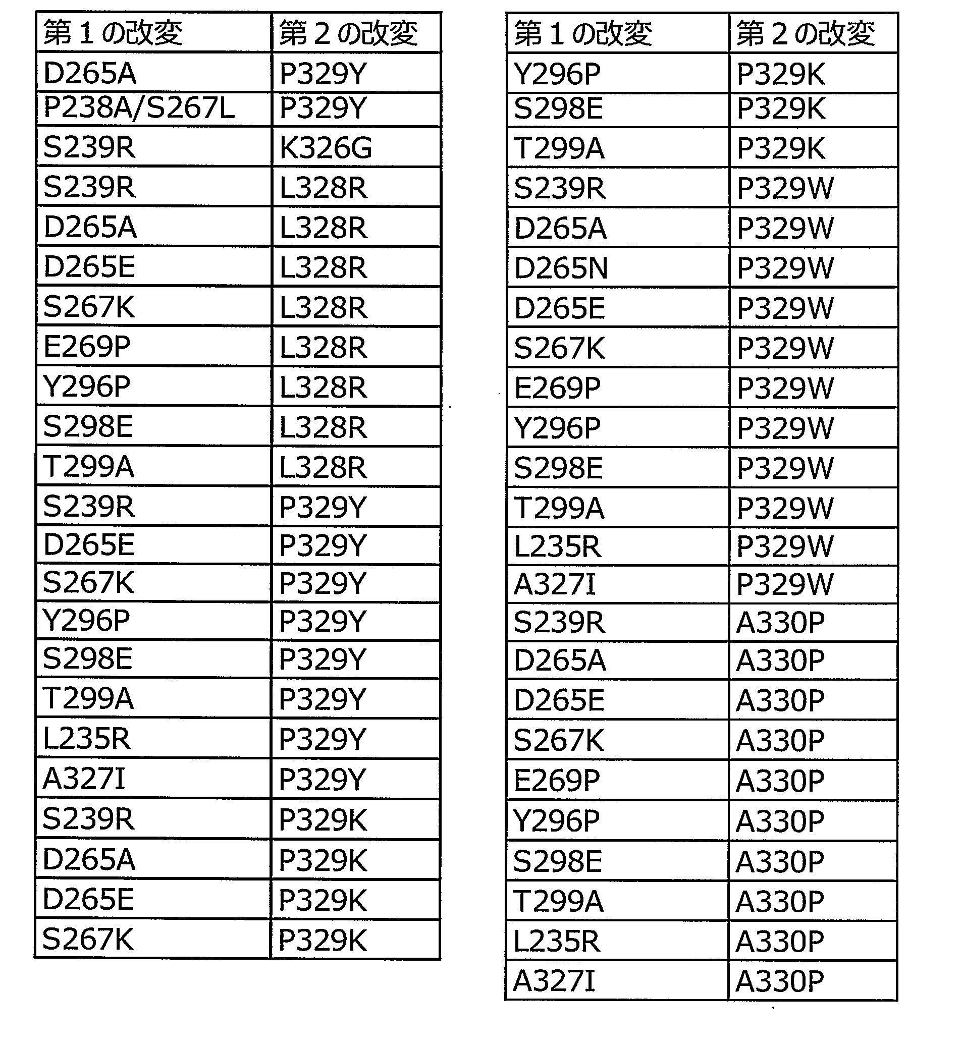
上記した置換後のアミノ酸残基は相互に置換可能なアミノ酸としてもよい。以下に、相互に置換可能なアミノ酸の例を示す。同一群に含まれるアミノ酸は相互に置換可能である。
A群:ロイシン、イソロイシン、ノルロイシン、バリン、ノルバリン、アラニン、2-アミノブタン酸、メチオニン、O-メチルセリン、t-ブチルグリシン、t-ブチルアラニン、シクロヘキシルアラニン
B群:アスパラギン酸、グルタミン酸、イソアスパラギン酸、イソグルタミン酸、2-アミノアジピン酸、2-アミノスベリン酸
C群:アスパラギン、グルタミン
D群:リジン、アルギニン、オルニチン、2,4-ジアミノブタン酸、2,3-ジアミノプロピオン酸
E群:プロリン、3-ヒドロキシプロリン、4-ヒドロキシプロリン
F群:セリン、スレオニン、ホモセリン
G群:フェニルアラニン、チロシン The amino acid residues after the above-mentioned substitution may be mutually substitutable amino acids. The following shows examples of amino acids that can be substituted for each other. Amino acids included in the same group can be substituted for each other.
Group A: leucine, isoleucine, norleucine, valine, norvaline, alanine, 2-aminobutanoic acid, methionine, O-methylserine, t-butylglycine, t-butylalanine, cyclohexylalanine B group: aspartic acid, glutamic acid, isoaspartic acid, Isoglutamic acid, 2-aminoadipic acid, 2-aminosuberic acid group C: asparagine, glutamine group D: lysine, arginine, ornithine, 2,4-diaminobutanoic acid, 2,3-diaminopropionic acid group E: proline, 3 -Hydroxyproline, 4-hydroxyproline F group: serine, threonine, homoserine G group: phenylalanine, tyrosine
上記置換されるアミノ酸は天然型と非天然型とを問わない。天然型アミノ酸としては、L-アラニン、L-アスパラギン、L-アスパラギン酸、L-グルタミン、L-グルタミン酸、グリシン、L-ヒスチジン、L-イソロイシン、L-ロイシン、L-リジン、L-アルギニン、L-メチオニン、L-フェニルアラニン、L-プロリン、L-セリン、L-スレオニン、L-トリプトファン、L-チロシン、L-バリン、L-システインなどが挙げられる。非天然型アミノ酸としては、アミノ基とカルボキシル基を有する種々のアミノ酸が挙げられるが、好ましくは各種天然アミノ酸の誘導体が望ましい。多くの非天然アミノ酸は、各試薬会社(Sigma-Aldrich社、TCI社)より入手できる。非天然アミノ酸については、文献(Chem.Today 2003, 65. ;Curr Opin Chem Biol. 2000, 6, 645.)に多くの開示がある。
The amino acids to be substituted may be natural or non-natural. Examples of natural amino acids include L-alanine, L-asparagine, L-aspartic acid, L-glutamine, L-glutamic acid, glycine, L-histidine, L-isoleucine, L-leucine, L-lysine, L-arginine, L-arginine, -Methionine, L-phenylalanine, L-proline, L-serine, L-threonine, L-tryptophan, L-tyrosine, L-valine, L-cysteine and the like. Examples of the non-natural amino acid include various amino acids having an amino group and a carboxyl group, and preferably, derivatives of various natural amino acids. Many unnatural amino acids are available from reagent companies (Sigma-Aldrich, TCI). There are many disclosures of unnatural amino acids in the literature (Chem. Today 2003, 65. Curr Opin Chem Biol 2000, 645).
第1のIgG半量体においては第1のCD16a結合領域におけるCD16a結合活性が改変により減弱しており、且つ第2のIgG半量体においては第2のCD16a結合領域におけるCD16a結合領域活性が改変により減弱している。このことから、第1のIgG半量体同士、又は第2のIgG半量体同士によりホモ会合体の抗体構造を構成しても、CD16aと結合することはできず、抗体の有する活性を発揮し得ない。
In the first IgG half, the CD16a binding activity in the first CD16a binding region is attenuated by the modification, and in the second IgG half, the CD16a binding region activity in the second CD16a binding region is attenuated by the modification doing. Therefore, even if the first IgG half-mer or the second IgG half-mer forms a homo-aggregate antibody structure, they cannot bind to CD16a and exhibit the activity of the antibody. Absent.
一方、第1及び第2のIgG半量体によりヘテロ会合体の抗体構造が構成されると、第1のIgG半量体における第2のCD16a結合領域と、第2のIgG半量体における第1のCD16a結合領域とを介してCD16aに対し結合可能となる。
On the other hand, when the antibody structure of the hetero-aggregate is constituted by the first and second IgG hemimers, the second CD16a binding region in the first IgG half and the first CD16a in the second IgG hemimer It becomes possible to bind to CD16a via the binding region.
第1及び第2のIgG半量体は互いに異なる抗原結合ドメインを有する。したがって、第1及び第2のIgG半量体からなるヘテロ会合体の抗体構造を構成することにより、互いに異なる2種の抗原を同一細胞上に発現している標的細胞に対して結合した場合にのみ、CD16aが結合しADCC活性を選択的に発揮し得る抗体組成物となる。
The first and second IgG hemmers have different antigen binding domains. Therefore, by forming the antibody structure of the hetero-association composed of the first and second IgG half-mers, only when the two different antigens are bound to a target cell expressing the same cell on the same cell, , CD16a can be bound to selectively exhibit ADCC activity.
第1及び第2のIgG半量体におけるヒンジドメインが、一部又は全体の置換もしくは欠失、または修飾によりジスルフィド結合が形成されないように改変されている。このことにより、第1のIgG半量体と第2のIgG半量体との間において、H鎖間におけるジスルフィド結合が形成されない。したがって、第1及び第2のIgG半量体の併存により、第1及び第2のIgG半量体を会合体又は半量体の平衡状態で存在させることが可能となり、両陽性細胞に対する高い選択性を発揮し得る。
ヒ ン ジ The hinge domain in the first and second IgG hemmers has been modified such that partial or total substitution or deletion or modification does not form a disulfide bond. As a result, no disulfide bond between the H chains is formed between the first IgG half and the second IgG half. Therefore, the coexistence of the first and second IgG halfmers enables the first and second IgG halfmers to exist in an equilibrium state of the aggregate or the halfmer, and exhibits high selectivity for both positive cells. I can do it.
第1のIgG半量体と第2のIgG半量体との間において、H鎖間におけるジスルフィド結合が形成されないような、ヒンジドメインにおける一部の置換としては、例えば、EUインデックスで表される226位及び229位の少なくとも一方のアミノ酸残基置換が挙げられる。
As a partial substitution in the hinge domain between the first IgG half and the second IgG half so that no disulfide bond is formed between H chains, for example, position 226 represented by the EU index And 229 at least one amino acid residue substitution.
上記の平衡状態で存在させるためには、第1及び第2のIgG半量体における、L鎖、H鎖のH鎖可変領域及びCH1ドメイン及びCH2ドメインのイムノグロブリンサブクラスに属するCH3ドメイン間相互作用と比較して、H鎖のCH3ドメインは、CH3ドメイン間相互作用が弱いことが好ましい。
In order to make it exist in the above-mentioned equilibrium state, interaction between the CH3 domain belonging to the immunoglobulin subclasses of the L chain, the H chain variable region and the CH1 domain and the CH2 domain in the first and second IgG half-mers is required. In comparison, the CH3 domain of the H chain preferably has weak interaction between CH3 domains.
具体的には例えば、第1及び第2のIgG半量体におけるH鎖のCH3ドメインは、CH3ドメイン間相互作用が、IgG1サブクラスのCH3ドメイン間相互作用よりも弱いことが好ましい。
Specifically, for example, it is preferable that the CH3 domain of the H chain in the first and second IgG hemimers has weaker interaction between CH3 domains than the interaction between CH3 domains of the IgG1 subclass.
CH3ドメイン間相互作用が、IgG1サブクラスのCH3ドメイン間相互作用よりもCH3ドメイン間相互作用の弱いCH3ドメインとしては、アミノ酸改変(アミノ酸の置換、欠失もしくは付加、または修飾等)によりCH3ドメイン間相互作用が減弱したIgG1のCH3ドメイン、IgG4のCH3ドメインが挙げられる。
The CH3 domain in which the interaction between CH3 domains is weaker than the interaction between CH3 domains of the IgG1 subclass includes the CH3 domain interaction by amino acid modification (amino acid substitution, deletion or addition, or modification, etc.). The CH3 domain of IgG1 and the CH3 domain of IgG4 whose action is attenuated are exemplified.
2.抗体組成物のエフェクター活性の制御
本発明の抗体組成物は、第1及び第2のIgG半量体におけるFcに依存したエフェクター活性を付与することもできる。抗体組成物のエフェクター活性は、種々の方法により制御することができる。 2. Control of Effector Activity of Antibody Composition The antibody composition of the present invention can also impart Fc-dependent effector activity in the first and second IgG hemmers. Effector activity of an antibody composition can be controlled by various methods.
本発明の抗体組成物は、第1及び第2のIgG半量体におけるFcに依存したエフェクター活性を付与することもできる。抗体組成物のエフェクター活性は、種々の方法により制御することができる。 2. Control of Effector Activity of Antibody Composition The antibody composition of the present invention can also impart Fc-dependent effector activity in the first and second IgG hemmers. Effector activity of an antibody composition can be controlled by various methods.
例えば、第1及び第2のIgG半量体が、CH2ドメインにおいて、さらにCD16a結合活性を増強させる少なくとも1のアミノ酸残基置換を含むことにより、さらにCD16a結合活性を増強させることが好ましい。このことにより、抗体組成物のエフェクター活性を増強することができる。
For example, it is preferable that the first and second IgG hemmers further enhance CD16a binding activity by including at least one amino acid residue substitution in the CH2 domain that further enhances CD16a binding activity. Thereby, the effector activity of the antibody composition can be enhanced.
抗体組成物のCD16a結合活性を増強するためのアミノ酸残基置換は、第1及び第2のIgG半量体におけるCD16a結合活性を減弱させるために改変された領域とは異なる領域におけるアミノ酸残基置換であることが好ましい。
The amino acid residue substitution for enhancing the CD16a binding activity of the antibody composition is an amino acid residue substitution in a region different from the region modified to reduce CD16a binding activity in the first and second IgG half-mers. Preferably, there is.
エフェクター活性とは、抗体のFcを介して引き起こされる抗体依存性の活性をいい、ADCC活性、CDC活性、又はマクロファージ若しくは樹状細胞などの食細胞による抗体依存性ファゴサイトーシス(Antibody-Dependent Cellular Phagocytosis activity;ADCP活性)などが知られている。本発明においてADCC活性及びCDC活性は、公知の測定方法[Cancer Immunol. Immunother., 36, 373 (1993)]を用いて測定できる。
The effector activity refers to an antibody-dependent activity caused through Fc of an antibody, and is dependent on ADCC activity, CDC activity, or antibody-dependent phagocytosis by phagocytic cells such as macrophages or dendritic cells (Antibody-Dependent Cellular Phagycytosis). activity; ADCP activity) and the like. In the present invention, the ADCC activity and the CDC activity can be measured using a known measuring method [Cancer Immunol. {Immunother., {36, {373} (1993)].
ADCC活性とは、標的細胞上の抗原に結合した抗体が、抗体のFcを介して免疫細胞のFc受容体と結合することにより免疫細胞(ナチュラルキラー細胞など)を活性化し、標的細胞を傷害する活性をいう。
ADCC activity means that an antibody bound to an antigen on a target cell activates an immune cell (such as a natural killer cell) by binding to an Fc receptor of an immune cell via Fc of the antibody, thereby damaging the target cell. Refers to activity.
Fc受容体(以下、FcRと記すこともある)とは、抗体のFcに結合する受容体であり、抗体の結合によりさまざまなエフェクター活性を誘導する。
Fc receptor (hereinafter sometimes referred to as FcR) is a receptor that binds to Fc of an antibody, and induces various effector activities by binding of the antibody.
FcRは抗体のサブクラスに対応しており、IgG、IgE、IgA、IgMはそれぞれFcγR、FcεR、FcαR、FcμRに特異的に結合する。更にFcγRには、FcγRI(CD64)、FcγRII(CD32)及びFcγRIII(CD16)のサブタイプが存在し、それぞれFcγRIA、FcγRIB、FcγRIC、FcγRIIA、FcγRIIB、FcγRIIC、FcγRIIIA(CD16a)、FcγRIIIBのアイソフォームが存在する。これらの異なるFcγRは異なる細胞上に存在している[Annu. Rev. Immunol. 9: 457-492 (1991)]。
FcR corresponds to the antibody subclass, and IgG, IgE, IgA, and IgM specifically bind to FcγR, FcεR, FcαR, and FcμR, respectively. Further, FcγR has subtypes of FcγRI (CD64), FcγRII (CD32) and FcγRIII (CD16), and FcγRIA, FcγRIB, FcγRIC, FcγRIIA, FcγRIIB, FcγRIIC, FcγRIIIA (CD16a) and FcγRIIIB of FcγRIIIA (CD16a) exist respectively. I do. These different FcγRs are present on different cells [Annu. Rev. Immunol. 9: 457-492 (1991)].
ヒトにおいては、FcγRIIIBは好中球に特異的に発現しており、FcγRIIIAは、単球、Natural Killer細胞(NK細胞)及び一部のT細胞に発現している。FcγRIIIAを介した抗体の結合は、NK細胞依存的なADCC活性を誘導する。
In humans, FcγRIIIB is specifically expressed on neutrophils, and FcγRIIIA is expressed on monocytes, Natural Killer cells (NK cells) and some T cells. Binding of antibodies through FcγRIIIA induces NK cell-dependent ADCC activity.
CDC活性とは標的細胞上の抗原に結合した抗体が血液中の補体関連タンパク質群からなる一連のカスケード(補体活性化経路)を活性化し、標的細胞を傷害する活性をいう。また、補体の活性化により生じるタンパク質断片により免疫細胞の遊走、活性化を誘導することができる。
CDC activity refers to an activity in which an antibody bound to an antigen on a target cell activates a series of cascades (complement activation pathway) composed of a group of complement-related proteins in blood to damage the target cell. In addition, migration and activation of immune cells can be induced by a protein fragment generated by activation of complement.
CDC活性のカスケードは、抗体のFcとの結合ドメインを有するC1qが、Fcに結合し、2つのセリンプロテアーゼであるC1r及びC1sと結合することでC1複合体を形成することで開始する。
The cascade of CDC activity begins when C1q, which has a binding domain to Fc of an antibody, binds to Fc and binds to two serine proteases, C1r and C1s, to form a C1 complex.
本発明の抗体組成物のエフェクター活性を制御する方法としては以下のような方法が挙げられる。
方法 Methods for controlling the effector activity of the antibody composition of the present invention include the following methods.
例えば、第1及び第2のIgG半量体において、IgG1サブクラスのFcのアミノ酸配列を用いて、EUインデックス297位のAsnに結合するN結合複合型糖鎖(以下、単にコンプレックス糖鎖と略記する場合もある)の還元末端に存在するN-アセチルグルコサミン(GlcNAc)にα-1,6結合するフコース(コアフコースともいう)の量を制御する方法(国際公開第2005/035586号、国際公開第2002/31140号、国際公開第00/61739号)、又は抗体のFcのアミノ酸残基を置換することで、抗体組成物のエフェクター活性を制御することができる。
For example, in the first and second IgG hemimers, an N-linked complex-type sugar chain that binds to Asn at EU index position 297 using the amino acid sequence of IgG1 subclass Fc (hereinafter, simply referred to as complex sugar chain) ), Which controls the amount of fucose (also referred to as core fucose) that binds α-1,6 to N-acetylglucosamine (GlcNAc) present at the reducing end (WO 2005/035586, WO 2002/2002). 31140, WO 00/61739), or by substituting the amino acid residue of Fc of an antibody, the effector activity of the antibody composition can be controlled.
1)糖鎖改変によるエフェクター活性の制御
第1及び第2のIgG半量体のFcに結合しているコンプレックス糖鎖の還元末端のN-アセチルグルコサミンに付加するフコースの含量を制御することで、抗体組成物のエフェクター活性を増加又は低下させることができる。 1) Control of effector activity by sugar chain modification By controlling the content of fucose added to N-acetylglucosamine at the reducing end of the complex sugar chain bound to Fc of the first and second IgG half-mers, the antibody The effector activity of the composition can be increased or decreased.
第1及び第2のIgG半量体のFcに結合しているコンプレックス糖鎖の還元末端のN-アセチルグルコサミンに付加するフコースの含量を制御することで、抗体組成物のエフェクター活性を増加又は低下させることができる。 1) Control of effector activity by sugar chain modification By controlling the content of fucose added to N-acetylglucosamine at the reducing end of the complex sugar chain bound to Fc of the first and second IgG half-mers, the antibody The effector activity of the composition can be increased or decreased.
IgG半量体のFcに結合しているN結合複合型糖鎖に付加するフコースの含量を低下させる方法としては、α1,6-フコース転移酵素遺伝子(FUT8)が欠損したCHO細胞を用いてIgG半量体を発現することで、フコースが結合していないIgG半量体を取得できる。フコースが結合していないIgG半量体からなる抗体組成物は高いADCC活性を有する。
As a method for reducing the content of fucose added to the N-linked complex type sugar chain bound to Fc of IgG half-mer, a method for reducing IgG content using CHO cells deficient in α1,6-fucose transferase gene (FUT8) is used. By expressing the body, an IgG halfmer to which fucose is not bound can be obtained. An antibody composition comprising an IgG half having no fucose bound has a high ADCC activity.
一方、IgG半量体のFcに結合しているN結合複合型糖鎖に付加するフコースの含量を増加させる方法としては、α1,6-フコース転移酵素遺伝子を導入した宿主細胞を用いてIgG半量体を発現させることで、フコースが結合しているIgG半量体を取得できる。フコースが結合しているIgG半量体からなる抗体組成物は、フコースが結合していないIgG半量体からなる抗体組成物よりも低いADCC活性を有する。
On the other hand, as a method for increasing the content of fucose added to the N-linked complex type sugar chain bound to Fc of an IgG half-mer, an IgG half-mer using a host cell into which an α1,6-fucose transferase gene has been introduced. By expressing, an IgG halfmer to which fucose is bound can be obtained. An antibody composition comprising an IgG half having fucose bound thereto has a lower ADCC activity than an antibody composition comprising an IgG half having no fucose bound.
IgG半量体のFcには、EUインデックス297位のAsn残基にN結合型糖鎖が結合するが、それ以外のFcのAsn残基には糖鎖は結合することは知られていない。従って、通常、抗体組成物1分子あたり2本のN-グリコシド結合糖鎖が結合している。
NN-linked sugar chains bind to Asn residues at EU index position 297 to Fc of IgG half-mer, but it is not known that sugar chains bind to other Asn residues of Fc. Therefore, two N-glycoside-linked sugar chains are usually bound per molecule of the antibody composition.
N結合型糖鎖としてはハイマンノース型、コンプレックス型及びハイブリッド型が知られており、いずれのN結合型糖鎖でもフコースが結合していない糖鎖であれば、フコースが結合している糖鎖と比べて高いADCC活性を得ることができる。
As the N-linked sugar chains, high mannose type, complex type and hybrid type are known. If any of the N-linked sugar chains is a sugar chain to which fucose is not linked, the sugar chain to which fucose is linked. Higher ADCC activity can be obtained.
IgG半量体のFcに結合するコンプレックス糖鎖としては、コア構造(トリマンノシルコア構造)の非還元末端側のマンノース(Man)に、1つ以上のN-アセチルグルコサミン(GlcNAc)又はガラクトース-N-アセチルグルコサミン(以下、Gal-GlcNAcと表記する)がα1-2結合又はα1-4結合している糖鎖が挙げられる。
Complex sugar chains that bind to Fc of an IgG half-mer include mannose (Man) on the non-reducing terminal side of the core structure (trimannosyl core structure), and one or more N-acetylglucosamine (GlcNAc) or galactose-N- A sugar chain in which acetylglucosamine (hereinafter referred to as Gal-GlcNAc) has an α1-2 bond or an α1-4 bond is exemplified.
更にGal-GlcNAcの非還元末端側にシアル酸、バイセクティングのN-アセチルグルコサミン(以下、bisecting GlcNAcと記す)などを有するコンプレックス型(複合型)糖鎖を挙げることができる。
Further, a complex type (complex type) sugar chain having sialic acid, bisecting N-acetylglucosamine (hereinafter referred to as bisecting GlcNAc) on the non-reducing terminal side of Gal-GlcNAc can be exemplified.
本発明において、コアフコース(core-fucose)又はα1,6-フコースとは、N-グリコシド結合複合型糖鎖還元末端のN-アセチルグルコサミン(以下、GlcNAcと記す場合もある)の6位とフコース(以下、Fucと記す場合もある)の1位がα結合した糖鎖構造をいう。また、N-グリコシド結合複合型糖鎖還元末端のN-アセチルグルコサミンにフコースが結合していないことを、単にフコースが無い又はコアフコースが無い糖鎖という。
In the present invention, core-fucose or α1,6-fucose refers to the 6-position of N-acetylglucosamine (hereinafter, also referred to as GlcNAc) at the reducing end of an N-glycoside-linked complex type sugar chain and fucose ( Hereinafter, it may be referred to as Fuc). In addition, the fact that fucose is not bound to N-acetylglucosamine at the reducing end of an N-glycoside-linked complex type sugar chain is simply referred to as a sugar chain having no fucose or no core fucose.
また、本発明において、コア構造又はトリマンノシルコア構造(tri-mannosyl core structure)とは、Manα1-6(Manα1-3)Manβ1-4GlcNAcβ1-4GlcNAc構造をいう。
In the present invention, the core structure or the tri-mannosyl core structure (tri-mannosyl core structure) refers to a Manα1-6 (Manα1-3) Manβ1-4GlcNAcβ1-4GlcNAc structure.
IgG半量体に結合している糖鎖として、2本鎖N-グリコシド結合コンプレックス糖鎖(バイアンテナリーコンプレックス糖鎖ともいう)は、下記化学式で示される。
糖 A double-stranded N-glycoside-linked complex sugar chain (also referred to as a biantennary complex sugar chain) as a sugar chain bound to an IgG halfmer is represented by the following chemical formula.

第1及び第2のIgG半量体は、EUインデックス297位のAsnにコンプレックス型糖鎖が結合したFcを有することが好ましい。上記の糖鎖構造を有していれば、単一または複数の異なる糖鎖構造を有していてもよい。具体的には、第1及び第2のIgG半量体における、Fcに結合する全N-グリコシド結合型糖鎖のうち、糖鎖還元末端のN-アセチルグルコサミンにフコースが結合していない糖鎖(コアフコースが無い糖鎖)の割合が20%以上である抗体組成物が挙げられる。
It is preferable that the first and second IgG halfmers have Fc in which a complex type sugar chain is bound to Asn at position 297 of the EU index. As long as it has the above sugar chain structure, it may have a single or plural different sugar chain structures. Specifically, among all N-glycoside-linked sugar chains that bind to Fc in the first and second IgG halfmers, sugar chains in which fucose is not linked to N-acetylglucosamine at the reducing end of the sugar chain ( An antibody composition in which the ratio of sugar chains without core fucose) is 20% or more.
コアフコースが無い糖鎖の割合としては、抗体組成物のADCC活性が増加すれば、いずれの割合も含まれるが、好ましくは20%以上、より好ましくは51%~100%、更に好ましくは80%~100%、特に好ましくは90%、91%、92%、93%、94%、95%、96%、97%、98%、99%、最も好ましくは100%の割合が挙げられる。
The ratio of sugar chains without core fucose includes any ratio as long as the ADCC activity of the antibody composition increases, but is preferably 20% or more, more preferably 51% to 100%, and still more preferably 80% to 80%. 100%, particularly preferably 90%, 91%, 92%, 93%, 94%, 95%, 96%, 97%, 98%, 99%, most preferably 100%.
コアフコースが無い糖鎖の割合が50%とは、例えば、第1及び第2のIgG半量体に結合しているN-グリコシド結合糖鎖の片方の糖鎖にフコースが結合していない分子が100%含まれる抗体組成物、又は第1及び第2のIgG半量体に結合しているN-グリコシド結合糖鎖の両方の糖鎖にフコースが結合していない分子が50%含まれ、かつ第1及び第2のIgG半量体に結合しているN-グリコシド結合糖鎖の両方の糖鎖にフコースが結合している分子が50%含まれる抗体組成物のいずれも含まれる。
The percentage of sugar chains without core fucose is 50%, for example, when 100% of molecules in which fucose is not bound to one of the N-glycoside-linked sugar chains bound to the first and second IgG half-mers. % Of the N-glycoside-linked sugar chain bound to the first and second IgG half-mers, or 50% of the molecule in which fucose is not bound to the first and second N-glycoside-linked sugar chains. And an antibody composition containing 50% of a molecule in which fucose is bound to both sugar chains of the N-glycoside-linked sugar chain bound to the second IgG half-mer.
本発明において、フコースが無い糖鎖としては、上記で示された化学式中、還元末端側のN-アセチルグルコサミンにフコースが結合していなければ、非還元末端の糖鎖の構造はいかなるものであってもよい。
In the present invention, the sugar chain without fucose has any structure of the sugar chain at the non-reducing end in the chemical formula shown above unless fucose is bonded to N-acetylglucosamine on the reducing end side. You may.
本発明において、糖鎖還元末端のN-アセチルグルコサミンにフコースが結合していない(コアフコースが無い)とは、実質的にフコースが結合していないことをいう。実質的にフコースが結合していないIgG半量体とは、具体的には、後述に記載の糖鎖分析において、フコースが実質的に検出できない程度のIgG半量体である場合をいう。実質的に検出できない程度とは、測定の検出限界以下であることをいう。全ての糖鎖にコアフコースが無い第1及び第2のIgG半量体からなる抗体組成物は、最も高いADCC活性を有する。
に お い て In the present invention, the expression that fucose is not bound to N-acetylglucosamine at the reducing end of the sugar chain (there is no core fucose) means that fucose is not substantially bound. Specifically, an IgG halfmer to which fucose is not substantially bound refers to a case in which fucose is substantially undetectable in sugar chain analysis described below. The term “substantially undetectable” refers to a level below the detection limit of measurement. The antibody composition consisting of the first and second IgG hemmers without core fucose in all sugar chains has the highest ADCC activity.
N-グリコシド結合複合型糖鎖が結合したFcを有するIgG半量体中における、フコースが無い糖鎖を有するIgG半量体の割合は、IgG半量体からヒドラジン分解又は酵素消化などの公知の方法[生物化学実験法23-糖蛋白質糖鎖研究法(学会出版センター)高橋禮子編(1989)]を用い、糖鎖を遊離させ、遊離させた糖鎖を蛍光標識又は同位元素標識し、標識した糖鎖をクロマトグラフィ法にて分離することによって決定することができる。
The ratio of IgG half having a fucose-free sugar chain to IgG half having Fc to which an N-glycoside-linked complex type sugar chain is bound can be determined by a known method such as hydrazinolysis or enzymatic digestion of the IgG half. Chemical Experimental Method 23-Glycoprotein Sugar Chain Research Method (Society Publishing Center), edited by Reiko Takahashi (1989)], and the released sugar chains are labeled with a fluorescent label or an isotope to label the sugar chains. Can be determined by chromatographic separation.
また、コンプレックス糖鎖が結合したFcを含むIgG半量体中に含まれる、フコースが無い糖鎖が結合したIgG半量体の割合は、遊離させた糖鎖をHPAED-PAD法(J. Liq. Chromatogr., 6, 1577, 1983)によって分析することで決定することができる。
In addition, the ratio of the IgG halfmer to which the sugar chain having no fucose is contained in the IgG halfmer containing Fc to which the complex sugar chain is bound is determined by the HPAED-PAD method (J. Liq. Chromatogr ., 6, 1577, 1983).
2)アミノ酸残基置換によるエフェクター活性の制御
本発明の抗体組成物は、第1のIgG半量体及び第2の半量体におけるFcの抗体サブクラスの変換又はFcのアミノ酸残基置換により、ADCC活性、ADCP活性及びCDC活性を増加又は低下させることができる。 2) Control of effector activity by amino acid residue substitution The antibody composition of the present invention provides ADCC activity by conversion of the antibody subclass of Fc or amino acid residue substitution of Fc in the first IgG half and the second half. ADCP activity and CDC activity can be increased or decreased.
本発明の抗体組成物は、第1のIgG半量体及び第2の半量体におけるFcの抗体サブクラスの変換又はFcのアミノ酸残基置換により、ADCC活性、ADCP活性及びCDC活性を増加又は低下させることができる。 2) Control of effector activity by amino acid residue substitution The antibody composition of the present invention provides ADCC activity by conversion of the antibody subclass of Fc or amino acid residue substitution of Fc in the first IgG half and the second half. ADCP activity and CDC activity can be increased or decreased.
IgG1サブクラスの抗体は、IgGサブクラスの中で最も高いADCC活性、CDC活性を有することが知られており、CH2ドメインのイムノグロブリンサブクラスはIgG1であることが好ましい。
It is known that an antibody of the IgG1 subclass has the highest ADCC activity and CDC activity among the IgG subclasses, and the immunoglobulin subclass of the CH2 domain is preferably IgG1.
Fcのアミノ酸残基置換としては例えば、米国特許出願公開第2007/0148165号明細書に記載のFcのアミノ酸配列を用いることで、抗体のCDC活性を増加させることができる。また、米国特許第6,737,056号明細書、米国特許第7,297,775号明細書及び米国特許第7,317,091号明細書に記載のアミノ酸残基置換を行うことで、抗体組成物のADCC活性又はCDC活性を増加させることも低下させることもできる。
The CDC activity of the antibody can be increased by using the amino acid sequence of Fc described in US Patent Application Publication No. 2007/0148165, for example, as the amino acid residue substitution of Fc. Further, by performing amino acid residue substitution described in US Pat. No. 6,737,056, US Pat. No. 7,297,775 and US Pat. No. 7,317,091, an antibody The ADCC or CDC activity of the composition can be increased or decreased.
ADCC活性を増強させる具体的なアミノ酸残基置換としては、P247I、A339D、F243L、R292P、Y300L、P396L、T393A、H433P、S239D、S298A、A330L、I332E、E333A及びK334Aなどが挙げられる。一方、ADCC活性を減少させる具体的なアミノ酸残基置換としては、L235E、P238A、N297A、K322A及びP331Sなどが挙げられる。
具体 Specific amino acid residue substitutions for enhancing ADCC activity include P247I, A339D, F243L, R292P, Y300L, P396L, T393A, H433P, S239D, S298A, A330L, I332E, E333A, and K334A. On the other hand, specific amino acid residue substitutions that decrease ADCC activity include L235E, P238A, N297A, K322A, and P331S.
CDC活性を増加させる具体的なアミノ酸残基置換としては、K326A、S267E、H268F、S324T、K274Q、N276K、Y296F、Y300F、K326W、K326Y、E333A、E333S、A339T、D356E、L358M、N384S、K392N、T394F、T394Y、V397M及びV422Iから選ばれる少なくとも1つのアミノ酸残基置換が挙げられる。
Specific amino acid residue substitutions to increase CDC activity include K326A, S267E, H268F, S324T, K274Q, N276K, Y296F, Y300F, K326W, K326Y, E333A, E333S, A339T, D356E, L358M, N384T, K392N. , T394Y, V397M and V422I.
いずれのアミノ酸残基置換を2つ以上組み合わせてCDC活性を増加させることもでき、目的に応じて置換するアミノ酸残基を増やすことができる。CDC活性を増加させるアミノ酸残基置換として好ましくは、N276K、A339T、T394F及びT394Yから選ばれる少なくとも1つのアミノ酸残基置換、N276K及びA339Tのアミノ酸残基置換、並びにK274Q、N276K、Y296F、Y300F、A339T、D356E、L358M、N384S、V397M及びV422Iのアミノ酸残基置換などが挙げられる。一方、CDC活性を減少させる具体的なアミノ酸残基置換としては、L235E、N297A、K322A、P329A及びP331Sなどが挙げられる。
C CDC activity can be increased by combining any two or more amino acid residue substitutions, and the number of amino acid residues to be substituted can be increased according to the purpose. The amino acid residue substitution that increases CDC activity is preferably at least one amino acid residue substitution selected from N276K, A339T, T394F and T394Y, the amino acid residue substitution of N276K and A339T, and K274Q, N276K, Y296F, Y300F, A339T. , D356E, L358M, N384S, V397M and V422I amino acid residue substitutions. On the other hand, specific amino acid residue substitutions that reduce CDC activity include L235E, N297A, K322A, P329A, and P331S.
またT250Q、M428L、M252Y、S254T、T256Eなどのアミノ酸変異をヒトIgG1サブクラスのFcに導入することにより血中半減期を延長することができる。またN297位にアミノ酸変異を導入することによりN結合型糖鎖を除去したFc又はヒトIgG2若しくはIgG4サブクラスのFc、IgG2とIgG4のキメラFcなどを用いることにより、ADCC活性、ADCP活性、CDC活性などの細胞傷害活性を低下させることができる。
Also, by introducing amino acid mutations such as T250Q, M428L, M252Y, S254T, and T256E into Fc of human IgG1 subclass, blood half-life can be extended. Also, ADCC activity, ADCP activity, CDC activity, etc., by using Fc from which N-linked sugar chains have been removed by introducing an amino acid mutation at position N297 or Fc of human IgG2 or IgG4 subclass, chimeric Fc of IgG2 and IgG4, etc. Cytotoxic activity can be reduced.
3.抗体組成物の製造方法
本発明の抗体組成物の製造方法は、以下の工程1~3を含む製造方法が挙げられる。
工程1:IgG半量体のアミノ酸配列をコードするDNAを含む組換えベクター(以下、IgG半量体発現用組換えベクターとも略す)を細胞へ導入して形質転換体を得る工程。
工程2:工程1で得られた形質転換体を培養し、培養物中にIgG半量体を蓄積させ、培養物からIgG半量体を採取する工程。
工程3:工程2で採取されたIgG半量体からなる抗体組成物を得る工程。
以下、各工程について説明する。 3. Method for Producing Antibody Composition A method for producing the antibody composition of the present invention includes a production method comprising the followingsteps 1 to 3.
Step 1: a step of introducing a recombinant vector containing a DNA encoding the amino acid sequence of an IgG half-mer (hereinafter also abbreviated as a recombinant vector for expressing an IgG half-mer) into cells to obtain a transformant.
Step 2: a step of culturing the transformant obtained inStep 1, accumulating an IgG half-mer in the culture, and collecting the IgG half-mer from the culture.
Step 3: a step of obtaining an antibody composition consisting of the IgG halfmer collected inStep 2.
Hereinafter, each step will be described.
本発明の抗体組成物の製造方法は、以下の工程1~3を含む製造方法が挙げられる。
工程1:IgG半量体のアミノ酸配列をコードするDNAを含む組換えベクター(以下、IgG半量体発現用組換えベクターとも略す)を細胞へ導入して形質転換体を得る工程。
工程2:工程1で得られた形質転換体を培養し、培養物中にIgG半量体を蓄積させ、培養物からIgG半量体を採取する工程。
工程3:工程2で採取されたIgG半量体からなる抗体組成物を得る工程。
以下、各工程について説明する。 3. Method for Producing Antibody Composition A method for producing the antibody composition of the present invention includes a production method comprising the following
Step 1: a step of introducing a recombinant vector containing a DNA encoding the amino acid sequence of an IgG half-mer (hereinafter also abbreviated as a recombinant vector for expressing an IgG half-mer) into cells to obtain a transformant.
Step 2: a step of culturing the transformant obtained in
Step 3: a step of obtaining an antibody composition consisting of the IgG halfmer collected in
Hereinafter, each step will be described.
[工程1]
工程1は、第1及び第2のIgG半量体の少なくとも一方のアミノ酸配列をコードするDNAを含む組換えベクターを細胞へ導入して形質転換体を得る工程である。
工程(1)は、具体的には、以下の工程(1-1)~(1-3)を含む。
(1-1)第1のIgG半量体における第1のCD16a結合領域におけるCD16a結合活性を改変により減弱させる工程。
(1-2)第2のIgG半量体における第2のCD16a結合領域におけるCD16a結合活性を改変により減弱させる工程。
(1-3)第1及び第2のIgG半量体のヒンジドメインにおいて一部又は全体の置換もしくは欠失、または修飾を行うことにより、H鎖間におけるジスルフィド結合が形成されないように改変する工程。 [Step 1]
Step 1 is a step of obtaining a transformant by introducing a recombinant vector containing DNA encoding the amino acid sequence of at least one of the first and second IgG halfmers into cells.
Step (1) specifically includes the following steps (1-1) to (1-3).
(1-1) a step of attenuating the CD16a binding activity in the first CD16a binding region of the first IgG half-mer by modification.
(1-2) a step of attenuating the CD16a binding activity in the second CD16a binding region of the second IgG half-mer by modification.
(1-3) A step of partially or wholly substituting, deleting, or modifying the hinge domains of the first and second IgG hemimers so as to prevent the formation of disulfide bonds between H chains.
工程1は、第1及び第2のIgG半量体の少なくとも一方のアミノ酸配列をコードするDNAを含む組換えベクターを細胞へ導入して形質転換体を得る工程である。
工程(1)は、具体的には、以下の工程(1-1)~(1-3)を含む。
(1-1)第1のIgG半量体における第1のCD16a結合領域におけるCD16a結合活性を改変により減弱させる工程。
(1-2)第2のIgG半量体における第2のCD16a結合領域におけるCD16a結合活性を改変により減弱させる工程。
(1-3)第1及び第2のIgG半量体のヒンジドメインにおいて一部又は全体の置換もしくは欠失、または修飾を行うことにより、H鎖間におけるジスルフィド結合が形成されないように改変する工程。 [Step 1]
Step (1) specifically includes the following steps (1-1) to (1-3).
(1-1) a step of attenuating the CD16a binding activity in the first CD16a binding region of the first IgG half-mer by modification.
(1-2) a step of attenuating the CD16a binding activity in the second CD16a binding region of the second IgG half-mer by modification.
(1-3) A step of partially or wholly substituting, deleting, or modifying the hinge domains of the first and second IgG hemimers so as to prevent the formation of disulfide bonds between H chains.
工程(1-1)については、例えば、第1のIgG半量体発現用組換えベクターの作製において、EUインデックスで表される235位、236位、237位、238位、239位、265位、266位、267位、268位、269位、294位、295位、296位、297位、298位、299位、301位、325位、327位、332位のアミノ酸残基から選ばれる少なくとも1のアミノ酸残基を置換する工程を、CH2ドメインのサブクラスに応じて適宜行うことが挙げられる。
In the step (1-1), for example, in the production of the first recombinant vector for expressing IgG half-mers, positions 235, 236, 237, 238, 239, 265, At least one selected from the amino acid residues at positions 266, 267, 268, 269, 294, 295, 296, 297, 298, 299, 301, 325, 327, and 332; The step of substituting the amino acid residue is appropriately performed depending on the subclass of the CH2 domain.
工程(1-2)については、例えば、第2のIgG半量体発現用組換えベクターの作製において、EUインデックスで表される235位、236位、237位、326位、327位、328位、329位、330位のアミノ酸残基から選ばれる少なくとも1のアミノ酸残基を置換する工程を、CH2ドメインのサブクラスに応じて適宜行うことが挙げられる。
In the step (1-2), for example, in the production of the second recombinant vector for expressing IgG half-mers, positions 235, 236, 237, 326, 327, 328, The step of substituting at least one amino acid residue selected from the amino acid residues at positions 329 and 330 may be appropriately performed according to the subclass of the CH2 domain.
工程(1-3)については、例えば、IgG半量体発現用組換えベクターの作製において、EUインデックスで表される226位及び229位の少なくとも一方のアミノ酸残基の置換を加える工程を、ヒンジドメインのサブクラスに応じて適宜行うことが挙げられる。
In the step (1-3), for example, the step of adding at least one amino acid residue at positions 226 and 229 represented by the EU index in the production of a recombinant vector for expressing an IgG half-mer is referred to as a hinge domain. May be performed as appropriate according to the subclass.
IgG半量体は、モレキュラー・クローニング第2版、カレント・プロトコールズ・イン・モレキュラー・バイオロジー、Antibodies, A Laboratory Manual, Cold Spring Harbor Laboratory, 1988、Monoclonal Antibodies: principles and practice, Third Edition,Acad.Press,1993, Antibody Engineering, A Practical Approach, IRL Press at Oxford University Press,1996等に記載された方法を用い、例えば、以下のように形質転換体中で発現させて取得することができる。
IgG half-mers are described in Molecular Cloning Second Edition, Current Protocols in Molecular Biology, Antibodies, A Laboratory Manual, Cold Spring Harbor Laboratory, 1988, MonoclonallonAntibodies: principles and practice, AThird. , 1993, Antibody Engineering, A Practical Approach, IRL Press at Oxford University Press, 1996, etc., for example, by expressing in a transformant as follows.
(1)IgG半量体発現用の組換えベクターの構築
IgG半量体発現用組換えベクターとは、本発明の抗体組成物を構成するIgG半量体のアミノ酸配列をコードする遺伝子が組み込まれた動物細胞用発現ベクターである。 (1) Construction of a recombinant vector for expressing an IgG half-mer A recombinant vector for expressing an IgG half-mer is an animal cell into which a gene encoding the amino acid sequence of an IgG half-mer constituting the antibody composition of the present invention has been incorporated. It is an expression vector for use.
IgG半量体発現用組換えベクターとは、本発明の抗体組成物を構成するIgG半量体のアミノ酸配列をコードする遺伝子が組み込まれた動物細胞用発現ベクターである。 (1) Construction of a recombinant vector for expressing an IgG half-mer A recombinant vector for expressing an IgG half-mer is an animal cell into which a gene encoding the amino acid sequence of an IgG half-mer constituting the antibody composition of the present invention has been incorporated. It is an expression vector for use.
組換えベクターは、動物細胞用発現ベクターにIgG半量体のアミノ酸配列をコードするDNAをクローニングすることにより構築することができる。
A recombinant vector can be constructed by cloning a DNA encoding the amino acid sequence of an IgG half-mer into an expression vector for animal cells.
DNAは、全DNAを合成してもよいし、polymerase chain reaction(PCR法)による合成も可能である(モレキュラー・クローニング第2版)。さらに、これらの手法を複数組み合わせることにより、IgG半量体をコードする遺伝子を作製することもできる。
DNA may be synthesized by total DNA or synthesized by polymerase chain reaction (PCR method) (Molecular Cloning 2nd edition). Furthermore, a gene encoding an IgG half-mer can be prepared by combining a plurality of these techniques.
動物細胞を宿主として用いる場合、発現ベクターとしては、動物細胞中で機能を発揮できるものであればいずれも用いることができる。例えば、pcDNAI、pCDM8(フナコシ社製)、pAGE107[日本国特開平3-22979号公報;Cytotechnology, 3, 133 (1990)]、pAS3-3(日本国特開平2-227075号公報)、pCDM8[Nature, 329, 840 (1987)]、pcDNAI/Amp(インビトロジェン社製)、pcDNA3.1(インビトロジェン社製)、pREP4(インビトロジェン社製)、pAGE103[J. Biochemistry, 101, 1307 (1987)]、pAGE210、pME18SFL3、pKANTEX93(国際公開第97/10354号)、N5KG1val(米国特許第6,001,358号明細書)及びTol2トランスポゾンベクター(国際公開第2010/143698号)などが挙げられる。
(4) When an animal cell is used as a host, any expression vector can be used as long as it can exert a function in the animal cell. For example, pcDNAI, pCDM8 (manufactured by Funakoshi), pAGE107 [Japanese Patent Application Laid-Open No. 3-22979; Cytotechnology, {3, {133} (1990)], pAS3-3 (Japanese Patent Application Laid-Open No. 2-2270075), pCDM8 [ Nature, {329, {840} (1987)], pcDNAI / Amp (Invitrogen), pcDNA3.1 (Invitrogen), pREP4 (Invitrogen), pAGE103 [J. Biochemistry, 101, {1307} (1987)], pAGE210 PME18SFL3, pKANTEX93 (WO 97/10354), N5KG1val (US Pat. No. 6,001,358), and Tol2 transposon vector (WO 2010/143698).
プロモーターとしては、動物細胞中で機能を発揮できるものであればいずれも用いることができる。例えば、サイトメガロウイルス(CMV)のimmediate early(IE)遺伝子のプロモーター、SV40の初期プロモーター、レトロウイルスのプロモーター、メタロチオネインプロモーター、ヒートショックプロモーター、SRαプロモーター、又はモロニーマウス白血病ウイルスのプロモーターあるいはエンハンサーが挙げられる。また、ヒトCMVのIE遺伝子のエンハンサーをプロモーターと共に用いてもよい。
Any promoter can be used as long as it can exhibit a function in animal cells. Examples include the promoter of the immediate early (IE) gene of cytomegalovirus (CMV), the early promoter of SV40, the promoter of retrovirus, the metallothionein promoter, the heat shock promoter, the SRα promoter, and the promoter or enhancer of Moloney murine leukemia virus. . Further, an enhancer of the IE gene of human CMV may be used together with the promoter.
発現ベクターは、抗体H鎖及びL鎖が別々のベクター上に存在するタイプあるいは同一のベクター上に存在するタイプ(以下、タンデム型と表記する)のどちらでも用いることができる。
The expression vector may be either of the type in which the antibody H chain and the L chain are present on separate vectors or the type in which the antibody is present on the same vector (hereinafter referred to as tandem type).
(2)可変領域をコードするcDNAの取得
任意の抗体のVH及びVLをコードするcDNAは次のようにして取得できる。任意の抗体を産生するハイブリドーマ細胞から抽出したmRNAを鋳型として用い、cDNAを合成する。合成したcDNAをファージ又はプラスミド等のベクターに挿入してcDNAライブラリーを作製する。 (2) Acquisition of cDNA encoding variable region cDNA encoding VH and VL of any antibody can be acquired as follows. CDNA is synthesized using, as a template, mRNA extracted from a hybridoma cell producing an arbitrary antibody. A cDNA library is prepared by inserting the synthesized cDNA into a vector such as a phage or a plasmid.
任意の抗体のVH及びVLをコードするcDNAは次のようにして取得できる。任意の抗体を産生するハイブリドーマ細胞から抽出したmRNAを鋳型として用い、cDNAを合成する。合成したcDNAをファージ又はプラスミド等のベクターに挿入してcDNAライブラリーを作製する。 (2) Acquisition of cDNA encoding variable region cDNA encoding VH and VL of any antibody can be acquired as follows. CDNA is synthesized using, as a template, mRNA extracted from a hybridoma cell producing an arbitrary antibody. A cDNA library is prepared by inserting the synthesized cDNA into a vector such as a phage or a plasmid.
前記ライブラリーより、既存の抗体の定常領域又は可変領域をコードするDNAをプローブとして用い、VHをコードするcDNAを有する組換えファージ或いは組換えプラスミド及びL鎖可変領域をコードするcDNAを有する組換えファージ又は組換えプラスミドをそれぞれ単離する。組換えファージ或いは組換えプラスミド上の目的の抗体のVH及びVLの全塩基配列を決定し、塩基配列よりVH及びVLの全アミノ酸配列を推定する。
From the library, using a DNA encoding the constant region or variable region of an existing antibody as a probe, a recombinant phage having a cDNA encoding VH or a recombinant plasmid and a recombinant phage having a cDNA encoding an L chain variable region The phage or recombinant plasmid is isolated, respectively. The entire nucleotide sequence of VH and VL of the target antibody on the recombinant phage or recombinant plasmid is determined, and the entire amino acid sequence of VH and VL is estimated from the nucleotide sequence.
任意の非ヒト動物抗体を生産するハイブリドーマ細胞は、抗体が結合する抗原を非ヒト動物に免疫し、周知の方法[モレキュラー・クローニング第2版、カレント・プロトコールズ・イン・モレキュラー・バイオロジー、Antibodies, A Laboratory manual, Cold Spring Harbor Laboratory, 1988、Monoclonal Antibodies: principles and practice, Third Edition, Acad. Press, 1993、Antibody Engineering, A Practical Approach, IRL Press at Oxford University Press,1996]に従って、免疫された動物の抗体産生細胞とミエローマ細胞とでハイブリドーマを作製する。次いでシングルセルクローニングしたハイブリドーマを選択し、これを培養し、培養上清から精製し、取得することができる。
Hybridoma cells producing any non-human animal antibody can be immunized by immunizing a non-human animal with the antigen to which the antibody binds, using well-known methods [Molecular Cloning 2nd Edition, Current Protocols in Molecular Biology, Antibodies , A Laboratory manual, Cold Spring Harbor Laboratory, 1988, Monoclonal Antibodies: principles and practice, Third Edition, Acad. Press, 1993, Antibody Engineering, A Practical Approach, IRL Press at Oxford, 1996 A hybridoma is prepared from the antibody-producing cells and myeloma cells. Next, a single cell cloned hybridoma is selected, cultured, purified from the culture supernatant, and obtained.
非ヒト動物としては、マウス、ラット、ハムスター又はウサギ等、ハイブリドーマ細胞を作製することが可能であれば、いかなるものも用いることができる。
As the non-human animal, any animal such as mouse, rat, hamster or rabbit can be used as long as hybridoma cells can be produced.
ハイブリドーマ細胞から全RNAを調製する方法としては、例えば、チオシアン酸グアニジン-トリフルオロ酢酸セシウム法[Methods in Enzymol., 154, 3 (1987)]、RNeasy kit(QIAGEN社製)、また全RNAからmRNAを調製する方法としては、オリゴ(dT)固定化セルロースカラム法[モレキュラー・クローニング:ア・ラボラトリー・マニュアル(Molecular Cloning: A Laboratory Manual),Cold Spring Harbor Lab. Press New York, 1989]等が挙げられる。
As a method for preparing total RNA from hybridoma cells, for example, guanidine thiocyanate-cesium trifluoroacetate method [Methods in Enzymol., 154, {3} (1987)], RNeasy kit (manufactured by QIAGEN), and mRNA from total RNA Examples of the method for preparing E. coli include an oligo (dT) -immobilized cellulose column method [Molecular cloning: A Laboratory Manual, Cold Spring Harbor Lab. Press New York, 1989] and the like. .
また、ハイブリドーマ細胞からmRNAを調製するキットとしては、例えば、Fast Track mRNA Isolation Kit(Invitrogen社製)、Quick Prep mRNA Purification Kit(Pharmacia社製)等が挙げられる。
キ ッ ト Examples of kits for preparing mRNA from hybridoma cells include, for example, Fast Track mRNA Isolation Kit (manufactured by Invitrogen), Quick Prep mRNA Purification Kit (manufactured by Pharmacia), and the like.
cDNAの合成及びcDNAライブラリー作製法としては、常法(Molecular Cloning: A Laboratory Manual, Cold Spring Harbor Lab. Press New York, 1989; Current Protocols in Molecular Biology, Supplement 1-34)、又は市販のキットを用いる方法が挙げられる。市販のキットとしては、例えば、Super Script(登録商標)Plasmid System for cDNA Synthesis and Plasmid Cloning(GIBCO BRL社製)又はZAP-cDNA Synthesis Kit(Stratagene社製)が挙げられる。
As a method for synthesizing cDNA and preparing a cDNA library, a conventional method (Molecular Cloning: A Laboratory Laboratory, Cold Spring Harbor Lab. Press New York, 1989; Current Protocols in Molecular Biology, Supplement 1-34) or a commercially available kit can be used. The method used is mentioned. Commercially available kits include, for example, SuperScript (registered trademark) Plasmid \ System \ for \ cDNA \ Synthesis \ and \ Plasmid \ Cloning (GIBCO \ BRL) or ZAP-cDNA \ Synthesis Kit (Stratagene).
cDNAライブラリーの作製の際、ハイブリドーマ細胞から抽出したmRNAを鋳型として合成したcDNAを組み込むベクターは、該cDNAを組み込めるベクターであればいかなるものでも用いることができる。
In preparing a cDNA library, any vector can be used as a vector for incorporating a cDNA synthesized using mRNA extracted from hybridoma cells as a template, as long as the vector can incorporate the cDNA.
例えば、ZAP Express(Strategies, 5, 58, 1992)、pBluescript II SK(+)(Nucleic Acids Research,17,9494,1989)、λZAP II(Stratagene社製)、λgt10、λgt11(DNA Cloning: A Practical Approach, I, 49, 1985)、Lambda BlueMid(Clontech社製)、λExCell、pT7T3 18U(Pharmacia社製)、pcD2(Mol. Cell. Biol., 3, 280, 1983)及びpUC18(Gene, 33, 103, 1985)等を用いることができる。
For example, ZAP Express (Strategies, $ 5, $ 58, $ 1992), pBluescript II \ SK (+) (Nucleic Acids Research, 17, 9494, 1989), λZAP \ II (Stratagene), λgt10 @ Actlon, , I, 49, 1985), Lambda eBlueMid (manufactured by Clontech), λExCell, pT7T3 18U (manufactured by Pharmacia), pcD2 (Mol. Cell. Biol., 3, 280, 1983) and pUC18 (103, 33, Gene, 33, Gene, 33) 1985) can be used.
ファージ又はプラスミドベクターにより構築されるcDNAライブラリーを導入する大腸菌としては該cDNAライブラリーを導入、発現及び維持できるものであればいかなるものでも用いることができる。
(4) As Escherichia coli for introducing a cDNA library constructed with a phage or a plasmid vector, any Escherichia coli can be used as long as the cDNA library can be introduced, expressed and maintained.
例えば、XL1-Blue MRF(Strategies, 5, 81 , 1992)、C600(Genetics, 39, 440, 1954)、Y1088、Y1090(Science, 222, 778, 1983)、NM522 ジャーナル・オブ・モレキュラー・バイオロジー(J. Mol. Biol., 166, 1, 1983)、K802(J. Mol. Biol., 16, 118, 1966)及びJM105(Gene, 38, 275, 1985)等が用いられる。
For example, XL1-Blue MRF (Strategies, 5, 81, 1992), C600 (Genetics, 39, 440, 1954), Y1088, Y1090 (Science, 222, 778, 1983), NM522 Journal of Molecular Biology ( J. Mol. Biol., 166, 1, 1983), K802 (J. Mol. Biol., 16, 118, 1966), JM105 (Gene, 38, 275, 1985) and the like are used.
cDNAライブラリーからの非ヒト動物抗体のVH及びVLをコードするcDNAクローンを選択する方法としては、アイソトープ又は蛍光などで標識したプローブを用いたコロニー・ハイブリダイゼーション法又はプラーク・ハイブリダイゼーション法(Molecular Cloning: A Laboratory Manual, Cold Spring Harbor Lab. Press New York, 1989)により選択することができる。
As a method for selecting a cDNA clone encoding the VH and VL of the non-human animal antibody from the cDNA library, a colony hybridization method or a plaque hybridization method (Molecular @ Cloning) using a probe labeled with isotope or fluorescence or the like is used. : A Laboratory Manual, Cold Spring Harbor Lab. Press New York, 1989.
また、プライマーを調製し、cDNA又はcDNAライブラリーを鋳型として、PCR(Molecular Cloning: A Laboratory Manual, Cold Spring Harbor Lab. Press New York, 1989;Current Protocols in Molecular Biology, Supplement 1-34)によりVH及びVLをコードするcDNAを調製することもできる。
In addition, primers are prepared, and using cDNA or cDNA library as a template, VH and PCR are performed by PCR (Molecular Cloning: A Laboratory Laboratory Manual, Cold Spring Harbor Laboratory, Press New York, 1989; Current Protocols in Molecular Molecular Biology, Supplement 1-34). CDNA encoding VL can also be prepared.
上記方法により選択されたcDNAを、適当な制限酵素などで切断後、pBluescript II SK(-)(Stratagene社製)等のプラスミドにクローニングし、通常用いられる塩基配列解析方法、例えば、Sangerらのジデオキシ法(Proc. Natl. Acad. Sci., U. S. A., 74, 5463, 1977)等の反応を行い、塩基配列自動分析装置、例えば、ABI PRISM377 DNAシークエンサー(Applied Biosystems社製)等の塩基配列分析装置を用いて分析することにより該cDNAの塩基配列を決定することができる。
After the cDNA selected by the above method is cleaved with an appropriate restriction enzyme or the like, it is cloned into a plasmid such as pBluescript {II} SK (-) (Stratagene), and a commonly used nucleotide sequence analysis method, for example, dideoxy by Sanger et al. Reaction (Proc. \ Natl. \ Acad. \ Sci., \ U. \ S. \ A., \ 74, \ 5463, \ 1977) and the like, and a base sequence automatic analyzer, for example, ABI \ PRISM377 \ DNA sequencer (Applied Biosystems). The nucleotide sequence of the cDNA can be determined by analysis using a nucleotide sequence analyzer.
決定した塩基配列からVH及びVLの全アミノ酸配列を推定し、既知の抗体のVH及びVLの全アミノ酸配列(Sequences of Proteins of Immunological Interest, US Dept. Health and Human Services, 1991)と比較することにより、取得したcDNAが分泌シグナル配列を含む抗体のVH及びVLを完全に含んでいるアミノ酸配列をコードしているかを確認することができる。
By estimating the entire amino acid sequence of VH and VL from the determined base sequence, and comparing it with the entire amino acid sequence of known antibodies VH and VL (Sequences of Proteins of Immunology Interest, US Dept. Health and Human Services, 1991) It can be confirmed that the obtained cDNA encodes an amino acid sequence completely containing VH and VL of the antibody containing the secretory signal sequence.
さらに、抗体可変領域のアミノ酸配列又は該可変領域をコードするDNAの塩基配列がすでに公知である場合には、以下の方法を用いて製造することができる。
Furthermore, when the amino acid sequence of the antibody variable region or the base sequence of the DNA encoding the variable region is already known, it can be produced by the following method.
アミノ酸配列が公知である場合には、コドンの使用頻度(Sequences of Proteins of Immunological Interest, US Dept. Health and Human Services, 1991)を考慮して該可変領域をコードするDNAの塩基配列を設計し、設計したDNAの塩基配列に基づき、100~150塩基前後の長さからなる数本の合成DNAを合成し、それらを用いてPCR法を行うか、完全長のDNAを合成することで、DNAを得ることができる。塩基配列が公知である場合には、その情報を基に上述と同様にしてDNAを得ることができる。
If the amino acid sequence is known, codon usage (Sequences of Proteins of Immunology Interest, US Dept. Health and Human Services, 1991) in consideration of the nucleotide sequence of the variable region is designed, Based on the base sequence of the designed DNA, several synthetic DNAs having a length of about 100 to 150 bases are synthesized, and PCR is performed using them, or the DNA is synthesized by synthesizing a full-length DNA. Obtainable. When the nucleotide sequence is known, DNA can be obtained based on the information in the same manner as described above.
(3)抗体の可変領域のアミノ酸配列の解析
分泌シグナル配列を含む抗体のVH及びVLの完全なアミノ酸配列に関しては、既知の抗体のVH及びVLのアミノ酸配列(Sequences of Proteins of Immunological Interest, US Dept. Health and Human Services,1991)と比較することにより、分泌シグナル配列の長さ及びN末端アミノ酸配列を推定でき、更には抗体が属するサブグループを知ることができる。また、VH及びVLの各CDRのアミノ酸配列についても、同様の方法で見出すことができる。 (3) Analysis of amino acid sequence of antibody variable region Regarding the complete amino acid sequence of VH and VL of an antibody including a secretory signal sequence, the amino acid sequence of VH and VL of a known antibody (Sequences of Proteins of Immunological Interest, US Dept. Health and Human Services, 1991), the length of the secretory signal sequence and the N-terminal amino acid sequence can be estimated, and the subgroup to which the antibody belongs can be known. In addition, the amino acid sequences of the CDRs of VH and VL can be found in the same manner.
分泌シグナル配列を含む抗体のVH及びVLの完全なアミノ酸配列に関しては、既知の抗体のVH及びVLのアミノ酸配列(Sequences of Proteins of Immunological Interest, US Dept. Health and Human Services,1991)と比較することにより、分泌シグナル配列の長さ及びN末端アミノ酸配列を推定でき、更には抗体が属するサブグループを知ることができる。また、VH及びVLの各CDRのアミノ酸配列についても、同様の方法で見出すことができる。 (3) Analysis of amino acid sequence of antibody variable region Regarding the complete amino acid sequence of VH and VL of an antibody including a secretory signal sequence, the amino acid sequence of VH and VL of a known antibody (Sequences of Proteins of Immunological Interest, US Dept. Health and Human Services, 1991), the length of the secretory signal sequence and the N-terminal amino acid sequence can be estimated, and the subgroup to which the antibody belongs can be known. In addition, the amino acid sequences of the CDRs of VH and VL can be found in the same manner.
(4)ヒト化抗体の可変領域をコードするcDNAの構築
ヒト化抗体のVH及びVLをコードするcDNAは、以下のようにして構築することができる。まず、目的の非ヒト動物抗体のVH及びVLのCDRを移植するヒト抗体のVH及びVLのフレームワーク領域(以下、FRと表記する)のアミノ酸配列を選択する。ヒト抗体のVH及びVLのFRのアミノ酸配列としては、ヒト抗体のものであれば、いかなるものでも用いることができる。 (4) Construction of cDNA encoding variable region of humanized antibody cDNA encoding VH and VL of the humanized antibody can be constructed as follows. First, the amino acid sequences of the framework regions (hereinafter, referred to as FR) of the VH and VL of the human antibody to which the CDRs of the VH and VL of the target non-human animal antibody are transplanted are selected. As the amino acid sequence of FRs of VH and VL of a human antibody, any amino acid sequence can be used as long as it is that of a human antibody.
ヒト化抗体のVH及びVLをコードするcDNAは、以下のようにして構築することができる。まず、目的の非ヒト動物抗体のVH及びVLのCDRを移植するヒト抗体のVH及びVLのフレームワーク領域(以下、FRと表記する)のアミノ酸配列を選択する。ヒト抗体のVH及びVLのFRのアミノ酸配列としては、ヒト抗体のものであれば、いかなるものでも用いることができる。 (4) Construction of cDNA encoding variable region of humanized antibody cDNA encoding VH and VL of the humanized antibody can be constructed as follows. First, the amino acid sequences of the framework regions (hereinafter, referred to as FR) of the VH and VL of the human antibody to which the CDRs of the VH and VL of the target non-human animal antibody are transplanted are selected. As the amino acid sequence of FRs of VH and VL of a human antibody, any amino acid sequence can be used as long as it is that of a human antibody.
例えば、Protein Data Bank等のデータベースに登録されているヒト抗体のVH及びVLのFRのアミノ酸配列、ヒト抗体のVH及びVLのFRの各サブグループの共通アミノ酸配列(Sequences of Proteins of Immunological Interest, US Dept. Health and Human Services, 1991)等が挙げられる。
For example, the amino acid sequences of human antibody VH and VL FR, and the common amino acid sequence of each subgroup of human antibody VH and VL FR registered in databases such as Protein \ Data \ Bank (Sequences \ of \ Proteins \ of \ Immunological \ Interest, \ US Dept. Health and Human Services, 1991).
その中でも、十分な活性を有するヒト化抗体を作製するためには、目的の非ヒト動物抗体のVH及びVLのFRのアミノ酸配列とできるだけ高い相同性(少なくとも60%以上)を有するアミノ酸配列を選択することが好ましい。
Among them, in order to prepare a humanized antibody having a sufficient activity, an amino acid sequence having as high a homology (at least 60% or more) as possible with the amino acid sequence of the FR of VH and VL of the target non-human animal antibody is selected. Is preferred.
次に、選択したヒト抗体のVH及びVLのFRのアミノ酸配列に目的の非ヒト動物抗体のVH及びVLのCDRのアミノ酸配列を移植し、ヒト化抗体のVH及びVLのアミノ酸配列を設計する。設計したアミノ酸配列を抗体の遺伝子の塩基配列に見られるコドンの使用頻度(Sequences of Proteins of Immunological Interest, US Dept. Health and Human Services, 1991)を考慮してDNAの塩基配列に変換し、ヒト化抗体のVH及びVLのアミノ酸配列をコードするDNAの塩基配列を設計する。設計したDNAの塩基配列を完全合成する。
Next, the amino acid sequences of the CDRs of the VH and VL of the target non-human animal antibody are transplanted into the amino acid sequences of the FRs of the selected human antibody VH and VL, and the amino acid sequences of the VH and VL of the humanized antibody are designed. The designed amino acid sequence is converted into a DNA base sequence in consideration of the frequency of codons found in the base sequence of the antibody gene (Sequences of Proteins of Immunology, US Dept. Health and Human Services, 1991) and humanized. The nucleotide sequence of the DNA encoding the VH and VL amino acid sequences of the antibody is designed. Completely synthesize the designed DNA base sequence.
また、両端に位置する合成DNAの5’末端に適当な制限酵素の認識配列を導入することで、上述3(1)で構築したIgG半量体発現用組換えベクターに容易にクローニングすることができる。PCR後、増幅産物をpBluescript II SK(-)(Stratagene社製)等のプラスミドにクローニングし、上述3(2)に記載の方法により、塩基配列を決定し、所望のヒト化抗体のVH及びVLのアミノ酸配列をコードするDNAの塩基配列を有するプラスミドを取得する。
In addition, by introducing an appropriate restriction enzyme recognition sequence into the 5 ′ end of the synthetic DNA located at both ends, the DNA can be easily cloned into the recombinant vector for expressing an IgG half-mer constructed in the above 3 (1). . After PCR, the amplification product is cloned into a plasmid such as pBluescript {II} SK (-) (Stratagene), the nucleotide sequence is determined by the method described in 3 (2) above, and the desired humanized antibody VH and VL are determined. To obtain a plasmid having the nucleotide sequence of DNA encoding the amino acid sequence of
(5)ヒト化抗体の可変領域のアミノ酸配列の改変
ヒト化抗体は、非ヒト動物抗体のVH及びVLのCDRのみをヒト抗体のVH及びVLのFRに移植しただけでは、その抗原結合活性は元の非ヒト動物抗体に比べて低下してしまうことが知られている(BIO / TECHNOLOGY, 9, 266, 1991)。 (5) Modification of Amino Acid Sequence of Variable Region of Humanized Antibody The humanized antibody has an antigen-binding activity that is obtained by grafting only the CDRs of VH and VL of a non-human animal antibody to the FRs of VH and VL of a human antibody. It is known that it is lower than that of the original non-human animal antibody (BIO / TECHNOLOGY, 9, 266, 1991).
ヒト化抗体は、非ヒト動物抗体のVH及びVLのCDRのみをヒト抗体のVH及びVLのFRに移植しただけでは、その抗原結合活性は元の非ヒト動物抗体に比べて低下してしまうことが知られている(BIO / TECHNOLOGY, 9, 266, 1991)。 (5) Modification of Amino Acid Sequence of Variable Region of Humanized Antibody The humanized antibody has an antigen-binding activity that is obtained by grafting only the CDRs of VH and VL of a non-human animal antibody to the FRs of VH and VL of a human antibody. It is known that it is lower than that of the original non-human animal antibody (BIO / TECHNOLOGY, 9, 266, 1991).
この原因としては、元の非ヒト動物抗体のVH及びVLでは、CDRのみならず、FRのいくつかのアミノ酸残基が直接的或いは間接的に抗原結合活性に関与しており、それらアミノ酸残基がCDRの移植に伴い、ヒト抗体のVH及びVLのFRの異なるアミノ酸残基へと変化してしまうことが考えられている。
This is because in the original non-human animal antibodies VH and VL, not only CDRs but also some amino acid residues of FR are directly or indirectly involved in the antigen binding activity. Is thought to change to different amino acid residues in the FRs of VH and VL of a human antibody with CDR transplantation.
この問題を解決するため、ヒト化抗体では、ヒト抗体のVH及びVLのFRのアミノ酸配列の中で、直接抗原との結合に関与しているアミノ酸残基又はCDRのアミノ酸残基と相互作用させて、抗体の立体構造を維持し、間接的に抗原との結合に関与しているアミノ酸残基を同定し、それらを元の非ヒト動物抗体に由来するアミノ酸残基に改変し、低下した抗原結合活性を上昇させることが行われている(BIO / TECHNOLOGY, 9, 266, 1991)。
To solve this problem, the humanized antibody interacts with the amino acid residues of the FRs of the VH and VL of the human antibody that are directly involved in binding to the antigen or the amino acid residues of the CDR. To maintain the antibody's three-dimensional structure, identify amino acid residues involved in indirect binding to the antigen, modify them to amino acid residues derived from the original non-human animal antibody, Increasing binding activity has been performed (BIO / TECHNOLOGY, 9, 266, 1991).
ヒト化抗体の作製においては、それら抗原結合活性に関わるFRのアミノ酸残基を如何に効率よく同定するかが、最も重要な点であり、そのためにX線結晶解析(J. Mol. Biol., 112, 535, 1977)或いはコンピューターモデリング(Protein Engineering, 7, 1501, 1994)等による抗体の立体構造の構築及び解析が行われている。
In the preparation of humanized antibodies, how to efficiently identify the amino acid residues of FRs involved in the antigen-binding activity is the most important point. For this reason, X-ray crystallography (J. Mol. Biol. 112, 535, 1977) or computer modeling (Protein Engineering, 7, 1501, 1994) has been used to construct and analyze the three-dimensional structure of antibodies.
これら抗体の立体構造の情報は、ヒト化抗体の作製に多くの有益な情報をもたらして来た。しかしながら、あらゆる抗体に適応可能なヒト化抗体の作製法は未だ確立されていない。現状ではそれぞれの抗体について数種の改変体を作製し、それぞれの抗原結合活性との相関を検討する等の種々の試行錯誤が必要である。
立体 Information on the three-dimensional structure of these antibodies has provided much useful information for the production of humanized antibodies. However, a method for producing a humanized antibody applicable to any antibody has not been established yet. At present, various trials and errors are necessary, such as preparing several variants of each antibody and examining the correlation with each antigen-binding activity.
ヒト抗体のVH及びVLのFRのアミノ酸残基の改変は、改変用合成DNAを用いて3(4)に記載のPCR法を行うことにより、達成できる。PCR後の増幅産物について3(2)に記載の方法により、塩基配列を決定し、目的の改変が施されたことを確認する。
(4) The modification of the amino acid residues of FRs of VH and VL of a human antibody can be achieved by performing the PCR method described in 3 (4) using the synthetic DNA for modification. The nucleotide sequence of the amplified product after PCR is determined by the method described in 3 (2) to confirm that the desired modification has been performed.
(6)IgG半量体の発現
上述3(1)のIgG半量体発現用組換えベクターを適当な動物細胞に導入することにより一過性又は安定的に第1及び第2のIgG半量体の少なくとも一方を生産する形質転換体を得ることができる。 (6) Expression of IgG half-mer At least the first and second IgG half-mers are transiently or stably transfected by introducing the recombinant vector for expressing an IgG half-mer of 3 (1) above into an appropriate animal cell. A transformant producing one can be obtained.
上述3(1)のIgG半量体発現用組換えベクターを適当な動物細胞に導入することにより一過性又は安定的に第1及び第2のIgG半量体の少なくとも一方を生産する形質転換体を得ることができる。 (6) Expression of IgG half-mer At least the first and second IgG half-mers are transiently or stably transfected by introducing the recombinant vector for expressing an IgG half-mer of 3 (1) above into an appropriate animal cell. A transformant producing one can be obtained.
(6-a)抗体組成物の一過性発現
(3)及び(6)で得られるIgG半量体発現用組換えベクター、又はそれらを改変した組換えベクターを用いて抗体組成物の一過性発現を行い、作製した多種類の抗体組成物の抗原結合活性を効率的に評価することができる。 (6-a) Transient expression of the antibody composition Transient expression of the antibody composition using the recombinant vector for expressing an IgG half-mer obtained in (3) and (6) or a recombinant vector obtained by modifying them Expression can be performed, and the antigen-binding activity of the prepared various antibody compositions can be efficiently evaluated.
(3)及び(6)で得られるIgG半量体発現用組換えベクター、又はそれらを改変した組換えベクターを用いて抗体組成物の一過性発現を行い、作製した多種類の抗体組成物の抗原結合活性を効率的に評価することができる。 (6-a) Transient expression of the antibody composition Transient expression of the antibody composition using the recombinant vector for expressing an IgG half-mer obtained in (3) and (6) or a recombinant vector obtained by modifying them Expression can be performed, and the antigen-binding activity of the prepared various antibody compositions can be efficiently evaluated.
IgG半量体発現用組換えベクターを導入する宿主細胞には、第1及び第2のIgG半量体の少なくとも一方を発現できる宿主細胞であれば、いかなる細胞でも用いることができる。例えば、COS-7細胞[American Type Culture Collection(ATCC)番号:CRL1651]が挙げられる(Methods in Nucleic Acids Res., CRC press, 283, 1991)。
As the host cell into which the recombinant vector for expressing an IgG half-mer is introduced, any cell can be used as long as it can express at least one of the first and second IgG half-mers. For example, COS-7 cells [American Type Culture Collection (ATCC) No .: CRL1651] (Methods in Nucleic Acids Res., CRC press, 283, 1991).
COS-7細胞へのIgG半量体発現用組換えベクターの導入には、DEAE-デキストラン法(Methods in Nucleic Acids Res., CRC press,1991)、又はリポフェクション法(Proc. Natl. Acad. Sci. USA, 84, 7413, 1987)などを用いる。
The introduction of a recombinant vector for expressing IgG half-mers into COS-7 cells can be carried out by the DEAE-dextran method (Methods in Nucleic Acids Res., CRC press, 1991) or the lipofection method (Proc. Natl. Acad. Sci. USA). , $ 84, $ 7413, $ 1987).
IgG半量体発現用組換えベクターの導入後、培養上清中のIgG半量体の発現量及び抗原結合活性は酵素免疫抗体法[Monoclonal Antibodies-Principles and practice, Third Edition, Academic Press(1996)、Antibodies - A Laboratory Manual, Cold Spring Harbor Laboratory(1988)、単クローン抗体実験マニュアル,講談社サイエンティフィック(1987)]などを用いて測定する。
After the introduction of the recombinant vector for expressing the IgG half-mer, the expression amount and antigen-binding activity of the IgG half-mer in the culture supernatant were determined by the enzyme immunoassay (Monoclonal Antibodies-Principles and practice, Third Edition, Academic Press (1996), Antibodies -A Laboratory Manual, Cold Spring Harbor Laboratory (1988), Monoclonal Antibody Experiment Manual, Kodansha Scientific (1987)].
(6-b)IgG半量体の安定発現
(1)で得られたIgG半量体発現用組換えベクターを適当な宿主細胞に導入することによりIgG半量体を安定に発現する形質転換体を得ることができる。 (6-b) Stable expression of IgG half-mer To obtain a transformant that stably expresses IgG half-mer by introducing the recombinant vector for expressing IgG half-mer obtained in (1) into a suitable host cell. Can be.
(1)で得られたIgG半量体発現用組換えベクターを適当な宿主細胞に導入することによりIgG半量体を安定に発現する形質転換体を得ることができる。 (6-b) Stable expression of IgG half-mer To obtain a transformant that stably expresses IgG half-mer by introducing the recombinant vector for expressing IgG half-mer obtained in (1) into a suitable host cell. Can be.
宿主細胞への組換えベクターの導入には、宿主細胞にDNAを導入する方法であればいずれも用いることができ、例えば、エレクトロポレーション法(Cytotechnology, 3, 133, 1990)、リン酸カルシウム法(日本国特開平2-227075号公報)、リポフェクション法(Proc. Natl. Acad. Sci. U. S. A., 84, 7413, 1987)、インジェクション法[マニピュレイティング・ザ・マウス・エンブリオ・ア・ラボラトリー・マニュアル]、パーティクルガン(遺伝子銃)を用いる方法(日本国特許第2606856号明細書、日本国特許第2517813号明細書)、DEAE-デキストラン法[バイオマニュアルシリーズ4―遺伝子導入と発現・解析法(羊土社)横田崇・新井賢一編(1994)]、ウイルスベクター法(マニピュレーティング・マウス・エンブリオ第2版)等が挙げられる。
Any method can be used for introducing a recombinant vector into a host cell, as long as the method introduces DNA into the host cell. For example, an electroporation method (Cytotechnology, # 3, # 133, # 1990), a calcium phosphate method (Japan) Japanese Unexamined Patent Publication (Kokai) No. 2-227075), lipofection method (Proc. Natl. Acad. Sci. U. S. A., 84, 7413, 1987), injection method [manipulating the mouse embryo a laboratory. Manual], a method using a particle gun (gene gun) (Japanese Patent No. 2606856, Japanese Patent No. 2517813), a DEAE-dextran method [Bio Manual Series 4—Gene introduction and expression / analysis method] (Yodosha) Takashi Yokota and Kenichi Arai (1994)], virus vector method (manipulating mouse) Embryo, Second Edition) and the like.
組換えベクターを導入する宿主細胞としては、第1及び第2のIgG半量体の少なくとも一方を発現させることができる宿主細胞であれば、いかなる細胞でも用いることができる。例えば、ヒト白血病細胞ナマルバ(Namalwa)細胞、サルCOS細胞、チャイニーズ・ハムスターの細胞であるCHO細胞、HBT5637(日本国特開昭63-299号公報)、ラットミエローマ細胞、マウスミエローマ細胞、シリアンハムスター腎臓由来細胞、胚性幹細胞、受精卵細胞等が挙げられる。
宿主 As the host cell into which the recombinant vector is introduced, any cell can be used as long as it can express at least one of the first and second IgG half-mers. For example, human leukemia cells Namalwa cells, monkey COS cells, Chinese hamster CHO cells, HBT5637 (Japanese Patent Application Laid-Open No. 63-299), rat myeloma cells, mouse myeloma cells, Syrian hamster kidney Derived cells, embryonic stem cells, fertilized egg cells and the like can be mentioned.
具体的には、例えば、PER.C6、CHO-K1(ATCC CCL-61)、DUKXB11(ATCC CCL-9096)、Pro-5(ATCC CCL-1781)、CHO-S(Life Technologies,Cat #11619)、Lec13細胞、ラットミエローマ細胞YB2/3HL.P2.G11.16Ag.20(ATCC番号:CRL1662、又はYB2/0ともいう)、マウスミエローマ細胞NS0、マウスミエローマ細胞SP2/0-Ag14(ATCC番号:CRL1581)、マウスP3X63-Ag8.653細胞(ATCC番号:CRL1580)、ジヒドロ葉酸還元酵素遺伝子(Dihydroforate Reductase、以下、dhfrと表記する)が欠損したCHO細胞(CHO/DG44)[Proc. Natl. Acad. Sci. USA, 77, 4216 (1980)]、シリアンハムスター細胞BHK、HBT563細胞、上述細胞株の亜株細胞及び上述細胞株を無血清培養下で馴化した細胞、非接着条件下の培養条件で馴化した細胞などが挙げられる。
Specifically, for example, PER. C6, CHO-K1 (ATCC @ CCL-61), DUKXB11 (ATCC @ CCL-9096), Pro-5 (ATCC @ CCL-1781), CHO-S (Life @ Technologies, Cat # 11619), Lec13 cells, rat myeloma cells YB2 // 3HL. P2. G11.16Ag. 20 (ATCC number: CRL1662 or YB2 / 0), mouse myeloma cell NS0, mouse myeloma cell SP2 / 0-Ag14 (ATCC number: CRL1581), mouse P3X63-Ag8.653 cell (ATCC number: CRL1580), dihydro CHO cells (CHO / DG44) lacking the folate reductase gene (Dihydroformate Reduced, hereinafter referred to as dhfr) [Proc. Natl. Acad. Sci. USA, 77, 4216} (1980)], Syrian hamster cells BHK, HBT563 Examples include cells, substrain cells of the above-mentioned cell lines, cells obtained by acclimating the above-mentioned cell lines under serum-free culture, cells obtained by culturing under non-adhesive conditions, and the like.
IgG半量体の生産に用いる細胞としては、FcにおけるEUインデックス297のAsnに結合する糖鎖のコアフコース量を低下又は欠損させる細胞を用いることもできる。具体的には、GDP-L-フコースの合成又はゴルジ体への輸送に関与する酵素又はコアフコースの結合に関与する酵素が低下又は欠損した細胞を選択するか、又は種々の人為的手法により得られた細胞を宿主細胞として用いることもできる。
As a cell used for production of IgG half-mer, a cell that reduces or lacks the amount of core fucose of a sugar chain binding to Asn of EU index 297 in Fc can also be used. Specifically, a cell in which the enzyme involved in the synthesis of GDP-L-fucose or the transport to the Golgi apparatus or the enzyme involved in the binding of core fucose is reduced or deleted is selected, or obtained by various artificial techniques. Can also be used as host cells.
具体的には、コアフコース糖鎖修飾に関連する酵素活性を減少又は欠損させる方法、又はコアフコース切断酵素活性を増加させる方法など、コアフコースを制御した細胞を作製することができる。
Specifically, a cell in which core fucose is controlled can be produced by a method of reducing or deficient enzyme activity related to sugar chain modification of core fucose or a method of increasing enzyme activity of cleaving core fucose.
コアフコース糖鎖修飾に関連する酵素としては、例えば、GDP-L-フコースの合成あるいは輸送に関与する酵素又はN-グリコシド結合複合型糖鎖のコアフコースの結合に関与する酵素が挙げられる。
酵素 Examples of the enzyme involved in the modification of the core fucose sugar chain include an enzyme involved in the synthesis or transport of GDP-L-fucose or an enzyme involved in the binding of the core fucose of the N-glycoside-linked complex type sugar chain.
GDP-L-フコースの合成又はゴルジ体への輸送に関与する酵素としては、具体的には例えば、GDP-マンノース 4,6-デヒドラターゼ(以下、GMDと表記する)、GDP-4-ケト-6-デオキシ-D-マンノース-3,5-エピメラーゼ(以下、Fxと表記する)、GDP-ベータ-L-フコース-ピロホスフォリラーゼ(GFPP)、フコキナーゼ、GDP-L-フコーストランスポーターなどが挙げられる。
Specific examples of enzymes involved in the synthesis of GDP-L-fucose or transport to the Golgi apparatus include, for example, GDP-mannose 4,6-dehydratase (hereinafter referred to as GMD), GDP-4-keto-6 -Deoxy-D-mannose-3,5-epimerase (hereinafter referred to as Fx), GDP-beta-L-fucose-pyrophosphorylase (GFPP), fucokinase, GDP-L-fucose transporter and the like. .
コアフコースの結合に関与する酵素としては、例えば、α1,6-フコシルトランスフェラーゼ(以下、FUT8と表記する)などが挙げられる。
酵素 Examples of enzymes involved in the binding of core fucose include α1,6-fucosyltransferase (hereinafter referred to as FUT8).
IgG半量体を生産する細胞としては、上述の1つの酵素活性を低下又は欠損させてもよいし、複数の酵素活性を組み合わせて低下又は欠損させてもいずれでもよい。
As a cell that produces a 半 IgG half-mer, one of the above-mentioned enzyme activities may be reduced or deleted, or a combination of a plurality of enzyme activities may be reduced or deleted.
上述の酵素活性を低下又は欠損させる方法としては、例えば、(a)酵素の遺伝子を標的とした遺伝子破壊の手法;(b)酵素の遺伝子のドミナントネガティブ体を導入する手法;(c)酵素についての突然変異を導入する手法;(d)酵素の遺伝子の転写又は翻訳を抑制する手法;(e)N-グリコシド結合糖鎖還元末端のN-アセチルグルコサミンの6位とフコースの1位がα結合した糖鎖構造を認識するレクチンに耐性である株を選択する手法などが挙げられる。
Examples of the method for reducing or deficient enzyme activity include the following: (a) a method of gene disruption targeting an enzyme gene; (b) a method of introducing a dominant negative body of an enzyme gene; (D) a technique for suppressing the transcription or translation of an enzyme gene; (e) an α-bond between the 6-position of N-acetylglucosamine at the reducing end of the N-glycoside-linked sugar chain and the 1-position of fucose. For selecting a strain that is resistant to a lectin that recognizes the sugar chain structure.
レクチンとしては、例えば、レンズマメレクチンLCA(Lens Culinaris由来のLentil Agglutinin)エンドウマメレクチンPSA(Pisum sativum由来のPea Lectin)、ソラマメレクチンVFA(Viciafaba由来のAgglutinin)、ヒイロチャワンタケレクチンAAL(Aleuria aurantia由来のLectin)などのα1,6フコースに結合するレクチンが挙げられる。
Examples of lectins include lentil lectin LCA (Lens @ Agglutinin from Lens @ Culinaris), pea lectin PSA (Pea @ Lectin from Pisum @ sativum), fava bean lectin VFA (Agglutainica Ala. ) And lectins that bind to α1,6 fucose.
具体的な細胞としては、例えば、FUT8遺伝子が欠損したCHO細胞(国際公開第2005/035586号、国際公開第2002/31140号、国際公開第2000/061739号)、レクチン耐性を獲得したLec13(Somatic Cell and Molecular genetics, 12, 55, 1986)、GDP-フコーストランスポーター遺伝子が欠損した細胞(国際公開第2003/085102号)、GDP-mannose 4,6-dehydratase(GMD)遺伝子が欠損した細胞(国際公開第2002/31140号)、WGAレクチン耐性細胞及びLCAレクチン耐性細胞(国際公開第2002/31140号)などが挙げられる。
Specific cells include, for example, CHO cells deficient in the FUT8 gene (WO 2005/035586, WO 2002/31140, WO 2000/061739), and Lec13 (Somatic) which has acquired lectin resistance. Cell and Molecular genetics, 12, 55, 1986), cells lacking the GDP-fucose transporter gene (WO 2003/085102), cells lacking the GDP-mannose 4,6-dehydratase (GMD) gene (International Publication No. 2002/31140), WGA lectin-resistant cells and LCA lectin-resistant cells (International Publication No. 2002/31140).
上述の方法以外に、N結合型糖鎖の合成系に関する酵素であるmannosidase I、mannosidase IIなどの酵素を阻害することにより、high mannose型のN結合型糖鎖が結合し、コアフコース量が減少したIgG半量体を発現させることもできる。
In addition to the above-described methods, by inhibiting enzymes such as mannosidase I and mannosidase II, which are enzymes relating to the N-linked sugar chain synthesis system, N-linked high-mannose type N-linked sugar chains were bound to reduce the amount of core fucose. An IgG halfmer can also be expressed.
また、N-acetylglucosamine transferase III(GnTIII)を過剰発現させた宿主細胞を用いることで、bisecting GlcNAcが結合したコンプレックス及びハイブリッド糖鎖が結合し、かつコアフコース量が減少したIgG半量体を生産することもできる。
In addition, by using a host cell overexpressing N-acetylglucosamine @ transferase @ III (GnTIII), it is possible to produce a complex in which bisecting @ GlcNAc is bound and a hybrid glycan, and an IgG half-mer having a reduced core fucose amount. it can.
組換えベクターの導入後、IgG半量体を安定に発現する形質転換体は、G418硫酸塩(以下、G418と表記する)、シクロヘキシミド(以下、CHXと略記する)、メトトレキセート(以下、MTXと略記する)などの薬剤を含む動物細胞培養用培地で培養することにより選択する(日本国特開平2-257891号公報)。
After the introduction of the recombinant vector, transformants that stably express an IgG half-mer include G418 sulfate (hereinafter abbreviated as G418), cycloheximide (hereinafter abbreviated as CHX), and methotrexate (hereinafter abbreviated as MTX). ) Is selected by culturing in an animal cell culture medium containing a drug such as (JP-A-2-25791).
動物細胞培養用培地としては、例えば、RPMI1640培地(インビトロジェン社製)、GIT培地(日本製薬社製)、EX-CELL301培地、EX-CELL302、EX-CELL325培地(JRH社製)、IMDM培地(インビトロジェン社製)、Hybridoma-SFM培地(インビトロジェン社製)、又はこれら培地にウシ胎児血清(以下、FBSと略記する)などの各種添加物を添加した培地などが挙げられる。
Examples of the animal cell culture medium include RPMI1640 medium (manufactured by Invitrogen), GIT medium (manufactured by Nippon Pharmaceutical), EX-CELL301 medium, EX-CELL302, EX-CELL325 medium (manufactured by JRH), IMDM medium (Invitrogen) ), A hybridoma-SFM medium (manufactured by Invitrogen), or a medium obtained by adding various additives such as fetal bovine serum (hereinafter abbreviated as FBS) to these mediums.
得られた形質転換体を培地中で培養することで培養上清中にIgG半量体を発現させて蓄積させる。培養上清中のIgG半量体の発現量及び抗原結合活性はELISA法などにより測定できる。また、dhfr遺伝子増幅系(日本国特開平2-257891号公報)などを利用して、形質転換体の産生するIgG半量体の発現量を向上させることができる。
(4) The obtained transformant is cultured in a medium to express and accumulate an IgG half-mer in the culture supernatant. The expression level of the IgG half-mer and the antigen-binding activity in the culture supernatant can be measured by ELISA or the like. In addition, the expression level of IgG half-mers produced by the transformant can be improved by using a dhfr gene amplification system (Japanese Patent Application Laid-Open No. 2-25791).
以上、動物細胞を宿主としたIgG半量体の発現方法を示したが、酵母、昆虫細胞、植物細胞又は動物個体あるいは植物個体においても、公知技術に基づいて動物細胞と同様の方法によりIgG半量体を発現させることができる。
As described above, the method for expressing an IgG halfmer using an animal cell as a host has been described. However, yeast half, an insect cell, a plant cell, or an individual animal or a plant individual can also be used in a similar manner to an animal cell based on a known technique. Can be expressed.
酵母を宿主細胞とする場合は、サッカロミセス属、シゾサッカロミセス属、クリュイベロミセス属、トリコスポロン属、シュワニオミセス属等に属する微生物、例えば、Saccharomyces cerevisiae、Schizosaccharomyces pombe、Kluyveromyces lactis、Trichosporon pullulans、Schwanniomyces alluvius等を挙げることができる。
When yeast is used as a host cell, microorganisms belonging to the genera Saccharomyces, Schizosaccharomyces, Kluyveromyces, Trichosporon, Schwaniomyces, and the like, such as Saccharomyces cerevisiae, Schizosaculomycospiespom, And the like.
組換えベクターの導入方法としては、酵母にDNAを導入する方法であればいずれも用いることができ、例えば、エレクトロポレーション法(Methods. Enzymol., 194,182,1990)、スフェロプラスト法(Proc. Natl. Acad. Sci. U. S. A, 84, 1929, 1978)、酢酸リチウム法(J. Bacteriology, 153, 163, 1983; Proc. Natl. Acad. Sci. U. S. A, 75, 1929, 1978)に記載の方法等が挙げられる。
As a method for introducing a recombinant vector, any method for introducing DNA into yeast can be used. For example, an electroporation method (Methods. Enzymol., 194, 182, 1990), a spheroplast method (Proc. Natl. Acad. Sci. U. S. A, 84, 1929, 1978), lithium acetate method (J. Bacteriology, 153, 163, 1983; Proc. Natl. Acad. Sci. U. S. A, 75, 1929) , 1978).
昆虫細胞を宿主として用いる場合には、例えば、カレント・プロトコールズ・イン・モレキュラー・バイオロジー(Baculovirus Expression Vectors, A Laboratory Manual, W. H. Freeman and Company, New York, 1992)、Bio / Technology, 6, 47, 1988等に記載された方法によって、IgG半量体を発現することができる。
When insect cells are used as a host, for example, current protocols in molecular biology (Baculovirus Expression Vectors, A Laboratory Manual, W. H. Freeman and Company, New York, 1992), Bio / Technology, 6, Δ47, Δ1988, etc., can be used to express an IgG half-mer.
[工程2]
工程2は、工程1で得られた形質転換体を培養し、培養物中にIgG半量体を生成・蓄積させ、培養物からIgG半量体を採取し、精製する工程である。 [Step 2]
Step 2 is a step of culturing the transformant obtained in Step 1, generating and accumulating IgG half-mers in the culture, collecting the IgG half-mer from the culture, and purifying it.
工程2は、工程1で得られた形質転換体を培養し、培養物中にIgG半量体を生成・蓄積させ、培養物からIgG半量体を採取し、精製する工程である。 [Step 2]
第1及び第2のIgG半量体は同一の形質転換体から採取してもよいし、各々を単独で発現する形質転換体から採取してもよい。通常は第1及び第2のIgG半量体は、各々を単独で発現する形質転換体から採取、精製後に混合して抗体組成物を調製する。
The first and second IgG half-mers may be collected from the same transformant, or may be collected from a transformant expressing each alone. Usually, the first and second IgG hemmers are collected from a transformant that expresses each alone, purified, and then mixed to prepare an antibody composition.
工程1で調製した宿主細胞がIgG半量体を発現する能力を有する場合には、以下に示す宿主細胞にIgG半量体を導入した後に、該細胞を培養し、該培養物から目的とするIgG半量体を採取できる。
When the host cell prepared in the step 1 has an ability to express an IgG half-mer, after introducing the IgG half-mer into the host cell described below, the cell is cultured, and the desired IgG half-amount is obtained from the culture. Can collect body.
さらに、遺伝子導入した動物又は植物の細胞を再分化させることにより、遺伝子が導入された動物個体(トランスジェニック非ヒト動物)又は植物個体(トランスジェニック植物)を造成し、これらの個体を用いてIgG半量体を採取してもよい。
Further, by redifferentiating the cells of the transgenic animal or plant, an individual animal (transgenic non-human animal) or individual plant (transgenic plant) into which the gene has been introduced is created, and IgG is produced using these individuals. Hemimers may be collected.
形質転換体が動物個体又は植物個体の場合は、通常の方法に従って、飼育又は栽培し、IgG半量体を生成蓄積させ、該動物個体又は植物個体より該IgG半量体を採取できる。
When the transformant is an animal or plant individual, it is bred or cultivated according to a usual method to produce and accumulate an IgG halfmer, and the IgG halfmer can be collected from the animal or plant individual.
動物個体を用いてIgG半量体を生産させる方法としては、例えば、公知の方法(American Journal of Clinical Nutrition, 63, 639S, 1996; American Journal of Clinical Nutrition, 63, 627S, 1996; Bio / Technology, 9, 830, 1991)に準じて遺伝子を導入して造成した動物中に目的とするIgG半量体を生産させる方法が挙げられる。
As a method for producing an IgG half-mer using an animal individual, for example, a known method (American Journal of Clinical Nutrition, 63, 639S, 1996; American Journal of Clinical Nutrition, 63, 627S, 1996; Bio / Technology, 9 830, 1991) to produce a desired IgG half-mer in an animal constructed by introducing a gene.
動物個体の場合は、例えば、IgG半量体をコードするDNAを導入したトランスジェニック非ヒト動物を飼育し、IgG半量体を該動物中に生成、蓄積させ、該動物中よりIgG半量体を採取できる。
In the case of an animal individual, for example, a transgenic non-human animal into which DNA encoding an IgG halfmer has been introduced is bred, an IgG halfmer is produced and accumulated in the animal, and the IgG halfmer can be collected from the animal. .
前記動物中の生成及び蓄積場所としては、例えば、該動物のミルク(日本国特開昭63-309192号公報)又は卵等を挙げることができる。この際に用いられるプロモーターとしては、動物で発現できるものであればいずれも用いることができる。例えば、乳腺細胞特異的なプロモーターであるαカゼインプロモーター、βカゼインプロモーター、βラクトグロブリンプロモーター及びホエー酸性プロテインプロモーター等が好適に用いられる。
生成 Examples of the place of production and accumulation in the animal include milk (eg, Japanese Patent Application Laid-Open No. 63-309192) or eggs of the animal. Any promoter can be used as long as it can be expressed in animals. For example, α-casein promoter, β-casein promoter, β-lactoglobulin promoter, whey acidic protein promoter, etc., which are promoters specific to breast cells, are preferably used.
植物個体を用いてIgG半量体を生産させる方法としては、例えばIgG半量体をコードするDNAを導入したトランスジェニック植物を公知の方法[組織培養,20(1994);組織培養,21(1995);Trends in Biotechnology, 15, 45 (1997)]に準じて栽培し、IgG半量体を該植物中に生成、蓄積させ、該植物中より該IgG半量体を採取する方法が挙げられる。
As a method for producing an IgG half-mer using a plant individual, for example, a transgenic plant into which a DNA encoding the IgG half-mer has been introduced can be prepared by a known method [tissue culture, 20 (1994); tissue culture, 21 (1995); Trends {in} Biotechnology, {15, 45} (1997)], producing and accumulating an IgG half-mer in the plant, and collecting the IgG half-mer from the plant.
IgG半量体は以下のようにして精製することができる。IgG半量体をコードする遺伝子を導入した形質転換体により製造されたIgG半量体は、例えばIgG半量体が、細胞内に可溶性タンパク質として発現した場合には、培養終了後、細胞を遠心分離により回収し、水系緩衝液に懸濁後、超音波破砕機、フレンチプレス、マントンガウリンホモゲナイザー、ダイノミル等により細胞を破砕し、無細胞抽出液を得る。
IgG half can be purified as follows. For example, when an IgG half-mer is produced from a transformant into which a gene encoding an IgG half-mer has been introduced, if the IgG half-mer is expressed as a soluble protein in the cell, the cell is recovered by centrifugation after completion of the culture. Then, after suspending in an aqueous buffer, the cells are crushed by an ultrasonic crusher, a French press, a Mantongaulin homogenizer, a Dynomill or the like to obtain a cell-free extract.
前記無細胞抽出液を遠心分離することにより得られる上清から、通常の酵素の単離精製法、即ち、溶媒抽出法、硫安等による塩析法、脱塩法、有機溶媒による沈殿法、ジエチルアミノエチル(DEAE)-セファロース、DIAION HPA-75(三菱ケミカル社製)等レジンを用いた陰イオン交換クロマトグラフィ法、S-Sepharose FF(Pharmacia社)等のレジンを用いた陽イオン交換クロマトグラフィ法、ブチルセファロース、フェニルセファロース等のレジンを用いた疎水性クロマトグラフィ法、分子篩を用いたゲルろ過法、アフィニティクロマトグラフィ法、クロマトフォーカシング法、等電点電気泳動等の電気泳動法等の手法を単独あるいは組み合わせて用い、IgG半量体を精製できる。
From the supernatant obtained by centrifuging the cell-free extract, a normal enzyme isolation and purification method, i.e., a solvent extraction method, a salting out method with ammonium sulfate, a desalting method, a precipitation method with an organic solvent, diethylamino Ethyl (DEAE) -Sepharose, anion exchange chromatography using a resin such as DIAION @ HPA-75 (manufactured by Mitsubishi Chemical), cation exchange chromatography using a resin such as S-Sepharose @ FF (Pharmacia), butyl sepharose , Using a method such as a hydrophobic chromatography method using a resin such as phenyl sepharose, a gel filtration method using a molecular sieve, an affinity chromatography method, a chromatofocusing method, an electrophoresis method such as isoelectric focusing, or a combination thereof, The IgG half can be purified.
本発明においては、アフィニティクロマトグラフィとして、CH結合体又はFc結合体を用いたアフィニティクロマトグラフィが用いられる(Monoclonal Antibodies - Principles and practice, Third edition, Academic Press,1996; Antibodies - A Laboratory Manual, Cold Spring Harbor Laboratory, 1988)。
In the present invention, affinity chromatography using a CH conjugate or an Fc conjugate is used as the affinity chromatography (Monoclonal Antibodies-Principles and practice, Third edition, Academic Press, 1996; tiAntibodies-A Laboratory Manual, Cold SpringatoryHarbor) , 1988).
また、IgG半量体が細胞内に不溶体を形成して発現した場合は、同様に細胞を回収後破砕し、遠心分離を行うことにより、沈殿画分としてIgG半量体の不溶体を回収する。回収したIgG半量体の不溶体をタンパク質変性剤で可溶化する。該可溶化液を希釈又は透析することにより、該IgG半量体を正常な立体構造に戻した後、上記と同様の単離精製法により該IgG半量体を精製できる。
In addition, when the IgG halfmer is expressed by forming an insoluble body in the cell, the cells are similarly collected, crushed, and centrifuged to collect the IgG halfmer insoluble body as a precipitate fraction. The recovered insoluble IgG halfmer is solubilized with a protein denaturant. After diluting or dialyzing the solubilized solution to return the IgG half-mer to a normal three-dimensional structure, the IgG half-mer can be purified by the same isolation and purification method as described above.
IgG半量体が細胞外に分泌された場合には、培養上清に該IgG半量体又はその誘導体を回収することができる。即ち、該培養物を上記と同様の遠心分離等の手法により処理することにより培養上清を取得し、該培養上清から、上記と同様の単離精製法を用いることにより、IgG半量体を精製できる。
When the IgG half-mer is secreted extracellularly, the IgG half-mer or its derivative can be collected in the culture supernatant. That is, a culture supernatant is obtained by treating the culture by a method such as centrifugation as described above, and an IgG halfmer is obtained from the culture supernatant by using the same isolation and purification method as described above. Can be purified.
CH結合体又はFc結合体として、具体的には、CH又はFcに結合するものであればタンパク質、樹脂などいかなるものであってもよく、例えば、Fc結合タンパク質、抗体H鎖定常領域(CH)に結合する抗体などが挙げられる。
As the CH conjugate or the Fc conjugate, any protein or resin may be used as long as it binds to CH or Fc. For example, an Fc binding protein, an antibody H chain constant region (CH) And an antibody that binds to.
Fc結合タンパク質として、例えば、Staphylococcus Aureus由来Protein A、hemolytic Streptococcus由来Protein G、Fc受容体及び該サブクラス(FcγRI、IIA、IIB、IIIA、IIIB)並びにこれらの結合部分断片などが挙げられる。
Examples of Fc-binding proteins include Staphylococcus Aureus-derived ProteinroA, hemolyticyStreptococcus-derived Protein G, Fc receptors and their subclasses (FcγRI, IIA, IIB, IIIA, IIIB) and binding partial fragments thereof.
CHに結合する抗体としては、例えば、CH1ドメイン、ヒンジドメイン、CH2ドメイン又はCH3ドメインに結合する抗体が挙げられる。
抗体 An antibody that binds to CH includes, for example, an antibody that binds to the CH1 domain, hinge domain, CH2 domain, or CH3 domain.
本発明においてCH結合体としてより好ましくは、Protein A、Protein G、抗CH1抗体及び該結合部分断片が挙げられる。
に お い て In the present invention, more preferable examples of the CH conjugate include Protein A, Protein G, anti-CH1 antibody and the binding partial fragment.
IgG半量体の精製方法として、例えば、[工程1](6)において上述した形質転換体を用いて培養した培養上清を、Protein Aカラム又はProtein Gカラムのロードした後、該カラムをリン酸バッファー(phosphate buffer saline、以下PBSと略記する)を用いて洗浄する。
As a method for purifying an IgG half-mer, for example, after loading the culture supernatant cultured using the transformant described above in [Step 1] (6) onto a ProteinPA column or a Protein G column, the column is treated with phosphoric acid. Washing is performed using a buffer (phosphate @ buffer @ saline, hereinafter abbreviated as PBS).
その後、低pH(pH2.0~6.0)のクエン酸バッファー等でカラムからIgG半量体を溶出させ、溶出液をアルカリ性のTrisバッファー等で中和する。中和された溶出液は十分量のPBS等で透析を行い、精製されたIgG半量体を取得することができる。
Thereafter, the IgG half-mer is eluted from the column with a low pH (pH 2.0 to 6.0) citrate buffer or the like, and the eluate is neutralized with an alkaline Tris buffer or the like. The neutralized eluate is dialyzed against a sufficient amount of PBS or the like to obtain a purified IgG half.
精製したIgG半量体の分子量は、ポリアクリルアミドゲル電気泳動法[Nature, 227, 680 (1970)]、又はウェスタンブロッティング法[Monoclonal Antibodies - Principles and practice, Third edition, Academic Press (1996); Antibodies - A Laboratory Manual, Cold Spring Harbor Laboratory (1988)]など用いて測定することができる。
The molecular weight of the purified IgG half was determined by polyacrylamide gel electrophoresis [Nature, 227, 680 (1970)], or Western blotting [Monoclonal Antibodies-Principles and practice, Third edition, Academic Press (1996); Antibodies-A Laboratory Manual, Cold Spring Harbor Laboratory (1988)].
[工程3]
工程3は、工程2で採取、精製された、第1及び第2のIgG半量体を混合し、抗体組成物を得る工程である。第1及び第2のIgG半量体の混合割合は、抗原に対する結合活性、抗原陽性培養細胞株に対する結合活性、各々のCD16a結合領域におけるCD16a結合活性の強さ、第1のIgG半量体と第2のIgG半量体とのCH3間の相互作用等により適宜設定することが好ましい。 [Step 3]
Step 3 is a step of mixing the first and second IgG hemmers collected and purified in step 2 to obtain an antibody composition. The mixing ratio of the first and second IgG half-mers is determined based on the binding activity to the antigen, the binding activity to the antigen-positive cultured cell line, the strength of the CD16a binding activity in each CD16a-binding region, the first IgG half-mer and the second IgG half-mer. It is preferable to set appropriately according to the interaction between CH3 and the IgG half-mer.
工程3は、工程2で採取、精製された、第1及び第2のIgG半量体を混合し、抗体組成物を得る工程である。第1及び第2のIgG半量体の混合割合は、抗原に対する結合活性、抗原陽性培養細胞株に対する結合活性、各々のCD16a結合領域におけるCD16a結合活性の強さ、第1のIgG半量体と第2のIgG半量体とのCH3間の相互作用等により適宜設定することが好ましい。 [Step 3]
4.抗体組成物の活性評価
精製したIgG半量体のタンパク質量、IgG半量体から構成される抗体組成物のFcR結合活性、C1q結合活性、抗原結合活性、又はADCC活性若しくはCDC活性等の細胞傷害活性を測定する方法としては、例えば、Molecular Cloning 2nd Edition, Current Protocols in Molecular Biology; Antibodies, A Laboratory manual, Cold Spring Harbor Laboratory, 1988; Monoclonal Antibodies: principles and practice, Third Edition, Acad. Press, 1993; Antibody Engineering, A Practical Approach, IRL Press at Oxford University Press, 1996等に記載の公知の方法が挙げられる。 4. Evaluation of the activity of the antibody composition The protein amount of the purified IgG half-mer, the FcR-binding activity, C1q-binding activity, antigen-binding activity, or cytotoxic activity such as ADCC activity or CDC activity of the antibody composition composed of the IgG half-mer. As a measuring method, for example, Molecular Cloning 2nd Edition, Current Protocols in Molecular Biology; Antibodies, A Laboratory manual, Cold Spring Harbor Laboratory, 1988; Monoclonal Antibodies: principles and practice, Third Edition, Acad.Press, 1993; Antibody Engineering , A Practical Approach, IRL Press at Oxford University Press, 1996 and the like.
精製したIgG半量体のタンパク質量、IgG半量体から構成される抗体組成物のFcR結合活性、C1q結合活性、抗原結合活性、又はADCC活性若しくはCDC活性等の細胞傷害活性を測定する方法としては、例えば、Molecular Cloning 2nd Edition, Current Protocols in Molecular Biology; Antibodies, A Laboratory manual, Cold Spring Harbor Laboratory, 1988; Monoclonal Antibodies: principles and practice, Third Edition, Acad. Press, 1993; Antibody Engineering, A Practical Approach, IRL Press at Oxford University Press, 1996等に記載の公知の方法が挙げられる。 4. Evaluation of the activity of the antibody composition The protein amount of the purified IgG half-mer, the FcR-binding activity, C1q-binding activity, antigen-binding activity, or cytotoxic activity such as ADCC activity or CDC activity of the antibody composition composed of the IgG half-mer. As a measuring method, for example, Molecular Cloning 2nd Edition, Current Protocols in Molecular Biology; Antibodies, A Laboratory manual, Cold Spring Harbor Laboratory, 1988; Monoclonal Antibodies: principles and practice, Third Edition, Acad.Press, 1993; Antibody Engineering , A Practical Approach, IRL Press at Oxford University Press, 1996 and the like.
具体的な例としては、抗体組成物の、抗原との結合活性、抗原陽性培養細胞株に対する結合活性はELISA法及び蛍光抗体法(Cancer Immunol. Immunother, 36, 373, 1993)等により測定できる。抗原陽性培養細胞株に対する細胞傷害活性は、CDC活性、ADCC活性等を測定することにより、評価することができる(Cancer Immunol. Immunother, 36, 373, 1993;米国特許出願公開第2004/0259150号明細書)。
As a specific example, the binding activity of the antibody composition to an antigen and the binding activity to an antigen-positive cultured cell line can be measured by an ELISA method and a fluorescent antibody method (Cancer Immunol. Immunolother, 36, 373, 1993). Cytotoxic activity against an antigen-positive cultured cell line can be evaluated by measuring CDC activity, ADCC activity, and the like (Cancer Immunol. Immunol, 36, 373, 1993; US Patent Application Publication No. 2004/0259150) book).
抗体組成物がCD16aへの結合活性を有することは、遺伝子組み換えCD16aタンパク質を作製して、結合活性を測定することで確認することができる(米国特許出願公開2004/0259150号明細書)。
The ability of the antibody composition to bind to CD16a can be confirmed by preparing a recombinant CD16a protein and measuring the binding activity (US Patent Application Publication No. 2004/0259150).
ADCC活性を測定する方法としては、例えば、放射性同位体、蛍光物質又は色素等で標識された標的細胞、抗体組成物及びエフェクター細胞を接触させた後、傷害された標的細胞から遊離される標識物質の活性又は遊離する酵素の生理活性等を測定する方法など挙げられる。
As a method of measuring ADCC activity, for example, a target substance labeled with a radioisotope, a fluorescent substance or a dye, a contact substance with an antibody composition and an effector cell, and then a labeled substance released from the damaged target cell For measuring the activity of the enzyme or the physiological activity of the released enzyme.
CDC活性を測定する方法としては、例えば、放射性同位体、蛍光物質又は色素等で標識された標的細胞、抗体組成物及び補体成分を含む血清等の生体試料を接触させた後、傷害された標的細胞から遊離される標識物質の活性又は遊離する酵素の生理活性を測定する方法など挙げられる。
As a method for measuring CDC activity, for example, a target cell labeled with a radioisotope, a fluorescent substance or a dye, or the like, is contacted with a biological sample such as a serum containing an antibody composition and a complement component, and then is injured. A method for measuring the activity of a labeling substance released from a target cell or the physiological activity of a released enzyme is exemplified.
5.糖鎖構造の分析
各種細胞で発現させたIgG半量体の糖鎖構造は、通常の糖タンパク質の糖鎖構造の解析に準じて行うことができる。 5. Analysis of sugar chain structure The sugar chain structure of an IgG halfmer expressed in various cells can be determined according to the analysis of the sugar chain structure of a normal glycoprotein.
各種細胞で発現させたIgG半量体の糖鎖構造は、通常の糖タンパク質の糖鎖構造の解析に準じて行うことができる。 5. Analysis of sugar chain structure The sugar chain structure of an IgG halfmer expressed in various cells can be determined according to the analysis of the sugar chain structure of a normal glycoprotein.
例えば、IgG半量体に結合している糖鎖はガラクトース(Gal)、マンノース(Man)若しくはフコース(Fuc)などの中性糖、N-アセチルグルコサミン(GlcNAc)などのアミノ糖又はシアル酸(Sial)などの酸性糖から構成されており、糖組成分析及び二次元糖鎖マップ法などを用いた糖鎖構造解析等の手法を用いて行うことができる。
For example, a sugar chain bound to an IgG half-mer is a neutral sugar such as galactose (Gal), mannose (Man) or fucose (Fuc), an amino sugar such as N-acetylglucosamine (GlcNAc), or sialic acid (Sial). And sugar chain structure analysis using a two-dimensional sugar chain map method or the like.
(1)中性糖・アミノ糖組成分析
IgG半量体の糖鎖の組成分析は、トリフルオロ酢酸等で、糖鎖の酸加水分解を行うことにより、中性糖又はアミノ糖を遊離し、その組成比を分析することができる。 (1) Neutral Sugar / Amino Sugar Composition Analysis The sugar chain composition analysis of an IgG half-mer is carried out by performing acid hydrolysis of the sugar chain with trifluoroacetic acid or the like to release a neutral sugar or amino sugar. The composition ratio can be analyzed.
IgG半量体の糖鎖の組成分析は、トリフルオロ酢酸等で、糖鎖の酸加水分解を行うことにより、中性糖又はアミノ糖を遊離し、その組成比を分析することができる。 (1) Neutral Sugar / Amino Sugar Composition Analysis The sugar chain composition analysis of an IgG half-mer is carried out by performing acid hydrolysis of the sugar chain with trifluoroacetic acid or the like to release a neutral sugar or amino sugar. The composition ratio can be analyzed.
具体的な方法として、例えば、Dionex社製糖組成分析装置を用いる方法が挙げられる。BioLCはHPAEC-PAD(high performance anion-exchange chromatography-pulsed amperometric detection)法(J. Liq. Chromatogr., 6, 1577, 1983)によって糖組成を分析する装置である。
A specific method is, for example, a method using a sugar composition analyzer manufactured by Dionex. BioLC is an apparatus for analyzing the sugar composition by HPAEC-PAD (high performance, anion, exchange, chromatography, pulsed, amperometric, detection) method (J. Liq., Chromatogr., 6, 1577, 1983).
また、2-アミノピリジンによる蛍光標識化法でも組成比を分析することができる。具体的には、公知の方法[Agric. Biol. Chem., 55(1), 283-284, 1991]に従って酸加水分解した試料を2-アミノピリジル化で蛍光ラベル化し、HPLC分析して組成比を算出することができる。
組成 Also, the composition ratio can be analyzed by a fluorescent labeling method using 2-aminopyridine. Specifically, a sample obtained by acid hydrolysis according to a known method [Agric. Biol. Chem., 55 (1), 283-284, 1991] is fluorescent-labeled by 2-aminopyridylation, and analyzed by HPLC for composition ratio. Can be calculated.
(2)糖鎖構造解析
IgG半量体における糖鎖の構造解析は、2次元糖鎖マップ法(Anal. Biochem., 171, 73, 1988;生物化学実験法23-糖蛋白質糖鎖研究法、学会出版センター、高橋禮子編、1989年)により行うことができる。2次元糖鎖マップ法は、例えば、X軸には逆相クロマトグラフィによる糖鎖の保持時間又は溶出位置を、Y軸には順相クロマトグラフィによる糖鎖の保持時間又は溶出位置を、それぞれプロットし、既知糖鎖のそれらの結果と比較することにより、糖鎖構造を推定する方法である。 (2) Sugar chain structure analysis Structural analysis of sugar chains in an IgG half-mer is carried out by a two-dimensional sugar chain mapping method (Anal. Biochem., 171, 73, 1988; Biological Chemistry Experimental Method 23-Glycoprotein Sugar Chain Research Method, Society Publishing Center, edited by Reiko Takahashi, 1989). In the two-dimensional sugar chain map method, for example, the retention time or the elution position of the sugar chain by reverse phase chromatography is plotted on the X axis, and the retention time or the elution position of the sugar chain by normal phase chromatography is plotted on the Y axis. This is a method for estimating the sugar chain structure by comparing the results with those of known sugar chains.
IgG半量体における糖鎖の構造解析は、2次元糖鎖マップ法(Anal. Biochem., 171, 73, 1988;生物化学実験法23-糖蛋白質糖鎖研究法、学会出版センター、高橋禮子編、1989年)により行うことができる。2次元糖鎖マップ法は、例えば、X軸には逆相クロマトグラフィによる糖鎖の保持時間又は溶出位置を、Y軸には順相クロマトグラフィによる糖鎖の保持時間又は溶出位置を、それぞれプロットし、既知糖鎖のそれらの結果と比較することにより、糖鎖構造を推定する方法である。 (2) Sugar chain structure analysis Structural analysis of sugar chains in an IgG half-mer is carried out by a two-dimensional sugar chain mapping method (Anal. Biochem., 171, 73, 1988; Biological Chemistry Experimental Method 23-Glycoprotein Sugar Chain Research Method, Society Publishing Center, edited by Reiko Takahashi, 1989). In the two-dimensional sugar chain map method, for example, the retention time or the elution position of the sugar chain by reverse phase chromatography is plotted on the X axis, and the retention time or the elution position of the sugar chain by normal phase chromatography is plotted on the Y axis. This is a method for estimating the sugar chain structure by comparing the results with those of known sugar chains.
具体的には、IgG半量体をヒドラジン分解して、IgG半量体から糖鎖を遊離し、2-アミノピリジン(以下、PAと略記する)による糖鎖の蛍光標識(J. Biochem., 95, 197, 1984)を行った後、ゲルろ過により糖鎖を過剰のPA化試薬などと分離し、逆相クロマトグラフィを行う。次いで、分取した糖鎖の各ピークについて順相クロマトグラフィを行う。これらの結果をもとに、2次元糖鎖マップ上にプロットし、糖鎖スタンダード(TaKaRa社製)、文献(Anal. Biochem., 171, 73, 1988)とのスポットの比較より糖鎖構造を推定することができる。
Specifically, an IgG half is degraded by hydrazine to release a sugar chain from the IgG half, and fluorescent labeling of the sugar chain with 2-aminopyridine (hereinafter abbreviated as PA) (J. @Biochem., # 95, 197, 1984), the sugar chain is separated from the excess PA-forming reagent and the like by gel filtration, and reverse phase chromatography is performed. Next, normal phase chromatography is performed on each peak of the separated sugar chain. Based on these results, the sugar chain structure was plotted on a two-dimensional sugar chain map, and the sugar chain structure was compared with the sugar chain standard (TaKaRa) and the literature (Anal. @Biochem., # 171, # 73, # 1988). Can be estimated.
さらに各糖鎖のMALDI-TOF-MSなどの質量分析を行い、2次元糖鎖マップ法により推定される構造を確認することができる。
Further, mass analysis of each sugar chain, such as MALDI-TOF-MS, can be performed to confirm the structure estimated by the two-dimensional sugar chain map method.
IgG半量体のFcのいずれの部分に糖鎖が結合しているのかは、還元アルキル化処理したIgG半量体をトリプシン、ペプシン、Lys-C、Asp-Nなどのエンドプロテアーゼで処理したものを、逆相クロマトグラフィ(LC)で分離した後に、質量分析計(MS)などで分析することにより確認できる。
Which part of the Fc of the IgG half is linked to the sugar chain can be determined by treating a reductively alkylated IgG half with an endoprotease such as trypsin, pepsin, Lys-C, or Asp-N. After separation by reverse phase chromatography (LC), it can be confirmed by analyzing with a mass spectrometer (MS) or the like.
即ち、目的とするIgG半量体のFcのアミノ酸配列に基づき、プロテアーゼ処理により生成しうるペプチドの分子量及び糖鎖が結合したペプチドの分子量と、MSの分析値が一致するか否かにより、実際に糖鎖が結合しているか否かを確かめることができる。
That is, based on the amino acid sequence of Fc of the desired IgG half-mer, whether the molecular weight of the peptide and the molecular weight of the peptide to which a sugar chain can be generated by protease treatment matches the MS analysis value, It is possible to confirm whether or not sugar chains are bound.
6.糖鎖構造の識別方法
IgG半量体におけるFcに結合する全N-グリコシド結合複合型糖鎖のうち、コアフコースの無い糖鎖の割合は、上記5.に記載の糖鎖構造の分析法を用いることにより識別できる。また、レクチンを用いた免疫学的定量方法を用いることによっても識別できる。 6. Method of discriminating sugar chain structure The ratio of sugar chains without core fucose among the total N-glycoside-linked complex sugar chains that bind to Fc in the IgG half-mer is determined by the method described in 5. above. Can be identified by using the sugar chain structure analysis method described in (1). Alternatively, it can be identified by using an immunological quantification method using lectin.
IgG半量体におけるFcに結合する全N-グリコシド結合複合型糖鎖のうち、コアフコースの無い糖鎖の割合は、上記5.に記載の糖鎖構造の分析法を用いることにより識別できる。また、レクチンを用いた免疫学的定量方法を用いることによっても識別できる。 6. Method of discriminating sugar chain structure The ratio of sugar chains without core fucose among the total N-glycoside-linked complex sugar chains that bind to Fc in the IgG half-mer is determined by the method described in 5. above. Can be identified by using the sugar chain structure analysis method described in (1). Alternatively, it can be identified by using an immunological quantification method using lectin.
レクチンを用いた免疫学的定量方法を用いたIgG半量体における糖鎖構造の識別は、文献[Monoclonal Antibodies: Principles and Applications, Wiley-Liss, Inc., (1995);酵素免疫測定法,第3版,医学書院(1987);改訂版,酵素抗体法,学際企画(1985)]等に記載のウエスタン染色、RIA(Radioimmunoassay)、VIA(Viroimmunoassay)、EIA(Enzymoimmunoassay)、FIA(Fluoroimmunoassay)、MIA(Metalloimmunoassay)などの免疫学的定量方法に準じて、例えば、以下のように行うことができる。
Discrimination of the sugar chain structure in an IgG half-mer using an immunological quantification method using a lectin is described in a literature [Monoclonal Antibodies: Principles and Applications, Wiley-Liss, Inc., (1995); Edition, Medical Shoin (1987); Revised Edition, Enzyme-Linked Immunoassay, Interdisciplinary Project (1985)], etc., RIA (Radioimmunoassay), VIA (Viroimmunoassay), EIA (Enzymoimmunoassay), FIA (Fluoroammoa According to an immunological quantification method such as Metalloimmunoassay, for example, it can be performed as follows.
IgG半量体の糖鎖構造を認識するレクチンを標識し、標識したレクチンと試料である抗体組成物を反応させる。次に、標識したレクチンと抗体組成物の複合体の量を測定する。
A lectin that recognizes the sugar chain structure of an IgG half-mer is labeled, and the labeled lectin is allowed to react with an antibody composition as a sample. Next, the amount of the complex of the labeled lectin and the antibody composition is measured.
IgG半量体の糖鎖構造を識別に用いられるレクチンとしては、例えば、WGA(T.vulgaris由来のwheat-germ agglutinin)、ConA(C.ensiformis由来のconcanavalin A)、RIC(R.communis由来の毒素)、L-PHA(P.vulgaris由来のleukoagglutinin)、LCA(L.culinaris由来のlentil agglutinin)、PSA(P.sativum由来のPea Lectin)、AAL(Aleuria aurantia Lectin)、ACL(Amaranthus caudatus Lectin)、BPL(Bauhinia purpurea Lectin)、DSL(Datura stramonium Lectin)、DBA(Dolichos biflorus Agglutinin)、EBL(Elderberry Balk Lectin)、ECL(Erythrina cristagalli Lectin)、EEL(Euonymus europaeus Lectin)、GNL(Galanthus nivalis Lectin)、GSL(Griffonia simplicifolia Lectin)、HPA(Helix pomatia Agglutinin)、HHL(Hippeastrum Hybrid Lectin)、Jacalin、LTL(Lotus tetragonolobus Lectin)、LEL(Lycopersicon esculentum Lectin)、MAL(Maackia amurensis Lectin)、MPL(Maclura pomifera Lectin)、NPL(Narcissus pseudonarcissus Lectin)、PNA(Peanut Agglutinin)、E-PHA(Phaseolus vulgaris Erythroagglutinin)、PTL(Psophocarpus tetragonolobus Lectin)、RCA(Ricinus communis Agglutinin)、STL(Solanum tuberosum Lectin)、SJA(Sophora japonica Agglutinin)、SBA(Soybean Agglutinin)、UEA(Ulex europaeus Agglutinin)、VVL(Vicia villosa Lectin)、WFA(Wisteria floribunda Agglutinin)が挙げられる。
Examples of lectins used to identify the sugar chain structure of an IgG half-mer include WGA (wheat-germ @ agglutinin derived from T. vulgaris), ConA (concanavalin @ A derived from C. ensiformis), and RIC (toxin derived from R. communis). ), L-PHA (leukoagglutinin from P. vulgaris), LCA (lentil @ agglutinin from L. culinaris), PSA (Pea @ Lectin from P. sativum), AAL (Aleuria@aurantiaL.A.L.A.L. BPL (Bauhinia purpurea Lectin), DSL (Dataura stramonium Lectin), DBA (Dolichos biflorus Agglutinin), EBL (Elderberry Balk Lectin), ECL (Erythrina cristagalli Lectin), EEL (Euonymus europaeus Lectin), GNL (Galanthus nivalis Lectin), GSL (Griffonia simplicifolia Lectin), HPA (Helix pomatia Agglutinin), HHL (Hippeastrum \ Hybrid \ Lectin), Jacalin, LTL (Lotus @ tetragonolobus @ Lectin), LEL (Lycopersicon @ e) culentum Lectin), MAL (Maackia amurensis Lectin), MPL (Maclura pomifera Lectin), NPL (Narcissus pseudonarcissus Lectin), PNA (Peanut Agglutinin), E-PHA (Phaseolus vulgaris Erythroagglutinin), PTL (Psophocarpus tetragonolobus Lectin), RCA (Ricinus communis Agglutinin, STL (Solanum tuberosum Lectin), SJA (Sophora japonica Agglutinin), SBA (Soybean Agglin) utinin), UEA (Ulex @ europaeus @ Agglutinin), VVL (Victoria @ vilosa @ Lectin), WFA (Wisteria @ floribunda @ Agglutinin).
コアフコースを特異的に認識するレクチンを用いることが好ましく、具体的には、例えば、レンズマメレクチンLCA(Lens Culinaris由来のLentil Agglutinin)エンドウマメレクチンPSA(Pisum sativum由来のPea Lectin)、ソラマメレクチンVFA(Vicia faba由来のAgglutinin)及びヒイロチャワンタケレクチンAAL(Aleuria aurantia由来のLectin)を挙げることができる。
It is preferable to use a lectin that specifically recognizes core fucose, and specifically, for example, lentil lectin LCA (Lentil Agglutinin from Lens Culinaris), pea lectin PSA (Pea の Lectin from Pisum sativum), and broad bean lectin VFA (Vicia) Fablu-derived Agglutinin) and the Japanese locust lectin AAL (Aleuria aurantia-derived Lectin).
7.本発明の抗体組成物の使用
本発明の抗体組成物は、互いに異なる抗原に対する抗原結合ドメインを有する第1及び第2のIgG半量体から構成されることで、互いに異なる2種の抗原を認識することができる。 7. Use of the Antibody Composition of the Present Invention The antibody composition of the present invention is composed of first and second IgG hemmers having antigen-binding domains for different antigens, thereby recognizing two different antigens. be able to.
本発明の抗体組成物は、互いに異なる抗原に対する抗原結合ドメインを有する第1及び第2のIgG半量体から構成されることで、互いに異なる2種の抗原を認識することができる。 7. Use of the Antibody Composition of the Present Invention The antibody composition of the present invention is composed of first and second IgG hemmers having antigen-binding domains for different antigens, thereby recognizing two different antigens. be able to.
従って、本発明の抗体組成物は、異なる2種類の標的とする抗原に合わせた分子形体をとることができるため、該2種類の抗原を発現する両陽性細胞に対して高い選択性を有する抗体組成物医薬品となる。
Therefore, since the antibody composition of the present invention can take a molecular form adapted to two different target antigens, an antibody having high selectivity for both positive cells expressing the two antigens The composition becomes a pharmaceutical product.
本発明の抗体組成物が結合する抗原としては、いずれの抗原であってもよく、好ましくは癌、免疫疾患、アレルギー疾患又は循環器疾患等に関する抗原分子が挙げられる。例えば、サイトカイン、ケモカイン、増殖因子及び該受容体並びにCD抗原などが挙げられる。
The antigen to which the antibody composition of the present invention binds may be any antigen, preferably an antigen molecule relating to cancer, an immune disease, an allergic disease, a cardiovascular disease, or the like. Examples include cytokines, chemokines, growth factors and their receptors and CD antigens.
サイトカイン又は増殖因子の受容体としては、例えば、インターフェロン(以下、IFNと記す)-α、IFN-β、IFN-γ、インターロイキン(以下、ILと記す)-2、IL-3、IL-4、IL-5、IL-6、IL-7、IL-8、IL-9、IL-10、IL-12、IL-13、IL-15、IL-17、IL-18、IL-21、IL-23、IL-27、顆粒球コロニー刺激因子(G-CSF)、顆粒球/マクロファージコロニー刺激因子(GM-CSF)、又はマクロファージコロニー刺激因子(M-CSF)に対する受容体などが挙げられる。
Examples of the cytokine or growth factor receptor include interferon (hereinafter, referred to as IFN) -α, IFN-β, IFN-γ, interleukin (hereinafter, referred to as IL) -2, IL-3, and IL-4 , IL-5, IL-6, IL-7, IL-8, IL-9, IL-10, IL-12, IL-13, IL-15, IL-17, IL-18, IL-21, IL -23, IL-27, granulocyte colony stimulating factor (G-CSF), granulocyte / macrophage colony stimulating factor (GM-CSF), or a receptor for macrophage colony stimulating factor (M-CSF).
ケモカイン受容体としては、例えば、SLC、ELC、I-309、TARC、MDC、MIP-3α、CTACKに対する受容体が挙げられる。
Chemokine receptors include, for example, receptors for SLC, ELC, I-309, TARC, MDC, MIP-3α, CTACK.
増殖因子の受容体としては、例えば、Epidermal Growth Factor(EGF)、vascular endothelial growth factor(VEGF)、angiopoietin、fibroblast growth factor(FGF)、hepatocyte growth factor(HGF)、platelet-derived growth factor(PDGF)、insulin-like growth factor(IGF)、erythropoietin(EPO)、TGFβ、Iephrin、angiopoietin、Frizzled ligand、SDF-1に対する受容体などが挙げられる。
Examples of the receptor for the growth factor include Epidermal Growth Factor (EGF), vascular endothelial growth factor (VEGF), angiopoietin, fibroblast growth factor (FGF), heptofactor (fGF), hepto-g-factor (fGF) Insulin-like {growth factor (IGF), erythropoietin (EPO), TGFβ, Iephrin, angiopoietin, Frizzled Δligand, receptor for SDF-1, and the like.
Cluster of differentiation(以下、CDと記載する)抗原としては、CD1a、CD1c(BDCA1)、CD1d、CD2、CD3、CD4、CD5、CD6、CD7、CD8、CD10、CD11a、CD11b、CD11c、CD14、CD16、CD18、CD19、CD20、CD21、CD22、CD23、CD24、CD25、CD26(DPP-4)、CD27、CD28、CD30、CD32、CD34、CD37、CD38、CD39、CD40、CD43、CD44、CD45、CD47、CD49、CD51、CD52、CD53、CD54、CD55、CD56、CD57、CD59、CD62E、CD62L、CD62P、CD64、CD66a(CEACAM1)、CD66b(NCA-95)、CD66c(NCA-50/90)、CD66d(CGM1)、CD66e(CEA)、CD66f(PSG)、CD68、CD69、CD70、CD72、CD73、CD74、CD75、CD76、CD77、CD78、CD79a、CD79b、CD80(B7.1)、CD81、CD82、CD83、CD84(SLAMF5)、CD85a(ILT-5)、CD85b(ILT8)、CD85c(LIR8)、CD85d(ILT4)、CD85f(ILT11)、CD85g(ILT7)、CD85h(ILT1)、CD85i(LIR6a)、CD85j(ILT2)、CD85k(ILT3)、CD85m(ILT10)、CD86(B7.2)、CD87、CD89、CD94(NKG2)、CD95(Fas)、CD98、CD103、CD107a(LAMP1)、CD114(G-CSFR)、CD115(M-CSFR)、CD116(GM-CSFR)、CD117(SCF-R)、CD119(IFNGR1)、CD121a(IL-1R1)、CD122(IL-2Rb)、CD123(IL-3Ra)、CD124(IL-4Ra)、CD125(IL-5Ra)、CD126(IL-6Ra)、CD127(IL-7Ra)、CD134(OX40)、CD135(FLT3)、CD137(4-1BB)、CD138(Syndecan-1)、CD140(PDGFR)、CD146(MUC18)、CD147(EMMRRIN)、CD152(CTLA-4)、CD158a(KIR2DL1)、CD158b1(KIR2DL2)、CD158b2(KIR2DL3)、CD158c(KIR2DS6)、CD158d(KIR2DL4)、CD158e1(KIR3DL1)、CD158e2(KIR3DS1)、CD158f(KIR2DL5)、CD158g(KIR2DS5)、CD158h(KIR2DS1)、CD158i(KIR2DS4)、CD158j(KIR2DS2)、CD158k(KIR3DL2)、CD159a(NKG2A)、CD159c(NKG2C)、CD161(NKRP1A)、CD162(PSGL-1)、CD163、CD169(SIGLEC1)、CD178(FasL)、CD183(CXCR3)、CD184(CXCR4)、CD185(CXCR5)、CD191(CCR1)、CD193(CCR3)、CD194(CCR4)、CD195(CCR5)、CD196(CCR6)、CD197(CCR7)、CD198(CCR8)、CD199(CCR9)、CD200(OX2)、CD206(MMR)、CD207(Langerin),CD209(DC-SIGN)、CD212(IL-12Rβ1)、CD213a1(IL-13Ra1)、CD213a2(IL-13Ra2)、CD215(IL-15RA)、CD217(IL-17R)、CD218a(IL-18Ra)、CD218b(IL-18Rβ)、CD223(LAG3)、CD226(DNAM-1)、CD229(SLAMF3)、CD252(OX40L)、CD269(BCMA)、CD272(BTLA)、CD274(PD-L1)、CD276(B7H3)、CD278(ICOS)、CD279(PD-1)、CD281(TLR1)、CD282(TLR2)、CD283(TLR3)、CD284(TLR4)、CD286(TLR6)、CD288(TLR8)、CD289(TLR9)、CD294(CRTH2)、CD301(MGL)、CD302(DCL1)、CD303(BDCA2)、CD304(BDCA4)、CD317(BST2)、CD324(E-cadherin)、CD326(EpCAM)、CD357(GITR)、CD358(DR6)、CD360(IL-21R)、CD365(TIM-1)、CD366(TIM-3)、CD369(DECTIN-1)、CD370(CLEC9A)、human leukocyte antigen(HLA)-Class II及びHLA-Iなどが挙げられる。
Cluster of differentiation (hereinafter referred to as CD) antigens include CD1a, CD1c (BDCA1), CD1d, CD2, CD3, CD4, CD5, CD6, CD7, CD8, CD10, CD11a, CD11b, CD11c, CD14, CD16, CD18, CD19, CD20, CD21, CD22, CD23, CD24, CD25, CD26 (DPP-4), CD27, CD28, CD30, CD32, CD34, CD37, CD38, CD39, CD40, CD43, CD44, CD45, CD47, CD49. , CD51, CD52, CD53, CD54, CD55, CD56, CD57, CD59, CD62E, CD62L, CD62P, CD64, CD66a (CEACAM1), CD66 (NCA-95), CD66c (NCA-50 / 90), CD66d (CGM1), CD66e (CEA), CD66f (PSG), CD68, CD69, CD70, CD72, CD73, CD74, CD75, CD76, CD77, CD78, CD79a, CD79b, CD80 (B7.1), CD81, CD82, CD83, CD84 (SLAMF5), CD85a (ILT-5), CD85b (ILT8), CD85c (LIR8), CD85d (ILT4), CD85f (ILT11), CD85g (ILT7), CD85h (ILT1), CD85i (LIR6a), CD85j (ILT2), CD85k (ILT3), CD85m (ILT10), CD86 (B7.2), CD87, CD89, CD94 (NKG2), CD9 (Fas), CD98, CD103, CD107a (LAMP1), CD114 (G-CSFR), CD115 (M-CSFR), CD116 (GM-CSFR), CD117 (SCF-R), CD119 (IFNGR1), CD121a (IL-). 1R1), CD122 (IL-2Rb), CD123 (IL-3Ra), CD124 (IL-4Ra), CD125 (IL-5Ra), CD126 (IL-6Ra), CD127 (IL-7Ra), CD134 (OX40), CD135 (FLT3), CD137 (4-1BB), CD138 (Syndecan-1), CD140 (PDGFR), CD146 (MUC18), CD147 (EMMRRRIN), CD152 (CTLA-4), CD158a (KIR2DL1), CD158b1 (KIR2DL2), CD158b2 (KIR2DL3), CD158c (KIR2DS6), CD158d (KIR2DL4), CD158e1 (KIR3DL1), CD158e2 (KIR3DS1), CD158f (KIR2DL5), CD158g (KIR2DS5), CD15G8K1IR8, CD15g (15) (KIR2DS2), CD158k (KIR3DL2), CD159a (NKG2A), CD159c (NKG2C), CD161 (NKRP1A), CD162 (PSGL-1), CD163, CD169 (SIGLEC1), CD178 (FasL), CD183 (CDX3). CXCR4), CD185 (CXCR5), CD191 (CCR1), CD19 (CCR3), CD194 (CCR4), CD195 (CCR5), CD196 (CCR6), CD197 (CCR7), CD198 (CCR8), CD199 (CCR9), CD200 (OX2), CD206 (MMR), CD207 (Langerin), CD209 (DC-SIGN), CD212 (IL-12Rβ1), CD213a1 (IL-13Ra1), CD213a2 (IL-13Ra2), CD215 (IL-15RA), CD217 (IL-17R), CD218a (IL-18Ra), CD218b ( IL-18Rβ), CD223 (LAG3), CD226 (DNAM-1), CD229 (SLAMF3), CD252 (OX40L), CD269 (BCMA), CD272 (BTLA), CD274 (PD-L1) , CD276 (B7H3), CD278 (ICOS), CD279 (PD-1), CD281 (TLR1), CD282 (TLR2), CD283 (TLR3), CD284 (TLR4), CD286 (TLR6), CD288 (TLR8), CD289 (CD289). TLR9), CD294 (CRTH2), CD301 (MGL), CD302 (DCL1), CD303 (BDCA2), CD304 (BDCA4), CD317 (BST2), CD324 (E-cadherin), CD326 (EpCAM), CD357 (GITR), CD358 (DR6), CD360 (IL-21R), CD365 (TIM-1), CD366 (TIM-3), CD369 (DECTIN-1), CD370 (CLEC9A), human @ leukycy Such as e antigen (HLA) -Class II and HLA-I and the like.
更に、腫瘍の病態形成に関わる抗原又は免疫機能を調節する抗体の抗原としては、例えば、ガングリオシドGM1、GM2、GD2、GD3、Lewis X、Lewis Y、CD3、CD4、CD40、CD40リガンド、B7ファミリー分子(例えば、CD80、CD86、CD274、B7-DC、B7-H2、B7-H3又はB7-H4)、B7ファミリー分子のリガンド(例えば、CD28、CTLA-4、ICOS、PD-1又はBTLA)、OX-40、OX-40リガンド、CD137、tumor necrosis factor(TNF)受容体ファミリー分子(例えば、DR3、DR4、DR5、BAFFR、LIGHT、TNFR1又はTNFR2)、TNF-related apoptosis-inducing ligand receptor(TRAIL)ファミリー分子、TRAILファミリー分子の受容体ファミリー(例えば、TRAIL-R1、TRAIL-R2、TRAIL-R3又はTRAIL-R4)、receptor activator of nuclear factor kappa B ligand(RANK)、RANKリガンド、CD25、葉酸受容体、Mesothelin、SIGLEC8、サイトカイン・ケモカイン受容体[例えば、IL-1RII、IL-12Rβ2、IL-17RB、IL-23R、IL-27Rα、IL-31R、IL-33Rα、IL-36R、transforming growth factor(TGF)βRII、CCR2、CCR10、CXCR1、CXCR2]、NK細胞受容体(例えば、NKG2D、E4BP4、NKp30、NKp44、NKp46、AhR)、T細胞受容体(例えば、TCRα/β、TCR Vβ11、TCRγ/δ、TSLPR、SLAM、SLAMF6、LAP,GARP、SR-A1、CD200R、DCR3、TIGIT)、B細胞受容体(例えば、BLYS、APRIL、TSLPR)、樹状細胞受容体(例えば、FCER1A、TLR7、CADM1、XCR1、BTLA、SIRPA、DCIR、TROP2、AXL、SIGLEC6、SIGLEC15、CX3CR1、S100A8、S100A9、ASGR1)などが挙げられる。
Furthermore, examples of antigens involved in tumor pathogenesis or antigens of antibodies that regulate immune functions include gangliosides GM1, GM2, GD2, GD3, Lewis X, Lewis Y, CD3, CD4, CD40, CD40 ligand, B7 family molecules. (Eg, CD80, CD86, CD274, B7-DC, B7-H2, B7-H3 or B7-H4), ligands for B7 family molecules (eg, CD28, CTLA-4, ICOS, PD-1 or BTLA), OX -40, OX-40 ligand, CD137, tumor necrosis factor (TNF) receptor family molecules (eg, DR3, DR4, DR5, BAFFR, LIGHT, TNFR1 or TNFR2), TNF-related apoptosis-ind sing ligand receptor (TRAIL) family molecules, receptor families of TRAIL family molecules (eg, TRAIL-R1, TRAIL-R2, TRAIL-R3 or TRAIL-R4), receptor receptor activator ofnuclear factor Kappa NRA, Kappa NRA , CD25, folate receptor, Mesothelin, SIGLEC8, cytokine chemokine receptor [eg, IL-1RII, IL-12Rβ2, IL-17RB, IL-23R, IL-27Rα, IL-31R, IL-33Rα, IL-36R , Transforming growth factor (TGF) βRII, CCR2, CCR10, CXCR1, CXC R2], NK cell receptors (eg, NKG2D, E4BP4, NKp30, NKp44, NKp46, AhR), T cell receptors (eg, TCRα / β, TCR Vβ11, TCRγ / δ, TSLPR, SLAM, SLAMF6, LAP, GARP , SR-A1, CD200R, DCR3, TIGIT), B cell receptor (eg, BLYS, APRIL, TSLPR), dendritic cell receptor (eg, FCER1A, TLR7, CADM1, XCR1, BTLA, SIRPA, DCIR, TROP2, AXL, SIGLEC6, SIGLEC15, CX3CR1, S100A8, S100A9, ASGR1).
受容体チロシンキナーゼとしては、例えば、EGF受容体、インスリン受容体、IGF-1受容体、NGF受容体、PDGF受容体、M-CSF受容体、FGF受容体、VEGF受容体及びEph受容体などが挙げられる。チロシンキナーゼ会合型受容体としては、例えば、サイトカイン受容体及びFc受容体などが挙げられる。また、細胞接着分子としては、例えば、カドヘリン及びインテグリンなどが挙げられる。Gタンパク共役型受容体としては、例えば、アデノシン受容体及びグルカゴン受容体などが挙げられる。
Examples of the receptor tyrosine kinase include EGF receptor, insulin receptor, IGF-1 receptor, NGF receptor, PDGF receptor, M-CSF receptor, FGF receptor, VEGF receptor, and Eph receptor. No. Examples of the tyrosine kinase-associated receptor include a cytokine receptor and an Fc receptor. In addition, examples of the cell adhesion molecule include cadherin and integrin. Examples of the G protein-coupled receptor include an adenosine receptor and a glucagon receptor.
具体的には、例えば受容体チロシンキナーゼとして、Epidermal Growth Factor Receptor(EGFR)、V-ERB-B2 Avian Erythroblastic Leukemia Viral Oncogene Homolog2(HER2)、V-ERB-B2 Avian Erythroblastic Leukemia Viral Oncogene Homolog3(HER3)、V-ERB-B2 Avian Erythroblastic Leukemia Viral Oncogene Homolog4(HER4)、Insulin Receptor(INSR)、Insulin-like Growth FactorI Receptor(IGF1R)、Nerve Growth Factor Receptor(NGFR)、Platelet-derived Growth Factor Receptor, Alpha(PDGFRA)、Platelet-derived Growth Factor Receptor, Beta(PDGFRB)、Colony-stimulating Factor Receptor(CSF1R)、Colony-stimulating Factor2 Receptor, Alpha(CSF2RA)、Colony-stimulating Factor3 Receptor, Granulocyte(CSF3R)、Fibroblast Growth Factor Receptor1(FGFR1)、Fibroblast Growth Factor Receptor2(FGFR2)、Fibroblast Growth Factor Receptor3(FGFR3)、Fibroblast Growth Factor Receptor4(FGFR4)、Kinase Insert Domain Receptor(KDR)、Ephrin Receptor EphA1(EPHA1)、Ephrin Receptor EphA2(EPHA2)、Ephrin Receptor EphA3(EPHA3)、などが挙げられる。
また、チロシンキナーゼ会合型受容体として、interleukin-1 receptor 1(IL-1R1)、interleukin-1 receptor accessory protein(IL-1RAP)、hepatocyte growth factor receptor(c-Met)、macrophage stimulating 1 receptor(RON)、platelet-derived growth factor receptor(PDGFR)、junctional adhesion molecule-like(JAML)、nectin-like protein 5(Necl-5)、tumor necrosis factor receptor 1(TNF-R1)、tumor necrosis factor receptor 2(TNF-R2)、TNF-related apoptosis-inducing ligand receptor 1(TRAIL-R1)、TNF-related apoptosis-inducing ligand receptor 2(TRAIL-R2)、death receptor 3(DR3)、death receptor 6(DR6)、receptor activator of NF-kB(RANK)、nerve growth factor receptor(NGFR)、lymphotoxin-beta receptor(LTβR)、OX40(TNFRSF4)、Fas(TNFRSF6)、4-1BB(TNFRSF9)、Fn14(TNFRSF12A)、TACI(TNFRSF13B)、BAFF-R(TNFRSF13C)、HVEM(TNFRSF14)、BCMA(TNFRSF17)、GITR(TNFRSF18)、TROY(TNFRSF19)、ectodysplasin A1 receptor(EDAR)、ectodysplasin A2 receptor(XEDAR)、receptor expressed in lymphoid tissues(RELT)、CD3、CD27、CD30、CD40、FcαRI、FcγRIII及びFcεRI、Fc Fragment of IgG,Receptor Transpoter,Alpha(FCGRT)、などが挙げられる。
また、細胞接着分子としては、Integrin,Alpha9(ITGA9)、P-selectin Glycoprotein Ligand-1(PSGL-1)、Cadherin11(CDH11)、Mucosal Vascular Addression Cell Adhesion Molecule1(MADCAM1)、Integrin,Alpha4(ITGA4)、Integrin,Beta4(ITGB4)、Gタンパク共役型受容体としては、Adenosine A2A Receptor(ADORA2A)、Adenosine A2B Receptor(ADORA2B)、Repulsive Guidance Moleculea(RGMA)、Glucagon Receptor(GCGR)、Prolactin Receptor(PRLR)、Glucagon-like Peptide1 Receptor(GLP1R)、などが挙げられる。 Specifically, as for example receptor tyrosine kinases, Epidermal Growth Factor Receptor (EGFR), V-ERB-B2 Avian Erythroblastic Leukemia Viral Oncogene Homolog2 (HER2), V-ERB-B2 Avian Erythroblastic Leukemia Viral Oncogene Homolog3 (HER3), V-ERB-B2 Avian Erythroblastic Leukemia Viral Oncogene Homlog 4 (HER4), Insulin Receptor (INSR), Insulin-like Growth FactorIRetroGreFN ctor Receptor (NGFR), Platelet-derived Growth Factor Receptor, Alpha (PDGFRA), Platelet-derived Growth Factor Receptor, Beta (PDGFRB), Colony-stimulating Factor Receptor (CSF1R), Colony-stimulating Factor2 Receptor, Alpha (CSF2RA), Colony-stimulatingFactor 3 Receptor, Granulocyte (CSF3R), Fibroblast Growth Factor Receptor 1 (FGFR1), Fibroblast Growth Factor Rec eptor2 (FGFR2), Fibroblast Growth Factor Receptor3 (FGFR3), Fibroblast Growth Factor Receptor4 (FGFR4), Kinase Insert Domain Receptor (KDR), Ephrin Receptor EphA1 (EPHA1), Ephrin Receptor EphA2 (EPHA2), Ephrin Receptor EphA3 (EPHA3), And the like.
Further, as tyrosine kinase-associated receptors, interleukin-1 receptor 1 (IL-1R1), interleukin-1 receptor accessor protein (IL-1RAP), hepatocyte growth factor receptor (c-Metometer gastrometer) , Platelet-derived growth factor receptor (PDGFR), junctional adhesion molecular-like (JAML), nectin-like protein 5 (Necl-5), tumor necrostor component eclosis factor receptor 2 (TNF-R2), TNF-related amortization-receptor (TNF-R2), TNF-related third-replacement-replacement-integrating relay (DR6), receptor activator of NF-kB (RANK), near growth factor receptor (NGFR), lymphotoxin-beta receptor (LTβR), OX40 (TNFRSF4) FN, FN (FN) SF12A), TACI (TNFRSF13B), BAFF-R (TNFRSF13C), HVEM (TNFRSF14), BCMA (TNFRSF17), GITR (TNFRSF18), TROY (TNFRSF19), ectodysplasin A1 receptor, Xetor receptor (EDAR). expressed in lymphoid tissues (RELT), CD3, CD27, CD30, CD40, FcαRI, FcγRIII and FcεRI, Fc Fragment of IgG, Receptor Transporter, Alpha (FCGRT), and the like.
Examples of the cell adhesion molecules include Integrin, Alpha9 (ITGA9), P-selectin Glycoprotein Ligand-1 (PSGL-1), Cadherin11 (CDH11), Mucosal Vascular Address Cell Ingestion Agency, MD1A1A1M1A1M1A1M1A1A1M1A1M1A1M1A1A1M1A1A1M1A1A1M1A1A1M1A1A1M1A1A1M1A1A1A1M1A1A1M1A1M1A1A1M1A1M1A1A1M1. Integrin, Beta4 (ITGB4), and G protein-coupled receptors include Adenosine A2A Receptor (ADORA2A), Adenosine A2B Receptor (ADORA2B), Repulsive Guidance, and GullucaGuleColgule GlucerCollegeGlucerCollegeMolecule. olactin Receptor (PRLR), Glucagon-like Peptide1 Receptor (GLP1R), and the like.
また、チロシンキナーゼ会合型受容体として、interleukin-1 receptor 1(IL-1R1)、interleukin-1 receptor accessory protein(IL-1RAP)、hepatocyte growth factor receptor(c-Met)、macrophage stimulating 1 receptor(RON)、platelet-derived growth factor receptor(PDGFR)、junctional adhesion molecule-like(JAML)、nectin-like protein 5(Necl-5)、tumor necrosis factor receptor 1(TNF-R1)、tumor necrosis factor receptor 2(TNF-R2)、TNF-related apoptosis-inducing ligand receptor 1(TRAIL-R1)、TNF-related apoptosis-inducing ligand receptor 2(TRAIL-R2)、death receptor 3(DR3)、death receptor 6(DR6)、receptor activator of NF-kB(RANK)、nerve growth factor receptor(NGFR)、lymphotoxin-beta receptor(LTβR)、OX40(TNFRSF4)、Fas(TNFRSF6)、4-1BB(TNFRSF9)、Fn14(TNFRSF12A)、TACI(TNFRSF13B)、BAFF-R(TNFRSF13C)、HVEM(TNFRSF14)、BCMA(TNFRSF17)、GITR(TNFRSF18)、TROY(TNFRSF19)、ectodysplasin A1 receptor(EDAR)、ectodysplasin A2 receptor(XEDAR)、receptor expressed in lymphoid tissues(RELT)、CD3、CD27、CD30、CD40、FcαRI、FcγRIII及びFcεRI、Fc Fragment of IgG,Receptor Transpoter,Alpha(FCGRT)、などが挙げられる。
また、細胞接着分子としては、Integrin,Alpha9(ITGA9)、P-selectin Glycoprotein Ligand-1(PSGL-1)、Cadherin11(CDH11)、Mucosal Vascular Addression Cell Adhesion Molecule1(MADCAM1)、Integrin,Alpha4(ITGA4)、Integrin,Beta4(ITGB4)、Gタンパク共役型受容体としては、Adenosine A2A Receptor(ADORA2A)、Adenosine A2B Receptor(ADORA2B)、Repulsive Guidance Moleculea(RGMA)、Glucagon Receptor(GCGR)、Prolactin Receptor(PRLR)、Glucagon-like Peptide1 Receptor(GLP1R)、などが挙げられる。 Specifically, as for example receptor tyrosine kinases, Epidermal Growth Factor Receptor (EGFR), V-ERB-B2 Avian Erythroblastic Leukemia Viral Oncogene Homolog2 (HER2), V-ERB-B2 Avian Erythroblastic Leukemia Viral Oncogene Homolog3 (HER3), V-ERB-B2 Avian Erythroblastic Leukemia Viral Oncogene Homlog 4 (HER4), Insulin Receptor (INSR), Insulin-like Growth FactorIRetroGreFN ctor Receptor (NGFR), Platelet-derived Growth Factor Receptor, Alpha (PDGFRA), Platelet-derived Growth Factor Receptor, Beta (PDGFRB), Colony-stimulating Factor Receptor (CSF1R), Colony-stimulating Factor2 Receptor, Alpha (CSF2RA), Colony-stimulating
Further, as tyrosine kinase-associated receptors, interleukin-1 receptor 1 (IL-1R1), interleukin-1 receptor accessor protein (IL-1RAP), hepatocyte growth factor receptor (c-Metometer gastrometer) , Platelet-derived growth factor receptor (PDGFR), junctional adhesion molecular-like (JAML), nectin-like protein 5 (Necl-5), tumor necrostor component eclosis factor receptor 2 (TNF-R2), TNF-related amortization-receptor (TNF-R2), TNF-related third-replacement-replacement-integrating relay (DR6), receptor activator of NF-kB (RANK), near growth factor receptor (NGFR), lymphotoxin-beta receptor (LTβR), OX40 (TNFRSF4) FN, FN (FN) SF12A), TACI (TNFRSF13B), BAFF-R (TNFRSF13C), HVEM (TNFRSF14), BCMA (TNFRSF17), GITR (TNFRSF18), TROY (TNFRSF19), ectodysplasin A1 receptor, Xetor receptor (EDAR). expressed in lymphoid tissues (RELT), CD3, CD27, CD30, CD40, FcαRI, FcγRIII and FcεRI, Fc Fragment of IgG, Receptor Transporter, Alpha (FCGRT), and the like.
Examples of the cell adhesion molecules include Integrin, Alpha9 (ITGA9), P-selectin Glycoprotein Ligand-1 (PSGL-1), Cadherin11 (CDH11), Mucosal Vascular Address Cell Ingestion Agency, MD1A1A1M1A1M1A1M1A1A1M1A1M1A1M1A1A1M1A1A1M1A1A1M1A1A1M1A1A1M1A1A1M1A1A1A1M1A1A1M1A1M1A1A1M1A1M1A1A1M1. Integrin, Beta4 (ITGB4), and G protein-coupled receptors include Adenosine A2A Receptor (ADORA2A), Adenosine A2B Receptor (ADORA2B), Repulsive Guidance, and GullucaGuleColgule GlucerCollegeGlucerCollegeMolecule. olactin Receptor (PRLR), Glucagon-like Peptide1 Receptor (GLP1R), and the like.
本発明の抗体組成物を含有する医薬は、治療薬として単独で投与することも可能ではあるが、通常は薬理学的に許容される一つ又はそれ以上の担体と一緒に混合し、製剤学の技術分野においてよく知られる任意の方法により製造した医薬製剤として提供するのが好ましい。
Although the medicament containing the antibody composition of the present invention can be administered alone as a therapeutic agent, it is usually mixed with one or more pharmacologically acceptable carriers to form a pharmaceutical preparation. It is preferably provided as a pharmaceutical formulation produced by any method well known in the art.
投与経路は、治療に際して最も効果的なものを使用するのが好ましい。例えば、経口投与、又は口腔内、気道内、直腸内、皮下、筋肉内及び静脈内等の非経口投与を挙げることができ、抗体組成物製剤の場合、好ましくは静脈内投与を挙げることができる。
It is preferable to use the most effective route for treatment. For example, oral administration or parenteral administration such as oral cavity, respiratory tract, rectum, subcutaneous, intramuscular and intravenous administration can be mentioned, and in the case of an antibody composition preparation, preferably intravenous administration can be mentioned. .
投与形態としては、例えば、噴霧剤、カプセル剤、錠剤、顆粒剤、シロップ剤、乳剤、座剤、注射剤、軟膏、テープ剤等が挙げられる。
Examples of the administration form include sprays, capsules, tablets, granules, syrups, emulsions, suppositories, injections, ointments, tapes and the like.
経口投与に適当な製剤としては、例えば、乳剤、シロップ剤、カプセル剤、錠剤、散剤、顆粒剤等が挙げられる。
製 剤 Suitable formulations for oral administration include, for example, emulsions, syrups, capsules, tablets, powders, granules and the like.
乳剤及びシロップ剤のような液体調製物は、水、ショ糖、ソルビトール、果糖等の糖類、ポリエチレングリコール若しくはプロピレングリコール等のグリコール類、ごま油若しくはオリーブ油、大豆油等の油類、p-ヒドロキシ安息香酸エステル類等の防腐剤、又はストロベリーフレーバー若しくはペパーミント等のフレーバー類等を添加剤として用いて製造できる。
Liquid preparations such as emulsions and syrups include water, sugars such as sucrose, sorbitol, fructose, glycols such as polyethylene glycol or propylene glycol, oils such as sesame oil or olive oil, soybean oil, p-hydroxybenzoic acid It can be produced using preservatives such as esters or flavors such as strawberry flavor or peppermint as additives.
カプセル剤、錠剤、散剤又は顆粒剤等は、乳糖、ブドウ糖、ショ糖若しくはマンニトール等の賦形剤、デンプン若しくはアルギン酸ナトリウム等の崩壊剤、ステアリン酸マグネシウム若しくはタルク等の滑沢剤、ポリビニルアルコール、ヒドロキシプロピルセルロース若しくはゼラチン等の結合剤、脂肪酸エステル等の界面活性剤、又はグリセリン等の可塑剤等を添加剤として用いて製造できる。
Capsules, tablets, powders, granules and the like are excipients such as lactose, glucose, sucrose or mannitol; disintegrants such as starch or sodium alginate; lubricants such as magnesium stearate or talc; polyvinyl alcohol; It can be produced using a binder such as propylcellulose or gelatin, a surfactant such as fatty acid ester, or a plasticizer such as glycerin as an additive.
非経口投与に適当な製剤としては、例えば、注射剤、座剤、噴霧剤等が挙げられる。
製 剤 Suitable formulations for parenteral administration include, for example, injections, suppositories, sprays and the like.
注射剤は、塩溶液若しくはブドウ糖溶液又は両者の混合物からなる担体等を用いて調製される。又は、抗体組成物を常法に従って凍結乾燥し、これに塩化ナトリウムを加えることによって粉末注射剤を調製することもできる。
The injection is prepared using a carrier or the like consisting of a salt solution or a glucose solution or a mixture of both. Alternatively, a powder injection may be prepared by freeze-drying the antibody composition according to a conventional method and adding sodium chloride thereto.
座剤はカカオ脂、水素化脂肪又はカルボン酸等の担体を用いて調製される。
Suppositories are prepared using carriers such as cocoa butter, hydrogenated fats or carboxylic acids.
また、噴霧剤は該抗体組成物そのもの、ないしは受容者の口腔及び気道粘膜を刺激せず、かつ該抗体組成物を微細な粒子として分散させ吸収を容易にさせる担体等を用いて調製される。
Sprays are also prepared using the antibody composition itself or a carrier that does not irritate the oral and respiratory mucosa of the recipient and that disperses the antibody composition as fine particles to facilitate absorption.
担体として、具体的には、乳糖、グリセリン等が例示される。該抗体組成物及び用いる担体の性質により、エアロゾル、ドライパウダー等の製剤が可能である。また、これらの非経口剤においても経口剤で添加剤として例示した成分を添加することもできる。
Specific examples of the carrier include lactose and glycerin. Formulations such as aerosols and dry powders are possible depending on the properties of the antibody composition and the carrier used. In these parenteral preparations, the components exemplified as additives for oral preparations can also be added.
投与量又は投与回数は、目的とする治療効果、投与方法、治療期間、年齢、体重等により異なるが、有効成分の量として、通常成人1日当たり10μg/kg~20mg/kgである。
The dose or the number of times of administration varies depending on the intended therapeutic effect, administration method, treatment period, age, body weight, etc., but the amount of the active ingredient is usually 10 μg / kg to 20 mg / kg per adult per day.
また、抗体組成物の各種腫瘍細胞に対する抗腫瘍効果を検討する方法は、インビトロ実験としては、CDC活性測定法、ADCC活性測定法等が挙げられ、インビボ実験としては、マウス等の実験動物での腫瘍系を用いた抗腫瘍実験等が挙げられる。
In addition, methods for examining the antitumor effect of the antibody composition on various tumor cells include CDC activity measuring methods and ADCC activity measuring methods as in vitro experiments, and in vivo experiments using experimental animals such as mice. Examples include antitumor experiments using tumor systems.
本発明の一態様としては、第1及び第2のIgG半量体を含むキットが挙げられる。キットには、任意で、適当な容器(例えば、ボトル、バイアル、試験管)、説明等が示されたラベル、フィルター、針、注射器、及び使用説明書を含む、その他の材料を含んでいてもよい。
の 一 One embodiment of the present invention includes a kit containing the first and second IgG halfmers. The kit may optionally include other materials, including suitable containers (eg, bottles, vials, test tubes), labels with instructions, filters, needles, syringes, and instructions for use. Good.
上記のキットにおいては、有効成分であるIgG半量体以外に、例えば、滅菌水、生理食塩水、植物油、界面活性剤、脂質、溶解補助剤、緩衝剤、タンパク質安定剤(例えば、BSA又はゼラチンなど)、保存剤、ブロッキング溶液、反応溶液、反応停止液、試料を処理するための試薬等が必要に応じて混合されていてもよい。
In the above kit, in addition to the IgG half as an active ingredient, for example, sterilized water, physiological saline, vegetable oil, surfactant, lipid, solubilizer, buffer, protein stabilizer (for example, BSA or gelatin, etc.) ), A preservative, a blocking solution, a reaction solution, a reaction stop solution, a reagent for treating a sample, and the like may be mixed as necessary.
上記キットの使用態様としては、例えば、(1)第1のIgG半量体及び第2のIgG半量体を予め混合した上で投与する方法、並びに(2)第1のIgG半量体及び第2のIgG半量体を別々に投与する方法が挙げられる。前記(2)第1のIgG半量体及び第2のIgG半量体を別々に投与する方法としては、例えば、第1のIgG半量体及び第2のIgG半量体を同時に又は逐次的に投与する方法が挙げられる。第1のIgG半量体及び第2の半量体の量及び比率は適宜調整できる。
Examples of the usage of the kit include (1) a method in which a first IgG half-mer and a second IgG half-mer are mixed and administered beforehand, and (2) a method in which the first IgG half-mer and the second IgG half-mer are mixed. There is a method of separately administering IgG half-mers. As the method of (2) separately administering the first IgG half-mer and the second IgG half-mer, for example, a method of simultaneously or sequentially administering the first IgG half-mer and the second IgG half-mer. Is mentioned. The amounts and ratios of the first IgG half and the second half can be appropriately adjusted.
また、本発明の一態様として、第1のIgG半量体と第2のIgG半量体とからなり、且つ互いに異なる第1の抗原及び第2の抗原に対する抗体組成物の製造に用いるための、第1のIgG半量体が挙げられる。
Further, as one embodiment of the present invention, a first IgG half and a second IgG half, which are used for producing an antibody composition against the first antigen and the second antigen which are different from each other, 1 IgG half-mer.
本発明の別の一態様として、第1のIgG半量体と第2のIgG半量体とからなり、且つ互いに異なる第1の抗原及び第2の抗原に対する抗体組成物の製造に用いるための、第2のIgG半量体が挙げられる。
In another aspect of the present invention, a first IgG half and a second IgG half, which are used for producing an antibody composition against a first antigen and a second antigen which are different from each other, 2 IgG half-mers.
本発明の一態様として、第2のIgG半量体と併用する、第1のIgG半量体が挙げられる。また、本発明の一態様として、第1のIgG半量体と併用する、第2のIgG半量体が挙げられる。ここで、第1のIgG半量体と第2のIgG半量体とを「併用する」とは、第1のIgG半量体と第2のIgG半量体を同時又は逐次的に投与して、第1のIgG半量体と第2のIgG半量体とからなり、かつ互いに異なる第1の抗原及び第2の抗原に対する抗体組成物とすることをいう。
の 一 One embodiment of the present invention includes a first IgG halfmer used in combination with a second IgG halfmer. One embodiment of the present invention includes a second IgG halfmer used in combination with the first IgG halfmer. Here, "to use together" the first IgG half-mer and the second IgG half-mer means that the first IgG half-mer and the second IgG half-mer are simultaneously or sequentially administered, and the first IgG half-mer and the second IgG half-mer are simultaneously administered. And an antibody composition for the first antigen and the second antigen, which are composed of an IgG half and a second IgG half.
以下、本発明を実施例により具体的に説明するが、本発明は下記実施例に限定されるものではない。
Hereinafter, the present invention will be specifically described with reference to examples, but the present invention is not limited to the following examples.
本発明者らは本発明の抗体分子の定常領域には、通常のヒトIgG1抗体と異なる下記の要素が必要であると着想した。
1)図4に示すよう、異なる抗原分子X及びYに対する抗体の半量体の混合物であること、すなわちヒンジ領域におけるH鎖間のジスルフィド結合による共有結合が存在しない「HL体」であること。
2)図3に示すように、抗原分子X、Yに対するそれぞれのHL体が、X、Yの両方を発現する標的細胞表面に結合した後に会合して通常のIgGと同様のH2L2体を形成し、CD16a結合領域を構成し、抗体の活性(例えば、ADCC活性)を惹起すること。
3)図3に示すように、単一の抗原分子のみを発現する細胞に対しては抗体の活性を惹起しないよう、XもしくはYに対するHL体同士が細胞表面で会合しても、CD16a結合領域を構成しないこと。 The present inventors have conceived that the constant region of the antibody molecule of the present invention requires the following elements different from ordinary human IgG1 antibodies.
1) As shown in FIG. 4, a mixture of half-mers of antibodies against different antigen molecules X and Y, that is, an “HL form” in which there is no covalent bond due to a disulfide bond between H chains in the hinge region.
2) As shown in FIG. 3, each HL form for antigen molecules X and Y binds to the target cell surface expressing both X and Y and then associates to form an H2L2 form similar to normal IgG. , Constituting the CD16a binding region, and eliciting an antibody activity (eg, ADCC activity).
3) As shown in FIG. 3, the CD16a-binding region is maintained even when HL forms for X or Y associate with each other on the cell surface so as not to elicit antibody activity against cells expressing only a single antigen molecule. Do not configure.
本実施例において、上記1)についてはヒンジドメインのシステインをアラニンに置換することにより試みた。
に お い て In this example, the above 1) was attempted by substituting cysteine in the hinge domain with alanine.
X線結晶構造解析により、ヒトIgG1のFcとCD16aとは非対称な結合様式を示すことが知られている(Nature 2000; 406: 267-73, J Biol Chem 2001; 276: 16469-77, Mizushima T, Genes Cells 2011; 16: 1071-80)。したがって、本発明者らは、上記2)及び3)を達成するために、上記のHL体のCH2ドメインに、かかる非対称の結合様式を利用した下記に述べるアミノ酸改変を導入することを着想した。
It is known by X-ray crystal structure analysis that Fc of human IgG1 and CD16a show an asymmetric binding mode (Nature 2000; 406: 267-73, J Biol Chem 2001; 276: 16469-77, Mizushima T) , Genes Cells 2011; 16: 1071-80). Therefore, the present inventors have conceived to introduce the following amino acid modification utilizing the asymmetric binding mode into the CH2 domain of the HL form in order to achieve the above 2) and 3).
図5に示すように、CD16aはFc中の二つのCH2ドメインに、それぞれ別の部位で接する。仮に二つのCH2ドメインをCH2-A、CH2-Bと名付け、CH2-A上でのCD16aとの相互作用部位を領域1、CH2-B上でのCD16aとの相互作用部位を領域2とする。
CDAs shown in FIG. 5, CD16a contacts two CH2 domains in Fc at different sites. Suppose that the two CH2 domains are named CH2-A and CH2-B, and the interaction site with CD16a on CH2-A is region 1 and the interaction site with CD16a on CH2-B is region 2.
領域1としてはL235、G236、G237、P238、S239、D265、V266、S267、H268、E269、E294、Q295、Y296、N297、S298、T299、R301、N325、A327、I332等が知られる。また、領域2としてはL235、G236、G237、K326、A327、L328、P329、A330等が知られる。
As region 1, L235, G236, G237, P238, S239, D265, V266, S267, H268, E269, E294, Q295, Y296, N297, S298, T299, R301, N325, A327, I332, etc. are known. Further, as the region 2, L235, G236, G237, K326, A327, L328, P329, A330 and the like are known.
図5に示すように、Fcの構造の対称性により、実際のFcとCD16aが結合している結合領域の反対側に、CH2-Aの領域2、及びCH2-Bの領域1からなる、実際にはCD16aとの結合に使われていない結合領域がもう一つ存在する。
As shown in FIG. 5, due to the symmetry of the structure of Fc, the actual region consisting of region 2 of CH2-A and region 1 of CH2-B is located on the opposite side of the bonding region where the actual Fc and CD16a are bonded. Has another binding region not used for binding to CD16a.
図6に示すように、このCD16aとの結合に使われていないCH2-Aの領域2、及びCH2-Bの領域1を、それぞれアミノ酸改変を導入して「破壊」する。この場合、そのような改変CH2-Aを有するHL体(X)同士、あるいは改変CH2-Bを有するHL体(Y)同士がホモ会合してH2L2体を構成した場合(XX、YY)には、領域1のみ、あるいは領域2のみしか存在しないこととなる。したがって、これらのホモ会合体は、CD16aは十分な親和性を持って結合することができない可能性が考えられる。これに対し、そのような改変CH2-A、改変CH2-Bをそれぞれ構成要素として有するHL体がヘテロ会合したH2L2体(XY)は、上記2)及び3)の条件を満たす可能性が考えられる。
(6) As shown in FIG. 6, the CH2-A region 2 and the CH2-B region 1 which are not used for binding to CD16a are "disrupted" by introducing amino acid modifications. In this case, when the HL forms (X) having such a modified CH2-A or the HL forms (Y) having the modified CH2-B are homo-associated to form an H2L2 form (XX, YY), , Only the region 1 or only the region 2 exists. Therefore, it is considered that these homoaggregates may not be able to bind CD16a with sufficient affinity. On the other hand, the H2L2 body (XY) in which the HL body having the modified CH2-A and the modified CH2-B as the respective constituent elements are hetero-associated may possibly satisfy the above conditions 2) and 3). .
[実施例1]
CD16a結合を「破壊」したヒトIgG1抗CCR6抗体の作製
CD16aの結合を減弱させることができるCH2上の部位を特定するため、上記の領域1又は領域2をアミノ酸改変によって「破壊」した抗体を作製した。すなわち、図7に示すように、通常のヒトIgG1のフォーマット上で、候補となる部位のアミノ酸残基を改変した。 [Example 1]
Preparation of a Human IgG1 Anti-CCR6 Antibody with “Disrupted” CD16a Binding In order to identify a site on CH2 that can attenuate CD16a binding, an antibody in which theabove region 1 or region 2 was “disrupted” by amino acid modification was prepared. did. That is, as shown in FIG. 7, amino acid residues at candidate sites were modified on a normal human IgG1 format.
CD16a結合を「破壊」したヒトIgG1抗CCR6抗体の作製
CD16aの結合を減弱させることができるCH2上の部位を特定するため、上記の領域1又は領域2をアミノ酸改変によって「破壊」した抗体を作製した。すなわち、図7に示すように、通常のヒトIgG1のフォーマット上で、候補となる部位のアミノ酸残基を改変した。 [Example 1]
Preparation of a Human IgG1 Anti-CCR6 Antibody with “Disrupted” CD16a Binding In order to identify a site on CH2 that can attenuate CD16a binding, an antibody in which the
この場合組換え抗体としてヒトIgG1を発現させるために二つのH鎖は発現ベクター上で同一の遺伝子でコードされCH2-AとCH2-Bの区別はなく両方に同時にアミノ酸残基が改変され、CD16aの結合部位は表裏の二か所が同時に破壊されるため、ADCC活性が低下する改変部位を探索することとなる。
In this case, in order to express human IgG1 as a recombinant antibody, the two H chains are encoded by the same gene on an expression vector, and there is no distinction between CH2-A and CH2-B. Since the two sites on the front and back of the binding site are simultaneously destroyed, a modified site where ADCC activity is reduced will be searched.
なお、本実施例において、特に説明の無い限り抗体分子は全てH鎖297位のアスパラギンに結合するN-結合型糖鎖のα1,6フコースを完全に除去されADCC活性が増強されているものを作製している。そのため抗体を発現するための宿主細胞には、フコース転移酵素(FUT8)ノックアウトCHO細胞(国際公開第2005/035586号、国際公開第02/31140号)を用いた。
In this example, unless otherwise specified, all the antibody molecules had completely removed α1,6 fucose of N-linked sugar chain binding to asparagine at position 297 of H chain and enhanced ADCC activity. We are making. Therefore, as a host cell for expressing the antibody, a fucose transferase (FUT8) knockout CHO cell (WO 2005/035586, WO 02/31140) was used.
(1)CD16a結合を「破壊」したヒトIgG1抗CCR6抗体発現ベクターの作製
図5及び6にCH2ドメイン上の「領域1」及び「領域2」の模式図を示す。図6に示すように、CD16aに対する結合を「破壊」するために、CH2ドメイン上の「領域1」でCD16aと結合するアミノ酸部位P238、D265、S267、CH2ドメイン上の「領域2」でCD16aと結合するアミノ酸部位K326、L328、P329に対して、アミノ酸改変を導入したヒトIgG1抗ヒトCCR6抗体(国際公開第2013/005649号)の発現ベクターを構築した。構築に用いた抗体の遺伝子配列とアミノ酸配列を表2に示す。 (1) Preparation of Human IgG1 Anti-CCR6 Antibody Expression Vector with “Disrupted” CD16a Binding FIGS. 5 and 6 are schematic diagrams of “region 1” and “region 2” on the CH2 domain. As shown in FIG. 6, in order to “break” the binding to CD16a, amino acids P238, D265, S267 that bind to CD16a at “region 1” on the CH2 domain, and CD16a at “region 2” on the CH2 domain An amino acid-modified human IgG1 anti-human CCR6 antibody (WO 2013/005649) expression vector was constructed by introducing amino acid modifications into the binding amino acid sites K326, L328 and P329. Table 2 shows the gene sequence and amino acid sequence of the antibody used in the construction.
図5及び6にCH2ドメイン上の「領域1」及び「領域2」の模式図を示す。図6に示すように、CD16aに対する結合を「破壊」するために、CH2ドメイン上の「領域1」でCD16aと結合するアミノ酸部位P238、D265、S267、CH2ドメイン上の「領域2」でCD16aと結合するアミノ酸部位K326、L328、P329に対して、アミノ酸改変を導入したヒトIgG1抗ヒトCCR6抗体(国際公開第2013/005649号)の発現ベクターを構築した。構築に用いた抗体の遺伝子配列とアミノ酸配列を表2に示す。 (1) Preparation of Human IgG1 Anti-CCR6 Antibody Expression Vector with “Disrupted” CD16a Binding FIGS. 5 and 6 are schematic diagrams of “
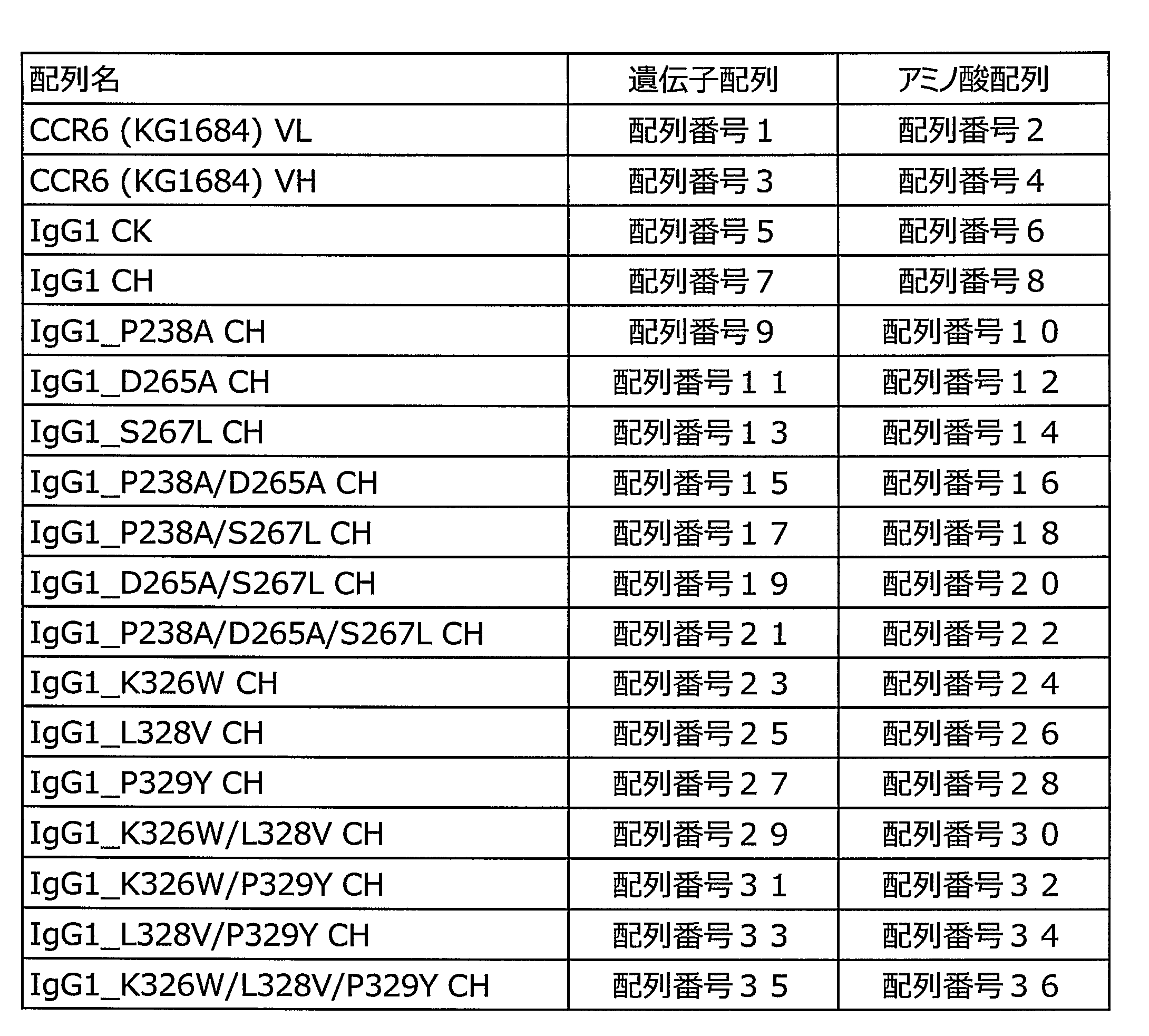
配列番号1、3、5、7に示す遺伝子配列からなる、ヒトIgG1抗ヒトCCR6抗体発現ベクターpCI-IgG1_KG1684を鋳型に用い、PrimeSTAR Max DNA Polymerase(タカラバイオ)及び改変部位を導入したプライマー(シグマオリゴ)を用い、PrimeSTAR Max DNA Polymeraseの添付文書に従いPCR反応を行った。
Using a human IgG1 anti-human CCR6 antibody expression vector pCI-IgG1_KG1684 consisting of the gene sequences shown in SEQ ID NOs: 1, 3, 5, and 7 as a template, PrimeSTAR Max Max DNA Polymerase (Takara Bio) and a primer (Sigma Oligo) into which a modified site was introduced ) Was performed according to the package insert of PrimeSTAR \ Max \ DNA \ Polymerase.
PCR反応はGeneAmp PCR System 9700(Applied Biosystems)を用い、98℃、1分間にて熱変性後、98℃にて10秒間、58℃にて5秒間、72℃にて5秒間の反応を30サイクル行った。0.8%アガロースゲルを用いた電気泳動に反応液を供し、QIAquick Gel Extraction Kit(Qiagen)を用いて、増幅断片を回収した。In-Fusion HD Cloning Kit(クロンテック)を用いてプラスミドpCI vector(プロメガ)と連結反応を行い、該反応液を用いて大腸菌DH5αコンピテントセル(タカラバイオ)を形質転換した。
The PCR reaction was performed using GeneAmp \ PCR \ System \ 9700 (Applied Biosystems) at 98 ° C for 1 minute, followed by 30 cycles of reaction at 98 ° C for 10 seconds, 58 ° C for 5 seconds, 58 ° C for 5 seconds and 72 ° C for 5 seconds. went. The reaction solution was subjected to electrophoresis using 0.8% agarose gel, and an amplified fragment was recovered using QIAquick \ Gel \ Extraction \ Kit (Qiagen). A ligation reaction was performed with the plasmid pCI vector (Promega) using In-Fusion HD Cloning Kit (Clontech), and Escherichia coli DH5α competent cells (Takara Bio) were transformed using the reaction solution.
得られた形質転換株のクローンより各プラスミドDNAを調製し、Big Dye Terminator Cycle Sequencing Kit v3.1(Applied Biosystems)を用いて添付の説明書に従って反応後、同社のDNAシーケンサーABI PRISM 3700 DNA Analyzerによりプラスミドに挿入されたDNAの塩基配列を解析した。
Each plasmid DNA was prepared from the clone of the obtained transformant, reacted with Big Dye Terminator Cycle Sequencing Kit v3.1 (Applied Biosystems) according to the attached instructions, and then reacted with the company's DNA sequencer ABI PRISM Al DNAnA using its DNA sequencer. The nucleotide sequence of the DNA inserted into the plasmid was analyzed.
(2)CD16a結合を「破壊」したヒトIgG1抗CCR6抗体の発現
以下の方法で、宿主細胞に(1)で作製された発現ベクターを導入した。宿主細胞には、FUT8ノックアウトCHO細胞(国際公開第2005/035586号、国際公開第02/31140号)使用した。プラスミド導入の方法は添付の説明書に従った。 (2) Expression of human IgG1 anti-CCR6 antibody that “disrupted” CD16a binding The expression vector prepared in (1) was introduced into host cells by the following method. FUT8 knockout CHO cells (WO 2005/035586, WO 02/31140) were used as host cells. The plasmid was introduced according to the attached instructions.
以下の方法で、宿主細胞に(1)で作製された発現ベクターを導入した。宿主細胞には、FUT8ノックアウトCHO細胞(国際公開第2005/035586号、国際公開第02/31140号)使用した。プラスミド導入の方法は添付の説明書に従った。 (2) Expression of human IgG1 anti-CCR6 antibody that “disrupted” CD16a binding The expression vector prepared in (1) was introduced into host cells by the following method. FUT8 knockout CHO cells (WO 2005/035586, WO 02/31140) were used as host cells. The plasmid was introduced according to the attached instructions.
培養液量は200mLで行い、37℃、5%CO2、125rpmの設定条件下で、5日間培養した。培養後、細胞懸濁液の遠心分離を行い、0.2μmフィルター(ThermoScientific)を通して改変抗体を含む培養上清を回収した。
The culture volume was 200 mL, and the cells were cultured at 37 ° C., 5% CO 2 , and 125 rpm for 5 days. After the culture, the cell suspension was centrifuged, and the culture supernatant containing the modified antibody was collected through a 0.2 μm filter (ThermoScientific).
各抗体の、精製した抗体サンプルの名称を表3に示す。
Table 3 shows the names of the purified antibody samples for each antibody.
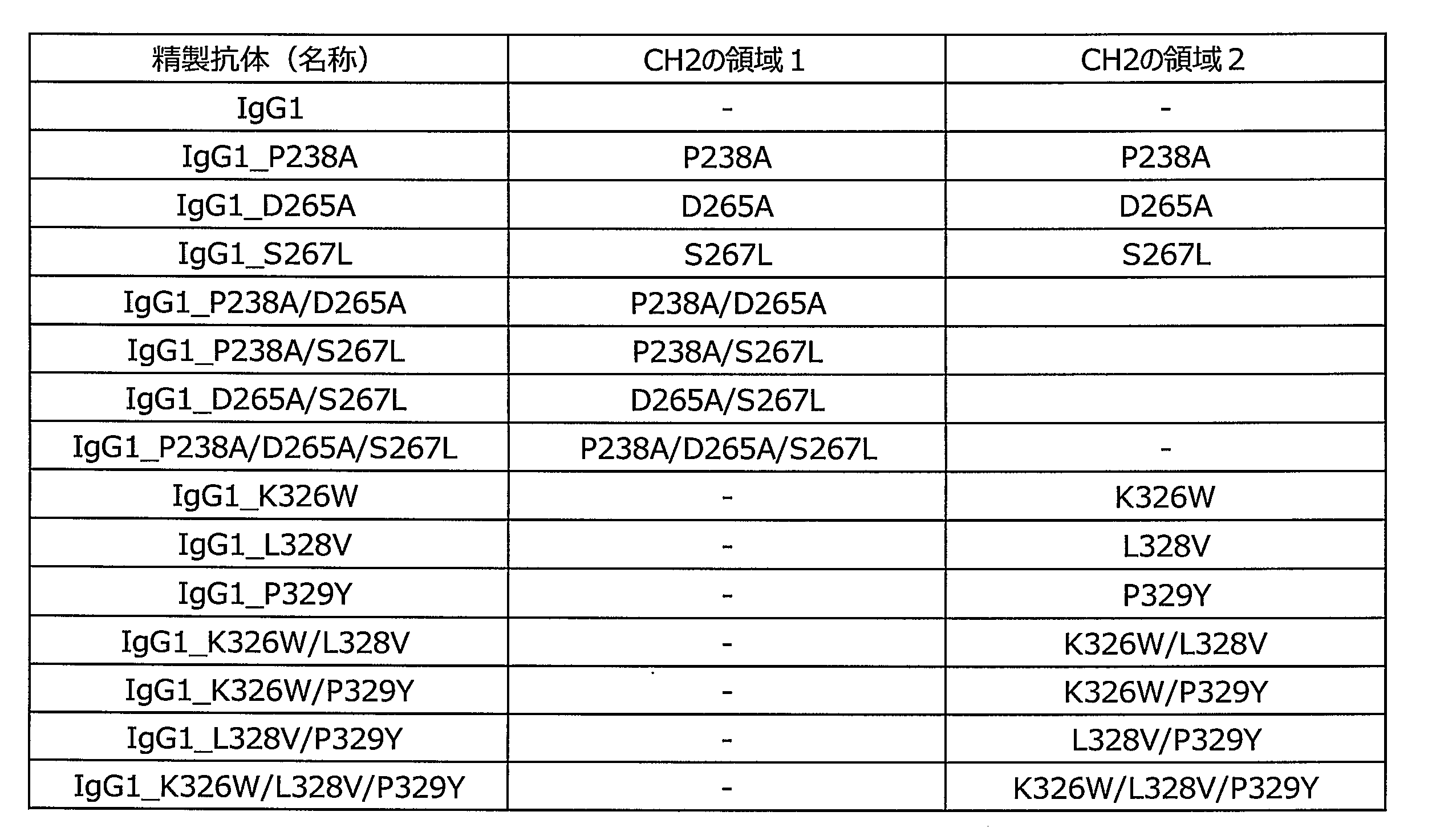
(3)CD16a結合を「破壊」したヒトIgG1抗CCR6抗体の精製
以下に示す、MabSelect SuRe(GEヘルスケア)を用いたアフィニティ精製により、改変抗体を精製した。レジンをPBSで平衡化した後、(2)で取得された培養上清をロードし、PBSで2回洗浄した。洗浄後、溶出バッファー(100mMクエン酸、pH3.5)を用いて抗体を溶出し、中和バッファー(2M Tris-HCl、pH8.0)を1/10量加えて中和した。 (3) Purification of human IgG1 anti-CCR6 antibody that “disrupted” CD16a binding The modified antibody was purified by affinity purification using MabSelect SuRe (GE Healthcare) shown below. After the resin was equilibrated with PBS, the culture supernatant obtained in (2) was loaded and washed twice with PBS. After washing, the antibody was eluted using an elution buffer (100 mM citric acid, pH 3.5), and neutralized by adding 1/10 volume of a neutralization buffer (2 M Tris-HCl, pH 8.0).
以下に示す、MabSelect SuRe(GEヘルスケア)を用いたアフィニティ精製により、改変抗体を精製した。レジンをPBSで平衡化した後、(2)で取得された培養上清をロードし、PBSで2回洗浄した。洗浄後、溶出バッファー(100mMクエン酸、pH3.5)を用いて抗体を溶出し、中和バッファー(2M Tris-HCl、pH8.0)を1/10量加えて中和した。 (3) Purification of human IgG1 anti-CCR6 antibody that “disrupted” CD16a binding The modified antibody was purified by affinity purification using MabSelect SuRe (GE Healthcare) shown below. After the resin was equilibrated with PBS, the culture supernatant obtained in (2) was loaded and washed twice with PBS. After washing, the antibody was eluted using an elution buffer (100 mM citric acid, pH 3.5), and neutralized by adding 1/10 volume of a neutralization buffer (2 M Tris-HCl, pH 8.0).
続いて、Amicon Ultra-4 Centerifugal Filter Units(ミリポア)を用いて限外濾過による濃縮及びバッファー(10mMクエン酸、150mM NaCl、pH6.0)置換を行い、Nanodrop8000(ThermoScientific)を使用して280nMにおける吸光度(A280)を測定し、抗体溶液の濃度測定及び調製を行った。
Subsequently, concentration by ultrafiltration and substitution of buffer (10 mM citric acid, 150 mM NaCl, pH 6.0) were performed using Amicon Ultra-4 Centrifugal Filter Units (Millipore), and absorbance at 280 nm using Nanodrop 8000 (Thermo Scientific). (A280) was measured, and the concentration and preparation of the antibody solution were measured.
(4)CD16a結合を「破壊」したヒトIgG1抗CCR6抗体のSDS-PAGEによる精製度の評価
各種CD16結合を欠損したヒトIgG1抗CCR6抗体精製サンプルの精製度を評価するため、抗体精製サンプル約1μgを用いて、公知の方法[Nature, 227 680 (1970)]に従ってSDS変性ポリアクリルアミド電気泳動(以下、SDS-PAGEと表記する)を行った。 (4) Evaluation of Purification by SDS-PAGE of Human IgG1 Anti-CCR6 Antibody with “Disrupted” CD16a Binding In order to evaluate the degree of purification of various purified human IgG1 anti-CCR6 antibodies lacking CD16 binding, about 1 μg of the antibody purified sample was evaluated. Was used to perform SDS-modified polyacrylamide electrophoresis (hereinafter, referred to as SDS-PAGE) according to a known method [Nature, 227 680 (1970)].
各種CD16結合を欠損したヒトIgG1抗CCR6抗体精製サンプルの精製度を評価するため、抗体精製サンプル約1μgを用いて、公知の方法[Nature, 227 680 (1970)]に従ってSDS変性ポリアクリルアミド電気泳動(以下、SDS-PAGEと表記する)を行った。 (4) Evaluation of Purification by SDS-PAGE of Human IgG1 Anti-CCR6 Antibody with “Disrupted” CD16a Binding In order to evaluate the degree of purification of various purified human IgG1 anti-CCR6 antibodies lacking CD16 binding, about 1 μg of the antibody purified sample was evaluated. Was used to perform SDS-modified polyacrylamide electrophoresis (hereinafter, referred to as SDS-PAGE) according to a known method [Nature, 227 680 (1970)].
その結果、還元条件下において、各種CD16結合を欠損したヒトIgG1抗CCR6抗体は、通常IgG1型と同様に、H鎖が約50キロダルトン(以下、kDaと表記する)、L鎖が約25kDa付近にバンドが認められた。また、非還元条件においては、約150kDa付近にバンドが認められたことから、作製した抗CCR6ドメイン交換抗体は目的のH鎖及びL鎖から構成されていることが確認された。
As a result, under the reducing conditions, human IgG1 anti-CCR6 antibody deficient in various CD16 bonds has an H chain of about 50 kDa (hereinafter referred to as kDa) and an L chain of about 25 kDa, as in the case of IgG1 type. A band was observed. In addition, under non-reducing conditions, a band was observed at about 150 kDa, confirming that the prepared anti-CCR6 domain exchange antibody was composed of the target H chain and L chain.
以上の結果より、本実施例の第3項で得られた各種CD16結合を欠損したヒトIgG1抗CCR6抗体の精製サンプル中には、それぞれH鎖及びL鎖から構成される目的のIgG分子が十分な割合で含まれることが確認された。
From the above results, in the purified sample of the human IgG1 anti-CCR6 antibody lacking various CD16 bindings obtained in the third section of the present example, the target IgG molecule composed of the H chain and the L chain was sufficient. It was confirmed that it was contained in a small proportion.
(5)CD16a結合領域を「破壊」したヒトIgG1抗CCR6抗体のADCC活性
エフェクター細胞としてNK細胞株であるNK-92(ATCC)にヒトCD16a (Val型)を遺伝子導入して安定的に発現させた、NK-92/CD16トランスフェクタントを、標的細胞としてヒトCCR6/CHOトランスフェクタントを用いた。培養していたそれぞれの細胞を回収・細胞をカウントし、それぞれ8x105cells/mL、2x105cells/mLになるようにRPMI培地(5%FBS/1%PSを添加したフェノールレッド不含のRPMI1640培地)を用いて調製した。 (5) ADCC activity of human IgG1 anti-CCR6 antibody with "disrupted" CD16a binding region Human CD16a (Val type) was transfected into NK cell line NK-92 (ATCC) as an effector cell to allow stable expression. In addition, NK-92 / CD16 transfectants were used and human CCR6 / CHO transfectants were used as target cells. Each cell was cultured by counting the recovery and cells, respectively 8x10 5 cells / mL, 2x10 5 cells / mL to become as RPMI medium (5% FBS / 1% phenol red-free RPMI1640 that PS was added Medium).
エフェクター細胞としてNK細胞株であるNK-92(ATCC)にヒトCD16a (Val型)を遺伝子導入して安定的に発現させた、NK-92/CD16トランスフェクタントを、標的細胞としてヒトCCR6/CHOトランスフェクタントを用いた。培養していたそれぞれの細胞を回収・細胞をカウントし、それぞれ8x105cells/mL、2x105cells/mLになるようにRPMI培地(5%FBS/1%PSを添加したフェノールレッド不含のRPMI1640培地)を用いて調製した。 (5) ADCC activity of human IgG1 anti-CCR6 antibody with "disrupted" CD16a binding region Human CD16a (Val type) was transfected into NK cell line NK-92 (ATCC) as an effector cell to allow stable expression. In addition, NK-92 / CD16 transfectants were used and human CCR6 / CHO transfectants were used as target cells. Each cell was cultured by counting the recovery and cells, respectively 8x10 5 cells / mL, 2x10 5 cells / mL to become as RPMI medium (5% FBS / 1% phenol red-free RPMI1640 that PS was added Medium).
96well plateに抗体溶液を50μL/wellずつ連続分注機で分注後、標的細胞を50μL/wellずつ分注した。エフェクター細胞を50μL/wellずつ分注し、1800rpmで2分間遠心分離し、細胞が均等に播種されていることを確認した。
After the antibody solution was dispensed into a 96-well plate at 50 μL / well by a continuous dispenser, target cells were dispensed at 50 μL / well. The effector cells were dispensed at 50 μL / well and centrifuged at 1800 rpm for 2 minutes to confirm that the cells were uniformly seeded.
CO2インキュベーターで37℃、3時間15分間静置後、Totalコントロールに10分の1量の可溶化溶液を分注し、37℃、45分間静置した。1800rpmで2分間遠心分離後、上清50μLをELISAプレートに分注した。基質(粉)を懸濁バッファー12mLに溶かし発色溶液を作製し、発色溶液を50μL/wellずつアプライし反応させた。停止溶液50μL/well加え、プレートリーダーにて吸光度(A450)を測定した。
After allowing to stand at 37 ° C. for 3 hours and 15 minutes in a CO 2 incubator, 1/10 of the solubilized solution was dispensed to a Total control and allowed to stand at 37 ° C. for 45 minutes. After centrifugation at 1800 rpm for 2 minutes, 50 μL of the supernatant was dispensed into an ELISA plate. The substrate (powder) was dissolved in 12 mL of the suspension buffer to prepare a color-developing solution, and the color-developing solution was applied at 50 μL / well and reacted. The stop solution was added at 50 μL / well, and the absorbance (A450) was measured with a plate reader.
なおADCC測定用キットとして、CytoTox96(R)Non-Radioactive Cytotoxicity Assay(プロメガ)を用いた。以下の式を用いてADCC活性(%)を算出した。
As an ADCC measurement kit, CytoTox96 (R) Non-Radioactive Cytotoxicity Assay (Promega) was used. ADCC activity (%) was calculated using the following equation.
ADCC活性(%)=100x(S-E-T)/(Max-T)
S=サンプル反応ウェル吸光度-培地ウェル吸光度
E=エフェクターウェル吸光度-培地ウェル吸光度
T=標的ウェル吸光度-培地ウェル吸光度
Max=100%反応ウェル-100%反応対照ウェル ADCC activity (%) = 100 × (SET) / (Max-T)
S = sample reaction well absorbance−medium well absorbance E = effector well absorbance−medium well absorbance T = target well absorbance−medium well absorbance Max = 100% reaction well−100% reaction control well
結果を図8に示す。図8に示されるように、CH2の領域1ではIgG1_D265A、IgG1_P238A/S267L、IgG1_D265A/S267Lからなる改変体が、標的細胞に対して顕著にADCC活性が低下することが確認された。
The results are shown in FIG. As shown in FIG. 8, in the CH2 region 1, it was confirmed that the variant comprising IgG1_D265A, IgG1_P238A / S267L, and IgG1_D265A / S267L significantly reduced ADCC activity with respect to target cells.
またCH2の領域2では、IgG1_P329Y、IgG1_K326W/P329Y、IgG1_L328V/P329Y、IgG1_K326W/L328V/P329Yからなる改変体が、顕著にADCC活性が低下することが確認され、いずれの改変体においてもP329Y改変が含まれることがわかった。
Further, in CH2 region 2, a variant consisting of IgG1_P329Y, IgG1_K326W / P329Y, IgG1_L328V / P329Y, and IgG1_K326W / L328V / P329Y was confirmed to have a remarkable decrease in ADCC activity, and any variant contains P329Y modification. I found out.
以上の結果より、CH2ドメイン上の「領域1」では、D265A又はP238A/S267Lのアミノ酸改変を導入することによって、CH2ドメイン上の「領域2」では少なくともP329Yのアミノ酸改変を導入することによって、CD16a結合部位を破壊できることが明らかとなった。
From the above results, CD16a was obtained by introducing an amino acid modification of D265A or P238A / S267L in “region 1” on the CH2 domain, and introducing an amino acid modification of at least P329Y in “region 2” on the CH2 domain. It was revealed that the binding site could be destroyed.
[実施例2]
CD16a結合非対称改変を導入したヒトIgG1抗CCR6一価抗体の作製
実施例1において、CD16aとFcの非対称の結合部位を破壊できるアミノ酸配列の一例として、CH2-Aでは「領域1」由来のD265A又はP238A/S267L、CH2-Bでは「領域2」由来のP329Yを選択した。 [Example 2]
Preparation of Human IgG1 Anti-CCR6 Monovalent Antibody Introducing CD16a Binding Asymmetric Modification In Example 1, as an example of an amino acid sequence capable of disrupting the asymmetric binding site between CD16a and Fc, CH2-A has D265A or “region 1” derived from “region 1”. For P238A / S267L and CH2-B, P329Y derived from “region 2” was selected.
CD16a結合非対称改変を導入したヒトIgG1抗CCR6一価抗体の作製
実施例1において、CD16aとFcの非対称の結合部位を破壊できるアミノ酸配列の一例として、CH2-Aでは「領域1」由来のD265A又はP238A/S267L、CH2-Bでは「領域2」由来のP329Yを選択した。 [Example 2]
Preparation of Human IgG1 Anti-CCR6 Monovalent Antibody Introducing CD16a Binding Asymmetric Modification In Example 1, as an example of an amino acid sequence capable of disrupting the asymmetric binding site between CD16a and Fc, CH2-A has D265A or “
本項では、CH2-AとCH2-Bのみにそれぞれ上記の改変を非対称に導入してADCC活性を評価するために、二分子のH鎖を個別の遺伝子にコードし発現させ、それらをヘテロ会合させることができる「一価抗体」(国際公開第2011/108502号)を基本骨格に用いることとした。図9にかかる一価抗体の一態様の模式図を示す。図9中、前記ヘテロ会合体は、「非対称改変体#1」である。
In this section, in order to evaluate the ADCC activity by introducing the above modifications asymmetrically into only CH2-A and CH2-B, respectively, two H chains are encoded and expressed in separate genes, and they are heteroassociated. A "monovalent antibody" (WO 2011/108502) that can be used is used as a basic skeleton. FIG. 10 shows a schematic diagram of one embodiment of the monovalent antibody according to FIG. In FIG. 9, the hetero-aggregate is “asymmetric variant # 1”.
(1)CD16a結合非対称改変一価抗体発現ベクターの作製
CH2-A(D265A又はP238A/S267L)、CH2-B(P329Y)を非対称あるいは対称に導入したヒトIgG1抗CCR6一価抗体(国際公開第2011/108502号)の発現ベクターを構築した。 (1) Preparation of CD16a-binding asymmetrically modified monovalent antibody expression vector A human IgG1 anti-CCR6 monovalent antibody into which CH2-A (D265A or P238A / S267L) or CH2-B (P329Y) has been introduced asymmetrically or symmetrically (International Publication No. 2011) No./108502).
CH2-A(D265A又はP238A/S267L)、CH2-B(P329Y)を非対称あるいは対称に導入したヒトIgG1抗CCR6一価抗体(国際公開第2011/108502号)の発現ベクターを構築した。 (1) Preparation of CD16a-binding asymmetrically modified monovalent antibody expression vector A human IgG1 anti-CCR6 monovalent antibody into which CH2-A (D265A or P238A / S267L) or CH2-B (P329Y) has been introduced asymmetrically or symmetrically (International Publication No. 2011) No./108502).
ヒトIgG1抗CCR6一価抗体発現ベクターpCI-mvG1_KG1684を鋳型に用い、PrimeSTAR Max DNA Polymerase(タカラバイオ)及び改変部位を導入したプライマー(シグマオリゴ)を用い、PrimeSTAR Max DNA Polymeraseの添付文書に従いPCR反応を行った。
Using the human IgG1 anti-CCR6 monovalent antibody expression vector pCI-mvG1_KG1684 as a template, PrimeSTAR Max DNA Polymerase (Takara Bio) and a primer (Sigma Oligomer) into which a modified site has been introduced, and perform a PCR reaction according to the attached document of PrimeSTAR Max Max DNA Polymerase. went.
PCR反応はGeneAmp PCR System 9700(Applied Biosystems)を用い、98℃、1分間にて熱変性後、98℃にて10秒間、58℃にて5秒間、72℃にて5秒間の反応を30サイクル行った。
The PCR reaction was performed using GeneAmp \ PCR \ System \ 9700 (Applied Biosystems) at 98 ° C for 1 minute, followed by 30 cycles of reaction at 98 ° C for 10 seconds, 58 ° C for 5 seconds, 58 ° C for 5 seconds and 72 ° C for 5 seconds. went.
0.8%アガロースゲルを用いた電気泳動に反応液を供し、QIAquick Gel Extraction Kit(Qiagen)を用いて、増幅断片を回収した。In-Fusion HD Cloning Kit(クロンテック)を用いてプラスミドpCI vector(プロメガ)と連結反応を行い、該反応液を用いて大腸菌DH5αコンピテントセル(タカラバイオ)を形質転換した。
The reaction solution was subjected to electrophoresis using a 0.8% agarose gel, and an amplified fragment was recovered using QIAquick \ Gel \ Extraction \ Kit (Qiagen). A ligation reaction was performed with the plasmid pCI vector (Promega) using In-Fusion HD Cloning Kit (Clontech), and Escherichia coli DH5α competent cells (Takara Bio) were transformed using the reaction solution.
得られた形質転換株のクローンより各プラスミドDNAを調製し、Big Dye Terminator Cycle Sequencing Kit v3.1(Applied Biosystems)を用いて添付の説明書に従って反応後、同社のDNAシーケンサーABI PRISM 3700 DNA Analyzerによりプラスミドに挿入されたDNAの塩基配列を解析した。
Each plasmid DNA was prepared from the clone of the obtained transformant, reacted with Big Dye Terminator Cycle Sequencing Kit v3.1 (Applied Biosystems) according to the attached instructions, and then reacted with the company's DNA sequencer ABI PRISM Al DNAnA using its DNA sequencer. The nucleotide sequence of the DNA inserted into the plasmid was analyzed.
(2)CD16a結合非対称改変一価抗体の発現
実施例1の第2項と同様の方法で、当該抗体の発現を行い、抗体を含む培養上清を回収した。各精製抗体サンプルとその改変部位を表4に示す。なお、改変部位におけるアミノ酸残基置換は実施例1と同様の方法により行った。 (2) Expression of CD16a-binding asymmetrically modified monovalent antibody The antibody was expressed in the same manner as initem 2 of Example 1, and the culture supernatant containing the antibody was collected. Table 4 shows the purified antibody samples and their modified sites. In addition, amino acid residue substitution at the modification site was performed in the same manner as in Example 1.
実施例1の第2項と同様の方法で、当該抗体の発現を行い、抗体を含む培養上清を回収した。各精製抗体サンプルとその改変部位を表4に示す。なお、改変部位におけるアミノ酸残基置換は実施例1と同様の方法により行った。 (2) Expression of CD16a-binding asymmetrically modified monovalent antibody The antibody was expressed in the same manner as in
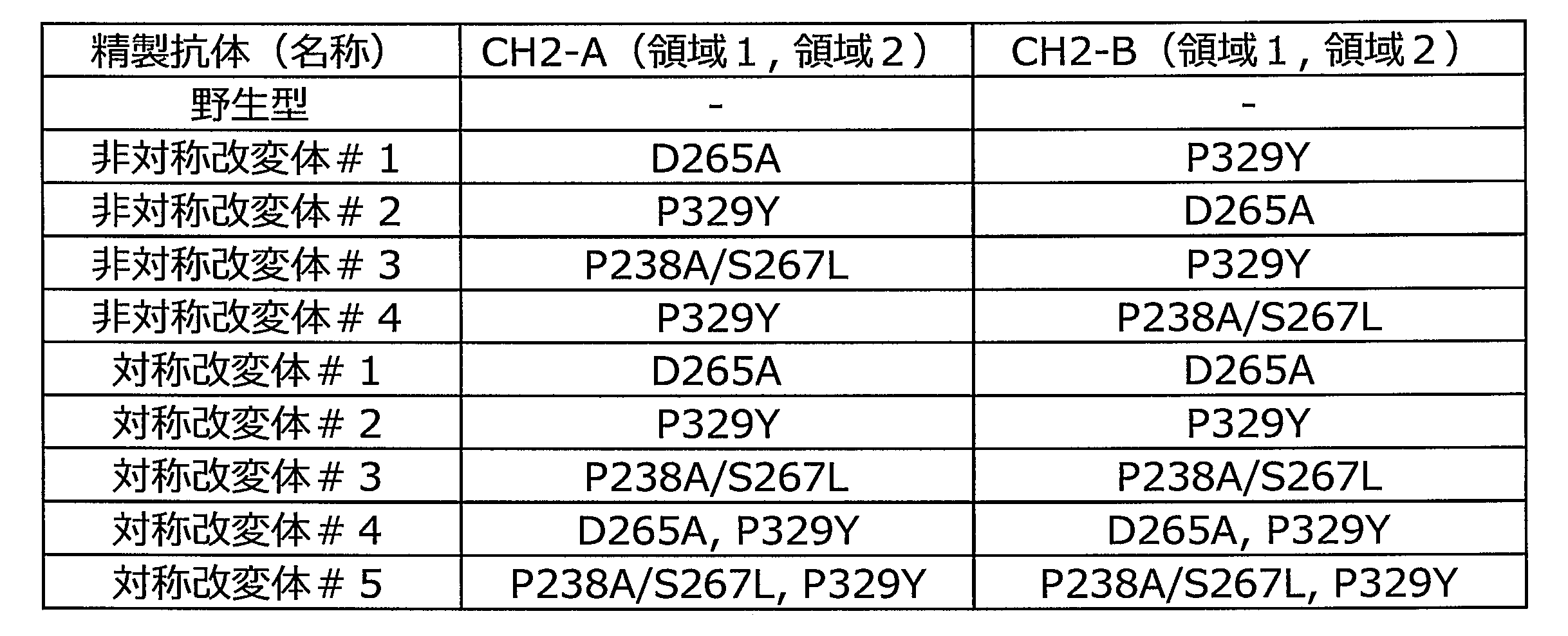
(3)CD16a結合非対称改変一価抗体の精製
実施例1の第3項と同様の方法で、当該抗体の精製を行った。溶出バッファーは100mMクエン酸、pH3.9を用いた。その後、AKTA FPLC(GEヘルスケア)及びSuperdex High-performance Column(GEヘルスケア)を用いて、該抗体溶液より単量体画分を分取した。AKTAシステム孔径0.22μmのメンブランフィルター(Millex-GV,ミリポア)で濾過滅菌することにより精製抗体を得た。Nanodrop8000(ThermoScientific)を使用して280nmにおける吸光度(A280)を測定した。 (3) Purification of CD16a-Binding Asymmetrically Modified Monovalent Antibody The antibody was purified in the same manner as in the third section of Example 1. The elution buffer used was 100 mM citric acid, pH 3.9. Thereafter, a monomer fraction was fractionated from the antibody solution using AKTA FPLC (GE Healthcare) and Superdex High-performance Column (GE Healthcare). The purified antibody was obtained by filtering and sterilizing with an AKTA system membrane filter (Millex-GV, Millipore) having a pore size of 0.22 μm. The absorbance at 280 nm (A280) was measured using Nanodrop 8000 (ThermoScientific).
実施例1の第3項と同様の方法で、当該抗体の精製を行った。溶出バッファーは100mMクエン酸、pH3.9を用いた。その後、AKTA FPLC(GEヘルスケア)及びSuperdex High-performance Column(GEヘルスケア)を用いて、該抗体溶液より単量体画分を分取した。AKTAシステム孔径0.22μmのメンブランフィルター(Millex-GV,ミリポア)で濾過滅菌することにより精製抗体を得た。Nanodrop8000(ThermoScientific)を使用して280nmにおける吸光度(A280)を測定した。 (3) Purification of CD16a-Binding Asymmetrically Modified Monovalent Antibody The antibody was purified in the same manner as in the third section of Example 1. The elution buffer used was 100 mM citric acid, pH 3.9. Thereafter, a monomer fraction was fractionated from the antibody solution using AKTA FPLC (GE Healthcare) and Superdex High-performance Column (GE Healthcare). The purified antibody was obtained by filtering and sterilizing with an AKTA system membrane filter (Millex-GV, Millipore) having a pore size of 0.22 μm. The absorbance at 280 nm (A280) was measured using Nanodrop 8000 (ThermoScientific).
(4)CD16a結合非対称改変を導入したヒトIgG1抗CCR6一価抗体のSDS-PAGEによる精製度の評価
各種CD16a結合非対称改変を導入したヒトIgG1抗CCR6一価抗体精製サンプルの精製度を評価するため、抗体精製サンプル約1μgを用いてSDS-PAGEを行った。 (4) Evaluation of Purification by SDS-PAGE of Human IgG1 Anti-CCR6 Monovalent Antibody Introduced with CD16a-Binding Asymmetric Modification To evaluate the purification degree of human IgG1 anti-CCR6 monovalent antibody-purified samples into which various CD16a-binding asymmetric modifications have been introduced SDS-PAGE was performed using about 1 μg of the purified antibody sample.
各種CD16a結合非対称改変を導入したヒトIgG1抗CCR6一価抗体精製サンプルの精製度を評価するため、抗体精製サンプル約1μgを用いてSDS-PAGEを行った。 (4) Evaluation of Purification by SDS-PAGE of Human IgG1 Anti-CCR6 Monovalent Antibody Introduced with CD16a-Binding Asymmetric Modification To evaluate the purification degree of human IgG1 anti-CCR6 monovalent antibody-purified samples into which various CD16a-binding asymmetric modifications have been introduced SDS-PAGE was performed using about 1 μg of the purified antibody sample.
その結果、還元条件下において、すべての改変体は、野生型一価抗体と同様に、H鎖及びFc融合L鎖が約50kDa付近にバンドが認められた。また、非還元条件においては、約100kDa付近にバンドが認められたことから、作製した非対称改変体及び対称改変体は目的のH鎖及びL鎖から構成されていることが確認された。
As a result, under the reducing conditions, all the variants showed bands in the vicinity of about 50 kDa for the H chain and the Fc-fused L chain, as in the case of the wild-type monovalent antibody. In addition, under non-reducing conditions, a band was observed at about 100 kDa, confirming that the prepared asymmetrically modified product and the symmetrically modified product were composed of the target H chain and L chain.
以上の結果より、本実施例の第3項で得られた各種CD16a結合非対称改変を導入したヒトIgG1抗CCR6一価抗体の精製サンプル中には、それぞれH鎖及びFc融合L鎖から構成される目的の一価抗体分子が十分な割合で含まれることが確認された。
From the above results, the purified sample of human IgG1 anti-CCR6 monovalent antibody into which various CD16a binding asymmetrical modifications obtained in the third section of the present example were introduced was composed of an H chain and an Fc fusion L chain, respectively. It was confirmed that the desired monovalent antibody molecule was contained in a sufficient ratio.
(5)CD16a結合非対称改変一価抗体のADCC活性
実施例1の第5項と同様の方法で、ADCC活性を測定した。結果を図10示す。図10に示されるように、CD16a結合の非対称改変を導入した一価抗体(非対称改変体#1~4)は、抗体濃度依存的に標的細胞を傷害することが確認された。一方、CD16a結合の対称改変を導入した一価抗体(対称改変体#1~#5)は、標的細胞に対してADCC活性を示さないことが確認された。 (5) ADCC activity of CD16a-binding asymmetrically modified monovalent antibody ADCC activity was measured in the same manner as in the fifth section of Example 1. The results are shown in FIG. As shown in FIG. 10, it was confirmed that monovalent antibodies (asymmetric variants # 1 to # 4) into which asymmetric modification of CD16a binding was introduced damage target cells in an antibody concentration-dependent manner. On the other hand, it was confirmed that the monovalent antibodies into which the symmetric modification of CD16a binding was introduced (symmetric variants # 1 to # 5) did not show ADCC activity on target cells.
実施例1の第5項と同様の方法で、ADCC活性を測定した。結果を図10示す。図10に示されるように、CD16a結合の非対称改変を導入した一価抗体(非対称改変体#1~4)は、抗体濃度依存的に標的細胞を傷害することが確認された。一方、CD16a結合の対称改変を導入した一価抗体(対称改変体#1~#5)は、標的細胞に対してADCC活性を示さないことが確認された。 (5) ADCC activity of CD16a-binding asymmetrically modified monovalent antibody ADCC activity was measured in the same manner as in the fifth section of Example 1. The results are shown in FIG. As shown in FIG. 10, it was confirmed that monovalent antibodies (
以上の結果より、CD16a結合の非対称改変を導入した一価抗体はヒトCD16aに対する結合性が維持され、標的細胞に対してADCC活性を発揮することできることが確認された。
From the above results, it was confirmed that the monovalent antibody into which the asymmetric modification of CD16a binding was introduced maintains the binding property to human CD16a and can exert ADCC activity on target cells.
一方で両方のCH2ドメインに同じ改変を導入した場合はADCC活性が失われることから、同じ改変CH2を有する抗体が細胞表面上でホモフィリックな会合をした場合にはADCC活性が誘導されないことが確認され、本発明が目指す二つの抗原に細胞表面上でヘテロ会合して初めてADCC活性を発揮するHL抗体を構成するための改変CH2ドメインが見出された。
On the other hand, since the ADCC activity is lost when the same modification is introduced into both CH2 domains, it is confirmed that ADCC activity is not induced when the antibody having the same modified CH2 forms a homophilic association on the cell surface. As a result, a modified CH2 domain for constructing an HL antibody exhibiting ADCC activity only after hetero-association on the cell surface with two antigens aimed at by the present invention has been found.
[実施例3]
本実施例では、実施例1,2で得られたヘテロ会合して初めてADCC活性を誘導できる改変CH2を、異なる抗原に対する二種類の抗体の半量体(HL体)に搭載し、二種類の抗原を共発現する標的細胞選択的にADCC活性が誘導できるか否かを検証した。 [Example 3]
In this example, the modified CH2 capable of inducing ADCC activity for the first time after hetero-association obtained in Examples 1 and 2 was mounted on a half-mer (HL form) of two kinds of antibodies against different antigens, It was verified whether ADCC activity can be selectively induced in target cells that co-express.
CH3ドメイン同士の相互作用はJ Immunol 2011; 187: 3238-3246で報告されている。ヒトIgG1のCH3ドメインの相互作用(KD値)は3.0×10⁻⁹M、ヒトIgG4のCH3ドメインのKD値は4.8x10⁻⁸Mで、ヒトIgG4のCH3ドメインの相互作用はIgG1よりも約6~7倍弱い。
Interaction between CH3 domains has been reported in J Immunol 2011; 187: 3238-3246. Interaction of the CH3 domain of human IgG1 (K D value) 3.0 × 10 -9 M, K D value of the CH3 domain of human IgG4 is 4.8 × 10 -8 M, the interaction of the CH3 domain of human IgG4 is It is about 6-7 times weaker than IgG1.
この性質とヒンジ領域におけるH鎖間のジスルフィド結合を切断するアミノ酸改変(C226A/C229A:AA)を利用することで抗体の半量体の作製を試みた。半量体のH鎖定常領域はヒトIgG1を基本骨格とし、CH3ドメインのみをヒトIgG4配列に交換し、さらにヒンジ領域にアミノ酸改変(AA)を加えたドメイン交換抗体(以下IgG1114_AA型と称する)を用いた。
(4) An attempt was made to produce a half-mer of an antibody by utilizing this property and an amino acid modification (C226A / C229A: AA) that cleaves a disulfide bond between H chains in the hinge region. The H-chain constant region of the H-mer is a domain exchange antibody (hereinafter referred to as IgG1114_AA type) in which human IgG1 is used as a basic skeleton, only the CH3 domain is replaced with a human IgG4 sequence, and an amino acid modification (AA) is added to the hinge region. Was.
加えて、ADCC活性を強化するために公知のADCC活性増強アミノ酸改変をCH2ドメインに施し、さらにAsn297に結合するN-結合型糖鎖のα1,6フコースを除去、最後にCD16a結合非対称アミノ酸改変を導入した。図11に、IgG1114_AA_AAA_D265A(/P329Y)型のIgG半量体の模式図を示す。二種類のモデル抗原として、CD4とCD70を用いた。
In addition, a known ADCC activity-enhancing amino acid modification is applied to the CH2 domain to enhance ADCC activity, and further, α1,6 fucose of the N-linked sugar chain binding to Asn297 is removed. Finally, a CD16a-binding asymmetric amino acid modification is performed. Introduced. FIG. 11 shows a schematic diagram of an IgG1114_AA_AAA_D265A (/ P329Y) IgG halfmer. CD4 and CD70 were used as two kinds of model antigens.
上記に示す改変を組み合わせた、抗CD4抗体(J. Immunol. 1992; 149:1779-1787)及び抗CD70抗体(国際公開第2007/03637号)を以下の手順に従い作製した。IgG1114_AA型に公知のADCC活性増強アミノ酸改変(S298A/E333A/K334A:AAA)改変、CD16a結合非対称アミノ酸改変(D265AあるいはP329Y)を導入したアミノ酸配列から構成される重鎖定常領域を有する抗CD4及びCD70抗体をIgG1114_AA_AAA_D265A(/P329Y)型抗CD4半量体、IgG1114_AA_AAA_D265A(/P329Y)型抗CD70半量体とそれぞれ称する。
抗 An anti-CD4 antibody (J. mmImmunol. 1992; 149: 1779-1787) and an anti-CD70 antibody (International Publication No. WO 2007/03637) prepared by combining the above modifications were prepared according to the following procedures. Anti-CD4 and CD70 having a heavy chain constant region comprising an amino acid sequence in which a known ADCC activity-enhancing amino acid modification (S298A / E333A / K334A: AAA) modification and a CD16a-binding asymmetric amino acid modification (D265A or P329Y) have been introduced into IgG1114_AA type. The antibodies are referred to as IgG1114_AA_AAA_D265A (/ P329Y) type anti-CD4 halfmer and IgG1114_AA_AAA_D265A (/ P329Y) type anti-CD70 halfmer, respectively.
設計された各種抗CD4及びCD70半量体の各ドメインが由来するサブクラス、及び重鎖定常領域のアミノ酸配列の対応を表5に示す。また、用いたCD4及びCD70に対する抗体の遺伝子配列とアミノ酸配列を表6に示す。
Table 5 shows the correspondence between the subclasses from which the respective domains of the designed anti-CD4 and CD70 hemimers are derived, and the amino acid sequences of the heavy chain constant regions. Table 6 shows the gene sequences and amino acid sequences of the antibodies against CD4 and CD70 used.
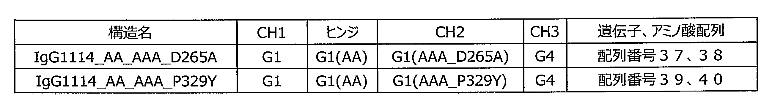

(1)抗CD4半量体及び抗CD70半量体発現ベクターの作製
ヒトIgG1抗CD4抗体発現ベクターpCI-IgG1_CD4(ibalizumab)(配列番号5、7、41、43)あるいは、ヒトIgG1抗CD70抗体発現ベクターpCI-IgG1_CD70(2H5)(配列番号5、7、45、47)より、制限酵素NheI及びNotIを用いて、約9kbpのDNA断片を切り出し精製した。 (1) Preparation of anti-CD4 hemimeric and anti-CD70 hemimeric expression vectors Human IgG1 anti-CD4 antibody expression vector pCI-IgG1_CD4 (ibalizumab) (SEQ ID NOs: 5, 7, 41, 43) or human IgG1 anti-CD70 antibody expression vector pCI A DNA fragment of about 9 kbp was excised and purified from IgG1_CD70 (2H5) (SEQ ID NOS: 5, 7, 45, 47) using restriction enzymes NheI and NotI.
ヒトIgG1抗CD4抗体発現ベクターpCI-IgG1_CD4(ibalizumab)(配列番号5、7、41、43)あるいは、ヒトIgG1抗CD70抗体発現ベクターpCI-IgG1_CD70(2H5)(配列番号5、7、45、47)より、制限酵素NheI及びNotIを用いて、約9kbpのDNA断片を切り出し精製した。 (1) Preparation of anti-CD4 hemimeric and anti-CD70 hemimeric expression vectors Human IgG1 anti-CD4 antibody expression vector pCI-IgG1_CD4 (ibalizumab) (SEQ ID NOs: 5, 7, 41, 43) or human IgG1 anti-CD70 antibody expression vector pCI A DNA fragment of about 9 kbp was excised and purified from IgG1_CD70 (2H5) (SEQ ID NOS: 5, 7, 45, 47) using restriction enzymes NheI and NotI.
精製したDNA断片と、ヒトIgG1抗体のCH1ドメイン、ヒンジドメイン(C226A/C229A改変を加えたもの)、CH2ドメイン(S298A/K333A/E334A改変、D265AあるいはP329Y改変を加えたもの)、ヒトIgG4抗体のCH3ドメインからなる人工合成遺伝子とをIn-Fusion HD Cloning Kit(クロンテック)を用いて連結反応を行い、該反応液を用いて大腸菌DH5αコンピテントセル(タカラバイオ)を形質転換した。得られた形質転換株のクローンより各プラスミドDNAを調製し、ファスマックでDNAの塩基配列を解析した。
Purified DNA fragment, human IgG1 antibody CH1 domain, hinge domain (C226A / C229A modified), CH2 domain (S298A / K333A / E334A modified, D265A or P329Y modified), human IgG4 antibody A ligation reaction was performed with an artificially synthesized gene comprising a CH3 domain using In-Fusion HD Cloning Kit (Clontech), and Escherichia coli DH5α competent cells (Takara Bio) were transformed using the reaction solution. Each plasmid DNA was prepared from the clone of the obtained transformant, and the nucleotide sequence of the DNA was analyzed by using Fassmac.
(2)各種抗CD4IgG1抗体及び抗CD70IgG1抗体並びにこれら抗体の半量体の発現
実施例1の第2項と同様の方法で、当該抗体の発現を行い、抗体を含む培養上清を回収した。 (2) Expression of various anti-CD4 IgG1 antibodies and anti-CD70 IgG1 antibodies and half-mers of these antibodies Expression of the antibodies was carried out in the same manner as in the second section of Example 1, and the culture supernatant containing the antibodies was collected.
実施例1の第2項と同様の方法で、当該抗体の発現を行い、抗体を含む培養上清を回収した。 (2) Expression of various anti-CD4 IgG1 antibodies and anti-CD70 IgG1 antibodies and half-mers of these antibodies Expression of the antibodies was carried out in the same manner as in the second section of Example 1, and the culture supernatant containing the antibodies was collected.
精製した抗体サンプルの名称を表7に示す。
Table 7 shows the names of the purified antibody samples.
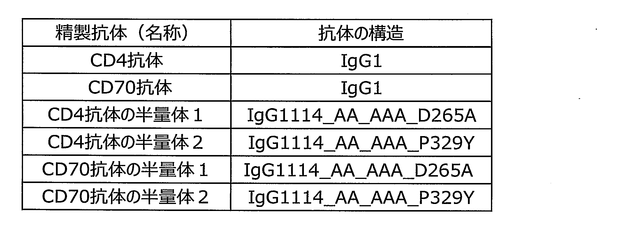
(3)抗CD4半量体及び抗CD70半量体の精製
実施例1の第3項と同様の方法で、当該抗体の精製を行い、抗体溶液の濃度測定及び調製を行った。 (3) Purification of anti-CD4 half-mer and anti-CD70 half-mer The antibody was purified in the same manner as in the third section of Example 1, and the concentration and preparation of the antibody solution were measured.
実施例1の第3項と同様の方法で、当該抗体の精製を行い、抗体溶液の濃度測定及び調製を行った。 (3) Purification of anti-CD4 half-mer and anti-CD70 half-mer The antibody was purified in the same manner as in the third section of Example 1, and the concentration and preparation of the antibody solution were measured.
(4)抗CD4半量体及び抗CD70半量体のSDS-PAGEによる精製度の評価
調製した各種抗体サンプルの精製度を評価するため、抗体精製サンプル約1μgを用いて、SDS-PAGEを行った。結果を図12に示す。 (4) Evaluation of Purity of Anti-CD4 Hemimer and Anti-CD70 Hemimer by SDS-PAGE In order to evaluate the degree of purification of the prepared various antibody samples, SDS-PAGE was performed using about 1 μg of the purified antibody sample. The result is shown in FIG.
調製した各種抗体サンプルの精製度を評価するため、抗体精製サンプル約1μgを用いて、SDS-PAGEを行った。結果を図12に示す。 (4) Evaluation of Purity of Anti-CD4 Hemimer and Anti-CD70 Hemimer by SDS-PAGE In order to evaluate the degree of purification of the prepared various antibody samples, SDS-PAGE was performed using about 1 μg of the purified antibody sample. The result is shown in FIG.
図12に示すように、精製した抗体はすべて、還元条件下でH鎖が約50kDa、L鎖が約25kDa付近にバンドが認められた。また、非還元条件では、抗CD4 IgG1抗体及び抗CD70 IgG1抗体は約150kDa、抗CD4半量体及び抗CD70半量体は約75kDa付近にバンドが認められたことから、作製したCD4及びCD70 IgG1抗体は目的のH鎖及びL鎖から構成された抗体構造であること、抗CD4半量体及び抗CD70半量体は目的のH鎖及びL鎖から構成され、半量体を形成しやすい抗体構造になっていることが確認された。
バ ン ド As shown in FIG. 12, all the purified antibodies showed bands at about 50 kDa for the H chain and about 25 kDa for the L chain under reducing conditions. Under non-reducing conditions, the anti-CD4 IgG1 antibody and the anti-CD70 IgG1 antibody had bands around 150 kDa, and the anti-CD4 half-mer and the anti-CD70 half-mer had bands around 75 kDa. Thus, the prepared CD4 and CD70 IgG1 antibodies were Antibody structure composed of the target H chain and L chain, anti-CD4 half-mer and anti-CD70 half-mer are composed of the target H chain and L chain, and have an antibody structure that easily forms a half-mer. It was confirmed that.
以上の結果より、本実施例の第3項で得られた抗CD4半量体及び抗CD70半量体精製サンプル中には、目的の抗体分子が十分な割合で含まれることが確認された。
From the above results, it was confirmed that the target antibody molecules were contained in a sufficient ratio in the purified anti-CD4 half-mer and anti-CD70 half-mer obtained in the third section of this example.
(5)抗CD4 IgG1抗体及び抗CD70 IgG1、並びにこれら抗体の半量体のCD4/CD70両陽性細胞、CD4及びCD70単陽性細胞に対するADCC活性
実施例1の第5項と同様の方法で、ADCC活性を測定した。標的細胞は、CD4単陽性細胞としてCD4/EL-4トランスフェクタント、CD70単陽性細胞としてMT-1、CD4/CD70両陽性細胞としてTL-Om1を用いた。これらの細胞におけるCD4及びCD70の発現量を、フローサイトメーターを用いて測定した結果を図13示す。 (5) ADCC activity of anti-CD4 IgG1 antibody and anti-CD70 IgG1, and half-mers of these antibodies on both CD4 / CD70-positive cells, CD4 and CD70 single-positive cells ADCC activity in the same manner as initem 5 of Example 1 Was measured. As target cells, CD4 / EL-4 transfectants were used as CD4 single positive cells, MT-1 was used as CD70 single positive cells, and TL-Om1 was used as both CD4 / CD70 positive cells. FIG. 13 shows the results of measuring the expression levels of CD4 and CD70 in these cells using a flow cytometer.
実施例1の第5項と同様の方法で、ADCC活性を測定した。標的細胞は、CD4単陽性細胞としてCD4/EL-4トランスフェクタント、CD70単陽性細胞としてMT-1、CD4/CD70両陽性細胞としてTL-Om1を用いた。これらの細胞におけるCD4及びCD70の発現量を、フローサイトメーターを用いて測定した結果を図13示す。 (5) ADCC activity of anti-CD4 IgG1 antibody and anti-CD70 IgG1, and half-mers of these antibodies on both CD4 / CD70-positive cells, CD4 and CD70 single-positive cells ADCC activity in the same manner as in
図13に示されるように、目的の抗原が細胞に発現していることが確認された。また、ADCC活性評価の結果を図14に示す。ポジティブコントロール抗体として、通常IgG1型抗CD4及びCD70抗体を用いた。
As shown in FIG. 13, it was confirmed that the target antigen was expressed in the cells. FIG. 14 shows the results of ADCC activity evaluation. Normally, IgG1-type anti-CD4 and CD70 antibodies were used as positive control antibodies.
図14に示すように、予想通り、CD4/EL-4トランスフェクタントに対しては抗CD4抗体が、MT-1に対しては抗CD70抗体が、TL-Om1に対しては抗CD4抗体及び抗CD70抗体がADCC活性を発揮することが示された。
As shown in FIG. 14, anti-CD4 antibody against CD4 / EL-4 transfectant, anti-CD70 antibody against MT-1, and anti-CD4 antibody against TL-Om1, as expected. And that the anti-CD70 antibody exerts ADCC activity.
一方、抗CD4抗体の半量体1と抗CD70抗体の半量体2を混合した抗体溶液、あるいは抗CD4抗体の半量体2と抗CD70抗体の半量体1を混合した抗体溶液は、CD4及びCD70単陽性細胞に対してはADCC活性を示さず、CD4/CD70両陽性細胞に対してのみ特異的にADCC活性を発揮することが示された。
On the other hand, an antibody solution in which anti-CD4 antibody half-mer 1 and anti-CD70 antibody half-mer 2 are mixed, or an antibody solution in which anti-CD4 antibody half-mer 2 and anti-CD70 antibody half-mer 1 are mixed are CD4 and CD70 monomers. It was shown that ADCC activity was not exhibited for positive cells, and ADCC activity was specifically exhibited only for both CD4 / CD70 positive cells.
[実施例4]
第1のCD16a結合領域で、EUインデックスで表される235位、239位、265位、267位、269位、296位、298位、299位、327位を、第2のCD16a結合領域で、EUインデックスで表される326位、328位、329位、330位をそれぞれ置換することによりCD16a結合を破壊した改変CH2を、異なる抗原に対する二種類の抗体の半量体(HL体)に搭載し、二種類の抗原を共発現する標的細胞選択的にADCC活性が誘導できるか否かを検証した。 [Example 4]
In the first CD16a binding region, positions 235, 239, 265, 267, 269, 296, 298, 299, and 327 represented by the EU index are represented by the second CD16a binding region, The modified CH2 in which the CD16a bond was destroyed by substituting thepositions 326, 328, 329, and 330 represented by the EU index, respectively, was mounted on a half-mer (HL form) of two kinds of antibodies against different antigens, It was verified whether ADCC activity can be selectively induced in target cells that co-express two types of antigens.
第1のCD16a結合領域で、EUインデックスで表される235位、239位、265位、267位、269位、296位、298位、299位、327位を、第2のCD16a結合領域で、EUインデックスで表される326位、328位、329位、330位をそれぞれ置換することによりCD16a結合を破壊した改変CH2を、異なる抗原に対する二種類の抗体の半量体(HL体)に搭載し、二種類の抗原を共発現する標的細胞選択的にADCC活性が誘導できるか否かを検証した。 [Example 4]
In the first CD16a binding region, positions 235, 239, 265, 267, 269, 296, 298, 299, and 327 represented by the EU index are represented by the second CD16a binding region, The modified CH2 in which the CD16a bond was destroyed by substituting the
設計された各種抗CD4半量体及びCD70半量体は、実施例3と同様の方法で調製し、ADCC活性を評価した。抗CD4半量体と抗CD70半量体を混合した抗体溶液は1μg/mLとなるように添加した。同じく、ポジティブコントロール抗体として使用した、通常IgG1型抗CD4抗体及び抗CD70抗体も1μg/mLとなるように添加した。設計された各種抗CD4半量体及びCD70半量体の各ドメインが由来するサブクラス、及び重鎖定常領域のアミノ酸配列の対応を表8に示す。
各種 The designed anti-CD4 half-mer and CD70 half-mer were prepared in the same manner as in Example 3, and the ADCC activity was evaluated. The antibody solution obtained by mixing the anti-CD4 half-mer and the anti-CD70 half-mer was added so as to be 1 μg / mL. Similarly, a normal IgG1-type anti-CD4 antibody and an anti-CD70 antibody, which were used as positive control antibodies, were also added at 1 μg / mL. Table 8 shows the correspondence between the designed subclasses of the respective domains of the anti-CD4 half-mer and CD70 half-mer, and the amino acid sequences of the heavy chain constant regions.

図15A~図15Hに示されるように、第1のCD16a結合領域を改変したCH2を搭載した抗CD4半量体と、第2のCD16a結合領域を改変したCH2を搭載した抗CD70半量体を混合した抗体溶液は、CD4及びCD70単陽性細胞に対してはADCC活性を示さず、CD4/CD70両陽性細胞に対してのみ特異的にADCC活性を発揮することが示された。
As shown in FIG. 15A to FIG. 15H, an anti-CD4 halfmer carrying CH2 modified with the first CD16a binding region and an anti-CD70 halfmer carrying CH2 modified with the second CD16a binding region were mixed. It was shown that the antibody solution did not show ADCC activity on CD4 and CD70 single positive cells, but exhibited ADCC activity specifically on only CD4 / CD70 positive cells.
本発明を特定の態様を用いて詳細に説明したが、本発明の意図と範囲を離れることなく様々な変更および変形が可能であることは、当業者にとって明らかである。なお、本出願は、2018年9月28日付けで出願された日本特許出願(特願2018-185367)に基づいており、その全体が引用により援用される。
Although the present invention has been described in detail with reference to specific embodiments, it will be apparent to those skilled in the art that various modifications and variations can be made without departing from the spirit and scope of the invention. This application is based on a Japanese patent application filed on Sep. 28, 2018 (Japanese Patent Application No. 2018-185367), which is incorporated by reference in its entirety.
配列番号1:ヒトCCR6(KG1684)VLの塩基配列
配列番号2:ヒトCCR6(KG1684)VLのアミノ酸配列
配列番号3:ヒトCCR6(KG1684)VHの塩基配列
配列番号4:ヒトCCR6(KG1684)VHのアミノ酸配列
配列番号5:ヒトIgG1 CKの塩基配列
配列番号6:ヒトIgG1 CKのアミノ酸配列
配列番号7:ヒトIgG1 CHの塩基配列
配列番号8:ヒトIgG1 CHのアミノ酸配列
配列番号9:ヒトIgG1_P238A CHの塩基配列
配列番号10:ヒトIgG1_P238A CHのアミノ酸配列
配列番号11:ヒトIgG1_D265A CHの塩基配列
配列番号12:ヒトIgG1_D265A CHのアミノ酸配列
配列番号13:ヒトIgG1_S267L CHの塩基配列
配列番号14:ヒトIgG1_S267L CHのアミノ酸配列
配列番号15:ヒトIgG1_P238A/D265A CHの塩基配列
配列番号16:ヒトIgG1_P238A/D265A CHのアミノ酸配列
配列番号17:ヒトIgG1_P238A/S267L CHの塩基配列
配列番号18:ヒトIgG1_P238A/S267L CHのアミノ酸配列
配列番号19:ヒトIgG1_D265A/S267L CHの塩基配列
配列番号20:ヒトIgG1_D265A/S267L CHのアミノ酸配列
配列番号21:ヒトIgG1_P238A/D265A/S267L CHの塩基配列
配列番号22:ヒトIgG1_P238A/D265A/S267L CHのアミノ酸配列
配列番号23:ヒトIgG1_K326W CHの塩基配列
配列番号24:ヒトIgG1_K326W CHのアミノ酸配列
配列番号25:ヒトIgG1_L328V CHの塩基配列
配列番号26:ヒトIgG1_L328V CHのアミノ酸配列
配列番号27:ヒトIgG1_P329Y CHの塩基配列
配列番号28:ヒトIgG1_P329Y CHのアミノ酸配列
配列番号29:ヒトIgG1_K326W/L328V CHの塩基配列
配列番号30:ヒトIgG1_K326W/L328V CHのアミノ酸配列
配列番号31:ヒトIgG1_K326W/P329Y CHの塩基配列
配列番号32:ヒトIgG1_K326W/P329Y CHのアミノ酸配列
配列番号33:ヒトIgG1_L328V/P329Y CHの塩基配列
配列番号34:ヒトIgG1_L328V/P329Y CHのアミノ酸配列
配列番号35:ヒトIgG1_K326W/L328V/P329Y CHの塩基配列
配列番号36:ヒトIgG1_K326W/L328V/P329Y CHのアミノ酸配列
配列番号37:ヒトIgG1114_AA_AAA_D265A CHの塩基配列
配列番号38:ヒトIgG1114_AA_AAA_D265A CHのアミノ酸配列
配列番号39:ヒトIgG1114_AA_AAA_P329Y CHの塩基配列
配列番号40:ヒトIgG1114_AA_AAA_P329Y CHのアミノ酸配列
配列番号41:ヒトCD4(ibalizumab)VLの塩基配列
配列番号42:ヒトCD4(ibalizumab)VLのアミノ酸配列
配列番号43:ヒトCD4(ibalizumab)VHの塩基配列
配列番号44:ヒトCD4(ibalizumab)VHのアミノ酸配列
配列番号45:ヒトCD70(2H5)VLの塩基配列
配列番号46:ヒトCD70(2H5)VLのアミノ酸配列
配列番号47:ヒトCD70(2H5)VHの塩基配列
配列番号48:ヒトCD70(2H5)VHのアミノ酸配列
配列番号49:ヒトIgG1114_AA_AAA_S239R CHの塩基配列
配列番号50:ヒトIgG1114_AA_AAA_S239R CHのアミノ酸配列
配列番号51:ヒトIgG1114_AA_AAA_D265N CHの塩基配列
配列番号52:ヒトIgG1114_AA_AAA_D265N CHのアミノ酸配列
配列番号53:ヒトIgG1114_AA_AAA_D265E CHの塩基配列
配列番号54:ヒトIgG1114_AA_AAA_D265E CHのアミノ酸配列
配列番号55:ヒトIgG1114_AA_AAA_S267K CHの塩基配列
配列番号56:ヒトIgG1114_AA_AAA_S267K CHのアミノ酸配列
配列番号57:ヒトIgG1114_AA_AAA_E269P CHの塩基配列
配列番号58:ヒトIgG1114_AA_AAA_E269P CHのアミノ酸配列
配列番号59:ヒトIgG1114_AA_AAA_Y296P CHの塩基配列
配列番号60:ヒトIgG1114_AA_AAA_Y296P CHのアミノ酸配列
配列番号61:ヒトIgG1114_AA_AAA_S298E CHの塩基配列
配列番号62:ヒトIgG1114_AA_AAA_S298E CHのアミノ酸配列
配列番号63:ヒトIgG1114_AA_AAA_T299A CHの塩基配列
配列番号64:ヒトIgG1114_AA_AAA_T299A CHのアミノ酸配列
配列番号65:ヒトIgG1114_AA_AAA_L235R CHの塩基配列
配列番号66:ヒトIgG1114_AA_AAA_L235R CHのアミノ酸配列
配列番号67:ヒトIgG1114_AA_AAA_A327I CHの塩基配列
配列番号68:ヒトIgG1114_AA_AAA_A327I CHのアミノ酸配列
配列番号69:ヒトIgG1114_AA_AAA_K326G CHの塩基配列
配列番号70:ヒトIgG1114_AA_AAA_K326G CHのアミノ酸配列
配列番号71:ヒトIgG1114_AA_AAA_L328R CHの塩基配列
配列番号72:ヒトIgG1114_AA_AAA_L328R CHのアミノ酸配列
配列番号73:ヒトIgG1114_AA_AAA_P329K CHの塩基配列
配列番号74:ヒトIgG1114_AA_AAA_P329K CHのアミノ酸配列
配列番号75:ヒトIgG1114_AA_AAA_P329W CHの塩基配列
配列番号76:ヒトIgG1114_AA_AAA_P329W CHのアミノ酸配列
配列番号77:ヒトIgG1114_AA_AAA_A330P CHの塩基配列
配列番号78:ヒトIgG1114_AA_AAA_A330P CHのアミノ酸配列 SEQ ID NO: 1: nucleotide sequence of human CCR6 (KG1684) VL SEQ ID NO 2: amino acid sequence of human CCR6 (KG1684) VL SEQ ID NO 3: nucleotide sequence of human CCR6 (KG1684) VH SEQ ID NO 4: human CCR6 (KG1684) VH Amino acid sequence SEQ ID NO: 5: nucleotide sequence of human IgG1 CK SEQ ID NO: 6: amino acid sequence of human IgG1 CK SEQ ID NO: 7: nucleotide sequence of human IgG1 CH SEQ ID NO: 8: amino acid sequence of human IgG1 CH SEQ ID NO: 9: human IgG1_P238A CH Nucleotide sequence SEQ ID NO: 10: amino acid sequence of human IgG1_P238A CH SEQ ID NO: 11: nucleotide sequence of human IgG1_D265A CH SEQ ID NO: 12: amino acid sequence of human IgG1_D265A CH SEQ ID NO: 13: nucleotide sequence of human IgG1_S267L CH : Amino acid sequence of human IgG1_S267L CH SEQ ID NO: 15: Nucleotide sequence of human IgG1_P238A / D265A CH SEQ ID NO: 16: Amino acid sequence of human IgG1_P238A / D265A CH SEQ ID NO: 17: Nucleotide sequence of human IgG1_P238A / S267L CH: SEQ ID NO: 18: Human IgG1 S267L CH amino acid sequence SEQ ID NO: 19: human IgG1_D265A / S267L CH nucleotide sequence SEQ ID NO: 20: human IgG1_D265A / S267L CH amino acid sequence SEQ ID NO: 21: human IgG1_P238A / D265A / S267L CH nucleotide sequence SEQ ID NO: 22: human IgG1P Amino acid sequence of D265A / S267L CH SEQ ID NO: 23: Nucleotide sequence of human IgG1_K326W CH SEQ ID NO: 2 : Amino acid sequence of human IgG1_K326W CH SEQ ID NO: 25: nucleotide sequence of human IgG1_L328V CH SEQ ID NO: 26: amino acid sequence of human IgG1_L328V CH SEQ ID NO: 27: nucleotide sequence of human IgG1_P329Y CH SEQ ID NO: 28: amino acid sequence of human IgG1_P329Y CH : Human IgG1_K326W / P328Y CH amino acid sequence SEQ ID NO: 30: human IgG1_K326W / L328V CH amino acid sequence SEQ ID NO: 31: human IgG1_K326W / P329Y CH nucleotide sequence SEQ ID NO: 32: human IgG1_K326W / P329Y CH amino acid sequence: 33 Nucleotide sequence of IgG1_L328V / P329Y CH SEQ ID NO: 34: Amino acid sequence of human IgG1_L328V / P329Y CH SEQ ID NO: 35: Nucleotide sequence of human IgG1_K326W / L328V / P329Y CH SEQ ID NO: 36: Amino acid sequence of human IgG1_K326W / L328V / P329Y CH SEQ ID NO: 37: Nucleotide sequence of human IgG1114_AA_AAA_D265A CH SEQ ID NO: 38: Human IgG1A_A_A_A_A 39: base sequence of human IgG1114_AA_AAA_P329Y CH SEQ ID NO: 40: amino acid sequence of human IgG1114_AA_AAA_P329Y CH SEQ ID NO: 41: base sequence of human CD4 (ibalizumab) VL SEQ ID NO: 42: human CD4 (ibalizumab) VL amino acid sequence of human CD4: 43 (Ibalizumab) VH nucleotide sequence SEQ ID NO: 44: human CD (Ibalizumab) VH amino acid sequence SEQ ID NO: 45: human CD70 (2H5) VL nucleotide sequence SEQ ID NO: 46: human CD70 (2H5) VL amino acid sequence SEQ ID NO: 47: human CD70 (2H5) VH nucleotide sequence SEQ ID NO: 48: Amino acid sequence of human CD70 (2H5) VH SEQ ID NO: 49: Base sequence of human IgG1114_AA_AAA_S239R CH SEQ ID NO: 50: Amino acid sequence of human IgG1114_AA_AAA_S239R CH SEQ ID NO: 51: Base sequence of human IgG1114_AA_AAA_D265N CH No. 53: base sequence of human IgG1114_AA_AAA_D265E CH SEQ ID NO. 54: human IgG1114_AA_AA Amino acid sequence of A_D265E CH SEQ ID NO: 55: Nucleotide sequence of human IgG1114_AA_AAA_S267K CH SEQ ID NO: 56: Amino acid sequence of human IgG1114_AA_AAA_S267K CH SEQ ID NO: 57: Nucleotide sequence of human IgG1114_AA_AAA_E269P CH; Nucleotide sequence of IgG1114_AA_AAA_Y296P CH SEQ ID NO: 60: Amino acid sequence of human IgG1114_AA_AAA_Y296P CH SEQ ID NO: 61: Nucleotide sequence of human IgG1114_AA_AAA_S298E CH SEQ ID NO: 62: Human IgG1114_AA_AAA_AAA_AAA_A98A3 SEQ ID NO: 64: amino acid sequence of human IgG1114_AA_AAA_T299A CH SEQ ID NO: 65: Nucleotide sequence of human IgG1114_AA_AAA_L235R CH SEQ ID NO: 66: Human IgG1114_AA_AAA_L235R CH amino acid sequence of amino acid sequence of amino acid sequence of human IgG1A_A235G Amino acid sequence of IgG1114_AA_AAA_A327I CH SEQ ID NO: 69: Nucleotide sequence of human IgG1114_AA_AAA_K326G CH SEQ ID NO: 70: Amino acid sequence of human IgG1114_AA_AAA_K326G CH SEQ ID NO: 71: Human sequence of human IgG1114_AAA72G3 Amino acid sequence of AA_AAA_L328R CH SEQ ID NO: 73: Nucleotide sequence of human IgG1114_AA_AAA_P329K CH SEQ ID NO: 74: Amino acid sequence of human IgG1114_AA_AAA_P329K CH SEQ ID NO: 75: Human IgG1114_AA_AAA_P329ACHAA sequence Nucleotide sequence of IgG1114_AA_AAA_A330P CH SEQ ID NO: 78: Amino acid sequence of human IgG1114_AA_AAA_A330P CH
Claims (30)
- 第1のIgG半量体と第2のIgG半量体とからなる、互いに異なる第1の抗原及び第2の抗原に対する抗体組成物であって、
第1のIgG半量体及び第2のIgG半量体は、それぞれ、1つのイムノグロブリン軽鎖(以下、L鎖と略記する)及び1つのイムノグロブリン重鎖(以下、H鎖と略記する)からなり、H鎖はH鎖可変領域、一部または全体の置換もしくは欠失、または修飾によりジスルフィド結合が形成されないように改変されたヒンジドメイン及びCH1~CH3ドメインを含み、CH2ドメインにおいて互いに異なる第1のFcγ受容体IIIA(以下、CD16a)結合領域及び第2のCD16a結合領域のいずれかに改変を有し、
第1のIgG半量体は、第1の抗原に結合する抗原結合ドメインを含み、且つ第1のCD16a結合領域におけるCD16a結合活性が改変により減弱しており、
第2のIgG半量体は、第2の抗原に結合する抗原結合ドメインを含み、且つ第2のCD16a結合領域におけるCD16a結合活性が改変により減弱しており、
第1のIgG半量体と第2のIgG半量体との間において、H鎖間におけるジスルフィド結合が形成されない、
抗体組成物。 An antibody composition comprising a first IgG half and a second IgG half, which are different from each other, against a first antigen and a second antigen,
Each of the first IgG half and the second IgG half is composed of one immunoglobulin light chain (hereinafter abbreviated as L chain) and one immunoglobulin heavy chain (hereinafter abbreviated as H chain). The heavy chain comprises a heavy chain variable region, a hinge domain and a CH1-CH3 domain that are modified so that a disulfide bond is not formed by partial or total substitution or deletion, or modification, and first chains that differ from each other in a CH2 domain. Having an alteration in any of the Fcγ receptor IIIA (hereinafter, CD16a) binding region and the second CD16a binding region,
The first IgG half-mer contains an antigen-binding domain that binds to a first antigen, and has a CD16a-binding activity in the first CD16a-binding region that is reduced by modification,
The second IgG half-mer contains an antigen-binding domain that binds to a second antigen, and has reduced CD16a-binding activity in the second CD16a-binding region by modification,
No disulfide bond between H chains is formed between the first IgG half and the second IgG half.
Antibody composition. - 第1の抗原及び第2の抗原を共発現する標的細胞にのみエフェクター機能を発揮し傷害する、請求項1に記載の抗体組成物。 The antibody composition according to claim 1, wherein the antibody composition exerts an effector function only on target cells that co-express the first antigen and the second antigen and damages them.
- 第1のCD16a結合領域及び第2のCD16a結合領域における改変がアミノ酸の置換、欠失もしくは付加、または修飾である請求項1または2に記載の抗体組成物。 The antibody composition according to claim 1 or 2, wherein the alteration in the first CD16a binding region and the second CD16a binding region is amino acid substitution, deletion or addition, or modification.
- 第1のCD16a結合領域が、EUインデックスで表される235位、236位、237位、238位、239位、265位、266位、267位、268位、269位、294位、295位、296位、297位、298位、299位、301位、325位、327位、332位のアミノ酸残基から選ばれる少なくとも1を含む請求項1~3のいずれか1項に記載の抗体組成物。 The first CD16a binding region is represented by the EU index at positions 235, 236, 237, 238, 239, 265, 266, 267, 268, 269, 294, 295, The antibody composition according to any one of claims 1 to 3, comprising at least one selected from the amino acid residues at positions 296, 297, 298, 299, 301, 325, 327, and 332. .
- 第1のCD16a結合領域における改変が、EUインデックスで表される235位、236位、237位、238位、239位、265位、266位、267位、268位、269位、294位、295位、296位、297位、298位、299位、301位、325位、327位、332位のアミノ酸残基から選ばれる少なくとも1のアミノ酸残基の置換である請求項4に記載の抗体組成物。 Modifications in the first CD16a binding region are indicated by EU index at positions 235, 236, 237, 238, 239, 265, 266, 267, 268, 269, 294, 295 The antibody composition according to claim 4, wherein the antibody composition is a substitution of at least one amino acid residue selected from the amino acid residues at positions 296, 297, 298, 299, 301, 325, 327, and 332. Stuff.
- 第1のCD16a結合領域における改変が、EUインデックスで表される235位、238位、239位、265位、267位、269位、296位、298位、299位、327位のアミノ酸残基から選ばれる少なくとも1のアミノ酸残基の置換である請求項5に記載の抗体組成物。 The alteration in the first CD16a binding region is defined by the amino acid residues at positions 235, 238, 239, 265, 267, 269, 296, 298, 299, and 327 represented by the EU index. The antibody composition according to claim 5, wherein the substitution is at least one selected amino acid residue.
- 第2のCD16a結合領域が、EUインデックスで表される235位、236位、237位、326位、327位、328位、329位、330位のアミノ酸残基から選ばれる少なくとも1を含む請求項1~6のいずれか1項に記載の抗体組成物。 The second CD16a binding region comprises at least one selected from the amino acid residues at positions 235, 236, 237, 326, 327, 328, 329 and 330 represented by the EU index. 7. The antibody composition according to any one of 1 to 6.
- 第2のCD16a結合領域における改変が、EUインデックスで表される235位、236位、237位、326位、327位、328位、329位、330位のアミノ酸残基から選ばれる少なくとも1から選ばれる少なくとも1のアミノ酸残基の置換である請求項7に記載の抗体組成物。 The modification in the second CD16a binding region is selected from at least one selected from the amino acid residues at positions 235, 236, 237, 326, 327, 328, 329, and 330 represented by the EU index. The antibody composition according to claim 7, wherein the substitution is at least one amino acid residue.
- 第2のCD16a結合領域が、EUインデックスで表される326位、328位、329位、330位のアミノ酸残基から選ばれる少なくとも1を含む請求項8に記載の抗体組成物。 The antibody composition according to claim 8, wherein the second CD16a binding region contains at least one selected from the amino acid residues at positions 326, 328, 329, and 330 represented by the EU index.
- 第1のCD16a結合領域における改変が、EUインデックスで表される235位、238位、239位、265位、267位、269位、296位、298位、299位、327位のアミノ酸残基から選ばれる少なくとも1のアミノ酸残基の置換であり、第2のCD16a結合領域における改変が、EUインデックスで表される326位、328位、329位、330位のアミノ酸残基から選ばれる少なくとも1のアミノ酸残基の置換である請求項1~9のいずれか1項に記載の抗体組成物。 The alteration in the first CD16a binding region is defined by the amino acid residues at positions 235, 238, 239, 265, 267, 269, 296, 298, 299, and 327 represented by the EU index. At least one amino acid residue selected from the amino acid residues at positions 326, 328, 329, and 330 represented by the EU index. The antibody composition according to any one of claims 1 to 9, which is a substitution of an amino acid residue.
- 第1のCD16a結合領域における改変が、EUインデックスで表される238位、265位、267位のアミノ酸残基から選ばれる少なくとも1のアミノ酸残基の置換である請求項1~10のいずれか1項に記載の抗体組成物。 The modification according to any one of claims 1 to 10, wherein the alteration in the first CD16a binding region is substitution of at least one amino acid residue selected from the amino acid residues at positions 238, 265 and 267 represented by the EU index. Item 14. The antibody composition according to Item.
- 第2のCD16a結合領域における改変が、EUインデックスで表される326位、328位、329位のアミノ酸残基から選ばれる少なくとも1のアミノ酸残基の置換である請求項1~11のいずれか1項に記載の抗体組成物。 12. The method according to claim 1, wherein the modification in the second CD16a binding region is substitution of at least one amino acid residue selected from the amino acid residues at positions 326, 328, and 329 represented by the EU index. Item 14. The antibody composition according to Item.
- 第1のIgG半量体及び第2のIgG半量体が、EUインデックスで表される226位及び229位の少なくとも一方のアミノ酸残基が置換されたヒンジドメインを含む請求項1~12のいずれか1項に記載の抗体組成物。 The first IgG half-mer and the second IgG half-mer comprise a hinge domain in which at least one amino acid residue at positions 226 and 229 represented by the EU index has been substituted. Item 14. The antibody composition according to Item.
- 第1のIgG半量体及び第2のIgG半量体における、L鎖、H鎖のH鎖可変領域及びCH1ドメイン及びCH2ドメインのイムノグロブリンサブクラスがIgG1である、請求項1~13のいずれか1項に記載の抗体組成物。 14. The immunoglobulin subclass of the L chain, the H chain variable region and the CH1 domain and the CH2 domain in the first IgG half and the second IgG half, respectively, is IgG1. 2. The antibody composition according to item 1.
- 第1のIgG半量体及び第2のIgG半量体における、H鎖のCH3ドメインが、IgG1サブクラスのCH3ドメインよりもCH3ドメイン間相互作用が弱い、請求項14に記載の抗体組成物。 抗体 The antibody composition according to claim 14, wherein the CH3 domain of the heavy chain in the first IgG half and the second IgG half has weaker interaction between CH3 domains than the CH3 domain of the IgG1 subclass.
- 第1のIgG半量体及び第2のIgG半量体における、H鎖のCH3ドメインのイムノグロブリンサブクラスがIgG4である、請求項15に記載の抗体組成物。 The antibody composition according to claim 15, wherein the immunoglobulin subclass of the CH3 domain of the H chain in the first IgG half and the second IgG half is IgG4.
- 第1のIgG半量体における第2のCD16a結合領域、及び第2のIgG半量体における第1のCD16a結合領域を介してCD16aに結合する請求項1~16のいずれか1項に記載の抗体組成物。 The antibody composition according to any one of claims 1 to 16, wherein the antibody binds to CD16a via the second CD16a binding region in the first IgG half-mer and the first CD16a binding region in the second IgG half-mer. Stuff.
- 第1のIgG半量体及び第2のIgG半量体における、Fc領域に結合する全N-グリコシド結合型糖鎖のうち、糖鎖還元末端のN-アセチルグルコサミンにフコースが結合していない糖鎖の割合が20%以上である、請求項1~17のいずれか1項に記載の抗体組成物。 Of the total N-glycoside-linked sugar chains that bind to the Fc region in the first IgG half and the second IgG half, the sugar chains in which fucose is not linked to N-acetylglucosamine at the reducing end of the sugar chain The antibody composition according to any one of claims 1 to 17, wherein the ratio is 20% or more.
- 第1のIgG半量体及び第2のIgG半量体が、CH2ドメインにおいて、さらにCD16a結合活性を増強させる少なくとも1のアミノ酸残基置換を含む請求項1~18のいずれか1項に記載の抗体組成物。 19. The antibody composition according to any one of claims 1 to 18, wherein the first IgG half and the second IgG half have at least one amino acid residue substitution in the CH2 domain that further enhances CD16a binding activity. Stuff.
- 第1のIgG半量体及び第2のIgG半量体が、CH2ドメインにおいて、EUインデックスで表されるS298A、E333A、K334Aから選ばれる少なくとも1のアミノ酸残基置換を含む請求項19に記載の抗体組成物。 20. The antibody composition according to claim 19, wherein the first IgG half-mer and the second IgG half-mer include at least one amino acid residue substitution selected from S298A, E333A, and K334A represented by the EU index in the CH2 domain. Stuff.
- 第2のIgG半量体と併用する第1のIgG半量体であって、第1のIgG半量体及び第2のIgG半量体は、それぞれ、1つのL鎖及び1つのH鎖からなり、H鎖はH鎖可変領域、一部または全体の置換もしくは欠失、または修飾によりジスルフィド結合が形成されないように改変されたヒンジドメイン及びCH1~CH3ドメインを含み、CH2ドメインにおいて互いに異なる第1のCD16a結合領域及び第2のCD16a結合領域のいずれかに改変を有し、
第1のIgG半量体は、第1の抗原に結合する抗原結合ドメインを含み、且つ第1のCD16a結合領域におけるCD16a結合活性が改変により減弱しており、
第2のIgG半量体は、第1の抗原とは異なる第2の抗原に結合する抗原結合ドメインを含み、且つ第2のCD16a結合領域におけるCD16a結合活性が改変により減弱しており、
第1のIgG半量体と第2のIgG半量体との間において、H鎖間におけるジスルフィド結合が形成されない、第1のIgG半量体。 A first IgG half-mer for use in combination with a second IgG half-mer, wherein the first IgG half-mer and the second IgG half-mer consist of one L chain and one H chain, respectively, Is a first CD16a binding region comprising a heavy chain variable region, a hinge domain and a CH1 to CH3 domain which are modified so that a disulfide bond is not formed by partial or total substitution or deletion, or modification, and which are different from each other in a CH2 domain. And any of the second CD16a binding region has a modification,
The first IgG half-mer contains an antigen-binding domain that binds to a first antigen, and has a CD16a-binding activity in the first CD16a-binding region that is reduced by modification,
The second IgG half-mer contains an antigen-binding domain that binds to a second antigen different from the first antigen, and the CD16a-binding activity in the second CD16a-binding region is attenuated by modification,
The first IgG half, wherein no disulfide bond between H chains is formed between the first IgG half and the second IgG half. - 第1のIgG半量体と第2のIgG半量体とからなり、且つ互いに異なる第1の抗原及び第2の抗原に対する抗体組成物の製造に用いるための、第1のIgG半量体であって、
第1のIgG半量体及び第2のIgG半量体は、それぞれ、1つのL鎖及び1つのH鎖からなり、H鎖はH鎖可変領域、一部または全体の置換もしくは欠失、または修飾によりジスルフィド結合が形成されないように改変されたヒンジドメイン及びCH1~CH3ドメインを含み、CH2ドメインにおいて互いに異なる第1のCD16a結合領域及び第2のCD16a結合領域のいずれかに改変を有し、
第1のIgG半量体は、第1の抗原に結合する抗原結合ドメインを含み、且つ第1のCD16a結合領域におけるCD16a結合活性が改変により減弱しており、
第2のIgG半量体は、第2の抗原に結合する抗原結合ドメインを含み、且つ第2のCD16a結合領域におけるCD16a結合活性が改変により減弱しており、
第1のIgG半量体と第2のIgG半量体との間において、H鎖間におけるジスルフィド結合が形成されない、
第1のIgG半量体。 A first IgG half, comprising a first IgG half and a second IgG half, for use in producing an antibody composition against a first antigen and a second antigen that are different from each other,
Each of the first IgG half and the second IgG half is composed of one L chain and one H chain, and the H chain is obtained by substituting or deleting, or modifying, a heavy chain variable region, part or whole. A hinge domain and a CH1 to CH3 domain that are modified so that a disulfide bond is not formed, wherein the CH2 domain has a modification in any of the first CD16a binding region and the second CD16a binding region that are different from each other;
The first IgG half-mer contains an antigen-binding domain that binds to a first antigen, and has a CD16a-binding activity in the first CD16a-binding region that is reduced by modification,
The second IgG half-mer contains an antigen-binding domain that binds to a second antigen, and has reduced CD16a-binding activity in the second CD16a-binding region by modification,
No disulfide bond between H chains is formed between the first IgG half and the second IgG half.
First IgG half. - 第1のIgG半量体と併用する第2のIgG半量体であって、第1のIgG半量体及び第2のIgG半量体は、それぞれ、1つのL鎖及び1つのH鎖からなり、H鎖はH鎖可変領域、一部または全体の置換もしくは欠失、または修飾によりジスルフィド結合が形成されないように改変されたヒンジドメイン及びCH1~CH3ドメインを含み、CH2ドメインにおいて互いに異なる第1のCD16a結合領域及び第2のCD16a結合領域のいずれかに改変を有し、
第1のIgG半量体は、第1の抗原に結合する抗原結合ドメインを含み、且つ第1のCD16a結合領域におけるCD16a結合活性が改変により減弱しており、
第2のIgG半量体は、第1の抗原と異なる第2の抗原に結合する抗原結合ドメインを含み、且つ第2のCD16a結合領域におけるCD16a結合活性が改変により減弱しており、
第1のIgG半量体と第2のIgG半量体との間において、H鎖間におけるジスルフィド結合が形成されない、第2のIgG半量体。 A second IgG half-mer used in combination with the first IgG half-mer, wherein the first IgG half-mer and the second IgG half-mer consist of one L chain and one H chain, respectively, Is a first CD16a binding region comprising a heavy chain variable region, a hinge domain and a CH1 to CH3 domain which are modified so that a disulfide bond is not formed by partial or total substitution or deletion, or modification, and which are different from each other in a CH2 domain. And any of the second CD16a binding region has a modification,
The first IgG half-mer contains an antigen-binding domain that binds to a first antigen, and has a CD16a-binding activity in the first CD16a-binding region that is reduced by modification,
The second IgG half-mer contains an antigen-binding domain that binds to a second antigen different from the first antigen, and the CD16a-binding activity in the second CD16a-binding region is attenuated by modification,
A second IgG half, wherein no disulfide bond between H chains is formed between the first IgG half and the second IgG half. - 第1のIgG半量体と第2のIgG半量体とからなり、且つ互いに異なる第1の抗原及び第2の抗原に対する抗体組成物の製造に用いるための、第2のIgG半量体であって、
第1のIgG半量体及び第2のIgG半量体は、それぞれ、1つのL鎖及び1つのH鎖からなり、H鎖はH鎖可変領域、一部または全体の置換もしくは欠失、または修飾によりジスルフィド結合が形成されないように改変されたヒンジドメイン及びCH1~CH3ドメインを含み、CH2ドメインにおいて互いに異なる第1のCD16a結合領域及び第2のCD16a結合領域のいずれかに改変を有し、
第1のIgG半量体は、第1の抗原に結合する抗原結合ドメインを含み、且つ第1のCD16a結合領域におけるCD16a結合活性が改変により減弱しており、
第2のIgG半量体は、第2の抗原に結合する抗原結合ドメインを含み、且つ第2のCD16a結合領域におけるCD16a結合活性が改変により減弱しており、
第1のIgG半量体と第2のIgG半量体との間において、H鎖間におけるジスルフィド結合が形成されない、
第2のIgG半量体。 A second IgG half, comprising a first IgG half and a second IgG half, for use in producing an antibody composition against a first antigen and a second antigen that are different from each other,
Each of the first IgG half and the second IgG half is composed of one L chain and one H chain, and the H chain is obtained by substituting or deleting, or modifying, a heavy chain variable region, part or whole. A hinge domain and a CH1 to CH3 domain that are modified so that a disulfide bond is not formed, wherein the CH2 domain has a modification in any of the first CD16a binding region and the second CD16a binding region that are different from each other;
The first IgG half-mer contains an antigen-binding domain that binds to a first antigen, and has a CD16a-binding activity in the first CD16a-binding region that is reduced by modification,
The second IgG half-mer contains an antigen-binding domain that binds to a second antigen, and has reduced CD16a-binding activity in the second CD16a-binding region by modification,
No disulfide bond between H chains is formed between the first IgG half and the second IgG half.
Second IgG half. - 以下のa)またはb)のDNA。
a)請求項21または22に記載の第1のIgG半量体のアミノ酸配列をコードするDNA
b)請求項23または24に記載の第2のIgG半量体のアミノ酸配列をコードするDNA DNA of the following a) or b).
a) DNA encoding the amino acid sequence of the first IgG half-mer according to claim 21 or 22.
b) DNA encoding the amino acid sequence of the second IgG half-mer according to claim 23 or 24. - 請求項25に記載のa)及びb)の少なくとも一方のDNAを含む組換えベクター。 A recombinant vector comprising at least one of the DNAs according to claim 25.
- 請求項26に記載の組換えベクターが導入された形質転換体。 A transformant into which the recombinant vector according to claim 26 has been introduced.
- 請求項27に記載の形質転換体を培地に培養し、培養物中に請求項21または22に記載の第1のIgG半量体及び請求項23または24に記載の第2のIgG半量体の少なくとも一方を蓄積させ、培養物から第1のIgG半量体及び第2のIgG半量体の少なくとも一方を採取する工程を含む抗体組成物の製造方法。 The transformant according to claim 27 is cultured in a medium, and at least the first IgG halfmer according to claim 21 or 22 and the second IgG halfmer according to claim 23 or 24 are cultured in the culture. A method for producing an antibody composition, comprising the step of accumulating one and collecting at least one of a first IgG half-mer and a second IgG half-mer from a culture.
- 第1のIgG半量体及び第2のIgG半量体を含むキットであって、
第1のIgG半量体及び第2のIgG半量体は、それぞれ、1つのL鎖及び1つのH鎖からなり、H鎖はH鎖可変領域、一部または全体の置換もしくは欠失、または修飾によりジスルフィド結合が形成されないように改変されたヒンジドメイン及びCH1~CH3ドメインを含み、CH2ドメインにおいて互いに異なる第1のCD16a結合領域及び第2のCD16a結合領域のいずれかに改変を有し、
第1のIgG半量体は、第1の抗原に結合する抗原結合ドメインを含み、且つ第1のCD16a結合領域におけるCD16a結合活性が改変により減弱しており、
第2のIgG半量体は、第2の抗原に結合する抗原結合ドメインを含み、且つ第2のCD16a結合領域におけるCD16a結合活性が改変により減弱しており、
第1のIgG半量体と第2のIgG半量体との間において、H鎖間におけるジスルフィド結合が形成されない、
キット。 A kit comprising a first IgG half and a second IgG half,
Each of the first IgG half and the second IgG half is composed of one L chain and one H chain, and the H chain is obtained by substituting or deleting, or modifying, a heavy chain variable region, part or whole. A hinge domain and a CH1 to CH3 domain that are modified so that a disulfide bond is not formed, wherein the CH2 domain has a modification in any of the first CD16a binding region and the second CD16a binding region that are different from each other;
The first IgG half-mer contains an antigen-binding domain that binds to a first antigen, and has a CD16a-binding activity in the first CD16a-binding region that is reduced by modification,
The second IgG half-mer contains an antigen-binding domain that binds to a second antigen, and has reduced CD16a-binding activity in the second CD16a-binding region by modification,
No disulfide bond between H chains is formed between the first IgG half and the second IgG half.
kit. - 請求項1~20のいずれか1項に記載の抗体組成物を用いる、第1の抗原及び第2の抗原を共発現する標的細胞にのみエフェクター機能を誘導する方法。
A method for inducing effector function only in target cells that co-express a first antigen and a second antigen, using the antibody composition according to any one of claims 1 to 20.
Priority Applications (3)
| Application Number | Priority Date | Filing Date | Title |
|---|---|---|---|
| EP19867148.9A EP3858994A4 (en) | 2018-09-28 | 2019-09-27 | Antibody composition |
| US17/280,403 US20210380684A1 (en) | 2018-09-28 | 2019-09-27 | Antibody composition |
| JP2020549496A JP7529568B2 (en) | 2018-09-28 | 2019-09-27 | Antibody Compositions |
Applications Claiming Priority (2)
| Application Number | Priority Date | Filing Date | Title |
|---|---|---|---|
| JP2018-185367 | 2018-09-28 | ||
| JP2018185367 | 2018-09-28 |
Publications (1)
| Publication Number | Publication Date |
|---|---|
| WO2020067541A1 true WO2020067541A1 (en) | 2020-04-02 |
Family
ID=69951969
Family Applications (1)
| Application Number | Title | Priority Date | Filing Date |
|---|---|---|---|
| PCT/JP2019/038421 WO2020067541A1 (en) | 2018-09-28 | 2019-09-27 | Antibody composition |
Country Status (5)
| Country | Link |
|---|---|
| US (1) | US20210380684A1 (en) |
| EP (1) | EP3858994A4 (en) |
| JP (1) | JP7529568B2 (en) |
| TW (1) | TW202028467A (en) |
| WO (1) | WO2020067541A1 (en) |
Cited By (1)
| Publication number | Priority date | Publication date | Assignee | Title |
|---|---|---|---|---|
| WO2021201236A1 (en) * | 2020-04-01 | 2021-10-07 | 協和キリン株式会社 | Antibody composition |
Families Citing this family (1)
| Publication number | Priority date | Publication date | Assignee | Title |
|---|---|---|---|---|
| CA3238399A1 (en) * | 2021-12-15 | 2023-06-22 | Torben LUND-HANSEN | Scfv and antibodies with reduced multimerisation |
Citations (29)
| Publication number | Priority date | Publication date | Assignee | Title |
|---|---|---|---|---|
| JPS63299A (en) | 1986-04-22 | 1988-01-05 | イミユネツクス・コ−ポレ−シヨン | Development of human g-csf protein |
| JPS63309192A (en) | 1987-02-17 | 1988-12-16 | ファーミング ビーブイ | Dna sequence in protein issued to mammary gland for efficient secretion |
| JPH02227075A (en) | 1988-09-29 | 1990-09-10 | Kyowa Hakko Kogyo Co Ltd | New polypeptide |
| JPH02257891A (en) | 1989-03-31 | 1990-10-18 | Kyowa Hakko Kogyo Co Ltd | Production of protein by recombinant animal cell |
| JPH0322979A (en) | 1989-06-19 | 1991-01-31 | Kyowa Hakko Kogyo Co Ltd | Novel plasminogen-activation factor |
| JP2517813B2 (en) | 1990-05-29 | 1996-07-24 | イー・アイ・デュポン・ドゥ・ヌムール・アンド・カンパニー | Improved method and apparatus for introducing biological material into living cells |
| WO1997010354A1 (en) | 1995-09-11 | 1997-03-20 | Kyowa Hakko Kogyo Co., Ltd. | ANTIBODY AGAINTS α-CHAIN OF HUMAN INTERLEUKIN 5 RECEPTOR |
| JP2606856B2 (en) | 1986-12-05 | 1997-05-07 | アグラシータス | Apparatus for injecting carrier particles carrying genetic material into living cells |
| US6001358A (en) | 1995-11-07 | 1999-12-14 | Idec Pharmaceuticals Corporation | Humanized antibodies to human gp39, compositions containing thereof |
| WO2000061739A1 (en) | 1999-04-09 | 2000-10-19 | Kyowa Hakko Kogyo Co., Ltd. | Method for controlling the activity of immunologically functional molecule |
| WO2002031140A1 (en) | 2000-10-06 | 2002-04-18 | Kyowa Hakko Kogyo Co., Ltd. | Cells producing antibody compositions |
| WO2003085102A1 (en) | 2002-04-09 | 2003-10-16 | Kyowa Hakko Kogyo Co., Ltd. | Cell with depression or deletion of the activity of protein participating in gdp-fucose transport |
| US6737056B1 (en) | 1999-01-15 | 2004-05-18 | Genentech, Inc. | Polypeptide variants with altered effector function |
| US20040259150A1 (en) | 2002-04-09 | 2004-12-23 | Kyowa Hakko Kogyo Co., Ltd. | Method of enhancing of binding activity of antibody composition to Fcgamma receptor IIIa |
| WO2005035586A1 (en) | 2003-10-08 | 2005-04-21 | Kyowa Hakko Kogyo Co., Ltd. | Fused protein composition |
| WO2007003637A1 (en) | 2005-07-06 | 2007-01-11 | Siemens Aktiengesellschaft | Resonator-detector for the acoustic identification of particles in a gaseous atmosphere and method for the design thereof |
| US20070148165A1 (en) | 2005-07-22 | 2007-06-28 | Kyowa Hakko Kogyo Co., Ltd. | Recombinant antibody composition |
| US7297775B2 (en) | 1998-04-02 | 2007-11-20 | Genentech, Inc. | Polypeptide variants |
| US7317091B2 (en) | 2002-03-01 | 2008-01-08 | Xencor, Inc. | Optimized Fc variants |
| JP2008511337A (en) * | 2004-09-02 | 2008-04-17 | ジェネンテック・インコーポレーテッド | Heteromultimeric molecule |
| WO2008090959A1 (en) * | 2007-01-24 | 2008-07-31 | Kyowa Hakko Kirin Co., Ltd. | Genetically recombinant antibody composition having enhanced effector activity |
| WO2010143698A1 (en) | 2009-06-11 | 2010-12-16 | 大学共同利用機関法人情報・システム研究機構 | Process for production of protein |
| WO2011108502A1 (en) | 2010-03-02 | 2011-09-09 | 協和発酵キリン株式会社 | Modified antibody composition |
| WO2013002362A1 (en) * | 2011-06-30 | 2013-01-03 | 中外製薬株式会社 | Heterodimerized polypeptide |
| WO2013005649A1 (en) | 2011-07-01 | 2013-01-10 | 協和発酵キリン株式会社 | Anti-human ccr6 antibody |
| WO2014104165A1 (en) * | 2012-12-27 | 2014-07-03 | 中外製薬株式会社 | Heterodimerized polypeptide |
| JP2016528166A (en) * | 2013-04-29 | 2016-09-15 | エフ.ホフマン−ラ ロシュ アーゲーF. Hoffmann−La Roche Aktiengesellschaft | Asymmetric antibodies with altered FC receptor binding and methods of use |
| WO2016159213A1 (en) * | 2015-04-01 | 2016-10-06 | 中外製薬株式会社 | Method for producing polypeptide hetero-oligomer |
| JP2018185367A (en) | 2017-04-24 | 2018-11-22 | 日本特殊陶業株式会社 | Wavelength conversion member |
Family Cites Families (2)
| Publication number | Priority date | Publication date | Assignee | Title |
|---|---|---|---|---|
| CN103097417B (en) * | 2010-04-20 | 2019-04-09 | 根马布股份公司 | Albumen of the FC containing heterodimeric antibodies and preparation method thereof |
| UA117901C2 (en) * | 2011-07-06 | 2018-10-25 | Ґенмаб Б.В. | Antibody variants and uses thereof |
-
2019
- 2019-09-27 US US17/280,403 patent/US20210380684A1/en active Pending
- 2019-09-27 JP JP2020549496A patent/JP7529568B2/en active Active
- 2019-09-27 EP EP19867148.9A patent/EP3858994A4/en active Pending
- 2019-09-27 TW TW108135272A patent/TW202028467A/en unknown
- 2019-09-27 WO PCT/JP2019/038421 patent/WO2020067541A1/en unknown
Patent Citations (29)
| Publication number | Priority date | Publication date | Assignee | Title |
|---|---|---|---|---|
| JPS63299A (en) | 1986-04-22 | 1988-01-05 | イミユネツクス・コ−ポレ−シヨン | Development of human g-csf protein |
| JP2606856B2 (en) | 1986-12-05 | 1997-05-07 | アグラシータス | Apparatus for injecting carrier particles carrying genetic material into living cells |
| JPS63309192A (en) | 1987-02-17 | 1988-12-16 | ファーミング ビーブイ | Dna sequence in protein issued to mammary gland for efficient secretion |
| JPH02227075A (en) | 1988-09-29 | 1990-09-10 | Kyowa Hakko Kogyo Co Ltd | New polypeptide |
| JPH02257891A (en) | 1989-03-31 | 1990-10-18 | Kyowa Hakko Kogyo Co Ltd | Production of protein by recombinant animal cell |
| JPH0322979A (en) | 1989-06-19 | 1991-01-31 | Kyowa Hakko Kogyo Co Ltd | Novel plasminogen-activation factor |
| JP2517813B2 (en) | 1990-05-29 | 1996-07-24 | イー・アイ・デュポン・ドゥ・ヌムール・アンド・カンパニー | Improved method and apparatus for introducing biological material into living cells |
| WO1997010354A1 (en) | 1995-09-11 | 1997-03-20 | Kyowa Hakko Kogyo Co., Ltd. | ANTIBODY AGAINTS α-CHAIN OF HUMAN INTERLEUKIN 5 RECEPTOR |
| US6001358A (en) | 1995-11-07 | 1999-12-14 | Idec Pharmaceuticals Corporation | Humanized antibodies to human gp39, compositions containing thereof |
| US7297775B2 (en) | 1998-04-02 | 2007-11-20 | Genentech, Inc. | Polypeptide variants |
| US6737056B1 (en) | 1999-01-15 | 2004-05-18 | Genentech, Inc. | Polypeptide variants with altered effector function |
| WO2000061739A1 (en) | 1999-04-09 | 2000-10-19 | Kyowa Hakko Kogyo Co., Ltd. | Method for controlling the activity of immunologically functional molecule |
| WO2002031140A1 (en) | 2000-10-06 | 2002-04-18 | Kyowa Hakko Kogyo Co., Ltd. | Cells producing antibody compositions |
| US7317091B2 (en) | 2002-03-01 | 2008-01-08 | Xencor, Inc. | Optimized Fc variants |
| WO2003085102A1 (en) | 2002-04-09 | 2003-10-16 | Kyowa Hakko Kogyo Co., Ltd. | Cell with depression or deletion of the activity of protein participating in gdp-fucose transport |
| US20040259150A1 (en) | 2002-04-09 | 2004-12-23 | Kyowa Hakko Kogyo Co., Ltd. | Method of enhancing of binding activity of antibody composition to Fcgamma receptor IIIa |
| WO2005035586A1 (en) | 2003-10-08 | 2005-04-21 | Kyowa Hakko Kogyo Co., Ltd. | Fused protein composition |
| JP2008511337A (en) * | 2004-09-02 | 2008-04-17 | ジェネンテック・インコーポレーテッド | Heteromultimeric molecule |
| WO2007003637A1 (en) | 2005-07-06 | 2007-01-11 | Siemens Aktiengesellschaft | Resonator-detector for the acoustic identification of particles in a gaseous atmosphere and method for the design thereof |
| US20070148165A1 (en) | 2005-07-22 | 2007-06-28 | Kyowa Hakko Kogyo Co., Ltd. | Recombinant antibody composition |
| WO2008090959A1 (en) * | 2007-01-24 | 2008-07-31 | Kyowa Hakko Kirin Co., Ltd. | Genetically recombinant antibody composition having enhanced effector activity |
| WO2010143698A1 (en) | 2009-06-11 | 2010-12-16 | 大学共同利用機関法人情報・システム研究機構 | Process for production of protein |
| WO2011108502A1 (en) | 2010-03-02 | 2011-09-09 | 協和発酵キリン株式会社 | Modified antibody composition |
| WO2013002362A1 (en) * | 2011-06-30 | 2013-01-03 | 中外製薬株式会社 | Heterodimerized polypeptide |
| WO2013005649A1 (en) | 2011-07-01 | 2013-01-10 | 協和発酵キリン株式会社 | Anti-human ccr6 antibody |
| WO2014104165A1 (en) * | 2012-12-27 | 2014-07-03 | 中外製薬株式会社 | Heterodimerized polypeptide |
| JP2016528166A (en) * | 2013-04-29 | 2016-09-15 | エフ.ホフマン−ラ ロシュ アーゲーF. Hoffmann−La Roche Aktiengesellschaft | Asymmetric antibodies with altered FC receptor binding and methods of use |
| WO2016159213A1 (en) * | 2015-04-01 | 2016-10-06 | 中外製薬株式会社 | Method for producing polypeptide hetero-oligomer |
| JP2018185367A (en) | 2017-04-24 | 2018-11-22 | 日本特殊陶業株式会社 | Wavelength conversion member |
Non-Patent Citations (63)
| Title |
|---|
| "Biochemical Experimentation Methods", vol. 23, 1989, JAPAN SCIENTIFIC SOCIETIES PRESS, article "Method for Studying Glycoprotein Sugar Chain" |
| "Biomanual Series 4-Methods of Gene Transfer, Expression and Analysis", 1994, YODOSHA, article "Manipulating Mouse Embryo" |
| "Current Protocols in Molecular Biology; Antibodies, A Laboratory manual", 1988, COLD SPRING HARBOR LABORATORY, article "Molecular Cloning" |
| "Enzyme Antibody Technique", 1985, GAKUSAI KIKAKU |
| "Enzyme Immunoassay", 1987, IGAKU-SHOIN LTD. |
| "Methods of Studies on Glycoprotein Sugar Chains", vol. 23, 1989, JAPAN SCIENTIFIC SOCIETIES PRESS, article "Biochemical Experimentation Methods" |
| "Monoclonal Antibodies: Principles and Applications", 1995, WILEY-LISS, INC., pages: 45 |
| "Monoclonal Antibodies: principles and practice", 1993, ACAD. PRESS |
| AALBERSE RCSCHUURMAN J, IMMUNOLOGY, vol. 105, 2002, pages 9 - 10 |
| AGRIC. BIOL. CHEM., vol. 55, no. 1, 1991, pages 283 - 284 |
| AMERICAN JOURNAL OF CLINICAL NUTRITION, vol. 63, 1996, pages 627S |
| ANAL. BIOCHEM., vol. 171, 1988, pages 73 |
| ANNU. REV. IMMUNOL., vol. 9, 1991, pages 457 - 492 |
| BECK A, MABS, vol. 4, 2012, pages 419 - 25 |
| BIO/TECHNOLOGY, vol. 9, 1991, pages 830 |
| BRINKMANN U ET AL., MABS, vol. 9, 2017, pages 182 - 212 |
| BYRNE H, TRENDS BIOTECHNOL, vol. 31, 2013, pages 621 - 32 |
| CARTER P., NAT REV CANCER, vol. 1, 2001, pages 118 - 29 |
| CARTRON GDACHEUX LSALLES G ET AL., BLOOD, vol. 99, 2002, pages 754 - 8 |
| CHEM. TODAY, 2003, pages 65 |
| CURR OPIN CHEM BIOL, vol. 6, 2000, pages 645 |
| CURRENT PROTOCOLS IN MOLECULAR BIOLOGY |
| CYTOTECHNOLOGY, vol. 3, 1990, pages 133 |
| DNA CLONING: A PRACTICAL APPROACH, vol. I, 1985, pages 49 |
| ESCOBAR-CABRERA, E. ET AL.: "Asymmetric Fc engineering for bispecific antibodies with reduced effector function", ANTIBODIES, vol. 6, no. 2, 16 May 2017 (2017-05-16), pages 7/1 - 7/16, XP055483933, DOI: 10.3390/antib6020007 * |
| FERNANDES CFC ET AL., FRONTIERS IN IMMUNOLOGY, vol. 8, 2017, pages 653 |
| FERRARA C, PROC NATL ACAD SCI, vol. 108, 2011, pages 12660 - 74 |
| GENE, vol. 38, 1985, pages 275 |
| GOEDE V, N ENGL J MED, vol. 370, 2014, pages 1101 - 10 |
| J. BACTERIOLOGY, vol. 153, 1983, pages 163 |
| J. BIOCHEM., vol. 95, 1984, pages 197 |
| J. BIOCHEMISTRY, vol. 101, 1987, pages 1307 |
| J. IMMUNOL., vol. 149, 1992, pages 1779 - 1787 |
| J. IMMUNOL., vol. 187, 2011, pages 3238 - 3246 |
| J. LIQ. CHROMATOGR., vol. 6, 1983, pages 1577 |
| J. MOL. BIOL., vol. 1, 1983, pages 166 |
| J. MOL. BIOL., vol. 112, 1977, pages 535 |
| J. MOL. BIOL., vol. 16, 1966, pages 118 |
| JOURNAL OF MOLECULAR BIOLOGY |
| LABRIJN AF, NAT BIOTECHNOL, vol. 27, 2009, pages 767 - 71 |
| MAZOR Y, MABS, vol. 7, 2015, pages 377 - 89 |
| METHODS ENZYMOL., vol. 194, 1990, pages 182 |
| METHODS IN ENZYMOL, vol. 154, 1987, pages 3 |
| MIZUSHIMA T, GENES CELLS, vol. 16, 2011, pages 1071 - 80 |
| MOL. CELL. BIOL., vol. 3, 1983, pages 280 |
| NATURE, vol. 227, 1970, pages 680 |
| NATURE, vol. 329, 1987, pages 840 |
| NIWA R, J PHARM SCI, 2015, pages 930 - 41 |
| NUCLEIC ACIDS RESEARCH, vol. 17, 1989, pages 9494 |
| PROC. NATL. ACAD. SCI. U. S. A., vol. 75, 1978, pages 1929 |
| PROC. NATL. ACAD. SCI. U. S. A., vol. 84, 1987, pages 7413 |
| PROC. NATL. ACAD. SCI. USA, vol. 77, 1980, pages 4216 |
| PROC. NATL. ACAD. SCI. USA, vol. 84, 1987, pages 7413 |
| PROTEIN ENGINEERING, vol. 7, 1994, pages 1501 |
| RADAEV S, J BIOL CHEM, vol. 276, 2001, pages 16469 - 77 |
| ROSEN A. ET AL., NATURE, vol. 267, 1977, pages 52 - 54 |
| SANGER ET AL., PROC. NATL. ACAD. SCI. U. S. A., vol. 74, 1977, pages 5463 |
| SCIENCE, vol. 222, 1983, pages 778 |
| SONDERMANN P, NATURE, vol. 406, 2000, pages 267 - 73 |
| STROHL WR, CURR OPIN BIOTECHNOL, vol. 20, 2009, pages 685 - 91 |
| TOMIZUKA K. ET AL., PROC NATL ACAD SCI USA., vol. 97, 2000, pages 722 - 7 |
| WENG WKLEVY R., J CLIN ONCOL, vol. 21, 2003, pages 3940 - 7 |
| WINTER G. ET AL., ANNU REV IMMUNOL., vol. 12, 1994, pages 433 - 55 |
Cited By (1)
| Publication number | Priority date | Publication date | Assignee | Title |
|---|---|---|---|---|
| WO2021201236A1 (en) * | 2020-04-01 | 2021-10-07 | 協和キリン株式会社 | Antibody composition |
Also Published As
| Publication number | Publication date |
|---|---|
| US20210380684A1 (en) | 2021-12-09 |
| JP7529568B2 (en) | 2024-08-06 |
| JPWO2020067541A1 (en) | 2021-08-30 |
| EP3858994A1 (en) | 2021-08-04 |
| EP3858994A4 (en) | 2022-06-22 |
| TW202028467A (en) | 2020-08-01 |
Similar Documents
| Publication | Publication Date | Title |
|---|---|---|
| US10494437B2 (en) | Heterodimer protein composition | |
| US10040861B2 (en) | Antibody variants composition | |
| CA2676529C (en) | Genetically recombinant antibody composition having enhanced effector activity | |
| JPWO2005035586A1 (en) | Fusion protein composition | |
| JP7529568B2 (en) | Antibody Compositions | |
| EP4056232A1 (en) | Dcr3 variant | |
| EP4130270A1 (en) | Antibody composition | |
| US20230406926A1 (en) | Bispecific antibody binding to cd3 |
Legal Events
| Date | Code | Title | Description |
|---|---|---|---|
| 121 | Ep: the epo has been informed by wipo that ep was designated in this application |
Ref document number: 19867148 Country of ref document: EP Kind code of ref document: A1 |
|
| ENP | Entry into the national phase |
Ref document number: 2020549496 Country of ref document: JP Kind code of ref document: A |
|
| NENP | Non-entry into the national phase |
Ref country code: DE |
|
| ENP | Entry into the national phase |
Ref document number: 2019867148 Country of ref document: EP Effective date: 20210428 |