WO2020027305A1 - Device and method for controlling brain waves and cell activity by photic stimulation, and device for improvement, prophylaxis, or enhancement of brain function - Google Patents
Device and method for controlling brain waves and cell activity by photic stimulation, and device for improvement, prophylaxis, or enhancement of brain function Download PDFInfo
- Publication number
- WO2020027305A1 WO2020027305A1 PCT/JP2019/030384 JP2019030384W WO2020027305A1 WO 2020027305 A1 WO2020027305 A1 WO 2020027305A1 JP 2019030384 W JP2019030384 W JP 2019030384W WO 2020027305 A1 WO2020027305 A1 WO 2020027305A1
- Authority
- WO
- WIPO (PCT)
- Prior art keywords
- light
- specific
- cell activity
- electroencephalogram
- violet
- Prior art date
Links
Images


Classifications
-
- A—HUMAN NECESSITIES
- A61—MEDICAL OR VETERINARY SCIENCE; HYGIENE
- A61N—ELECTROTHERAPY; MAGNETOTHERAPY; RADIATION THERAPY; ULTRASOUND THERAPY
- A61N5/00—Radiation therapy
- A61N5/06—Radiation therapy using light
-
- A—HUMAN NECESSITIES
- A61—MEDICAL OR VETERINARY SCIENCE; HYGIENE
- A61N—ELECTROTHERAPY; MAGNETOTHERAPY; RADIATION THERAPY; ULTRASOUND THERAPY
- A61N5/00—Radiation therapy
- A61N5/06—Radiation therapy using light
- A61N5/0613—Apparatus adapted for a specific treatment
- A61N5/0622—Optical stimulation for exciting neural tissue
-
- A—HUMAN NECESSITIES
- A61—MEDICAL OR VETERINARY SCIENCE; HYGIENE
- A61M—DEVICES FOR INTRODUCING MEDIA INTO, OR ONTO, THE BODY; DEVICES FOR TRANSDUCING BODY MEDIA OR FOR TAKING MEDIA FROM THE BODY; DEVICES FOR PRODUCING OR ENDING SLEEP OR STUPOR
- A61M21/00—Other devices or methods to cause a change in the state of consciousness; Devices for producing or ending sleep by mechanical, optical, or acoustical means, e.g. for hypnosis
-
- A—HUMAN NECESSITIES
- A61—MEDICAL OR VETERINARY SCIENCE; HYGIENE
- A61N—ELECTROTHERAPY; MAGNETOTHERAPY; RADIATION THERAPY; ULTRASOUND THERAPY
- A61N5/00—Radiation therapy
- A61N5/06—Radiation therapy using light
- A61N5/0613—Apparatus adapted for a specific treatment
- A61N5/0618—Psychological treatment
-
- A—HUMAN NECESSITIES
- A61—MEDICAL OR VETERINARY SCIENCE; HYGIENE
- A61M—DEVICES FOR INTRODUCING MEDIA INTO, OR ONTO, THE BODY; DEVICES FOR TRANSDUCING BODY MEDIA OR FOR TAKING MEDIA FROM THE BODY; DEVICES FOR PRODUCING OR ENDING SLEEP OR STUPOR
- A61M21/00—Other devices or methods to cause a change in the state of consciousness; Devices for producing or ending sleep by mechanical, optical, or acoustical means, e.g. for hypnosis
- A61M2021/0005—Other devices or methods to cause a change in the state of consciousness; Devices for producing or ending sleep by mechanical, optical, or acoustical means, e.g. for hypnosis by the use of a particular sense, or stimulus
- A61M2021/0044—Other devices or methods to cause a change in the state of consciousness; Devices for producing or ending sleep by mechanical, optical, or acoustical means, e.g. for hypnosis by the use of a particular sense, or stimulus by the sight sense
-
- A—HUMAN NECESSITIES
- A61—MEDICAL OR VETERINARY SCIENCE; HYGIENE
- A61M—DEVICES FOR INTRODUCING MEDIA INTO, OR ONTO, THE BODY; DEVICES FOR TRANSDUCING BODY MEDIA OR FOR TAKING MEDIA FROM THE BODY; DEVICES FOR PRODUCING OR ENDING SLEEP OR STUPOR
- A61M2205/00—General characteristics of the apparatus
- A61M2205/05—General characteristics of the apparatus combined with other kinds of therapy
- A61M2205/051—General characteristics of the apparatus combined with other kinds of therapy with radiation therapy
-
- A—HUMAN NECESSITIES
- A61—MEDICAL OR VETERINARY SCIENCE; HYGIENE
- A61N—ELECTROTHERAPY; MAGNETOTHERAPY; RADIATION THERAPY; ULTRASOUND THERAPY
- A61N5/00—Radiation therapy
- A61N5/06—Radiation therapy using light
- A61N2005/0626—Monitoring, verifying, controlling systems and methods
-
- A—HUMAN NECESSITIES
- A61—MEDICAL OR VETERINARY SCIENCE; HYGIENE
- A61N—ELECTROTHERAPY; MAGNETOTHERAPY; RADIATION THERAPY; ULTRASOUND THERAPY
- A61N5/00—Radiation therapy
- A61N5/06—Radiation therapy using light
- A61N2005/0626—Monitoring, verifying, controlling systems and methods
- A61N2005/0629—Sequential activation of light sources
-
- A—HUMAN NECESSITIES
- A61—MEDICAL OR VETERINARY SCIENCE; HYGIENE
- A61N—ELECTROTHERAPY; MAGNETOTHERAPY; RADIATION THERAPY; ULTRASOUND THERAPY
- A61N5/00—Radiation therapy
- A61N5/06—Radiation therapy using light
- A61N2005/0635—Radiation therapy using light characterised by the body area to be irradiated
- A61N2005/0642—Irradiating part of the body at a certain distance
-
- A—HUMAN NECESSITIES
- A61—MEDICAL OR VETERINARY SCIENCE; HYGIENE
- A61N—ELECTROTHERAPY; MAGNETOTHERAPY; RADIATION THERAPY; ULTRASOUND THERAPY
- A61N5/00—Radiation therapy
- A61N5/06—Radiation therapy using light
- A61N2005/0635—Radiation therapy using light characterised by the body area to be irradiated
- A61N2005/0643—Applicators, probes irradiating specific body areas in close proximity
- A61N2005/0645—Applicators worn by the patient
- A61N2005/0647—Applicators worn by the patient the applicator adapted to be worn on the head
- A61N2005/0648—Applicators worn by the patient the applicator adapted to be worn on the head the light being directed to the eyes
-
- A—HUMAN NECESSITIES
- A61—MEDICAL OR VETERINARY SCIENCE; HYGIENE
- A61N—ELECTROTHERAPY; MAGNETOTHERAPY; RADIATION THERAPY; ULTRASOUND THERAPY
- A61N5/00—Radiation therapy
- A61N5/06—Radiation therapy using light
- A61N2005/0658—Radiation therapy using light characterised by the wavelength of light used
- A61N2005/0662—Visible light
- A61N2005/0663—Coloured light
Definitions
- the present invention relates to an apparatus and a method for controlling electroencephalogram and cell activity by photostimulation by irradiating light of a specific wavelength such as violet light at a specific blinking frequency, and an apparatus for improving, preventing or increasing brain function.
- Non-patent Document 1 shows that light of a specific wavelength is effective in preventing myopia and suppressing myopia.
- Patent Document 7 shows that light of a specific wavelength is effective in preventing myopia and suppressing myopia.
- Patent Literature 2 and Non-Patent Literature 4 show that, in a study using Alzheimer's disease mice, when synchronization of gamma-wave oscillation was restored in the brain, amyloid ⁇ protein accumulated in the brain was removed. Has been reported.
- the present invention is a result obtained by the inventors of the present invention has found that irradiation of the violet light at a blinking frequency has caused an electroencephalogram, and further studied based on the findings. It is an object of the present invention to provide an apparatus and a method for controlling electroencephalogram and cell activity by light stimulation such as violet light or the like, which is irradiated with light of a specific wavelength at a normal light or at a specific blinking frequency.
- an object of the present invention is to irradiate violet light or white light at a normal light or a specific blinking frequency to improve or prevent depression, suppress or prevent stress, improve or increase concentration, improve or reduce Alzheimer's disease. It is an object of the present invention to provide a device for preventing, improving or preventing cognitive function, in other words, to provide a device for improving, preventing or increasing brain function.
- the apparatus for controlling brain wave and cell activity by photostimulation is an apparatus for performing brain wave control or cell activity control by irradiating a subject with light of a specific wavelength at a normal light or a specific blinking frequency.
- a light source that emits light of a specific wavelength at an ordinary light or a specific blinking frequency, and a brain wave of the subject that receives the light emits light that induces a specific brain wave that is the same or substantially the same as or different from the irradiation state of the light.
- a controller that performs the control.
- the brain wave of the subject who has received the light becomes the same or substantially the same as the light irradiation state (a normal light or a specific blinking frequency)
- various brains can be generated.
- a stimulus can be applied or cell activity in the body can be increased.
- illuminating a blinking frequency or the like it is possible to induce a specific brain wave that is the same or substantially the same or different from the illuminated state of the illuminated light. It can be applied to mental and physical improvement effects and therapeutic effects.
- the light is violet light.
- the subject can be irradiated with a violet light having a wavelength outside the visible light region, and without feeling flickering or glare like white light, a specific brain wave that is the same or substantially the same as or different from the blinking frequency of the light. Can be guided.
- Violet light has a wavelength of 360 to 400 nm, and the wavelength light has a lower visual sensitivity than white light, and is a wavelength range in which a subject does not feel uncomfortable or hard to feel.
- the light irradiation state is a normal light or a blinking frequency of more than 0 Hz to 150 Hz. According to the present invention, it is possible to induce a specific brain wave which is the same, substantially the same, or different from the irradiation state.
- the light is irradiated within a range of 0.5 to 1000 ⁇ W / cm 2 in irradiance.
- the present invention it is possible to irradiate violet light or the like within the range of the irradiance, so that the frequency of the electroencephalogram and the site thereof can be arbitrarily controlled.
- the characteristic phenomena described above occur even with a very small amount of weak light (light with low light sensitivity), and application to effects on the brain and cell activity (including gene expression control). Can be expected.
- the control unit transmits and receives the light to and from an isolation controller such as a portable terminal, and includes a light irradiation state (including a normal light or a blinking frequency), irradiance, and irradiation.
- the irradiation conditions such as time, irradiation start time, irradiation end time, constant light or blinking frequency are changed and executed.
- the desired effects can be obtained by arbitrarily setting the irradiation conditions suitable for generating desired brain waves and cell activities. Further, it is possible to measure and evaluate how the irradiation of the specific wavelength light at the blinking frequency or the like affects brain waves and cell activity, and to what extent the influence on the mind and body is obtained, and can be applied to practical use.
- the light source is a light source in front of or near the face such as glasses with a light source, a desktop light source, and a light source mounted on a mobile terminal.
- ADVANTAGE OF THE INVENTION According to this invention, since specific light can be radiated from a light source in front of or near the face such as glasses with a light source, which is easy to wear and does not cause uncomfortable feeling on a daily basis, the practicability is high, and it is always used in various situations and environments Can be irradiated.
- the light source may be a non-installed light source such as a portable light source or an installed light source such as indoor lighting, a table lamp, and a dedicated device. According to the present invention, it is possible to provide devices of various light source forms according to the use environment.
- An electroencephalogram function control method by light stimulation is a method of irradiating a subject with light of a specific wavelength at a regular light or a specific blinking frequency to control brain waves or control cell activity, wherein the light is emitted.
- the subject's brain wave is controlled to emit light for inducing a specific brain wave which is the same as or substantially the same as or different from the normal light or the specific blinking frequency.
- a device for improving, preventing or increasing brain function is a device for improving, preventing or increasing brain function by irradiating a subject with violet light or white light at a normal light or a specific blinking frequency.
- a light source that emits the violet light or white light
- an emission cycle control unit that causes the violet light or white light to be a regular light or a specific blinking frequency, and irradiates the violet light or white light for a specific time or a specific period.
- a light emission time control unit that performs: improving or preventing depression, suppressing or preventing stress, improving or increasing concentration, improving or preventing Alzheimer's disease, improving sleep, and the like.
- the device is used for the purpose.
- An apparatus for improving or preventing brain function is an apparatus for improving or preventing cognitive function by irradiating a subject with a violet light or a normal light of white light, and emitting the violet light or white light. And a light emission time control unit that emits the violet light or white light for a specific time or for a specific period.
- an electroencephalogram and cell activity control for irradiating light of a specific wavelength such as a violet light at an ordinary light or a specific blinking frequency to cause a subject to generate a specific electroencephalogram which is the same or substantially the same or different from the irradiation state thereof
- a specific electroencephalogram that is the same or substantially the same as or different from the light irradiation state can be induced from the subject's brain wave that has received light, and can provide various stimuli to the mind, body and brain.
- a device for irradiating a subject with violet light or white light improves or prevents depression, suppresses or prevents stress, improves or increases concentration, improves or prevents Alzheimer's disease, improves cognitive function or Prevention, sleep improvement, etc. can be performed.
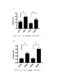
- (A) is a result of generating a 10 Hz wave in the occipital lobe by irradiation of a violet light with a blinking frequency of 10 Hz
- (B) is a result of generating a 60 Hz wave in the occipital lobe by irradiation of a violet light with a blinking frequency of 60 Hz. The result.
- (A) is a result of generating 10 Hz waves in the frontal lobe by irradiation of violet light with a blinking frequency of 10 Hz
- (B) is a result of generating 60 Hz waves in the frontal lobe by irradiation of violet light with a blinking frequency of 60 Hz. is there.
- (A) shows the result that no 60 Hz wave was generated in the frontal lobe by irradiation of violet light with a blinking frequency of 10 Hz
- (B) shows the result that 10 Hz wave was not generated in the frontal lobe by irradiation of violet light with a blinking frequency of 60 Hz. is there.
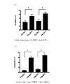
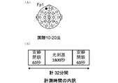
- FIG. 1 is an explanatory diagram of the left prefrontal cortex Fp1 (International 10-20 Law), which is a measurement location, and (B) is a measurement time of 30 minutes (1800 seconds), and a rest time of 1 minute before and after that.
- FIG. It is a test result of the significant difference of a power spectrum. It is a result which shows the suppression effect of the stress by the frequency stimulation of 40 Hz. It is a graph which shows the average value of the stress value with respect to the stimulus under each condition. It is the result of the reduction of phosphorylated tau by 40 Hz frequency stimulation. It is a result of the reduction of phosphorylated tau by 40 Hz frequency stimulation similar to FIG. It is an evaluation result of fear memory performed in the CFC test.
- a device and a method for controlling electroencephalogram and cell activity by light stimulation and a device for improving, preventing or increasing brain function according to the present invention will be described with reference to the drawings.
- the present invention is not limited to the contents of the following embodiments and examples, and includes various modifications and application examples within the scope including the gist of the present invention.
- the apparatus for controlling brain waves and cell activity by light stimulation is an apparatus for performing electroencephalogram control or cell activity control by irradiating a subject with light of a specific wavelength at a regular light or a specific blinking frequency, and controlling the cell activity.
- a light source that irradiates light with a normal light or a specific blinking frequency, and a control unit that controls the emission of light that induces a specific brain wave in which the brain wave of the subject who received the light is the same or substantially the same as or different from the light irradiation state. And having the following.
- the method of controlling the electroencephalogram function by light stimulation is a method of performing electroencephalogram control or cell activity control by irradiating a subject with a specific wavelength of light at a constant light or a specific blinking frequency, wherein the subject receiving the light Is characterized in that light emission control of light for inducing a specific brain wave which is the same or substantially the same as or different from the irradiation state of the light is performed.
- This brain wave and cell activity control device because the brain wave of the subject who receives the light induces a specific brain wave that is the same or substantially the same as or different from the light irradiation state, by controlling the light irradiation state, A stimulus can be applied or cell activity in the body can be increased.
- a stimulus can be applied or cell activity in the body can be increased.
- when irradiating the blinking frequency, etc. it is possible to generate brain waves in the same or almost the same state as the irradiation state of the irradiated light or specific brain waves different from the irradiation state, so it is possible to control the stimulation state to the brain It can be applied to various mental and physical improvement effects and therapeutic effects.
- sound, vibration, a magnetic field, an electric field, and the like can be applied together with the irradiation of the light in the irradiation state such as the blinking frequency, and a combined action of both can be caused.
- FIG. 8 shows a commonly understood brain wave classification.
- electrical vibrations occur via neural circuits, and such brain waves are generally delta waves of about 4 Hz or less, theta waves of about 4 to 7 Hz, alpha waves of about 8 to 13 Hz, and about 14 to 14 Hz. It is classified into a beta wave of 30 Hz and a gamma wave of about 30 Hz or more.
- the blinking frequencies of 10 Hz and 60 Hz are used as an example, and the range can be said to be the range of alpha waves and gamma waves.
- alpha waves are called brain waves generated when the body and mind are relaxed, and gamma waves are called brain waves generated when the body is excited.
- gamma waves are called brain waves generated when the body is excited.
- different specific brain waves can be induced.
- the present invention has a remarkable feature in that brain waves having the same or substantially the same frequency as the blinking frequency of irradiation light or brain waves having a different specific frequency can be induced, and the generation of such brain waves is controlled by light irradiation conditions.
- the subject can be provided with a psychosomatic action based on the above-described brain waves, for example, emotion, motivation, memory, concentration, relaxation, elevation, awakening, sleep, sleep, sleep induction, and dreaming.
- human experiments have yielded results in various points, such as improving sleep, relaxing, and dreaming (dreams are effective for memory formation immediately before non-REM sleep).
- cell activity based on brain waves and effects on diseases for example, fluctuations in brain transmitters, fluctuations in body secretions, effects on proteins, Alzheimer's disease, brain dysfunction, age-related macular degeneration of the retina, dependence, Depression, dissociative disorder, obsessive-compulsive disorder, sleep disorder, eating disorder, bipolar disorder (manic depression), application disorder, schizophrenia, dementia, personality disorder, developmental disorder, panic disorder, PTSD, gender identity It is expected to act on sexual dysfunction, epilepsy and the like, and on cell activity, and is expected to affect various mental and physical or physical effects (including therapeutic effects).
- diseases for example, fluctuations in brain transmitters, fluctuations in body secretions, effects on proteins, Alzheimer's disease, brain dysfunction, age-related macular degeneration of the retina, dependence, Depression, dissociative disorder, obsessive-compulsive disorder, sleep disorder, eating disorder, bipolar disorder (manic depression), application disorder, schizophrenia, dementia, personality disorder, developmental disorder, panic disorder, PTSD, gender identity It is expected to act on sexual dysfunction, epilepsy and the
- FIG. 2 to FIG. 4 show measurements of brain waves when light is illuminated by blinking.
- irradiation with violet light at a blinking frequency of 10 Hz resulted in the generation of 10 Hz waves in the occipital lobe and frontal lobe.
- FIGS. 2B and 3B by irradiating violet light at a blinking frequency of 60 Hz, a result was obtained in which 60 Hz waves were generated in the occipital lobe and frontal lobe.
- FIG. 2 (A) and FIG. 3 (A) irradiation with violet light at a blinking frequency of 10 Hz resulted in the generation of 10 Hz waves in the occipital lobe and frontal lobe.
- FIGS. 2B and 3B by irradiating violet light at a blinking frequency of 60 Hz, a result was obtained in which 60 Hz waves were generated in the occipital lobe and frontal lobe.
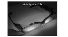
- a 375 nm LED having an irradiance of a maximum output of 310 ⁇ W / cm 2 was attached to a frame (see FIG. 5).
- This LED exhibits the spectral wavelength shown in FIG. 7, and emits violet light defined from 360 to 400 nm.
- This light source has a control unit (electronic circuit unit) capable of adjusting the output within the range of the maximum output or less.
- it has a function of arbitrarily changing the blinking frequency, and can emit a violet light in a range of 0 (a “normal light”; DC light) to 150 Hz.
- the irradiation conditions in this experiment were performed at blinking frequencies of 10 Hz and 60 Hz. In the light source used, the duty ratio of the blinking frequency is set to 50%.
- an eye open / close test at rest / wake when the eyes were closed was performed in the following order.
- the brain wave was measured for 5 minutes in a normal state in which the user blinks naturally without wearing glasses.
- the subject was wearing glasses and irradiating the eyes with a violet light having a blinking frequency of 10 Hz for 5 minutes to measure brain waves.
- the brain waves were measured while irradiating the eyes with violet light changed to the blinking frequency of 60 Hz for 5 minutes.
- the electroencephalogram was measured using an electroencephalograph (manufactured by Nihon Kohden Corp., EEG-1200 series model).
- FIGS. 9 to 11 show data of application software in a case where the present inventor wears glasses having a light source (FIG. 5) as needed to conduct daily life.
- FIGS. 9 and 11 show the ratios contributing to a good sleep level when the flashing frequency of the violet light is radiated at 10 Hz, 40 Hz and 60 Hz, and
- FIG. 10 shows the ratios for decreasing the good sleep level. It is. Irradiation of the flashing frequency of the violet light generates the same or substantially the same brain wave as the flashing frequency, and thus it was confirmed that the generated brain wave has a certain effect on the degree of good sleep.
- the wavelength of the light used was light in the violet light range of 360 to 400 nm, and the frequency was 10 Hz, 40 Hz, and 60 Hz. It is expected that similar phenomena will occur at frequencies (for example, 9 Hz, 30 Hz, 60 Hz or more).
- the wavelength of the light used here is in the violet light range, it can be expected that light in other wavelength ranges will generate the same or substantially the same brain wave as the blinking frequency of the irradiated light.
- brain waves are known to affect psychosomatic effects such as emotion, motivation, memory, concentration, relaxation, uplifting, arousal, and the like, and a device capable of producing such psychosomatic effects can be provided.
- cell activity based on brain waves and effects on diseases can be expected.
- the wavelength of light emitted from the light source is not particularly limited, but violet light defined in the range of 360 to 400 nm is used in the above experiment. In addition to violet light, other wavelengths can be expected to have the same effect, and can be used in combination with violet light. In addition, since white light has a certain effect as shown in the results described later, the white light may be light emitted from a light source or a part of the light.
- a light source capable of an oscillation frequency of 0 (normal light, DC light) to 150 Hz can be preferably applied.
- the frequency can be adjusted in units of 0.5 Hz or 1 Hz by setting of the control unit, and light with an arbitrary blinking frequency can be generated.
- Increasing the blinking frequency also has the advantage that blinking does not matter, although there are individual differences.
- the blinking frequency is not limited to 10 Hz or 60 Hz used in the experimental example.
- the irradiance from the light source may be variable or may be constant.
- the one having a maximum output of 310 ⁇ W / cm 2 is used, but the present invention is not limited to this.
- those in the range of 1 ⁇ W / cm 2 (0.01 W / m 2 ) to 1000 ⁇ W / cm 2 (10 W / m 2 ) for example, 0.5 ⁇ W / cm 2 (0.005 W / m 2 ) to 500 ⁇ W / cm 2 (5 W / m 2 ), 0.5-1000 ⁇ W / cm 2 , etc.
- the generation of the brain wave equal to or substantially the same as the blinking frequency occurs even at a low irradiance, it is possible to generate a predetermined brain wave by irradiating the blinking frequency even when the eye is opened or closed. Further, since the light source having such irradiance can be easily applied to glasses and other portable irradiation devices, it can be worn even in daily life. In particular, it has been confirmed that the characteristic phenomena described above occur even with a small amount of weak light (light with low photosensitivity), and it affects the brain and cell activity (used in the sense of controlling gene expression). The application to is expected.
- Light may be specified by relative luminous efficiency. Since the features of the present invention can be realized even at low relative luminous efficiency, it is possible to irradiate a flashing violet light that causes brain wave stimulation under low relative luminous efficiency, and to stimulate a desired site without burden on the subject. it can.
- the irradiation time of the light is preferably set arbitrarily according to the purpose, and may be short or long.
- the light can be arbitrarily intermittent (fixed or irregular) or continuous.
- the light source is preferably glasses with a light source.
- the glasses have a light source that emits a blinking frequency with glasses that are easy to wear and that do not cause any discomfort on a daily basis, so that they are highly practical and can always be worn in various scenes and environments.
- the light source may be a light source in front of or near the face such as a desk light source, a light source mounted on a mobile terminal, or a non-installed light source such as a portable light source, or indoor lighting, a table lamp, a dedicated device, or the like.
- the light source may be an installation type light source, and may be devices of various light source forms according to the use environment.
- the control unit is a unit that controls the irradiation state of the light from the light source (normal light or blinking frequency).
- the control unit may be provided with a power supply (not shown) for supplying power to the light source, such a power supply may be a battery, or a cable is routed to a battery mounted at another position. It may be something. In the case where it does not move at one place, it may be connected to a home power supply or the like.
- the control unit executes change of irradiation conditions such as blinking frequency of light, irradiance, irradiation time, irradiation start time, irradiation end time, blinking frequency and the like by transmission and reception with an isolation controller such as a portable terminal. Is preferred. Since such a control unit controls the above-described various irradiation conditions in isolation, it is possible to arbitrarily set the irradiation conditions suitable for generating a desired brain wave or cell activity and obtain a desired effect. Further, it is possible to measure and evaluate how the irradiation of the specific wavelength light at the blinking frequency affects brain waves and cell activity, and to what extent the influence on the mind and body is obtained, and can be applied to practical use.
- irradiation conditions such as blinking frequency of light, irradiance, irradiation time, irradiation start time, irradiation end time, blinking frequency and the like by transmission and reception with an isolation controller such as a portable terminal. I
- control unit may include a light source controller and a timer function.
- the controller include a function of changing the frequency and the irradiance, setting the irradiation time, and the like.
- the timer function may be one that can set the irradiance time of light. Such a controller or timer function may be provided integrally with the appliance, or may be a separate member.
- the brain wave and cell activity control apparatus by light stimulation according to the present invention irradiates light of a specific wavelength such as violet light at an ordinary light or a specific blinking frequency, and has the same or substantially the same irradiation state.
- An electroencephalogram can be generated in the subject, or a different specific electroencephalogram can be induced in the subject.
- a specific electroencephalogram that is the same or substantially the same as or different from the blinking frequency of the light can be induced from the brain wave of the subject who has received the light, and various stimuli can be applied to the mind, body and brain.
- An apparatus for improving, preventing or increasing brain function is an apparatus for improving, preventing or increasing brain function by irradiating a subject with a violet light or white light at a normal light or a specific blinking frequency, wherein the violet is used.
- a light source that emits light or white light
- a light emission cycle control unit that sets the violet light or white light as an ordinary light or a specific blinking frequency, and a light emission time during which the violet light or white light is irradiated for a specific time or for a specific period
- a control unit which is used for any one or more purposes selected from the group consisting of improving or preventing depression, suppressing or preventing stress, improving or increasing concentration, improving or preventing Alzheimer's disease, and improving sleep.
- Device which is used for any one or more purposes selected from the group consisting of improving or preventing depression, suppressing or preventing stress, improving or increasing concentration, improving or preventing Alzheimer's disease, and improving sleep.
- the apparatus for improving or preventing brain function is an apparatus for irradiating a subject with ordinary light of violet light or white light to improve or prevent cognitive function, and emits the violet light or white light.
- a light source, and a light emission time control unit that irradiates the violet light or white light for a specific time or for a specific period is provided.
- violet light is represented by “VL”.
- mice were irradiated with an ordinary white light stimulus (WL continuous), an ordinary VL stimulus (VL continuous), and a VL 40 Hz frequency stimulus (VL 40 Hz) to give a light stimulus.
- the mice used were C57BL / 6 mice of 8 to 13 weeks of age.
- TST tail-suspension-test
- FST forced swim test
- the degree of depression is an index of depression as to how much ⁇ I give up '', and as non-patent literature on the evaluation, ⁇ Journal of Visualized Experiments January 2012, 59, e3638, Page 1 of 5 '', ⁇ Journal of Visualized Experiments January 2012, 59, e3769, Page 1 of 5, "Neuron 53, 337 – 351, February 1, 2007 a 2007 Elsevier Inc. 337".
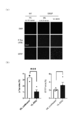
- FIG. 12 (A) shows the results of evaluation of depression induction using lipopolysaccharide (LPS / Lipopolysaccharide) and improvement of depression by VL 40 Hz frequency stimulation.
- LPS lipopolysaccharide
- VL 40 Hz frequency stimulation was performed for a total of 8 days before and on the day of LPS administration, and depression was evaluated by Tail suspension test (TST / tail suspension test).
- the reference used for the LPS-induced depression induction experiment is https://www.nature.com/articles/s41598-019-42286-8.
- FIG. 12 (A) reduction in the symptoms of depression was observed by VL 40 Hz frequency stimulation.
- the immobility time of TST was measured with ANY-maze (Video Tracking System / Muromachi Kikai Co., Ltd.), and statistically significant difference between the control group, LPS group, and LPS + 40 Hz frequency stimulation group was obtained using GraphPad Prism 8.0 software. Was tested and shown in FIG. In addition, “*” is p ⁇ 0.05, and “**” is p ⁇ 0.01.
- FIG. 12 (B) is an evaluation result of depression induction using CUMS (Chronic unpredictable mild stress / unpredictable chronic mild stress) and improvement of depression by VL regular lighting or 40 Hz frequency stimulation.
- the reference used for experiments in which depression was induced by CUMS is https://www.sciencedirect.com/science/article/pii/S0149763418304378?via%3Dihub.
- the mice were subjected to relatively mild various stresses on a daily basis for a long period of time (7 weeks) to induce chronic psychological depression. During that time, VL normal light stimulation or VL 40 Hz frequency stimulation was performed, and depression was evaluated by TST. As shown in FIG. 12 (B), the depression symptoms were improved by the VL ordinary light stimulation or the VL 40 Hz frequency stimulation.
- EEG electroencephalogram
- VL and white light were used, and light stimulation was performed with blinking and constant lighting (referred to as “normal lighting”).
- filtering was performed as preprocessing, a power spectrum was calculated by FFT (fast Fourier transform), and a significance test was performed based on the result.
- the number of subjects was 162 in total, the measurement location was the left prefrontal cortex Fp1 (International 10-20 method) shown in FIG. 13 (A), and the measurement time was 30 minutes (1800 seconds) as shown in FIG. 13 (B). Before and after that, a rest time of 1 minute was set.
- Light stimuli at the time of measurement were white light ordinary light stimulation, white light frequency stimulation, VL ordinary light stimulation, and VL frequency stimulation.
- the VL stimulation was performed with VL glasses shown in FIG. 5, and the white light stimulation was performed with a frequency stimulus of 40 Hz.
- a simple electroencephalograph (sampling frequency: 512 Hz, MindWave mobile BMD version, Neurosky Inc.) was used as the measuring device.
- the analysis was performed according to the following procedure.
- the electroencephalogram was measured by applying light stimulation to 81 VL stimuli and 81 white light stimuli under eight conditions (seven conditions for light stimulation and one condition for extinguishing light).
- the light stimulation time was 30 minutes (1800 seconds) as shown in FIG.
- the obtained measurement data was subjected to noise removal (filtering to make 1 to 70 Hz effective) and power spectrum calculation (Hilbert transform, spline interpolation) to calculate a one-sided power spectrum. This procedure was performed as a standard procedure.
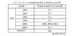
- the test of the significant difference was performed to analyze the frequency component of the electroencephalogram affected by the light stimulation. Specifically, a two-sided two-sample t-test (significance level: 5%) was performed on healthy men and women. With respect to a 1 Hz power spectrum (mean of the subject), seven conditions for each of frequency stimulation and ordinary light stimulation were compared with always-off, and a significant difference test was performed. Note that the comparison was also made for the power spectrum of 2 to 45 Hz.
- FIG. 14 shows the result of the significant difference test of the power spectrum (at the time of light stimulation vs. at the time of light-out). As shown in the results of FIG.
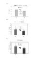
- FIG. 15 (A) shows stress values before and after the case without stimulation
- FIG. 15 (B) shows stress values before and after the VL 40 Hz frequency stimulation
- FIG. 15 (C) shows stress values before and after the white light 40 Hz frequency stimulation. It is. It was found that 40 Hz frequency stimulation reduced stress compared to before and after the unstimulated task.
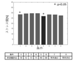
- FIG. 16 is a graph showing the average of stress values for stimuli under each condition (“*” indicates p ⁇ 0.05). Healthy men and women were given VL frequency stimuli of 10 Hz, 12 Hz, 13 Hz, 15 Hz, 40 Hz, and 60 Hz, and ordinary light stimuli. Noise removal and power spectrum analysis were performed from brain waves measured using a simple electroencephalograph, and stress values (%) were tested. From these results, the stress was reduced by the frequency stimulation of 10 Hz and 40 Hz.
- mice were illuminated with regular light white light stimulation (WL continuous), white light 40 Hz frequency stimulation (WL 40 Hz), regular light VL stimulation (VL continuous), and VL 40 Hz frequency stimulation (VL 40 Hz). It was given and evaluated.
- tau is known in humans as a gene causing Alzheimer's disease. It is known that a mutation in this gene causes Alzheimer's disease.
- S301P was found as a mutation in Parkinson's disease or a familial hereditary disease of frontotemporal dementia.
- evaluation was performed using a mouse having human S301P tau, and in fact, phosphorylation of tau is a diagnosis of Alzheimer's disease, and is detected using an antibody called AT8 (pSer202 / Thr205).
- AT8 pSer202 / Thr205
- the first paper evaluated as Alzheimer's disease using S301P mice was https://www.ncbi.nlm.nih.gov/pubmed/17270732 and was applied in this experiment.
- FIG. 17 and FIG. 18 show the results of reduction of phosphorylated tau by 40 Hz frequency stimulation.
- A is a measurement image of DAPI, p-Tau, and GFAP
- B is a graph of GFAP area% obtained by analyzing the image.
- a three-month-old S301P mutant mouse was given a normal light white light stimulus, a white light 40 Hz frequency stimulus, and a VL 40 Hz frequency stimulus, each for four weeks.
- Phosphorylated tau (Ser202, Thr205) was detected using AT8 antibody.
- 40 Hz frequency stimulation of VL reduces the accumulation of phosphorylated tau of Hippocampus (left graph of FIG. 17 (B)), decreases the accumulation of phosphorylated tau of CA3 region (graph of FIG. 18 (B)), Astrocyte GFAP (the graph on the right side of FIG. 17 (B)) increased.
- fear memory is evaluated by the time during which an electric shock freezes and stops moving. It freezes immediately after giving the electrical stimulus, but if you don't remember the previous day by putting it in a machine that gives you an electric shock the next day, the freeze time will decrease (Ref: https: // www. ncbi.nlm.nih.gov/books/NBK5223/).
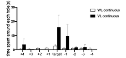
- Spatial memory is that nocturnal mice try to escape to dark places when the mouse is exposed to strong light on a white disk. The disc has 20 holes and 19 are closed, but only one can escape into the hole. The mice will be trained to remember the location of that one hole for six days. One day later, and one week later, it was possible to evaluate how much the location of the hole was memorized (reference: https://www.nature.com/protocolexchange/protocols/349Reiserer).
- the fear memory was evaluated by the CFC test (Contextual fear conditional test).
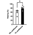
- Fear memory was assessed by CFC after 64 week-old aged mice were given 7 weeks of constant light white light stimulation or constant light VL stimulation. As shown in FIG. 19, it was confirmed that the freezing score of CFC was improved by the VL stimulation with the ordinary light.
- the activity was evaluated. 75-week-old aged mice were given a 12-week constant light VL stimulation, and the activity was evaluated using running @ wheel. As shown in FIG. 21, the activity score of the aged mouse was improved in each of the white light 40 Hz frequency stimulation, the ordinary light VL stimulation, and the VL 40 Hz frequency stimulation.
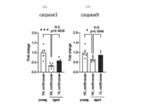
- FIGS. 67-week-old aged mice were given a constant light VL stimulation for 13 weeks.
- the gene expression changes between the mouse and a 15-week-old young mouse (young) and a 67-week-old aged mouse (aged) under normal light white light conditions were compared. “*” Is p ⁇ 0.05, “**” is p ⁇ 0.01, and “***” is p ⁇ 0.001.
- the expression of opn5 gene increased (see FIG. 22 (A)), the apoptosis-related gene Bax increased (see FIG. 22 (B)), and the gene of Bcl2 was stimulated by constant-light VL stimulation.
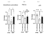
- the expression is increased (see FIG. 22 (C)), the apoptosis-related gene Caspase3 is increased (see FIG. 23 (A)), the gene expression of Caspase9 is increased (see FIG. 23 (B)), and oxidative stress and mitochondria-related
- the gene expression of the genes Glutathione peroxidase and PGC1 ⁇ was increased (see FIGS. 24 (A) and (B)), and the expression of the cell cycle and senescence-related p21 gene was increased (see FIG. 24 (C)).
- OPN5 VL-specific photoreceptor
- the light stimulus in the present invention described above is not necessarily transmitted through the eyes, and is considered to be directly acting on the brain through the skull (see: https://www.cell.com/current-). biology / fulltext / S0960-9822 (14) 00603-4). According to the present invention, it can be considered from the results in FIG. 22 described above that the VL irradiation increases the OPN5 gene expression.
- the apparatus for irradiating a subject with violet light or white light can improve or prevent depression, suppress or prevent stress, and concentrate. Improvement or increase, improvement or prevention of Alzheimer's disease, improvement or prevention of cognitive function, and sleep improvement.
Abstract
Description
本発明に係る光刺激による脳波及び細胞活性制御装置は、特定波長の光を常灯又は特定の点滅周波数で被験者に照射して脳波制御又は細胞活性制御を行う装置であって、前記特定波長の光を常灯又は特定の点滅周波数で照射する光源と、前記光を受けた前記被験者の脳波が前記光の照射状態と同じ若しくは略同じ又は異なる特定の脳波を誘導する光を発光制御する制御部とを有する、ことを特徴とする。また、光刺激による脳波機能制御方法についても、特定波長の光を常灯又は特定の点滅周波数で被験者に照射して脳波制御又は細胞活性制御を行う方法であって、前記光を受けた前記被験者の脳波が前記光の照射状態と同じ又は略同じ又は異なる特定の脳波を誘導する光を発光制御する、ことを特徴とする。 [EEG and cell activity control device by light stimulation]
The apparatus for controlling brain waves and cell activity by light stimulation according to the present invention is an apparatus for performing electroencephalogram control or cell activity control by irradiating a subject with light of a specific wavelength at a regular light or a specific blinking frequency, and controlling the cell activity. A light source that irradiates light with a normal light or a specific blinking frequency, and a control unit that controls the emission of light that induces a specific brain wave in which the brain wave of the subject who received the light is the same or substantially the same as or different from the light irradiation state. And having the following. Also, the method of controlling the electroencephalogram function by light stimulation is a method of performing electroencephalogram control or cell activity control by irradiating a subject with a specific wavelength of light at a constant light or a specific blinking frequency, wherein the subject receiving the light Is characterized in that light emission control of light for inducing a specific brain wave which is the same or substantially the same as or different from the irradiation state of the light is performed.
図2~図4は、光を点滅照射したときの脳波を測定したものである。図2(A)及び図3(A)に示すように、10Hzの点滅周波数でバイオレットライトを照射することにより、後頭葉と前頭葉で10Hz波が発生している結果が得られた。また、図2(B)及び図3(B)に示すように、60Hzの点滅周波数でバイオレットライトを照射することにより、後頭葉及び前頭葉で60Hz波が発生している結果が得られた。一方、図4では、10Hzの点滅周波数でバイオレットライトを照射した際、前頭葉で60Hz波は発生せず、60Hzの点滅周波数でバイオレットライトを照射した際も、前頭葉で10Hz波は発生しなかった。これらの結果は、照射光の点滅周波数と同じ又は略同じ周波数の脳波が発生するという極めて驚くべき現象を示している。しかも、この実験では、弱い光(低い放射照度)で眩しくなく邪魔感が白色光に比べて小さいバイオレットライトを点滅照射しているので、白色光等の可視光の場合にしばしば起こる眩しさや邪魔感やちらつき感がなく、特に点滅周波数を点滅が気にならない程度に上げることで、日常生活での使用に耐える実用性を備えるという結果が得られている。 [Generation of brain waves by light stimulation]
FIG. 2 to FIG. 4 show measurements of brain waves when light is illuminated by blinking. As shown in FIG. 2 (A) and FIG. 3 (A), irradiation with violet light at a blinking frequency of 10 Hz resulted in the generation of 10 Hz waves in the occipital lobe and frontal lobe. In addition, as shown in FIGS. 2B and 3B, by irradiating violet light at a blinking frequency of 60 Hz, a result was obtained in which 60 Hz waves were generated in the occipital lobe and frontal lobe. On the other hand, in FIG. 4, when the violet light was irradiated at the blinking frequency of 10 Hz, no 60 Hz wave was generated in the frontal lobe, and when the violet light was irradiated at the blinking frequency of 60 Hz, no 10 Hz wave was generated in the frontal lobe. These results show a very surprising phenomenon that brain waves having the same or substantially the same frequency as the blinking frequency of the irradiation light are generated. In addition, in this experiment, the violet light, which is not dazzling with weak light (low irradiance) and has a small disturbing feeling compared to white light, is illuminated by flashing, so that the glare and disturbing feeling that often occur with visible light such as white light are given. The result is that there is no flickering, and especially when the blinking frequency is raised to such a degree that the blinking is not bothersome, practicality that can be used in daily life is provided.
光源が発光する光の波長は特に限定されないが、上記実験では360~400nmで定義されるバイオレットライトを使用している。バイオレットライトの他、他の波長でも同様の効果があるものと期待でき、バイオレットライトとともに併用して利用することもできる。また、白色光についても、後述の結果に示すように、一定の効果を示していることから、白色光についても、光源から発する光としてもよいし、光の一部としてもよい。 (light source)
The wavelength of light emitted from the light source is not particularly limited, but violet light defined in the range of 360 to 400 nm is used in the above experiment. In addition to violet light, other wavelengths can be expected to have the same effect, and can be used in combination with violet light. In addition, since white light has a certain effect as shown in the results described later, the white light may be light emitted from a light source or a part of the light.
制御部は、光源からの光の照射状態(常灯や点滅周波数)をコントロールする部分である。制御部は、光源に電力を供給するための電源(図示しない)を備えたものであってもよく、そうした電源はバッテリーであってもよいし、別の位置に装着したバッテリーまでケーブルで引き回したものであってもよい。また、一箇所で動かない場合には、家庭用電源等に接続する形態であってもよい。 (Control unit)
The control unit is a unit that controls the irradiation state of the light from the light source (normal light or blinking frequency). The control unit may be provided with a power supply (not shown) for supplying power to the light source, such a power supply may be a battery, or a cable is routed to a battery mounted at another position. It may be something. In the case where it does not move at one place, it may be connected to a home power supply or the like.
本発明では、さらにバイオレットライトや白色光による光刺激の影響についての研究を行った。その結果、バイオレットライトや白色光の照射による光刺激により、鬱の改善又は予防、ストレスの抑制又は予防、集中力の改善又は増加、アルツハイマー病の改善又は予防、認知機能の改善又は予防等が期待できることがわかった。バイオレットライトや白色光による光刺激でのこうした効果が見られたことは従来にない初めての知見であり、本発明に係る脳機能を改善、予防又は増大する装置に至った。 [Brain function improvement or augmentation device]
In the present invention, further research was conducted on the effect of light stimulation by violet light or white light. As a result, light stimulation by violet light or white light irradiation is expected to improve or prevent depression, suppress or prevent stress, improve or increase concentration, improve or prevent Alzheimer's disease, improve or prevent cognitive function, etc. I knew I could do it. It was the first ever finding that such effects were obtained by photostimulation with violet light or white light, leading to a device for improving, preventing or increasing brain function according to the present invention.
常灯白色光刺激(WL continuous)、常灯VL刺激(VL continuous)、VLの40Hz周波数刺激(VL 40Hz)を、それぞれのマウスに照射し、光刺激を与えた。マウスは、8~13週齢のC57BL/6マウスを用いた。 (Improvement of depression)
Each mouse was irradiated with an ordinary white light stimulus (WL continuous), an ordinary VL stimulus (VL continuous), and a
VLによるストレスの抑制及び集中力の増加について解析した。ストレスのデータについて、脳波(electroencephalogram;EEG)の測定方法とその評価方法は、「Sensors 2018,18(12),4477」(https://doi.org/10.3390/s18124477、https://www.mdpi.com/1424-8220/18/12/4477/htm)に記載の非特許文献に記載の方法で行った。 (Reducing stress, increasing concentration)
An analysis was performed on the suppression of stress and the increase in concentration due to VL. For stress data, electroencephalogram (EEG) measurement method and its evaluation method are described in “Sensors 2018, 18 (12), 4477” (https://doi.org/10.3390/s18124477, https: // www. mdpi.com/1424-8220/18/12/4477/htm).
アルツハイマー病の改善効果について、常灯白色光刺激(WL continuous)、白色光の40Hz周波数刺激(WL 40Hz)、常灯VL刺激(VL continuous)、VLの40Hz周波数刺激(VL 40Hz)をそれぞれマウスに与えて評価した。 (Improvement of Alzheimer's disease)
Regarding the improvement effect of Alzheimer's disease, mice were illuminated with regular light white light stimulation (WL continuous),
高齢マウスにおける認知機能の改善効果について、常灯白色光刺激(WL continuous)、白色光の40Hz周波数刺激(WL 40Hz)、常灯VL刺激(VL continuous)、VLの40Hz周波数刺激(VL 40Hz)をそれぞれマウスに与えて評価した。 (Improvement of cognitive function)
Regarding the effect of improving cognitive function in aged mice, white light stimulation (WL continuous), 40 Hz frequency stimulation of white light (
As shown in the above results, according to the apparatus for improving or increasing brain function according to the present invention, the apparatus for irradiating a subject with violet light or white light can improve or prevent depression, suppress or prevent stress, and concentrate. Improvement or increase, improvement or prevention of Alzheimer's disease, improvement or prevention of cognitive function, and sleep improvement.
Claims (10)
- 特定波長の光を常灯又は特定の点滅周波数で被験者に照射して脳波制御又は細胞活性制御を行う装置であって、前記特定波長の光を常灯又は特定の点滅周波数で照射する光源と、前記光を受けた前記被験者の脳波が前記光の照射状態と同じ若しくは略同じ又は異なる特定の脳波を誘導する光を発光制御する制御部とを有する、ことを特徴とする、光刺激による脳波及び細胞活性制御装置。 A device for performing electroencephalogram control or cell activity control by irradiating a subject with light of a specific wavelength at a regular light or a specific blinking frequency, and a light source for irradiating the light of the specific wavelength with a regular light or a specific blinking frequency, A control unit that controls the emission of light that induces a specific brain wave that is the same or substantially the same as or different from the irradiation state of the light, and the brain wave of the subject that has received the light, characterized in that: Cell activity control device.
- 前記光がバイオレットライトである、請求項1に記載の脳波及び細胞活性制御装置。 The brain wave and cell activity control device according to claim 1, wherein the light is violet light.
- 前記光の照射状態が、常灯又は0Hz超~150Hzの点滅周波数である、請求項1又は2に記載の脳波及び細胞活性制御装置。 3. The electroencephalogram and cell activity control device according to claim 1, wherein the light irradiation state is a normal light or a blinking frequency of more than 0 Hz to 150 Hz.
- 前記光を、放射照度で0.5~1000μW/cm2の範囲内で照射する、請求項1~3のいずれか1項に記載の脳波及び細胞活性制御装置。 The electroencephalogram and cell activity control apparatus according to any one of claims 1 to 3, wherein the light is applied at an irradiance of 0.5 to 1000 μW / cm 2 .
- 前記制御部は、携帯端末等の隔離コントローラーとの間の送受信により、前記光の照射状態(常灯又は点滅周波数を含む。)、放射照度、照射時間、照射開始時間、照射終了時間、常灯又は点滅周波数等の照射条件を変更して実行する、請求項1~4のいずれか1項に記載の脳波及び細胞活性制御装置。 The control unit transmits and receives the light to and from an isolation controller such as a portable terminal, and includes an irradiation state of the light (including an ordinary light or a blinking frequency), an irradiance, an irradiation time, an irradiation start time, an irradiation end time, and an ordinary light. The electroencephalogram and cell activity control device according to any one of claims 1 to 4, wherein the device is executed by changing irradiation conditions such as a blinking frequency.
- 前記光源は、光源付きめがね、卓上光源、移動体端末装着光源等の顔前又は近傍設置型の光源である、請求項1~5のいずれか1項に記載の脳波及び細胞活性制御装置。 The electroencephalogram and cell activity control device according to any one of claims 1 to 5, wherein the light source is a light source in front of or near the face, such as glasses with a light source, a desktop light source, and a light source mounted on a mobile terminal.
- 前記光源は、携帯光源等の非設置型光源、又は、室内照明、卓上スタンド、専用装置等の設置型光源である、請求項1~5のいずれか1項に記載の脳波及び細胞活性制御装置。 The electroencephalogram and cell activity control device according to any one of claims 1 to 5, wherein the light source is a non-installation type light source such as a portable light source or an installation type light source such as indoor lighting, a table lamp, and a dedicated device. .
- 特定波長の光を常灯又は特定の点滅周波数で被験者に照射して脳波制御又は細胞活性制御を行う方法であって、前記光を受けた前記被験者の脳波が前記常灯又は特定の点滅周波数と同じ又は略同じ又は異なる特定の脳波を誘導する光を発光制御する、ことを特徴とする、光刺激による脳波及び細胞活性制御方法。 A method of performing electroencephalogram control or cell activity control by irradiating a subject with light of a specific wavelength at a regular light or a specific blinking frequency, wherein the brain wave of the subject receiving the light has the normal light or a specific blinking frequency. A method for controlling electroencephalogram and cell activity by light stimulation, comprising controlling light emission of light that induces the same or substantially the same or different specific electroencephalogram.
- バイオレットライト又は白色光を常灯又は特定の点滅周波数で被験者に照射して脳機能を改善、予防又は増大する装置であって、前記バイオレットライト又は白色光を発光する光源と、前記バイオレットライト又は白色光を常灯又は特定の点滅周波数とする発光周期制御部と、前記バイオレットライト又は白色光を特定の時間又は特定の期間照射する発光時間制御部と、を備え、鬱の改善又は予防、ストレスの抑制又は予防、集中力の改善又は増加、アルツハイマー病の改善又は予防、睡眠改善等をから選ばれるいずれか1又は2以上の目的で使用される装置である、ことを特徴とする、脳機能改善、予防又は増大装置。 A device for improving, preventing or increasing brain function by irradiating a subject with a violet light or white light at an ordinary light or a specific blinking frequency, wherein the light source emitting the violet light or white light, and the violet light or white A light emission cycle control unit that uses light as a normal light or a specific blinking frequency, and a light emission time control unit that irradiates the violet light or white light for a specific time or for a specific period, for improving or preventing depression, reducing stress Improvement of brain function, characterized in that the device is used for one or more purposes selected from among suppression or prevention, improvement or increase of concentration, improvement or prevention of Alzheimer's disease, improvement of sleep, and the like. , Prevention or augmentation device.
- バイオレットライト又は白色光の常灯光を被験者に照射して認知機能を改善又は予防する装置であって、前記バイオレットライト又は白色光を発光する光源と、前記バイオレットライト又は白色光を特定の時間又は特定の期間照射する発光時間制御部と、を備える、ことを特徴とする、脳機能改善又は予防装置。
An apparatus for improving or preventing a cognitive function by irradiating a violet light or white light ordinary light to a subject, wherein the light source that emits the violet light or white light and the violet light or white light for a specific time or specific time And a light emission time control unit for irradiating for a period of time.
Priority Applications (9)
| Application Number | Priority Date | Filing Date | Title |
|---|---|---|---|
| EP19843694.1A EP3831447A4 (en) | 2018-08-01 | 2019-08-01 | Device and method for controlling brain waves and cell activity by photic stimulation, and device for improvement, prophylaxis, or enhancement of brain function |
| CN201980051391.0A CN112533672A (en) | 2018-08-01 | 2019-08-01 | Device and method for controlling brain waves and cell activity based on light stimulation and device for improving, preventing or enhancing brain function |
| JP2019565489A JP7096554B2 (en) | 2018-08-01 | 2019-08-01 | EEG and cell activity control device by light stimulation |
| KR1020217004302A KR20210040977A (en) | 2018-08-01 | 2019-08-01 | Device and method for controlling brain wave and cell activity by photostimulation, and device for improving, preventing or increasing brain function |
| BR112021001647-3A BR112021001647A2 (en) | 2018-08-01 | 2019-08-01 | device and method for controlling brain wave and cell activity based on light stimulation, and device for improving, preventing or increasing brain function |
| US17/264,176 US20210308481A1 (en) | 2018-08-01 | 2019-08-01 | Brain wave and cell activity control device and method based on light stimulation, and device for improvement, prevention, or increase in brain function |
| IL280447A IL280447A (en) | 2018-08-01 | 2021-01-27 | Brain wave and cell activity control device and method based on light stimulation, and device for improvement, prevention, or increase in brain function |
| JP2022045285A JP2022075862A (en) | 2018-08-01 | 2022-03-22 | Brain wave and cell activity control apparatus and method using light stimulation and apparatus for improving, protecting, or enhancing brain function |
| JP2024005571A JP2024038426A (en) | 2018-08-01 | 2024-01-17 | Device and method for controlling brain waves and cell activity using optical stimulation, and device for improving, preventing or increasing brain function |
Applications Claiming Priority (2)
| Application Number | Priority Date | Filing Date | Title |
|---|---|---|---|
| JP2018145270 | 2018-08-01 | ||
| JP2018-145270 | 2018-08-01 |
Publications (1)
| Publication Number | Publication Date |
|---|---|
| WO2020027305A1 true WO2020027305A1 (en) | 2020-02-06 |
Family
ID=69232261
Family Applications (1)
| Application Number | Title | Priority Date | Filing Date |
|---|---|---|---|
| PCT/JP2019/030384 WO2020027305A1 (en) | 2018-08-01 | 2019-08-01 | Device and method for controlling brain waves and cell activity by photic stimulation, and device for improvement, prophylaxis, or enhancement of brain function |
Country Status (8)
| Country | Link |
|---|---|
| US (1) | US20210308481A1 (en) |
| EP (1) | EP3831447A4 (en) |
| JP (3) | JP7096554B2 (en) |
| KR (1) | KR20210040977A (en) |
| CN (1) | CN112533672A (en) |
| BR (1) | BR112021001647A2 (en) |
| IL (1) | IL280447A (en) |
| WO (1) | WO2020027305A1 (en) |
Cited By (3)
| Publication number | Priority date | Publication date | Assignee | Title |
|---|---|---|---|---|
| JP2022049654A (en) * | 2020-09-16 | 2022-03-29 | 株式会社坪田ラボ | Non-thinning suppression device for choroid |
| WO2023153478A1 (en) * | 2022-02-10 | 2023-08-17 | 株式会社坪田ラボ | Method for improving physiological conditions by photostimulation and device to be used therein |
| WO2023238956A1 (en) * | 2022-06-10 | 2023-12-14 | 学校法人順天堂 | Method for treating parkinson's disease through photostimulation, and device used in said method |
Families Citing this family (3)
| Publication number | Priority date | Publication date | Assignee | Title |
|---|---|---|---|---|
| CN113892956B (en) * | 2021-11-02 | 2022-08-02 | 中国人民解放军总医院第一医学中心 | Device capable of regulating and controlling light beam to excite brain wave through vision |
| WO2023131332A1 (en) * | 2022-01-10 | 2023-07-13 | 温州医科大学附属眼视光医院 | Device and method for regulating release of adenosine in organism, and use |
| KR20230118369A (en) * | 2022-02-04 | 2023-08-11 | 한국과학기술원 | Short-term visual stimulation protocol using organic light emitting platform for efficient brain function control |
Citations (4)
| Publication number | Priority date | Publication date | Assignee | Title |
|---|---|---|---|---|
| JP2001149478A (en) * | 1999-11-29 | 2001-06-05 | Shinichi Daitoku | Light stimulus system |
| JP2005245984A (en) * | 2003-10-31 | 2005-09-15 | Ccs Inc | Phototherapy apparatus |
| WO2015186723A1 (en) | 2014-06-03 | 2015-12-10 | 株式会社 坪田ラボ | Myopia prevention article |
| US20170304584A1 (en) | 2015-11-24 | 2017-10-26 | Li-Huei Tsai | Systems and methods for preventing, mitigating, and/or treating dementia |
Family Cites Families (14)
| Publication number | Priority date | Publication date | Assignee | Title |
|---|---|---|---|---|
| US7073510B2 (en) * | 2000-02-11 | 2006-07-11 | The General Hospital Corporation | Photochemical tissue bonding |
| US10683494B2 (en) * | 2001-11-01 | 2020-06-16 | Pthera LLC | Enhanced stem cell therapy and stem cell production through the administration of low level light energy |
| JP2003135414A (en) * | 2001-11-08 | 2003-05-13 | Sangaku Renkei Kiko Kyushu:Kk | Brain wave measuring method by asynchronous light input activation, visual defect judging method using the same, and brain wave measuring device therefor |
| US20180256917A9 (en) * | 2009-06-19 | 2018-09-13 | Teng Lew Lim | Self-administrable method, system and apparatus for non-invasive neurostimulation therapy of the brain |
| WO2012080559A1 (en) * | 2010-12-17 | 2012-06-21 | Valkee Oy | Audio-optical arrangement, accessory, earpiece unit and audio device |
| WO2013061597A1 (en) * | 2011-10-25 | 2013-05-02 | 学校法人金沢医科大学 | Light exposure device for improving cognitive symptoms and depression symptoms, chamber having light exposure device, and lighting equipment for improving cognitive symptoms and depression symptoms |
| US9857149B2 (en) * | 2013-03-14 | 2018-01-02 | Optech Ventures, Llc | Light-based incapacitating apparatus and method |
| US20140277292A1 (en) * | 2013-03-15 | 2014-09-18 | Photokinetics, Inc. | Systems and methods for light modulation of autonomic tone or function |
| CN105265025B (en) * | 2013-04-04 | 2017-09-01 | 瑟卡蒂安齐尔克莱特有限公司 | Protect the illuminator of neuroendocrine function round the clock |
| WO2016127183A1 (en) * | 2015-02-06 | 2016-08-11 | Noothera Technologies, Llc | Systems and methods for directed energy therapeutics |
| US10398869B2 (en) * | 2015-02-16 | 2019-09-03 | Babak KHABIRI | Oxygen delivery and ventilation monitoring systems |
| JP2017006347A (en) * | 2015-06-22 | 2017-01-12 | コニカミノルタ株式会社 | Optical treatment apparatus |
| BR112018067942A2 (en) * | 2016-03-08 | 2019-01-15 | Once Innovations Inc | system for dragging a circadian rhythm from a living organism, circadian rhythm dragging device, and method for dragging a circadian rhythm from a living organism |
| EP3541467B1 (en) * | 2016-11-17 | 2024-01-03 | Cognito Therapeutics, Inc. | System for neural stimulation via visual stimulation |
-
2019
- 2019-08-01 BR BR112021001647-3A patent/BR112021001647A2/en unknown
- 2019-08-01 JP JP2019565489A patent/JP7096554B2/en active Active
- 2019-08-01 KR KR1020217004302A patent/KR20210040977A/en active Search and Examination
- 2019-08-01 WO PCT/JP2019/030384 patent/WO2020027305A1/en unknown
- 2019-08-01 EP EP19843694.1A patent/EP3831447A4/en active Pending
- 2019-08-01 US US17/264,176 patent/US20210308481A1/en active Pending
- 2019-08-01 CN CN201980051391.0A patent/CN112533672A/en active Pending
-
2021
- 2021-01-27 IL IL280447A patent/IL280447A/en unknown
-
2022
- 2022-03-22 JP JP2022045285A patent/JP2022075862A/en active Pending
-
2024
- 2024-01-17 JP JP2024005571A patent/JP2024038426A/en active Pending
Patent Citations (4)
| Publication number | Priority date | Publication date | Assignee | Title |
|---|---|---|---|---|
| JP2001149478A (en) * | 1999-11-29 | 2001-06-05 | Shinichi Daitoku | Light stimulus system |
| JP2005245984A (en) * | 2003-10-31 | 2005-09-15 | Ccs Inc | Phototherapy apparatus |
| WO2015186723A1 (en) | 2014-06-03 | 2015-12-10 | 株式会社 坪田ラボ | Myopia prevention article |
| US20170304584A1 (en) | 2015-11-24 | 2017-10-26 | Li-Huei Tsai | Systems and methods for preventing, mitigating, and/or treating dementia |
Non-Patent Citations (6)
| Title |
|---|
| "Neuron", vol. 53, 1 February 2007, ELSEVIER INC., pages: 337 - 351 |
| HIDEMASA TORII ET AL., EBIOMEDICINE, ''DOI: HTTP://DX.DOI.ORG/10.1016/J.EBIOM.2016.12.007 |
| JOURNAL OF VISUALIZED EXPERIMENTS, vol. 59, January 2012 (2012-01-01), pages e3769 |
| KAZUO TSUBOTA: "Blue Light - Threat to Internal Clock", SHUEISHA, 20 November 2013 (2013-11-20) |
| MEGUMI HATORIKAZUO TSUBOTA: "Anti-Aging Medicine", JOURNAL OF JAPANESE SOCIETY OF ANTI-AGING MEDICINE, vol. 11, no. 3, 2015 |
| NATURE, vol. 540, 8 December 2016 (2016-12-08), pages 231 - 235 |
Cited By (4)
| Publication number | Priority date | Publication date | Assignee | Title |
|---|---|---|---|---|
| JP2022049654A (en) * | 2020-09-16 | 2022-03-29 | 株式会社坪田ラボ | Non-thinning suppression device for choroid |
| JP7210060B2 (en) | 2020-09-16 | 2023-01-23 | 株式会社坪田ラボ | Device for suppressing choroidal thinning and method for operating the same |
| WO2023153478A1 (en) * | 2022-02-10 | 2023-08-17 | 株式会社坪田ラボ | Method for improving physiological conditions by photostimulation and device to be used therein |
| WO2023238956A1 (en) * | 2022-06-10 | 2023-12-14 | 学校法人順天堂 | Method for treating parkinson's disease through photostimulation, and device used in said method |
Also Published As
| Publication number | Publication date |
|---|---|
| BR112021001647A2 (en) | 2021-05-04 |
| IL280447A (en) | 2021-03-25 |
| EP3831447A1 (en) | 2021-06-09 |
| JPWO2020027305A1 (en) | 2021-01-07 |
| US20210308481A1 (en) | 2021-10-07 |
| JP7096554B2 (en) | 2022-07-06 |
| EP3831447A4 (en) | 2022-06-08 |
| KR20210040977A (en) | 2021-04-14 |
| JP2022075862A (en) | 2022-05-18 |
| JP2024038426A (en) | 2024-03-19 |
| CN112533672A (en) | 2021-03-19 |
Similar Documents
| Publication | Publication Date | Title |
|---|---|---|
| WO2020027305A1 (en) | Device and method for controlling brain waves and cell activity by photic stimulation, and device for improvement, prophylaxis, or enhancement of brain function | |
| Plitnick et al. | The effects of red and blue light on alertness and mood at night | |
| Neuling et al. | Orchestrating neuronal networks: sustained after-effects of transcranial alternating current stimulation depend upon brain states | |
| Moliadze et al. | After-effects of 10 Hz tACS over the prefrontal cortex on phonological word decisions | |
| Kato et al. | Effect of pupil size on flicker ERGs recorded with RETeval system: new mydriasis-free full-field ERG system | |
| Khosla et al. | Differential ear effects of profound unilateral deafness on the adult human central auditory system | |
| Vanneste et al. | Comparing immediate transient tinnitus suppression using tACS and tDCS: a placebo-controlled study | |
| Scheuermaier et al. | Improved cognitive morning performance in healthy older adults following blue-enriched light exposure on the previous evening | |
| de Tommaso et al. | Virtual visual effect of hospital waiting room on pain modulation in healthy subjects and patients with chronic migraine | |
| Munro et al. | The short-term effects of recorded ocean sound with and without alpha frequency binaural beats on tinnitus perception | |
| WO2018025419A1 (en) | Head part photic stimulation device, head part photic stimulation method, and program | |
| Sergeeva et al. | Transcorneal alternating current stimulation induces EEG “aftereffects” only in rats with an intact visual system but not after severe optic nerve damage | |
| Vartanian et al. | Using flickering light to enhance nonimage-forming visual stimulation in humans | |
| JP6037082B1 (en) | Photostimulator, photostimulation method and program | |
| Dai et al. | Effect of quantity and intensity of pulsed light on human non-visual physiological responses | |
| Fedotchev et al. | Effects of audio–visual stimulation automatically controlled by the bioelectric potentials from human brain and heart | |
| Kawasaki et al. | Can extra daytime light exposure improve well-being and sleep? A pilot study of patients with glaucoma | |
| Medithe et al. | Study on the impact of light on human physiology and electroencephalogram | |
| Konstantinov et al. | Characteristics of the perception of acoustic images of intrinsic electrical activity by the brain | |
| RU2306852C1 (en) | Method for rehabilitation of human emotional-effective disorders | |
| Easwaran | Brainwave entrainment using visual-auditory stimulation as therapy for sleep disorders | |
| Johansson et al. | Sensitivity of the human visual system to amplitude modulated light | |
| Vodolazhskaya et al. | Detailed Studies of Ontogenetic Changes in EEG Parameters in Men and Women during the Reproductive Period | |
| WO2024090566A1 (en) | Method for treating and inhibiting progress of retinal degeneration using violet light, and device used in said method | |
| US20230126680A1 (en) | Modulation of the theta-gamma neural code with controlled light therapeutics |
Legal Events
| Date | Code | Title | Description |
|---|---|---|---|
| ENP | Entry into the national phase |
Ref document number: 2019565489 Country of ref document: JP Kind code of ref document: A |
|
| 121 | Ep: the epo has been informed by wipo that ep was designated in this application |
Ref document number: 19843694 Country of ref document: EP Kind code of ref document: A1 |
|
| NENP | Non-entry into the national phase |
Ref country code: DE |
|
| REG | Reference to national code |
Ref country code: BR Ref legal event code: B01A Ref document number: 112021001647 Country of ref document: BR |
|
| ENP | Entry into the national phase |
Ref document number: 2019843694 Country of ref document: EP Effective date: 20210301 |
|
| ENP | Entry into the national phase |
Ref document number: 112021001647 Country of ref document: BR Kind code of ref document: A2 Effective date: 20210128 |