WO2020004216A1 - 被検試料に含まれる酸性ムコ多糖又はその塩の分析方法、コンドロイチン硫酸又はその塩の定量方法及び製品の品質管理試験方法 - Google Patents
被検試料に含まれる酸性ムコ多糖又はその塩の分析方法、コンドロイチン硫酸又はその塩の定量方法及び製品の品質管理試験方法 Download PDFInfo
- Publication number
- WO2020004216A1 WO2020004216A1 PCT/JP2019/024482 JP2019024482W WO2020004216A1 WO 2020004216 A1 WO2020004216 A1 WO 2020004216A1 JP 2019024482 W JP2019024482 W JP 2019024482W WO 2020004216 A1 WO2020004216 A1 WO 2020004216A1
- Authority
- WO
- WIPO (PCT)
- Prior art keywords
- salt
- test
- acidic mucopolysaccharide
- test sample
- membrane
- Prior art date
- Legal status (The legal status is an assumption and is not a legal conclusion. Google has not performed a legal analysis and makes no representation as to the accuracy of the status listed.)
- Ceased
Links
Images


Classifications
-
- G—PHYSICS
- G01—MEASURING; TESTING
- G01N—INVESTIGATING OR ANALYSING MATERIALS BY DETERMINING THEIR CHEMICAL OR PHYSICAL PROPERTIES
- G01N27/00—Investigating or analysing materials by the use of electric, electrochemical, or magnetic means
- G01N27/26—Investigating or analysing materials by the use of electric, electrochemical, or magnetic means by investigating electrochemical variables; by using electrolysis or electrophoresis
- G01N27/27—Association of two or more measuring systems or cells, each measuring a different parameter, where the measurement results may be either used independently, the systems or cells being physically associated, or combined to produce a value for a further parameter
-
- G—PHYSICS
- G01—MEASURING; TESTING
- G01N—INVESTIGATING OR ANALYSING MATERIALS BY DETERMINING THEIR CHEMICAL OR PHYSICAL PROPERTIES
- G01N27/00—Investigating or analysing materials by the use of electric, electrochemical, or magnetic means
- G01N27/26—Investigating or analysing materials by the use of electric, electrochemical, or magnetic means by investigating electrochemical variables; by using electrolysis or electrophoresis
- G01N27/416—Systems
- G01N27/447—Systems using electrophoresis
Definitions
- the present invention relates to a method for analyzing an acidic mucopolysaccharide or a salt thereof contained in a test sample.
- the present invention also relates to a method for quantifying chondroitin sulfate or a salt thereof contained in a test sample.
- the present invention also relates to a quality control test method for a product containing the acidic mucopolysaccharide or a salt thereof.
- the acidic mucopolysaccharide is a mucopolysaccharide having an acidic group such as a carboxylic acid in the structure.
- Acidic mucopolysaccharides are classified into chondroitin sulfate, hyaluronic acid, dermatan sulfate and the like according to the type and combination of uronic acid (or galactose) and amino sugar constituting the disaccharide unit. Since acidic mucopolysaccharides exhibit physiological activity in vivo, their qualitative and quantitative determinations are useful in predicting or determining the effects expected of health foods and pharmaceuticals, and in quality control of products.
- Chondroitin sulfate is an acidic mucopolysaccharide having a repeating structure of a disaccharide unit in which D-glucuronic acid is bound to N-acetyl-D-galactosamine, and in which some of the hydroxyl groups of N-acetyl-D-galactosamine are sulfated. It is.
- an HPLC method is known in which chondroitin sulfate is hydrolyzed with chondroitinase ABC, and the amount of unsaturated disaccharide produced is measured by HPLC.
- chondroitin sulfate include barium sulfate gravimetric method for detecting compounds having a sulfate group, carbazole sulfuric acid method for detecting glucuronic acid and desulfide, and Alcian blue colorimetric method for detecting acidic mucopolysaccharide. There is an analysis method.
- Non-Patent Document 1 a sample containing an acidic mucopolysaccharide such as chondroitin sulfate was electrophoresed on a nitrocellulose membrane Immobilon (product name, Millipore) and subjected to Alcian blue staining to separate and identify the acidic mucopolysaccharide. It is described.
- the membrane used in Non-Patent Document 1 is a mixed membrane of nitrocellulose and cellulose acetate.
- the HPLC method is known as a method for quantifying chondroitin sulfate, but when analyzing a product or the like by the HPLC method, excipients or other raw materials (eg, a polyphenol that inhibits an enzyme) mixed with the product are used. There was a problem that it was easily affected by.
- the HPLC method requires an expensive and large-sized HPLC device and the like, and a hydrolyzing enzyme used for decomposing chondroitin sulfate and a standard product for identifying chondroitin sulfate are also expensive. It may not be sold. For this reason, there is a demand for a method capable of easily and simply analyzing an acidic mucopolysaccharide or a salt thereof at a lower cost without using such a large-sized apparatus.
- a quantification method other than the HPLC method is also known as described above, the barium sulfate gravimetric method has a problem in specificity because a sulfate group other than the acidic mucopolysaccharide is also quantified.
- Non-Patent Document 1 a plurality of types of acidic mucopolysaccharides are separated by electrophoresis, and the band density on the membrane is measured by a densitometer. It is thought that it is difficult to carry out with high accuracy.
- An object of the present invention is to provide an analysis method capable of confirming and quantifying the type of an acidic mucopolysaccharide or a salt thereof contained in a sample containing an acidic mucopolysaccharide or a salt thereof in a simple step.
- the present inventors have intensively studied to solve the above problems, and among them, a colorimetric method (colorimetric method) such as the Alcian Blue colorimetric method, which is one of methods for quantifying acidic mucopolysaccharide.
- a colorimetric method colorimetric method
- Alcian Blue colorimetric method which is one of methods for quantifying acidic mucopolysaccharide.
- the total amount and quality of the acidic mucopolysaccharide or its salt contained in the test sample can be evaluated by a simple process.
- the analysis method combining the qualitative test and the quantitative test as described above can also be used for quantification of the acidic mucopolysaccharide or its salt when the test sample contains only one kind of acidic mucopolysaccharide or its salt.
- a method for analyzing an acidic mucopolysaccharide or a salt thereof contained in a test sample by combining a qualitative test and a quantitative test by a colorimetric method has not been reported so far.
- the present invention relates to the following analysis method and the like.
- a test sample containing an acidic mucopolysaccharide or a salt thereof is subjected to a qualitative test and a quantitative test, and the qualitative test is performed by subjecting the test sample to electrophoresis using a membrane.
- the membrane after electrophoresis, a staining step of staining with Alcian blue, and, from the band pattern obtained by the staining, including a detection step of confirming the type of acidic mucopolysaccharide or salt thereof in the test sample,
- the quantitative test is included in the test sample, including a colorimetric determination step of quantifying the acidic mucopolysaccharide or a salt thereof in the test sample by a colorimetric method using a dye capable of detecting the acidic mucopolysaccharide or a salt thereof. For analyzing acidic mucopolysaccharides or salts thereof.
- test sample containing chondroitin sulfate or a salt thereof to a qualitative test and a quantitative test
- the qualitative test comprises subjecting the test sample to electrophoresis using a membrane;
- the membrane after the electrophoresis, a staining step of staining with Alcian blue, and a detection step of confirming the type of acidic mucopolysaccharide or a salt thereof in the test sample from the band pattern obtained by the staining
- the test includes a colorimetric quantification step of quantifying the acidic mucopolysaccharide or a salt thereof in the test sample by a colorimetric method using a dye capable of detecting the acidic mucopolysaccharide or a salt thereof.
- the quantitative result obtained by the quantitative test is converted to the chondroitin sulfate. Determination method of to the content of its salts, chondroitin sulfate or a salt thereof contained in the test sample.
- a quality control test method for a product containing the acidic mucopolysaccharide or a salt thereof Using the above product as a test sample, a test sample analysis step of analyzing by the analysis method according to any one of the above [1] to [8], a standard sample containing an acidic mucopolysaccharide or a salt thereof whose concentration and type are known.
- the standard sample analysis step of analyzing by the above analysis method, and comparing the qualitative test result and the quantitative test result of the test sample obtained in the above analysis with the qualitative test result and the quantitative test result of the standard sample A quality control test method for a product containing an acidic mucopolysaccharide or a salt thereof, comprising an evaluation step of evaluating the quality of the product.
- the analysis method which can confirm and quantify the kind of acidic mucopolysaccharide or its salt contained in the sample containing acidic mucopolysaccharide or its salt by a simple process is provided.
- INDUSTRIAL APPLICABILITY The analysis method of the present invention is useful for analysis of acidic mucopolysaccharide or a salt thereof contained in foods, pharmaceuticals, and the like, quality control of products containing the acidic mucopolysaccharide or a salt thereof, and the like.
- FIG. 1 is a photograph showing a band pattern of an acidic mucopolysaccharide preparation and a shark cartilage raw material (I) obtained by electrophoresis using a cellulose acetate membrane.
- FIG. 2 is a photograph showing a band pattern of an acidic mucopolysaccharide preparation and shark cartilage raw material (II) obtained by electrophoresis using a cellulose acetate membrane.
- FIG. 3 is a photograph showing a band pattern of an acidic mucopolysaccharide preparation and shark cartilage raw material (I) obtained by electrophoresis using a nitrocellulose membrane.
- FIG. 1 is a photograph showing a band pattern of an acidic mucopolysaccharide preparation and a shark cartilage raw material (I) obtained by electrophoresis using a cellulose acetate membrane.
- FIG. 2 is a photograph showing a band pattern of an acidic mucopolysaccharide preparation and shark cartilage raw material (II) obtained by electrophor
- FIG. 4 is a photograph showing band patterns of an acidic mucopolysaccharide preparation and a shark cartilage raw material (II) obtained by electrophoresis using a nitrocellulose membrane.
- FIG. 5 is a photograph showing band patterns of an enzyme-treated product of a shark cartilage raw material and an enzyme-treated product of an acidic mucopolysaccharide preparation obtained by electrophoresis using a cellulose acetate membrane or a nitrocellulose membrane ((a)). : Cellulose acetate membrane, (b): nitrocellulose membrane).
- FIG. 4 is a photograph showing band patterns of an acidic mucopolysaccharide preparation and a shark cartilage raw material (II) obtained by electrophoresis using a nitrocellulose membrane.
- FIG. 5 is a photograph showing band patterns of an enzyme-treated product of a shark cartilage raw material and an enzyme-treated product of an acidic mucopolysaccharide preparation obtained by electrophoresis using a cellulose acetate
- FIG. 6 is a photograph showing a band pattern obtained by electrophoresing a sample of a raw material of shark cartilage and chondroitin sulfate C sodium without enzyme treatment, a sample treated with actinase, and a sample treated with actinase E and chondroitinase ABC on a cellulose acetate membrane.
- FIG. 7 is a schematic diagram of the band pattern of the photograph shown in FIG. 1 ((a): a schematic diagram of FIG. 1 (a), (b): a schematic diagram of FIG. 1 (b)).
- FIG. 8 is a schematic diagram of the band pattern of the photograph shown in FIG. 2 ((a): a schematic diagram of FIG. 2 (a), (b): a schematic diagram of FIG. 2 (b)).
- FIG. 9 is a schematic diagram of the band pattern of the photograph shown in FIG. 3 ((a): a schematic diagram of FIG. 3 (a), (b): a schematic diagram of FIG. 3 (b)).
- FIG. 10 is a schematic diagram of the band pattern of the photograph shown in FIG. 4 ((a): a schematic diagram of FIG. 4 (a), (b): a schematic diagram of FIG. 4 (b)).
- FIG. 11 is a schematic diagram of the band pattern of the photograph shown in FIG. 5 ((a): a schematic diagram of FIG. 5 (a), (b): a schematic diagram of FIG. 5 (b)).
- FIG. 12 is a schematic diagram of the band pattern of the photograph shown in FIG.
- the analysis method of the present invention is a method for analyzing an acidic mucopolysaccharide or a salt thereof contained in a test sample, including subjecting the test sample containing the acidic mucopolysaccharide or a salt thereof to a qualitative test and a quantitative test.
- the test sample in the qualitative test, is subjected to electrophoresis using a membrane, an electrophoretic membrane is stained with Alcian blue, and a band obtained by the staining is obtained.
- the quantitative test includes a colorimetric quantification step of quantifying the acidic mucopolysaccharide or a salt thereof in the test sample by a colorimetric method using a dye capable of detecting the acidic mucopolysaccharide or a salt thereof.
- a dye capable of detecting an acidic mucopolysaccharide or a salt thereof is also simply referred to as a dye.
- the acidic mucopolysaccharide is an acidic polysaccharide in which uronic acid (iduronic acid or glucuronic acid) or galactose has a repeating structure of a disaccharide unit bonded to an amino sugar, and has an acidic group (carboxyl group, sulfate group) in the structure. It is.
- Acid mucopolysaccharides usually have an unbranched linear skeleton. Acidic mucopolysaccharides include chondroitin sulfate, hyaluronic acid, dermatan sulfate, heparin, heparan sulfate, keratan sulfate, and the like.
- the test sample in the analysis method of the present invention may be any as long as it contains at least one kind of acidic mucopolysaccharide or a salt thereof.
- the salt is not particularly limited, and examples thereof include alkali metal salts such as sodium and potassium; and alkaline earth metal salts such as calcium and magnesium. Preferred are alkali metal salts, and more preferred are sodium salts.
- the acidic mucopolysaccharide is preferably at least one selected from the group consisting of chondroitin sulfate, hyaluronic acid, dermatan sulfate, heparin and heparan sulfate.
- the analysis method of the present invention is suitably used for analyzing a test sample containing one or more of these acidic mucopolysaccharides or salts thereof.
- chondroitin sulfate or hyaluronic acid is more preferred as the acidic mucopolysaccharide, and chondroitin sulfate is more preferred.
- chondroitin sulfate refers to chondroitin sulfate A, chondroitin sulfate C, chondroitin sulfate D, and chondroitin sulfate E, and may be one type of these or two or more types.
- Dermatan sulfate (chondroitin sulfate B) is not included in chondroitin sulfate in the present invention.
- chondroitin sulfate chondroitin sulfate C is preferable.
- chondroitin sulfate C includes a case where chondroitin sulfate C is contained as a main component and at least one other chondroitin sulfate is contained.
- Test samples containing acidic mucopolysaccharides or salts thereof include, for example, foods (including health foods), pharmaceuticals, cosmetics, and their raw materials (eg, shark cartilage extract, porcine cartilage extract, squid cartilage extract, salmon) Cartilage extract).
- the combination of the above qualitative test and quantitative test enables confirmation and quantification of the type of acidic mucopolysaccharide or a salt thereof in the test sample in a simple step.
- the type of acidic mucopolysaccharide or a salt thereof refers to the type of acidic mucopolysaccharide contained in the acidic mucopolysaccharide or a salt thereof.
- the analysis method of the present invention can be used, for example, to confirm that the test sample contains one or more acid mucopolysaccharides or salts thereof, and to quantify the acid mucopolysaccharides or salts thereof in the test sample. It can be performed.
- the fact that the acidic mucopolysaccharide or a salt thereof is one (or two or more) means that the acidic mucopolysaccharide or the salt thereof contains one (or two or more) acidic mucopolysaccharides.
- the test sample contains only one kind of acidic mucopolysaccharide or a salt thereof
- confirmation (identification) of the type of the acidic mucopolysaccharide or a salt thereof, and the acid mucopolysaccharide in the test sample are performed.
- quantification of a salt thereof can be performed.
- the analysis method of the present invention when the acidic mucopolysaccharide or salt thereof contained in the test sample is two or more, confirmation of the type of acidic mucopolysaccharide or salt thereof contained in the test sample, and their Can be used to determine the total amount.
- the order in which the qualitative test and the quantitative test are performed is not particularly limited. After conducting a qualitative test and confirming the type of acidic mucopolysaccharide or salt thereof contained in the test sample, a quantitative test may be performed, and the acid mucopolysaccharide or salt thereof in the test sample was quantified by a quantitative test. Thereafter, a qualitative test may be performed, or a qualitative test and a quantitative test may be performed in parallel. In one embodiment, it is preferable to perform a quantitative test after performing a qualitative test.
- the type of acidic mucopolysaccharide or salt thereof contained in the test sample can be confirmed by performing the electrophoresis step, the staining step, and the detection step in this order.
- the test sample is subjected to electrophoresis using a membrane (membrane electrophoresis).
- a membrane membrane electrophoresis
- a porous membrane usable for electrophoresis can be used.
- a film include a nitrocellulose film, a cellulose acetate film, and a mixed film of nitrocellulose and cellulose acetate.
- the above-mentioned membrane is preferably a nitrocellulose membrane, or a mixed membrane of nitrocellulose and cellulose acetate.
- a nitrocellulose membrane or a mixed membrane of nitrocellulose and cellulose acetate When a nitrocellulose membrane or a mixed membrane of nitrocellulose and cellulose acetate is used, the separation of chondroitin sulfate or a salt thereof from other acidic mucopolysaccharide or a salt thereof is particularly good. Therefore, electrophoresis using a nitrocellulose membrane or a mixed membrane of nitrocellulose and cellulose acetate is advantageous in detecting chondroitin sulfate or a salt thereof.
- the test sample contains chondroitin sulfate or its salt, or when confirming the presence of chondroitin sulfate or its salt in the test sample, use a nitrocellulose membrane or a mixed membrane of nitrocellulose and cellulose acetate. Is preferred.
- a cellulose acetate membrane it is preferable to use a cellulose acetate membrane.
- a cellulose acetate membrane When a cellulose acetate membrane is used, the separation of hyaluronic acid or a salt thereof from other acidic mucopolysaccharide or a salt thereof is good.
- a test sample contains hyaluronic acid or a salt thereof, or when the presence of hyaluronic acid or a salt thereof in a test sample is confirmed, it is preferable to use a cellulose acetate membrane.
- separation of chondroitin sulfate or a salt thereof from other acidic mucopolysaccharide or a salt thereof is good.
- a test sample is subjected to chondroitinase ABC treatment as described below, and the band pattern of the obtained enzyme-treated product and the chondroitinase ABC treatment It is also preferable to compare with the band pattern of the test sample which has not been subjected to the above.
- the pore size of the membrane is not particularly limited.
- a membrane having a pore size of 0.1 to 0.6 ⁇ m can be used. If the membrane has a hydrophobic coating, it is preferable to wash the membrane with a hydrophilic solvent before use.
- the test sample can be dissolved or suspended in a solvent, if desired, and subjected to membrane electrophoresis as a test sample solution.
- the solvent used for preparing the test sample solution is not particularly limited, but water, electrophoresis solution, or the like can be used.
- Electrophoresis conditions for electrophoresis are not particularly limited, and a general method can be used. It is preferable to appropriately set the electrophoresis solution used for the electrophoresis and the energizing conditions in consideration of the resolution of the obtained band pattern and the like.
- the electrophoresis may be performed by a constant current method or a constant voltage method, and is preferably performed by a constant current method.
- the current during the migration can be, for example, 5 to 100 mA.
- the migration time can be appropriately set according to the magnitude of the current and the like.
- a buffer having a pH of 3 to 7 is preferably used for the electrophoresis running solution.
- a buffer that can be used as the electrophoresis running buffer include an acetate buffer such as a barium acetate buffer and an acetic acid-pyridine buffer. Since the separation of the acidic mucopolysaccharide or a salt thereof is good, when using a nitrocellulose membrane, a mixed membrane of nitrocellulose and cellulose acetate, a barium acetate buffer is preferable, and when using a cellulose acetate membrane, An acetate-pyridine buffer is preferred.
- the pH can be measured with a commercially available pH meter.
- electrophoresis is preferably performed at less than 10 ° C, more preferably at 0 to 7 ° C, and still more preferably at 0 to 5 ° C. At the time of electrophoresis, it is preferable to perform electrophoresis by setting the electrophoresis tank to the above temperature.
- the membrane after the electrophoresis is stained with Alcian blue (staining step).
- Alcian blue binds to an acidic mucopolysaccharide or a salt thereof, and thus can detect an acidic mucopolysaccharide or a salt thereof contained in a test sample with high sensitivity. Staining with Alcian blue also has the advantage of simple operation.
- the quantitative analysis obtained by using Alcian Blue for the detection of acidic mucopolysaccharide or a salt thereof in the qualitative test and the colorimetric method (Alcian Blue colorimetric method) using Alcian Blue in the quantitative test described below The results reflect the content of the acidic mucopolysaccharide or salt thereof confirmed in the qualitative test.
- Staining can be performed by immersing the membrane in an Alcian blue solution for a predetermined time, for example, 5 to 60 minutes. After staining, the membrane is destained using a destaining solution.
- a decolorizing solution a normal decolorizing solution can be used, and an acidic aqueous solution is preferable.
- the acidic aqueous solution include an acetic acid aqueous solution and a formic acid aqueous solution.
- the membrane after decolorization exhibits a band pattern (also referred to as an electrophoresis pattern) composed of one or more bands having different mobilities (also referred to as moving distances) depending on the acidic mucopolysaccharide or a salt thereof. . When the test sample does not contain acidic mucopolysaccharide or its salt, no band is usually detected.
- the type of acidic mucopolysaccharide or salt thereof in the test sample is confirmed from the band pattern obtained by the staining. The band pattern can be confirmed visually.
- the type of acidic mucopolysaccharide or salt thereof contained in the test sample is specified (identified), and one type of acid mucopolysaccharide or salt thereof contained in the test sample is identified. Or two or more types. In one embodiment, whether the test sample contains one or more acid mucopolysaccharides or salts thereof can be confirmed by the number of bands.
- a sample of the acidic mucopolysaccharide or a salt thereof (standard substance) or a standard sample in which the type of the acidic mucopolysaccharide or the salt contained is known is used. It is preferable to obtain a band pattern.
- the type of the acidic mucopolysaccharide or a salt thereof can be confirmed by, for example, comparing with the moving distance of a band of a sample of the acidic mucopolysaccharide or a salt thereof.
- a qualitative test is performed on a standard sample having a known kind of acidic mucopolysaccharide or a salt thereof by the same method, and the band pattern of the standard sample is compared with the band pattern of the test sample. Thereby, the type of acidic mucopolysaccharide or a salt thereof in the test sample can be confirmed.
- a sample of the same acidic mucopolysaccharide or salt thereof is used as the standard, or the same origin is used. It is preferable to use a standard sample containing an acidic mucopolysaccharide or a salt thereof.
- the acidic mucopolysaccharide or salt thereof contained in the test sample is predicted to be derived from shark cartilage
- a sample of the acidic mucopolysaccharide or salt thereof derived from shark cartilage is used, or the sample is derived from shark cartilage. It is preferable to use a standard sample containing the acidic mucopolysaccharide or a salt thereof.
- an enzyme-treated product of the test sample is prepared as described below, and by comparing the band patterns of the test sample and the enzyme-treated product, the acidic mucopolysaccharide or You may confirm the kind of the salt.
- the analysis method of the present invention is suitable for analyzing chondroitin sulfate or a salt thereof derived from shark cartilage.
- the membrane electrophoresis is performed, acidic mucopolysaccharide other than chondroitin sulfate or a salt thereof and chondroitin sulfate derived from shark cartilage or a salt thereof can be satisfactorily separated. Therefore, the qualitative test is advantageous for detecting chondroitin sulfate or a salt thereof derived from shark cartilage.
- the analysis method of the present invention is particularly preferably used for analysis of chondroitin sulfate or a salt thereof derived from shark cartilage contained in a test sample.
- the qualitative test may include steps other than the above-described electrophoresis step, staining step, and detection step.
- the test sample is treated with an enzyme that degrades an acid mucopolysaccharide or a salt thereof contained in the test sample (hereinafter, also simply referred to as an acid mucopolysaccharide degrading enzyme), and the enzyme-treated product (also referred to as an enzyme-treated sample). May be performed.
- the test sample is treated with an acid mucopolysaccharide degrading enzyme, and the resulting enzyme-treated product is also subjected to an electrophoresis step and a staining step to obtain a band pattern.
- the band patterns of the test sample and the enzyme-treated product.
- the present invention by comparing the moving distance or band pattern of a band obtained by electrophoresis with a sample or a standard sample as described above, The type of acidic mucopolysaccharide or salt thereof can be confirmed, and the band patterns of the electrophoresis of the test sample (unenzymatically treated) and its acid mucopolysaccharide-degrading enzyme-treated product should be compared. Thereby, the type of acidic mucopolysaccharide or a salt thereof in the test sample can be specified more reliably.
- the acidic mucopolysaccharide degrading enzyme may be selected according to the type of acidic mucopolysaccharide or salt thereof contained in the test sample.
- chondroitin sulfate or its salt chondroitinase ABC can be used.
- test sample when the test sample contains chondroitin sulfate or a salt thereof, or when confirming the presence of chondroitin sulfate or a salt thereof in the test sample, the test sample is treated with chondroitinase ABC (degraded). ) To prepare a treated enzyme product.
- a test sample containing acidic mucopolysaccharide or a salt thereof is treated with chondroitinase ABC, and the resulting enzyme-treated product is subjected to an electrophoresis step and a staining step to obtain a band pattern, By comparing the band patterns of the test sample (test sample without enzyme treatment) and the enzyme-treated product, it is possible to more accurately confirm whether or not the test sample contains chondroitin sulfate or a salt thereof. .
- the band of the acidic mucopolysaccharide or a salt thereof detected in the test sample disappears by the enzyme treatment with chondroitinase ABC, it is confirmed that the test sample contains chondroitin sulfate or a salt thereof.
- the comparison between the band patterns of the test sample and the enzyme-treated product can be performed in combination with the comparison with the movement distance of the band of the standard or the comparison with the band pattern of the standard sample.
- the test sample is treated with chondroitinase ABC as described above. It is preferable to prepare an enzyme-treated product and compare the band patterns of the test sample and the enzyme-treated product.
- the analysis method of the present invention comprises an electrophoresis step for a test sample (a test sample not treated with an acid mucopolysaccharase), an enzyme-treated product of the test sample, and a standard (or a standard sample). And performing a staining process to obtain a band pattern, and comparing the band pattern of the test sample with the band pattern of the standard (or standard sample), and the band pattern of the test sample with the enzyme-treated product. It is preferable to confirm the type of acidic mucopolysaccharide or salt thereof contained in the test sample.
- the type of acidic mucopolysaccharide or salt thereof contained in the test sample is confirmed, and then the test sample is subjected to acid mucopolysaccharide degrading enzyme. It is preferable to prepare the enzyme-treated product by performing the treatment, obtain a band pattern of the enzyme-treated product, and compare it with the band pattern of the test sample.
- the acid mucopolysaccharide-degrading enzyme an enzyme capable of decomposing the acidic mucopolysaccharide or a salt thereof may be used according to the type of the acid mucopolysaccharide or a salt thereof confirmed by comparison with a standard.
- the type of the acidic mucopolysaccharide or its salt may be the type confirmed by comparison with the standard. It is confirmed.
- the analysis method of the present invention may include a protein removal treatment step.
- a deproteinization treatment may be performed before the treatment.
- the degradation of the acidic mucopolysaccharide or a salt thereof by the acidic mucopolysaccharide-degrading enzyme proceeds more rapidly.
- the protein removal treatment is not particularly limited as long as the effects of the present invention are not impaired.
- a method of treating a test sample with a protease a method of changing the pH of a test sample (for example, setting the pH to 12 or more); To remove the protein by precipitation.
- a method of treating with a protease is preferable because the operation is simple and the enzymatic treatment with chondroitinase ABC or the like is simple thereafter.
- the protease those which do not decompose the acidic mucopolysaccharide or its salt can be used, and actinase E is preferable.
- the test sample is preferably treated with actinase E before the chondroitinase ABC treatment. It is preferable to degrade the protein in the test sample with actinase E and then treat it with chondroitinase ABC, since the degradation of chondroitin sulfate or a salt thereof by the enzyme occurs more quickly.
- Enzymatic treatment with acid mucopolysaccharide degrading enzyme such as chondroitinase ABC and actinase E can be performed by a general method, and may be selected according to the type of enzyme.
- the pH during the treatment with an enzyme such as actinase E or chondroitinase ABC is preferably 7 to 9, and the temperature is preferably 25 to 50 ° C.
- the treatment time with actinase E can be, for example, 10 to 24 hours.
- the treatment with chondroitinase ABC can be performed, for example, for 0.5 to 2 hours.
- the enzyme treatment may be deactivated as necessary, the enzyme may be removed from the sample by a known method such as centrifugation, and the sample may be subjected to membrane electrophoresis.
- the quantitative test includes a colorimetric quantification step of quantifying the acidic mucopolysaccharide or a salt thereof in the test sample by a colorimetric method using a dye capable of detecting the acidic mucopolysaccharide or a salt thereof.
- a colorimetric method using a dye for example, a dye is added to a test sample, the absorbance of a solution such as a coloring solution is measured with a spectrophotometer, and an acidic mucopolysaccharide or a salt thereof is obtained from a calibration curve prepared using a standard. Can be determined.
- the content of the acidic mucopolysaccharide or its salt in the test sample is usually determined as the content of the acidic mucopolysaccharide or its salt used in the sample.
- the content of the acidic mucopolysaccharide or salt thereof thus obtained can be used as a quantitative result obtained by a quantitative test.
- the preparation may be selected according to the acid mucopolysaccharide or its salt.
- a dye (reagent) that can be used for detecting an acidic mucopolysaccharide or a salt thereof can be used.
- it is a dye that binds to the acidic mucopolysaccharide or a salt thereof, and more preferably, a dye that binds to the acidic mucopolysaccharide or a salt thereof and is optically detectable.
- a dye that binds to the acidic mucopolysaccharide or a salt thereof and is optically detectable From the viewpoint of analysis accuracy and the like, Alcian Blue and carbazole are preferable as the dye, and Alcian Blue is more preferable.
- the amount of acidic mucopolysaccharide or a salt thereof is determined by the Alcian Blue colorimetric method.
- the Alcian blue colorimetric method can be performed by a usual method.
- Alcian blue colorimetric method when an Alcian blue solution is added to a test sample, Alcian blue and the acidic mucopolysaccharide or a salt thereof form a complex and precipitate. The precipitate is redissolved in an alkaline solution, and the absorbance of the resulting solution is measured with a spectrophotometer.
- the measurement wavelength is preferably from 570 to 660 nm, more preferably from 590 to 640 nm.
- the amount of acidic mucopolysaccharide or a salt thereof can be calculated based on a calibration curve prepared in advance using a standard.
- Alcian Blue for example, it is possible to quantify the acidic mucopolysaccharide or a salt thereof in the test sample by the method described in Examples.
- the Alcian Blue colorimetric method has the advantages of simple operation and high reproducibility as a method for quantifying acidic mucopolysaccharide.
- carbazole is used as the above dye, the amount of acidic mucopolysaccharide or a salt thereof can be determined by the carbazole sulfate method.
- the carbazole sulfate method can be performed by a usual method.
- test sample When a carbazole solution and concentrated sulfuric acid are added to the test sample, if the test sample contains uronic acid, the sample turns red-purple.
- the absorbance of the color solution is measured with a spectrophotometer, the uronic acid content is calculated from the calibration curve of the sample, and the amount of chondroitin sulfate or a salt thereof is determined.
- the measurement wavelength is preferably from 500 to 600 nm, more preferably from 520 to 550 nm.
- a test sample may be treated with a protease or the like as necessary.
- the protease those which do not decompose the acidic mucopolysaccharide or its salt can be used, and the above-mentioned actinase E and the like are preferable.
- Alcian Blue it is more preferable to use Alcian Blue as a dye in the quantitative test.
- Alcian Blue in qualitative and quantitative tests, the analysis accuracy is higher when analyzing products containing components other than acidic mucopolysaccharide or its salts (hereinafter, also referred to as other components).
- Use of Alcian Blue is also preferable in that the operation in the quantitative test is simpler.
- Alcian Blue is used in qualitative and quantitative tests, only acidic mucopolysaccharide or its salts are detected or quantified in these tests. That is, the quantitative result obtained in the quantitative test reflects the content of the acidic mucopolysaccharide or its salt confirmed in the qualitative test.
- the quantitative result obtained in the quantitative test using the test sample is Can be determined to be the content of the acidic mucopolysaccharide or salt thereof contained in. Therefore, by combining the qualitative test and the quantitative test in this way and using the same dye (Alcian Blue) for the detection of acidic mucopolysaccharide or a salt thereof in these tests, the test sample can be prepared in a simple step. The type and the type of acidic mucopolysaccharide or salt thereof can be accurately confirmed and quantified.
- the analysis method of the present invention can be used as a method for quantifying the acidic mucopolysaccharide or its salt.
- foods, pharmaceuticals, cosmetics, and the like containing the acidic mucopolysaccharide or its salt, and the type and amount of the acidic mucopolysaccharide or its salt contained in these raw materials are analyzed and identified in a simple step. can do.
- the analysis method of the present invention is also useful, for example, in quality control of such foods, pharmaceuticals, cosmetics, and raw materials thereof.
- the analysis method of the present invention can be used as an analysis method for chondroitin sulfate or a salt thereof contained in a test sample.
- a method for analyzing a test sample containing chondroitin sulfate or a salt thereof it is preferable to confirm the presence of chondroitin sulfate or a salt thereof in the test sample from the band pattern obtained by the staining in the detection step.
- the analysis method of the present invention includes subjecting a test sample containing chondroitin sulfate or a salt thereof to a qualitative test and a quantitative test, wherein the qualitative test is performed by subjecting the test sample to electrophoresis using a membrane.
- the electrophoresis step includes a colorimetric quantification step of quantifying the acidic mucopolysaccharide or a salt thereof in the test sample by a colorimetric method using a dye capable of detecting the acidic mucopolysaccharide or a salt thereof. May be a method for analyzing chondroitin sulfate or a salt thereof.
- the quantification obtained by the quantitative test is performed.
- the result can be the content of chondroitin sulfate or its salt.
- the method for analyzing chondroitin sulfate or a salt thereof can be used as a method for quantifying chondroitin sulfate or a salt thereof in a test sample.
- the present invention also includes the following method for quantifying chondroitin sulfate or a salt thereof in a test sample.
- a test sample containing chondroitin sulfate or a salt thereof includes subjecting to a qualitative test and a quantitative test.
- An acid mucopolysaccharide or a salt thereof in the test sample includes a colorimetric quantification step of quantifying the acid mucopolysaccharide or a salt thereof by a colorimetric method using a dye capable of detecting the acid mucopolysaccharide or a salt thereof.
- the quantitative result obtained by the above quantitative test is compared with the chondroitin sulfate or its salt.
- the content of salts, chondroitin sulfate or quantification method of a salt thereof in a test sample is the same as those in the above-described analysis method. Chondroitin sulfate and its preferred embodiments are the same as in the above-mentioned analysis method.
- ⁇ Quality control test method for samples containing acidic mucopolysaccharide or salt thereof> In the quality control test method for a product containing an acidic mucopolysaccharide or a salt thereof according to the present invention, a product containing the acidic mucopolysaccharide or a salt thereof is used as a test sample, and the test sample is analyzed by the above-described analysis method of the present invention.
- a sample analysis step a standard sample containing an acidic mucopolysaccharide or a salt thereof of which concentration and type are known, a standard sample analysis step of analyzing the sample by the above-described analysis method, and a qualitative test result of the test sample obtained by the analysis And evaluating the quality of the test sample by comparing the result of the quantitative test with the result of the qualitative test and the result of the quantitative test of the standard sample.
- the product containing the acidic mucopolysaccharide or a salt thereof is not particularly limited, and examples thereof include foods, pharmaceuticals, cosmetics, and raw materials containing the acidic mucopolysaccharide or a salt thereof, which are used as test samples in the above-described analysis method.
- any of the test sample analysis step and the standard sample analysis step may be performed first.
- the qualitative test and quantitative test, and their preferred embodiments, are the same as those in the above-described analysis method.
- the acidic mucopolysaccharide or a salt thereof is the same as in the above-described analysis method.
- the standard sample is a sample containing the acidic mucopolysaccharide or a salt thereof, and the concentration (content) and type of the acidic mucopolysaccharide or the salt thereof are known.
- the standard sample may be appropriately selected according to the test sample. For example, foods, pharmaceuticals, cosmetics, and the like containing a predetermined amount of a predetermined acidic mucopolysaccharide or a salt thereof, or a raw material thereof may be used as a standard sample.
- the acidic mucopolysaccharide contained in the test sample is compared.
- the type of the acidic mucopolysaccharide or a salt thereof contained in the test sample is the same as that of the standard sample, and the amount of the acidic mucopolysaccharide or the salt thereof contained in the test sample is the same as or equal to the standard sample.
- the number is larger, it can be determined that the product as the test sample satisfies the quality standard.
- the type of acidic mucopolysaccharide or salt thereof contained in the test sample is determined by comparing qualitative test results because the type of acidic mucopolysaccharide or salt thereof contained in the standard sample is known. Can be confirmed.
- quality control of a product containing an acidic mucopolysaccharide or a salt thereof it is important to control the type and amount of the acidic mucopolysaccharide or a salt thereof contained in the product.
- an acidic mucopolysaccharide or a salt thereof contained in a product can be simply and objectively analyzed, and the quality of the product can be evaluated.
- Such a quality control method is useful in quality control of foods, pharmaceuticals, cosmetics, and raw materials containing the acidic mucopolysaccharide or a salt thereof.
- Chem) -Shark cartilage raw material (I) (Product name: Shark cartilage extract powder, manufactured by Nippon Pharmaceutical Co., Ltd.) -Shark cartilage raw material (II) (Product name: SCP, manufactured by Maruha Nichiro Co., Ltd.) ⁇ Alcian Blue 8GX (Funakoshi Co., Ltd.) ⁇ Pyridine, acetic acid, barium acetate (all grades, manufactured by Nacalai Tesque, Inc.) ⁇ Actinase E (manufactured by Kaken Pharmaceutical Co., Ltd.) -Chondroitinase ABC (Sigma-Aldrich)
- the shark cartilage material (I) and the shark cartilage material (II) contain a shark cartilage extract.
- the shark cartilage material (I) and the shark cartilage material (II) contain 20% by weight of chondroitin sulfate C (reported by the manufacturer).
- chondroitin sulfate C sodium salt
- heparan sulfate sodium heparin and dermatan sulfate were used as standards.
- Actinase E solution (II) Distilled water was added to actinase E to prepare a solution having an actinase E concentration of 1.0 mg / mL. 9) Chondroitinase ABC solution Chondroitinase ABC was dissolved in distilled water and adjusted to 10 U / mL.
- the membrane was taken out from the electrophoresis running solution (1.0 M acetic acid / pyridine buffer) and lightly dried. 5) Each sample for analysis was spotted on the cellulose acetate membrane in a band shape. 6) After pouring the electrophoresis running solution (1.0 M acetic acid / pyridine buffer) on the cathode side and the anode side of the electrophoresis tank, the cellulose acetate membrane was set on the electrophoresis tank so that the sample spot came on the cathode side. 7) The lid of the electrophoresis tank was closed and allowed to stand for 10 minutes to equilibrate the inside. 8) Electrophoresis was performed at a constant current of 11 mA for 10 minutes. 9) After completion of electrophoresis, the membrane was taken out, air-dried, immersed in an Alcian blue solution for 10 minutes, and stained. 10) The membrane was decolorized with a 1% acetic acid aqueous solution.
- FIGS. 5 shows the results of membrane electrophoresis of the enzyme-treated product of the acidic mucopolysaccharide preparation prepared by the method (5-4) and the enzyme-treated product of shark cartilage raw material.
- FIG. 1 is a photograph showing a band pattern of an acidic mucopolysaccharide preparation and a shark cartilage raw material (I) obtained by electrophoresis using a cellulose acetate membrane.
- FIG. 1 (a) shows the results of electrophoresis of the acidic mucopolysaccharide preparation and the shark cartilage raw material (I), respectively.
- FIG. 1 (b) shows the results obtained by overlaying the acidic mucopolysaccharide preparation and the shark cartilage raw material (I). This is the result of spotting and electrophoresis.
- Table 1 shows samples of lanes 1 to 9 in FIG. 1A and lanes 10 to 15 in FIG. 1B.
- FIG. 7 shows a schematic diagram of the band pattern of the photograph shown in FIG. 1 ((a): a schematic diagram of FIG. 1 (a), (b): a schematic diagram of FIG. 1 (b)).
- FIG. 2 is a photograph showing a band pattern of an acidic mucopolysaccharide preparation and shark cartilage raw material (II) obtained by electrophoresis using a cellulose acetate membrane.
- FIG. 2A shows the results of electrophoresis of the acidic mucopolysaccharide preparation and the shark cartilage raw material (II).
- FIG. 2 (b) shows the result of electrophoresis of an acidic mucopolysaccharide preparation and a shark cartilage raw material (II) which were overlapped and spotted.
- Table 2 shows samples of lanes 1 to 9 in FIG. 2A and lanes 10 to 15 in FIG. 2B.
- FIG. 8 shows a schematic diagram of the band pattern of the photograph shown in FIG. 2 ((a): a schematic diagram of FIG. 2 (a), (b): a schematic diagram of FIG. 2 (b)).
- FIG. 3 is a photograph showing the band patterns of the acidic mucopolysaccharide preparation and the shark cartilage raw material (I) obtained by electrophoresis using the nitrocellulose membrane.
- FIG. 3 (a) shows the results of electrophoresis of the acidic mucopolysaccharide preparation and the shark cartilage raw material (I), respectively, and
- FIG. 3 (b) shows the results obtained by overlaying the acidic mucopolysaccharide preparation and the shark cartilage raw material (I). This is the result of spotting and electrophoresis.
- Table 3 shows samples of lanes 1 to 8 in FIG. 3A and lanes 9 to 14 in FIG. 3B.
- FIG. 9 shows a schematic diagram of the band pattern of the photograph shown in FIG. 3 ((a): a schematic diagram of FIG. 3 (a), (b): a schematic diagram of FIG. 3 (b)).
- FIG. 4 is a photograph showing band patterns of an acidic mucopolysaccharide preparation and a shark cartilage raw material (II) obtained by electrophoresis using a nitrocellulose membrane.
- FIG. 4A shows the results of electrophoresis of the acidic mucopolysaccharide preparation and the shark cartilage raw material (II), respectively.
- FIG. 4 (b) shows the result of electrophoresis by spotting the acidic mucopolysaccharide preparation and the shark cartilage raw material (II) on top of each other.
- Table 4 shows samples of lanes 1 to 8 in FIG. 4A and lanes 9 to 14 in FIG. 4B.
- FIG. 10 shows a schematic diagram of the band pattern of the photograph shown in FIG. 4 ((a): a schematic diagram of FIG. 4 (a), (b): a schematic diagram of FIG. 4 (b)).
- FIG. 5 shows the results of an enzyme-treated product of shark cartilage raw material and an acidic mucopolysaccharide sample obtained by electrophoresis using a cellulose acetate membrane or a nitrocellulose membrane. It is a photograph which shows the band pattern of an enzyme-treated product ((a): a cellulose acetate membrane, (b): a nitrocellulose membrane). Table 5 shows the samples of lanes 1 to 8 in FIGS. 5 (a) and 5 (b).
- FIG. 11 shows a schematic diagram of the band pattern of the photograph shown in FIG. 5 ((a): a schematic diagram of FIG. 5 (a), (b): a schematic diagram of FIG. 5 (b)).
- the band stained with Alcian blue after membrane electrophoresis was the same for all shark cartilage materials (manufacturer and Lot. Difference). It was one.
- the band observed in the shark cartilage raw material was shifted from the sodium hyaluronate, heparan sulfate, and dermatan sulfate bands. Did not match (lanes 11, 13 and 15 in FIGS. 1 and 2).
- the band of the shark cartilage raw material matched the chondroitin sulfate sodium of the preparation (lane 12 in FIGS. 1 and 2).
- the band spread (lane 14 in FIGS. 1 and 2).
- electrophoresis was performed using a nitrocellulose membrane, only the band of the raw material of shark cartilage and the band of sodium chondroitin sulfate as a standard did not coincide with the bands of other acidic mucopolysaccharides (FIGS. 3 and 4). Lanes 10-14). From this, it was confirmed that the acidic mucopolysaccharide contained in the shark cartilage-derived raw material was only chondroitin sulfate, and other acidic mucopolysaccharide was not contained.
- the bands derived from the shark cartilage materials (I) and (II) overlapped with the sodium chondroitin sulfate. Further, the band where the shark cartilage raw material was overlapped with sodium heparin had an expanded band.
- Shark cartilage raw materials (I) and (II), and a sample of sodium chondroitin sulfate and heparin sodium were subjected to enzymatic hydrolysis with chondroitinase ABC by the method described in (5-4), and the membrane electrophoresis was performed as described above. As a result, disappearance of the bands was observed only for the shark cartilage raw materials (I) and (II) and the sample of sodium chondroitin sulfate (FIG. 5).
- shark cartilage has been reported to contain not only chondroitin sulfate but also hyaluronic acid and dermatan sulfate as acidic mucopolysaccharides (Higashi @ K., @ Et @ al, @ PloS @ One, # 10 (6), @ e0131502 (2015).).
- heparan sulfate and sodium heparin which are typical acidic mucopolysaccharides, were added to the standard, and membrane electrophoresis of shark cartilage raw materials (I) and (II) was performed.
- the acidic mucopolysaccharide contained had a property to be decomposed by chondroitinase ABC. Since there is only one band stained with Alcian Blue and the migration distance matches that of chondroitin sulfate, the acidic mucopolysaccharide contained in this material is only chondroitin sulfate, and other acidic mucopolysaccharides are included. Not confirmed. Furthermore, the above band has the property of being decomposed by chondroitinase ABC, which confirms that the acidic mucopolysaccharide contained in the present raw material is only chondroitin sulfate and does not contain other acidic mucopolysaccharide.
- quantification by a colorimetric method is carried out by utilizing the characteristics of a functional group contained in a compound, so that specificity often becomes a problem.
- the band stained with the Alcian Blue reagent in the shark cartilage raw material evaluated this time is only chondroitin sulfate
- the quantitative results obtained by the Alcian Blue colorimetric determination for the raw material indicate the content of chondroitin sulfate or its salt. It can be said that it reflects.
- Example 2 For the shark cartilage raw materials (I) and (II) used in Example 1, the content of chondroitin sulfate or a salt thereof was quantified by Alcian blue colorimetry. The quantification by the Alcian blue colorimetric method is carried out according to the method described in a food hygiene magazine (Yabe Yoshie et al., Analysis of sodium chondroitin sulfate added to food, 28 (1), p13-18 (1987)). Was.
- test solution 1 Actinase E solution A phosphate buffer was added to 0.5 g of actinase E (Kaken Pharmaceutical Co., Ltd.) to make 50 mL. 2) Alcian blue reagent solution To 1 g of Alcian blue 8GX, 4.2 mL of hydrochloric acid, 1.38 g of sodium dihydrogen phosphate and water were added to prepare 100 mL. 3) 305 mL of a 0.2 mol / L disodium phosphate solution of a phosphate buffer and 195 mL of a 0.2 mol / L monosodium phosphate solution were mixed, and water was added to make 1000 mL. The pH of this solution was 7.
- test Solution To 2 g of the pulverized sample, 40 mL of a pH 7.0 phosphate buffer was added. Further, 5 mL of an actinase E solution (10 mg / mL) was added and a shaking operation was performed. Then, after standing at 40 ° C. overnight, the volume was adjusted to 100 mL with water. The solution filtered with filter paper was used as a test solution.
- the amount of chondroitin sulfate or a salt thereof in the test solution was calculated.
- the content of chondroitin sulfate or a salt thereof determined here is an amount as sodium chondroitin sulfate.
- Example 2 The qualitative test (Example 1) confirmed that the acidic mucopolysaccharide or its salt contained in the shark cartilage raw materials (I) and (II) was chondroitin sulfate or its salt alone.
- the quantitative result obtained in Example 2) is the content of chondroitin sulfate or its salt in the shark cartilage raw materials (I) and (II).
- Example 1 The following test was performed to confirm that chondroitin sulfate or a salt thereof was not decomposed by actinase E. Reagents and raw materials such as chondroitin sulfate C sodium salt and shark cartilage raw materials (I) and (II) are the same as those in Example 1.
- Example 1 Except for the above, the method described in (5-4-2) of Example 1 was used to obtain a sample of the shark cartilage raw material (I) without the enzyme treatment and a sample of the shark cartilage raw material (II) without the enzyme treatment (actinase E treatment and chondrolysis). (A sample not treated with itinase ABC).
- FIG. 6 is a photograph showing a band pattern obtained by electrophoresing a raw material of shark cartilage and a sample of chondroitin sulfate C sodium without enzyme treatment, a sample treated with actinase, and a sample treated with enzyme with a cellulose acetate membrane.
- Table 6 shows the samples in lanes 1 to 9 in FIG. In the column of actinase treatment, "+” is shown in the column of actinase treatment, and “-” is shown in the column of actinase treatment. In the column of chondroitinase treatment, “+” is shown in the column of chondroitinase ABC treatment, and “-” is shown in the column of chondroitinase treatment.
- the sample amount spotted on the membrane was 5.0 ⁇ g in lanes 1 to 6, and 0.8 ⁇ g in lanes 7 to 9.
- FIG. 12 shows a schematic diagram of the band pattern of the photograph shown in FIG.
- Lanes 1, 4 and 7 in FIG. 6 are samples without enzyme treatment. Lanes 2, 5 and 8 are actinase treated samples. In FIG. 6, in lanes 1, 2, 4, 5, and 8, a band was detected at the same position as in lane 7 (a sample of chondroitin sulfate C sodium). Lanes 3, 6, and 9 are enzyme-treated samples treated with actinase E and chondroitinase ABC. In lanes 3, 6 and 9, the band derived from chondroitin sulfate C sodium disappeared. From these results, it was confirmed that chondroitin sulfate or a salt thereof was degraded by chondroitinase ABC but not by actinase E.
Landscapes
- Health & Medical Sciences (AREA)
- Life Sciences & Earth Sciences (AREA)
- Chemical & Material Sciences (AREA)
- Molecular Biology (AREA)
- Biochemistry (AREA)
- Electrochemistry (AREA)
- Physics & Mathematics (AREA)
- Analytical Chemistry (AREA)
- Chemical Kinetics & Catalysis (AREA)
- General Health & Medical Sciences (AREA)
- General Physics & Mathematics (AREA)
- Immunology (AREA)
- Pathology (AREA)
- Polysaccharides And Polysaccharide Derivatives (AREA)
- Investigating Or Analysing Biological Materials (AREA)
- Cosmetics (AREA)
- Measuring Or Testing Involving Enzymes Or Micro-Organisms (AREA)
Abstract
本発明は、酸性ムコ多糖又はその塩を含む被検試料を、定性試験及び定量試験に供することを含み、上記定性試験は、上記被検試料を、膜を用いた電気泳動に供する電気泳動工程、電気泳動後の膜を、アルシアンブルーで染色する染色工程、及び、上記染色で得られるバンドパターンから、上記被検試料中の酸性ムコ多糖又はその塩の種類を確認する検出工程を含み、上記定量試験は、上記被検試料中の酸性ムコ多糖又はその塩を、酸性ムコ多糖又はその塩を検出可能な色素を用いる比色法により定量する比色定量工程を含む、上記被検試料に含まれる酸性ムコ多糖又はその塩の分析方法に関する。
Description
本発明は、被検試料に含まれる酸性ムコ多糖又はその塩の分析方法に関する。また、本発明は、被検試料に含まれるコンドロイチン硫酸又はその塩の定量方法に関する。また、本発明は、酸性ムコ多糖又はその塩を含む製品の品質管理試験方法に関する。
酸性ムコ多糖は、構造中にカルボン酸などの酸性基を有するムコ多糖である。酸性ムコ多糖は、その二糖単位を構成するウロン酸(又はガラクトース)及びアミノ糖の種類及び組合せにより、コンドロイチン硫酸、ヒアルロン酸、デルマタン硫酸等に分類されている。酸性ムコ多糖は、生体内で生理活性を示すことから、その定性及び定量は、健康食品や医薬品に期待される効果を予測又は判定する場合や、製品の品質管理等において有用である。
コンドロイチン硫酸は、D-グルクロン酸がN-アセチル-D-ガラクトサミンに結合した二糖単位の繰り返し構造を有し、N-アセチル-D-ガラクトサミンの一部の水酸基が硫酸化されている酸性ムコ多糖である。
コンドロイチン硫酸の定量法として、コンドロイチン硫酸をコンドロイチナーゼABCで加水分解し、生じる不飽和二糖類量をHPLCで測定するHPLC法が知られている。HPLC法以外のコンドロイチン硫酸量の定量方法として、硫酸基を持つ化合物を検出する硫酸バリウム重量法、グルクロン酸や脱硫化物を検出するカルバゾール硫酸法、酸性ムコ多糖を検出するアルシアンブルー比色法などの分析方法がある。
コンドロイチン硫酸の定量法として、コンドロイチン硫酸をコンドロイチナーゼABCで加水分解し、生じる不飽和二糖類量をHPLCで測定するHPLC法が知られている。HPLC法以外のコンドロイチン硫酸量の定量方法として、硫酸基を持つ化合物を検出する硫酸バリウム重量法、グルクロン酸や脱硫化物を検出するカルバゾール硫酸法、酸性ムコ多糖を検出するアルシアンブルー比色法などの分析方法がある。
複数種の酸性ムコ多糖を分離する方法として、膜電気泳動法が検討されている。非特許文献1には、コンドロイチン硫酸などの酸性ムコ多糖を含む試料をニトロセルロース膜イモビロン(製品名、ミリポア)で電気泳動し、アルシアンブルー染色を行って酸性ムコ多糖の分離及び同定を行ったことが記載されている。この非特許文献1で使用された膜は、ニトロセルロースとセルロースアセテートの混合膜である。
Volpi N., Anal. Biochem., 240, 114-118 (1996).
酸性ムコ多糖を健康食品や医薬品の有効成分として配合する場合には、製品に含まれる酸性ムコ多糖の種類及び量の特定は重要である。しかしながら、酸性ムコ多糖は、構成単位の構造が類似していることから、特異性も類似する場合が多く、分析が難しい成分の1つである。上述したようにコンドロイチン硫酸の定量法としてHPLC法が知られているが、HPLC法で製品などを分析する際は、製品に配合する賦形剤や他の原料(例えば酵素を阻害するポリフェノールなど)の影響を受けやすいという問題があった。またHPLC法では、高価で大型のHPLC装置等が必要であり、コンドロイチン硫酸の分解に用いる加水分解酵素や、コンドロイチン硫酸を同定するための標準品も高価であり、由来によっては適した標準品が販売されていない場合もある。このため、このような大型の装置を使用せずにより低コストで、簡便に酸性ムコ多糖又はその塩を分析できる方法が望まれている。上記のようにHPLC法以外の定量方法も知られているが、硫酸バリウム重量法は、酸性ムコ多糖以外の硫酸基も定量されるため、特異性の点で課題がある。カルバゾール硫酸法及びアルシアンブルー比色法による定量においても、試料に含まれる酸性ムコ多糖の種類を確認することはできない。非特許文献1では、電気泳動で複数種の酸性ムコ多糖を分離し、膜上のバンド密度を密度計で測定しているが、このバンドの密度測定では、健康食品等の酸性ムコ多糖の定量を精度よく行うことは困難と考えられる。
本発明の目的は、簡便な工程で、酸性ムコ多糖又はその塩を含む試料に含まれる酸性ムコ多糖又はその塩の種類の確認及び定量を行うことができる分析方法を提供することにある。
本発明者らは、上記課題を解決するために鋭意研究し、その中で、酸性ムコ多糖を定量する方法の一つであるアルシアンブルー比色法等の比色法(比色定量法)に着目した。そして、酸性ムコ多糖又はその塩を含む被検試料を膜電気泳動に供して展開させた後、アルシアンブルーで染色を行い、得られるバンドパターンから該試料に含まれる酸性ムコ多糖又はその塩の種類を確認する定性試験と、被検試料中の酸性ムコ多糖又はその塩を比色法により定量する定量試験とを組み合わせると、被検試料に含まれる酸性ムコ多糖又はその塩の総量と質(種類)を簡便な工程で評価できることを見出した。このように定性試験と定量試験とを組み合わせる分析方法は、被検試料に含まれる酸性ムコ多糖又はその塩が1種である場合には、当該酸性ムコ多糖又はその塩の定量にも利用できる。このように、定性試験と、比色定量法による定量試験とを組み合わせて被検試料に含まれる酸性ムコ多糖又はその塩を分析する方法は、これまで報告されていない。
即ち、本発明は以下の分析方法等に関する。
〔1〕酸性ムコ多糖又はその塩を含む被検試料を、定性試験及び定量試験に供することを含み、上記定性試験は、上記被検試料を、膜を用いた電気泳動に供する電気泳動工程、電気泳動後の膜を、アルシアンブルーで染色する染色工程、及び、上記染色で得られるバンドパターンから、上記被検試料中の酸性ムコ多糖又はその塩の種類を確認する検出工程を含み、上記定量試験は、上記被検試料中の酸性ムコ多糖又はその塩を、酸性ムコ多糖又はその塩を検出可能な色素を用いる比色法により定量する比色定量工程を含む、上記被検試料に含まれる酸性ムコ多糖又はその塩の分析方法。
〔2〕上記酸性ムコ多糖が、コンドロイチン硫酸、ヒアルロン酸、デルマタン硫酸、ヘパリン及びヘパラン硫酸からなる群より選択される1以上である上記〔1〕に記載の分析方法。
〔3〕上記酸性ムコ多糖が、コンドロイチン硫酸である上記〔1〕又は〔2〕に記載の分析方法。
〔4〕上記酸性ムコ多糖又はその塩を検出可能な色素が、アルシアンブルー又はカルバゾールである上記〔1〕~〔3〕のいずれかに記載の分析方法。
〔5〕上記酸性ムコ多糖又はその塩を検出可能な色素が、アルシアンブルーである上記〔1〕~〔4〕のいずれかに記載の分析方法。
〔6〕上記定性試験によって、上記被検試料に含まれる酸性ムコ多糖又はその塩が1種であることが確認された場合に、上記定量試験により得られる定量結果を、上記酸性ムコ多糖又はその塩の含量とする、上記〔1〕~〔5〕のいずれかに記載の分析方法。
〔7〕上記膜は、ニトロセルロース膜、セルロースアセテート膜又はニトロセルロースとセルロースアセテートとの混合膜である上記〔1〕~〔6〕のいずれかに記載の分析方法。
〔8〕上記膜は、ニトロセルロース膜又はセルロースアセテート膜である上記〔1〕~〔7〕のいずれかに記載の分析方法。
〔9〕コンドロイチン硫酸又はその塩を含む被検試料を、定性試験及び定量試験に供することを含み、上記定性試験は、上記被検試料を、膜を用いた電気泳動に供する電気泳動工程、電気泳動後の膜を、アルシアンブルーで染色する染色工程、及び、上記染色で得られるバンドパターンから、上記被検試料中の酸性ムコ多糖又はその塩の種類を確認する検出工程を含み、上記定量試験は、上記被検試料中の酸性ムコ多糖又はその塩を、酸性ムコ多糖又はその塩を検出可能な色素を用いる比色法により定量する比色定量工程を含み、上記定性試験によって、上記被検試料に含まれる酸性ムコ多糖又はその塩がコンドロイチン硫酸又はその塩のみであることが確認された場合に、上記定量試験により得られる定量結果を、上記コンドロイチン硫酸又はその塩の含量とする、上記被検試料に含まれるコンドロイチン硫酸又はその塩の定量方法。
〔10〕酸性ムコ多糖又はその塩を含む製品の品質管理試験方法であって、
上記製品を被検試料として、上記〔1〕~〔8〕のいずれかに記載の分析方法により分析する被検試料分析工程、濃度及び種類が既知の酸性ムコ多糖又はその塩を含む標準試料を、上記分析方法により分析する標準試料分析工程、及び、上記分析で得られる被検試料の定性試験結果及び定量試験結果と、標準試料の定性試験結果及び定量試験結果とを対比して、上記製品の品質を評価する評価工程を含む、酸性ムコ多糖又はその塩を含む製品の品質管理試験方法。
本発明によれば、簡便な工程で、酸性ムコ多糖又はその塩を含む試料に含まれる酸性ムコ多糖又はその塩の種類の確認及び定量を行うことができる分析方法が提供される。本発明の分析方法は、食品、医薬品等に含まれる酸性ムコ多糖又はその塩の分析、酸性ムコ多糖又はその塩を含む製品の品質管理等において有用である。
<酸性ムコ多糖又はその塩の分析方法>
本発明の分析方法は、酸性ムコ多糖又はその塩を含む被検試料を、定性試験及び定量試験に供することを含む、被検試料に含まれる酸性ムコ多糖又はその塩の分析方法である。本発明において、定性試験は、上記被検試料を、膜を用いた電気泳動に供する電気泳動工程、電気泳動後の膜を、アルシアンブルーで染色する染色工程、及び、上記染色で得られるバンドパターンから、上記被検試料中の酸性ムコ多糖又はその塩の種類を確認する検出工程を含む。定量試験は、上記被検試料中の酸性ムコ多糖又はその塩を、酸性ムコ多糖又はその塩を検出可能な色素を用いる比色法により定量する比色定量工程を含む。
本明細書中、酸性ムコ多糖又はその塩を検出可能な色素を、単に色素ともいう。
酸性ムコ多糖は、ウロン酸(イズロン酸若しくはグルクロン酸)又はガラクトースが、アミノ糖に結合した二糖単位の繰り返し構造を有し、その構造中に酸性基(カルボキシル基、硫酸基)を有する酸性多糖である。酸性ムコ多糖は、通常は枝分かれしていない直鎖状の骨格を有している。酸性ムコ多糖として、コンドロイチン硫酸、ヒアルロン酸、デルマタン硫酸、ヘパリン、ヘパラン硫酸、ケラタン硫酸等が挙げられる。本発明の分析方法における被検試料は、酸性ムコ多糖又はその塩を1種以上含むものであればよい。塩は特に限定されず、ナトリウム、カリウム等のアルカリ金属塩;カルシウム、マグネシウムなどのアルカリ土類金属塩等が挙げられる。好ましくはアルカリ金属塩であり、ナトリウム塩がより好ましい。
本発明において、酸性ムコ多糖は、好ましくはコンドロイチン硫酸、ヒアルロン酸、デルマタン硫酸、ヘパリン及びヘパラン硫酸からなる群より選択される1以上である。本発明の分析方法は、これらの1又は2以上の酸性ムコ多糖又はその塩を含む被検試料の分析に好適に使用される。中でも、酸性ムコ多糖として、コンドロイチン硫酸又はヒアルロン酸がより好ましく、コンドロイチン硫酸がさらに好ましい。本発明の分析方法は、コンドロイチン硫酸又はその塩を含む被検試料の分析により好適に使用される。本発明において、コンドロイチン硫酸は、コンドロイチン硫酸A、コンドロイチン硫酸C、コンドロイチン硫酸D、コンドロイチン硫酸Eを指し、これらの1種であってもよく、2種以上であってもよい。デルマタン硫酸(コンドロイチン硫酸B)は、本発明におけるコンドロイチン硫酸に含まれない。コンドロイチン硫酸として、コンドロイチン硫酸Cが好ましい。なお本発明において、コンドロイチン硫酸Cとは、コンドロイチン硫酸Cを主成分として含み、他のコンドロイチン硫酸1種以上を含む場合を含むものである。
酸性ムコ多糖又はその塩を含む被検試料としては、例えば、食品(健康食品を含む)、医薬品、化粧品、これらの原料(例えば、サメ軟骨抽出物、ブタ軟骨抽出物、イカ軟骨抽出物、サケ軟骨抽出物等)等が挙げられる。
酸性ムコ多糖又はその塩を含む被検試料としては、例えば、食品(健康食品を含む)、医薬品、化粧品、これらの原料(例えば、サメ軟骨抽出物、ブタ軟骨抽出物、イカ軟骨抽出物、サケ軟骨抽出物等)等が挙げられる。
本発明の分析方法においては、上記の定性試験と定量試験とを組み合わせることによって、簡便な工程で、被検試料中の酸性ムコ多糖又はその塩の種類の確認及び定量を行うことができる。本明細書中、酸性ムコ多糖又はその塩の種類とは、酸性ムコ多糖又はその塩に含まれる酸性ムコ多糖の種類を指す。
本発明の分析方法は、例えば、被検試料に含まれる酸性ムコ多糖又はその塩が1種又は2種以上であることの確認、及び、該被検試料中の酸性ムコ多糖又はその塩の定量を行うことができる。酸性ムコ多糖又はその塩が1種(又は2種以上)であるとは、酸性ムコ多糖又はその塩に含まれる酸性ムコ多糖が1種(又は2種以上)であることを指す。例えば、被検試料に含まれる酸性ムコ多糖又はその塩が1種である場合には、当該酸性ムコ多糖又はその塩の種類の確認(同定)、及び、該被検試料中の該酸性ムコ多糖又はその塩の定量を行うことができる。本発明の分析方法は、被検試料に含まれる酸性ムコ多糖又はその塩が2種以上である場合には、被検試料に含まれる酸性ムコ多糖又はその塩の種類の確認、及び、それらの合計量の定量に使用することができる。
本発明の分析方法は、例えば、被検試料に含まれる酸性ムコ多糖又はその塩が1種又は2種以上であることの確認、及び、該被検試料中の酸性ムコ多糖又はその塩の定量を行うことができる。酸性ムコ多糖又はその塩が1種(又は2種以上)であるとは、酸性ムコ多糖又はその塩に含まれる酸性ムコ多糖が1種(又は2種以上)であることを指す。例えば、被検試料に含まれる酸性ムコ多糖又はその塩が1種である場合には、当該酸性ムコ多糖又はその塩の種類の確認(同定)、及び、該被検試料中の該酸性ムコ多糖又はその塩の定量を行うことができる。本発明の分析方法は、被検試料に含まれる酸性ムコ多糖又はその塩が2種以上である場合には、被検試料に含まれる酸性ムコ多糖又はその塩の種類の確認、及び、それらの合計量の定量に使用することができる。
本発明においては、定性試験及び定量試験を行う順番は特に限定されない。定性試験を行って、被検試料に含まれる酸性ムコ多糖又はその塩の種類を確認した後、定量試験を行ってもよく、定量試験により被検試料中の酸性ムコ多糖又はその塩を定量した後、定性試験を行ってもよく、定性試験及び定量試験を並行して行ってもよい。一態様においては、定性試験を行った後、定量試験を行うことが好ましい。
上記定性試験では、電気泳動工程、染色工程及び検出工程をこの順番で行うことにより、被検試料に含まれる酸性ムコ多糖又はその塩の種類を確認することができる。
電気泳動工程では、被検試料を、膜を用いた電気泳動(膜電気泳動)に供する。膜には、電気泳動に使用可能な多孔膜を使用することができる。このような膜として、ニトロセルロース膜、セルロースアセテート膜、ニトロセルロースとセルロースアセテートとの混合膜等が挙げられる。一態様においては、上記膜として、ニトロセルロース膜、ニトロセルロースとセルロースアセテートとの混合膜が好ましい。これらの膜を用いると、電気泳動で得られるバンドパターンにおいて、酸性ムコ多糖又はその塩のバンド形状がシャープであり、分離が良好となる。また、ニトロセルロース膜、ニトロセルロースとセルロースアセテートとの混合膜を用いると、特に、コンドロイチン硫酸又はその塩と、これ以外の酸性ムコ多糖又はその塩との分離が良好である。このためニトロセルロース膜又はニトロセルロースとセルロースアセテートとの混合膜を用いる電気泳動は、コンドロイチン硫酸又はその塩の検出において有利である。被検試料にコンドロイチン硫酸又はその塩が含まれる場合、又は、被検試料中のコンドロイチン硫酸又はその塩の存在を確認する場合は、ニトロセルロース膜又はニトロセルロースとセルロースアセテートとの混合膜を使用することが好ましい。
また、別の一態様においては、セルロースアセテート膜を使用することが好ましい。セルロースアセテート膜を使用すると、ヒアルロン酸又はその塩と、これ以外の酸性ムコ多糖又はその塩との分離が良好である。例えば、被検試料にヒアルロン酸又はその塩が含まれる場合、又は、被検試料中のヒアルロン酸又はその塩の存在を確認する場合は、セルロースアセテート膜を用いることが好ましい。また、セルロースアセテート膜を使用すると、コンドロイチン硫酸又はその塩と、これ以外の酸性ムコ多糖又はその塩との分離が良好である。コンドロイチン硫酸又はその塩の検出にセルロースアセテート膜を用いる場合には、例えば、後述するように被検試料にコンドロイチナーゼABC処理を行い、得られる酵素処理物のバンドパターンと、コンドロイチナーゼABC処理を行っていない被検試料のバンドパターンとを比較することも好ましい。
また、別の一態様においては、セルロースアセテート膜を使用することが好ましい。セルロースアセテート膜を使用すると、ヒアルロン酸又はその塩と、これ以外の酸性ムコ多糖又はその塩との分離が良好である。例えば、被検試料にヒアルロン酸又はその塩が含まれる場合、又は、被検試料中のヒアルロン酸又はその塩の存在を確認する場合は、セルロースアセテート膜を用いることが好ましい。また、セルロースアセテート膜を使用すると、コンドロイチン硫酸又はその塩と、これ以外の酸性ムコ多糖又はその塩との分離が良好である。コンドロイチン硫酸又はその塩の検出にセルロースアセテート膜を用いる場合には、例えば、後述するように被検試料にコンドロイチナーゼABC処理を行い、得られる酵素処理物のバンドパターンと、コンドロイチナーゼABC処理を行っていない被検試料のバンドパターンとを比較することも好ましい。
膜のポアサイズ等は特に限定されないが、例えば、ポアサイズが0.1~0.6μmの膜を使用することができる。膜に疎水性のコーティングがされている場合は、膜を使用前に親水性溶媒で洗浄することが好ましい。
被検試料は、所望により溶媒に溶解又は懸濁して、被検試料溶液として膜電気泳動に供することができる。被検試料溶液の調製に使用される溶媒は特に限定されないが、水、泳動液等を使用することができる。電気泳動の泳動条件等は特に限定されず、一般的な方法を使用することができる。電気泳動に用いる泳動液や通電条件は、得られるバンドパターンの分解能等を考慮して、適宜設定することが好ましい。電気泳動は、定電流法で行ってもよく、定電圧法で行ってもよく、好ましくは定電流法で行う。泳動の際の電流は、例えば、5~100mAとすることができる。泳動時間は、電流の大きさ等に応じて適宜設定することができる。
酸性ムコ多糖又はその塩の分離の観点から、泳動液には、好ましくはpH3~7の緩衝液を用いる。泳動液として使用できる緩衝液として、例えば、酢酸バリウム緩衝液、酢酸-ピリジン緩衝液等の酢酸緩衝液等が挙げられる。酸性ムコ多糖又はその塩の分離が良好であることから、ニトロセルロース膜、ニトロセルロースとセルロースアセテートとの混合膜を使用する場合は、酢酸バリウム緩衝液が好ましく、セルロースアセテート膜を使用する場合は、酢酸-ピリジン緩衝液が好ましい。pHは、市販のpHメーターで測定することができる。
一態様において、泳動パターンがより安定する観点から、電気泳動は、10℃未満で行われることが好ましく、より好ましくは0~7℃、さらに好ましくは0~5℃で行われる。電気泳動の際に、泳動槽を上記温度にして電気泳動を行うことが好ましい。
上記電気泳動後の膜は、アルシアンブルーで染色する(染色工程)。アルシアンブルーは、酸性ムコ多糖又はその塩に結合するため、被検試料に含まれる酸性ムコ多糖又はその塩を感度よく検出することができる。アルシアンブルーを用いる染色は、操作が簡便であるという利点もある。また、定性試験で酸性ムコ多糖又はその塩の検出にアルシアンブルーを用い、後記する定量試験でもアルシアンブルーを用いる比色法(アルシアンブルー比色法)により定量を行うと、得られる定量結果は、定性試験で確認された酸性ムコ多糖又はその塩の含量を反映したものとなる。
染色は、アルシアンブルー溶液に膜を所定時間、例えば、5~60分間浸すことにより行うことができる。
染色後は、脱色液を用いて膜を脱色する。脱色液には、通常の脱色液を使用することができ、酸性水溶液が好ましい。酸性水溶液として、例えば酢酸水溶液、ギ酸水溶液等が挙げられる。脱色後の膜は、酸性ムコ多糖又はその塩によって移動度(移動距離ともいうこともできる)が異なる、1又は2以上のバンドから構成されるバンドパターン(電気泳動パターンということもできる)を示す。被検試料に酸性ムコ多糖又はその塩が含まれない場合は、通常、バンドは検出されない。
染色後は、脱色液を用いて膜を脱色する。脱色液には、通常の脱色液を使用することができ、酸性水溶液が好ましい。酸性水溶液として、例えば酢酸水溶液、ギ酸水溶液等が挙げられる。脱色後の膜は、酸性ムコ多糖又はその塩によって移動度(移動距離ともいうこともできる)が異なる、1又は2以上のバンドから構成されるバンドパターン(電気泳動パターンということもできる)を示す。被検試料に酸性ムコ多糖又はその塩が含まれない場合は、通常、バンドは検出されない。
検出工程においては、上記染色で得られるバンドパターンから、上記被検試料中の酸性ムコ多糖又はその塩の種類を確認する。バンドパターンは、目視で確認することができる。酸性ムコ多糖又はその塩の種類の確認には、被検試料に含まれる酸性ムコ多糖又はその塩の種類を特定(同定)すること、被検試料に含まれる酸性ムコ多糖又はその塩が1種であるか又は2種以上であるかを特定することが含まれる。一態様においては、被検試料に含まれる酸性ムコ多糖又はその塩が1種であるか又は2種以上であるかは、バンドの数によって確認することができる。
定性試験においては、被検試料に加えて、酸性ムコ多糖又はその塩の標品(標準物質)、又は、含まれている酸性ムコ多糖又はその塩の種類が既知である標準試料を使用してバンドパターンを得ることが好ましい。酸性ムコ多糖又はその塩の種類の確認は、例えば、酸性ムコ多糖又はその塩の標品のバンドの移動距離との比較から行うことができる。また別の態様においては、含まれる酸性ムコ多糖又はその塩の種類が既知の標準試料について、同じ方法で定性試験を行い、標準試料のバンドパターンと、被検試料のバンドパターンとを比較することにより、被検試料中の酸性ムコ多糖又はその塩の種類を確認することもできる。一態様において、被検試料中の酸性ムコ多糖又はその塩の由来が予測される場合は、標品として、由来が同じ酸性ムコ多糖又はその塩の標品を使用するか、又は、由来が同じ酸性ムコ多糖又はその塩を含む標準試料を使用することが好ましい。例えば、被検試料に含まれる酸性ムコ多糖又はその塩がサメ軟骨由来であると予測される場合は、サメ軟骨由来の酸性ムコ多糖又はその塩の標品を使用するか、又は、サメ軟骨由来の酸性ムコ多糖又はその塩を含む標準試料を使用することが好ましい。
また、被検試料に加えて、後記のように被検試料の酵素処理物を準備し、被検試料及び該酵素処理物のバンドパターンを比較することによって、被検試料中の酸性ムコ多糖又はその塩の種類を確認してもよい。
一態様において、本発明の分析方法は、サメ軟骨由来のコンドロイチン硫酸又はその塩の分析に好適である。上記の膜電気泳動を行うと、コンドロイチン硫酸以外の酸性ムコ多糖又はその塩と、サメ軟骨由来のコンドロイチン硫酸又はその塩とを良好に分離することができる。このため定性試験は、サメ軟骨由来のコンドロイチン硫酸又はその塩の検出に有利である。本発明の分析方法は、被検試料に含まれるサメ軟骨由来のコンドロイチン硫酸又はその塩の分析に好適に特に使用される。電気泳動において、ニトロセルロース膜又はニトロセルロースとセルロースアセテートとの混合膜を用いると、コンドロイチン硫酸以外の酸性ムコ多糖又はその塩と、サメ軟骨由来のコンドロイチン硫酸又はその塩との分離が特に良好である。
定性試験は、上記の電気泳動工程、染色工程及び検出工程以外の工程を含んでいてもよい。一態様において、被検試料に含まれる酸性ムコ多糖又はその塩を分解する酵素(以下、単に酸性ムコ多糖分解酵素ともいう)で被検試料を処理して酵素処理物(酵素処理試料ということもできる)を調製する工程を行ってもよい。この場合は、被検試料を酸性ムコ多糖分解酵素で処理し、得られる酵素処理物についても、電気泳動工程及び染色工程を行ってバンドパターンを得て、被検試料(酵素処理していない被検試料)及び酵素処理物のバンドパターンを比較することが好ましい。本発明においては、被検試料(酵素処理していないもの)について、上述したように電気泳動で得られるバンドの移動距離又はバンドパターンを標品又は標準試料と比較することにより、被検試料に含まれる酸性ムコ多糖又はその塩の種類を確認することができるが、さらに、被検試料(酵素処理していないもの)及びその酸性ムコ多糖分解酵素処理物の電気泳動のバンドパターンを比較することにより、被検試料中の酸性ムコ多糖又はその塩の種類をより確実に特定することができる。酸性ムコ多糖分解酵素は、被検試料に含まれる酸性ムコ多糖又はその塩の種類に応じて選択すればよく、例えばコンドロイチン硫酸又はその塩の場合は、コンドロイチナーゼABCを使用することができる。被検試料で検出される特定の酸性ムコ多糖又はその塩のバンドが、当該酸性ムコ多糖又はその塩を分解する酵素で処理した酵素処理物で消失した(検出されない)場合は、被検試料に当該酸性ムコ多糖又はその塩が含まれることが確認される。
一態様において、被検試料にコンドロイチン硫酸又はその塩が含まれる場合、又は、被検試料中のコンドロイチン硫酸又はその塩の存在を確認する場合は、被検試料をコンドロイチナーゼABCで処理(分解)して酵素処理物を調製する工程を行ってもよい。被検試料に加えて、酸性ムコ多糖又はその塩を含む被検試料をコンドロイチナーゼABCで処理し、得られる酵素処理物について、電気泳動工程及び染色工程を行ってバンドパターンを得て、被検試料(酵素処理していない被検試料)及び酵素処理物のバンドパターンを比較することによって、被検試料にコンドロイチン硫酸又はその塩が含まれるか否かを、より精度よく確認することができる。例えば、被検試料で検出される酸性ムコ多糖又はその塩のバンドが、コンドロイチナーゼABCによる酵素処理物で消失した場合は、被検試料にコンドロイチン硫酸又はその塩が含まれることが確認される。被検試料及び酵素処理物のバンドパターンの比較は、上述した標品のバンドの移動距離との比較又は標準試料のバンドパターンとの比較と組み合わせて行うことができる。一態様において、コンドロイチン硫酸又はその塩を含む被検試料を分析する場合、又は、コンドロイチン硫酸又はその塩の存在を確認する場合は、上記のように被検試料をコンドロイチナーゼABCで処理して酵素処理物を調製し、被検試料及び酵素処理物のバンドパターンを比較することが好ましい。
一態様において、本発明の分析方法は、被検試料(酸性ムコ多糖分解酵素で処理していない被検試料)、被検試料の酵素処理物及び標品(又は標準試料)について、電気泳動工程及び染色工程を行ってバンドパターンを得て、被検試料と標品(又は標準試料)のバンドパターンとの比較、及び、該被検試料と酵素処理物とのバンドパターンの比較を行い、被検試料に含まれる酸性ムコ多糖又はその塩の種類を確認することが好ましい。この場合、例えば、まず被検試料及び標品のバンドパターンの比較から、被検試料に含まれる酸性ムコ多糖又はその塩の種類を確認し、次に、被検試料を酸性ムコ多糖分解酵素で処理して酵素処理物を調製し、酵素処理物のバンドパターンを得て、被検試料のバンドパターンと比較することが好ましい。酸性ムコ多糖分解酵素は、標品との比較から確認された酸性ムコ多糖又はその塩の種類に応じて、該酸性ムコ多糖又はその塩を分解可能な酵素を使用すればよい。被検試料で検出される酸性ムコ多糖又はその塩のバンドが、酵素処理物で検出されない場合、当該酸性ムコ多糖又はその塩の種類が、標品との比較で確認された種類であることが確認される。
本発明の分析方法は、除タンパク質処理工程を含んでもよい。例えば、酸性ムコ多糖分解酵素で被検試料を処理する場合は、当該処理の前に除タンパク質処理を行ってもよい。除タンパク質処理を行うと、酸性ムコ多糖分解酵素による酸性ムコ多糖又はその塩の分解がより速やかに進行する。除タンパク質処理は、本発明の効果を損なわない限り特に限定されず、被検試料をプロテアーゼで処理する方法;被検試料のpHを変化させる方法(例えば、pHを12以上にする);有機溶剤の添加によりタンパク質を沈殿として除く方法等が挙げられる。中でも、操作が簡便であり、かつその後のコンドロイチナーゼABC等の酵素処理が簡便なことから、プロテアーゼで処理する方法が好ましい。プロテアーゼには、酸性ムコ多糖又はその塩を分解しないものを使用でき、アクチナーゼEが好ましい。
上記コンドロイチナーゼABC処理を行う場合は、コンドロイチナーゼABC処理の前に、被検試料をアクチナーゼEで処理しておくことが好ましい。アクチナーゼEにより被検試料中のタンパク質を分解し、その後コンドロイチナーゼABCで処理すると、酵素によるコンドロイチン硫酸又はその塩の分解がより速やかに起こるため好ましい。
コンドロイチナーゼABCなどの酸性ムコ多糖分解酵素、アクチナーゼE等の酵素処理は、一般的な方法で行うことができ、酵素の種類に応じて選択すればよい。例えば、アクチナーゼE、コンドロイチナーゼABC等の酵素処理の際のpHは7~9が好ましく、温度は25~50℃が好ましい。アクチナーゼEによる処理時間は、例えば、10~24時間とすることができる。コンドロイチナーゼABCによる処理は、例えば、0.5~2時間とすることができる。酵素処理を行った場合は、必要に応じて酵素を失活させ、遠心分離等の公知の方法で試料から酵素を除去して、膜電気泳動に供すればよい。
定量試験は、上記被検試料中の酸性ムコ多糖又はその塩を、酸性ムコ多糖又はその塩を検出可能な色素を用いる比色法により定量する比色定量工程を含む。
色素を用いる比色法においては、例えば、被検試料に色素を添加し、呈色液等の溶液の吸光度を分光光度計で測定し、標品により作成した検量線から酸性ムコ多糖又はその塩の含量を求めることができる。比色法による定量では、被検試料中の酸性ムコ多糖又はその塩の含量は、通常、標品に使用した酸性ムコ多糖又はその塩の含量として求められる。このように得られる酸性ムコ多糖又はその塩の含量を、定量試験により得られる定量結果として使用することができる。標品は酸性ムコ多糖又はその塩に応じて選択すればよい。
定量試験で使用する色素には、酸性ムコ多糖又はその塩の検出に使用できる色素(試薬)を用いることができる。好ましくは、酸性ムコ多糖又はその塩と結合する色素であり、酸性ムコ多糖又はその塩と結合し、光学的に検出可能な色素がより好ましい。分析の精度等の観点から、色素として、アルシアンブルー、カルバゾールが好ましく、アルシアンブルーがより好ましい。
上記色素としてアルシアンブルーを用いる場合、アルシアンブルー比色法により酸性ムコ多糖又はその塩の定量を行う。アルシアンブルー比色法は、通常の方法で行うことができる。アルシアンブルー比色法では、被検試料にアルシアンブルーの溶液を添加すると、アルシアンブルーと酸性ムコ多糖又はその塩とが複合体を形成して沈殿する。この沈殿をアルカリ性の溶液で再溶解させ、得られる溶液の吸光度を分光光度計で測定する。測定波長は、570~660nmが好ましく、590~640nmがより好ましい。この吸光度から、あらかじめ標品により作成した検量線を基に酸性ムコ多糖又はその塩の量を算出することができる。アルシアンブルーを使用する場合、例えば、実施例に記載の方法で、被検試料中の酸性ムコ多糖又はその塩の定量を行うことが可能である。アルシアンブルー比色法は、酸性ムコ多糖の定量方法として、操作が簡便で、再現性が高いという利点がある。
上記色素としてカルバゾールを用いる場合、酸性ムコ多糖又はその塩の定量は、カルバゾール硫酸法により行うことができる。カルバゾール硫酸法は、通常の方法で行うことができる。被検試料にカルバゾール溶液と濃硫酸とを添加すると、被検試料にウロン酸が含まれていれば赤紫色に呈色する。呈色液の吸光度を分光光度計で測定し、標品の検量線からウロン酸含量を算出し、コンドロイチン硫酸又はその塩の量を求める。測定波長は、500~600nmが好ましく、520~550nmがより好ましい。定量試験においては、必要に応じて被検試料をプロテアーゼ等で処理してもよい。プロテアーゼは、酸性ムコ多糖又はその塩を分解しないものを使用でき、上記のアクチナーゼE等が好ましい。
上記色素としてカルバゾールを用いる場合、酸性ムコ多糖又はその塩の定量は、カルバゾール硫酸法により行うことができる。カルバゾール硫酸法は、通常の方法で行うことができる。被検試料にカルバゾール溶液と濃硫酸とを添加すると、被検試料にウロン酸が含まれていれば赤紫色に呈色する。呈色液の吸光度を分光光度計で測定し、標品の検量線からウロン酸含量を算出し、コンドロイチン硫酸又はその塩の量を求める。測定波長は、500~600nmが好ましく、520~550nmがより好ましい。定量試験においては、必要に応じて被検試料をプロテアーゼ等で処理してもよい。プロテアーゼは、酸性ムコ多糖又はその塩を分解しないものを使用でき、上記のアクチナーゼE等が好ましい。
本発明においては、定量試験において、色素としてアルシアンブルーを使用することがより好ましい。定性試験及び定量試験においてアルシアンブルーを使用することにより、酸性ムコ多糖又はその塩以外の成分(以下、その他の成分ともいう)を含む製品等を分析する際に、より分析の精度が高いという利点がある。またアルシアンブルーを用いると、定量試験における操作がより簡便である点でも好ましい。定性試験及び定量試験においてアルシアンブルーを使用すると、これらの試験で酸性ムコ多糖又はその塩だけが検出又は定量される。つまり定量試験で得られる定量結果は、定性試験で確認された酸性ムコ多糖又はその塩の含量を反映したものとなる。例えば、定性試験で、被検試料に含まれる酸性ムコ多糖又はその塩が1種であると確認された場合には、当該被検試料を用いた定量試験で得られる定量結果は、被検試料に含まれる当該酸性ムコ多糖又はその塩の含量であると判断することができる。従って、このように定性試験と定量試験とを組み合わせて、これらの試験において酸性ムコ多糖又はその塩の検出に同じ色素(アルシアンブルー)を使用することによって、簡便な工程で、被検試料中の酸性ムコ多糖又はその塩の種類の確認及び定量を精度よく行うことができる。
一態様において、上記定性試験によって、上記被検試料に含まれる酸性ムコ多糖又はその塩が1種であることが確認された場合に、上記定量試験により得られる定量結果を、上記酸性ムコ多糖又はその塩の含量とすることができる。従って、本発明の分析方法は、被検試料に含まれる酸性ムコ多糖又はその塩が1種である場合は、当該酸性ムコ多糖又はその塩の定量方法として使用することができる。本発明の分析方法によれば、酸性ムコ多糖又はその塩を含む食品、医薬品、化粧品等や、これらの原料に含まれる酸性ムコ多糖又はその塩の種類及び量を簡便な工程で分析して特定することができる。本発明の分析方法は、例えば、このような食品、医薬品、化粧品、その原料等の品質管理等においても有用である。
本発明の分析方法は、被検試料に含まれるコンドロイチン硫酸又はその塩の分析方法として使用され得る。コンドロイチン硫酸又はその塩を含む被検試料の分析方法とする場合、検出工程において、上記染色で得られるバンドパターンから、上記被検試料中のコンドロイチン硫酸又はその塩の存在を確認することが好ましい。
本発明の分析方法は、コンドロイチン硫酸又はその塩を含む被検試料を、定性試験及び定量試験に供することを含み、上記定性試験は、上記被検試料を、膜を用いた電気泳動に供する電気泳動工程、電気泳動後の膜を、アルシアンブルーで染色する染色工程、及び、上記染色で得られるバンドパターンから、上記被検試料中の酸性ムコ多糖又はその塩の種類を確認する検出工程を含み、上記定量試験は、上記被検試料中の酸性ムコ多糖又はその塩を、酸性ムコ多糖又はその塩を検出可能な色素を用いる比色法により定量する比色定量工程を含む、被検試料に含まれるコンドロイチン硫酸又はその塩の分析方法であってよい。このような分析方法においては、定性試験において、上記被検試料に含まれる酸性ムコ多糖又はその塩がコンドロイチン硫酸又はその塩のみであることが確認された場合には、上記定量試験により得られる定量結果を、コンドロイチン硫酸又はその塩の含量とすることができる。上記コンドロイチン硫酸又はその塩の分析方法は、被検試料中のコンドロイチン硫酸又はその塩の定量方法として使用され得る。
本発明の分析方法は、コンドロイチン硫酸又はその塩を含む被検試料を、定性試験及び定量試験に供することを含み、上記定性試験は、上記被検試料を、膜を用いた電気泳動に供する電気泳動工程、電気泳動後の膜を、アルシアンブルーで染色する染色工程、及び、上記染色で得られるバンドパターンから、上記被検試料中の酸性ムコ多糖又はその塩の種類を確認する検出工程を含み、上記定量試験は、上記被検試料中の酸性ムコ多糖又はその塩を、酸性ムコ多糖又はその塩を検出可能な色素を用いる比色法により定量する比色定量工程を含む、被検試料に含まれるコンドロイチン硫酸又はその塩の分析方法であってよい。このような分析方法においては、定性試験において、上記被検試料に含まれる酸性ムコ多糖又はその塩がコンドロイチン硫酸又はその塩のみであることが確認された場合には、上記定量試験により得られる定量結果を、コンドロイチン硫酸又はその塩の含量とすることができる。上記コンドロイチン硫酸又はその塩の分析方法は、被検試料中のコンドロイチン硫酸又はその塩の定量方法として使用され得る。
本発明は、以下の被検試料中のコンドロイチン硫酸又はその塩の定量方法も包含する。
コンドロイチン硫酸又はその塩を含む被検試料を、定性試験及び定量試験に供することを含み、上記定性試験は、上記被検試料を、膜を用いた電気泳動に供する電気泳動工程、電気泳動後の膜を、アルシアンブルーで染色する染色工程、及び、上記染色で得られるバンドパターンから、上記被検試料中の酸性ムコ多糖又はその塩の種類を確認する検出工程を含み、上記定量試験は、上記被検試料中の酸性ムコ多糖又はその塩を、酸性ムコ多糖又はその塩を検出可能な色素を用いる比色法により定量する比色定量工程を含み、上記定性試験によって、上記被検試料に含まれる酸性ムコ多糖又はその塩がコンドロイチン硫酸又はその塩のみであることが確認された場合に、上記定量試験により得られる定量結果を、上記コンドロイチン硫酸又はその塩の含量とする、被検試料中のコンドロイチン硫酸又はその塩の定量方法。
上記コンドロイチン硫酸又はその塩の定量方法において、定性試験及び定量試験並びにその好ましい態様は、上記の分析方法と同様である。コンドロイチン硫酸及びその好ましい態様などは上記の分析方法と同様である。
<酸性ムコ多糖又はその塩を含む試料の品質管理試験方法>
本発明の酸性ムコ多糖又はその塩を含む製品の品質管理試験方法においては、酸性ムコ多糖又はその塩を含む製品を被検試料として、該被検試料を、上述した本発明の分析方法により分析する被検試料分析工程、濃度及び種類が既知の酸性ムコ多糖又はその塩を含む標準試料を、上記分析方法により分析する標準試料分析工程、及び、上記分析で得られる被検試料の定性試験結果及び定量試験結果と、標準試料の定性試験結果及び定量試験結果とを対比して、上記被検試料の品質を評価する評価工程を含む。
酸性ムコ多糖又はその塩を含む製品は特に限定されず、上記の分析方法で被検試料として使用される、酸性ムコ多糖又はその塩を含む食品、医薬品、化粧品、これらの原料等が挙げられる。本発明において、被検試料分析工程及び標準試料分析工程は、いずれの工程を先に行ってもよい。定性試験及び定量試験並びにこれらの好ましい態様などは、上述した分析方法におけるものと同じである。酸性ムコ多糖又はその塩は、上述した分析方法と同じである。標準試料は、酸性ムコ多糖又はその塩を含み、該酸性ムコ多糖又はその塩の濃度(含量)及び種類が既知の試料である。標準試料は、被検試料に応じて適宜選択すればよい。例えば、所定の酸性ムコ多糖又はその塩を基準量含む食品、医薬品、化粧品等や、その原料を標準試料として使用すればよい。
被検試料分析工程で得られる被検試料の定性試験結果及び定量試験結果を、標準試料の定性試験結果及び定量試験結果とそれぞれ対比することにより、被検試料(製品)に含まれる酸性ムコ多糖又はその塩の種類及び量が、標準試料と同じであるか否かを確認することができる。一態様においては、例えば、被検試料に含まれる酸性ムコ多糖又はその塩の種類が標準試料と同じであり、被検試料に含まれる酸性ムコ多糖又はその塩の量が標準試料と同じかそれより多い場合に、当該被検試料である製品が品質基準を満たしていると判断することができる。また、一態様においては、標準試料に含まれる酸性ムコ多糖又はその塩の種類は既知であることから、定性試験結果を比較することにより、被検試料に含まれる酸性ムコ多糖又はその塩の種類を確認することができる。酸性ムコ多糖又はその塩を含む製品の品質管理においては、当該製品に含まれる酸性ムコ多糖又はその塩の種類及び量の管理が重要である。本発明によれば、簡便に、かつ、客観的に、製品に含まれる酸性ムコ多糖又はその塩を分析することができ、該製品の品質を評価することができる。このような品質管理方法は、酸性ムコ多糖又はその塩を含む食品、医薬品、化粧品、これらの原料等の品質管理において有用である。
以下、本発明を実施例に基づき詳細に説明するが、本発明はこれらに限定されるものではない。
<実施例1>
酸性ムコ多糖及びサメ軟骨原料を電気泳動に供したのち、アルシアンブルーにより染色し、そのバンドの移動距離からサメ軟骨原料中の酸性ムコ多糖の定性を行った。
酸性ムコ多糖及びサメ軟骨原料を電気泳動に供したのち、アルシアンブルーにより染色し、そのバンドの移動距離からサメ軟骨原料中の酸性ムコ多糖の定性を行った。
(1)分析に使用した機器など
・膜電気泳動装置(日本エイドー社製、NA-4700)
・電源装置AE-3121 リニアパワー2000(ATTO社製)
・セルロースアセテート膜(製品名:SELECA-VSP、6×11cm、アドバンテック社製)
・ニトロセルロース膜(製品名:Nitrocellulose membranes,0.45μm、6×11cm又は7×8.5cm、Bio-rad社製)
・ニトロセルロースとセルロースアセテートとの混合膜(製品名:Nitrocellulose membrane、品番:MF membrane filters(0.45μm HA)、メルク株式会社)
・膜電気泳動装置(日本エイドー社製、NA-4700)
・電源装置AE-3121 リニアパワー2000(ATTO社製)
・セルロースアセテート膜(製品名:SELECA-VSP、6×11cm、アドバンテック社製)
・ニトロセルロース膜(製品名:Nitrocellulose membranes,0.45μm、6×11cm又は7×8.5cm、Bio-rad社製)
・ニトロセルロースとセルロースアセテートとの混合膜(製品名:Nitrocellulose membrane、品番:MF membrane filters(0.45μm HA)、メルク株式会社)
(2)分析に用いた試薬及び原料
・ヒアルロン酸ナトリウム(MCH Medchemexpress社製)
・コンドロイチン硫酸Cナトリウム(ナカライテスク株式会社製)
・ヘパラン硫酸(MCH Medchemexpress社製)
・ヘパリンナトリウム(Santa Cruz Biotechnology社製)
・デルマタン硫酸(Tronto res. Chem社製)
・サメ軟骨原料(I)(製品名:サメ軟骨エキスパウダー、日本薬品株式会社製)
・サメ軟骨原料(II)(製品名:SCP、マルハニチロ株式会社製)
・アルシアンブルー 8GX(フナコシ株式会社製)
・ピリジン、酢酸、酢酸バリウム(いずれも特級、ナカライテスク株式会社製)
・アクチナーゼE(科研製薬株式会社製)
・コンドロイチナーゼABC(Sigma-Aldrich社製)
サメ軟骨原料(I)及びサメ軟骨原料(II)は、サメ軟骨抽出物を含有する。サメ軟骨原料(I)及びサメ軟骨原料(II)には、コンドロイチン硫酸Cが20重量%含まれる(メーカーの報告値)。
上記のヒアルロン酸ナトリウム、コンドロイチン硫酸Cナトリウム、ヘパラン硫酸、ヘパリンナトリウム及びデルマタン硫酸は、標品として使用した。
(3)試薬の調製
1)25%メタノール水溶液
250mLのメタノールに蒸留水を加えて1Lとした。
2)1.0M酢酸/ピリジン緩衝液(酢酸-ピリジン緩衝液)(pH3.5)
酢酸60gに800mLの蒸留水を加えたのちピリジンにてpH3.5に調整し、1Lとした。
3)0.1M酢酸バリウム緩衝液(pH5.0)
酢酸バリウム25.5gに800mLの蒸留水を加えたのち酢酸にてpH5.0に調整し、1Lとした。
4)アルシアンブルー溶液(染色液)
アルシアンブルー 8GX 5.0gに0.3%酢酸水溶液を加えて1Lとした。
5)1%酢酸水溶液
酢酸(10mL)に蒸留水を加えて1Lとした。
6)5%酢酸水溶液
酢酸(50mL)に蒸留水を加えて1Lとした。
7)アクチナーゼE液(I)
アクチナーゼEに蒸留水を加えて、アクチナーゼE濃度3.4mg/mLの溶液を調製した。
8)アクチナーゼE液(II)
アクチナーゼEに蒸留水を加えて、アクチナーゼE濃度1.0mg/mLの溶液を調製した。
9)コンドロイチナーゼABC液
コンドロイチナーゼABCを蒸留水で溶解し、10U/mLに調整して調製した。
(4)分析用試料の調製
酸性ムコ多糖標品5種(ヒアルロン酸ナトリウム、コンドロイチン硫酸Cナトリウム、ヘパラン硫酸、ヘパリンナトリウム、デルマタン硫酸)については、それぞれ蒸留水にて0.1mg/mL水溶液を調製した。サメ軟骨原料(I)及び(II)については、それぞれ蒸留水にて1.0mg/mL水溶液を調製した。得られた標品水溶液、サメ軟骨原料水溶液を分析用試料とした。
酸性ムコ多糖標品5種(ヒアルロン酸ナトリウム、コンドロイチン硫酸Cナトリウム、ヘパラン硫酸、ヘパリンナトリウム、デルマタン硫酸)については、それぞれ蒸留水にて0.1mg/mL水溶液を調製した。サメ軟骨原料(I)及び(II)については、それぞれ蒸留水にて1.0mg/mL水溶液を調製した。得られた標品水溶液、サメ軟骨原料水溶液を分析用試料とした。
(5)分析手順
(5-1)セルロースアセテート膜を用いた評価
1)セルロースアセテート膜に、分析用試料をスポットするための線(5mm)を鉛筆で入れた。より具体的には、スポット用の線は、膜の下辺と平行に、5mm間隔で、膜の下辺から1cmの場所に引いた。スポット用の線は、膜の右辺及び左辺から1cm以上離した。
2)セルロースアセテート膜を25%メタノール水溶液に10分以上浸した。
3)セルロースアセテート膜を泳動液(1.0M酢酸/ピリジン緩衝液)に30分浸した。
4)泳動液(1.0M酢酸/ピリジン緩衝液)から膜を取り出し、軽く乾かした。
5)セルロースアセテート膜に分析用試料をそれぞれバンド状にスポットした。
6)泳動槽の陰極側及び陽極側に泳動液(1.0M酢酸/ピリジン緩衝液)を注いだのち、陰極側に試料スポットがくるように、セルロースアセテート膜を泳動槽にセットした。
7)泳動槽の蓋を閉め、10分間静置し、内部を平衡化した。
8)11mAの定電流で10分間電気泳動を行った。
9)電気泳動終了後、膜を取り出し、風乾し、アルシアンブルー溶液に10分間浸し、染色した。
10)1%酢酸水溶液で膜を脱色した。
(5-2)ニトロセルロース膜を用いた評価
1)上記したセルロースアセテート膜の場合と同様に、ニトロセルロース膜に、分析用試料をスポットするための線(5mm)を鉛筆で入れた。
2)ニトロセルロース膜(6×11cm又は7×8.5cm)を泳動液(0.1M酢酸バリウム緩衝液)に10分以上浸した。
3)泳動液(0.1M酢酸バリウム緩衝液)から膜を取り出し、軽く乾かした。
4)ニトロセルロース膜に分析用試料をそれぞれバンド状にスポットした。
5)泳動槽の陰極側及び陽極側に泳動液(0.1M酢酸バリウム緩衝液)を注いだのち、陰極側に試料スポットがくるように、ニトロセルロース膜を泳動槽にセットした。
6)泳動槽の蓋を閉め、10分間静置し、内部を平衡化した。
7)17mAの定電流で40分間電気泳動を行った。
8)電気泳動終了後、膜を取り出し、軽く乾かし、アルシアンブルー溶液に10分間浸し、染色した。
9)5%酢酸水溶液で膜を脱色した。
電気泳動の際に、泳動槽の温度を10℃未満(例えば4℃)とすると、泳動結果がより安定した。
(5-3)ニトロセルロースとセルロースアセテートとの混合膜を用いた評価
上記(5-2)のニトロセルロース膜を用いた評価の方法において、ニトロセルロース膜の代わりにニトロセルロースとセルロースアセテートとの混合膜を用いた以外は、同じ方法で実験を行った。
上記(5-2)のニトロセルロース膜を用いた評価の方法において、ニトロセルロース膜の代わりにニトロセルロースとセルロースアセテートとの混合膜を用いた以外は、同じ方法で実験を行った。
(5-4)コンドロイチナーゼABCによる酵素加水分解によるコンドロイチン硫酸の確認
(5-4-1)酸性ムコ多糖標品の酵素分解(酵素処理)
1)酸性ムコ多糖標品のコンドロイチン硫酸Cナトリウム及びヘパリンナトリウムに蒸留水を加え、それぞれ4mg/mL水溶液を調製した。
2)酸性ムコ多糖標品水溶液60μLにアクチナーゼE液(I)75μL及び蒸留水150μLを加え、40℃で17時間酵素分解させてアクチナーゼE処理液を得た。
3)アクチナーゼE処理液を沸騰水浴中に10分間静置し、酵素を失活させた。
4)3)で得た溶液を室温まで冷却後、コンドロイチナーゼABC液を15μL加え、37℃で1時間酵素分解させて、コンドロイチナーゼ処理液を得た。
5)コンドロイチナーゼ処理液を沸騰水浴中に10分間静置し、酵素を失活させて、酵素処理物とした。
6)5)で得た酵素処理物を室温まで冷却後、遠心分離して上清を蒸留水で3倍希釈し、上記のセルロースアセテート膜及びニトロセルロース膜を用いた評価の方法に則り分析を行った。
(5-4-2)サメ軟骨原料の酵素分解
1)サメ軟骨原料(I)及び(II)のそれぞれ500mgに蒸留水を加え、10mLとして、原料溶解液(サメ軟骨原料(I)の溶液及びサメ軟骨原料(II)の溶液)を得た。
2)1)で得た原料溶解液100μLにアクチナーゼE液(II)850μLを加え、40℃で17時間酵素分解させてアクチナーゼE処理液を得た。
3)アクチナーゼE処理液を沸騰水浴中に10分間静置し、酵素を失活させた。
4)3)で得た溶液を室温まで冷却後、コンドロイチナーゼABC液50μLを加え、37℃で1時間酵素分解させて、コンドロイチナーゼ処理液を得た。
5)コンドロイチナーゼ処理液を沸騰水浴中に10分間静置し、酵素を失活させて、酵素処理物とした。
6)5)で得た酵素処理物を室温まで冷却後、遠心分離して上清を蒸留水で3倍希釈し、上記のセルロースアセテート膜及びニトロセルロース膜を用いた評価の方法に則り分析を行った。
酵素分解(酵素処理)なしのサンプルについては、上記(5-4-1)及び(5-4-2)の手順2)及び4)において酵素液ではなく同量の蒸留水を加え、その他の手順は酵素処理物(酵素分解サンプル)と同様に操作を行って調製した。
(6)結果
酸性ムコ多糖標品及びサメ軟骨原料を膜電気泳動した結果を、図1~4に示す。上記(5-4)の方法で調製した酸性ムコ多糖標品の酵素処理物及びサメ軟骨原料の酵素処理物を膜電気泳動した結果を、図5に示す。
酸性ムコ多糖標品及びサメ軟骨原料を膜電気泳動した結果を、図1~4に示す。上記(5-4)の方法で調製した酸性ムコ多糖標品の酵素処理物及びサメ軟骨原料の酵素処理物を膜電気泳動した結果を、図5に示す。
(6-1)セルロースアセテート膜を使用した評価結果
図1は、セルロースアセテート膜を用いた電気泳動で得られた酸性ムコ多糖標品及びサメ軟骨原料(I)のバンドパターンを示す写真である。図1(a)は、酸性ムコ多糖標品及びサメ軟骨原料(I)をそれぞれ電気泳動した結果であり、図1(b)は、酸性ムコ多糖標品及びサメ軟骨原料(I)を重ねてスポットして電気泳動した結果である。図1(a)のレーン1~9及び図1(b)のレーン10~15のサンプルを表1に示す。レーン6~9のサメ軟骨原料(I)はそれぞれロットが異なり、レーン10~15で使用したサメ軟骨原料(I)は、レーン6と同じロットのものである。図1及び後述する図2~4において、スポットしたサンプル量は、酸性ムコ多糖標品が0.3μg、サメ軟骨原料が2.0μgであった。参考として、図7に、図1に示す写真のバンドパターンの模式図を示す((a):図1(a)の模式図、(b):図1(b)の模式図)。
図1は、セルロースアセテート膜を用いた電気泳動で得られた酸性ムコ多糖標品及びサメ軟骨原料(I)のバンドパターンを示す写真である。図1(a)は、酸性ムコ多糖標品及びサメ軟骨原料(I)をそれぞれ電気泳動した結果であり、図1(b)は、酸性ムコ多糖標品及びサメ軟骨原料(I)を重ねてスポットして電気泳動した結果である。図1(a)のレーン1~9及び図1(b)のレーン10~15のサンプルを表1に示す。レーン6~9のサメ軟骨原料(I)はそれぞれロットが異なり、レーン10~15で使用したサメ軟骨原料(I)は、レーン6と同じロットのものである。図1及び後述する図2~4において、スポットしたサンプル量は、酸性ムコ多糖標品が0.3μg、サメ軟骨原料が2.0μgであった。参考として、図7に、図1に示す写真のバンドパターンの模式図を示す((a):図1(a)の模式図、(b):図1(b)の模式図)。

図2は、セルロースアセテート膜を用いた電気泳動で得られた酸性ムコ多糖標品及びサメ軟骨原料(II)のバンドパターンを示す写真である。図2(a)は、酸性ムコ多糖標品及びサメ軟骨原料(II)をそれぞれ電気泳動した結果である。図2(b)は、酸性ムコ多糖標品及びサメ軟骨原料(II)を重ねてスポットして電気泳動した結果である。図2(a)のレーン1~9及び図2(b)のレーン10~15のサンプルを表2に示す。レーン6~9のサメ軟骨原料(II)はそれぞれロットが異なり、レーン10~15で使用したサメ軟骨原料(II)は、レーン6と同じロットのものである。参考として、図8に、図2に示す写真のバンドパターンの模式図を示す((a):図2(a)の模式図、(b):図2(b)の模式図)。
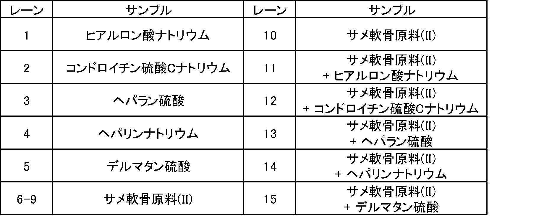
(6-2)ニトロセルロース膜を使用した評価結果
図3は、ニトロセルロース膜を用いた電気泳動で得られた酸性ムコ多糖標品及びサメ軟骨原料(I)のバンドパターンを示す写真である。図3(a)は、酸性ムコ多糖標品及びサメ軟骨原料(I)をそれぞれ電気泳動した結果であり、図3(b)は、酸性ムコ多糖標品及びサメ軟骨原料(I)を重ねてスポットして電気泳動した結果である。図3(a)のレーン1~8及び図3(b)のレーン9~14のサンプルを表3に示す。レーン6~8のサメ軟骨原料(I)はそれぞれロットが異なり、レーン9~14で使用したサメ軟骨原料(I)は、レーン6と同じロットのものである。参考として、図9に、図3に示す写真のバンドパターンの模式図を示す((a):図3(a)の模式図、(b):図3(b)の模式図)。
図3は、ニトロセルロース膜を用いた電気泳動で得られた酸性ムコ多糖標品及びサメ軟骨原料(I)のバンドパターンを示す写真である。図3(a)は、酸性ムコ多糖標品及びサメ軟骨原料(I)をそれぞれ電気泳動した結果であり、図3(b)は、酸性ムコ多糖標品及びサメ軟骨原料(I)を重ねてスポットして電気泳動した結果である。図3(a)のレーン1~8及び図3(b)のレーン9~14のサンプルを表3に示す。レーン6~8のサメ軟骨原料(I)はそれぞれロットが異なり、レーン9~14で使用したサメ軟骨原料(I)は、レーン6と同じロットのものである。参考として、図9に、図3に示す写真のバンドパターンの模式図を示す((a):図3(a)の模式図、(b):図3(b)の模式図)。
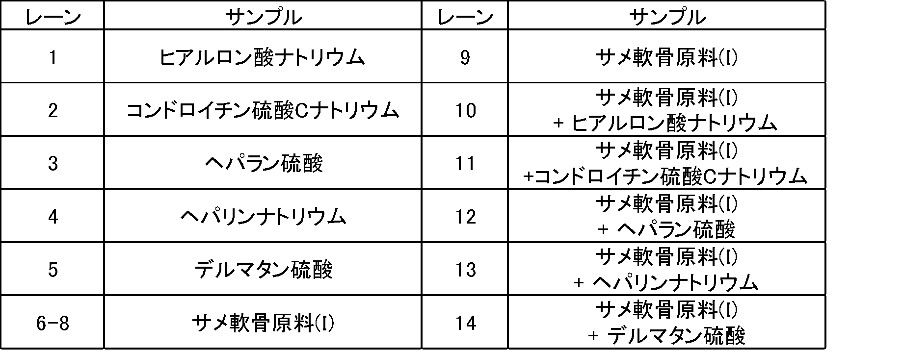
図4は、ニトロセルロース膜を用いた電気泳動で得られた酸性ムコ多糖標品及びサメ軟骨原料(II)のバンドパターンを示す写真である。図4(a)は、酸性ムコ多糖標品及びサメ軟骨原料(II)をそれぞれ電気泳動した結果を示す。図4(b)は、酸性ムコ多糖標品及びサメ軟骨原料(II)を重ねてスポットして電気泳動した結果である。図4(a)のレーン1~8及び図4(b)のレーン9~14のサンプルを表4に示す。レーン6~8のサメ軟骨原料(II)はそれぞれロットが異なり、レーン9~14で使用したサメ軟骨原料(II)は、レーン6と同じロットのものである。参考として、図10に、図4に示す写真のバンドパターンの模式図を示す((a):図4(a)の模式図、(b):図4(b)の模式図)。
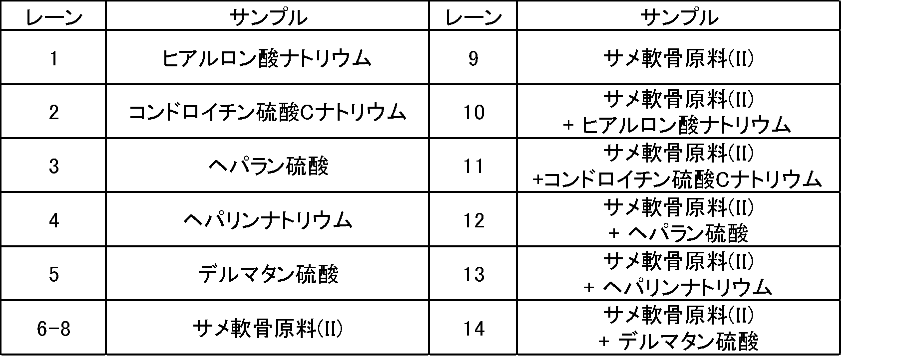
(6-3)ニトロセルロースとセルロースアセテートとの混合膜
ニトロセルロースとセルロースアセテートとの混合膜を使用した場合も、図3及び図4に示すニトロセルロース膜を使用した場合と同様の結果(バンドパターン)が得られた。
ニトロセルロースとセルロースアセテートとの混合膜を使用した場合も、図3及び図4に示すニトロセルロース膜を使用した場合と同様の結果(バンドパターン)が得られた。
(6-4)酵素加水分解によるコンドロイチン硫酸の確認の結果
図5は、セルロースアセテート膜又はニトロセルロース膜を用いた電気泳動で得られた、サメ軟骨原料の酵素処理物及び酸性ムコ多糖標品の酵素処理物のバンドパターンを示す写真である((a):セルロースアセテート膜、(b):ニトロセルロース膜)。図5(a)及び(b)のレーン1~8のサンプルを表5に示す。表5中、酵素処理有は、上記の酵素処理物(酵素分解サンプル)、酵素処理無は、上記でアクチナーゼE及びコンドロイチナーゼABC処理をしなかったサンプルである。電気泳動の際にスポットしたサンプル量は、サメ軟骨原料又はその酵素処理物は、2.0μg、酸性ムコ多糖標品又はその酵素処理物は、0.8μgである。参考として、図11に、図5に示す写真のバンドパターンの模式図を示す((a):図5(a)の模式図、(b):図5(b)の模式図)。
図5は、セルロースアセテート膜又はニトロセルロース膜を用いた電気泳動で得られた、サメ軟骨原料の酵素処理物及び酸性ムコ多糖標品の酵素処理物のバンドパターンを示す写真である((a):セルロースアセテート膜、(b):ニトロセルロース膜)。図5(a)及び(b)のレーン1~8のサンプルを表5に示す。表5中、酵素処理有は、上記の酵素処理物(酵素分解サンプル)、酵素処理無は、上記でアクチナーゼE及びコンドロイチナーゼABC処理をしなかったサンプルである。電気泳動の際にスポットしたサンプル量は、サメ軟骨原料又はその酵素処理物は、2.0μg、酸性ムコ多糖標品又はその酵素処理物は、0.8μgである。参考として、図11に、図5に示す写真のバンドパターンの模式図を示す((a):図5(a)の模式図、(b):図5(b)の模式図)。
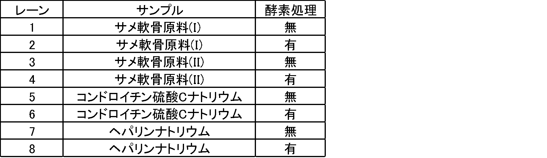
いずれのサメ軟骨原料(メーカー及びLot.違い)も、図1~2のレーン6~10及び図3~4のレーン6~9に示す通り、膜電気泳動後にアルシアンブルーにより染色されるバンドは一つであった。セルロースアセテート膜を用いた電気泳動において、サメ軟骨原料と酸性ムコ多糖標品を重ねてスポットしたところ、サメ軟骨原料で観察されるバンドはヒアルロン酸ナトリウム、ヘパラン硫酸、デルマタン硫酸のバンドとは移動距離が一致しないことが分かった(図1及び2のレーン11、13及び15)。サメ軟骨原料と酸性ムコ多糖標品を重ねてスポットしたところ、サメ軟骨原料のバンドと標品のコンドロイチン硫酸ナトリウムとは一致したが(図1及び2のレーン12)、ヘパリンナトリウムと重ねたスポットはバンドが拡がった(図1及び2のレーン14)。ニトロセルロース膜を用いた電気泳動を行ったところ、サメ軟骨原料のバンドと標品のコンドロイチン硫酸ナトリウムのバンドのみが一致し、他の酸性ムコ多糖のバンドとは一致しなかった(図3及び4のレーン10~14)。このことから、サメ軟骨由来原料に含有される酸性ムコ多糖はコンドロイチン硫酸のみであり、その他の酸性ムコ多糖は含まれないことが確認された。
上記のように、セルロースアセテート膜電気泳動においては、サメ軟骨原料(I)及び(II)由来のバンドは、コンドロイチン硫酸ナトリウムとバンドが重なった。また、サメ軟骨原料をヘパリンナトリウムと重ねたスポットはバンドが拡がった。サメ軟骨原料(I)及び(II)、並びに、コンドロイチン硫酸ナトリウムとヘパリンナトリウムの標品について上記(5-4)に記載の方法でコンドロイチナーゼABCによる酵素加水分解を行い、上記の膜電気泳動を行ったところ、サメ軟骨原料(I)及び(II)並びにコンドロイチン硫酸ナトリウムの標品のみバンドの消失が認められた(図5)。
これまでサメ軟骨には、酸性ムコ多糖としてコンドロイチン硫酸の他、ヒアルロン酸やデルマタン硫酸の含有が報告されている(Higashi K., et al, PloS One, 10(6), e0131502(2015).)。これらに加えて、代表的な酸性ムコ多糖であるヘパラン硫酸、ヘパリンナトリウムを標品に加え、サメ軟骨原料(I)及び(II)の膜電気泳動を行った。セルロースアセテート膜で評価したところ、本原料から検出されるバンドは一つで、コンドロイチン硫酸のバンドと移動距離が一致したが、ヘパリンともバンドが一部重なった。支持体をニトロセルロース膜に変えて評価したところ、本原料のバンドはコンドロイチン硫酸と移動距離が一致した。さらにサメ軟骨原料の酸性ムコ多糖の確認のため、コンドロイチナーゼABCによる酵素加水分解を行ったところ、サメ軟骨原料及びコンドロイチン硫酸の標品のバンドの消失が認められたことから、サメ軟骨原料に含まれる酸性ムコ多糖はコンドロイチナーゼABCによる分解を受ける性質であることが確認できた。アルシアンブルーにより染色されるバンドが一つであることとその移動距離がコンドロイチン硫酸と一致することから、本原料に含有される酸性ムコ多糖はコンドロイチン硫酸のみであり、その他の酸性ムコ多糖は含まれないことが確認された。さらに上記バンドはコンドロイチナーゼABCにより分解する性質であることからも、本原料に含有される酸性ムコ多糖はコンドロイチン硫酸のみであり、その他の酸性ムコ多糖は含まれないことが裏付けられた。一般的に比色法による定量は、化合物に含まれる官能基の特性を利用して行うため特異性が問題となる場合が多い。しかし、今回評価したサメ軟骨原料においてアルシアンブルー試薬により染色されるバンドはコンドロイチン硫酸のみであることから、当該原料についてアルシアンブルー比色定量により得られる定量結果はコンドロイチン硫酸又はその塩の含量を反映しているものといえる。
<実施例2>
実施例1で使用したサメ軟骨原料(I)及び(II)について、アルシアンブルー比色法にてコンドロイチン硫酸又はその塩の含量を定量した。アルシアンブルー比色法による定量は、食品衛生学雑誌(矢部芳枝他、食品中に添加したコンドロイチン硫酸ナトリウムの分析、28(1),p13-18(1987))に記載の方法に準じて行った。
実施例1で使用したサメ軟骨原料(I)及び(II)について、アルシアンブルー比色法にてコンドロイチン硫酸又はその塩の含量を定量した。アルシアンブルー比色法による定量は、食品衛生学雑誌(矢部芳枝他、食品中に添加したコンドロイチン硫酸ナトリウムの分析、28(1),p13-18(1987))に記載の方法に準じて行った。
(1)試液の調製
1)アクチナーゼE溶液
アクチナーゼE(科研製薬株式会社)0.5gにリン酸緩衝液を加え50mLとした。
2)アルシアンブルー試液
アルシアンブルー 8GX 1gに塩酸4.2mL、リン酸二水素ナトリウム1.38g及び水を加え100mLとして調製した。
3)リン酸緩衝液
0.2mol/Lリン酸二ナトリウム溶液305mLと0.2mol/Lリン酸一ナトリウム溶液195mLを混和し、水を加え1000mLとした。この液のpHは7であった。
1)アクチナーゼE溶液
アクチナーゼE(科研製薬株式会社)0.5gにリン酸緩衝液を加え50mLとした。
2)アルシアンブルー試液
アルシアンブルー 8GX 1gに塩酸4.2mL、リン酸二水素ナトリウム1.38g及び水を加え100mLとして調製した。
3)リン酸緩衝液
0.2mol/Lリン酸二ナトリウム溶液305mLと0.2mol/Lリン酸一ナトリウム溶液195mLを混和し、水を加え1000mLとした。この液のpHは7であった。
(2)標準溶液及び検量線用標準溶液の調製
コンドロイチン硫酸ナトリウム(特級)100mgを正確に量り、水で溶解し100mLに定容した(濃度:1000μg/mL)。この溶液を1mL分取し、水で正確に10mLとしたものを標準溶液とした(濃度:100μg/mL)。
この標準溶液を、1.0μg/mL、4.0μg/mL、7.0μg/mL、10μg/mL、20μg/mLになるように水で順次希釈したものを検量線用標準溶液とした。
コンドロイチン硫酸ナトリウム(特級)100mgを正確に量り、水で溶解し100mLに定容した(濃度:1000μg/mL)。この溶液を1mL分取し、水で正確に10mLとしたものを標準溶液とした(濃度:100μg/mL)。
この標準溶液を、1.0μg/mL、4.0μg/mL、7.0μg/mL、10μg/mL、20μg/mLになるように水で順次希釈したものを検量線用標準溶液とした。
(3)試験溶液の調製
粉砕した試料2gにpH7.0リン酸緩衝液を40mL加えた。さらに、アクチナーゼE溶液(10mg/mL)を5mL加えて振とう操作を行った。その後、40℃で一晩放置した後、水で100mLに定容した。ろ紙でろ過を行った溶液を、試験溶液とした。
粉砕した試料2gにpH7.0リン酸緩衝液を40mL加えた。さらに、アクチナーゼE溶液(10mg/mL)を5mL加えて振とう操作を行った。その後、40℃で一晩放置した後、水で100mLに定容した。ろ紙でろ過を行った溶液を、試験溶液とした。
(4)比色定量法
試験溶液1mLにアルシアンブルー試液を0.2mL加えて20分間放置した。析出した沈殿は、3000rpmで5分間遠心した。上澄液は捨て、次いで沈殿を水3mLで洗浄し、再び遠心分離を繰返した。得られた沈殿は、モノエタノールアミン10mLに溶解させて吸光度測定用試料とした。吸光度測定用試料について、分光光度計で615nmにおける吸光度を測定した。検量線用標準溶液についても同様の操作で発色を行ったものを使用した。検量線用標準溶液の吸光度から得られる検量線を用いて、試験溶液中のコンドロイチン硫酸又はその塩の量を算出した。ここで求められるコンドロイチン硫酸又はその塩の含量は、コンドロイチン硫酸ナトリウムとしての量である。
試験溶液1mLにアルシアンブルー試液を0.2mL加えて20分間放置した。析出した沈殿は、3000rpmで5分間遠心した。上澄液は捨て、次いで沈殿を水3mLで洗浄し、再び遠心分離を繰返した。得られた沈殿は、モノエタノールアミン10mLに溶解させて吸光度測定用試料とした。吸光度測定用試料について、分光光度計で615nmにおける吸光度を測定した。検量線用標準溶液についても同様の操作で発色を行ったものを使用した。検量線用標準溶液の吸光度から得られる検量線を用いて、試験溶液中のコンドロイチン硫酸又はその塩の量を算出した。ここで求められるコンドロイチン硫酸又はその塩の含量は、コンドロイチン硫酸ナトリウムとしての量である。
(5)結果
サメ軟骨原料(I)のコンドロイチン硫酸又はその塩の含量は、20.6g/100g、サメ軟骨原料(II)のコンドロイチン硫酸又はその塩の含量は、20.9g/100gであった。
サメ軟骨原料(I)のコンドロイチン硫酸又はその塩の含量は、20.6g/100g、サメ軟骨原料(II)のコンドロイチン硫酸又はその塩の含量は、20.9g/100gであった。
上記より、定性試験(実施例1)によりサメ軟骨原料(I)及び(II)に含まれる酸性ムコ多糖は、全てコンドロイチン硫酸(コンドロイチン硫酸100%)であることを確認した。また、比色法による定量試験(実施例2)により、これらのサメ軟骨原料(I)及び(II)のコンドロイチン硫酸又はその塩の含有量がそれぞれ20.6g/100g、20.9g/100gであることが確認できた。定性試験(実施例1)で、サメ軟骨原料(I)及び(II)に含まれる酸性ムコ多糖又はその塩が、コンドロイチン硫酸又はその塩のみであることが確認されたことから、定量試験(実施例2)により得られた定量結果は、サメ軟骨原料(I)及び(II)中のコンドロイチン硫酸又はその塩の含量である。
従って、試料を膜電気泳動に供して、アルシアンブルー染色で得られるバンドパターンから当該試料に含まれる酸性ムコ多糖又はその塩の種類を確認する定性試験と、比色法による酸性ムコ多糖又はその塩の定量試験を行うことにより、簡便な工程で、試料中の酸性ムコ多糖又はその塩の種類の確認及び定量を行うことができた。
従って、試料を膜電気泳動に供して、アルシアンブルー染色で得られるバンドパターンから当該試料に含まれる酸性ムコ多糖又はその塩の種類を確認する定性試験と、比色法による酸性ムコ多糖又はその塩の定量試験を行うことにより、簡便な工程で、試料中の酸性ムコ多糖又はその塩の種類の確認及び定量を行うことができた。
<試験例1>
コンドロイチン硫酸又はその塩がアクチナーゼEによって分解されないことを確認するため、以下の試験を行った。コンドロイチン硫酸Cナトリウム、サメ軟骨原料(I)及び(II)等の試薬、原料等は実施例1と同じである。
コンドロイチン硫酸又はその塩がアクチナーゼEによって分解されないことを確認するため、以下の試験を行った。コンドロイチン硫酸Cナトリウム、サメ軟骨原料(I)及び(II)等の試薬、原料等は実施例1と同じである。
(1)分析用サンプルの調製
(1-1)サメ軟骨原料の酵素分解サンプル(アクチナーゼ(+)、コンドロイチナーゼ(+))
実施例1の(5-4-2)(サメ軟骨原料の酵素分解)に記載の方法で、サメ軟骨原料(I)の酵素処理サンプル及びサメ軟骨原料(II)の酵素処理サンプル(アクチナーゼE及びコンドロイチナーゼABCで処理したサンプル)を調製した。
(1-1)サメ軟骨原料の酵素分解サンプル(アクチナーゼ(+)、コンドロイチナーゼ(+))
実施例1の(5-4-2)(サメ軟骨原料の酵素分解)に記載の方法で、サメ軟骨原料(I)の酵素処理サンプル及びサメ軟骨原料(II)の酵素処理サンプル(アクチナーゼE及びコンドロイチナーゼABCで処理したサンプル)を調製した。
(1-2)サメ軟骨原料のアクチナーゼ処理サンプル(アクチナーゼ(+)、コンドロイチナーゼ(-))
サメ軟骨原料をコンドロイチナーゼABCで処理しなかった以外は、上記(1-1)と同じ方法でサンプルを調製した。具体的には、実施例1の(5-4-2)に記載の方法において、手順4)においてコンドロイチナーゼABC液の代わりに同量の蒸留水を加えた。上記以外は、実施例1の(5-4-2)に記載の方法で、サメ軟骨原料(I)のアクチナーゼ処理サンプル及びサメ軟骨原料(II)のアクチナーゼ処理サンプル(アクチナーゼE処理を行い、コンドロイチナーゼABC処理を行わなかったサンプル)を調製した。
サメ軟骨原料をコンドロイチナーゼABCで処理しなかった以外は、上記(1-1)と同じ方法でサンプルを調製した。具体的には、実施例1の(5-4-2)に記載の方法において、手順4)においてコンドロイチナーゼABC液の代わりに同量の蒸留水を加えた。上記以外は、実施例1の(5-4-2)に記載の方法で、サメ軟骨原料(I)のアクチナーゼ処理サンプル及びサメ軟骨原料(II)のアクチナーゼ処理サンプル(アクチナーゼE処理を行い、コンドロイチナーゼABC処理を行わなかったサンプル)を調製した。
(1-3)サメ軟骨原料の酵素処理なしサンプル(アクチナーゼ(-)、コンドロイチナーゼ(-))
サメ軟骨原料をアクチナーゼE及びコンドロイチナーゼABCで処理しなかった以外は、上記(1-1)と同じ方法でサンプルを調製した。具体的には、実施例1の(5-4-2)に記載の方法において、手順2)及び4)においてアクチナーゼE液(II)及びコンドロイチナーゼABC液の代わりに同量の蒸留水を加えた。上記以外は、実施例1の(5-4-2)に記載の方法で、サメ軟骨原料(I)の酵素処理なしサンプル及びサメ軟骨原料(II)の酵素処理なしサンプル(アクチナーゼE処理及びコンドロイチナーゼABC処理を行わなかったサンプル)を調製した。
サメ軟骨原料をアクチナーゼE及びコンドロイチナーゼABCで処理しなかった以外は、上記(1-1)と同じ方法でサンプルを調製した。具体的には、実施例1の(5-4-2)に記載の方法において、手順2)及び4)においてアクチナーゼE液(II)及びコンドロイチナーゼABC液の代わりに同量の蒸留水を加えた。上記以外は、実施例1の(5-4-2)に記載の方法で、サメ軟骨原料(I)の酵素処理なしサンプル及びサメ軟骨原料(II)の酵素処理なしサンプル(アクチナーゼE処理及びコンドロイチナーゼABC処理を行わなかったサンプル)を調製した。
(1-4)コンドロイチン硫酸Cナトリウムの酵素処理サンプル(アクチナーゼ(+)、コンドロイチナーゼ(+))
実施例1の(5-4-1)(酸性ムコ多糖標品の酵素分解)に記載の方法で、標品のコンドロイチン硫酸CナトリウムをアクチナーゼE及びコンドロイチナーゼABCで処理して、コンドロイチン硫酸Cナトリウムの酵素処理サンプルを調製した。
実施例1の(5-4-1)(酸性ムコ多糖標品の酵素分解)に記載の方法で、標品のコンドロイチン硫酸CナトリウムをアクチナーゼE及びコンドロイチナーゼABCで処理して、コンドロイチン硫酸Cナトリウムの酵素処理サンプルを調製した。
(1-5)コンドロイチン硫酸Cナトリウムのアクチナーゼ処理サンプル(アクチナーゼ(+)、コンドロイチナーゼ(-))
コンドロイチン硫酸CナトリウムをコンドロイチナーゼABCで処理しなかった以外は、上記(1-4)と同じ方法でサンプルを調製した。具体的には、実施例1の(5-4-1)に記載の方法において、手順4)においてコンドロイチナーゼABC液の代わりに同量の蒸留水を加えた。上記以外は、実施例1の(5-4-1)に記載の方法で、コンドロイチン硫酸Cナトリウムのアクチナーゼ処理サンプルを調製した。
コンドロイチン硫酸CナトリウムをコンドロイチナーゼABCで処理しなかった以外は、上記(1-4)と同じ方法でサンプルを調製した。具体的には、実施例1の(5-4-1)に記載の方法において、手順4)においてコンドロイチナーゼABC液の代わりに同量の蒸留水を加えた。上記以外は、実施例1の(5-4-1)に記載の方法で、コンドロイチン硫酸Cナトリウムのアクチナーゼ処理サンプルを調製した。
(1-6)コンドロイチン硫酸Cナトリウムの酵素処理なしサンプル(アクチナーゼ(-)、コンドロイチナーゼ(-))
コンドロイチン硫酸CナトリウムをアクチナーゼE及びコンドロイチナーゼABCで処理しなかった以外は、上記(1-4)と同じ方法でサンプルを調製した。具体的には、実施例1の(5-4-1)に記載の方法において、コンドロイチン硫酸Cナトリウム(標品)を使用して、手順2)及び4)においてアクチナーゼE液(I)及びコンドロイチナーゼABC液の代わりに同量の蒸留水を加えた。上記以外は、実施例1の(5-4-1)に記載の方法で、コンドロイチン硫酸Cナトリウムの酵素処理なしサンプルを調製した。
コンドロイチン硫酸CナトリウムをアクチナーゼE及びコンドロイチナーゼABCで処理しなかった以外は、上記(1-4)と同じ方法でサンプルを調製した。具体的には、実施例1の(5-4-1)に記載の方法において、コンドロイチン硫酸Cナトリウム(標品)を使用して、手順2)及び4)においてアクチナーゼE液(I)及びコンドロイチナーゼABC液の代わりに同量の蒸留水を加えた。上記以外は、実施例1の(5-4-1)に記載の方法で、コンドロイチン硫酸Cナトリウムの酵素処理なしサンプルを調製した。
(2)セルロースアセテート膜を用いた評価
上記(1)の(1-1)~(1-6)で調製したサンプルを用いて、実施例1の(5-1)のセルロースアセテート膜を用いた評価の方法で分析を行った。
膜電気泳動の結果を、図6に示す。
上記(1)の(1-1)~(1-6)で調製したサンプルを用いて、実施例1の(5-1)のセルロースアセテート膜を用いた評価の方法で分析を行った。
膜電気泳動の結果を、図6に示す。
図6は、サメ軟骨原料及びコンドロイチン硫酸Cナトリウムの酵素処理なしサンプル、アクチナーゼ処理サンプル、並びに、酵素処理サンプルをセルロースアセテート膜で電気泳動したバンドパターンを示す写真である。表6に、図6のレーン1~9のサンプルを示す。アクチナーゼE処理を行ったサンプルはアクチナーゼ処理の欄に「+」、行わなかったサンプルは「-」と記載した。コンドロイチナーゼABC処理を行ったサンプルはコンドロイチナーゼ処理の欄に「+」、行わなかったサンプルは「-」と記載した。膜にスポットしたサンプル量は、レーン1~6は5.0μg、レーン7~9は0.8μgであった。参考として、図12に、図6に示す写真のバンドパターンの模式図を示す。
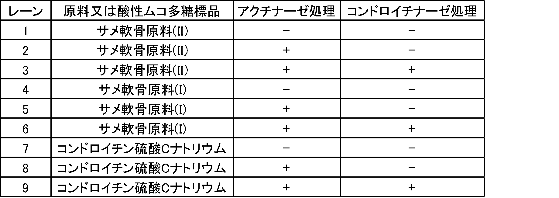
図6のレーン1、4及び7は、酵素処理なしサンプルである。レーン2、5及び8は、アクチナーゼ処理サンプルである。図6のレーン1、2、4、5及び8では、レーン7(コンドロイチン硫酸Cナトリウムの標品)と同じ位置にバンドが検出された。レーン3、6及び9は、アクチナーゼE及びコンドロイチナーゼABCで処理した酵素処理サンプルである。レーン3、6及び9では、コンドロイチン硫酸Cナトリウム由来のバンドが消失した。これらの結果から、コンドロイチン硫酸又はその塩は、コンドロイチナーゼABCで分解されるが、アクチナーゼEでは分解されないことが確認された。
Claims (10)
- 酸性ムコ多糖又はその塩を含む被検試料を、定性試験及び定量試験に供することを含み、
前記定性試験は、前記被検試料を、膜を用いた電気泳動に供する電気泳動工程、
電気泳動後の膜を、アルシアンブルーで染色する染色工程、及び、
前記染色で得られるバンドパターンから、前記被検試料中の酸性ムコ多糖又はその塩の種類を確認する検出工程を含み、
前記定量試験は、前記被検試料中の酸性ムコ多糖又はその塩を、酸性ムコ多糖又はその塩を検出可能な色素を用いる比色法により定量する比色定量工程を含む、
前記被検試料に含まれる酸性ムコ多糖又はその塩の分析方法。 - 前記酸性ムコ多糖が、コンドロイチン硫酸、ヒアルロン酸、デルマタン硫酸、ヘパリン及びヘパラン硫酸からなる群より選択される1以上である請求項1に記載の分析方法。
- 前記酸性ムコ多糖が、コンドロイチン硫酸である請求項1又は2に記載の分析方法。
- 前記酸性ムコ多糖又はその塩を検出可能な色素が、アルシアンブルー又はカルバゾールである請求項1~3のいずれか一項に記載の分析方法。
- 前記酸性ムコ多糖又はその塩を検出可能な色素が、アルシアンブルーである請求項1~4のいずれか一項に記載の分析方法。
- 前記定性試験によって、前記被検試料に含まれる酸性ムコ多糖又はその塩が1種であることが確認された場合に、前記定量試験により得られる定量結果を、前記酸性ムコ多糖又はその塩の含量とする、請求項1~5のいずれか一項に記載の分析方法。
- 前記膜は、ニトロセルロース膜、セルロースアセテート膜又はニトロセルロースとセルロースアセテートとの混合膜である請求項1~6のいずれか一項に記載の分析方法。
- 前記膜は、ニトロセルロース膜又はセルロースアセテート膜である請求項1~7のいずれか一項に記載の分析方法。
- コンドロイチン硫酸又はその塩を含む被検試料を、定性試験及び定量試験に供することを含み、
前記定性試験は、前記被検試料を、膜を用いた電気泳動に供する電気泳動工程、
電気泳動後の膜を、アルシアンブルーで染色する染色工程、及び、
前記染色で得られるバンドパターンから、前記被検試料中の酸性ムコ多糖又はその塩の種類を確認する検出工程を含み、
前記定量試験は、前記被検試料中の酸性ムコ多糖又はその塩を、酸性ムコ多糖又はその塩を検出可能な色素を用いる比色法により定量する比色定量工程を含み、
前記定性試験によって、前記被検試料に含まれる酸性ムコ多糖又はその塩がコンドロイチン硫酸又はその塩のみであることが確認された場合に、前記定量試験により得られる定量結果を、前記コンドロイチン硫酸又はその塩の含量とする、
前記被検試料に含まれるコンドロイチン硫酸又はその塩の定量方法。 - 酸性ムコ多糖又はその塩を含む製品の品質管理試験方法であって、
前記製品を被検試料として、請求項1~8のいずれか一項に記載の分析方法により分析する被検試料分析工程、
濃度及び種類が既知の酸性ムコ多糖又はその塩を含む標準試料を、前記分析方法により分析する標準試料分析工程、及び、
前記分析で得られる被検試料の定性試験結果及び定量試験結果と、標準試料の定性試験結果及び定量試験結果とを対比して、前記製品の品質を評価する評価工程を含む、
酸性ムコ多糖又はその塩を含む製品の品質管理試験方法。
Priority Applications (2)
| Application Number | Priority Date | Filing Date | Title |
|---|---|---|---|
| CN201980043220.3A CN112352156A (zh) | 2018-06-29 | 2019-06-20 | 试验样品中所含的酸性粘多糖或其盐的分析方法、硫酸软骨素或其盐的定量方法及制品的品质管理试验方法 |
| JP2020527457A JP7384793B2 (ja) | 2018-06-29 | 2019-06-20 | 被検試料に含まれる酸性ムコ多糖又はその塩の分析方法、コンドロイチン硫酸又はその塩の定量方法及び製品の品質管理試験方法 |
Applications Claiming Priority (4)
| Application Number | Priority Date | Filing Date | Title |
|---|---|---|---|
| JP2018-124971 | 2018-06-29 | ||
| JP2018124971 | 2018-06-29 | ||
| JP2018-203804 | 2018-10-30 | ||
| JP2018203804 | 2018-10-30 |
Publications (1)
| Publication Number | Publication Date |
|---|---|
| WO2020004216A1 true WO2020004216A1 (ja) | 2020-01-02 |
Family
ID=68985086
Family Applications (1)
| Application Number | Title | Priority Date | Filing Date |
|---|---|---|---|
| PCT/JP2019/024482 Ceased WO2020004216A1 (ja) | 2018-06-29 | 2019-06-20 | 被検試料に含まれる酸性ムコ多糖又はその塩の分析方法、コンドロイチン硫酸又はその塩の定量方法及び製品の品質管理試験方法 |
Country Status (4)
| Country | Link |
|---|---|
| JP (1) | JP7384793B2 (ja) |
| CN (1) | CN112352156A (ja) |
| TW (1) | TWI848957B (ja) |
| WO (1) | WO2020004216A1 (ja) |
Families Citing this family (1)
| Publication number | Priority date | Publication date | Assignee | Title |
|---|---|---|---|---|
| CN114137056B (zh) * | 2021-10-14 | 2024-11-05 | 广州市东源药业科技有限公司 | 一种芦荟多糖有效成分检测的电泳方法 |
Citations (4)
| Publication number | Priority date | Publication date | Assignee | Title |
|---|---|---|---|---|
| JPH03191855A (ja) * | 1989-12-21 | 1991-08-21 | Olympus Optical Co Ltd | 電気泳動装置 |
| JP2005016989A (ja) * | 2003-06-23 | 2005-01-20 | Taisho Pharmaceut Co Ltd | コンドロイチン硫酸又はその塩の分析方法 |
| JP2005024336A (ja) * | 2003-06-30 | 2005-01-27 | Japan Science & Technology Corp | 酸性多糖類の分析方法および酸性多糖類分析用キット |
| JP2018083810A (ja) * | 2008-11-25 | 2018-05-31 | ティシュージーン,インク. | プライミング細胞療法 |
Family Cites Families (2)
| Publication number | Priority date | Publication date | Assignee | Title |
|---|---|---|---|---|
| JP4696236B2 (ja) * | 2005-02-16 | 2011-06-08 | 国立大学法人広島大学 | コンドロイチンの製造方法 |
| EP2326336B1 (en) * | 2008-07-30 | 2015-05-13 | Mesynthes Limited | Tissue scaffolds derived from forestomach extracellular matrix |
-
2019
- 2019-06-20 JP JP2020527457A patent/JP7384793B2/ja active Active
- 2019-06-20 CN CN201980043220.3A patent/CN112352156A/zh active Pending
- 2019-06-20 WO PCT/JP2019/024482 patent/WO2020004216A1/ja not_active Ceased
- 2019-06-27 TW TW108122603A patent/TWI848957B/zh active
Patent Citations (4)
| Publication number | Priority date | Publication date | Assignee | Title |
|---|---|---|---|---|
| JPH03191855A (ja) * | 1989-12-21 | 1991-08-21 | Olympus Optical Co Ltd | 電気泳動装置 |
| JP2005016989A (ja) * | 2003-06-23 | 2005-01-20 | Taisho Pharmaceut Co Ltd | コンドロイチン硫酸又はその塩の分析方法 |
| JP2005024336A (ja) * | 2003-06-30 | 2005-01-27 | Japan Science & Technology Corp | 酸性多糖類の分析方法および酸性多糖類分析用キット |
| JP2018083810A (ja) * | 2008-11-25 | 2018-05-31 | ティシュージーン,インク. | プライミング細胞療法 |
Non-Patent Citations (1)
| Title |
|---|
| VOLPI, NICOLA: "Electrophoresis Separation of Glycosaminoglycans on Nitrocellulose Membranes", ANALYTICAL BIOCHEMISTRY, vol. 240, no. 0337, 1996, pages 114 - 118 * |
Also Published As
| Publication number | Publication date |
|---|---|
| TW202012914A (zh) | 2020-04-01 |
| CN112352156A (zh) | 2021-02-09 |
| JPWO2020004216A1 (ja) | 2021-08-02 |
| TWI848957B (zh) | 2024-07-21 |
| JP7384793B2 (ja) | 2023-11-21 |
Similar Documents
| Publication | Publication Date | Title |
|---|---|---|
| Felz et al. | Chemical characterization methods for the analysis of structural extracellular polymeric substances (EPS) | |
| Auray-Blais et al. | Efficient analysis of urinary glycosaminoglycans by LC-MS/MS in mucopolysaccharidoses type I, II and VI | |
| Uslu et al. | Electrooxidation of the antiviral drug valacyclovir and its square-wave and differential pulse voltammetric determination in pharmaceuticals and human biological fluids | |
| Casu et al. | Infrared spectra of glycosaminoglycans in deuterium oxide and deuterium chloride solution: Quantitative evaluation of uronic acid and acetamidodeoxyhexose moieties | |
| US9841401B2 (en) | Capillary electrophoresis method for analyzing collagen | |
| Estrella et al. | Graphitized carbon LC− MS characterization of the chondroitin sulfate oligosaccharides of aggrecan | |
| Han et al. | Structural analysis of urinary glycosaminoglycans from healthy human subjects | |
| Hennessey et al. | Urinary glycosaminoglycan excretion as a biochemical marker in patients with bladder carcinoma | |
| Deng et al. | Simultaneous determination of eight monoalkyl phthalate esters in porcine tissue by solid-phase extraction and liquid chromatography–tandem mass spectrometry | |
| Hsieh et al. | Quantitative analysis of oligosaccharides derived from sulfated glycosaminoglycans by nanodiamond-based affinity purification and matrix-assisted laser desorption/ionization mass spectrometry | |
| Mohamed et al. | Implementation of infrared and Raman modalities for glycosaminoglycan characterization in complex systems | |
| Monakhova et al. | Nuclear magnetic resonance spectroscopy as a tool for the quantitative analysis of water and ions in pharmaceuticals: Example of heparin | |
| Yates et al. | Recent innovations in the structural analysis of heparin | |
| Roberts et al. | Separation of high-molecular-mass carrageenan polysaccharides by capillary electrophoresis with laser-induced fluorescence detection | |
| JP7384793B2 (ja) | 被検試料に含まれる酸性ムコ多糖又はその塩の分析方法、コンドロイチン硫酸又はその塩の定量方法及び製品の品質管理試験方法 | |
| Restaino et al. | High‐performance capillary electrophoresis to determine intact keratan sulfate and hyaluronic acid in animal origin chondroitin sulfate samples and food supplements | |
| Jia et al. | Dyes inspired sensor arrays for discrimination of glycosaminoglycans | |
| CN105136967B (zh) | 用于鉴别贡菊和亳菊的反相薄层色谱方法 | |
| Burmistrova et al. | Quality control of heparin injections: comparison of four established methods | |
| Burns et al. | Chondroitin Sulfate: A Critical Review of Generic and Specific Problems in Its Characterization and Determination—An Exemplar of a Material with an Unknown or Variable Composition (UVCB) | |
| Campanella et al. | Salicylic acid determination in cow urine and drugs using a bienzymatic sensor | |
| HK40045176A (en) | Method for analyzing acidic mucopolysaccaride or salt thereof contained in test specimen, method for quantitative determination of chondroitin sulfate or salt thereof, and method for testing product quality control | |
| Liu et al. | Hemagglutinating activity and conformation of a lactose-binding lectin from mushroom Agrocybe cylindracea | |
| Pennock et al. | Screening for mucopolysaccharidoses | |
| de Vooys et al. | Investigating cracks and crazes on coated steel with simultaneous SVET and EIS |
Legal Events
| Date | Code | Title | Description |
|---|---|---|---|
| 121 | Ep: the epo has been informed by wipo that ep was designated in this application |
Ref document number: 19827012 Country of ref document: EP Kind code of ref document: A1 |
|
| ENP | Entry into the national phase |
Ref document number: 2020527457 Country of ref document: JP Kind code of ref document: A |
|
| NENP | Non-entry into the national phase |
Ref country code: DE |
|
| 122 | Ep: pct application non-entry in european phase |
Ref document number: 19827012 Country of ref document: EP Kind code of ref document: A1 |