WO2019198731A1 - 抗体断片及び該抗体断片を含む二重特異性抗体 - Google Patents
抗体断片及び該抗体断片を含む二重特異性抗体 Download PDFInfo
- Publication number
- WO2019198731A1 WO2019198731A1 PCT/JP2019/015528 JP2019015528W WO2019198731A1 WO 2019198731 A1 WO2019198731 A1 WO 2019198731A1 JP 2019015528 W JP2019015528 W JP 2019015528W WO 2019198731 A1 WO2019198731 A1 WO 2019198731A1
- Authority
- WO
- WIPO (PCT)
- Prior art keywords
- seq
- amino acid
- acid sequence
- nos
- cdr1
- Prior art date
Links
Images


Classifications
-
- A—HUMAN NECESSITIES
- A61—MEDICAL OR VETERINARY SCIENCE; HYGIENE
- A61K—PREPARATIONS FOR MEDICAL, DENTAL OR TOILETRY PURPOSES
- A61K39/00—Medicinal preparations containing antigens or antibodies
- A61K39/395—Antibodies; Immunoglobulins; Immune serum, e.g. antilymphocytic serum
-
- A—HUMAN NECESSITIES
- A61—MEDICAL OR VETERINARY SCIENCE; HYGIENE
- A61P—SPECIFIC THERAPEUTIC ACTIVITY OF CHEMICAL COMPOUNDS OR MEDICINAL PREPARATIONS
- A61P35/00—Antineoplastic agents
-
- C—CHEMISTRY; METALLURGY
- C07—ORGANIC CHEMISTRY
- C07K—PEPTIDES
- C07K16/00—Immunoglobulins [IGs], e.g. monoclonal or polyclonal antibodies
- C07K16/18—Immunoglobulins [IGs], e.g. monoclonal or polyclonal antibodies against material from animals or humans
- C07K16/28—Immunoglobulins [IGs], e.g. monoclonal or polyclonal antibodies against material from animals or humans against receptors, cell surface antigens or cell surface determinants
- C07K16/30—Immunoglobulins [IGs], e.g. monoclonal or polyclonal antibodies against material from animals or humans against receptors, cell surface antigens or cell surface determinants from tumour cells
-
- C—CHEMISTRY; METALLURGY
- C07—ORGANIC CHEMISTRY
- C07K—PEPTIDES
- C07K16/00—Immunoglobulins [IGs], e.g. monoclonal or polyclonal antibodies
- C07K16/46—Hybrid immunoglobulins
-
- C—CHEMISTRY; METALLURGY
- C12—BIOCHEMISTRY; BEER; SPIRITS; WINE; VINEGAR; MICROBIOLOGY; ENZYMOLOGY; MUTATION OR GENETIC ENGINEERING
- C12N—MICROORGANISMS OR ENZYMES; COMPOSITIONS THEREOF; PROPAGATING, PRESERVING, OR MAINTAINING MICROORGANISMS; MUTATION OR GENETIC ENGINEERING; CULTURE MEDIA
- C12N15/00—Mutation or genetic engineering; DNA or RNA concerning genetic engineering, vectors, e.g. plasmids, or their isolation, preparation or purification; Use of hosts therefor
- C12N15/09—Recombinant DNA-technology
- C12N15/11—DNA or RNA fragments; Modified forms thereof; Non-coding nucleic acids having a biological activity
- C12N15/62—DNA sequences coding for fusion proteins
-
- C—CHEMISTRY; METALLURGY
- C12—BIOCHEMISTRY; BEER; SPIRITS; WINE; VINEGAR; MICROBIOLOGY; ENZYMOLOGY; MUTATION OR GENETIC ENGINEERING
- C12N—MICROORGANISMS OR ENZYMES; COMPOSITIONS THEREOF; PROPAGATING, PRESERVING, OR MAINTAINING MICROORGANISMS; MUTATION OR GENETIC ENGINEERING; CULTURE MEDIA
- C12N15/00—Mutation or genetic engineering; DNA or RNA concerning genetic engineering, vectors, e.g. plasmids, or their isolation, preparation or purification; Use of hosts therefor
- C12N15/09—Recombinant DNA-technology
- C12N15/63—Introduction of foreign genetic material using vectors; Vectors; Use of hosts therefor; Regulation of expression
-
- C—CHEMISTRY; METALLURGY
- C12—BIOCHEMISTRY; BEER; SPIRITS; WINE; VINEGAR; MICROBIOLOGY; ENZYMOLOGY; MUTATION OR GENETIC ENGINEERING
- C12N—MICROORGANISMS OR ENZYMES; COMPOSITIONS THEREOF; PROPAGATING, PRESERVING, OR MAINTAINING MICROORGANISMS; MUTATION OR GENETIC ENGINEERING; CULTURE MEDIA
- C12N5/00—Undifferentiated human, animal or plant cells, e.g. cell lines; Tissues; Cultivation or maintenance thereof; Culture media therefor
- C12N5/10—Cells modified by introduction of foreign genetic material
Definitions
- the present invention includes an antibody fragment, a bispecific antibody comprising the antibody fragment, a nucleic acid molecule comprising a base sequence encoding the antibody fragment or the bispecific antibody, the antibody fragment or the bispecific antibody.
- the present invention relates to a pharmaceutical composition and a kit comprising the antibody fragment or the bispecific antibody.
- Immunotherapy is used as a safe therapeutic for cancer (malignant tumors).
- an antibody that specifically shows cytotoxic activity against cancer is used.
- Human epidermal growth factor receptor 2 (HER2) is a glycoprotein receptor-type tyrosine kinase (molecular weight of about 185 kDa), also called EGFR2, ErbB2, CD340, or NEU.
- HER2 is overexpressed in several cancers including breast cancer, including its mutant and defective types, and is attracting attention as a therapeutic target.
- An antibody targeting HER2 is one of cancer cell surface antigens that are expected to have therapeutic effects by targeting.
- the basic structure of an antibody is a structure in which a heavy chain and a light chain form a heterotetramer by a disulfide bond, but such an IgG-type antibody has a high molecular weight and cannot penetrate into the deep part of a tumor. Therefore, development of low molecular weight antibodies containing antibody variable region fragments is progressing.
- Non-patent Document 1 The variable region fragment of a camel-derived antibody (single Variable domain of the Heavy chain of a Heavy-chain camel antibody: VHH) has high affinity and high specificity despite the monomer domain.
- VHH that specifically binds to various targets has been obtained by performing in vitro selection operations such as phage display using a group of antibodies obtained by immunization of llamas (non-patent literature). twenty three).
- IgG-type antibodies cannot use T cells that are more active than natural killer cells, and low molecular weight antibodies that can use T cells have been developed.
- the valence of binding to the cell is 1, and the binding affinity with the antigen is low.
- Non-patent Documents 5 and 6 Non-patent Documents 5 and 6
- Increasing the binding affinity between antibody and antigen can be said to be an effective approach because it increases the damage activity due to the multivalent design of the antibody, but multivalent design by shortening and removing the linker sequence between the heavy and light chains Is effective to some extent for the formation of multivalent aggregates (Non-patent Documents 5 and 6), but has not been designed so that only a single multivalent aggregate is formed.
- An object of the present invention is to provide an antibody fragment that specifically binds to human HER2, a bispecific antibody having high cancer cytotoxicity comprising a variable region comprising such an antibody fragment, the antibody fragment or the bispecificity It is to provide a pharmaceutical composition containing an antibody and a kit containing the antibody fragment or the bispecific antibody.
- the present inventors adopted a bi-bivalent antibody composed of camel antibody VHH and scFvF (single chain antibody fragment), which are single domains developed by the present inventors, as a format.
- VHH camel antibody
- scFvF single chain antibody fragment
- FR1-CDR1-FR2-CDR2-FR3-CDR3-FR4 FR1, FR2, FR3, and FR4 are framework areas.
- CDR1, CDR2, and CDR3 are complementarity determining regions and satisfy at least one of the following (I) to (III):
- CDR1 has an amino acid sequence represented by any of SEQ ID NOs: 5 to 11, 53 to 55, and 59 to 64, or any one of SEQ ID NOs: 5 to 11, 53 to 55, and 59 to 64 Or an amino acid sequence represented by any one of SEQ ID NOs: 5 to 11, 53 to 55, and 59 to 64.
- CDR2 has the amino acid sequence represented by any of SEQ ID NOs: 12 to 18, or the amino acid sequence represented by any of SEQ ID NOs: 12 to 18 Having an amino acid sequence containing 1 to several deletions, substitutions or additions, or having an amino acid sequence having 80% or more identity with the amino acid sequence represented by any of SEQ ID NOs: 12 to 18
- III CDR3 is represented by any one of SEQ ID NOs: 19-25, 56-58, and 65-84 Having an amino acid sequence, or having an amino acid sequence containing 1 to several deletions, substitutions or additions in the amino acid sequence represented by any of SEQ ID NOs: 19-25, 56-58, and 65-84, Alternatively, it has an amino acid sequence having 80% or more identity with the amino acid sequence represented by any of SEQ ID NOs: 19 to 25, 56 to 58, and 65 to 84.
- the CDR1 has an amino acid sequence represented by any of SEQ ID NOs: 5 to 11, 53 to 55, and 59 to 64
- the CDR2 represents an amino acid sequence represented by any of SEQ ID NOs: 12 to 18.
- the antibody fragment according to [1] wherein the CDR3 has an amino acid sequence represented by any one of SEQ ID NOs: 19 to 25, 56 to 58, and 65 to 84.
- [5] The antibody fragment according to any one of [1] to [4], which has an amino acid sequence represented by any of SEQ ID NOs: 26 to 32.
- a first variable region comprising the antibody fragment of any one of [1] to [5], and a second variable region that specifically binds to an antigen different from HER2, A bispecific antibody, wherein the first variable region and the second variable region are contained on a single polypeptide chain.
- a nucleic acid molecule comprising a base sequence encoding the antibody fragment according to any one of [1] to [5] or the bispecific antibody according to any one of [6] to [9] .
- [12] An expression vector comprising the nucleic acid molecule according to [11]. [13] A host cell transformed with the expression vector according to [12]. [14] A pharmaceutical composition comprising the antibody fragment according to any one of [1] to [5] or the bispecific antibody according to any one of [6] to [9]. [15] A kit comprising the antibody fragment according to any one of [1] to [5] or the bispecific antibody according to any one of [6] to [9].
- the antibody fragment of the present invention can target tumor cells having human HER2 on the cell surface.
- bispecific antibodies comprising such antibody fragments as variable regions can specifically bind to tumor cells and have extremely high cytotoxicity even at low concentrations, so clinical and cancer cells were used. Useful for research.
- Lane 1 Precolumn, Lane 2: FT, Lane 3: 20 mM imidazole elution, Lane 4: 50 mM imidazole elution, Lane 5: 300 mM imidazole elution, Lane 6: 1 M imidazole elution, Lane 7: 20 mM imidazole elution, Lane 8: 50 mM imidazole Elution, lane 9: 300 mM imidazole elution, lane 10: 1 M imidazole elution. Lanes 3-6 were performed on column 1 and lanes 7-10 were performed on column 2. Antibody: 3 ⁇ L His-prob-HRP, conditions: increment, standard.
- Lane 1 Precolumn
- Lane 2 FT1st
- Lane 3 FT2nd
- Lane 4 PBS
- Lane 5 20 mM imidazole elution M
- Lane 6 50 mM imidazole elution
- Lane 7 300 mM imidazole elution
- Lane 8 1 M imidazole elution
- Lane 9 marker
- Lane 10 PBS
- Lane 11 20 mM imidazole elution
- Lane 12 50 mM imidazole elution
- Lane 13 300 mM imidazole elution
- Lane 14 1 M imidazole elution.
- Lanes 4-8 are the first FT
- lanes 10-14 are the second FT.
- Antibody 3 ⁇ L His-prob-HRP, conditions: increment, standard.
- C Size exclusion chromatography (SEC) using a fraction eluted with 300 mM Imidazole.
- D Size exclusion chromatography (SEC) using 1M Imidazole elution fraction.
- A Non-reducing SDS-PAGE of size exclusion chromatography (SEC) elution peak and
- B Western Blotting. Lanes 1-14 are the same as in FIG. 9A.
- Antibody 3 ⁇ L His-prob-HRP, conditions: increment, standard.
- A Single cloning of domain II-Fc stable expression cell line by limiting dilution method and expression level evaluation by ELISA.
- the horizontal axis corresponds to each strain, and the number of each strain is omitted.
- B Evaluation of expression level by ELISA after expansion of selected 12 clones.
- C SDS-PAGE after purification of 2G4, 3E10, and 4D10 clones, and (D) Western blotting results. Proteins indicated by arrows: Number of amino acids: 383, Molecular weight: 42047.65, theoretical pI: 6.52, Ext. Coefficient 44485. Detected with 3 ⁇ L ⁇ anti-human IgG-Fc HRP. Acrylamide concentration 12.5%. The result of the biopanning with respect to the HER2 sputum fragment protein prepared in Examples 3-5.
- A)-(F) output / input ratio (-) of phage in each round of FIGS. 12 (A)-(C).
- A)-(C) Evaluation of binding activity against 4 varieties of HER2 fragment protein using the eluted phages in each round of FIGS. 12 (A)-(C).
- A)-(F) Graph showing cytotoxicity evaluation by purified bispecific antibody. Schematic diagram illustrating the T cell recruiting antibody creation process (left) using standard techniques and the new creation process (right) without phage monoclonalization.
- A Schematic diagram showing the mechanism of action of antibody BiBian detection,
- B Calibration curve by purified BBiBian,
- C ⁇ BiBian concentration determination in medium supernatant by Tag-sandwich ELISA,
- D Fig. 18
- C Enlarged graph of Y axis.
- an antibody fragment that specifically binds to human HER2 and having an amino acid sequence of a single polypeptide chain having the structure of the following formula (1) .
- FR1-CDR1-FR2-CDR2-FR3-CDR3-FR4 FR1, FR2, FR3, and FR4 are framework areas.
- CDR1, CDR2, and CDR3 are complementarity determining regions, Satisfies at least one of the following (I) to (III):
- CDR1 has an amino acid sequence represented by any of SEQ ID NOs: 5 to 11, 53 to 55, and 59 to 64, or any one of SEQ ID NOs: 5 to 11, 53 to 55, and 59 to 64 Or an amino acid sequence represented by any one of SEQ ID NOs: 5 to 11, 53 to 55, and 59 to 64.
- CDR2 has the amino acid sequence represented by any of SEQ ID NOs: 12 to 18, or the amino acid sequence represented by any of SEQ ID NOs: 12 to 18 Having an amino acid sequence containing 1 to several deletions, substitutions or additions, or having an amino acid sequence having 80% or more identity with the amino acid sequence represented by any of SEQ ID NOs: 12 to 18
- III CDR3 is represented by any one of SEQ ID NOs: 19-25, 56-58, and 65-84 Having an amino acid sequence, or having an amino acid sequence containing 1 to several deletions, substitutions or additions in the amino acid sequence represented by any of SEQ ID NOs: 19-25, 56-58, and 65-84, Alternatively, it has an amino acid sequence having 80% or more identity with the amino acid sequence represented by any of SEQ ID NOs: 19 to 25, 56 to 58, and 65 to 84.
- SEQ ID NO: 5 corresponds to the amino acid sequence of amino acid sequence 26 to 35 of the variable region fragment of clone H44 having binding activity to HER2 antigen
- SEQ ID NO: 6 is the clone H48 having binding activity to HER2 antigen.
- SEQ ID NO: 7 corresponds to amino acid sequence 26-35 of the variable region fragment of clone d8D having binding activity to HER2 ⁇ antigen
- SEQ ID NO: 8 corresponds to amino acid sequence 26-35 of the variable region fragment of clone h11E having high binding to HER2 antigen
- SEQ ID NO: 9 is variable region fragment of clone f8C having high binding to HER2 antigen
- SEQ ID NO: 10 corresponds to the amino acid sequence of amino acid sequence 26 to 35, and the amino acid sequence of the variable region fragment of clone f10D having high binding to the HER2 antigen
- SEQ ID NO: 11 corresponds to amino acid sequence 26-35 of the amino acid sequence of the variable region fragment of clone f11E having high binding to HER2 antigen.
- SEQ ID NO: 12 corresponds to the amino acid sequence of amino acid sequence 50 to 56 of the variable region fragment of clone H44 having binding activity to HER2 antigen
- SEQ ID NO: 13 is the amino acid of variable region fragment of clone H48 having binding activity to HER2 antigen
- SEQ ID NO: 14 corresponds to the amino acid sequence 50 to 59 of the variable region fragment of clone d8D having binding activity to the HER2 antigen
- SEQ ID NO: 15 corresponds to the HER2 antigen.
- amino acid sequence 50-59 of the variable region fragment of clone h11E having high binding to SEQ ID NO: 16 is the amino acid sequence 50- of the variable region fragment of clone f8C having high binding to the HER2 antigen
- SEQ ID NO: 17 is the 50th to 59th amino acid sequence of the variable region fragment of clone f10D having high binding to the HER2 antigen
- SEQ ID NO: 18 corresponds to amino acid sequence 50-59 of the amino acid sequence of the variable region fragment of clone f11E having high binding to HER2 antigen.
- SEQ ID NO: 19 corresponds to amino acid sequence 96-114 of the variable region fragment of clone H44 having binding activity to HER2 antigen
- SEQ ID NO: 20 is the amino acid of variable region fragment of clone H48 having binding activity to HER2 antigen
- SEQ ID NO: 21 corresponds to the amino acid sequence 99 to 110 of the variable region fragment of clone d8D having binding activity to the HER2 ⁇ antigen
- SEQ ID NO: 22 corresponds to the amino acid sequence of the HER2 antigen.
- amino acid sequence 99 to 110th amino acid sequence of the variable region fragment of clone h11E having high binding to SEQ ID NO: 23 is the amino acid sequence 99 to 99 of variable region fragment of clone f8C having high binding to HER2 antigen
- amino acid sequence 99-104 of the variable region fragment of clone f10D having high binding to the HER2 antigen
- amino acid sequence, SEQ ID NO: 25 corresponding to the amino acid sequence 99-113 of the amino acid sequence of the variable region fragment of clone f11E having high binding to HER2 antigen.
- SEQ ID NO: 53 corresponds to the amino acid sequence of CDR1 of modified BiBian clone 6 ⁇ 3 having high binding affinity for HER2 (positions 54 to 59 of the amino acid sequence of SEQ ID NO: 52), and SEQ ID NOs: 54 to 55 and 59 ⁇ 64 are variants of CDR1 of such clone 6x3.
- SEQ ID NO: 56 corresponds to the amino acid sequence of CDR3 of the modified BiBian clone 6 ⁇ 3 having high binding affinity for HER2 (positions 118 to 122 of the amino acid sequence of SEQ ID NO: 52), and SEQ ID NOs: 65 to 71 are involved. It is a variant of CDR3 of clone 6 ⁇ 3.
- SEQ ID NO: 57 corresponds to the amino acid sequence of CDR3 of the modified BiBian clone 6 ⁇ 3 having high binding affinity for HER2 (positions 123 to 128 of the amino acid sequence of SEQ ID NO: 52), SEQ ID NO: 58 and SEQ ID NO: 72 ⁇ 84 are variants of CDR3 of such clone 6x3.
- sequences are pre-processed in an optimal state for comparison in order to determine sequence identity in two amino acid sequences or base sequences. For example, by making a gap in one sequence, the alignment with the other sequence is optimized. Thereafter, the amino acid residues or bases at each site are compared. When the same amino acid residue or base as the corresponding site in the second sequence is present at a site in the first sequence, the sequences are identical at that site. The identity in the two sequences is expressed as a percentage of the total number of sites (all amino acids or all bases) of the number of sites that are identical between the sequences.
- the identity of an amino acid sequence and a base sequence can be determined by comparing amino acid sequences using a known algorithm such as the Lipman-Pearson method (Science, 227, 1435, 1985).
- the target antigen of the antibody fragment is human HER2.
- Tumors include but are not limited to breast cancer, stomach cancer, ovarian cancer, uterine cancer, pancreatic cancer, colon cancer, rectal cancer, prostate cancer, salivary gland cancer, kidney cancer, and lung cancer. .
- FR1, FR2, FR3, and FR4 are not particularly limited, but preferably each have the amino acid sequence represented by SEQ ID NOs: 1 to 4, or one to several missing amino acid sequences represented by SEQ ID NOs: 1 to 4 It has an amino acid sequence containing a deletion, substitution or addition, or has an amino acid sequence having 70% or more identity with the amino acid sequence represented by SEQ ID NOs: 1 to 4.
- SEQ ID NOs: 1 to 4 are as follows.
- X 1 X 2 QLVESGGX 3 LVQX 4 GGSLRLSX 5 CAS (SEQ ID NO: 1) WX 1 RQAPGKX 2 REX 3 VA (SEQ ID NO: 2) YX 1 DSVKGRFTISRDNAKX 2 TVYLQMNSLKPEDTAVYYCX 3 X 4 (SEQ ID NO: 3) X 1 GX 2 GTX 3 VX 4 VSS (SEQ ID NO: 4)
- SEQ ID NO: 1 is Q or E
- X 2 is V or L
- X 3 is T or G
- X 4 is A or P
- X 5 is F, Y or V.
- X 1 is E, Q or G
- X 2 is F, L or W
- X 3 is S, A or T.
- X 1 is N, K or S
- X 2 is A, K, N or R
- X 3 is A, T, K or V
- X 4 is R or Q
- X 1 is W or R
- X 2 is R or Q
- X 3 is R or Q
- X 4 is T or I.
- FR1, FR2, FR3, and FR4 have an amino acid sequence containing 1 to several deletions, substitutions or additions in the amino acid sequences represented by SEQ ID NOs: 1 to 4, the number of deletions, substitutions or additions is: The number is preferably 0 to 3, more preferably 0, 1 or 2, and still more preferably 0 or 1.
- FR1, FR2, FR3, and FR4 have an amino acid sequence having 70% or more identity with the amino acid sequence represented by SEQ ID NO: 1-4, the identity is preferably 80% or more, and 90% or more More preferably.
- FR1, FR2, FR3, and FR4 of the antibody fragment have an amino acid sequence having at least 90% sequence identity with each of the amino acid sequences of SEQ ID NOs: 1 to 4.
- FR1, FR2, FR3, and FR4 have an amino acid sequence that is 95% sequence identical to each of the amino acid sequences of SEQ ID NOs: 1-4.
- FR1, FR2, FR3, and FR4 have an amino acid sequence that is at least 98% sequence identical to each of the amino acid sequences of SEQ ID NOs: 1-4.
- FR1, FR2, FR3, and FR4 have 100% sequence identity with each of the amino acid sequences of SEQ ID NOs: 1-4.
- CDR1 has the amino acid sequence represented by any of SEQ ID NOs: 5-11, 53-55, and 59-64, or of SEQ ID NOs: 5-11, 53-55, and 59-64
- the amino acid sequence represented by any one of the amino acid sequences including one to several deletions, substitutions or additions, or represented by any one of SEQ ID NOs: 5-11, 53-55, and 59-64 It has an amino acid sequence having 80% identity or more with the amino acid sequence.
- CDR1 has an amino acid sequence containing 1 to several deletions, substitutions or additions in the amino acid sequence represented by any of SEQ ID NOs: 5-11, 53-55, and 59-64
- the number of additions is preferably 0 to 3, more preferably 0, 1, or 2 and even more preferably 0 or 1.
- CDR1 has an amino acid sequence having 80% or more identity with the amino acid sequences represented by SEQ ID NOs: 5 to 11, 53 to 55, and 59 to 64, the identity is preferably 90% or more, 95 More preferably, it is at least%.
- CDR2 has the amino acid sequence represented by any of SEQ ID NOs: 12-18, or one to several deletions in the amino acid sequence represented by any of SEQ ID NOs: 12-18, It has an amino acid sequence including substitution or addition, or has an amino acid sequence having 80% or more identity with the amino acid sequence represented by any of SEQ ID NOs: 12 to 18.
- the number of deletions, substitutions or additions is 0 to 3 It is preferable that the number is 0, 1, or 2, more preferably 0 or 1.
- the identity is preferably 90% or more, and more preferably 95% or more.
- CDR3 has the amino acid sequence represented by any of SEQ ID NOs: 19-25, 56-58, and 65-84, or SEQ ID NOs: 19-25, 56-58, and 65-84. Having one to several deletions, substitutions or additions in the amino acid sequence represented by any of the above, or represented by any one of SEQ ID NOs: 19-25, 56-58, and 65-84 And an amino acid sequence having 80% identity or more.
- CDR3 has an amino acid sequence containing 1 to several deletions, substitutions or additions in the amino acid sequence represented by any of SEQ ID NOs: 19 to 25, 56 to 58, and 65 to 84, The number of additions is preferably 0 to 3, more preferably 0, 1, or 2 and even more preferably 0 or 1.
- CDR2 has an amino acid sequence having 80% or more identity with the amino acid sequences represented by 19 to 25, 56 to 58, and 65 to 84, the identity is preferably 90% or more, and 95% or more More preferably.
- CDR3 is 1 in the amino acid sequence represented by any of SEQ ID NOs: 56-58 and 65-84, and the amino acid sequence represented by any of SEQ ID NOs: 56-58 and 65-84.
- amino acid sequence containing several deletions, substitutions or additions, or an amino acid sequence having 80% or more identity with the amino acid sequence represented by any of SEQ ID NOs: 56 to 58 and 65 to 84 is 1 in the amino acid sequence represented by any of SEQ ID NOs: 56-58 and 65-84, and the amino acid sequence represented by any of SEQ ID NOs: 56-58 and 65-84.
- CDR3 comprises one to several deletions in the amino acid sequence represented by any of SEQ ID NOs: 56, 65-71, and the amino acid sequence represented by any of SEQ ID NOs: 56, 65-71
- An amino acid sequence comprising 1 to several deletions, substitutions or additions in the amino acid sequence represented by any of SEQ ID NOs: 57 to 58 and 72 to 84, or SEQ ID NOs: 57 to 58 and It has an amino acid sequence represented by any of 72 to 84 and an amino acid sequence having 80% or more identity.
- CDR3 has an amino acid sequence containing 1 to several deletions, substitutions or additions in the amino acid sequence represented by any of SEQ ID NOs: 19 to 25, 56 to 58, and 65 to 84
- the number of additions is preferably 0 to 3, more preferably 0, 1, or 2 and even more preferably 0 or 1.
- CDR2 has an amino acid sequence having 80% or more identity with the amino acid sequences represented by 19 to 25, 56 to 58, and 65 to 84, the identity is preferably 90% or more, and 95% or more More preferably.
- the antibody fragment is represented by any of CDR1, SEQ ID NO: 12-18 having the amino acid sequence represented by any of SEQ ID NOs: 5-11, 53-55, and 59-64. It has at least one of CDR2 having an amino acid sequence and CDR3 having an amino acid sequence represented by any of SEQ ID NOs: 19 to 25, 56 to 58, and 65 to 84.
- the antibody fragment has the amino acid sequence represented by any of SEQ ID NOs: 5-11, 53-55, and 59-64, SEQ ID NOs: 5-11, 53-55, and 59-64.
- An amino acid sequence comprising 1 to several deletions, substitutions or additions in the amino acid sequence represented by any of the above, or an amino acid sequence represented by any of SEQ ID NOs: 5-11, 53-55, and 59-64;
- An amino acid sequence comprising 1 to several deletions, substitutions or additions in the amino acid sequence represented by any one of the above, or the amino acid sequence represented by any one of SEQ ID NOs: 19 to 25, 56 to 58, and 65 to 84 And an amino acid sequence having 80% or more identity.
- the antibody fragment has the amino acid sequence represented by any of SEQ ID NOs: 5-11, 53-55, and 59-64, SEQ ID NOs: 5-11, 53-55, and 59-64.
- the antibody fragment has the amino acid sequence represented by any of SEQ ID NOs: 5-11, 53-55, and 59-64, SEQ ID NOs: 5-11, 53-55, and 59-64.
- An amino acid sequence comprising 1 to several deletions, substitutions or additions in the amino acid sequence represented by any of the above, or an amino acid sequence represented by any of SEQ ID NOs: 5-11, 53-55, and 59-64;
- the antibody fragment is represented by any of CDR1, SEQ ID NO: 12-18 having the amino acid sequence represented by any of SEQ ID NOs: 5-11, 53-55, and 59-64. It has at least one of CDR2 having an amino acid sequence and CDR3 having an amino acid sequence represented by any of SEQ ID NOs: 19 to 25, 56 to 58, and 65 to 84.
- the antibody fragment has CDR1, CDR2 and / or CDR3 of (i) to (xii) below.
- CDR1 SEQ ID NO: 5, CDR2: SEQ ID NO: 12, and CDR3: SEQ ID NO: 19
- CDR1 SEQ ID NO: 6, CDR2: SEQ ID NO: 13, and CDR3: SEQ ID NO: 20
- CDR1 SEQ ID NO: 7, CDR2: SEQ ID NO: 14, and CDR3: SEQ ID NO: 21
- CDR1 SEQ ID NO: 8
- CDR1 SEQ ID NO: 9, CDR2: SEQ ID NO: 16, and CDR3: SEQ ID NO: 23
- CDR1 SEQ ID NO: 10
- CDR2 SEQ ID NO: 17, and CDR3: SEQ ID NO: 24
- CDR1 SEQ ID NO: 11
- CDR1 SEQ ID NO: 11
- CDR1 SEQ ID NO
- GRTFSSYAMG SEQ ID NO: 5
- GNIFSLNAMA SEQ ID NO: 6
- GRSLSSYAMG SEQ ID NO: 7
- GLSFSTFTMG SEQ ID NO: 8
- GGTFWRYAMG SEQ ID NO: 9
- GRTFITNAMG SEQ ID NO: 10
- GSTFSRNVMS SEQ ID NO: 11
- AIRLKTD SEQ ID NO: 12
- RITTGGSPR SEQ ID NO: 13
- AISWSGGSTY SEQ ID NO: 14
- AIGRTGGPAA SEQ ID NO: 15
- HANWSGGRIY SEQ ID NO: 16
- AIRWNGASTY SEQ ID NO: 17
- TISSGGGTTS SEQ ID NO: 18
- WLRRGYGASWYLTGADPRY SEQ ID NO: 19
- VKFSPNIREY SEQ ID NO: 20
- GSVWDDTQGPYW SEQ ID NO: 21
- GSSWYRGDEYDY SEQ ID NO: 22
- the first variable region of the bispecific antibody can be designed based on a variable region that is a single domain in a cancer cell-bound camel antibody (hereinafter, camel antibody VHH).
- the antigen on the T cell is preferably an antigen located on the effector T cell, and as such an antigen, human CD2 antigen, human CD3 antigen, human CD5 antigen, human CD16 antigen, human CD25 antigen, human CD28 Examples include, but are not limited to, antigens, human NKG2D antigen, T cell receptor (TCR) and the like.
- the first variable region of the bispecific antibody has an amino acid sequence of a single polypeptide chain having the structure of the above formula (1).
- FR1-CDR1-FR2-CDR2-FR3-CDR3-FR4 FR1, FR2, FR3, and FR4 are framework areas.
- CDR1, CDR2, and CDR3 are complementarity determining regions and satisfy at least one of the following (I) to (III):
- CDR1 has an amino acid sequence represented by any of SEQ ID NOs: 5 to 11, 53 to 55, and 59 to 64, or any one of SEQ ID NOs: 5 to 11, 53 to 55, and 59 to 64 Or an amino acid sequence represented by any one of SEQ ID NOs: 5 to 11, 53 to 55, and 59 to 64.
- CDR2 has the amino acid sequence represented by any of SEQ ID NOs: 12 to 18, or the amino acid sequence represented by any of SEQ ID NOs: 12 to 18 Having an amino acid sequence containing 1 to several deletions, substitutions or additions, or having an amino acid sequence having 80% or more identity with the amino acid sequence represented by any of SEQ ID NOs: 12 to 18
- III CDR3 is represented by any one of SEQ ID NOs: 19-25, 56-58, and 65-84 Having an amino acid sequence, or having an amino acid sequence containing 1 to several deletions, substitutions or additions in the amino acid sequence represented by any of SEQ ID NOs: 19-25, 56-58, and 65-84, Alternatively, it has an amino acid sequence having 80% or more identity with the amino acid sequence represented by any of SEQ ID NOs: 19 to 25, 56 to 58, and 65 to 84.
- the FR1, FR2, FR3, and FR4 of the first variable region of the bispecific antibody are not particularly limited.
- FR1, FR2, FR3, and FR4 of the first variable region of the bispecific antibody have an amino acid sequence represented by SEQ ID NOs: 1-4 or represented by SEQ ID NOs: 1-4.
- the amino acid sequence has an amino acid sequence containing 1 to several deletions, substitutions or additions, or has an amino acid sequence having 70% or more identity with the amino acid sequence represented by SEQ ID NOs: 1 to 4.
- FR1, FR2, FR3, and FR4 have an amino acid sequence containing 1 to several deletions, substitutions or additions in the amino acid sequences represented by SEQ ID NOs: 1 to 4, the number of deletions, substitutions or additions is: The number is preferably 0 to 3, more preferably 0, 1 or 2, and still more preferably 0 or 1.
- FR1, FR2, FR3, and FR4 have an amino acid sequence having 70% or more identity with the amino acid sequence represented by SEQ ID NO: 1-4, the identity is preferably 80% or more, and 90% or more More preferably.
- FR1, FR2, FR3, and FR4 of the first variable region of the bispecific antibody have an amino acid sequence that is at least 90% sequence identical to each of the amino acid sequences of SEQ ID NOs: 1-4. .
- FR1, FR2, FR3, and FR4 have an amino acid sequence that is 95% sequence identical to each of the amino acid sequences of SEQ ID NOs: 1-4.
- FR1, FR2, FR3, and FR4 have an amino acid sequence that is at least 98% sequence identical to each of the amino acid sequences of SEQ ID NOs: 1-4.
- FR1, FR2, FR3, and FR4 have 100% sequence identity with each of the amino acid sequences of SEQ ID NOs: 1-4.
- CDR1 of the first variable region has an amino acid sequence represented by any of SEQ ID NOs: 5-11, 53-55, and 59-64, or SEQ ID NOs: 5-11, 53-55.
- any one of the amino acid sequences represented by any one of 59 to 64 which has one to several deletions, substitutions or additions, or SEQ ID NOs: 5 to 11, 53 to 55, and 59 to 64 It has an amino acid sequence having 80% or more identity with any of the amino acid sequences represented.
- CDR1 has an amino acid sequence containing 1 to several deletions, substitutions or additions in the amino acid sequence represented by any of SEQ ID NOs: 5-11, 53-55, and 59-64
- the number of additions is preferably 0 to 3, more preferably 0, 1, or 2 and even more preferably 0 or 1.
- CDR1 has an amino acid sequence having 80% or more identity with the amino acid sequences represented by SEQ ID NOs: 5 to 11, 53 to 55, and 59 to 64, the identity is preferably 90% or more, 95 More preferably, it is at least%.
- CDR2 of the first variable region has the amino acid sequence represented by any of SEQ ID NOs: 12-18, or from 1 in the amino acid sequence represented by any of SEQ ID NOs: 12-18. It has an amino acid sequence containing several deletions, substitutions or additions, or has an amino acid sequence having 80% or more identity with the amino acid sequence represented by any of SEQ ID NOs: 12 to 18.
- the number of deletions, substitutions or additions is 0 to 3 It is preferable that the number is 0, 1, or 2, more preferably 0 or 1.
- the identity is preferably 90% or more, and more preferably 95% or more.
- CDR3 of the first variable region has an amino acid sequence represented by any of SEQ ID NOs: 19-25, 56-58, and 65-84, or SEQ ID NOs: 19-25, 56-
- the amino acid sequence represented by any one of 58 and 65 to 84 has an amino acid sequence containing 1 to several deletions, substitutions or additions, or SEQ ID NOs: 19 to 25, 56 to 58, and 65 to 84 Or an amino acid sequence having 80% or more identity.
- CDR3 has an amino acid sequence containing 1 to several deletions, substitutions or additions in the amino acid sequence represented by any of SEQ ID NOs: 19 to 25, 56 to 58, and 65 to 84
- the number of additions is preferably 0 to 3, more preferably 0, 1, or 2 and even more preferably 0 or 1.
- CDR2 has an amino acid sequence having 80% or more identity with the amino acid sequences represented by 19 to 25, 56 to 58, and 65 to 84, the identity is preferably 90% or more, and 95% or more More preferably.
- CDR3 is 1 in the amino acid sequence represented by any of SEQ ID NOs: 56-58 and 65-84, and the amino acid sequence represented by any of SEQ ID NOs: 56-58 and 65-84.
- amino acid sequence containing several deletions, substitutions or additions, or an amino acid sequence having 80% or more identity with the amino acid sequence represented by any of SEQ ID NOs: 56 to 58 and 65 to 84 is 1 in the amino acid sequence represented by any of SEQ ID NOs: 56-58 and 65-84, and the amino acid sequence represented by any of SEQ ID NOs: 56-58 and 65-84.
- CDR3 comprises one to several deletions in the amino acid sequence represented by any of SEQ ID NOs: 56, 65-71, and the amino acid sequence represented by any of SEQ ID NOs: 56, 65-71
- An amino acid sequence comprising 1 to several deletions, substitutions or additions in the amino acid sequence represented by any of SEQ ID NOs: 57 to 58 and 72 to 84, or SEQ ID NOs: 57 to 58 and It has an amino acid sequence represented by any of 72 to 84 and an amino acid sequence having 80% or more identity.
- CDR3 has an amino acid sequence containing 1 to several deletions, substitutions or additions in the amino acid sequence represented by any of SEQ ID NOs: 19 to 25, 56 to 58, and 65 to 84
- the number of additions is preferably 0 to 3, more preferably 0, 1, or 2 and even more preferably 0 or 1.
- CDR2 has an amino acid sequence having 80% or more identity with the amino acid sequences represented by 19 to 25, 56 to 58, and 65 to 84, the identity is preferably 90% or more, and 95% or more More preferably.
- the first variable region is an amino acid sequence represented by any of SEQ ID NOs: 5-11, 53-55, and 59-64, SEQ ID NOs: 5-11, 53-55, and 59
- amino acid sequence represented by any one of -64 it is represented by any one of amino acid sequences containing 1 to several deletions, substitutions or additions, or any of SEQ ID NOs: 5-11, 53-55, and 59-64
- An amino acid sequence having 80% or more identity with the amino acid sequence an amino acid sequence represented by any of SEQ ID NOs: 19 to 25, 56 to 58, and 65 to 84, SEQ ID NOs: 19 to 25, 56 to 58, and An amino acid sequence containing 1 to several deletions, substitutions or additions in the amino acid sequence represented by any of 65 to 84, or represented by any of SEQ ID NOs: 19 to 25, 56 to 58, and 65 to 84
- amino acid sequence having 80% or more identity is an amino acid sequence represented by any of SEQ ID NOs: 5-11, 53-55
- the first variable region is an amino acid sequence represented by any of SEQ ID NOs: 5-11, 53-55, and 59-64, SEQ ID NOs: 5-11, 53-55, and 59
- amino acid sequence represented by any one of -64 it is represented by any one of amino acid sequences containing 1 to several deletions, substitutions or additions, or any of SEQ ID NOs: 5-11, 53-55, and 59-64
- the first variable region is an amino acid sequence represented by any of SEQ ID NOs: 5-11, 53-55, and 59-64, SEQ ID NOs: 5-11, 53-55, and 59
- amino acid sequence represented by any one of -64 it is represented by any one of amino acid sequences containing 1 to several deletions, substitutions or additions, or any of SEQ ID NOs: 5-11, 53-55, and 59-64 1 in the amino acid sequence having 80% identity or more with the amino acid sequence, the amino acid sequence represented by any one of SEQ ID NOs: 56 and 65 to 71, and the amino acid sequence represented by any one of SEQ ID NOs: 56 and 65 to 71
- the first variable region is represented by any of CDR1 having an amino acid sequence represented by any of SEQ ID NOs: 5-11, 53-55, and 59-64, represented by any of SEQ ID NOs: 12-18. And at least one of CDR3 having the amino acid sequence represented by any one of SEQ ID NOs: 19 to 25, 56 to 58, and 65 to 84.
- the first variable region has CDR1, CDR2 and / or CDR3 of (i) to (xii) below.
- CDR1 SEQ ID NO: 5, CDR2: SEQ ID NO: 12, and CDR3: SEQ ID NO: 19
- CDR1 SEQ ID NO: 6, CDR2: SEQ ID NO: 13, and CDR3: SEQ ID NO: 20
- CDR1 SEQ ID NO: 7, CDR2: SEQ ID NO: 14, and CDR3: SEQ ID NO: 21
- CDR1: SEQ ID NO: 8 CDR2: SEQ ID NO: 15, and CDR3: SEQ ID NO: 22
- CDR1 SEQ ID NO: 9, CDR2: SEQ ID NO: 16, and CDR3: SEQ ID NO: 23
- CDR1 SEQ ID NO: 10
- CDR2 SEQ ID NO: 17, and CDR3: SEQ ID NO: 24
- CDR1 SEQ ID NO: 11
- the second variable region of the bispecific antibody is not particularly limited as long as it specifically binds to an antigen different from HER2.
- Fab, dAb, scFv, diabody, unibody, SMIPs It may be an antibody fragment having a CDR region, such as TandAbs, BiTEs, Fc fusion protein, and combinations thereof.
- the second variable region of the bispecific antibody comprises a heavy chain variable region and a light chain variable region, more preferably a heavy chain variable region and a light chain variable region from the same parent antibody.
- a diabody including These set of variable regions constitutes scFv and binds to an antigen different from HER2.
- Antigens different from HER2 include, but are not limited to, antigens on T cells. If the antigen to which the second variable region of the bispecific antibody specifically binds is an antigen on a T cell, the second variable region should consist of a portion of a known antibody against the antigen on the T cell. Can do.
- the amino acid sequence of the variable region of the heavy chain can be SEQ ID NO: 46
- the amino acid sequence of the variable region of the light chain can be SEQ ID NO: 47.
- variable region of the heavy chain and the variable region of the light chain may be on the N-terminal side of the bispecific antibody (VH-VL type), and the variable region of the light chain is bispecific It may be on the N-terminal side of a sex antibody (VL-VH type).
- a peptide linker consisting of a plurality of amino acids may or may not be included between the variable region of the heavy chain and the variable region of the light chain. When a peptide linker is included, the amino acid length of the peptide linker is usually 1 to 30, preferably 1 to 15.
- variable regions may be directly bonded.
- humanization at the N-terminal side in each single-chain antibody is performed. It is preferable that 1 to several amino acids at the C-terminal of the variable region or 1 to several amino acids at the N-terminal of the humanized variable region on the C-terminal side are removed.
- An example of such a type of single-chain antibody is a single-chain antibody in which one or two amino acids at the C-terminal of the variable region of the heavy chain have been removed in the VH-VL type.
- the bispecific antibody of the present invention optionally includes a heavy chain constant region (CH1, CH2, CH3), a light chain constant region (CL).
- a hinge region or the like may be included.
- the bispecific antibody of the present invention associates IgG with dimer, trimer, tetramer, pentamer, or monomers or multimers thereof. It is possible to form an Fc region fusion by fusing Fc regions.
- the structure of the Fc region of IgG is well known and can be appropriately selected by those skilled in the art.
- IC 50 of the antibody fragment or bispecific antibody of the present invention is preferably not more than 50 nM, more preferably 10nM or less, more preferably 1nM or less, and more preferably not more than 0.5 nM, more preferably Is 0.3 nM or less, more preferably 0.2 nM or less.
- the dissociation constant Kd of the antibody fragment or bispecific antibody of the present invention is preferably 1 ⁇ 10 5 nM or less, more preferably 1 ⁇ 10 4 nM or less, more preferably 5 ⁇ 10 3 nM or less. More preferably, it is 1 ⁇ 10 3 nM or less.
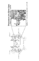
- FIG. 1 is referred to as BiBian (Bi specific Bi valent an tibody ), showing an example of a T cell recruitment antibody of format for making bispecific antibodies of the present invention.
- a T cell recruiting antibody consists of a single polypeptide chain that specifically binds to a first variable region that specifically binds to a target antigen on a cancer cell and an antigen on a T cell that is different from the target antigen. And a second variable region.
- the first variable region consists of a variable region VHH, which is a single domain of a cancer cell-bound camel antibody.
- the second variable region consists of a heavy chain variable region and a light chain variable region of an antibody that specifically binds to an antigen on a T cell (T cell binding antibody), and the heavy chain variable region and the light chain These variable regions are linked by peptide linkers (GGGGG) 1 to (GGGGG) 3 .
- the first variable region and the second variable region are linked via an IgA hinge region.
- a PelB signal sequence is added to the 5 ′ end of the first variable region.
- BiBian is a single gene consisting of three domains, and multi-functionalization is achieved by spontaneous association of homodimers designed from T cell-targeted Fv fragments. There is no concern about the formation of new aggregates.
- BiBian can also be prepared using a microbial expression system. Therefore, the bispecific antibody of the present invention consisting of three domains produced based on BiBian can also be prepared by a microbial expression system without the concern of non-functional aggregate formation seen in diabody. .
- the bispecific antibody of the present invention uses such a T cell recruiting antibody as a format, sends out a domain library of domains binding to human HER2, and creates a wide variety of T cell recruiting antibody gene groups therefrom. It can be obtained by screening and isolation by injury activity screening.
- a conjugated antibody in which the antibody fragment of the first aspect or the antibody of the second aspect is bound to a functional molecule other than the antibody.
- the functional molecule other than the antibody includes at least one compound of the group consisting of a fluorescent substance, a luminescent substance, a low molecular compound, a nucleic acid, a radioactive substance, a drug, a toxin, a cytokine, an albumin, an enzyme, and a non-peptidic polymer.
- a fluorescent substance refers to a substance that emits light when light energy is used for excitation.
- a light-emitting substance is a substance that emits light by transitioning from a high excited state to a lower excited state or a ground state, and in this specification, refers to a substance that emits light using energy generated by a chemical reaction as an excitation source.
- a substance that emits light by using energy generated by a chemical reaction as an excitation source is called a chemiluminescent substance, and a substance that emits light by a chemical reaction in a living body is called a bioluminescent substance.
- non-peptidic polymers include polyethylene glycol (PEG) and hyaluronic acid.
- the drug include therapeutic drugs.
- therapeutic agents include immunomodulators, anti-angiogenic agents, antiproliferative agents, pro-apoptotic agents, chemotherapeutic agents and the like.
- a functional molecule other than an antibody is chemically or genetically bound to the antibody fragment of the first aspect or the antibody of the second aspect by a technique established in the art.
- Preferred functional molecules other than antibodies are fluorescent substances, drugs, or nucleic acids.
- nucleic acid molecule comprising a base sequence encoding the antibody fragment of the first aspect or the bispecific antibody of the second aspect.
- nucleic acid is a molecule encoding a single-chain polypeptide
- the chemical structure and the acquisition route thereof are not particularly limited, and include, for example, gDNA, cDNA, chemically synthesized DNA, mRNA, and the like.
- a nucleic acid encoding the variable region of the bispecific antibody of the second aspect is prepared, it can be synthesized by an overlap PCR method based on a previously designed amino acid sequence.
- telomere can be isolated from a cDNA library by hybridization based on the sequence described in the literature, or by the polymerase chain reaction (PCR) technique.
- PCR polymerase chain reaction
- the DNA is placed in an expression vector, which is then placed in an E. coli cell, COS cell, Chinese hamster ovary cell (CHO cell) ⁇ ⁇ , or myeloma cell that does not produce immunoglobulin.
- a host cell can be transfected and a monoclonal antibody synthesized in the recombinant host cell.
- the PCR reaction can be performed by a method known in the art or a method or modification method substantially similar thereto, for example, R.Saiki, et al., Science, 230: 1350, 1985; R.
- the PCR method can be performed using a commercially available kit suitable for the PCR method, and can also be performed according to a protocol that has been clarified by the kit manufacturer or the kit vendor.
- nucleic acid encoding the single-chain polypeptide constituting the bispecific antibody of the present invention obtained in this way or each region contained in it is appropriately obtained by a desired peptide or amino acid according to the purpose by means known to those skilled in the art.
- modification of a nucleic acid means insertion, deletion or substitution of a base in at least one codon encoding an amino acid residue in the obtained original nucleic acid.
- altering the amino acid sequence itself constituting a single-chain polypeptide by replacing a codon encoding an original amino acid residue with a codon encoding another amino acid residue.
- the nucleic acid encoding the single-chain polypeptide can be modified so that the codon (optimum codon) suitable for the host cell such as CHO cell is used without changing the amino acid itself.
- the codon optimum codon
- an expression vector comprising the nucleic acid molecule of the fourth aspect.
- the antibody fragment of the first aspect of the present invention and the bispecific antibody of the second aspect can be produced using methods known to those skilled in the art, for example, various means such as genetic engineering techniques or chemical synthesis. I can do it.
- genetic engineering techniques for example, a replicable expression vector containing a nucleic acid encoding a polypeptide constituting the antibody fragment or bispecific antibody is prepared, and a host cell is transformed with the vector, Culturing transformed host cells to express single-chain polypeptides, recovering and purifying the polypeptides, associating the single-chain polypeptides, and separating and recovering the formed antibody molecules Can be manufactured.
- replicable expression vector and “expression vector” refer to a piece of DNA (usually double-stranded), in which the DNA contains Can be inserted with foreign DNA fragments.
- Foreign DNA is defined as heterologous DNA, which is a DNA cage that is not found naturally in the subject host cell.
- Vectors are used to carry foreign DNA or heterologous DNA strands into appropriate host cells. Once in the host cell, the vector can replicate independently of the host chromosomal DNA cage, and several copies of the vector and its inserted (foreign) DNA cage can be generated.
- the vector contains the elements essential to allow translation of the foreign DNA into a polypeptide. Thus, many molecules of polypeptides encoded by foreign DNA can be synthesized rapidly.
- Such vectors are operably linked to an appropriate control sequence so that the DNA sequence is expressed in an appropriate host (ie, to allow expression of foreign DNA). It means a “DNA construct” containing the determined DNA sequence.
- control sequences include a promoter for transcription transcription, any operator sequence to control such transcription, sequences encoding appropriate mRNA ribosome binding sites, enhancers, reardenylation sequences, and transcription and translation. (translation) An array for controlling the end of the cage can be mentioned.
- the vector can appropriately contain various sequences known to those skilled in the art, for example, restriction enzyme cleavage sites, marker genes (selection genes) such as drug resistance genes, signal sequences, leader sequences, and the like as necessary.
- sequences or elements can be appropriately selected and used by those skilled in the art depending on conditions such as the type of foreign DNA, the host cell used, the culture medium, and the like. Further, for the purpose of facilitating the detection and purification of the produced single-chain polypeptide, a sequence encoding various peptide tags known to those skilled in the art (for example, c-myc tag and His-tag) is stored. It can be included at the end of the sequence corresponding to this polypeptide.
- the vector can be in any form such as a plasmid, a phage particle, or simply a genomic insert. Once introduced into a suitable host by transformation, the vector can replicate or function independently of the resident genome. Alternatively, the vector may be one that is integrated into the genome.
- a host cell transformed with the expression vector of the fifth aspect is provided.
- any cell known to those skilled in the art can be used.
- typical host cells include prokaryotic cells such as E. coli and Chinese hamster ovary cells (CHO cells).
- Mammalian cells such as rabbits and human-derived cells, and eukaryotic cells such as yeast and insect cells.
- the transformed bacteria can be cultured under any mortgage conditions and methods known to those skilled in the art.
- IPTG IPTG of about 0.5 mM
- a single-chain polypeptide obtained by such expression in a host cell is generally recovered from the culture medium as a secreted polypeptide, but if it is produced directly without a secretion signal, the host It can be recovered from cell lysates. If the single-chain polypeptide is membrane-bound, it can be released from the membrane using a suitable detergent (eg, Triton-X100). The purification operation can be carried out by appropriately combining methods known to those skilled in the art.
- Affinity chromatography is one of the preferred purification techniques with high efficiency utilizing the affinity with a peptide tag of a single-chain polypeptide.
- the purification operation is preferably performed after the single-chain polypeptide is solubilized and denatured.
- This solubilization treatment can be performed using any agent known to those skilled in the art as a dissociator such as alcohols such as ethanol, various reagents, guanidine hydrochloride, urea and the like.
- the bispecific antibody of the present invention is produced by associating (unwinding) the same or two kinds of single-chain polypeptides thus purified, and separating and recovering the formed antibody. I can do it.
- the association treatment means that a single single-chain polypeptide is returned to a state having a desired biological activity by returning it to an appropriate spatial arrangement. Therefore, the association treatment also has the meaning of returning the polypeptides or domains to the associated state, so it can also be referred to as “reassociation”, and in the sense of having the desired biological activity. It can also be called reconstruction, or it can be called refolding.
- the association treatment can be performed by any method known to those skilled in the art. For example, the concentration of the denaturing agent (for example, guanidine hydrochloride) in the buffer solution containing the single-chain polypeptide is decreased stepwise by, for example, dialysis. The method is preferred.
- the antibody fragment or bispecific antibody of the present invention is prepared from, for example, a culture medium supernatant of a cultured host cell, a periplasma fraction, a cell-soluble fraction, or a cell-insoluble fraction. Is possible.
- a pharmaceutical composition comprising the antibody fragment of the first aspect or the bispecific antibody of the second aspect.
- the pharmaceutical composition of the present invention comprises, as an active ingredient, one selected from the group consisting of the antibody fragment or bispecific antibody of the present invention, a nucleic acid, a vector, and a transformed host cell.
- an active ingredient has an action of significantly eliminating, killing, or damaging (positive) tumor cells expressing the epidermal growth factor receptor in vitro and in vivo. Therefore, the pharmaceutical composition of the present invention can be used as an antitumor agent against such tumor cells.
- the effective amount of the active ingredient of the present invention can be appropriately determined by those skilled in the art depending on, for example, the therapeutic purpose, the type of tumor, the site and size of the subject to be administered, various conditions of the patient, and the administration route.
- a typical single dose or daily dose will depend on the above conditions and, if possible, first in vitro and then, for example, using assays known in the art for tumor cell survival or growth.
- appropriate dose ranges can be determined with appropriate animal models that can extrapolate dose ranges for human patients.
- the pharmaceutical composition of the present invention contains various pharmaceutically acceptable pharmacological agents well known to those skilled in the art in addition to the active ingredient, depending on various conditions such as the type of active ingredient, pharmaceutical form, administration method / purpose, and pathological condition of the administration target.
- Components eg, carriers, excipients, buffers, stabilizers, etc.
- the pharmaceutical composition of the present invention is a tablet, solution, powder, gel, spray, or microcapsule, colloidal distribution system (liposome, microemulsion, etc.), macroemulsion, etc., depending on the above various conditions.
- colloidal distribution system liposome, microemulsion, etc.
- macroemulsion etc.
- administration methods include intravenous, intraperitoneal, intracerebral, intraspinal, intramuscular, intraocular, intraarterial, in particular intrabiliary or intralesional injection or injection, and sustained release system formulations. It is done.
- the active substances of the invention can be administered continuously by infusion or by bulk injection.
- the antibody is previously bound to the cell before administration by mixing the active ingredient such as the bispecific antibody of the present invention with the cell before administration.
- Sustained release formulations are generally of a form from which the active substance of the present invention can be released for a period of time, and suitable examples of sustained release preparations include solid hydrophobic polymers containing proteins.
- a semi-permeable carrier is included, which is in the form of a molded article such as a film or microcapsule.
- the pharmaceutical composition of the present invention is prepared by methods known to those skilled in the art, for example, the Japanese Pharmacopoeia Manual Editorial Committee, 13th revision, Japanese Pharmacopoeia Manual, issued on July 10, 1996, Yodogawa Shoten Co., Ltd. In view of the description, it can be appropriately selected and manufactured from among them.
- kits comprising the antibody fragment of the first aspect or the bispecific antibody of the second aspect.
- the kit includes a container, a syringe needle, a pharmaceutically acceptable medium, an alcohol cotton cloth, a bandage, or instructions for using the bispecific antibody. It can also be equipped with a letter.
- a first variable region that specifically binds to a target antigen on a tumor cell, and a second variable region that specifically binds to an antigen different from the target antigen A method of screening for a bispecific antibody, wherein the first variable region and the second variable region are contained on a single polypeptide chain.
- the target antigen on the tumor cell is preferably HER2, more preferably human HER2.
- the antigen different from the target antigen is preferably an antigen on a T cell different from the target antigen.
- the method of screening for a bispecific antibody comprises panning a first vector comprising a llama-derived naive VHH library gene and polyclonal phagemid vectors having various VHHs that show positive binding to the target antigen.
- bispecific antibodies that specifically bind to tumor cells and have cytotoxic activity can be screened with high accuracy in a short period of time.
- the target antigen is preferably HER2, more preferably human HER2.
- the selection of clones based on the cytotoxic activity is usually determined based on whether or not the cytotoxic activity is higher than the reference value, and a clone having a higher cytotoxic activity than the reference value is selected.
- the reference value may be a known cytotoxic activity value of the T cell against the target antigen, a fractional multiple thereof, or an integral multiple thereof, and can be appropriately determined by those skilled in the art.
- the method for screening for a bispecific antibody comprises panning a first vector comprising a llama-derived naive VHH library gene and polyclonal phagemids with various VHHs that show positive binding to the target antigen.
- Preparing a vector; cleaving the VHH; and a second vector having a second variable region that specifically binds to an antigen different from the target antigen the first variable region comprising the cleaved VHH and the first variable region 2 variable regions are inserted into a single polypeptide chain, host cells transformed with the second vector are monoclonalized, and clones are selected based on cytotoxic activity (Second screening method).
- bispecific antibodies that specifically bind to tumor cells and have cytotoxic activity can be screened with higher accuracy in a shorter period of time than in the first screening method.
- the target antigen in the above method for screening a bispecific antibody is preferably HER2, more preferably human HER2.
- the selection of clones based on the cytotoxic activity is usually determined based on whether or not the cytotoxic activity is higher than the reference value, and a clone having a higher cytotoxic activity than the reference value is selected.
- the reference value may be a known cytotoxic activity value of the T cell against the target antigen, a fractional multiple thereof, or an integral multiple thereof, and can be appropriately determined by those skilled in the art.
- Example 1 Construction of Format Antibody BiBian Gene Sfi I sites were added to both the vector (FIG. 2A) and insert (FIG. 2B) fragments by PCR.
- the vector side was prepared by inverse PCR using pRA1-Ia1 VHH-HL type OKT3 scFv (SEQ ID NO: 33) as a template (FIG. 2A). Both the vector and the insert were digested with Sfi I at 50 ° C for 3 hr.
- the vector side was ligated after Dpn I treatment to prevent self-ligation, and the BiBian expression vector was constructed by confirming that the target VHH sequence was introduced by transformation, miniprep, and sequencing (FIG. 2C).
- Example 2 Preparation of Target Antigen HER2 Fragment
- HER2 that was reported to be overexpressed in multiple cancers including breast cancer was selected as the antigen targeted by the VHH fragment.
- the HER2 molecule itself can carry out signal transduction without a ligand.
- the HER2 molecule itself can form a HER2 homodimer without binding to the ligand, and can also form a dimer with other ErbB molecules that bind to the ligand and change its structure, interacting with the ligand Can transmit growth signals when not in use. For this reason, there is a study that considers dimerization inhibition design to be effective for blocking proliferation signals via HER2, and Matthew et al.
- HER2 dimerization interaction region (Dimerization Arm) (Matthew CF, et al., Cancer Cell, 2004, 5, 317-328) (Fig. 3) can be an effective target site in the approach of suppressing the growth of cancer cells by inhibiting dimerization.
- Domain II is a disulfide-rich domain, it may be difficult to prepare Domain II with a functional structure. Therefore, among (1) Domain II, Dimerization Arm identified as an interaction region at the time of dimerization His tag fused with HER2 fragment protein (Dimer Arm) and (2) Domain II fused with His tag Two kinds of HER2 fragment protein (Domain II-His) were prepared by E. coli expression system. In addition, (3) HER2 fragment protein (Domain II-Fc) fused with Fc tag to Domain II was prepared using a mammalian cell expression system, and a total of HER2 fragment proteins were prepared.
- FIG. 4 shows the base sequence (SEQ ID NO: 34) and amino acid sequence (SEQ ID NO: 35) of a vector insert for preparing the HER2 fragment protein Dimer Arm.
- FIG. 5 shows the base sequence (SEQ ID NO: 38) and amino acid sequence (SEQ ID NO: 39) of a vector insert for preparing the HER2 fragment protein Domain II-His.
- the pRA-OKT3 vector was digested with Nco I and Spe I, and amplified by using the following primers from the pKHI-HER2 vector and inserted into the Nco I and Spe I digested fragment.
- FIG. 6 shows the base sequence (SEQ ID NO: 42) and amino acid sequence (SEQ ID NO: 43) of a vector insert for preparing the HER2 fragment protein Domain II-Fc.
- FIG. 7 shows a vector map of the pcDNA3.1 (+)-Her2 DomainII-HumanFc vector constructed to prepare Domain II-Fc.
- Example 3 Preparation of Dimer Arm Escherichia coli BL21 (DE3) strain was cultured under the conditions shown in Table 1, and Dimer Arm was purified from the medium supernatant fraction after cell separation. Specifically, E. coli BL21 (DE3) transformed with a pET vector containing the Dimer Arm gene fragment was plated on an LB agar medium containing 100 ⁇ g / mL ampicillin and cultured at 28 ° C. for 20-24 hours. . Then, 5 colonies formed on the plate medium were inoculated into 50 mL of LB liquid medium containing 100 ⁇ g / mL ampicillin and cultured overnight at 28 ° C. with shaking.
- Example 4 Preparation of Domain II-His Escherichia coli BL21 (DE3) was cultured under the conditions shown in Table 2, and Dimain II-His was purified from the culture supernatant fraction after cell separation.
- E. coli BL21 (DE3) transformed with a pET vector containing the Dimer Arm gene fragment was plated on an LB agar medium containing 100 ⁇ g / mL ampicillin and cultured at 28 ° C. for 20-24 hours. . Then, 5 colonies formed on the plate medium were inoculated into a 50 mL LB liquid medium containing 100 ⁇ g / mL ampicillin and cultured overnight at 28 ° C. with shaking.
- the solution was roughly purified by immobilized metal chelate affinity chromatography (IMAC) using a Ni-sepharose column.
- IMAC immobilized metal chelate affinity chromatography
- the roughly purified Dimer Arm was purified by size exclusion chromatography using a PBS buffer containing 1 mM EDTA as a developing solution to obtain a final purified product.
- Example 5 Preparation of Domain II-Fc It is known that there are as many as 11 pairs of disulfide bonds in the Domain II region of HER2, and it was predicted that the expression would be difficult when Escherichia coli was used as a host. Therefore, an expression system using mammalian cells was constructed at the same time as the E. coli expression system. For expression in mammalian cells, we designed Domain II-Fc, which was fused to the C-terminus of an Fc domain derived from a human IgG1 antibody that has been reported to improve expression and stability by fusion to various proteins ( The nucleotide sequence and amino acid sequence are already described in Example 2).
- transient expression was attempted using CHO cells and EKHEK293T cells. Although the expression of ⁇ Domain ⁇ II-Fc was confirmed by dot blotting using the culture supernatant, the expected yield was not obtained. Therefore, we aimed to establish a stable expression strain using CHO sputum cells.
- the culture supernatant after mass culture was purified using Abcapture Extra (Protenova, Japan), and the eluate was subjected to yield comparison using -SDS-PAGE and Western Blotting, and 2G4 was determined as the best clone (Fig. , D). Domain II-Fc expressed by clone 2G4 was purified by Protein A column and then dialyzed against 1 M EDTA / PBS.
- HER2 Domain II targeted in this chapter is a complex structure having 11 pairs of disulfide bonds, and any HER2 fragment protein maintains a functional structure. It may not be. Therefore, in addition to the three HER2 fragment proteins prepared in Examples 3 to 5 (Dimer Arm, Domain II-His, Domain II-Fc), it is known that the HER2-specific antibody already marketed binds. We decided to purchase HER2 (ACROBiosystems, DE, USA) and perform panning combining these four antigens.
- helper phage M13KO7 (1 ⁇ 10 11 cfu / ml ⁇ 20 ⁇ L), leave it at 30 ° C. for 45 min-1 hr, add kanamycin to a final concentration of 50 ⁇ g / ml, and culture overnight (100 rpm, 30 ° C.).
- After collecting E. coli add 10 ml of 25% PEG 6000 / 2.5 M NaCl to the supernatant of 40 ml so that the final concentration is 5% PEG 6000 / 0.5 M NaCl, and add 1 ml in ice for 1 hr. Left to stand. After centrifugation (60 min, 10000 rpm, 0 ° C.), the phage precipitate was suspended in 1.5 mL of 10% glycerol / PBS solution to prepare a phage library.
- MAXISORP immune-tube (444202; thermo scientific, USA) 0.05% sodium azide / PBS 2 mL, HER2 antigen protein (final concentration; Dimer Arm: 10 ⁇ g / ml, Domain II-His: 10 ⁇ g / ml, Domain II-Fc: 10 ⁇ g / ml, Whole HER2: 2 ⁇ g / ml) was added and allowed to stand at 4 ° C., overnight or 25 ° C. for 1-2 hours, and allowed to solidify. After washing with PBS, the whole immune tube was blocked with 3% Skim milk / PBS solution (25 ° C., 30 min) and washed 5 times with PBS.
- the output phage / input phage (O / I) in the 2nd round in ver.1,2,3 is 0.54 ⁇ 10 -5 , 0.89 ⁇ 10 -5 , 2.4 ⁇ 10 -5 , respectively, 1st round
- the enrichment factor (2nd (O / I) / 1st (O / I)) was 109, 36, and 52 times (Fig. 13A-C). Since this suggested that the binding phages were concentrated, the binding activity was evaluated by ELISA using the initial library, the first round phage, and the second round phage in a polyclonal state (FIGS. 14A-C).
- ver.1 As a result, all of ver.1, 2, and 3 showed a tendency to enrich for binding phages.
- ver.2 and ver.3 it was found that the binding activity against the HER2 antigen used in the 1st round (ver.2: Domain Ii-His, ver.3: Domain II-Fc) was improved after the 2nd round.
- ⁇ ver.1 the binding activity of 2 round eluted phage for Dimer Arm used in the 1st round was slightly reduced, but the binding activity for whole HER2 was improved 2 times, and binding activity for Domain II-His and Domain II-Fc There was also an improvement.
- Example 7 Construction of a bispecific antibody in the BiBian format by a standard method through phage monoclonalization Evaluation of binding activity of monoclonal phage by ELISA Plate TG-1 strain infected with 2nd panning phage on LB / amp plate and incubate at 37 ° C for 12 hr, then colony one deep colony plate (P-2ML-SQ -C; Axygen, Japan) was used to inoculate phages. For each panning version, 96 clones were isolated, for a total of 288 clones, and monoclonal phages were prepared based on the phage preparation protocol in 96-well plates.
- binding activity to the HER2 molecule expressed on the cell surface was evaluated by phage-cell ELISA using HER2 -positive cells DG44 (HER2 +) (data not shown).
- HER2 + HER2 +
- the selected clones were determined for amino acid sequence by base sequence analysis using a DNA sequencer after obtaining a phagemid vector. A comparison of the amino acid sequences of the 6 clones using clustal omega revealed that all of them were unique sequences. Therefore, the bispecific antibody gene in which the BiBian scFv region gene was linked to the VHH gene from each of the 6 ⁇ ⁇ ⁇ ⁇ clones ( We decided to construct a modified BiBian gene.
- VHH clone gene selected in the previous section was amplified by culture PCR, then the non-specific amplification part was separated by agarose gel electrophoresis, and the VHH gene was extracted by gel extraction. Extracted. After the restriction enzyme treatment with Sfi I, an insert gene fragment was prepared by PCI and ethanol precipitation. On the vector side, pRA1-pelB-Ia1 VHH-OKT3 scFv-His-cmyc plasmid was used as a template. Ia1 VHH was removed by inverse PCR, and Sfi I sites were added by primers downstream of pelB and upstream of OKT3 scFv (Table 3). The preparation was performed in the same procedure as the insert. After ligation, the gene was prepared based on the protocol, and the sequencer confirmed that the VHH clone was correctly inserted into the BiBian format.
- Escherichia coli BL21 (DE3) transformed with a pET vector containing a gene fragment of a double characteristic antibody was plated on an LB agar medium containing 100 ⁇ g / mL ampicillin and cultured at 28 ° C. for 20 to 24 hours. Then, 5 colonies formed on the plate medium were inoculated into 50 ⁇ mL LB liquid medium containing 100 ⁇ g / mL ampicillin and cultured overnight at 28 ° C. with shaking. next. A 5 mL portion of the culture solution was transferred to a 500 mL 2 ⁇ YT liquid medium and cultured at 28 ° C. with shaking.
- Cytotoxic Activity Test Using Novel Bispecific Antibody Cytotoxic activity was evaluated by MTS assay using modified BiBian, which is the novel bispecific antibody described in the previous section (FIGS. 15A-F).
- modified BiBian which is the novel bispecific antibody described in the previous section (FIGS. 15A-F).
- a HER2-positive as cancer cells breast cancer-derived SK-BR3 cells (1 ⁇ 10 4 cells / well ), were used T cells as T-LAK (5 ⁇ 10 4 cells / well).
- T-LAK 5 ⁇ 10 4 cells / well.
- two clones, BiBi-H44 and BiBi-H48 were found to have a high injury activity exceeding 50% even at a final concentration of 2 nM.
- H44 clone (SEQ ID NO: 26) and H48 clone (SEQ ID NO: 27) are clones isolated from panning conditions ver. 2 (Domain I-His x whole HER2), and panning conditions ver. 2 express high damage activity. It was suggested that clones could be easily obtained.
- Example 8 New creation process of T cell recruiting antibody without monoclonalization of phage
- the clone obtained by panning in Example 6 can express cytotoxic activity against HER2-positive cells by BiBianization. The result showed it. Therefore, it is compatible with the Dolag ArT method (Domain library approach for generating antibody recruiting T-cell), which screens T cell recruiting antibodies with a combination of domains with optimal cytotoxic activity from multiple antibody fragment populations in units of antibody fragments. An attempt was made to create a T cell recruiting antibody library creation process.
- Dolag ArT method Domain library approach for generating antibody recruiting T-cell
- Example 7 What was carried out in Example 7 is a standard technique from the acquisition of binding molecules starting from biopanning to the evaluation of T cell recruitment antibody formation and its damage activity.
- this standard method there were only about 6 clones that could be evaluated for injury activity by hatching BiBian.
- the number of clones that can be evaluated for damage activity decreases when BiBian hatches, from each clone individually construct a modified BiBian gene that is a bispecific antibody linking the BiHian scFv region gene to the VHH gene This is due to the fact that it is necessary to cultivate 2 ⁇ 6 1L Erlenmeyer flasks for each clone when preparing and purifying a sufficient amount of modified BiBian.
- Hha I is a restriction enzyme that recognizes GCGC sequences. Theoretically, GCGC sequences appear once every 256 bp. For this reason, it is considered that the sequence diversity can be evaluated from the band pattern after digestion with Hha I, and the two polyclonal phagemid vectors after the 2nd round and the H44 and H48 clones isolated in the previous section were used as controls at 37 ° C, 3 hr. Digested under reaction conditions.
- E. coli transformed with the BiBian expression vector was cultured in a 96-deep well plate, and after 36 hours of IPTG induction, the culture supernatant was centrifuged (3000 rpm, 60 min, And the cytotoxic activity was evaluated by MTS assay using the culture supernatant.
- the activity was evaluated using a culture supernatant diluted 10-fold in animal cell culture medium. As a result, 83 out of 264 clones showed a high damage activity of 70% or more.
- clone d8D, clone h11E, clone f8C, clone f10D and clone f11E are SEQ ID NO: 28, SEQ ID NO: 29, SEQ ID NO: 30, SEQ ID NO: 31 and SEQ ID NO: 32, respectively.
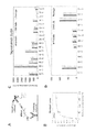
- Example 7 Comparison of New Process and Standard Method Example 7 and Example 8 enabled screening by “injury activity”, which is the final target function, instead of the standard method by binding activity.
- injury activity which is the final target function
- Example 7 only 10 clones were actually able to evaluate the damage activity, but in the method of this example, about 300 clones were evaluated for damage activity and promising clones were selected from them. We were able to. Among the 300 clones, there were several clones that promoted the growth of cancer cells. In this way, it is not always like strong binding activity equal strong injury activity, so it is important to evaluate in the final function.
- FIG. 21A and FIG. 21B show the IC 50 of the clones obtained by the standard method and the direct BiBiaization method, respectively. Based on this, we tried to evaluate the effectiveness of the method using a box-and-whisker plot (Fig. 21C). As a result, it was shown that the direct BiBianization method of this example has a tendency to obtain clones with excellent IC 50 more easily than the standard method of Example 7 in both population distribution and median. .
- Example 7 when the conventional standard method of Example 7 is used, it takes about 5 weeks to assess the injury activity using monoclonal antibody to purified T-cell recruiting antibody. On the other hand, since the procedure of this example is completed in 2 weeks (14 days) from polyclonal phagemid preparation to sequence sequencing, the new process contributes to improving throughput in terms of time. From this, it can be said that the importance / effectiveness of the method of the present example was also demonstrated from the viewpoint of the quality of clones that can be obtained and the temporal viewpoint.
- Example 9 Improvement of binding affinity of modified BiBian to HER2
- clone 6x3 has an amino acid sequence of YAMGWY (SEQ ID NO: 53) near CDR1 and FNGLQY (SEQ ID NO: 57) near CDR3 (Table 4).
- CDR1-2 corresponds to amino acid sequence 32 to 37 of BiBian protein (SEQ ID NO: 52) of clone 6 ⁇ 3
- CDR3-1 corresponds to 96 to 96 of amino acid sequence of BiBian protein (SEQ ID NO: 52) of clone 6 ⁇ 3.
- CDR3-2 corresponds to the 101st to 106th amino acids of the clone 6 ⁇ 3 BiBian protein (SEQ ID NO: 52).
- a modified BiBian 6 ⁇ 3 vector having the insert shown in FIG. 22 (the nucleotide sequence is represented by SEQ ID NO: 51 and the amino acid sequence is represented by SEQ ID NO: 52) was prepared, and “1. Construction of polyclonal modified BiBian gene in Example 8” was prepared. And dozens of mutant clones were monoclonalized (data not shown).
- CDR1-2 has the amino acid sequence of YAMGWY (SEQ ID NO: 53), RRVYWA (SEQ ID NO: 54), or RSFRWY (SEQ ID NO: 55), and CDR3-2
- amino acid sequence of FNGLQY (SEQ ID NO: 57) or RLRERG (SEQ ID NO: 58) is included, it was found that the Kd value tends to be low (data not shown).
- variant 1 and variant 2 had a lower Kd value than clone 6 ⁇ 3 and improved binding affinity to HER2.
- variants of CDR1 and CDR3 of clone 6 ⁇ 3 those having the sequences shown in Table 4, those having the sequences shown in Table 5, and those having a combination sequence thereof can be used. More specifically, the variants of CDR1 and CDR3 of clone 6 ⁇ 3 may be as follows.
- Variant having CDR1-2 of Table 4 or 5 Variant having CDR3-1 of Table 4 or 5 Variant having CDR3-2 of Table 4 or 5 Having CDR1-2, CDR3-1 of Table 4 or 5 Variant Table 4 or 5, CDR1-2, Variant having CD3-2 Variant Table 4 or 5, Variant having CDR1-2, CDR3-1, CD3-2 Variant having CDR1-2 of Table 4; Combination with variants having CDR3-1 in Table 5 Combination with variants having CDR1-2 in Table 4 and variants having CDR3-2 in Table 5 Variants having CDR1-2 in Table 4 Combination of variants having CDR3-1 in Table 4 and variants having CDR3-2 in Table 5 Variants having CDR1-2 in Table 4 and variants having CDR3-1 in Table 5 Combination with variants having CDR3-2 of 5 in Table 4 Variants having CDR1-2 of Table 4, variants having CDR3-1 of Table 5, and CDR3-2 of 5 of Table 5 Combinations with variants Variants having CDR1-2 in Table 5 and CDR3-1 in Table 4 Combinations of variants having CDR1-2
Landscapes
- Health & Medical Sciences (AREA)
- Chemical & Material Sciences (AREA)
- Life Sciences & Earth Sciences (AREA)
- Genetics & Genomics (AREA)
- Organic Chemistry (AREA)
- Engineering & Computer Science (AREA)
- General Health & Medical Sciences (AREA)
- Immunology (AREA)
- Biomedical Technology (AREA)
- Bioinformatics & Cheminformatics (AREA)
- Biotechnology (AREA)
- Molecular Biology (AREA)
- Biochemistry (AREA)
- Wood Science & Technology (AREA)
- Zoology (AREA)
- Medicinal Chemistry (AREA)
- General Engineering & Computer Science (AREA)
- Biophysics (AREA)
- Microbiology (AREA)
- Proteomics, Peptides & Aminoacids (AREA)
- Veterinary Medicine (AREA)
- Animal Behavior & Ethology (AREA)
- Cell Biology (AREA)
- Plant Pathology (AREA)
- Physics & Mathematics (AREA)
- Public Health (AREA)
- Pharmacology & Pharmacy (AREA)
- General Chemical & Material Sciences (AREA)
- Nuclear Medicine, Radiotherapy & Molecular Imaging (AREA)
- Chemical Kinetics & Catalysis (AREA)
- Mycology (AREA)
- Epidemiology (AREA)
- Peptides Or Proteins (AREA)
- Micro-Organisms Or Cultivation Processes Thereof (AREA)
- Medicines Containing Antibodies Or Antigens For Use As Internal Diagnostic Agents (AREA)
Abstract
ヒトHER2に特異的に結合する抗体断片は、下記式(1)の構造を有する単一ポリペプチド鎖のアミノ酸配列を有する。 FR1-CDR1-FR2-CDR2-FR3-CDR3-FR4 … (1) (式中、FR1、FR2、FR3、及びFR4はフレームワーク領域であり、CDR1、CDR2、及びCDR3は相補性決定領域であり、以下の(I)~(III)の少なくとも一つを満たす: (I)CDR1は、配列番号5~11、53~55、及び59~64のいずれかで表されるアミノ酸配列を有するか、配列番号5~11、53~55、及び59~64のいずれかで表されるアミノ酸配列において1から数個の欠失、置換若しくは付加を含むアミノ酸配列を有するか、又は配列番号5~11、53~55、及び59~64のいずれかで表されるアミノ酸配列と80%以上の同一性を有するアミノ酸配列を有する (II)CDR2は配列番号12~18のいずれかで表されるアミノ酸配列を有するか、配列番号12~18のいずれかで表されるアミノ酸配列において1から数個の欠失、置換若しくは付加を含むアミノ酸配列を有するか、又は配列番号12~18のいずれかで表されるアミノ酸配列と80%以上の同一性を有するアミノ酸配列を有する (III)CDR3は配列番号19~25、56~58、及び65~84のいずれかで表されるアミノ酸配列を有するか、配列番号19~25、56~58、及び65~84のいずれかで表されるアミノ酸配列において1から数個の欠失、置換若しくは付加を含むアミノ酸配列を有するか、又は配列番号19~25、56~58、及び65~84のいずれかで表されるアミノ酸配列と80%以上の同一性を有するアミノ酸配列を有する。)
Description
本発明は、抗体断片、該抗体断片を含む二重特異性抗体、該抗体断片又は該二重特異性抗体をコードする塩基配列を含む核酸分子、該抗体断片又は該二重特異性抗体を含む医薬組成物、及び該抗体断片又は該二重特異性抗体を含むキットに関する。
がん(悪性腫瘍)に対する安全な治療薬として、免疫療法が用いられている。がんに対する免疫療法では、がんに対して特異的に細胞傷害活性を示す抗体が使用されている。
Human epidermal growth factor Receptor 2 (HER2)は細胞表面に存在する糖蛋白質の受容体型チロシンキナーゼ (分子量 約 185kDa)であり、EGFR2,ErbB2,CD340,あるいは NEU とも呼ばれる。HER2はその変異型や欠損型も含めて乳癌をはじめとする複数のがんで過剰発現しており、治療標的として注目されている。HER2 を標的とした抗体は、標的化による治療効果が期待されるがん細胞表面抗原の一つである。
抗体の基本構造は、重鎖と軽鎖がジスルフィド結合によりヘテロテトラマーを形成したものであるが、このようなIgG型の抗体は高分子量であるため、腫瘍深部まで浸透することができない。このため、抗体の可変領域断片を含む低分子量の抗体の開発が進んでいる。
近年、Hamers-Casterman らはヒトコブラクダやラマなどのラマ科の動物において、CH1 ドメインを含まず重鎖のみからなる抗体が存在することを報告した(非特許文献1)。ラクダ由来抗体の可変領域断片(single Variable domain of the Heavy chain of a Heavy-chain camel antibody : VHH)は、単量体ドメインにもかかわらず、高親和性・高特異性を有している。そこで、ラマ科の動物の免疫化により取得した抗体群をライブラリーとしてファージ提示法等の生体外選択操作を行うことで種々の標的に特異的に結合するVHH が取得されている(非特許文献2, 3)。
一方で、抗原に対する親和性を高めるために、抗体を多量体化又は多価化する技術も知られている。例えばHarwood らは三量体形成ドメインとして知られる collagen XVIIItrimerization domain を抗CD3 scFv1 分子及び抗EGFR VHH 抗体3 分子と融合させることで、EGFR に対して三価の組換え抗体を設計し、かかる組換え抗体が同一ドメインから構成される一重一価の組換え抗体よりも高い傷害活性を発揮することを報告している (非特許文献4)。
従来のIgG型の抗体では、ナチュラルキラー細胞よりも活性の高いT細胞を利用することができず、T細胞を利用することができる低分子量の抗体は開発されているが、低分子であるため、細胞へ結合する価数が1であり、抗原との結合親和性が低い。
抗体と抗原の結合親和性増大は抗体の多価設計により傷害活性向上をもたらすため、有効なアプローチであるといえるが、重鎖と軽鎖間のリンカー配列の短縮化・除去による多価化設計は多価な会合体形成にある程度有効であるものの(非特許文献5, 6)、単一の多価会合体のみが形成されるような設計には至っていない。
EMBO J 2000, 19, 921-930
Structure 2013, 21, 1214-1224
Protein Eng Des Sel 2008, 21, 1-10.
Oncoimmunology 2018, 7, e1377874
J Immunol Methods 1999, 231, 177-189
Protein Sci 2003, 12, 734-747
本発明の目的は、ヒトHER2に特異的に結合する抗体断片、かかる抗体断片からなる可変領域を備えた高いがん細胞傷害性を有する二重特異性抗体、該抗体断片又は該二重特異性抗体を含む医薬組成物、及び該抗体断片又は該二重特異性抗体を含むキットを提供することにある。
本発明者らは、上記の目的を達成すべく、本発明者らにより開発されたシングルドメインであるラクダ抗体VHH及びscFv (単鎖抗体断片)から構成される二重二価抗体をフォーマットとして採用して作製された、ヒトHER2に特異的に結合する抗体が、高い細胞傷害活性を示すことを見出し、本発明を完成するに至った。
すなわち本発明は、以下の項に記載の主題を包含する。
[1]ヒトHER2に特異的に結合する抗体断片であって、下記式(1)の構造を有する単一ポリペプチド鎖のアミノ酸配列を有する抗体断片。
[1]ヒトHER2に特異的に結合する抗体断片であって、下記式(1)の構造を有する単一ポリペプチド鎖のアミノ酸配列を有する抗体断片。
FR1-CDR1-FR2-CDR2-FR3-CDR3-FR4 … (1)
(式中、
FR1、FR2、FR3、及びFR4はフレームワーク領域であり、
CDR1、CDR2、及びCDR3は相補性決定領域であり、以下の(I)~(III)の少なくとも一つを満たす:
(I)CDR1は、配列番号5~11、53~55、及び59~64のいずれかで表されるアミノ酸配列を有するか、配列番号5~11、53~55、及び59~64のいずれかで表されるアミノ酸配列において1から数個の欠失、置換若しくは付加を含むアミノ酸配列を有するか、又は配列番号5~11、53~55、及び59~64のいずれかで表されるアミノ酸配列と80%以上の同一性を有するアミノ酸配列を有する
(II)CDR2は配列番号12~18のいずれかで表されるアミノ酸配列を有するか、配列番号12~18のいずれかで表されるアミノ酸配列において1から数個の欠失、置換若しくは付加を含むアミノ酸配列を有するか、又は配列番号12~18のいずれかで表されるアミノ酸配列と80%以上の同一性を有するアミノ酸配列を有する
(III)CDR3は配列番号19~25、56~58、及び65~84のいずれかで表されるアミノ酸配列を有するか、配列番号19~25、56~58、及び65~84のいずれかで表されるアミノ酸配列において1から数個の欠失、置換若しくは付加を含むアミノ酸配列を有するか、又は配列番号19~25、56~58、及び65~84のいずれかで表されるアミノ酸配列と80%以上の同一性を有するアミノ酸配列を有する。)
[2]前記CDR1は配列番号5~11、53~55、及び59~64のいずれかで表されるアミノ酸配列を有し、前記CDR2は配列番号12~18のいずれかで表されるアミノ酸配列を有し、前記CDR3は配列番号19~25、56~58、及び65~84のいずれかで表されるアミノ酸配列を有する[1]に記載の抗体断片。
[3]下記(i)~(xii)のCDR1、CDR2及び/又はCDR3を有する[1]又は[2]に記載の抗体断片。
(i) CDR1:配列番号5、CDR2:配列番号12、CDR3:配列番号19
(ii) CDR1:配列番号6、CDR2:配列番号13、CDR3:配列番号20
(iii) CDR1:配列番号7、CDR2:配列番号14、CDR3:配列番号21
(iv) CDR1:配列番号8、CDR2:配列番号15、CDR3:配列番号22
(v) CDR1:配列番号9、CDR2:配列番号16、CDR3:配列番号23
(vi) CDR1:配列番号10、CDR2:配列番号17、CDR3:配列番号24
(vii) CDR1:配列番号11、CDR2:配列番号18、CDR3:配列番号25
(viii) CDR1:配列番号53
(ix) CDR1:配列番号53、及び配列番号CDR3:配列番号57
(x) CDR1:配列番号54
(xi) CDR1:配列番号55
(xii) CDR3:配列番号58
[4]FR1、FR2、FR3、及びFR4は配列番号1~4で表されるアミノ酸配列を有するか、配列番号1~4で表されるアミノ酸配列において1から数個の欠失、置換若しくは付加を含むアミノ酸配列を有するか、又は配列番号1~4で表されるアミノ酸配列と80%以上の同一性を有するアミノ酸配列を有する[1]~[3]のいずれか一項に記載の抗体断片。
[5]配列番号26~32のいずれかで表されるアミノ酸配列を有する[1]~[4]のいずれか一項に記載の抗体断片。
[6][1]~[5]のいずれか一項に記載の抗体断片からなる第1の可変領域と、HER2とは異なる抗原に特異的に結合する第2の可変領域とを備え、前記第1の可変領域と前記第2の可変領域が単一のポリペプチド鎖上に含まれる、二重特異性抗体。
[7]前記第2の可変領域が、T細胞上の抗原に特異的に結合する、[6]に記載の二重特異性抗体。
[8]前記第2の可変領域が、抗CD3抗体の重鎖の可変領域と、抗CD3抗体の軽鎖の可変領域とを含む[7]に記載の二重特異性抗体。
[9]単量体、二量体、三量体、四量体、五量体又はそれらのFc領域融合体である[6]~[8]のいずれか一項に記載の二重特異性抗体。
[10][1]~[5]のいずれか一項に記載の抗体断片又は[6]~[9]のいずれか一項に記載の二重特異性抗体と、蛍光物質、発光物質、低分子化合物、核酸、放射性物質、薬剤、毒素、サイトカイン、アルブミン、酵素、及び非ペプチド性ポリマーからなる群の少なくとも1種の化合物とが結合されたコンジュゲート抗体。
[11][1]~[5]のいずれか一項に記載の抗体断片又は[6]~[9]のいずれか一項に記載の二重特異性抗体をコードする塩基配列を含む核酸分子。
[12][11]に記載の核酸分子を含む発現ベクター。
[13][12]に記載の発現ベクターにより形質転換した宿主細胞。
[14][1]~[5]のいずれか一項に記載の抗体断片又は[6]~[9]のいずれか一項に記載の二重特異性抗体を含有する医薬組成物。
[15][1]~[5]のいずれか一項に記載の抗体断片又は[6]~[9]のいずれか一項に記載の二重特異性抗体を備えたキット。
本発明の抗体断片は、ヒトHER2を細胞表面に有する腫瘍細胞を標的とすることができる。また、かかる抗体断片を可変領域として備えた二重特異性抗体は、腫瘍細胞を標的として特異的に結合できると共に低濃度でも非常に高い細胞傷害性を有するため、臨床やがん細胞を用いた研究に有用である。
本発明の第1の態様によれば、ヒトHER2に特異的に結合する抗体断片であって、下記式(1)の構造を有する単一ポリペプチド鎖のアミノ酸配列を有する抗体断片が提供される。
FR1-CDR1-FR2-CDR2-FR3-CDR3-FR4 … (1)
式中、
FR1、FR2、FR3、及びFR4はフレームワーク領域であり、
CDR1、CDR2、及びCDR3は相補性決定領域であり、
以下の(I)~(III)の少なくとも一つを満たす:
(I)CDR1は、配列番号5~11、53~55、及び59~64のいずれかで表されるアミノ酸配列を有するか、配列番号5~11、53~55、及び59~64のいずれかで表されるアミノ酸配列において1から数個の欠失、置換若しくは付加を含むアミノ酸配列を有するか、又は配列番号5~11、53~55、及び59~64のいずれかで表されるアミノ酸配列と80%以上の同一性を有するアミノ酸配列を有する
(II)CDR2は配列番号12~18のいずれかで表されるアミノ酸配列を有するか、配列番号12~18のいずれかで表されるアミノ酸配列において1から数個の欠失、置換若しくは付加を含むアミノ酸配列を有するか、又は配列番号12~18のいずれかで表されるアミノ酸配列と80%以上の同一性を有するアミノ酸配列を有する
(III)CDR3は配列番号19~25、56~58、及び65~84のいずれかで表されるアミノ酸配列を有するか、配列番号19~25、56~58、及び65~84のいずれかで表されるアミノ酸配列において1から数個の欠失、置換若しくは付加を含むアミノ酸配列を有するか、又は配列番号19~25、56~58、及び65~84のいずれかで表されるアミノ酸配列と80%以上の同一性を有するアミノ酸配列を有する。
後述するように、配列番号5はHER2 抗原に対する結合活性を有するクローンH44の可変領域断片のアミノ酸配列26~35番目のアミノ酸配列に対応し、配列番号6はHER2 抗原に対する結合活性を有するクローンH48の可変領域断片のアミノ酸配列26~35番目のアミノ酸配列に対応し、配列番号7はHER2 抗原に対する結合活性を有するクローンd8Dの可変領域断片のアミノ酸配列26~35番目のアミノ酸配列に対応し、配列番号8はHER2 抗原に対して高い結合を有するクローンh11Eの可変領域断片のアミノ酸配列26~35番目のアミノ酸配列に対応し、配列番号9はHER2 抗原に対して高い結合を有するクローンf8Cの可変領域断片のアミノ酸配列26~35番目のアミノ酸配列に対応し、配列番号10はHER2 抗原に対して高い結合を有するクローンf10Dの可変領域断片のアミノ酸配列26~35番目のアミノ酸配列に対応し、配列番号11はHER2 抗原に対して高い結合を有するクローンf11Eの可変領域断片のアミノ酸配列26~35番目のアミノ酸配列に対応する。
配列番号12はHER2 抗原に対する結合活性を有するクローンH44の可変領域断片のアミノ酸配列50~56番目のアミノ酸配列に対応し、配列番号13はHER2 抗原に対する結合活性を有するクローンH48の可変領域断片のアミノ酸配列50~57番目のアミノ酸配列に対応し、配列番号14はHER2 抗原に対する結合活性を有するクローンd8Dの可変領域断片のアミノ酸配列50~59番目のアミノ酸配列に対応し、配列番号15はHER2 抗原に対して高い結合を有するクローンh11Eの可変領域断片のアミノ酸配列50~59番目のアミノ酸配列に対応し、配列番号16はHER2 抗原に対して高い結合を有するクローンf8Cの可変領域断片のアミノ酸配列50~59番目のアミノ酸配列に対応し、配列番号17はHER2 抗原に対して高い結合を有するクローンf10Dの可変領域断片のアミノ酸配列50~59番目のアミノ酸配列に対応し、配列番号18はHER2 抗原に対して高い結合を有するクローンf11Eの可変領域断片のアミノ酸配列50~59番目のアミノ酸配列に対応する。
配列番号19はHER2 抗原に対する結合活性を有するクローンH44の可変領域断片のアミノ酸配列96~114番目のアミノ酸配列に対応し、配列番号20はHER2 抗原に対する結合活性を有するクローンH48の可変領域断片のアミノ酸配列98~107番目のアミノ酸配列に対応し、配列番号21はHER2 抗原に対する結合活性を有するクローンd8Dの可変領域断片のアミノ酸配列99~110番目のアミノ酸配列に対応し、配列番号22はHER2 抗原に対して高い結合を有するクローンh11Eの可変領域断片のアミノ酸配列99~110番目のアミノ酸配列に対応し、配列番号23はHER2 抗原に対して高い結合を有するクローンf8Cの可変領域断片のアミノ酸配列99~105番目のアミノ酸配列に対応し、配列番号24はHER2 抗原に対して高い結合を有するクローンf10Dの可変領域断片のアミノ酸配列99~104番目のアミノ酸配列に対応し、配列番号25はHER2 抗原に対して高い結合を有するクローンf11Eの可変領域断片のアミノ酸配列99~113番目のアミノ酸配列に対応する。
配列番号53は、HER2に対して高い結合親和性を有する改変BiBianクローン6×3のCDR1のアミノ酸配列(配列番号52のアミノ酸配列の54~59番目)に対応し、配列番号54~55及び59~64はかかるクローン6×3のCDR1の変異型である。
配列番号56は、HER2に対して高い結合親和性を有する改変BiBianクローン6×3のCDR3のアミノ酸配列(配列番号52のアミノ酸配列の118~122番目)に対応し、配列番号65~71はかかるクローン6×3のCDR3の変異型である。
配列番号57は、HER2に対して高い結合親和性を有する改変BiBianクローン6×3のCDR3のアミノ酸配列(配列番号52のアミノ酸配列の123~128目)に対応し、配列番号58及び配列番号72~84はかかるクローン6×3のCDR3の変異型である。
本明細書におけるアミノ酸配列又は塩基配列における配列同一性に関し、2つのアミノ酸配列又は塩基配列における配列同一性を決定するために、配列は比較に最適な状態に前処理される。例えば、一方の配列にギャップを入れることにより、他方の配列とのアラインメントの最適化を行なう。その後、各部位におけるアミノ酸残基又は塩基が比較される。第一の配列におけるある部位に、第二の配列の相当する部位と同じアミノ酸残基又は塩基が存在する場合、それらの配列は、その部位において同一である。2つの配列における同一性は、配列間での同一である部位数の全部位(全アミノ酸又は全塩基)数に対する百分率で示される。アミノ酸配列及び塩基配列の同一性は、例えば、Lipman-Pearson法(Science, 227,1435, 1985)等の公知のアルゴリズムを用いてアミノ酸配列を比較することにより決定することができる。
抗体断片の標的抗原は、ヒトHER2である。腫瘍としては、乳がん、胃がん、卵巣がん、子宮がん、膵臓がん、結腸がん、直腸がん、前立腺がん、唾液腺がん、腎臓がん、及び肺がんが含まれるがこれらに限定されない。
FR1、FR2、FR3、及びFR4は特に限定されないが、好ましくはそれぞれ配列番号1~4で表されるアミノ酸配列を有するか、配列番号1~4で表されるアミノ酸配列において1から数個の欠失、置換若しくは付加を含むアミノ酸配列を有するか、又は配列番号1~4で表されるアミノ酸配列と70%以上の同一性を有するアミノ酸配列を有する。
配列番号1~4のアミノ酸配列は以下の通りである。
X1X2QLVESGGX3LVQX4GGSLRLSX5CAS (配列番号1)
WX1RQAPGKX2REX3VA (配列番号2)
YX1DSVKGRFTISRDNAKX2TVYLQMNSLKPEDTAVYYCX3X4 (配列番号3)
X1GX2GTX3VX4VSS (配列番号4)
配列番号1において、X1はQ又はE、X2はV又はL、X3はT又はG、X4はA又はP、X5はF、Y又はVである。
X1X2QLVESGGX3LVQX4GGSLRLSX5CAS (配列番号1)
WX1RQAPGKX2REX3VA (配列番号2)
YX1DSVKGRFTISRDNAKX2TVYLQMNSLKPEDTAVYYCX3X4 (配列番号3)
X1GX2GTX3VX4VSS (配列番号4)
配列番号1において、X1はQ又はE、X2はV又はL、X3はT又はG、X4はA又はP、X5はF、Y又はVである。
配列番号2において、X1はE、Q又はG、X2はF、L又はW、X3はS、A又はTである。
配列番号3において、X1はN、K又はS、X2はA、K、N、又はR、X3はA、T、K又はV、X4はR又はQ、
配列番号4において、X1はW又はR、X2はR又はQ、X3はR又はQ、X4はT又はIである。
配列番号4において、X1はW又はR、X2はR又はQ、X3はR又はQ、X4はT又はIである。
FR1、FR2、FR3、及びFR4が配列番号1~4で表されるアミノ酸配列において1から数個の欠失、置換若しくは付加を含むアミノ酸配列を有する場合、欠失、置換若しくは付加の数は、0~3個であることが好ましく、0個、1個、又は2個であることがより好ましく、0個又は1個であることがさらに好ましい。
FR1、FR2、FR3、及びFR4が配列番号1~4で表されるアミノ酸配列と70%以上の同一性を有するアミノ酸配列を有する場合、同一性は80%以上であることが好ましく、90%以上であることがさらに好ましい。
一実施形態において、上記抗体断片のFR1、FR2、FR3、及びFR4は、配列番号1~4のアミノ酸配列のそれぞれと少なくとも90%の配列同一性のアミノ酸配列を有する。別の実施形態において、FR1、FR2、FR3、及びFR4は配列番号1~4のアミノ酸配列のそれぞれと95%の配列同一性のアミノ酸配列を有する。別の実施形態において、FR1、FR2、FR3、及びFR4は配列番号1~4のアミノ酸配列のそれぞれと少なくとも98%の配列同一性のアミノ酸配列を有する。別の実施形態において、FR1、FR2、FR3、及びFR4は配列番号1~4のアミノ酸配列のそれぞれと100%の配列同一性を有する。
一実施形態において、CDR1は、配列番号5~11、53~55、及び59~64のいずれかで表されるアミノ酸配列を有するか、配列番号5~11、53~55、及び59~64のいずれかで表されるアミノ酸配列において1から数個の欠失、置換若しくは付加を含むアミノ酸配列を有するか、又は配列番号5~11、53~55、及び59~64のいずれかで表されるアミノ酸配列と80%以上の同一性を有するアミノ酸配列を有する。CDR1が配列番号5~11、53~55、及び59~64のいずれかで表されるアミノ酸配列において1から数個の欠失、置換若しくは付加を含むアミノ酸配列を有する場合、欠失、置換若しくは付加の数は、0~3個であることが好ましく、0個、1個、又は2個であることがより好ましく、0個又は1個であることがさらに好ましい。CDR1が配列番号5~11、53~55、及び59~64で表されるアミノ酸配列と80%以上の同一性を有するアミノ酸配列を有する場合、同一性は90%以上であることが好ましく、95%以上であることがさらに好ましい。
別の実施形態において、CDR2は、配列番号12~18のいずれかで表されるアミノ酸配列を有するか、配列番号12~18のいずれかで表されるアミノ酸配列において1から数個の欠失、置換若しくは付加を含むアミノ酸配列を有するか、又は配列番号12~18のいずれかで表されるアミノ酸配列と80%以上の同一性を有するアミノ酸配列を有する。CDR2が配列番号12~18のいずれかで表されるアミノ酸配列において1から数個の欠失、置換若しくは付加を含むアミノ酸配列を有する場合、欠失、置換若しくは付加の数は、0~3個であることが好ましく、0個、1個、又は2個であることがより好ましく、0個又は1個であることがさらに好ましい。CDR2が12~18で表されるアミノ酸配列と80%以上の同一性を有するアミノ酸配列を有する場合、同一性は90%以上であることが好ましく、95%以上であることがさらに好ましい。
別の実施形態において、CDR3は、配列番号19~25、56~58、及び65~84のいずれかで表されるアミノ酸配列を有するか、配列番号19~25、56~58、及び65~84のいずれかで表されるアミノ酸配列において1から数個の欠失、置換若しくは付加を含むアミノ酸配列を有するか、又は配列番号19~25、56~58、及び65~84のいずれかで表されるアミノ酸配列と80%以上の同一性を有するアミノ酸配列を有する。CDR3が配列番号19~25、56~58、及び65~84のいずれかで表されるアミノ酸配列において1から数個の欠失、置換若しくは付加を含むアミノ酸配列を有する場合、欠失、置換若しくは付加の数は、0~3個であることが好ましく、0個、1個、又は2個であることがより好ましく、0個又は1個であることがさらに好ましい。CDR2が19~25、56~58、及び65~84で表されるアミノ酸配列と80%以上の同一性を有するアミノ酸配列を有する場合、同一性は90%以上であることが好ましく、95%以上であることがさらに好ましい。
別の実施形態において、CDR3は、配列番号56~58、及び65~84のいずれかで表されるアミノ酸配列、配列番号56~58、及び65~84のいずれかで表されるアミノ酸配列において1から数個の欠失、置換若しくは付加を含むアミノ酸配列を、又は配列番号56~58、及び65~84のいずれかで表されるアミノ酸配列と80%以上の同一性を有するアミノ酸配列を有する。
別の実施形態において、CDR3は、配列番号56、65~71のいずれかで表されるアミノ酸配列、配列番号56、65~71のいずれかで表されるアミノ酸配列において1から数個の欠失、置換若しくは付加を含むアミノ酸配列、又は配列番号56、65~71のいずれかで表されるアミノ酸配列と80%以上の同一性を有するアミノ酸配列と、配列番号57~58及び72~84のいずれかで表されるアミノ酸配列、配列番号57~58及び72~84のいずれかで表されるアミノ酸配列において1から数個の欠失、置換若しくは付加を含むアミノ酸配列、又は配列番号57~58及び72~84のいずれかで表されるアミノ酸配列と80%以上の同一性を有するアミノ酸配列とを有する。
CDR3が配列番号19~25、56~58、及び65~84のいずれかで表されるアミノ酸配列において1から数個の欠失、置換若しくは付加を含むアミノ酸配列を有する場合、欠失、置換若しくは付加の数は、0~3個であることが好ましく、0個、1個、又は2個であることがより好ましく、0個又は1個であることがさらに好ましい。CDR2が19~25、56~58、及び65~84で表されるアミノ酸配列と80%以上の同一性を有するアミノ酸配列を有する場合、同一性は90%以上であることが好ましく、95%以上であることがさらに好ましい。
別の実施形態において、上記抗体断片は、配列番号5~11、53~55、及び59~64のいずれかで表されるアミノ酸配列を有するCDR1、配列番号12~18のいずれかで表されるアミノ酸配列を有するCDR2、及び配列番号19~25、56~58、及び65~84のいずれかで表されるアミノ酸配列を有するCDR3のうちの少なくとも一つを有する。
別の実施形態において、上記抗体断片は、配列番号5~11、53~55、及び59~64のいずれかで表されるアミノ酸配列、配列番号5~11、53~55、及び59~64のいずれかで表されるアミノ酸配列において1から数個の欠失、置換若しくは付加を含むアミノ酸配列、又は配列番号5~11、53~55、及び59~64のいずれかで表されるアミノ酸配列と80%以上の同一性を有するアミノ酸配列と、配列番号19~25、56~58、及び65~84のいずれかで表されるアミノ酸配列、配列番号19~25、56~58、及び65~84のいずれかで表されるアミノ酸配列において1から数個の欠失、置換若しくは付加を含むアミノ酸配列、又は配列番号19~25、56~58、及び65~84のいずれかで表されるアミノ酸配列と80%以上の同一性を有するアミノ酸配列とを有する。
別の実施形態において、上記抗体断片は、配列番号5~11、53~55、及び59~64のいずれかで表されるアミノ酸配列、配列番号5~11、53~55、及び59~64のいずれかで表されるアミノ酸配列において1から数個の欠失、置換若しくは付加を含むアミノ酸配列、又は配列番号5~11、53~55、及び59~64のいずれかで表されるアミノ酸配列と80%以上の同一性を有するアミノ酸配列と、配列番号57~58及び72~84のいずれかで表されるアミノ酸配列において1から数個の欠失、置換若しくは付加を含むアミノ酸配列、又は配列番号57~58及び72~84のいずれかで表されるアミノ酸配列と80%以上の同一性を有するアミノ酸配列とを有する。
別の実施形態において、上記抗体断片は、配列番号5~11、53~55、及び59~64のいずれかで表されるアミノ酸配列、配列番号5~11、53~55、及び59~64のいずれかで表されるアミノ酸配列において1から数個の欠失、置換若しくは付加を含むアミノ酸配列、又は配列番号5~11、53~55、及び59~64のいずれかで表されるアミノ酸配列と80%以上の同一性を有するアミノ酸配列と、配列番号56、65~71のいずれかで表されるアミノ酸配列、配列番号56、65~71のいずれかで表されるアミノ酸配列において1から数個の欠失、置換若しくは付加を含むアミノ酸配列、又は配列番号56、65~71のいずれかで表されるアミノ酸配列と80%以上の同一性を有するアミノ酸配列と、配列番号57~58及び72~84のいずれかで表されるアミノ酸配列において1から数個の欠失、置換若しくは付加を含むアミノ酸配列、又は配列番号57~58及び72~84のいずれかで表されるアミノ酸配列と80%以上の同一性を有するアミノ酸配列とを有する。
別の実施形態において、上記抗体断片は、配列番号5~11、53~55、及び59~64のいずれかで表されるアミノ酸配列を有するCDR1、配列番号12~18のいずれかで表されるアミノ酸配列を有するCDR2、及び配列番号19~25、56~58、及び65~84のいずれかで表されるアミノ酸配列を有するCDR3のうちの少なくとも一つを有する。
別の実施形態において、上記抗体断片は、下記(i)~(xii)のCDR1、CDR2及び/又はCDR3を有する。
(i) CDR1:配列番号5、CDR2:配列番号12、及びCDR3:配列番号19
(ii) CDR1:配列番号6、CDR2:配列番号13、及びCDR3:配列番号20
(iii) CDR1:配列番号7、CDR2:配列番号14、及びCDR3:配列番号21
(iv) CDR1:配列番号8、CDR2:配列番号15、及びCDR3:配列番号22
(v) CDR1:配列番号9、CDR2:配列番号16、及びCDR3:配列番号23
(vi) CDR1:配列番号10、CDR2:配列番号17、及びCDR3:配列番号24
(vii) CDR1:配列番号11、CDR2:配列番号18、及びCDR3:配列番号25
(viii) CDR1:配列番号53
(ix) CDR1:配列番号53、及びCDR3: 配列番号57
(x) CDR1:配列番号54
(xi) CDR1:配列番号55
(xii) CDR3:配列番号58
配列番号5~25, 53~55,57~58のアミノ酸配列は以下の通りである。
GRTFSSYAMG(配列番号5)
GNIFSLNAMA(配列番号6)
GRSLSSYAMG(配列番号7)
GLSFSTFTMG(配列番号8)
GGTFWRYAMG(配列番号9)
GRTFITNAMG(配列番号10)
GSTFSRNVMS(配列番号11)
AIRLKTD(配列番号12)
RITTGGSPR(配列番号13)
AISWSGGSTY(配列番号14)
AIGRTGGPAA(配列番号15)
HANWSGGRIY(配列番号16)
AIRWNGASTY(配列番号17)
TISSGGGTTS(配列番号18)
WLRRGYGASWYLTGADPRY(配列番号19)
VKFSPNIREY(配列番号20)
GSVWDDTQGPYW(配列番号21)
GSSWYRGDEYDY(配列番号22)
LSGGRQY(配列番号23)
ARANIA(配列番号24)
IVVTGKLMNLQVGEY(配列番号25)
YAMGWY(配列番号53)
RRVYWA(配列番号54)
RSFRWY(配列番号55)
RSFYWG(配列番号57)
FNGLQY(配列番号57)
RLRERG(配列番号58)
さらなる実施形態において、上記抗体断片は、配列番号26~32のいずれかで表されるアミノ酸配列を有する。
QVQLVESGGTLVQAGGSLRLSCAASGRTFSSYAMGWFRQAPGKEREFVAAIRLKTDYSDSVKGRFTISRDNAKNTVYLQMNSLKPEDTAVYYCAAWLRRGYGASWYLTGADPRYWGRGTRVTVSS(配列番号26)
QLQLVESGGGLVQPGGSLRLSCVASGNIFSLNAMAWYRQAPGKQRELVARITTGGSPRYADSVKGRF TISRDNAKKTVYLQMNSLKPEDTAVYYCKTVKFSPNIREYWGQGTQVIVSS(配列番号27)
QLQLVESGGGLVQAGGSLRLSCAASGRSLSSYAMGWFRQAPGKERELVAAISWSGGSTYYADSVKGR FTISRDNAKNTVYLQMNSLKPEDTAVYYCNAGSVWDDTQGPYWGQGTQVTVSS(配列番号28)
EVQLVESGGGLVQAGGSLRVSCAASGLSFSTFTMGWFRQAPGKEREFVAAIGRTGGPAAYADSVKGRFTISIDNAKSTLYLQMNSLKSEDTAVYYCAKGSSWYRGDEYDYWGQGTQVTVSS(配列番号29)
ELQLVESGGGLVQAGGSLRLSCAASGGTFWRYAMGWFRQAPGKEREFVAHANWSGGRIYYTDSVKGRFTISRDNAKNTVYLQMNSLKPEDTAVYYCRVLSGGRQYWGQGTQVTVSS(配列番号30)
QLQLVESGGGLVQAGGSLRLSCAASGRTFITNAMGWFRQAPGKEREFVAAIRWNGASTYYADSVKGRFTISRDNAKNTVYLQMNSLKPEDTAVYYCKTARANIARGQGTQVTVSS(配列番号31)
QLQLVESGGGLVQPGGSLLLSCAASGSTFSRNVMSWVRQAPGKGLEWVSTISSGGGTTSYADSVKGRFTISRDNAKNVVYLQMNNLKPEDTAVYYCNAIVVTGKLMNLQVGEYWDQGTQVTVSS(配列番号32)
本発明の第2の態様によれば、第1の態様の抗体断片からなる第1の可変領域と、HER2とは異なる抗原に特異的に結合する第2の可変領域とを備え、第1の可変領域と第2の可変領域が単一のポリペプチド鎖上に含まれる、二重特異性抗体が提供される。
二重特異性抗体の第1の可変領域は、がん細胞結合ラクダ抗体中のシングルドメインである可変領域(以下、ラクダ抗体VHH)に基づいて設計することができる。
T細胞上の抗原は、エフェクターT細胞上に位置する抗原であることが好ましく、そのような抗原としては、ヒトCD2抗原、ヒトCD3抗原、ヒトCD5抗原、ヒトCD16抗原、ヒトCD25抗原、ヒトCD28抗原、ヒトNKG2D抗原、T細胞受容体(TCR)等が挙げられるが、これらに限定されない。
一実施形態において、二重特異性抗体の第1の可変領域は、上記式(1)の構造を有する単一ポリペプチド鎖のアミノ酸配列を有する。
FR1-CDR1-FR2-CDR2-FR3-CDR3-FR4 … (1)
式中、
FR1、FR2、FR3、及びFR4はフレームワーク領域であり、
CDR1、CDR2、及びCDR3は相補性決定領域であり、以下の(I)~(III)の少なくとも一つを満たす:
(I)CDR1は、配列番号5~11、53~55、及び59~64のいずれかで表されるアミノ酸配列を有するか、配列番号5~11、53~55、及び59~64のいずれかで表されるアミノ酸配列において1から数個の欠失、置換若しくは付加を含むアミノ酸配列を有するか、又は配列番号5~11、53~55、及び59~64のいずれかで表されるアミノ酸配列と80%以上の同一性を有するアミノ酸配列を有する
(II)CDR2は配列番号12~18のいずれかで表されるアミノ酸配列を有するか、配列番号12~18のいずれかで表されるアミノ酸配列において1から数個の欠失、置換若しくは付加を含むアミノ酸配列を有するか、又は配列番号12~18のいずれかで表されるアミノ酸配列と80%以上の同一性を有するアミノ酸配列を有する
(III)CDR3は配列番号19~25、56~58、及び65~84のいずれかで表されるアミノ酸配列を有するか、配列番号19~25、56~58、及び65~84のいずれかで表されるアミノ酸配列において1から数個の欠失、置換若しくは付加を含むアミノ酸配列を有するか、又は配列番号19~25、56~58、及び65~84のいずれかで表されるアミノ酸配列と80%以上の同一性を有するアミノ酸配列を有する。
二重特異性抗体の第1の可変領域のFR1、FR2、FR3、及びFR4は特に限定されない。
一実施形態において、二重特異性抗体の第1の可変領域のFR1、FR2、FR3、及びFR4は、配列番号1~4で表されるアミノ酸配列を有するか、配列番号1~4で表されるアミノ酸配列において1から数個の欠失、置換若しくは付加を含むアミノ酸配列を有するか、又は配列番号1~4で表されるアミノ酸配列と70%以上の同一性を有するアミノ酸配列を有する。
FR1、FR2、FR3、及びFR4が配列番号1~4で表されるアミノ酸配列において1から数個の欠失、置換若しくは付加を含むアミノ酸配列を有する場合、欠失、置換若しくは付加の数は、0~3個であることが好ましく、0個、1個、又は2個であることがより好ましく、0個又は1個であることがさらに好ましい。
FR1、FR2、FR3、及びFR4が配列番号1~4で表されるアミノ酸配列と70%以上の同一性を有するアミノ酸配列を有する場合、同一性は80%以上であることが好ましく、90%以上であることがさらに好ましい。
一実施形態において、二重特異性抗体の第1の可変領域のFR1、FR2、FR3、及びFR4は、配列番号1~4のアミノ酸配列のそれぞれと少なくとも90%の配列同一性のアミノ酸配列を有する。別の実施形態において、FR1、FR2、FR3、及びFR4は配列番号1~4のアミノ酸配列のそれぞれと95%の配列同一性のアミノ酸配列を有する。別の実施形態において、FR1、FR2、FR3、及びFR4は配列番号1~4のアミノ酸配列のそれぞれと少なくとも98%の配列同一性のアミノ酸配列を有する。別の実施形態において、FR1、FR2、FR3、及びFR4は配列番号1~4のアミノ酸配列のそれぞれと100%の配列同一性を有する。
一実施形態において、第1の可変領域のCDR1は、配列番号5~11、53~55、及び59~64のいずれかで表されるアミノ酸配列を有するか、配列番号5~11、53~55、及び59~64のいずれかで表されるアミノ酸配列において1から数個の欠失、置換若しくは付加を含むアミノ酸配列を有するか、又は配列番号5~11、53~55、及び59~64のいずれかで表されるアミノ酸配列と80%以上の同一性を有するアミノ酸配列を有する。CDR1が配列番号5~11、53~55、及び59~64のいずれかで表されるアミノ酸配列において1から数個の欠失、置換若しくは付加を含むアミノ酸配列を有する場合、欠失、置換若しくは付加の数は、0~3個であることが好ましく、0個、1個、又は2個であることがより好ましく、0個又は1個であることがさらに好ましい。CDR1が配列番号5~11、53~55、及び59~64で表されるアミノ酸配列と80%以上の同一性を有するアミノ酸配列を有する場合、同一性は90%以上であることが好ましく、95%以上であることがさらに好ましい。
別の実施形態において、第1の可変領域のCDR2は、配列番号12~18のいずれかで表されるアミノ酸配列を有するか、配列番号12~18のいずれかで表されるアミノ酸配列において1から数個の欠失、置換若しくは付加を含むアミノ酸配列を有するか、又は配列番号12~18のいずれかで表されるアミノ酸配列と80%以上の同一性を有するアミノ酸配列を有する。CDR2が配列番号12~18のいずれかで表されるアミノ酸配列において1から数個の欠失、置換若しくは付加を含むアミノ酸配列を有する場合、欠失、置換若しくは付加の数は、0~3個であることが好ましく、0個、1個、又は2個であることがより好ましく、0個又は1個であることがさらに好ましい。CDR2が12~18で表されるアミノ酸配列と80%以上の同一性を有するアミノ酸配列を有する場合、同一性は90%以上であることが好ましく、95%以上であることがさらに好ましい。
別の実施形態において、第1の可変領域のCDR3は、配列番号19~25、56~58、及び65~84のいずれかで表されるアミノ酸配列を有するか、配列番号19~25、56~58、及び65~84のいずれかで表されるアミノ酸配列において1から数個の欠失、置換若しくは付加を含むアミノ酸配列を有するか、又は配列番号19~25、56~58、及び65~84のいずれかで表されるアミノ酸配列と80%以上の同一性を有するアミノ酸配列を有する。CDR3が配列番号19~25、56~58、及び65~84のいずれかで表されるアミノ酸配列において1から数個の欠失、置換若しくは付加を含むアミノ酸配列を有する場合、欠失、置換若しくは付加の数は、0~3個であることが好ましく、0個、1個、又は2個であることがより好ましく、0個又は1個であることがさらに好ましい。CDR2が19~25、56~58、及び65~84で表されるアミノ酸配列と80%以上の同一性を有するアミノ酸配列を有する場合、同一性は90%以上であることが好ましく、95%以上であることがさらに好ましい。
別の実施形態において、CDR3は、配列番号56~58、及び65~84のいずれかで表されるアミノ酸配列、配列番号56~58、及び65~84のいずれかで表されるアミノ酸配列において1から数個の欠失、置換若しくは付加を含むアミノ酸配列を、又は配列番号56~58、及び65~84のいずれかで表されるアミノ酸配列と80%以上の同一性を有するアミノ酸配列を有する。
別の実施形態において、CDR3は、配列番号56、65~71のいずれかで表されるアミノ酸配列、配列番号56、65~71のいずれかで表されるアミノ酸配列において1から数個の欠失、置換若しくは付加を含むアミノ酸配列、又は配列番号56、65~71のいずれかで表されるアミノ酸配列と80%以上の同一性を有するアミノ酸配列と、配列番号57~58及び72~84のいずれかで表されるアミノ酸配列、配列番号57~58及び72~84のいずれかで表されるアミノ酸配列において1から数個の欠失、置換若しくは付加を含むアミノ酸配列、又は配列番号57~58及び72~84のいずれかで表されるアミノ酸配列と80%以上の同一性を有するアミノ酸配列とを有する。
CDR3が配列番号19~25、56~58、及び65~84のいずれかで表されるアミノ酸配列において1から数個の欠失、置換若しくは付加を含むアミノ酸配列を有する場合、欠失、置換若しくは付加の数は、0~3個であることが好ましく、0個、1個、又は2個であることがより好ましく、0個又は1個であることがさらに好ましい。CDR2が19~25、56~58、及び65~84で表されるアミノ酸配列と80%以上の同一性を有するアミノ酸配列を有する場合、同一性は90%以上であることが好ましく、95%以上であることがさらに好ましい。
別の実施形態において、上記第1の可変領域は、配列番号5~11、53~55、及び59~64のいずれかで表されるアミノ酸配列、配列番号5~11、53~55、及び59~64のいずれかで表されるアミノ酸配列において1から数個の欠失、置換若しくは付加を含むアミノ酸配列、又は配列番号5~11、53~55、及び59~64のいずれかで表されるアミノ酸配列と80%以上の同一性を有するアミノ酸配列と、配列番号19~25、56~58、及び65~84のいずれかで表されるアミノ酸配列、配列番号19~25、56~58、及び65~84のいずれかで表されるアミノ酸配列において1から数個の欠失、置換若しくは付加を含むアミノ酸配列、又は配列番号19~25、56~58、及び65~84のいずれかで表されるアミノ酸配列と80%以上の同一性を有するアミノ酸配列とを有する。
別の実施形態において、上記第1の可変領域は、配列番号5~11、53~55、及び59~64のいずれかで表されるアミノ酸配列、配列番号5~11、53~55、及び59~64のいずれかで表されるアミノ酸配列において1から数個の欠失、置換若しくは付加を含むアミノ酸配列、又は配列番号5~11、53~55、及び59~64のいずれかで表されるアミノ酸配列と80%以上の同一性を有するアミノ酸配列と、配列番号57~58及び72~84のいずれかで表されるアミノ酸配列において1から数個の欠失、置換若しくは付加を含むアミノ酸配列、又は配列番号57~58及び72~84のいずれかで表されるアミノ酸配列と80%以上の同一性を有するアミノ酸配列とを有する。
別の実施形態において、上記第1の可変領域は、配列番号5~11、53~55、及び59~64のいずれかで表されるアミノ酸配列、配列番号5~11、53~55、及び59~64のいずれかで表されるアミノ酸配列において1から数個の欠失、置換若しくは付加を含むアミノ酸配列、又は配列番号5~11、53~55、及び59~64のいずれかで表されるアミノ酸配列と80%以上の同一性を有するアミノ酸配列と、配列番号56、65~71のいずれかで表されるアミノ酸配列、配列番号56、65~71のいずれかで表されるアミノ酸配列において1から数個の欠失、置換若しくは付加を含むアミノ酸配列、又は配列番号56、65~71のいずれかで表されるアミノ酸配列と80%以上の同一性を有するアミノ酸配列と、配列番号57~58及び72~84のいずれかで表されるアミノ酸配列において1から数個の欠失、置換若しくは付加を含むアミノ酸配列、又は配列番号57~58及び72~84のいずれかで表されるアミノ酸配列と80%以上の同一性を有するアミノ酸配列とを有する。
別の実施形態において、第1の可変領域は、配列番号5~11、53~55、及び59~64のいずれかで表されるアミノ酸配列を有するCDR1、配列番号12~18のいずれかで表されるアミノ酸配列を有するCDR2、及び配列番号19~25、56~58、及び65~84のいずれかで表されるアミノ酸配列を有するCDR3のうちの少なくとも一つを有する。
別の実施形態において、第1の可変領域は、下記(i)~(xii)のCDR1、CDR2及び/又はCDR3を有する。
(i) CDR1:配列番号5、CDR2:配列番号12、及びCDR3:配列番号19
(ii) CDR1:配列番号6、CDR2:配列番号13、及びCDR3:配列番号20
(iii) CDR1:配列番号7、CDR2:配列番号14、及びCDR3:配列番号21
(iv) CDR1:配列番号8、CDR2:配列番号15、及びCDR3:配列番号22
(v) CDR1:配列番号9、CDR2:配列番号16、及びCDR3:配列番号23
(vi) CDR1:配列番号10、CDR2:配列番号17、及びCDR3:配列番号24
(vii) CDR1:配列番号11、CDR2:配列番号18、及びCDR3:配列番号25
(viii) CDR1:配列番号53
(ix) CDR1:配列番号53、及び配列番号CDR3:57
(x) CDR1:配列番号54
(xi) CDR1:配列番号55
(xii) CDR3:配列番号58
さらなる実施形態において、本発明の二重特異性抗体の第1の可変領域が配列番号26~32のいずれかで表されるアミノ酸配列を有する。
二重特異性抗体の第2の可変領域は、HER2とは異なる抗原に特異的に結合する限り特に限定されず、例えばFab、dAb、scFv、ダイアボディ(diabody)、ユニボディ(unibody)、SMIPs、TandAbs、BiTEs、Fc融合タンパク質及びこれらの組合せ物等の、CDR領域を有する抗体断片であってもよい。
好ましくは二重特異性抗体の第2の可変領域は、重鎖の可変領域と軽鎖の可変領域とを含み、より好ましくは同じ親抗体由来の重鎖の可変領域と軽鎖の可変領域とを含むダイアボディである。これらの一組の可変領域によりscFvを構成し、HER2とは異なる抗原に結合する。HER2とは異なる抗原としては、T細胞上の抗原が挙げられるがこれに限定されない。二重特異性抗体の第2の可変領域が特異的に結合する抗原がT細胞上の抗原である場合、第2の可変領域はT細胞上の抗原に対する公知の抗体の一部から構成することができる。例えばヒトCD2抗原に対する抗体、ヒトCD3抗原に対する抗体、ヒトCD5抗原に対する抗体、ヒトCD16抗原に対する抗体、ヒトCD25抗原に対する抗体、ヒトCD28抗原に対する抗体、ヒトNKG2D抗原に対する抗体、及びT細胞受容体(TCR)に対する抗体は各種のものが公知であり、当業者にはかかる抗体の構造を元に本発明の二重特異性抗体の第2の可変領域を作製することができる。例えば、第2の可変領域をヒトCD3抗原に対する抗体から作製する場合、重鎖の可変領域のアミノ酸配列は配列番号46、軽鎖の可変領域のアミノ酸配列は配列番号47とすることができる。
重鎖の可変領域と軽鎖の可変領域は、重鎖の可変領域が二重特異性抗体のN末端側にあってもよい(VH-VL型)し、軽鎖の可変領域が二重特異性抗体のN末端側にあってもよい(VL-VH型)。重鎖の可変領域と軽鎖の可変領域の間には複数のアミノ酸からなるペプチドリンカーが含まれてもよいし、含まれなくてもよい。ペプチドリンカーが含まれる場合、ペプチドリンカーのアミノ酸の長さは通常1~30個、好ましくは1~15個である。
ペプチドリンカーを含まない場合、二つの可変領域が直接結合していても良い。このような場合には、各第2の可変領域の一本鎖抗体の三次元的自由度を高めて多量体化を促進させるために、各一本鎖抗体においてN末端側にあるヒト型化可変領域のC末端の1ないし数個のアミノ酸、又は、C末端側にあるヒト型化可変領域のN末端の1ないし数個のアミノ酸が除去されていることが好ましい。このような型の一本鎖抗体の一例として、VH-VL型において重鎖の可変領域のC末端のアミノ酸が1つ又は2つ除去された一本鎖抗体を挙げることができる。
本発明の二重特異性抗体は、第1の可変領域及び第2の可変領域に加えて、任意選択で、重鎖の定常領域(CH1,CH2,CH3)、軽鎖の定常領域(CL)、ヒンジ領域等を含んでもよい。
また、本発明の二重特異性抗体は、単量体に加えて、会合して二量体、三量体、四量体、五量体、又はそれらの単量体又は多量体にIgGのFc領域を融合させたFc領域融合体を形成することが可能である。IgGのFc領域の構造は周知であり、当業者には適宜選択することができる。
本発明の抗体断片又は二重特異性抗体のIC50は好ましくは50nM以下であり、より好ましくは10nM以下であり、より好ましくは1nM以下であり、より好ましくは0.5nM以下であり、より好ましくは0.3nM以下であり、より好ましくは0.2nM以下である。
本発明の抗体断片又は二重特異性抗体の解離定数Kdは好ましくは1×10⁵nM以下であり、より好ましくは1×10⁴nM以下であり、より好ましくは5×10³nM以下であり、より好ましくは1×10³nM以下である。
図1は、BiBian(Bispecific Bivalent antibody)とも称する、本発明の二重特異性抗体を作製するためのフォーマットとなるT細胞リクルート抗体の例を示す。T細胞リクルート抗体は、単一のポリペプチド鎖からなり、がん細胞上の標的抗原に特異的に結合する第1の可変領域と、標的抗原とは異なるT細胞上の抗原に特異的に結合する第2の可変領域とを備えている。第1の可変領域は、がん細胞結合ラクダ抗体のシングルドメインである可変領域VHHからなる。第2の可変領域は、T細胞上の抗原に特異的に結合する抗体(T細胞結合抗体)の重鎖の可変領域と軽鎖の可変領域からなり、該重鎖の可変領域と該軽鎖の可変領域はペプチドリンカー(GGGGG)1~(GGGGG)3で結合されている。第1の可変領域と第2の可変領域は、IgAヒンジ領域を介して連結されている。第1の可変領域の5'末端にはPelBシグナル配列が付加されている。
このBiBianは3つのドメインからなる単一遺伝子であり、T細胞標的Fv 断片から設計されたホモ二量体の自発的会合により多価化を達成しているため、ダイアボディにみられる非機能的な会合体形成の懸念がない。また、BiBianは微生物発現系による調製が可能である。そのため、BiBianに基づいて作製された3つのドメインからなる本発明の二重特異性抗体も、ダイアボディにみられる非機能的な会合体形成の懸念がなく、微生物発現系による調製が可能である。
本発明の二重特異性抗体は、このようなT細胞リクルート抗体をフォーマットとして、ヒトHER2に結合性のドメインのドメインライブラリーを送出し、そこから多種多様なT細胞リクルート抗体遺伝子群を創出し、傷害活性スクリーニングにより選別、単離して得ることができる。
本発明の第3の態様によれば、第1の態様の抗体断片又は第2の態様の抗体と、抗体以外の機能分子とが結合されたコンジュゲート抗体が提供される。抗体以外の機能分子としては、蛍光物質、発光物質、低分子化合物、核酸、放射性物質、薬剤、毒素、サイトカイン、アルブミン、酵素、及び非ペプチド性ポリマーからなる群の少なくとも1種の化合物が挙げられる。蛍光物質は、光のエネルギーが励起に用いられて発光する物質を指す。発光物質は、高い励起状態からより低い励起状態又は基底状態に遷移することにより光を発する物質であり、本明細書では化学反応により生じるエネルギーが励起源として用いられて発光する物質を指す。化学反応により生じるエネルギーが励起源として用いられて発光する物質を化学発光物質、化学反応の中でも特に生体内の化学反応により発光する物質を生物発光物質と呼ぶ。非ペプチド性ポリマーとしてはポリエチレングリコール(PEG)、ヒアルロン酸等が挙げられる。薬剤としては治療薬が挙げられる。治療薬としては、免疫調節薬、抗血管新生薬、抗増殖薬、アポトーシス促進剤、化学療法剤等が挙げられる。抗体以外の機能分子は、第1の態様の抗体断片又は第2の態様の抗体に対し、当該技術分野で確立された手法により化学的又は遺伝子工学的に結合される。
好ましい抗体以外の機能分子は、蛍光物質、薬剤、又は核酸である。
本発明の第4の態様によれば、上記の第1の態様の抗体断片又は第2の態様の二重特異性抗体をコードする塩基配列を含む核酸分子が提供される。なお「核酸」は、一本鎖ポリペプチドをコードする分子であれば、その化学構造及び取得経路に特に制限はなく、例えば、gDNA、cDNA、化学合成DNA及びmRNA等を含むものとする。
第1の態様の抗体断片、第2の態様の二重特異性抗体、又は該二重特異性抗体の一部(例えば第1の可変領域、第2の可変領域)をコードする塩基配列、若しくは第2の態様の二重特異性抗体の可変領域をコードする核酸を作製する場合、予め設計されたアミノ酸配列に基づきオーバーラップPCR法により合成することができる。
具体的には、cDNAライブラリーから、文献記載の配列に基づいてハイブリダイゼーションにより、あるいはポリメラーゼチェインリアクション(PCR) 技術により単離されうる。一旦単離されれば、DNA は発現ベクター中に配置され、次いでこれを、大腸菌(E. coli)細胞、COS 細胞、チャイニーズハムスター卵巣細胞(CHO細胞) 、又はイムノグロブリンを産生しないミエローマ細胞等の宿主細胞にトランスフェクションさせ、該組換え宿主細胞中でモノクローナル抗体を合成させることができる。PCR 反応は、当該分野で公知の方法あるいはそれと実質的に同様な方法や改変法により行うことができるが、例えば R.Saiki, et al., Science, 230: 1350, 1985; R. Saiki, et al., Science, 239: 487,88 ; H. A. Erlich ed., PCR Technology, Stockton Press, 1989 ; D. M. Glover et al. ed., "DNA Cloning", 2nd ed., Vol. 1, (The Practical Approach Series), IRL Press, Oxford University Press (1995) ; M. A. Innis et al. ed.,"PCR Protocols: aguide to methods and applications", Academic Press, New York (1990)); M. J. McPherson, P. Quirke and G. R. Taylor (Ed.), PCR: a practical approach, IRL Press, Oxford (1991); M. A. Frohman et al., Proc. Natl. Acad. Sci. USA, 85, 8998-9002 (1988)などに記載された方法あるいはそれを修飾したり、改変した方法に従って行うことができる。またPCR 法は、それに適した市販のキットを用いて行うことができ、キット製造業者あるいはキット販売業者により明らかにされているプロトコルに従って実施することもできる。
DNA など核酸の配列決定は、例えばSanger et al., Proc. Natl. Acad. Sci. USA 74:5463-5467 (1977)などを参考にすることができる。また一般的な組換えDNA 技術は、J. Sambrook, E. F. Fritsch & T. Maniatis (ed.),"Molecular Cloning: A Laboratory Manual (2nd edition)", Cold Spring Harbor Laboratory Press, Cold Spring Harbor, New York (1989)及び D. M. Glover et al. (ed.), "DNA Cloning", 2nd ed., Vol. 1 to4, (The Practical Approach Series), IRL Press, Oxford University Press (1995) などを参考にできる。
こうして取得された本発明の二重特異性抗体を構成する一本鎖ポリペプチド又はそれに含まれる各領域をコードする核酸は、目的に応じて、当業者に公知の手段により適宜所望のペプチド又はアミノ酸をコードするように改変することができる。この様にDNA を遺伝子的に改変又は修飾する技術は、Mutagenesis: a Practical Approach, M.J.Mcpherson (Ed.), (IRL Press, Oxford, UK(1991) における総説において示されており、例えば、位置指定変異導入法(部位特異的変異導入法)、カセット変異誘発法及びポリメラーゼチェインリアクション(PCR) 変異生成法を挙げることができる。
ここで、核酸の「改変」とは、得られたオリジナルの核酸において、アミノ酸残基をコードする少なくとも一つのコドンにおける、塩基の挿入、欠失又は置換を意味する。例えば、オリジナルのアミノ酸残基をコードするコドンを、別のアミノ酸残基をコードするコドンにより置換することにより一本鎖ポリペプチドを構成するアミノ酸配列自体を改変する方法がある。
又は、アミノ酸自体は変更せずに、CHO細胞等の宿主細胞にあったコドン(至適コドン)を使用するように、一本鎖ポリペプチドをコードする核酸を改変することも出来る。このように至適コドンに改変することによって、宿主細胞内における一本鎖ポリペプチドの発現効率等の向上を図ることが出来る。
本発明の第5の態様によれば、第4の態様の核酸分子を含む発現ベクターが提供される。
本発明の第1の態様の抗体断片及び第2の態様の二重特異性抗体は、当業者に公知の方法、例えば、遺伝子工学的手法又は化学合成等の各種手段を用いて製造することが出来る。遺伝子工学的手法としては、例えば、該抗体断片又は二重特異性抗体を構成するポリペプチドをコードする核酸を含有する複製可能な発現ベクターを作製し、このベクターで宿主細胞を形質転換せしめ、該形質転換された宿主細胞を培養して一本鎖ポリペプチドを発現させ、該ポリペプチドを回収・精製し、それらの一本鎖ポリペプチドを会合させ、形成された抗体分子を分離・回収することによって製造することが出来る。
ここで、「複製可能な発現ベクター(replicable expression vector)」及び「発現ベクター(expression vector) 」は、DNA(通常は二本鎖である)の断片(piece) をいい、該DNAは、その中に外来のDNAの断片を挿入せしめることができる。外来のDNAは、異種DNA(heterologous DNA)として定義され、このものは、対象宿主細胞においては天然では見出されないDNA である。ベクターは、外来DNA又は異種DNA を適切な宿主細胞に運ぶために使用される。一旦、宿主細胞中に入ると、ベクターは、宿主染色体DNA とは独立に複製することが可能であり、そしてベクター及びその挿入された(外来)DNA のいくつかのコピーが生成され得る。さらに、ベクターは外来DNAのポリペプチドへの翻訳を可能にするのに不可欠なエレメントを含む。従って、外来DNAによってコードされるポリペプチドの多くの分子が迅速に合成されることができる。
このようなベクターは、適切な宿主中で DNA配列を発現するように、適切な制御配列(control sequence)とそれが機能するように(operably)(即ち、外来DNAが発現できるように)連結せしめられたDNA配列を含有する DNA構築物(DNA construct) を意味している。そうした制御配列としては、転写(transcription) させるためのプロモーター、そうした転写を制御するための任意のオペレーター配列、適切なmRNAリボソーム結合部位をコードしている配列、エンハンサー、リアデニル化配列、及び転写や翻訳(translation) の終了を制御する配列等が挙げられる。更にベクターは、当業者に公知の各種の配列、例えば、制限酵素切断部位、薬剤耐性遺伝子等のマーカー遺伝子(選択遺伝子)、シグナル配列、リーダー配列等を必要に応じて適宜含むことが出来る。これらの各種配列又は要素は、外来DNAの種類、使用する宿主細胞、培養培地等の条件に応じて、当業者が適宜選択して使用することが出来る。更に、製造した一本鎖ポリペプチドの検出及び精製等を容易にする目的のために、当業者に公知の各種のペプチドタグ(例えば、c-mycタグ及びHis-tag)をコードする配列を一本鎖ポリペプチドに対応する配列の末端等に含ませることが出来る。
該ベクターは、プラスミド、ファージ粒子、あるいは単純にゲノムの挿入体(genomic insert)等の任意の形態が可能である。一旦、適切な宿主の中に形質転換で導入せしめられると、該ベクターは宿住のゲノムとは独立して複製したり機能するものであり得る。又は、該ベクターはゲノムの中に組み込まれるものであってもよい。
本発明の抗体断片又は二重特異性抗体を構成する一本鎖ポリペプチドを製造する為に使用する各種の発現ベクターは、当業者であれば、当該技術分野における公知技術を用いて容易に作製することが出来る。
本発明の第6の態様によれば、第5の態様の発現ベクターにより形質転換した宿主細胞が提供される。
宿主細胞としては当業者に公知の任意の細胞を使用することができるが、例えば、代表的な宿主細胞としては、大腸菌(E. coli) 等の原核細胞、及び、チャイニーズハムスター卵巣細胞(CHO細胞) 、ヒト由来細胞などの哺乳動物細胞、酵母、昆虫細胞等の真核細胞が挙げることができる。形質転換菌は当業者に公知の任意の抵当な条件・方法で培養することができる。宿主として、例えば、BL21 star(DE3)株、培地として2xYT培地、培養温度28℃前後、0.5 mM 程度のIPTGで発現を誘導することによって、培養上清又は可溶性画分における本発明の二重特異性抗体の収量を大幅に向上させることができ、製造効率を高めることが可能となる。
このような宿主細胞における発現等により得られた一本鎖ポリペプチドは一般に分泌されたポリペプチドとして培養培地から回収されるが、それが分泌シグナルを持たずに直接に産生された場合には宿主細胞溶解物から回収することが出来る。一本鎖ポリペプチドが膜結合性である場合には、適当な洗浄剤(例えば、トライトン-X100) を使用して膜から遊離せしめることができる。精製操作は当業者に公知の任の方法を適宜組み合わせて行うことが出来る。例えば、必用に応じてPEG化等の化学修飾を行った後、遠心分離、硫酸アンモニウム沈殿、クロスフロー濃縮、ヒドロキシルアパタイトクロマトグラフィー、ゲル電気泳動、透析、イオン交換カラム上での分画、エタノール沈殿、逆相HPLC、シリカでのクロマトグラフィー、ヘパリンセファロースでのクロマトグラフィー、陰イオン又は陽イオン樹脂クロマトグラフィー(ポリアスパラギン酸カラム等)、クロマトフォーカシング、SDS-PAGE、及びアフィニティクロマトグラフィーによって好適に精製される。アフィニティクロマトグラフィーは、一本鎖ポリペプチドが有するぺプチドタグとの親和力を利用した効率が高い好ましい精製技術の一つである。
回収された一本鎖ポリペプチドは不溶性画分に含まれている場合には、精製操作は、一本鎖ポリペプチドを可溶化し変性状態にした上で行うことが好ましい。この可溶化処理は、エタノールなどのアルコール類、各種試薬類、グアニジン塩酸塩、尿素などの解離剤として当業者に公知の任意の薬剤を使用して行うことが出来る。更に、こうして精製された同一種類又は2種類の一本鎖ポリペプチドを会合(巻き戻し)せしめ、形成された抗体を分離して回収することによって、本発明の二重特異性抗体を製造することが出来る。
会合処理は、単独の一本鎖ポリペプチドを適切な空間的配置に戻すことによって、所望の生物活性を有する状態に戻すことを意味する。従って、会合処理は、ポリペプチド同志あるいはドメイン同志を会合した状態に戻すという意味も有しているので「再会合」ともいうことができるし、所望の生物活性を有するものにするという意味で、再構成ということもでき、或いは、リフォールディング (refolding)とも呼ぶことが出来る。会合処理は当業者に公知の任意の方法で行うことが出来るが、例えば、透析操作により、一本鎖ポリペプチドを含むバッファ溶液中の変性剤(例えば、塩酸グアニジン)の濃度を段階的に下げる方法が好ましい。この過程で、凝集抑制剤、及び酸化剤を反応系に適宜添加することによって、酸化反応の促進を図ることも可能である。形成された多量体化低分子抗体の分離及び回収も当業者に公知の任意の方法で行うことが出来る。
以上に示したように、本発明の抗体断片又は二重特異性抗体は、例えば、培養宿主細胞の培養培地上清、ペリプラズマ画分、菌体内可溶性画分、又は、菌体内不溶性画分から調製することが可能である。
本発明の第7の態様によれば、第1の態様の抗体断片又は第2の態様の二重特異性抗体を含有する医薬組成物が提供される。
本発明の医薬組成物は、本発明の抗体断片又は二重特異性抗体、核酸、ベクター、及び形質転換された宿主細胞から成る群から選ばれたものを有効成分として含有することを特徴とする。かかる有効成分は、以下の実施例に示されているように、インビトロ及びインビボで上皮細胞成長因子受容体を発現する(陽性)腫瘍細胞を有意に排除・殺傷・傷害する作用を有しているので、本発明の医薬組成物はこのような腫瘍細胞に対する抗腫瘍剤として使用することが出来る。
本発明の有効成分の有効量は、例えば治療目的、腫瘍の種類、部位及び大きさ等の投与対象における病状、患者の諸条件、及び投与経路等によって当業者が適宜決めることが出来る。典型的な1回の投与量又は日用量は、上記の条件に応じ、可能ならば、例えば当分野で既知の腫瘍細胞の生存又は生長についての検定法を使用して、まずインビトロで、そして次に、人間の患者のための用量範囲を外挿し得る適切な動物モデルで、適当な用量範囲を決定することもできる。
本発明の医薬組成物には、有効成分の種類、薬剤形態、投与方法・目的、投与対象の病態等の各種条件に応じて、有効成分に加えて当業者に周知の薬学上許容し得る各種成分(例えば、担体、賦形剤、緩衝剤、安定化剤、等)を適宜添加することが出来る。
本発明の医薬組成物は、上記各種条件に応じて、錠剤、液剤、粉末、ゲル、及び、噴霧剤、或いは、マイクロカプセル、コロイド状分配系(リポソーム、マイクロエマルジョン等)、及びマクロエマルジョン等の種々薬剤形態をとり得る。
投与方法としては、静脈内、腹腔内、脳内、脊髄内、筋肉内、眼内、動脈内、特には胆管内、又は病変内経路による注入又は注射、及び持続放出型システム製剤による方法が挙げられる。本発明の活性物質は、輸液により連続的に、又は大量注射により投与されることができる。尚、本発明の医薬組成物を投与する場合には、食作用又は細胞傷害活性を有する細胞と共に投与することが好ましい。或いは、投与前に本発明の二重特異性抗体のような有効成分と上記細胞とを混合することによって、投与前に該抗体を予め該細胞に結合させておくことが好ましい。
持続放出製剤は、一般的には、そこから本発明の活性物質をある程度の時間放出することのできる形態のものであり、持続放出調製物の好適な例は、蛋白質を含む固体疎水性ポリマーの半透過性担体を含み、該担体は、例えばフィルム又はマイクロカプセル等の成型物の形態のものである。
本発明の医薬組成物は、当業者に公知の方法、例えば日本薬局方解説書編集委員会編、第十三改正 日本薬局方解説書、平成8年7月10日発行、株式会社廣川書店などの記載を参考にしてそれらのうちから必要に応じて適宜選択して製造することができる。
本発明の第8の態様によれば、第1の態様の抗体断片又は第2の態様の二重特異性抗体を備えたキットが提供される。キットは、本発明の二重特異性抗体の他に、二重特異性抗体を収容するための容器、注射針、薬学的に許容される媒体、アルコール綿布、絆創膏、又は使用方法を記載した指示書等を備えることもできる。
本発明の第9の態様によれば、腫瘍細胞上の標的抗原に特異的に結合する第1の可変領域と、前記標的抗原とは異なる抗原に特異的に結合する第2の可変領域とを備え、前記第1の可変領域と前記第2の可変領域が単一のポリペプチド鎖上に含まれる、二重特異性抗体をスクリーニングする方法が提供される。腫瘍細胞上の標的抗原は好ましくはHER2であり、より好ましくはヒトHER2である。前記標的抗原とは異なる抗原は、好ましくは標的抗原とは異なるT細胞上の抗原である。
一実施形態において、上記二重特異性抗体をスクリーニングする方法は、ラマ由来ナイーブVHHライブラリー遺伝子を含む第1のベクターをパニングし、標的抗原へ結合陽性を示す種々のVHHを有するポリクローナルなファージミドベクターを調製すること、前記VHHを含む第一のベクターにより形質転換された宿主細胞をモノクローナル化すること、モノクローナル化したベクターから標的抗原との結合活性に基づいてクローンを選択すること、選択クローンから腫瘍細胞上の標的抗原に特異的に結合する第1の可変領域を切断し、標的抗原とは異なる抗原に特異的に結合する第2の可変領域を有する第2のベクターに、第1の可変領域と第2の可変領域とが単一のポリペプチド鎖上に含まれるように挿入すること、前記第2のベクターにより形質転換された宿主細胞をクローニングすること、及び細胞傷害活性に基づいて宿主細胞のクローンを選択することを含む(第1のスクリーニング方法)。
このような構成によれば、腫瘍細胞に特異的に結合し、細胞傷害活性を有する二重特異性抗体を短期間で高精度にスクリーニングすることができる。
標的抗原は好ましくはHER2であり、より好ましくはヒトHER2である。
前記細胞傷害活性に基づくクローンの選択は通常、基準値よりも細胞傷害活性が高いか否かで判定し、基準値よりも細胞傷害活性が高いクローンを選択する。基準値は、標的抗原に対するT細胞の公知の細胞傷害活性の値、又はその分数倍、もしくはその整数倍であってもよく、当業者には適宜決定することができる。
別の実施形態において、上記二重特異性抗体をスクリーニングする方法は、ラマ由来ナイーブVHHライブラリー遺伝子を含む第1のベクターをパニングし、標的抗原へ結合陽性を示す種々のVHHを有するポリクローナルなファージミドベクターを調製すること、前記VHHを切断し、標的抗原とは異なる抗原に特異的に結合する第2の可変領域を有する第2のベクターに、上記切断したVHHからなる第1の可変領域と第2の可変領域とが単一のポリペプチド鎖上に含まれるように挿入すること、前記第2のベクターにより形質転換された宿主細胞をモノクローナル化すること、及び細胞傷害活性に基づいてクローンを選択することを含む(第2のスクリーニング方法)。
このような構成によれば、腫瘍細胞に特異的に結合し、細胞傷害活性を有する二重特異性抗体を、上記第1のスクリーニング方法よりもさらに短期間で高精度にスクリーニングすることができる。
上記二重特異性抗体をスクリーニングする方法における標的抗原は好ましくはHER2であり、より好ましくはヒトのHER2である。
前記細胞傷害活性に基づくクローンの選択は通常、基準値よりも細胞傷害活性が高いか否かで判定し、基準値よりも細胞傷害活性が高いクローンを選択する。基準値は、標的抗原に対するT細胞の公知の細胞傷害活性の値、又はその分数倍、もしくはその整数倍であってもよく、当業者には適宜決定することができる。
以下に実施例を挙げて本発明をより具体的に説明するが、本発明はこれらに限定されない。
実施例1 フォーマット抗体BiBian遺伝子の構築
PCR によりベクター(図2A)とインサート(図2B)の両断片に Sfi I サイトを付加した。ベクター側は pRA1-Ia1 VHH-HL 型 OKT3 scFv (配列番号33)を鋳型として利用し(図2A)、inverse PCR により調製した。ベクターとインサートいずれも Sfi I によって 50 ℃, 3 hr の反応で制限酵素処理した。ベクター側はセルフライゲーションを防ぐために Dpn I 処理後にライゲーションし、形質転換、ミニプレップ、シークエンシングにより目的 VHH 配列が導入されていることを確認することで BiBian 発現ベクターを構築した(図2C)。
PCR によりベクター(図2A)とインサート(図2B)の両断片に Sfi I サイトを付加した。ベクター側は pRA1-Ia1 VHH-HL 型 OKT3 scFv (配列番号33)を鋳型として利用し(図2A)、inverse PCR により調製した。ベクターとインサートいずれも Sfi I によって 50 ℃, 3 hr の反応で制限酵素処理した。ベクター側はセルフライゲーションを防ぐために Dpn I 処理後にライゲーションし、形質転換、ミニプレップ、シークエンシングにより目的 VHH 配列が導入されていることを確認することで BiBian 発現ベクターを構築した(図2C)。
実施例2 標的抗原HER2断片の調製
本実施例では、VHH 断片が標的とする抗原として、乳癌をはじめとして複数のがんでの過剰発現が報告されているHER2を選択した。
本実施例では、VHH 断片が標的とする抗原として、乳癌をはじめとして複数のがんでの過剰発現が報告されているHER2を選択した。
HER2 分子には対応するリガンドはいまだ見つかっておらず、現在のところ対応するリガンドは存在しないと言われている。HER2 分子自身は、リガンドがなくてもシグナル伝達を行うことができる。HER2 分子自身は、リガンドに結合しなくても HER2 ホモ二量体を形成することができ、また、リガンドと結合して構造変化した他の ErbB 分子とも二量体を形成でき、リガンドと相互作用していない状態で増殖シグナルを伝達できる 。このため、HER2 を介した増殖シグナルの遮断には二量体化阻害デザインが有効と考える研究があり、Matthew らが報告している HER2 二量体化時に相互作用する領域 (Dimerization Arm)(Matthew. C.F., et al., Cancer Cell, 2004, 5, 317-328)(図3)が二量体化阻害によるがん細胞の増殖抑制というアプローチにおいて有効な標的部位になり得る。
そこで、本発明者らはHER2 のなかでも二量体形成に関与している Domain II に対する VHH 抗体の取得を目指した。Domain II はジスルフィドリッチなドメインであるため、機能的な構造を維持した Domain II 調製が困難となる可能性がある。そこで、(1) Domain II の中でも二量体化時に相互作用領域として同定されている Dimerization Arm に His タグを融合させた HER2 断片蛋白質(Dimer Arm)及び(2)Domain II に His タグを融合させた HER2 断片蛋白質(Domain II-His)の 2 種を大腸菌発現系により調製した。また(3)Domain II にFc タグを融合させた HER2 断片蛋白質 (Domain II-Fc)を哺乳細胞発現系により調製することで、合計 3 種の HER2 断片蛋白質の調製を試みた。
HER2 断片蛋白質Dimer Armを調製するためのベクターのインサートの塩基配列(配列番号34)及びアミノ酸配列(配列番号35)を図4に示す。
pRA-OKT3 ベクター(Scientific Reports, 7, 2862 (2017). DOI:10.1038/s41598-017-03101-4参照)を Nco I,Spe I 消化したものに、pKHI-HER2 ベクターから下記プライマーで増幅し Nco I,Spe I 消化した断片を挿入することで作成した。
Fw プライマー;NcoI-DimerArm
NNNCCATGGCCGGATCCCTGCACTGCCCAGCC (配列番号36)
Rv プライマー;DimerArm-His-SpeI
NNNACTAGTGCTATTAATGGTGGTGATGATGGTGCGCCGCGACGAGGGTGCAGGATCCCA (配列番号37)
HER2 断片蛋白質Domain II-Hisを調製するためのベクターのインサートの塩基配列(配列番号38)及びアミノ酸配列(配列番号39)を図5に示す。
Fw プライマー;NcoI-DimerArm
NNNCCATGGCCGGATCCCTGCACTGCCCAGCC (配列番号36)
Rv プライマー;DimerArm-His-SpeI
NNNACTAGTGCTATTAATGGTGGTGATGATGGTGCGCCGCGACGAGGGTGCAGGATCCCA (配列番号37)
HER2 断片蛋白質Domain II-Hisを調製するためのベクターのインサートの塩基配列(配列番号38)及びアミノ酸配列(配列番号39)を図5に示す。
pRA-OKT3 ベクターを Nco I, Spe I 消化したものに、pKHI-HER2 ベクターから下記プライマーで増幅しNco I, Spe I消化した断片を挿入することで作成した。
Fw プライマー;NcoI-her2 DomainII
NNNCCATGGCCGGATCCACGCGCACTGTCTGTGC (配列番号40)
Rv プライマー;her2 DomainII-His-SpeI
NNNACTAGTGCTATTAATGGTGGTGATGATGGTGCGCCGCATTGGCACTGGTAACTGCCCT (配列番号41)
HER2 断片蛋白質Domain II-Fcを調製するためのベクターのインサートの塩基配列(配列番号42)及びアミノ酸配列(配列番号43)を図6に示す。
Fw プライマー;NcoI-her2 DomainII
NNNCCATGGCCGGATCCACGCGCACTGTCTGTGC (配列番号40)
Rv プライマー;her2 DomainII-His-SpeI
NNNACTAGTGCTATTAATGGTGGTGATGATGGTGCGCCGCATTGGCACTGGTAACTGCCCT (配列番号41)
HER2 断片蛋白質Domain II-Fcを調製するためのベクターのインサートの塩基配列(配列番号42)及びアミノ酸配列(配列番号43)を図6に示す。
pcDNA-3.1(+)ベクターを Sfi I 消化したものに、pKHI-HER2 ベクターから下記プライマーで増幅・Sfi I 消化した断片を挿入することで作成した。
Fw プライマー;SfiI-her2 DomainII
NNNGGCCCAGCCGGCCACGCGCACTGTCTGTGC (配列番号44)
Rv プライマー;her2 DomainII-SfiI
NNNGGCCGCAGAGGCCGCATTGGCACTGGTAACTGCCCT (配列番号45)
Domain II-Fc を調製するために構築した pcDNA3.1(+)-Her2 DomainII-HumanFc ベクターのベクターマップを図7に示す。
Fw プライマー;SfiI-her2 DomainII
NNNGGCCCAGCCGGCCACGCGCACTGTCTGTGC (配列番号44)
Rv プライマー;her2 DomainII-SfiI
NNNGGCCGCAGAGGCCGCATTGGCACTGGTAACTGCCCT (配列番号45)
Domain II-Fc を調製するために構築した pcDNA3.1(+)-Her2 DomainII-HumanFc ベクターのベクターマップを図7に示す。
実施例3 Dimer Armの調製
表1に示した条件で大腸菌 BL21(DE3)株を培養し、菌体分離後の培地上清画分から Dimer Armを精製した。具体的には、Dimer Armの遺伝子断片を含むpET系ベクターで形質転換した大腸菌BL21(DE3)を100 μg/mLのアンピシリンを含むLB寒天培地にプレーティングし、28℃で20~24時間培養した。そして、プレート培地に形成された5コロニーを100μg/mLのアンピシリンを含む50 mLのLB液体培地に植菌し、28℃で一晩振盪培養した。次に、その培養液から5 mL分を500 mLの2×YT液体培地に植え継ぎ、28℃で振盪培養した。培養液のO.D.600が0.8となった時点で振盪培養を停止し、フラスコを氷中に10分間静置した後、イソプロピル-β-チオガラクトピラノシド(IPTG)を終濃度1 mMとなるように添加した。IPTG添加後の誘導培養温度は28℃として、引き続き16時間振盪培養した。その後遠心分離により培地上清画分を回収し、Ni-sepharoseカラムを用いた固定化金属キレートアフィニティークロマトグラフィー(IMAC)により粗精製した。粗精製したDimer Armは、1mM EDTAを含むPBS緩衝液を展開液としたサイズ排除クロマトグラフィーを用いて精製することで、最終精製物とした。
表1に示した条件で大腸菌 BL21(DE3)株を培養し、菌体分離後の培地上清画分から Dimer Armを精製した。具体的には、Dimer Armの遺伝子断片を含むpET系ベクターで形質転換した大腸菌BL21(DE3)を100 μg/mLのアンピシリンを含むLB寒天培地にプレーティングし、28℃で20~24時間培養した。そして、プレート培地に形成された5コロニーを100μg/mLのアンピシリンを含む50 mLのLB液体培地に植菌し、28℃で一晩振盪培養した。次に、その培養液から5 mL分を500 mLの2×YT液体培地に植え継ぎ、28℃で振盪培養した。培養液のO.D.600が0.8となった時点で振盪培養を停止し、フラスコを氷中に10分間静置した後、イソプロピル-β-チオガラクトピラノシド(IPTG)を終濃度1 mMとなるように添加した。IPTG添加後の誘導培養温度は28℃として、引き続き16時間振盪培養した。その後遠心分離により培地上清画分を回収し、Ni-sepharoseカラムを用いた固定化金属キレートアフィニティークロマトグラフィー(IMAC)により粗精製した。粗精製したDimer Armは、1mM EDTAを含むPBS緩衝液を展開液としたサイズ排除クロマトグラフィーを用いて精製することで、最終精製物とした。
IMAC 精製後の溶出画分を SDS-ポリアクリルアミド電気泳動 (SDS-PAGE) により確認したところ、300 mM 及び 1 M imidazole 濃度の溶出画分に目的とされる分子量をもつ蛋白質が確認された (図8A)。またカラム非吸着画分 (FT)を用いて再度 IMAC 精製し、1 M imidazole/PBS で溶出したところ、こちらでも目的分子量付近にバンドを確認できた(図8B)。各溶出画分のうち最も精製度が高かった 1 M imidazole/PBS での溶出画分(図8Aのレーン10)を用いて後述のバイオパニングを行った。

実施例4 Domain II-His の調製
表2に示した条件で大腸菌 BL21(DE3)株を培養し、菌体分離後の培地上清画分から Dimain II-Hisを精製した。具体的には、Dimer Armの遺伝子断片を含むpET系ベクターで形質転換した大腸菌BL21(DE3)を100 μg/mLのアンピシリンを含むLB寒天培地にプレーティングし、28℃で20~24時間培養した。そして、プレート培地に形成された5コロニーを100 μg/mLのアンピシリンを含む50 mLのLB液体培地に植菌し、28℃で一晩振盪培養した。次に、その培養液から5 mL分を500 mLの2×YT液体培地に植え継ぎ、28℃で振盪培養した。培養液のO.D.600が0.8となった時点で振盪培養を停止し、フラスコを氷中に10分間静置した後、イソプロピル-β-チオガラクトピラノシド(IPTG)を終濃度1 mMとなるように添加した。IPTG添加後の誘導培養温度は15℃として、引き続き16時間以上振盪培養した。その後、遠心分離により沈殿画分を回収し、沈殿した菌体を超音波破砕した後、再び遠心分離を行い、上清画分を回収した。そして、その溶液をNi-sepharoseカラムを用いた固定化金属キレートアフィニティークロマトグラフィー(IMAC)により粗精製した。粗精製したDimer Armは、1mM EDTAを含むPBS緩衝液を展開液としたサイズ排除クロマトグラフィーを用いて精製することで、最終精製物とした。
表2に示した条件で大腸菌 BL21(DE3)株を培養し、菌体分離後の培地上清画分から Dimain II-Hisを精製した。具体的には、Dimer Armの遺伝子断片を含むpET系ベクターで形質転換した大腸菌BL21(DE3)を100 μg/mLのアンピシリンを含むLB寒天培地にプレーティングし、28℃で20~24時間培養した。そして、プレート培地に形成された5コロニーを100 μg/mLのアンピシリンを含む50 mLのLB液体培地に植菌し、28℃で一晩振盪培養した。次に、その培養液から5 mL分を500 mLの2×YT液体培地に植え継ぎ、28℃で振盪培養した。培養液のO.D.600が0.8となった時点で振盪培養を停止し、フラスコを氷中に10分間静置した後、イソプロピル-β-チオガラクトピラノシド(IPTG)を終濃度1 mMとなるように添加した。IPTG添加後の誘導培養温度は15℃として、引き続き16時間以上振盪培養した。その後、遠心分離により沈殿画分を回収し、沈殿した菌体を超音波破砕した後、再び遠心分離を行い、上清画分を回収した。そして、その溶液をNi-sepharoseカラムを用いた固定化金属キレートアフィニティークロマトグラフィー(IMAC)により粗精製した。粗精製したDimer Armは、1mM EDTAを含むPBS緩衝液を展開液としたサイズ排除クロマトグラフィーを用いて精製することで、最終精製物とした。
IMAC 精製後の溶出画分を SDS-ポリアクリルアミド電気泳動 (SDS-PAGE) 及びWestern Blottingにより確認したところ、300 mM及び 1 M Imidazole 濃度の溶出画分に目的分子量のバンドが確認できた (図9A,B)。 そこで、両溶出画分を用いて SEC による更なる精製を行ったところ、複数の溶出ピークが得られた (図9C,D)。
Domain II は 11 対のジスルフィド結合を有するため、分子間架橋による複数分子複合体が形成されている可能性がある。そこで、非還元 SDS-PAGE を用いてジスルフィド結合を維持させた状態で評価したところ、溶出位置の違いによって様々なサイズの会合体がジスルフィド結合を介して形成されていることが分かった (図10)。そのうち、最も精製度が高かった画分 (図9(D) の B3 画分)をパニングに用いることにした。

実施例5 Domain II-Fc の調製
HER2 の Domain II 領域には 11 対ものジスルフィド結合があることが知られており、大腸菌を宿主とした際に難発現性になることが予見された。そこで、哺乳細胞を用いた発現系も大腸菌発現系と同時に構築した。哺乳細胞を用いた発現にあたり、種々の蛋白質への融合で発現量の向上、安定性向上が報告されているヒト IgG1 抗体由来の Fc ドメインを C 末端に融合させたDomain II-Fc を設計した (塩基配列及びアミノ酸配列は実施例2に記載済)。
HER2 の Domain II 領域には 11 対ものジスルフィド結合があることが知られており、大腸菌を宿主とした際に難発現性になることが予見された。そこで、哺乳細胞を用いた発現系も大腸菌発現系と同時に構築した。哺乳細胞を用いた発現にあたり、種々の蛋白質への融合で発現量の向上、安定性向上が報告されているヒト IgG1 抗体由来の Fc ドメインを C 末端に融合させたDomain II-Fc を設計した (塩基配列及びアミノ酸配列は実施例2に記載済)。
まず、CHO 細胞及び HEK293T 細胞を用いた一過性発現を試みた。培養上清を用いたドットブロッティングにより Domain II-Fc の発現は確認されたものの期待された収量は得られなった。そこで、CHO 細胞を用いた安定発現株の樹立を目指した。
リポフェクション法によりpcDNA3.1(+)-Domain II-Fc プラスミドを CHO 細胞に形質導入後、G418 含有 RPMI培地で拡大培養した後、限界希釈法により 96-well プレート 4 枚に 1 cell/well になるように形質導入後 CHO 細胞を播種した。播種 15 日後に培養上清を用いて ELISA による発現量評価を行い(図11A)、高発現な 12 クローン(1B6、1A9、2G4、2A5、2B8、3B5、3E8、3E10、3H10、4A6、4B7、4D10)を単離し、24-well プレートを用いた拡大培養を行った。ELISA による発現量評価を再度行い、その結果から 2G4、3E10、4D10の3つのクローンを大量培養クローン候補に決定した(図11B)。
大量培養後の培養上清を Abcapture Extra(プロテノバ、 Japan)を用いて Protein A 精製を行い、その溶出液を SDS-PAGE、 Western Blotting により収量比較した結果、2G4 をベストクローンとして決定した(図11C,D)。クローン 2G4 により発現させた Domain II-Fc は Protein A カラムにて精製した後、 1 mM EDTA/PBS に透析することで精製した。
実施例6 M13ファージを用いたバイオパニング
前述したとおり、本章にて標的としている HER2 Domain II は 11 対のジスルフィド結合を有する複雑な構造であり、いずれの HER2 断片蛋白質も機能的な構造を維持していない可能性がある。そこで、実施例3~5にて調製した HER2 断片蛋白質三種 (Dimer Arm、 Domain II-His、Domain II-Fc)に加え、既に上市されている HER2 特異的抗体が結合することが分かっている whole HER2 (ACROBiosystems、 DE、 USA)を購入し、これら四種の抗原を組み合わせたパニングを行うことにした。
前述したとおり、本章にて標的としている HER2 Domain II は 11 対のジスルフィド結合を有する複雑な構造であり、いずれの HER2 断片蛋白質も機能的な構造を維持していない可能性がある。そこで、実施例3~5にて調製した HER2 断片蛋白質三種 (Dimer Arm、 Domain II-His、Domain II-Fc)に加え、既に上市されている HER2 特異的抗体が結合することが分かっている whole HER2 (ACROBiosystems、 DE、 USA)を購入し、これら四種の抗原を組み合わせたパニングを行うことにした。
本研究では、3 種のパニング方法 (ver.1、 ver.2、 ver.3) を用いて生体外選択法を行った(図12A-C)。3 種のパニング法は、いずれも 1st ラウンドにおいて HER2 の二量体化阻害が期待できる領域に対する配列認識型の結合性ファージを選択し、2nd ラウンドにおいて配列認識型ファージ群の中から正しい構造に対しても結合可能な配列・構造認識型のファージが濃縮されるようデザインした。
本実施例で行ったパニング法の詳細について説明する。琉球大学 岸本英博教授と村上明一助教より提供を受けたラマ由来ナイーブ VHH ライブラリー遺伝子(ライブラリー規模:1.0×10¹⁰ cfu)を用いて、エレクトロポレーション法により大腸菌TG-1 株を形質転換し、プレーティング後 37℃、一晩インキュベートした。その後、スプレッダーによりコロニーをこそぎとり、O.D.600 が 0.1 になるように 1 mg/ml アンピシリンを含む 50 ml培地に添加し、O.D. 600 が約 0.5 となるまで培養した。ヘルパーファージ M13KO7 (1×10¹¹ cfu/ml×20 μL)を感染させ、30℃、45 min~1 hr 静置後、終濃度 50 μg/ml になるようにカナマイシンを添加し一晩培養(100 rpm、30℃)した。大腸菌を集菌後、終濃度 5% PEG 6000 / 0.5 M NaCl 溶液になるように 40 ml の培地上清に対して、25% PEG 6000 / 2.5 M NaCl 溶液を 10 ml 加え氷中にて 1 hr 静置した。遠心分離後(60 min、10000 rpm、0℃)、ファージ沈殿物を 10% glycerol / PBS 溶液 1.5 mL に懸濁しファージライブラリーを調製した。
MAXISORP immune-tube (444202; thermo scientific、 USA)に 0.05% アジ化ナトリウム/PBS 2 mL、各 HER2 抗原蛋白質 (終濃度; Dimer Arm: 10 μg/ml、 Domain II-His: 10 μg/ml、 Domain II-Fc: 10 μg/ml、Whole HER2: 2 μg/ml)を添加し 4℃、一晩 又は 25℃、1~2 hr 静置して固相化させた。PBS で洗浄後、3% Skim milk/PBS 溶液で immune tube 全体をブロッキング(25℃、 30 min)させ、PBS で 5 回洗浄した。3 ml の 5%BSA/ 5%HSA /3%Skim milk/ PBS を添加し、Diversity が確保できる量(50~100 μL)のファージライブラリー溶液を加えてボルテックスした後、30 min 間室温静置させてからゆるやかに回転攪拌(30 min~1 hr、6 rpm、室温)し、再び 30 min~1 hr 室温静置したのちに0.05% Tween 20/ PBS で 10 回洗浄した。1 ml の 100 mM トリエチルアミン水溶液にて結合しているファージを溶出後、1 mL の 0.5 M Tris-HCl 溶液と混合して中和させた。再度1 ml の100mM トリエチルアミン水溶液を添加し、ゆるやかに回転攪拌(10 min、6 rpm、室温)後に先ほどの中和液に回収した。溶出ファージは 1.5 ml 分をグリセロールストック、残り 1.5 mL 分を O.D.600が約 0.5 となるまで培養した TG-1 に感染させた。30 ℃、 45 min~1 hr 静置後、遠心操作により上清を除去し、2×YT/ 1% glucose/ Amp+プレート2 枚にプレーティングすることでファージミドベクターを保持した TG-1 をコロニー化させた。これらパニング操作をラウンド毎に抗原を変更しながら 2 ラウンド繰り返した。
パニングの結果、ver.1,2,3 における 2nd ラウンドでの output phage / input phage(O/I)はそれぞれ 0.54×10⁻⁵、 0.89×10⁻⁵、 2.4×10⁻⁵ であり、1st ラウンドからの enrichment factor(2nd(O/I) / 1st(O/I))は 109、 36、 52 倍であった(図13A-C)。これより結合性ファージの濃縮が示唆されたため、ポリクローナルな状態で initial library、 1st round eluted phage、 2nd round eluted phage を用いた ELISAによる結合活性評価を行った(図14A-C)。
本実施例で行ったELISAの詳細について説明する。96-wellプレートに1~2 μg/mLになるようにPBSにて希釈調製した各種HER2断片蛋白質を50 μL添加し、25℃で1時間穏やかにプレートを振盪させて固相化した。その後、溶液を除き150 μLのPBSで1回洗浄した。続いてPBS/ 0.1% Tween20/ 3% skim milkを150 μL添加し、25℃で1時間ブロッキングした。反応溶液を除き150 μLのPBSで1回洗浄後、精製ファージ又はファージ含有培地上清を50 μL添加し、室温で30分間反応させてから反応溶液を除き、PBS/ 0.1% Tween 20で3回洗浄を行った。続いて、PBS/0.1% Tween20/ 0.5% BSAで5000倍希釈したAnti-M13-HRP (GE healthcare, CT, USA)を50 μL添加して25℃で40分間抗体反応を行った後、反応溶液を除きPBS/ 0.1% Tween 20で3回洗浄した。その後、TMB溶液 (1-Step(登録商標) Ultra TMB-ELISA Substrate Solution; Thermo Fisher Scientific, MA, USA)を50 μL添加することで呈色反応を行い、2M H2SO4溶液を50 μL添加することで反応を停止させた。450 nmの吸光度はプレートリーダー (Synergy(登録商標) H4 Hybrid Multi-Mode Microplate Reader; BioTek, VT, USA)にて測定した。
その結果、ver.1,2,3全てで結合性ファージの濃縮傾向がみられた.ver.2,3では、1st ラウンドに用いた HER2 抗原 (ver.2: Domain Ii-His、 ver.3: Domain II-Fc) に対する結合活性が 2nd ラウンド後にも向上していることがわかった。一方 ver.1 では、1st ラウンドに用いた Dimer Arm に対する 2nd round eluted phage の結合活性は微減したものの、whole HER2 に対する結合活性は 2 倍向上し、かつ Domain II-His、 Domain II-Fc に対する結合活性も向上がみられた。以上の結果より Her2 Domain II に対する結合性ファージは濃縮されていると判断し、 2nd round eluted phage pool からファージのモノクローナル化による二重特異性抗体の構築(実施例7)及びポリクローナル 改変BiBian 遺伝子の構築(実施例8)を試みた。
実施例7 ファージのモノクローナル化を経る標準手法によるBiBianをフォーマットとする二重特異性抗体の構築
1.ELISAによるモノクローナルファージの結合活性評価
2nd パニング後のファージが感染した TG-1 株を LB/amp プレートにプレーティングし,37℃12 hr インキュベートしたのち,1 コロニーずつ Deep well plate(P-2ML-SQ-C; Axygen、Japan)に播種することでファージをモノクローナル化した。パニング version 毎に 96 クローン、合計で 288クローンを単離し 96-well プレートにおけるファージ調製プロトコールに基づいてモノクローナルファージを調製した。
1.ELISAによるモノクローナルファージの結合活性評価
2nd パニング後のファージが感染した TG-1 株を LB/amp プレートにプレーティングし,37℃12 hr インキュベートしたのち,1 コロニーずつ Deep well plate(P-2ML-SQ-C; Axygen、Japan)に播種することでファージをモノクローナル化した。パニング version 毎に 96 クローン、合計で 288クローンを単離し 96-well プレートにおけるファージ調製プロトコールに基づいてモノクローナルファージを調製した。
次に,ELISA による結合活性評価を行うことで有望クローンの特定を試みた。まず、HER2 陽性細胞 DG44 (HER2+)を用いた phage-cell ELISA により細胞表面へと発現している HER2 分子への結合活性を評価した (データ非図示)。その結果、パニング ver.2(Domain II-His × whole HER2)より単離した2つのクローンと、パニング ver.3(Domain II-Fc × whole HER2)より単離した 1つのクローンとの 3 クローンにおいて高い結合活性 (abs. at 450 nm > 1.5) が観測され(データ非図示) 、中程度の結合活性 (1.5 > abs. at 450 nm > 0.4) が3つの別のクローンにおいて観測された(データ非図示)。
また、細胞表面 HER2 に対する選択的結合を評価するため,DG44-HER2 細胞に対する結合活性を CHO(-)細胞に対する結合活性で割った値を S/N ratio としてクローン毎に算出した (データ非図示)。DG44-HER2 細胞に対する選択的な結合(S/N≧1.0) がみられたのは 88 クローン中 version 1、2、3 でそれぞれ 34、41、24 クローンであった。
次に,パニングに用いた各 HER2 断片蛋白質に対する結合活性を phage ELISA により評価した。その結果、パニングデザイン ver.1(Dimer Arm × whole HER2)に由来するクローン及び ver.2(Domain II-His × whole HER2)に由来するクローンにおいて,3 種の HER2 抗原いずれに対しても高い結合活性が(abs. at 450 nm > 1.5)が観測され、これら 3クローンは His タグ、Fc タグではなく、HER2 部分を認識していることが示唆された。また、パニングデザイン ver.3(Domain II-Fc × whole HER2)に由来するクローンは,そのほとんどが negative control である IgG 抗体、及び Hippap-Fc に対して結合活性を示した(データ非図示)。これは、単離されたクローンが Domain II-Fc、及び IgG,Hippap-Fc における共通部分である Fc 領域に結合していることを示唆しており、ver.3 の 1st ラウンドにおいて Fc 結合性ファージの濃縮が起こった可能性が高い。
以上から、phage-cell ELISA、phage ELISA のいずれにおいても高い結合活性が確認された 1つのクローン(H44クローン)、phage-cell ELISA にて高い結合活性が観測された2つのクローン(H48クローン、Fc59クローン)、ELISAにて高い結合活性が観測された2つのクローン(A63クローン、A96クローン)、ELISAにてさらに phage-cell ELISA において中程度の結合活性が観測された 3 つのクローンのうちDG44-HER2 細胞に対して最も選択的な結合(S/N ratio= 1.97)が確認された 1クローン(A64クローン)の合計 6 クローンを BiBian 化させることにした。
選定したクローンは、ファージミドベクターを取得後に DNA シークエンサーによる塩基配列解析によってアミノ酸配列を決定した。6 クローンのアミノ酸配列を clustal omega を用いて比較したところ、いずれもユニーク配列であることがわかったため,6 クローンからそれぞれVHH遺伝子にBiBianのscFv領域の遺伝子を連結した二重特異性抗体の遺伝子(改変BiBian 遺伝子)を構築することにした。
2.有望クローンを用いた遺伝子の構築
前項にて選出した有望 VHH クローン遺伝子は、culture PCR にて VHH 遺伝子部分を増幅後アガロースゲル電気泳動にて非特異的な増幅部分を分離、ゲル抽出操作によって VHH 遺伝子を抽出した。Sfi I にて制限酵素処理後、PCI, エタノール沈殿によりインサート遺伝子断片を調製した。ベクター側は、pRA1-pelB-Ia1 VHH-OKT3 scFv-His-cmyc プラスミドを鋳型とした。インバース PCR によって Ia1 VHH を抜け落とし、かつ pelB の下流及び OKT3 scFv の上流にプライマーにより Sfi I サイトを付加させた(表3)。調製はインサートと同様の手順で行った。ライゲーション後はプロトコールに基づいて遺伝子を調製し、シークエンサーによってVHH クローンが BiBianのフォーマットに正しく挿入されていることを確認した。
前項にて選出した有望 VHH クローン遺伝子は、culture PCR にて VHH 遺伝子部分を増幅後アガロースゲル電気泳動にて非特異的な増幅部分を分離、ゲル抽出操作によって VHH 遺伝子を抽出した。Sfi I にて制限酵素処理後、PCI, エタノール沈殿によりインサート遺伝子断片を調製した。ベクター側は、pRA1-pelB-Ia1 VHH-OKT3 scFv-His-cmyc プラスミドを鋳型とした。インバース PCR によって Ia1 VHH を抜け落とし、かつ pelB の下流及び OKT3 scFv の上流にプライマーにより Sfi I サイトを付加させた(表3)。調製はインサートと同様の手順で行った。ライゲーション後はプロトコールに基づいて遺伝子を調製し、シークエンサーによってVHH クローンが BiBianのフォーマットに正しく挿入されていることを確認した。

3.有望クローンから構築した二重特異性抗体の構築
構築した pRA1-BiBian 発現ベクターを用いて、BiBianをフォーマットとする各種二重特異性抗体(以下、「改変BiBian」と記す場合がある)の調製を試みた。
構築した pRA1-BiBian 発現ベクターを用いて、BiBianをフォーマットとする各種二重特異性抗体(以下、「改変BiBian」と記す場合がある)の調製を試みた。
二重特性抗体の遺伝子断片を含むpET系ベクターで形質転換した大腸菌BL21(DE3)を100 μg/mLのアンピシリンを含むLB寒天培地にプレーティングし、28℃で20~24時間培養した。そして、プレート培地に形成された5コロニーを100 μg/mLのアンピシリンを含む50 mLのLB液体培地に植菌し、28℃で一晩振盪培養した。次に。その培養液から5 mL分を500 mLの2×YT液体培地に植え継ぎ、28℃で振盪培養した。培養液のO.D.600が0.8となった時点で振盪培養を停止し、フラスコを氷中に10分間静置した後、イソプロピル-β-チオガラクトピラノシド(IPTG)を終濃度1 mMとなるように添加した。IPTG添加後の誘導培養温度は15℃として、引き続き16時間以上振盪培養した。その後、遠心分離により沈殿画分を回収し、沈殿した菌体を超音波破砕した後、再び遠心分離を行い、上清画分を回収した。そして、その溶液をNi-sepharoseカラムを用いた固定化金属キレートアフィニティークロマトグラフィー(IMAC)により粗精製した。粗精製したDimer Armは、1mM EDTAを含むPBS緩衝液を展開液としたサイズ排除クロマトグラフィーを用いて精製することで、最終精製物とした。
IMAC 精製を行ったところ、6 種全てにおいて 300 mM Imidazole 濃度の PBS 溶出画分に目的分子量をもつ蛋白質の溶出が最も多く観測された (データ非図示)。そこで次に、300 mM Imidazole/PBS の溶出画分を用いて SEC による精製を行い、VHH 融合 scFv の Dimer 画分(BiBianフォーマット)、及び Monomer 画分が溶出されうる 170~210 mL 付近に現れた溶出ピークを SDSPAGE と Western Blotting により評価した。その結果、6クローンの改変BiBianのDimer 画分はクローンによって差があるものの、174 mL~195 mL の間に溶出することがわかった(データ非図示)。
4.新規二重特異性抗体を用いた細胞傷害活性試験
前項の新規な二重特異性抗体である改変BiBianを用いて細胞傷害活性を MTS assay により評価した(図15A-F)。本アッセイでは、がん細胞として HER2 陽性であり乳癌由来の SK-BR3 細胞 (1×10⁴ cells/well)、T 細胞として T-LAK (5×10⁴ cells/well)を用いた。その結果、改変BiBian 6種で傷害活性が確認された。また、BiBi-H44とBiBi-H48の2クローンは終濃度 2 nM においても 50%を超える高い傷害活性を有することがわかった。H44クローン(配列番号26)及びH48クローン(配列番号27)はパニング条件 ver. 2(Domain I-His×whole HER2)から単離されたクローンであり、パニング条件 ver.2 は高い傷害活性を発現するクローンが取得されやすい可能性が示唆された。
前項の新規な二重特異性抗体である改変BiBianを用いて細胞傷害活性を MTS assay により評価した(図15A-F)。本アッセイでは、がん細胞として HER2 陽性であり乳癌由来の SK-BR3 細胞 (1×10⁴ cells/well)、T 細胞として T-LAK (5×10⁴ cells/well)を用いた。その結果、改変BiBian 6種で傷害活性が確認された。また、BiBi-H44とBiBi-H48の2クローンは終濃度 2 nM においても 50%を超える高い傷害活性を有することがわかった。H44クローン(配列番号26)及びH48クローン(配列番号27)はパニング条件 ver. 2(Domain I-His×whole HER2)から単離されたクローンであり、パニング条件 ver.2 は高い傷害活性を発現するクローンが取得されやすい可能性が示唆された。
実施例8 ファージのモノクローナル化を経ないT細胞リクルート抗体の新規創出プロセス
実施例6のパニングで取得されたクローンは BiBian 化することで HER2 陽性細胞に対して傷害活性を発現できることが 実施例7の結果より示された。そこで、抗体断片を単位とした複数の抗体断片集団から最適な細胞傷害活性をもつドメインの組み合わせをもつT細胞リクルート抗体をスクリーニングするDolag ArT 法(Domain library approach for generating antibody recruiting T-cell)に適合した、T細胞リクルート 抗体ライブラリーの創出プロセスの構築を試みた。
実施例6のパニングで取得されたクローンは BiBian 化することで HER2 陽性細胞に対して傷害活性を発現できることが 実施例7の結果より示された。そこで、抗体断片を単位とした複数の抗体断片集団から最適な細胞傷害活性をもつドメインの組み合わせをもつT細胞リクルート抗体をスクリーニングするDolag ArT 法(Domain library approach for generating antibody recruiting T-cell)に適合した、T細胞リクルート 抗体ライブラリーの創出プロセスの構築を試みた。
実施例7で行ったのは、バイオパニングを出発点とした結合性分子の取得、及び T細胞リクルート 抗体化とその傷害活性を評価するまでの標準的な手法である。しかし、この標準手法では BiBian 化して傷害活性を評価できるクローンは 6種程度が限界であった。傷害活性を評価できるクローン数が少なくなってしまうのはBiBian 化する際に各クローンから個別に、VHH遺伝子にBiBianのscFv領域の遺伝子を連結した二重特異性抗体である改変BiBian遺伝子を構築する必要がある点、そして改変BiBianを十分量調製・精製する際にクローンごとに 1L 三角フラスコ 2~6 本での培養が必要である点に原因がある。
そこで、本実施例ではファージのモノクローナル化、及び各クローンの個別遺伝子構築を経ずにポリクローナルファージミドベクターから直接ポリクローナル改変BiBian遺伝子を構築することでパニングプロセスと Dolag ArT 法を有機的に連結させる新規手法を発想し(図16)、その有効性を検証した。
1.ポリクローナルの改変BiBian 遺伝子の構築と改変BiBian のモノクローナル化
まず、パニング後の精製ファージを大腸菌 TG-1 株に感染させ、LB/Amp+プレートにプレーティング、37 ℃で 一晩インキュベートした。形成されたコロニーを確認した後、スプレッダーでこそぎとりミニプレップキットを用いてポリクローナルなファージミドベクターを調製した。
まず、パニング後の精製ファージを大腸菌 TG-1 株に感染させ、LB/Amp+プレートにプレーティング、37 ℃で 一晩インキュベートした。形成されたコロニーを確認した後、スプレッダーでこそぎとりミニプレップキットを用いてポリクローナルなファージミドベクターを調製した。
調製したプラスミドが実際に種々の VHH 配列を含んでいるかを Hha I 消化による評価を試みた。Hha I は GCGC 配列を認識する制限酵素であり、理論的には 256 bp に 1 回の確率で GCGC配列が出現する.このため配列多様性をHha I消化後のバンドパターンから評価できると考え、2nd ラウンド後の 3 種のポリクローナルファージミドベクター、及び前項にて単離した H44、H48 クローンをコントロールとして 37 ℃, 3 hr の反応条件にて消化した。その結果、H44、 H48クローンは単一遺伝子であるため,特定のバンドパターンが確認されたのに対し、3 種のポリクローナルファージミドベクターはいずれもスメアバンドが確認された(データ非図示)。これより、ポリクローナルファージミドには多様性がある状態で調製できたことが示唆された。その後 Sfi I 処理により、VHH の上流、下流域に位置する Sfi I サイトをカットし電気泳動、及びゲル抽出により調製した。ライゲーション反応には,0.20 pmol 分のインサート、及び実施例7と同様の手法で調製したベクターを 0.04 pmol 分用いた。その後、蛋白質発現に適したBL21(DE3) 株にエレクトロポレーション法により形質転換をしたところ、パニング ver.1, 2, 3から調製した形質転換体が 3200, 2912, 4000 コロニーずつ得られた。また、インサートの代わりに ddH2O をライゲーションに用いたコントロールサンプルで形成されたコロニーは 3 であった。これよりコロニーはそのほとんどがパニングに由来する VHH を保持した改変 BiBian 発現ベクターであると判断し、コロニーピッキングにより各プレートから 88 クローンのモノクローナル化を行った。
2.培地上清を用いたモノクローナル化改変BiBianの傷害活性評価
培地上清を用いた傷害活性評価を行うにあたり、まず、評価対象とする SK-BR3 細胞に対する培地上清の毒性を評価した(図17)。pRA5-vacant ベクター(Scientific Reports, 7, 2862 (2017). DOI:10.1038/s41598-017-03101-4参照)で形質転換された大腸菌 BL21(DE3)株の IPTG 誘導後 36 hr 培養した培地上清を、動物細胞用培地 (RPMI1640, 10%FBS、1×Antibiotic-Antimycotic solution)を用いて希釈調製した。そして、その溶液を SK-BR3 細胞に対して添加し、72 hr 後に検出したところ、TFK-1 細胞の場合と同様に 10 倍以上に希釈することで培地上清に由来する毒性の影響がなくなることがわかった。
培地上清を用いた傷害活性評価を行うにあたり、まず、評価対象とする SK-BR3 細胞に対する培地上清の毒性を評価した(図17)。pRA5-vacant ベクター(Scientific Reports, 7, 2862 (2017). DOI:10.1038/s41598-017-03101-4参照)で形質転換された大腸菌 BL21(DE3)株の IPTG 誘導後 36 hr 培養した培地上清を、動物細胞用培地 (RPMI1640, 10%FBS、1×Antibiotic-Antimycotic solution)を用いて希釈調製した。そして、その溶液を SK-BR3 細胞に対して添加し、72 hr 後に検出したところ、TFK-1 細胞の場合と同様に 10 倍以上に希釈することで培地上清に由来する毒性の影響がなくなることがわかった。
決定した培養条件に基づいて、BiBian 発現ベクターで形質転換された大腸菌を 96-deep wellプレートにより培養し、IPTG 誘導後 36 hr 培養した培地上清をスウィングローターによる遠心操作(3000 rpm, 60 min, 4℃)によって回収し、培地上清を用いた MTS assay によって細胞傷害活性を評価した。本アッセイでは、がん細胞として HER2 陽性であり乳癌由来の SKBR3 細胞 (1×10⁴ cells/well)、T 細胞として T-LAK (5×10⁴ cells/well)を用いた。まず、動物細胞用培地にて 10 倍希釈した培地上清を用いて活性を評価したところ、264 クローン中 83 クローンで 70%以上の高い傷害活性が確認された。そこで、培地上清を PBS で 100 倍希釈した条件で再度傷害活性を評価したところ、18 クローンで 70%以上の高い傷害活性が確認された.18 クローンのうち、11 クローンが ver.1 由来、4 クローンが ver.2 由来、3 クローンが ver.3 由来であった。また、ver.2 の no.10, 74, 75 といったクローンでは希釈倍率依存的な細胞増殖亢進作用がみられた。
3.選抜されたクローンを用いた定量的傷害活性評価
前項にて選出された 18 クローンを再度同条件にて培養し、tag-sandwich ELISA (Journal of Bioscience and Bioengineering Available online 13 March 2018 doi.org/10.1016/j.jbiosc.2018.02.007参照)を用いて培地上清中のBiBian 濃度を定量した。検量線には Ia1 VHH-OKT3 scFv BiBianを用いた(図18A,B)。また、培地上清の 50 倍及び 100 倍希釈条件で算出された濃度の平均値を濃度と定義した。
前項にて選出された 18 クローンを再度同条件にて培養し、tag-sandwich ELISA (Journal of Bioscience and Bioengineering Available online 13 March 2018 doi.org/10.1016/j.jbiosc.2018.02.007参照)を用いて培地上清中のBiBian 濃度を定量した。検量線には Ia1 VHH-OKT3 scFv BiBianを用いた(図18A,B)。また、培地上清の 50 倍及び 100 倍希釈条件で算出された濃度の平均値を濃度と定義した。
Tag-sandwich ELISA による濃度定量の結果、最も高発現だったのは d5Bクローンで定量された濃度は 2436 nM であった.一方,最も低発現量だったのが 19 nM の d8Dであり,クローン間で発現量には大きな差異があることがわかった (図18C,D)。
次に定量結果をもとに濃度調製した溶液を用いて定量的な細胞傷害活性評価を行った(図19A-R)。本アッセイでは、がん細胞として HER2 陽性であり乳癌由来の SK-BR3 細胞 (1×10⁴ cells/well)、T 細胞として T-LAK (5×10⁴ cells/well)を用いた。アッセイの結果、2 nM でも 50%以上の傷害活性を有する 6 クローン(d8D, d11C, f8C, f10D, f11E, h11E) の同定に成功した。
DNA シークエンサーにより配列を解析したところ、d8D, d11C クローンは配列が一致し,同一クローンであることがわかった。残り 4 クローンはいずれもユニーク配列であり、5.3.3 での標準的手法によりスクリーニングされてきた VHH 配列と比較してもユニークなクローンであり、かつクローン d8D、 f8C、 及び h11E は 実施例6で同定されたクローンよりも高い細胞傷害性をもつ可能性がある。クローンd8D、クローンh11E、クローンf8C、クローン f10D及びクローン f11Eのアミノ酸配列はそれぞれ配列番号28、配列番号29、配列番号30、配列番号31及び配列番号32である。
4.精製した選抜クローンを用いた細胞傷害活性評価
次に培地上清への発現条件 (IPTG 濃度 0.1 mM / 誘導後温度 20℃ / 誘導後培養時間 36 hr)に基づいて、前項にて高活性候補クローンとして同定された 5 クローン (d8D (d11C は同一配列),f8C, f10D, f11E, h11E)を対象として、各サンプル 1.5 L ずつ培養・精製を行った。クローン h11E, f8C, f10D は培地上清を回収し,IMAC により精製した。一方で、クローン d8D, f11E は培地上清への発現がほとんど確認できなかったため、菌体内可溶性画分を回収し IMAC により精製した。IMAC 精製後サンプルは、ゲルろ過クロマトグラフィーによりさらに精製し、これを用いて再度 MTS assay による細胞傷害活性を評価した(図20A-E)。
次に培地上清への発現条件 (IPTG 濃度 0.1 mM / 誘導後温度 20℃ / 誘導後培養時間 36 hr)に基づいて、前項にて高活性候補クローンとして同定された 5 クローン (d8D (d11C は同一配列),f8C, f10D, f11E, h11E)を対象として、各サンプル 1.5 L ずつ培養・精製を行った。クローン h11E, f8C, f10D は培地上清を回収し,IMAC により精製した。一方で、クローン d8D, f11E は培地上清への発現がほとんど確認できなかったため、菌体内可溶性画分を回収し IMAC により精製した。IMAC 精製後サンプルは、ゲルろ過クロマトグラフィーによりさらに精製し、これを用いて再度 MTS assay による細胞傷害活性を評価した(図20A-E)。
その結果、5サンプルとも精製した状態でも傷害活性を示し、IC50 はそれぞれ、d8D:0.16 nM , f8C: 0.05 nM, f10D: 0.13 nM, f11E: 0.47 nM, h11E: 32.5 nM であった。実施例7で取得したクローンで最も高い傷害活性であったのは BiBi-H48の IC50=0.30 nM であったため、本手法により高い傷害活性を有する新規改変 BiBian の取得に成功したといえる。
5.フローサイトメトリーを用いた解離定数(Kd)の測定
HER2を提示する動物細胞を2×10⁶個含む培地をマイクロチューブに分注し、遠心分離後上清を除去し、細胞を500 μLのPBS/0.01%BSAで懸濁する。再度遠心分離操作を行い、培地成分を除去する洗浄操作を行った後、C-mycタグ付きのHER2標的VHHを様々な濃度で調製したPBS緩衝液 100 μLで細胞を懸濁し1時間緩やかに振とうした。遠心分離により上清を除去した後、PBS/0.01%BSAで洗浄操作を行い、一次抗体(Myc-Tag (9B11) Mouse mAb #2276)をPBS/0.01%BSAで1000倍希釈した溶液 50 μLで細胞を懸濁し、30分間緩やかに振とうした。そして、500 μLのPBS/0.01%BSAで懸濁後、遠心分離し上清を除去し、洗浄操作を行った。次に、PBS/0.01%BSAで100倍希釈した二次検出抗体(Goat anti-Mouse IgG (H+L) Secondary Antibody, DyLight 650 #84545 Thermo Fisher Scientific) 50μLで細胞を懸濁し、30分間緩やかに振とうした。500 μLのPBS/0.01%BSAで懸濁後、遠心分離し上清を除去し、PBS/0.01%BSA 300μLで懸濁した後メッシュフィルターを行い、フローサイトメトリー(FACS Accuri)測定用試験管へ入れた。そして、FACS Accuriにて、各サンプル中の各細胞の蛍光強度を測定する。各サンプルでのVHH濃度に対する蛍光強度をプロットすることでKd値を算出した。
HER2を提示する動物細胞を2×10⁶個含む培地をマイクロチューブに分注し、遠心分離後上清を除去し、細胞を500 μLのPBS/0.01%BSAで懸濁する。再度遠心分離操作を行い、培地成分を除去する洗浄操作を行った後、C-mycタグ付きのHER2標的VHHを様々な濃度で調製したPBS緩衝液 100 μLで細胞を懸濁し1時間緩やかに振とうした。遠心分離により上清を除去した後、PBS/0.01%BSAで洗浄操作を行い、一次抗体(Myc-Tag (9B11) Mouse mAb #2276)をPBS/0.01%BSAで1000倍希釈した溶液 50 μLで細胞を懸濁し、30分間緩やかに振とうした。そして、500 μLのPBS/0.01%BSAで懸濁後、遠心分離し上清を除去し、洗浄操作を行った。次に、PBS/0.01%BSAで100倍希釈した二次検出抗体(Goat anti-Mouse IgG (H+L) Secondary Antibody, DyLight 650 #84545 Thermo Fisher Scientific) 50μLで細胞を懸濁し、30分間緩やかに振とうした。500 μLのPBS/0.01%BSAで懸濁後、遠心分離し上清を除去し、PBS/0.01%BSA 300μLで懸濁した後メッシュフィルターを行い、フローサイトメトリー(FACS Accuri)測定用試験管へ入れた。そして、FACS Accuriにて、各サンプル中の各細胞の蛍光強度を測定する。各サンプルでのVHH濃度に対する蛍光強度をプロットすることでKd値を算出した。
その結果、5つのサンプルのうちKd値を測定したd8D, f8C, f10DのクローンではBiBi-H48(Kd=6.1×10⁴)よりもKd値が低かったため(図21A,B)、HER2を提示する標的細胞に対する結合親和性が高い。このため、本手法により高い結合親和性を有する新規改変 BiBian の取得に成功したといえる。
6.新規プロセスと標準的手法の比較
実施例7及び実施例8により、結合活性による標準的手法ではなく最終目標機能である「傷害活性」によるスクリーニングが可能となった。実施例7の標準的手法において実際に傷害活性を評価できたのは 10 クローンのみであったが、本実施例の手法では約 300 のクローンの傷害活性を評価し、その中から有望クローンを選抜することができた。また、300 のクローンの中にはがん細胞の増殖を促進しているようなクローンが複数見受けられた。このように必ずしも強い結合活性イコール強い傷害活性のようにはならないからこそ、最終機能での評価が肝要となる。
実施例7及び実施例8により、結合活性による標準的手法ではなく最終目標機能である「傷害活性」によるスクリーニングが可能となった。実施例7の標準的手法において実際に傷害活性を評価できたのは 10 クローンのみであったが、本実施例の手法では約 300 のクローンの傷害活性を評価し、その中から有望クローンを選抜することができた。また、300 のクローンの中にはがん細胞の増殖を促進しているようなクローンが複数見受けられた。このように必ずしも強い結合活性イコール強い傷害活性のようにはならないからこそ、最終機能での評価が肝要となる。
図21A及び図21Bに標準的手法、及び直接的 BiBia 化手法のそれぞれにより得られたクローンの IC50 を示す。これをもとに手法の有効性の箱ひげ図による評価を試みた (図21C)。その結果、集団の分布、中央値ともに本実施例の直接的 BiBian 化手法のほうが実施例7の標準的手法と比較して IC50 の優れたクローンが取得されやすい傾向があることが示された。
また、実施例7の従来の標準的手法を用いるとファージのモノクローナル化~精製した T-細胞リクルート抗体を用いた傷害活性評価まで 5 週間ほどかかる。一方、本実施例の手法はポリクローナルファージミド調製~配列のシークエンシングまで 2 週間(14 日間)で完了するため、新規プロセスは時間の面でもスループット性向上に寄与する。これより、取得可能なクローンの質及び時間的な観点からも本実施例の手法の重要性・有効性を示せたといえる。
実施例9 改変BiBianのHER2に対する結合親和性の向上
実施例8において、低い解離定数Kd値を有する1つのクローンが同定されたため、これをクローン6×3と名付けた。クローン6×3はCDR1付近にYAMGWY(配列番号53)、CDR3付近にFNGLQY(配列番号57)のアミノ酸配列を有する(表4)。
実施例8において、低い解離定数Kd値を有する1つのクローンが同定されたため、これをクローン6×3と名付けた。クローン6×3はCDR1付近にYAMGWY(配列番号53)、CDR3付近にFNGLQY(配列番号57)のアミノ酸配列を有する(表4)。
CDR1-2はクローン6×3のBiBian タンパク質(配列番号52)のアミノ酸配列の32~37番目に対応し、CDR3-1はクローン6×3のBiBian タンパク質(配列番号52)のアミノ酸配列の96~100番目に対応し、CDR3-2はクローン6×3のBiBian タンパク質(配列番号52)のアミノ酸配列の101~106番目に対応する。
図22に示したインサート(塩基配列を配列番号51、アミノ酸配列を配列番号52で表す)を備えた改変BiBian 6×3ベクターを作製し、実施例8の「1.ポリクローナルの改変BiBian 遺伝子の構築と改変BiBian のモノクローナル化」の手順に基づき、数十の変異型のクローンのモノクローナル化を行った(データ非図示)。
同定されたクローン6×3の変異体のうち、CDR1-2にYAMGWY(配列番号53)、RRVYWA(配列番号54)、又はRSFRWY(配列番号55)のアミノ酸配列を有する場合と、CDR3-2にFNGLQY(配列番号57)又はRLRERG(配列番号58)のアミノ酸配列を有する場合に、Kd値が低い傾向にあることが判明した(データ非図示)。
このため、実施例8の手順に従って、表4に示すようなCDR1-2、CDR3-1、及び/又はCDR3-2を備えたクローン6×3の変異体として、variant 1、variant 2、variant 3、及びvariant 4と称するHER2標的VHHを作製し、variant 1及びvariant 2に関してはKdを測定した。その結果、図23に示すように、variant 1及びvariant 2ではクローン6×3よりもKd値がさらに低く、HER2に対する結合親和性が向上していることが確認された。
なお、クローン6×3のCDR1及びCDR3の変異型としては、表4に示した配列をもつもの、表5に示した配列をもつもの、及びその組み合わせ配列をもつものも使用し得る。より詳しくは、クローン6×3のCDR1及びCDR3の変異型は以下のものであってもよい。表4又は5のCDR1-2を有する変異型
表4又は5のCDR3-1を有する変異型
表4又は5のCDR3-2を有する変異型表4又は5のCDR1-2、CDR3-1を有する変異型
表4又は5のCDR1-2、CD3-2を有する変異型
表4又は5のCDR1-2、CDR3-1、CD3-2を有する変異型
表4のCDR1-2を有する変異型と、表5のCDR3-1を有する変異型との組み合わせ
表4のCDR1-2を有する変異型と、表5のCDR3-2を有する変異型との組み合わせ
表4のCDR1-2を有する変異型と、表4のCDR3-1を有する変異型と、表5の5のCDR3-2を有する変異型との組み合わせ
表4のCDR1-2を有する変異型と、表5のCDR3-1を有する変異型と、表4の5のCDR3-2を有する変異型との組み合わせ
表4のCDR1-2を有する変異型と、表5のCDR3-1を有する変異型と、表5の5のCDR3-2を有する変異型との組み合わせ
表5のCDR1-2を有する変異型と、表4のCDR3-1を有する変異型との組み合わせ
表5のCDR1-2を有する変異型と、表4のCDR3-2を有する変異型との組み合わせ
表5のCDR1-2を有する変異型と、表5のCDR3-1を有する変異型と、表4のCDR3-2を有する変異型との組み合わせ
表5のCDR1-2を有する変異型と、表4のCDR3-1を有する変異型と、表5の5のCDR3-2を有する変異型との組み合わせ
表5のCDR1-2を有する変異型と、表4のCDR3-1を有する変異型と、表4の5のCDR3-2を有する変異型との組み合わせ


Claims (15)
- ヒトHER2に特異的に結合する抗体断片であって、下記式(1)の構造を有する単一ポリペプチド鎖のアミノ酸配列を有する抗体断片。
FR1-CDR1-FR2-CDR2-FR3-CDR3-FR4 … (1)
(式中、
FR1、FR2、FR3、及びFR4はフレームワーク領域であり、
CDR1、CDR2、及びCDR3は相補性決定領域であり、以下の(I)~(III)の少なくとも一つを満たす:
(I)CDR1は、配列番号5~11、53~55、及び59~64のいずれかで表されるアミノ酸配列を有するか、配列番号5~11、53~55、及び59~64のいずれかで表されるアミノ酸配列において1から数個の欠失、置換若しくは付加を含むアミノ酸配列を有するか、又は配列番号5~11、53~55、及び59~64のいずれかで表されるアミノ酸配列と80%以上の同一性を有するアミノ酸配列を有する
(II)CDR2は配列番号12~18のいずれかで表されるアミノ酸配列を有するか、配列番号12~18のいずれかで表されるアミノ酸配列において1から数個の欠失、置換若しくは付加を含むアミノ酸配列を有するか、又は配列番号12~18のいずれかで表されるアミノ酸配列と80%以上の同一性を有するアミノ酸配列を有する
(III)CDR3は配列番号19~25、56~58、及び65~84のいずれかで表されるアミノ酸配列を有するか、配列番号19~25、56~58、及び65~84のいずれかで表されるアミノ酸配列において1から数個の欠失、置換若しくは付加を含むアミノ酸配列を有するか、又は配列番号19~25、56~58、及び65~84のいずれかで表されるアミノ酸配列と80%以上の同一性を有するアミノ酸配列を有する。) - 前記CDR1は配列番号5~11、53~55、及び59~64のいずれかで表されるアミノ酸配列を有し、前記CDR2は配列番号12~18のいずれかで表されるアミノ酸配列を有し、前記CDR3は配列番号19~25、56~58、及び65~84のいずれかで表されるアミノ酸配列を有する請求項1に記載の抗体断片。
- 下記(i)~(xii)のCDR1、CDR2及び/又はCDR3を有する請求項1又は2に記載の抗体断片。
(i) CDR1:配列番号5、CDR2:配列番号12、及びCDR3:配列番号19
(ii) CDR1:配列番号6、CDR2:配列番号13、及びCDR3:配列番号20
(iii) CDR1:配列番号7、CDR2:配列番号14、及びCDR3:配列番号21
(iv) CDR1:配列番号8、CDR2:配列番号15、及びCDR3:配列番号22
(v) CDR1:配列番号9、CDR2:配列番号16、及びCDR3:配列番号23
(vi) CDR1:配列番号10、CDR2:配列番号17、及びCDR3:配列番号24
(vii) CDR1:配列番号11、CDR2:配列番号18、及びCDR3:配列番号25
(viii) CDR1:配列番号53
(ix) CDR1:配列番号53、及びCDR3:配列番号57
(x) CDR1:配列番号54
(xi) CDR1:配列番号55
(xii) CDR3:配列番号58 - FR1、FR2、FR3、及びFR4は配列番号1~4で表されるアミノ酸配列を有するか、配列番号1~4で表されるアミノ酸配列において1から数個の欠失、置換若しくは付加を含むアミノ酸配列を有するか、又は配列番号1~4で表されるアミノ酸配列と80%以上の同一性を有するアミノ酸配列を有する請求項1~3のいずれか一項に記載の抗体断片。
- 配列番号26~32のいずれかで表されるアミノ酸配列を有する請求項1~4のいずれか一項に記載の抗体断片。
- 請求項1~5のいずれか一項に記載の抗体断片からなる第1の可変領域と、HER2とは異なる抗原に特異的に結合する第2の可変領域とを備え、前記第1の可変領域と前記第2の可変領域が単一のポリペプチド鎖上に含まれる、二重特異性抗体。
- 前記第2の可変領域が、T細胞上の抗原に特異的に結合する、請求項6に記載の二重特異性抗体。
- 前記第2の可変領域が、抗CD3抗体の重鎖の可変領域と、抗CD3抗体の軽鎖の可変領域とを含む請求項7に記載の二重特異性抗体。
- 単量体、二量体、三量体、四量体、五量体又はそれらのFc領域融合体である請求項6~8のいずれか一項に記載の二重特異性抗体。
- 請求項1~5のいずれか一項に記載の抗体断片又は請求項6~9のいずれか一項に記載の二重特異性抗体と、蛍光物質、発光物質、低分子化合物、核酸、放射性物質、薬剤、毒素、サイトカイン、アルブミン、酵素、及び非ペプチド性ポリマーからなる群の少なくとも1種の化合物とが結合されたコンジュゲート抗体。
- 請求項1~5のいずれか一項に記載の抗体断片又は請求項6~9のいずれか一項に記載の二重特異性抗体をコードする塩基配列を含む核酸分子。
- 請求項11に記載の核酸分子を含む発現ベクター。
- 請求項12に記載の発現ベクターにより形質転換した宿主細胞。
- 請求項1~5のいずれか一項に記載の抗体断片又は請求項6~9のいずれか一項に記載の二重特異性抗体を含有する医薬組成物。
- 請求項1~5のいずれか一項に記載の抗体断片又は請求項6~9のいずれか一項に記載の二重特異性抗体を備えたキット。
Priority Applications (1)
| Application Number | Priority Date | Filing Date | Title |
|---|---|---|---|
| JP2020513417A JPWO2019198731A1 (ja) | 2018-04-10 | 2019-04-09 | 抗体断片及び該抗体断片を含む二重特異性抗体 |
Applications Claiming Priority (2)
| Application Number | Priority Date | Filing Date | Title |
|---|---|---|---|
| JP2018075339 | 2018-04-10 | ||
| JP2018-075339 | 2018-04-10 |
Publications (1)
| Publication Number | Publication Date |
|---|---|
| WO2019198731A1 true WO2019198731A1 (ja) | 2019-10-17 |
Family
ID=68164195
Family Applications (1)
| Application Number | Title | Priority Date | Filing Date |
|---|---|---|---|
| PCT/JP2019/015528 WO2019198731A1 (ja) | 2018-04-10 | 2019-04-09 | 抗体断片及び該抗体断片を含む二重特異性抗体 |
Country Status (2)
| Country | Link |
|---|---|
| JP (1) | JPWO2019198731A1 (ja) |
| WO (1) | WO2019198731A1 (ja) |
Cited By (1)
| Publication number | Priority date | Publication date | Assignee | Title |
|---|---|---|---|---|
| WO2023047557A1 (ja) * | 2021-09-27 | 2023-03-30 | 国立大学法人東北大学 | 機械学習によるタンパク質設計法 |
Citations (1)
| Publication number | Priority date | Publication date | Assignee | Title |
|---|---|---|---|---|
| US20110059090A1 (en) * | 2007-11-27 | 2011-03-10 | Ablynx N.V. | Amino acid sequences directed against her2 and polypeptides comprising the same for the treatment of cancers and/or tumors |
-
2019
- 2019-04-09 WO PCT/JP2019/015528 patent/WO2019198731A1/ja active Application Filing
- 2019-04-09 JP JP2020513417A patent/JPWO2019198731A1/ja active Pending
Patent Citations (1)
| Publication number | Priority date | Publication date | Assignee | Title |
|---|---|---|---|---|
| US20110059090A1 (en) * | 2007-11-27 | 2011-03-10 | Ablynx N.V. | Amino acid sequences directed against her2 and polypeptides comprising the same for the treatment of cancers and/or tumors |
Non-Patent Citations (1)
| Title |
|---|
| MURAKAMI AKIKAZU, ET AL.: "Rapid in Vitro Evolution System of VHH antibodies by Using Target antigen Specific CDR3 and Natural Alpaca CDR1-2 Regions", ABSTRACT OF CONSORTIUM OF BIOLOGICAL SCIENCES 2017, 2017 * |
Cited By (2)
| Publication number | Priority date | Publication date | Assignee | Title |
|---|---|---|---|---|
| WO2023047557A1 (ja) * | 2021-09-27 | 2023-03-30 | 国立大学法人東北大学 | 機械学習によるタンパク質設計法 |
| WO2023048294A1 (ja) | 2021-09-27 | 2023-03-30 | 株式会社レボルカ | 機械学習によるタンパク質設計法 |
Also Published As
| Publication number | Publication date |
|---|---|
| JPWO2019198731A1 (ja) | 2021-04-22 |
Similar Documents
| Publication | Publication Date | Title |
|---|---|---|
| US11560435B2 (en) | MUC1* antibodies | |
| CN109096396B (zh) | 一种抗pd-l1人源化纳米抗体及其应用 | |
| CA2728076C (en) | Antigen binding polypeptides | |
| WO2017197667A1 (zh) | 抗人pd-l1人源化单克隆抗体及其应用 | |
| Yazaki et al. | Mammalian expression and hollow fiber bioreactor production of recombinant anti-CEA diabody and minibody for clinical applications | |
| JP5725508B2 (ja) | Lh型二重特異性抗体 | |
| WO2012020622A1 (ja) | ヒト型化抗egfr抗体リジン置換可変領域断片及びその利用 | |
| JP6886491B2 (ja) | 前立腺特異幹細胞抗原に対する抗体およびその使用 | |
| JP2002508924A (ja) | エピトープへの結合能力を保持する結合部位ドメインの同定のための新規方法 | |
| AU2012227883A1 (en) | Bi- and monospecific, asymmetric antibodies and methods of generating the same | |
| JP2018046811A (ja) | 異常細胞を標的とする多重特異性結合分子 | |
| JP2021512122A (ja) | 活性化可能抗体ならびにその作製及び使用方法 | |
| WO2015146438A1 (ja) | ヒト上皮増殖因子受容体を標的とした二重特異性抗体 | |
| JP5818003B2 (ja) | ヒト型化抗egfr抗体可変領域の高機能性変異体 | |
| JP2023542900A (ja) | 4-1bbを標的とする単一ドメイン抗体、その融合タンパク質、その医薬組成物及び使用 | |
| WO2019198731A1 (ja) | 抗体断片及び該抗体断片を含む二重特異性抗体 | |
| WO2015172341A1 (zh) | 针对磷脂酰肌醇蛋白多糖-3和t细胞抗原的双特异性抗体 | |
| Moradi-Kalbolandi et al. | Soluble expression and characterization of a new scFv directed to human CD123 | |
| JP2005333993A (ja) | 新規なダイアボディ型二重特異性抗体 | |
| WO2024078288A1 (zh) | 一种抗rcp抗体及其制备方法 | |
| IL228442A (en) | Specific asymmetric antibodies against me and methods for their preparation |
Legal Events
| Date | Code | Title | Description |
|---|---|---|---|
| 121 | Ep: the epo has been informed by wipo that ep was designated in this application |
Ref document number: 19784790 Country of ref document: EP Kind code of ref document: A1 |
|
| ENP | Entry into the national phase |
Ref document number: 2020513417 Country of ref document: JP Kind code of ref document: A |
|
| NENP | Non-entry into the national phase |
Ref country code: DE |
|
| 122 | Ep: pct application non-entry in european phase |
Ref document number: 19784790 Country of ref document: EP Kind code of ref document: A1 |