WO2019181714A1 - テロメア結合タンパク質を阻害する化合物、及びそれを含むテロメア結合タンパク質阻害剤 - Google Patents
テロメア結合タンパク質を阻害する化合物、及びそれを含むテロメア結合タンパク質阻害剤 Download PDFInfo
- Publication number
- WO2019181714A1 WO2019181714A1 PCT/JP2019/010480 JP2019010480W WO2019181714A1 WO 2019181714 A1 WO2019181714 A1 WO 2019181714A1 JP 2019010480 W JP2019010480 W JP 2019010480W WO 2019181714 A1 WO2019181714 A1 WO 2019181714A1
- Authority
- WO
- WIPO (PCT)
- Prior art keywords
- telomere
- compound
- nucleic acid
- binding protein
- group
- Prior art date
- Legal status (The legal status is an assumption and is not a legal conclusion. Google has not performed a legal analysis and makes no representation as to the accuracy of the status listed.)
- Ceased
Links
- 0 CC(C)(C)c1ccc(*)c(N)c1 Chemical compound CC(C)(C)c1ccc(*)c(N)c1 0.000 description 4
- HJRJRUMKQCMYDL-UHFFFAOYSA-N [O-][N+](c(cc1[N+]([O-])=O)cc([N+]([O-])=O)c1Cl)=O Chemical compound [O-][N+](c(cc1[N+]([O-])=O)cc([N+]([O-])=O)c1Cl)=O HJRJRUMKQCMYDL-UHFFFAOYSA-N 0.000 description 1
Images


Classifications
-
- C—CHEMISTRY; METALLURGY
- C07—ORGANIC CHEMISTRY
- C07C—ACYCLIC OR CARBOCYCLIC COMPOUNDS
- C07C217/00—Compounds containing amino and etherified hydroxy groups bound to the same carbon skeleton
- C07C217/78—Compounds containing amino and etherified hydroxy groups bound to the same carbon skeleton having amino groups and etherified hydroxy groups bound to carbon atoms of six-membered aromatic rings of the same carbon skeleton
- C07C217/80—Compounds containing amino and etherified hydroxy groups bound to the same carbon skeleton having amino groups and etherified hydroxy groups bound to carbon atoms of six-membered aromatic rings of the same carbon skeleton having amino groups and etherified hydroxy groups bound to carbon atoms of non-condensed six-membered aromatic rings
- C07C217/82—Compounds containing amino and etherified hydroxy groups bound to the same carbon skeleton having amino groups and etherified hydroxy groups bound to carbon atoms of six-membered aromatic rings of the same carbon skeleton having amino groups and etherified hydroxy groups bound to carbon atoms of non-condensed six-membered aromatic rings of the same non-condensed six-membered aromatic ring
- C07C217/92—Compounds containing amino and etherified hydroxy groups bound to the same carbon skeleton having amino groups and etherified hydroxy groups bound to carbon atoms of six-membered aromatic rings of the same carbon skeleton having amino groups and etherified hydroxy groups bound to carbon atoms of non-condensed six-membered aromatic rings of the same non-condensed six-membered aromatic ring the nitrogen atom of at least one of the amino groups being further bound to a carbon atom of a six-membered aromatic ring
-
- A—HUMAN NECESSITIES
- A61—MEDICAL OR VETERINARY SCIENCE; HYGIENE
- A61K—PREPARATIONS FOR MEDICAL, DENTAL OR TOILETRY PURPOSES
- A61K31/00—Medicinal preparations containing organic active ingredients
- A61K31/13—Amines
- A61K31/135—Amines having aromatic rings, e.g. ketamine, nortriptyline
- A61K31/136—Amines having aromatic rings, e.g. ketamine, nortriptyline having the amino group directly attached to the aromatic ring, e.g. benzeneamine
-
- A—HUMAN NECESSITIES
- A61—MEDICAL OR VETERINARY SCIENCE; HYGIENE
- A61P—SPECIFIC THERAPEUTIC ACTIVITY OF CHEMICAL COMPOUNDS OR MEDICINAL PREPARATIONS
- A61P35/00—Antineoplastic agents
-
- C—CHEMISTRY; METALLURGY
- C07—ORGANIC CHEMISTRY
- C07C—ACYCLIC OR CARBOCYCLIC COMPOUNDS
- C07C225/00—Compounds containing amino groups and doubly—bound oxygen atoms bound to the same carbon skeleton, at least one of the doubly—bound oxygen atoms not being part of a —CHO group, e.g. amino ketones
- C07C225/22—Compounds containing amino groups and doubly—bound oxygen atoms bound to the same carbon skeleton, at least one of the doubly—bound oxygen atoms not being part of a —CHO group, e.g. amino ketones having amino groups bound to carbon atoms of six-membered aromatic rings of the carbon skeleton
-
- C—CHEMISTRY; METALLURGY
- C07—ORGANIC CHEMISTRY
- C07C—ACYCLIC OR CARBOCYCLIC COMPOUNDS
- C07C323/00—Thiols, sulfides, hydropolysulfides or polysulfides substituted by halogen, oxygen or nitrogen atoms, or by sulfur atoms not being part of thio groups
- C07C323/23—Thiols, sulfides, hydropolysulfides or polysulfides substituted by halogen, oxygen or nitrogen atoms, or by sulfur atoms not being part of thio groups containing thio groups and nitrogen atoms, not being part of nitro or nitroso groups, bound to the same carbon skeleton
- C07C323/31—Thiols, sulfides, hydropolysulfides or polysulfides substituted by halogen, oxygen or nitrogen atoms, or by sulfur atoms not being part of thio groups containing thio groups and nitrogen atoms, not being part of nitro or nitroso groups, bound to the same carbon skeleton having the sulfur atom of at least one of the thio groups bound to a carbon atom of a six-membered aromatic ring of the carbon skeleton
- C07C323/33—Thiols, sulfides, hydropolysulfides or polysulfides substituted by halogen, oxygen or nitrogen atoms, or by sulfur atoms not being part of thio groups containing thio groups and nitrogen atoms, not being part of nitro or nitroso groups, bound to the same carbon skeleton having the sulfur atom of at least one of the thio groups bound to a carbon atom of a six-membered aromatic ring of the carbon skeleton having at least one of the nitrogen atoms bound to a carbon atom of the same non-condensed six-membered aromatic ring
- C07C323/35—Thiols, sulfides, hydropolysulfides or polysulfides substituted by halogen, oxygen or nitrogen atoms, or by sulfur atoms not being part of thio groups containing thio groups and nitrogen atoms, not being part of nitro or nitroso groups, bound to the same carbon skeleton having the sulfur atom of at least one of the thio groups bound to a carbon atom of a six-membered aromatic ring of the carbon skeleton having at least one of the nitrogen atoms bound to a carbon atom of the same non-condensed six-membered aromatic ring the thio group being a sulfide group
- C07C323/37—Thiols, sulfides, hydropolysulfides or polysulfides substituted by halogen, oxygen or nitrogen atoms, or by sulfur atoms not being part of thio groups containing thio groups and nitrogen atoms, not being part of nitro or nitroso groups, bound to the same carbon skeleton having the sulfur atom of at least one of the thio groups bound to a carbon atom of a six-membered aromatic ring of the carbon skeleton having at least one of the nitrogen atoms bound to a carbon atom of the same non-condensed six-membered aromatic ring the thio group being a sulfide group the sulfur atom of the sulfide group being further bound to a carbon atom of a six-membered aromatic ring
-
- C—CHEMISTRY; METALLURGY
- C07—ORGANIC CHEMISTRY
- C07K—PEPTIDES
- C07K14/00—Peptides having more than 20 amino acids; Gastrins; Somatostatins; Melanotropins; Derivatives thereof
- C07K14/435—Peptides having more than 20 amino acids; Gastrins; Somatostatins; Melanotropins; Derivatives thereof from animals; from humans
- C07K14/46—Peptides having more than 20 amino acids; Gastrins; Somatostatins; Melanotropins; Derivatives thereof from animals; from humans from vertebrates
- C07K14/47—Peptides having more than 20 amino acids; Gastrins; Somatostatins; Melanotropins; Derivatives thereof from animals; from humans from vertebrates from mammals
Definitions
- the present invention relates to a compound that inhibits telomere-binding protein and a telomere-binding protein inhibitor containing the same.
- telomere At the end of human chromosomal DNA, there is a double-stranded DNA consisting of a repetitive sequence of 5'-TTAGGG-3 'called telomere.
- the 3 'end protrudes from the extreme end of the telomere, and a single-stranded DNA portion of 75 to 300 bases called G tail is formed.
- G tail is protected by forming a loop except when accessed by telomerase, which is a telomere extending enzyme, or during DNA replication (see, for example, Non-Patent Document 1).
- telomeres have been known that the double-stranded part occupying the majority of telomeres is shortened each time cell division is repeated, and this is involved in cell aging.
- POT1 which is a protein that does not bind to double-stranded telomeric DNA but binds to G tail
- protein PIP1 that binds to them have been discovered.
- G tail of telomeres is related to functions completely different from the double-stranded part, such as direct signals of cell death and various cellular responses as described below.
- telomeres have telomere-binding proteins that bind to them, and telomere-binding proteins such as TRF1 (Teromere repeat binding factor) and TRF2 are known.
- TRF1 Turomere repeat binding factor
- TRF2 Turomere repeat binding factor
- the G tail is shortened (for example, see Non-Patent Document 2).
- the G tail is shortened, and further, fusion of chromosome ends is caused.
- the G tail is shortened and cell proliferation stops and ages (for example, see Non-Patent Document 2). In this case as well, since the total telomere length does not change, shortening of the G tail is considered to trigger aging.
- TRF1 and TRF2 various proteins such as ATM, NBS1, and MRN are required for G tail loop formation.
- Signals sensitive to various DNA damaging agents and DNA damage caused by radiation show shortening of the G tail even if telomere shortening is not observed.
- ATM is a causative gene for vasodilatory disease
- NBS1 is a causative gene for Nimygen syndrome.
- Nimiehen syndrome is a rare autosomal recessive disorder characterized by high carcinogenicity, immunodeficiency, chromosomal instability, and radiosensitivity. Recruitment of these proteins to the G tail indicates the relationship with each of the above diseases.
- inhibition of the function of TRF2 functioning as a G tail loop glue induces ATM-dependent apoptosis (see, for example, Non-Patent Document 3).
- an anticancer agent that specifically acts on the G tail causes G tail shortening without causing telomere shortening and causes cancer cells to die (see, for example, Non-Patent Document 4). From these results, it is considered that drugs and stresses that cause DNA damage transmit signals to cells via the G tail and cause various cellular responses. It is also known that the tumor suppressor gene product p53, which is known to be mutated in many cancers, binds to the G tail (see, for example, Non-Patent Document 5), and even in diseases associated with cancer and aging, the G tail It is clear that this change is a signal.
- telomere-binding protein is important for maintaining the G tail, it is considered that inhibiting the telomere-binding protein may be used for diagnosis of various diseases, development of therapeutic agents, and the like.
- inhibiting the telomere-binding protein may be used for diagnosis of various diseases, development of therapeutic agents, and the like.
- there is no known compound that inhibits telomere binding protein there is no known compound that inhibits telomere binding protein.
- the present invention has been made in view of the above problems, and an object of the present invention is to obtain a compound that inhibits telomere-binding protein, and to make the compound applicable to diagnosis and treatment of diseases. There is.
- the present inventors have completed the present invention by finding a compound that inhibits telomere-binding protein as a result of intensive studies.
- the compound according to the present invention is a compound represented by the following chemical formula.
- R 1 is oxygen or sulfur
- R 2 to R 6 are independently of each other from hydrogen, an alkyl group having 1 to 6 carbon atoms, an alkoxy group having 1 to 6 carbon atoms, an acyl group having 1 to 6 carbon atoms, and a nitro group. Selected.
- R 1 is oxygen or sulfur
- R 2 and R 4 are independently of each other hydrogen or a nitro group
- R 3 is hydrogen, nitro group, methyl group, methoxy group or butyl group
- R 5 is hydrogen, methyl group, methoxy group or acetyl group
- R 6 is preferably hydrogen or a butyl group.
- the compound according to the present invention is more preferably represented by any of the following chemical formulas.
- the compound according to the present invention can inhibit telomere-binding protein from binding to telomeric DNA, and therefore can inhibit loop formation in the G tail. As a result, shortening of the G tail can be promoted, and cell aging and cell death can be induced. Therefore, the compound according to the present invention may be used as a reagent for inducing cell senescence or cell death, and further has potential for application to the development of therapeutic agents for various diseases such as cancer.
- the telomere binding protein inhibitor according to the present invention includes any one of the above compounds.
- the telomere binding protein inhibitor according to the present invention is, for example, TRF1, TRF2, or POT1.
- the telomere-binding protein inhibitor according to the present invention includes the above-mentioned compound, and thus can inhibit the telomere-binding protein from binding to telomeric DNA as described above. For this reason, loop formation in the G tail can be inhibited, shortening of the G tail can be promoted, and cell senescence and cell death can be induced.
- the present invention relates to a pharmaceutical composition for the treatment or prevention of cancer comprising the above compound, and the treatment or prevention of cancer. It also relates to the use of the above compounds for the manufacture of a pharmaceutical composition for use. Furthermore, the present invention also relates to a method comprising administering the above compound for treating or preventing cancer of a cancer patient.
- the compound according to the present invention and the telomere-binding protein inhibitor containing the compound can inhibit the telomere-binding protein from binding to telomeric DNA, and thus can inhibit loop formation in the G tail. As a result, shortening of the G tail can be promoted, and cell aging and cell death can be induced.
- (A) is a photograph obtained with a fluorescence microscope
- (b) is a graph showing the results of measuring 53BP1 (TIF) localized in telomeres using image analysis software for the results shown in (a). is there.
- the compound according to this embodiment is a compound that inhibits telomere binding protein.
- the compound according to this embodiment is represented by the following chemical formula.
- R 1 is oxygen or sulfur
- R 2 to R 6 are independently of each other from hydrogen, an alkyl group having 1 to 6 carbon atoms, an alkoxy group having 1 to 6 carbon atoms, an acyl group having 1 to 6 carbon atoms, and a nitro group. Selected.
- R 1 is oxygen or sulfur
- R 2 and R 4 are independently of each other hydrogen or a nitro group
- R 3 is hydrogen, nitro group, methyl group, methoxy group or butyl group
- R 5 is hydrogen, methyl group, methoxy group or acetyl group
- R 6 is preferably hydrogen or a butyl group.
- the telomere binding protein is, for example, TRF1, TRF2, POT1, and the like.
- the said compound inhibits that the said telomere binding protein bind
- the compound in the present embodiment has such characteristics, it can be used as a telomere-binding protein inhibitor, and can also be used as a pharmaceutical composition for treating or preventing cancer.
- telomere-binding protein was screened using a DSE-FRET assay (see the first aspect of Patent Document 1 above).
- candidate compounds 12212 compounds in a compound library held by the National Institute of Advanced Industrial Science and Technology were used. The principle of the DSE-FRET assay is described in Patent Document 1, but will be briefly described below.
- the DSE-FRET assay is a complex (nucleic acid duplex) in which two nucleic acid double-stranded parts (nucleic acid double-stranded A and nucleic acid double-stranded B) are bonded to each other at their terminal sequences. And measuring the amount of a new nucleic acid duplex produced by a structural change of the (stranded complex).
- the nucleic acid duplex A is composed of a nucleic acid A1 that is a nucleic acid single strand and a nucleic acid A2 that is a nucleic acid single strand, and binds to a nucleic acid duplex B in a terminal sequence.
- the resulting nucleic acid duplex is composed of a nucleic acid B1 that is a nucleic acid single strand and a nucleic acid B2 that is a nucleic acid single strand, and is a nucleic acid duplex that can bind to the nucleic acid duplex A at the terminal sequence.
- nucleic acid A1, nucleic acid A2, nucleic acid B1, and nucleic acid B2 can be designed as follows.
- Nucleic acid A1 A nucleic acid single strand having a first nucleotide sequence and a second nucleotide sequence (terminal sequence).
- Nucleic acid A2 a nucleic acid single strand having a sequence corresponding to the first nucleotide sequence and a third nucleotide sequence (terminal sequence).
- Nucleic acid B1 A nucleic acid single strand having a sequence corresponding to the second nucleotide sequence (terminal sequence) and a fourth nucleotide sequence.
- Nucleic acid B2 A nucleic acid single strand having a sequence corresponding to the third nucleotide sequence (terminal sequence) and a sequence corresponding to the fourth nucleotide sequence.
- nucleic acid A1 One mode of specific structures of the nucleic acid A1, the nucleic acid A2, the nucleic acid B1, and the nucleic acid B2 is as shown in FIG.
- the nucleic acid A1, nucleic acid A2, nucleic acid B1, and nucleic acid B2 are designed to have a binding site for a nucleic acid binding protein.
- the binding site of the nucleic acid binding protein was a binding sequence of the telomere binding protein, particularly TRF2. Details of the arrangement will be described later.
- This method utilizes the fact that structural changes between nucleic acid duplexes are inhibited by the binding of nucleic acid binding proteins.
- a nucleic acid duplex complex formed by binding a nucleic acid duplex A composed of nucleic acids A1 and A2 and a nucleic acid duplex B composed of nucleic acids B1 and B2 to each other at their terminal sequences is a chain.
- the structure changes due to the exchange reaction. Specifically, the nucleic acid duplex complex in which the terminal sequences of the nucleic acid duplex A and the nucleic acid duplex B shown in FIG. 2 (a) bind to each other is converted into the structure shown in FIG. 2 (b) by the strand exchange reaction. Then, the structure further changes to the structure shown in FIG.
- FIG. 2 (e) when a nucleic acid binding protein binds to a nucleic acid double-stranded complex composed of nucleic acid double strand A and nucleic acid double strand B, FIG.
- the structural change proceeds to some extent by the strand exchange reaction, the protein inhibits the strand exchange reaction at the portion where the protein is bound. Therefore, the chain exchange reaction is interrupted in this portion, and as a result, the structure shown in FIG. 2 (g) cannot be changed.
- the degree of structural change due to the strand exchange reaction that is, the degree of telomere binding protein binding to the telomere sequence
- Such a measurement can be easily performed by labeling the nucleic acid duplex.
- the telomere sequence is obtained by labeling the 5 ′ end of the nucleic acid A1 with a fluorescent substance, labeling the 3 ′ end of the nucleic acid B1 with a quenching substance, and measuring the fluorescence intensity of the fluorescent substance. It is possible to measure the degree of binding of telomere-binding protein.
- the 5 ′ end of the nucleic acid A1 is labeled with a fluorescent substance and the 3 ′ end of the nucleic acid B1 is labeled with a substance that quenches the fluorescent substance
- two nucleic acids as shown in FIG.
- the strand exchange reaction proceeds, and as shown in FIGS. 2B and 2C, the 5 ′ end of the nucleic acid A1 approaches the 3 ′ end of the nucleic acid B1, that is, as the fluorescent substance and the quenching substance approach each other. , The fluorescence intensity is reduced.
- the strand exchange reaction proceeds and, as shown in FIG.
- the nucleic acid double strand C and the nucleic acid double strand D which are the final products, are formed, they are quenched by the label of the nucleic acid B1. For this reason, the amount of the nucleic acid double-stranded complex, the nucleic acid double-stranded C, and the nucleic acid double-stranded D can be easily measured by measuring the fluorescence value.
- the combination of the positions labeled with the fluorescent substance and the quenching substance is not limited to the combination of the above positions, and may be any position where the distance between the fluorescent substance and the quenching substance is close to or away from each other as the chain exchange reaction proceeds. Further, the quenching substance and the fluorescent substance can be interchanged.
- the position of the fluorescent substance and the quenching substance should be close to each other by the progress of the strand exchange reaction.
- the chain exchange does not proceed, and the fluorescent substance and the quenching substance are separated from each other, so that fluorescence is emitted. That is, according to this method, it is possible to measure the degree of TRF2 binding or inhibition by the fluorescence intensity.
- a synthetic oligonucleotide TLM-06 corresponding to the nucleic acid A2 and a synthetic oligonucleotide TLM-01-5F corresponding to the nucleic acid A1 whose 5 ′ end is labeled with FAM (fluorescent substance) are double-stranded in 20 ⁇ L. It was mixed in the forming solution (10 mM HEPES-NaOH (pH 7.9), 50 mM KCl, 30 mM NaCl, 0.1 mM EDTA, 2.5 mM DTT, 10% Glycerol, 0.05% IGEPAL CA-630).
- T01F / 06 corresponding to the nucleic acid double strand A having a single strand at the end was prepared by heat denaturation and annealing.
- T01C / 06 has a TRF2 binding sequence.
- T02D / 05 corresponding to the nucleic acid double-stranded B having a single strand at the terminal was prepared by heat denaturation and annealing.
- T02D / 05 has a TRF2 binding sequence. All synthetic oligonucleotides were used at 20 pmol. For all of the labels thereafter, those produced by requesting synthesis from Japan Bioservice were used.
- Heat denaturation and annealing were performed under the following temperature conditions. 95 °C 120 seconds-90 °C 30 seconds-85 °C 90 seconds-80 °C 90 seconds-77 °C 90 seconds-75 °C 90 seconds-72 °C 90 seconds-70 °C 90 seconds-67 °C 90 seconds-65 °C 90 seconds- 62 °C 90 seconds-60 °C 90 seconds-57 °C 90 seconds-55 °C 90 seconds-52 °C 90 seconds-50 °C 90 seconds-47 °C 90 seconds-45 °C 90 seconds-42 °C 90 seconds-40 °C 90 seconds- 37 °C 90 seconds-35 °C 90 seconds-32 °C 90 seconds-30 °C 90 seconds.
- TLM-01-5F 5 'FAM- AGTTGAG TTAGGGTTAGGGT TAGGGTTAGG G CAGGcggtg tctcgctcgc 3' (SEQ ID NO: 1)
- TLM-02-3D 5 'gcgagcgagacaccgCCTG C CCTAACCCTA ACCCTAACCC TAA CTCAACT -Dabcyl 3' (SEQ ID NO: 2)
- TLM-05 5'AGTTGAG TTA GGGTTAGGGT TAGGGTTAGG G CAGGcacca caccattccc 3 '(SEQ ID NO: 3)
- TLM-06 5'gggaatggtg tggtgCCTG C CCTAACCCTA ACCCTAACCC TAA CTCAACT 3 '(SEQ ID NO: 3)
- TLM-06 5'gggaatggtg tggtgCCTG C CCTAACCCTA ACCCTAACCC TAA CTCAACT 3 '
- reaction solution 10 mM HEPES-NaOH pH 7.9, 150 mM KCl, 0.1 mM EDTA, 5 mM DTT, 10% glycerol, 0.05% IGEPAL CA-630, 20 ⁇ L
- reaction solution 10 mM HEPES-NaOH pH 7.9, 150 mM KCl, 0.1 mM EDTA, 5 mM DTT, 10% glycerol, 0.05% IGEPAL CA-630, 20 ⁇ L
- 100 fmol of T02D / 05 was added to T01F / 06 to make 50 ⁇ L, followed by reaction at 25 ° C. for 120 minutes.
- the fluorescence value of Cy3 was measured using a fluorescence plate reader EnVision (Perkin elmer).
- compound # 10 having the following chemical formula was obtained as a compound having a high detected fluorescence intensity, that is, inhibiting the binding of TRF2 to the binding site.
- R 1 is oxygen or sulfur; R 2 and R 4 are independently of each other hydrogen or a nitro group; R 3 is hydrogen, nitro group, methyl group, methoxy group or butyl group, R 5 is hydrogen, methyl group, methoxy group or acetyl group, R 6 is hydrogen or a butyl group.
- each of the above compounds is synthesized by a usual synthesis method.
- the synthesis method of Compound # 198 is shown below.
- HeLa 1.2.11 cells were treated with compound # 198 (20 ⁇ M) or DMSO as a control for 24 hours, and then fixed with 1% formaldehyde. Thereafter, the cells were lysed with Lysis buffer (1% SDS, 10 mM EDTA (pH 8.0), 50 mM Tris-HCl (pH 8.0)), and chromatin was fragmented by ultrasound. Thereafter, chromatin immunoprecipitation was performed using TRF2 antibody (santacruz, sc-8528) or normalmouse IgG (santacruz, sc-2025), and DNA was adsorbed to Hybond N + membrane (Amersham, RPN82B), followed by DIG-labeled telomere probe.
- Lysis buffer 1% SDS, 10 mM EDTA (pH 8.0), 50 mM Tris-HCl (pH 8.0)
- chromatin was fragmented by ultrasound.
- chromatin immunoprecipitation was performed
- FIG. 3A is a photograph of HybondN + membrane
- FIG. 3B is a graph showing the result of calculating the amount of telomeric DNA by quantifying the signal.
- telomeric DNA As shown in FIG. 3, the detection of telomeric DNA was suppressed to 50% or less when Compound # 198 was treated as compared with the control using DMSO. This result suggests that Compound # 198 inhibits TRF2 from binding to telomeric DNA.
- HeLa 1.2.11 cells seeded on an 8 well tissue culture culture slide (Matsunami Glass Industry, scs-008) were treated with Compound # 198 (20 ⁇ M) or DMSO as a control for 24 hours. Thereafter, the plate was washed twice with PBS ( ⁇ ), treated with 0.25% Triton X-100 (WAKO, 591-1191) / PBS ( ⁇ ) for 2 minutes on ice, and treated with 4% paraformaldehyde (MERCK, 1. (04.05.1000) / PBS ( ⁇ ) was treated at room temperature for 15 minutes and then washed twice with PBS ( ⁇ ).
- Triton X-100 / PBS 0.5% Triton X-100 / PBS ( ⁇ ) was treated on ice for 10 minutes, and then washed 5 times with PBS ( ⁇ ).
- a TRF2 antibody Novus, NB100-56506
- 3% BSA / 0.05% Tween / PBS diluted 200-fold with 3% BSA / 0.05% Tween / PBS ( ⁇ ) was reacted at 37 ° C. for 1 hour, and then twice with PBS ( ⁇ ). Washed.
- AlexaFluor 488 Goat anti-mouse IgG (invitrogen, A11001) diluted 500-fold with 3% BSA / 0.05% Tween / PBS (-) was reacted at room temperature for 45 minutes and washed three times with PBS (-) Then, 0.25 ⁇ g / mL DAPI (Dojindo Laboratories, 340-07971) was reacted at room temperature for 5 minutes and washed 3 times with PBS ( ⁇ ). After enclosing the slide glass, the slide glass was observed with a fluorescence microscope (Zwiss, Axiovert 200M). The number of TRF2 foci in the nucleus was measured using image analysis software Columbias.
- FIG. 4A is a photograph of a cell obtained using a fluorescence microscope.
- an enlarged white square is indicated by “Enlarged”, and an arrow in “Enlarged”.
- FIG. 4B is a graph showing the ratio of the number of TRF2foci when the number of TRF2foci in the nucleus is measured using a fluorescence microscope and the control is 1.00.
- TRF2 is present in the nucleus stained with DAPI, and TRF2 is considered to be bound to DNA in the nucleus.
- TRF2 is considered to be bound to DNA in the nucleus.
- cells treated with compound # 198 much of TRF2 is present outside the cell nucleus. This result suggests that Compound # 198 inhibits TRF2 from binding to the TRF2 binding site of DNA in the nucleus.
- telomere FISH telomere FISH
- HeLa 1.2.11 cells seeded on 8-well tissue culture culture slides were treated with Compound # 198 (10 ⁇ M) or DMSO as a control for 24 hours. Thereafter, the plate was washed twice with PBS ( ⁇ ), treated with 0.25% Triton X-100 (WAKO, 591-1191) / PBS ( ⁇ ) for 2 minutes on ice, and treated with 4% paraformaldehyde (MERCK, 1. (04.005.1000) / PBS ( ⁇ ) was treated at room temperature for 15 minutes. Thereafter, the plate was washed twice with PBS ( ⁇ ), treated with 0.5% Triton X-100 / PBS ( ⁇ ) on ice for 10 minutes, and then washed 5 times with PBS ( ⁇ ).
- Compound # 198 10 ⁇ M
- DMSO DMSO
- Blocking solution (1 mg / ml BSA, 3% goatserum, 0.1% Triton X-100, 1 mM EDTA pH 8.0) was treated at room temperature for 30 minutes, and 53BP1 antibody (Novus, NB100) diluted 1000 times with Blocking solution. -304) was reacted at 37 ° C. for 1 hour, and then washed twice with PBS ( ⁇ ).
- Alexa Fluor 488 Goat anti-Rabbit IgG invitrogen, A11008
- Blocking solution was reacted at room temperature for 45 minutes, washed 3 times with PBS ( ⁇ ), and 4% paraformaldehyde / PBS ( ⁇ ) at room temperature.
- FIG. 5A is a photograph of a cell obtained using a fluorescence microscope, and the arrow indicates a portion where co-localization between telomere and 53BP1 is observed.
- FIG. 5B is a graph showing the result of calculating the ratio by measuring the number of cells in which co-localization occurs, that is, TIF occurs, using a fluorescence microscope.
- telomere abnormality As described above, it is thought that Compound # 198 promotes telomere abnormality.
- telomere abnormalities are known to be related to cell aging and cell death, whether or not compound # 198 causes cell apoptosis was examined using FACS. The method and result will be described below.
- the HeLa 1.2.11 cells were treated with 20 ⁇ M compound # 198 or DMSO for 48 hours, and then the cells together with the supernatant were collected in a 15 mL tube, centrifuged at 1000 rpm for 3 minutes, and the supernatant was removed. The cells were resuspended with 5 ml of PBS ( ⁇ ), centrifuged at 1000 rpm for 3 minutes, and the supernatant was removed. Resuspend the cells with 500 ⁇ l of 1 ⁇ Binding buffer (MBL, 4695-300), transfer 90 ⁇ l of this to a 5 mL tube (BDAlcon, 352052), and add 10 ⁇ l of Annexin V-FITC (MBL, 4700-100). Stained. The sample was incubated for 15 minutes in the dark and analyzed. Annexin V-positive cells were detected with a Cellsorter (SONY, SH-800). The results are shown in FIG.
- compound # 198 induces cellular apoptosis by another method
- compound # 198 promoted the expression of truncated caspase 3, which is known as a crucial factor in apoptosis? Whether or not was analyzed by Western blotting. The method and result will be described below.
- a 10 ⁇ g sample was run on an 8% acrylamide gel in a running buffer (25 mM Tris, 192 mM Glycine, 0.1% (w / v) SDS) at a constant voltage (120 V). Thereafter, the protein was transferred to a PVDF membrane filter Immobilon-P (MILLIPORE), and the target protein was detected using an antigen-antibody reaction.
- the antibodies used were B-actin (SIGMA, A5441) or Cleavedcase 3 (CST, D175) as the primary antibody, and Peroxidase-labeled Goat anti-mouse / anti-rabbit secondary antibody (Jackson Immuno-Research, 111-035-003 /) as the secondary antibody. 115-035-003), and the signal was detected using a luminoanalyzer (ImageQuant, LAS4000). The result is shown in FIG.
- HeLa 1.2.11 cells were seeded at 1 ⁇ 10 4 cells / 35 mm dish, and the following day, compound # 198 (5 ⁇ M, 10 ⁇ M, 20 ⁇ M) or DMSO as a control was treated (the treatment day is the first day). Cells were counted on day 1, day 3, day 5 and day 7. The result is shown in FIG.
- FIG. 9A shows a photograph of the Giemsa-stained plate
- FIG. 9B is a graph showing the result of calculating the colony generation rate.
- the compound according to the present invention inhibits telomere binding of telomere-binding protein, and as a result, inhibits G tail formation in telomere and shortens G tail.
- the compound according to the present invention is thought to cause cell growth inhibition and induction of apoptosis. Therefore, the compound according to the present invention may be used as a reagent for inducing cell senescence or cell death, and further has potential for application to the development of therapeutic agents for various diseases such as cancer.
Landscapes
- Chemical & Material Sciences (AREA)
- Organic Chemistry (AREA)
- Health & Medical Sciences (AREA)
- Veterinary Medicine (AREA)
- General Health & Medical Sciences (AREA)
- Medicinal Chemistry (AREA)
- Public Health (AREA)
- Pharmacology & Pharmacy (AREA)
- Life Sciences & Earth Sciences (AREA)
- Animal Behavior & Ethology (AREA)
- General Chemical & Material Sciences (AREA)
- Nuclear Medicine, Radiotherapy & Molecular Imaging (AREA)
- Chemical Kinetics & Catalysis (AREA)
- Epidemiology (AREA)
- Acyclic And Carbocyclic Compounds In Medicinal Compositions (AREA)
- Organic Low-Molecular-Weight Compounds And Preparation Thereof (AREA)
- Pharmaceuticals Containing Other Organic And Inorganic Compounds (AREA)
Abstract
本発明に係る化合物は、下記化学式で示される化合物であって、 上記化学式中において、R1は、酸素又は硫黄であり、R2~R6は、互いに独立して、水素、炭素原子数が1~6のアルキル基、炭素原子数が1~6のアルコキシ基、炭素原子数が1~6のアシル基及びニトロ基から選択されることを特徴とする。
Description
本発明は、テロメア結合タンパク質を阻害する化合物、及びそれを含むテロメア結合タンパク質阻害剤に関する。
ヒトの染色体DNAの末端には、テロメアとよばれる5’-TTAGGG-3’の繰り返し配列からなる二本鎖DNAが存在する。テロメアの最末端は、3’末端が突出しており、Gテイルと呼ばれる75~300塩基の一本鎖DNA部分が形成されている。通常、Gテイルは、テロメア延長酵素であるテロメラーゼがアクセスする場合やDNAの複製時以外は、ループを形成して保護された状態となっている(例えば、非特許文献1参照)。
従来から、テロメアのうち大部分を占める2本鎖部分は、細胞分裂が繰り返される度に短くなり、これが細胞老化に関与することが知られている。また、近年、二本鎖テロメアDNAには結合しないがGテイルには結合するタンパク質であるPOT1やそれらに結合するタンパク質PIP1などが発見された。さらに、テロメアのGテイルが、2本鎖部分とは全く異なる機能、例えば、下記のように細胞死の直接的シグナルや様々な細胞応答等に関係していることが明らかになってきた。
テロメアにはそれに結合するテロメア結合タンパク質が存在し、該テロメア結合タンパク質は、TRF1(Telomere repeat binding factor)及びTRF2等が知られているが、癌細胞では、TRF2がないと、Gテイルのループ形成ができなくなり、Gテイルの短縮が起こることが明らかとなっている(例えば、非特許文献2参照)。この場合、テロメア全長には変化が見られないにもかかわらず、Gテイルの短縮がみられ、さらには染色体末端の融合を引き起こしている。正常細胞の場合も、TRF2の機能を細胞内で消失させるとGテイルの短縮がおこり細胞増殖が停止して、老化することが知られている(例えば、非特許文献2参照)。この場合も、テロメア全長は変化しないことから、Gテイルの短縮が老化の引き金になっていると考えられている。
上記TRF1やTRF2のみならず、ATM、NBS1、MRNなど様々なタンパク質がGテイルのループ形成に要求されることが分かってきている。様々なDNA傷害剤や放射線によるDNAの傷害に感受性のシグナルでは、テロメアの短縮が見られなくてもGテイルの短縮がみられる。これは、DNAの修復に必要なタンパク質(ATM、NBS1及びMRNなど)がリクルートされてくることからも明らかである。ATMは、血管拡張性疾患の原因遺伝子、NBS1は、ナイミーヘン症候群の原因遺伝子である。なお、ナイミーヘン症候群は、高発ガン性、免疫不全、染色体不安定性、放射線感受性を特徴とする稀な常染色体劣性遺伝疾患である。これらのタンパク質がGテイルにリクルートされることは、上記各疾患との関わりを示している。実際に、Gテイルのループののり付けとして機能しているTRF2の機能を阻害すると、ATMに依存したアポトーシスが誘導される(例えば、非特許文献3参照)。
さらに、Gテイルに特異的に作用する抗癌剤は、テロメアの短縮を伴わずにGテイルの短縮を引き起こし、癌細胞を死に至らしめることも分かってきている(例えば、非特許文献4参照)。これらの結果から、DNA障害をもたらす薬剤やストレスが、Gテイルを介して細胞にシグナルを伝え、様々な細胞応答を引き起こしているものと考えられる。また、多くの癌で変異が知られている癌抑制遺伝子産物p53は、Gテイルに結合していることもわかっており(例えば、非特許文献5参照)、癌及び老化に伴う疾患でもGテイルの変化がシグナルとなっていることが明らかである。
グリフィス・ジェイ・ディー,コミュー・エル,ローゼンフィールド・エス,スタンセル・アール・エム,ビアンチ・エー,モス・エイチ及びデ・ランジェ・ティ(Griffith JD,Comeau L,Rosenfield S,Stansel RM,Bianchi A,Moss H and de Lange T.),Cell: 97(1999),503-14.
ファン・スティーンセル・ビィ,スモゴルゼウスカ・エイ及びデ・ランジェ・ティ.(van Steensel B,Smogorzewska A and de LangeT.),92(1998),Cell :401-13.
カールセダー・ジェイ,ブロッコリ・ディ,ダイ・ワイ,ハーディ・エス及びデ・ランジェ・ティ.(Karlseder J,Broccoli D,Dai Y,Hardy S and de Lange T.),Science: 283(1999),1321-5.
ゴメス・ディー,パテルスキ・アール,レマルテリュー・ティー,シン‐ヤ・ケイ,メルグニージェイ・エル及びリュオウ・ジェイ・エフ.(Gomez D,Paterski R,Lemarteleur T,Shin-Ya K,Mergny JL and Riou JF.),J Biol Chem:279(2004),41487-94.
スタンセル・アール・エム,サブラマニアン・ディー及びグリフィス・ジェイ・ディー.(Stansel RM,Subramanian D and Griffith JD.),J Biol Chem: 277(2002),11625-8.
上述のように、テロメア結合タンパク質がGテイルの維持に重要であるため、テロメア結合タンパク質を阻害することが種々の疾患の診断や治療薬開発などに利用できる可能性があると考えられる。しかしながら、テロメア結合タンパク質を阻害するような化合物は知られていない。
本発明は、前記の問題に鑑みてなされたものであり、その目的は、テロメア結合タンパク質を阻害する化合物を得ることにあり、また、当該化合物を疾患の診断や治療等に適用できるようにすることにある。
前記の目的を達成するために、本発明者らは、鋭意研究の結果、テロメア結合タンパク質を阻害する化合物を見出して本発明を完成した。
具体的に、本発明に係る化合物は、下記化学式で示される化合物であることを特徴とする。
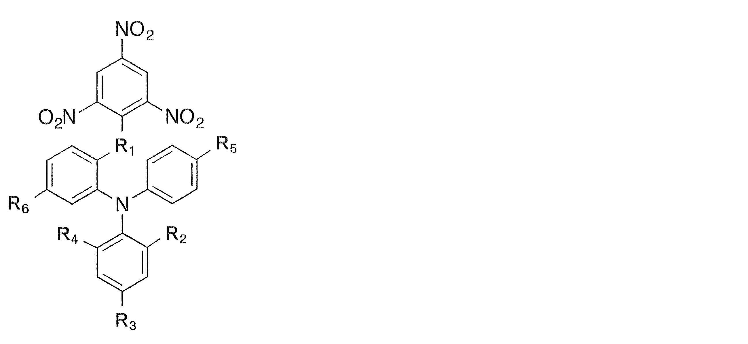
上記化学式中において、
R1は、酸素又は硫黄であり、
R2~R6は、互いに独立して、水素、炭素原子数が1~6のアルキル基、炭素原子数が1~6のアルコキシ基、炭素原子数が1~6のアシル基及びニトロ基から選択される。
また、本発明に係る化合物は、上記化学式中において、
R1は、酸素又は硫黄であり、
R2及びR4は、互いに独立して、水素又はニトロ基であり、
R3は、水素、ニトロ基、メチル基、メトキシ基又はブチル基であり、
R5は、水素、メチル基、メトキシ基又はアセチル基であり、
R6は、水素又はブチル基であることが好ましい。
R1は、酸素又は硫黄であり、
R2及びR4は、互いに独立して、水素又はニトロ基であり、
R3は、水素、ニトロ基、メチル基、メトキシ基又はブチル基であり、
R5は、水素、メチル基、メトキシ基又はアセチル基であり、
R6は、水素又はブチル基であることが好ましい。
また、本発明に係る化合物は、下記化学式のいずれかで示されることがより好ましい。

本発明に係る化合物によると、テロメア結合タンパク質がテロメアDNAに結合することを阻害できるため、Gテイルにおけるループ形成を阻害できる。その結果、Gテイルの短縮を促すことができて、細胞老化や細胞死を誘導することができる。従って、本発明に係る化合物は、細胞老化や細胞死を誘導する試薬として利用できる可能性があり、さらには、癌等の種々の疾患の治療薬開発等への応用の可能性もある。
本発明に係るテロメア結合タンパク質阻害剤は、上記化合物のいずれかを含むことを特徴とする。
本発明に係るテロメア結合タンパク質阻害剤において、テロメア結合タンパク質は、例えばTRF1、TRF2又はPOT1等である。
本発明に係るテロメア結合タンパク質阻害剤によると、上記化合物を含むため、上述の通り、テロメア結合タンパク質がテロメアDNAに結合することを阻害できる。このため、Gテイルにおけるループ形成を阻害でき、Gテイルの短縮を促すことができて、細胞老化や細胞死を誘導することができる。
以上から、本発明に係る上記化合物は、癌の治療や予防に利用できると考えられ、このため、本発明は上記の化合物を含む癌の治療又は予防用医薬組成物に関し、癌の治療又は予防用医薬組成物を製造するための上記化合物の使用にも関する。さらに、本発明は、上記の化合物を癌患者の癌を治療又は予防するために投与することを含む方法にも関する。
本発明に係る化合物、及びそれを含むテロメア結合タンパク質阻害剤によると、テロメア結合タンパク質がテロメアDNAに結合することを阻害できるため、Gテイルにおけるループ形成を阻害できる。その結果、Gテイルの短縮を促すことができて、細胞老化や細胞死を誘導することができる。
以下、本発明を実施するための形態を図面に基づいて説明する。以下の好ましい実施形態の説明は、本質的に例示に過ぎず、本発明、その適用方法或いはその用途を制限することを意図するものではない。
本実施形態に係る化合物は、テロメア結合タンパク質を阻害する化合物である。また、本実施形態に係る化合物は、下記化学式で示される。

なお、上記化学式中において、
R1は、酸素又は硫黄であり、
R2~R6は、互いに独立して、水素、炭素原子数が1~6のアルキル基、炭素原子数が1~6のアルコキシ基、炭素原子数が1~6のアシル基及びニトロ基から選択される。
また、本実施形態に係る化合物は、上記化学式中において、
R1は、酸素又は硫黄であり、
R2及びR4は、互いに独立して、水素又はニトロ基であり、
R3は、水素、ニトロ基、メチル基、メトキシ基又はブチル基であり、
R5は、水素、メチル基、メトキシ基又はアセチル基であり、
R6は、水素又はブチル基であることが好ましい。
R1は、酸素又は硫黄であり、
R2及びR4は、互いに独立して、水素又はニトロ基であり、
R3は、水素、ニトロ基、メチル基、メトキシ基又はブチル基であり、
R5は、水素、メチル基、メトキシ基又はアセチル基であり、
R6は、水素又はブチル基であることが好ましい。
本実施形態において、テロメア結合タンパク質は、例えばTRF1、TRF2及びPOT1等である。また、本実施形態において、上記化合物は、特に当該テロメア結合タンパク質がテロメアに結合することを阻害する。
本実施形態における化合物はこのような特徴を有するため、テロメア結合タンパク質阻害剤に利用することもでき、さらに、癌の治療又は予防用の医薬組成物に利用することもできる。
以下に、本発明に係る化合物、及びそれを含むテロメア結合タンパク質を詳細に説明するための実施例を示す。
まず、DSE-FRETアッセイ(上記特許文献1の特に第一の態様を参照)を用いて、テロメア結合タンパク質を阻害する化合物をスクリーニングした。なお、候補化合物には、独立行政法人産業技術総合研究所が保有する化合物ライブラリーの12212種の化合物を用いた。DSE-FRETアッセイの原理は特許文献1に記載されているが、以下にも簡単に説明する。
特許文献1に示すように、DSE-FRETアッセイは、二つの核酸二本鎖部分(核酸二本鎖A及び核酸二本鎖B)がそれらの末端配列において互いに結合してなる複合体(核酸二本鎖複合体)の構造変化により生じる新たな核酸二本鎖の量を測定することを特徴とする方法である。本願の図1に示すように、核酸二本鎖Aは、核酸一本鎖である核酸A1と核酸一本鎖である核酸A2とからなり、かつ、末端配列において核酸二本鎖Bと結合し得る核酸二本鎖である。核酸二本鎖Bは、核酸一本鎖である核酸B1と核酸一本鎖である核酸B2とからなり、かつ、末端配列において核酸二本鎖Aと結合し得る核酸二本鎖である。
核酸A1、核酸A2、核酸B1、核酸B2はそれぞれ、以下のように設計することができる。
・核酸A1:第一のヌクレオチド配列および第二のヌクレオチド配列(末端配列)を有する核酸一本鎖。
・核酸A2:第一のヌクレオチド配列に対応する配列および第三のヌクレオチド配列(末端配列)を有する核酸一本鎖。
・核酸B1:第二のヌクレオチド配列に対応する配列(末端配列)および第四のヌクレオチド配列を有する核酸一本鎖。
・核酸B2:第三のヌクレオチド配列に対応する配列(末端配列)および第四のヌクレオチド配列に対応する配列を有する核酸一本鎖。
・核酸A1:第一のヌクレオチド配列および第二のヌクレオチド配列(末端配列)を有する核酸一本鎖。
・核酸A2:第一のヌクレオチド配列に対応する配列および第三のヌクレオチド配列(末端配列)を有する核酸一本鎖。
・核酸B1:第二のヌクレオチド配列に対応する配列(末端配列)および第四のヌクレオチド配列を有する核酸一本鎖。
・核酸B2:第三のヌクレオチド配列に対応する配列(末端配列)および第四のヌクレオチド配列に対応する配列を有する核酸一本鎖。
核酸A1、核酸A2、核酸B1及び核酸B2の具体的な構造の一態様は、本願の図1に示すとおりである。
核酸A1、核酸A2、核酸B1、核酸B2については、核酸結合タンパク質の結合部位を有するように設計される。本実施例では、テロメア結合タンパク質のテロメア配列への結合及びその阻害を評価することを目的とするため、上記核酸結合タンパク質の結合部位は、テロメア結合タンパク質、特にTRF2の結合配列とした。配列の詳細は後に説明する。
当該方法は、核酸二本鎖間の構造変化が、核酸結合タンパク質の結合により阻害されることを利用するものである。核酸A1及び核酸A2で構成された核酸二本鎖Aと、核酸B1及び核酸B2で構成された核酸二本鎖Bがそれらの末端配列において互いに結合してなる核酸二本鎖複合体は、鎖交換反応により構造変化をする。具体的に、図2(a)に示す核酸二本鎖A及び核酸二本鎖Bの末端配列が互いに結合する核酸二本鎖複合体が、鎖交換反応により図2(b)に示す構造に変化し、さらに図2(c)に示す構造に変化する。なお、図2(a)~(c)の構造変化は可逆的変化である。図2(c)に示す構造から、さらに鎖交換反応が進行することにより、図2(d)に示すように、核酸A1及び核酸B1で構成された核酸二本鎖Cと、核酸A2及び核酸B2で構成された核酸二本鎖Dとが生成される。なお、図2(c)から(d)への構造変化は不可逆反応である。これに対して、核酸結合タンパク質(テロメア結合タンパク質)が核酸二本鎖A又は核酸二本鎖Bのいずれかに結合すると上記構造変化は阻害される。具体的に、図2(e)に示すように、核酸二本鎖A及び核酸二本鎖Bで構成された核酸二本鎖複合体に核酸結合タンパク質が結合した場合、図2(f)に示すように鎖交換反応によって、ある程度の構造変化が進むものの、タンパク質が結合した部分においてタンパク質が鎖交換反応を阻害する。よって、この部分において鎖交換反応が中断されて、その結果、図2(g)に示す構造には変化することができない。
従って、核酸二本鎖A、Bと、核酸二本鎖C、Dとの量を測定することによって、上記鎖交換反応による構造変化の程度、すなわち、テロメア配列へのテロメア結合タンパク質の結合の程度を測定することができる。このような測定は、核酸二本鎖を標識することにより容易に行うことができる。以下の方法に限られないが、例えば核酸A1の5’末端を蛍光物質で標識し、核酸B1の3’末端を消光物質で標識し、当該蛍光物質の蛍光強度を測定することによって、テロメア配列へのテロメア結合タンパク質の結合程度の測定が可能となる。
例えば、核酸A1のうち、5’末端を蛍光物質で標識し、核酸B1のうち、3’末端は前記蛍光物質を消光する物質で標識した場合、図2(a)に示すように核酸二本鎖A、Bにより核酸二本鎖複合体が形成されている時は蛍光を発する。一方、鎖交換反応が進行し、図2(b)、(c)に示すように、核酸A1の5’末端と核酸B1の3’末端とが近づく、すなわち蛍光物質と消光物質とが近づくに従って、蛍光強度が低減する。さらに、鎖交換反応が進行し、図2(d)に示すように、最終産物である核酸二本鎖C及び核酸二本鎖Dを形成すると、核酸B1の標識によって消光される。このため、蛍光値を測定することにより、容易に核酸二本鎖複合体と、核酸二本鎖C及び核酸二本鎖Dの量を測定することができる。なお、蛍光物質及び消光物質で標識する位置の組合せは、上記位置の組合せに限らず、鎖交換反応の進行で蛍光物質と消光物質との距離が近接する又は離間する位置であればよい。また、消光物質と蛍光物質は入れ替えることもできる。
以上の通り、TRF2結合部位を有する上記核酸のいずれかにTRF2が結合しない場合(結合が阻害されている場合)は、上記鎖交換反応が進行して蛍光物質と消光物質の位置が近接することにより消光され、一方、TRF2が結合する場合は、上記鎖交換が進行せず、蛍光物質と消光物質とが離間しているため蛍光が発せられる。すなわち、本方法によると、蛍光強度により、TRF2の結合又はその阻害の程度を測定することが可能となる。
以下に、本実施例で行ったDSE-FRETアッセイの方法及び結果を説明する。
まず、上記核酸A2に対応する合成オリゴヌクレオチドTLM-06と、5’末端をFAM(蛍光物質)で標識した上記核酸A1に対応する合成オリゴヌクレオチドTLM-01-5Fとを、20μLの二本鎖形成溶液(10mM HEPES-NaOH(pH7.9)、50mM KCl、30mM NaCl、0.1mM EDTA、2.5mM DTT、10% Glycerol、0.05% IGEPAL CA-630)中に混合した。その後、加熱変性とアニーリングによって末端に一本鎖を持つ上記核酸二本鎖Aに対応する二本鎖T01F/06を調製した。T01C/06はTRF2結合配列を持つ。また、上記核酸B2に対応する合成オリゴヌクレオチドTLM-05と、3’末端をDabcyl(消光物質)で標識した上記核酸B1に対応する合成オリゴヌクレオチドTLM-02-3Dとを、20μLの二本鎖形成溶液(10mM HEPES-NaOH(pH7.9)、50mM KCl、30mM NaCl、0.1mM EDTA、2.5mM DTT、10% Glycerol、0.05% IGEPAL CA-630)中に混合した。その後、加熱変性とアニーリングによって末端に一本鎖を持つ上記核酸二本鎖Bに対応する二本鎖T02D/05を調製した。T02D/05はTRF2結合配列を持つ。合成オリゴヌクレオチドは全て20pmol使用した。なお、以後すべての標識については、日本バイオサービス社に合成を依頼して作製されたものを使用した。
加熱変性とアニーリングは以下の温度条件で行った。
95℃ 120秒 - 90℃ 30秒 - 85℃ 90秒 - 80℃ 90秒 - 77℃ 90秒 - 75℃ 90秒 - 72℃ 90秒 - 70℃ 90秒 - 67℃ 90秒 - 65℃ 90秒 - 62℃ 90秒 - 60℃ 90秒 - 57℃ 90秒 - 55℃ 90秒 - 52℃ 90秒 - 50℃ 90秒 - 47℃ 90秒 - 45℃ 90秒 - 42℃ 90秒 - 40℃ 90秒 - 37℃ 90秒 - 35℃ 90秒 - 32℃ 90秒 - 30℃ 90秒。
95℃ 120秒 - 90℃ 30秒 - 85℃ 90秒 - 80℃ 90秒 - 77℃ 90秒 - 75℃ 90秒 - 72℃ 90秒 - 70℃ 90秒 - 67℃ 90秒 - 65℃ 90秒 - 62℃ 90秒 - 60℃ 90秒 - 57℃ 90秒 - 55℃ 90秒 - 52℃ 90秒 - 50℃ 90秒 - 47℃ 90秒 - 45℃ 90秒 - 42℃ 90秒 - 40℃ 90秒 - 37℃ 90秒 - 35℃ 90秒 - 32℃ 90秒 - 30℃ 90秒。
使用した配列は以下の通りである。なお、下線を引いた部分はTRF2結合配列であり、小文字の部分は核酸複合体を形成した際に一本鎖となる配列である。
TLM-01-5F:
5’ FAM- AGTTGAGTTAGGGTTAGGGT TAGGGTTAGG GCAGGcggtg tctcgctcgc 3’(配列番号1)
TLM-02-3D:
5’ gcgagcgagacaccgCCTGC CCTAACCCTA ACCCTAACCC TAACTCAACT -Dabcyl 3’(配列番号2)
TLM-05:
5’AGTTGAGTTA GGGTTAGGGT TAGGGTTAGG GCAGGcacca caccattccc 3’(配列番号3)
TLM-06:
5’gggaatggtg tggtgCCTGC CCTAACCCTA ACCCTAACCC TAACTCAACT 3’(配列番号4)
TLM-01-5F:
5’ FAM- AGTTGAGTTAGGGTTAGGGT TAGGGTTAGG GCAGGcggtg tctcgctcgc 3’(配列番号1)
TLM-02-3D:
5’ gcgagcgagacaccgCCTGC CCTAACCCTA ACCCTAACCC TAACTCAACT -Dabcyl 3’(配列番号2)
TLM-05:
5’AGTTGAGTTA GGGTTAGGGT TAGGGTTAGG GCAGGcacca caccattccc 3’(配列番号3)
TLM-06:
5’gggaatggtg tggtgCCTGC CCTAACCCTA ACCCTAACCC TAACTCAACT 3’(配列番号4)
100fmolのT01F/06と50μMの候補化合物を反応溶液(10mM HEPES-NaOH pH7.9、150mM KCl、0.1mM EDTA、5mM DTT、10% glycerol、0.05% IGEPAL CA-630、20μL)中に混合して、25℃で30分間反応させた。その後、T01F/06に、100fmolのT02D/05をそれぞれ加えて50μLとした後、25℃で120分間反応させた。Cy3の蛍光値の測定は、蛍光プレートリーダーEnVision(Perkin elmer社)を用いた。
候補化合物のスクリーニングの結果、検出された蛍光強度が大きく、すなわち、TRF2がその結合部位に結合することを阻害する化合物として下記化学式をもつ化合物#10が得られた。
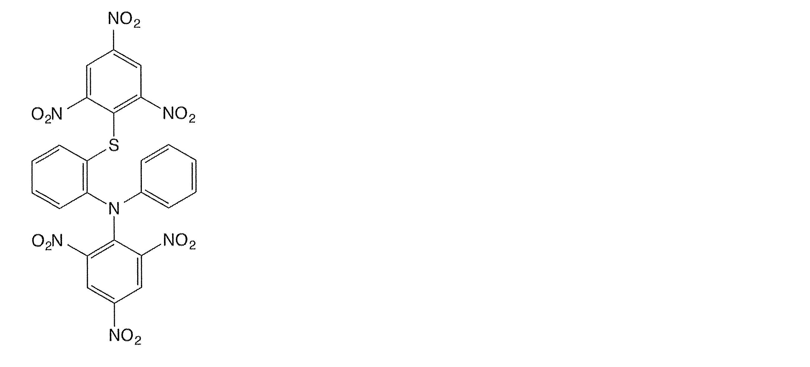
また、得られた化合物#10の構造に基づいて、これと類似する構造を有する化合物を合成し、上記スクリーニングを行った。その結果、検出された蛍光強度が大きく、TRF2がその結合部位に結合することを阻害する化合物は以下の表1に示すとおりである。なお、候補化合物を反応溶液に混合していないコントロールの場合の蛍光強度を基準として、各化合物を反応溶液に混合した場合の蛍光強度の低減率を阻害率として算出した。
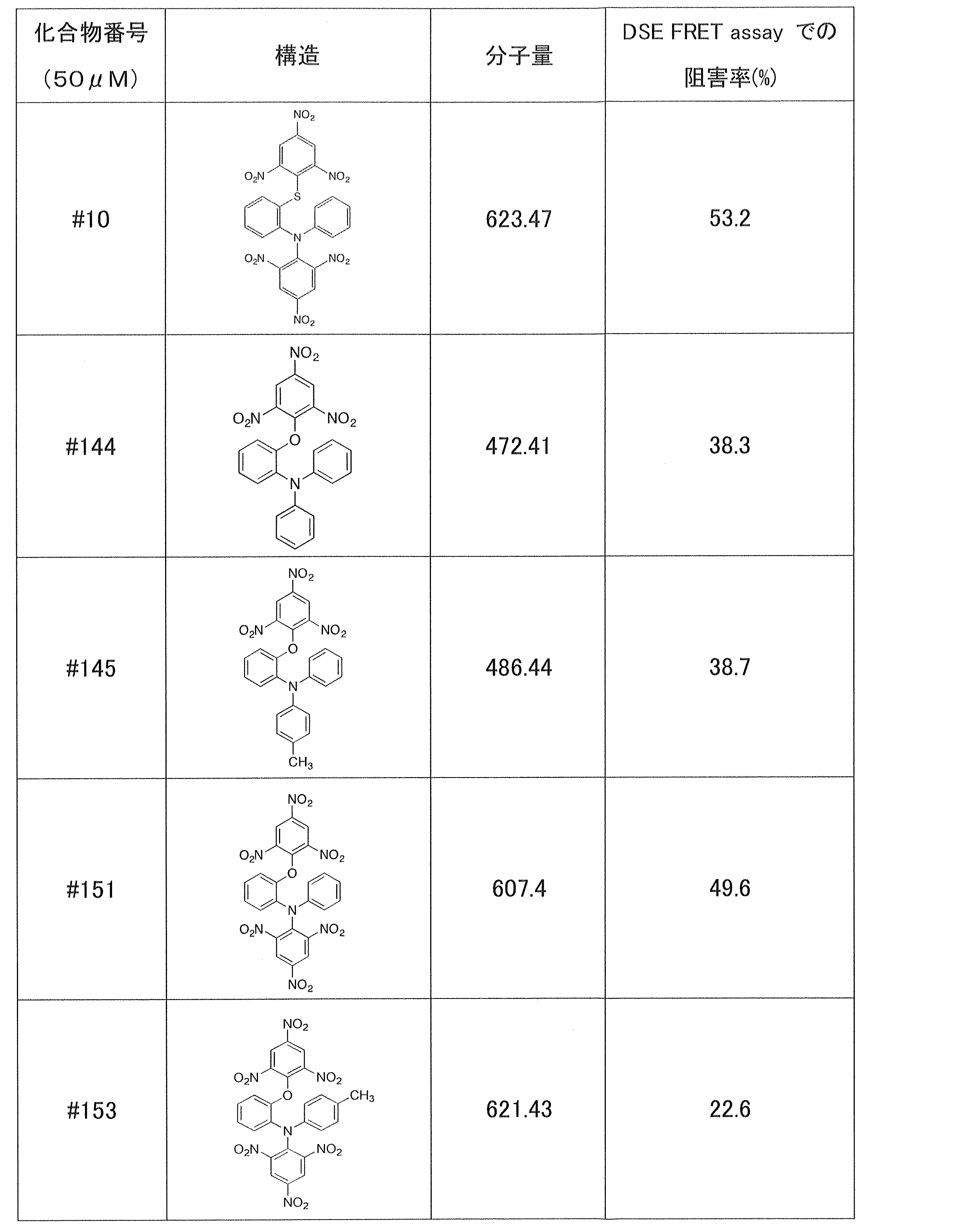

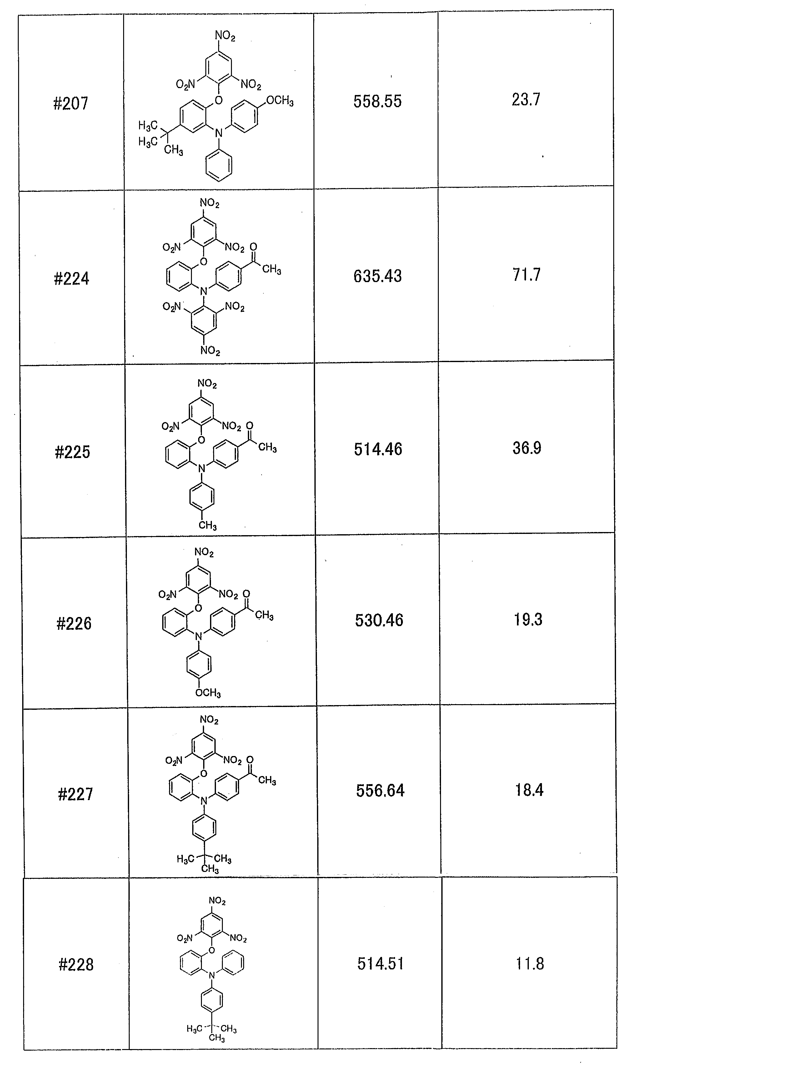
上記表1に示した化合物は、下記化学式を共通の骨格として有していることが分かる。
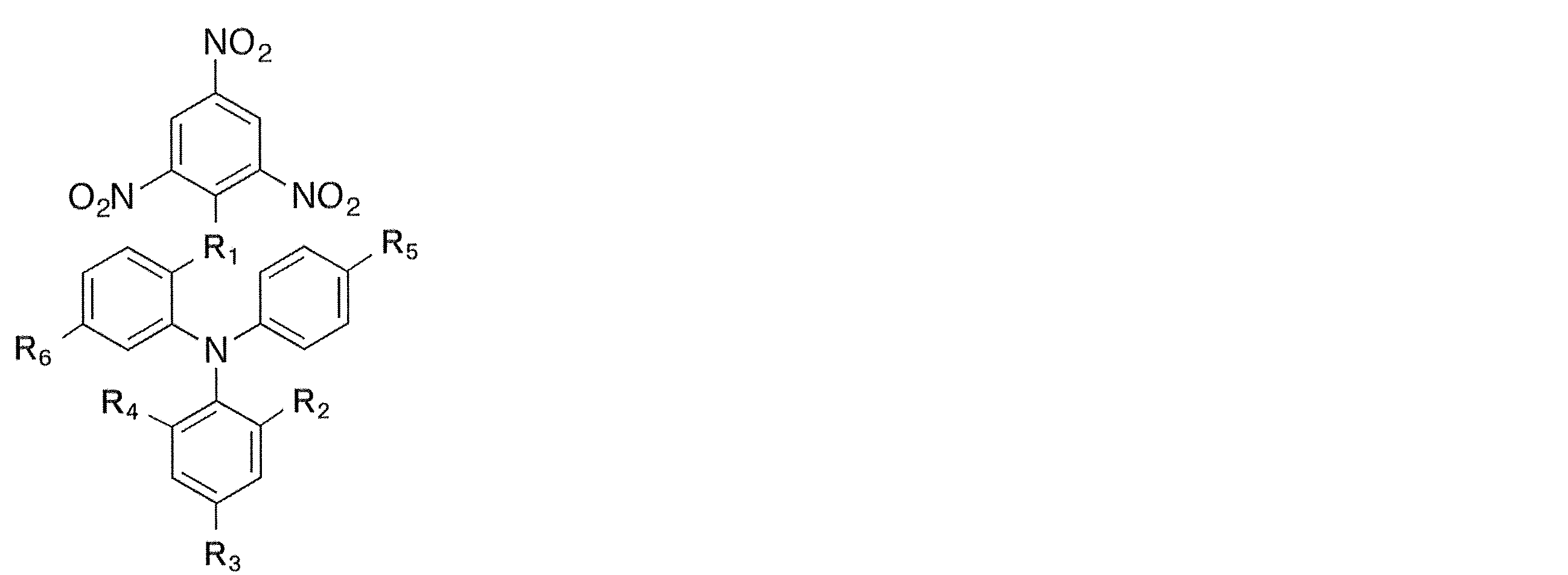
なお、上記化学式中において、
R1は、酸素又は硫黄であり、
R2及びR4は、互いに独立して、水素又はニトロ基であり、
R3は、水素、ニトロ基、メチル基、メトキシ基又はブチル基であり、
R5は、水素、メチル基、メトキシ基又はアセチル基であり、
R6は、水素又はブチル基である。
また、上記各化合物は、通常の合成法にて合成されるものである。例として、以下に化合物#198の合成方法を示す。
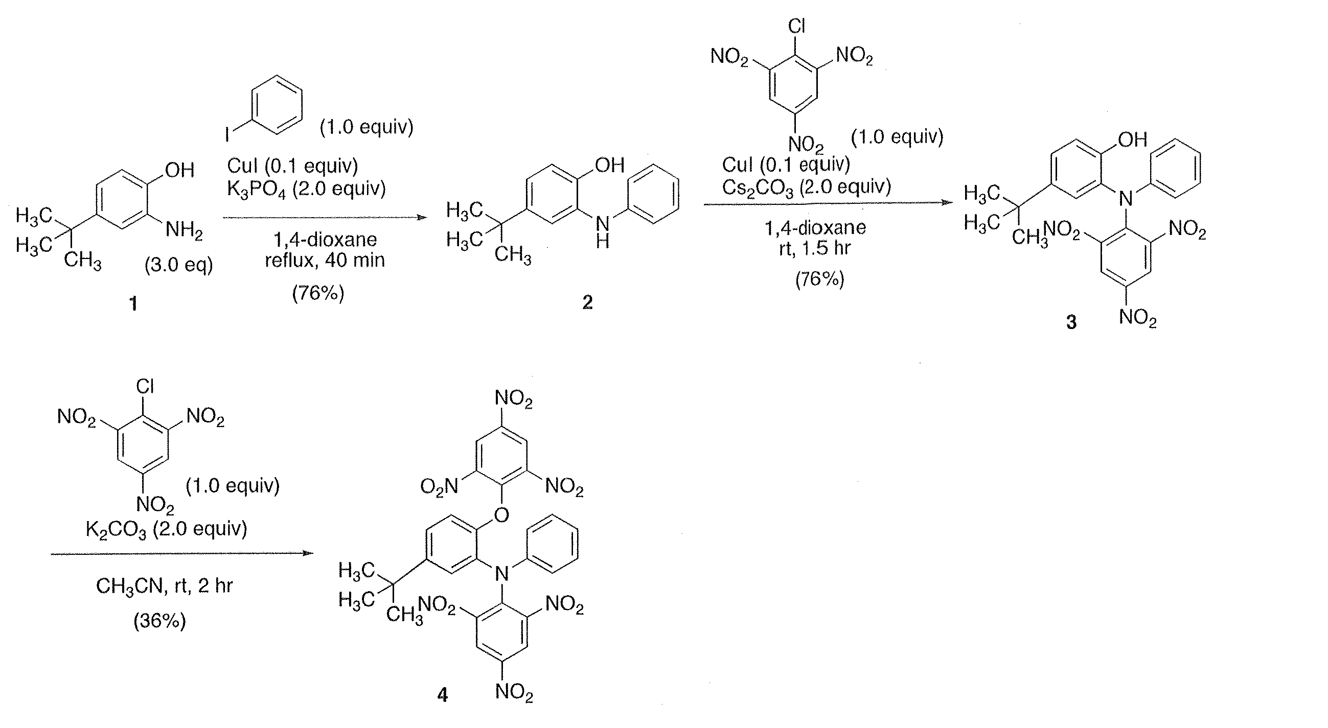
その他の化合物においても、当業者であれば上記化合物#198の合成法の部分的変更、すなわち原料であるアミノフェノール誘導体およびハロゲン化アリールとして適当な置換其を有するものを用いることによって容易に合成可能である。例えば化合物#207では、上記化合物#198の合成法のうち[化6]中の2から3への合成を、2,4,6‐トリニトロクロロベンゼンの代わりに4-ヨードアニソールを用いることは、当業者には容易に想到可能である。
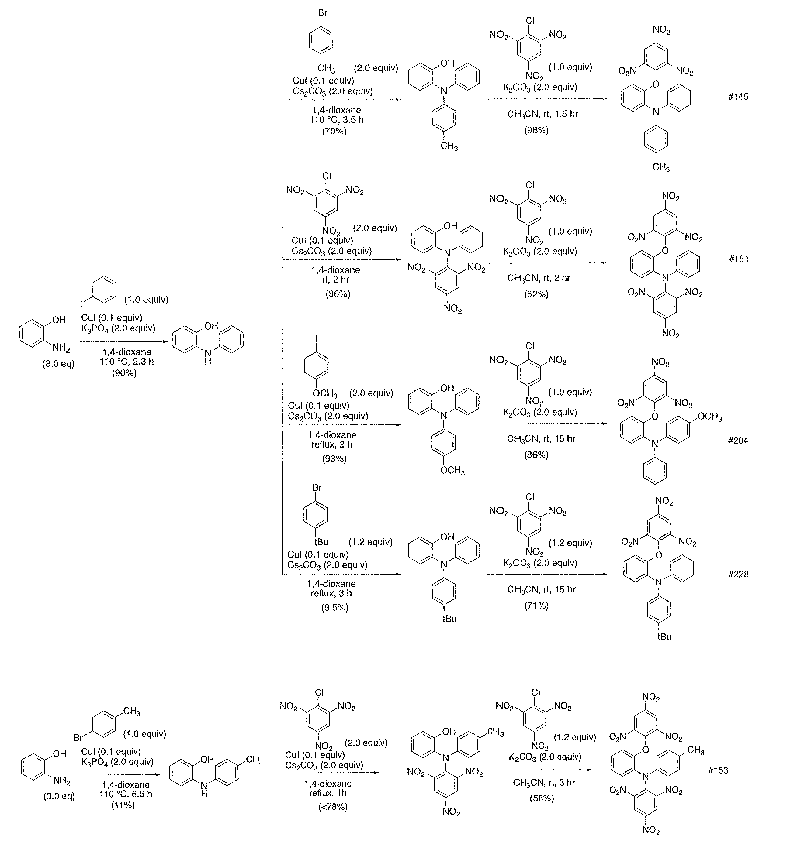
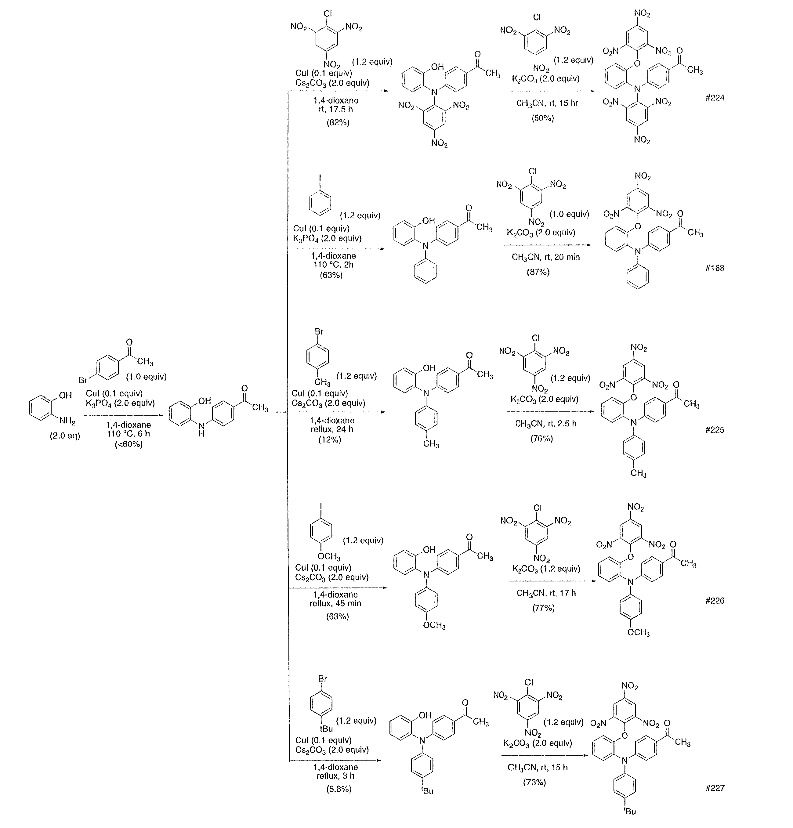
ここで、本実施例で用いた他の化合物の具体的な合成法を以下に示す。
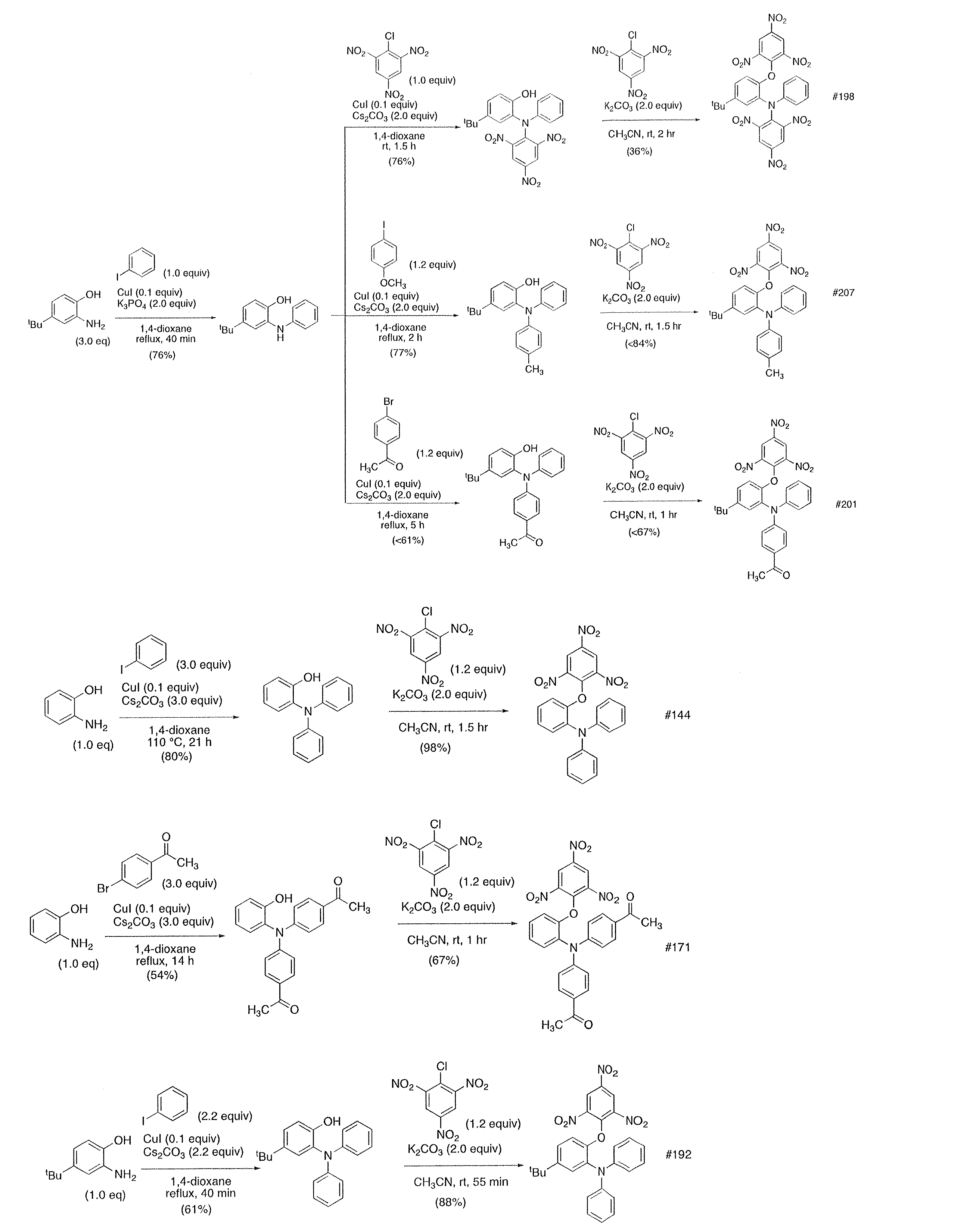
次に、上記スクリーニングの結果において、比較的に高いTRF2の阻害効果が認められた化合物#198を用いて、TRF2の阻害効果についてさらに検討した。そのために、TRF2抗体を用いたクロマチン免疫沈降試験(ChIPアッセイ)を行った。その方法及び結果を以下に説明する。
HeLa1.2.11細胞に化合物#198(20μM)又はコントロールとしてのDMSOを24時間処理後、1%ホルムアルデヒドで固定した。その後、細胞をLysisbuffer(1%SDS、10mM EDTA(pH8.0)、50mM Tris-HCl(pH8.0))で溶かし、超音波によりクロマチンを断片化した。その後、TRF2抗体(santacruz社,sc-8528)又はnormalmouse IgG(santacruz、sc-2025)を用いてクロマチン免疫沈降を行い、Hybond N+ メンブレン(Amersham社、RPN82B)にDNAを吸着後、DIG標識テロメアプローブ(Digoligo nucleotide Tailingkit(Roche社、03 353 583910)を用いて、100pmolの3’-(CCCTAA)4-5’オリゴヌクレオチドの3’末端に、Digoxigeninを標識したもの)を反応させ、ルミノアナライザー(ImageQuant社、LAS4000)にてシグナルを検出した。画像解析ソフトImageJを用いてシグナルを定量し、10%inputに対するTRF2抗体による免疫沈降物中のテロメアDNA量を算出した。その結果を図3に示す。なお、図3(a)はHybondN+ メンブレンの写真であり、(b)は上記シグナル定量をしてテロメアDNA量を算出した結果を示すグラフである。
図3に示すように、DMSOを用いたコントロールと比較して、化合物#198を処理した場合ではテロメアDNAの検出は50%以下に抑制された。この結果から、化合物#198によって、TRF2がテロメアDNAに結合することが阻害されることが示唆される。
次に、化合物#198の処理の有無によるTRF2の細胞内局在を蛍光免疫染色法で検討した。その方法及び結果を以下に説明する。
8well組織培養用カルチャースライド(松波硝子工業、scs-008)に播種したHeLa1.2.11細胞に化合物#198(20μM)又はコントロールとしてのDMSOを24時間処理した。その後、PBS(-)で2回洗浄し、0.25%TritonX-100(WAKO社,591-12191)/PBS(-)を氷上で2分間処理し、4% パラホルムアルデヒド(MERCK社,1.04005.1000)/PBS(-)を室温で15分間処理した後、PBS(-)で2回洗浄した。さらに0.5%TritonX-100/PBS(-)を氷上で10分間処理した後、PBS(-)で5回洗浄した。次に、3%BSA/0.05%Tween/PBS(-)で200倍希釈したTRF2抗体(Novus社、NB100-56506)を37℃で1時間反応させた後、PBS(-)で2回洗浄した。次に、3%BSA/0.05%Tween/PBS(-)で500倍希釈したAlexaFluor 488 Goat anti-mouse IgG(invitrogen, A11001)を室温で45分間反応させ、PBS(-)で3回洗浄し、0.25μg/mLのDAPI(同仁化学研究所、340-07971)を室温で5分間反応させ、PBS(-)で3回洗浄した。スライドガラスを封入後、蛍光顕微鏡(Zwiss社、Axiovert 200M)にて観測した。画像解析ソフトColumbusを用いて核内のTRF2foci数を計測した。その結果を図4に示す。なお、図4(a)は、蛍光顕微鏡を用いて得られた細胞の写真であり、図4(a)において、白い四角の領域を拡大したもの「Enlarged」で示し、「Enlarged」中の矢印は核外に存在するTRF2を示す。図4(b)は、蛍光顕微鏡を用いて核内のTRF2foci数を計測し、コントロールを1.00としたときのTRF2foci数の割合を示すグラフである。
図4に示すように、DMSOで処理されたコントロールでは、DAPIにより染まった核内にTRF2が存在し、TRF2が核内のDNAに結合していると考えられる。一方、化合物#198により処理された細胞では、TRF2の多くが細胞の核の外側に存在している。この結果から、化合物#198によってTRF2が核内でDNAのTRF2結合部位に結合することを阻害することが示唆される。
次に、テロメアFISH法を用いて、テロメア異常における化合物#198の影響について検討した。具体的に、テロメアにおいて異常が生じると、テロメアにおける53BP1の局在を特徴とするTIF(telomere-inducedDNA damage foci)が生じることが知られているため、テロメアFISH法を用いて、化合物#198がテロメアにおける53BP1の局在化に影響するか否かを検討した。以下にその方法及び結果を説明する。
8well組織培養用カルチャースライドに播種したHeLa1.2.11細胞に化合物 #198(10μM)又はコントロールとしてのDMSOを24時間処理した。その後、PBS(-)で2回洗浄し、0.25%TritonX-100(WAKO社,591-12191)/PBS(-)を氷上で2分間処理し、4%パラホルムアルデヒド(MERCK社,1.04005.1000)/PBS(-)を室温で15分間処理した。その後、PBS(-)で2回洗浄し、0.5%TritonX-100/PBS(-)を氷上で10分間処理した後、PBS(-)で5回洗浄した。次に、Blockingsolution (1mg/ml BSA,3% goatserum,0.1% Triton X-100, 1mMEDTA pH8.0)を室温で30分間処理し、Blocking solutionで1000倍に希釈した53BP1抗体(Novus,NB100-304)を37℃で1時間反応させた後、PBS(-)で2回洗浄した。次に、Blockingsolutionで500倍に希釈したAlexa Fluor488 Goat anti-RabbitIgG(invitrogen,A11008)を室温で45分間反応させ、PBS(-)で3回洗浄し、4%パラホルムアルデヒド/PBS(-)を室温で5分間処理し、PBS(-)で2回洗浄した。その後、70%,95%,100%EtOHを順に3,2,2分間処理した。風乾後、Cy-3標識3’-(CCCTAA)4-5’プローブ(PANAGENE社,F1002)を80℃で3分間反応させた。その後、washingsolution(70%formamide,10mM Tris-HCl(pH 7.2)) で洗浄し、PBS(-)で3回洗浄後、0.25μg/mLのDAPIを室温で5分間反応させ、さらにPBS(-)で3回洗浄した。スライドガラスを封入後、蛍光顕微鏡(Zwiss社、Axiovert200M)にて観測した。画像解析ソフトColumbusを用いてテロメアに局在している53BP1(TIF)を計測した。核1個あたりTIFが4個以上あったものをTIF陽性細胞とした。その結果を図5に示す。なお、図5(a)は蛍光顕微鏡を用いて得られた細胞の写真であり、矢印はテロメアと53BP1との共局在が見られる部分を示す。図5(b)は、蛍光顕微鏡を用いてその共局在が生じている、すなわちTIFが生じている細胞数を計測し、その割合を算出した結果を示すグラフである。
図5に示すように、コントロールでは、テロメアと53BP1との共局在はほとんど見られなかったが、化合物#198で処理した細胞では、テロメアと53BP1との共局在が認められた。すなわち、化合物#198で処理した細胞では、TIFが生じており、テロメアに異常が生じていると考えられる。これは、化合物#198がTRF2のテロメアへの結合を阻害していることに起因していると考えられる。
上記の通り、化合物#198がテロメア異常を促すと考えられる。ここで、テロメア異常は細胞老化や細胞死に関連することが知られているため、化合物#198により細胞のアポトーシスを引き起こすか否かについてFACSを利用して検討した。その方法及び結果を以下に説明する。
HeLa1.2.11細胞を20μMの化合物#198又はDMSOで48時間処理した後に、上清ごと細胞を15mLチューブに回収し、1000rpmで3分間遠心し、上清を取り除いた。5mlのPBS(-)で細胞を再懸濁し1000rpmで3分遠心し、上清を除いた。500μlの1×Bindingbuffer(MBL社、4695-300) で細胞を再懸濁し、このうち90μlを5mLチューブ(BDFalcon社、352052)へ移し、10μlのAnnexinV-FITC(MBL社、4700-100)を加え染色した。遮光した状態で15分インキュベートし、解析を行った。Cellsorter(SONY社、SH-800)にてAnnexin V陽性細胞を検出した。結果を図6に示す。
図6に示すように、DMSO処理したコントロールと比較して、化合物#198で処理した場合では多くの細胞がAnnexinV陽性であった。すなわち、化合物#198によって細胞のアポトーシスが誘導されることが示唆される。
さらに、別の方法により化合物#198によって細胞のアポトーシスが誘導されることを証明するために、アポトーシスの極めて重要な因子として知られている切断型カスパーゼ3の発現が化合物#198により促進されるか否かをウエスタンブロット法によって解析した。その方法及び結果を以下に説明する。
HeLa1.2.11細胞を20μMの化合物#198又はDMSOで48時間処理した後に、上清と共に細胞を15mLチューブに回収し、1000rpmで3分間遠心し、上清を取り除いた。細胞ペレットを2×Samplebuffer(117mM Tris-HCl(pH6.8),13%Glycerol,3.7%SDS,200mM DTT,0.004%Bromo Phenol Blue)で溶解し、95℃で5分間熱変性を行った。8%アクリルアミドゲルに10μgのサンプルをRunningbuffer(25 mM Tris、192mM Glycine、0.1%(w/v)SDS)中、定電圧(120V)で泳動した。その後、PVDFメンブレンフィルター Immobilon-P(MILLIPORE)に転写し、抗原抗体反応を用いて目的のタンパクを検出した。用いた抗体は一次抗体にB-actin(SIGMA社、A5441)又はCleavedcaspase3(CST社、D175)、二次抗体にPeroxidase標識Goat抗マウス/抗ラビット二次抗体(JacksonImmuno Research、111-035-003/115-035-003)を用い、シグナルの検出はルミノアナライザー (ImageQuant社、LAS4000)を用いて検出した。その結果を図7に示す。
図7に示すように、DMSOを処理したコントロールでは、切断型カスパーゼ3はほぼ見られなかったが、一方、化合物#198を処理した細胞では切断型カスパーゼ3が検出された。この結果から、化合物#198によって、切断型カスパーゼ3の生成が促進され、すなわち細胞のアポトーシスが誘導されると考えられる。
次に、化合物#198の細胞増殖に対する影響を検討した。その方法及び結果を以下に示す。
HeLa1.2.11細胞を1×10⁴cells/35mmdishで播種し、その翌日に、化合物#198(5μM、10μM、20μM)又はコントロールとしてのDMSOを処理し(処理日を1日目とする)、1日目、3日目、5日目及び7日目に細胞を計数した。その結果を図8に示す。
図8に示すように、化合物#198の処理濃度依存的に、細胞増殖が抑制された。
また、#198以外における上記各候補化合物によるHeLa1.2.11細胞の増殖に対する抑制効果を上記と同様に測定し、IC50を算出した結果を以下の表2に示す。

表2に示すように、化合物#198以外の候補化合物も細胞増殖の抑制効果を奏することが明らかである。
次に、HeLa1.2.11細胞を1×10³cells/100mmdishに播種した翌日に化合物#198(5~20μM)又はコントロールとしてのDMSOを処理し、播種してから10日後、形成されたコロニーを100%EtOHで固定後、4%ギムザ染色液(武藤化学株式会社、1500-2)/PBS(-)を用いて染色し、計測した。その結果を図9に示す。なお、図9(a)は上記ギムザ染色をしたプレートの写真を示し、図9(b)コロニーの生成割合を算出した結果を示すグラフである。
図9に示すように、化合物#198の処理濃度依存的にコロニーの生成が抑制された。
以上、各実施例の結果から、本発明に係る化合物は、テロメア結合タンパク質のテロメア結合を阻害し、その結果、テロメアにおけるGテイル形成を阻害して、Gテイルを短縮させると考えられる。これにより、本発明に係る化合物は、細胞の増殖抑制及びアポトーシスの誘導を引き起こすと考えられる。従って、本発明に係る化合物は、細胞老化や細胞死を誘導する試薬として利用できる可能性があり、さらには、癌等の種々の疾患の治療薬開発等への応用の可能性もある。
Claims (8)
- 下記化学式で示される化合物であって

上記化学式中において、
R1は、酸素又は硫黄であり、
R2~R6は、互いに独立して、水素、炭素原子数が1~6のアルキル基、炭素原子数が1~6のアルコキシ基、炭素原子数が1~6のアシル基、及びニトロ基のうちから選択されることを特徴とする化合物。 - 上記化学式中において
R1は、酸素又は硫黄であり、
R2及びR4は、互いに独立して、水素又はニトロ基であり、
R3は、水素、ニトロ基、メチル基、メトキシ基又はブチル基であり、
R5は、水素、メチル基、メトキシ基又はアセチル基であり、
R6は、水素又はブチル基である、
ことを特徴とする請求項1に記載の化合物。 -
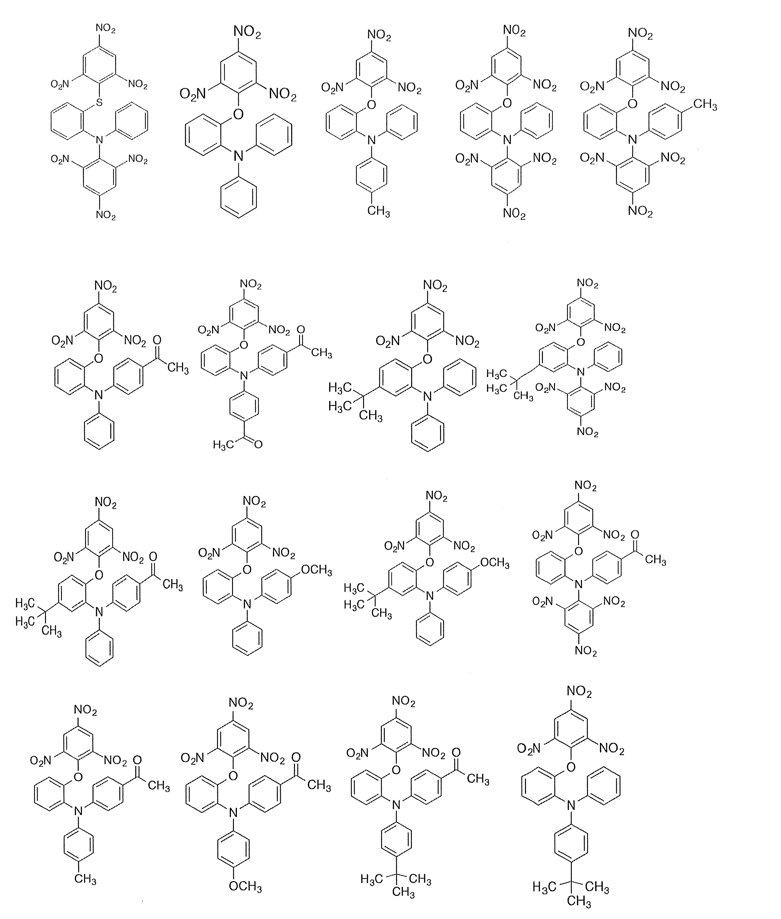
上記化学式のいずれかで示されることを特徴とする請求項2に記載の化合物。 - テロメア結合タンパク質がテロメアに結合することを阻害するための請求項1~3のいずれか1項に記載の化合物を含むテロメア結合タンパク質阻害剤。
- 前記テロメア結合タンパク質は、TRF1、TRF2又はPOT1であることを特徴とする請求項4に記載のテロメア結合タンパク質阻害剤。
- 請求項1~3のいずれか1項に記載の化合物を含む癌の治療又は予防用医薬組成物。
- 癌の治療又は予防用医薬組成物を製造するための請求項1~3のいずれか1項に記載の化合物の使用。
- 請求項1~3のいずれか1項に記載の化合物を癌の治療又は予防のために患者に投与することを含む方法。
Priority Applications (2)
| Application Number | Priority Date | Filing Date | Title |
|---|---|---|---|
| JP2020508284A JP7284518B2 (ja) | 2018-03-20 | 2019-03-14 | テロメア結合タンパク質を阻害する化合物、及びそれを含むテロメア結合タンパク質阻害剤 |
| US16/982,517 US20210024455A1 (en) | 2018-03-20 | 2019-03-14 | Compound which inhibits telomere-binding protein, and telomere-binding protein inhibitor containing same |
Applications Claiming Priority (2)
| Application Number | Priority Date | Filing Date | Title |
|---|---|---|---|
| JP2018-052087 | 2018-03-20 | ||
| JP2018052087 | 2018-03-20 |
Publications (1)
| Publication Number | Publication Date |
|---|---|
| WO2019181714A1 true WO2019181714A1 (ja) | 2019-09-26 |
Family
ID=67987628
Family Applications (1)
| Application Number | Title | Priority Date | Filing Date |
|---|---|---|---|
| PCT/JP2019/010480 Ceased WO2019181714A1 (ja) | 2018-03-20 | 2019-03-14 | テロメア結合タンパク質を阻害する化合物、及びそれを含むテロメア結合タンパク質阻害剤 |
Country Status (3)
| Country | Link |
|---|---|
| US (1) | US20210024455A1 (ja) |
| JP (1) | JP7284518B2 (ja) |
| WO (1) | WO2019181714A1 (ja) |
Cited By (1)
| Publication number | Priority date | Publication date | Assignee | Title |
|---|---|---|---|---|
| WO2023182897A1 (en) * | 2022-03-25 | 2023-09-28 | Politechnika Gdańska | Inhibitors of interactions between trf1-tin2 or trf2-tin2 telomeric proteins for use in anticancer therapy |
Citations (7)
| Publication number | Priority date | Publication date | Assignee | Title |
|---|---|---|---|---|
| JPH07238066A (ja) * | 1994-02-28 | 1995-09-12 | Green Cross Corp:The | ベンゼンスルホン酸誘導体、その製造法およびその用途 |
| JP2001072592A (ja) * | 1999-07-01 | 2001-03-21 | Kyowa Hakko Kogyo Co Ltd | テロメラーゼ阻害剤 |
| WO2001030771A1 (en) * | 1999-10-28 | 2001-05-03 | Kyowa Hakko Kogyo Co., Ltd. | Thiazolidinedione derivatives |
| WO2002044120A1 (en) * | 2000-11-29 | 2002-06-06 | Karo Bio Ab | Compounds active at the glucocorticoid receptor iii |
| JP2004524349A (ja) * | 2001-03-23 | 2004-08-12 | アベンティス・ファーマ・ソシエテ・アノニム | 化学的誘導体および抗テロメラーゼ剤としてのその使用 |
| WO2009093667A1 (ja) * | 2008-01-22 | 2009-07-30 | Hiroshima University | 核酸結合蛋白質アッセイ法およびキット |
| JP2012507504A (ja) * | 2008-10-31 | 2012-03-29 | アンスティテュ・キュリ | 癌の処置用のポリ−ヘテロアリール誘導体 |
Family Cites Families (1)
| Publication number | Priority date | Publication date | Assignee | Title |
|---|---|---|---|---|
| WO2017115753A1 (ja) * | 2015-12-28 | 2017-07-06 | コニカミノルタ株式会社 | 架橋化合物、有機エレクトロルミネッセンス素子材料、有機エレクトロルミネッセンス素子、表示装置及び照明装置 |
-
2019
- 2019-03-14 US US16/982,517 patent/US20210024455A1/en not_active Abandoned
- 2019-03-14 JP JP2020508284A patent/JP7284518B2/ja active Active
- 2019-03-14 WO PCT/JP2019/010480 patent/WO2019181714A1/ja not_active Ceased
Patent Citations (7)
| Publication number | Priority date | Publication date | Assignee | Title |
|---|---|---|---|---|
| JPH07238066A (ja) * | 1994-02-28 | 1995-09-12 | Green Cross Corp:The | ベンゼンスルホン酸誘導体、その製造法およびその用途 |
| JP2001072592A (ja) * | 1999-07-01 | 2001-03-21 | Kyowa Hakko Kogyo Co Ltd | テロメラーゼ阻害剤 |
| WO2001030771A1 (en) * | 1999-10-28 | 2001-05-03 | Kyowa Hakko Kogyo Co., Ltd. | Thiazolidinedione derivatives |
| WO2002044120A1 (en) * | 2000-11-29 | 2002-06-06 | Karo Bio Ab | Compounds active at the glucocorticoid receptor iii |
| JP2004524349A (ja) * | 2001-03-23 | 2004-08-12 | アベンティス・ファーマ・ソシエテ・アノニム | 化学的誘導体および抗テロメラーゼ剤としてのその使用 |
| WO2009093667A1 (ja) * | 2008-01-22 | 2009-07-30 | Hiroshima University | 核酸結合蛋白質アッセイ法およびキット |
| JP2012507504A (ja) * | 2008-10-31 | 2012-03-29 | アンスティテュ・キュリ | 癌の処置用のポリ−ヘテロアリール誘導体 |
Non-Patent Citations (7)
| Title |
|---|
| DATABASE REGISTRY [online] 2001, XP55637640, Database accession no. 352638-37-0 * |
| GOMEZ, D. ET AL.: "Interaction of Telomestatin with the Telomeric Single-strand Overhang", THE JOURNAL OF BIOLOGICAL CHEMISTRY, vol. 279, no. 40, 2004, pages 41487 - 41494, XP55637634 * |
| SHIROMA YOSHITOMO: "TRF2- telomere DNA binding inhibitor obtained by applying DSE-FREAT assay", JAPANESE ASSOCIATION FOR MOLECULAR TARGET THERAPY OF CANCER PROGRAM, w7-5, 2017 * |
| SHIROMA, YOSHITOMO: "Development and functional analysis of telomere binding protein inhibitors using DSEFRET assay", ANNUAL MEETING OF THE JAPANESE BIOCHEMICAL SOCIETY, 2P-1435, 2017 * |
| SHIROMA, YOSHITOMO: "Effects of telomere binding protein TRF2 binding inhibitor on cancer cells", ABSTRACTS OF THE 76TH ANNUAL MEETING OF THE JAPANESE CANCER ASSOCIATION, J-3119, 2017 * |
| SHIROMA, YOSHITOMO: "Search for telomere binding protein TRF2 inhibitor using DSE-FRET method", ABSTRACTS OF THE 19TH ANNUAL MEETING OF JAPANESE ASSOCIATION FOR MOLECULAR TARGET THERAPY OF CANCER PROGRAM, P3-3, 2015 * |
| SHIROMA, YOSHITOMO: "Telomere binding protein TRF2 inhibitor obtained using DSE-FRET method", ABSTRACTS OF THE 20TH ANNUAL MEETING OF JAPANESE ASSOCIATION FOR MOLECULAR TARGET THERAPY OF CANCER PROGRAM, P24-2, 2016 * |
Cited By (1)
| Publication number | Priority date | Publication date | Assignee | Title |
|---|---|---|---|---|
| WO2023182897A1 (en) * | 2022-03-25 | 2023-09-28 | Politechnika Gdańska | Inhibitors of interactions between trf1-tin2 or trf2-tin2 telomeric proteins for use in anticancer therapy |
Also Published As
| Publication number | Publication date |
|---|---|
| JP7284518B2 (ja) | 2023-05-31 |
| JPWO2019181714A1 (ja) | 2021-04-08 |
| US20210024455A1 (en) | 2021-01-28 |
Similar Documents
| Publication | Publication Date | Title |
|---|---|---|
| Seiler et al. | H3B-8800, an orally available small-molecule splicing modulator, induces lethality in spliceosome-mutant cancers | |
| Gooding et al. | The lncRNA BORG drives breast cancer metastasis and disease recurrence | |
| Rezaeian et al. | A hypoxia-responsive TRAF6–ATM–H2AX signalling axis promotes HIF1α activation, tumorigenesis and metastasis | |
| Henderson et al. | Detection of G-quadruplex DNA in mammalian cells | |
| Ramachandran et al. | Hypoxia-induced SETX links replication stress with the unfolded protein response | |
| Singh et al. | MiCEE is a ncRNA-protein complex that mediates epigenetic silencing and nucleolar organization | |
| Xie et al. | miR-106a promotes growth and metastasis of non-small cell lung cancer by targeting PTEN | |
| Arsic et al. | The p53 isoform delta133p53ß regulates cancer cell apoptosis in a RhoB-dependent manner | |
| Ning et al. | Characterization of the Merkel cell carcinoma miRNome | |
| Srivastava et al. | Histone H3K9 acetylation level modulates gene expression and may affect parasite growth in human malaria parasite Plasmodium falciparum | |
| Yin et al. | RBM24 exacerbates bladder cancer progression by forming a Runx1t1/TCF4/miR-625-5p feedback loop | |
| Das et al. | AXL and CAV-1 play a role for MTH1 inhibitor TH1579 sensitivity in cutaneous malignant melanoma | |
| JP2019527037A (ja) | がんのための診断及び治療方法 | |
| JP2012187105A (ja) | GSK3β阻害効果に基づく抗がん剤の評価方法 | |
| Wang et al. | RETRACTED ARTICLE: microRNA-769 is downregulated in colorectal cancer and inhibits cancer progression by directly targeting cyclin-dependent kinase 1 | |
| Dai et al. | Genomic analysis of xCT-mediated regulatory network: Identification of novel targets against AIDS-associated lymphoma | |
| de Oliveira et al. | Possible involvement of Hsp90 in the regulation of telomere length and telomerase activity during the Leishmania amazonensis developmental cycle and population proliferation | |
| Yang et al. | Comprehensive analyses reveal effects on tumor immune infiltration and immunotherapy response of APOBEC mutagenesis and its molecular mechanisms in esophageal squamous cell carcinoma | |
| Zhao et al. | QSER1 preserves the suppressive status of the pro-apoptotic genes to prevent apoptosis | |
| Zhang et al. | ARHGAP15 promotes metastatic colonization in gastric cancer by suppressing RAC1-ROS pathway | |
| Kim et al. | The SET domain Is essential for Metnase functions in replication restart and the 5’end of SS-overhang cleavage | |
| Patil et al. | A mechanistic study on the metastasis inducing function of FUS‐CHOP fusion protein in liposarcoma | |
| EP2726869B1 (en) | Somatic mutations in atrx in brain cancer | |
| Zhang et al. | D ifferential senescence capacities in meibomian gland carcinoma and basal cell carcinoma | |
| WO2019181714A1 (ja) | テロメア結合タンパク質を阻害する化合物、及びそれを含むテロメア結合タンパク質阻害剤 |
Legal Events
| Date | Code | Title | Description |
|---|---|---|---|
| 121 | Ep: the epo has been informed by wipo that ep was designated in this application |
Ref document number: 19772147 Country of ref document: EP Kind code of ref document: A1 |
|
| ENP | Entry into the national phase |
Ref document number: 2020508284 Country of ref document: JP Kind code of ref document: A |
|
| NENP | Non-entry into the national phase |
Ref country code: DE |
|
| 122 | Ep: pct application non-entry in european phase |
Ref document number: 19772147 Country of ref document: EP Kind code of ref document: A1 |