WO2019078263A1 - Procédé d'obtention de jonction neuromusculaire artificielle à partir de cellules souches pluripotentes - Google Patents
Procédé d'obtention de jonction neuromusculaire artificielle à partir de cellules souches pluripotentes Download PDFInfo
- Publication number
- WO2019078263A1 WO2019078263A1 PCT/JP2018/038690 JP2018038690W WO2019078263A1 WO 2019078263 A1 WO2019078263 A1 WO 2019078263A1 JP 2018038690 W JP2018038690 W JP 2018038690W WO 2019078263 A1 WO2019078263 A1 WO 2019078263A1
- Authority
- WO
- WIPO (PCT)
- Prior art keywords
- cells
- pluripotent stem
- stem cells
- myod
- neuromuscular junction
- Prior art date
Links
Images


Classifications
-
- C—CHEMISTRY; METALLURGY
- C12—BIOCHEMISTRY; BEER; SPIRITS; WINE; VINEGAR; MICROBIOLOGY; ENZYMOLOGY; MUTATION OR GENETIC ENGINEERING
- C12N—MICROORGANISMS OR ENZYMES; COMPOSITIONS THEREOF; PROPAGATING, PRESERVING, OR MAINTAINING MICROORGANISMS; MUTATION OR GENETIC ENGINEERING; CULTURE MEDIA
- C12N5/00—Undifferentiated human, animal or plant cells, e.g. cell lines; Tissues; Cultivation or maintenance thereof; Culture media therefor
- C12N5/06—Animal cells or tissues; Human cells or tissues
- C12N5/0697—Artificial constructs associating cells of different lineages, e.g. tissue equivalents
-
- A—HUMAN NECESSITIES
- A61—MEDICAL OR VETERINARY SCIENCE; HYGIENE
- A61K—PREPARATIONS FOR MEDICAL, DENTAL OR TOILETRY PURPOSES
- A61K35/00—Medicinal preparations containing materials or reaction products thereof with undetermined constitution
- A61K35/12—Materials from mammals; Compositions comprising non-specified tissues or cells; Compositions comprising non-embryonic stem cells; Genetically modified cells
- A61K35/30—Nerves; Brain; Eyes; Corneal cells; Cerebrospinal fluid; Neuronal stem cells; Neuronal precursor cells; Glial cells; Oligodendrocytes; Schwann cells; Astroglia; Astrocytes; Choroid plexus; Spinal cord tissue
-
- A—HUMAN NECESSITIES
- A61—MEDICAL OR VETERINARY SCIENCE; HYGIENE
- A61K—PREPARATIONS FOR MEDICAL, DENTAL OR TOILETRY PURPOSES
- A61K35/00—Medicinal preparations containing materials or reaction products thereof with undetermined constitution
- A61K35/12—Materials from mammals; Compositions comprising non-specified tissues or cells; Compositions comprising non-embryonic stem cells; Genetically modified cells
- A61K35/34—Muscles; Smooth muscle cells; Heart; Cardiac stem cells; Myoblasts; Myocytes; Cardiomyocytes
-
- A—HUMAN NECESSITIES
- A61—MEDICAL OR VETERINARY SCIENCE; HYGIENE
- A61K—PREPARATIONS FOR MEDICAL, DENTAL OR TOILETRY PURPOSES
- A61K35/00—Medicinal preparations containing materials or reaction products thereof with undetermined constitution
- A61K35/12—Materials from mammals; Compositions comprising non-specified tissues or cells; Compositions comprising non-embryonic stem cells; Genetically modified cells
- A61K35/48—Reproductive organs
- A61K35/54—Ovaries; Ova; Ovules; Embryos; Foetal cells; Germ cells
- A61K35/545—Embryonic stem cells; Pluripotent stem cells; Induced pluripotent stem cells; Uncharacterised stem cells
-
- A—HUMAN NECESSITIES
- A61—MEDICAL OR VETERINARY SCIENCE; HYGIENE
- A61L—METHODS OR APPARATUS FOR STERILISING MATERIALS OR OBJECTS IN GENERAL; DISINFECTION, STERILISATION OR DEODORISATION OF AIR; CHEMICAL ASPECTS OF BANDAGES, DRESSINGS, ABSORBENT PADS OR SURGICAL ARTICLES; MATERIALS FOR BANDAGES, DRESSINGS, ABSORBENT PADS OR SURGICAL ARTICLES
- A61L27/00—Materials for grafts or prostheses or for coating grafts or prostheses
- A61L27/36—Materials for grafts or prostheses or for coating grafts or prostheses containing ingredients of undetermined constitution or reaction products thereof, e.g. transplant tissue, natural bone, extracellular matrix
- A61L27/38—Materials for grafts or prostheses or for coating grafts or prostheses containing ingredients of undetermined constitution or reaction products thereof, e.g. transplant tissue, natural bone, extracellular matrix containing added animal cells
-
- A—HUMAN NECESSITIES
- A61—MEDICAL OR VETERINARY SCIENCE; HYGIENE
- A61P—SPECIFIC THERAPEUTIC ACTIVITY OF CHEMICAL COMPOUNDS OR MEDICINAL PREPARATIONS
- A61P21/00—Drugs for disorders of the muscular or neuromuscular system
-
- C—CHEMISTRY; METALLURGY
- C07—ORGANIC CHEMISTRY
- C07K—PEPTIDES
- C07K14/00—Peptides having more than 20 amino acids; Gastrins; Somatostatins; Melanotropins; Derivatives thereof
- C07K14/435—Peptides having more than 20 amino acids; Gastrins; Somatostatins; Melanotropins; Derivatives thereof from animals; from humans
- C07K14/46—Peptides having more than 20 amino acids; Gastrins; Somatostatins; Melanotropins; Derivatives thereof from animals; from humans from vertebrates
- C07K14/47—Peptides having more than 20 amino acids; Gastrins; Somatostatins; Melanotropins; Derivatives thereof from animals; from humans from vertebrates from mammals
- C07K14/4701—Peptides having more than 20 amino acids; Gastrins; Somatostatins; Melanotropins; Derivatives thereof from animals; from humans from vertebrates from mammals not used
- C07K14/4702—Regulators; Modulating activity
- C07K14/4705—Regulators; Modulating activity stimulating, promoting or activating activity
-
- C—CHEMISTRY; METALLURGY
- C07—ORGANIC CHEMISTRY
- C07K—PEPTIDES
- C07K14/00—Peptides having more than 20 amino acids; Gastrins; Somatostatins; Melanotropins; Derivatives thereof
- C07K14/435—Peptides having more than 20 amino acids; Gastrins; Somatostatins; Melanotropins; Derivatives thereof from animals; from humans
- C07K14/475—Growth factors; Growth regulators
-
- C—CHEMISTRY; METALLURGY
- C12—BIOCHEMISTRY; BEER; SPIRITS; WINE; VINEGAR; MICROBIOLOGY; ENZYMOLOGY; MUTATION OR GENETIC ENGINEERING
- C12N—MICROORGANISMS OR ENZYMES; COMPOSITIONS THEREOF; PROPAGATING, PRESERVING, OR MAINTAINING MICROORGANISMS; MUTATION OR GENETIC ENGINEERING; CULTURE MEDIA
- C12N5/00—Undifferentiated human, animal or plant cells, e.g. cell lines; Tissues; Cultivation or maintenance thereof; Culture media therefor
- C12N5/06—Animal cells or tissues; Human cells or tissues
- C12N5/0602—Vertebrate cells
- C12N5/0618—Cells of the nervous system
- C12N5/0619—Neurons
-
- C—CHEMISTRY; METALLURGY
- C12—BIOCHEMISTRY; BEER; SPIRITS; WINE; VINEGAR; MICROBIOLOGY; ENZYMOLOGY; MUTATION OR GENETIC ENGINEERING
- C12N—MICROORGANISMS OR ENZYMES; COMPOSITIONS THEREOF; PROPAGATING, PRESERVING, OR MAINTAINING MICROORGANISMS; MUTATION OR GENETIC ENGINEERING; CULTURE MEDIA
- C12N5/00—Undifferentiated human, animal or plant cells, e.g. cell lines; Tissues; Cultivation or maintenance thereof; Culture media therefor
- C12N5/06—Animal cells or tissues; Human cells or tissues
- C12N5/0602—Vertebrate cells
- C12N5/0618—Cells of the nervous system
- C12N5/0622—Glial cells, e.g. astrocytes, oligodendrocytes; Schwann cells
-
- C—CHEMISTRY; METALLURGY
- C12—BIOCHEMISTRY; BEER; SPIRITS; WINE; VINEGAR; MICROBIOLOGY; ENZYMOLOGY; MUTATION OR GENETIC ENGINEERING
- C12N—MICROORGANISMS OR ENZYMES; COMPOSITIONS THEREOF; PROPAGATING, PRESERVING, OR MAINTAINING MICROORGANISMS; MUTATION OR GENETIC ENGINEERING; CULTURE MEDIA
- C12N5/00—Undifferentiated human, animal or plant cells, e.g. cell lines; Tissues; Cultivation or maintenance thereof; Culture media therefor
- C12N5/06—Animal cells or tissues; Human cells or tissues
- C12N5/0602—Vertebrate cells
- C12N5/0652—Cells of skeletal and connective tissues; Mesenchyme
- C12N5/0658—Skeletal muscle cells, e.g. myocytes, myotubes, myoblasts
-
- C—CHEMISTRY; METALLURGY
- C12—BIOCHEMISTRY; BEER; SPIRITS; WINE; VINEGAR; MICROBIOLOGY; ENZYMOLOGY; MUTATION OR GENETIC ENGINEERING
- C12Q—MEASURING OR TESTING PROCESSES INVOLVING ENZYMES, NUCLEIC ACIDS OR MICROORGANISMS; COMPOSITIONS OR TEST PAPERS THEREFOR; PROCESSES OF PREPARING SUCH COMPOSITIONS; CONDITION-RESPONSIVE CONTROL IN MICROBIOLOGICAL OR ENZYMOLOGICAL PROCESSES
- C12Q1/00—Measuring or testing processes involving enzymes, nucleic acids or microorganisms; Compositions therefor; Processes of preparing such compositions
- C12Q1/02—Measuring or testing processes involving enzymes, nucleic acids or microorganisms; Compositions therefor; Processes of preparing such compositions involving viable microorganisms
-
- G—PHYSICS
- G01—MEASURING; TESTING
- G01N—INVESTIGATING OR ANALYSING MATERIALS BY DETERMINING THEIR CHEMICAL OR PHYSICAL PROPERTIES
- G01N33/00—Investigating or analysing materials by specific methods not covered by groups G01N1/00 - G01N31/00
- G01N33/48—Biological material, e.g. blood, urine; Haemocytometers
- G01N33/50—Chemical analysis of biological material, e.g. blood, urine; Testing involving biospecific ligand binding methods; Immunological testing
- G01N33/5005—Chemical analysis of biological material, e.g. blood, urine; Testing involving biospecific ligand binding methods; Immunological testing involving human or animal cells
- G01N33/5008—Chemical analysis of biological material, e.g. blood, urine; Testing involving biospecific ligand binding methods; Immunological testing involving human or animal cells for testing or evaluating the effect of chemical or biological compounds, e.g. drugs, cosmetics
- G01N33/5044—Chemical analysis of biological material, e.g. blood, urine; Testing involving biospecific ligand binding methods; Immunological testing involving human or animal cells for testing or evaluating the effect of chemical or biological compounds, e.g. drugs, cosmetics involving specific cell types
- G01N33/5058—Neurological cells
-
- G—PHYSICS
- G01—MEASURING; TESTING
- G01N—INVESTIGATING OR ANALYSING MATERIALS BY DETERMINING THEIR CHEMICAL OR PHYSICAL PROPERTIES
- G01N33/00—Investigating or analysing materials by specific methods not covered by groups G01N1/00 - G01N31/00
- G01N33/48—Biological material, e.g. blood, urine; Haemocytometers
- G01N33/50—Chemical analysis of biological material, e.g. blood, urine; Testing involving biospecific ligand binding methods; Immunological testing
- G01N33/5005—Chemical analysis of biological material, e.g. blood, urine; Testing involving biospecific ligand binding methods; Immunological testing involving human or animal cells
- G01N33/5008—Chemical analysis of biological material, e.g. blood, urine; Testing involving biospecific ligand binding methods; Immunological testing involving human or animal cells for testing or evaluating the effect of chemical or biological compounds, e.g. drugs, cosmetics
- G01N33/5044—Chemical analysis of biological material, e.g. blood, urine; Testing involving biospecific ligand binding methods; Immunological testing involving human or animal cells for testing or evaluating the effect of chemical or biological compounds, e.g. drugs, cosmetics involving specific cell types
- G01N33/5061—Muscle cells
-
- C—CHEMISTRY; METALLURGY
- C12—BIOCHEMISTRY; BEER; SPIRITS; WINE; VINEGAR; MICROBIOLOGY; ENZYMOLOGY; MUTATION OR GENETIC ENGINEERING
- C12N—MICROORGANISMS OR ENZYMES; COMPOSITIONS THEREOF; PROPAGATING, PRESERVING, OR MAINTAINING MICROORGANISMS; MUTATION OR GENETIC ENGINEERING; CULTURE MEDIA
- C12N2501/00—Active agents used in cell culture processes, e.g. differentation
- C12N2501/10—Growth factors
- C12N2501/13—Nerve growth factor [NGF]; Brain-derived neurotrophic factor [BDNF]; Cilliary neurotrophic factor [CNTF]; Glial-derived neurotrophic factor [GDNF]; Neurotrophins [NT]; Neuregulins
-
- C—CHEMISTRY; METALLURGY
- C12—BIOCHEMISTRY; BEER; SPIRITS; WINE; VINEGAR; MICROBIOLOGY; ENZYMOLOGY; MUTATION OR GENETIC ENGINEERING
- C12N—MICROORGANISMS OR ENZYMES; COMPOSITIONS THEREOF; PROPAGATING, PRESERVING, OR MAINTAINING MICROORGANISMS; MUTATION OR GENETIC ENGINEERING; CULTURE MEDIA
- C12N2501/00—Active agents used in cell culture processes, e.g. differentation
- C12N2501/60—Transcription factors
-
- C—CHEMISTRY; METALLURGY
- C12—BIOCHEMISTRY; BEER; SPIRITS; WINE; VINEGAR; MICROBIOLOGY; ENZYMOLOGY; MUTATION OR GENETIC ENGINEERING
- C12N—MICROORGANISMS OR ENZYMES; COMPOSITIONS THEREOF; PROPAGATING, PRESERVING, OR MAINTAINING MICROORGANISMS; MUTATION OR GENETIC ENGINEERING; CULTURE MEDIA
- C12N2506/00—Differentiation of animal cells from one lineage to another; Differentiation of pluripotent cells
- C12N2506/45—Differentiation of animal cells from one lineage to another; Differentiation of pluripotent cells from artificially induced pluripotent stem cells
Definitions
- the present invention relates to a method for preparing a neuromuscular junction in vitro using pluripotent stem cells, and an artificial neural junction obtained by the method and its use.
- the neuromuscular junction (Neuromuscular junction: NMJ) is a junction between motor nerve terminals and muscles, and acetylcholine released from the nerve terminals causes muscle contraction.
- the formation and maintenance of NMJ is a multistep process that requires controlled and continuous interactions between NMJ components such as neurons and muscle cells. It is known that the onset process of NMJ-related diseases involves not only NMJ dysfunction but also disorders at its developmental stage.
- NMJ derived from human pluripotent stem cells (hiPSCs) which reproduces formation and maintenance of NMJ in vivo, can be an excellent model for elucidation of pathophysiology of these diseases and discovery of their therapeutic agents. Recently, several attempts have been made to reconstruct NMJ in vitro.
- Non-Patent Document 1 and Non-Patent Document 2 NMJ models obtained by co-culturing hiPSC-derived motor neurons (MN) and primary muscle cells or co-cultures of hiPSC-derived MN and hiPSC-derived muscle cells are acetylcholine Although it was shown to form an assembly of receptors (AChR), it did not reach maturity in terms of morphology and function, and was insufficient as an NMJ model.
- MN hiPSC-derived motor neurons
- AChR assembly of receptors
- An object of the present invention is to provide a method for preparing functional and mature NMJ in vitro simply and efficiently.
- the present inventors transiently express MyoD (myogenic differentiation) gene in pluripotent stem cells, and then switch culture conditions from muscle induction to nerve induction
- MyoD myogenic differentiation
- the present invention provides the following. [1] (i) transiently expressing MyoD in pluripotent stem cells to induce muscle differentiation, and (ii) culturing the cells obtained in (i) in a medium containing neurotrophic factor Inducing neural differentiation, A method of manufacturing an artificial neuromuscular junction, including: [2] The method according to [1], wherein transient expression of MyoD in step (i) is performed by induction of expression using a drug. [3] [1] or [2], wherein the neurotrophic factor of step (ii) is Glial cell line-derived Neurotrophic Factor (GDNF), Brain-derived Neurotrophic Factor (BDNF) and Neurotrophin 3 (NT-3) Method described.
- GDNF Glial cell line-derived Neurotrophic Factor
- BDNF Brain-derived Neurotrophic Factor
- NT-3 Neurotrophin 3
- step (i) is performed for 4 to 20 days.
- step (ii) is performed for 20 to 120 days.
- step (ii) is performed for 20 to 120 days.
- the artificial neuromuscular junction includes motor neurons, myocytes and Schwann cells.
- the pluripotent stem cells are induced pluripotent stem cells.
- the pluripotent stem cells are human pluripotent stem cells.
- a cell population comprising motor neurons, myocytes and Schwann cells, each derived from pluripotent stem cells.
- a pharmaceutical composition comprising the cell population according to [9].
- a method of screening or evaluating a therapeutic agent for a disease caused by a disorder of the neuromuscular junction including [12] (i) transiently expressing MyoD in pluripotent stem cells to induce muscle differentiation, and (ii) culturing the cells obtained in (i) in a medium containing neurotrophic factor Inducing neural differentiation, In the manufacturing process of the artificial neuromuscular junction, including the step of causing the test substance to exist in the step (i) and / or (ii), and analyzing the morphology or cell composition of the artificial neuromuscular junction obtained.
- NMJ can be efficiently obtained in an in vitro system.
- the formed NMJ has the features of NMJ including AChR, and exhibits NMJ-mediated muscle contraction by nerve. It has been reported that Schwann cells are involved in the maturation of nerve endings, the maturation of AChR cluster and the maintenance of NMJ (The Journal of Neuroscience, September 21, 2016, 36 (38): 9770-9781). According to this, a more mature functional NMJ containing Schwann cells is obtained.
- human NMJ can be formed in a short period of about 30 days, and can be cultured and maintained for a long period of about 100 days after the formation.
- the in vivo synapse formation step is reproduced, which is useful for understanding NMJ synapse formation, elucidating the onset of NMJ related diseases, screening of candidate therapeutic agents and the like.
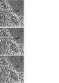
- A MN having nerves distributed in myotubes (m). High electron density region observed on muscle plasma membrane.
- B Multi-branched axon terminals observed in myotubes. A high electron density region (arrows) is localized at each axonal end. Mitochondria (mt) was seen nearby.
- C Necrosis observed at the end of axon (arrow).
- D Active zone (arrowhead), juxtaposed postsynaptic thickening (PSD, arrows), synaptic cleft observed between them.
- E The synaptic vesicle (sv) docks to the active zone (arrow).
- Myotubes contracted in response to elevated Ca 2+ Functional analysis of in vitro hNMJ at day 20 of NMJ differentiation (upper left photo). Ca imaging in 5 ROIs. The addition of calcium enhanced transient Ca 2+ elevation and was arrested by curare treatment. Functional analysis of in vitro hNMJ at day 20 of NMJ differentiation (top photo). Motion analysis at four ROIs shows the propagation of myotube contraction. Functional analysis of in vitro hNMJ at day 20 of NMJ differentiation. Myotubes within 4 ROIs started to contract independently 15 minutes after gap junction inhibition by GAP27. Kinetic analysis by photoactivation of in vitro hNMJ cultures (photographs under a, c, b).
- 201B7 MYOD -SMN KD cells (SMN KD) and 201B7 MYOD in the cells (Control), transmission electron microscopy image showing the intracellular myotube and axon terminals.
- L indicates a lipid droplet
- Mt indicates a mitochondria
- Mf indicates a myotube
- SV indicates a synaptic vesicle
- Ax indicates an axon
- Nu indicates a nucleus.
- Differentiation to hNMJ from iPSCs (201B7 MYOD- SMN KD ) knocked down SMN protein (upper photograph).
- the method for producing the artificial nerve junction of the present invention is (I) transiently expressing MyoD in pluripotent stem cells to induce muscle differentiation, and (ii) culturing the cells obtained in (i) in a medium containing neurotrophic factor to differentiate neurons Including the step of inducing.
- Artificial neuromuscular junctions means neuromuscular junctions derived from pluripotent stem cells in vitro.
- the neuromuscular junction means a structure in which acetylcholine is released from the terminal end of motor nerve cells and can be received by a receptor present in myotubes.
- the presence of a neuromuscular junction means, for example, colocalization of a synaptic vesicle protein (eg, SV2) expressed by motor neurons and an acetylcholine receptor expressed by myotubes by immunostaining or fluorescence microscopy It can be confirmed by a synaptic vesicle protein (eg, SV2) expressed by motor neurons and an acetylcholine receptor expressed by myotubes by immunostaining or fluorescence microscopy It can be confirmed by
- the artificial neuromuscular junction comprises motor neurons, myocytes (myotubes) and Schwann cells.
- myocytes myotubes
- Schwann cells can be identified using positivity such as S-100 marker as an index.
- the proportions of motor neurons, myocytes, and Schwann cells in the artificial neuromuscular junction are not particularly limited as long as they can maintain the function as the neuromuscular junction.
- a pluripotent stem cell is a stem cell having pluripotency capable of differentiating into many cells existing in a living body and also having proliferation ability, and the intermediate mesoderm used in the present invention Included are any cells that are induced into cells.
- pluripotent stem cells include, but are not limited to, embryonic stem (ES) cells, cloned embryonic derived embryonic stem (ntES) cells obtained by nuclear transfer, spermatogonial stem cells ("GS cells”), embryonic It includes germ cells (“EG cells”), induced pluripotent stem (iPS) cells, cultured fibroblasts and pluripotent cells derived from bone marrow stem cells (Muse cells).
- ES embryonic stem
- ntES cloned embryonic derived embryonic stem
- GS cells spermatogonial stem cells
- EG cells spermatogonial stem cells
- iPS induced pluripotent stem
- Muse cells pluripotent stem cells derived from bone marrow stem
- the reprogramming factor includes, for example, Oct3 / 4, Sox2, Sox1, Sox3, Sox15, Sox17, Sox17, Klf4, Klf2, c-Myc, N-Myc, L-Myc, Nanog, Lin28, Fbx15, ERas, ECAT15.
- Tcl1, beta-catenin, Lin28b, Sall1, Sall4, Esrrb, Nr5a2, Tbx3 or Glis1 and other genes or gene products are exemplified, and these reprogramming factors may be used alone or in combination. Also good.
- Somatic cells include, but are not limited to, fetal (child) somatic cells, neonatal (child) somatic cells, and any mature healthy or diseased somatic cells, and also primary culture cells. , Passage cells, and cell lines are also included.
- somatic cells are, for example, (1) tissue stem cells (somatic stem cells) such as neural stem cells, hematopoietic stem cells, mesenchymal stem cells, dental pulp stem cells, (2) tissue precursor cells, (3) blood cells (peripheral Blood cells, cord blood cells etc.), lymphocytes, epithelial cells, endothelial cells, muscle cells, fibroblasts (skin cells etc.), hair cells, hepatocytes, gastric mucous cells, enterocytes, splenocytes, pancreatic cells (pancreatic exocrine cells) Etc.), differentiated cells such as brain cells, lung cells, kidney cells and adipocytes.
- tissue stem cells such as neural stem cells, hematopoietic stem cells, mesenchy
- somatic cells when iPS cells are used as a material of cells for transplantation, it is desirable to use somatic cells in which the HLA genotypes of individuals to be transplanted are the same or substantially the same from the viewpoint that rejection does not occur.
- substantially identical means that the HLA genotypes coincide with the degree to which the immunosuppressive agent can suppress the immune response to the transplanted cells, for example, HLA-A and HLA-B.
- 3 loci of HLA-DR or 4 loci added with HLA-C are somatic cells having a matching HLA type.
- MyoD used in the present invention includes human myogenic differentiation 1 (MYOD1) consisting of the amino acid sequence shown in SEQ ID NO: 2 and orthologues thereof in other mammals, and their transcript variants, splice variants etc. . Alternatively, it has an amino acid identity of 90% or more, preferably 95% or more, more preferably 97% or more with any of the above proteins, and a function equivalent to that of the protein (eg, transcription activation of muscle specific promoter) And the like).
- MYOD1 human myogenic differentiation 1
- MYOD1 human myogenic differentiation 1
- a nucleic acid encoding MyoD human myogenic differentiation 1 (MYOD1) cDNA consisting of a nucleotide sequence represented by base numbers 221 to 1183 of SEQ ID NO: 1 and its orthologs in other mammals, and their transcript variants, splicing A mutant etc. are mentioned.
- it has a nucleotide identity of 90% or more, preferably 95% or more, more preferably 97% or more with any of the above nucleic acids, and a function equivalent to a protein encoded by the nucleic acid (eg, muscle-specific It may be a nucleic acid encoding a protein having a transcriptional activation of a promoter, etc.).
- stringent conditions bind a complex or a probe as taught by Berger and Kimmel (1987, Guide to Molecular Cloning Techniques in Methods in Enzymology, Vol. 152, Academic Press, San Diego Calif.) It can be determined based on the melting temperature (Tm) of the nucleic acid.
- Tm melting temperature
- the washing conditions after hybridization the conditions of “0.1 ⁇ SSC, 0.1% SDS, 60 ° C.” can be mentioned, and it is preferable that the hybridization state is maintained even when washing under such conditions. .
- the nucleic acid encoding MyoD may be DNA or RNA, or may be a DNA / RNA chimera. Also, the nucleic acid may be single-stranded, double-stranded DNA, double-stranded RNA or DNA: RNA hybrid. Preferably it is double stranded DNA or single stranded RNA.
- the RNA may be RNA incorporated with 5-methylcytidine and pseudouridine (TriLink Biotechnologies) to inhibit degradation, or may be a modified RNA by phosphatase treatment.
- the method for transiently expressing MyoD in pluripotent stem cells is not particularly limited, and for example, the following method can be used.
- expression means that the MyoD protein is transcribed and translated from the nucleic acid in the cell in the case of the nucleic acid encoding MyoD, and in the case of the MyoD protein, It is synonymous with the introduction of the protein into cells.
- vectors such as viruses, plasmids, artificial chromosomes can be introduced into pluripotent stem cells by techniques such as lipofection, liposomes, microinjection and the like.
- viral vectors include retrovirus vectors, lentivirus vectors, adenovirus vectors, adeno-associated virus vectors, Sendai virus vectors and the like.
- an artificial chromosome vector a human artificial chromosome (HAC), a yeast artificial chromosome (YAC), a bacterial artificial chromosome (BAC, PAC) etc. are illustrated.
- a plasmid a mammalian cell plasmid is exemplified.
- the vector may contain control sequences such as a promoter, enhancer, ribosome binding sequence, terminator, polyadenylation site and the like so that the DNA encoding MyoD can be expressed, and further, if necessary, a drug resistance gene.
- control sequences such as a promoter, enhancer, ribosome binding sequence, terminator, polyadenylation site and the like so that the DNA encoding MyoD can be expressed, and further, if necessary, a drug resistance gene.
- kanamycin resistance gene, ampicillin resistance gene, puromycin resistance gene etc. thymidine kinase gene
- selection marker sequences such as diphtheria toxin gene etc.
- fluorescent protein reporter gene sequences such as ⁇ -glucuronidase (GUS), FLAG etc.
- SV40 promoter As promoters, SV40 promoter, LTR promoter, CMV (cytomegalovirus) promoter, RSV (Rous sarcoma virus) promoter, MoMuLV (Moloney murine leukemia virus) LTR, HSV-TK (herpes simplex virus thymidine kinase) promoter, EF- ⁇ promoter, CAG
- the promoter and the TRE promoter (a CMV minimal promoter having a Tet response element consisting of 7 consecutive tetO sequences) are exemplified.
- the MyoD gene may be introduced into pluripotent stem cells using a transient expression vector, but in order to control expression more strictly, an inducible expression system is used. It is preferred to use.
- the induction expression system includes a drug induction expression system using tetracycline or its derivative (eg, doxycycline) and the like, and it is preferable to use a pluripotent stem cell into which a gene construct capable of drug induction expression of MyoD has been introduced in advance. .
- the TRE promoter described above it is desirable to simultaneously express a fusion protein with tetR and VP16AD or a fusion protein with reverse tetR (rtetR) and VP16AD (rtTA) in the same cell.
- rtetR reverse tetR
- rtTA reverse tetR
- MyoD can be expressed transiently while adding.
- the vector incorporates the expression cassette comprising the promoter and the DNA encoding MyoD linked to the chromosome of pluripotent stem cells, and if necessary, transposon sequences before and after this expression cassette for excision if necessary. May be included. Although it does not specifically limit as a transposon sequence, piggyBac is illustrated. In another embodiment, LoxP sequences may be included before and after the expression cassette for the purpose of removing the expression cassette.
- MyoD When MyoD is in the form of RNA, it may be introduced into pluripotent stem cells by techniques such as electroporation, lipofection, microinjection and the like. When MyoD is in the form of a protein, it may be introduced into pluripotent stem cells by methods such as lipofection, fusion with cell membrane permeable peptides (eg, TAT and polyarginine derived from HIV), microinjection and the like.
- cell membrane permeable peptides eg, TAT and polyarginine derived from HIV
- the period in which MyoD is transiently expressed in pluripotent stem cells to induce myocytes may be a period in which muscle differentiation is sufficiently performed, and can be appropriately changed depending on the type and nature of pluripotent stem cells used, For example, about 4 to 20 days are mentioned, about 4 to 12 days are preferable, and about 6 to 10 days are more preferable. For example, when using the above-mentioned drug induction expression system, it is preferable to add and culture a drug for this period.
- MyoD is RNA or protein
- the introduction may be performed multiple times so that MyoD exists in the cell in the above period.
- the culture conditions for pluripotent stem cells at the time of inducing myocytes in a state where MyoD is transiently expressed in pluripotent stem cells are preferably adhesion culture conditions.
- it is coated with a cell adhesion molecule such as extracellular matrix, specifically, Matrigel (BD), type I collagen, type IV collagen, gelatin, laminin, heparan sulfate proteoglycan, or entactin, and combinations thereof
- BD Matrigel
- type I collagen type IV collagen
- GMEM Gasgow Minimum Essential Medium
- IMDM Iscove's Modified Dulbecco's Medium: Iscove's Modified Dulbecco's Medium
- 199 medium Eagle's Minimum Essential Medium (Eagle's Minimum Essential Medium) (EMEM)
- EMEM Eagle's Minimum Essential Medium
- ⁇ MEM Dulbecco's modified Eagle's Medium
- DMEM Ham's F12 medium
- RPMI 1640 medium RPMI 1640 medium
- Fischer's medium a mixed medium thereof are included.
- serum substitutes such as albumin, transferrin, fatty acids, insulin, collagen precursors, trace elements, Knockout Serum Replacement (KSR) (serum substitute for FBS in ES cell culture), ITS-supplements, and mixtures thereof, etc. Is included.
- KSR Knockout Serum Replacement
- Preferred conditions for inducing differentiation are conditions in which pluripotent stem cells adhered to culture dishes coated with Matrigel are cultured in ⁇ MEM medium containing 10% KSR.
- the culture temperature is not particularly limited, but is about 30 to 40 ° C., preferably about 37 ° C., and the culture is performed under an atmosphere of CO 2 -containing air, and the CO 2 concentration is preferably about 2 to 5%. .
- the induction of muscle differentiation can be confirmed by the presence of muscle markers such as MHC and MEF2c.
- the cell population containing the muscle cell manufactured in this way may be not a single cell population but a cell population containing other types of cells.
- Neurotrophic factors are ligands to membrane receptors that play an important role in the survival and functional maintenance of motor neurons, and, for example, Nerve Growth Factor (NGF), Brain-derived Neurotrophic Factor (BDNF), Neurotrophin 3 ( NT-3), Neurotrophin 4/5 (NT-4 / 5), Neurotrophin 6 (NT-6), basic FGF, acidic FGF, FGF-5, Epidermal Growth Factor (EGF), Hepatocyte Growth Factor (HGF), Insulin , Insulin Like Growth Factor 1 (IGF 1), Insulin Like Growth Factor 2 (IGF 2), Glia cell line-derived Neurotrophic Factor (GDNF), TGF-b2, TGF-b3, Interleukin 6 (IL-6), Ciliary Neurotrophic Factor (CNTF) and LIF.
- NGF Nerve Growth Factor
- BDNF Brain-derived Neurotrophic Factor
- NT-3 Neurotrophin 3
- Neurotrophin 4/5 NT-4 / 5
- Neurotrophin 6 Neurotrophin 6
- Preferred neurotrophic factors in the present invention are NT-3, GDNF and BDNF.
- Neurotrophic factors are commercially available from, for example, Wako, R & D systems, etc. and can be easily used, but may be obtained by forced expression in cells by methods known to those skilled in the art.
- the concentration of NT-3 in the culture solution can be, for example, 0.1 ng / mL to 100 ng / mL, preferably 1 ng / mL to 50 ng / mL, more preferably 5 ng / mL to 20 ng / mL.
- the concentration of GDNF in the culture solution can be, for example, 0.1 ng / mL to 100 ng / mL, preferably 1 ng / mL to 50 ng / mL, more preferably 5 ng / mL to 20 ng / mL.
- the concentration of BDNF in the culture solution can be, for example, 0.1 ng / mL to 100 ng / mL, preferably 1 ng / mL to 50 ng / mL, more preferably 5 ng / mL to 20 ng / mL.
- the culture solution used in step (ii) can be prepared using a culture medium used for culturing animal cells as a basal medium.
- a culture medium used for culturing animal cells
- a basal medium for example, Glasgow's Minimal Essential Medium (GMEM) medium, IMDM medium, Medium 199 medium, Eagle's Minimum Essential Medium (EMEM) medium, ⁇ MEM medium, Dulbecco's modified Eagle's Medium (DMEM) medium, Ham's F12 medium, RPMI 1640 Media, Fischer's media, Neurobasal Medium (Life Technologies), and mixed media thereof are included.
- the medium may contain serum or may be serum free.
- the medium is, for example, albumin, transferrin, Knockout Serum Replacement (KSR) (serum substitute for FBS in ES cell culture), N2 supplement (Invitrogen), B27 supplement (Invitrogen), fatty acid, insulin, collagen It may also contain one or more serum substitutes such as precursors, trace elements, 2-mercaptoethanol, 3'-thiolglycerol, lipids, amino acids, L-glutamine, Glutamax (Invitrogen), non-essential amino acids, vitamins, It may also contain one or more substances such as growth factors, small molecule compounds, antibiotics, antioxidants, pyruvate, buffers, mineral salts and the like.
- a preferred culture is Neurobasal Medium containing N2 supplement, B27 supplement, NT-3, GDNF, and BDNF.
- neural differentiation can be induced by stopping the expression of MyoD and changing the medium to a medium for neural differentiation.
- culture temperature is not particularly limited, but is about 30 to 40 ° C., preferably about 37 ° C., and culture is performed under an atmosphere of CO 2 -containing air, and the CO 2 concentration is preferably about 2 to 5%.
- the culture period is not particularly limited as long as motor neurons and Schwann cells appear, but it is desirable that step (ii) be performed for at least 20 days. More preferably, it is 20 days to 120 days, still more preferably 30 days to 100 days.
- an artificial neuromuscular junction ie a cell population comprising motor neurons, myocytes and Schwann cells.
- kits for Producing Artificial Neuromuscular Junction from Pluripotent Stem Cells include cells, culture fluid, additives, culture vessels, and the like used in the above-described steps (i) and (ii).
- the kit includes cells, culture fluid, additives, culture vessels, and the like used in the above-described steps (i) and (ii).
- the kit may further include a document or instruction manual describing the procedure of the production process.
- the artificial neuromuscular junction of the present invention can be obtained as a cell culture containing motor neurons, myocytes and Schwann cells.
- the cell culture containing the neuromuscular junction is a pathological condition (eg, myasthenia gravis, Lambert-Eaton syndrome, Miller-Fisher syndrome, congenital) caused by a disorder (dysfunction or hypoplasia) of the neuromuscular junction. It is useful as a model system for the pathology of myasthenia gravis syndrome and spinal muscular atrophy. Therefore, it can be used, for example, as a cell preparation for treating a pathological condition caused by a neuromuscular junction disorder or as a screening system for a therapeutic agent for the pathological condition.
- the present invention relates to a pharmaceutical composition (cell preparation) comprising an artificial neuromuscular junction obtained by the method described above, a therapeutic agent for a pathological condition caused by a disorder of a neuromuscular junction, comprising the artificial neuromuscular junction, Methods are provided for treating conditions caused by disorders of the neuromuscular junction, comprising administering a therapeutically effective amount of the artificial neuromuscular junction.
- a method of administering a therapeutic agent to a patient in need of treatment for example, a method of locally administering the obtained artificial neuromuscular junction (cell culture containing muscle cells and Schwann cells) to the affected area, etc. It can be mentioned.
- the number of cells in the cell culture contained in the therapeutic agent is appropriately adjusted in accordance with the degree of disease and the like.
- cell preparation contains dimethyl sulfoxide (DMSO), serum albumin, etc. to protect the cells, and antibiotics etc. to prevent bacterial contamination and growth.
- DMSO dimethyl sulfoxide
- other pharmaceutically acceptable ingredients eg, carriers, excipients, disintegrants, buffers, emulsifiers, suspensions, soothing agents, stabilizers, preservatives, preservatives, physiological saline, etc. It may be contained in a cell preparation.
- Drug screening method or evaluation method In the present invention, a test substance is added to a cell population containing motor neurons, myocytes and Schwann cells obtained as described above, and then cultured. There is provided a method of screening or evaluating a therapeutic agent for a disease caused by a neuromuscular junction disorder, which comprises the step of evaluating muscle contraction or calcium concentration in the cells after culture.
- test substance may be, for example, cell extract, cell culture supernatant, microbial fermentation product, extract from marine organisms, plant extract, purified protein or crude protein, peptide, non-peptide compound, synthetic low molecular weight compound, and natural
- the compounds are exemplified.
- the test substance may also be (1) a biological library, (2) a synthetic library method using deconvolution, (3) a "one-bead one-compound” library method, and (4) It can be obtained using any of the many approaches in combinatorial library methods known in the art including synthetic library methods using affinity chromatography sorting. While biological library methods using affinity chromatography sorting are limited to peptide libraries, the other four approaches can be applied to peptides, non-peptide oligomers, or small molecule libraries of compounds (Lam (1997) ) Anticancer Drug Des. 12: 145-67). Examples of methods of synthesis of molecular libraries can be found in the art (DeWitt et al. (1993) Proc. Natl. Acad. Sci.
- Compound libraries can be solutions (see Houghten (1992) Bio / Techniques 13: 412-21) or beads (Lam (1991) Nature 354: 82-4), chips (Fodor (1993) Nature 364: 555-. 6), bacteria (US Pat. No. 5,223,409), spores (US Pat. Nos. 5,571,698, 5,403,484, and 5,223,409), plasmids (Cull et al. (1992) Proc. Natl. Acad. Sci. USA 89: 1865-9) or phage (Scott and Smith (1990) Science 249: 386-90; Devlin (1990) Science 249: 404-6; Cwirla et al. (1990) Proc. Natl. Acad. Sci. USA 87 Felici (1991) J. Mol. Biol. 222: 301-10; U.S. Patent Application No. 2002103360).
- Agents that enhance muscle contraction or calcium concentration relative to a control can be selected or evaluated as potential therapeutic agents for diseases caused by neuromuscular junction disorders.
- Another embodiment of the method of screening or evaluating a drug of the present invention is (I) transiently expressing MyoD in pluripotent stem cells to induce muscle differentiation, and (ii) culturing the cells obtained in (i) in a medium containing neurotrophic factor to differentiate neurons Guiding process, In the manufacturing process of the artificial neuromuscular junction, including the step of causing the test substance to exist in the step (i) and / or (ii), and analyzing the morphology or cell composition of the artificial neuromuscular junction obtained.
- a method for screening or evaluating a therapeutic agent for a disease caused by a disorder of a neuromuscular junction which selects or evaluates a drug.
- a process of manufacturing an artificial neuromuscular junction is performed using iPS cells derived from a patient with a disease caused by a disorder of the neuromuscular junction, and formation of the artificial neuromuscular junction is insufficient when no test substance is added. If the formation of the artificial neuromuscular junction is improved when the test substance is added, the substance can be a therapeutic agent for a disease caused by a disorder in the neuromuscular junction of the disease.
- the cell line MyoD-201B7 transfects a 201B7 human iPS cell line with a doxycycline (DOX) inducible MYOD1-expressing piggy bac vector as described by Tanaka et al., PLOS ONE 2013, Volume 8, Issue 4, e61540. It was built by doing. MYOD1 was expressed with mCherry protein and rtTA, and expression of MYOD1 was detected by luminescence of mCherry protein.
- DOX doxycycline
- MyoD-201B7 cells were seeded on a Matrigel-coated plate, and 1 ⁇ g / mL of doxycycline was added to a muscle differentiation medium (10% KSR + ⁇ MEM) to induce muscle differentiation.
- a muscle differentiation medium (10% KSR + ⁇ MEM)
- neural differentiation was induced by changing the medium to Neurobasal medium supplemented with neurotrophic factors (BDNF, GDNF, NT3; 10 ng / ml each, R & D systems). Thereafter, the medium was changed every 3 to 4 days and culture was continued.
- BDNF neurotrophic factors
- mCherry positive cells were sorted using FACS Aria BD and FACS Diva software. The selected cells were replated (8.8 ⁇ 10 4 cells / well) in Matrigel-coated 96-well plates and cultured in muscle differentiation medium with or without doxycycline. On day 20 the ratio of myotubes to neurons was quantified by immunocytochemistry.
- the motion vector analysis ⁇ 7s ILCE-7S (Sony Corporation) was used to continuously capture phase contrast images using a red filtered light source and fluorescence (Fluo 8 green) at 15 fps. Perform motion quantification using red separation image in target region (ROI) and brightness analysis using green separation image using SI 8000 cell motion imaging system (Sony Corporation) providing motion vector analysis based on block matching method did. Both motion imaging and motion analysis were performed at 38 fps using SI 8000 cell motion imaging system (Sony Corporation).
- Calcium imaging MyoD-201B7 cells were seeded at a density of 2.2 ⁇ 10 4 cells / well and allowed to differentiate into NMJ in Costar 96 well black wall / clear bottom plates. The growth medium was removed on day 20 and 100 ⁇ L of Fluo-8 recording medium was added to the cells for 1 hour at 37 ° C. The cells were washed twice with PBS and then imaged on an In Cell Analyzer 2000. The fluorescence intensity was expressed as the F / F0 ratio.
- MyoD-201B7 cells were seeded at a density of 2.2 ⁇ 10 4 cells / well in Matrigel coated 96 well black wall / transparent bottom plates. Half of the medium in each well was replaced daily until the cultures began to contract. The growth medium was removed and cells were loaded with 100 ⁇ L of Fluo-8 recording medium for 1 hour at 37 ° C. The cells were washed twice with PBS and then differentiation media was added to the wells. The Ca oscillations were monitored on a FDSS microplate reader, data points were collected at 1 minute intervals, and those data points were analyzed using FDSS software for frequency and amplitude quantification.
- Immunocytochemistry (ICC) labeling Primary antibodies used were anti-neurofilament (Millipore #MAB 5254, 1: 500), anti-synaptic vesicle protein 2 (DSHB # SV2, 1:20), anti-MHC (Millipore) No. A4.1025, 1: 1000), Anti-Islet1 (DSHB No. 40.2D6, 1: 100), Anti-HB9 (DSHB No. 81.5 C10, 1: 100), Anti-ChAT (Millipore No. AB144P, 1: 100) and It was anti-Tuj1 (Covance number MMS 435 P, 1: 1000).
- Plasmid containing the light genetic activation ⁇ br/> light activation domain (pLenti-Synapsin-hChR2 (H134R ) -EYFP-WPRE, Addgene plasmid No. 20945) was deposited from Karl Deisseroth (Karl Deisseroth) to Addgene I bought one and used it.
- a lentiviral transduction system (ViraPower hiperform lentiviral expression system, Invitrogen) was used to transduce the target gene into neural cells. NMJ cultures were treated with virus-containing medium at 5% CO 2 for 48 hours at 37 ° C. After viral infection, the medium was removed and neurobasal medium was added for expression of target gene. The medium was changed every 3 to 4 days. EYFP expression was examined by confocal microscopy with appropriate optical settings. Photoactivation was performed with a 488 nm laser wavelength. Bright field images were simultaneously imaged for muscle movement analysis in addition to EYFP signal.
- HiPSCs are efficiently induced in mature myotubes by transient expression of MYOD1 with a doxycycline (DOX) inducible vector.
- DOX doxycycline
- FIG. 1C, 1D Electron microscopy (EM) images showed Schwann cells overlying axonal end dilation and end button binding to myotubes in NMJ cultures (FIG. 1G and 1I). Synaptic vesicles and mitochondria accumulated in the presynaptic and postsynaptic sites of the NMJ, which is clearly divided by the high electron density activity zone (FIG. 1H, I). From these observations, our in vitro NMJ contained the major cell types of NMJ and was structurally also characterized by NMJ.
- agrin a neural factor that accumulates and stabilizes AChR at synaptic sites
- Administration of agrin increased the area of NMJ in long-term culture, while in the presence of anti-agrin antibody, only remnants of myotubes and degraded axon terminals were left in the culture (figure 2g, h).
- agrin like in vivo, has been found to play an important role in hNMJ formation and maintenance in our in vitro system.
- Gap junctions which are intercellular channels between myotubes and myotubes that allow intercellular propagation of electrical activity, are transiently expressed during NMJ development.
- myotube contractions were not completely synchronized, but they propagated to the adjacent area (FIG. 3C).
- the propagation of the contraction was interrupted by the gap junction inhibitor gap27, indicating that this contraction is caused by the propagation of action potentials through the gap junction ( Figure 3D).
- Immunostaining and image analysis Medium was removed from NMJ cultures, washed three times for 1 minute with PBS, and fixed for 10 minutes at room temperature with 4% paraformaldehyde / PBS. It was washed 3 times with 0.1% BSA / PBS and permeabilized with 4% paraformaldehyde + 0.1% Triton for 10 minutes at room temperature. Three times of 5 minutes of rehydration in 0.1% BSA / PBS, blocking treatment with 0.1% BSA / PBS + 0.5% Tween-20 for 1 hour at room temperature, and in a petri dish equipped with a parafilm layer The coverslip was moved to dryness.
- the primary antibody (antibody against Islet 1 or HB9) diluted in PBS is reacted at room temperature for 1 hour, washed three times with PBS, and reacted with a secondary antibody diluted in PBS for 45 minutes at room temperature (under light) ), Washed three times for 1 minute with PBS. Analysis was performed using a flow cytometer (Aria 2, BD) and FlowJo software (BD).
- 201B7 MYOD- SMN KD cells were subjected to immunostaining by the same protocol as the aforementioned immunostaining using an antibody against NF + SV2 or AChR as a primary antibody.
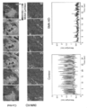
- SMN protein in 201B7 MYOD-SMN KD cells was found to be reduced by about 60% relative to the expression level of 201B7 MYOD cells not knocked down the SMN protein (FIG. 6A).
- the area of NMJ in 201B7 MYOD-SMN KD cells (SMN KD) was significantly smaller than that of 201B7 MYOD cells (Control) (FIG. 6B).
- SEM images of 201B7 MYOD cell (Control) cultures very elongated myotubes and bundles of axons were observed, and myotubes anchored at the extended axon terminals were observed (FIG. 6C).
- 201B7 MYOD- SMN KD cell (SMN KD) cultures myotubes were flat and thin and there were fewer axonal ends than in 201B7 MYOD cell (Control) cultures. Also, the details of the intracellular myotube and NMJ related structures were totally different from the 201B7 MYOD cell (Control) culture. Mitochondrial morphology and condition in 201B7 MYOD- SMN KD cells were damaged, and few of the myofibers were intact in their structure (FIG. 6D).
- the in vitro NMJ culture method of the present invention is composed of three types of cells, skeletal muscle, MN and Schwann cells, by a simple two-dimensional culture procedure of transiently expressing MyoD in iPSC and thereafter inducing neural differentiation.
- the method of the present invention is well-balanced in myogenesis and neurogenesis in NMJ construction, and reproduces a complex reciprocal signal between each cell such as nerve, muscle and Schwann cells in formation and maintenance of NMJ.
- the obtained NMJ shows the feature of the NMJ which is mature in appearance and function, it can be a platform of the disease model, and it is possible to use the cells for the disease based on the development or dysfunction of the NMJ. It contributes to therapy and drug effects.
Abstract
La présente invention concerne la fourniture d'un procédé permettant de préparer facilement et efficacement une jonction neuromusculaire fonctionnelle et mature (NMJ) in vitro. Cet objectif est atteint grâce à un procédé de production d'une jonction neuromusculaire artificielle, ledit procédé comprenant : (i) une étape d'expression transitoire de MyoD dans des cellules souches pluripotentes pour induire une différenciation myogène ; et (ii) une étape de culture des cellules obtenues dans (i) dans un milieu contenant un facteur neurotrophique pour induire une différenciation neuronale.
Priority Applications (3)
| Application Number | Priority Date | Filing Date | Title |
|---|---|---|---|
| US16/757,339 US20210363496A1 (en) | 2017-10-17 | 2018-10-17 | Method for obtaining artificial neuromuscular junction from pluripotent stem cells |
| EP18868426.0A EP3699267A4 (fr) | 2017-10-17 | 2018-10-17 | Procédé d'obtention de jonction neuromusculaire artificielle à partir de cellules souches pluripotentes |
| JP2019549321A JP7140400B2 (ja) | 2017-10-17 | 2018-10-17 | 多能性幹細胞から人工神経筋接合部を得る方法 |
Applications Claiming Priority (2)
| Application Number | Priority Date | Filing Date | Title |
|---|---|---|---|
| JP2017-201364 | 2017-10-17 | ||
| JP2017201364 | 2017-10-17 |
Publications (1)
| Publication Number | Publication Date |
|---|---|
| WO2019078263A1 true WO2019078263A1 (fr) | 2019-04-25 |
Family
ID=66174451
Family Applications (1)
| Application Number | Title | Priority Date | Filing Date |
|---|---|---|---|
| PCT/JP2018/038690 WO2019078263A1 (fr) | 2017-10-17 | 2018-10-17 | Procédé d'obtention de jonction neuromusculaire artificielle à partir de cellules souches pluripotentes |
Country Status (4)
| Country | Link |
|---|---|
| US (1) | US20210363496A1 (fr) |
| EP (1) | EP3699267A4 (fr) |
| JP (1) | JP7140400B2 (fr) |
| WO (1) | WO2019078263A1 (fr) |
Citations (34)
| Publication number | Priority date | Publication date | Assignee | Title |
|---|---|---|---|---|
| US5223409A (en) | 1988-09-02 | 1993-06-29 | Protein Engineering Corp. | Directed evolution of novel binding proteins |
| US20020103360A1 (en) | 1998-09-01 | 2002-08-01 | Yang Pan | Novel protein related to melanoma-inhibiting protein and uses thereof |
| WO2007069666A1 (fr) | 2005-12-13 | 2007-06-21 | Kyoto University | Facteur de reprogrammation nucleaire |
| WO2008118820A2 (fr) | 2007-03-23 | 2008-10-02 | Wisconsin Alumni Research Foundation | Reprogrammation d'une cellule somatique |
| WO2009007852A2 (fr) | 2007-06-15 | 2009-01-15 | Izumi Bio, Inc | Cellules multipotentes/pluripotentes et procédés s'y rapportant |
| WO2009032194A1 (fr) | 2007-08-31 | 2009-03-12 | Whitehead Institute For Biomedical Research | Stimulation de la voie wnt dans la reprogrammation de cellules somatiques |
| WO2009057831A1 (fr) | 2007-10-31 | 2009-05-07 | Kyoto University | Procédé de re-programmation nucléaire |
| WO2009058413A1 (fr) | 2007-10-29 | 2009-05-07 | Shi-Lung Lin | Génération de cellules de type cellules souches embryonnaires humaines à l'aide d'arn intronique |
| WO2009075119A1 (fr) | 2007-12-10 | 2009-06-18 | Kyoto University | Procédé efficace d'initialisation de noyaux |
| WO2009079007A1 (fr) | 2007-12-17 | 2009-06-25 | Gliamed, Inc. | Cellules souches et procédé de reprogrammation de cellules somatiques de mammifères adultes |
| WO2009091659A2 (fr) | 2008-01-16 | 2009-07-23 | Shi-Lung Lin | Génération de cellules pluripotentes de type cellules souches embryonnaires sans tumeur utilisant des agents d'arn recombinant inductible |
| WO2009101407A2 (fr) | 2008-02-11 | 2009-08-20 | Cambridge Enterprise Limited | Reprogrammation perfectionnée de cellules de mammifère et cellules ainsi obtenues |
| WO2009101084A1 (fr) | 2008-02-13 | 2009-08-20 | Fondazione Telethon | Procédé pour la reprogrammation de cellules différenciées |
| WO2009102983A2 (fr) | 2008-02-15 | 2009-08-20 | President And Fellows Of Harvard College | Induction efficace de cellules souches multipotentes à l’aide de composés à petites molécules |
| WO2009117439A2 (fr) | 2008-03-17 | 2009-09-24 | The Scripps Research Institute | Approches chimiques et génétiques combinées pour la génération de cellules souches pluripotentes induites |
| WO2009114949A1 (fr) | 2008-03-20 | 2009-09-24 | UNIVERSITé LAVAL | Procédés permettant de déprogrammer les cellules somatiques et utilisations de ces procédés |
| WO2009126250A2 (fr) | 2008-04-07 | 2009-10-15 | Nupotential, Inc. | Reprogrammation d’une cellule par induction d’un gène pluripotent par interférence arn |
| WO2009157593A1 (fr) | 2008-06-27 | 2009-12-30 | Kyoto University | Procédé consistant à établir efficacement des cellules souches pluripotentes induites |
| WO2010009015A2 (fr) | 2008-07-14 | 2010-01-21 | Oklahoma Medical Research Foundation | Production de cellules pluripotentes par inhibition de la fonction bright/arid3a |
| WO2010033906A2 (fr) | 2008-09-19 | 2010-03-25 | President And Fellows Of Harvard College | Induction efficace de cellules souches pluripotentes au moyen de composés à petite molécule |
| WO2010033920A2 (fr) | 2008-09-19 | 2010-03-25 | Whitehead Institute For Biomedical Research | Compositions et procédés pour améliorer la reprogrammation d'une cellule |
| WO2010042800A1 (fr) | 2008-10-10 | 2010-04-15 | Nevada Cancer Institute | Procédés de reprogrammation de cellules somatiques et procédés d'utilisation de telles cellules |
| WO2010050626A1 (fr) | 2008-10-30 | 2010-05-06 | Kyoto University | Procédé pour produire des cellules souches pluripotentes induites |
| WO2010056831A2 (fr) | 2008-11-12 | 2010-05-20 | Nupotential, Inc. | Reprogrammation d'une cellule par induction d'un gène pluripotent à l'aide d'un modulateur de hdac |
| WO2010068955A2 (fr) | 2008-12-13 | 2010-06-17 | Dna Microarray | Dosage de niche micro-environnementale pour criblage de cellules souches pluripotentes induites (cips) |
| WO2010098419A1 (fr) | 2009-02-27 | 2010-09-02 | Kyoto University | Nouvelle substance de reprogrammation nucléaire |
| WO2010102267A2 (fr) | 2009-03-06 | 2010-09-10 | Ipierian, Inc. | Inhibiteurs de la voie tgf-bêta pour l'amélioration de la reprogrammation cellulaire de cellules humaines |
| WO2010111422A2 (fr) | 2009-03-25 | 2010-09-30 | The Salk Institute For Biological Studies | Génération de cellules souches pluripotentes induites en utilisant deux facteurs et l'inactivation de p53 |
| WO2010111409A2 (fr) | 2009-03-25 | 2010-09-30 | The Salk Institute For Biological Studies | Cellules souches pluripotentes |
| WO2010115050A2 (fr) | 2009-04-01 | 2010-10-07 | The Regents Of The University Of California | Microarn spécifiques de cellules souches embryonnaires, qui favorisent la pluripotence induite |
| WO2010124290A2 (fr) | 2009-04-24 | 2010-10-28 | Whitehead Institute For Biomedical Research | Compositions et procédés pour dériver ou cultiver des cellules pluripotentes |
| WO2010147612A1 (fr) | 2009-06-18 | 2010-12-23 | Lixte Biotechnology, Inc. | Procédés de modulation de la régulation cellulaire par inhibition de p53 |
| WO2010147395A2 (fr) | 2009-06-16 | 2010-12-23 | Korea Research Institute Of Bioscience And Biotechnology | Composition de milieu comprenant un neuropeptide y pour la génération, le maintien, la croissance non différenciée prolongée de cellules souches pluripotentes et procédé de culture de cellules souches pluripotentes l'utilisant |
| WO2017062854A1 (fr) * | 2015-10-07 | 2017-04-13 | Memorial Sloan-Kettering Cancer Center | Procédés in vitro d'identification de modulateurs de l'activité de jonction neuromusculaire |
Family Cites Families (1)
| Publication number | Priority date | Publication date | Assignee | Title |
|---|---|---|---|---|
| CA2798777C (fr) * | 2010-04-23 | 2016-09-06 | University Of Central Florida Research Foundation, Inc. | Formation de jonctions neuromusculaires dans un systeme defini |
-
2018
- 2018-10-17 US US16/757,339 patent/US20210363496A1/en not_active Abandoned
- 2018-10-17 JP JP2019549321A patent/JP7140400B2/ja active Active
- 2018-10-17 EP EP18868426.0A patent/EP3699267A4/fr not_active Withdrawn
- 2018-10-17 WO PCT/JP2018/038690 patent/WO2019078263A1/fr unknown
Patent Citations (38)
| Publication number | Priority date | Publication date | Assignee | Title |
|---|---|---|---|---|
| US5403484A (en) | 1988-09-02 | 1995-04-04 | Protein Engineering Corporation | Viruses expressing chimeric binding proteins |
| US5571698A (en) | 1988-09-02 | 1996-11-05 | Protein Engineering Corporation | Directed evolution of novel binding proteins |
| US5223409A (en) | 1988-09-02 | 1993-06-29 | Protein Engineering Corp. | Directed evolution of novel binding proteins |
| US20020103360A1 (en) | 1998-09-01 | 2002-08-01 | Yang Pan | Novel protein related to melanoma-inhibiting protein and uses thereof |
| WO2007069666A1 (fr) | 2005-12-13 | 2007-06-21 | Kyoto University | Facteur de reprogrammation nucleaire |
| WO2008118820A2 (fr) | 2007-03-23 | 2008-10-02 | Wisconsin Alumni Research Foundation | Reprogrammation d'une cellule somatique |
| WO2009007852A2 (fr) | 2007-06-15 | 2009-01-15 | Izumi Bio, Inc | Cellules multipotentes/pluripotentes et procédés s'y rapportant |
| WO2009032194A1 (fr) | 2007-08-31 | 2009-03-12 | Whitehead Institute For Biomedical Research | Stimulation de la voie wnt dans la reprogrammation de cellules somatiques |
| WO2009058413A1 (fr) | 2007-10-29 | 2009-05-07 | Shi-Lung Lin | Génération de cellules de type cellules souches embryonnaires humaines à l'aide d'arn intronique |
| WO2009057831A1 (fr) | 2007-10-31 | 2009-05-07 | Kyoto University | Procédé de re-programmation nucléaire |
| WO2009075119A1 (fr) | 2007-12-10 | 2009-06-18 | Kyoto University | Procédé efficace d'initialisation de noyaux |
| WO2009079007A1 (fr) | 2007-12-17 | 2009-06-25 | Gliamed, Inc. | Cellules souches et procédé de reprogrammation de cellules somatiques de mammifères adultes |
| WO2009091659A2 (fr) | 2008-01-16 | 2009-07-23 | Shi-Lung Lin | Génération de cellules pluripotentes de type cellules souches embryonnaires sans tumeur utilisant des agents d'arn recombinant inductible |
| WO2009101407A2 (fr) | 2008-02-11 | 2009-08-20 | Cambridge Enterprise Limited | Reprogrammation perfectionnée de cellules de mammifère et cellules ainsi obtenues |
| WO2009101084A1 (fr) | 2008-02-13 | 2009-08-20 | Fondazione Telethon | Procédé pour la reprogrammation de cellules différenciées |
| WO2009102983A2 (fr) | 2008-02-15 | 2009-08-20 | President And Fellows Of Harvard College | Induction efficace de cellules souches multipotentes à l’aide de composés à petites molécules |
| WO2009117439A2 (fr) | 2008-03-17 | 2009-09-24 | The Scripps Research Institute | Approches chimiques et génétiques combinées pour la génération de cellules souches pluripotentes induites |
| WO2009114949A1 (fr) | 2008-03-20 | 2009-09-24 | UNIVERSITé LAVAL | Procédés permettant de déprogrammer les cellules somatiques et utilisations de ces procédés |
| WO2009126250A2 (fr) | 2008-04-07 | 2009-10-15 | Nupotential, Inc. | Reprogrammation d’une cellule par induction d’un gène pluripotent par interférence arn |
| WO2009126655A2 (fr) | 2008-04-07 | 2009-10-15 | Nupotential, Inc. | Reprogrammation d’une cellule par induction d’un gène pluripotent par utilisation d’un modulateur à petite molécule |
| WO2009126251A2 (fr) | 2008-04-07 | 2009-10-15 | Nupotential, Inc. | Reprogrammation d’une cellule par induction d’un gène pluripotent à l’aide d’un modulateur hdac |
| WO2009157593A1 (fr) | 2008-06-27 | 2009-12-30 | Kyoto University | Procédé consistant à établir efficacement des cellules souches pluripotentes induites |
| WO2010009015A2 (fr) | 2008-07-14 | 2010-01-21 | Oklahoma Medical Research Foundation | Production de cellules pluripotentes par inhibition de la fonction bright/arid3a |
| WO2010033906A2 (fr) | 2008-09-19 | 2010-03-25 | President And Fellows Of Harvard College | Induction efficace de cellules souches pluripotentes au moyen de composés à petite molécule |
| WO2010033920A2 (fr) | 2008-09-19 | 2010-03-25 | Whitehead Institute For Biomedical Research | Compositions et procédés pour améliorer la reprogrammation d'une cellule |
| WO2010042800A1 (fr) | 2008-10-10 | 2010-04-15 | Nevada Cancer Institute | Procédés de reprogrammation de cellules somatiques et procédés d'utilisation de telles cellules |
| WO2010050626A1 (fr) | 2008-10-30 | 2010-05-06 | Kyoto University | Procédé pour produire des cellules souches pluripotentes induites |
| WO2010056831A2 (fr) | 2008-11-12 | 2010-05-20 | Nupotential, Inc. | Reprogrammation d'une cellule par induction d'un gène pluripotent à l'aide d'un modulateur de hdac |
| WO2010068955A2 (fr) | 2008-12-13 | 2010-06-17 | Dna Microarray | Dosage de niche micro-environnementale pour criblage de cellules souches pluripotentes induites (cips) |
| WO2010098419A1 (fr) | 2009-02-27 | 2010-09-02 | Kyoto University | Nouvelle substance de reprogrammation nucléaire |
| WO2010102267A2 (fr) | 2009-03-06 | 2010-09-10 | Ipierian, Inc. | Inhibiteurs de la voie tgf-bêta pour l'amélioration de la reprogrammation cellulaire de cellules humaines |
| WO2010111422A2 (fr) | 2009-03-25 | 2010-09-30 | The Salk Institute For Biological Studies | Génération de cellules souches pluripotentes induites en utilisant deux facteurs et l'inactivation de p53 |
| WO2010111409A2 (fr) | 2009-03-25 | 2010-09-30 | The Salk Institute For Biological Studies | Cellules souches pluripotentes |
| WO2010115050A2 (fr) | 2009-04-01 | 2010-10-07 | The Regents Of The University Of California | Microarn spécifiques de cellules souches embryonnaires, qui favorisent la pluripotence induite |
| WO2010124290A2 (fr) | 2009-04-24 | 2010-10-28 | Whitehead Institute For Biomedical Research | Compositions et procédés pour dériver ou cultiver des cellules pluripotentes |
| WO2010147395A2 (fr) | 2009-06-16 | 2010-12-23 | Korea Research Institute Of Bioscience And Biotechnology | Composition de milieu comprenant un neuropeptide y pour la génération, le maintien, la croissance non différenciée prolongée de cellules souches pluripotentes et procédé de culture de cellules souches pluripotentes l'utilisant |
| WO2010147612A1 (fr) | 2009-06-18 | 2010-12-23 | Lixte Biotechnology, Inc. | Procédés de modulation de la régulation cellulaire par inhibition de p53 |
| WO2017062854A1 (fr) * | 2015-10-07 | 2017-04-13 | Memorial Sloan-Kettering Cancer Center | Procédés in vitro d'identification de modulateurs de l'activité de jonction neuromusculaire |
Non-Patent Citations (39)
| Title |
|---|
| BARIK, ARNAB ET AL.: "Schwann Cells in Neuromuscular Junction Formation and Maintenance", JOURNAL OF NEUROSCIENCE, vol. 36, no. 38, 21 September 2016 (2016-09-21), pages 9770 - 9781, XP055597060, ISSN: 0270-6474, DOI: 10.1523/JNEUROSCI.0174-16.2016 * |
| BERGERKIMMEL: "Guide to Molecular Cloning Techniques Methods in Enzymology", vol. 152, 1987, ACADEMIC PRESS |
| CARELL ET AL., ANGEW. CHEM. INT. ED. ENGL., vol. 33, 1994, pages 2061 |
| CHO ET AL., SCIENCE, vol. 261, 1993, pages 1303 - 5 |
| CULL ET AL., PROC. NATL. ACAD. SCI. USA, vol. 89, 1992, pages 1865 - 9 |
| CWIRLA ET AL., PROC. NATL. ACAD. SCI. USA, vol. 87, 1990, pages 6378 - 82 |
| DEMESTRA M, STEM CELL RES., vol. 15, no. 2, 2015, pages 328 - 36 |
| DEMESTRE, M ET AL.: "Formation and characterisation of neuromuscular junctions between hiPSC derived motoneurons and myotubes", STEM CELL RESEARCH, vol. 15, 2015, pages 328 - 336, XP029291317, DOI: 10.1016/j.scr.2015.07.005 * |
| DEWITT ET AL., PROC. NATL. ACAD. SCI. USA, vol. 90, 1993, pages 6909 - 13 |
| EMINLI S ET AL., STEM CELLS, vol. 26, 2008, pages 2467 - 2474 |
| ERB ET AL., PROC. NATL. ACAD. SCI. USA, vol. 91, 1994, pages 11422 - 6 |
| FELICI, J. MOL. BIOL., vol. 222, 1991, pages 301 - 10 |
| FENG B ET AL., NAT. CELL BIOL., vol. 11, 2009, pages 197 - 203 |
| FODOR, NATURE, vol. 364, 1993, pages 555 - 6 |
| HAN J ET AL., NATURE, vol. 463, 2010, pages 1096 - 100 |
| HENG JC ET AL., CELL STEM CELL, vol. 6, 2010, pages 167 - 74 |
| HOUGHTEN, BIOLTECHNIQUES, vol. 13, 1992, pages 412 - 21 |
| HUANGFU D ET AL., NAT. BIOTECHNOL., vol. 26, 2008, pages 1269 - 1275 |
| ICHIDA JK ET AL., CELL STEM CELL, vol. 5, 2009, pages 491 - 503 |
| KIM JB ET AL., NATURE, vol. 461, 2009, pages 649 - 643 |
| KIM, HAN-SEOP ET AL.: "Schwann Cell Precursors from Human Pluripotent Stem Cells as a Potential Therapeutic Target for Myelin Repair", STEM CELL REPORTS, vol. 8, 6 June 2017 (2017-06-06), pages 1714 - 1726, XP055505353, DOI: 10.1016/j.stemcr.2017.04.011 * |
| KIM, JU ANG ET AL.: "MYOD mediates skeletal myogenic differentiation of human amniotic fluid stem cells and regeneration of muscle injury", STEM CELL RESEARCH & THERAPY, vol. 4, no. 147, 2013, pages 1 - 11, XP021171894 * |
| LAM, ANTICANCER DRUG DES., vol. 12, 1997, pages 145 - 67 |
| LAM, NATURE, vol. 354, 1991, pages 82 - 4 |
| LYSSIOTIS CA ET AL., PROC NATL ACAD SCI USA., vol. 106, 2009, pages 8912 - 8917 |
| MAEKAWA M ET AL., NATURE, vol. 474, 2011, pages 225 - 9 |
| MALI P ET AL., STEM CELLS, vol. 28, 2010, pages 713 - 720 |
| R. L. JUDSON ET AL., NAT. BIOTECHNOL., vol. 27, 2009, pages 459 - 461 |
| SCOTTSMITH, SCIENCE, vol. 249, 1990, pages 404 - 90 |
| See also references of EP3699267A4 |
| STEINBECK JA., CELL STEM CELL, vol. 18, no. 1, 7 January 2016 (2016-01-07), pages 134 - 43 |
| STEINBECK, JULIUS A. ET AL.: "Functional Connectivity under Optogenetic Control Allows Modeling of Human Neuromuscular Disease", CELL STEM CELL, vol. 18, 2016, pages 134 - 143, XP029381714, DOI: 10.1016/j.stem.2015.10.002 * |
| TANAKA ET AL., PLOS ONE, vol. 8, no. 4, 2013, pages e61540 |
| TANAKA, AKIHITO ET AL.: "Efficient and Reproducible Myogenic Differentiation from Human iPS Cells: Prospects for Modeling Miyoshi Myopathy In Vitro", PLOS ONE, vol. 8, no. 4, 2013, pages e61540, XP055544983, DOI: 10.1371/journal.pone.0061540 * |
| TANJI KURENAI: "Differentiation process of human non-muscle cells muscularly converted by MyoD forced ecpression: Through the experimental innervation by fetal rat spinal motor nerve cells", MEDICAL JOURNAL OF KINKI UNIVERSITY, vol. 21, no. 1, 1996, pages 25 - 36, XP009520472, ISSN: 0385-8367 * |
| THE JOURNAL OF NEUROSCIENCE, vol. 36, no. 38, 21 September 2016 (2016-09-21), pages 9770 - 9781 |
| YOSHIDA, MICHIKO ET AL.: "Modeling the Early Phenotype at the Neuromuscular Junction of Spinal Muscular Atrophy Using Patient-Derived iPSCs", STEM CELL REPORTS, vol. 4, no. 4, 2015, pages 561 - 568, XP055597065, ISSN: 2213-6711, DOI: 10.1016/j.stemcr.2015.02.010 * |
| ZHAO Y ET AL., CELL STEM CELL, vol. 3, 2008, pages 132 - 135 |
| ZUCKERMANN ET AL., J. MED. CHEM., vol. 37, 1994, pages 1233 - 51 |
Also Published As
| Publication number | Publication date |
|---|---|
| US20210363496A1 (en) | 2021-11-25 |
| JPWO2019078263A1 (ja) | 2021-02-25 |
| EP3699267A4 (fr) | 2021-10-27 |
| EP3699267A1 (fr) | 2020-08-26 |
| JP7140400B2 (ja) | 2022-09-21 |
Similar Documents
| Publication | Publication Date | Title |
|---|---|---|
| US20230038168A1 (en) | Lineage Reprogramming to Induced Cardiac Progenitor Cells (iCPC) By Defined Factors | |
| US9249391B2 (en) | Methods of generating neural stem cells | |
| US20180072988A1 (en) | Generation of functional cells from stem cells | |
| US20090186414A1 (en) | Methods of Generating Cardiomyocytes and Cardiac Progenitors and Compositions | |
| JP2014533491A (ja) | 多能性幹細胞から骨格筋細胞への分化誘導方法 | |
| US10947500B2 (en) | Neural cells expressing adenovirus E4ORF1, and methods of making and using the same | |
| WO2016114354A1 (fr) | Procédé de sélection de cellule progénitrice des muscles squelettiques | |
| JP7236738B2 (ja) | 神経前駆細胞の選別方法 | |
| WO2021172542A1 (fr) | Procédé de production de cardiomyocytes matures | |
| US11046932B2 (en) | Method of producing renal cells from differentiated cells | |
| WO2023063187A1 (fr) | Procédé de production d'organoïde | |
| US20220333070A1 (en) | Induction of functional astrocytes from pluripotent stem cells | |
| JP7140400B2 (ja) | 多能性幹細胞から人工神経筋接合部を得る方法 | |
| Sugimoto et al. | Effect of NeuroD2 expression on neuronal differentiation in mouse embryonic stem cells | |
| WO2013124309A1 (fr) | Reprogrammation directe de cellules somatiques en cellules souches neurales | |
| JP6990921B2 (ja) | 上位運動ニューロンの誘導方法 | |
| EP3196295A1 (fr) | Procédé de production de cellules rénales à partir de fibroblastes | |
| WO2022102742A1 (fr) | Marqueur de surface cellulaire pour une purification à haut rendement de cellules de lignée de muscle squelettique et de cellules souches de muscle squelettique, et son utilisation | |
| WO2020090836A1 (fr) | Procédé de production de cellules | |
| Michels | Unravelling the function of circRmst in the development of midbrain dopaminergic neurons | |
| Menon et al. | Human induced pluripotent stem cell-derived pericytes as scalable and editable source to study direct lineage reprogramming into induced neurons | |
| Wong | Directed Conversion of Human Retinal Ganglion Cells by Overexpression of Transcription Factors | |
| WO2018043303A1 (fr) | Procédé de production de neurone à sérotonine | |
| JP2022157515A (ja) | 多能性幹細胞、神経細胞及びその応用 | |
| jÞ SCg | The Use of Induced Pluripotent Stem Cells in Drug Development |
Legal Events
| Date | Code | Title | Description |
|---|---|---|---|
| 121 | Ep: the epo has been informed by wipo that ep was designated in this application |
Ref document number: 18868426 Country of ref document: EP Kind code of ref document: A1 |
|
| ENP | Entry into the national phase |
Ref document number: 2019549321 Country of ref document: JP Kind code of ref document: A |
|
| NENP | Non-entry into the national phase |
Ref country code: DE |
|
| ENP | Entry into the national phase |
Ref document number: 2018868426 Country of ref document: EP Effective date: 20200518 |