WO2019078263A1 - Method for obtaining artificial neuromuscular junction from pluripotent stem cells - Google Patents
Method for obtaining artificial neuromuscular junction from pluripotent stem cells Download PDFInfo
- Publication number
- WO2019078263A1 WO2019078263A1 PCT/JP2018/038690 JP2018038690W WO2019078263A1 WO 2019078263 A1 WO2019078263 A1 WO 2019078263A1 JP 2018038690 W JP2018038690 W JP 2018038690W WO 2019078263 A1 WO2019078263 A1 WO 2019078263A1
- Authority
- WO
- WIPO (PCT)
- Prior art keywords
- cells
- pluripotent stem
- stem cells
- myod
- neuromuscular junction
- Prior art date
Links
Images


Classifications
-
- C—CHEMISTRY; METALLURGY
- C12—BIOCHEMISTRY; BEER; SPIRITS; WINE; VINEGAR; MICROBIOLOGY; ENZYMOLOGY; MUTATION OR GENETIC ENGINEERING
- C12N—MICROORGANISMS OR ENZYMES; COMPOSITIONS THEREOF; PROPAGATING, PRESERVING, OR MAINTAINING MICROORGANISMS; MUTATION OR GENETIC ENGINEERING; CULTURE MEDIA
- C12N5/00—Undifferentiated human, animal or plant cells, e.g. cell lines; Tissues; Cultivation or maintenance thereof; Culture media therefor
- C12N5/06—Animal cells or tissues; Human cells or tissues
- C12N5/0697—Artificial constructs associating cells of different lineages, e.g. tissue equivalents
-
- A—HUMAN NECESSITIES
- A61—MEDICAL OR VETERINARY SCIENCE; HYGIENE
- A61K—PREPARATIONS FOR MEDICAL, DENTAL OR TOILETRY PURPOSES
- A61K35/00—Medicinal preparations containing materials or reaction products thereof with undetermined constitution
- A61K35/12—Materials from mammals; Compositions comprising non-specified tissues or cells; Compositions comprising non-embryonic stem cells; Genetically modified cells
- A61K35/30—Nerves; Brain; Eyes; Corneal cells; Cerebrospinal fluid; Neuronal stem cells; Neuronal precursor cells; Glial cells; Oligodendrocytes; Schwann cells; Astroglia; Astrocytes; Choroid plexus; Spinal cord tissue
-
- A—HUMAN NECESSITIES
- A61—MEDICAL OR VETERINARY SCIENCE; HYGIENE
- A61K—PREPARATIONS FOR MEDICAL, DENTAL OR TOILETRY PURPOSES
- A61K35/00—Medicinal preparations containing materials or reaction products thereof with undetermined constitution
- A61K35/12—Materials from mammals; Compositions comprising non-specified tissues or cells; Compositions comprising non-embryonic stem cells; Genetically modified cells
- A61K35/34—Muscles; Smooth muscle cells; Heart; Cardiac stem cells; Myoblasts; Myocytes; Cardiomyocytes
-
- A—HUMAN NECESSITIES
- A61—MEDICAL OR VETERINARY SCIENCE; HYGIENE
- A61K—PREPARATIONS FOR MEDICAL, DENTAL OR TOILETRY PURPOSES
- A61K35/00—Medicinal preparations containing materials or reaction products thereof with undetermined constitution
- A61K35/12—Materials from mammals; Compositions comprising non-specified tissues or cells; Compositions comprising non-embryonic stem cells; Genetically modified cells
- A61K35/48—Reproductive organs
- A61K35/54—Ovaries; Ova; Ovules; Embryos; Foetal cells; Germ cells
- A61K35/545—Embryonic stem cells; Pluripotent stem cells; Induced pluripotent stem cells; Uncharacterised stem cells
-
- A—HUMAN NECESSITIES
- A61—MEDICAL OR VETERINARY SCIENCE; HYGIENE
- A61L—METHODS OR APPARATUS FOR STERILISING MATERIALS OR OBJECTS IN GENERAL; DISINFECTION, STERILISATION OR DEODORISATION OF AIR; CHEMICAL ASPECTS OF BANDAGES, DRESSINGS, ABSORBENT PADS OR SURGICAL ARTICLES; MATERIALS FOR BANDAGES, DRESSINGS, ABSORBENT PADS OR SURGICAL ARTICLES
- A61L27/00—Materials for grafts or prostheses or for coating grafts or prostheses
- A61L27/36—Materials for grafts or prostheses or for coating grafts or prostheses containing ingredients of undetermined constitution or reaction products thereof, e.g. transplant tissue, natural bone, extracellular matrix
- A61L27/38—Materials for grafts or prostheses or for coating grafts or prostheses containing ingredients of undetermined constitution or reaction products thereof, e.g. transplant tissue, natural bone, extracellular matrix containing added animal cells
-
- A—HUMAN NECESSITIES
- A61—MEDICAL OR VETERINARY SCIENCE; HYGIENE
- A61P—SPECIFIC THERAPEUTIC ACTIVITY OF CHEMICAL COMPOUNDS OR MEDICINAL PREPARATIONS
- A61P21/00—Drugs for disorders of the muscular or neuromuscular system
-
- C—CHEMISTRY; METALLURGY
- C07—ORGANIC CHEMISTRY
- C07K—PEPTIDES
- C07K14/00—Peptides having more than 20 amino acids; Gastrins; Somatostatins; Melanotropins; Derivatives thereof
- C07K14/435—Peptides having more than 20 amino acids; Gastrins; Somatostatins; Melanotropins; Derivatives thereof from animals; from humans
- C07K14/46—Peptides having more than 20 amino acids; Gastrins; Somatostatins; Melanotropins; Derivatives thereof from animals; from humans from vertebrates
- C07K14/47—Peptides having more than 20 amino acids; Gastrins; Somatostatins; Melanotropins; Derivatives thereof from animals; from humans from vertebrates from mammals
- C07K14/4701—Peptides having more than 20 amino acids; Gastrins; Somatostatins; Melanotropins; Derivatives thereof from animals; from humans from vertebrates from mammals not used
- C07K14/4702—Regulators; Modulating activity
- C07K14/4705—Regulators; Modulating activity stimulating, promoting or activating activity
-
- C—CHEMISTRY; METALLURGY
- C07—ORGANIC CHEMISTRY
- C07K—PEPTIDES
- C07K14/00—Peptides having more than 20 amino acids; Gastrins; Somatostatins; Melanotropins; Derivatives thereof
- C07K14/435—Peptides having more than 20 amino acids; Gastrins; Somatostatins; Melanotropins; Derivatives thereof from animals; from humans
- C07K14/475—Growth factors; Growth regulators
-
- C—CHEMISTRY; METALLURGY
- C12—BIOCHEMISTRY; BEER; SPIRITS; WINE; VINEGAR; MICROBIOLOGY; ENZYMOLOGY; MUTATION OR GENETIC ENGINEERING
- C12N—MICROORGANISMS OR ENZYMES; COMPOSITIONS THEREOF; PROPAGATING, PRESERVING, OR MAINTAINING MICROORGANISMS; MUTATION OR GENETIC ENGINEERING; CULTURE MEDIA
- C12N5/00—Undifferentiated human, animal or plant cells, e.g. cell lines; Tissues; Cultivation or maintenance thereof; Culture media therefor
- C12N5/06—Animal cells or tissues; Human cells or tissues
- C12N5/0602—Vertebrate cells
- C12N5/0618—Cells of the nervous system
- C12N5/0619—Neurons
-
- C—CHEMISTRY; METALLURGY
- C12—BIOCHEMISTRY; BEER; SPIRITS; WINE; VINEGAR; MICROBIOLOGY; ENZYMOLOGY; MUTATION OR GENETIC ENGINEERING
- C12N—MICROORGANISMS OR ENZYMES; COMPOSITIONS THEREOF; PROPAGATING, PRESERVING, OR MAINTAINING MICROORGANISMS; MUTATION OR GENETIC ENGINEERING; CULTURE MEDIA
- C12N5/00—Undifferentiated human, animal or plant cells, e.g. cell lines; Tissues; Cultivation or maintenance thereof; Culture media therefor
- C12N5/06—Animal cells or tissues; Human cells or tissues
- C12N5/0602—Vertebrate cells
- C12N5/0618—Cells of the nervous system
- C12N5/0622—Glial cells, e.g. astrocytes, oligodendrocytes; Schwann cells
-
- C—CHEMISTRY; METALLURGY
- C12—BIOCHEMISTRY; BEER; SPIRITS; WINE; VINEGAR; MICROBIOLOGY; ENZYMOLOGY; MUTATION OR GENETIC ENGINEERING
- C12N—MICROORGANISMS OR ENZYMES; COMPOSITIONS THEREOF; PROPAGATING, PRESERVING, OR MAINTAINING MICROORGANISMS; MUTATION OR GENETIC ENGINEERING; CULTURE MEDIA
- C12N5/00—Undifferentiated human, animal or plant cells, e.g. cell lines; Tissues; Cultivation or maintenance thereof; Culture media therefor
- C12N5/06—Animal cells or tissues; Human cells or tissues
- C12N5/0602—Vertebrate cells
- C12N5/0652—Cells of skeletal and connective tissues; Mesenchyme
- C12N5/0658—Skeletal muscle cells, e.g. myocytes, myotubes, myoblasts
-
- C—CHEMISTRY; METALLURGY
- C12—BIOCHEMISTRY; BEER; SPIRITS; WINE; VINEGAR; MICROBIOLOGY; ENZYMOLOGY; MUTATION OR GENETIC ENGINEERING
- C12Q—MEASURING OR TESTING PROCESSES INVOLVING ENZYMES, NUCLEIC ACIDS OR MICROORGANISMS; COMPOSITIONS OR TEST PAPERS THEREFOR; PROCESSES OF PREPARING SUCH COMPOSITIONS; CONDITION-RESPONSIVE CONTROL IN MICROBIOLOGICAL OR ENZYMOLOGICAL PROCESSES
- C12Q1/00—Measuring or testing processes involving enzymes, nucleic acids or microorganisms; Compositions therefor; Processes of preparing such compositions
- C12Q1/02—Measuring or testing processes involving enzymes, nucleic acids or microorganisms; Compositions therefor; Processes of preparing such compositions involving viable microorganisms
-
- G—PHYSICS
- G01—MEASURING; TESTING
- G01N—INVESTIGATING OR ANALYSING MATERIALS BY DETERMINING THEIR CHEMICAL OR PHYSICAL PROPERTIES
- G01N33/00—Investigating or analysing materials by specific methods not covered by groups G01N1/00 - G01N31/00
- G01N33/48—Biological material, e.g. blood, urine; Haemocytometers
- G01N33/50—Chemical analysis of biological material, e.g. blood, urine; Testing involving biospecific ligand binding methods; Immunological testing
- G01N33/5005—Chemical analysis of biological material, e.g. blood, urine; Testing involving biospecific ligand binding methods; Immunological testing involving human or animal cells
- G01N33/5008—Chemical analysis of biological material, e.g. blood, urine; Testing involving biospecific ligand binding methods; Immunological testing involving human or animal cells for testing or evaluating the effect of chemical or biological compounds, e.g. drugs, cosmetics
- G01N33/5044—Chemical analysis of biological material, e.g. blood, urine; Testing involving biospecific ligand binding methods; Immunological testing involving human or animal cells for testing or evaluating the effect of chemical or biological compounds, e.g. drugs, cosmetics involving specific cell types
- G01N33/5058—Neurological cells
-
- G—PHYSICS
- G01—MEASURING; TESTING
- G01N—INVESTIGATING OR ANALYSING MATERIALS BY DETERMINING THEIR CHEMICAL OR PHYSICAL PROPERTIES
- G01N33/00—Investigating or analysing materials by specific methods not covered by groups G01N1/00 - G01N31/00
- G01N33/48—Biological material, e.g. blood, urine; Haemocytometers
- G01N33/50—Chemical analysis of biological material, e.g. blood, urine; Testing involving biospecific ligand binding methods; Immunological testing
- G01N33/5005—Chemical analysis of biological material, e.g. blood, urine; Testing involving biospecific ligand binding methods; Immunological testing involving human or animal cells
- G01N33/5008—Chemical analysis of biological material, e.g. blood, urine; Testing involving biospecific ligand binding methods; Immunological testing involving human or animal cells for testing or evaluating the effect of chemical or biological compounds, e.g. drugs, cosmetics
- G01N33/5044—Chemical analysis of biological material, e.g. blood, urine; Testing involving biospecific ligand binding methods; Immunological testing involving human or animal cells for testing or evaluating the effect of chemical or biological compounds, e.g. drugs, cosmetics involving specific cell types
- G01N33/5061—Muscle cells
-
- C—CHEMISTRY; METALLURGY
- C12—BIOCHEMISTRY; BEER; SPIRITS; WINE; VINEGAR; MICROBIOLOGY; ENZYMOLOGY; MUTATION OR GENETIC ENGINEERING
- C12N—MICROORGANISMS OR ENZYMES; COMPOSITIONS THEREOF; PROPAGATING, PRESERVING, OR MAINTAINING MICROORGANISMS; MUTATION OR GENETIC ENGINEERING; CULTURE MEDIA
- C12N2501/00—Active agents used in cell culture processes, e.g. differentation
- C12N2501/10—Growth factors
- C12N2501/13—Nerve growth factor [NGF]; Brain-derived neurotrophic factor [BDNF]; Cilliary neurotrophic factor [CNTF]; Glial-derived neurotrophic factor [GDNF]; Neurotrophins [NT]; Neuregulins
-
- C—CHEMISTRY; METALLURGY
- C12—BIOCHEMISTRY; BEER; SPIRITS; WINE; VINEGAR; MICROBIOLOGY; ENZYMOLOGY; MUTATION OR GENETIC ENGINEERING
- C12N—MICROORGANISMS OR ENZYMES; COMPOSITIONS THEREOF; PROPAGATING, PRESERVING, OR MAINTAINING MICROORGANISMS; MUTATION OR GENETIC ENGINEERING; CULTURE MEDIA
- C12N2501/00—Active agents used in cell culture processes, e.g. differentation
- C12N2501/60—Transcription factors
-
- C—CHEMISTRY; METALLURGY
- C12—BIOCHEMISTRY; BEER; SPIRITS; WINE; VINEGAR; MICROBIOLOGY; ENZYMOLOGY; MUTATION OR GENETIC ENGINEERING
- C12N—MICROORGANISMS OR ENZYMES; COMPOSITIONS THEREOF; PROPAGATING, PRESERVING, OR MAINTAINING MICROORGANISMS; MUTATION OR GENETIC ENGINEERING; CULTURE MEDIA
- C12N2506/00—Differentiation of animal cells from one lineage to another; Differentiation of pluripotent cells
- C12N2506/45—Differentiation of animal cells from one lineage to another; Differentiation of pluripotent cells from artificially induced pluripotent stem cells
Definitions
- the present invention relates to a method for preparing a neuromuscular junction in vitro using pluripotent stem cells, and an artificial neural junction obtained by the method and its use.
- the neuromuscular junction (Neuromuscular junction: NMJ) is a junction between motor nerve terminals and muscles, and acetylcholine released from the nerve terminals causes muscle contraction.
- the formation and maintenance of NMJ is a multistep process that requires controlled and continuous interactions between NMJ components such as neurons and muscle cells. It is known that the onset process of NMJ-related diseases involves not only NMJ dysfunction but also disorders at its developmental stage.
- NMJ derived from human pluripotent stem cells (hiPSCs) which reproduces formation and maintenance of NMJ in vivo, can be an excellent model for elucidation of pathophysiology of these diseases and discovery of their therapeutic agents. Recently, several attempts have been made to reconstruct NMJ in vitro.
- Non-Patent Document 1 and Non-Patent Document 2 NMJ models obtained by co-culturing hiPSC-derived motor neurons (MN) and primary muscle cells or co-cultures of hiPSC-derived MN and hiPSC-derived muscle cells are acetylcholine Although it was shown to form an assembly of receptors (AChR), it did not reach maturity in terms of morphology and function, and was insufficient as an NMJ model.
- MN hiPSC-derived motor neurons
- AChR assembly of receptors
- An object of the present invention is to provide a method for preparing functional and mature NMJ in vitro simply and efficiently.
- the present inventors transiently express MyoD (myogenic differentiation) gene in pluripotent stem cells, and then switch culture conditions from muscle induction to nerve induction
- MyoD myogenic differentiation
- the present invention provides the following. [1] (i) transiently expressing MyoD in pluripotent stem cells to induce muscle differentiation, and (ii) culturing the cells obtained in (i) in a medium containing neurotrophic factor Inducing neural differentiation, A method of manufacturing an artificial neuromuscular junction, including: [2] The method according to [1], wherein transient expression of MyoD in step (i) is performed by induction of expression using a drug. [3] [1] or [2], wherein the neurotrophic factor of step (ii) is Glial cell line-derived Neurotrophic Factor (GDNF), Brain-derived Neurotrophic Factor (BDNF) and Neurotrophin 3 (NT-3) Method described.
- GDNF Glial cell line-derived Neurotrophic Factor
- BDNF Brain-derived Neurotrophic Factor
- NT-3 Neurotrophin 3
- step (i) is performed for 4 to 20 days.
- step (ii) is performed for 20 to 120 days.
- step (ii) is performed for 20 to 120 days.
- the artificial neuromuscular junction includes motor neurons, myocytes and Schwann cells.
- the pluripotent stem cells are induced pluripotent stem cells.
- the pluripotent stem cells are human pluripotent stem cells.
- a cell population comprising motor neurons, myocytes and Schwann cells, each derived from pluripotent stem cells.
- a pharmaceutical composition comprising the cell population according to [9].
- a method of screening or evaluating a therapeutic agent for a disease caused by a disorder of the neuromuscular junction including [12] (i) transiently expressing MyoD in pluripotent stem cells to induce muscle differentiation, and (ii) culturing the cells obtained in (i) in a medium containing neurotrophic factor Inducing neural differentiation, In the manufacturing process of the artificial neuromuscular junction, including the step of causing the test substance to exist in the step (i) and / or (ii), and analyzing the morphology or cell composition of the artificial neuromuscular junction obtained.
- NMJ can be efficiently obtained in an in vitro system.
- the formed NMJ has the features of NMJ including AChR, and exhibits NMJ-mediated muscle contraction by nerve. It has been reported that Schwann cells are involved in the maturation of nerve endings, the maturation of AChR cluster and the maintenance of NMJ (The Journal of Neuroscience, September 21, 2016, 36 (38): 9770-9781). According to this, a more mature functional NMJ containing Schwann cells is obtained.
- human NMJ can be formed in a short period of about 30 days, and can be cultured and maintained for a long period of about 100 days after the formation.
- the in vivo synapse formation step is reproduced, which is useful for understanding NMJ synapse formation, elucidating the onset of NMJ related diseases, screening of candidate therapeutic agents and the like.
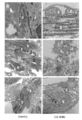
- A MN having nerves distributed in myotubes (m). High electron density region observed on muscle plasma membrane.
- B Multi-branched axon terminals observed in myotubes. A high electron density region (arrows) is localized at each axonal end. Mitochondria (mt) was seen nearby.
- C Necrosis observed at the end of axon (arrow).
- D Active zone (arrowhead), juxtaposed postsynaptic thickening (PSD, arrows), synaptic cleft observed between them.
- E The synaptic vesicle (sv) docks to the active zone (arrow).
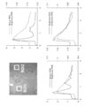
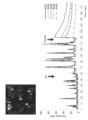
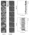
- Myotubes contracted in response to elevated Ca 2+ Functional analysis of in vitro hNMJ at day 20 of NMJ differentiation (upper left photo). Ca imaging in 5 ROIs. The addition of calcium enhanced transient Ca 2+ elevation and was arrested by curare treatment. Functional analysis of in vitro hNMJ at day 20 of NMJ differentiation (top photo). Motion analysis at four ROIs shows the propagation of myotube contraction. Functional analysis of in vitro hNMJ at day 20 of NMJ differentiation. Myotubes within 4 ROIs started to contract independently 15 minutes after gap junction inhibition by GAP27. Kinetic analysis by photoactivation of in vitro hNMJ cultures (photographs under a, c, b).
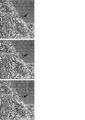
- 201B7 MYOD -SMN KD cells (SMN KD) and 201B7 MYOD in the cells (Control), transmission electron microscopy image showing the intracellular myotube and axon terminals.
- L indicates a lipid droplet
- Mt indicates a mitochondria
- Mf indicates a myotube
- SV indicates a synaptic vesicle
- Ax indicates an axon
- Nu indicates a nucleus.
- Differentiation to hNMJ from iPSCs (201B7 MYOD- SMN KD ) knocked down SMN protein (upper photograph).
- the method for producing the artificial nerve junction of the present invention is (I) transiently expressing MyoD in pluripotent stem cells to induce muscle differentiation, and (ii) culturing the cells obtained in (i) in a medium containing neurotrophic factor to differentiate neurons Including the step of inducing.
- Artificial neuromuscular junctions means neuromuscular junctions derived from pluripotent stem cells in vitro.
- the neuromuscular junction means a structure in which acetylcholine is released from the terminal end of motor nerve cells and can be received by a receptor present in myotubes.
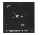
- the presence of a neuromuscular junction means, for example, colocalization of a synaptic vesicle protein (eg, SV2) expressed by motor neurons and an acetylcholine receptor expressed by myotubes by immunostaining or fluorescence microscopy It can be confirmed by a synaptic vesicle protein (eg, SV2) expressed by motor neurons and an acetylcholine receptor expressed by myotubes by immunostaining or fluorescence microscopy It can be confirmed by
- the artificial neuromuscular junction comprises motor neurons, myocytes (myotubes) and Schwann cells.
- myocytes myotubes
- Schwann cells can be identified using positivity such as S-100 marker as an index.
- the proportions of motor neurons, myocytes, and Schwann cells in the artificial neuromuscular junction are not particularly limited as long as they can maintain the function as the neuromuscular junction.
- a pluripotent stem cell is a stem cell having pluripotency capable of differentiating into many cells existing in a living body and also having proliferation ability, and the intermediate mesoderm used in the present invention Included are any cells that are induced into cells.
- pluripotent stem cells include, but are not limited to, embryonic stem (ES) cells, cloned embryonic derived embryonic stem (ntES) cells obtained by nuclear transfer, spermatogonial stem cells ("GS cells”), embryonic It includes germ cells (“EG cells”), induced pluripotent stem (iPS) cells, cultured fibroblasts and pluripotent cells derived from bone marrow stem cells (Muse cells).
- ES embryonic stem
- ntES cloned embryonic derived embryonic stem
- GS cells spermatogonial stem cells
- EG cells spermatogonial stem cells
- iPS induced pluripotent stem
- Muse cells pluripotent stem cells derived from bone marrow stem
- the reprogramming factor includes, for example, Oct3 / 4, Sox2, Sox1, Sox3, Sox15, Sox17, Sox17, Klf4, Klf2, c-Myc, N-Myc, L-Myc, Nanog, Lin28, Fbx15, ERas, ECAT15.
- Tcl1, beta-catenin, Lin28b, Sall1, Sall4, Esrrb, Nr5a2, Tbx3 or Glis1 and other genes or gene products are exemplified, and these reprogramming factors may be used alone or in combination. Also good.
- Somatic cells include, but are not limited to, fetal (child) somatic cells, neonatal (child) somatic cells, and any mature healthy or diseased somatic cells, and also primary culture cells. , Passage cells, and cell lines are also included.
- somatic cells are, for example, (1) tissue stem cells (somatic stem cells) such as neural stem cells, hematopoietic stem cells, mesenchymal stem cells, dental pulp stem cells, (2) tissue precursor cells, (3) blood cells (peripheral Blood cells, cord blood cells etc.), lymphocytes, epithelial cells, endothelial cells, muscle cells, fibroblasts (skin cells etc.), hair cells, hepatocytes, gastric mucous cells, enterocytes, splenocytes, pancreatic cells (pancreatic exocrine cells) Etc.), differentiated cells such as brain cells, lung cells, kidney cells and adipocytes.
- tissue stem cells such as neural stem cells, hematopoietic stem cells, mesenchy
- somatic cells when iPS cells are used as a material of cells for transplantation, it is desirable to use somatic cells in which the HLA genotypes of individuals to be transplanted are the same or substantially the same from the viewpoint that rejection does not occur.
- substantially identical means that the HLA genotypes coincide with the degree to which the immunosuppressive agent can suppress the immune response to the transplanted cells, for example, HLA-A and HLA-B.
- 3 loci of HLA-DR or 4 loci added with HLA-C are somatic cells having a matching HLA type.
- MyoD used in the present invention includes human myogenic differentiation 1 (MYOD1) consisting of the amino acid sequence shown in SEQ ID NO: 2 and orthologues thereof in other mammals, and their transcript variants, splice variants etc. . Alternatively, it has an amino acid identity of 90% or more, preferably 95% or more, more preferably 97% or more with any of the above proteins, and a function equivalent to that of the protein (eg, transcription activation of muscle specific promoter) And the like).
- MYOD1 human myogenic differentiation 1
- MYOD1 human myogenic differentiation 1
- a nucleic acid encoding MyoD human myogenic differentiation 1 (MYOD1) cDNA consisting of a nucleotide sequence represented by base numbers 221 to 1183 of SEQ ID NO: 1 and its orthologs in other mammals, and their transcript variants, splicing A mutant etc. are mentioned.
- it has a nucleotide identity of 90% or more, preferably 95% or more, more preferably 97% or more with any of the above nucleic acids, and a function equivalent to a protein encoded by the nucleic acid (eg, muscle-specific It may be a nucleic acid encoding a protein having a transcriptional activation of a promoter, etc.).
- stringent conditions bind a complex or a probe as taught by Berger and Kimmel (1987, Guide to Molecular Cloning Techniques in Methods in Enzymology, Vol. 152, Academic Press, San Diego Calif.) It can be determined based on the melting temperature (Tm) of the nucleic acid.
- Tm melting temperature
- the washing conditions after hybridization the conditions of “0.1 ⁇ SSC, 0.1% SDS, 60 ° C.” can be mentioned, and it is preferable that the hybridization state is maintained even when washing under such conditions. .
- the nucleic acid encoding MyoD may be DNA or RNA, or may be a DNA / RNA chimera. Also, the nucleic acid may be single-stranded, double-stranded DNA, double-stranded RNA or DNA: RNA hybrid. Preferably it is double stranded DNA or single stranded RNA.
- the RNA may be RNA incorporated with 5-methylcytidine and pseudouridine (TriLink Biotechnologies) to inhibit degradation, or may be a modified RNA by phosphatase treatment.
- the method for transiently expressing MyoD in pluripotent stem cells is not particularly limited, and for example, the following method can be used.
- expression means that the MyoD protein is transcribed and translated from the nucleic acid in the cell in the case of the nucleic acid encoding MyoD, and in the case of the MyoD protein, It is synonymous with the introduction of the protein into cells.
- vectors such as viruses, plasmids, artificial chromosomes can be introduced into pluripotent stem cells by techniques such as lipofection, liposomes, microinjection and the like.
- viral vectors include retrovirus vectors, lentivirus vectors, adenovirus vectors, adeno-associated virus vectors, Sendai virus vectors and the like.
- an artificial chromosome vector a human artificial chromosome (HAC), a yeast artificial chromosome (YAC), a bacterial artificial chromosome (BAC, PAC) etc. are illustrated.
- a plasmid a mammalian cell plasmid is exemplified.
- the vector may contain control sequences such as a promoter, enhancer, ribosome binding sequence, terminator, polyadenylation site and the like so that the DNA encoding MyoD can be expressed, and further, if necessary, a drug resistance gene.
- control sequences such as a promoter, enhancer, ribosome binding sequence, terminator, polyadenylation site and the like so that the DNA encoding MyoD can be expressed, and further, if necessary, a drug resistance gene.
- kanamycin resistance gene, ampicillin resistance gene, puromycin resistance gene etc. thymidine kinase gene
- selection marker sequences such as diphtheria toxin gene etc.
- fluorescent protein reporter gene sequences such as ⁇ -glucuronidase (GUS), FLAG etc.
- SV40 promoter As promoters, SV40 promoter, LTR promoter, CMV (cytomegalovirus) promoter, RSV (Rous sarcoma virus) promoter, MoMuLV (Moloney murine leukemia virus) LTR, HSV-TK (herpes simplex virus thymidine kinase) promoter, EF- ⁇ promoter, CAG
- the promoter and the TRE promoter (a CMV minimal promoter having a Tet response element consisting of 7 consecutive tetO sequences) are exemplified.
- the MyoD gene may be introduced into pluripotent stem cells using a transient expression vector, but in order to control expression more strictly, an inducible expression system is used. It is preferred to use.
- the induction expression system includes a drug induction expression system using tetracycline or its derivative (eg, doxycycline) and the like, and it is preferable to use a pluripotent stem cell into which a gene construct capable of drug induction expression of MyoD has been introduced in advance. .
- the TRE promoter described above it is desirable to simultaneously express a fusion protein with tetR and VP16AD or a fusion protein with reverse tetR (rtetR) and VP16AD (rtTA) in the same cell.
- rtetR reverse tetR
- rtTA reverse tetR
- MyoD can be expressed transiently while adding.
- the vector incorporates the expression cassette comprising the promoter and the DNA encoding MyoD linked to the chromosome of pluripotent stem cells, and if necessary, transposon sequences before and after this expression cassette for excision if necessary. May be included. Although it does not specifically limit as a transposon sequence, piggyBac is illustrated. In another embodiment, LoxP sequences may be included before and after the expression cassette for the purpose of removing the expression cassette.
- MyoD When MyoD is in the form of RNA, it may be introduced into pluripotent stem cells by techniques such as electroporation, lipofection, microinjection and the like. When MyoD is in the form of a protein, it may be introduced into pluripotent stem cells by methods such as lipofection, fusion with cell membrane permeable peptides (eg, TAT and polyarginine derived from HIV), microinjection and the like.
- cell membrane permeable peptides eg, TAT and polyarginine derived from HIV
- the period in which MyoD is transiently expressed in pluripotent stem cells to induce myocytes may be a period in which muscle differentiation is sufficiently performed, and can be appropriately changed depending on the type and nature of pluripotent stem cells used, For example, about 4 to 20 days are mentioned, about 4 to 12 days are preferable, and about 6 to 10 days are more preferable. For example, when using the above-mentioned drug induction expression system, it is preferable to add and culture a drug for this period.
- MyoD is RNA or protein
- the introduction may be performed multiple times so that MyoD exists in the cell in the above period.
- the culture conditions for pluripotent stem cells at the time of inducing myocytes in a state where MyoD is transiently expressed in pluripotent stem cells are preferably adhesion culture conditions.
- it is coated with a cell adhesion molecule such as extracellular matrix, specifically, Matrigel (BD), type I collagen, type IV collagen, gelatin, laminin, heparan sulfate proteoglycan, or entactin, and combinations thereof
- BD Matrigel
- type I collagen type IV collagen
- GMEM Gasgow Minimum Essential Medium
- IMDM Iscove's Modified Dulbecco's Medium: Iscove's Modified Dulbecco's Medium
- 199 medium Eagle's Minimum Essential Medium (Eagle's Minimum Essential Medium) (EMEM)
- EMEM Eagle's Minimum Essential Medium
- ⁇ MEM Dulbecco's modified Eagle's Medium
- DMEM Ham's F12 medium
- RPMI 1640 medium RPMI 1640 medium
- Fischer's medium a mixed medium thereof are included.
- serum substitutes such as albumin, transferrin, fatty acids, insulin, collagen precursors, trace elements, Knockout Serum Replacement (KSR) (serum substitute for FBS in ES cell culture), ITS-supplements, and mixtures thereof, etc. Is included.
- KSR Knockout Serum Replacement
- Preferred conditions for inducing differentiation are conditions in which pluripotent stem cells adhered to culture dishes coated with Matrigel are cultured in ⁇ MEM medium containing 10% KSR.
- the culture temperature is not particularly limited, but is about 30 to 40 ° C., preferably about 37 ° C., and the culture is performed under an atmosphere of CO 2 -containing air, and the CO 2 concentration is preferably about 2 to 5%. .
- the induction of muscle differentiation can be confirmed by the presence of muscle markers such as MHC and MEF2c.
- the cell population containing the muscle cell manufactured in this way may be not a single cell population but a cell population containing other types of cells.
- Neurotrophic factors are ligands to membrane receptors that play an important role in the survival and functional maintenance of motor neurons, and, for example, Nerve Growth Factor (NGF), Brain-derived Neurotrophic Factor (BDNF), Neurotrophin 3 ( NT-3), Neurotrophin 4/5 (NT-4 / 5), Neurotrophin 6 (NT-6), basic FGF, acidic FGF, FGF-5, Epidermal Growth Factor (EGF), Hepatocyte Growth Factor (HGF), Insulin , Insulin Like Growth Factor 1 (IGF 1), Insulin Like Growth Factor 2 (IGF 2), Glia cell line-derived Neurotrophic Factor (GDNF), TGF-b2, TGF-b3, Interleukin 6 (IL-6), Ciliary Neurotrophic Factor (CNTF) and LIF.
- NGF Nerve Growth Factor
- BDNF Brain-derived Neurotrophic Factor
- NT-3 Neurotrophin 3
- Neurotrophin 4/5 NT-4 / 5
- Neurotrophin 6 Neurotrophin 6
- Preferred neurotrophic factors in the present invention are NT-3, GDNF and BDNF.
- Neurotrophic factors are commercially available from, for example, Wako, R & D systems, etc. and can be easily used, but may be obtained by forced expression in cells by methods known to those skilled in the art.
- the concentration of NT-3 in the culture solution can be, for example, 0.1 ng / mL to 100 ng / mL, preferably 1 ng / mL to 50 ng / mL, more preferably 5 ng / mL to 20 ng / mL.
- the concentration of GDNF in the culture solution can be, for example, 0.1 ng / mL to 100 ng / mL, preferably 1 ng / mL to 50 ng / mL, more preferably 5 ng / mL to 20 ng / mL.
- the concentration of BDNF in the culture solution can be, for example, 0.1 ng / mL to 100 ng / mL, preferably 1 ng / mL to 50 ng / mL, more preferably 5 ng / mL to 20 ng / mL.
- the culture solution used in step (ii) can be prepared using a culture medium used for culturing animal cells as a basal medium.
- a culture medium used for culturing animal cells
- a basal medium for example, Glasgow's Minimal Essential Medium (GMEM) medium, IMDM medium, Medium 199 medium, Eagle's Minimum Essential Medium (EMEM) medium, ⁇ MEM medium, Dulbecco's modified Eagle's Medium (DMEM) medium, Ham's F12 medium, RPMI 1640 Media, Fischer's media, Neurobasal Medium (Life Technologies), and mixed media thereof are included.
- the medium may contain serum or may be serum free.
- the medium is, for example, albumin, transferrin, Knockout Serum Replacement (KSR) (serum substitute for FBS in ES cell culture), N2 supplement (Invitrogen), B27 supplement (Invitrogen), fatty acid, insulin, collagen It may also contain one or more serum substitutes such as precursors, trace elements, 2-mercaptoethanol, 3'-thiolglycerol, lipids, amino acids, L-glutamine, Glutamax (Invitrogen), non-essential amino acids, vitamins, It may also contain one or more substances such as growth factors, small molecule compounds, antibiotics, antioxidants, pyruvate, buffers, mineral salts and the like.
- a preferred culture is Neurobasal Medium containing N2 supplement, B27 supplement, NT-3, GDNF, and BDNF.
- neural differentiation can be induced by stopping the expression of MyoD and changing the medium to a medium for neural differentiation.
- culture temperature is not particularly limited, but is about 30 to 40 ° C., preferably about 37 ° C., and culture is performed under an atmosphere of CO 2 -containing air, and the CO 2 concentration is preferably about 2 to 5%.
- the culture period is not particularly limited as long as motor neurons and Schwann cells appear, but it is desirable that step (ii) be performed for at least 20 days. More preferably, it is 20 days to 120 days, still more preferably 30 days to 100 days.
- an artificial neuromuscular junction ie a cell population comprising motor neurons, myocytes and Schwann cells.
- kits for Producing Artificial Neuromuscular Junction from Pluripotent Stem Cells include cells, culture fluid, additives, culture vessels, and the like used in the above-described steps (i) and (ii).
- the kit includes cells, culture fluid, additives, culture vessels, and the like used in the above-described steps (i) and (ii).
- the kit may further include a document or instruction manual describing the procedure of the production process.
- the artificial neuromuscular junction of the present invention can be obtained as a cell culture containing motor neurons, myocytes and Schwann cells.
- the cell culture containing the neuromuscular junction is a pathological condition (eg, myasthenia gravis, Lambert-Eaton syndrome, Miller-Fisher syndrome, congenital) caused by a disorder (dysfunction or hypoplasia) of the neuromuscular junction. It is useful as a model system for the pathology of myasthenia gravis syndrome and spinal muscular atrophy. Therefore, it can be used, for example, as a cell preparation for treating a pathological condition caused by a neuromuscular junction disorder or as a screening system for a therapeutic agent for the pathological condition.
- the present invention relates to a pharmaceutical composition (cell preparation) comprising an artificial neuromuscular junction obtained by the method described above, a therapeutic agent for a pathological condition caused by a disorder of a neuromuscular junction, comprising the artificial neuromuscular junction, Methods are provided for treating conditions caused by disorders of the neuromuscular junction, comprising administering a therapeutically effective amount of the artificial neuromuscular junction.
- a method of administering a therapeutic agent to a patient in need of treatment for example, a method of locally administering the obtained artificial neuromuscular junction (cell culture containing muscle cells and Schwann cells) to the affected area, etc. It can be mentioned.
- the number of cells in the cell culture contained in the therapeutic agent is appropriately adjusted in accordance with the degree of disease and the like.
- cell preparation contains dimethyl sulfoxide (DMSO), serum albumin, etc. to protect the cells, and antibiotics etc. to prevent bacterial contamination and growth.
- DMSO dimethyl sulfoxide
- other pharmaceutically acceptable ingredients eg, carriers, excipients, disintegrants, buffers, emulsifiers, suspensions, soothing agents, stabilizers, preservatives, preservatives, physiological saline, etc. It may be contained in a cell preparation.
- Drug screening method or evaluation method In the present invention, a test substance is added to a cell population containing motor neurons, myocytes and Schwann cells obtained as described above, and then cultured. There is provided a method of screening or evaluating a therapeutic agent for a disease caused by a neuromuscular junction disorder, which comprises the step of evaluating muscle contraction or calcium concentration in the cells after culture.
- test substance may be, for example, cell extract, cell culture supernatant, microbial fermentation product, extract from marine organisms, plant extract, purified protein or crude protein, peptide, non-peptide compound, synthetic low molecular weight compound, and natural
- the compounds are exemplified.
- the test substance may also be (1) a biological library, (2) a synthetic library method using deconvolution, (3) a "one-bead one-compound” library method, and (4) It can be obtained using any of the many approaches in combinatorial library methods known in the art including synthetic library methods using affinity chromatography sorting. While biological library methods using affinity chromatography sorting are limited to peptide libraries, the other four approaches can be applied to peptides, non-peptide oligomers, or small molecule libraries of compounds (Lam (1997) ) Anticancer Drug Des. 12: 145-67). Examples of methods of synthesis of molecular libraries can be found in the art (DeWitt et al. (1993) Proc. Natl. Acad. Sci.
- Compound libraries can be solutions (see Houghten (1992) Bio / Techniques 13: 412-21) or beads (Lam (1991) Nature 354: 82-4), chips (Fodor (1993) Nature 364: 555-. 6), bacteria (US Pat. No. 5,223,409), spores (US Pat. Nos. 5,571,698, 5,403,484, and 5,223,409), plasmids (Cull et al. (1992) Proc. Natl. Acad. Sci. USA 89: 1865-9) or phage (Scott and Smith (1990) Science 249: 386-90; Devlin (1990) Science 249: 404-6; Cwirla et al. (1990) Proc. Natl. Acad. Sci. USA 87 Felici (1991) J. Mol. Biol. 222: 301-10; U.S. Patent Application No. 2002103360).
- Agents that enhance muscle contraction or calcium concentration relative to a control can be selected or evaluated as potential therapeutic agents for diseases caused by neuromuscular junction disorders.
- Another embodiment of the method of screening or evaluating a drug of the present invention is (I) transiently expressing MyoD in pluripotent stem cells to induce muscle differentiation, and (ii) culturing the cells obtained in (i) in a medium containing neurotrophic factor to differentiate neurons Guiding process, In the manufacturing process of the artificial neuromuscular junction, including the step of causing the test substance to exist in the step (i) and / or (ii), and analyzing the morphology or cell composition of the artificial neuromuscular junction obtained.
- a method for screening or evaluating a therapeutic agent for a disease caused by a disorder of a neuromuscular junction which selects or evaluates a drug.
- a process of manufacturing an artificial neuromuscular junction is performed using iPS cells derived from a patient with a disease caused by a disorder of the neuromuscular junction, and formation of the artificial neuromuscular junction is insufficient when no test substance is added. If the formation of the artificial neuromuscular junction is improved when the test substance is added, the substance can be a therapeutic agent for a disease caused by a disorder in the neuromuscular junction of the disease.
- the cell line MyoD-201B7 transfects a 201B7 human iPS cell line with a doxycycline (DOX) inducible MYOD1-expressing piggy bac vector as described by Tanaka et al., PLOS ONE 2013, Volume 8, Issue 4, e61540. It was built by doing. MYOD1 was expressed with mCherry protein and rtTA, and expression of MYOD1 was detected by luminescence of mCherry protein.
- DOX doxycycline
- MyoD-201B7 cells were seeded on a Matrigel-coated plate, and 1 ⁇ g / mL of doxycycline was added to a muscle differentiation medium (10% KSR + ⁇ MEM) to induce muscle differentiation.
- a muscle differentiation medium (10% KSR + ⁇ MEM)
- neural differentiation was induced by changing the medium to Neurobasal medium supplemented with neurotrophic factors (BDNF, GDNF, NT3; 10 ng / ml each, R & D systems). Thereafter, the medium was changed every 3 to 4 days and culture was continued.
- BDNF neurotrophic factors
- mCherry positive cells were sorted using FACS Aria BD and FACS Diva software. The selected cells were replated (8.8 ⁇ 10 4 cells / well) in Matrigel-coated 96-well plates and cultured in muscle differentiation medium with or without doxycycline. On day 20 the ratio of myotubes to neurons was quantified by immunocytochemistry.
- the motion vector analysis ⁇ 7s ILCE-7S (Sony Corporation) was used to continuously capture phase contrast images using a red filtered light source and fluorescence (Fluo 8 green) at 15 fps. Perform motion quantification using red separation image in target region (ROI) and brightness analysis using green separation image using SI 8000 cell motion imaging system (Sony Corporation) providing motion vector analysis based on block matching method did. Both motion imaging and motion analysis were performed at 38 fps using SI 8000 cell motion imaging system (Sony Corporation).
- Calcium imaging MyoD-201B7 cells were seeded at a density of 2.2 ⁇ 10 4 cells / well and allowed to differentiate into NMJ in Costar 96 well black wall / clear bottom plates. The growth medium was removed on day 20 and 100 ⁇ L of Fluo-8 recording medium was added to the cells for 1 hour at 37 ° C. The cells were washed twice with PBS and then imaged on an In Cell Analyzer 2000. The fluorescence intensity was expressed as the F / F0 ratio.
- MyoD-201B7 cells were seeded at a density of 2.2 ⁇ 10 4 cells / well in Matrigel coated 96 well black wall / transparent bottom plates. Half of the medium in each well was replaced daily until the cultures began to contract. The growth medium was removed and cells were loaded with 100 ⁇ L of Fluo-8 recording medium for 1 hour at 37 ° C. The cells were washed twice with PBS and then differentiation media was added to the wells. The Ca oscillations were monitored on a FDSS microplate reader, data points were collected at 1 minute intervals, and those data points were analyzed using FDSS software for frequency and amplitude quantification.
- Immunocytochemistry (ICC) labeling Primary antibodies used were anti-neurofilament (Millipore #MAB 5254, 1: 500), anti-synaptic vesicle protein 2 (DSHB # SV2, 1:20), anti-MHC (Millipore) No. A4.1025, 1: 1000), Anti-Islet1 (DSHB No. 40.2D6, 1: 100), Anti-HB9 (DSHB No. 81.5 C10, 1: 100), Anti-ChAT (Millipore No. AB144P, 1: 100) and It was anti-Tuj1 (Covance number MMS 435 P, 1: 1000).
- Plasmid containing the light genetic activation ⁇ br/> light activation domain (pLenti-Synapsin-hChR2 (H134R ) -EYFP-WPRE, Addgene plasmid No. 20945) was deposited from Karl Deisseroth (Karl Deisseroth) to Addgene I bought one and used it.
- a lentiviral transduction system (ViraPower hiperform lentiviral expression system, Invitrogen) was used to transduce the target gene into neural cells. NMJ cultures were treated with virus-containing medium at 5% CO 2 for 48 hours at 37 ° C. After viral infection, the medium was removed and neurobasal medium was added for expression of target gene. The medium was changed every 3 to 4 days. EYFP expression was examined by confocal microscopy with appropriate optical settings. Photoactivation was performed with a 488 nm laser wavelength. Bright field images were simultaneously imaged for muscle movement analysis in addition to EYFP signal.
- HiPSCs are efficiently induced in mature myotubes by transient expression of MYOD1 with a doxycycline (DOX) inducible vector.
- DOX doxycycline
- FIG. 1C, 1D Electron microscopy (EM) images showed Schwann cells overlying axonal end dilation and end button binding to myotubes in NMJ cultures (FIG. 1G and 1I). Synaptic vesicles and mitochondria accumulated in the presynaptic and postsynaptic sites of the NMJ, which is clearly divided by the high electron density activity zone (FIG. 1H, I). From these observations, our in vitro NMJ contained the major cell types of NMJ and was structurally also characterized by NMJ.
- agrin a neural factor that accumulates and stabilizes AChR at synaptic sites
- Administration of agrin increased the area of NMJ in long-term culture, while in the presence of anti-agrin antibody, only remnants of myotubes and degraded axon terminals were left in the culture (figure 2g, h).
- agrin like in vivo, has been found to play an important role in hNMJ formation and maintenance in our in vitro system.
- Gap junctions which are intercellular channels between myotubes and myotubes that allow intercellular propagation of electrical activity, are transiently expressed during NMJ development.
- myotube contractions were not completely synchronized, but they propagated to the adjacent area (FIG. 3C).
- the propagation of the contraction was interrupted by the gap junction inhibitor gap27, indicating that this contraction is caused by the propagation of action potentials through the gap junction ( Figure 3D).
- Immunostaining and image analysis Medium was removed from NMJ cultures, washed three times for 1 minute with PBS, and fixed for 10 minutes at room temperature with 4% paraformaldehyde / PBS. It was washed 3 times with 0.1% BSA / PBS and permeabilized with 4% paraformaldehyde + 0.1% Triton for 10 minutes at room temperature. Three times of 5 minutes of rehydration in 0.1% BSA / PBS, blocking treatment with 0.1% BSA / PBS + 0.5% Tween-20 for 1 hour at room temperature, and in a petri dish equipped with a parafilm layer The coverslip was moved to dryness.
- the primary antibody (antibody against Islet 1 or HB9) diluted in PBS is reacted at room temperature for 1 hour, washed three times with PBS, and reacted with a secondary antibody diluted in PBS for 45 minutes at room temperature (under light) ), Washed three times for 1 minute with PBS. Analysis was performed using a flow cytometer (Aria 2, BD) and FlowJo software (BD).
- 201B7 MYOD- SMN KD cells were subjected to immunostaining by the same protocol as the aforementioned immunostaining using an antibody against NF + SV2 or AChR as a primary antibody.
- SMN protein in 201B7 MYOD-SMN KD cells was found to be reduced by about 60% relative to the expression level of 201B7 MYOD cells not knocked down the SMN protein (FIG. 6A).
- the area of NMJ in 201B7 MYOD-SMN KD cells (SMN KD) was significantly smaller than that of 201B7 MYOD cells (Control) (FIG. 6B).
- SEM images of 201B7 MYOD cell (Control) cultures very elongated myotubes and bundles of axons were observed, and myotubes anchored at the extended axon terminals were observed (FIG. 6C).
- 201B7 MYOD- SMN KD cell (SMN KD) cultures myotubes were flat and thin and there were fewer axonal ends than in 201B7 MYOD cell (Control) cultures. Also, the details of the intracellular myotube and NMJ related structures were totally different from the 201B7 MYOD cell (Control) culture. Mitochondrial morphology and condition in 201B7 MYOD- SMN KD cells were damaged, and few of the myofibers were intact in their structure (FIG. 6D).
- the in vitro NMJ culture method of the present invention is composed of three types of cells, skeletal muscle, MN and Schwann cells, by a simple two-dimensional culture procedure of transiently expressing MyoD in iPSC and thereafter inducing neural differentiation.
- the method of the present invention is well-balanced in myogenesis and neurogenesis in NMJ construction, and reproduces a complex reciprocal signal between each cell such as nerve, muscle and Schwann cells in formation and maintenance of NMJ.
- the obtained NMJ shows the feature of the NMJ which is mature in appearance and function, it can be a platform of the disease model, and it is possible to use the cells for the disease based on the development or dysfunction of the NMJ. It contributes to therapy and drug effects.
Abstract
The present invention addresses the problem of providing a method for easily and efficiently preparing a functional and matured neuromuscular junction (NMJ) in vitro. This problem is solved by a method for producing an artificial neuromuscular junction, said method comprising: (i) a step for transiently expressing MyoD in pluripotent stem cells to induce myogenic differentiation; and (ii) a step for culturing the cells obtained in (i) in a medium containing a neurotrophic factor to induce neuronal differentiation.
Description
本発明は、多能性幹細胞を利用して、神経筋接合部をインビトロで調製する方法並びに該方法で得られた人工神経接合部およびその利用に関する。
The present invention relates to a method for preparing a neuromuscular junction in vitro using pluripotent stem cells, and an artificial neural junction obtained by the method and its use.
神経筋接合部(Neuromuscular junction: NMJ)は、運動神経終末と筋肉の接合部であり、神経終末から放出されるアセチルコリンにより、筋収縮が引き起こされる。
NMJの形成と維持は、神経細胞や筋細胞などのNMJの構成要素間での制御された連続的相互作用を必要とする多段階プロセスである。NMJ関連疾患の発病プロセスにはNMJの機能障害だけでなくその発生段階での障害も関与することが知られている。インビボのNMJの形成と維持を再現するヒト多能性幹細胞(hiPSC)由来のNMJはこれらの疾患の病態生理学の解明とそれらの治療薬の発見にとって優れたモデルとなりうる。近年、インビトロでNMJを再構築する幾つかの試みがなされた。例えば、非特許文献1および非特許文献2では、それぞれ、hiPSC由来運動ニューロン(MN)と初代筋細胞との共培養またはhiPSC由来MNとhiPSC由来筋細胞との共培養により得られるNMJモデルがアセチルコリン受容体(AChR)の集合体を形成することが示されたが、形態面及び機能面での成熟には至っておらず、NMJモデルとして不十分であった。 The neuromuscular junction (Neuromuscular junction: NMJ) is a junction between motor nerve terminals and muscles, and acetylcholine released from the nerve terminals causes muscle contraction.
The formation and maintenance of NMJ is a multistep process that requires controlled and continuous interactions between NMJ components such as neurons and muscle cells. It is known that the onset process of NMJ-related diseases involves not only NMJ dysfunction but also disorders at its developmental stage. NMJ derived from human pluripotent stem cells (hiPSCs), which reproduces formation and maintenance of NMJ in vivo, can be an excellent model for elucidation of pathophysiology of these diseases and discovery of their therapeutic agents. Recently, several attempts have been made to reconstruct NMJ in vitro. For example, in Non-PatentDocument 1 and Non-Patent Document 2, NMJ models obtained by co-culturing hiPSC-derived motor neurons (MN) and primary muscle cells or co-cultures of hiPSC-derived MN and hiPSC-derived muscle cells are acetylcholine Although it was shown to form an assembly of receptors (AChR), it did not reach maturity in terms of morphology and function, and was insufficient as an NMJ model.
NMJの形成と維持は、神経細胞や筋細胞などのNMJの構成要素間での制御された連続的相互作用を必要とする多段階プロセスである。NMJ関連疾患の発病プロセスにはNMJの機能障害だけでなくその発生段階での障害も関与することが知られている。インビボのNMJの形成と維持を再現するヒト多能性幹細胞(hiPSC)由来のNMJはこれらの疾患の病態生理学の解明とそれらの治療薬の発見にとって優れたモデルとなりうる。近年、インビトロでNMJを再構築する幾つかの試みがなされた。例えば、非特許文献1および非特許文献2では、それぞれ、hiPSC由来運動ニューロン(MN)と初代筋細胞との共培養またはhiPSC由来MNとhiPSC由来筋細胞との共培養により得られるNMJモデルがアセチルコリン受容体(AChR)の集合体を形成することが示されたが、形態面及び機能面での成熟には至っておらず、NMJモデルとして不十分であった。 The neuromuscular junction (Neuromuscular junction: NMJ) is a junction between motor nerve terminals and muscles, and acetylcholine released from the nerve terminals causes muscle contraction.
The formation and maintenance of NMJ is a multistep process that requires controlled and continuous interactions between NMJ components such as neurons and muscle cells. It is known that the onset process of NMJ-related diseases involves not only NMJ dysfunction but also disorders at its developmental stage. NMJ derived from human pluripotent stem cells (hiPSCs), which reproduces formation and maintenance of NMJ in vivo, can be an excellent model for elucidation of pathophysiology of these diseases and discovery of their therapeutic agents. Recently, several attempts have been made to reconstruct NMJ in vitro. For example, in Non-Patent
本発明は、簡便かつ効率よく、機能的かつ成熟したNMJをインビトロで調製する方法を提供することを課題とする。
An object of the present invention is to provide a method for preparing functional and mature NMJ in vitro simply and efficiently.
本発明者らは上記課題を解決するために鋭意検討を行った結果、多能性幹細胞においてMyoD(myogenic differentiation)遺伝子を一時的に発現させ、次いで、培養条件を筋誘導から神経誘導に切り替えることで運動ニューロン、筋細胞(筋管)およびシュワン細胞を含む機能的かつ成熟したNMJを効率よく分化誘導できることを見出し、本発明を完成するに至った。
As a result of intensive studies to solve the above problems, the present inventors transiently express MyoD (myogenic differentiation) gene in pluripotent stem cells, and then switch culture conditions from muscle induction to nerve induction In the present invention, it has been found that functional and mature NMJs including motor neurons, myocytes (myotubes) and Schwann cells can be efficiently induced to differentiate, and the present invention has been accomplished.
本発明は以下を提供する。
[1](i)多能性幹細胞にMyoDを一過的に発現させて筋分化を誘導する工程、および
(ii)(i)で得られた細胞を神経栄養因子を含む培地で培養して神経分化を誘導する工程、
を含む、人工神経筋接合部の製造方法。
[2]工程(i)のMyoDの一過的発現が薬剤を用いた発現誘導によって行われる、[1]に記載の方法。
[3]工程(ii)の神経栄養因子がGlial cell line-derived Neurotrophic Factor (GDNF) 、Brain-derived Neurotrophic Factor (BDNF)およびNeurotrophin 3 (NT-3)である、[1]または[2]に記載の方法。
[4]工程(i)が4~20日間行われる、[1]~[3]のいずれかに記載の方法。
[5]工程(ii)が20~120日間行われる、[1]~[4]のいずれかに記載の方法。
[6]人工神経筋接合部が運動ニューロン、筋細胞、およびシュワン細胞を含む、[1]~[5]のいずれか一項に記載の方法。
[7]多能性幹細胞が人工多能性幹細胞である、[1]~[6]のいずれかに記載の方法。
[8]多能性幹細胞がヒト多能性幹細胞である、[7]に記載の方法。
[9]それぞれ多能性幹細胞から誘導された、運動ニューロン、筋細胞、およびシュワン細胞を含む、細胞集団。
[10][9]に記載の細胞集団を含む医薬組成物。
[11][9]に記載の細胞集団に被検物質を添加して培養する工程、
培養後、該細胞における筋収縮またはカルシウム濃度を評価する工程、
を含む、神経筋接合部の障害によって引き起こされる疾患の治療薬のスクリーニングまたは評価方法。
[12](i)多能性幹細胞にMyoDを一過的に発現させて筋分化を誘導する工程、および(ii)(i)で得られた細胞を神経栄養因子を含む培地で培養して神経分化を誘導する工程、
を含む、人工神経筋接合部の製造工程において、工程(i)及び/または(ii)に被検物質を存在させ、得られた人工神経筋接合部の形態または細胞組成を解析することにより、被検物質を評価する、神経筋接合部の障害によって引き起こされる疾患の治療薬のスクリーニングまたは評価方法。 The present invention provides the following.
[1] (i) transiently expressing MyoD in pluripotent stem cells to induce muscle differentiation, and (ii) culturing the cells obtained in (i) in a medium containing neurotrophic factor Inducing neural differentiation,
A method of manufacturing an artificial neuromuscular junction, including:
[2] The method according to [1], wherein transient expression of MyoD in step (i) is performed by induction of expression using a drug.
[3] [1] or [2], wherein the neurotrophic factor of step (ii) is Glial cell line-derived Neurotrophic Factor (GDNF), Brain-derived Neurotrophic Factor (BDNF) and Neurotrophin 3 (NT-3) Method described.
[4] The method according to any one of [1] to [3], wherein step (i) is performed for 4 to 20 days.
[5] The method according to any one of [1] to [4], wherein step (ii) is performed for 20 to 120 days.
[6] The method according to any one of [1] to [5], wherein the artificial neuromuscular junction includes motor neurons, myocytes and Schwann cells.
[7] The method according to any one of [1] to [6], wherein the pluripotent stem cells are induced pluripotent stem cells.
[8] The method according to [7], wherein the pluripotent stem cells are human pluripotent stem cells.
[9] A cell population comprising motor neurons, myocytes and Schwann cells, each derived from pluripotent stem cells.
[10] A pharmaceutical composition comprising the cell population according to [9].
[11] A process of adding a test substance to the cell population described in [9] and culturing it,
Evaluating the muscle contraction or calcium concentration in the cells after culture;
A method of screening or evaluating a therapeutic agent for a disease caused by a disorder of the neuromuscular junction, including
[12] (i) transiently expressing MyoD in pluripotent stem cells to induce muscle differentiation, and (ii) culturing the cells obtained in (i) in a medium containing neurotrophic factor Inducing neural differentiation,
In the manufacturing process of the artificial neuromuscular junction, including the step of causing the test substance to exist in the step (i) and / or (ii), and analyzing the morphology or cell composition of the artificial neuromuscular junction obtained. A method for screening or evaluating a therapeutic agent for a disease caused by a disorder of a neuromuscular junction, which evaluates a test substance.
本発明によれば、インビトロの系で効率よくNMJを得ることができる。形成されたNMJは、AChRを始めとするNMJの特徴を備え、NMJを介した神経による筋収縮を呈する。
シュワン細胞は神経終末の成熟、AChRクラスタの成熟およびNMJの維持にかかわっているという報告があり(The Journal of Neuroscience, September 21, 2016, 36(38):9770-9781)、本発明の方法によれば、シュワン細胞を含有する、より成熟した機能的なNMJが得られる。
本発明の方法により、約30日程度の短期間でヒトNMJが形成可能であり、形成後約100日程度の長期間にわたって培養・維持することが可能である。
本発明の方法によれば、インビボシナプス形成ステップが再現されるがこれはNMJシナプス形成の理解からNMJ関連疾患の発病の解明および候補治療薬のスクリーニング等に有用である。 According to the present invention, NMJ can be efficiently obtained in an in vitro system. The formed NMJ has the features of NMJ including AChR, and exhibits NMJ-mediated muscle contraction by nerve.
It has been reported that Schwann cells are involved in the maturation of nerve endings, the maturation of AChR cluster and the maintenance of NMJ (The Journal of Neuroscience, September 21, 2016, 36 (38): 9770-9781). According to this, a more mature functional NMJ containing Schwann cells is obtained.
According to the method of the present invention, human NMJ can be formed in a short period of about 30 days, and can be cultured and maintained for a long period of about 100 days after the formation.
According to the method of the present invention, the in vivo synapse formation step is reproduced, which is useful for understanding NMJ synapse formation, elucidating the onset of NMJ related diseases, screening of candidate therapeutic agents and the like.
<人工神経接合部の製造法>
本発明の人工神経接合部の製造法は、
(i)多能性幹細胞にMyoDを一過的に発現させて筋分化を誘導する工程、および
(ii)(i)で得られた細胞を神経栄養因子を含む培地で培養して神経分化を誘導する工程
を含む。 <Manufacturing method of artificial neural junction>
The method for producing the artificial nerve junction of the present invention is
(I) transiently expressing MyoD in pluripotent stem cells to induce muscle differentiation, and (ii) culturing the cells obtained in (i) in a medium containing neurotrophic factor to differentiate neurons Including the step of inducing.
<人工神経筋接合部>
人工神経筋接合部とは、インビトロで多能性幹細胞から誘導された神経筋接合部を意味する。神経筋接合部とは、運動神経細胞の突起末端よりアセチルコリンが放出され、筋管細胞に存在する受容体が受け取ることができる構造を意味する。神経筋接合部の存在は、例えば、免疫染色や蛍光顕微鏡観察を行い、運動神経細胞が発現するシナプス小胞タンパク質(例えば、SV2)と筋管細胞が発現するアセチルコリン受容体が共局在することによって確認することができる。 <Artificial neuromuscular junction>
Artificial neuromuscular junctions means neuromuscular junctions derived from pluripotent stem cells in vitro. The neuromuscular junction means a structure in which acetylcholine is released from the terminal end of motor nerve cells and can be received by a receptor present in myotubes. The presence of a neuromuscular junction means, for example, colocalization of a synaptic vesicle protein (eg, SV2) expressed by motor neurons and an acetylcholine receptor expressed by myotubes by immunostaining or fluorescence microscopy It can be confirmed by
人工神経筋接合部は運動ニューロン、筋細胞(筋管)、およびシュワン(Schwann)細胞を含むことが好ましい。なお、人工神経筋接合部としての機能が維持されている限り、これら以外の細胞を有していてもよい。
運動ニューロンは、HB9、Islet1、ChATから選択される一つ以上のマーカーの陽性を指標にして同定することができる。
筋細胞は、ミオシン重鎖(MHC)およびMEF2cから選択される一つ以上のマーカーの陽性を指標にして同定することができる。
シュワン細胞は、S-100マーカーなどの陽性を指標にして同定することができる。
人工神経筋接合部は運動ニューロン、筋細胞、およびシュワン細胞の割合は、神経筋接合部としての機能が維持できる割合であれば特に制限されない。 Preferably, the artificial neuromuscular junction comprises motor neurons, myocytes (myotubes) and Schwann cells. In addition, as long as the function as an artificial neuromuscular junction is maintained, you may have cells other than these.
A motor neuron can be identified by using as an index the positive of one or more markers selected from HB9, Islet1, and ChAT.
Myocytes can be identified based on the positive of one or more markers selected from myosin heavy chain (MHC) and MEF2c.
Schwann cells can be identified using positivity such as S-100 marker as an index.
The proportions of motor neurons, myocytes, and Schwann cells in the artificial neuromuscular junction are not particularly limited as long as they can maintain the function as the neuromuscular junction.
<多能性幹細胞>
本発明において多能性幹細胞とは、生体に存在する多くの細胞に分化可能である多能性を有し、かつ、増殖能をも併せもつ幹細胞であり、本発明で使用される中間中胚葉細胞に誘導される任意の細胞が包含される。多能性幹細胞には、特に限定されないが、例えば、胚性幹(ES)細胞、核移植により得られるクローン胚由来の胚性幹(ntES)細胞、精子幹細胞(「GS細胞」)、胚性生殖細胞(「EG細胞」)、人工多能性幹(iPS)細胞、培養線維芽細胞や骨髄幹細胞由来の多能性細胞(Muse細胞)などが含まれる。好ましい多能性幹細胞は、製造工程において胚、卵子等の破壊をしないで入手可能であるという観点から、iPS細胞であり、より好ましくはヒトiPS細胞である。 <Pluripotent stem cells>
In the present invention, a pluripotent stem cell is a stem cell having pluripotency capable of differentiating into many cells existing in a living body and also having proliferation ability, and the intermediate mesoderm used in the present invention Included are any cells that are induced into cells. Examples of pluripotent stem cells include, but are not limited to, embryonic stem (ES) cells, cloned embryonic derived embryonic stem (ntES) cells obtained by nuclear transfer, spermatogonial stem cells ("GS cells"), embryonic It includes germ cells ("EG cells"), induced pluripotent stem (iPS) cells, cultured fibroblasts and pluripotent cells derived from bone marrow stem cells (Muse cells). Preferred pluripotent stem cells are iPS cells, more preferably human iPS cells, from the viewpoint of availability without destroying embryos, eggs and the like in the production process.
iPS細胞の製造方法は当該分野で公知であり、任意の体細胞へ初期化因子を導入することによって製造され得る。ここで、初期化因子とは、例えば、Oct3/4、Sox2、Sox1、Sox3、Sox15、Sox17、Klf4、Klf2、c-Myc、N-Myc、L-Myc、Nanog、Lin28、Fbx15、ERas、ECAT15-2、Tcl1、beta-catenin、Lin28b、Sall1、Sall4、Esrrb、Nr5a2、Tbx3またはGlis1等の遺伝子または遺伝子産物が例示され、これらの初期化因子は、単独で用いても良く、組み合わせて用いても良い。初期化因子の組み合わせとしては、WO2007/069666、WO2008/118820、WO2009/007852、WO2009/032194、WO2009/058413、WO2009/057831、WO2009/075119、WO2009/079007、WO2009/091659、WO2009/101084、WO2009/101407、WO2009/102983、WO2009/114949、WO2009/117439、WO2009/126250、WO2009/126251、WO2009/126655、WO2009/157593、WO2010/009015、WO2010/033906、WO2010/033920、WO2010/042800、WO2010/050626、WO2010/056831、WO2010/068955、WO2010/098419、WO2010/102267、WO2010/111409、WO2010/111422、WO2010/115050、WO2010/124290、WO2010/147395、WO2010/147612、Huangfu D,et al.(2008),Nat.Biotechnol.,26:795-797、Shi Y,et al.(2008),Cell Stem Cell,2:525-528、Eminli S,et al.(2008),Stem Cells.26:2467-2474、Huangfu D,et al.(2008),Nat.Biotechnol.26:1269-1275、Shi Y,et al.(2008),Cell Stem Cell,3,568-574、Zhao Y,et al.(2008),Cell Stem Cell,3:475-479、Marson A,(2008),Cell Stem Cell,3,132-135、Feng B,et al.(2009),Nat.Cell Biol.11:197-203、R.L.Judson et al.,(2009),Nat.Biotechnol.,27:459-461、Lyssiotis CA,et al.(2009),Proc Natl Acad Sci U S A.106:8912-8917、Kim JB,et al.(2009),Nature.461:649-643、Ichida JK,et al.(2009),Cell Stem Cell.5:491-503、Heng JC,et al.(2010),Cell Stem Cell.6:167-74、Han J,et al.(2010),Nature.463:1096-100、Mali P,et al.(2010),Stem Cells.28:713-720、Maekawa M,et al.(2011),Nature.474:225-9.に記載の組み合わせが例示される。
Methods for producing iPS cells are known in the art and can be produced by introducing a reprogramming factor into any somatic cell. Here, the reprogramming factor includes, for example, Oct3 / 4, Sox2, Sox1, Sox3, Sox15, Sox17, Sox17, Klf4, Klf2, c-Myc, N-Myc, L-Myc, Nanog, Lin28, Fbx15, ERas, ECAT15. -2, Tcl1, beta-catenin, Lin28b, Sall1, Sall4, Esrrb, Nr5a2, Tbx3 or Glis1 and other genes or gene products are exemplified, and these reprogramming factors may be used alone or in combination. Also good. As a combination of initialization factors, WO2007 / 069666, WO2008 / 118820, WO2009 / 007852, WO2009 / 032194, WO2009 / 058413, WO2009 / 057831, WO2009 / 075119, WO2009 / 079007, WO2009 / 091659, WO2009 / 101084, WO2009 / 01084 101407, WO2009 / 102983, WO2009 / 114949, WO2009 / 117439, WO2009 / 126250, WO2009 / 126251, WO2009 / 126655, WO2009 / 157593, WO2010 / 009015, WO2010 / 033906, WO2010 / 033920, WO2010 / 042800, WO2010 / 05 626, WO2010 / 056831, WO2010 / 068955, WO2010 / 098419, WO2010 / 102267, WO2010 / 111409, WO2010 / 111422, WO2010 / 115050, WO2010 / 124290, WO2010 / 147395, WO2010 / 147612, Huangfu D, et al. (2008), Nat. Biotechnol. , 26: 795- 797, Shi Y, et al. (2008), Cell Stem Cell, 2: 525-528, Eminli S, et al. (2008), Stem Cells. 26: 2467-2474, Huangfu D, et al. (2008), Nat. Biotechnol. 26: 1269-1275, Shi Y, et al. (2008), Cell Stem Cell, 3, 568-574, Zhao Y, et al. (2008), Cell Stem Cell, 3: 475-479, Marson A, (2008), Cell Stem Cell, 3, 132-135, Feng B, et al. (2009), Nat. Cell Biol. 11: 197-203, R.S. L. Judson et al. , (2009), Nat. Biotechnol. , 27: 459-461, Lyssiotis CA, et al. (2009), Proc Natl Acad Sci US A. 106: 8912-8917, Kim JB, et al. (2009), Nature. 461: 649-643, Ichida JK, et al. (2009), Cell Stem Cell. 5: 491-503, Heng JC, et al. (2010), Cell Stem Cell. 6: 167-74, Han J, et al. (2010), Nature. 463: 1096-100, Mali P, et al. (2010), Stem Cells. 28: 713-720, Maekawa M, et al. (2011), Nature. 474: 225-9. The combination as described in is illustrated.
体細胞には、非限定的に、胎児(仔)の体細胞、新生児(仔)の体細胞、および成熟した健全なもしくは疾患性の体細胞のいずれも包含されるし、また、初代培養細胞、継代細胞、および株化細胞のいずれも包含される。具体的には、体細胞は、例えば(1)神経幹細胞、造血幹細胞、間葉系幹細胞、歯髄幹細胞等の組織幹細胞(体性幹細胞)、(2)組織前駆細胞、(3)血液細胞(末梢血細胞、臍帯血細胞等)、リンパ球、上皮細胞、内皮細胞、筋肉細胞、線維芽細胞(皮膚細胞等)、毛細胞、肝細胞、胃粘膜細胞、腸細胞、脾細胞、膵細胞(膵外分泌細胞等)、脳細胞、肺細胞、腎細胞および脂肪細胞等の分化した細胞などが例示される。
Somatic cells include, but are not limited to, fetal (child) somatic cells, neonatal (child) somatic cells, and any mature healthy or diseased somatic cells, and also primary culture cells. , Passage cells, and cell lines are also included. Specifically, somatic cells are, for example, (1) tissue stem cells (somatic stem cells) such as neural stem cells, hematopoietic stem cells, mesenchymal stem cells, dental pulp stem cells, (2) tissue precursor cells, (3) blood cells (peripheral Blood cells, cord blood cells etc.), lymphocytes, epithelial cells, endothelial cells, muscle cells, fibroblasts (skin cells etc.), hair cells, hepatocytes, gastric mucous cells, enterocytes, splenocytes, pancreatic cells (pancreatic exocrine cells) Etc.), differentiated cells such as brain cells, lung cells, kidney cells and adipocytes.
また、iPS細胞を移植用細胞の材料として用いる場合、拒絶反応が起こらないという観点から、移植先の個体のHLA遺伝子型が同一または実質的に同一である体細胞を用いることが望ましい。ここで、「実質的に同一」とは、移植した細胞に対して免疫抑制剤により免疫反応が抑制できる程度にHLA遺伝子型が一致していることであり、例えば、HLA-A、HLA-BおよびHLA-DRの3遺伝子座またはHLA-Cを加えた4遺伝子座が一致するHLA型を有する体細胞である。
In addition, when iPS cells are used as a material of cells for transplantation, it is desirable to use somatic cells in which the HLA genotypes of individuals to be transplanted are the same or substantially the same from the viewpoint that rejection does not occur. Here, "substantially identical" means that the HLA genotypes coincide with the degree to which the immunosuppressive agent can suppress the immune response to the transplanted cells, for example, HLA-A and HLA-B. And 3 loci of HLA-DR or 4 loci added with HLA-C are somatic cells having a matching HLA type.
多能性幹細胞にMyoDを一過的に発現させて筋分化を誘導する工程、
本発明で用いられるMyoDとしては、配列番号2で表されるアミノ酸配列からなるヒトmyogenic differentiation 1(MYOD1)および他の哺乳動物におけるそのオルソログ、並びにそれらの転写変異体、スプライシング変異体などが挙げられる。あるいは、上記いずれかのタンパク質と90%以上、好ましくは95%以上、より好ましくは97%以上のアミノ酸同一性を有し、且つ該タンパク質と同等の機能(例、筋特異的プロモーターの転写活性化など)を有するタンパク質であってもよい。
MyoDをコードする核酸としては、配列番号1の塩基番号221~1183で表されるヌクレオチド配列からなるヒトmyogenic differentiation 1(MYOD1)cDNAおよび他の哺乳動物におけるそのオルソログ、並びにそれらの転写変異体、スプライシング変異体などが挙げられる。あるいは、上記いずれかの核酸と90%以上、好ましくは95%以上、より好ましくは97%以上のヌクレオチド同一性を有し、且つ該核酸にコードされるタンパク質と同等の機能(例、筋特異的プロモーターの転写活性化など)を有するタンパク質をコードする核酸であってもよい。あるいは、上記いずれかの核酸の相補鎖とストリンジェントな条件でハイブリダイズすることができる程度の相補関係を有するセンス鎖を有するものであってもよい。なお、ここでストリンジェントな条件は、Berger and Kimmel(1987, Guide to Molecular Cloning Techniques Methods in Enzymology, Vol. 152, Academic Press, San Diego CA)に教示されるように、複合体或いはプローブを結合する核酸の融解温度(Tm)に基づいて決定することができる。例えばハイブリダイズ後の洗浄条件として、「0. 1×SSC、0.1%SDS、60℃」の条件を挙げることができ、かかる条件で洗浄してもハイブリダイズ状態を維持するものであることが好ましい。 Transiently expressing MyoD in pluripotent stem cells to induce muscle differentiation,
MyoD used in the present invention includes human myogenic differentiation 1 (MYOD1) consisting of the amino acid sequence shown in SEQ ID NO: 2 and orthologues thereof in other mammals, and their transcript variants, splice variants etc. . Alternatively, it has an amino acid identity of 90% or more, preferably 95% or more, more preferably 97% or more with any of the above proteins, and a function equivalent to that of the protein (eg, transcription activation of muscle specific promoter) And the like).
As a nucleic acid encoding MyoD, human myogenic differentiation 1 (MYOD1) cDNA consisting of a nucleotide sequence represented by base numbers 221 to 1183 of SEQ ID NO: 1 and its orthologs in other mammals, and their transcript variants, splicing A mutant etc. are mentioned. Alternatively, it has a nucleotide identity of 90% or more, preferably 95% or more, more preferably 97% or more with any of the above nucleic acids, and a function equivalent to a protein encoded by the nucleic acid (eg, muscle-specific It may be a nucleic acid encoding a protein having a transcriptional activation of a promoter, etc.). Alternatively, it may have a sense strand having a degree of complementarity that can hybridize with the complementary strand of any of the above nucleic acids under stringent conditions. Here, stringent conditions bind a complex or a probe as taught by Berger and Kimmel (1987, Guide to Molecular Cloning Techniques in Methods in Enzymology, Vol. 152, Academic Press, San Diego Calif.) It can be determined based on the melting temperature (Tm) of the nucleic acid. For example, as the washing conditions after hybridization, the conditions of “0.1 × SSC, 0.1% SDS, 60 ° C.” can be mentioned, and it is preferable that the hybridization state is maintained even when washing under such conditions. .
MyoDをコードする核酸は、DNAであってもRNAであってもよく、あるいはDNA/RNAキメラであってもよい。また、該核酸は、一本鎖であっても、二本鎖DNA、二本鎖RNAもしくはDNA: RNAハイブリッドであってもよい。好ましくは二本鎖DNAもしくは一本鎖RNAである。当該RNAは、分解を抑制するため、5-メチルシチジンおよびシュードウリジン(pseudouridine)(TriLink Biotechnologies)を取り込ませたRNAを用いても良く、フォスファターゼ処理による修飾RNAであってもよい。
The nucleic acid encoding MyoD may be DNA or RNA, or may be a DNA / RNA chimera. Also, the nucleic acid may be single-stranded, double-stranded DNA, double-stranded RNA or DNA: RNA hybrid. Preferably it is double stranded DNA or single stranded RNA. The RNA may be RNA incorporated with 5-methylcytidine and pseudouridine (TriLink Biotechnologies) to inhibit degradation, or may be a modified RNA by phosphatase treatment.
MyoDを多能性幹細胞において一過的に発現させる方法は特に限定されないが、例えば、以下の方法を用いることができる。尚、ここで「発現」とは、MyoDをコードする核酸である場合においては、細胞内で、該核酸からMyoDタンパク質が転写および翻訳され生成することを意味し、MyoDタンパク質である場合においては、該タンパク質が細胞内に導入されることと同義である。
The method for transiently expressing MyoD in pluripotent stem cells is not particularly limited, and for example, the following method can be used. Here, "expression" means that the MyoD protein is transcribed and translated from the nucleic acid in the cell in the case of the nucleic acid encoding MyoD, and in the case of the MyoD protein, It is synonymous with the introduction of the protein into cells.
MyoDがDNAの形態の場合、例えば、ウイルス、プラスミド、人工染色体などのベクターをリポフェクション、リポソーム、マイクロインジェクションなどの手法によって多能性幹細胞内に導入することができる。ウイルスベクターとしては、レトロウイルスベクター、レンチウイルスベクター、アデノウイルスベクター、アデノ随伴ウイルスベクター、センダイウイルスベクターなどが例示される。また、人工染色体ベクターとしては、ヒト人工染色体(HAC)、酵母人工染色体(YAC)、細菌人工染色体(BAC、PAC)などが例示される。プラスミドとしては、哺乳動物細胞用プラスミドが例示される。
ベクターには、MyoDをコードするDNAが発現可能なように、プロモーター、エンハンサー、リボゾーム結合配列、ターミネーター、ポリアデニル化サイトなどの制御配列を含むことができるし、さらに、必要に応じて、薬剤耐性遺伝子(例えば、カナマイシン耐性遺伝子、アンピシリン耐性遺伝子、ピューロマイシン耐性遺伝子など)、チミジンキナーゼ遺伝子、ジフテリアトキシン遺伝子などの選択マーカー配列、蛍光タンパク質、βグルクロニダーゼ(GUS)、FLAGなどのレポーター遺伝子配列などを含むことができる。プロモーターとして、SV40プロモーター、LTRプロモーター、CMV(cytomegalovirus)プロモーター、RSV(Rous sarcoma virus)プロモーター、MoMuLV(Moloney mouse leukemia virus)LTR、HSV-TK(herpes simplex virus thymidine kinase)プロモーター、EF-αプロモーター、CAGプロモーターおよびTREプロモーター(tetO配列が7回連続したTet応答配列をもつCMV最小プロモーター)が例示される。 When MyoD is in the form of DNA, for example, vectors such as viruses, plasmids, artificial chromosomes can be introduced into pluripotent stem cells by techniques such as lipofection, liposomes, microinjection and the like. Examples of viral vectors include retrovirus vectors, lentivirus vectors, adenovirus vectors, adeno-associated virus vectors, Sendai virus vectors and the like. Moreover, as an artificial chromosome vector, a human artificial chromosome (HAC), a yeast artificial chromosome (YAC), a bacterial artificial chromosome (BAC, PAC) etc. are illustrated. As a plasmid, a mammalian cell plasmid is exemplified.
The vector may contain control sequences such as a promoter, enhancer, ribosome binding sequence, terminator, polyadenylation site and the like so that the DNA encoding MyoD can be expressed, and further, if necessary, a drug resistance gene. (Eg, kanamycin resistance gene, ampicillin resistance gene, puromycin resistance gene etc.), thymidine kinase gene, selection marker sequences such as diphtheria toxin gene etc., fluorescent protein, reporter gene sequences such as β-glucuronidase (GUS), FLAG etc. Can. As promoters, SV40 promoter, LTR promoter, CMV (cytomegalovirus) promoter, RSV (Rous sarcoma virus) promoter, MoMuLV (Moloney murine leukemia virus) LTR, HSV-TK (herpes simplex virus thymidine kinase) promoter, EF-α promoter, CAG The promoter and the TRE promoter (a CMV minimal promoter having a Tet response element consisting of 7 consecutive tetO sequences) are exemplified.
MyoDを一過的に発現させるためには、一過的発現ベクターを用いてMyoD遺伝子を多能性幹細胞に導入してもよいが、より厳密に発現を制御するためには、誘導発現システムを使用することが好ましい。誘導発現システムとしてはテトラサイクリンやその誘導体(例えば、ドキシサイクリン)などを用いた薬剤誘導発現システムが挙げられ、予めMyoDの薬剤誘導発現が可能な遺伝子コンストラクトが導入された多能性幹細胞を用いることが好ましい。
例えば、上記のTREプロモーターを用いた場合、同一の細胞において、tetRおよびVP16ADとの融合タンパク質またはリバース(reverse)tetR(rtetR)およびVP16ADとの融合タンパク質(rtTA)を同時に発現させることが望ましい。このTet-Onシステムでは薬剤が存在しないときは該融合タンパク質がTREに結合せず、転写は起こらないが、薬剤を添加することにより該融合タンパク質がTREプロモーターに結合し、転写が起こるので、薬剤を添加している間に一過的にMyoDを発現させることができる。 In order to transiently express MyoD, the MyoD gene may be introduced into pluripotent stem cells using a transient expression vector, but in order to control expression more strictly, an inducible expression system is used. It is preferred to use. The induction expression system includes a drug induction expression system using tetracycline or its derivative (eg, doxycycline) and the like, and it is preferable to use a pluripotent stem cell into which a gene construct capable of drug induction expression of MyoD has been introduced in advance. .
For example, when the TRE promoter described above is used, it is desirable to simultaneously express a fusion protein with tetR and VP16AD or a fusion protein with reverse tetR (rtetR) and VP16AD (rtTA) in the same cell. In this Tet-On system, the fusion protein does not bind to TRE and transcription does not occur in the absence of a drug, but the addition of a drug causes the fusion protein to bind to the TRE promoter and transcription occurs. MyoD can be expressed transiently while adding.
また、上記ベクターには、多能性幹細胞の染色体へ、プロモーターとそれに結合するMyoDをコードするDNAからなる発現カセットを取り込み、さらに必要に応じて切除するために、この発現カセットの前後にトランスポゾン配列を有していてもよい。トランスポゾン配列として特に限定されないが、piggyBacが例示される。他の態様として、発現カセットを除去する目的のため、発現カセットの前後にLoxP配列を有してもよい。
In addition, the vector incorporates the expression cassette comprising the promoter and the DNA encoding MyoD linked to the chromosome of pluripotent stem cells, and if necessary, transposon sequences before and after this expression cassette for excision if necessary. May be included. Although it does not specifically limit as a transposon sequence, piggyBac is illustrated. In another embodiment, LoxP sequences may be included before and after the expression cassette for the purpose of removing the expression cassette.
また、MyoDがRNAの形態の場合、例えばエレクトロポレーション、リポフェクション、マイクロインジェクションなどの手法によって多能性幹細胞内に導入してもよい。MyoDがタンパク質の形態の場合、例えばリポフェクション、細胞膜透過性ペプチド(例えば、HIV由来のTATおよびポリアルギニン)との融合、マイクロインジェクションなどの手法によって多能性幹細胞内に導入してもよい。
When MyoD is in the form of RNA, it may be introduced into pluripotent stem cells by techniques such as electroporation, lipofection, microinjection and the like. When MyoD is in the form of a protein, it may be introduced into pluripotent stem cells by methods such as lipofection, fusion with cell membrane permeable peptides (eg, TAT and polyarginine derived from HIV), microinjection and the like.
MyoDを多能性幹細胞内で一過的に発現させて筋細胞を誘導する期間は、筋分化が十分行われる期間であればよく、用いる多能性幹細胞の種類や性質によって適宜変更できるが、例えば、約4~20日が挙げられ、約4~12日が好ましく、約6~10日がより好ましい。例えば、上記の薬剤誘導発現システムを使用する場合は、この期間薬剤を加えて培養することが好ましい。また、他の態様として、トランスポゾン配列を有するベクターを用いる場合、上記の期間経過後、トランスポゼースを細胞内に導入することで発現を停止する方法およびLoxP配列を有するベクターを用いる場合、所望の期間経過後、Creを細胞内に導入することで発現を停止する方法などが例示される。
一方、MyoDがRNAまたはタンパク質の場合、上記の期間においてMyoDが細胞内で存在するように導入を複数回行ってもよい。 The period in which MyoD is transiently expressed in pluripotent stem cells to induce myocytes may be a period in which muscle differentiation is sufficiently performed, and can be appropriately changed depending on the type and nature of pluripotent stem cells used, For example, about 4 to 20 days are mentioned, about 4 to 12 days are preferable, and about 6 to 10 days are more preferable. For example, when using the above-mentioned drug induction expression system, it is preferable to add and culture a drug for this period. In another embodiment, when using a vector having a transposon sequence, a method of stopping expression by introducing transposase into cells after the above-mentioned period has elapsed, and using a vector having a LoxP sequence, a desired period of time lapse After that, a method of stopping expression by introducing Cre into cells is exemplified.
On the other hand, when MyoD is RNA or protein, the introduction may be performed multiple times so that MyoD exists in the cell in the above period.
MyoDを多能性幹細胞内で一過的に発現させた状態で筋細胞を誘導する際の多能性幹細胞の培養条件は、接着培養条件であることが好ましい。例えば、細胞外マトリクスなどの細胞接着分子、具体的には、マトリゲル(BD)、I型コラーゲン、IV型コラーゲン、ゼラチン、ラミニン、ヘパラン硫酸プロテオグリカン、またはエンタクチン、およびこれらの組み合わせを用いてコーティング処理された培養皿を用い、動物細胞の培養に用いられる培地を基本培地として血清または血清代替物を添加した培地で培養することが好ましい。ここで、基本培地としては、例えば、GMEM(グラスゴー最小必須培地:Glasgow Minimum Essential Medium)、IMDM(イスコフ改変ダルベッコ培地:Iscove's Modified Dulbecco's Medium)、199培地、イーグル最小必須培地(Eagle’s Minimum Essential Medium)(EMEM)、αMEM培地、ダルベッコ改変イーグル培地(Dulbecco’s modified Eagle’s Medium)(DMEM)、Ham’s F12培地、RPMI 1640培地、フィッシャー培地(Fischer’s medium)、およびこれらの混合培地などが包含される。また、血清代替物として、アルブミン、トランスフェリン、脂肪酸、インスリン、コラーゲン前駆体、微量元素、Knockout Serum Replacement(KSR)(ES細胞培養時のFBSの血清代替物)、ITS-サプリメントおよびこれらの混合物などが包含される。
好ましい分化誘導条件は、マトリゲルでコーティングされた培養皿に接着させた多能性幹細胞を10%KSRを含有するαMEM培地で培養する条件である。 The culture conditions for pluripotent stem cells at the time of inducing myocytes in a state where MyoD is transiently expressed in pluripotent stem cells are preferably adhesion culture conditions. For example, it is coated with a cell adhesion molecule such as extracellular matrix, specifically, Matrigel (BD), type I collagen, type IV collagen, gelatin, laminin, heparan sulfate proteoglycan, or entactin, and combinations thereof It is preferable to use the culture dish and culture the medium used for culturing animal cells as a basic medium in a medium to which serum or serum substitute is added. Here, as a basic medium, for example, GMEM (Glasgow Minimum Essential Medium), IMDM (Iscove's Modified Dulbecco's Medium: Iscove's Modified Dulbecco's Medium), 199 medium, Eagle's Minimum Essential Medium (Eagle's Minimum Essential Medium) ( EMEM), αMEM medium, Dulbecco's modified Eagle's Medium (DMEM), Ham's F12 medium, RPMI 1640 medium, Fischer's medium, and a mixed medium thereof are included. In addition, serum substitutes such as albumin, transferrin, fatty acids, insulin, collagen precursors, trace elements, Knockout Serum Replacement (KSR) (serum substitute for FBS in ES cell culture), ITS-supplements, and mixtures thereof, etc. Is included.
Preferred conditions for inducing differentiation are conditions in which pluripotent stem cells adhered to culture dishes coated with Matrigel are cultured in αMEM medium containing 10% KSR.
培養温度は、特に限定されないが、約30~40℃、好ましくは約37℃であり、CO2含有空気の雰囲気下で培養が行われ、CO2濃度は、好ましくは約2~5%である。
The culture temperature is not particularly limited, but is about 30 to 40 ° C., preferably about 37 ° C., and the culture is performed under an atmosphere of CO 2 -containing air, and the CO 2 concentration is preferably about 2 to 5%. .
筋分化が誘導されたことはMHCやMEF2cなどの筋マーカーの存在により確認できる。なお、このように製造された筋細胞を含む細胞集団は、単一の細胞集団ではなく、他の種類の細胞が含有された細胞集団であってもよい。
The induction of muscle differentiation can be confirmed by the presence of muscle markers such as MHC and MEF2c. In addition, the cell population containing the muscle cell manufactured in this way may be not a single cell population but a cell population containing other types of cells.
工程(i)で得られた細胞を神経栄養因子を含む培地で培養して神経分化を誘導する工程
<神経栄養因子>
神経栄養因子とは、運動ニューロンの生存と機能維持に重要な役割を果たしている膜受容体へのリガンドであり、例えば、Nerve Growth Factor (NGF)、Brain-derived Neurotrophic Factor (BDNF)、Neurotrophin 3 (NT-3)、Neurotrophin 4/5 (NT-4/5)、Neurotrophin 6 (NT-6)、basic FGF、acidic FGF、FGF-5、Epidermal Growth Factor (EGF)、Hepatocyte Growth Factor (HGF)、Insulin、Insulin Like Growth Factor 1 (IGF 1)、Insulin Like Growth Factor 2 (IGF 2)、Glia cell line-derived Neurotrophic Factor (GDNF)、TGF-b2、TGF-b3、Interleukin 6 (IL-6)、Ciliary Neurotrophic Factor (CNTF)およびLIFなどが挙げられる。本発明において好ましい神経栄養因子は、NT-3、GDNF、およびBDNFである。神経栄養因子は、例えばWako社やR&D systems社等から市販されており容易に利用することが可能であるが、当業者に公知の方法によって細胞へ強制発現させることによって得てもよい。 Culturing the cells obtained in step (i) in a medium containing neurotrophic factor to induce neural differentiation <neurotrophic factor>
Neurotrophic factors are ligands to membrane receptors that play an important role in the survival and functional maintenance of motor neurons, and, for example, Nerve Growth Factor (NGF), Brain-derived Neurotrophic Factor (BDNF), Neurotrophin 3 ( NT-3),Neurotrophin 4/5 (NT-4 / 5), Neurotrophin 6 (NT-6), basic FGF, acidic FGF, FGF-5, Epidermal Growth Factor (EGF), Hepatocyte Growth Factor (HGF), Insulin , Insulin Like Growth Factor 1 (IGF 1), Insulin Like Growth Factor 2 (IGF 2), Glia cell line-derived Neurotrophic Factor (GDNF), TGF-b2, TGF-b3, Interleukin 6 (IL-6), Ciliary Neurotrophic Factor (CNTF) and LIF. Preferred neurotrophic factors in the present invention are NT-3, GDNF and BDNF. Neurotrophic factors are commercially available from, for example, Wako, R & D systems, etc. and can be easily used, but may be obtained by forced expression in cells by methods known to those skilled in the art.
<神経栄養因子>
神経栄養因子とは、運動ニューロンの生存と機能維持に重要な役割を果たしている膜受容体へのリガンドであり、例えば、Nerve Growth Factor (NGF)、Brain-derived Neurotrophic Factor (BDNF)、Neurotrophin 3 (NT-3)、Neurotrophin 4/5 (NT-4/5)、Neurotrophin 6 (NT-6)、basic FGF、acidic FGF、FGF-5、Epidermal Growth Factor (EGF)、Hepatocyte Growth Factor (HGF)、Insulin、Insulin Like Growth Factor 1 (IGF 1)、Insulin Like Growth Factor 2 (IGF 2)、Glia cell line-derived Neurotrophic Factor (GDNF)、TGF-b2、TGF-b3、Interleukin 6 (IL-6)、Ciliary Neurotrophic Factor (CNTF)およびLIFなどが挙げられる。本発明において好ましい神経栄養因子は、NT-3、GDNF、およびBDNFである。神経栄養因子は、例えばWako社やR&D systems社等から市販されており容易に利用することが可能であるが、当業者に公知の方法によって細胞へ強制発現させることによって得てもよい。 Culturing the cells obtained in step (i) in a medium containing neurotrophic factor to induce neural differentiation <neurotrophic factor>
Neurotrophic factors are ligands to membrane receptors that play an important role in the survival and functional maintenance of motor neurons, and, for example, Nerve Growth Factor (NGF), Brain-derived Neurotrophic Factor (BDNF), Neurotrophin 3 ( NT-3),
培養液中におけるNT-3の濃度は、例えば、0.1ng/mL~100 ng/mL、好ましくは1ng/mL~50ng/mL、より好ましくは5ng/mL~20ng/mLとすることができる。
The concentration of NT-3 in the culture solution can be, for example, 0.1 ng / mL to 100 ng / mL, preferably 1 ng / mL to 50 ng / mL, more preferably 5 ng / mL to 20 ng / mL.
培養液中におけるGDNFの濃度は、例えば、0.1ng/mL~100 ng/mL、好ましくは1ng/mL~50ng/mL、より好ましくは5ng/mL~20ng/mLとすることができる。
The concentration of GDNF in the culture solution can be, for example, 0.1 ng / mL to 100 ng / mL, preferably 1 ng / mL to 50 ng / mL, more preferably 5 ng / mL to 20 ng / mL.
培養液中におけるBDNFの濃度は、例えば、0.1ng/mL~100 ng/mL、好ましくは1ng/mL~50ng/mL、より好ましくは5ng/mL~20ng/mLとすることができる。
The concentration of BDNF in the culture solution can be, for example, 0.1 ng / mL to 100 ng / mL, preferably 1 ng / mL to 50 ng / mL, more preferably 5 ng / mL to 20 ng / mL.
本発明において、工程(ii)で用いる培養液は、動物細胞の培養に用いられる培地を基礎培地として調製することができる。基礎培地としては、例えば、Glasgow's Minimal Essential Medium(GMEM)培地、IMDM培地、Medium 199培地、Eagle's Minimum Essential Medium (EMEM)培地、αMEM培地、Dulbecco's modified Eagle's Medium (DMEM)培地、Ham's F12培地、RPMI 1640培地、Fischer's培地、Neurobasal Medium(ライフテクノロジーズ)およびこれらの混合培地などが包含される。培地には、血清が含有されていてもよいし、あるいは無血清でもよい。必要に応じて、培地は、例えば、アルブミン、トランスフェリン、Knockout Serum Replacement(KSR)(ES細胞培養時のFBSの血清代替物)、N2サプリメント(Invitrogen)、B27サプリメント(Invitrogen)、脂肪酸、インスリン、コラーゲン前駆体、微量元素、2-メルカプトエタノール、3'-チオールグリセロールなどの1つ以上の血清代替物を含んでもよいし、脂質、アミノ酸、L-グルタミン、Glutamax(Invitrogen)、非必須アミノ酸、ビタミン、増殖因子、低分子化合物、抗生物質、抗酸化剤、ピルビン酸、緩衝剤、無機塩類などの1つ以上の物質も含有し得る。好ましい培養液は、N2サプリメント、B27サプリメント、NT-3、GDNF、およびBDNFを含有するNeurobasal Mediumである。
In the present invention, the culture solution used in step (ii) can be prepared using a culture medium used for culturing animal cells as a basal medium. As a basal medium, for example, Glasgow's Minimal Essential Medium (GMEM) medium, IMDM medium, Medium 199 medium, Eagle's Minimum Essential Medium (EMEM) medium, αMEM medium, Dulbecco's modified Eagle's Medium (DMEM) medium, Ham's F12 medium, RPMI 1640 Media, Fischer's media, Neurobasal Medium (Life Technologies), and mixed media thereof are included. The medium may contain serum or may be serum free. As necessary, the medium is, for example, albumin, transferrin, Knockout Serum Replacement (KSR) (serum substitute for FBS in ES cell culture), N2 supplement (Invitrogen), B27 supplement (Invitrogen), fatty acid, insulin, collagen It may also contain one or more serum substitutes such as precursors, trace elements, 2-mercaptoethanol, 3'-thiolglycerol, lipids, amino acids, L-glutamine, Glutamax (Invitrogen), non-essential amino acids, vitamins, It may also contain one or more substances such as growth factors, small molecule compounds, antibiotics, antioxidants, pyruvate, buffers, mineral salts and the like. A preferred culture is Neurobasal Medium containing N2 supplement, B27 supplement, NT-3, GDNF, and BDNF.
工程(i)において筋分化を誘導した後、MyoDの発現を停止し、培地を神経分化用の培地に変更することで、神経分化を誘導することができる。
After inducing myogenic differentiation in step (i), neural differentiation can be induced by stopping the expression of MyoD and changing the medium to a medium for neural differentiation.
培養条件について、培養温度は、特に限定されないが、約30~40℃、好ましくは約37℃であり、CO2含有空気の雰囲気下で培養が行われ、CO2濃度は、好ましくは約2~5%である。
With regard to culture conditions, culture temperature is not particularly limited, but is about 30 to 40 ° C., preferably about 37 ° C., and culture is performed under an atmosphere of CO 2 -containing air, and the CO 2 concentration is preferably about 2 to 5%.
培養期間は、運動ニューロンおよびシュワン細胞が出現する期間であれば、特に限定されないが、工程(ii)は、少なくとも20日間行われることが望ましい。より好ましくは20日間から120日間であり、さらに好ましくは、30日間から100日間である。
The culture period is not particularly limited as long as motor neurons and Schwann cells appear, but it is desirable that step (ii) be performed for at least 20 days. More preferably, it is 20 days to 120 days, still more preferably 30 days to 100 days.
これにより、人工神経筋接合部、すなわち、運動ニューロン、筋細胞およびシュワン細胞を含む細胞集団を得ることができる。
This makes it possible to obtain an artificial neuromuscular junction, ie a cell population comprising motor neurons, myocytes and Schwann cells.
多能性幹細胞から人工神経筋接合部を作製するキット
本発明での他の実施態様において、多能性幹細胞から人工神経筋接合部を作製するキットが含まれる。当該キットには、上述した工程(i)および(ii)に使用する細胞、培養液、添加剤または培養容器等が含まれる。例えば、MyoDが誘導可能な状態で導入された多能性幹細胞、テトラサイクリンまたはその誘導体などの薬剤、神経栄養因子、細胞外マトリクスでコートされた培養器、基礎培地から成る群より選択される1種類以上の試薬を含むキットが挙げられる。本キットには、さらに製造工程の手順を記載した書面や説明書を含んでもよい。 Kit for Producing Artificial Neuromuscular Junction from Pluripotent Stem Cells In another embodiment of the present invention, a kit for producing artificial neuromuscular junction from pluripotent stem cells is included. The kit includes cells, culture fluid, additives, culture vessels, and the like used in the above-described steps (i) and (ii). For example, one type selected from the group consisting of pluripotent stem cells into which MyoD can be introduced, drugs such as tetracycline or derivatives thereof, neurotrophic factors, incubators coated with extracellular matrix, and basal medium There is a kit containing the above reagents. The kit may further include a document or instruction manual describing the procedure of the production process.
本発明での他の実施態様において、多能性幹細胞から人工神経筋接合部を作製するキットが含まれる。当該キットには、上述した工程(i)および(ii)に使用する細胞、培養液、添加剤または培養容器等が含まれる。例えば、MyoDが誘導可能な状態で導入された多能性幹細胞、テトラサイクリンまたはその誘導体などの薬剤、神経栄養因子、細胞外マトリクスでコートされた培養器、基礎培地から成る群より選択される1種類以上の試薬を含むキットが挙げられる。本キットには、さらに製造工程の手順を記載した書面や説明書を含んでもよい。 Kit for Producing Artificial Neuromuscular Junction from Pluripotent Stem Cells In another embodiment of the present invention, a kit for producing artificial neuromuscular junction from pluripotent stem cells is included. The kit includes cells, culture fluid, additives, culture vessels, and the like used in the above-described steps (i) and (ii). For example, one type selected from the group consisting of pluripotent stem cells into which MyoD can be introduced, drugs such as tetracycline or derivatives thereof, neurotrophic factors, incubators coated with extracellular matrix, and basal medium There is a kit containing the above reagents. The kit may further include a document or instruction manual describing the procedure of the production process.
神経筋接合部を含有する細胞培養物
本発明の人工神経筋接合部は運動ニューロン、筋細胞およびシュワン細胞を含む細胞培養物として得ることができる。
当該神経筋接合部を含有する細胞培養物は、神経筋接合部の障害(機能不全や形成不全)によって引き起こされる病態(例えば、重症筋無力症、ランバート-イートン症候群、ミラー・フィッシャー症候群、先天性筋無力症症候群、脊髄性筋委縮症)の病態のモデル系として有用である。したがって、例えば、神経筋接合部の障害によって引き起こされる病態を治療するための細胞製剤や当該病態の治療薬のスクリーニング系として使用できる。 Cell Culture Containing a Neuromuscular Junction The artificial neuromuscular junction of the present invention can be obtained as a cell culture containing motor neurons, myocytes and Schwann cells.
The cell culture containing the neuromuscular junction is a pathological condition (eg, myasthenia gravis, Lambert-Eaton syndrome, Miller-Fisher syndrome, congenital) caused by a disorder (dysfunction or hypoplasia) of the neuromuscular junction. It is useful as a model system for the pathology of myasthenia gravis syndrome and spinal muscular atrophy. Therefore, it can be used, for example, as a cell preparation for treating a pathological condition caused by a neuromuscular junction disorder or as a screening system for a therapeutic agent for the pathological condition.
本発明は、上述した方法により得られた人工神経筋接合部を含む医薬組成物(細胞製剤)、該人工神経筋接合部を含む、神経筋接合部の障害によって引き起こされる病態の治療剤、該人工神経筋接合部の治療有効量を投与する工程を包含する神経筋接合部の障害によって引き起こされる病態を治療する方法をそれぞれ提供する。
治療を必要とする患者への治療剤の投与方法としては、例えば、得られた人工神経筋接合部(筋細胞およびシュワン細胞を含む細胞培養物)を患部に注射等で局所投与する方法などが挙げられる。治療剤に含まれる細胞培養物の細胞数は、疾患の程度などに合わせて適宜調整される。医薬組成物(細胞製剤)の使用においては、該細胞を保護するためにジメチルスルフォキシド(DMSO)や血清アルブミン等を、また、細菌の混入及び増殖を防ぐために抗生物質等を細胞製剤に含有させてもよい。さらに、製剤上許容される他の成分(例えば、担体、賦形剤、崩壊剤、緩衝剤、乳化剤、懸濁剤、無痛化剤、安定剤、保存剤、防腐剤、生理食塩水など)を細胞製剤に含有させてもよい。 The present invention relates to a pharmaceutical composition (cell preparation) comprising an artificial neuromuscular junction obtained by the method described above, a therapeutic agent for a pathological condition caused by a disorder of a neuromuscular junction, comprising the artificial neuromuscular junction, Methods are provided for treating conditions caused by disorders of the neuromuscular junction, comprising administering a therapeutically effective amount of the artificial neuromuscular junction.
As a method of administering a therapeutic agent to a patient in need of treatment, for example, a method of locally administering the obtained artificial neuromuscular junction (cell culture containing muscle cells and Schwann cells) to the affected area, etc. It can be mentioned. The number of cells in the cell culture contained in the therapeutic agent is appropriately adjusted in accordance with the degree of disease and the like. In the use of a pharmaceutical composition (cell preparation), cell preparation contains dimethyl sulfoxide (DMSO), serum albumin, etc. to protect the cells, and antibiotics etc. to prevent bacterial contamination and growth. You may In addition, other pharmaceutically acceptable ingredients (eg, carriers, excipients, disintegrants, buffers, emulsifiers, suspensions, soothing agents, stabilizers, preservatives, preservatives, physiological saline, etc.) It may be contained in a cell preparation.
薬剤のスクリーニング方法または評価方法
本発明では、上記のようにして得られた運動ニューロン、筋細胞およびシュワン細胞を含む細胞集団に被検物質を添加して培養する工程、
培養後、該細胞における筋収縮またはカルシウム濃度を評価する工程を含む、神経筋接合部の障害によって引き起こされる疾患の治療薬のスクリーニングまたは評価方法を提供する。 Drug screening method or evaluation method In the present invention, a test substance is added to a cell population containing motor neurons, myocytes and Schwann cells obtained as described above, and then cultured.
There is provided a method of screening or evaluating a therapeutic agent for a disease caused by a neuromuscular junction disorder, which comprises the step of evaluating muscle contraction or calcium concentration in the cells after culture.
被検物質は、例えば、細胞抽出物、細胞培養上清、微生物発酵産物、海洋生物由来の抽出物、植物抽出物、精製タンパク質又は粗タンパク質、ペプチド、非ペプチド化合物、合成低分子化合物、及び天然化合物が例示される。
The test substance may be, for example, cell extract, cell culture supernatant, microbial fermentation product, extract from marine organisms, plant extract, purified protein or crude protein, peptide, non-peptide compound, synthetic low molecular weight compound, and natural The compounds are exemplified.
被検物質はまた、(1)生物学的ライブラリー、(2)デコンヴォルーションを用いる合成ライブラリー法、(3)「1ビーズ1化合物(one-bead one-compound)」ライブラリー法、及び(4)アフィニティクロマトグラフィ選別を使用する合成ライブラリー法を含む当技術分野で公知のコンビナトリアルライブラリー法における多くのアプローチのいずれかを使用して得ることができる。アフィニティクロマトグラフィー選別を使用する生物学的ライブラリー法はペプチドライブラリーに限定されるが、その他の4つのアプローチはペプチド、非ペプチドオリゴマー、又は化合物の低分子化合物ライブラリーに適用できる(Lam (1997) Anticancer Drug Des. 12: 145-67)。分子ライブラリーの合成方法の例は、当技術分野において見出され得る(DeWitt et al. (1993) Proc. Natl.Acad. Sci. USA 90: 6909-13; Erb et al. (1994) Proc. Natl. Acad. Sci. USA 91: 11422-6; Zuckermann et al. (1994) J. Med. Chem. 37: 2678-85; Cho et al. (1993) Science 261: 1303-5; Carell et al. (1994) Angew. Chem. Int. Ed. Engl. 33: 2059; Carell et al. (1994) Angew. Chem. Int. Ed. Engl. 33: 2061; Gallop et al. (1994) J. Med. Chem. 37: 1233-51)。化合物ライブラリーは、溶液(Houghten (1992) Bio/Techniques 13: 412-21を参照のこと)又はビーズ(Lam (1991) Nature 354: 82-4)、チップ(Fodor (1993) Nature 364: 555-6)、細菌(米国特許第5,223,409号)、胞子(米国特許第5,571,698号、同第5,403,484号、及び同第5,223,409号)、プラスミド(Cull et al. (1992) Proc. Natl. Acad. Sci. USA 89: 1865-9)若しくはファージ(Scott and Smith (1990) Science 249: 386-90; Devlin (1990) Science 249: 404-6; Cwirla et al. (1990) Proc. Natl. Acad. Sci. USA 87: 6378-82; Felici (1991) J. Mol. Biol. 222: 301-10; 米国特許出願第2002103360号)として作製され得る。
The test substance may also be (1) a biological library, (2) a synthetic library method using deconvolution, (3) a "one-bead one-compound" library method, and (4) It can be obtained using any of the many approaches in combinatorial library methods known in the art including synthetic library methods using affinity chromatography sorting. While biological library methods using affinity chromatography sorting are limited to peptide libraries, the other four approaches can be applied to peptides, non-peptide oligomers, or small molecule libraries of compounds (Lam (1997) ) Anticancer Drug Des. 12: 145-67). Examples of methods of synthesis of molecular libraries can be found in the art (DeWitt et al. (1993) Proc. Natl. Acad. Sci. USA 90: 6909-13; Erb et al. (1994) Proc. Natl. Acad. Sci. USA 91: 11422-6; Zuckermann et al. (1994) J. Med. Chem. 37: 2678-85; Cho et al. (1993) Science 261: 1303-5; Carell et al. (1994) Angew. Chem. Int. Ed. Engl. 33: 2059; Carell et al. (1994) Angew. Chem. Int. Ed. Engl. 33: 2061; Gallop et al. (1994) J. Med. Chem. 37: 1233-51). Compound libraries can be solutions (see Houghten (1992) Bio / Techniques 13: 412-21) or beads (Lam (1991) Nature 354: 82-4), chips (Fodor (1993) Nature 364: 555-. 6), bacteria (US Pat. No. 5,223,409), spores (US Pat. Nos. 5,571,698, 5,403,484, and 5,223,409), plasmids (Cull et al. (1992) Proc. Natl. Acad. Sci. USA 89: 1865-9) or phage (Scott and Smith (1990) Science 249: 386-90; Devlin (1990) Science 249: 404-6; Cwirla et al. (1990) Proc. Natl. Acad. Sci. USA 87 Felici (1991) J. Mol. Biol. 222: 301-10; U.S. Patent Application No. 2002103360).
筋収縮またはカルシウム濃度を溶媒などの対照と比較して増強する物質を神経筋接合部の障害によって引き起こされる疾患の治療薬候補物質として選択または評価することができる。
Agents that enhance muscle contraction or calcium concentration relative to a control, such as a solvent, can be selected or evaluated as potential therapeutic agents for diseases caused by neuromuscular junction disorders.
本発明の薬剤のスクリーニング方法または評価方法の他の態様は、
(i)多能性幹細胞にMyoDを一過的に発現させて筋分化を誘導する工程、および
(ii)(i)で得られた細胞を神経栄養因子を含む培地で培養して神経分化を誘導する工程、
を含む、人工神経筋接合部の製造工程において、工程(i)及び/または(ii)に被検物質を存在させ、得られた人工神経筋接合部の形態または細胞組成を解析することにより、薬剤を選択または評価する、神経筋接合部の障害によって引き起こされる疾患の治療薬のスクリーニング方法または評価方法である。 Another embodiment of the method of screening or evaluating a drug of the present invention is
(I) transiently expressing MyoD in pluripotent stem cells to induce muscle differentiation, and (ii) culturing the cells obtained in (i) in a medium containing neurotrophic factor to differentiate neurons Guiding process,
In the manufacturing process of the artificial neuromuscular junction, including the step of causing the test substance to exist in the step (i) and / or (ii), and analyzing the morphology or cell composition of the artificial neuromuscular junction obtained. A method for screening or evaluating a therapeutic agent for a disease caused by a disorder of a neuromuscular junction, which selects or evaluates a drug.
例えば、神経筋接合部の障害によって引き起こされる疾患の患者由来のiPS細胞を用いて人工神経筋接合部の製造工程を行い、被検物質無添加では人工神経筋接合部の形成が不十分なところ、被検物質を添加した時に人工神経筋接合部の形成が改善された場合、その物質は疾患の神経筋接合部の障害によって引き起こされる疾患の治療薬となりうる。
For example, a process of manufacturing an artificial neuromuscular junction is performed using iPS cells derived from a patient with a disease caused by a disorder of the neuromuscular junction, and formation of the artificial neuromuscular junction is insufficient when no test substance is added. If the formation of the artificial neuromuscular junction is improved when the test substance is added, the substance can be a therapeutic agent for a disease caused by a disorder in the neuromuscular junction of the disease.
以下、実施例を挙げて本発明をより具体的に説明するが、本発明の態様は以下の実施例に限定されるものではない。
EXAMPLES Hereinafter, the present invention will be more specifically described by way of examples, but the embodiments of the present invention are not limited to the following examples.
材料と方法
細胞株
MyoD-201B7は、Tanaka et al., PLOS ONE 2013, Volume 8, Issue 4, e61540に記載されたように、201B7ヒトiPS細胞株にドキシサイクリン(DOX)誘導性MYOD1発現piggy bacベクターを形質移入することにより構築された。MYOD1はmCherryタンパク質およびrtTAとともに発現され、MYOD1の発現はmCherryタンパク質の発光により検出した。 Materials and methods
The cell line MyoD-201B7 transfects a 201B7 human iPS cell line with a doxycycline (DOX) inducible MYOD1-expressing piggy bac vector as described by Tanaka et al., PLOS ONE 2013, Volume 8,Issue 4, e61540. It was built by doing. MYOD1 was expressed with mCherry protein and rtTA, and expression of MYOD1 was detected by luminescence of mCherry protein.
細胞株
MyoD-201B7は、Tanaka et al., PLOS ONE 2013, Volume 8, Issue 4, e61540に記載されたように、201B7ヒトiPS細胞株にドキシサイクリン(DOX)誘導性MYOD1発現piggy bacベクターを形質移入することにより構築された。MYOD1はmCherryタンパク質およびrtTAとともに発現され、MYOD1の発現はmCherryタンパク質の発光により検出した。 Materials and methods
The cell line MyoD-201B7 transfects a 201B7 human iPS cell line with a doxycycline (DOX) inducible MYOD1-expressing piggy bac vector as described by Tanaka et al., PLOS ONE 2013, Volume 8,
神経筋接合部の分化
Tanaka et al., PLOS ONE 2013に記載された手順に従って、MyoD-201B7株の筋分化を行った。具体的には、マトリゲル被覆プレート上にMyoD-201B7細胞を播種し、筋分化培地(10%KSR+αMEM)にドキシサイクリンを1μg/mL添加することで筋分化を誘導した。ドキシサイクリン添加後10日目の時点で培地を神経栄養因子(BDNF、GDNF、NT3;10ng/mlずつ、R&Dシステムズ)を添加したニューロベーサル(Neurobasal)培地に変更することで神経分化を誘導した。その後、同培地を3~4日毎に交換して培養を継続した。 Differentiation of the neuromuscular junction
MyoD-201B7 strain was subjected to myogenic differentiation according to the procedure described in Tanaka et al., PLOS ONE 2013. Specifically, MyoD-201B7 cells were seeded on a Matrigel-coated plate, and 1 μg / mL of doxycycline was added to a muscle differentiation medium (10% KSR + αMEM) to induce muscle differentiation. At 10 days after the addition of doxycycline, neural differentiation was induced by changing the medium to Neurobasal medium supplemented with neurotrophic factors (BDNF, GDNF, NT3; 10 ng / ml each, R & D systems). Thereafter, the medium was changed every 3 to 4 days and culture was continued.
mCherry陽性細胞のフローソーティング
筋分化の2日目にFACS Aria BDおよびFACS Divaソフトウェアを使用してmCherry陽性細胞を選別した。選別された細胞をマトリゲル被覆96ウェルプレートに再播種し(8.8×10⁴細胞/ウェル)、ドキシサイクリンを含む、または含まない筋分化培地中で培養した。20日目にニューロンに対する筋管の比率を免疫細胞化学により定量した。 Flow sorting of mCherry positive cells On the second day of muscle differentiation, mCherry positive cells were sorted using FACS Aria BD and FACS Diva software. The selected cells were replated (8.8 × 10 4 cells / well) in Matrigel-coated 96-well plates and cultured in muscle differentiation medium with or without doxycycline. Onday 20 the ratio of myotubes to neurons was quantified by immunocytochemistry.
筋分化の2日目にFACS Aria BDおよびFACS Divaソフトウェアを使用してmCherry陽性細胞を選別した。選別された細胞をマトリゲル被覆96ウェルプレートに再播種し(8.8×10⁴細胞/ウェル)、ドキシサイクリンを含む、または含まない筋分化培地中で培養した。20日目にニューロンに対する筋管の比率を免疫細胞化学により定量した。 Flow sorting of mCherry positive cells On the second day of muscle differentiation, mCherry positive cells were sorted using FACS Aria BD and FACS Diva software. The selected cells were replated (8.8 × 10 4 cells / well) in Matrigel-coated 96-well plates and cultured in muscle differentiation medium with or without doxycycline. On
運動ベクトル分析
α7s ILCE-7S(ソニー株式会社)を使用して赤色フィルター処理光源および蛍光(Fluo8、緑色)を使用した位相差像を15fpsで連続撮像した。ブロックマッチング法に基づく運動ベクトル分析を提供するSI8000セルモーションイメージングシステム(ソニー株式会社)を使用して目的領域(ROI)における赤色分離像を使用する運動定量と緑色分離像を使用する明度分析を実施した。
SI8000セルモーションイメージングシステム(ソニー株式会社)を使用して38fpsで移動像の撮像と運動分析の両方を実施した。 The motion vector analysis α7s ILCE-7S (Sony Corporation) was used to continuously capture phase contrast images using a red filtered light source and fluorescence (Fluo 8 green) at 15 fps. Perform motion quantification using red separation image in target region (ROI) and brightness analysis using green separation image using SI 8000 cell motion imaging system (Sony Corporation) providing motion vector analysis based on block matching method did.
Both motion imaging and motion analysis were performed at 38 fps using SI 8000 cell motion imaging system (Sony Corporation).
カルシウムイメージング
MyoD-201B7細胞を2.2×10⁴細胞/ウェルの密度で播種し、Costar96ウェル黒壁/透明底プレート内でNMJに分化させた。20日目に増殖培地を取り除き、37℃で1時間にわたって細胞に100μLのFluo-8記録培地を添加した。それらの細胞をPBSで2度洗浄し、次にIn Cell Analyzer 2000で撮像した。F/F0比率として蛍光強度を表した。 Calcium imaging MyoD-201B7 cells were seeded at a density of 2.2 × 10 4 cells / well and allowed to differentiate into NMJ in Costar 96 well black wall / clear bottom plates. The growth medium was removed on day 20 and 100 μL of Fluo-8 recording medium was added to the cells for 1 hour at 37 ° C. The cells were washed twice with PBS and then imaged on an In Cell Analyzer 2000. The fluorescence intensity was expressed as the F / F0 ratio.
MyoD-201B7細胞を2.2×10⁴細胞/ウェルの密度で播種し、Costar96ウェル黒壁/透明底プレート内でNMJに分化させた。20日目に増殖培地を取り除き、37℃で1時間にわたって細胞に100μLのFluo-8記録培地を添加した。それらの細胞をPBSで2度洗浄し、次にIn Cell Analyzer 2000で撮像した。F/F0比率として蛍光強度を表した。 Calcium imaging MyoD-201B7 cells were seeded at a density of 2.2 × 10 4 cells / well and allowed to differentiate into NMJ in Costar 96 well black wall / clear bottom plates. The growth medium was removed on
FDSS(浜松ホトニクス)による解析
MyoD-201B7細胞を2.2×10⁴細胞/ウェルの密度でマトリゲル被覆96ウェル黒壁/透明底プレートに播種した。それらの培養物が収縮し始めるまで毎日各ウェルの半量の培地を交換した。増殖培地を取り除き、37℃で1時間にわたって細胞に100μLのFluo-8記録培地を負荷した。それらの細胞をPBSで2度洗浄し、次にウェルに分化培地を添加した。FDSSマイクロプレートリーダーでCaオシレーションをモニターし、1分の間隔でデータポイントを収集し、頻度および振幅の定量用のFDSSソフトウェアを使用してそれらのデータポイントを分析した。 Analysis by FDSS (Hamamatsu Photonics) MyoD-201B7 cells were seeded at a density of 2.2 × 10 4 cells / well in Matrigel coated 96 well black wall / transparent bottom plates. Half of the medium in each well was replaced daily until the cultures began to contract. The growth medium was removed and cells were loaded with 100 μL of Fluo-8 recording medium for 1 hour at 37 ° C. The cells were washed twice with PBS and then differentiation media was added to the wells. The Ca oscillations were monitored on a FDSS microplate reader, data points were collected at 1 minute intervals, and those data points were analyzed using FDSS software for frequency and amplitude quantification.
MyoD-201B7細胞を2.2×10⁴細胞/ウェルの密度でマトリゲル被覆96ウェル黒壁/透明底プレートに播種した。それらの培養物が収縮し始めるまで毎日各ウェルの半量の培地を交換した。増殖培地を取り除き、37℃で1時間にわたって細胞に100μLのFluo-8記録培地を負荷した。それらの細胞をPBSで2度洗浄し、次にウェルに分化培地を添加した。FDSSマイクロプレートリーダーでCaオシレーションをモニターし、1分の間隔でデータポイントを収集し、頻度および振幅の定量用のFDSSソフトウェアを使用してそれらのデータポイントを分析した。 Analysis by FDSS (Hamamatsu Photonics) MyoD-201B7 cells were seeded at a density of 2.2 × 10 4 cells / well in Matrigel coated 96 well black wall / transparent bottom plates. Half of the medium in each well was replaced daily until the cultures began to contract. The growth medium was removed and cells were loaded with 100 μL of Fluo-8 recording medium for 1 hour at 37 ° C. The cells were washed twice with PBS and then differentiation media was added to the wells. The Ca oscillations were monitored on a FDSS microplate reader, data points were collected at 1 minute intervals, and those data points were analyzed using FDSS software for frequency and amplitude quantification.
統計
統計分析にはMicrosoft Excel 2013の統計関数を使用した。結果を平均値±SEMとして表した。ステューデントのt検定およびウィルコクソンの順位和検定を用いて統計学的有意性を決定した。P<0.05を有意であると見なした。 Statistics For statistical analysis, Microsoft Excel 2013 statistical functions were used. The results were expressed as mean ± SEM. Statistical significance was determined using Student's t-test and Wilcoxon rank sum test. P <0.05 was considered to be significant.
統計分析にはMicrosoft Excel 2013の統計関数を使用した。結果を平均値±SEMとして表した。ステューデントのt検定およびウィルコクソンの順位和検定を用いて統計学的有意性を決定した。P<0.05を有意であると見なした。 Statistics For statistical analysis, Microsoft Excel 2013 statistical functions were used. The results were expressed as mean ± SEM. Statistical significance was determined using Student's t-test and Wilcoxon rank sum test. P <0.05 was considered to be significant.
透過電子顕微鏡法
NMJ培養物を4℃で0.1Mリン酸緩衝液(pH7.4)中の2%パラホルムアルデヒドと2%グルタルアルデヒド中で一晩にわたって前固定した。1%四酸化オスミウム溶液中で後固定を室温で1時間にわたって行った。エタノール(30%、50%、70%、90%、95%および100%)内で試料を脱水し、Epon樹脂中に包埋した。超薄切片(80nm)を切断し、酢酸ウラニルとアルカリ性クエン酸鉛で染色した。透過電子顕微鏡(H-7650、日立)でそれらの標本を検査した。 Transmission Electron Microscopy NMJ cultures were pre-fixed overnight in 2% paraformaldehyde and 2% glutaraldehyde in 0.1 M phosphate buffer (pH 7.4) at 4 ° C. Post-fixation in 1% osmium tetraoxide solution was performed at room temperature for 1 hour. The samples were dehydrated in ethanol (30%, 50%, 70%, 90%, 95% and 100%) and embedded in Epon resin. Ultrathin sections (80 nm) were cut and stained with uranyl acetate and alkaline lead citrate. The specimens were examined with a transmission electron microscope (H-7650, Hitachi).
NMJ培養物を4℃で0.1Mリン酸緩衝液(pH7.4)中の2%パラホルムアルデヒドと2%グルタルアルデヒド中で一晩にわたって前固定した。1%四酸化オスミウム溶液中で後固定を室温で1時間にわたって行った。エタノール(30%、50%、70%、90%、95%および100%)内で試料を脱水し、Epon樹脂中に包埋した。超薄切片(80nm)を切断し、酢酸ウラニルとアルカリ性クエン酸鉛で染色した。透過電子顕微鏡(H-7650、日立)でそれらの標本を検査した。 Transmission Electron Microscopy NMJ cultures were pre-fixed overnight in 2% paraformaldehyde and 2% glutaraldehyde in 0.1 M phosphate buffer (pH 7.4) at 4 ° C. Post-fixation in 1% osmium tetraoxide solution was performed at room temperature for 1 hour. The samples were dehydrated in ethanol (30%, 50%, 70%, 90%, 95% and 100%) and embedded in Epon resin. Ultrathin sections (80 nm) were cut and stained with uranyl acetate and alkaline lead citrate. The specimens were examined with a transmission electron microscope (H-7650, Hitachi).
走査電子顕微鏡法
SEM観察用の試料を4℃で2%パラホルムアルデヒドと2%グルタルアルデヒド中で一晩にわたって前固定した。1%四酸化オスミウム溶液中で後固定を室温で1時間にわたって行った。濃度等級を割り当てたエタノール(30%、50%、70%、90%、95%および100%)内で試料を脱水し、残留水分を除去するために臨界点乾燥機で乾燥した。それらの試料に陰極スプレー法によりプラチナを被覆し、その後で走査電子顕微鏡(S-4700、日立)で撮像した。 Scanning Electron Microscopy Samples for SEM observation were pre-fixed in 2% paraformaldehyde and 2% glutaraldehyde at 4 ° C. overnight. Post-fixation in 1% osmium tetraoxide solution was performed at room temperature for 1 hour. The samples were dewatered in concentration grade assigned ethanol (30%, 50%, 70%, 90%, 95% and 100%) and dried in a critical point drier to remove residual moisture. The samples were coated with platinum by cathodic spraying and then imaged with a scanning electron microscope (S-4700, Hitachi).
SEM観察用の試料を4℃で2%パラホルムアルデヒドと2%グルタルアルデヒド中で一晩にわたって前固定した。1%四酸化オスミウム溶液中で後固定を室温で1時間にわたって行った。濃度等級を割り当てたエタノール(30%、50%、70%、90%、95%および100%)内で試料を脱水し、残留水分を除去するために臨界点乾燥機で乾燥した。それらの試料に陰極スプレー法によりプラチナを被覆し、その後で走査電子顕微鏡(S-4700、日立)で撮像した。 Scanning Electron Microscopy Samples for SEM observation were pre-fixed in 2% paraformaldehyde and 2% glutaraldehyde at 4 ° C. overnight. Post-fixation in 1% osmium tetraoxide solution was performed at room temperature for 1 hour. The samples were dewatered in concentration grade assigned ethanol (30%, 50%, 70%, 90%, 95% and 100%) and dried in a critical point drier to remove residual moisture. The samples were coated with platinum by cathodic spraying and then imaged with a scanning electron microscope (S-4700, Hitachi).
免疫細胞化学(ICC)標識
使用した一次抗体は、抗ニューロフィラメント(Millipore番号MAB5254、1:500)、抗シナプス小胞プロテイン2(DSHB番号SV2、1:20)、抗MHC(Millipore番号A4.1025、1:1000)、抗Islet1(DSHB番号40.2D6、1:100)、抗HB9(DSHB番号81.5C10、1:100)、抗ChAT(Millipore番号AB144P、1:100)および抗Tuj1(Covance番号MMS435P、1:1000)であった。複合体化抗BTX-647(Invitrogen)を0.5ug/mlの濃度で使用した。共焦点顕微鏡(Fluo View-1000、オリンパス、またはLSM-710、Zeiss)により試料を画像化した。 Immunocytochemistry (ICC) labeling Primary antibodies used were anti-neurofilament (Millipore #MAB 5254, 1: 500), anti-synaptic vesicle protein 2 (DSHB # SV2, 1:20), anti-MHC (Millipore) No. A4.1025, 1: 1000), Anti-Islet1 (DSHB No. 40.2D6, 1: 100), Anti-HB9 (DSHB No. 81.5 C10, 1: 100), Anti-ChAT (Millipore No. AB144P, 1: 100) and It was anti-Tuj1 (Covance number MMS 435 P, 1: 1000). Complexed anti-BTX-647 (Invitrogen) was used at a concentration of 0.5 ug / ml. Samples were imaged by confocal microscopy (Fluo View-1000, Olympus, or LSM-710, Zeiss).
使用した一次抗体は、抗ニューロフィラメント(Millipore番号MAB5254、1:500)、抗シナプス小胞プロテイン2(DSHB番号SV2、1:20)、抗MHC(Millipore番号A4.1025、1:1000)、抗Islet1(DSHB番号40.2D6、1:100)、抗HB9(DSHB番号81.5C10、1:100)、抗ChAT(Millipore番号AB144P、1:100)および抗Tuj1(Covance番号MMS435P、1:1000)であった。複合体化抗BTX-647(Invitrogen)を0.5ug/mlの濃度で使用した。共焦点顕微鏡(Fluo View-1000、オリンパス、またはLSM-710、Zeiss)により試料を画像化した。 Immunocytochemistry (ICC) labeling Primary antibodies used were anti-neurofilament (Millipore #MAB 5254, 1: 500), anti-synaptic vesicle protein 2 (DSHB # SV2, 1:20), anti-MHC (Millipore) No. A4.1025, 1: 1000), Anti-Islet1 (DSHB No. 40.2D6, 1: 100), Anti-HB9 (DSHB No. 81.5 C10, 1: 100), Anti-ChAT (Millipore No. AB144P, 1: 100) and It was anti-Tuj1 (Covance number MMS 435 P, 1: 1000). Complexed anti-BTX-647 (Invitrogen) was used at a concentration of 0.5 ug / ml. Samples were imaged by confocal microscopy (Fluo View-1000, Olympus, or LSM-710, Zeiss).
光遺伝学的活性化
光活性化ドメインを含むプラスミド(pLenti-Synapsin-hChR2(H134R)-EYFP-WPRE、Addgeneプラスミド番号20945)はカール・ダイセロス(Karl Deisseroth)からAddgeneに寄託されたものを購入して用いた。標的遺伝子を神経性細胞に形質導入するためにレンチウイルス形質導入システム(ViraPower hiperformレンチウイルス発現システム、Invitrogen)を使用した。NMJ培養物を37℃で48時間にわたって5%CO2でウイルスを含む培地で処理した。ウイルス感染後、その培地を取り除き、ニューロベーサル培地を標的遺伝子の発現のために加えた。その培地を3~4日毎に交換した。適切な光学的設定を用いた共焦点顕微鏡観察によりEYFP発現を検査した。光活性化を488nmのレーザー波長により実施した。EYFPシグナルに加えて筋肉運動分析のために明視野像を同時に撮像した。 Plasmid containing the light genetic activation <br/> light activation domain (pLenti-Synapsin-hChR2 (H134R ) -EYFP-WPRE, Addgene plasmid No. 20945) was deposited from Karl Deisseroth (Karl Deisseroth) to Addgene I bought one and used it. A lentiviral transduction system (ViraPower hiperform lentiviral expression system, Invitrogen) was used to transduce the target gene into neural cells. NMJ cultures were treated with virus-containing medium at 5% CO 2 for 48 hours at 37 ° C. After viral infection, the medium was removed and neurobasal medium was added for expression of target gene. The medium was changed every 3 to 4 days. EYFP expression was examined by confocal microscopy with appropriate optical settings. Photoactivation was performed with a 488 nm laser wavelength. Bright field images were simultaneously imaged for muscle movement analysis in addition to EYFP signal.
光活性化ドメインを含むプラスミド(pLenti-Synapsin-hChR2(H134R)-EYFP-WPRE、Addgeneプラスミド番号20945)はカール・ダイセロス(Karl Deisseroth)からAddgeneに寄託されたものを購入して用いた。標的遺伝子を神経性細胞に形質導入するためにレンチウイルス形質導入システム(ViraPower hiperformレンチウイルス発現システム、Invitrogen)を使用した。NMJ培養物を37℃で48時間にわたって5%CO2でウイルスを含む培地で処理した。ウイルス感染後、その培地を取り除き、ニューロベーサル培地を標的遺伝子の発現のために加えた。その培地を3~4日毎に交換した。適切な光学的設定を用いた共焦点顕微鏡観察によりEYFP発現を検査した。光活性化を488nmのレーザー波長により実施した。EYFPシグナルに加えて筋肉運動分析のために明視野像を同時に撮像した。 Plasmid containing the light genetic activation <br/> light activation domain (pLenti-Synapsin-hChR2 (H134R ) -EYFP-WPRE, Addgene plasmid No. 20945) was deposited from Karl Deisseroth (Karl Deisseroth) to Addgene I bought one and used it. A lentiviral transduction system (ViraPower hiperform lentiviral expression system, Invitrogen) was used to transduce the target gene into neural cells. NMJ cultures were treated with virus-containing medium at 5% CO 2 for 48 hours at 37 ° C. After viral infection, the medium was removed and neurobasal medium was added for expression of target gene. The medium was changed every 3 to 4 days. EYFP expression was examined by confocal microscopy with appropriate optical settings. Photoactivation was performed with a 488 nm laser wavelength. Bright field images were simultaneously imaged for muscle movement analysis in addition to EYFP signal.
定量
MetaMorphソフトウェア(モレキュラーデバイス、米国)により画像分析と定量を行った。 Image analysis and quantification was performed with quantitative MetaMorph software (Molecular Devices, USA).
MetaMorphソフトウェア(モレキュラーデバイス、米国)により画像分析と定量を行った。 Image analysis and quantification was performed with quantitative MetaMorph software (Molecular Devices, USA).
結果と考察
ドキシサイクリン(DOX)誘導性ベクターによるMYOD1の一時的発現によってhiPSCは効率的に成熟筋管(myotubes)に誘導される。この方法を用いることによって我々はまずDox誘導性コンストラクトを形質移入した201B7-iPSCクローン(201B7MYOD)を筋管に分化させた。10日目に神経細胞分化に適した培地に切り替えることで(図1A)、20日目に筋管の表面上に集合し、軸索末端に対して並置されたAChRが検出され、NMJ様の構造体を得ることができた(図1E)。その構造体中にはシュワン細胞も検出された(図1F)。
シナプス前細胞種およびシナプス後細胞種に関係するマーカーが検出された(図1C、1D)。
電子顕微鏡(EM)像よりNMJ培養物の中で筋管に結合する軸索末端の膨張および終末ボタンの上を覆うシュワン細胞が示された(図1Gおよび図1I)。
シナプス小胞とミトコンドリアが、高電子密度活動帯によって明確に分割される(図1H、I)NMJの前シナプス部位と後シナプス部位に蓄積した。
これらの観察結果より我々のインビトロNMJはNMJの主要な細胞種を含有しており、構造的にもNMJの特徴を備えていた。 Results and Discussion HiPSCs are efficiently induced in mature myotubes by transient expression of MYOD1 with a doxycycline (DOX) inducible vector. By using this method we first differentiated the 201B7-iPSC clone (201B7 MYOD ) transfected with a Dox inducible construct into myotubes . By switching to a medium suitable for neural cell differentiation on day 10 (FIG. 1A), AChR assembled on the surface of myotubes onday 20 and juxtaposed to the axonal end are detected, NMJ-like A structure could be obtained (FIG. 1E). Schwann cells were also detected in the construct (FIG. 1F).
Markers related to presynaptic and postsynaptic cell types were detected (FIG. 1C, 1D).
Electron microscopy (EM) images showed Schwann cells overlying axonal end dilation and end button binding to myotubes in NMJ cultures (FIG. 1G and 1I).
Synaptic vesicles and mitochondria accumulated in the presynaptic and postsynaptic sites of the NMJ, which is clearly divided by the high electron density activity zone (FIG. 1H, I).
From these observations, our in vitro NMJ contained the major cell types of NMJ and was structurally also characterized by NMJ.
ドキシサイクリン(DOX)誘導性ベクターによるMYOD1の一時的発現によってhiPSCは効率的に成熟筋管(myotubes)に誘導される。この方法を用いることによって我々はまずDox誘導性コンストラクトを形質移入した201B7-iPSCクローン(201B7MYOD)を筋管に分化させた。10日目に神経細胞分化に適した培地に切り替えることで(図1A)、20日目に筋管の表面上に集合し、軸索末端に対して並置されたAChRが検出され、NMJ様の構造体を得ることができた(図1E)。その構造体中にはシュワン細胞も検出された(図1F)。
シナプス前細胞種およびシナプス後細胞種に関係するマーカーが検出された(図1C、1D)。
電子顕微鏡(EM)像よりNMJ培養物の中で筋管に結合する軸索末端の膨張および終末ボタンの上を覆うシュワン細胞が示された(図1Gおよび図1I)。
シナプス小胞とミトコンドリアが、高電子密度活動帯によって明確に分割される(図1H、I)NMJの前シナプス部位と後シナプス部位に蓄積した。
これらの観察結果より我々のインビトロNMJはNMJの主要な細胞種を含有しており、構造的にもNMJの特徴を備えていた。 Results and Discussion HiPSCs are efficiently induced in mature myotubes by transient expression of MYOD1 with a doxycycline (DOX) inducible vector. By using this method we first differentiated the 201B7-iPSC clone (201B7 MYOD ) transfected with a Dox inducible construct into myotubes . By switching to a medium suitable for neural cell differentiation on day 10 (FIG. 1A), AChR assembled on the surface of myotubes on
Markers related to presynaptic and postsynaptic cell types were detected (FIG. 1C, 1D).
Electron microscopy (EM) images showed Schwann cells overlying axonal end dilation and end button binding to myotubes in NMJ cultures (FIG. 1G and 1I).
Synaptic vesicles and mitochondria accumulated in the presynaptic and postsynaptic sites of the NMJ, which is clearly divided by the high electron density activity zone (FIG. 1H, I).
From these observations, our in vitro NMJ contained the major cell types of NMJ and was structurally also characterized by NMJ.
次に我々はhNMJ培養物がインビボのシナプス形成を通した構造的および形態的変化を再現するか観察した。20日目にいわゆる「プラーク様」クラスタリングという神経終末によって占められている、全てではないが輪郭がはっきりとした領域がAChRによって形成され、未熟な胚性NMJであることを表した。60日目までにそのプラークに穴が開き、MN軸索末端と完全に並び、接合部に「プレッツェル様」の外観を与えた(図1B)。AChRの形態変化と並行してγからεへのAChRサブユニットの変換も起こった(図1B)。インビボのNMJの発生では多重神経支配が最初に起こり、各筋管への1本を除いて全ての軸索の入力が神経可塑性の結果として取り除かれる。我々の培養条件では後期ステージになるとニューロンとNMJの数が減少し(図1E)、インビトロでのhNMJの成熟中に神経選別が実際に起こることを示した。EM画像分析を通して筋肉の構造線維とZ線、多分岐軸索末端およびネクローシス様のシナプス除去の過程が観察された(図2a~c)。内側に密集したシナプス小胞を有する成熟NMJの軸索末端はシナプス後肥厚部に対して並列している細胞マトリックス沈着部、活動帯を示した(図2d、e)。神経下間隙(subneural cleft)の突出している空間が筋鞘によって囲まれており、その空間にAChRが局在した(図2f)。
このように、我々はNMJ培養系において一連のhNMJ発生ステージを観察することができた。 Next, we observed or did hNMJ cultures replicate structural and morphological changes through synapse formation in vivo. Onday 20, but not all areas delineated by the nerve terminals occupied by so-called "plaque-like" clustering were formed by AChR, indicating that they were immature embryonic NMJs. By day 60, the plaque had been punctured, perfectly aligned with the MN axonal end, giving the junction a "pretzel-like" appearance (FIG. 1B). In parallel to the morphological change of AChR, conversion of AChR subunit from γ to ε also occurred (FIG. 1B). In the development of NMJ in vivo, multiple innervation occurs first, with all but one axonal input to each myotube removed as a result of neuroplasticity. The number of neurons and NMJ decreased at later stages in our culture conditions (FIG. 1E), indicating that neural sorting actually occurs during the maturation of hNMJ in vitro. The process of structural fiber and Z-ray of the muscle, hyperbranched axon terminal and necrosis-like synaptic removal were observed through EM image analysis (Fig. 2a-c). The axonal end of the mature NMJ with the synaptic vesicles tightly packed inside showed cell matrix deposition, an active zone parallel to the postsynaptic thickening (Fig. 2d, e). The protruding space of the subneural cleft was surrounded by a muscle sheath, and AChR was localized in that space (FIG. 2 f).
Thus, we were able to observe a series of hNMJ developmental stages in the NMJ culture system.
このように、我々はNMJ培養系において一連のhNMJ発生ステージを観察することができた。 Next, we observed or did hNMJ cultures replicate structural and morphological changes through synapse formation in vivo. On
Thus, we were able to observe a series of hNMJ developmental stages in the NMJ culture system.
シナプス部位にAChRを蓄積し、且つ、安定化させる神経因子であるアグリンのインビトロNMJ系に対する寄与を調べた。アグリンの投与は長期培養物においてNMJの領域を増大させたが、一方で抗アグリン抗体の存在下では筋管の残余物と劣化した軸索末端だけがその培養物の中に残された(図2g、h)。したがって、アグリンはインビボと同様、我々のインビトロ系において、hNMJの形成と維持に重要な役割を果たすことがわかった。
We investigated the contribution of agrin, a neural factor that accumulates and stabilizes AChR at synaptic sites, to the in vitro NMJ system. Administration of agrin increased the area of NMJ in long-term culture, while in the presence of anti-agrin antibody, only remnants of myotubes and degraded axon terminals were left in the culture (figure 2g, h). Thus, agrin, like in vivo, has been found to play an important role in hNMJ formation and maintenance in our in vitro system.
15日目~25日目に各培養物の全領域が突発的な同期された収縮様運動をよく示した。その運動は筋管の細胞内Ca2+濃度の上昇と完全に同期され、且つ、骨格筋弛緩薬ダントロレンによって阻害されたが、これはその運動が実際に筋管収縮であることを示している(図3A)。さらに、筋管の興奮はニューロンからのAchの放出を促進するCa2+によって増強され、且つ、AChRの競合的阻害剤であるクラーレによって縮小された。これはAChRを介して筋肉の収縮が引き起こされたことを示している(図3B)。したがって、筋管の収縮はニューロンと筋管との間の機能的シナプス連結を介した突発的な神経細胞活性によって支配されると考えられた。
電気的活動の細胞間伝播を可能にする筋管と筋管との間の細胞間チャネルであるギャップジャンクションがNMJの発生中に一時的に発現される。我々が運動ベクトル分析のフレームレートを増加することにより収縮を詳しく観察したとき、筋管の収縮は完全に同期されていなかったが、それは隣接する領域に伝播した(図3C)。その収縮の伝播はギャップジャンクション阻害剤gap27によって中断され、この収縮がギャップジャンクションを介した活動電位の伝播によって引き起こされることを示した(図3D)。
このように、我々のインビトロNMJがインビボNMJ発生の初期ステージにおける重要な特徴を再現していることがこれらの機能分析から示された。 Fromday 15 to day 25 the whole area of each culture showed a sudden synchronized contraction-like movement well. The movement was completely synchronized with the increase in intracellular Ca 2+ concentration in myotubes and was inhibited by the skeletal muscle relaxant dantrolene, indicating that the movement is indeed myotube contraction ( Figure 3A). In addition, the excitation of the myotubes was enhanced by Ca 2 + , which promotes the release of Ach from neurons, and reduced by curare, a competitive inhibitor of AChR. This indicates that muscle contraction was triggered via AChR (FIG. 3B). Therefore, it was thought that contraction of myotubes was governed by sudden neuronal activity via functional synaptic connections between neurons and myotubes.
Gap junctions, which are intercellular channels between myotubes and myotubes that allow intercellular propagation of electrical activity, are transiently expressed during NMJ development. When we closely observed contractions by increasing the frame rate of motion vector analysis, myotube contractions were not completely synchronized, but they propagated to the adjacent area (FIG. 3C). The propagation of the contraction was interrupted by the gap junction inhibitor gap27, indicating that this contraction is caused by the propagation of action potentials through the gap junction (Figure 3D).
Thus, these functional analyzes show that our in vitro NMJ reproduces key features at the early stages of in vivo NMJ development.
電気的活動の細胞間伝播を可能にする筋管と筋管との間の細胞間チャネルであるギャップジャンクションがNMJの発生中に一時的に発現される。我々が運動ベクトル分析のフレームレートを増加することにより収縮を詳しく観察したとき、筋管の収縮は完全に同期されていなかったが、それは隣接する領域に伝播した(図3C)。その収縮の伝播はギャップジャンクション阻害剤gap27によって中断され、この収縮がギャップジャンクションを介した活動電位の伝播によって引き起こされることを示した(図3D)。
このように、我々のインビトロNMJがインビボNMJ発生の初期ステージにおける重要な特徴を再現していることがこれらの機能分析から示された。 From
Gap junctions, which are intercellular channels between myotubes and myotubes that allow intercellular propagation of electrical activity, are transiently expressed during NMJ development. When we closely observed contractions by increasing the frame rate of motion vector analysis, myotube contractions were not completely synchronized, but they propagated to the adjacent area (FIG. 3C). The propagation of the contraction was interrupted by the gap junction inhibitor gap27, indicating that this contraction is caused by the propagation of action potentials through the gap junction (Figure 3D).
Thus, these functional analyzes show that our in vitro NMJ reproduces key features at the early stages of in vivo NMJ development.
次に、我々はレンチウイルス感染により長期NMJ培養物に光活性化チャネルを導入することでニューロンを活性化した。NMJ分化の100日目にsynapsin1プロモーターにより駆動されるチャネルロドプシン-EYFP(ChR2-EYFP)の発現によってNMJの分子的改変神経性細胞を可視化した(図4a)。青色光によってのみ筋肉収縮が引き起こされ、このことからその光活性化系の確立に成功していることが示された(図4b)。青色光レーザーによって刺激されてもChR2-EYFPが無いウェルでは収縮が観察されなかった(データを示さず)。その培養物へのクラーレ投与により収縮が阻害され、このインビトロ系ではAch-Ach軸によって実際に筋肉収縮が引き起こされることが示された(図4c)。
このように、光遺伝学的NMJ系により長期培養中の我々のNMJの機能的成熟が証明された。 Next, we activated neurons by introducing light activated channels into long-term NMJ cultures by lentiviral infection. Molecularly modified neuronal cells of NMJ were visualized by expression of channelrhodopsin-EYFP (ChR2-EYFP) driven bysynapsin 1 promoter on day 100 of NMJ differentiation (FIG. 4a). Only blue light caused muscle contraction, which indicated that the photoactivation system was successfully established (Figure 4b). No contraction was observed in wells without ChR2-EYFP when stimulated by a blue light laser (data not shown). Curare administration to the culture inhibited contraction, and in this in vitro system it was shown that the Ach-Ach axis actually causes muscle contraction (FIG. 4c).
Thus, the optogenetic NMJ system has demonstrated functional maturation of our NMJ in long-term culture.
このように、光遺伝学的NMJ系により長期培養中の我々のNMJの機能的成熟が証明された。 Next, we activated neurons by introducing light activated channels into long-term NMJ cultures by lentiviral infection. Molecularly modified neuronal cells of NMJ were visualized by expression of channelrhodopsin-EYFP (ChR2-EYFP) driven by
Thus, the optogenetic NMJ system has demonstrated functional maturation of our NMJ in long-term culture.
次に、我々はNMJ培養における運動神経の存在比を、免疫染色とフローサイトメトリーで確認した。詳細は次の通りである。
Next, we confirmed the abundance ratio of motor neurons in NMJ cultures by immunostaining and flow cytometry. The details are as follows.
免疫染色と画像解析
NMJ培養から培地を除去し、PBSで1分間の洗浄を3回行い、4%パラホルムアルデヒド/PBSを用いて室温で10分間固定した。0.1% BSA/PBSで3回洗浄し、4%パラホルムアルデヒド+0.1% Tritonを用いて室温で10分間透過処理をした。0.1% BSA/PBSで5分間の再水和を3回行い、0.1% BSA/PBS + 0.5% Tween-20を用いて室温で1時間のブロッキング処理をし、パラフィルム層を備えたペトリ皿にカバースリップを移動して乾燥させた。50μlのPBSで希釈した一次抗体(Islet1又はNeuNに対する抗体)を用いて室温で1時間反応させ、PBSで5分間の洗浄を3回行い、PBSで希釈した二次抗体を用いて室温で1時間反応させ(遮光下)、PBSで5分間の洗浄を3回行った。スライドをddH2Oで数回洗浄して乾燥した。5μlのグリセロール:PBS (1:1)を加え、共焦点顕微鏡(LSM-710, Zeiss)で観察した。画像解析にはMetaMorphソフトウェア (Molecular devices、米国) を用いた。 Immunostaining and image analysis Medium was removed from NMJ cultures, washed three times for 1 minute with PBS, and fixed for 10 minutes at room temperature with 4% paraformaldehyde / PBS. It was washed 3 times with 0.1% BSA / PBS and permeabilized with 4% paraformaldehyde + 0.1% Triton for 10 minutes at room temperature. Three times of 5 minutes of rehydration in 0.1% BSA / PBS, blocking treatment with 0.1% BSA / PBS + 0.5% Tween-20 for 1 hour at room temperature, and in a petri dish equipped with a parafilm layer The coverslip was moved to dryness. React for 1 hour at room temperature with primary antibody (antibody against Islet1 or NeuN) diluted in 50 μl PBS, wash 3 times for 5 minutes with PBS, 1 hour at room temperature with secondary antibody diluted in PBS The reaction was carried out (under the dark), and the cells were washed 3 times with PBS for 5 minutes. The slides were washed several times with ddH 2 O and dried. 5 μl of glycerol: PBS (1: 1) was added and observed with a confocal microscope (LSM-710, Zeiss). MetaMorph software (Molecular devices, USA) was used for image analysis.
NMJ培養から培地を除去し、PBSで1分間の洗浄を3回行い、4%パラホルムアルデヒド/PBSを用いて室温で10分間固定した。0.1% BSA/PBSで3回洗浄し、4%パラホルムアルデヒド+0.1% Tritonを用いて室温で10分間透過処理をした。0.1% BSA/PBSで5分間の再水和を3回行い、0.1% BSA/PBS + 0.5% Tween-20を用いて室温で1時間のブロッキング処理をし、パラフィルム層を備えたペトリ皿にカバースリップを移動して乾燥させた。50μlのPBSで希釈した一次抗体(Islet1又はNeuNに対する抗体)を用いて室温で1時間反応させ、PBSで5分間の洗浄を3回行い、PBSで希釈した二次抗体を用いて室温で1時間反応させ(遮光下)、PBSで5分間の洗浄を3回行った。スライドをddH2Oで数回洗浄して乾燥した。5μlのグリセロール:PBS (1:1)を加え、共焦点顕微鏡(LSM-710, Zeiss)で観察した。画像解析にはMetaMorphソフトウェア (Molecular devices、米国) を用いた。 Immunostaining and image analysis Medium was removed from NMJ cultures, washed three times for 1 minute with PBS, and fixed for 10 minutes at room temperature with 4% paraformaldehyde / PBS. It was washed 3 times with 0.1% BSA / PBS and permeabilized with 4% paraformaldehyde + 0.1% Triton for 10 minutes at room temperature. Three times of 5 minutes of rehydration in 0.1% BSA / PBS, blocking treatment with 0.1% BSA / PBS + 0.5% Tween-20 for 1 hour at room temperature, and in a petri dish equipped with a parafilm layer The coverslip was moved to dryness. React for 1 hour at room temperature with primary antibody (antibody against Islet1 or NeuN) diluted in 50 μl PBS, wash 3 times for 5 minutes with PBS, 1 hour at room temperature with secondary antibody diluted in PBS The reaction was carried out (under the dark), and the cells were washed 3 times with PBS for 5 minutes. The slides were washed several times with ddH 2 O and dried. 5 μl of glycerol: PBS (1: 1) was added and observed with a confocal microscope (LSM-710, Zeiss). MetaMorph software (Molecular devices, USA) was used for image analysis.
フローサイトメトリー
NMJ培養から培地を除去し、トリプシンで細胞を剥離し、PBSで1分間の洗浄を3回行い、4%パラホルムアルデヒド/PBSを用いて室温で10分間固定した。0.1% BSA/PBSで3回洗浄し、4%パラホルムアルデヒド+0.1% Tritonを用いて室温で5分間透過処理をした。0.1% BSA/PBSで1分間の洗浄を3回行い、0.1% BSA/PBSを用いて室温で20分間のブロッキング処理をした。PBSで希釈した一次抗体(Islet1又はHB9に対する抗体)を用いて室温で1時間反応させ、PBSで洗浄を3回行い、PBSで希釈した二次抗体を用いて室温で45分間反応させ(遮光下)、PBSで1分間の洗浄を3回行った。フローサイトメーター(Aria 2, BD)とFlowJoソフトウェア(BD)を用いて解析を行った。 Media was removed from flow cytometry NMJ cultures, cells were detached with trypsin, washed three times for 1 minute with PBS, and fixed with 4% paraformaldehyde / PBS for 10 minutes at room temperature. It was washed three times with 0.1% BSA / PBS and permeabilized with 4% paraformaldehyde + 0.1% Triton for 5 minutes at room temperature. The plate was washed three times for 1 minute with 0.1% BSA / PBS, and blocked with 0.1% BSA / PBS for 20 minutes at room temperature. The primary antibody (antibody againstIslet 1 or HB9) diluted in PBS is reacted at room temperature for 1 hour, washed three times with PBS, and reacted with a secondary antibody diluted in PBS for 45 minutes at room temperature (under light) ), Washed three times for 1 minute with PBS. Analysis was performed using a flow cytometer (Aria 2, BD) and FlowJo software (BD).
NMJ培養から培地を除去し、トリプシンで細胞を剥離し、PBSで1分間の洗浄を3回行い、4%パラホルムアルデヒド/PBSを用いて室温で10分間固定した。0.1% BSA/PBSで3回洗浄し、4%パラホルムアルデヒド+0.1% Tritonを用いて室温で5分間透過処理をした。0.1% BSA/PBSで1分間の洗浄を3回行い、0.1% BSA/PBSを用いて室温で20分間のブロッキング処理をした。PBSで希釈した一次抗体(Islet1又はHB9に対する抗体)を用いて室温で1時間反応させ、PBSで洗浄を3回行い、PBSで希釈した二次抗体を用いて室温で45分間反応させ(遮光下)、PBSで1分間の洗浄を3回行った。フローサイトメーター(Aria 2, BD)とFlowJoソフトウェア(BD)を用いて解析を行った。 Media was removed from flow cytometry NMJ cultures, cells were detached with trypsin, washed three times for 1 minute with PBS, and fixed with 4% paraformaldehyde / PBS for 10 minutes at room temperature. It was washed three times with 0.1% BSA / PBS and permeabilized with 4% paraformaldehyde + 0.1% Triton for 5 minutes at room temperature. The plate was washed three times for 1 minute with 0.1% BSA / PBS, and blocked with 0.1% BSA / PBS for 20 minutes at room temperature. The primary antibody (antibody against
結果
免疫染色の結果から、運動神経は30日目くらいから認められ、その割合は60日目で神経細胞全体の27.4%(NeuN陽性細胞に対するIslet1陽性細胞の割合)であることがわかった(図5A)。
また、フローサイトメトリーの結果から、培養30日目においては全細胞数の13.16%がHB9陽性細胞であり、19.61%がIslet1陽性細胞であり、培養60日目においては全細胞数の4.56%がHB9陽性細胞であり、10.84%がIslet1陽性細胞であることがわかった(図5B)。 Results From the results of immunostaining, motor nerves were observed from aboutday 30 and the ratio was 27.4% of the whole nerve cells (ratio of Islet1 positive cells to NeuN positive cells) at 60 days I understand (Figure 5A).
In addition, based on the results of flow cytometry, 13.16% of the total number of cells are HB9 positive cells, 19.61% are Islet 1 positive cells onday 30 of culture, and 4.56% of all cells on day 60 of culture It was found that the cells were HB9 positive cells and 10.84% were Islet 1 positive cells (FIG. 5B).
免疫染色の結果から、運動神経は30日目くらいから認められ、その割合は60日目で神経細胞全体の27.4%(NeuN陽性細胞に対するIslet1陽性細胞の割合)であることがわかった(図5A)。
また、フローサイトメトリーの結果から、培養30日目においては全細胞数の13.16%がHB9陽性細胞であり、19.61%がIslet1陽性細胞であり、培養60日目においては全細胞数の4.56%がHB9陽性細胞であり、10.84%がIslet1陽性細胞であることがわかった(図5B)。 Results From the results of immunostaining, motor nerves were observed from about
In addition, based on the results of flow cytometry, 13.16% of the total number of cells are HB9 positive cells, 19.61% are Islet 1 positive cells on
次に、我々は脊髄性筋萎縮症患者の病態モデル構築のため、MyoD-201B7細胞のSMN(survival of motor neuron)タンパクをノックダウンした細胞(201B7MYOD-SMNKD細胞)を作成し、上記と同様の方法によりNMJを形成させた。詳細は次の通りである。
Next, in order to construct a pathological model for spinal muscular atrophy patients, we created cells (201B7 MYOD -SMN KD cells) in which SMN (survival of motor neuron) protein was knocked down from MyoD-201B7 cells, and NMJ was formed in the same manner. The details are as follows.
ウェスタンブロット
作成した201B7MYOD-SMNKD細胞におけるSMNタンパク発現をウェスタンブロット法で解析した。 Western Blotting SMN protein expression in prepared 201B7 MYOD-SMN KD cells was analyzed by Western blotting.
作成した201B7MYOD-SMNKD細胞におけるSMNタンパク発現をウェスタンブロット法で解析した。 Western Blotting SMN protein expression in prepared 201B7 MYOD-SMN KD cells was analyzed by Western blotting.
免疫染色
培養30日目の201B7MYOD-SMNKD細胞について、一次抗体としてNF+SV2又はAChRに対する抗体を用いて前述の免疫染色と同様のプロトコールで免疫染色をした。 Immunostaining On the 30th day of culture, 201B7 MYOD- SMN KD cells were subjected to immunostaining by the same protocol as the aforementioned immunostaining using an antibody against NF + SV2 or AChR as a primary antibody.
培養30日目の201B7MYOD-SMNKD細胞について、一次抗体としてNF+SV2又はAChRに対する抗体を用いて前述の免疫染色と同様のプロトコールで免疫染色をした。 Immunostaining On the 30th day of culture, 201B7 MYOD- SMN KD cells were subjected to immunostaining by the same protocol as the aforementioned immunostaining using an antibody against NF + SV2 or AChR as a primary antibody.
走査電子顕微鏡法
PBS(pH7.4)で希釈した、2%パラホルムアルデヒド+2%グルタルアルデヒドで組織を1時間固定し、0.1Mカコジル酸緩衝液で5分間、3回の完全な洗浄を行い、0.1Mカコジル酸緩衝液で希釈した1%四酸化オスミウムで組織を60分間固定し、0.1Mカコジル酸緩衝液で15分間、3回の完全な洗浄を行った。リン酸緩衝液で希釈した下記グレードのエタノールで順に脱水した:30%エタノールで5分間、50%エタノールで5分間、70%エタノールで5分間、90%エタノールで10分間、95%エタノールで15分間を2回、100%エタノールで30分間を3回。その後、臨界点乾燥し、スタッドに固定し、スパッタ被覆し、デシケーターに保管し、走査電子顕微鏡(S-4700, Hitachi)で観察した。 Fix the tissue for 1 hour with 2% paraformaldehyde + 2% glutaraldehyde diluted in scanning electron microscopy PBS (pH 7.4) and perform three complete washes in 0.1 M cacodylate buffer for 5 minutes, Tissues were fixed for 60 minutes with 1% osmium tetraoxide diluted in 0.1 M cacodylate buffer and three complete washes were performed with 0.1 M cacodylate buffer for 15 minutes. Dehydrated sequentially with the following grade of ethanol diluted in phosphate buffer: 5 minutes with 30% ethanol, 5 minutes with 50% ethanol, 5 minutes with 70% ethanol, 10 minutes with 90% ethanol, 15 minutes with 95% ethanol Twice, 3 times with 100% ethanol for 30 minutes. It was then critical point dried, fixed to studs, sputter coated, stored in a desiccator and observed with a scanning electron microscope (S-4700, Hitachi).
PBS(pH7.4)で希釈した、2%パラホルムアルデヒド+2%グルタルアルデヒドで組織を1時間固定し、0.1Mカコジル酸緩衝液で5分間、3回の完全な洗浄を行い、0.1Mカコジル酸緩衝液で希釈した1%四酸化オスミウムで組織を60分間固定し、0.1Mカコジル酸緩衝液で15分間、3回の完全な洗浄を行った。リン酸緩衝液で希釈した下記グレードのエタノールで順に脱水した:30%エタノールで5分間、50%エタノールで5分間、70%エタノールで5分間、90%エタノールで10分間、95%エタノールで15分間を2回、100%エタノールで30分間を3回。その後、臨界点乾燥し、スタッドに固定し、スパッタ被覆し、デシケーターに保管し、走査電子顕微鏡(S-4700, Hitachi)で観察した。 Fix the tissue for 1 hour with 2% paraformaldehyde + 2% glutaraldehyde diluted in scanning electron microscopy PBS (pH 7.4) and perform three complete washes in 0.1 M cacodylate buffer for 5 minutes, Tissues were fixed for 60 minutes with 1% osmium tetraoxide diluted in 0.1 M cacodylate buffer and three complete washes were performed with 0.1 M cacodylate buffer for 15 minutes. Dehydrated sequentially with the following grade of ethanol diluted in phosphate buffer: 5 minutes with 30% ethanol, 5 minutes with 50% ethanol, 5 minutes with 70% ethanol, 10 minutes with 90% ethanol, 15 minutes with 95% ethanol Twice, 3 times with 100% ethanol for 30 minutes. It was then critical point dried, fixed to studs, sputter coated, stored in a desiccator and observed with a scanning electron microscope (S-4700, Hitachi).
透過電子顕微鏡法
100mMのリン酸緩衝液(pH7.0)で希釈した、2%パラホルムアルデヒド+2%グルタルアルデヒドでサンプルを2~24時間にわたり前固定し、0.1Mカコジル酸緩衝液で希釈した1%四酸化オスミウムで1~2時間にわたり室温で後固定し、次の順で脱水した:30%アセトンで15分間、50%アセトンで15分間、70%アセトンで15分間、90%アセトンで15分間、100%アセトンで30分間を3回。その後、樹脂包埋(Epon mixを用い、アセトンで15分間の処理を2回)、樹脂浸透(2:1混合のアセトン:樹脂で1時間、次いで、1:1混合のアセトン:樹脂で1時間、1:2混合のアセトン:樹脂で1時間、100%樹脂でオーバーナイト)をし、その後、新鮮な樹脂に置換して1時間処理した。その後、鋳型中で新鮮な樹脂に包埋し、60~70℃で72時間重合した。超薄切片化し、透過型電子顕微鏡(H-7650, Hitachi)で観察し、成熟筋管(myotubes)の収縮の運動解析を行った。記録及び解析は、SI8000 Cell Motion Imaging System(Sony Corporation)を用いて行った。 Transmission electron microscopy Samples were pre-fixed with 2% paraformaldehyde + 2% glutaraldehyde diluted in 100 mM phosphate buffer (pH 7.0) for 2-24 hours and diluted in 0.1M cacodylate buffer 1 % Post fixated with osmium tetraoxide for 1-2 hours at room temperature and dehydrated in the following order: 15 minutes with 30% acetone, 15 minutes with 50% acetone, 15 minutes with 70% acetone, 15 minutes with 90% acetone , 3 times with 100% acetone for 30 minutes. Then, resin embedding (Epon mix, treated twice with acetone for 15 minutes), resin permeation (2: 1 mixed acetone: resin for 1 hour, then 1: 1 mixed acetone: resin for 1 hour 1: 2 mixed acetone: resin for 1 hour, 100% resin overnight), then replaced with fresh resin and treated for 1 hour. Then, it was embedded in fresh resin in a mold and polymerized at 60-70 ° C. for 72 hours. Ultrathin sections were observed using a transmission electron microscope (H-7650, Hitachi), and kinetic analysis of contraction of mature myotubes was performed. Recording and analysis were performed using SI 8000 Cell Motion Imaging System (Sony Corporation).
100mMのリン酸緩衝液(pH7.0)で希釈した、2%パラホルムアルデヒド+2%グルタルアルデヒドでサンプルを2~24時間にわたり前固定し、0.1Mカコジル酸緩衝液で希釈した1%四酸化オスミウムで1~2時間にわたり室温で後固定し、次の順で脱水した:30%アセトンで15分間、50%アセトンで15分間、70%アセトンで15分間、90%アセトンで15分間、100%アセトンで30分間を3回。その後、樹脂包埋(Epon mixを用い、アセトンで15分間の処理を2回)、樹脂浸透(2:1混合のアセトン:樹脂で1時間、次いで、1:1混合のアセトン:樹脂で1時間、1:2混合のアセトン:樹脂で1時間、100%樹脂でオーバーナイト)をし、その後、新鮮な樹脂に置換して1時間処理した。その後、鋳型中で新鮮な樹脂に包埋し、60~70℃で72時間重合した。超薄切片化し、透過型電子顕微鏡(H-7650, Hitachi)で観察し、成熟筋管(myotubes)の収縮の運動解析を行った。記録及び解析は、SI8000 Cell Motion Imaging System(Sony Corporation)を用いて行った。 Transmission electron microscopy Samples were pre-fixed with 2% paraformaldehyde + 2% glutaraldehyde diluted in 100 mM phosphate buffer (pH 7.0) for 2-24 hours and diluted in 0.1
結果
201B7MYOD-SMNKD細胞におけるSMNタンパク発現量は、SMNタンパクをノックダウンしていない201B7MYOD細胞の同発現量に対して60%程度低下することがわかった(図6A)。
201B7MYOD-SMNKD細胞(SMN KD)におけるNMJの領域は、201B7MYOD細胞(Control)のそれよりも顕著に小さかった(図6B)。
201B7MYOD細胞(Control)培養のSEM画像では、非常に伸長したmyotubeと軸索の束が観察され、広がった軸索末端にアンカーしたmyotubeが観察された(図6C)。
201B7MYOD-SMNKD細胞(SMN KD)培養では、201B7MYOD細胞(Control)培養よりも、myotubeが平坦で薄く、軸索末端は少なかった。また、細胞内のmyotube、及びNMJ関連構造の細部は、201B7MYOD細胞(Control)培養との間で全く異なっていた。201B7MYOD-SMNKD細胞におけるミトコンドリアの形態及び状態は損傷しており、筋繊維はその構造の中で完全な状態であるものは少なかった(図6D)。
機能的な差異を評価するために観察したNMJ依存性筋肉収縮の振幅とパターンについては、201B7MYOD細胞(Control)に比べ、201B7MYOD-SMNKD細胞(SMN KD)では、収縮領域が小さかった。また、201B7MYOD-SMNKD細胞(SMN KD)では収縮の同期は崩壊しており、201B7MYOD細胞(Control)に比べ、筋肉収縮の最大速さは小さかった(図6E)。
201B7MYOD-SMNKD細胞では、筋肉収縮の間、myotubeの崩壊がよく観察されたが、201B7MYOD細胞では脆弱性は観察されなかった(図6F)。
この現象は、SMA-myotubeが筋肉収縮に影響を受けやすい可能性を有しており、SMA患者で見られる筋委縮と関連があることを示唆していると考えられる。 result
The expression level of SMN protein in 201B7 MYOD-SMN KD cells was found to be reduced by about 60% relative to the expression level of 201B7 MYOD cells not knocked down the SMN protein (FIG. 6A).
The area of NMJ in 201B7 MYOD-SMN KD cells (SMN KD) was significantly smaller than that of 201B7 MYOD cells (Control) (FIG. 6B).
In SEM images of 201B7 MYOD cell (Control) cultures, very elongated myotubes and bundles of axons were observed, and myotubes anchored at the extended axon terminals were observed (FIG. 6C).
In 201B7 MYOD- SMN KD cell (SMN KD) cultures, myotubes were flat and thin and there were fewer axonal ends than in 201B7 MYOD cell (Control) cultures. Also, the details of the intracellular myotube and NMJ related structures were totally different from the 201B7 MYOD cell (Control) culture. Mitochondrial morphology and condition in 201B7 MYOD- SMN KD cells were damaged, and few of the myofibers were intact in their structure (FIG. 6D).
The amplitude and pattern of the NMJ dependent muscle contraction observed to evaluate the functional differences compared to 201B7 MYOD cells (Control), the 201B7 MYOD -SMN KD cells (SMN KD), shrinkage area was small. In addition, in 201B7 MYOD- SMN KD cells (SMN KD), the synchronization of contraction was disrupted, and the maximum rate of muscle contraction was smaller than in 201B7 MYOD cells (Control) (FIG. 6E).
In 201B7 MYOD- SMN KD cells, myotube collapse was often observed during muscle contraction, but no vulnerability was observed in 201B7 MYOD cells (FIG. 6F).
This phenomenon is considered to indicate that SMA-myotube may be susceptible to muscle contraction and is related to the muscle atrophy seen in SMA patients.
以上のように、NMJ形成頻度の低下と、筋肉細胞の脆弱化が見られ、NMJからの刺激によって収縮する筋肉細胞の領域が低下することがわかった。
したがって、本発明のインビトロNMJ誘導方法が疾患モデルや薬剤評価に使用できることが明らかとなった。 As described above, it was found that the frequency of NMJ formation was decreased and muscle cells were weakened, and the area from which muscle cells contracted by stimulation from NMJ was decreased.
Therefore, it became clear that the in vitro NMJ induction method of the present invention can be used for disease model and drug evaluation.
本発明のインビトロNMJ培養法により、iPSCにおいてMyoDを一時的に発現させ、その後神経分化を誘導するという簡便な二次元培養の操作で、骨格筋、MNおよびシュワン細胞という3種類の細胞から構成されるNMJがインビボの発生過程に従って分化した。本発明の方法はNMJ構築における筋形成と神経形成のバランスが適切であり、NMJの形成と維持における神経、筋およびシュワン細胞等の各細胞間の複雑な相互のシグナルを再現している。そして、得られたNMJは外観的にも機能的にも成熟したNMJの特徴を示しているので、疾患モデルのプラットフォームとすることができ、NMJの発生や機能障害に基づく疾患に対して、細胞療法や薬剤効果などに貢献するものである。
According to the in vitro NMJ culture method of the present invention, it is composed of three types of cells, skeletal muscle, MN and Schwann cells, by a simple two-dimensional culture procedure of transiently expressing MyoD in iPSC and thereafter inducing neural differentiation. NMJ differentiated according to the in vivo developmental process. The method of the present invention is well-balanced in myogenesis and neurogenesis in NMJ construction, and reproduces a complex reciprocal signal between each cell such as nerve, muscle and Schwann cells in formation and maintenance of NMJ. And, since the obtained NMJ shows the feature of the NMJ which is mature in appearance and function, it can be a platform of the disease model, and it is possible to use the cells for the disease based on the development or dysfunction of the NMJ. It contributes to therapy and drug effects.
Claims (12)
- (i)多能性幹細胞にMyoDを一過的に発現させて筋分化を誘導する工程、および
(ii)(i)で得られた細胞を神経栄養因子を含む培地で培養して神経分化を誘導する工程、
を含む、人工神経筋接合部の製造方法。 (I) transiently expressing MyoD in pluripotent stem cells to induce muscle differentiation, and (ii) culturing the cells obtained in (i) in a medium containing neurotrophic factor to differentiate neurons Guiding process,
A method of manufacturing an artificial neuromuscular junction, including: - 工程(i)のMyoDの一過的発現が薬剤を用いた発現誘導によって行われる、請求項1に記載の方法。 The method according to claim 1, wherein the transient expression of MyoD in step (i) is performed by induction of expression using a drug.
- 工程(ii)の神経栄養因子がGlial cell line-derived Neurotrophic Factor (GDNF) 、Brain-derived Neurotrophic Factor (BDNF)およびNeurotrophin 3 (NT-3)である、請求項1または2に記載の方法。 The method according to claim 1 or 2, wherein the neurotrophic factor in step (ii) is Glial cell line-derived Neurotrophic Factor (GDNF), Brain-derived Neurotrophic Factor (BDNF) and Neurotrophin 3 (NT-3).
- 工程(i)が4~20日間行われる、請求項1~3のいずれか一項に記載の方法。 The method according to any one of claims 1 to 3, wherein step (i) is performed for 4 to 20 days.
- 工程(ii)が20~120日間行われる、請求項1~4のいずれか一項に記載の方法。 The method according to any one of the preceding claims, wherein step (ii) is performed for 20 to 120 days.
- 人工神経筋接合部が運動ニューロン、筋細胞、およびシュワン細胞を含む、請求項1~5のいずれか一項に記載の方法。 The method according to any one of the preceding claims, wherein the artificial neuromuscular junction comprises motor neurons, myocytes and Schwann cells.
- 多能性幹細胞が人工多能性幹細胞である、請求項1~6のいずれか一項に記載の方法。 The method according to any one of claims 1 to 6, wherein the pluripotent stem cells are induced pluripotent stem cells.
- 多能性幹細胞がヒト多能性幹細胞である、請求項7に記載の方法。 8. The method of claim 7, wherein the pluripotent stem cells are human pluripotent stem cells.
- それぞれ多能性幹細胞から誘導された、運動ニューロン、筋細胞、およびシュワン細胞を含む、細胞集団。 Cell populations comprising motor neurons, myocytes and Schwann cells, each derived from pluripotent stem cells.
- 請求項9に記載の細胞集団を含む医薬組成物。 A pharmaceutical composition comprising the cell population according to claim 9.
- 請求項9に記載の細胞集団に被検物質を添加して培養する工程、
培養後、該細胞における筋収縮またはカルシウム濃度を評価する工程、
を含む、神経筋接合部の障害によって引き起こされる疾患の治療薬のスクリーニングまたは評価方法。 Adding a test substance to the cell population according to claim 9 and culturing it;
Evaluating the muscle contraction or calcium concentration in the cells after culture;
A method of screening or evaluating a therapeutic agent for a disease caused by a disorder of the neuromuscular junction, including - (i)多能性幹細胞にMyoDを一過的に発現させて筋分化を誘導する工程、および
(ii)(i)で得られた細胞を神経栄養因子を含む培地で培養して神経分化を誘導する工程、
を含む、人工神経筋接合部の製造工程において、工程(i)及び/または(ii)に被検物質を存在させ、得られた人工神経筋接合部の形態または細胞組成を解析することにより、被検物質を評価する、神経筋接合部の障害によって引き起こされる疾患の治療薬のスクリーニングまたは評価方法。 (I) transiently expressing MyoD in pluripotent stem cells to induce muscle differentiation, and (ii) culturing the cells obtained in (i) in a medium containing neurotrophic factor to differentiate neurons Guiding process,
In the manufacturing process of the artificial neuromuscular junction, including the step of causing the test substance to exist in the step (i) and / or (ii), and analyzing the morphology or cell composition of the artificial neuromuscular junction obtained. A method for screening or evaluating a therapeutic agent for a disease caused by a disorder of a neuromuscular junction, which evaluates a test substance.
Priority Applications (3)
| Application Number | Priority Date | Filing Date | Title |
|---|---|---|---|
| EP18868426.0A EP3699267A4 (en) | 2017-10-17 | 2018-10-17 | Method for obtaining artificial neuromuscular junction from pluripotent stem cells |
| US16/757,339 US20210363496A1 (en) | 2017-10-17 | 2018-10-17 | Method for obtaining artificial neuromuscular junction from pluripotent stem cells |
| JP2019549321A JP7140400B2 (en) | 2017-10-17 | 2018-10-17 | Methods for obtaining artificial neuromuscular junctions from pluripotent stem cells |
Applications Claiming Priority (2)
| Application Number | Priority Date | Filing Date | Title |
|---|---|---|---|
| JP2017201364 | 2017-10-17 | ||
| JP2017-201364 | 2017-10-17 |
Publications (1)
| Publication Number | Publication Date |
|---|---|
| WO2019078263A1 true WO2019078263A1 (en) | 2019-04-25 |
Family
ID=66174451
Family Applications (1)
| Application Number | Title | Priority Date | Filing Date |
|---|---|---|---|
| PCT/JP2018/038690 WO2019078263A1 (en) | 2017-10-17 | 2018-10-17 | Method for obtaining artificial neuromuscular junction from pluripotent stem cells |
Country Status (4)
| Country | Link |
|---|---|
| US (1) | US20210363496A1 (en) |
| EP (1) | EP3699267A4 (en) |
| JP (1) | JP7140400B2 (en) |
| WO (1) | WO2019078263A1 (en) |
Citations (34)
| Publication number | Priority date | Publication date | Assignee | Title |
|---|---|---|---|---|
| US5223409A (en) | 1988-09-02 | 1993-06-29 | Protein Engineering Corp. | Directed evolution of novel binding proteins |
| US20020103360A1 (en) | 1998-09-01 | 2002-08-01 | Yang Pan | Novel protein related to melanoma-inhibiting protein and uses thereof |
| WO2007069666A1 (en) | 2005-12-13 | 2007-06-21 | Kyoto University | Nuclear reprogramming factor |
| WO2008118820A2 (en) | 2007-03-23 | 2008-10-02 | Wisconsin Alumni Research Foundation | Somatic cell reprogramming |
| WO2009007852A2 (en) | 2007-06-15 | 2009-01-15 | Izumi Bio, Inc | Multipotent/pluripotent cells and methods |
| WO2009032194A1 (en) | 2007-08-31 | 2009-03-12 | Whitehead Institute For Biomedical Research | Wnt pathway stimulation in reprogramming somatic cells |
| WO2009057831A1 (en) | 2007-10-31 | 2009-05-07 | Kyoto University | Nuclear reprogramming method |
| WO2009058413A1 (en) | 2007-10-29 | 2009-05-07 | Shi-Lung Lin | Generation of human embryonic stem-like cells using intronic rna |
| WO2009075119A1 (en) | 2007-12-10 | 2009-06-18 | Kyoto University | Effective nucleus initialization method |
| WO2009079007A1 (en) | 2007-12-17 | 2009-06-25 | Gliamed, Inc. | Stem-like cells and method for reprogramming adult mammalian somatic cells |
| WO2009091659A2 (en) | 2008-01-16 | 2009-07-23 | Shi-Lung Lin | Generation of tumor-free embryonic stem-like pluripotent cells using inducible recombinant rna agents |
| WO2009101407A2 (en) | 2008-02-11 | 2009-08-20 | Cambridge Enterprise Limited | Improved reprogramming of mammalian cells, and the cells obtained |
| WO2009101084A1 (en) | 2008-02-13 | 2009-08-20 | Fondazione Telethon | Method for reprogramming differentiated cells |
| WO2009102983A2 (en) | 2008-02-15 | 2009-08-20 | President And Fellows Of Harvard College | Efficient induction of pluripotent stem cells using small molecule compounds |
| WO2009117439A2 (en) | 2008-03-17 | 2009-09-24 | The Scripps Research Institute | Combined chemical and genetic approaches for generation of induced pluripotent stem cells |
| WO2009114949A1 (en) | 2008-03-20 | 2009-09-24 | UNIVERSITé LAVAL | Methods for deprogramming somatic cells and uses thereof |
| WO2009126251A2 (en) | 2008-04-07 | 2009-10-15 | Nupotential, Inc. | Reprogramming a cell by inducing a pluripotent gene through use of an hdac modulator |
| WO2009157593A1 (en) | 2008-06-27 | 2009-12-30 | Kyoto University | Method of efficiently establishing induced pluripotent stem cells |
| WO2010009015A2 (en) | 2008-07-14 | 2010-01-21 | Oklahoma Medical Research Foundation | Production of pluripotent cells through inhibition of bright/arid3a function |
| WO2010033906A2 (en) | 2008-09-19 | 2010-03-25 | President And Fellows Of Harvard College | Efficient induction of pluripotent stem cells using small molecule compounds |
| WO2010033920A2 (en) | 2008-09-19 | 2010-03-25 | Whitehead Institute For Biomedical Research | Compositions and methods for enhancing cell reprogramming |
| WO2010042800A1 (en) | 2008-10-10 | 2010-04-15 | Nevada Cancer Institute | Methods of reprogramming somatic cells and methods of use for such cells |
| WO2010050626A1 (en) | 2008-10-30 | 2010-05-06 | Kyoto University | Method for producing induced pluripotent stem cells |
| WO2010056831A2 (en) | 2008-11-12 | 2010-05-20 | Nupotential, Inc. | Reprogramming a cell by inducing a pluripotent gene through use of an hdac modulator |
| WO2010068955A2 (en) | 2008-12-13 | 2010-06-17 | Dna Microarray | MICROENVIRONMENT NICHE ASSAY FOR CiPS SCREENING |
| WO2010098419A1 (en) | 2009-02-27 | 2010-09-02 | Kyoto University | Novel nuclear reprogramming substance |
| WO2010102267A2 (en) | 2009-03-06 | 2010-09-10 | Ipierian, Inc. | Tgf-beta pathway inhibitors for enhancement of cellular reprogramming of human cells |
| WO2010111409A2 (en) | 2009-03-25 | 2010-09-30 | The Salk Institute For Biological Studies | Pluripotent stem cells |
| WO2010111422A2 (en) | 2009-03-25 | 2010-09-30 | The Salk Institute For Biological Studies | Induced pluripotent stem cell generation using two factors and p53 inactivation |
| WO2010115050A2 (en) | 2009-04-01 | 2010-10-07 | The Regents Of The University Of California | Embryonic stem cell specific micrornas promote induced pluripotency |
| WO2010124290A2 (en) | 2009-04-24 | 2010-10-28 | Whitehead Institute For Biomedical Research | Compositions and methods for deriving or culturing pluripotent cells |
| WO2010147395A2 (en) | 2009-06-16 | 2010-12-23 | Korea Research Institute Of Bioscience And Biotechnology | Medium composition comprising neuropeptide y for the generation, maintenance, prologned undifferentiated growth of pluripotent stem cells and method of culturing pluripotent stem cell using the same |
| WO2010147612A1 (en) | 2009-06-18 | 2010-12-23 | Lixte Biotechnology, Inc. | Methods of modulating cell regulation by inhibiting p53 |
| WO2017062854A1 (en) * | 2015-10-07 | 2017-04-13 | Memorial Sloan-Kettering Cancer Center | In vitro methods of identifying modulators of neuromuscular junction activity |
Family Cites Families (1)
| Publication number | Priority date | Publication date | Assignee | Title |
|---|---|---|---|---|
| EP2585171B1 (en) * | 2010-04-23 | 2018-12-26 | University Of Central Florida Research Foundation, Inc. | Formation of neuromuscular junctions in a defined system |
-
2018
- 2018-10-17 WO PCT/JP2018/038690 patent/WO2019078263A1/en unknown
- 2018-10-17 US US16/757,339 patent/US20210363496A1/en not_active Abandoned
- 2018-10-17 JP JP2019549321A patent/JP7140400B2/en active Active
- 2018-10-17 EP EP18868426.0A patent/EP3699267A4/en not_active Withdrawn
Patent Citations (38)
| Publication number | Priority date | Publication date | Assignee | Title |
|---|---|---|---|---|
| US5403484A (en) | 1988-09-02 | 1995-04-04 | Protein Engineering Corporation | Viruses expressing chimeric binding proteins |
| US5571698A (en) | 1988-09-02 | 1996-11-05 | Protein Engineering Corporation | Directed evolution of novel binding proteins |
| US5223409A (en) | 1988-09-02 | 1993-06-29 | Protein Engineering Corp. | Directed evolution of novel binding proteins |
| US20020103360A1 (en) | 1998-09-01 | 2002-08-01 | Yang Pan | Novel protein related to melanoma-inhibiting protein and uses thereof |
| WO2007069666A1 (en) | 2005-12-13 | 2007-06-21 | Kyoto University | Nuclear reprogramming factor |
| WO2008118820A2 (en) | 2007-03-23 | 2008-10-02 | Wisconsin Alumni Research Foundation | Somatic cell reprogramming |
| WO2009007852A2 (en) | 2007-06-15 | 2009-01-15 | Izumi Bio, Inc | Multipotent/pluripotent cells and methods |
| WO2009032194A1 (en) | 2007-08-31 | 2009-03-12 | Whitehead Institute For Biomedical Research | Wnt pathway stimulation in reprogramming somatic cells |
| WO2009058413A1 (en) | 2007-10-29 | 2009-05-07 | Shi-Lung Lin | Generation of human embryonic stem-like cells using intronic rna |
| WO2009057831A1 (en) | 2007-10-31 | 2009-05-07 | Kyoto University | Nuclear reprogramming method |
| WO2009075119A1 (en) | 2007-12-10 | 2009-06-18 | Kyoto University | Effective nucleus initialization method |
| WO2009079007A1 (en) | 2007-12-17 | 2009-06-25 | Gliamed, Inc. | Stem-like cells and method for reprogramming adult mammalian somatic cells |
| WO2009091659A2 (en) | 2008-01-16 | 2009-07-23 | Shi-Lung Lin | Generation of tumor-free embryonic stem-like pluripotent cells using inducible recombinant rna agents |
| WO2009101407A2 (en) | 2008-02-11 | 2009-08-20 | Cambridge Enterprise Limited | Improved reprogramming of mammalian cells, and the cells obtained |
| WO2009101084A1 (en) | 2008-02-13 | 2009-08-20 | Fondazione Telethon | Method for reprogramming differentiated cells |
| WO2009102983A2 (en) | 2008-02-15 | 2009-08-20 | President And Fellows Of Harvard College | Efficient induction of pluripotent stem cells using small molecule compounds |
| WO2009117439A2 (en) | 2008-03-17 | 2009-09-24 | The Scripps Research Institute | Combined chemical and genetic approaches for generation of induced pluripotent stem cells |
| WO2009114949A1 (en) | 2008-03-20 | 2009-09-24 | UNIVERSITé LAVAL | Methods for deprogramming somatic cells and uses thereof |
| WO2009126251A2 (en) | 2008-04-07 | 2009-10-15 | Nupotential, Inc. | Reprogramming a cell by inducing a pluripotent gene through use of an hdac modulator |
| WO2009126655A2 (en) | 2008-04-07 | 2009-10-15 | Nupotential, Inc. | Reprogramming a cell by inducing a pluripotent gene through use of a small molecule modulator |
| WO2009126250A2 (en) | 2008-04-07 | 2009-10-15 | Nupotential, Inc. | Reprogramming a cell by inducing a pluripotent gene through rna interference |
| WO2009157593A1 (en) | 2008-06-27 | 2009-12-30 | Kyoto University | Method of efficiently establishing induced pluripotent stem cells |
| WO2010009015A2 (en) | 2008-07-14 | 2010-01-21 | Oklahoma Medical Research Foundation | Production of pluripotent cells through inhibition of bright/arid3a function |
| WO2010033906A2 (en) | 2008-09-19 | 2010-03-25 | President And Fellows Of Harvard College | Efficient induction of pluripotent stem cells using small molecule compounds |
| WO2010033920A2 (en) | 2008-09-19 | 2010-03-25 | Whitehead Institute For Biomedical Research | Compositions and methods for enhancing cell reprogramming |
| WO2010042800A1 (en) | 2008-10-10 | 2010-04-15 | Nevada Cancer Institute | Methods of reprogramming somatic cells and methods of use for such cells |
| WO2010050626A1 (en) | 2008-10-30 | 2010-05-06 | Kyoto University | Method for producing induced pluripotent stem cells |
| WO2010056831A2 (en) | 2008-11-12 | 2010-05-20 | Nupotential, Inc. | Reprogramming a cell by inducing a pluripotent gene through use of an hdac modulator |
| WO2010068955A2 (en) | 2008-12-13 | 2010-06-17 | Dna Microarray | MICROENVIRONMENT NICHE ASSAY FOR CiPS SCREENING |
| WO2010098419A1 (en) | 2009-02-27 | 2010-09-02 | Kyoto University | Novel nuclear reprogramming substance |
| WO2010102267A2 (en) | 2009-03-06 | 2010-09-10 | Ipierian, Inc. | Tgf-beta pathway inhibitors for enhancement of cellular reprogramming of human cells |
| WO2010111409A2 (en) | 2009-03-25 | 2010-09-30 | The Salk Institute For Biological Studies | Pluripotent stem cells |
| WO2010111422A2 (en) | 2009-03-25 | 2010-09-30 | The Salk Institute For Biological Studies | Induced pluripotent stem cell generation using two factors and p53 inactivation |
| WO2010115050A2 (en) | 2009-04-01 | 2010-10-07 | The Regents Of The University Of California | Embryonic stem cell specific micrornas promote induced pluripotency |
| WO2010124290A2 (en) | 2009-04-24 | 2010-10-28 | Whitehead Institute For Biomedical Research | Compositions and methods for deriving or culturing pluripotent cells |
| WO2010147395A2 (en) | 2009-06-16 | 2010-12-23 | Korea Research Institute Of Bioscience And Biotechnology | Medium composition comprising neuropeptide y for the generation, maintenance, prologned undifferentiated growth of pluripotent stem cells and method of culturing pluripotent stem cell using the same |
| WO2010147612A1 (en) | 2009-06-18 | 2010-12-23 | Lixte Biotechnology, Inc. | Methods of modulating cell regulation by inhibiting p53 |
| WO2017062854A1 (en) * | 2015-10-07 | 2017-04-13 | Memorial Sloan-Kettering Cancer Center | In vitro methods of identifying modulators of neuromuscular junction activity |
Non-Patent Citations (39)
| Title |
|---|
| BARIK, ARNAB ET AL.: "Schwann Cells in Neuromuscular Junction Formation and Maintenance", JOURNAL OF NEUROSCIENCE, vol. 36, no. 38, 21 September 2016 (2016-09-21), pages 9770 - 9781, XP055597060, ISSN: 0270-6474, DOI: 10.1523/JNEUROSCI.0174-16.2016 * |
| BERGERKIMMEL: "Guide to Molecular Cloning Techniques Methods in Enzymology", vol. 152, 1987, ACADEMIC PRESS |
| CARELL ET AL., ANGEW. CHEM. INT. ED. ENGL., vol. 33, 1994, pages 2061 |
| CHO ET AL., SCIENCE, vol. 261, 1993, pages 1303 - 5 |
| CULL ET AL., PROC. NATL. ACAD. SCI. USA, vol. 89, 1992, pages 1865 - 9 |
| CWIRLA ET AL., PROC. NATL. ACAD. SCI. USA, vol. 87, 1990, pages 6378 - 82 |
| DEMESTRA M, STEM CELL RES., vol. 15, no. 2, 2015, pages 328 - 36 |
| DEMESTRE, M ET AL.: "Formation and characterisation of neuromuscular junctions between hiPSC derived motoneurons and myotubes", STEM CELL RESEARCH, vol. 15, 2015, pages 328 - 336, XP029291317, DOI: 10.1016/j.scr.2015.07.005 * |
| DEWITT ET AL., PROC. NATL. ACAD. SCI. USA, vol. 90, 1993, pages 6909 - 13 |
| EMINLI S ET AL., STEM CELLS, vol. 26, 2008, pages 2467 - 2474 |
| ERB ET AL., PROC. NATL. ACAD. SCI. USA, vol. 91, 1994, pages 11422 - 6 |
| FELICI, J. MOL. BIOL., vol. 222, 1991, pages 301 - 10 |
| FENG B ET AL., NAT. CELL BIOL., vol. 11, 2009, pages 197 - 203 |
| FODOR, NATURE, vol. 364, 1993, pages 555 - 6 |
| HAN J ET AL., NATURE, vol. 463, 2010, pages 1096 - 100 |
| HENG JC ET AL., CELL STEM CELL, vol. 6, 2010, pages 167 - 74 |
| HOUGHTEN, BIOLTECHNIQUES, vol. 13, 1992, pages 412 - 21 |
| HUANGFU D ET AL., NAT. BIOTECHNOL., vol. 26, 2008, pages 1269 - 1275 |
| ICHIDA JK ET AL., CELL STEM CELL, vol. 5, 2009, pages 491 - 503 |
| KIM JB ET AL., NATURE, vol. 461, 2009, pages 649 - 643 |
| KIM, HAN-SEOP ET AL.: "Schwann Cell Precursors from Human Pluripotent Stem Cells as a Potential Therapeutic Target for Myelin Repair", STEM CELL REPORTS, vol. 8, 6 June 2017 (2017-06-06), pages 1714 - 1726, XP055505353, DOI: 10.1016/j.stemcr.2017.04.011 * |
| KIM, JU ANG ET AL.: "MYOD mediates skeletal myogenic differentiation of human amniotic fluid stem cells and regeneration of muscle injury", STEM CELL RESEARCH & THERAPY, vol. 4, no. 147, 2013, pages 1 - 11, XP021171894 * |
| LAM, ANTICANCER DRUG DES., vol. 12, 1997, pages 145 - 67 |
| LAM, NATURE, vol. 354, 1991, pages 82 - 4 |
| LYSSIOTIS CA ET AL., PROC NATL ACAD SCI USA., vol. 106, 2009, pages 8912 - 8917 |
| MAEKAWA M ET AL., NATURE, vol. 474, 2011, pages 225 - 9 |
| MALI P ET AL., STEM CELLS, vol. 28, 2010, pages 713 - 720 |
| R. L. JUDSON ET AL., NAT. BIOTECHNOL., vol. 27, 2009, pages 459 - 461 |
| SCOTTSMITH, SCIENCE, vol. 249, 1990, pages 404 - 90 |
| See also references of EP3699267A4 |
| STEINBECK JA., CELL STEM CELL, vol. 18, no. 1, 7 January 2016 (2016-01-07), pages 134 - 43 |
| STEINBECK, JULIUS A. ET AL.: "Functional Connectivity under Optogenetic Control Allows Modeling of Human Neuromuscular Disease", CELL STEM CELL, vol. 18, 2016, pages 134 - 143, XP029381714, DOI: 10.1016/j.stem.2015.10.002 * |
| TANAKA ET AL., PLOS ONE, vol. 8, no. 4, 2013, pages e61540 |
| TANAKA, AKIHITO ET AL.: "Efficient and Reproducible Myogenic Differentiation from Human iPS Cells: Prospects for Modeling Miyoshi Myopathy In Vitro", PLOS ONE, vol. 8, no. 4, 2013, pages e61540, XP055544983, DOI: 10.1371/journal.pone.0061540 * |
| TANJI KURENAI: "Differentiation process of human non-muscle cells muscularly converted by MyoD forced ecpression: Through the experimental innervation by fetal rat spinal motor nerve cells", MEDICAL JOURNAL OF KINKI UNIVERSITY, vol. 21, no. 1, 1996, pages 25 - 36, XP009520472, ISSN: 0385-8367 * |
| THE JOURNAL OF NEUROSCIENCE, vol. 36, no. 38, 21 September 2016 (2016-09-21), pages 9770 - 9781 |
| YOSHIDA, MICHIKO ET AL.: "Modeling the Early Phenotype at the Neuromuscular Junction of Spinal Muscular Atrophy Using Patient-Derived iPSCs", STEM CELL REPORTS, vol. 4, no. 4, 2015, pages 561 - 568, XP055597065, ISSN: 2213-6711, DOI: 10.1016/j.stemcr.2015.02.010 * |
| ZHAO Y ET AL., CELL STEM CELL, vol. 3, 2008, pages 132 - 135 |
| ZUCKERMANN ET AL., J. MED. CHEM., vol. 37, 1994, pages 1233 - 51 |
Also Published As
| Publication number | Publication date |
|---|---|
| EP3699267A1 (en) | 2020-08-26 |
| US20210363496A1 (en) | 2021-11-25 |
| EP3699267A4 (en) | 2021-10-27 |
| JPWO2019078263A1 (en) | 2021-02-25 |
| JP7140400B2 (en) | 2022-09-21 |
Similar Documents
| Publication | Publication Date | Title |
|---|---|---|
| US20230038168A1 (en) | Lineage Reprogramming to Induced Cardiac Progenitor Cells (iCPC) By Defined Factors | |
| US9249391B2 (en) | Methods of generating neural stem cells | |
| US20180072988A1 (en) | Generation of functional cells from stem cells | |
| US20090186414A1 (en) | Methods of Generating Cardiomyocytes and Cardiac Progenitors and Compositions | |
| Barretto et al. | ASCL1-and DLX2-induced GABAergic neurons from hiPSC-derived NPCs | |
| JP2014533491A (en) | Method for inducing differentiation from pluripotent stem cells to skeletal muscle cells | |
| WO2016114354A1 (en) | Method for selecting skeletal muscle progenitor cell | |
| JP7236738B2 (en) | Method for sorting neural progenitor cells | |
| US11046932B2 (en) | Method of producing renal cells from differentiated cells | |
| WO2023063187A1 (en) | Production method for organoid | |
| US20220333070A1 (en) | Induction of functional astrocytes from pluripotent stem cells | |
| JP7140400B2 (en) | Methods for obtaining artificial neuromuscular junctions from pluripotent stem cells | |
| WO2021172542A1 (en) | Mature-cardiomyocyte production method | |
| Sugimoto et al. | Effect of NeuroD2 expression on neuronal differentiation in mouse embryonic stem cells | |
| WO2013124309A1 (en) | Direct reprogramming of somatic cells into neural stem cells | |
| JP6990921B2 (en) | How to induce upper motor neurons | |
| EP3196295A1 (en) | Method of producing renal cells from fibroblasts | |
| WO2022102742A1 (en) | Cell surface marker for high efficiency purification of skeletal muscle lineage cells and skeletal muscle stem cells, and use thereof | |
| WO2020090836A1 (en) | Cell production method | |
| Michels | Unravelling the function of circRmst in the development of midbrain dopaminergic neurons | |
| Menon et al. | Human induced pluripotent stem cell-derived pericytes as scalable and editable source to study direct lineage reprogramming into induced neurons | |
| Wong | Directed Conversion of Human Retinal Ganglion Cells by Overexpression of Transcription Factors | |
| WO2018043303A1 (en) | Method for producing serotonin neuron | |
| JP2022157515A (en) | Pluripotent stem cell, neurocyte, and application of the same | |
| jÞ SCg | The Use of Induced Pluripotent Stem Cells in Drug Development |
Legal Events
| Date | Code | Title | Description |
|---|---|---|---|
| 121 | Ep: the epo has been informed by wipo that ep was designated in this application |
Ref document number: 18868426 Country of ref document: EP Kind code of ref document: A1 |
|
| ENP | Entry into the national phase |
Ref document number: 2019549321 Country of ref document: JP Kind code of ref document: A |
|
| NENP | Non-entry into the national phase |
Ref country code: DE |
|
| ENP | Entry into the national phase |
Ref document number: 2018868426 Country of ref document: EP Effective date: 20200518 |