WO2018203514A1 - Procédé d'évaluation d'analyse d'image, programme informatique et dispositif d'évaluation d'analyse d'image - Google Patents
Procédé d'évaluation d'analyse d'image, programme informatique et dispositif d'évaluation d'analyse d'image Download PDFInfo
- Publication number
- WO2018203514A1 WO2018203514A1 PCT/JP2018/017114 JP2018017114W WO2018203514A1 WO 2018203514 A1 WO2018203514 A1 WO 2018203514A1 JP 2018017114 W JP2018017114 W JP 2018017114W WO 2018203514 A1 WO2018203514 A1 WO 2018203514A1
- Authority
- WO
- WIPO (PCT)
- Prior art keywords
- color
- color information
- image
- variation
- calculating
- Prior art date
Links
Images
Classifications
-
- G—PHYSICS
- G06—COMPUTING; CALCULATING OR COUNTING
- G06T—IMAGE DATA PROCESSING OR GENERATION, IN GENERAL
- G06T7/00—Image analysis
- G06T7/10—Segmentation; Edge detection
- G06T7/11—Region-based segmentation
-
- G—PHYSICS
- G06—COMPUTING; CALCULATING OR COUNTING
- G06T—IMAGE DATA PROCESSING OR GENERATION, IN GENERAL
- G06T7/00—Image analysis
- G06T7/90—Determination of colour characteristics
-
- G—PHYSICS
- G06—COMPUTING; CALCULATING OR COUNTING
- G06T—IMAGE DATA PROCESSING OR GENERATION, IN GENERAL
- G06T2207/00—Indexing scheme for image analysis or image enhancement
- G06T2207/10—Image acquisition modality
- G06T2207/10004—Still image; Photographic image
-
- G—PHYSICS
- G06—COMPUTING; CALCULATING OR COUNTING
- G06T—IMAGE DATA PROCESSING OR GENERATION, IN GENERAL
- G06T2207/00—Indexing scheme for image analysis or image enhancement
- G06T2207/10—Image acquisition modality
- G06T2207/10024—Color image
-
- G—PHYSICS
- G06—COMPUTING; CALCULATING OR COUNTING
- G06T—IMAGE DATA PROCESSING OR GENERATION, IN GENERAL
- G06T2207/00—Indexing scheme for image analysis or image enhancement
- G06T2207/20—Special algorithmic details
- G06T2207/20112—Image segmentation details
-
- G—PHYSICS
- G06—COMPUTING; CALCULATING OR COUNTING
- G06T—IMAGE DATA PROCESSING OR GENERATION, IN GENERAL
- G06T2207/00—Indexing scheme for image analysis or image enhancement
- G06T2207/30—Subject of image; Context of image processing
- G06T2207/30004—Biomedical image processing
- G06T2207/30088—Skin; Dermal
Definitions
- the present invention relates to a method, an apparatus, and a computer program for analyzing and evaluating an image using color diversity existing in a still image.
- the present invention has been made in view of the above-described conventional problems, and by evaluating variations in pixel color and brightness shading as a coefficient of variation, the surface of the sample, the internal structure, and the complexity of the color.
- a method and an apparatus for evaluating the state of the above are provided.
- An image analysis evaluation method, apparatus, and computer program include the following steps or means. (1) acquiring color information of each pixel in a predetermined region of a still image; calculating a numerical value indicating color diversity from the acquired color information; calculating an average value of the color information; And calculating a coefficient of variation based on the calculated numerical value indicating the color diversity and an average value of the color information, and using the coefficient of variation as an index for evaluating the surface state.
- the color information is the luminance or brightness of a pixel
- the numerical value indicating the color diversity is a variation in the brightness or brightness.
- the luminance or brightness variation is a standard deviation of brightness or brightness.
- the variation coefficient is a numerical value obtained by dividing a numerical value indicating the diversity of the color information by an average value of the color information.
- the method includes a step of calculating a region area of the region indicated by the variation coefficient exceeding a threshold value, and a determination step of determining whether or not the region area exceeds the threshold value.
- the present invention has the following effects.
- determining the color diversity of the region based on the color information of the pixel included in the region in the image of interest, the surface of the object in the still image, or the state of the structure included in the interior, or The color complexity can be quantified. Therefore, the structure and color complexity can be objectively evaluated as numerical values regardless of the subjectivity and experience of the observer.
- a still image is decomposed into countless areas, and numerical values related to color diversity are calculated for each area, or for each pixel or a small number of pixel groups, various colors for areas surrounding it are calculated.
- numerical value related to the property it is possible to evaluate the planar (or spatial) distribution of the surface state. As a result, it is possible to easily and objectively know which part on the image is complicated in structure and color.
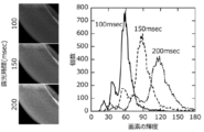
- FIG. 4 is a diagram (left side) showing the digitized image, and a graph (right side) in which the complexity of the structure of the image is quantified by the standard deviation or variation coefficient of the image.
- Non-alcoholic fatty liver (Nash model) specimens stained with pigments of oil droplets in the cross section of mouse liver (upper image) with various light sources (top), standard deviation of the image, and It is the table
- the present invention acquires color information for all or a part of pixels in an entire area of a still image, or at least one area provided in the image, and at least two or more areas.
- the present invention calculates the diversity of color information consisting of pixels, the structure of the object in the image, the brightness of the image with the complexity of the color, or the diversity of the color within the area Analyzes and evaluates the complexity of structure and color.
- An apparatus for acquiring an image used in the present invention may be any apparatus that can record a captured image as digital data as image information, such as a digital camera system that enables coaxial illumination.
- a conventionally known one may be used as appropriate.
- the image acquisition device is physically or logically connected to the image analysis evaluation device according to the present invention.
- the image analysis evaluation apparatus includes processing means for calculating and processing data, and storage means for storing still image data acquired by the image acquisition means.
- the storage means is for implementing the present invention.
- a computer program and predetermined data are stored, respectively, and the processing means processes data according to a predetermined command by the computer program or the like.
- Color information is acquired from the image data obtained from the image acquisition device, the diversity of the color information is calculated, and used as an index for evaluating the state of the subject surface. Therefore, as the color information used in the present invention, a method using three color elements of red, green, and blue (numeric values in the RGB color space), which is a method used in many electronic image devices, will be described below. To do.
- the color information of each pixel is the luminance or brightness of the red, green, and blue color elements of the pixel, and the luminance of at least one or more of the red, green, and blue color elements in the RGB color space. May be used as they are, or secondary numerical values calculated from the color information may be obtained and used.
- a predetermined coefficient for example, red “0.298912”, green “0.586611”, blue, A method is known in which “0.114478” is multiplied and then added to calculate, but a grayscale gradation processed by a predetermined method as described above may be used.
- any one of the luminances in each color element may be used alone, or may be calculated by combining two or more.
- two or more color elements may be simply averaged or may be given by multiplying each element by a different predetermined weighting factor.
- the coefficient of variation is calculated individually from the three color elements of red, green, and blue, and the maximum value, the minimum value, or the second largest value is used. May be.
- the above is the image color information acquisition method using numerical values in the RGB color space.
- the color information used in the present invention is defined by the HSV color space, HSB color space, HLS color space, or HSL color space. Luminance or brightness may be used.
- a method for examining how much variation exists in an area for at least one element of color information will be described. It is obtained by examining the color information of the RGB color space for each pixel in a predetermined area in the image and evaluating how much the intensity of at least one color element of red, green, and blue varies within the area. .
- a value obtained by dividing the variance value obtained from the value of each element by the average value of the luminance of the pixels in the area may be used, and a value (standard deviation, etc.) based on the deviation calculated therefrom is used in the area.
- a value divided by the average value of the luminances of the pixels, that is, a variation coefficient may be used. These values may be arbitrarily selected and used as necessary.
- ⁇ Target area for color diversity calculation in image> The diversity of the color information obtained in this way may be calculated for the entire image, or alternatively, the image may be arbitrarily divided into a grid and the color diversity for each area within the grid May be calculated.
- each pixel in the image or some adjacent pixels may be grouped into a pixel area, and the diversity of color information may be obtained for the surrounding pixels or pixel area.
- the diversity of the color information obtained in this way has the advantage that the resolution is higher than that of the above-described method for dividing the image into a grid pattern, and that the spatial position information completely matches the original still image.
- the immediacy is low because of the large calculation cost. For this reason, it is preferable to use it when a detailed analysis is performed on an image once recorded.
- the original still image may be enlarged or reduced as appropriate.
- a method of enlargement a method of complementing the luminance of pixels filling the gap with a function such as a bilinear method, a bicubic method, a Lanczos method is preferable.
- a reduction method any method such as nearest neighbor method, bilinear method, bicubic method, and Lanczos method may be used. These can be used as needed.
- the color diversity obtained for each region such as a grid, pixel, or pixel set from a still image is an image that shows the state of structures and color complexity in the image by arranging in two dimensions. May be stored and displayed.
- the image may be expressed in grayscale that increases or decreases the brightness depending on the diversity value, and is obtained based on the diversity value to make the difference easier to recognize.
- a heat map color may be used, and in this way, it is possible to emphasize and display a complicated portion of the structure in the image.
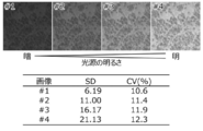
- Fig. 1 is a plot of the number of pixels for each luminance of the image pixels, taking a part of the plastic part with the surface processed with various exposure times.
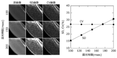
- FIG. 2 shows the standard deviation (SD) of the luminance of the color element of the pixel at this time and the coefficient of variation (CV (%)) obtained by dividing the standard deviation by the average value.
- SD standard deviation
- CV coefficient of variation
- the liver of a mouse that developed non-alcoholic fatty liver was cross-sectioned, and the fat contained was stained with Oil-RedO and photographed under a microscope using illumination light sources of various brightness.
- SD standard deviation
- CV coefficient of variation
- the standard deviation increased as the illumination light source was brightened as shown in the table in the figure.
- the coefficient of variation has a constant value regardless of the brightness of the illumination light source. This indicates that the coefficient of variation can be used as a numerical value that is not easily affected by the brightness of the illumination light source.
- the present invention relates to an organ constituting a living body, or an organ, and a tissue itself, or a cross section thereof, and a structure included in an image obtained by imaging biological and medical samples such as cultured cells.
- Cosmetic evaluation such as wrinkle analysis of color complexity and surface condition of human skin and nails, evaluation of surface liquid layer state (oil film and moisturizing state) using interference fringes reflected in image data at the time of shooting, It is an image analysis method that can be applied to a wide range of applications, such as material distortion analysis, astronomical photography, and aerial photography.
Landscapes
- Engineering & Computer Science (AREA)
- Computer Vision & Pattern Recognition (AREA)
- Physics & Mathematics (AREA)
- General Physics & Mathematics (AREA)
- Theoretical Computer Science (AREA)
- Image Analysis (AREA)
- Investigating Or Analysing Materials By Optical Means (AREA)
Abstract
Priority Applications (4)
| Application Number | Priority Date | Filing Date | Title |
|---|---|---|---|
| EP18794142.2A EP3629286A4 (fr) | 2017-05-01 | 2018-04-27 | Procédé d'évaluation d'analyse d'image, programme informatique et dispositif d'évaluation d'analyse d'image |
| CN201880028917.9A CN110603566B (zh) | 2017-05-01 | 2018-04-27 | 图像解析评价方法、计算机程序、图像解析评价装置 |
| JP2019515714A JP7171549B2 (ja) | 2017-05-01 | 2018-04-27 | 画像解析評価方法、コンピュータプログラム、画像解析評価装置 |
| US16/609,669 US11200699B2 (en) | 2017-05-01 | 2018-04-27 | Image analysis evaluation method, computer program, and image analysis evaluation device |
Applications Claiming Priority (2)
| Application Number | Priority Date | Filing Date | Title |
|---|---|---|---|
| JP2017090950 | 2017-05-01 | ||
| JP2017-090950 | 2017-05-01 |
Publications (1)
| Publication Number | Publication Date |
|---|---|
| WO2018203514A1 true WO2018203514A1 (fr) | 2018-11-08 |
Family
ID=64016103
Family Applications (1)
| Application Number | Title | Priority Date | Filing Date |
|---|---|---|---|
| PCT/JP2018/017114 WO2018203514A1 (fr) | 2017-05-01 | 2018-04-27 | Procédé d'évaluation d'analyse d'image, programme informatique et dispositif d'évaluation d'analyse d'image |
Country Status (5)
| Country | Link |
|---|---|
| US (1) | US11200699B2 (fr) |
| EP (1) | EP3629286A4 (fr) |
| JP (1) | JP7171549B2 (fr) |
| CN (1) | CN110603566B (fr) |
| WO (1) | WO2018203514A1 (fr) |
Families Citing this family (3)
| Publication number | Priority date | Publication date | Assignee | Title |
|---|---|---|---|---|
| JP7248024B2 (ja) * | 2018-06-28 | 2023-03-29 | 株式会社ニコン | 細胞状態評価装置、顕微鏡装置、細胞状態評価方法およびプログラム |
| CN112837319B (zh) * | 2021-03-29 | 2022-11-08 | 深圳大学 | 真实失真图像质量智能评价方法、装置、设备及介质 |
| CN116151636B (zh) * | 2023-04-23 | 2023-09-15 | 中铁建工集团建筑安装有限公司 | 一种动态线性移动的视觉观览的评价方法 |
Citations (4)
| Publication number | Priority date | Publication date | Assignee | Title |
|---|---|---|---|---|
| JPH08308634A (ja) | 1995-05-23 | 1996-11-26 | Pola Chem Ind Inc | 肌の評価装置 |
| JPH0938045A (ja) | 1995-05-23 | 1997-02-10 | Pola Chem Ind Inc | 肌の評価方法 |
| JP2006141734A (ja) * | 2004-11-19 | 2006-06-08 | Olympus Corp | 画像処理装置、画像処理方法、及び画像処理プログラム |
| JP2014104132A (ja) * | 2012-11-27 | 2014-06-09 | Kao Corp | 肌画像分析装置及び肌画像分析方法 |
Family Cites Families (17)
| Publication number | Priority date | Publication date | Assignee | Title |
|---|---|---|---|---|
| JP3681956B2 (ja) * | 1999-05-31 | 2005-08-10 | 日本バイリーン株式会社 | 被測定物の状態評価方法および状態評価装置 |
| JP2001128017A (ja) * | 1999-10-27 | 2001-05-11 | Fuji Xerox Co Ltd | 画像評価装置および画像評価方法 |
| US7565007B2 (en) * | 2001-01-25 | 2009-07-21 | Nikon Corporation | Image processing method, image processing program, and image processing apparatus |
| JP2005174133A (ja) * | 2003-12-12 | 2005-06-30 | Fuji Xerox Co Ltd | 画像評価装置、画像評価方法およびプログラム |
| CN1604139A (zh) * | 2004-10-28 | 2005-04-06 | 上海交通大学 | 图像融合评价系统的构建方法 |
| JP2006318181A (ja) * | 2005-05-12 | 2006-11-24 | Fuji Photo Film Co Ltd | 画像評価方法、装置及びプログラム |
| JP2007208940A (ja) * | 2006-02-06 | 2007-08-16 | Canon Inc | 色評価用カラーチャートデータの作成方法及びその情報処理装置 |
| JP5119003B2 (ja) * | 2007-11-30 | 2013-01-16 | 大王製紙株式会社 | 使い捨ておむつ |
| CN101609500B (zh) * | 2008-12-01 | 2012-07-25 | 公安部第一研究所 | 出入境数字人像相片质量评估方法 |
| JP2010268426A (ja) * | 2009-04-15 | 2010-11-25 | Canon Inc | 画像処理装置、画像処理方法およびプログラム |
| US20150141278A1 (en) * | 2012-05-30 | 2015-05-21 | Clarient Diagnostic Services, Inc. | Multiplexed assay method for lung cancer classification |
| JP5616989B2 (ja) * | 2013-02-14 | 2014-10-29 | 東芝テリー株式会社 | 画像処理装置および閾値設定処理プログラム |
| JP6282123B2 (ja) * | 2014-01-23 | 2018-02-21 | キヤノン株式会社 | 画像処理装置、画像処理方法及びプログラム |
| KR102406215B1 (ko) * | 2016-02-17 | 2022-06-08 | 삼성전자주식회사 | 컬러 프린지 보정 방법 및 이를 이용한 이미지 데이터의 프로세싱 방법 |
| JP6814799B2 (ja) * | 2016-05-30 | 2021-01-20 | 興和株式会社 | 涙液状態評価方法およびその装置 |
| EP3620103B1 (fr) * | 2017-05-01 | 2023-11-01 | Kyoto Prefectural Public University Corporation | Procédé et dispositif d'évaluation de la dynamique d'une couche de liquide lacrymal |
| JP7010057B2 (ja) * | 2018-02-26 | 2022-01-26 | オムロン株式会社 | 画像処理システムおよび設定方法 |
-
2018
- 2018-04-27 JP JP2019515714A patent/JP7171549B2/ja active Active
- 2018-04-27 CN CN201880028917.9A patent/CN110603566B/zh active Active
- 2018-04-27 EP EP18794142.2A patent/EP3629286A4/fr active Pending
- 2018-04-27 WO PCT/JP2018/017114 patent/WO2018203514A1/fr unknown
- 2018-04-27 US US16/609,669 patent/US11200699B2/en active Active
Patent Citations (4)
| Publication number | Priority date | Publication date | Assignee | Title |
|---|---|---|---|---|
| JPH08308634A (ja) | 1995-05-23 | 1996-11-26 | Pola Chem Ind Inc | 肌の評価装置 |
| JPH0938045A (ja) | 1995-05-23 | 1997-02-10 | Pola Chem Ind Inc | 肌の評価方法 |
| JP2006141734A (ja) * | 2004-11-19 | 2006-06-08 | Olympus Corp | 画像処理装置、画像処理方法、及び画像処理プログラム |
| JP2014104132A (ja) * | 2012-11-27 | 2014-06-09 | Kao Corp | 肌画像分析装置及び肌画像分析方法 |
Also Published As
| Publication number | Publication date |
|---|---|
| EP3629286A4 (fr) | 2021-01-13 |
| CN110603566B (zh) | 2023-10-20 |
| CN110603566A (zh) | 2019-12-20 |
| JP7171549B2 (ja) | 2022-11-15 |
| US20200151909A1 (en) | 2020-05-14 |
| EP3629286A1 (fr) | 2020-04-01 |
| US11200699B2 (en) | 2021-12-14 |
| JPWO2018203514A1 (ja) | 2020-03-12 |
Similar Documents
| Publication | Publication Date | Title |
|---|---|---|
| US7454046B2 (en) | Method and system for analyzing skin conditions using digital images | |
| US20200098105A1 (en) | Methods and Systems for Assessing Cell Morphology | |
| Ganesan et al. | Segmentation and edge detection of color images using CIELAB color space and edge detectors | |
| JP5544764B2 (ja) | 画像処理装置および方法、並びにプログラム | |
| US20080080766A1 (en) | Apparatus and Method for Analyzing Skin Using L*a*b* Colorspace | |
| Pech et al. | Abundance estimation of rocky shore invertebrates at small spatial scale by high-resolution digital photography and digital image analysis | |
| JP6814799B2 (ja) | 涙液状態評価方法およびその装置 | |
| WO2018203514A1 (fr) | Procédé d'évaluation d'analyse d'image, programme informatique et dispositif d'évaluation d'analyse d'image | |
| Yu et al. | A false color image fusion method based on multi-resolution color transfer in normalization YCBCR space | |
| US8559714B2 (en) | Post processing for improved generation of intrinsic images | |
| Bautista et al. | Improving the visualization and detection of tissue folds in whole slide images through color enhancement | |
| CN101523169A (zh) | 用于使用L*a*b*色空间来分析皮肤的装置和方法 | |
| JP2006507579A (ja) | 核多形性の組織学的評価 | |
| US11210791B2 (en) | Computer-implemented method for locating possible artifacts in a virtually stained histology image | |
| Madooei et al. | Detecting specular highlights in dermatological images | |
| Bautista et al. | Digital staining of unstained pathological tissue samples through spectral transmittance classification | |
| JP5203159B2 (ja) | 画像処理方法、画像処理システムおよび画像処理プログラム | |
| WO2019181072A1 (fr) | Procédé de traitement d'image, programme informatique et support d'enregistrement | |
| JP5860970B2 (ja) | 固有画像の生成を改良するための後処理 | |
| Sahidan et al. | Local and global contrast stretching for color contrast enhancement on ziehl-neelsen tissue section slide images | |
| JP2017012384A (ja) | シワ状態分析装置及びシワ状態分析方法 | |
| JP6730051B2 (ja) | 肌状態評価方法 | |
| Bautista et al. | Multispectral enhancement towards digital staining | |
| JP2011030592A (ja) | 皮脂の分泌状態測定装置 | |
| Lecca | Color vision is a spatial process: The Retinex theory |
Legal Events
| Date | Code | Title | Description |
|---|---|---|---|
| 121 | Ep: the epo has been informed by wipo that ep was designated in this application |
Ref document number: 18794142 Country of ref document: EP Kind code of ref document: A1 |
|
| ENP | Entry into the national phase |
Ref document number: 2019515714 Country of ref document: JP Kind code of ref document: A |
|
| NENP | Non-entry into the national phase |
Ref country code: DE |
|
| ENP | Entry into the national phase |
Ref document number: 2018794142 Country of ref document: EP Effective date: 20191202 |
|
| ENP | Entry into the national phase |
Ref document number: 2018794142 Country of ref document: EP Effective date: 20191202 |