WO2018199186A1 - 造血前駆細胞マーカー - Google Patents
造血前駆細胞マーカー Download PDFInfo
- Publication number
- WO2018199186A1 WO2018199186A1 PCT/JP2018/016864 JP2018016864W WO2018199186A1 WO 2018199186 A1 WO2018199186 A1 WO 2018199186A1 JP 2018016864 W JP2018016864 W JP 2018016864W WO 2018199186 A1 WO2018199186 A1 WO 2018199186A1
- Authority
- WO
- WIPO (PCT)
- Prior art keywords
- cells
- positive
- cell
- cell population
- fraction
- Prior art date
- Legal status (The legal status is an assumption and is not a legal conclusion. Google has not performed a legal analysis and makes no representation as to the accuracy of the status listed.)
- Ceased
Links
Classifications
-
- C—CHEMISTRY; METALLURGY
- C12—BIOCHEMISTRY; BEER; SPIRITS; WINE; VINEGAR; MICROBIOLOGY; ENZYMOLOGY; MUTATION OR GENETIC ENGINEERING
- C12N—MICROORGANISMS OR ENZYMES; COMPOSITIONS THEREOF; PROPAGATING, PRESERVING, OR MAINTAINING MICROORGANISMS; MUTATION OR GENETIC ENGINEERING; CULTURE MEDIA
- C12N5/00—Undifferentiated human, animal or plant cells, e.g. cell lines; Tissues; Cultivation or maintenance thereof; Culture media therefor
- C12N5/06—Animal cells or tissues; Human cells or tissues
- C12N5/0602—Vertebrate cells
- C12N5/0634—Cells from the blood or the immune system
- C12N5/0636—T lymphocytes
-
- C—CHEMISTRY; METALLURGY
- C12—BIOCHEMISTRY; BEER; SPIRITS; WINE; VINEGAR; MICROBIOLOGY; ENZYMOLOGY; MUTATION OR GENETIC ENGINEERING
- C12N—MICROORGANISMS OR ENZYMES; COMPOSITIONS THEREOF; PROPAGATING, PRESERVING, OR MAINTAINING MICROORGANISMS; MUTATION OR GENETIC ENGINEERING; CULTURE MEDIA
- C12N5/00—Undifferentiated human, animal or plant cells, e.g. cell lines; Tissues; Cultivation or maintenance thereof; Culture media therefor
- C12N5/06—Animal cells or tissues; Human cells or tissues
- C12N5/0602—Vertebrate cells
- C12N5/0634—Cells from the blood or the immune system
- C12N5/0636—T lymphocytes
- C12N5/0638—Cytotoxic T lymphocytes [CTL] or lymphokine activated killer cells [LAK]
-
- C—CHEMISTRY; METALLURGY
- C12—BIOCHEMISTRY; BEER; SPIRITS; WINE; VINEGAR; MICROBIOLOGY; ENZYMOLOGY; MUTATION OR GENETIC ENGINEERING
- C12N—MICROORGANISMS OR ENZYMES; COMPOSITIONS THEREOF; PROPAGATING, PRESERVING, OR MAINTAINING MICROORGANISMS; MUTATION OR GENETIC ENGINEERING; CULTURE MEDIA
- C12N5/00—Undifferentiated human, animal or plant cells, e.g. cell lines; Tissues; Cultivation or maintenance thereof; Culture media therefor
- C12N5/06—Animal cells or tissues; Human cells or tissues
- C12N5/0602—Vertebrate cells
- C12N5/0652—Cells of skeletal and connective tissues; Mesenchyme
- C12N5/0657—Cardiomyocytes; Heart cells
-
- C—CHEMISTRY; METALLURGY
- C12—BIOCHEMISTRY; BEER; SPIRITS; WINE; VINEGAR; MICROBIOLOGY; ENZYMOLOGY; MUTATION OR GENETIC ENGINEERING
- C12N—MICROORGANISMS OR ENZYMES; COMPOSITIONS THEREOF; PROPAGATING, PRESERVING, OR MAINTAINING MICROORGANISMS; MUTATION OR GENETIC ENGINEERING; CULTURE MEDIA
- C12N5/00—Undifferentiated human, animal or plant cells, e.g. cell lines; Tissues; Cultivation or maintenance thereof; Culture media therefor
- C12N5/06—Animal cells or tissues; Human cells or tissues
- C12N5/0602—Vertebrate cells
- C12N5/0696—Artificially induced pluripotent stem cells, e.g. iPS
-
- G—PHYSICS
- G01—MEASURING; TESTING
- G01N—INVESTIGATING OR ANALYSING MATERIALS BY DETERMINING THEIR CHEMICAL OR PHYSICAL PROPERTIES
- G01N33/00—Investigating or analysing materials by specific methods not covered by groups G01N1/00 - G01N31/00
- G01N33/48—Biological material, e.g. blood, urine; Haemocytometers
- G01N33/50—Chemical analysis of biological material, e.g. blood, urine; Testing involving biospecific ligand binding methods; Immunological testing
- G01N33/53—Immunoassay; Biospecific binding assay; Materials therefor
- G01N33/569—Immunoassay; Biospecific binding assay; Materials therefor for microorganisms, e.g. protozoa, bacteria, viruses
- G01N33/56966—Animal cells
-
- C—CHEMISTRY; METALLURGY
- C12—BIOCHEMISTRY; BEER; SPIRITS; WINE; VINEGAR; MICROBIOLOGY; ENZYMOLOGY; MUTATION OR GENETIC ENGINEERING
- C12N—MICROORGANISMS OR ENZYMES; COMPOSITIONS THEREOF; PROPAGATING, PRESERVING, OR MAINTAINING MICROORGANISMS; MUTATION OR GENETIC ENGINEERING; CULTURE MEDIA
- C12N2501/00—Active agents used in cell culture processes, e.g. differentation
- C12N2501/50—Cell markers; Cell surface determinants
- C12N2501/505—CD4; CD8
-
- C—CHEMISTRY; METALLURGY
- C12—BIOCHEMISTRY; BEER; SPIRITS; WINE; VINEGAR; MICROBIOLOGY; ENZYMOLOGY; MUTATION OR GENETIC ENGINEERING
- C12N—MICROORGANISMS OR ENZYMES; COMPOSITIONS THEREOF; PROPAGATING, PRESERVING, OR MAINTAINING MICROORGANISMS; MUTATION OR GENETIC ENGINEERING; CULTURE MEDIA
- C12N2506/00—Differentiation of animal cells from one lineage to another; Differentiation of pluripotent cells
- C12N2506/11—Differentiation of animal cells from one lineage to another; Differentiation of pluripotent cells from blood or immune system cells
Definitions
- the present invention relates to a method for producing CD8 positive cells using one or more hematopoietic progenitor cell markers (HPC markers), a cell population expressing one or more HPC markers, and the like.
- HPC markers hematopoietic progenitor cell markers
- T cells play a central role in the immune system against foreign pathogens such as bacteria and viruses and abnormal cells such as cancer cells.
- CTL cytotoxic T cells
- TCR T cell receptors
- CD8 single positive (SP) cells are CD8 single positive (SP) cells.
- the CD8SP cell is an immature cell that does not express TCR and has neither CD4 nor CD8 in the thymus (CD4 / CD8 double negative (in this specification, double negative may be referred to as DN).
- DN double negative
- DP double positives
- T cells play a central role in the immune system, so if T cells can be replenished and regenerated, they are extremely useful for the prevention or treatment of diseases such as tumors, infections, and autoimmune disorders. It becomes an effective means.
- hematopoietic progenitor cells and T cells can be obtained by a method of inducing hematopoietic progenitor cells (HematopoieticgenProgenitor Cell; sometimes referred to as HPC in this specification) from iPS cells or a method of inducing CD4 / CD8DP cells. Attempts have been made to obtain lineage cells.
- Non-Patent Document 1 describes that CD34 / CD43DP cells can be induced by co-culturing iPS cells with feeder cells (specifically, OP9 cells).
- Patent Document 1 and Non-Patent Document 2 describe a method for separating HPC from a cell population containing HPC derived from human pluripotent stem cells using CD43 and CD34, CD31 or CD144 as indicators, and the separated cells. It is described that the properties of induced hematopoietic cells differ depending on the method of differentiation into cells of the T cell lineage and whether or not Activin / Nodal is activated in the early stage of induction when differentiating from pluripotent stem cells to HPC. Has been.
- Patent Document 2 describes a method for producing CD4 / CD8DP under feeder-free conditions.
- Non-patent document 3 describes various methods for inducing blood cells including hematopoietic progenitor cells from human pluripotent stem cells and the reason why clinical use of blood cells induced in vitro has not progressed. Low induction efficiency is mentioned.
- the present invention provides cells of T cell lineage (eg, CD8 positive cells (eg, CD4 / CD8DP cells, CD8SP cells)) at higher and / or higher concentrations in order to stably provide cell therapy ( That is, an object is to provide a method for producing the cells. Moreover, this invention makes it a subject to provide the cell population containing the hematopoietic progenitor cell for solving the said subject.
- CD8 positive cells eg, CD4 / CD8DP cells, CD8SP cells
- the present inventors have identified a marker (HPC marker) that is expressed on the surface of HPC in order to develop a method for producing CD4 / CD8DP cells that solve the above problems. Furthermore, HPC isolated from a cell population containing HPC using one or more of the markers has high efficiency of differentiating into CD8 positive cells (eg, CD4 / CD8DP cells) and has a high proliferation ability, ie, 1 It has been found that by using more than one HPC marker, more CD8 positive cells (eg, CD4 / CD8DP cells) can be obtained and / or at higher concentrations within the cell population. Furthermore, it has been found that the production method of the present invention can provide stable CD8 positive cells (eg, CD4 / CD8DP cells) with little variation in yield, and the present invention has been completed.
- HPC marker a marker that is expressed on the surface of HPC in order to develop a method for producing CD4 / CD8DP cells that solve the above problems.
- Step 1 From a cell population containing hematopoietic progenitor cells, from CD24, CD62L, CD90, CD143, CD263, Notch3, CD32, CD39, CD49a, CD164, CD317, CD200, CD218a, CD7, CD144, CD56, CD226, CD262, and CD325
- a step 2 a step of differentiating the cells separated in step 1 into CD4 / CD8 double positive cells
- the cells selected in step 1 are CD235a And the method according to [1], wherein the cell does not express CD14 and
- step 1 from the cell population containing hematopoietic progenitor cells, CD24, CD62L, CD90, CD143, CD263, Notch3, CD32, CD39, CD49a, CD164, CD317, CD200, CD218a, CD7, CD144, CD56, CD226
- the first group shown in the step 1 is a group consisting of CD24, CD62L, CD90, CD143, CD263, Notch3, CD200, CD218a, CD7 and CD144.
- step 1 one or more molecules selected from the second group consisting of CD49f, CD51, CD102, CD42b, CD61, CD62P, CD69, CD102 and CD156c are selected from a cell population containing hematopoietic progenitor cells.
- the method according to [5] wherein the second group shown in Step 1 is a group consisting of CD49f, CD51 and CD102.
- step 7 The method according to any one of [1] to [6], wherein the cell population containing the hematopoietic progenitor cells in Step 1 is a cell population obtained by inducing differentiation of pluripotent stem cells.
- the method of inducing includes, for example, a method comprising culturing CD4 / CD8 double positive cells in the presence of an adrenocortical hormone agent, an antibody and / or a cytokine, preferably the adrenal cortex
- the pluripotent stem cells are human induced pluripotent stem cells.
- [15] Selected from the first group consisting of CD24, CD62L, CD90, CD143, CD263, Notch3, CD32, CD39, CD49a, CD164, CD317, CD200, CD218a, CD7, CD144, CD56, CD226, CD262 and CD325 Hematopoietic progenitor cells that express one or more molecules and / or one or more molecules selected from the second group consisting of CD49f, CD51, CD102, CD42b, CD61, CD62P, CD69, CD102 and CD156c A cell population in which the ratio of hematopoietic progenitor cells (the number of hematopoietic progenitor cells / the total number of cells contained in the cell population) is 40% or more.
- the cell population according to [15], wherein [17] The cell population according to [15], wherein the ratio is a ratio of hematopoietic progenitor cells that do not further express one or more molecules selected from the second group consisting of CD49f, CD51, and CD102.
- a reagent for separating hematopoietic progenitor cells comprising an antibody against each of one or more molecules selected from the group consisting of CD62P, CD69, CD102 and CD156c. [19] The reagent according to [18], further comprising one or more antibodies against CD235a, CD14, CD45, CD34 and / or CD43.
- a method for detecting a surface protein of a hematopoietic progenitor cell comprising the hematopoietic progenitor cell, CD24, CD62L, CD90, CD143, CD263, Notch3, CD32, CD39, CD49a, CD164, CD317, CD200, CD218a, CD7 , CD144, CD56, CD226, CD262, CD325, CD49f, CD51, CD102, CD42b, CD61, CD62P, CD69, CD102, CD109 or CD156c.
- the substance comprises an antibody or a fragment thereof.
- the substance further contains a fluorophore.
- a method for selecting hematopoietic progenitor cells from a cell population containing hematopoietic progenitor cells, the presence or absence of one or more proteins on the surface of cells by any of the methods [20] to [24] And (i) CD45, CD34 and / or CD43 and the following proteins: CD24, CD62L, C90, CD143, CD263, Notch3, CD32, CD39, CD49a, CD164, CD317, CD200, CD218a, CD7, CD144, CD56, CD226, CD262 and CD325: any one or any combination of these are detected on the surface of the cells and / or (ii) When CD14 and / or CD235a is not detected on the surface of the cell, and the following proteins: CD49f, CD51, CD102, CD42b, CD61, CD62P, CD69, CD102, CD109 and CD156c: are not detected And selecting the cells.
- CD8 positive cells for example, CD4 / CD8DP cells, CD8SP cells
- CD8 positive cells for example, CD4 / CD8DP cells, CD8SP cells
- the production method of the present invention can provide CD8 positive cells (for example, CD4 / CD8DP cells, CD8SP cells) stably with little variation in yield. Accordingly, the production method of the present invention provides an improved method for separating hematopoietic progenitor cells from a cell population. This provides an improved cell source for generating CD8 positive cells.
- This novel production method is surprising to the inventors that certain HPC positive markers and HPC negative markers can be used to greatly improve the quality and / or quantity of a CD8 positive cell population derived from HPC. And based on unexpected findings.
- This beneficial source of CD8 positive cells of the present invention provides an improved CD8 positive cell population for clinical applications (eg, cell therapy, diagnostic purposes, and in vitro applications).
- the present invention is (step 1) a cell expressing one or more hematopoietic progenitor cell markers (sometimes abbreviated as “HPC marker” in the present specification) from a cell population containing hematopoietic progenitor cells, and / or Alternatively, a step of separating cells that do not express one or more HPC markers, and (step 2) a cell separated by step 1 is a CD8 positive cell (for example, CD4 / CD8 double positive cell (hereinafter referred to as “CD4 A method of producing CD8 positive cells (for example, CD4 / CD8DP cells) (hereinafter abbreviated as “production method of the present invention”), which includes a step of differentiation into “/ CD8DP cells”. To do.
- CD8 positive cell for example, CD4 / CD8 double positive cell
- production method of the present invention production method of the present invention
- CD8 positive cell means a T cell expressing CD8.
- the cell include CD4 / CD8DP cell and CD8 single positive cell (herein, “CD8SP cell”). May be abbreviated as ")".
- CD4 / CD8DP cell means a T cell that expresses both CD4 and CD8, and “CD8SP cell” means a T cell that does not express CD4 but expresses CD8. means.
- Examples of CD8SP cells include cytotoxic T cells and their progenitor cells.
- T cell means, for example, a cell expressing an antigen receptor called T cell receptor (TCR) on its surface and its precursor cell (eg, TCR). (Pro T cells not expressing (TCR ⁇ ), pre-T cells in which TCR ⁇ and pre-TCR ⁇ are associated).
- TCR T cell receptor
- hematopoietic progenitor cell means a CD34 positive cell, preferably a CD34 / CD43DP cell.
- the origin of hematopoietic progenitor cells used in the present invention is not limited.
- hematopoietic progenitor cells induced in vitro by known methods for example, the methods described in Patent Documents 1 and 2 and Non-Patent Documents 1 to 3.
- hematopoietic progenitor cells obtained by inducing differentiation of pluripotent stem cells or hematopoietic progenitor cells isolated from living tissue by a known technique may be used.
- pluripotent stem cells examples include embryonic stem cells (embryonic stem cells: ES cells), induced pluripotent stem cells (induced PS cells), embryonic tumor cells (EC cells), embryonic germ stem cells (EG cells) are mentioned, and iPS cells (more preferably human iPS cells) are preferred.
- ES cells embryonic stem cells
- induced PS cells induced pluripotent stem cells
- EC cells embryonic tumor cells
- EG cells embryonic germ stem cells
- iPS cells more preferably human iPS cells
- the pluripotent stem cell is any cell derived from an ES cell or a human embryo, the cell is a cell prepared without destroying the embryo, even if the cell is prepared by destroying the embryo. However, it is preferably a cell produced without destroying the embryo.
- the iPS cell can be produced by introducing a specific reprogramming factor into a somatic cell in the form of DNA or protein, and has almost the same characteristics as an ES cell, such as pluripotency and proliferation ability by self-replication.
- a specific reprogramming factor such as pluripotency and proliferation ability by self-replication.
- the iPS cells may be prepared from somatic cells by a method known per se, or iPS cells that have already been established and stocked may be used.
- the somatic cells from which iPS cells used in the present invention are derived are not limited, but are preferably cells derived from peripheral blood or cells derived from umbilical cord blood.
- a pluripotent stem cell is derived,
- mammals such as a mouse
- Hematopoietic progenitor cells can be produced by subjecting the pluripotent stem cells to a method known per se.
- the pluripotent stem cell is an iPS cell
- hematopoietic progenitor cells can be produced by, for example, the method described in International Publication No. 2017/221975.
- the biological tissue is not limited as long as it contains hematopoietic progenitor cells, and examples thereof include peripheral blood, lymph nodes, bone marrow, thymus, spleen, and umbilical cord blood.
- peripheral blood and umbilical cord blood are preferable from the viewpoint of low invasiveness to animals and easy preparation.
- the hematopoietic progenitor cells and pluripotent stem cells used in the present invention are preferably GMP (Good Manufacturing Practice) standard cells from the viewpoint of application to therapy.
- ⁇ HPC marker '' is a molecule that can be expressed on the surface of hematopoietic progenitor cells, CD24, CD62L, CD90, CD143, CD263, Notch3, CD32, CD39, CD49a, CD164, CD317, CD200, CD218a, Any molecule of CD7, CD144, CD56, CD226, CD262, and CD325 (in the specification, these markers may be referred to as “HPC positive marker”, “positive HPC selection marker”, or “positive HPC marker”)
- HPC positive marker any molecule of CD49f, CD51, CD102, CD42b, CD61, CD62P, CD69, CD102 and CD156c, which is a molecule that is not normally expressed on the surface of hematopoietic progenitor cells
- these markers are referred to as “HPC negative Marker ",” negative HPC selectable marker ", or” negative HPC marker ” (in the specification, HPC positive marker and HPC negative marker are
- HPC positive markers of the present invention CD24, CD62L, CD90, CD143, CD263, Notch3, CD200, CD218a, CD200, CD7 or CD144 are preferred, and CD90, CD143, CD218a or CD200 is more preferred.
- CD24, CD62L, CD90, CD143, CD263, Notch3, CD32, CD39, CD49a, CD164, CD317, CD200 or CD218a are preferred, and CD24, CD62L, CD90, CD143, CD263, Notch3, CD200 or CD218a are preferred. More preferred.
- the cell expressing the HPC positive marker may be referred to as an HPC marker positive cell (or HPC marker positive cell).
- the cell in the case of a cell expressing a CD24 molecule, the cell is referred to as a CD24 positive cell.
- the HPC negative markers of the present invention CD49f, CD51 or CD102 is preferable.
- the cells that do not express the HPC negative marker may be referred to as HPC marker negative cells (or HPC marker negative cells).
- CD8 positive cells preferably CD4 / CD8 double positive cells, express (a) CD34 and CD43 and (b) CD62L, CD24, CD90, CD143 from a cell population comprising hematopoietic progenitor cells. , CD218a, CD263 or Notch3, and (c) by isolating cells that do not express CD235a and CD14.
- the CD8 positive cells preferably CD4 / CD8 double positive cells, express (a) CD34 and CD43 and (b) CD90, CD143, CD200, CD218a from a cell population comprising hematopoietic progenitor cells. Or (c) by isolating cells that do not express CD235a and CD14.
- the CD8 positive cells preferably CD4 / CD8 double positive cells, express (a) CD34 and CD43 and (b) CD7, CD144, CD56, CD226 from a cell population comprising hematopoietic progenitor cells. , And expressing either CD262 or CD325 and (c) isolating cells that do not express CD235a and CD14.
- the CD8 positive cells preferably CD4 / CD8 double positive cells, express (a) CD34 and CD43 and (b) CD49f, CD51, CD426, CD61 from a cell population comprising hematopoietic progenitor cells. , CD62P, CD69 or CD156c, and (c) is produced by isolating cells that do not express CD235a and CD14.
- “express” means that expression can be detected by a method used for separating cells
- “not expressed” means detection by the detection sensitivity of the method used for separating cells. It means you can't.
- Examples of the method used when separating cells include the following method using flow cytometry and mass cytometry, and magnetic cell separation.
- the cells separated by the above step 1 are cells that express one or more HPC positive markers and cells that do not express one or more HPC negative markers.
- the cells separated by the above step 1 are cells expressing CD34 and / or CD43 in addition to one or more HPC positive markers of the present invention, and / or one or more books. It is a cell that does not express the HPC negative marker of the invention and expresses CD34 and / or CD43.
- the cells to be separated are preferably CD235a negative, CD14 negative and / or CD45 positive.
- the step 1 comprises, from a cell population containing hematopoietic progenitor cells, CD235a negative, CD14 negative, CD45 positive, CD34 positive and CD43 positive, and one or more HPCs of the present invention. This is done by isolating positive marker positive and / or one or more HPC negative marker negative cells of the present invention.
- “separate cells” selects a cell that expresses one or more HPC positive markers and / or does not express one or more HPC negative markers, and isolates the selected cells.
- An embodiment and an embodiment in which the selected cell is subjected to the next differentiation step are included.
- each term such as “comprising” or “comprises” optionally includes “consistingconsistof” or “consists of”. May be replaced.
- Flow cytometry or mass cytometry is used as a method for separating cells expressing one or more HPC positive markers and / or not expressing one or more HPC negative markers from a cell population containing hematopoietic progenitor cells.
- Methods, magnetic cell separation methods, etc., and these methods can be carried out using known methods.
- a cell that expresses one or more HPC positive markers and / or a cell that does not express one or more HPC negative markers includes a cell (for example, an antibody) that specifically binds to each HPC marker. It can isolate
- Such substances include those to which a detectable label (eg, GFP) has been added, and those to which no label has been added.
- a detectable label eg, GFP
- the separation can be performed by further using a substance with a detectable label that directly or indirectly recognizes the substance.
- a substance with a detectable label that directly or indirectly recognizes the substance.
- the substance is an antibody
- a fluorescent dye, a metal isotope or a bead is directly or indirectly supported on the antibody, thereby labeling an HPC marker on the cell surface. It is possible to separate cells based on the label. Only one type of antibody or two or more types of antibodies may be used.
- Examples of a method for differentiating cells isolated from cells that contain hematopoietic progenitor cells as cells expressing one or more HPC positive markers of the present invention and / or not expressing HPC negative markers into CD8 positive cells include: Known methods (for example, methods described in International Publication No. 2016/076415, JournalJof Leukocyte Biology 96 (2016) 1165-1175, Cell Reports (2 (2012) 1722-1735, International Publication No. 2017/221975)) Can be mentioned.
- IMDM Iscove's Modified Dulbecco's Medium
- EMEM Eagle's Minimum Essential Medium
- ⁇ MEM Dulbecco's modified Eagle's Medium
- DMEM Dulbecco's modified Eagle's Medium
- Ham's F12 medium Ham's F12 medium
- RPMI 1640 medium Fischer's medium
- Neurobasal Medium Neurobasal Medium (Life Technologies), and mixed media thereof.
- the medium may or may not contain serum.
- the medium contains, for example, vitamin Cs (eg ascorbic acid, vitamin C phosphate), albumin, insulin, transferrin, selenium, fatty acids, trace elements, 2-mercaptoethanol, thiolglycerol, lipids, amino acids
- vitamin Cs eg ascorbic acid, vitamin C phosphate
- albumin e.g ascorbic acid, vitamin C phosphate
- insulin transferrin
- selenium fatty acids
- trace elements 2-mercaptoethanol
- thiolglycerol lipids
- amino acids amino acids
- substances such as L-glutamine, non-essential amino acids, vitamins, growth factors, low molecular weight compounds, antibiotics, antioxidants, pyruvate, buffers, inorganic salts, and cytokines may be included.
- the medium preferably contains a cytokine.
- cytokines include IL-7, FLT-3L, SCF, TPO, and combinations thereof, and FLT-3L and IL-7 are preferred.
- the concentration of IL-7 in the medium ranges from 1 ng / ml to 50 ng / ml (eg, 1 ng / ml, 2 ng / ml, 3 ng / ml, 4 ng / ml, 5 ng / ml, 6 ng / ml).
- the concentration of FLT-3L in the medium ranges from 1 ng / ml to 100 ng / ml (eg, 1 ng / ml, 2 ng / ml, 3 ng / ml, 4 ng / ml, 5 ng / ml, 6 ng / ml).
- the medium preferably contains Notch ligand (eg, Dll1, Dll4, Dll1, or a fusion protein of Dll4 and Fc). When using a Notch ligand, it may be added to the medium or coated on the culture vessel. Alternatively, feeder cells that express a Notch ligand may be used.
- Notch ligand eg, Dll1, Dll4, Dll1, or a fusion protein of Dll4 and Fc.
- the hematopoietic progenitor cells obtained in step 1 may be subjected to adhesion culture or suspension culture.
- the culture vessel may be coated and used, or co-cultured with feeder cells or the like. From the viewpoint of application to treatment, it is preferably feeder-free.
- the substrate for coating a culture vessel include fibronectin fragments (eg, retronectin), pronectin, vitronectin, matrigel (BD), type I collagen, type IV collagen, gelatin, laminin, heparan sulfate proteoglycan, entactin, and These combinations are mentioned.
- feeder cells to be co-cultured examples include bone marrow stromal cell line OP9 cells, mouse mesenchymal cell line 10T1 / 2 cells, and Tst-4 cells.
- the exchange of the feeder cells can be performed by transferring the target cells in culture onto the feeder cells previously seeded. The exchange is preferably performed every 2 to 5 days.
- the culture temperature condition for inducing CD4 / CD8 DP cells by culturing hematopoietic progenitor cells is not particularly limited, but is preferably about 37 ° C to 42 ° C, more preferably about 37 to 39 ° C.
- the culture period can be appropriately determined by those skilled in the art while monitoring the number of CD4 / CD8DP cells.
- the number of culture days for inducing CD4 / CD8 DP cells from hematopoietic progenitor cells is not particularly limited, but it is 10 days or longer (eg, 12, 14, 16, 18, 20, or More than that), and preferably 60 days or less.
- CD4 / CD8DP cells can be efficiently obtained in large quantities by culturing for 21 to 28 days.
- the obtained CD4 / CD8DP cells may be isolated and used as a cell population containing other cell types (for example, CD8SP cells).
- CD4, CD8, CD3, CD45 molecules and the like can be used as an index.
- the isolation method a method well known to those skilled in the art can be used. For example, the method is performed by labeling with an antibody against a molecule as an index, and using the above-described flow cytometry or mass cytometry, magnetic cell separation method, Or the method of refine
- CD8SP cells can be produced by subjecting the CD4 / CD8DP cells obtained by the production method of the present invention to the step of inducing differentiation into CD8SP cells (a method for producing CD8SP cells in this way). May be abbreviated as “the method for producing the CD8SP cell of the present invention”). For the method of producing CD4 / CD8DP cells, see 1. As described in.
- basal medium and medium used in the method for producing CD8SP cells of the present invention the same basal medium and medium as described in 1. can be mentioned.
- the medium may contain a corticosteroid.
- corticosteroids include glucocorticoids and derivatives thereof, and the glucocorticoids include, for example, cortisone acetate, hydrocortisone, fludrocortisone acetate, prednisolone, triamcinolone, methylprednisolone, dexamethasone, betamethasone, Beclomethasone propionate is mentioned. Of these, dexamethasone is preferable.
- a method for producing CD8 single positive cells comprises culturing CD4 / CD8 double positive cells in the presence of an adrenocortical hormone agent, preferably dexamethasone, to obtain said CD4 / CD8 double positive cells. Inducing CD8 single positive cells.
- an adrenocortical hormone agent preferably dexamethasone
- dexamethasone When dexamethasone is used as a corticosteroid, its concentration in the culture solution is preferably 1 nM to 100 nM (eg, 1 nM, 5 nM, 10 nM, 20 nM, 30 nM, 40 nM, 50 nM, 60 nM, 70 nM, 80 nM, 90 nM, 100 nM) Of these, 10 nM is preferable.
- the medium may contain antibodies (eg, anti-CD3 antibody, anti-CD28 antibody, anti-CD2 antibody), cytokines (eg, IL-7, IL-2, IL-15) and the like.
- antibodies eg, anti-CD3 antibody, anti-CD28 antibody, anti-CD2 antibody
- cytokines eg, IL-7, IL-2, IL-15
- the anti-CD3 antibody used in the present invention is not particularly limited as long as it is an antibody that specifically recognizes CD3, and examples thereof include an antibody produced from an OKT3 clone.
- the anti-CD3 antibody may be one to which magnetic beads or the like are bound, and instead of adding the anti-CD3 antibody to the medium, the TCD is cultured on a culture container in which the anti-CD3 antibody is bound to the surface. Stimulation may be performed by culturing lymphocytes for a certain period of time.
- the concentration of the anti-CD3 antibody in the medium is 10 ng / ml to 1000 ng / ml (eg, 10 ng / ml, 50 ng / ml, 100 ng / ml, 200 ng / ml, 300 ng / ml, 400 ng / ml, 500 ng / ml, 600 ng / ml, 700 ng / ml, 800 ng / ml, 900 ng / ml, 1000 ng / ml) are preferable, and 500 ng / ml is particularly preferable.
- the concentration of other antibodies can also be appropriately determined by those skilled in the art based on culture conditions and the like.
- a method for producing CD8 single positive cells comprises culturing CD4 / CD8 double positive cells in the presence of an antibody, preferably an anti-CD3 antibody, to cultivate said CD4 / CD8 double positive cells to CD8. Inducing to single positive cells.
- the concentration of IL-2 used in the present invention in the medium is 10 U / ml to 1000 U / ml (example: 10 U / ml, 20 U / ml, 30 U / ml, 40 U / ml, 50 U / ml). 60 U / ml, 70 U / ml, 80 U / ml, 90 U / ml, 100 U / ml, 500 U / ml, 1000 U / ml), with 100 U / ml being preferred.
- the concentration of IL-7 or IL15 used in the present invention in the medium is 1 ng / ml to 100 ng / ml (eg, 1 ng / ml, 5 ng / ml, 10 ng / ml, 20 ng / ml, 30 ng).
- a method for producing CD8 single positive cells comprises culturing CD4 / CD8 double positive cells in the presence of a cytokine, preferably IL-2, to convert said CD4 / CD8 double positive cells to CD8. Inducing to single positive cells.
- a cytokine preferably IL-2
- the temperature condition for culturing CD4 / CD8DP cells is not particularly limited, but is preferably about 37 ° C to 42 ° C, more preferably about 37 to 39 ° C.
- the culture period can be appropriately determined by those skilled in the art while monitoring the number of CD8-positive T cells.
- the number of days is not particularly limited as long as CD8SP cells can be obtained, but it is preferably 1 day or more, 3 days or more, 7 days or more, and preferably 60 days or less.
- the CD8SP cells produced by the production method of the present invention are not limited to cells obtained by differentiating from CD4 / CD8DP cells (for example, cells obtained by the above 2.).
- a CD4 / CD8DP cell from a cell isolated from a cell population containing hematopoietic progenitor cells that expresses one or more HPC positive markers of the present invention and / or does not express one or more HPC negative markers.
- CD8SP cells induced without passing through are also included in the CD8SP cells produced by the production method of the present invention.
- Cell population containing hematopoietic progenitor cells expressing one or more types of HPC markers The present invention expresses one or more types of HPC positive markers and / or does not express one or more types of HPC negative markers. Is a high-frequency cell population (hereinafter abbreviated as “cell population of the present invention”).
- the cell population is, for example, a cell population containing hematopoietic progenitor cells, wherein cells that express one or more HPC positive markers and / or do not express one or more HPC negative markers are used according to the separation method described above. Can be obtained by separating them.
- the definition of HPC markers, specific molecules, and the definition of hematopoietic progenitor cells are as described above.
- the cell population of the present invention is a ratio of hematopoietic progenitor cells expressing one or more HPC positive markers and / or not expressing one or more HPC negative markers (the number of hematopoietic progenitor cells / total cells contained in the cell population). Number) is higher than the proportion contained in the hematopoietic progenitor cell population obtained by conventional methods (eg, CD34 positive, CD34 positive and CD43 positive, or CD43 positive and CD34, CD31 or CD144 positive) However, it is preferably 40% or more (eg, 50% or more, 60% or more, 70% or more, 80% or more, 90% or more, 100%).
- the cell population of the present invention can be obtained by the method described in Step 1 of the production method of the present invention.
- the percentage of hematopoietic progenitor cells that express one or more HPC positive markers and / or do not express one or more HPC negative markers can be measured by flow cytometry.
- the cell population of the present invention is compared to a cell population that expresses one or more HPC positive markers and / or does not isolate cells that do not express one or more HPC negative markers.
- the efficiency of differentiation into CD4 / CD8DP cells and CD8SP cells is high, and the proliferation ability is also excellent.
- Reagent for separation of "cell population of the present invention” The present invention also provides an HPC marker of the present invention, i.e., CD24, CD62L, CD90, CD143, CD263, Notch3, CD32, CD39, CD49a, CD164, CD317, CD200, CD218a, CD7, CD144, CD56, CD226, CD262, CD325, CD49f, CD51, CD102, CD42b, CD61, CD62P, CD69, CD102 and CD156c, preferably CD24, CD62L, CD90, CD143, CD263, Notch3, CD200CD218a, CD7, CD144
- a reagent for separating a cell population of the present invention comprising an antibody against each of one or more markers selected from the group consisting of CD49f, CD51 and CD102 provide.
- the reagent of the present invention contains antibodies against two or more HPC markers
- the reagent can be provided as a reagent kit containing each antibody in a separate reagent.
- the antibody contained in the reagent of the present invention can be provided in a form bound to, for example, a fluorescent dye, a metal isotope, or a bead (eg, magnetic bead) according to the separation means used in Step 1 of the production method of the present invention. .
- the reagent of the present invention may be an anti-CD4 antibody and / or an anti-CD8 antibody and optionally other cell surface markers known to be expressed or not expressed in CD4 / CD8DP cells and / or CD8SP cells (for example, , CD3, CD45, CD235a, CD14, CD45, CD34, CD43) as a reagent for separating CD4 / CD8DP cells and CD8SP cells induced to differentiate from the cell population of the present invention. Can be used.
- the reagent of the present invention may further contain reagents (eg, basal medium, medium additive, etc.) for inducing differentiation of hematopoietic progenitor cells into CD4 / CD8DP cells and further into CD8SP cells. As described above, 1. And 2. The substances exemplified in (1) are similarly mentioned.
- Example 1 (Examination using TKT3V1-7 strain) As a cell population containing hematopoietic progenitor cells (hereinafter sometimes referred to as HPC), iPS cells (TKT3V1-7 strain) are prepared by known methods (for example, Cell Reports 2 (2012) 1722-1735 and International Publication No. 2017/221975). The floating cell population differentiated according to the method described in No. 1) was used.
- HPC hematopoietic progenitor cells
- TKT3V1-7 strain was seeded at 3 ⁇ 10 5 cells / well on a 6-well plate treated with ultra-low adhesion (Day 0), and EB medium (10 ⁇ g / ml human insulin, 5.5 ⁇ g / well in StemPro34) ml human transferrin, 5 ng / ml sodium selenite, 2 mM L-glutamine, 45 mM ⁇ -monothioglycerol, and 50 ⁇ g / ml ascorbic acid), 10 ng / ml BMP4, 5 ng / ml bFGF, 15 ng / ml VEGF, 2 [mu] M SB431542, added were 5-day culture at a low oxygen conditions (5% 0 2) (Day5 ).
- EB medium 10 ⁇ g / ml human insulin, 5.5 ⁇ g / well in StemPro34
- ml human transferrin 5 ng / ml sodium selenite, 2
- the cell population subjected to the above staining was subjected to sorting by FACSAria.
- the cell fraction obtained from each sample group is shown below.
- fractions A to G each indicate the following.
- Fraction A CD235a negative, CD14 negative, CD34 positive, CD43 positive
- Fraction B CD235a negative, CD14 negative, CD34 positive, CD43 positive, CD62L positive
- Fraction C CD235a negative, CD14 negative, CD34 positive, CD43 positive, CD62L Negative Fraction D: CD235a negative, CD14 negative, CD34 positive, CD43 positive, CD24 positive
- Fraction E CD235a negative, CD14 negative, CD34 positive, CD43 positive, CD24 negative
- Fraction E CD235a negative, CD14 negative, CD34 positive, CD43 positive, CD24 negative
- Fraction F CD235a negative, CD14 negative, CD34 positive, CD43 positive, CD90 positive
- Fraction G CD235a negative, CD14 negative, CD34 positive, CD43 positive, CD90 negative
- Fraction H CD235a negative, CD14 negative, CD34 positive, CD43 positive, CD143 positive
- Fraction I CD235a negative, CD14 negative CD34 positive, CD43 positive,
- the cells contained in the fractions A to O can be obtained from lymphocytes according to known methods (for example, the method described in Journal of Leukocyte Biology 96 (2016) 1165-1175 and International Publication No. 2017/221975). Differentiated into cells. Specifically, each cell population of fractions A to O was seeded at 48 cells / well in a 48-well-plate coated with Recombinat h-DLL4 / Fc chimera (Sino Biological) and Retrotrontin (Takara Bio). The cells were cultured under the conditions of 5% CO 2 and 37 ° C. During the culture period, the medium was changed every 2 or 3 days.
- OP9 medium (15% FBS, 2 mM L) supplemented with 50 ng / ml SCF, 50 ng / ml IL-7, 50 ng / ml Flt3L, 100 ng / ml TPO, 15 ⁇ M SB203580, 30 ng / ml SDF-1 ⁇ (Includes glutamine, 100 Uml penicillin, 100 ng / ml streptomycin, 55 ⁇ 2-mercaptoethanol, 50 ⁇ g / ml ascorbic acid, 10 ⁇ g / ml human insulin, 5.5 ⁇ g / ml human transferrin, 5 ng / ml sodium selenite) was used.
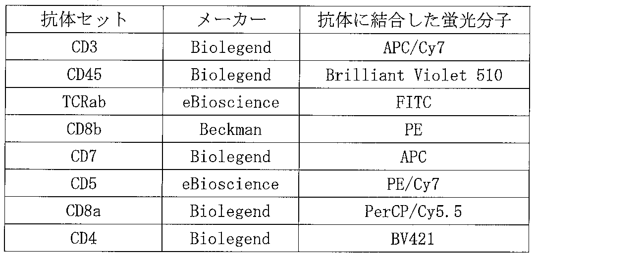
- the cells were subcultured to 48-well-plates with the same coating. On the 21st day from the start of the culture, all cells were collected, counted using a hemocytometer, and then stained using the following antibody set.
- the stained cell population was subjected to FACSAria sorting.
- the cell fraction obtained by the sorting is shown below.
- the lymphocyte fraction is the same as the method described in Cytometry (Communications in Clinical Cytometry) 18 (1994) 199-208, using FSC (forward scatter) and SSC (side scatter) as indices.
- the separated fraction Fraction P: Lymphocyte fraction
- Fraction Q Lymphocyte fraction, CD3 positive, CD45 positive, CD5 positive, CD7 positive
- Fraction R Lymphocyte fraction, CD3 positive, CD45 positive, CD7 positive, CD8a positive, CD4 Positive fraction
- S Lymphocyte fraction, CD3 positive, CD45 positive, CD7 positive, CD8a positive, CD4 negative
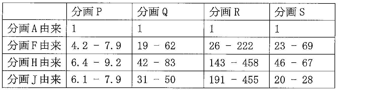
- Table 4 shows the number of cells on the 21st day of culture in each sample derived from fractions A to O (ratio with the number of cells in the sample derived from fraction A).
- Table 4 shows that fraction B (CD62L positive), fraction D (CD24 positive), fraction F (CD90 positive), and fraction H (CD143 positive) compared to the cell population contained in the sample derived from fraction A
- fraction J CD218a positive
- fraction L CD263 positive
- fraction N Notch3 positive
- Table 5 shows the number of cells in cell fraction P to S obtained by sorting with FACS Aria on the 21st day of culture (ratio with the number of cells in cell fraction P to S in the sample derived from fraction A).
- CD4 / CD8 double positive cells or CD8 positive cells
- CD90, CD143 or CD218a a high proportion of CD4 / CD8 double positive cells (or CD8 positive cells) by isolating cells that express CD90, CD143 or CD218a as a cell population containing hematopoietic progenitor cells and differentiating the isolated cells It has been shown that cell populations comprising can be produced.
- Example 2 (Study using Ff-I01s04 strain) As a cell population containing hematopoietic progenitor cells (hereinafter sometimes referred to as HPC), a floating cell population obtained by subjecting iPS cells (Ff-I01s04 strain) to the method described in Example 1 was used.
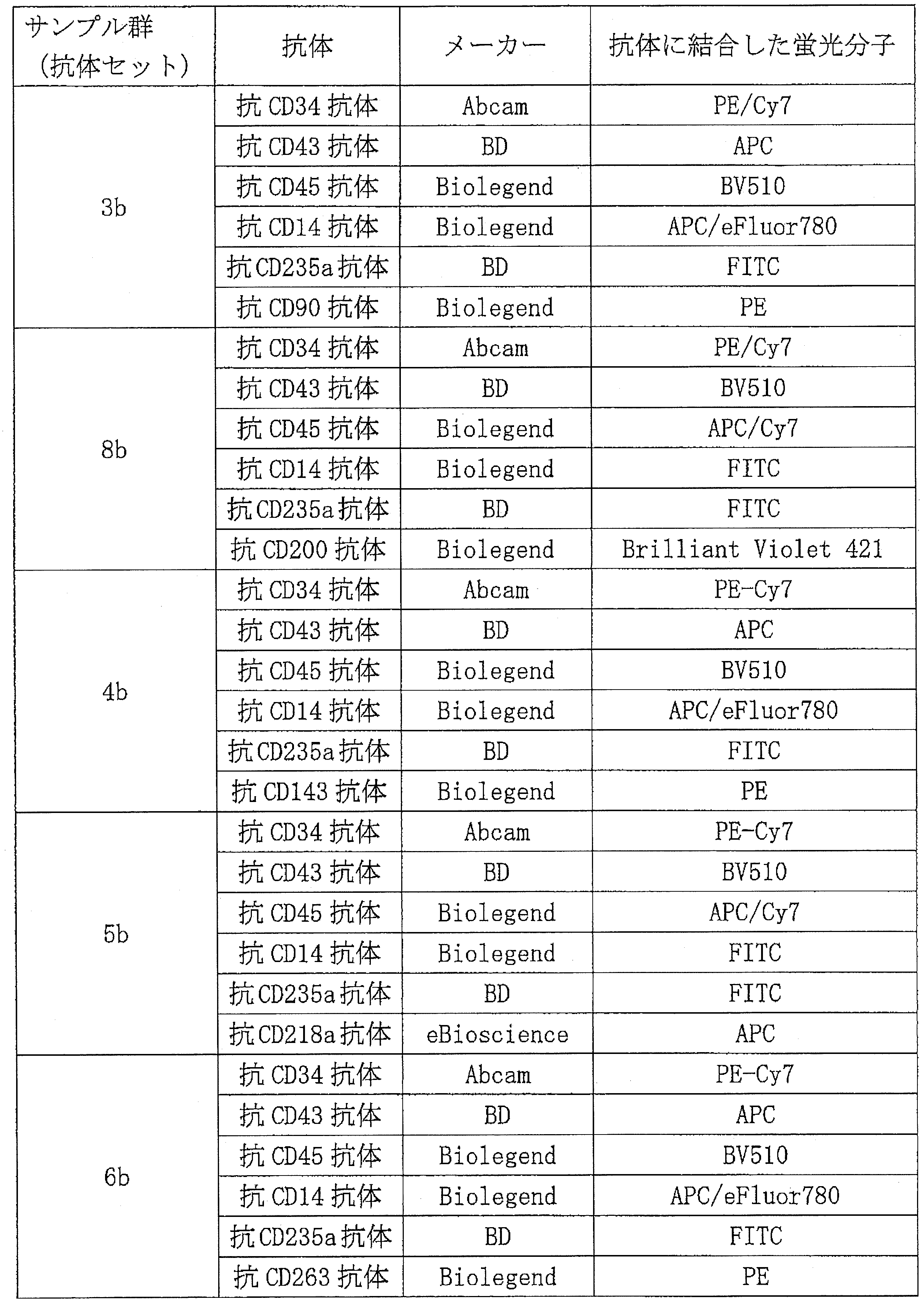
- the cell population containing HPC is the antibody set 3, 4, 5, 6 shown in Example 1 for each sample group (in this example, expressed as 3b, 4b, 5b, 6b, respectively) or the antibodies shown below Stained with set 8b.
- the cell population subjected to the above staining was subjected to sorting by FACSAria.
- the cell fraction obtained from each sample group is shown below.
- fractions f to m, t and u each indicate the following.
- Fraction f CD235a negative, CD14 negative, CD34 positive, CD43 positive, CD90 positive
- Fraction g CD235a negative, CD14 negative, CD34 positive, CD43 positive, CD90 negative
- Fraction h CD235a negative, CD14 negative, CD34 positive, CD43 Positive, CD143 positive
- Fraction i CD235a negative, CD14 negative, CD34 positive, CD43 positive, CD143 negative
- Fraction j CD235a negative, CD14 negative, CD34 positive, CD43 positive, CD218a positive
- Fraction k CD235a negative, CD14 negative, CD34 positive, CD43 positive, CD218a negative
- Fraction l CD235a negative, CD14 negative, CD34 positive, CD43 positive, CD263 positive
- Fraction m CD235a negative, CD14 negative, CD34 positive, CD43 positive, CD263 negative
- Fraction t CD235a negative , CD14 negative, CD34 positive, CD43
- the cells contained in the above fractions f to m, t and u were subjected to the method described in Example 1 to be differentiated into lymphocyte cells. On day 28 of the start of culture, all cells were collected and stained using the following antibody set.
- the stained cell population was subjected to FACSAria sorting.
- the cell fraction obtained by the sorting is shown below.
- Fraction p Lymphocyte fraction fraction q: Lymphocyte fraction, CD45 positive, CD5 positive, CD7 positive fraction r: Lymphocyte fraction, CD45 positive, CD7 positive, CD8a positive, CD4 positive fraction s: Lymph Spherical fraction, CD45 positive, CD7 positive, CD8a positive, CD4 negative
- Table 9 shows the number of cells on the 28th day from the start of culture in each sample derived from fractions f to m, t and u (ratio with the number of cells at the start of culture).
- fraction f is fractioned with respect to fraction g, fraction h with respect to fraction i, fraction j with respect to fraction k, and fraction l with respect to fraction m. It was found that t was excellent in proliferation ability for fraction u. That is, it was found that all cell populations contained in samples derived from CD90 positive, CD143 positive, CD218a positive, CD263 positive or CD200 positive were excellent in proliferating ability.
- Table 10 shows the number of cells in cell fraction p-s obtained by sorting by FACS Aria on the 28th day of culture when cells contained in fractions fm, t, and u were seeded at 1000 cells / well. Show.
- CD45 positive and CD5 positive when cells derived from each marker positive cell were used compared to when cells derived from each marker negative cell were used.
- CD7-positive, CD8a-positive, and CD4-positive cells or
- CD45-positive, CD5-positive, CD7-positive, CD8a-positive, and CD4-negative cells can be prepared with high yield.
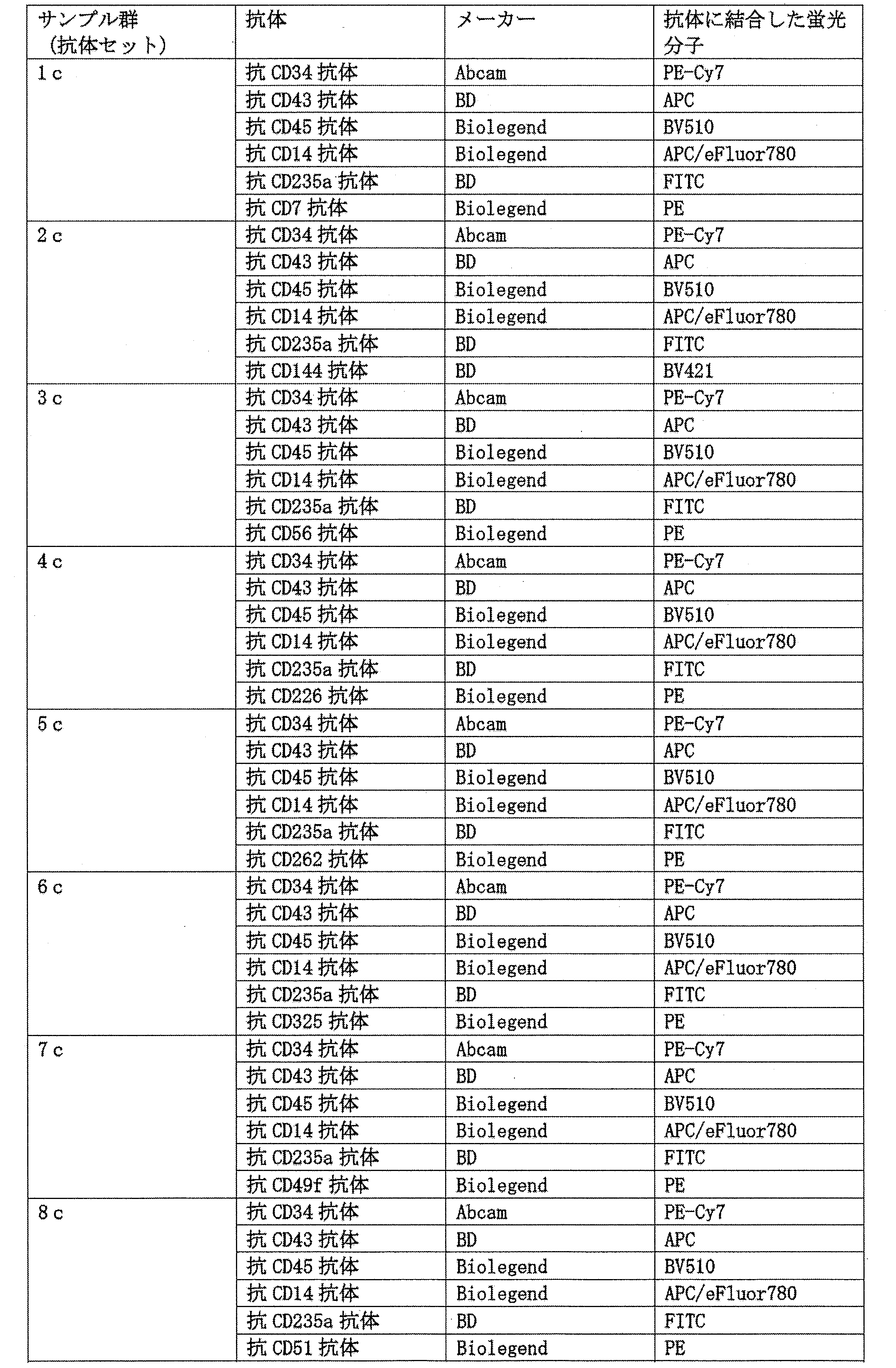
- Example 3 (Examination using Ff-I01s04 strain) As a cell population containing hematopoietic progenitor cells (hereinafter sometimes referred to as HPC), a floating cell population obtained by subjecting iPS cells (Ff-I01s04 strain) to the method described in Example 1 was used. The floating cell population containing HPC was stained using the following antibody set for each sample group.
- HPC hematopoietic progenitor cells
- the cell population subjected to the above staining was subjected to sorting by FACSAria.
- the cell fraction obtained from each sample group is shown below.
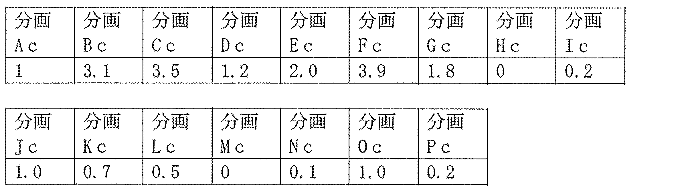
- fractions Ac to Pc indicate the following.
- Fraction Ac CD235a negative, CD14 negative, CD34 positive, CD43 positive
- Fraction Bc CD235a negative, CD14 negative, CD34 positive, CD43 positive, CD7 positive
- Fraction Cc CD235a negative, CD14 negative, CD34 positive, CD43 positive, CD144 Positive
- Fraction Dc CD235a negative, CD14 negative, CD34 positive, CD43 positive, CD56 positive
- Fraction Ec CD235a negative, CD14 negative, CD34 positive, CD43 positive, CD226 positive
- Fc CD235a negative, CD14 negative, CD34 positive, CD43 positive, CD262 positive
- Fraction Gc CD235a negative, CD14 negative, CD34 positive, CD43 positive, CD325 positive
- Fraction Hc CD235a negative, CD14 negative, CD34 positive, CD43 positive, CD49f positive
- Fraction Ic CD235a negative, CD14 negative CD34 positive, CD43 positive, CD51 positive
- Fraction Jc CD235a negative, CD14 negative,
- the cells contained in the fractions Ac to Pc were subjected to the method described in Example 1 to differentiate into lymphocyte cells. On the 21st day from the start of the culture, all cells were collected, counted using a hemocytometer, and then stained using the following antibody set.
- the stained cell population was subjected to FACSAria sorting.
- the cell fraction obtained by the sorting is shown below.
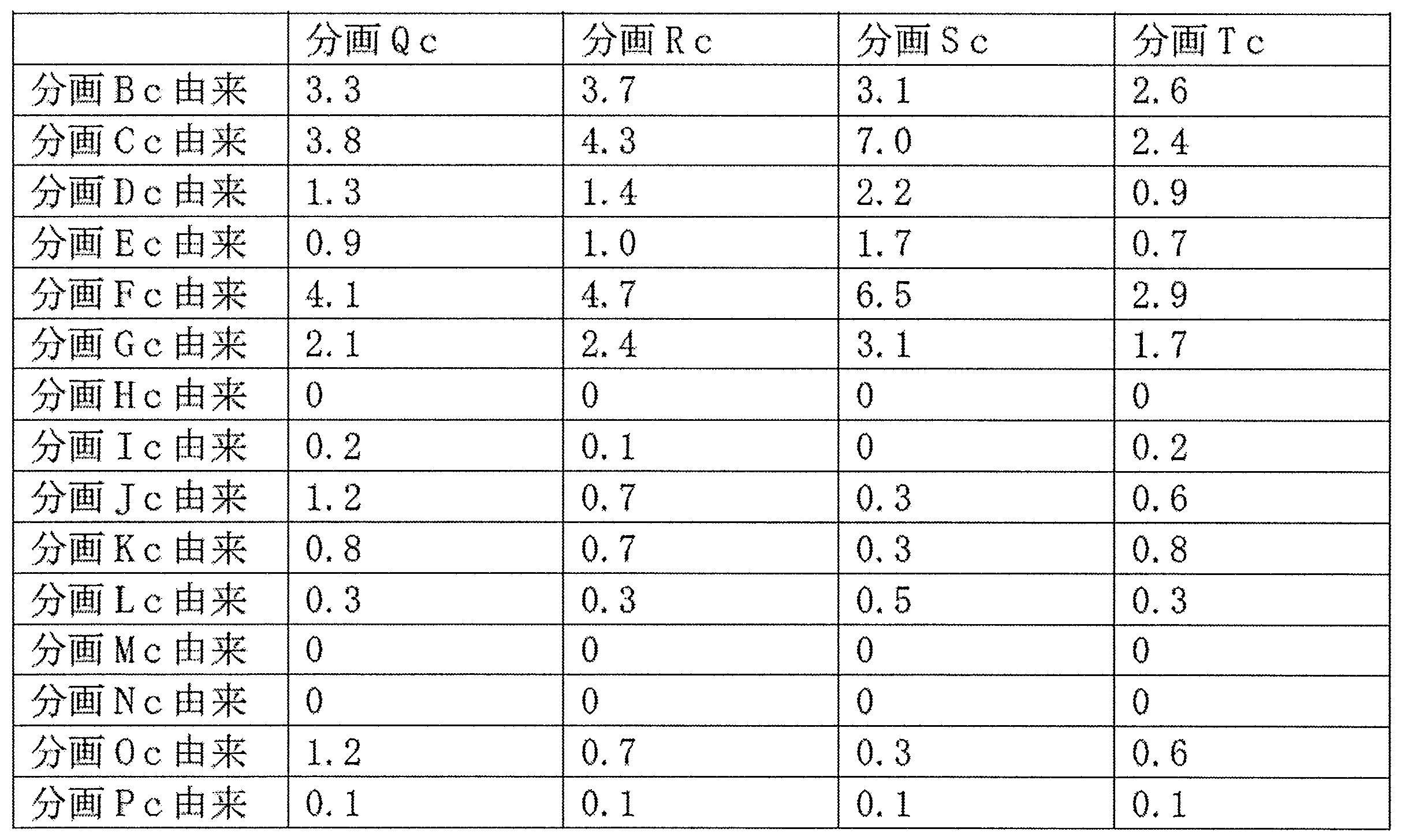
- Fraction Qc Lymphocyte fraction
- Fraction Rc Lymphocyte fraction, CD3 positive, CD45 positive, CD5 positive, CD7 positive
- Fraction Sc Lymphocyte fraction, CD3 positive, CD45 positive, CD7 positive, CD8a positive
- Tc Lymphocyte fraction, CD3 positive, CD45 positive, CD7 positive, CD8a positive, CD4 negative
- Table 14 shows the number of cells on the 21st day from the start of culture in each sample derived from fractions Ac to Pc (ratio with the number of cells in the sample derived from fraction Ac).
- fraction Bc (CD7 positive), fraction Cc (CD144 positive), fraction Dc (CD56 positive), fraction Ec (CD226 positive) It was found that the cell populations contained in the samples derived from fraction Fc (CD262 positive) and fraction Gc ⁇ (CD325 positive) all have excellent proliferation ability. Also, fraction Hc (CD49f positive), fraction Ic (CD51 positive), fraction Kc (CD42b positive), fraction Lc (CD61 positive), fraction Mc (CD62P positive), fraction Nc (CD69 positive) and It was found that all the cell populations contained in the sample derived from the fraction Pc (CD156c positive) were inferior in proliferation ability.
- Table 15 shows the number of cells in the cell fraction Qc to Tc obtained by sorting by FACS Aria on the 21st day of culture (ratio with the number of cells in the cell fraction Qc to Tc in the sample derived from the fraction Ac).
- fraction Ac derived from fraction Hc, fraction Ic, fraction Jc, fraction Kc, fraction Lc, fraction Mc, fraction Nc, fraction Oc, or fraction Pc. It is shown that a cell population containing CD3 positive CD45 positive CD7 positive CD8 positive CD4 positive cell or CD3 positive CD45 positive CD7 positive CD8a positive CD4 negative cell can be prepared compared to It was done.
- a cell population containing hematopoietic progenitor cells cells expressing any one or more of CD7, CD144, CD56, CD226, CD262, and CD325, or CD49f, CD51, CD102, CD42b, CD61, CD62P, CD69, CD102
- more and / or higher concentration of immature T cells and mature T cells can be obtained, and the cells thus obtained can prevent diseases such as tumors, infectious diseases, autoimmune disorders, etc. Or it is useful for treatment.
Landscapes
- Health & Medical Sciences (AREA)
- Engineering & Computer Science (AREA)
- Life Sciences & Earth Sciences (AREA)
- Biomedical Technology (AREA)
- Immunology (AREA)
- Chemical & Material Sciences (AREA)
- Biotechnology (AREA)
- Zoology (AREA)
- Organic Chemistry (AREA)
- Genetics & Genomics (AREA)
- Bioinformatics & Cheminformatics (AREA)
- Wood Science & Technology (AREA)
- Cell Biology (AREA)
- General Health & Medical Sciences (AREA)
- Microbiology (AREA)
- Biochemistry (AREA)
- Hematology (AREA)
- General Engineering & Computer Science (AREA)
- Urology & Nephrology (AREA)
- Molecular Biology (AREA)
- Food Science & Technology (AREA)
- General Physics & Mathematics (AREA)
- Virology (AREA)
- Tropical Medicine & Parasitology (AREA)
- Pathology (AREA)
- Medicinal Chemistry (AREA)
- Physics & Mathematics (AREA)
- Analytical Chemistry (AREA)
- Transplantation (AREA)
- Developmental Biology & Embryology (AREA)
- Cardiology (AREA)
- Rheumatology (AREA)
- Micro-Organisms Or Cultivation Processes Thereof (AREA)
- Measuring Or Testing Involving Enzymes Or Micro-Organisms (AREA)
- Peptides Or Proteins (AREA)
Abstract
Description
T細胞は、細菌やウイルスなどの外来の病原体や癌細胞などの異常な細胞に対する免疫システムにおいて中心的な役割を果たしている。なかでも、細胞傷害性T細胞(CTL)は、その細胞表面上に存在するT細胞受容体(TCR)を介して、抗原提示細胞のクラス1主要組織適合抗原と共に提示された、ウイルスや腫瘍等由来の抗原ペプチドを認識し、異物である該抗原ペプチドを提示する細胞に対して、特異的に細胞傷害活性を発揮する。
[1] 以下の工程を含む、CD4/CD8 ダブルポジティブ細胞を製造する方法。
工程1:造血前駆細胞を含む細胞集団から、CD24、CD62L、CD90、CD143、CD263、Notch3、CD32、CD39、CD49a、CD164、CD317、CD200、CD218a、CD7、CD144、CD56、CD226、CD262及びCD325からなる第1の群より選択される1種以上の分子を発現する細胞、及び/又はCD49f、CD51、CD102、CD42b、CD61、CD62P、CD69、CD102及びCD156cからなる第2の群より選択される1種以上の分子を発現しない細胞を分離する工程、並びに
工程2:工程1により分離された細胞を、CD4/CD8 ダブルポジティブ細胞に分化させる工程
[2] 前記工程1において選択される細胞が、CD235a及びCD14を発現しておらず、かつ、CD45、CD34及びCD43を発現している細胞である、[1]に記載の方法。
[3] 前記工程1が、造血前駆細胞を含む細胞集団から、CD24、CD62L、CD90、CD143、CD263、Notch3、CD32、CD39、CD49a、CD164、CD317、CD200、CD218a、CD7、CD144、CD56、CD226、CD262及びCD325からなる第1の群より選択される1種以上の分子を発現する細胞を分離する工程である、[1]又は[2]に記載の方法。
[4] 前記工程1に示される第1の群が、CD24、CD62L、CD90、CD143、CD263、Notch3、CD200、CD218a、CD7及びCD144からなる群である、[3]に記載の方法。
[5] 前記工程1が、造血前駆細胞を含む細胞集団から、CD49f、CD51、CD102、CD42b、CD61、CD62P、CD69、CD102及びCD156cからなる第2の群より選択される1種以上の分子を発現しない細胞を分離する工程である、[1]又は[2]に記載の方法。
[6] 前記工程1に示される第2の群が、CD49f、CD51及びCD102からなる群である、[5]に記載の方法。
[7] 前記工程1の造血前駆細胞を含む細胞集団が、多能性幹細胞を分化誘導することにより得られる細胞集団である、[1]~[6]のいずれかに記載の方法。
[8] 前記多能性幹細胞がヒト人工多能性幹細胞である、[7]に記載の方法。
[9-1] [1]~[8]のいずれかの方法により得られたCD4/CD8 ダブルポジティブ細胞をCD8 シングルポジティブ細胞に誘導する工程を含む、CD8 シングルポジティブ細胞の製造方法。
[9-2] 前記誘導する工程が、例えば、副腎皮質ホルモン剤、抗体及び/又はサイトカインの存在下でCD4/CD8ダブルポジティブ細胞を培養する工程を含む方法であって、好ましくは、該副腎皮質ホルモン剤がデキサメタゾンであり、及び/又は該抗体が抗CD3抗体であり、及び/又はサイトカインはIL-2である、[9-1]に記載の方法。
[10] 前記CD8 シングルポジティブ細胞が細胞傷害性T細胞である、[9-1]又は[9-2]に記載の方法。
[11] 造血前駆細胞を含む細胞集団から、CD24、CD62L、CD90、CD143、CD263、Notch3、CD32、CD39、CD49a、CD164、CD317、CD200、CD218a、CD7、CD144、CD56、CD226、CD262及びCD325からなる第1の群より選択される1種以上の分子を発現する細胞、及び/又はCD49f、CD51、CD102、CD42b、CD61、CD62P、CD69、CD102及びCD156cからなる第2の群より選択される1種以上の分子を発現しない細胞を分離して得られる細胞集団。
[12] 分離された細胞が、CD235a及びCD14を発現しておらず、かつ、CD45、CD34及びCD43を発現している細胞である、[11]に記載の細胞集団。
[13] 前記造血前駆細胞を含む細胞集団が、多能性幹細胞を分化誘導することにより得られる細胞集団である、[11]又は[12]に記載の細胞集団。
[14] 前記多能性幹細胞がヒト人工多能性幹細胞である、[13]に記載の細胞集団。
[15] CD24、CD62L、CD90、CD143、CD263、Notch3、CD32、CD39、CD49a、CD164、CD317、CD200、CD218a、CD7、CD144、CD56、CD226、CD262及びCD325からなる第1の群より選択される1種以上の分子を発現する造血前駆細胞、及び/又はCD49f、CD51、CD102、CD42b、CD61、CD62P、CD69、CD102及びCD156cからなる第2の群より選択される1種以上の分子を発現しない造血前駆細胞の割合(該造血前駆細胞数/細胞集団に含まれる全細胞数)が40%以上である細胞集団。
[16] 前記割合が、CD24、CD62L、CD90、CD143、CD263、Notch3、CD200、CD218a、CD7及びCD144からなる第1の群より選択される1種以上の分子をさらに発現する造血前駆細胞の割合である、[15]に記載の細胞集団。
[17] 前記割合が、CD49f、CD51及びCD102 からなる第2の群より選択される1種以上の分子をさらに発現しない造血前駆細胞の割合である、[15]に記載の細胞集団。
[18] CD24、CD62L、CD90、CD143、CD263、Notch3、CD32、CD39、CD49a、CD164、CD317、CD200、CD218a、CD7、CD144、CD56、CD226、CD262、CD325、CD49f、CD51、CD102、CD42b、CD61、CD62P、CD69、CD102及びCD156cからなる群より選択される1種以上の分子の各々に対する抗体を含んでなる、造血前駆細胞の分離用試薬。
[19] さらにCD235a、CD14、CD45、CD34及び/又はCD43に対する抗体を1種以上含んでなる、[18]に記載の試薬。
[20] 造血前駆細胞の表面タンパク質を検出する方法であって、該造血前駆細胞と、CD24、CD62L、CD90、CD143、CD263、Notch3、CD32、CD39、CD49a、CD164、CD317、CD200、CD218a、CD7、CD144、CD56、CD226、CD262、CD325、CD49f、CD51、CD102、CD42b、CD61、CD62P、CD69、CD102、CD109又はCD156cに特異的に結合する物質とを接触させる工程を含む、方法。
[21] 前記物質が抗体又はその断片を含む、[20]に記載の方法。
[22] 前記物質がさらにフルオロフォアを含む、[20]又は[21]に記載の方法。
[23] 前記造血前駆細胞に、CD34、CD43、CD14又はCD235aに特異的に結合する物質を接触させる工程をさらに含む、[20]~[22]のいずれかに記載の方法。
[24] 造血前駆細胞の表面上の複数のタンパク質を検出する方法であって、該方法は、造血前駆細胞を、複数のタンパク質の各々に特異的に結合する物質と接触させる工程を含み、該複数のタンパク質が、CD24、CD62L、CD90、CD143、CD263、Notch3、CD32、CD39、CD49a、CD164、CD317、CD200、CD218a、CD7、CD144、CD56、CD226、CD262、CD325、CD49f、CD51、CD102、CD42b、CD61、CD62P、CD69、CD102、CD109及びCD156cの2、3、4、5、6、7、8、9、10、11、12、13、14、15、16、17、18、19、20、21、22、23、24、25、26、27若しくは28つの、又は全29つの任意の組み合わせを含む、方法。
[25] 造血前駆細胞を含む細胞集団から造血前駆細胞を選択する方法であって、[20]~[24]のいずれかの方法により細胞の表面上の1種以上のタンパク質の存在又は非存在を検出する工程、並びに該1種以上のタンパク質の存在又は非存在に基づき該細胞を選択する工程であって、(i)次のタンパク質:CD24、CD62L、CD90、CD143、CD263、Notch3、CD32、CD39、CD49a、CD164、CD317、CD200、CD218a、CD7、CD144、CD56、CD226、CD262及びCD325:の任意の1つ又は任意の組み合わせが該細胞の表面で検出される場合、及び/又は(ii)次のタンパク質:CD49f、CD51、CD102、CD42b、CD61、CD62P、CD69、CD102、CD109及びCD156c:の任意の1つ若しくは任意の組み合わせが該細胞の表面で検出されない場合に、該細胞を選択する工程、を含む方法。
[26] 次のタンパク質:CD24、CD62L、CD90、CD143、CD263、Notch3、CD200、CD218a、CD7及びCD144:の任意の1つ又は任意の組み合わせが細胞の表面上で検出される場合に、該細胞を選択する、[25]に記載の方法。
[27] 次のタンパク質:CD49f、CD51及びCD102:の任意の1つ若しくは任意の組み合わせが細胞の表面上で検出されない場合に、該細胞を選択する、[25]又は[26]に記載の方法。
[28] 造血前駆細胞を含む細胞集団から造血前駆細胞を選択する方法であって、[20]~[24]のいずれかの方法により細胞の表面上の1種以上のタンパク質の存在又は非存在を検出する工程、並びに該1種以上のタンパク質の存在又は非存在に基づき、(i)CD45、CD34及び/又はCD43と、次のタンパク質:CD24、CD62L、C90、CD143、CD263、Notch3、CD32、CD39、CD49a、CD164、CD317、CD200、CD218a、CD7、CD144、CD56、CD226、CD262及びCD325:の任意の1つ若しくは任意の組み合わせとが、該細胞の表面上で検出された場合、並びに/又は(ii)CD14及び/若しくはCD235aが該細胞の表面上で検出されず、かつ次のタンパク質:CD49f、CD51、CD102、CD42b、CD61、CD62P、CD69、CD102、CD109及びCD156c:も検出されなかった場合に、該細胞を選択する工程を含む、方法。
本発明は、(工程1)造血前駆細胞を含む細胞集団から、1種以上の造血前駆細胞マーカー(本明細書中、「HPCマーカー」と略記することがある。)を発現する細胞、及び/又は1種以上のHPCマーカーを発現しない細胞を分離する工程、及び(工程2)工程1により分離された細胞を、CD8ポジティブ細胞(例えば、CD4/CD8ダブルポジティブ細胞(本明細書中、「CD4/CD8DP細胞」と略記することがある。)に分化させる工程を含む、CD8ポジティブ細胞(例えば、CD4/CD8DP細胞)を製造する方法(以下「本発明の製法」と略記する。)などを提供する。
本発明の製法の工程2で用いる細胞培養用の基礎培地としては、例えばIscove's Modified Dulbecco's Medium(IMDM)培地、Medium 199培地、Eagle's Minimum Essential Medium (EMEM)培地、αMEM培地、Dulbecco's modified Eagle's Medium (DMEM)培地、Ham's F12培地、RPMI 1640培地、Fischer's培地、Neurobasal Medium(ライフテクノロジーズ)及びこれらの混合培地が挙げられる。培地には、血清が含有されていてもよいし、含有されていなくてもよい。必要に応じて、培地には、例えば、ビタミンC類(例:アスコルビン酸、リン酸ビタミンC)、アルブミン、インスリン、トランスフェリン、セレン、脂肪酸、微量元素、2-メルカプトエタノール、チオールグリセロール、脂質、アミノ酸、L-グルタミン、非必須アミノ酸、ビタミン、増殖因子、低分子化合物、抗生物質、抗酸化剤、ピルビン酸、緩衝剤、無機塩類、サイトカインなどの1つ以上の物質を含有させてもよい。
本発明の製法により得られたCD4/CD8DP細胞をCD8SP細胞に分化誘導する工程に付すことにより、CD8SP細胞を製造することができる(このようにCD8SP細胞を製造する方法を、「本発明のCD8SP細胞の製法」と略記することがある。)。CD4/CD8DP細胞を製造する方法については、1.に記載の通りである。
本発明は、1種以上のHPCポジティブマーカーを発現する、及び/又は1種以上のHPCネガティブマーカーを発現しない造血前駆細胞を高頻度で含む細胞集団(以下「本発明の細胞集団」と略記する。)を提供する。前記細胞集団は、例えば、造血前駆細胞を含む細胞集団から、1種以上のHPCポジティブマーカーを発現する、及び/又は1種以上のHPCネガティブマーカーを発現しない細胞を上記に記載の分離方法を用いて分離することで得ることができる。HPCマーカーの定義、具体的な分子、及び造血前駆細胞の定義については、上記に記載の通りである。
本発明はまた、本発明のHPCマーカー、即ち、CD24、CD62L、CD90、CD143、CD263、Notch3、CD32、CD39、CD49a、CD164、CD317、CD200、CD218a、CD7、CD144、CD56、CD226、CD262、CD325、CD49f、CD51、CD102、CD42b、CD61、CD62P、CD69、CD102及びCD156c、好ましくは、CD24、CD62L、CD90、CD143、CD263、Notch3、CD200CD218a、CD7、CD144、CD49f、CD51及びCD102からなる群より選択される1種以上のマーカーの各々に対する抗体を含んでなる、本発明の細胞集団の分離用試薬(以下、「本発明の試薬」と略記する)を提供する。本発明の試薬が2種以上のHPCマーカーに対する抗体を含む場合、該試薬は、各抗体を別個の試薬中に含む試薬キットとして提供され得る。本発明の試薬に含まれる抗体は、本発明の製法の工程1に用いられる分離手段に応じて、例えば、蛍光色素、金属同位体又はビーズ(例:磁気ビーズ)に結合した形態で提供され得る。
また、本発明の試薬は、造血前駆細胞をCD4/CD8DP細胞に、さらにはCD8SP細胞に分化誘導するための試薬類(例えば、基礎培地、培地添加物など)をさらに含んでもよく、当該試薬類としては、上記1.及び上記2.で例示された物質が同様に挙げられる。
造血前駆細胞(以下、HPCと称することがある)を含む細胞集団として、iPS細胞(TKT3V1-7株)を公知の方法(例えば、Cell Reports 2(2012)1722-1735や国際公開第2017/221975号に記載された方法)に準じて分化させた浮遊細胞集団を用いた。具体的には、超低接着処理された6well plateにTKT3V1-7株を3 x 10⁵個/ウェルで播種し(Day0)、EB培地(StemPro34に10 μg/mlヒトインスリン、5. 5 μg/mlヒトトランスフェリン、5 ng/ml 亜セレン酸ナトリウム、2mM L-グルタミン、45mM α-モノチオグリセロー ル、および50 μg/mlアスコルビン酸を添加)に10 ng/ml BMP4、5ng/ml bFGF 、15ng/ml VEGF、2μM SB431542、を加えて、低酸素条件下(5% 02)にて5日間 培養を行った(Day5)。 続いて、50ng/ml SCF、30ng/ml TPO、10ng/ml Flt3Lを添加し、さらに5~9 日間培養を行い(~Day14)、浮遊細胞集団を得た。なお、培養期間中は2日または3日ごとに培地交換を行った。HPCを含む上記浮遊細胞集団を、サンプル群ごとに以下の抗体セットを用いて染色した。


分画A:CD235a陰性、CD14陰性、CD34陽性、CD43陽性
分画B:CD235a陰性、CD14陰性、CD34陽性、CD43陽性、CD62L陽性
分画C:CD235a陰性、CD14陰性、CD34陽性、CD43陽性、CD62L陰性
分画D:CD235a陰性、CD14陰性、CD34陽性、CD43陽性、CD24陽性
分画E:CD235a陰性、CD14陰性、CD34陽性、CD43陽性、CD24陰性
分画F:CD235a陰性、CD14陰性、CD34陽性、CD43陽性、CD90陽性
分画G:CD235a陰性、CD14陰性、CD34陽性、CD43陽性、CD90陰性
分画H:CD235a陰性、CD14陰性、CD34陽性、CD43陽性、CD143陽性
分画I:CD235a陰性、CD14陰性、CD34陽性、CD43陽性、CD143陰性
分画J:CD235a陰性、CD14陰性、CD34陽性、CD43陽性、CD218a陽性
分画K:CD235a陰性、CD14陰性、CD34陽性、CD43陽性、CD218a陰性
分画L:CD235a陰性、CD14陰性、CD34陽性、CD43陽性、CD263陽性
分画M:CD235a陰性、CD14陰性、CD34陽性、CD43陽性、CD263陰性
分画N:CD235a陰性、CD14陰性、CD34陽性、CD43陽性、Notch3陽性
分画O:CD235a陰性、CD14陰性、CD34陽性、CD43陽性、Notch3陰性

分画P:リンパ球分画
分画Q:リンパ球分画、CD3陽性、CD45陽性、CD5陽性、CD7陽性
分画R:リンパ球分画、CD3陽性、CD45陽性、CD7陽性、CD8a陽性、CD4陽性
分画S:リンパ球分画、CD3陽性、CD45陽性、CD7陽性、CD8a陽性、CD4陰性

造血前駆細胞(以下、HPCと称することがある)を含む細胞集団として、iPS細胞(Ff-I01s04株)を実施例1に記載された方法に供して得られた浮遊細胞集団を用いた。
HPCを含む細胞集団は、サンプル群ごとに実施例1に示した抗体セット3、4、5、6(本実施例では、それぞれ3b、4b、5b、6bと表記する)、または以下に示す抗体セット8bを用いて染色した。

分画f:CD235a陰性、CD14陰性、CD34陽性、CD43陽性、CD90陽性
分画g:CD235a陰性、CD14陰性、CD34陽性、CD43陽性、CD90陰性
分画h:CD235a陰性、CD14陰性、CD34陽性、CD43陽性、CD143陽性
分画i:CD235a陰性、CD14陰性、CD34陽性、CD43陽性、CD143陰性
分画j:CD235a陰性、CD14陰性、CD34陽性、CD43陽性、CD218a陽性
分画k:CD235a陰性、CD14陰性、CD34陽性、CD43陽性、CD218a陰性
分画l:CD235a陰性、CD14陰性、CD34陽性、CD43陽性、CD263陽性
分画m:CD235a陰性、CD14陰性、CD34陽性、CD43陽性、CD263陰性
分画t:CD235a陰性、CD14陰性、CD34陽性、CD43陽性、CD200陽性
分画u:CD235a陰性、CD14陰性、CD34陽性、CD43陽性、CD200陰性
分画p:リンパ球分画
分画q:リンパ球分画、CD45陽性、CD5陽性、CD7陽性
分画r:リンパ球分画、CD45陽性、CD7陽性、CD8a陽性、CD4陽性
分画s:リンパ球分画、CD45陽性、CD7陽性、CD8a陽性、CD4陰性


これらの結果から、造血前駆細胞を含む細胞集団として、CD90、CD143、CD200、CD218aまたはCD263を発現する細胞を分離して、分離された細胞を分化させることにより、CD8ポジティブ細胞(例:CD4/CD8DP細胞、CD8SP細胞)を高い割合で含む細胞集団を製造することができることが示された。
造血前駆細胞(以下、HPCと称することがある)を含む細胞集団として、iPS細胞(Ff-I01s04株)を実施例1に記載された方法に供して得られた浮遊細胞集団を用いた。HPCを含む上記浮遊細胞集団を、サンプル群ごとに以下の抗体セットを用いて染色した。


分画Ac:CD235a陰性、CD14陰性、CD34陽性、CD43陽性
分画Bc:CD235a陰性、CD14陰性、CD34陽性、CD43陽性、CD7陽性
分画Cc:CD235a陰性、CD14陰性、CD34陽性、CD43陽性、CD144陽性
分画Dc:CD235a陰性、CD14陰性、CD34陽性、CD43陽性、CD56陽性
分画Ec:CD235a陰性、CD14陰性、CD34陽性、CD43陽性、CD226陽性
分画Fc:CD235a陰性、CD14陰性、CD34陽性、CD43陽性、CD262陽性
分画Gc:CD235a陰性、CD14陰性、CD34陽性、CD43陽性、CD325陽性
分画Hc:CD235a陰性、CD14陰性、CD34陽性、CD43陽性、CD49f陽性
分画Ic:CD235a陰性、CD14陰性、CD34陽性、CD43陽性、CD51陽性
分画Jc:CD235a陰性、CD14陰性、CD34陽性、CD43陽性、CD102陽性
分画Kc:CD235a陰性、CD14陰性、CD34陽性、CD43陽性、CD42b陽性
分画Lc:CD235a陰性、CD14陰性、CD34陽性、CD43陽性、CD61陽性
分画Mc:CD235a陰性、CD14陰性、CD34陽性、CD43陽性、CD62P陽性
分画Nc:CD235a陰性、CD14陰性、CD34陽性、CD43陽性、CD69陽性
分画Oc:CD235a陰性、CD14陰性、CD34陽性、CD43陽性、CD102陽性
分画Pc:CD235a陰性、CD14陰性、CD34陽性、CD43陽性、CD156c陽性

分画Qc:リンパ球分画
分画Rc:リンパ球分画、CD3陽性、CD45陽性、CD5陽性、CD7陽性
分画Sc:リンパ球分画、CD3陽性、CD45陽性、CD7陽性、CD8a陽性、CD4陽性
分画Tc:リンパ球分画、CD3陽性、CD45陽性、CD7陽性、CD8a陽性、CD4陰性
Claims (19)
- 以下の工程を含む、CD4/CD8 ダブルポジティブ細胞を製造する方法。
工程1:造血前駆細胞を含む細胞集団から、CD24、CD62L、CD90、CD143、CD263、Notch3、CD32、CD39、CD49a、CD164、CD317、CD200、CD218a、CD7、CD144、CD56、CD226、CD262及びCD325からなる第1の群より選択される1種以上の分子を発現する細胞、及び/又はCD49f、CD51、CD102、CD42b、CD61、CD62P、CD69、CD102及びCD156cからなる第2の群より選択される1種以上の分子を発現しない細胞を分離する工程、並びに
工程2:工程1により分離された細胞を、CD4/CD8 ダブルポジティブ細胞に分化させる工程 - 前記工程1において選択される細胞が、CD235a及びCD14を発現しておらず、かつ、CD45、CD34及びCD43を発現している細胞である、請求項1に記載の方法。
- 前記工程1が、造血前駆細胞を含む細胞集団から、CD24、CD62L、CD90、CD143、CD263、Notch3、CD32、CD39、CD49a、CD164、CD317、CD200、CD218a、CD7、CD144、CD56、CD226、CD262及びCD325からなる第1の群より選択される1種以上の分子を発現する細胞を分離する工程である、請求項1又は2に記載の方法。
- 前記工程1に示される第1の群が、CD24、CD62L、CD90、CD143、CD263、Notch3、CD200、CD218a、CD7及びCD144からなる群である、請求項3に記載の方法。
- 前記工程1が、造血前駆細胞を含む細胞集団から、CD49f、CD51、CD102、CD42b、CD61、CD62P、CD69、CD102及びCD156cからなる第2の群より選択される1種以上の分子を発現しない細胞を分離する工程である、請求項1又は2に記載の方法。
- 前記工程1に示される第2の群が、CD49f、CD51及びCD102からなる群である、請求項5に記載の方法。
- 前記工程1の造血前駆細胞を含む細胞集団が、多能性幹細胞を分化誘導することにより得られる細胞集団である、請求項1~6のいずれか1項に記載の方法。
- 前記多能性幹細胞がヒト人工多能性幹細胞である、請求項7に記載の方法。
- 請求項1~8のいずれかの方法により得られたCD4/CD8 ダブルポジティブ細胞をCD8 シングルポジティブ細胞に誘導する工程を含む、CD8 シングルポジティブ細胞の製造方法。
- 前記CD8 シングルポジティブ細胞が細胞傷害性T細胞である、請求項9に記載の方法。
- 造血前駆細胞を含む細胞集団から、CD24、CD62L、CD90、CD143、CD263、Notch3、CD32、CD39、CD49a、CD164、CD317、CD200、CD218a、CD7、CD144、CD56、CD226、CD262及びCD325からなる第1の群より選択される1種以上の分子を発現する細胞、及び/又はCD49f、CD51、CD102、CD42b、CD61、CD62P、CD69、CD102及びCD156cからなる第2の群より選択される1種以上の分子を発現しない細胞を分離して得られる細胞集団。
- 分離された細胞が、CD235a及びCD14を発現しておらず、かつ、CD45、CD34及びCD43を発現している細胞である、請求項11に記載の細胞集団。
- 前記造血前駆細胞を含む細胞集団が、多能性幹細胞を分化誘導することにより得られる細胞集団である、請求項11又は12に記載の細胞集団。
- 前記多能性幹細胞がヒト人工多能性幹細胞である、請求項13に記載の細胞集団。
- CD24、CD62L、CD90、CD143、CD263、Notch3、CD32、CD39、CD49a、CD164、CD317、CD200、CD218a、CD7、CD144、CD56、CD226、CD262及びCD325からなる第1の群より選択される1種以上の分子を発現する造血前駆細胞、及び/又はCD49f、CD51、CD102、CD42b、CD61、CD62P、CD69、CD102及びCD156cからなる第2の群より選択される1種以上の分子を発現しない造血前駆細胞の割合(該造血前駆細胞数/細胞集団に含まれる全細胞数)が40%以上である細胞集団。
- 前記割合が、CD24、CD62L、CD90、CD143、CD263、Notch3、CD200、CD218a、CD7及びCD144からなる第1の群より選択される1種以上の分子を発現する造血前駆細胞の割合である、請求項15に記載の細胞集団。
- 前記割合が、CD49f、CD51及びCD102からなる第2の群より選択される1種以上の分子を発現しない造血前駆細胞の割合である、請求項15に記載の細胞集団。
- CD24、CD62L、CD90、CD143、CD263、Notch3、CD32、CD39、CD49a、CD164、CD317、CD200、CD218a、CD7、CD144、CD56、CD226、CD262、CD325、CD49f、CD51、CD102、CD42b、CD61、CD62P、CD69、CD102及びCD156cからなる群より選択される1種以上の分子の各々に対する抗体を含んでなる、造血前駆細胞の分離用試薬。
- さらにCD235a、CD14、CD45、CD34及び/又はCD43に対する抗体を1種以上含んでなる、請求項18に記載の試薬。
Priority Applications (13)
| Application Number | Priority Date | Filing Date | Title |
|---|---|---|---|
| CN201880027852.6A CN110691843B (zh) | 2017-04-26 | 2018-04-25 | 造血祖细胞标志物 |
| SG11201909795R SG11201909795RA (en) | 2017-04-26 | 2018-04-25 | Hematopoietic progenitor cell marker |
| AU2018257307A AU2018257307B2 (en) | 2017-04-26 | 2018-04-25 | Hematopoietic progenitor cell marker |
| JP2019514588A JP7430877B2 (ja) | 2017-04-26 | 2018-04-25 | 造血前駆細胞マーカー |
| US16/607,967 US20210102167A1 (en) | 2017-04-26 | 2018-04-25 | Hematopoietic progenitor cell marker |
| BR112019022288-0A BR112019022288A2 (pt) | 2017-04-26 | 2018-04-25 | métodos para produzir células duplo-positivas cd4/cd8 e células simples-positivas cd8, população celular, e, reagente para separar células progenitoras hematopoiéticas. |
| EA201992483A EA201992483A1 (ru) | 2017-04-26 | 2018-04-25 | Маркер гемопоэтических клеток-предшественников |
| CA3061289A CA3061289A1 (en) | 2017-04-26 | 2018-04-25 | Hematopoietic progenitor cell marker |
| KR1020197033583A KR102803233B1 (ko) | 2017-04-26 | 2018-04-25 | 조혈 전구 세포 마커 |
| EP18791069.0A EP3617307A4 (en) | 2017-04-26 | 2018-04-25 | HEMATOPOIETIC PRECURSOR CELL MARKER |
| NZ758956A NZ758956B2 (en) | 2018-04-25 | Hematopoietic progenitor cell marker | |
| IL270126A IL270126B2 (en) | 2017-04-26 | 2018-04-25 | Hematopoietic stem cell marker |
| JP2023139109A JP7610817B2 (ja) | 2017-04-26 | 2023-08-29 | 造血前駆細胞マーカー |
Applications Claiming Priority (2)
| Application Number | Priority Date | Filing Date | Title |
|---|---|---|---|
| JP2017-087723 | 2017-04-26 | ||
| JP2017087723 | 2017-04-26 |
Publications (1)
| Publication Number | Publication Date |
|---|---|
| WO2018199186A1 true WO2018199186A1 (ja) | 2018-11-01 |
Family
ID=63918584
Family Applications (1)
| Application Number | Title | Priority Date | Filing Date |
|---|---|---|---|
| PCT/JP2018/016864 Ceased WO2018199186A1 (ja) | 2017-04-26 | 2018-04-25 | 造血前駆細胞マーカー |
Country Status (13)
| Country | Link |
|---|---|
| US (1) | US20210102167A1 (ja) |
| EP (1) | EP3617307A4 (ja) |
| JP (2) | JP7430877B2 (ja) |
| KR (1) | KR102803233B1 (ja) |
| CN (1) | CN110691843B (ja) |
| AU (1) | AU2018257307B2 (ja) |
| BR (1) | BR112019022288A2 (ja) |
| CA (1) | CA3061289A1 (ja) |
| EA (1) | EA201992483A1 (ja) |
| IL (1) | IL270126B2 (ja) |
| SG (1) | SG11201909795RA (ja) |
| TW (1) | TWI860972B (ja) |
| WO (1) | WO2018199186A1 (ja) |
Cited By (2)
| Publication number | Priority date | Publication date | Assignee | Title |
|---|---|---|---|---|
| WO2023182328A1 (ja) | 2022-03-23 | 2023-09-28 | 国立大学法人京都大学 | 制御性t細胞の製造方法 |
| WO2024071010A1 (ja) | 2022-09-26 | 2024-04-04 | 国立大学法人京都大学 | T細胞の製造方法 |
Families Citing this family (2)
| Publication number | Priority date | Publication date | Assignee | Title |
|---|---|---|---|---|
| WO2023247722A1 (en) * | 2022-06-22 | 2023-12-28 | Fondazione Telethon Ets | Method for isolating hemogenic endothelial cells |
| CN118613579A (zh) * | 2023-01-18 | 2024-09-06 | 深圳市人民医院 | 造血祖细胞的谱系及其命运决定基因的鉴定及应用 |
Citations (5)
| Publication number | Priority date | Publication date | Assignee | Title |
|---|---|---|---|---|
| WO2011041912A1 (en) * | 2009-10-09 | 2011-04-14 | University Health Network | Self-renewing single human hematopoietic stem cells, an early lymphoid progenitor and methods of enriching the same |
| WO2013075222A1 (en) | 2011-11-21 | 2013-05-30 | University Health Network | Populations of hematopoietic progenitors and methods of enriching stem cells therefor |
| WO2016076415A1 (ja) | 2014-11-13 | 2016-05-19 | 国立大学法人京都大学 | 多能性幹細胞からt細胞への誘導方法 |
| JP2017087723A (ja) | 2015-11-11 | 2017-05-25 | ゼロックス コーポレイションXerox Corporation | マイクロ波エネルギ及びナノ粒子を使用して3次元印刷物体から支持構造を除去するシステム及び方法 |
| WO2017221975A1 (ja) | 2016-06-23 | 2017-12-28 | 国立大学法人京都大学 | Cd4cd8両陽性t細胞の製造方法 |
Family Cites Families (5)
| Publication number | Priority date | Publication date | Assignee | Title |
|---|---|---|---|---|
| US8034613B2 (en) * | 2005-06-01 | 2011-10-11 | Wisconsin Alumni Research Foundation | Multipotent lymphohematopoietic progenitor cells |
| WO2015163185A1 (ja) * | 2014-04-24 | 2015-10-29 | 学校法人関西医科大学 | ヒト造血幹細胞濃縮画分の製造方法 |
| US10590387B2 (en) * | 2015-04-16 | 2020-03-17 | The Board Of Regents Of The University Of Texas System | CD133+ cells and method for expanding |
| WO2017004127A1 (en) * | 2015-06-29 | 2017-01-05 | The General Hospital Corporation | Embigin inhibition for promotion of hematopoietic stem and progenitor cell expansion |
| SG11201803145RA (en) * | 2015-11-04 | 2018-05-30 | Fate Therapeutics Inc | Methods and compositions for inducing hematopoietic cell differentiation |
-
2018
- 2018-04-25 CA CA3061289A patent/CA3061289A1/en active Pending
- 2018-04-25 JP JP2019514588A patent/JP7430877B2/ja active Active
- 2018-04-25 TW TW107114033A patent/TWI860972B/zh active
- 2018-04-25 IL IL270126A patent/IL270126B2/en unknown
- 2018-04-25 EP EP18791069.0A patent/EP3617307A4/en active Pending
- 2018-04-25 CN CN201880027852.6A patent/CN110691843B/zh active Active
- 2018-04-25 AU AU2018257307A patent/AU2018257307B2/en active Active
- 2018-04-25 KR KR1020197033583A patent/KR102803233B1/ko active Active
- 2018-04-25 WO PCT/JP2018/016864 patent/WO2018199186A1/ja not_active Ceased
- 2018-04-25 US US16/607,967 patent/US20210102167A1/en active Pending
- 2018-04-25 SG SG11201909795R patent/SG11201909795RA/en unknown
- 2018-04-25 EA EA201992483A patent/EA201992483A1/ru unknown
- 2018-04-25 BR BR112019022288-0A patent/BR112019022288A2/pt unknown
-
2023
- 2023-08-29 JP JP2023139109A patent/JP7610817B2/ja active Active
Patent Citations (6)
| Publication number | Priority date | Publication date | Assignee | Title |
|---|---|---|---|---|
| WO2011041912A1 (en) * | 2009-10-09 | 2011-04-14 | University Health Network | Self-renewing single human hematopoietic stem cells, an early lymphoid progenitor and methods of enriching the same |
| WO2013075222A1 (en) | 2011-11-21 | 2013-05-30 | University Health Network | Populations of hematopoietic progenitors and methods of enriching stem cells therefor |
| JP2014533494A (ja) * | 2011-11-21 | 2014-12-15 | ユニバーシティ・ヘルス・ネットワーク | 造血前駆体の集団および造血前駆体のための幹細胞を富化する方法 |
| WO2016076415A1 (ja) | 2014-11-13 | 2016-05-19 | 国立大学法人京都大学 | 多能性幹細胞からt細胞への誘導方法 |
| JP2017087723A (ja) | 2015-11-11 | 2017-05-25 | ゼロックス コーポレイションXerox Corporation | マイクロ波エネルギ及びナノ粒子を使用して3次元印刷物体から支持構造を除去するシステム及び方法 |
| WO2017221975A1 (ja) | 2016-06-23 | 2017-12-28 | 国立大学法人京都大学 | Cd4cd8両陽性t細胞の製造方法 |
Non-Patent Citations (10)
| Title |
|---|
| CHOI K.D. ET AL., STEM CELLS, vol. 27, 2009, pages 559 - 567 |
| CYTOMETRY (COMMUNICATIONS IN CLINICAL CYTOMETRY, vol. 18, 1994, pages 199 - 208 |
| JOURNAL OF LEUKOCYTE BIOLOGY, vol. 96, 2016, pages 1165 - 1175 |
| KENNEDY M. ET AL., CELL REPORTS, vol. 2, 2012, pages 1722 - 1735 |
| LIU S. ET AL., CYTOTHERAPY, vol. 17, 2015, pages 344 - 358 |
| NAKAGAWA M. ET AL., NAT. BIOTECHNOL., vol. 26, 2008, pages 101 - 106 |
| TAKAHASHI K. ET AL., CELL, vol. 131, 2007, pages 861 - 872 |
| TAKAHASHI K.YAMANAKA S., CELL, vol. 126, 2006, pages 663 - 676 |
| TIMMERMANS, F. ET AL.: "Generation of T Cells from Human Embryonic Stem Cell -Derived Hematopoietic Zones", THE JOURNAL OF IMMUNOLOGY, vol. 182, no. 11, 19 May 2009 (2009-05-19), pages 6879 - 6888, XP008148410 * |
| YU J. ET AL., SCIENCE, vol. 318, 2007, pages 1917 - 1920 |
Cited By (2)
| Publication number | Priority date | Publication date | Assignee | Title |
|---|---|---|---|---|
| WO2023182328A1 (ja) | 2022-03-23 | 2023-09-28 | 国立大学法人京都大学 | 制御性t細胞の製造方法 |
| WO2024071010A1 (ja) | 2022-09-26 | 2024-04-04 | 国立大学法人京都大学 | T細胞の製造方法 |
Also Published As
| Publication number | Publication date |
|---|---|
| BR112019022288A2 (pt) | 2020-05-19 |
| CA3061289A1 (en) | 2019-10-23 |
| TWI860972B (zh) | 2024-11-11 |
| CN110691843A (zh) | 2020-01-14 |
| EP3617307A4 (en) | 2021-01-27 |
| JP7430877B2 (ja) | 2024-02-14 |
| TW201843301A (zh) | 2018-12-16 |
| SG11201909795RA (en) | 2019-11-28 |
| NZ758956A (en) | 2024-11-29 |
| EP3617307A1 (en) | 2020-03-04 |
| IL270126B1 (en) | 2024-12-01 |
| EA201992483A1 (ru) | 2020-03-06 |
| AU2018257307B2 (en) | 2024-11-07 |
| JP2023164892A (ja) | 2023-11-14 |
| AU2018257307A1 (en) | 2019-11-21 |
| CN110691843B (zh) | 2023-10-20 |
| IL270126A (en) | 2019-12-31 |
| JPWO2018199186A1 (ja) | 2020-05-14 |
| KR20200002909A (ko) | 2020-01-08 |
| US20210102167A1 (en) | 2021-04-08 |
| JP7610817B2 (ja) | 2025-01-09 |
| IL270126B2 (en) | 2025-04-01 |
| KR102803233B1 (ko) | 2025-05-07 |
Similar Documents
| Publication | Publication Date | Title |
|---|---|---|
| AU2023203874B2 (en) | Methods and compositions for inducing hematopoietic cell differentiation | |
| JP6529541B2 (ja) | 幹細胞よりナチュラルキラー細胞を発生させる方法 | |
| US11578310B2 (en) | Method for producing CD4/CD8 double-positive T cells | |
| JP7610817B2 (ja) | 造血前駆細胞マーカー | |
| TW202345878A (zh) | 控制性t細胞之製造方法 | |
| EP3693456A1 (en) | Production method for ips cell-derived genetically diverse t cell colony | |
| CN110997904A (zh) | 改进造血移植物的方法 | |
| US20240218328A1 (en) | Compositions and methods for differentiating and expanding b lineage cells | |
| JP2024525910A (ja) | 多能性幹細胞から免疫細胞の集団を作製する方法 | |
| HK40020008A (en) | Hematopoietic progenitor cell marker | |
| EA045419B1 (ru) | Маркер гемопоэтических клеток-предшественников | |
| HK1253871B (en) | Methods and compositions for inducing hematopoietic cell differentiation |
Legal Events
| Date | Code | Title | Description |
|---|---|---|---|
| 121 | Ep: the epo has been informed by wipo that ep was designated in this application |
Ref document number: 18791069 Country of ref document: EP Kind code of ref document: A1 |
|
| ENP | Entry into the national phase |
Ref document number: 2019514588 Country of ref document: JP Kind code of ref document: A |
|
| ENP | Entry into the national phase |
Ref document number: 3061289 Country of ref document: CA |
|
| NENP | Non-entry into the national phase |
Ref country code: DE |
|
| REG | Reference to national code |
Ref country code: BR Ref legal event code: B01A Ref document number: 112019022288 Country of ref document: BR |
|
| ENP | Entry into the national phase |
Ref document number: 20197033583 Country of ref document: KR Kind code of ref document: A |
|
| ENP | Entry into the national phase |
Ref document number: 2018257307 Country of ref document: AU Date of ref document: 20180425 Kind code of ref document: A |
|
| ENP | Entry into the national phase |
Ref document number: 2018791069 Country of ref document: EP Effective date: 20191126 |
|
| ENP | Entry into the national phase |
Ref document number: 112019022288 Country of ref document: BR Kind code of ref document: A2 Effective date: 20191024 |
|
| WWG | Wipo information: grant in national office |
Ref document number: 758956 Country of ref document: NZ |
|
| WWG | Wipo information: grant in national office |
Ref document number: MX/A/2019/012831 Country of ref document: MX |