WO2018193904A1 - Anti-inflammatory agent - Google Patents
Anti-inflammatory agent Download PDFInfo
- Publication number
- WO2018193904A1 WO2018193904A1 PCT/JP2018/014999 JP2018014999W WO2018193904A1 WO 2018193904 A1 WO2018193904 A1 WO 2018193904A1 JP 2018014999 W JP2018014999 W JP 2018014999W WO 2018193904 A1 WO2018193904 A1 WO 2018193904A1
- Authority
- WO
- WIPO (PCT)
- Prior art keywords
- opb
- test substance
- composition
- pluronic
- test
- Prior art date
Links
Images


Classifications
-
- A—HUMAN NECESSITIES
- A61—MEDICAL OR VETERINARY SCIENCE; HYGIENE
- A61P—SPECIFIC THERAPEUTIC ACTIVITY OF CHEMICAL COMPOUNDS OR MEDICINAL PREPARATIONS
- A61P29/00—Non-central analgesic, antipyretic or antiinflammatory agents, e.g. antirheumatic agents; Non-steroidal antiinflammatory drugs [NSAID]
-
- A—HUMAN NECESSITIES
- A61—MEDICAL OR VETERINARY SCIENCE; HYGIENE
- A61K—PREPARATIONS FOR MEDICAL, DENTAL OR TOILETRY PURPOSES
- A61K31/00—Medicinal preparations containing organic active ingredients
- A61K31/13—Amines
- A61K31/155—Amidines (), e.g. guanidine (H2N—C(=NH)—NH2), isourea (N=C(OH)—NH2), isothiourea (—N=C(SH)—NH2)
-
- A—HUMAN NECESSITIES
- A61—MEDICAL OR VETERINARY SCIENCE; HYGIENE
- A61K—PREPARATIONS FOR MEDICAL, DENTAL OR TOILETRY PURPOSES
- A61K47/00—Medicinal preparations characterised by the non-active ingredients used, e.g. carriers or inert additives; Targeting or modifying agents chemically bound to the active ingredient
- A61K47/30—Macromolecular organic or inorganic compounds, e.g. inorganic polyphosphates
- A61K47/34—Macromolecular compounds obtained otherwise than by reactions only involving carbon-to-carbon unsaturated bonds, e.g. polyesters, polyamino acids, polysiloxanes, polyphosphazines, copolymers of polyalkylene glycol or poloxamers
-
- A—HUMAN NECESSITIES
- A61—MEDICAL OR VETERINARY SCIENCE; HYGIENE
- A61K—PREPARATIONS FOR MEDICAL, DENTAL OR TOILETRY PURPOSES
- A61K8/00—Cosmetics or similar toiletry preparations
- A61K8/18—Cosmetics or similar toiletry preparations characterised by the composition
- A61K8/30—Cosmetics or similar toiletry preparations characterised by the composition containing organic compounds
- A61K8/40—Cosmetics or similar toiletry preparations characterised by the composition containing organic compounds containing nitrogen
- A61K8/43—Guanidines
-
- A—HUMAN NECESSITIES
- A61—MEDICAL OR VETERINARY SCIENCE; HYGIENE
- A61K—PREPARATIONS FOR MEDICAL, DENTAL OR TOILETRY PURPOSES
- A61K9/00—Medicinal preparations characterised by special physical form
- A61K9/0012—Galenical forms characterised by the site of application
- A61K9/0053—Mouth and digestive tract, i.e. intraoral and peroral administration
- A61K9/006—Oral mucosa, e.g. mucoadhesive forms, sublingual droplets; Buccal patches or films; Buccal sprays
-
- A—HUMAN NECESSITIES
- A61—MEDICAL OR VETERINARY SCIENCE; HYGIENE
- A61K—PREPARATIONS FOR MEDICAL, DENTAL OR TOILETRY PURPOSES
- A61K9/00—Medicinal preparations characterised by special physical form
- A61K9/08—Solutions
-
- A—HUMAN NECESSITIES
- A61—MEDICAL OR VETERINARY SCIENCE; HYGIENE
- A61K—PREPARATIONS FOR MEDICAL, DENTAL OR TOILETRY PURPOSES
- A61K9/00—Medicinal preparations characterised by special physical form
- A61K9/10—Dispersions; Emulsions
-
- A—HUMAN NECESSITIES
- A61—MEDICAL OR VETERINARY SCIENCE; HYGIENE
- A61K—PREPARATIONS FOR MEDICAL, DENTAL OR TOILETRY PURPOSES
- A61K9/00—Medicinal preparations characterised by special physical form
- A61K9/48—Preparations in capsules, e.g. of gelatin, of chocolate
- A61K9/50—Microcapsules having a gas, liquid or semi-solid filling; Solid microparticles or pellets surrounded by a distinct coating layer, e.g. coated microspheres, coated drug crystals
- A61K9/5005—Wall or coating material
- A61K9/5021—Organic macromolecular compounds
- A61K9/5031—Organic macromolecular compounds obtained otherwise than by reactions only involving carbon-to-carbon unsaturated bonds, e.g. polyethylene glycol, poly(lactide-co-glycolide)
-
- A—HUMAN NECESSITIES
- A61—MEDICAL OR VETERINARY SCIENCE; HYGIENE
- A61P—SPECIFIC THERAPEUTIC ACTIVITY OF CHEMICAL COMPOUNDS OR MEDICINAL PREPARATIONS
- A61P1/00—Drugs for disorders of the alimentary tract or the digestive system
- A61P1/02—Stomatological preparations, e.g. drugs for caries, aphtae, periodontitis
-
- A—HUMAN NECESSITIES
- A61—MEDICAL OR VETERINARY SCIENCE; HYGIENE
- A61P—SPECIFIC THERAPEUTIC ACTIVITY OF CHEMICAL COMPOUNDS OR MEDICINAL PREPARATIONS
- A61P11/00—Drugs for disorders of the respiratory system
-
- A—HUMAN NECESSITIES
- A61—MEDICAL OR VETERINARY SCIENCE; HYGIENE
- A61P—SPECIFIC THERAPEUTIC ACTIVITY OF CHEMICAL COMPOUNDS OR MEDICINAL PREPARATIONS
- A61P39/00—General protective or antinoxious agents
-
- A—HUMAN NECESSITIES
- A61—MEDICAL OR VETERINARY SCIENCE; HYGIENE
- A61Q—SPECIFIC USE OF COSMETICS OR SIMILAR TOILETRY PREPARATIONS
- A61Q11/00—Preparations for care of the teeth, of the oral cavity or of dentures; Dentifrices, e.g. toothpastes; Mouth rinses
Definitions
- the composition for improving and / or preventing inflammation of the present invention is administered to a patient in need of improvement or prevention (treatment) of inflammation, thereby preventing inflammation.
- test substance 100 mL was poured into a container (250 mL, manufactured by Corning) containing two mouse pure oral care sponges (manufactured by Kawamoto Sangyo Co., Ltd.). [2] The air in the sponge was evacuated and sufficiently immersed. [3] It was applied to the oral cavity for about 2 minutes.
- the present invention provides a composition for improving and / or preventing inflammation applicable to a wide range of inflammatory diseases. Further, by using the composition of the present invention as a composition for improving and / or preventing oral mucositis by treating cancer, inhibition of communication function of patients receiving chemotherapy, radiation therapy, sleep disorder Since it can prevent a decrease in QOL, such as pain and dysphagia (decrease in food intake), and interference with Dose compliance of chemotherapy and radiation therapy, it is highly industrially useful.
Abstract
Description
(1)オラネキシジン又はその薬理学上許容される塩を含む、炎症の改善用及び/又は予防用の組成物。
(2)オラネキシジン又はその薬理学上許容される塩が、オラネキシジングルコン酸塩であることを特徴とする上記(1)に記載の組成物。
(3)ポリオキシプロピレン鎖(POP)及び該POPを挟む2個のポリオキシエチレン鎖(POE)からなるブロック共重合体であるポロキサマーをさらに含むことを特徴とする上記(1)又は(2)に記載の組成物。
(4)炎症が、口内炎、口腔粘膜炎、歯肉炎、及び肺炎から選択されることを特徴とする上記(1)~(3)のいずれかに記載の組成物。
(5)炎症ががんの治療による口腔粘膜炎であり、
0.01~1.5%(W/V)のオラネキシジングルコン酸塩と、
ポリオキシプロピレン鎖(POP)及び該POPを挟む2個のポリオキシエチレン鎖(POE)からなるブロック共重合体であるポロキサマーと
を含むことを特徴とする上記(1)~(4)のいずれかに記載の組成物。
(6)ポロキサマーが、ポリオキシエチレン(42)ポリオキシプロピレン(67)グリコール(Pluronic P-123)、ポリオキシエチレン(54)ポリオキシプロピレン(39)グリコール(Pluronic P-85)、及びポリオキシエチレン(196)ポリオキシプロピレン(67)グリコール(Pluronic F-127)から選択されることを特徴とする上記(3)~(5)のいずれかに記載の組成物。
(7)ポロキサマーが、ポリオキシエチレン(42)ポリオキシプロピレン(67)グリコール(Pluronic P-123)であることを特徴とする上記(6)に記載の組成物。
(8)オラネキシジングルコン酸塩の濃度が、0.05~0.5%(W/V)であることを特徴とする上記(1)~(7)のいずれかに記載の組成物。
(9)ポロキサマーの濃度が、0.1~5.0%(W/V)であることを特徴とする上記(3)~(8)のいずれかに記載の組成物。
(10)液剤又は含嗽剤であることを特徴とする上記(1)~(9)のいずれかに記載の組成物。
(11)がんの治療が、化学療法、放射線治療、又は化学療法と放射線治療との同時併用療法であることを特徴とする上記(5)~(10)のいずれかに記載の組成物。 That is, the present invention is as follows.
(1) A composition for improving and / or preventing inflammation comprising olanexidine or a pharmacologically acceptable salt thereof.
(2) The composition according to (1) above, wherein the olanexidine or a pharmacologically acceptable salt thereof is olanexidine gluconate.
(3) The above (1) or (2), further comprising a poloxamer which is a block copolymer comprising a polyoxypropylene chain (POP) and two polyoxyethylene chains (POE) sandwiching the POP A composition according to 1.
(4) The composition according to any one of (1) to (3) above, wherein the inflammation is selected from stomatitis, oral mucositis, gingivitis, and pneumonia.
(5) The inflammation is oral mucositis due to cancer treatment,
0.01-1.5% (W / V) olanexidine gluconate;
Any one of (1) to (4) above, comprising a polyoxypropylene chain (POP) and a poloxamer which is a block copolymer composed of two polyoxyethylene chains (POE) sandwiching the POP A composition according to 1.
(6) Poloxamers are polyoxyethylene (42) polyoxypropylene (67) glycol (Pluronic P-123), polyoxyethylene (54) polyoxypropylene (39) glycol (Pluronic P-85), and polyoxyethylene. (196) The composition according to any one of (3) to (5) above, which is selected from polyoxypropylene (67) glycol (Pluronic F-127).
(7) The composition as described in (6) above, wherein the poloxamer is polyoxyethylene (42) polyoxypropylene (67) glycol (Pluronic P-123).
(8) The composition according to any one of (1) to (7) above, wherein the concentration of oranexidine gluconate is 0.05 to 0.5% (W / V).
(9) The composition according to any one of (3) to (8) above, wherein the poloxamer concentration is 0.1 to 5.0% (W / V).
(10) The composition according to any one of (1) to (9) above, which is a liquid agent or a gargle.
(11) The composition according to any one of (5) to (10) above, wherein the cancer treatment is chemotherapy, radiation therapy, or simultaneous combination therapy of chemotherapy and radiation therapy.
本試験では簡易細菌カウンタと培養法を併用して、試験物質(殺菌消毒剤として0.1%(w/v)オラネキシジングルコン酸塩及び0.47%ポビドンヨード、陰性対照薬として生理食塩液)のカニクイザル口腔内細菌に対する殺菌効力を比較、検討した。 1. Oral bactericidal efficacy test using cynomolgus monkeys In this test, a simple bacterial counter and a culture method were used in combination, and the test substance (0.1% (w / v) oranexidine gluconate and 0.47% as a bactericidal disinfectant was used. We compared and examined the bactericidal efficacy of povidone iodine and physiological saline as a negative control against oral bacteria in cynomolgus monkeys.
被験物質はオラネジン(登録商標)消毒液1.5%(以下「1.5%OPB」などという、オラネキシジングルコン酸塩として1.508%(w/v)を含む液、株式会社大塚製薬工場製)を15倍希釈し、オラネキシジングルコン酸塩濃度が0.1%(w/v)となるように調製した(0.1% OPB)。対照物質のイソジンガーグル液7%(以下「7%PVP-I」などという、Meiji Seika ファルマ株式会社製)は15倍希釈し(0.47%PVP-I)、生理食塩液(Saline、株式会社大塚製薬工場製)はそのまま用いた。 1-1 Test substance Test substance is a solution containing 1.508% (w / v) of oranedin (registered trademark) disinfectant 1.5% (hereinafter referred to as “1.5% OPB”, etc., as olanexin gluconate) , Otsuka Pharmaceutical Factory Co., Ltd.) was diluted 15 times and prepared so that the concentration of olanexidine gluconate was 0.1% (w / v) (0.1% OPB). The control substance
使用時年齢2歳11ヶ月~3歳11ヶ月齢のカニクイザル(雄、カンボジア産、株式会社イブバイオサイエンス製)を用いた。 1-2 Test animals Cynomolgus monkeys (male, produced in Cambodia, manufactured by Eve Biosciences Inc.) 2
各動物の口腔内を試験部位とした。9頭の動物を使用し、0.1% OPB、0.47% PVP-I及びSalineを塗布するため、1群当たりの試験部位は3箇所とした。試験物質塗布前、塗布10分後、6時間後及び24時間後の計4回細菌を採取した。 1-2-1. Group composition The oral cavity of each animal was used as the test site. Nine animals were used, and 0.1% OPB, 0.47% PVP-I and Saline were applied, so there were 3 test sites per group. Bacteria were collected four times before application of the test substance, 10 minutes after application, 6 hours and 24 hours later.
以下の表1のように動物番号及び試料番号を割り振った。尚、動物番号1~3のベースライン細菌数を測定し、細菌数が多い順にsaline、0.1% OPB、0.47% PVP-Iによる試験に供した。同様に、動物番号4~6は細菌数が多い順に0.1% OPB、0.47% PVP-I、salineによる試験に供し、動物番号7~9は細菌数が多い順に0.47% PVP-I、saline、0.1% OPBによる試験に供した。 1-2-2 Animal Number and Sample Number Animal numbers and sample numbers were assigned as shown in Table 1 below. The number of baseline bacteria of

カニクイザルにケタラール(ケタミンとして50mg/mL、第一三共プロファーマ株式会社製)とセラクタール2%注射液(キシラジンとして2.0g/100mL、バイエル薬品株式会社製)の2:1の混合液を体重1kg当たり0.5mLを筋注し、全身麻酔した。 1-3-1 Anesthesia: Cynomolgus monkeys with ketal (50 mg / mL as ketamine, manufactured by Daiichi Sankyo Propharma Co., Ltd.) and
〔1〕マウスピュア口腔ケアスポンジ(川本産業株式会社製)を2本入れたコンテナ(250mL、コーニング株式会社製)に試験物質100mLを注いだ。
〔2〕スポンジ内の空気を抜き十分に浸した。
〔3〕口腔内に約2分間塗布した。 1-3-2 Application [1] 100 mL of the test substance was poured into a container (250 mL, manufactured by Corning) containing two mouse pure oral care sponges (manufactured by Kawamoto Sangyo Co., Ltd.).
[2] The air in the sponge was evacuated and sufficiently immersed.
[3] It was applied to the oral cavity for about 2 minutes.
〔1〕滅菌済み手袋で滅菌綿棒を用いて、サル口腔内から細菌を採取した(口腔内両側壁を2回ずつ前後にこすり取った)。
〔2〕5mLサンプリング液(10%(w/v)ポリソルベート80、0.04%(w/v)リン酸二水素カリウム、0.1%(w/v)TritonX-100、1.01%(w/v)無水リン酸一水素ナトリウム、2%(w/v)大豆レシチン、5%(w/v)ポリオキシエチレン(20)セチルエーテル、H7.8~7.9)の中に綿棒を入れた。 1-3-3
[1] Bacteria were collected from the oral cavity of a monkey using a sterile cotton swab with sterilized gloves (the both side walls of the oral cavity were scraped back and forth twice each).
[2] 5 mL sampling solution (10% (w / v)
〔1〕定圧検体採取器具(DU-AE01NT-H、パナソニックヘルスケア社製)に測定消耗品(DU-AC02NP-H、パナソニックヘルスケア社製)の綿棒を取り付けた。
〔2〕サルの舌上に綿棒を定圧で押し当て、約1cmの間隔で3回前後にこすった。 1-3-4
[1] A cotton swab of a measurement consumable (DU-AC02NP-H, manufactured by Panasonic Healthcare) was attached to a constant-pressure sample collecting device (DU-AE01NT-H, manufactured by Panasonic Healthcare).
[2] A cotton swab was pressed onto the tongue of the monkey at a constant pressure and rubbed back and forth three times at intervals of about 1 cm.
新GMP微生物試験法2)、食品衛生検査指針3)を参考にカンテン平板混釈法及びカンテン平板表面塗沫法を実施した。
〔1〕1-3-3で細菌を回収したサンプリング液を激しく攪拌し、これを回収菌液とした。
〔2〕回収菌液0.5mLを10倍希釈し、更に同様の操作により希釈を繰り返して10倍希釈系列(5段階)を作製した。
〔3〕回収菌液及び段階希釈された希釈液を1mLずつシャーレに分注した。これに約47℃で保存した測定培地(TSA+)を約15mL加えて混釈平板を作製した。また、回収菌液及び段階希釈された希釈液を100μLずつ血液寒天培地及びMS寒天培地に分注し、コンラージ棒を用いて表面塗沫した。
〔4〕測定培地(TSA+)の固化後、混釈平板を倒置し、コロニー計数が可能となるまで培養した。また、表面塗沫平板は倒置し、コロニー計数が可能となるまで、嫌気性条件下で培養した。
〔5〕混釈平板及び表面塗沫平板で増殖したコロニーをコロニーカウンター(DC-3、アズワン株式会社)を用いて計数した。コロニー数が多すぎてコロニー間の区別ができない混釈平板は、TNTC(too numerous to count)とし計数しなかった。 1-3-5 Determination of Bacterial Bacteria in the Oral Bacteria Using the Plate Culture Method The plate plate mixing method and plate plate surface smearing method were carried out with reference to the new GMP microorganism test method 2) and food hygiene inspection guidelines 3).
[1] The sampling solution from which the bacteria were collected in 1-3- 3 was vigorously stirred to obtain a recovered bacterial solution.
[2] 0.5 mL of the recovered bacterial solution was diluted 10 times, and further diluted by the same operation to prepare a 10-fold dilution series (5 steps).
[3] The recovered bacterial solution and the serially diluted diluent were dispensed into a petri dish by 1 mL. About 15 mL of measurement medium (TSA +) stored at about 47 ° C. was added thereto to prepare a pour plate. Further, 100 μL of the recovered bacterial solution and serially diluted dilution were dispensed into blood agar medium and MS agar medium, and the surface was smeared using a conage bar.
[4] After solidifying the measurement medium (TSA +), the pour plate was inverted and cultured until colony counting was possible. The surface smear plate was inverted and cultured under anaerobic conditions until colony counting was possible.
[5] Colonies grown on the pouch plate and the surface smear plate were counted using a colony counter (DC-3, ASONE Corporation). A pour plate that has too many colonies to distinguish between colonies was not counted as TNTC (too numerous to count).
〔1〕細菌カウンタ(DU-AA01、パナソニックヘルスケア社製)の蓋を開けた。
〔2〕測定消耗品のセンサーチップを細菌カウンタに取り付けた。
〔3〕測定消耗品のディスポーザブルカップを細菌カウンタにセットした。
〔4〕ディスポーザブルカップ中心に、細菌を採取した綿棒をセットした。
〔5〕細菌カウンタの蓋を閉じた。 1-3-6 Measurement of Bacterial Bacterial Count in Buccal Counter [1] The lid of the bacterial counter (DU-AA01, manufactured by Panasonic Healthcare) was opened.
[2] The sensor chip of the measurement consumable was attached to the bacteria counter.
[3] A disposable cup of the measurement consumable was set on the bacteria counter.
[4] A cotton swab from which bacteria were collected was set in the center of the disposable cup.
[5] The bacteria counter lid was closed.
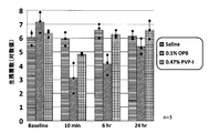
結果を図1に示す。ベースライン口腔内細菌数は1.73×10⁵~4.20×10⁶であった。Saline塗布後の口腔内細菌数はほぼ一定であった。試験物質塗布10分後の生菌数は、Saline、0.1%OPB及び0.47%PVP-Iでそれぞれ、6.48×10⁵CFU、4.65×10³CFU及び2.35×10⁴CFU、塗布6時間後でそれぞれ、1.66×10⁶CFU、1.47×10³CFU及び4.93×10⁵CFU、24時間後でそれぞれ、4.67×10⁵CFU、5.58×10⁴CFU及び1.22×10⁶CFUであった。 (1) Aerobic culture The results are shown in FIG. Baseline oral bacterial counts ranged from 1.73 × 10 5 to 4.20 × 10 6 . The number of bacteria in the oral cavity after applying Saline was almost constant. The viable cell counts 10 minutes after application of the test substance were 6.48 × 10 5 CFU, 4.65 × 10 3 CFU, and 2.35 × for Saline, 0.1% OPB, and 0.47% PVP-I, respectively. 10 4 CFU, 6 hours after application, 1.66 × 10 6 CFU, 1.47 × 10 3 CFU and 4.93 × 10 5 CFU, respectively, and 4.67 × 10 5 CFU, 5 hours after application, respectively. .58 × 10 4 CFU and 1.22 × 10 6 CFU.
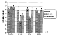
結果を図2に示す。ベースライン口腔内細菌数は2.85×10⁵~7.60×10⁷であった。Saline塗布後の口腔内細菌数はほぼ一定であった。試験物質塗布10分後の生菌数は、Saline、0.1%OPB及び0.47%PVP-Iでそれぞれ、1.26×10⁶CFU、5.97×10³CFU及び7.33×10⁴CFU、塗布6時間後でそれぞれ、5.51×10⁶CFU、2.79×10⁴CFU及び2.20×10⁶CFU、24時間後でそれぞれ、1.71×10⁶CFU、4.30×10⁵CFU及び7.81×10⁶CFUであった。 (2) The results of culture in a streptococcal selective medium are shown in FIG. Baseline oral bacterial counts ranged from 2.85 × 10 5 to 7.60 × 10 7 . The number of bacteria in the oral cavity after applying Saline was almost constant. The viable cell counts 10 minutes after application of the test substance were 1.26 × 10 6 CFU, 5.97 × 10 3 CFU and 7.33 × for Saline, 0.1% OPB and 0.47% PVP-I, respectively. 10 4 CFU, 6 hours after application, 5.51 × 10 6 CFU, 2.79 × 10 4 CFU and 2.20 × 10 6 CFU, respectively, 1.71 × 10 6 CFU, 4 hours after application It was .30 × 10 5 CFU and 7.81 × 10 6 CFU.
結果を図3に示す。ベースライン口腔内細菌数は2.45×10⁵~1.65×10⁷であった。Saline塗布後の口腔内細菌数はほぼ一定であった。試験物質塗布10分後の生菌数は、Saline、0.1%OPB及び0.47%PVP-Iでそれぞれ、2.70×10⁶CFU、1.33×10⁴CFU及び7.33×10⁴CFU、塗布6時間後でそれぞれ、3.22×10⁶CFU、1.38×10⁴CFU及び1.80×10⁶CFU、24時間後でそれぞれ、1.71×10⁶CFU、3.04×10⁵CFU及び1.40×10⁶CFUであった。 (3) Anaerobic culture The results are shown in FIG. Baseline oral bacterial counts ranged from 2.45 × 10 5 to 1.65 × 10 7 . The number of bacteria in the oral cavity after applying Saline was almost constant. The viable cell counts 10 minutes after application of the test substance were 2.70 × 10 6 CFU, 1.33 × 10 4 CFU, and 7.33 × for Saline, 0.1% OPB, and 0.47% PVP-I, respectively. 10 4 CFU, 6 hours after application, 3.22 × 10 6 CFU, 1.38 × 10 4 CFU and 1.80 × 10 6 CFU, 24 hours later, 1.71 × 10 6 CFU, 3 respectively 0.04 × 10 5 CFU and 1.40 × 10 6 CFU.
結果を図4に示す。ベースライン口腔内細菌数は1.29×10⁶~>1.00×10⁸であった。試験物質塗布10分後の生菌数は、Saline、0.1%OPB及び0.47%PVP-Iでそれぞれ、2.84×10⁶CFU、<3.49×10⁵CFU及び5.09×10⁵CFU、塗布6時間後でそれぞれ、2.04×10⁷CFU、5.58×10⁵CFU及び8.98×10⁶CFU、24時間後でそれぞれ、4.10×10⁷CFU、3.14×10⁷CFU及び3.14×10⁷CFUであった。 1-4-2 Bacterial counts in the oral cavity using a bacterial counter The results are shown in FIG. Baseline oral bacterial counts ranged from 1.29 × 10 6 to> 1.00 × 10 8 . The viable cell counts 10 minutes after application of the test substance were 2.84 × 10 6 CFU, <3.49 × 10 5 CFU and 5.09 for Saline, 0.1% OPB and 0.47% PVP-I, respectively. × 10 5 CFU, 6 hours after application, 2.04 × 10 7 CFU, 5.58 × 10 5 CFU and 8.98 × 10 6 CFU, respectively, and 4.10 × 10 7 CFU after 24 hours, 3.14 × 10 7 CFU and 3.14 × 10 7 CFU.
本試験では、正常ハムスター口腔内粘膜に対する含嗽剤(殺菌消毒剤)の殺菌効力を、他剤と比較検討することを目的とした。殺菌活性の持続性を検討するため、試験物質含嗽後~24時間まで時点(前置、直後、8時間後、24時間後)をとり、細菌カウンタ及び培養法を用い細菌数を計測した。 2.
The purpose of this study was to compare the bactericidal efficacy of a mouthwash (bactericidal disinfectant) on the oral mucosa of normal hamsters with other agents. In order to examine the persistence of the bactericidal activity, time points (before, immediately after, 8 hours, and 24 hours after) containing the test substance were taken, and the number of bacteria was counted using a bacterial counter and culture method.
被験物質と対照物質を合わせて試験物質とした。 2-1 Test substance The test substance and the control substance were combined as a test substance.
名称 :0.1%OPB-1
組成 :オラネキシジングルコン酸塩 0.10w/v%
Pluronic L-44 0.07w/v%
Pluronic P-123 1.0w/v% 2-1-1
Name: 0.1% OPB-1
Composition: olanexidine gluconate 0.10 w / v%
Pluronic L-44 0.07w / v%
Pluronic P-123 1.0 w / v%
名称 :0.1% OPB-2
組成 :オラネキシジングルコン酸塩 0.10w/v%
Pluronic L-44 0.07w/v%
Pluronic P-123 1.0w/v%
Lipidure(登録商標) 1.0w/v% 2-1-2
Name: 0.1% OPB-2
Composition: olanexidine gluconate 0.10 w / v%
Pluronic L-44 0.07w / v%
Pluronic P-123 1.0 w / v%
Lipidure (registered trademark) 1.0 w / v%
名称/略称:基剤/Base
組成 :Pluronic L-44 0.07w/v%
Pluronic P-123 1.0w/v% 2-1-3
Name / Abbreviation: Base / Base
Composition: Pluronic L-44 0.07 w / v%
Pluronic P-123 1.0 w / v%
名称/略称:Peridex(登録商標)/0.12%CHG
組成 :クロルヘキシジングルコン酸塩 0.12w/v% 2-1-4
Name / abbreviation: Peridex (registered trademark) /0.12% CHG
Composition: Chlorhexidine gluconate 0.12 w / v%
名称/略称:イソジンガーグル液0.47%/0.47%PVP-I
組成 :イソジンガーグル液7%(7%PVP-I、Meiji Seikaファルマ株式会社製)の15倍希釈液 2-1-5
Name / abbreviation: isodinger gargle solution 0.47% / 0.47% PVP-I
Composition: 15-fold diluted solution of
入荷時6週齢のハムスター、Slc:Syrian、雄を用い、各群4匹で試験を行った。 2-2 Animals Used A 6-week-old hamster, Slc: Syrian, and male were used at the time of arrival, and the test was conducted with 4 animals in each group.
ガス麻酔[導入麻酔:空気3.0L/minに3%イソフルラン(マイラン製薬株式会社製)、持続麻酔は適宜濃度を調整]を実施した。 2-3-1 Anesthesia Gas anesthesia [introduced anesthesia: 3% isoflurane (made by Mylan Pharmaceutical Co., Ltd., air at 3.0 L / min), concentration adjusted appropriately for continuous anesthesia] was performed.
麻酔下にて、ハムスターを仰臥位に固定し、片頬袋に試験物質を1mLずつ注入した。30秒後、試験物質を排出し、余分な頬袋内の試験物質を滅菌綿棒で吸い取った。 2-3-2 Test substance administration Under anesthesia, the hamster was fixed in the supine position, and 1 mL of the test substance was injected into each cheek pouch. After 30 seconds, the test substance was drained and the test substance in the excess cheek pouch was blotted with a sterile cotton swab.
試験物質投与前、0hr、8hr、24hrの計4時点に、麻酔下で両頬袋内から滅菌綿棒を用いて細菌を採取した。採取後の綿棒は5mL SCDLP培地に浸漬後撹拌し、細菌計数用サンプルとした。 2-3-3 Bacterial collection Bacteria were collected from both cheek bags under anesthesia using sterile cotton swabs at a total of 4 time points of 0 hr, 8 hr, and 24 hr before administration of the test substance. The collected swab was immersed in 5 mL SCDLP medium and stirred to prepare a sample for counting bacteria.
新GMP微生物試験法1)、食品衛生検査指針2)を参考にカンテン平板混釈法を実施した。
〔1〕細菌計数用サンプル500μLを採取し、4.5mLの希釈液を用いて、101倍~106倍の10倍希釈系列を作製した。
〔2〕細菌計数用サンプル原液及び各希釈菌液1mLずつ滅菌シャーレに分注した。
〔3〕上記のシャーレに、速やかに約47℃に設定した恒温槽で保温した測定培地(TSA+)15mLを分注した。
〔4〕測定培地の固化後、混釈平板をインキュベーター内に倒置し、コロニーが計数できるまで35℃で培養した(約2日間)。
〔5〕培養後、混釈平板で増殖したコロニーを目視にて計数した。コロニー数が多すぎてコロニー間の区別ができない混釈平板は、TNTC(too numerous to count)とし計数
しなかった。
〔6〕コロニー数に希釈倍率を乗じて、生残菌数を算出した。 2-3-4 Measurement of the number of surviving bacteria The Kanten plate pour method was carried out with reference to the new GMP microorganism test method 1) and the food hygiene inspection guideline 2).
[1] A 500 μL sample for bacterial counting was collected and a 10-fold dilution series of 10 1 to 10 6 times was prepared using 4.5 mL of diluent.
[2] Dispense 1 mL each of the stock solution for counting bacteria and 1 mL of each diluted bacterial solution into a sterile petri dish.
[3] 15 mL of measurement medium (TSA +) kept warm in a thermostat set at about 47 ° C. was quickly dispensed into the petri dish.
[4] After the measurement medium was solidified, the pour plate was inverted in an incubator and cultured at 35 ° C. until colonies could be counted (about 2 days).
[5] After cultivation, colonies that grew on the pouch plate were visually counted. A pour plate that has too many colonies to distinguish between colonies was not counted as TNTC (too numerous to count).
[6] The number of surviving bacteria was calculated by multiplying the number of colonies by the dilution factor.
結果を図5及び表2に示す。 2-4 Results The results are shown in FIG.

本試験では、正常ハムスター口腔内粘膜に対する含嗽剤(殺菌消毒剤)の殺菌効力に及ぼすオラネキシジン濃度の影響を検討することを目的とした。 3. Oral sterilization
The purpose of this study was to examine the effect of olanexidine concentration on the bactericidal efficacy of a mouthwash (bactericidal disinfectant) on the oral mucosa of normal hamsters.
被験物質と対照物質を合わせて試験物質とした。 3-1 Test substance The test substance and the control substance were combined as a test substance.
名称:0.1%OPB-1
組成:オラネキシジングルコン酸塩...0.10w/v%
ポリオキシエチレン(20)ポリオキシプロピレン(20)グリコール...0.07w/v%
ポリオキシエチレン(160)ポリオキシプロピレン(30)グリコール...0.10w/v% 3-1-1
Name: 0.1% OPB-1
Composition: olanexidine gluconate. . . 0.10 w / v%
Polyoxyethylene (20) polyoxypropylene (20) glycol. . . 0.07w / v%
Polyoxyethylene (160) polyoxypropylene (30) glycol. . . 0.10 w / v%
名称:0.1%OPB-2
組成:オラネキシジングルコン酸塩...0.10w/v%
ポリオキシエチレン(20)ポリオキシプロピレン(20)グリコール...0.07w/v%
ポリオキシエチレン(160)ポリオキシプロピレン(30)...1.00w/v% 3-1-2
Name: 0.1% OPB-2
Composition: olanexidine gluconate. . . 0.10 w / v%
Polyoxyethylene (20) polyoxypropylene (20) glycol. . . 0.07w / v%
Polyoxyethylene (160) polyoxypropylene (30). . . 1.00w / v%
名称:0.5%OPB-3
組成:オラネキシジングルコン酸塩...0.50w/v%
ポリオキシエチレン(20)ポリオキシプロピレン(20)...0.36w/v%
ポリオキシエチレン(160)ポリオキシプロピレン(30)...5.00w/v% 3-1-3
Name: 0.5% OPB-3
Composition: olanexidine gluconate. . . 0.50 w / v%
Polyoxyethylene (20) polyoxypropylene (20). . . 0.36w / v%
Polyoxyethylene (160) polyoxypropylene (30). . . 5.00 w / v%
名称:1%OPB-2
組成:オラネキシジングルコン酸塩...1.00w/v%
ポリオキシエチレン(20)ポリオキシプロピレン(20)グリコール...0.72w/v%
ポリオキシエチレン(160)ポリオキシプロピレン(30)グリコール...10.00w/v% 3-1-4
Name: 1% OPB-2
Composition: olanexidine gluconate. . . 1.00w / v%
Polyoxyethylene (20) polyoxypropylene (20) glycol. . . 0.72 w / v%
Polyoxyethylene (160) polyoxypropylene (30) glycol. . . 10.00w / v%
名称:基剤
組成:ポリオキシエチレン(20)ポリオキシプロピレン(20)グリコール...0.07w/v%
ポリオキシエチレン(160)ポリオキシプロピレン(30)グリコール...0.10w/v% 3-1-5 Control substance name: Base composition: Polyoxyethylene (20) polyoxypropylene (20) glycol. . . 0.07w / v%
Polyoxyethylene (160) polyoxypropylene (30) glycol. . . 0.10 w / v%
入荷時6週齢のハムスター、Slc:Syrian、雄を用い、各群3匹で試験を行った。 3-2 Animals used Three hamsters, Slc: Syrian, males, 6 weeks old at the time of arrival, were used for the test.
ガス麻酔[導入麻酔:空気3.0L/minに3%イソフルラン(マイラン製薬株式会社製)、持続麻酔は適宜濃度を調整]を実施した。 3-3-1 Anesthesia Gas anesthesia [introduction anesthesia: 3% isoflurane (produced by Mylan Pharmaceutical Co., Ltd. at air of 3.0 L / min), concentration adjusted appropriately for continuous anesthesia] was performed.
麻酔下にて、ハムスターを仰臥位に固定し、片頬袋に試験物質を1mLずつ注入した。1分後、試験物質を排出し、余分な頬袋内の試験物質を滅菌綿棒で吸い取った。 3-3-2 Test substance administration Under anesthesia, the hamster was fixed in the supine position, and 1 mL of the test substance was injected into each cheek bag. After 1 minute, the test substance was drained and the test substance in the excess cheek bag was blotted with a sterile cotton swab.
試験物質投与前、0hr、1hr、3hr、6hrの計5時点に、麻酔下で両頬袋内から滅菌綿棒を用いて細菌を採取した。採取後の綿棒は5mL SCDLP培地に浸漬後撹拌し、細菌計数用サンプルとした。 3-3-3 Bacterial collection Bacteria were collected from both cheek bags under anesthesia using sterile cotton swabs at a total of 5 time points of 0 hr, 1 hr, 3 hr, and 6 hr before administration of the test substance. The collected swab was immersed in 5 mL SCDLP medium and stirred to prepare a sample for counting bacteria.
生残菌数の測定は、2-3-4と同様の方法で行った。 3-3-4 Measurement of the number of surviving bacteria The number of surviving bacteria was measured by the same method as in 2-3-4.
結果を図6に示す。結果より、0.1%OPBで持続的(6時間後)に細菌数を低値に抑えることができることがわかった。また、OPB濃度が0.5w/v%以上で、持続活性がより優れていた。 3-4 Results The results are shown in FIG. From the results, it was found that the number of bacteria can be suppressed to a low value continuously (after 6 hours) with 0.1% OPB. In addition, the OPB concentration was 0.5 w / v% or more, and the sustained activity was more excellent.
本試験では、0.1%オラネキシジングルコン酸塩の基剤組成を変えた検討製剤をハムスターの頬袋に14日間反復投与し、刺激性の程度を比較検討した。 4). Oral
In this study, a study preparation in which the base composition of 0.1% olanexidine gluconate was changed was repeatedly administered to a hamster cheek pouch for 14 days, and the degree of irritation was compared.
名称 :0.1%OPB-1
組成 :オラネキシジングルコン酸塩 0.10w/v%
Pluronic L-44 0.07w/v% 4-1-1
Name: 0.1% OPB-1
Composition: olanexidine gluconate 0.10 w / v%
Pluronic L-44 0.07w / v%
名称 :0.1%OPB-2
組成 :オラネキシジングルコン酸塩 0.10w/v%
Pluronic L-44 0.07w/v%
Pluronic L-31 1.0w/v% 4-1-2
Name: 0.1% OPB-2
Composition: olanexidine gluconate 0.10 w / v%
Pluronic L-44 0.07w / v%
Pluronic L-31 1.0 w / v%
名称 :0.1%OPB-3
組成 :オラネキシジングルコン酸塩 0.10w/v%
Pluronic L-44 0.07w/v%
Pluronic P-123 1.0w/v% 4-1-3
Name: 0.1% OPB-3
Composition: olanexidine gluconate 0.10 w / v%
Pluronic L-44 0.07w / v%
Pluronic P-123 1.0 w / v%
名称 :0.1%OPB-4
組成 :オラネキシジングルコン酸塩 0.10w/v%
Pluronic L-44 0.07w/v%
Pluronic P-85 1.0w/v% 4-1-4
Name: 0.1% OPB-4
Composition: olanexidine gluconate 0.10 w / v%
Pluronic L-44 0.07w / v%
Pluronic P-85 1.0 w / v%
名称 :0.1%OPB-5
組成 :オラネキシジングルコン酸塩 0.10w/v%
Pluronic L-44 0.07w/v%
Pluronic F-127 1.0w/v% 4-1-5
Name: 0.1% OPB-5
Composition: olanexidine gluconate 0.10 w / v%
Pluronic L-44 0.07w / v%
Pluronic F-127 1.0 w / v%
名称 :0.1%OPB-6
組成 :オラネキシジングルコン酸塩 0.10w/v%
Pluronic L-44 0.14w/v%
Pluronic F-68 1.0w/v% 4-1-6
Name: 0.1% OPB-6
Composition: olanexidine gluconate 0.10 w / v%
Pluronic L-44 0.14w / v%
Pluronic F-68 1.0 w / v%
名称 :0.1%OPB-7
組成 :オラネキシジングルコン酸塩 0.10w/v%
Pluronic L-44 0.07w/v%
Trehalose 5.0w/v% 4-1-7
Name: 0.1% OPB-7
Composition: olanexidine gluconate 0.10 w / v%
Pluronic L-44 0.07w / v%
Trehalose 5.0 w / v%
入荷時8週齢のハムスター、Slc:Syrian、雄を用い、各群3匹で試験を行った。 4-2 Animals used Three-group animals were tested using hamsters, Slc: Syrian, and
右頬袋に被験物質1mLを適用した。 (1)
〔1〕ガス麻酔により麻酔を導入した[導入麻酔:空気3.0L/minに3%イソフルラン(マイラン製薬株式会社製)]。
〔2〕維持麻酔下(濃度は適宜調整)にて動物を仰臥位に固定し、綿棒を用いて動物の頬袋を引き出し、片手で引き出した頬袋を軽くつまんだ。
〔3〕生理食塩液及び綿棒で頬袋粘膜に付着した飼料等の異物を取り除き、清潔にした後頬袋を元に戻した。
〔4〕1mLシリンジ及び経口投与用ゾンデを用いて、右頬袋には被験物質1mLを適用し、左頬袋には1mLシリンジに取り付けた空の経口投与用ゾンデを挿入・抜去した。
〔5〕適用30秒後、被験物質が気道内へ逆流しないように動物を腹臥位に反転させ排出した。口腔内の余分な被験物質は綿棒を用いて全て除去した。
〔6〕適用部位の頬粘膜の色調等を観察・記録し、動物の頚部にハムスター用カラーを装着した後、動物をケージに戻した。
〔7〕上記の操作を1日2回(朝・夕)、14日間繰り返した。 (2) Application method [1] Anesthesia was introduced by gas anesthesia [introduction anesthesia: 3% isoflurane (produced by Mylan Pharmaceutical Co., Ltd. in air 3.0 L / min)].
[2] The animal was fixed in a supine position under maintenance anesthesia (concentration was adjusted as appropriate), the animal's cheek bag was pulled out using a cotton swab, and the cheek bag pulled out with one hand was lightly pinched.
[3] Foreign matter such as feed adhering to the cheek pouch mucosa was removed with a physiological saline solution and a cotton swab, and the cheek pouch was returned to its original position after being cleaned.
[4] Using a 1 mL syringe and an oral administration sonde, 1 mL of the test substance was applied to the right cheek bag, and an empty oral administration sonde attached to the 1 mL syringe was inserted and removed from the left cheek bag.
[5] Thirty seconds after application, the animal was inverted to the prone position and discharged so that the test substance did not flow back into the respiratory tract. Any excess test substance in the oral cavity was removed using a cotton swab.
[6] The color tone and the like of the buccal mucosa at the application site were observed and recorded, and a hamster collar was attached to the neck of the animal, and then the animal was returned to the cage.
[7] The above operation was repeated twice a day (morning and evening) for 14 days.
各群の全例について、適用期間中は、試験物質の適用前及び適用終了時に一般状態観察を行った(Day1~Day14)。また、適用期間終了日の翌日にも観察を行った(Day15)。 (1) Observation of general condition General conditions were observed for all cases in each group before and after application of the test substance during the application period (
各群の全例において、適用期間中は、試験物質の適用前に体重を測定した(Day1~Day14)。また、適用期間終了日の翌日にも測定を行った(Day15)。ただし、体重計の故障のためDay13、14は体重を測定しなかった。 (2) Body weight measurement In all cases of each group, body weight was measured before application of the test substance during the application period (
各群の全例の頬袋について、適用期間中は、試験物質の適用前及び適用終了時に頬袋粘膜の状態を観察し、評点化を記録した(Day1~Day14)。また、適用期間終了日の翌日(24±2時間)にも観察を行った(Day15)。観察部位は各試験物質の接触部位頬粘膜とした。なお、肉眼的観察の評価法は以下の表3(ISO 10993-10, Annex B.3「Table B.2 Grading system for oral and penile reactions」)に記載されている観察基準及び評価点に従って、紅斑及び痂皮形成の程度に評価点(口内炎grade)をつけた。また、その他に見られた所見についても記録した。得られた観察結果をもとに、各群について試験物質ごとに、各動物の粘膜における評価点を合計し、観察数及び動物数で除して平均値(小数第一位四捨五入)を求め、総合評価の参考資料とした。 (3) Macroscopic observation method of application site For all the cheek bags of each group, during the application period, the condition of the cheek pouch mucosa was observed before and at the end of the application of the test substance, and the scoring was recorded (
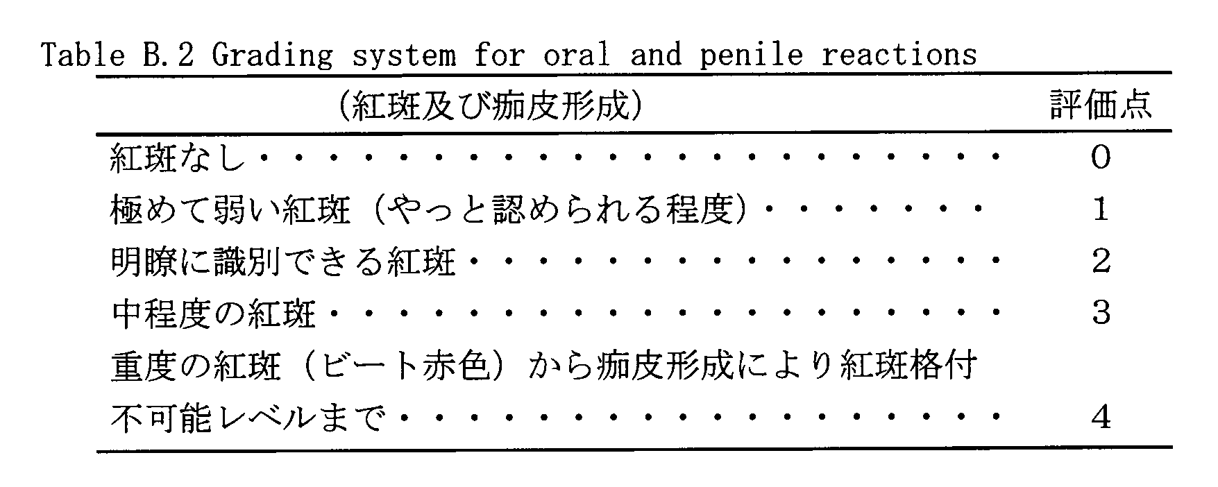
各動物について、適用期間終了日の翌日の肉眼的観察の終了後、イソフルラン麻酔下で放血致死させ、左右頬袋を採取し、10%中性緩衝ホルマリン液で固定した。常法に従いHE染色の標本を作製し、病理学的検査を実施した。なお、肉眼的観察の評価法はISO 10993-10, Annex B.3 「Table B.3 Grading system for microscopic examination for oral, penile, rectal and vaginal tissue reaction」に記載されている基準に従って、上皮、白血球浸潤、血管充血及び浮腫の各項目について所見又はグレードを記録した。また、その他に認められた所見についても記録した。 (4) Pathological examination After completion of macroscopic observation on the day after the end of the application period for each animal, blood was lethal under isoflurane anesthesia, and left and right cheek bags were collected and fixed with 10% neutral buffered formalin solution. According to a conventional method, a HE-stained specimen was prepared and a pathological examination was performed. The evaluation method for macroscopic observation is epithelial, leukocyte, in accordance with the criteria described in ISO 10993-10, Annex B.3 `` Table B.3 Grading system for microscopic examination for oral, penile, rectal and vaginal tissue reaction ''. Observations or grades were recorded for each item of invasion, vascular hyperemia and edema. In addition, other findings were recorded.
観察期間中の一般状態及び体重推移を参考にし、頬粘膜の肉眼的観察結果及び病理学的観察結果から得られた各試験物質の反応程度をもとにして、各試験物質の口腔粘膜への影響を総合的に評価した。 (5) Comprehensive evaluation Each test substance based on the degree of reaction of each test substance obtained from gross observation results and pathological observation results of the buccal mucosa with reference to the general state and weight transition during the observation period Was comprehensively evaluated on the oral mucosa.
いずれの動物にも異常は認められなかった。 4-4-1 General condition No abnormalities were observed in any animals.
結果を図7に示した。0.1%OPB-5群は体重がほとんど変化しなかった。他の群は平均値が徐々に増加した。 4-4-2 Body weight The results are shown in FIG. The 0.1% OPB-5 group showed almost no change in body weight. The other groups gradually increased in average value.
結果を図8に示し、最終日の各群の頬袋を図9のPhoto1~8に示した。全ての製剤で紅斑等の刺激性がほとんど認められなかった。しかし、0.1%OPB-1、-2、-6、-7及び-8では、白板様症状(角化亢進、肥厚)が認められた。一方、0.1%OPB-3、-4及び-5では全く異常が認められなかった。 4-4-3 Macroscopic observation of application site The results are shown in FIG. 8, and cheek bags of each group on the final day are shown in
結果を図10に示した。ISO 10993-10、Annex B.3「Table B.3 Grading system for microscopic examination for oral, penile, rectal and vaginal tissue reaction」に記載されている評価基準に従って、上皮(細胞変性、化生及びびらん)、白血球浸潤、血管充血及び浮腫のグレード付けにより、個体毎の炎症指数及び群毎の炎症指数の平均を算出した。また、評価基準以外の所見についても記録した。その結果、平均値は0.1%OPB-3群では変化は認められなかった。0.1%OPB-1群を含む他の群では、上皮の細胞変性及び極小~中等度の白血球浸潤が認められ、炎症指数は1~3の極小と評価された。これらの群では、評価基準以外の所見として、軽微な細胞間浮腫及び軽微から軽度の過角化が認められた。 4-4-4 Histopathological examination The results are shown in FIG. In accordance with the evaluation criteria described in ISO 10993-10, Annex B.3 “Table B.3 Grading system for microscopic examination for oral, penile, rectal and vaginal tissue reaction”, epithelium (cell degeneration, metaplasia and erosion), The average of the inflammation index for each individual and the inflammation index for each group was calculated by grading leukocyte infiltration, vascular hyperemia and edema. In addition, findings other than the evaluation criteria were recorded. As a result, the average value was not changed in the 0.1% OPB-3 group. In other groups, including the 0.1% OPB-1 group, epithelial cell degeneration and minimal to moderate leukocyte infiltration were observed, and the inflammation index was evaluated to be a minimum of 1-3. In these groups, slight intercellular edema and slight to mild hyperkeratosis were observed as findings other than the evaluation criteria.
実施例4の刺激性試験において、基剤Pluronic P-123が、オラネキシジングルコン酸塩の口腔粘膜適用製剤の基剤として有用であることが示唆された。そこで、本試験では、刺激のない製剤を作製するための基剤としてPluronic P-123を採用し、引き続きOPB濃度及び基剤濃度の検討を行った。試験系は、ハムスター頬袋への反復投与とし、期間を2週間から4週間に延ばし実施した。 5). Oral mucosal irritation
In the irritation test of Example 4, it was suggested that the base Pluronic P-123 is useful as a base for an oral mucosal preparation of olanexidine gluconate. Therefore, in this test, Pluronic P-123 was adopted as a base for preparing an unstimulated preparation, and OPB concentration and base concentration were subsequently examined. The test system was repeated administration to the hamster cheek pouch, and the period was extended from 2 weeks to 4 weeks.
名称 :0.1%OPB-1
組成 :オラネキシジングルコン酸塩 0.10w/v%
Pluronic L-44 0.07w/v%
Pluronic P-123 0.50w/v% 5-1-1
Name: 0.1% OPB-1
Composition: olanexidine gluconate 0.10 w / v%
Pluronic L-44 0.07w / v%
Pluronic P-123 0.50w / v%
名称 :0.1%OPB-2
組成 :オラネキシジングルコン酸塩 0.10w/v%
Pluronic L-44 0.07w/v%
Pluronic P-123 1.0 w/v% 5-1-2
Name: 0.1% OPB-2
Composition: olanexidine gluconate 0.10 w / v%
Pluronic L-44 0.07w / v%
Pluronic P-123 1.0 w / v%
名称 :0.1%OPB-3
組成 :オラネキシジングルコン酸塩 0.10w/v%
Pluronic L-44 0.07w/v%
Pluronic P-123 0.50w/v%
Lipidure(登録商標) 1.0 w/v% 5-1-3
Name: 0.1% OPB-3
Composition: olanexidine gluconate 0.10 w / v%
Pluronic L-44 0.07w / v%
Pluronic P-123 0.50w / v%
Lipidure (registered trademark) 1.0 w / v%
名称 :0.1%OPB-4
組成 :オラネキシジングルコン酸塩 0.10w/v%
Pluronic L-44 0.07w/v%
Pluronic P-123 1.0 w/v%
Lipidure(登録商標) 1.0 w/v% 5-1-4
Name: 0.1% OPB-4
Composition: olanexidine gluconate 0.10 w / v%
Pluronic L-44 0.07w / v%
Pluronic P-123 1.0 w / v%
Lipidure (registered trademark) 1.0 w / v%
名称 :0.3%OPB-1
組成 :オラネキシジングルコン酸塩 0.30w/v%
Pluronic L-44 0.22w/v%
Pluronic P-123 1.50w/v% 5-1-5
Name: 0.3% OPB-1
Composition: Oranexidine gluconate 0.30 w / v%
Pluronic L-44 0.22w / v%
Pluronic P-123 1.50 w / v%
名称 :0.3%OPB-2
組成 :オラネキシジングルコン酸塩 0.30w/v%
Pluronic L-44 0.22w/v%
Pluronic P-123 3.0 w/v% 5-1-6
Name: 0.3% OPB-2
Composition: Oranexidine gluconate 0.30 w / v%
Pluronic L-44 0.22w / v%
Pluronic P-123 3.0 w / v%
名称 :0.3%OPB-3
組成 :オラネキシジングルコン酸塩 0.30w/v%
Pluronic L-44 0.22w/v%
Pluronic P-85 1.50w/v% 5-1-7
Name: 0.3% OPB-3
Composition: Oranexidine gluconate 0.30 w / v%
Pluronic L-44 0.22w / v%
Pluronic P-85 1.50 w / v%
名称 :0.3%OPB-4
組成 :オラネキシジングルコン酸塩 0.30w/v%
Pluronic L-44 0.22w/v%
Pluronic P-85 3.0 w/v% 5-1-8
Name: 0.3% OPB-4
Composition: Oranexidine gluconate 0.30 w / v%
Pluronic L-44 0.22w / v%
Pluronic P-85 3.0 w / v%
名称 :0.5%OPB-1
組成 :オラネキシジングルコン酸塩 0.50w/v%
Pluronic L-44 0.36w/v%
Pluronic P-123 2.50w/v% 5-1-9
Name: 0.5% OPB-1
Composition: Oranexidine gluconate 0.50 w / v%
Pluronic L-44 0.36w / v%
Pluronic P-123 2.50 w / v%
名称 :0.5%OPB-2
組成 :オラネキシジングルコン酸塩 0.50w/v%
Pluronic L-44 0.36w/v%
Pluronic P-123 5.0 w/v% 5-1-10
Name: 0.5% OPB-2
Composition: Oranexidine gluconate 0.50 w / v%
Pluronic L-44 0.36w / v%
Pluronic P-123 5.0 w / v%
名称 :0.5%OPB-3
組成 :オラネキシジングルコン酸塩 0.50w/v%
Pluronic L-44 0.36w/v%
Pluronic P-123 2.50w/v%
Lipidure(登録商標) 1.0 w/v% 5-1-11
Name: 0.5% OPB-3
Composition: Oranexidine gluconate 0.50 w / v%
Pluronic L-44 0.36w / v%
Pluronic P-123 2.50 w / v%
Lipidure (registered trademark) 1.0 w / v%
名称 :0.5%OPB-4
組成 :オラネキシジングルコン酸塩 0.50w/v%
Pluronic L-44 0.36w/v%
Pluronic P-123 5.0 w/v%
Lipidure(登録商標) 1.0 w/v% 5-1-12
Name: 0.5% OPB-4
Composition: Oranexidine gluconate 0.50 w / v%
Pluronic L-44 0.36w / v%
Pluronic P-123 5.0 w / v%
Lipidure (registered trademark) 1.0 w / v%
入荷時8週齢のハムスター、Slc:Syrian、雄を用い、各群3匹で試験を行った。 5-2 Animals used Three-group animals were tested using 8 weeks old hamsters, Slc: Syrian and males at the time of arrival.
左頬袋に被験物質1mLを適用した。 (1)
〔1〕ガス麻酔により麻酔を導入した[導入麻酔:空気3.0L/minに3%イソフルラン(マイラン製薬株式会社製)]。
〔2〕維持麻酔下(濃度は適宜調整)にて動物を仰臥位に固定し、綿棒を用いて動物の頬袋を引き出し、片手で引き出した頬袋を軽くつまんだ。
〔3〕生理食塩液及び綿棒で頬袋粘膜に付着した飼料等の異物を取り除き、清潔にした後頬袋を元に戻した。
〔4〕1mLシリンジ及び経口投与用ゾンデを用いて、左頬袋には被験物質1mLを適用し、右頬袋には1mLシリンジに取り付けた空の経口投与用ゾンデを挿入・抜去した。
〔5〕適用30秒後、被験物質が気道内へ逆流しないように動物を腹臥位に反転させ排出した。口腔内の余分な被験物質は綿棒を用いて全て除去した。
〔6〕適用部位の頬粘膜の色調等を観察・記録し、動物をケージに戻した。
〔7〕上記の操作を1日2回(朝・夕)、28日間繰り返した。 (2) Application method [1] Anesthesia was introduced by gas anesthesia [introduction anesthesia: 3% isoflurane (produced by Mylan Pharmaceutical Co., Ltd. in air 3.0 L / min)].
[2] The animal was fixed in a supine position under maintenance anesthesia (concentration was adjusted as appropriate), the animal's cheek bag was pulled out using a cotton swab, and the cheek bag pulled out with one hand was lightly pinched.
[3] Foreign matter such as feed adhering to the cheek pouch mucosa was removed with a physiological saline solution and a cotton swab, and the cheek pouch was returned to its original position after being cleaned.
[4] Using a 1 mL syringe and an oral administration sonde, 1 mL of the test substance was applied to the left cheek bag, and an empty oral administration sonde attached to the 1 mL syringe was inserted and removed from the right cheek bag.
[5] Thirty seconds after application, the animal was inverted to the prone position and discharged so that the test substance did not flow back into the respiratory tract. Any excess test substance in the oral cavity was removed using a cotton swab.
[6] The color tone of the buccal mucosa at the application site was observed and recorded, and the animal was returned to the cage.
[7] The above operation was repeated twice a day (morning and evening) for 28 days.
各群の全例について、適用期間中は、試験物質の適用前及び適用終了時に一般状態観察を行った(Day1~Day28)。また、適用期間終了日の翌日にも観察を行った(Day29)。 (1) Observation of general state For all cases in each group, the general state was observed before and after the application of the test substance during the application period (
各群の全例において、適用期間中は、試験物質の適用前に体重を測定した(Day1~Day28)。また、適用期間終了日の翌日にも測定を行った(Day29)。 (2) Body weight measurement In all cases of each group, body weight was measured before application of the test substance during the application period (
各群の全例の頬袋について、適用期間中は、試験物質の適用前に頬袋粘膜の状態を観察し、評点化を記録した(Day1~Day28)。また、適用期間終了日の翌日(24±2時間)にも観察を行った(Day29)。観察部位は各試験物質の接触部位頬粘膜とした。なお、肉眼的観察の評価法は前記の表3(ISO 10993-10, Annex B.3「Table B.2 Grading system for oral and penile reactions」)に記載されている観察基準及び評価点に従って、紅斑及び痂皮形成の程度に評価点(口内炎grade)をつけた。また、その他に見られた所見についても記録した。得られた観察結果をもとに、各群について試験物質ごとに、各動物の粘膜における評価点を合計し、観察数及び動物数で除して平均値(小数第一位四捨五入)を求め、総合評価の参考資料とした。 (3) Macroscopic observation method of application site For all cases of cheek pouches in each group, during the application period, the condition of the cheek pouch mucosa was observed before application of the test substance, and scoring was recorded (
各動物について、適用期間終了日の翌日の肉眼的観察の終了後、イソフルラン麻酔下で放血致死させ、左右頬袋を採取し、10%中性緩衝ホルマリン液で固定した。常法に従いHE染色の標本を作製し、病理学的検査を実施した。なお、肉眼的観察の評価法はISO 10993-10, Annex B.3 「Table B.3 Grading system for microscopic examination for oral, penile, rectal and vaginal tissue reaction」に記載されている基準に従って、上皮、白血球浸潤、血管充血及び浮腫の各項目について所見又はグレードを記録した。また、その他に認められた所見についても記録した。 (4) Pathological examination After completion of macroscopic observation on the day after the end of the application period for each animal, blood was lethal under isoflurane anesthesia, and left and right cheek bags were collected and fixed with 10% neutral buffered formalin solution. According to a conventional method, a HE-stained specimen was prepared and a pathological examination was performed. The evaluation method for macroscopic observation is epithelial, leukocyte, in accordance with the criteria described in ISO 10993-10, Annex B.3 `` Table B.3 Grading system for microscopic examination for oral, penile, rectal and vaginal tissue reaction ''. Observations or grades were recorded for each item of invasion, vascular hyperemia and edema. In addition, other findings were recorded.
観察期間中の一般状態及び体重推移を参考にし、頬粘膜の肉眼的観察結果及び病理学的観察結果から得られた各試験物質の反応程度をもとにして、各試験物質の口腔粘膜への影響を総合的に評価した。 (5) Comprehensive evaluation Each test substance based on the degree of reaction of each test substance obtained from gross observation results and pathological observation results of the buccal mucosa with reference to the general state and weight transition during the observation period Was comprehensively evaluated on the oral mucosa.
いずれの動物にも異常は認められなかった。 5-4-1 General condition No abnormalities were observed in any animals.
すべての群で経時的に体重が増加し、群間による差はほとんど無かった。 5-4-2 Body weight Body weight increased over time in all groups with little difference between groups.
結果を図11に示した。全ての製剤で紅斑等の刺激性が認められなかった(評価点0)。しかし、0.3%濃度以上のOPBでは、白板様症状(角化亢進、肥厚)が認められた。一方、0.1%濃度のOPBでは全く異常が認められなかった。 5-4-3 Macroscopic observation of the application site The results are shown in FIG. No irritation such as erythema was observed in all preparations (evaluation score 0). However, white board-like symptoms (increased keratinization, thickening) were observed with OPB at a concentration of 0.3% or more. On the other hand, no abnormality was observed with 0.1% OPB.
結果を図12に示した。ISO 10993-10、Annex B.3「Table B.3 Grading system for microscopic examination for oral, penile, rectal and vaginal tissue reaction」に記載されている評価基準に従って、上皮(細胞変性、化生及びびらん)、白血球浸潤、血管充血及び浮腫のグレード付けにより、個体毎の炎症指数及び群毎の炎症指数の平均を算出した。また、評価基準以外の所見についても記録した。その結果、0.1%OPBの各群では炎症性の反応は認められなかった。0.3%濃度以上のOPB各群では、上皮の変性及び白血球浸潤が認められ、炎症指数は1~3でいずれも極小の反応であった。評価基準以外の所見として、0.1%OPB群では一部の個体に軽微~軽度の過角化が認められ、0.3%濃度以上のOPB各群では、軽微~中程度の過角化及び軽微な棘細胞増生が認められた。その他コントロール群(右頬袋:Sham-ope側)で認められた表皮内微小膿瘍は自然発症性の病変と考えられた。 5-4-4 Histopathological examination The results are shown in FIG. In accordance with the evaluation criteria described in ISO 10993-10, Annex B.3 “Table B.3 Grading system for microscopic examination for oral, penile, rectal and vaginal tissue reaction”, epithelium (cell degeneration, metaplasia and erosion), The average of the inflammation index for each individual and the inflammation index for each group was calculated by grading leukocyte infiltration, vascular hyperemia and edema. In addition, findings other than the evaluation criteria were recorded. As a result, no inflammatory reaction was observed in each group of 0.1% OPB. In each OPB group having a concentration of 0.3% or more, epithelial degeneration and leukocyte infiltration were observed, and the inflammation index was 1 to 3, and all were minimal responses. As a finding other than the evaluation criteria, slight to mild hyperkeratinization was observed in some individuals in the 0.1% OPB group, and slight to moderate hyperkeratinization in each OPB group with a concentration of 0.3% or higher. In addition, slight spine cell proliferation was observed. Intraepidermal microabscess observed in the other control group (right cheek pouch: Sham-ope side) was considered to be a spontaneous lesion.
本試験では、5-FU誘発口内炎モデルにおけるOPB製剤の効力を試験した。具体的には、5-FU誘発口内炎モデルにおいて、0.1%OPBで口腔内を含嗽した際の口腔内細菌数の経時的な測定及び口内炎評価を行った。 6.5
In this study, the efficacy of the OPB formulation in a 5-FU-induced stomatitis model was tested. Specifically, in the 5-FU-induced stomatitis model, the time-lapse measurement of the number of bacteria in the oral cavity and the evaluation of stomatitis when the oral cavity was impregnated with 0.1% OPB were performed.
被験物質と対照物質を合わせて試験物質とした。 6-1 Test substance The test substance and the control substance were combined as a test substance.
名称:0.1%OPB
組成:オラネキシジングルコン酸塩...0.10w/v%
ポリオキシエチレン(20)ポリオキシプロピレン(20)グリコール...0.14w/v%
ポリオキシエチレン(160)ポリオキシプロピレン(30)グリコール...0.10w/v% 6-1-1 Test substance name: 0.1% OPB
Composition: olanexidine gluconate. . . 0.10 w / v%
Polyoxyethylene (20) polyoxypropylene (20) glycol. . . 0.14 w / v%
Polyoxyethylene (160) polyoxypropylene (30) glycol. . . 0.10 w / v%
名称:基剤
組成:ポリオキシエチレン(20)ポリオキシプロピレン(20)グリコール...0.07w/v%
ポリオキシエチレン(160)ポリオキシプロピレン(30)グリコール...0.10w/v% 6-1-2 Control substance name: Base composition: Polyoxyethylene (20) polyoxypropylene (20) glycol. . . 0.07w / v%
Polyoxyethylene (160) polyoxypropylene (30) glycol. . . 0.10 w / v%
入荷時6週齢のハムスター、Slc:Syrian、雄を用い、各群5匹で試験を行った。 6-2 Animals Used A hamster, Slc: Syrian, male, which was 6 weeks old at the time of arrival, was used to test 5 animals in each group.
ガス麻酔[導入麻酔:空気3.0L/minに3%イソフルラン(マイラン製薬株式会社製)、持続麻酔は適宜濃度を調整]を実施した。 6-3-1 Anesthesia Gas anesthesia [introductory anesthesia: 3% isoflurane (produced by Mylan Pharmaceutical Co., Ltd. at air of 3.0 L / min), the concentration is appropriately adjusted for continuous anesthesia] was performed.
麻酔下にて、5-FUを60mg/kgとなるようにハムスターの腹腔内に投与した。投与はday0、day2の計2回とした。 6-3-2 Preparation of stomatitis model Under anesthesia, 5-FU was administered intraperitoneally to a hamster so as to be 60 mg / kg. Administration was made twice,
麻酔下にて、ハムスターを仰臥位に固定し、片頬袋に試験物質を1mLずつ注入した。30秒後、試験物質を排出し、余分な頬袋内の試験物質を滅菌綿棒で吸い取った。この含嗽操作による投与は1日2回とした。投与は口内炎の障害がピークに達した後は実施しなかった。 6-3-3 Test substance administration Under anesthesia, the hamster was fixed in the supine position, and 1 mL of the test substance was injected into each cheek bag. After 30 seconds, the test substance was drained and the test substance in the excess cheek pouch was blotted with a sterile cotton swab. Administration by this gargle operation was performed twice a day. Administration was not performed after the peak of stomatitis was reached.
Day0、4、7、10、17において、1回目の試験物質投与前、0hr、6hr後の計4時点に、麻酔下で両頬袋内から滅菌綿棒を用いて細菌を採取した。但し、day10、17において試験物質適用しなかった場合は、1回のみの採取とした。採取後の綿棒は5mL SCDLP培地に浸漬後撹拌し、細菌計数用サンプルとした。 6-3-4 Bacteria collection In
新GMP微生物試験法1)、食品衛生検査指針2)を参考にカンテン平板混釈法を実施した。
〔1〕細菌計数用サンプル500μLを採取し、4.5mLの希釈液を用いて、101倍~104倍の10倍希釈系列を作製した。
〔2〕細菌計数用サンプル原液及び各希釈菌液1mLずつ滅菌シャーレに分注した。
〔3〕上記のシャーレに、速やかに約47℃に設定した恒温槽で保温した測定培地(TSA+)15mLを分注した。
〔4〕測定培地の固化後、混釈平板をインキュベーター内に倒置し、コロニーが計数できるまで35℃で培養した(約2日間)。
〔5〕培養後、混釈平板で増殖したコロニーを目視にて計数した。コロニー数が多すぎてコロニー間の区別ができない混釈平板は、TNTC(too numerous to count)とし計数しなかった。 6-3-5 Measurement of the number of surviving bacteria The Kanten plate pour method was carried out with reference to the new GMP microorganism test method 1) and the food hygiene inspection guideline 2).
[1] A 500 μL sample for bacterial counting was collected and a 10-fold dilution series of 10 1 to 10 4 times was prepared using 4.5 mL of diluent.
[2] Dispense 1 mL each of the stock solution for counting bacteria and 1 mL of each diluted bacterial solution into a sterile petri dish.
[3] 15 mL of measurement medium (TSA +) kept warm in a thermostat set at about 47 ° C. was quickly dispensed into the petri dish.
[4] After the measurement medium was solidified, the pour plate was inverted in an incubator and cultured at 35 ° C. until colonies could be counted (about 2 days).
[5] After cultivation, colonies that grew on the pouch plate were visually counted. A pour plate that has too many colonies to distinguish between colonies was not counted as TNTC (too numerous to count).
6-3-5の項に基づいて採用したコロニー数に希釈倍数を乗じ生残菌数(CFU/mL)を求めた。採用コロニー数は小数点以下第2位を四捨五入して表示した。下式により生残菌数(CFU/swab)を計算した。
A:採用コロニー数
生残菌数(CFU/swab)=A×希釈倍数×サンプル液量(5mL) 6-3-6 Calculation of Surviving Bacteria The number of surviving bacteria (CFU / mL) was obtained by multiplying the number of colonies adopted based on the item 6-3-5 by the dilution factor. The number of colonies adopted was rounded off to the second decimal place. The number of surviving bacteria (CFU / swab) was calculated by the following formula.
A: Adopted colony survival cell count (CFU / swab) = A × dilution factor × sample solution amount (5 mL)
B:ベースラインの生菌数対数値
C:試験物質塗布後の生菌数対数値
Log Reduction=B-C Furthermore, Log Reduction was calculated from the logarithmic value of the number of surviving bacteria by the following formula. Log Reduction is displayed by rounding off the third decimal place. The logarithmic value of 1 or less survival bacteria was set to 0.
B: Viable count of base line logarithm value C: Logarithm of viable count after application of test substance Log Reduction = BC
各群の生菌数(CFU/swab)とその対数値の平均値及び標準偏差を求めた。生菌数は小数点以下第1位を四捨五入して整数で表示した。生菌数の対数値は小数点以下第3位を四捨五入して表示した。なお、生菌数が0の場合は生菌数の対数値は0とした。また、探索試験のため検定は行わなかった。 6-3-7 Statistical Analysis The average number and standard deviation of the viable cell count (CFU / swab) and the logarithmic value of each group were determined. The number of viable bacteria was rounded off to the first decimal place and displayed as an integer. The logarithm of the viable count was rounded off to the second decimal place. In addition, when the viable count was 0, the logarithmic value of the viable count was 0. In addition, no test was conducted for exploratory testing.
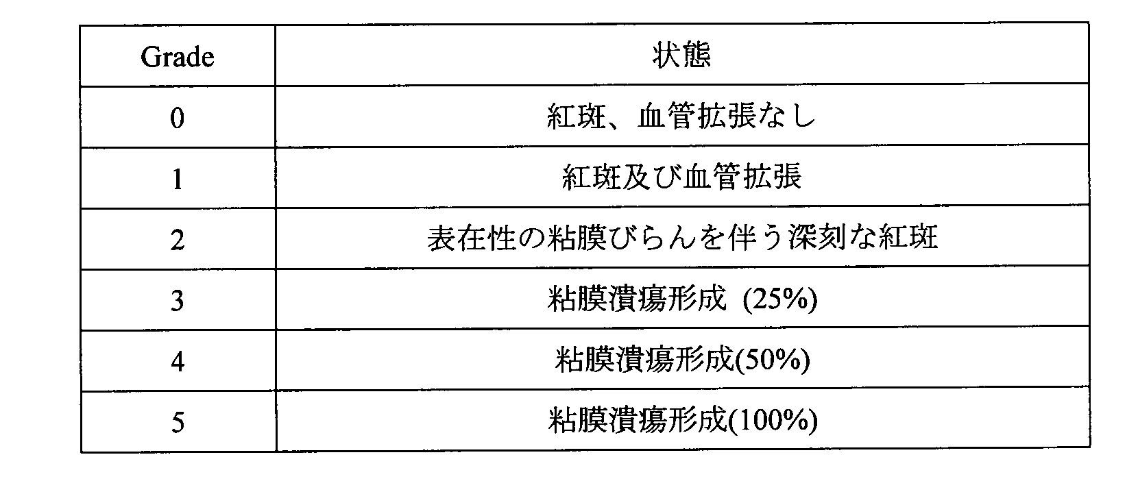
以下の表4に基づき、口内炎gradeを評価した。 6-3-8 Stomatitis Evaluation Stomatitis grade was evaluated based on Table 4 below.
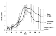
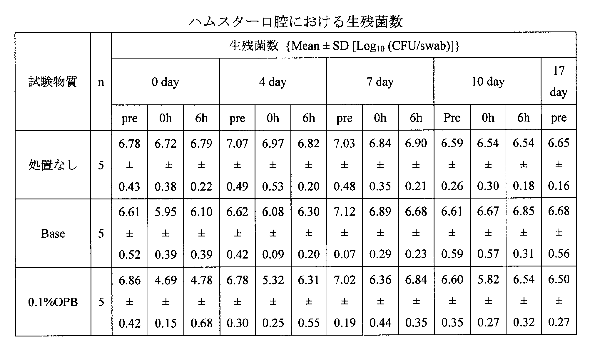
結果を表5及び図13に示す。Day0、4、10においては、0.1%OPB群で投与後の細菌数減少が顕著であったが、口内炎の重症化が顕著であったday7では、細菌数減少値が微量であった。 6-4-1 Number of bacteria The results are shown in Table 5 and FIG. In
結果を図14に示した。0.1%OPB群において、口内炎gradeが顕著に低かった。 6-4-2 Stomatitis grade
The results are shown in FIG. Stomatitis grade was remarkably low in the 0.1% OPB group.
本試験では、5-FU誘発ハムスター口内炎モデルにおいて、0.1%オラネキシジングルコン酸塩と0.1%CHGの口内炎軽減効果を比較検討した。
In this study, the effect of reducing stomatitis by 0.1% oranexidine gluconate and 0.1% CHG was compared in a 5-FU-induced hamster stomatitis model.
被験物質と対照物質を合わせて試験物質とした。 7-1 Test substance The test substance and the control substance were combined as a test substance.
名称 :0.1%OPB
組成 :オラネキシジングルコン酸塩 0.10w/v%
Pluronic L-44 0.07w/v% 7-1-1
Name: 0.1% OPB
Composition: olanexidine gluconate 0.10 w / v%
Pluronic L-44 0.07w / v%
名称 :Peridex(登録商標)/0.1%CHG
製造元 :3M ESPE Dental Products
組成 :クロルヘキシジングルコン酸塩 0.12w/v% 7-1-2
Name: Peridex (registered trademark) /0.1% CHG
Manufacturer: 3M ESPE Dental Products
Composition: Chlorhexidine gluconate 0.12 w / v%
名称 :基剤
組成 :ポリオキシエチレン(20)ポリオキシプロピレン(20)グリコール...0.07w/v% 7-1-3 Control substance name: Base composition: Polyoxyethylene (20) polyoxypropylene (20) glycol. . . 0.07w / v%
本試験には、口腔内の常在菌であるスタフィロコッカス・アウレウス(Staphylococcusaureus、ATCC番号:6538、Microbiologics, Inc.社製)を用いた。 7-2 Bacteria used Staphylococcus aureus (Staphylococcusaureus, ATCC number: 6538, manufactured by Microbiologics, Inc.), which is a resident bacteria in the oral cavity, was used in this test.
入荷時6週齢のハムスター、Slc:Syrian、雄を用い、各群5匹で試験を行った。 7-3 Animals Used A hamster, Slc: Syrian, male, 6 weeks old at the time of arrival, was used to test 5 animals in each group.
〔1〕保管されている細菌ペレットの入ったバイアルを取り出し室温に戻した。
〔2〕バイアルから細菌ペレットを1個取り出し、滅菌チューブに移した。
〔3〕0.5mLの生理食塩液を加えた。
〔4〕滅菌綿棒で細菌ペレットを押しつぶし、菌懸濁液とした。
〔5〕TSA平板に菌懸濁液を滅菌綿棒で直径約2cmの円形エリアに接種し、白金耳を用いて接種エリアから画線接種した。
〔6〕画線接種されたTSA平板を倒置し、コロニーを形成するまで培養した。
〔7〕形成されたコロニーの中から単一のコロニーを選び、白金線で採取してカジトン培地に穿刺した。
〔8〕穿刺したカジトン培地を、菌の増殖が確認できるまで培養した。
〔9〕菌の増殖を確認後、冷蔵保管した(設定値2~8℃)。
〔10〕カジトン培地で冷蔵保管された試験菌の一部を白金線で採取し、MHB培地5mLを入れた14mL滅菌チューブに移し、菌が増殖するまで静置培養した。
〔11〕培養後、培養液10μLを滅菌チップで採取し、再度MHB培地5mLを入れた14mL滅菌チューブに移し、菌が増殖するまで静置培養した。
〔12〕培養後、MHB培地で継代培養した試験菌培養液約5mLを15mLコニカルチューブに回収し、生理食塩液を8mL添加した後、緩やかに攪拌した。
〔13〕3000rpm、10分間、23℃で遠心(冷却遠心機5800、ロータRS-720、株式会社久保田製作所製)し、上清を廃棄した。
〔14〕沈殿している試験菌に蒸留水(大塚蒸留水、株式会社大塚製薬工場製)1mLを添加し、懸濁した。
〔15〕菌懸濁液を14mL滅菌チューブに移し、濁度をMcFarland Standard(品番70900、シスメックス・ビオメリュー株式会社製)を用いて判定した。McFarland 5となるように、生理食塩液を加え、菌液濃度を調節した。
〔16〕McFarland 5とした菌懸濁液を、試験菌液とした。 7-4-1 Preparation of test bacterial solution [1] The stored vial containing the bacterial pellet was taken out and returned to room temperature.
[2] One bacterial pellet was removed from the vial and transferred to a sterile tube.
[3] 0.5 mL of physiological saline was added.
[4] The bacterial pellet was crushed with a sterile cotton swab to obtain a bacterial suspension.
[5] The bacterial suspension was inoculated on a TSA plate into a circular area having a diameter of about 2 cm with a sterilized cotton swab and streaked from the inoculation area using a platinum ear.
[6] The streaked TSA plate was inverted and cultured until colonies were formed.
[7] From the formed colonies, a single colony was selected, collected with a platinum wire, and punctured into the kajiton medium.
[8] The punctured kajiton medium was cultured until fungal growth was confirmed.
[9] After confirming the growth of the bacteria, it was stored refrigerated (set value 2-8 ° C.).
[10] A part of the test bacteria stored refrigerated in the kajiton medium was collected with a platinum wire, transferred to a 14 mL sterilized tube containing 5 mL of MHB medium, and statically cultured until the bacteria grew.
[11] After culturing, 10 μL of the culture solution was collected with a sterile chip, transferred again to a 14 mL sterilized tube containing 5 mL of MHB medium, and statically cultured until the bacteria grew.
[12] After culturing, about 5 mL of the test bacteria culture solution subcultured in MHB medium was collected in a 15 mL conical tube, 8 mL of physiological saline was added, and then gently stirred.
[13] Centrifugation was performed at 3000 rpm for 10 minutes at 23 ° C. (cooling centrifuge 5800, rotor RS-720, manufactured by Kubota Corporation), and the supernatant was discarded.
[14] 1 mL of distilled water (Otsuka distilled water, manufactured by Otsuka Pharmaceutical Co., Ltd.) was added to and suspended in the test bacteria that had precipitated.
[15] The bacterial suspension was transferred to a 14 mL sterilized tube, and the turbidity was determined using McFarland Standard (product number 70900, manufactured by Sysmex Biomelieu). A physiological saline solution was added so as to obtain
[16] A bacterial suspension of
ガス麻酔[導入麻酔:空気3.0L/minに3%イソフルラン(マイラン製薬株式会社)、持続麻酔は適宜濃度を調整]を実施した。 7-4-2 Anesthesia Gas anesthesia [introduced anesthesia: 3% isoflurane (Mylan Pharmaceutical Co., Ltd., air 3.0 L / min, continuous anesthesia was adjusted as appropriate)] was performed.
麻酔下にて、5-FUを60mg/kgとなるようにハムスターの腹腔内に投与した。投与はday0、day2の計2回とした。Day4に麻酔下にて、ハムスターの頬袋を摘出した。頬袋に溜まっている餌・実験動物用床敷を取り除き、生理食塩液を含ませたカット綿で軽く拭き取った。精密ワイヤーブラシ(φ2.34mm、サンフレックス株式会社製)で頬袋の表層(角質層)をブラッシングした。ブラッシング後、頬袋を口腔内に戻した。 7-4-3 Preparation of stomatitis model Under anesthesia, 5-FU was administered intraperitoneally to a hamster so as to be 60 mg / kg. Administration was made twice,
麻酔下にて、ハムスター頬袋に綿棒を用いて1日2回塗布(群分け日から4日間)した。ただし、4日目は午前のみの1回投与とした。 7-4-4 Test substance administration Under anesthesia, the hamster cheek pouch was applied twice a day (4 days from the grouping date) using a cotton swab. However, on the fourth day, it was a single dose only in the morning.
麻酔下にて、7-4-1で調製した試験菌液を、午前の試験物質投与前にハムスター頬袋に白金耳を用いて1日1回塗布(群分け日から5日間)した。 7-4-5 Test Bacterial Solution Application Under anesthesia, the test bacterial solution prepared in 7-4-1 was applied once a day using a platinum ear to the hamster cheek pouch before administration of the test substance in the morning. 5 days from the day).
前記の表4に基づき、口内炎gradeを評価した。 7-4-6 Stomatitis Evaluation Stomatitis grade was evaluated based on Table 4 above.
各群のgradeの平均値及び標準偏差を求め、平均値のみでグラフを作成した。探索的な解析のため検定は行わなかった。 7-4-7 Statistical Analysis The average value and standard deviation of the grade of each group were obtained, and a graph was created using only the average value. No test was performed for exploratory analysis.
結果を図15に示した。0.1%OPB群は基剤及び0.1%CHG群と比較して口内炎重症化を軽減する傾向であった。基剤群と0.1%CHG群はほとんど同じgradeであり差は無かった。口内炎の重症化軽減効果から、0.1%OPBの方が、0.1%CHGと比較して塗布した細菌若しくは頬袋粘膜の常在菌に対する殺菌効力が優れていることが考えられた。 7-5 Results The results are shown in FIG. The 0.1% OPB group tended to reduce the severity of stomatitis compared to the base and 0.1% CHG groups. The base group and the 0.1% CHG group were almost the same grade, and there was no difference. From the effect of reducing the severity of stomatitis, it was considered that 0.1% OPB was superior in bactericidal efficacy against bacteria or bacterial mucosa resident bacteria applied compared to 0.1% CHG.
本試験では、5-FU・放射線照射併用により誘発されたハムスター口内炎モデルにおいて、Pluronic P-123を基剤とした0.1%OPB製剤を含嗽法により適用し、口内炎軽減効果を比較検討した。 8. Efficacy test in hamster stomatitis model induced by combined use of 5-FU and radiation In this study, in a hamster stomatitis model induced by combined use of 5-FU and radiation, a pulmonary P-123 was used as a base. A 1% OPB preparation was applied by the gargle method, and the effect of reducing stomatitis was compared.
被験物質と対照物質を合わせて試験物質とした。 8-1 Test substance The test substance and the control substance were combined as a test substance.
名称 :OPB
組成 :オラネキシジングルコン酸塩 0.10w/v%
Pluronic L-44 0.07w/v%
Pluronic P-123 1.0 w/v% 8-1-1 Test substance name: OPB
Composition: olanexidine gluconate 0.10 w / v%
Pluronic L-44 0.07w / v%
Pluronic P-123 1.0 w / v%
名称/略称:基剤/Base
組成 :Pluronic L-44 0.07w/v%
Pluronic P-123 1.0 w/v% 8-1-2 Control substance name / abbreviation: Base / Base
Composition: Pluronic L-44 0.07 w / v%
Pluronic P-123 1.0 w / v%
入荷時6週齢のハムスター、Slc:Syrian、雄を用い、各群8匹で試験を行った。 8-2 Animals used A test was conducted using 8 hamsters, Slc: Syrian, and males aged 6 weeks at the time of arrival in each group.
ソムノペンチル(登録商標)(共立製薬株式会社製)を40mg/kgとなるように腹腔内に投与した。 (1) At the time of irradiation Somnopentyl (registered trademark) (manufactured by Kyoritsu Pharmaceutical Co., Ltd.) was intraperitoneally administered so as to be 40 mg / kg.
ガス麻酔[導入麻酔:空気3.0L/minに3%イソフルラン(マイラン製薬株式会社製)、持続麻酔は適宜濃度を調整]を実施した。 (2) Stomatitis evaluation and test substance administration Gas anesthesia [introductory anesthesia: 3% isoflurane (manufactured by Mylan Pharmaceutical Co., Ltd. in air 3.0 L / min, continuous anesthesia adjusted to a suitable concentration)] was performed.
Day0にて、麻酔下でハムスターの頬袋を綿棒で摘出した。頬袋に溜まっている餌・実験動物用床敷を取り除き、生理食塩水を含ませたカット綿で軽く拭き取った。成型したアクリル板上に体、頬袋を共に固定し、照射部位の頬袋以外を鉛で覆い、[棚板距離:12.5cm、管電圧:160 kV、管電流:6.2 mA]の条件で放射線を照射(40Gy)した。但し、照射は1個体に付き片頬袋とし、各群左頬袋と右頬袋に4匹ずつとなるように振り分けた。 (1) Irradiation At
Day0、5、10の計3回、5-FUを60mg/kgとなるようにハムスターの腹腔内に投与した。 (2) 5-FU administration 5-FU was administered intraperitoneally at a dose of 60 mg / kg three times,
麻酔下にて、ハムスターを仰臥位に固定し、片頬袋に試験物質を1mLずつ注入した。30秒後、試験物質を排出し、余分な頬袋内の試験物質を滅菌綿棒で吸い取った。この含嗽操作による投与は1日2回とした。投与は口内炎の障害がピークに達した後は実施しなかった。 8-4-3 Test substance administration Under anesthesia, the hamster was fixed in the supine position, and 1 mL of the test substance was injected into each cheek bag. After 30 seconds, the test substance was drained and the test substance in the excess cheek pouch was blotted with a sterile cotton swab. Administration by this gargle operation was performed twice a day. Administration was not performed after the peak of stomatitis was reached.
前記の表4に基づき、口内炎gradeを評価した。 8-4-4 Stomatitis Evaluation Stomatitis grade was evaluated based on Table 4 above.
各群の口内炎gradeの平均値及び標準偏差を求めグラフを作成した。探索的な解析のため検定は行わなかった。 8-4-5 Statistical analysis A graph was prepared by calculating the average value and standard deviation of stomatitis grade of each group. No test was performed for exploratory analysis.
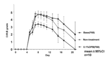
結果を図16に示した。口内炎gradeの最大値はbase群が4.9、OPB群が4.3であり、さらに立ち上がりもbase群の方が早かった。モデルの強度が強かった影響も有り、day40においても潰瘍が治らない個体もいたため完全に全例治癒はしなかったが、OPB群の方が明らかに治癒が早かった。 8-5 Results The results are shown in FIG. The maximum value of stomatitis grade was 4.9 in the base group and 4.3 in the OPB group, and the rise was earlier in the base group. There was also an effect that the strength of the model was strong, and even in
本試験では、ラット歯肉炎モデルにおいて、0.1%オラネキシジングルコン酸塩の歯肉炎に対する治療効果を検討した。 9. Efficacy Test in Rat Gingivitis Model In this study, the therapeutic effect of 0.1% oranexidine gluconate on gingivitis was examined in a rat gingivitis model.
被験物質と対照物質を合わせて試験物質とした。 9-1 Test substance The test substance and the control substance were combined as a test substance.
名称 :OPB
組成 :オラネキシジングルコン酸塩 0.10w/v%
Pluronic L-44 0.07w/v%
Pluronic P-123 1.0 w/v% 9-1-1 Test substance name: OPB
Composition: olanexidine gluconate 0.10 w / v%
Pluronic L-44 0.07w / v%
Pluronic P-123 1.0 w / v%
名称/略称:基剤/Base
組成 :Pluronic L-44 0.07w/v%
Pluronic P-123 1.0 w/v% 9-1-2 Control substance name / abbreviation: Base / Base
Composition: Pluronic L-44 0.07 w / v%
Pluronic P-123 1.0 w / v%
本試験には、歯肉炎起因菌であるポルフィロモナス・ジンジバリス(Porphyromonas gingivalis、ATCC番号:33277、Microbiologics, Inc.社製)を用いた。 9-2 Bacteria used Porphyromonas gingivalis (ATCC number: 33277, manufactured by Microbiologics, Inc.), a gingivitis-causing bacterium, was used in this test.
入荷時5週齢のラット、Jcl:Wistar、雄を用い、各群5匹で試験を行った。 9-3 Animals used The test was performed with 5 rats in each group using rats, Jcl: Wistar, and males aged 5 weeks at the time of arrival.
〔1〕保管されている細菌ペレットの入ったバイアルを取り出し嫌気チャンバーに入れた。
〔2〕バイアルから細菌ペレットを1個取り出し、滅菌チューブに移した。
〔3〕0.5mLの調製TSB培地を加えた。
〔4〕滅菌綿棒で細菌ペレットを押しつぶし、菌懸濁液とした。
〔5〕菌懸濁液をCDC嫌気性菌用ヒツジ血液寒天培地に接種した。
〔6〕嫌気条件下(37℃)で3~4日間培養した。
〔7〕形成されたコロニーの中から単一のコロニーを選び、再度同様にCDC嫌気性菌用ヒツジ血液寒天培地に接種した。
〔8〕菌の増殖を確認後、3mLの調製TSB培地を加えスプレッダーで懸濁し、グリセロールストックを作製した。
〔9〕上記ストックを冷凍保存した。
〔10〕ストックからCDC嫌気性菌用ヒツジ血液寒天培地に接種し嫌気条件下で培養した。
〔11〕菌の増殖を確認後、適量の調製TSB培地で懸濁し、一部を取りだし濁度をMcFarland Standardを用いてMcFarland5とした。希釈倍率を計算し、残存している懸濁液から1×10¹⁰CFU/mLとなるように菌液濃度を調整した。
〔12〕上記を試験菌液とした。 9-4-1 Preparation of test bacteria (All operations except storage were performed in an anaerobic chamber)
[1] The stored vial containing the bacterial pellet was taken out and placed in an anaerobic chamber.
[2] One bacterial pellet was removed from the vial and transferred to a sterile tube.
[3] 0.5 mL of prepared TSB medium was added.
[4] The bacterial pellet was crushed with a sterile cotton swab to obtain a bacterial suspension.
[5] The bacterial suspension was inoculated on a sheep blood agar medium for CDC anaerobic bacteria.
[6] The cells were cultured for 3-4 days under anaerobic conditions (37 ° C).
[7] A single colony was selected from the formed colonies and again inoculated on a CDC anaerobic sheep blood agar medium.
[8] After confirming the growth of the bacteria, 3 mL of prepared TSB medium was added and suspended with a spreader to prepare a glycerol stock.
[9] The above stock was stored frozen.
[10] The CDC anaerobic fungus sheep blood agar medium was inoculated and cultured under anaerobic conditions.
[11] After confirming the growth of the bacteria, the suspension was suspended in an appropriate amount of prepared TSB medium, a part was taken out, and the turbidity was set to
[12] The above was used as a test bacterial solution.
ペントバルビタールナトリウム水溶液を腹腔内に40mg/kg投与した。 (1) Catan thread insertion, necropsy: Pentobarbital sodium aqueous solution was intraperitoneally administered at 40 mg / kg.
ガス麻酔[導入麻酔:空気1.0L/minに5%イソフルラン(マイラン製薬株式会社)、持続麻酔は適宜濃度を調整]を実施した。 (2) Bacterial inoculation, test substance administration Gas anesthesia [introduced anesthesia: 5% isoflurane (Mylan Pharmaceutical Co., Ltd. in air 1.0 L / min, continuous anesthesia adjusted for appropriate concentration)] was performed.
麻酔後、専用台に背位で固定し下額を持ち上げ、上顎右第一臼歯と第二臼歯の間にカタン糸を挿入した。 9-4-3 Catan thread insertion After anesthesia, the back was fixed to a dedicated platform, the lower forehead was lifted, and a catan thread was inserted between the maxillary right first molar and second molar.
麻酔後、試験菌液0.2mLをカタン糸挿入部位に接種した。この操作は2時間おきに実施した。 9-4-4 Bacterial inoculation After anesthesia, 0.2 mL of the test bacterial solution was inoculated into the catan thread insertion site. This operation was performed every 2 hours.
麻酔後、試験物質1mLを口腔内に洗浄投与した。この操作は1日2回とした。 9-4-5 Test substance administration After anesthesia, 1 mL of the test substance was washed and administered into the oral cavity. This operation was performed twice a day.
カタン糸挿入日から剖検日まで1日1回実施した。 (1) General condition It was carried out once a day from the date of insertion of the catan thread to the date of necropsy.
カタン糸挿入日から剖検時まで計2回実施した。 (2) Body weight measurement The measurement was carried out twice in total from the date of insertion of the catan thread to the time of autopsy.
麻酔下で、腹大動脈を切断し放血死させ、剖検を行った。 (3) Necropsy Under anesthesia, the abdominal aorta was cut and exsanguinated to perform necropsy.
摘出した上顎を10v/v%中性緩衝ホルマリン液で固定し、脱脂、脱灰後、HE染色標本を作製した。各標本につき炎症性変化について病理検査した。 (4) Histopathological examination The excised maxilla was fixed with 10 v / v% neutral buffered formalin solution, degreased and decalcified, and then a HE-stained specimen was prepared. Each specimen was pathologically examined for inflammatory changes.
各群の体重の平均値及び標準偏差を算出する。検定は行わなかった。 9-4-7 Statistical analysis The mean value and standard deviation of the body weight of each group are calculated. The test was not performed.
一般状態に異常は認められず、体重に群間の差は無かった。
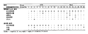
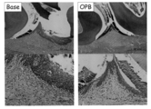
病理学的検査結果を図17に示した。また、HE染色標本の顕微鏡写真を図18に示す。
OPB投与群では、歯肉の重層扁平上皮における好中球の浸潤が8/10例、細胞間水腫が1/10例及び潰瘍が1/10例で何れも軽微に認められた。歯肉の固有層における好中球の浸潤が8/10例及び出血が1/10例で何れも軽微に認められた。一方、base投与群では、歯肉の重層扁平上皮における好中球の浸潤が軽微8/10例及び軽度2/10例で認められた。細胞間水腫が2/10例、過角化が2/10例、表皮肥厚が2/10例及び潰瘍が1/10例で何れも軽微に認められた。歯肉の固有層における好中球の浸潤が軽微6/10例及び軽度3/10例で認められた。出血及び水腫が何れも1/10例で軽微に認められた。 9-5 Results No abnormalities were observed in the general condition, and there was no difference between groups in body weight.
The results of pathological examination are shown in FIG. Further, a micrograph of the HE-stained specimen is shown in FIG.
In the OPB administration group, neutrophil infiltration into the stratified squamous epithelium of gingiva was observed in 8/10 cases, intercellular edema in 1/10 cases and ulcers in 1/10 cases, all of which were slight. Neutral infiltration in the gingival lamina propria was observed in 8/10 cases and bleeding in 1/10 cases. On the other hand, in the base administration group, infiltration of neutrophils into the stratified squamous epithelium of gingiva was observed in slight 8/10 cases and mild 2/10 cases. Intercellular edema was observed in 2/10 cases, hyperkeratosis in 2/10 cases, epidermal thickening in 2/10 cases, and ulcers in 1/10 cases. Neutrophil infiltration in the gingival lamina was found in mild 6/10 cases and mild 3/10 cases. Bleeding and edema were both slight in 1/10 cases.
歯肉の重層扁平上皮における好中球の浸潤、細胞間水腫、過角化、表皮肥厚及び固有層における好中球の浸潤及び水腫はいずれも炎症に関連した変化であり、処置に起因して生じたと考えられた。これらの何れの変化もbase投与群と比較してOPB投与群で頻度及び程度共に低い傾向にあったことから、OPB投与による炎症軽減効果が認められた。 9-6 Discussion Neutrophil infiltration, intercellular edema, hyperkeratosis, epidermal thickening and neutrophil infiltration in the lamina propria and edema in the gingival stratified squamous epithelium are all inflammation-related changes. It was thought to have occurred. Since all these changes tended to be lower in frequency and degree in the OPB administration group than in the base administration group, the inflammation reducing effect by OPB administration was recognized.
本試験では、ラットの誤嚥性肺炎モデルにおいて、0.1%オラネキシジングルコン酸塩の肺炎に対する治療効果を検討した。 10. Efficacy Test in Rat Pneumonia Model In this study, the therapeutic effect of 0.1% oranexidine gluconate on pneumonia was examined in a rat aspiration pneumonia model.
被験物質と対照物質を合わせて試験物質とした。 10-1 Test substance The test substance and the control substance were combined as a test substance.
名称 :OPB
組成 :オラネキシジングルコン酸塩 0.10w/v%
Pluronic L-44 0.07w/v%
Pluronic P-123 1.0 w/v% 10-1-1 Test substance name: OPB
Composition: olanexidine gluconate 0.10 w / v%
Pluronic L-44 0.07w / v%
Pluronic P-123 1.0 w / v%
名称/略称:基剤/Base
組成 :Pluronic L-44 0.07w/v%
Pluronic P-123 1.0 w/v% 10-1-2 Control substance name / abbreviation: Base / Base
Composition: Pluronic L-44 0.07 w / v%
Pluronic P-123 1.0 w / v%
入荷時7週齢のラット、Crl:CD(SD)、雄を用いて試験を行った。 10-2 Animals used The test was conducted using 7-week-old rats, Crl: CD (SD), and males at the time of arrival.
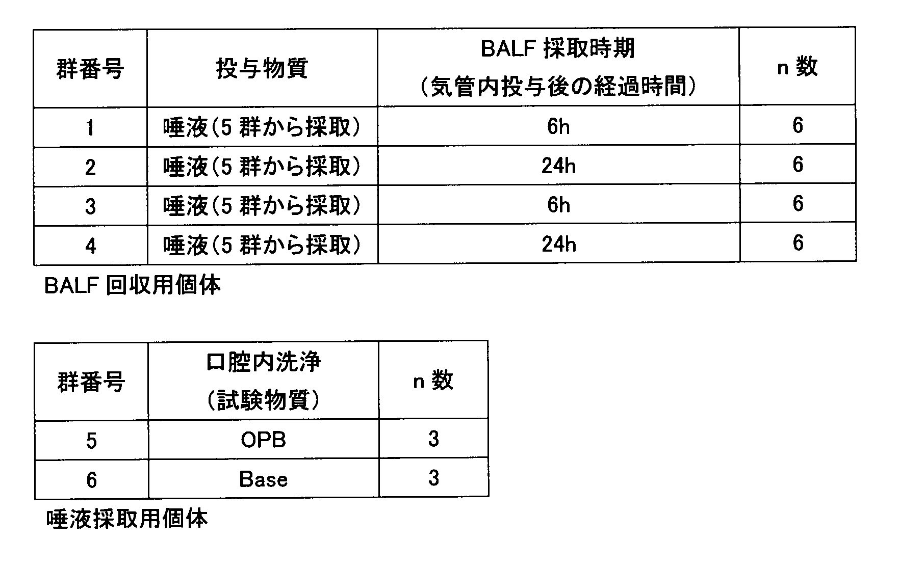
気管内投与日の朝に体重を測定し、層別無作為化割付により以下の表6に示す4群(1~4群)に割り付けた。割付から外れた6匹を層別無作為化割付により以下の表6に示す2群(5,6群)に割り付け、唾液採取用個体とした。 10-3 Group Composition Body weight was measured in the morning of the intratracheal administration day, and was assigned to 4 groups (1 to 4 groups) shown in Table 6 below by stratified randomized assignment. Six animals that were out of the allocation were allocated to two groups (
ソムノペンチルを腹腔内に40mg/kgとなるように投与した。 (1) Oral lavage, saliva collection Somnopentyl was administered intraperitoneally to 40 mg / kg.
ガス麻酔[導入麻酔:空気3.0L/minに3%イソフルラン(マイラン製薬株式会社)、持続麻酔は適宜濃度を調整]を実施した。 (2) At the time of intratracheal administration, when collecting bronchoalveolar lavage fluid (BALF) Gas anesthesia [Introductory anesthesia: 3% isoflurane (Mylan Pharmaceutical Co., Ltd. for air at 3.0 L / min, adjust the concentration appropriately for continuous anesthesia)] did.
麻酔下で、試験物質を滅菌綿棒に含浸させ口腔内に十分量塗布した。 10-4-2 Intraoral washing Under anesthesia, the test substance was impregnated into a sterile cotton swab and applied in a sufficient amount in the oral cavity.
麻酔下で、0.1%塩酸ピロカルピン(5mg/kg)を腹腔内投与した。過剰分泌される唾液を回収した。回収した唾液は、中和剤入り栄養培地に加え静置した。その後、遠心(r.t.,3000rpm,10min)し、沈渣を同量の生理食塩液で懸濁し、気管内投与液とした。 10-4-3 Saliva Collection Under anesthesia, 0.1% pilocarpine hydrochloride (5 mg / kg) was intraperitoneally administered. The excessively secreted saliva was collected. The collected saliva was added to a nutrient medium containing a neutralizing agent and allowed to stand. Thereafter, the mixture was centrifuged (rt, 3000 rpm, 10 min), and the sediment was suspended in the same amount of physiological saline to obtain an intratracheal administration solution.
唾液採取後、麻酔下で咽頭鏡を用い気管内に投与用チューブを留置し、唾液若しくは生理食塩液を0.1mL投与した。 10-4-4 Intratracheal administration After collecting saliva, the tube for administration was placed in the trachea under anesthesia using a laryngoscope, and 0.1 mL of saliva or physiological saline was administered.
気管内投与6,24時間後、麻酔下で正中切開し、腹大動脈切開により放血し安楽死させた。その後、肺を露出させ、気管支起始部にカテーテルを挿入した。そのラインから0.1%BSA、0.05mM EDTA-2Na含有PBS溶液(以下、PBS)8mLで3回洗浄し(各回2回注入回収を繰り返す)、洗浄液を採取した(BALF)。BALFを遠心(200g,4℃,10min)し、上清は別の保存チューブに分取し、LDH濃度測定、サイトカイン類測定(ELISA)用とした。沈渣はPBS 1mLで懸濁し血液学的検査用とした。 10-4-5
気管内投与終了後、唾液採取用の動物は過麻酔下で放血させ安楽死させた。 10-4-6 Handling of saliva collection animal After the intratracheal administration, the saliva collection animal was exsanguinated and euthanized under hyperanesthesia.
キット添付のプロトコールに従い測定した。 10-4-7 Cytokine (TNF-α, IL-6) concentration measurement The concentration was measured according to the protocol attached to the kit.
懸濁した沈渣に対して、多項目自動血球計数装置を用いて血液学的解析を実施した。 10-4-8 Hematological examination Hematological analysis was performed on the suspended sediment using a multi-item automatic blood cell counter.
採取したBALF上清に対して、自動分析装置7180(株式会社日立ハイテクノロジーズ)を用いLDH濃度を測定した。 10-4-9 Biochemical examination The LDH concentration was measured on the collected BALF supernatant using an automatic analyzer 7180 (Hitachi High-Technologies Corporation).
探索的な解析のため検定は行わなかった。 10-4-10 Statistical analysis No test was performed due to exploratory analysis.
血液学的検査及び生化学的検査の結果を図19に示した。
血液学的検査からは両群間に差が無かった。IL-6に関しては24時間値においてOPB群で2.5倍ほど低い値となった。TNF-αに関しては、両時点でOPB群の方が低い値となった。
したがって、OPBで口腔内を処理した方が、唾液の誤嚥による肺での炎症性反応は低いことが示唆された。 10-5 Results The results of hematological and biochemical tests are shown in FIG.
There was no difference between the two groups from hematology. Regarding IL-6, the value in the OPB group was about 2.5 times lower at 24 hours. Regarding TNF-α, the OPB group had lower values at both time points.
Therefore, it was suggested that the treatment of the oral cavity with OPB has a lower inflammatory reaction in the lung due to saliva aspiration.
実施例6~10より、オラネキシジンには口内炎、歯肉炎、肺炎に対して抗炎症作用を有することが示された。炎症は、LPSやLTAを認識する受容体であるToll-like receptor 4(TLR-4)、Toll-like receptor 2(TLR-2)を介した免疫応答と関連していることが明らかになっている(ChemMedChem. 2016 Jan 19;11(2):154-65、Biotechnol Adv. 2012 Jan-Feb;30(1):251-60、J Dent Res. 2016 Jul;95(7):725-33)。
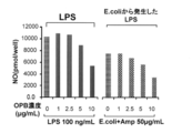
オラネキシジンは、TLR-4及びTLR-2に対するアンタゴニスト(様)作用によって炎症を抑えている可能性がある。そこで、本試験ではこのことを明らかにするため、TLR-4、TLR-2やレポーター遺伝子(SEAP)を安定発現したヒト由来細胞を用いたレポーターアッセイにより、オラネキシジンのTLR-4及びTLR-2に対するアンタゴニスト(様)作用を確認する。 11. Examination of anti-inflammatory action of olanexidine using TLR Reporter Cell Line From Examples 6 to 10, it was shown that olanexidine has an anti-inflammatory action against stomatitis, gingivitis and pneumonia. Inflammation has been shown to be associated with immune responses via Toll-like receptor 4 (TLR-4) and Toll-like receptor 2 (TLR-2), receptors that recognize LPS and LTA. (ChemMedChem. 2016
Oranexidine may suppress inflammation by antagonistic (like) action on TLR-4 and TLR-2. Therefore, in order to clarify this in this study, a reporter assay using human-derived cells stably expressing TLR-4, TLR-2 and a reporter gene (SEAP) was performed on olanexidine against TLR-4 and TLR-2. Confirm antagonist (like) action.
名称 :1.5%OPB
組成 :オラネキシジングルコン酸塩 1.5w/v% 11-1 Test substance name: 1.5% OPB
Composition: Oranexidine gluconate 1.5 w / v%
以下の表7に記載の細胞を用いた。 11-2 cells The cells described in Table 7 below were used.

-使用培地
前培養:DMEM+FBS(終濃度10%)+ペニシリン(終濃度100unit/mL)+ストレプトマイシン(終濃度100μg/mL)
サンプル投与、投与後培養:DMEM+FBS(終濃度5%)
-活性測定:SEAP assay kit(Novus Biologicals社製)
-タンパク量定量:BCA protein assay(フナコシ株式会社製) -Cells used: HEK293-TLR4 expressing cells-Media used Preculture: DMEM + FBS (
Sample administration, culture after administration: DMEM + FBS (
-Activity measurement: SEAP assay kit (Novus Biologicals)
-Protein amount quantification: BCA protein assay (Funakoshi Co., Ltd.)
DMEMで調整し、96well plate(コラーゲン処理)に撒いて、40h培養(37℃、5%CO2)。
〔2〕1.5%OPBを、以下の表8に示す濃度になるよう5%FBSを用いて希釈する。 [1] 10% FBS so that the cells become 1.0 × 10 5 cells / well / 100 μL
Adjust with DMEM, spread on 96 well plate (collagen treatment) and culture for 40 h (37 ° C., 5% CO 2 ).
[2] Dilute 1.5% OPB with 5% FBS to the concentration shown in Table 8 below.

〔4〕培地を除去後、OPB培地50μL、LPS培地50μLの順番で培地を加え、8h培養(37℃、5%CO2)。
〔5〕培養後、上清を50μL別の96well plateに移し、SEAP assayを行う。
〔6〕上清の残りを除去した96well plateに、0.1%SDS-O.1N NaOHを50μL加えて、over nightで凍結(-20℃)させる。融解後、BCA protein assayを行う。
〔7〕レポーター遺伝子であるSEAPの発現量をタンパク質濃度で較正し、OPB濃度毎の阻害率を算出した。 [3] LPS is prepared using 5% FBS so as to be 20 ng / mL (final concentration: 10 ng / mL).
[4] After removing the medium, the medium was added in the order of 50 μL of OPB medium and 50 μL of LPS medium, and cultured for 8 hours (37 ° C., 5% CO 2 ).
[5] After the culture, the supernatant is transferred to another 96-well plate of 50 μL, and SEAP assay is performed.
[6] To 96 well plate from which the remaining supernatant was removed, 0.1% SDS-
[7] The expression level of SEAP as a reporter gene was calibrated with the protein concentration, and the inhibition rate for each OPB concentration was calculated.
-使用培地
前培養:DMEM+FBS(終濃度10%)+ペニシリン(終濃度100unit/mL)+ストレプトマイシン(終濃度100μg/mL)
サンプル投与、投与後培養:DMEM+FBS(終濃度1%)
-活性測定:SEAP assay kit(Novus Biologicals社製)
-タンパク量定量:BCA protein assay(フナコシ株式会社製) -Cells used: HEK293-TLR2 expressing cells-Media used Preculture: DMEM + FBS (
Sample administration, culture after administration: DMEM + FBS (
-Activity measurement: SEAP assay kit (Novus Biologicals)
-Protein amount quantification: BCA protein assay (Funakoshi Co., Ltd.)
〔2〕1.5%OPBを、前記表8に示す濃度になるよう1%FBSを用いて希釈する。
〔3〕LTAを2μg/mL(終濃度1μg/mL)になるよう1%FBSを用いて調製する。
〔4〕培地を除去後、OPB培地50μL、LTA培地50μLの順番で培地を加え、12h培養(37℃、5%CO2)。
〔5〕培養後、上清を50μL別の96well plateに移し、SEAP assayを行う。
〔6〕上清の残りを除去した96well plateに、0.1%SDS-0.1N NaOHを50μL加えて、over nightで凍結(-20℃)させる。融解後、BCA protein assayを行う。
〔7〕レポーター遺伝子であるSEAPの発現量をタンパク質濃度で較正し、OPB濃度毎の阻害率を算出した。 [1] The cells were adjusted with 10% FBS DMEM so as to be 1.0 × 10 5 cells / well / 100 μL, spread on 96 well plates (collagen treatment), and cultured for 36 h (37 ° C., 5% CO 2 ).
[2] Dilute 1.5% OPB with 1% FBS to the concentration shown in Table 8 above.
[3] LTA is prepared using 1% FBS so as to be 2 μg / mL (
[4] After removing the medium, the medium was added in the order of 50 μL of OPB medium and 50 μL of LTA medium, and cultured for 12 h (37 ° C., 5% CO 2 ).
[5] After the culture, the supernatant is transferred to another 96-well plate of 50 μL, and SEAP assay is performed.
[6] Add 96 μL of 0.1% SDS-0.1N NaOH to the 96-well plate from which the remaining supernatant is removed, and freeze (−20 ° C.) with over night. After thawing, BCA protein assay is performed.
[7] The expression level of SEAP as a reporter gene was calibrated with the protein concentration, and the inhibition rate for each OPB concentration was calculated.
結果を図20に示す。
OPB濃度が上昇するにしたがい、SEAPの阻害率が高まっており(IC50:約10μg/mL)、OPBが、TLR4、TLR2に対するアンタゴニスト(様)作用を示すことが明らかになった。この結果より、OPBが、TLR4、TLR2を介した免疫応答を阻害することにより、抗炎症作用を有することが示唆された。 11-4 Results The results are shown in FIG.
As the OPB concentration increased, the inhibition rate of SEAP increased (IC 50 : about 10 μg / mL), and it became clear that OPB exhibits an antagonistic (like) action on TLR4 and TLR2. From these results, it was suggested that OPB has an anti-inflammatory effect by inhibiting the immune response via TLR4 and TLR2.
マウスマクロファージ様細胞株RAW264.7は、LPSやLTAで刺激すると、それらを認識する受容体を介して免疫応答を開始し、炎症性メディエーターであるNOの産生を行う。そこで、オラネキシジンがLPS刺激による炎症に対する抗炎症作用を有するかどうかを、RAW264.7細胞からのNO産生を指標として確認する。 12 Examination of anti-inflammatory action of olanexidine using mouse macrophage-like cell line RAW264.7 When mouse macrophage-like cell line RAW264.7 is stimulated with LPS or LTA, it initiates an immune response via a receptor that recognizes them, It produces NO, an inflammatory mediator. Therefore, whether oranexidine has an anti-inflammatory action against inflammation induced by LPS is confirmed using NO production from RAW264.7 cells as an index.
名称 :1.5%OPB
組成 :オラネキシジングルコン酸塩 1.5w/v% 12-1 Test substance name: 1.5% OPB
Composition: Oranexidine gluconate 1.5 w / v%
以下の表9に記載の細胞を用いた。 12-2 cells The cells listed in Table 9 below were used.
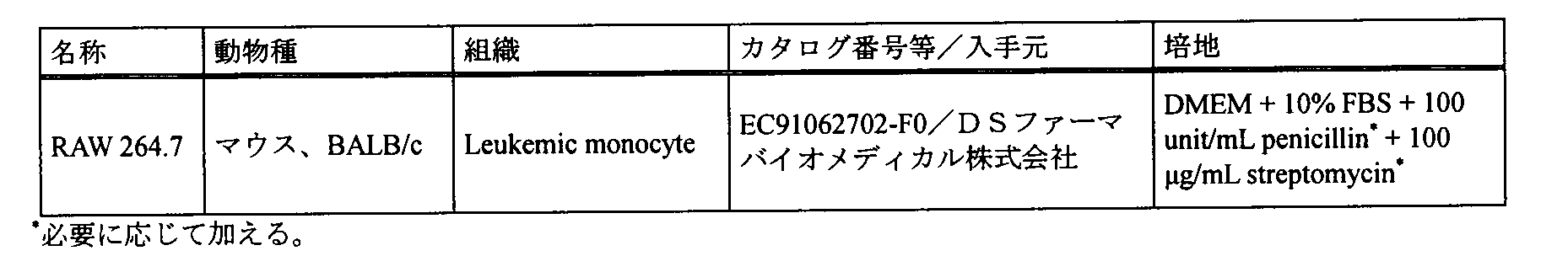
-使用培地
前培養:DMEM+FBS(終濃度10%)+ペニシリン(終濃度100unit/mL)+ストレプトマイシン(終濃度100μg/mL)
サンプル投与、投与後培養:DMEM+FBS(終濃度5%)-活性測定:Nitrate/Nitrite Colorimetric Assay Kit(Griess Reagents社製)
-タンパク量定量:細胞を染色しにくく、値が不安定になるため測定せず。 -Cells used: RAW264.7
-Media used Pre-culture: DMEM + FBS (
Sample administration, culture after administration: DMEM + FBS (
-Quantification of protein amount: Not measured because it is difficult to stain cells and the value becomes unstable.
〔2〕1.5%OPBを、前記表8に示した濃度になるよう5%FBSを用いて希釈する。
〔3〕LPSを200ng/mL(終濃度100ng/mL)になるよう5%FBSを用いて調製する。
〔4〕培地を除去後、OPB培地50μL、LPS培地50μLの順番で培地を加え、8h培養(37℃、5%CO2)。
〔5〕培養後、上清を50μL別の96well plateに移し、Nitrate/Nitrite colorimetric assayを行う。 [1] The cells were adjusted with 10% FBS DMEM to 1.0 × 10 5 cells / well / 100 μL, spread on 96-well plates (no treatment), and cultured for 24 h (37 ° C., 5% CO 2 ).
[2] Dilute 1.5% OPB with 5% FBS to the concentration shown in Table 8 above.
[3] LPS is prepared using 5% FBS so as to be 200 ng / mL (
[4] After removing the medium, the medium was added in the order of 50 μL of OPB medium and 50 μL of LPS medium, and cultured for 8 hours (37 ° C., 5% CO 2 ).
[5] After culturing, the supernatant is transferred to another 96-well plate of 50 μL, and Nitrate / Nitrite colorimetric assay is performed.
-使用培地
前培養:DMEM+FBS(終濃度10%)+ペニシリン(終濃度100unit/mL)+ストレプトマイシン(終濃度100μg/mL)
サンプル投与、投与後培養:DMEM+FBS(終濃度1%)
-活性測定:Nitrate/Nitrite Colorimetric Assay Kit(Griess Reagents社製)
-タンパク量定量:細胞を染色しにくく、値が不安定になるため測定せず。 -Cells used: RAW264.7
-Media used Pre-culture: DMEM + FBS (
Sample administration, culture after administration: DMEM + FBS (
-Activity measurement: Nitrate / Nitrite Colorimetric Assay Kit (Griess Reagents)
-Quantification of protein amount: Not measured because it is difficult to stain cells and the value becomes unstable.
〔2〕1.5%OPBを、前記表8に示した濃度になるよう1%FBSを用いて調製する。
〔3〕培地を除去後、OPB培地50μLを加え、37℃、5%CO2で静置する。
〔4〕MHBで一晩培養した大腸菌を、McF 1になるよう調製し、5%DMEMで300倍希釈する。更に、Ampicillinを終濃度50μg/mLになるよう添加する。
〔5〕OPB培地を添加したプレートに、更に調製した菌培地50μLを加え、密閉して24h培養する(37℃、5%CO2)。
〔6〕培養後上清を50μL別の96well plateに移し、Nitrate/Nitrite colorimetric assayを行う。 [1] The cells were adjusted with 10% FBS DMEM to 1.0 × 10 5 cells / well / 100 μL, spread on 96-well plates (no treatment), and cultured for 24 h (37 ° C., 5% CO 2 ).
[2] 1.5% OPB is prepared using 1% FBS so as to have the concentration shown in Table 8 above.
[3] After removing the medium, add 50 μL of OPB medium and allow to stand at 37 ° C., 5% CO 2 .
[4] E. coli cultured overnight in MHB is adjusted to
[5] Further, 50 μL of the prepared fungal medium is added to the plate to which the OPB medium has been added, sealed and cultured for 24 hours (37 ° C., 5% CO 2 ).
[6]
結果を図21に示す。
OPB濃度が上昇するにしたがい、NO産生が減少しており、OPBがNO産生阻害作用を示すことが明らかになった(IC50:約10μg/mL)。この結果より、OPBが抗炎症作用を有することが示唆された。 12-4 Results The results are shown in FIG.
As the OPB concentration increased, NO production decreased, and it became clear that OPB exhibited a NO production inhibitory action (IC 50 : about 10 μg / mL). This result suggested that OPB has an anti-inflammatory effect.
Claims (11)
- オラネキシジン又はその薬理学上許容される塩を含む、炎症の改善用及び/又は予防用の組成物。 A composition for improving and / or preventing inflammation comprising olanexidine or a pharmacologically acceptable salt thereof.
- オラネキシジン又はその薬理学上許容される塩が、オラネキシジングルコン酸塩であることを特徴とする請求項1に記載の組成物。 2. The composition according to claim 1, wherein the olanexidine or a pharmacologically acceptable salt thereof is an olanexidine gluconate.
- ポリオキシプロピレン鎖(POP)及び該POPを挟む2個のポリオキシエチレン鎖(POE)からなるブロック共重合体であるポロキサマーをさらに含むことを特徴とする請求項1又は2に記載の組成物。 The composition according to claim 1 or 2, further comprising a poloxamer which is a block copolymer composed of a polyoxypropylene chain (POP) and two polyoxyethylene chains (POE) sandwiching the POP.
- 炎症が、口内炎、口腔粘膜炎、歯肉炎、及び肺炎から選択されることを特徴とする請求項1~3のいずれかに記載の組成物。 4. The composition according to claim 1, wherein the inflammation is selected from stomatitis, oral mucositis, gingivitis, and pneumonia.
- 炎症ががんの治療による口腔粘膜炎であり、
0.01~1.5%(W/V)のオラネキシジングルコン酸塩と、
ポリオキシプロピレン鎖(POP)及び該POPを挟む2個のポリオキシエチレン鎖(POE)からなるブロック共重合体であるポロキサマーと
を含むことを特徴とする請求項1~4のいずれかに記載の組成物。 Inflammation is oral mucositis due to cancer treatment,
0.01-1.5% (W / V) olanexidine gluconate;
5. A poloxamer, which is a block copolymer comprising a polyoxypropylene chain (POP) and two polyoxyethylene chains (POE) sandwiching the POP, according to any one of claims 1 to 4. Composition. - ポロキサマーが、ポリオキシエチレン(42)ポリオキシプロピレン(67)グリコール(Pluronic P-123)、ポリオキシエチレン(54)ポリオキシプロピレン(39)グリコール(Pluronic P-85)、及びポリオキシエチレン(196)ポリオキシプロピレン(67)グリコール(Pluronic F-127)から選択されることを特徴とする請求項3~5のいずれかに記載の組成物。 Poloxamers are polyoxyethylene (42) polyoxypropylene (67) glycol (Pluronic P-123), polyoxyethylene (54) polyoxypropylene (39) glycol (Pluronic P-85), and polyoxyethylene (196). 6. The composition according to claim 3, wherein the composition is selected from polyoxypropylene (67) glycol (Pluronic F-127).
- ポロキサマーが、ポリオキシエチレン(42)ポリオキシプロピレン(67)グリコール(Pluronic P-123)であることを特徴とする請求項6に記載の組成物。 The composition according to claim 6, wherein the poloxamer is polyoxyethylene (42) polyoxypropylene (67) glycol (Pluronic P-123).
- オラネキシジングルコン酸塩の濃度が、0.05~0.5%(W/V)であることを特徴とする請求項1~7のいずれかに記載の組成物。 The composition according to any one of claims 1 to 7, wherein the concentration of olanexidine gluconate is 0.05 to 0.5% (W / V).
- ポロキサマーの濃度が、0.1~5.0%(W/V)であることを特徴とする請求項3~8のいずれかに記載の組成物。 The composition according to any one of claims 3 to 8, wherein the poloxamer concentration is 0.1 to 5.0% (W / V).
- 液剤又は含嗽剤であることを特徴とする、請求項1~9のいずれかに記載の組成物。 The composition according to any one of claims 1 to 9, which is a liquid agent or a gargle.
- がんの治療が、化学療法、放射線治療、又は化学療法と放射線治療との同時併用療法であることを特徴とする、請求項5~10のいずれかに記載の組成物。 The composition according to any one of claims 5 to 10, wherein the cancer treatment is chemotherapy, radiation therapy, or simultaneous combination therapy of chemotherapy and radiation therapy.
Priority Applications (11)
| Application Number | Priority Date | Filing Date | Title |
|---|---|---|---|
| KR1020197029589A KR102253831B1 (en) | 2017-04-19 | 2018-04-10 | Anti-inflammatory |
| ES18787649T ES2956040T3 (en) | 2017-04-19 | 2018-04-10 | Olanexidine as an anti-inflammatory agent |
| SG11201908629W SG11201908629WA (en) | 2017-04-19 | 2018-04-10 | Anti-inflammatory agent |
| EP18787649.5A EP3628314B1 (en) | 2017-04-19 | 2018-04-10 | Olanexidine as anti-inflammatory agent |
| RU2019132614A RU2738845C1 (en) | 2017-04-19 | 2018-04-10 | Anti-inflammatory agent |
| US16/605,363 US11229615B2 (en) | 2017-04-19 | 2018-04-10 | Method of treating and/or preventing inflammation by administering olanexidine |
| JP2019513564A JP6802365B2 (en) | 2017-04-19 | 2018-04-10 | Anti-inflammatory agent |
| AU2018256060A AU2018256060B2 (en) | 2017-04-19 | 2018-04-10 | Anti-inflammatory agent |
| CN201880024116.5A CN110494132A (en) | 2017-04-19 | 2018-04-10 | Anti-inflammatory agent |
| CA3058592A CA3058592C (en) | 2017-04-19 | 2018-04-10 | Olanexidine as anti-inflammatory agent |
| PH12019550207A PH12019550207A1 (en) | 2017-04-19 | 2019-10-02 | Anti-inflammatory agent |
Applications Claiming Priority (2)
| Application Number | Priority Date | Filing Date | Title |
|---|---|---|---|
| JP2017-082793 | 2017-04-19 | ||
| JP2017082793 | 2017-04-19 |
Publications (1)
| Publication Number | Publication Date |
|---|---|
| WO2018193904A1 true WO2018193904A1 (en) | 2018-10-25 |
Family
ID=63855845
Family Applications (1)
| Application Number | Title | Priority Date | Filing Date |
|---|---|---|---|
| PCT/JP2018/014999 WO2018193904A1 (en) | 2017-04-19 | 2018-04-10 | Anti-inflammatory agent |
Country Status (13)
| Country | Link |
|---|---|
| US (1) | US11229615B2 (en) |
| EP (1) | EP3628314B1 (en) |
| JP (1) | JP6802365B2 (en) |
| KR (1) | KR102253831B1 (en) |
| CN (1) | CN110494132A (en) |
| AU (1) | AU2018256060B2 (en) |
| CA (1) | CA3058592C (en) |
| ES (1) | ES2956040T3 (en) |
| PH (1) | PH12019550207A1 (en) |
| RU (1) | RU2738845C1 (en) |
| SG (1) | SG11201908629WA (en) |
| TW (1) | TWI675658B (en) |
| WO (1) | WO2018193904A1 (en) |
Citations (7)
| Publication number | Priority date | Publication date | Assignee | Title |
|---|---|---|---|---|
| WO2004105745A1 (en) * | 2003-05-28 | 2004-12-09 | Otsuka Pharmaceutical Co., Ltd. | Aqueous olanexidine solution, method of preparing the same, and disinfectant |
| JP2004352635A (en) * | 2003-05-28 | 2004-12-16 | Otsuka Pharmaceut Co Ltd | Composition for disinfection and/or sterilization |
| JP2005022995A (en) * | 2003-06-30 | 2005-01-27 | Otsuka Pharmaceut Co Ltd | Antiseptic and/or sterilizing composition |
| JP2005289959A (en) | 2003-05-28 | 2005-10-20 | Otsuka Pharmaceut Co Ltd | Aqueous solution of olanexidine, method for preparing the same and disinfectant |
| JP2005343964A (en) * | 2004-06-01 | 2005-12-15 | Otsuka Pharmaceut Factory Inc | Aqueous solution containing polyalkylene glycol and method for preparing the same |
| JP2009526624A (en) * | 2006-02-14 | 2009-07-23 | アレジャンス・コーポレーション | Liquid applicator and method for reducing the concentration of by-products from disinfectants |
| JP2017078046A (en) * | 2015-10-21 | 2017-04-27 | 株式会社大塚製薬工場 | Composition for improving/preventing oral mucositis by treatment of cancer |
Family Cites Families (10)
| Publication number | Priority date | Publication date | Assignee | Title |
|---|---|---|---|---|
| CA2064664C (en) | 1991-04-05 | 2000-01-11 | Hiroshi Ishikawa | Biguanide derivatives, manufacturing method thereof, and disinfectants containing the derivatives |
| US20020013331A1 (en) | 2000-06-26 | 2002-01-31 | Williams Robert O. | Methods and compositions for treating pain of the mucous membrane |
| US6685917B2 (en) * | 2000-11-22 | 2004-02-03 | Rxkinetix, Inc. | Treatment of mucositis |
| TW200509893A (en) * | 2003-05-28 | 2005-03-16 | Otsuka Pharma Co Ltd | A composition for sterilizing and/ or disinfecting |
| KR20070088636A (en) | 2004-11-17 | 2007-08-29 | 오츠카 아메리카 파마슈티컬, 인코포레이티드 | Fluid application device and method |
| WO2006122162A2 (en) * | 2005-05-10 | 2006-11-16 | The University Of Chicago | Use of peptides derived from the growth factor amp-18 for the treatment of mucositis |
| TWI556845B (en) * | 2008-12-30 | 2016-11-11 | 大塚美國製藥股份有限公司 | Fluid application device and method |
| US9801916B2 (en) * | 2013-02-07 | 2017-10-31 | Tomás Bernardo Galvan Gonzalez | Oral antiseptic composition useful for treating oral mucositis |
| US20150231095A1 (en) * | 2013-04-18 | 2015-08-20 | Steven A. Melman | Antimicrobial compositions and uses therefore |
| NO342617B1 (en) | 2014-06-18 | 2018-06-18 | Meda Otc Ab | Oral formulation comprising an antibacterial agent to prevent and / or treat halitosis, bad breath, dry mouth or sore throat |
-
2018
- 2018-04-10 ES ES18787649T patent/ES2956040T3/en active Active
- 2018-04-10 WO PCT/JP2018/014999 patent/WO2018193904A1/en unknown
- 2018-04-10 CN CN201880024116.5A patent/CN110494132A/en active Pending
- 2018-04-10 JP JP2019513564A patent/JP6802365B2/en active Active
- 2018-04-10 AU AU2018256060A patent/AU2018256060B2/en active Active
- 2018-04-10 CA CA3058592A patent/CA3058592C/en active Active
- 2018-04-10 US US16/605,363 patent/US11229615B2/en active Active
- 2018-04-10 SG SG11201908629W patent/SG11201908629WA/en unknown
- 2018-04-10 RU RU2019132614A patent/RU2738845C1/en active
- 2018-04-10 EP EP18787649.5A patent/EP3628314B1/en active Active
- 2018-04-10 KR KR1020197029589A patent/KR102253831B1/en active IP Right Grant
- 2018-04-11 TW TW107112457A patent/TWI675658B/en active
-
2019
- 2019-10-02 PH PH12019550207A patent/PH12019550207A1/en unknown
Patent Citations (7)
| Publication number | Priority date | Publication date | Assignee | Title |
|---|---|---|---|---|
| WO2004105745A1 (en) * | 2003-05-28 | 2004-12-09 | Otsuka Pharmaceutical Co., Ltd. | Aqueous olanexidine solution, method of preparing the same, and disinfectant |
| JP2004352635A (en) * | 2003-05-28 | 2004-12-16 | Otsuka Pharmaceut Co Ltd | Composition for disinfection and/or sterilization |
| JP2005289959A (en) | 2003-05-28 | 2005-10-20 | Otsuka Pharmaceut Co Ltd | Aqueous solution of olanexidine, method for preparing the same and disinfectant |
| JP2005022995A (en) * | 2003-06-30 | 2005-01-27 | Otsuka Pharmaceut Co Ltd | Antiseptic and/or sterilizing composition |
| JP2005343964A (en) * | 2004-06-01 | 2005-12-15 | Otsuka Pharmaceut Factory Inc | Aqueous solution containing polyalkylene glycol and method for preparing the same |
| JP2009526624A (en) * | 2006-02-14 | 2009-07-23 | アレジャンス・コーポレーション | Liquid applicator and method for reducing the concentration of by-products from disinfectants |
| JP2017078046A (en) * | 2015-10-21 | 2017-04-27 | 株式会社大塚製薬工場 | Composition for improving/preventing oral mucositis by treatment of cancer |
Non-Patent Citations (6)
| Title |
|---|
| "Report on the Deliberation Results, Evaluation and Licensing Division, Pharmaceutical and Food Safety Bureau, Ministry of Health, Labour and Welfare", REPORT ON THE DELIBERATION RESULTS, 16 September 2008 (2008-09-16), pages 1 - 68, XP009132612 * |
| BIOTECHNOL ADV., vol. 30, no. 1, January 2012 (2012-01-01), pages 251 - 60 |
| CHEMMEDCHEM., vol. 11, no. 2, 19 January 2016 (2016-01-19), pages 154 - 65 |
| HAGI, AKIFUMI ET AL.: "Bactericidal effects and mechanism of action of olanexidine gluconate, a new antiseptic", ANTIMICROBIAL AGENTS AND CHEMOTHERAPY, vol. 59, no. 8, August 2015 (2015-08-01), pages 4551 - 4559, XP055558250, ISSN: 0066-4804 * |
| J DENT RES., vol. 95, no. 7, July 2016 (2016-07-01), pages 725 - 33 |
| See also references of EP3628314A4 |
Also Published As
| Publication number | Publication date |
|---|---|
| AU2018256060B2 (en) | 2020-08-06 |
| KR102253831B1 (en) | 2021-05-20 |
| ES2956040T3 (en) | 2023-12-12 |
| AU2018256060A1 (en) | 2019-10-17 |
| TWI675658B (en) | 2019-11-01 |
| JPWO2018193904A1 (en) | 2020-01-09 |
| CA3058592A1 (en) | 2018-10-25 |
| US11229615B2 (en) | 2022-01-25 |
| RU2738845C1 (en) | 2020-12-17 |
| CN110494132A (en) | 2019-11-22 |
| CA3058592C (en) | 2021-11-09 |
| PH12019550207A1 (en) | 2020-09-21 |
| US20210121417A1 (en) | 2021-04-29 |
| EP3628314A4 (en) | 2020-10-28 |
| TW201841631A (en) | 2018-12-01 |
| EP3628314B1 (en) | 2023-07-26 |
| JP6802365B2 (en) | 2020-12-16 |
| EP3628314A1 (en) | 2020-04-01 |
| SG11201908629WA (en) | 2019-10-30 |
| KR20190123780A (en) | 2019-11-01 |
Similar Documents
| Publication | Publication Date | Title |
|---|---|---|
| US9107920B2 (en) | Extract of Trigonella foenum-graecum | |
| WO2019183876A1 (en) | Oral care compositions for promoting gum health | |
| JPH03128316A (en) | Liquid polymer composition and use thereof | |
| BR112020016315B1 (en) | COMPOSITIONS FOR ORAL TREATMENT TO PROMOTE GUMS HEALTH | |
| BR112020017909B1 (en) | COMPOSITIONS FOR ORAL TREATMENT TO PROMOTE GINGIVAS HEALTH | |
| ES2946767T3 (en) | Hypertonic Antimicrobial Therapeutic Compositions | |
| JP2011153101A (en) | Microbial adhesion prevention agent for oral cavity | |
| CN107184476A (en) | A kind of antibacterial mouthwash and preparation method thereof | |
| TW201440802A (en) | Oral care composition containing ionic liquids | |
| US20160243018A1 (en) | Tooth remineralization compositions and methods | |
| CN102271675A (en) | Anti-inflammatory compounds and compositions thereof | |
| Bahna et al. | Antiseptic effect of a novel alcohol-free mouthwash: a convenient prophylactic alternative for high-risk patients | |
| Niu et al. | Remineralising dentine caries using an artificial antimicrobial peptide: An in vitro study | |
| WO2016006838A1 (en) | Composition for preventing or alleviating periodontal disease, having increased anti-inflammatory and sterilizing strength | |
| WO2018193904A1 (en) | Anti-inflammatory agent | |
| JP6602639B2 (en) | Composition for improving or preventing oral mucositis by treating cancer | |
| CN107280978A (en) | Remineralization of teeth technology based on chitosan-amorphous calcium phosphate | |
| JP2021155364A (en) | Oral composition | |
| JPS63253019A (en) | Composition for oral cavity | |
| US9452124B2 (en) | Oral care composition for enhancing delivery of active agents to mucosa/soft oral tissue and tooth surfaces in the oral cavity | |
| Bindu et al. | An in vitro comparison of antimicrobial efficacy and residual antimicrobial activity of root canal irrigants Qmix, 3% sodium hypochlorite, 2% chlorhexidine against Enterococcus faecalis | |
| AU2012350708B2 (en) | Novel composition | |
| KR20220008722A (en) | Oral cavity cleanin composition comprising carrageenan | |
| Singh | A Comparative Anti-Microbial Efficacy of Root Canal Disinfection Using Conventional Method of 3% Sodium Hypochlorite and 17% Edta with 0.25% Hydro-Alcoholic Solution of Turmeric and 7.5% Hydro-Alcoholic Solution of Neem-An in Vivo Study | |
| JP2021155363A (en) | Oral composition |
Legal Events
| Date | Code | Title | Description |
|---|---|---|---|
| 121 | Ep: the epo has been informed by wipo that ep was designated in this application |
Ref document number: 18787649 Country of ref document: EP Kind code of ref document: A1 |
|
| ENP | Entry into the national phase |
Ref document number: 2019513564 Country of ref document: JP Kind code of ref document: A |
|
| ENP | Entry into the national phase |
Ref document number: 3058592 Country of ref document: CA |
|
| ENP | Entry into the national phase |
Ref document number: 20197029589 Country of ref document: KR Kind code of ref document: A |
|
| ENP | Entry into the national phase |
Ref document number: 2018256060 Country of ref document: AU Date of ref document: 20180410 Kind code of ref document: A |
|
| NENP | Non-entry into the national phase |
Ref country code: DE |
|
| ENP | Entry into the national phase |
Ref document number: 2018787649 Country of ref document: EP Effective date: 20191119 |