WO2018123540A1 - ノックアウト非ヒト動物、ノックアウト非ヒト動物の作製方法、及び筋萎縮性側索硬化症又は認知症の治療薬又は予防薬のスクリーニング方法 - Google Patents
ノックアウト非ヒト動物、ノックアウト非ヒト動物の作製方法、及び筋萎縮性側索硬化症又は認知症の治療薬又は予防薬のスクリーニング方法 Download PDFInfo
- Publication number
- WO2018123540A1 WO2018123540A1 PCT/JP2017/044372 JP2017044372W WO2018123540A1 WO 2018123540 A1 WO2018123540 A1 WO 2018123540A1 JP 2017044372 W JP2017044372 W JP 2017044372W WO 2018123540 A1 WO2018123540 A1 WO 2018123540A1
- Authority
- WO
- WIPO (PCT)
- Prior art keywords
- exon
- knockout
- tdp
- gene
- human animal
- Prior art date
- Legal status (The legal status is an assumption and is not a legal conclusion. Google has not performed a legal analysis and makes no representation as to the accuracy of the status listed.)
- Ceased
Links
Images







Classifications
-
- A—HUMAN NECESSITIES
- A01—AGRICULTURE; FORESTRY; ANIMAL HUSBANDRY; HUNTING; TRAPPING; FISHING
- A01K—ANIMAL HUSBANDRY; AVICULTURE; APICULTURE; PISCICULTURE; FISHING; REARING OR BREEDING ANIMALS, NOT OTHERWISE PROVIDED FOR; NEW BREEDS OF ANIMALS
- A01K67/00—Rearing or breeding animals, not otherwise provided for; New or modified breeds of animals
- A01K67/027—New or modified breeds of vertebrates
- A01K67/0275—Genetically modified vertebrates, e.g. transgenic
- A01K67/0276—Knock-out vertebrates
-
- C—CHEMISTRY; METALLURGY
- C07—ORGANIC CHEMISTRY
- C07K—PEPTIDES
- C07K14/00—Peptides having more than 20 amino acids; Gastrins; Somatostatins; Melanotropins; Derivatives thereof
- C07K14/435—Peptides having more than 20 amino acids; Gastrins; Somatostatins; Melanotropins; Derivatives thereof from animals; from humans
- C07K14/46—Peptides having more than 20 amino acids; Gastrins; Somatostatins; Melanotropins; Derivatives thereof from animals; from humans from vertebrates
- C07K14/47—Peptides having more than 20 amino acids; Gastrins; Somatostatins; Melanotropins; Derivatives thereof from animals; from humans from vertebrates from mammals
-
- A—HUMAN NECESSITIES
- A01—AGRICULTURE; FORESTRY; ANIMAL HUSBANDRY; HUNTING; TRAPPING; FISHING
- A01K—ANIMAL HUSBANDRY; AVICULTURE; APICULTURE; PISCICULTURE; FISHING; REARING OR BREEDING ANIMALS, NOT OTHERWISE PROVIDED FOR; NEW BREEDS OF ANIMALS
- A01K2217/00—Genetically modified animals
- A01K2217/07—Animals genetically altered by homologous recombination
- A01K2217/075—Animals genetically altered by homologous recombination inducing loss of function, i.e. knock out
-
- A—HUMAN NECESSITIES
- A01—AGRICULTURE; FORESTRY; ANIMAL HUSBANDRY; HUNTING; TRAPPING; FISHING
- A01K—ANIMAL HUSBANDRY; AVICULTURE; APICULTURE; PISCICULTURE; FISHING; REARING OR BREEDING ANIMALS, NOT OTHERWISE PROVIDED FOR; NEW BREEDS OF ANIMALS
- A01K2227/00—Animals characterised by species
- A01K2227/10—Mammal
- A01K2227/105—Murine
-
- A—HUMAN NECESSITIES
- A01—AGRICULTURE; FORESTRY; ANIMAL HUSBANDRY; HUNTING; TRAPPING; FISHING
- A01K—ANIMAL HUSBANDRY; AVICULTURE; APICULTURE; PISCICULTURE; FISHING; REARING OR BREEDING ANIMALS, NOT OTHERWISE PROVIDED FOR; NEW BREEDS OF ANIMALS
- A01K2267/00—Animals characterised by purpose
- A01K2267/03—Animal model, e.g. for test or diseases
- A01K2267/0306—Animal model for genetic diseases
- A01K2267/0318—Animal model for neurodegenerative disease, e.g. non- Alzheimer's
-
- G—PHYSICS
- G01—MEASURING; TESTING
- G01N—INVESTIGATING OR ANALYSING MATERIALS BY DETERMINING THEIR CHEMICAL OR PHYSICAL PROPERTIES
- G01N33/00—Investigating or analysing materials by specific methods not covered by groups G01N1/00 - G01N31/00
- G01N33/48—Biological material, e.g. blood, urine; Haemocytometers
- G01N33/50—Chemical analysis of biological material, e.g. blood, urine; Testing involving biospecific ligand binding methods; Immunological testing
- G01N33/5005—Chemical analysis of biological material, e.g. blood, urine; Testing involving biospecific ligand binding methods; Immunological testing involving human or animal cells
- G01N33/5008—Chemical analysis of biological material, e.g. blood, urine; Testing involving biospecific ligand binding methods; Immunological testing involving human or animal cells for testing or evaluating the effect of chemical or biological compounds, e.g. drugs, cosmetics
- G01N33/5082—Supracellular entities, e.g. tissue, organisms
- G01N33/5088—Supracellular entities, e.g. tissue, organisms of vertebrates
Definitions
- the present invention relates to a knockout non-human animal, a method for producing the knockout non-human animal, and a screening method for a therapeutic or preventive agent for amyotrophic lateral sclerosis or dementia.
- ALS Amyotrophic lateral sclerosis
- ALS Next-generation sequencer technology has revealed the genes responsible for ALS one after another.
- an optineurin (OPTN) gene As a causative gene of ALS, an optineurin (OPTN) gene, a TAR-DNA binding protein 43 kD (TDP-43) gene (TARDBP), a superoxide dismutase 1 (SOD1) gene, a fused in sarcoma / translocatedLamplandTranscoLUS
- OPTN optineurin
- TDP-43 TDP-43) gene
- SOD1 superoxide dismutase 1
- C9orf72 translocatedLamplandTranscoLUS
- TDP-43 is recognized as a causative agent in 90% of ALS cases in the autopsy brain.
- TDP-43 is a component of ubiquitin-positive inclusions that appear in degenerative sites such as frontotemporal lobe dementia in addition to ALS.
- TDP-43 is a type of heterogeneous nuclear ribonucleic acid protein (heterogeneous ribonucleoprotein, hnRNP).
- hnRNP heterogeneous nuclear ribonucleic acid protein
- TDP-43 binds to mRNA or hnRNP and is involved in mRNA stabilization, alternative splicing of hnRNA and transcriptional regulation.
- Patent Document 1 discloses a knock-in mouse modified to express mutant TDP-43. In the mouse brain section, abnormal inclusion bodies were observed in the cytoplasm. Patent Document 2 shows that OPTN is not expressed in specimens derived from hereditary ALS patients and sporadic ALS patients due to mutations in the OPTN gene.
- Non-patent document 1 reports a mouse in which an Optn gene encoding OPTN is knocked out.
- the mice are generated using Optn tmla (EUCOMM) Wtsi mice and lack exon 3 of the Optn gene.
- EUCOMM Optn tmla
- progression of myelination failure and axonal degeneration was observed in the central nervous system of the mouse.
- TDP-43 positive inclusion bodies and Golgi fragmentation which is a pathological feature of ALS, are not observed in motor neurons.
- abnormal inclusion bodies are observed in the cytoplasm, but TDP-43 is mutated. Since 90% of ALS patients have no mutation in TDP-43, it is considered that the formation mechanism of abnormal inclusion bodies in mice disclosed in Patent Document 1 is different from that of actual ALS. Accordingly, there is a need for a model animal that can more faithfully reproduce the onset mechanism of human disease.
- the present invention has been made in view of the above circumstances, and is a knockout non-human animal that can faithfully reproduce the onset mechanism of human disease, a method for producing the knock-out non-human animal, and amyotrophic lateral sclerosis or cognition. It aims at providing the screening method of the therapeutic agent or prophylactic agent of a disease.
- a region including a part of exon 8, a part of exon 9 and a part of exon 10 in the optineurin gene is replaced with a foreign gene.
- the foreign gene is A drug resistance gene, It is good as well.
- the knockout non-human animal according to the first aspect of the present invention Significantly more TDP-43 positive inclusions are detected in spinal motor neurons and motor nerve fibers than in the wild type, It is good as well.
- the knockout non-human animal according to the first aspect of the present invention In spinal motor neurons, significantly more fragmented Golgi bodies are detected than in the wild type, It is good as well.
- the knockout non-human animal according to the first aspect of the present invention A rodent, It is good as well.
- the knockout non-human animal according to the first aspect of the present invention A mouse, It is good as well.
- the knockout non-human animal according to the first aspect of the present invention Amyotrophic lateral sclerosis model animal or dementia model animal, It is good as well.
- a method for producing a knockout non-human animal according to the second aspect of the present invention comprises: A substitution step is included in which a region including a part of exon 8, a part of exon 9 and a part of exon 10 in the optineurin gene is replaced with a foreign gene.
- a screening method for a therapeutic or prophylactic agent for amyotrophic lateral sclerosis or dementia according to the third aspect of the present invention is used.
- the onset mechanism of human disease can be reproduced more faithfully.
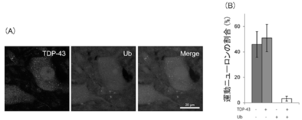
- (A) is a view showing an image of KO lumbar spinal cord double-stained with an anti-TDP-43 antibody and an anti-ubiquitin (Ub) antibody.
- (A) is a view showing an image of the lumbar spinal cord of KO double-stained with an anti-TDP-43 antibody and an anti-sequesterome 1 / p62 (p62) antibody.
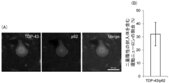
- (A) shows an image of KO motor neurons double-stained with anti-TDP-43 antibody and anti-p62 antibody.
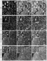
- FIG. 1 is a diagram showing images of gastrocnemius muscle sections stained with hematoxylin and eosin (HE) in WT and KO at 24 months of age.
- B shows images of gastrocnemius sections of 24-month-old WT and KO immunostained with anti-TDP-43 antibody.
- C shows images of gastrocnemius muscle sections of 24-month-old WT and KO immunostained with anti-p62 antibody.
- D shows images of gastrocnemius muscle sections of 24-month-old WT and KO immunostained with anti-Ub antibody. The length of the bar in the image corresponds to 20 ⁇ m.
- the knockout non-human animal according to the present embodiment is not particularly limited unless it is a human, and is a vertebrate, preferably a mammal, more preferably a rodent.
- examples of the knockout non-human animal include mouse, rat, guinea pig, hamster, rabbit, cat, dog, sheep, pig, goat, cow and monkey.
- the knockout non-human animal is preferably a mouse.
- a region containing a part of exon 8, a whole exon 9 and a part of exon 10 in the Optn gene (hereinafter also referred to as “region containing exon 8 etc.”) is a foreign gene.
- region containing exon 8 etc. a region containing a part of exon 8, a whole exon 9 and a part of exon 10 in the Optn gene.
- region including exon 8 and the like includes a part of exon 8, all of exon 9, part of exon 10, intron between exon 8 and exon 9, and exon 9 and exon 10. It is the region containing the intron between.
- the part of exon 8 of the Optn gene refers to any base from the second and following from the 5 'end of exon 8 to the base at the 3' end of exon 8.
- the part of exon 10 of the Optn gene refers to any base excluding the base at the 3 'end from the 5' end of exon 10.
- the region including exon 8 and the like is a region from the 21st base from the 5 'end of exon 8 to the 12th base from the 3' end to the 5 'end of exon 10.
- the method for producing the knockout mouse includes a substitution step of substituting a region containing exon 8 of the Optn gene with a foreign gene.
- a region including exon 8 of the wild-type Optn gene in mouse embryonic stem (ES) cells may be replaced with a foreign gene.
- Replacement of a region containing exon 8 and the like with a foreign gene can be performed by a known method. For example, a homologous recombination method, a retrovirus vector method, a lentivirus vector method, an artificial virus vector method, or the like can be used to replace a region containing exon 8 and the like with a foreign gene.
- TALEN zinc fingerer nuclease
- ZFN zinc fingerer nuclease
- CRISPR-Cas Clustered Regularly Interspersed Short Palindomic Repeat and CRISPR-Associative Technology etc.
- the foreign gene is not particularly limited as long as OPTN does not normally express after replacement.
- a drug resistance gene is preferred as the foreign gene.
- the drug resistance gene is, for example, a neomycin resistance gene or a puromycin resistance gene.
- a positive-negative selection method may be employed.
- a drug susceptibility gene may be used in addition to the drug resistance gene.
- a targeting vector for substituting the region containing exon 8 or the like of the optn gene in the wild type with a neomycin resistance gene by homologous recombination technology.
- a targeting vector is introduced into mouse ES cells by electroporation or the like, DNA is extracted from the obtained neomycin resistant clones, and ES clones homologously recombined are selected by polymerase chain reaction (PCR) and Southern blotting.
- the selected ES clone is microinjected into a mouse fertilized egg blastocyst and returned to the womb of a wild-type mouse, and a heterozygous male and female are obtained by mating with the wild-type mouse.
- a homozygous mouse of the Optn gene in which the region containing exon 8 and the like is replaced with a foreign gene can be obtained.
- the ES cell into which the targeting vector has been introduced is not an ES cell obtained by crossing the same strain of mice, it is preferable to purify the mouse strain by repeating backcrossing in the same strain of mice.
- OPTN deficiency can be confirmed by the fact that OPTN is not detected by Western blotting using a mouse anti-OPTN antibody targeting the spinal cord extract of the knockout mouse.
- TDP-43 positive inclusion bodies are detected in spinal motor neurons and motor nerve fibers than in the wild type.
- significantly more fragmented Golgi bodies are detected in spinal motor neurons than in the wild type.
- TDP-43 positive inclusion bodies and Golgi bodies were used to immunize lumbar spinal cord sections of knockout mice using an anti-TDP-43 antibody and an antibody against the Golgi marker, for example, anti-GM130 antibody, respectively. It can be detected by histochemical staining.
- a lumbar spinal cord section of a wild-type mouse may be similarly immunohistochemically stained to evaluate TDP-43 positive inclusion bodies and Golgi bodies.
- the number of motor neurons having TDP-43 positive inclusion bodies is counted for a plurality of sections, the average number of motor neurons in the section is calculated, and the average number of wild-type and knockout mice May be tested by a statistical method such as t-test.
- the number of signals indicating a Golgi apparatus having a predetermined intensity or higher independent of localization may be compared between the wild type and the knockout mouse based on the image processing of the stained image.
- the knockout mice are TDP-43 positive in 18-40%, 25-35%, or 28-32% of spinal motor neurons at 18 months of age, 20 months of age or more, 22 months of age or more, or 24 months of age or more The body is formed.
- p62 positive aggregates that are autophagy receptor proteins are detected in spinal motor neurons of the knockout mice.
- Spinal motor neurons from which p62 positive aggregates are detected often have TDP-43 positive inclusions.
- 20-40%, 25-35% or 28-32% of spinal motor neurons with both p62 positive aggregates and TDP-43 positive inclusions have inclusion bodies mixed with TDP-43 and p62.
- morphological changes are observed in the sciatic nerve. Morphological changes of the sciatic nerve are, for example, an increase in nerve fibers not encapsulated in S100 ⁇ -positive Schwann cells suggesting overlapping and demyelination of nerve fibers and S100 ⁇ , and an increase in nerve fibers thinner than the wild type.
- the knockout mice show significantly more TDP-43 positive inclusion bodies in and around the nerve fibers than in the wild type. In the sciatic nerve, the knockout mice show significantly more p62 positive aggregates in the nerve fibers than the wild type.
- the knockout mouse is not particularly limited. From the detection or increase of TDP-43 positive inclusion bodies, p62 positive aggregates and the detection or increase of Golgi fragmentation in spinal motor neurons and motor nerve fibers, the knockout mice are suitable for neurodegenerative diseases, particularly ALS model animals. Or it is suitable as a dementia model animal. For example, the knockout mouse is useful for elucidating the onset mechanism and understanding of the pathology of ALS and dementia, particularly frontotemporal dementia.
- the use of the knockout mouse is preferably a screening method for a therapeutic or prophylactic agent for ALS and dementia.
- the screening method using the knockout mouse includes, for example, an administration step of administering a test substance to the knockout mouse, and an evaluation step of evaluating an index relating to ALS or dementia of the knockout mouse.
- an administration step it is preferable to use adult intermediate and late knockout mice in order to more faithfully reproduce the pathology of ALS and dementia.
- a test substance such as a compound, antibody, peptide and DNA aptamer is administered to the knockout mouse.
- the method of administration is not particularly limited, but it is preferable to select an appropriate method for the test substance. Examples of the administration method include oral administration, intraperitoneal administration, intravenous injection, and subcutaneous injection.
- the test substance may be administered once or multiple times. What is necessary is just to determine the density
- Examples of the indicators in the evaluation step include behavioral indicators such as walking, muscle strength, neurodegeneration, spinal motor neurons and TDP-43 positive inclusions in motor nerve fibers, p62 positive aggregates, and pathological evaluation such as fragmentation of Golgi bodies. .
- an index relating to ALS or dementia may be compared between the knockout mouse to which the test substance is administered and the knockout mouse to which the test substance is not administered.
- a test substance that has improved ALS or dementia using an index may be selected as a candidate for a therapeutic or prophylactic agent for ALS or dementia.
- the knockout mouse can faithfully reproduce the onset mechanism of human diseases, particularly ALS and dementia, from its pathological characteristics as shown in Example 2 below.
- a part of the knockout mouse for example, an organ, a tissue, and a cell, can also be used for a screening method for a therapeutic or prophylactic agent for ALS and dementia.
- FIG. 1 shows an outline of a gene targeting method for producing an Optn knockout mouse.
- a targeting vector was constructed to replace a region containing exon 8 and the like in the Optn gene in WT with a neomycin resistance cassette (PGK Neo pA) by homologous recombination technology. More specifically, the mouse genome Dec.
- the targeting vector was designed such that chr2: 5033142-5035063 in 2011 (GRCm38 / mm10) was replaced with a neomycin resistance cassette.
- a targeting vector was introduced into TT2 embryonic stem cells derived from F1 embryos obtained by mating male CBA mice with female C57BL / 6 mice by electroporation, and neomycin-resistant colonies were collected.
- Germline transmission was confirmed by PCR using DNA collected from the tail of the mouse as a template according to a conventional method.
- primers for PCR of the WT allele a forward primer and a reverse primer were designed at positions indicated by arrows P1 and P2 in FIG. 1, respectively.
- a forward primer and a reverse primer were designed at the positions indicated by arrows P1 and P3 in FIG. 1 as PCR primers for the KO allele, respectively.
- the WT forward primer and the KO allele forward primer are common, and their base sequences are shown in SEQ ID NO: 1.
- the nucleotide sequences of the WT reverse primer and the KO allele reverse primer are shown in SEQ ID NOs: 2 and 3, respectively.
- the size of the PCR product is 466 bp.
- the size of the PCR product is 840 bp.
- the mouse lumbar spinal cord was homogenized on ice in a 5-fold volume (w / v) radioimmunoprecipitation assay (RIPA) buffer.
- the composition of the RIPA buffer is 150 mM NaCl, 1% NP-40, 1% sodium deoxycholate, 0.1% sodium dodecyl sulfate, 1X protease inhibitor cocktail (cComplete ULTRA Tablets, manufactured by Roche Diagnostics), 1X phosphatase inhibitor Cocktail (PhosSTOP, Phosphatase Inhibitor Cocktail, Roche Diagnostics) and 50 mM Tris-HCl (pH 7.4).
- the homogenized sample was centrifuged at 12000 rpm for 15 minutes at 4 ° C., and the supernatant was collected. 20 ⁇ g of RIPA extract was applied to each lane of a 10% polyacrylamide gel.
- the antibodies used for Western blotting of spinal cord extracts were mouse anti-OPTN antibody (N-terminal, sc-166576, 1: 800, manufactured by Santa Cruz Biotechnology), rabbit anti-OPTN antibody (internal segment, ab101589, 1: 800, Abcam plc; C-terminal, # 100000, 1: 1000, Cayman Chemical), horseradish peroxidase complex goat anti-mouse IgG antibody (1: 10000, R & D Systems) and horseradish peroxidase complex goat anti-rabbit It is an IgG antibody (1: 10000, manufactured by R & D Systems).
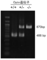
- FIG. 2 shows PCR product bands separated by electrophoresis. A 466 bp band was observed in WT (+ / +) having the Optn gene, whereas a 870 bp band was confirmed in KO ( ⁇ / ⁇ ).
- KO did not detect any N-terminal, internal segment or C-terminal of OPTN in the spinal cord extract. KO was raised up to 24 months of age, but there was no clear effect on survival, weight and motor function.
- Example 2 Analysis of pathological features using immunohistochemical staining
- Mice were deeply anesthetized by intraperitoneal administration of 75 mg / kg pentobarbital and perfusion-fixed with a phosphate buffer containing 4% paraformaldehyde.
- the sciatic nerve in the middle of the spinal cord or thigh was taken out and fixed in 4% paraformaldehyde phosphate buffer overnight and 2 hours, respectively.
- a frozen section having a thickness of 20 ⁇ m was prepared with a cryostat.
- the sections were immersed in phosphate buffered saline (PBS) containing 5% normal porcine serum for 1 hour, and further immersed in PBS containing 0.05% Triton-X100 containing primary antibodies (PBS-T) at 4 ° C. In the evening, the sections were immersed. After reacting with the primary antibody, the sections were washed and immersed in PBS-T containing appropriate Alexa Fluor TM 488/594 complex secondary antibody (1: 500, Vector Laboratories). After reacting with the secondary antibody, the section was encapsulated with Vectashield (Vector Laboratories). In order to suppress autofluorescence, lumbar spinal sections were treated with Zudan black solution before sealing with Vectashield. Sections were imaged and analyzed using a confocal laser microscope (LSM510, Carl Zeiss AG).
- LSM510 confocal laser microscope
- the primary antibodies used above were rabbit anti-TDP-43 antibody (10882-2-AP, 1: 5000, manufactured by Proteintech Group), goat anti-ChAT antibody (AB144P, 1: 500 EMD manufactured by Millipore), guinea pig anti-p62 antibody (GP62-C, 1: 5000, manufactured by PROGEN Biotechnik GmbH), rabbit anti-p62 antibody (# 018-22141, 1: 5000, manufactured by Wako Pure Chemical Industries), rabbit anti-Ub antibody (Z0458, 1: 2000, Agilent) Technologies), rabbit anti-S100 ⁇ antibody (ab52642, 1: 200, manufactured by Abcam plc), mouse anti-NF antibody (ab24564, 1: 2000, manufactured by Abcam plc) and mouse anti-GM130 antibody (610) 23,1: 1000, is a BD Bioscience Co., Ltd.).
- the number of motoneurons was counted by collecting one lumbar spinal slice every 10 intervals and preparing a total of 12 and immunostaining with anti-ChAT antibody.
- immunostaining after proceeding to the primary antibody reaction, the sections were sequentially reacted with a biotin-labeled secondary antibody, VECTASTAIN Elite ABC Kit (manufactured by Vector Laboratories), and developed with diaminobenzidine. For counterstaining, hematoxylin staining was performed.
- FIG. 4 shows the lumbar spine stained with anti-OPTN antibody. OPTN was detected in the WT lumbar spinal cord, whereas OPTN was not detected in KO. Next, the presence or absence of inclusion bodies found in ALS patients was confirmed in KO motor neurons by immunostaining with anti-TDP-43 antibody.
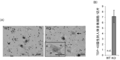
- FIG. 5 (A) shows the lumbar spinal cord in 24-month-old KO and WT stained with anti-TDP-43 antibody and counterstained with hematoxylin. In some KO motor neurons, a stained image in which TDP-43 was scattered in the cytoplasm but not in the nucleus was obtained. In some of the motor neurons, TDP-43 positive punctate inclusions were confirmed in the cytoplasm as indicated by arrows.
- TDP-43 positive inclusion bodies were formed in about 30% of spinal motor neurons of KO of 24 months old (see FIG. 5 (B)).
- KO spinal cord myelin was examined by Kluber-Barrera staining. There was no clear change in both gray matter and white matter. The number of KO spinal motoneurons was examined, but there was no significant change with respect to WT. Cerebral cortical motor areas and hippocampus were examined by HE staining, but no clear histological changes were observed.
- TDP-43 positive inclusion bodies were considered to be ubiquitin positive, they were examined by double staining with anti-TDP-43 antibody and anti-Ub antibody, but as shown in FIG. Ub positive inclusion bodies could not be confirmed. However, as shown in FIG. 6 (B), all Ub positive inclusions present were motor neurons having TDP-43 positive inclusions.
- FIG. 7 (A) shows the lumbar spinal cord of KO double-stained with anti-TDP-43 antibody and anti-p62 antibody. p62 positive aggregates were identified in many motor neurons. As shown in FIG. 7B, most of the motor neurons were motor neurons having TDP-43 positive inclusion bodies.
- FIG. 8 (A) shows KO motor neurons double-stained with anti-TDP-43 antibody and anti-p62 antibody.
- FIG. 8 (B) it was found that about 30% of motor neurons having TDP-43 positive inclusion bodies and p62 aggregates have inclusion bodies in which TDP-43 and p62 are mixed. Therefore, inclusion bodies containing TDP-43 / p62, which is a characteristic of human ALS, were confirmed in KO. From these, it was suggested that the homeostasis of the protein may be broken due to OPTN deficiency.
- FIG. 9 shows motor neurons in the spinal cord stained with an antibody against GM130, a marker of the Golgi apparatus.
- GM130 a marker of the Golgi apparatus.
- KO a fragmented GM130 positive signal was observed in motor neurons.
- TDP-43 positive inclusions were also formed in the KO sciatic nerve. As shown in FIG. 11, in WT, TDP-43 was scattered around nerve fibers at 24 months of age. In contrast, in KO, it was detected in and around nerve fibers at 12 months of age. The size of the inclusion bodies increased at 18 and 24 months of age. P62 positive aggregates were also observed in nerve fibers of the sciatic nerve. This suggests that in KO, morphological changes in motor neurons occur earlier in axons than in cell bodies.
- TDP-43 positive and p62 positive inclusion bodies were not detected in the fibers of the intramuscular nerve even at 24 months of age.
- Ub positive inclusion bodies were observed in the fibers of the intramuscular nerve.
- KO in which a region containing exon 8 of the Optn gene was replaced with a neomycin resistance gene has pathological features of ALS and dementia in humans.
- the present invention is suitable for ALS and dementia model animals and screening methods for ALS and dementia therapeutics or preventives.
Landscapes
- Life Sciences & Earth Sciences (AREA)
- Health & Medical Sciences (AREA)
- Chemical & Material Sciences (AREA)
- General Health & Medical Sciences (AREA)
- Environmental Sciences (AREA)
- Zoology (AREA)
- Organic Chemistry (AREA)
- Medicinal Chemistry (AREA)
- Engineering & Computer Science (AREA)
- Genetics & Genomics (AREA)
- Biochemistry (AREA)
- Molecular Biology (AREA)
- Proteomics, Peptides & Aminoacids (AREA)
- Gastroenterology & Hepatology (AREA)
- Biophysics (AREA)
- Biotechnology (AREA)
- Veterinary Medicine (AREA)
- Animal Behavior & Ethology (AREA)
- Animal Husbandry (AREA)
- Biodiversity & Conservation Biology (AREA)
- Toxicology (AREA)
- Investigating Or Analysing Biological Materials (AREA)
Abstract
ノックアウト非ヒト動物は、オプチニューリン遺伝子におけるエクソン8の一部、エクソン9の全部及びエクソン10の一部を含む領域が外来遺伝子に置換されている。前記外来遺伝子は、薬剤耐性遺伝子である、こととしてもよい。該ノックアウト非ヒト動物は、脊髄運動ニューロン及び運動神経線維に、野生型より有意に多くのTDP-43陽性封入体が検出される、こととしてもよい。
Description
本発明は、ノックアウト非ヒト動物、ノックアウト非ヒト動物の作製方法、及び筋萎縮性側索硬化症又は認知症の治療薬又は予防薬のスクリーニング方法に関する。
筋萎縮性側索硬化症(Amyotrophic Lateral Sclerosis、ALS)は、主に大脳皮質運動野、脳幹及び脊髄における運動ニューロンが次第に変性する神経変性疾患である。ほとんどのALS患者は発症して1~5年以内に人工呼吸器が必要になるか死に至る。ALS患者の大半が孤発性であり、約10%程度が家族性である。
次世代シークエンサー技術によりALSの原因遺伝子が次々と明らかになっている。ALSの原因遺伝子として、オプチニューリン(OPTN)遺伝子、TAR-DNA binding protein 43kD(TDP-43)遺伝子(TARDBP)、superoxide dismutase 1(SOD1)遺伝子、fused in sarcoma/translocated in liposarcoma(FUS/TLS)遺伝子、chromosome 9 open reading frame 72(C9orf72)遺伝子等の20以上もの遺伝子が同定されている。
TDP-43は、剖検脳においてALS症例の90%に病因物質として認められる。TDP-43は、ALSに加え、前頭側頭葉型認知症等の変性部位に出現するユビキチン陽性封入体の構成因子である。TDP-43は、不均一核内リボ核酸タンパク質(heterogeneous nuclear ribonucleoprotein、hnRNP)の一種である。TDP-43は、mRNA又はhnRNPと結合し、mRNAの安定化、hnRNAの選択的スプライシング及び転写調節等に関与する。
特許文献1には、変異TDP-43を発現するように改変したノックインマウスが開示されている。当該マウスの脳切片では、細胞質に異常封入体が観察された。特許文献2では、OPTN遺伝子の変異のため、遺伝性ALS患者及び孤発性ALS患者由来の検体において、OPTNが発現しないことが示されている。
マウスにおいてOPTNをコードするOptn遺伝子をノックアウトしたマウスが非特許文献1に報告されている。当該マウスは、Optntm1a(EUCOMM)Wtsiマウスを用いて作製され、Optn遺伝子のエクソン3を欠く。非特許文献1によれば、当該マウスの中枢神経系で髄鞘形成不全及び軸索変性の進展が見られた。
Yasushi Ito、外20名、「RIPK1 mediates axonal degeneration by promoting inflammation and necroptosis in ALS」、2016年、Science、353、6299、p.603-608
上記特許文献2及び非特許文献1のマウスでは、運動ニューロン内にTDP-43陽性封入体及びALSの病理学的特徴であるゴルジ体の断片化が観察されていない。上記特許文献1に開示されたマウスでは、細胞質に異常封入体が認められるものの、TDP-43に変異が入っている。ALS患者の90%ではTDP-43には変異がないため、上記特許文献1に開示されたマウスにおける異常封入体の形成機序は、実際のALSと異なることが考えられる。したがって、ヒトの疾患の発症メカニズムをより忠実に再現できるモデル動物が求められている。
本発明は、上記実情に鑑みてなされたものであり、ヒトの疾患の発症メカニズムをより忠実に再現できるノックアウト非ヒト動物、ノックアウト非ヒト動物の作製方法、及び筋萎縮性側索硬化症又は認知症の治療薬又は予防薬のスクリーニング方法を提供することを目的とする。
本発明の第1の観点に係るノックアウト非ヒト動物は、
オプチニューリン遺伝子におけるエクソン8の一部、エクソン9の全部及びエクソン10の一部を含む領域が外来遺伝子に置換されている。
オプチニューリン遺伝子におけるエクソン8の一部、エクソン9の全部及びエクソン10の一部を含む領域が外来遺伝子に置換されている。
この場合、前記外来遺伝子は、
薬剤耐性遺伝子である、
こととしてもよい。
薬剤耐性遺伝子である、
こととしてもよい。
上記本発明の第1の観点に係るノックアウト非ヒト動物は、
脊髄運動ニューロン及び運動神経線維に、野生型より有意に多くのTDP-43陽性封入体が検出される、
こととしてもよい。
脊髄運動ニューロン及び運動神経線維に、野生型より有意に多くのTDP-43陽性封入体が検出される、
こととしてもよい。
上記本発明の第1の観点に係るノックアウト非ヒト動物は、
脊髄運動ニューロンに、野生型より有意に多くの断片化されたゴルジ体が検出される、
こととしてもよい。
脊髄運動ニューロンに、野生型より有意に多くの断片化されたゴルジ体が検出される、
こととしてもよい。
上記本発明の第1の観点に係るノックアウト非ヒト動物は、
げっ歯類である、
こととしてもよい。
げっ歯類である、
こととしてもよい。
上記本発明の第1の観点に係るノックアウト非ヒト動物は、
マウスである、
こととしてもよい。
マウスである、
こととしてもよい。
上記本発明の第1の観点に係るノックアウト非ヒト動物は、
筋萎縮性側索硬化症モデル動物又は認知症モデル動物である、
こととしてもよい。
筋萎縮性側索硬化症モデル動物又は認知症モデル動物である、
こととしてもよい。
本発明の第2の観点に係るノックアウト非ヒト動物の作製方法は、
オプチニューリン遺伝子におけるエクソン8の一部、エクソン9の全部及びエクソン10の一部を含む領域を外来遺伝子に置換する置換ステップを含む。
オプチニューリン遺伝子におけるエクソン8の一部、エクソン9の全部及びエクソン10の一部を含む領域を外来遺伝子に置換する置換ステップを含む。
本発明の第3の観点に係る筋萎縮性側索硬化症又は認知症の治療薬又は予防薬のスクリーニング方法は、
上記本発明の第1の観点に係るノックアウト非ヒト動物を用いる。
上記本発明の第1の観点に係るノックアウト非ヒト動物を用いる。
本発明によれば、ヒトの疾患の発症メカニズムをより忠実に再現できる。
本発明に係る実施の形態について添付の図面を参照して説明する。なお、本発明は下記の実施の形態及び図面によって限定されるものではない。
(実施の形態)
本実施の形態に係るノックアウト非ヒト動物は、ヒトでなければ特に限定されず、脊椎動物、好ましくは哺乳類、より好ましくはげっ歯類である。具体的には、ノックアウト非ヒト動物としては、マウス、ラット、モルモット、ハムスター、ウサギ、ネコ、イヌ、ヒツジ、ブタ、ヤギ、ウシ及びサル等が挙げられる。汎用性及び利便性を考慮すると、好適にはノックアウト非ヒト動物はマウスである。
本実施の形態に係るノックアウト非ヒト動物は、ヒトでなければ特に限定されず、脊椎動物、好ましくは哺乳類、より好ましくはげっ歯類である。具体的には、ノックアウト非ヒト動物としては、マウス、ラット、モルモット、ハムスター、ウサギ、ネコ、イヌ、ヒツジ、ブタ、ヤギ、ウシ及びサル等が挙げられる。汎用性及び利便性を考慮すると、好適にはノックアウト非ヒト動物はマウスである。
以下では、本実施の形態に係るノックアウト非ヒト動物として、マウスを用いた場合について説明する。本実施の形態に係るノックアウトマウスは、Optn遺伝子におけるエクソン8の一部、エクソン9の全部及びエクソン10の一部を含む領域(以下、「エクソン8等を含む領域」ともいう)が外来遺伝子に置換されている。より詳細には、エクソン8等を含む領域は、エクソン8の一部、エクソン9の全部、エクソン10の一部に加え、エクソン8とエクソン9との間のイントロン及びエクソン9とエクソン10との間のイントロンを含む領域である。
Optn遺伝子のエクソン8の一部とは、エクソン8の5’末端から2番目以降の任意の塩基からエクソン8の3’末端の塩基までをいう。Optn遺伝子のエクソン10の一部とは、エクソン10の5’末端から3’末端の塩基を除く任意の塩基までをいう。好適には、エクソン8等を含む領域は、エクソン8の5’末端から21番目の塩基から、エクソン10の3’末端から5’末端に向かって12番目の塩基までの領域である。
当該ノックアウトマウスの作製方法は、Optn遺伝子のエクソン8等を含む領域を外来遺伝子に置換する置換ステップを含む。置換ステップでは、例えば、マウスの胚性幹(ES)細胞における野生型のOptn遺伝子のエクソン8等を含む領域を外来遺伝子に置換すればよい。エクソン8等を含む領域と外来遺伝子との置換は、公知の方法で行うことができる。例えば、エクソン8等を含む領域と外来遺伝子との置換には、相同組換え法、レトロウイルスベクター法、レンチウイルスベクター法及び人工ウイルスベクター法等を用いることができる。また、エクソン8等を含む領域と外来遺伝子との置換に、TALEN、zinc finger nuclease(ZFN)及びCRISPR-Cas(Clustered Regularly Interspaced Short Palindromic Repeat and CRISPR-associated protein)システム等のゲノム編集技術を用いてもよい。
外来遺伝子は、置換後にOPTNが正常に発現しないものであれば特に限定されない。Optn遺伝子のエクソン8等を含む領域が外来遺伝子に置換されたESクローンの選別のために、外来遺伝子としては薬剤耐性遺伝子が好ましい。薬剤耐性遺伝子は、例えば、ネオマイシン耐性遺伝子及びピューロマイシン耐性遺伝子である。なお、相同組換えされたESクローンの選別を効率化するために、ポジティブ-ネガティブ選別法を採用してもよい。この場合、薬剤耐性遺伝子に加えて、薬剤感受性遺伝子を使用してもよい。
例えば、エクソン8等を含む領域と外来遺伝子との置換には、野生型におけるOptn遺伝子のエクソン8等を含む領域を相同組換え技術によってネオマイシン耐性遺伝子に置換するためのターゲッティングベクターを用いるのが好ましい。ターゲッティングベクターをマウスES細胞に電気穿孔法等で導入し、得られたネオマイシン耐性クローンからDNAを抽出し、polymerase chain reaction(PCR)及びサザンブロット法により相同組換えされたESクローンを選別する。
選別したESクローンをマウス受精卵胚盤胞に顕微注入し、野生型マウスの胎内に戻して得られるキメラマウスと、野生型マウスとの交配によりヘテロ接合体の雄及び雌が得られる。次に、ヘテロ接合体の雄及び雌を交配することで、エクソン8等を含む領域が外来遺伝子に置換されたOptn遺伝子のホモ接合体マウスを得ることができる。なお、ターゲッティングベクターを導入したES細胞が同一系統のマウスの交配で得られたES細胞ではない場合、同一系統のマウスで戻し交配を繰り返して、マウスの系統を純化させるのが好ましい。
OPTNの欠損は、上記ノックアウトマウスの脊髄抽出物を対象としたマウス抗OPTN抗体を用いたウエスタンブロット法で、OPTNが検出されないことで確認できる。
次に、本実施の形態に係るノックアウトマウスの病理学的特徴を以下に説明する。該ノックアウトマウスでは、脊髄運動ニューロン及び運動神経線維に、野生型より有意に多くのTDP-43陽性封入体が検出される。また、当該ノックアウトマウスでは、脊髄運動ニューロンに、野生型より有意に多くの断片化されたゴルジ体が検出される。TDP-43陽性封入体及びゴルジ体は、下記実施例2に示すように、それぞれ抗TDP-43抗体及びゴルジ体のマーカーに対する抗体、例えば抗GM130抗体を用いて、ノックアウトマウスの腰髄切片を免疫組織化学染色することで検出できる。この場合、比較のために、野生型のマウスの腰髄切片も同様に免疫組織化学染色し、TDP-43陽性封入体及びゴルジ体を評価すればよい。
ここで「有意に多く」とは、統計学上の有意な差をもって多いことを意味する。例えば、免疫組織化学染色で比較する場合、複数の切片についてTDP-43陽性封入体を有する運動ニューロンの個数を計数し、切片における運動ニューロンの平均個数を算出し、野生型及びノックアウトマウスの平均個数の差を、t検定等の統計学的手法で検定すればよい。ゴルジ体の断片化については、染色像の画像処理に基づいて、例えば局在が独立した所定強度以上のゴルジ体を示すシグナルの個数を野生型とノックアウトマウスとで比較すればよい。
上記ノックアウトマウスは、18か月齢以上、20か月齢以上、22か月齢以上、又は24か月齢以上で脊髄運動ニューロンの20~40%、25~35%又は28~32%にTDP-43陽性封入体が形成される。
また、上記ノックアウトマウスの脊髄運動ニューロンでは、オートファジー受容体タンパク質であるp62陽性凝集体が検出される。p62陽性凝集体が検出される脊髄運動ニューロンは、TDP-43陽性封入体を有する場合が多い。さらに、p62陽性凝集体及びTDP-43陽性封入体の両方を有する脊髄運動ニューロンの20~40%、25~35%又は28~32%は、TDP-43及びp62の混在した封入体を有する。
上記ノックアウトマウスは、坐骨神経に形態学的変化が見られる。坐骨神経の形態学的変化は、例えば、神経線維とS100βの重なり及び脱髄を示唆するS100β陽性シュワン細胞に包まれていない神経線維の増加、及び野生型よりも細い神経線維の増加である。また、坐骨神経においても上記ノックアウトマウスでは、神経線維内及び神経線維の周囲にTDP-43陽性封入体が野生型より有意に多く見られる。また、坐骨神経において上記ノックアウトマウスでは、神経線維内にp62陽性凝集体が野生型より有意に多く見られる。
上記ノックアウトマウスの用途は特に限定されない。脊髄運動ニューロン及び運動神経線維におけるTDP-43陽性封入体、p62陽性凝集体の検出又は増加及びゴルジ体の断片化の検出又は増加等から、上記ノックアウトマウスは、神経変性疾患、特にはALSモデル動物又は認知症モデル動物として好適である。例えば、上記ノックアウトマウスは、ALS及び認知症、特に前頭側頭葉型認知症の発症メカニズムの解明及び病態の理解に有用である。
上記ノックアウトマウスの用途として、好ましくはALS及び認知症の治療薬又は予防薬のスクリーニング方法が挙げられる。上記ノックアウトマウスを用いる当該スクリーニング方法は、例えば、ノックアウトマウスに被験物質を投与する投与ステップと、該ノックアウトマウスのALS又は認知症に関する指標を評価する評価ステップとを含む。投与ステップでは、成体の中期及び後期のノックアウトマウスを用いるのがALS及び認知症の病理をより忠実に再現する点で好ましい。
上記ノックアウトマウスへの被験物質の投与では、化合物、抗体、ペプチド及びDNAアプタマー等の被験物質を、上記ノックアウトマウスに投与する。投与の方法は、特に限定されないが、被験物質に適切な方法を選択するのが好ましい。投与の方法としては、例えば、経口投与、腹腔内投与、静脈注射及び皮下注射等が挙げられる。被験物質の投与は、単回でもよいし、複数回であってもよい。被験物質の濃度は、被験物質の種類によって適宜決定すればよい。また、被験物質の濃度を数段階希釈した複数の濃度の被験物質を投与してもよい。
評価ステップにおける指標としては、歩行等の行動指標、筋力、神経変性、脊髄運動ニューロン及び運動神経線維におけるTDP-43陽性封入体、p62陽性凝集体及びゴルジ体の断片化等の病理評価が挙げられる。評価ステップでは、被験物質を投与された上記ノックアウトマウスと、被験物質を投与されなかった上記ノックアウトマウスとの間で、ALS又は認知症に関する指標を比較してもよい。指標によってALS又は認知症を改善した被験物質をALS又は認知症の治療薬又は予防薬の候補として選抜すればよい。
以上詳細に説明したように、上記ノックアウトマウスは、下記実施例2に示すように、その病理学的特徴からヒトの疾患、特にはALS及び認知症の発症メカニズムをより忠実に再現できる。
なお、上記ノックアウトマウスの一部、例えば、臓器、組織及び細胞もALS及び認知症の治療薬又は予防薬のスクリーニング方法に使用することができる。
以下の実施例により、本発明をさらに具体的に説明するが、本発明は実施例によって限定されるものではない。
(実施例1:Optnノックアウトマウスの作製)
図1は、Optnノックアウトマウス作製のためのジーンターゲッティング法の概略を示す。まず、WTにおけるOptn遺伝子におけるエクソン8等を含む領域を相同組換え技術によってネオマイシン抵抗性カセット(PGK Neo pA)に置換するためのターゲッティングベクターを構築した。より詳細には、マウスゲノム Dec.2011(GRCm38/mm10)におけるchr2:5033142-5035063がネオマイシン抵抗性カセットと入れ替わるようにターゲッティングベクターを設計した。雄のCBAマウスと雌のC57BL/6マウスとを交配させて得られるF1胚由来のTT2胚性幹細胞にターゲッティングベクターを電気穿孔法で導入し、ネオマイシン耐性コロニーを回収した。
図1は、Optnノックアウトマウス作製のためのジーンターゲッティング法の概略を示す。まず、WTにおけるOptn遺伝子におけるエクソン8等を含む領域を相同組換え技術によってネオマイシン抵抗性カセット(PGK Neo pA)に置換するためのターゲッティングベクターを構築した。より詳細には、マウスゲノム Dec.2011(GRCm38/mm10)におけるchr2:5033142-5035063がネオマイシン抵抗性カセットと入れ替わるようにターゲッティングベクターを設計した。雄のCBAマウスと雌のC57BL/6マウスとを交配させて得られるF1胚由来のTT2胚性幹細胞にターゲッティングベクターを電気穿孔法で導入し、ネオマイシン耐性コロニーを回収した。
次に、サザンブロッティング法で、ネオマイシン耐性コロニーから適切な組換え体のコロニーを選別した。選別された2つの独立したクローンをICRマウスの8細胞期胚に顕微注入し、雌の野生型C57BL/6マウスの胎内に戻してキメラマウスを出産させ、育成した。少なくとも5回の戻し交配を経て得られたヘテロ接合体の雄及び雌を交配して、以下の実施例で用いるOptn遺伝子が改変されていない野生型(以下、「WT」とする)及びOptn遺伝子がノックアウトされたOptnノックアウトマウス(以下、「KO」とする)の同腹子を得た。
生殖細胞系伝達は、常法に従って、マウスの尾から採取したDNAをテンプレートとしたPCRで確認した。WTアレルのPCRのプライマーとして、フォワードプライマー及びリバースプライマーを、それぞれ図1のP1及びP2の矢印に示される位置に設計した。一方、KOアレルのPCRのプライマーとして、フォワードプライマー及びリバースプライマーを、それぞれ図1のP1及びP3の矢印に示される位置に設計した。WTのフォワードプライマー及びKOアレルのフォワードプライマーは共通で、その塩基配列を配列番号1に示す。WTのリバースプライマーの塩基配列及びKOアレルのリバースプライマーの塩基配列をそれぞれ配列番号2、3に示す。WTアレルの場合、PCR産物のサイズは466bpである。KOアレルの場合、PCR産物のサイズは840bpである。
マウスの腰髄を、5倍量(w/v)の放射性免疫沈降法(radio immunoprecipitation assay、RIPA)バッファー中で、氷上均質化させた。RIPAバッファーの組成は、150mM NaCl、1%NP-40、1%デオキシコール酸ナトリウム、0.1%ドデシル硫酸ナトリウム、1X プロテアーゼ阻害剤カクテル(cOmplete ULTRA Tablets、Roche Diagnostics社製)、1X フォスファターゼ阻害剤カクテル(PhosSTOP Phosphatase Inhibitor Cocktail、Roche Diagnostics社製)及び50mM Tris-HCl(pH7.4)である。均質化した試料を、4℃、12000rpmで15分間遠心分離し、上清を回収した。10%ポリアクリルアミドゲルの各レーンにRIPA抽出物を20μgずつ流した。
脊髄抽出物のウエスタンブロット法に用いた抗体は、マウス抗OPTN抗体(N末端、sc-166576、1:800、Santa Cruz Biotechnology社製)、ウサギ抗OPTN抗体(内部セグメント、ab101592、1:800、Abcam plc社製;C末端、#100000、1:1000、Cayman Chemical社製)、西洋ワサビペルオキシダーゼ複合体ヤギ抗マウスIgG抗体(1:10000、R&D Systems社製)及び西洋ワサビペルオキシダーゼ複合体ヤギ抗ウサギIgG抗体(1:10000、R&D Systems社製)である。
(結果)
図2は、電気泳動で分離したPCR産物のバンドを示す。Optn遺伝子を有するWT(+/+)には、466bpのバンドが認められたのに対して、KO(-/-)では、870bpのバンドが確認された。OPTNのタンパク質に関しても、図3のウエスタンブロット法の結果に示すように、KOでは、脊髄抽出物にOPTNのN末端、内部セグメント及びC末端のいずれも検出されなかった。KOは24か月齢まで飼育したが、生存期間、体重及び運動機能において明確な影響は現れなかった。
図2は、電気泳動で分離したPCR産物のバンドを示す。Optn遺伝子を有するWT(+/+)には、466bpのバンドが認められたのに対して、KO(-/-)では、870bpのバンドが確認された。OPTNのタンパク質に関しても、図3のウエスタンブロット法の結果に示すように、KOでは、脊髄抽出物にOPTNのN末端、内部セグメント及びC末端のいずれも検出されなかった。KOは24か月齢まで飼育したが、生存期間、体重及び運動機能において明確な影響は現れなかった。
(実施例2:免疫組織化学染色を用いた病理学的特徴の解析)
75mg/kgのペントバルビタールを腹腔内に投与することでマウスを深麻酔し、4%パラホルムアルデヒドを含むリン酸バッファーで灌流固定した。脊髄又は大腿の中間の坐骨神経を採り出して4%パラホルムアルデヒドリン酸バッファーにそれぞれ一晩及び2時間後固定した。固定した組織をスクロース置換後、20μmの厚さの凍結切片をクライオスタットで作製した。
75mg/kgのペントバルビタールを腹腔内に投与することでマウスを深麻酔し、4%パラホルムアルデヒドを含むリン酸バッファーで灌流固定した。脊髄又は大腿の中間の坐骨神経を採り出して4%パラホルムアルデヒドリン酸バッファーにそれぞれ一晩及び2時間後固定した。固定した組織をスクロース置換後、20μmの厚さの凍結切片をクライオスタットで作製した。
5%正常ブタ血清を含むリン酸緩衝生理食塩水(PBS)に1時間、切片を浸漬し、さらに、一次抗体を含む0.05%Triton-X100含有PBS(PBS-T)に4℃で一晩、切片を浸漬した。一次抗体を反応させた後、切片を洗浄し、適切なAlexa Fluor(商標)488/594複合体二次抗体(1:500、Vector Laboratories社製)を含むPBS-Tに浸漬した。二次抗体を反応させた後、切片をVectashield(Vector Laboratories社製)で封入した。なお、腰髄の切片については、自家蛍光を抑制するために、Vectashieldで封入する前にズダンブラック溶液で処理した。共焦点レーザー顕微鏡(LSM510、Carl Zeiss AG社製)を用いて切片を撮像し、解析した。
上記で使用した一次抗体は、ウサギ抗TDP-43抗体(10782-2-AP、1:5000、Proteintech Group社製)、ヤギ抗ChAT抗体(AB144P、1:500EMD Millipore社製)、モルモット抗p62抗体(GP62-C、1:5000、PROGEN Biotechnik GmbH社製)、ウサギ抗p62抗体(#018-22141、1:5000、和光純薬工業社製)、ウサギ抗Ub抗体(Z0458、1:2000、Agilent Technologies社製)、ウサギ抗S100β抗体(ab52642、1:200、Abcam plc社製)、マウス抗NF抗体(ab24564、1:2000、Abcam plc社製)及びマウス抗GM130抗体(610823、1:1000、BD Bioscience社製)である。
運動ニューロンの計数は、腰髄切片を10枚間隔ごとに1枚採取して計12枚用意し、抗ChAT抗体で免疫染色して行った。免疫染色では、一次抗体反応まで進めた後、切片をビオチン標識二次抗体、VECTASTAIN Elite ABC Kit(Vector Laboratories社製)に順次反応させ、ジアミノベンジジンで発色させた。対比染色はヘマトキシリン染色を行った。
筋肉内の神経の免疫染色には、凍結した腓腹筋を30μmの厚さの切片とし、30分間、4%パラホルムアルデヒドリン酸バッファーで固定した。固定後、切片を抗TDP-43抗体、抗Ub抗体又は抗p62抗体で免疫染色した。免疫染色した切片を、デジタルカメラ(DP72、Olympus社製)を備えた光学顕微鏡(ECLIPSE E600、Nikon Instruments社製)で撮像した。画像の加工及び解析には、ZEN(Carl Zeiss AG社製)、Image-J及びAdobe Photoshop CS 5.1(Adobe Systems社製)を用いた。
Student’s t検定を含む統計処理は、統計解析ソフトEZRで行った。P値が0.05未満の時に統計学的有意な差とした。
(結果)
図4は、抗OPTN抗体で染色した腰髄を示す。WTの腰髄ではOPTNが検出されたのに対し、KOでは、OPTNは検出されなかった。次に、ALS患者で見られる封入体の有無を、抗TDP-43抗体を使った免疫染色でKOの運動ニューロンにおいて確認した。図5(A)は、抗TDP-43抗体で染色し、さらにヘマトキシリンで対比染色した24か月齢のKO及びWTにおける腰髄を示す。KOの運動ニューロンの一部ではTDP-43が核内になく細胞質内に散在している染色像が得られた。それらの中の一部の運動ニューロンにおいて、矢印で示すように細胞質内にTDP-43陽性の点状封入体が確認できた。
図4は、抗OPTN抗体で染色した腰髄を示す。WTの腰髄ではOPTNが検出されたのに対し、KOでは、OPTNは検出されなかった。次に、ALS患者で見られる封入体の有無を、抗TDP-43抗体を使った免疫染色でKOの運動ニューロンにおいて確認した。図5(A)は、抗TDP-43抗体で染色し、さらにヘマトキシリンで対比染色した24か月齢のKO及びWTにおける腰髄を示す。KOの運動ニューロンの一部ではTDP-43が核内になく細胞質内に散在している染色像が得られた。それらの中の一部の運動ニューロンにおいて、矢印で示すように細胞質内にTDP-43陽性の点状封入体が確認できた。
定量解析の結果、24か月齢のKOの脊髄運動ニューロンの約30%でTDP-43陽性封入体が形成されていた(図5(B)参照)。なお、クリューバー・バレラ染色でKOの脊髄のミエリンについて調べた。灰白質及び白質ともに明確な変化は見られなかった。KOの脊髄運動ニューロンの数を調べたがWTに対して大きな変化はなかった。大脳皮質運動野及び海馬をHE染色で調べたが、明確な組織学的変化は見られなかった。
TDP-43陽性封入体がユビキチン陽性であることが考えられたため、抗TDP-43抗体及び抗Ub抗体による二重染色で調べたが、図6(A)に示すように、ほとんどの運動ニューロンでUb陽性封入体が確認できなかった。しかし、図6(B)に示すように、存在したUb陽性封入体は全てTDP-43陽性封入体を有する運動ニューロンであった。
次に、オートファジー受容体タンパク質p62陽性凝集体について調べた。図7(A)は、抗TDP-43抗体及び抗p62抗体で二重染色したKOの腰髄を示す。p62陽性凝集体が多数の運動ニューロンで確認された。図7(B)に示すように、該運動ニューロンの多くがTDP-43陽性封入体を有する運動ニューロンであった。
図8(A)は、抗TDP-43抗体及び抗p62抗体で二重染色したKOの運動ニューロンを示す。図8(B)に示すように、TDP-43陽性封入体とp62凝集体とを有する運動ニューロンの約30%がTDP-43とp62の混在した封入体を有することがわかった。したがって、KOにおいて、ヒトALSの特徴であるTDP-43/p62を含む封入体を確認できた。これらのことから、OPTN欠損によりタンパク質の恒常性が破綻している可能性が示唆された。
TDP-43/p62を含む封入体に加えて、ヒトALSにおける顕著な病理学的特徴であるゴルジ体の断片化を評価した。図9は、ゴルジ体のマーカーであるGM130に対する抗体で染色した脊髄における運動ニューロンを示す。KOでは、運動ニューロンにおいて断片化したGM130陽性のシグナルが観察された。KOの運動ニューロンの約10%でゴルジ体の断片化が起きていた。
続いて、KOにおける運動ニューロンの軸索を含む坐骨神経への影響を調べた。図10に示すように、WTでは24か月齢でもほとんどの神経線維がS100β陽性シュワン細胞によって髄鞘を形成していることが確認できた。しかし、KOでは12か月齢で、形態学的変化を示唆する神経線維とS100βのシグナルの重なりが見られた。このことは、KOでは運動ニューロンの軸索が形態的に影響を受けていることを示唆している。さらに、KOでは、生後経過とともに軸索の径が細くなっていた。また、KOでは、18か月齢及び24か月齢においてかなりの数の神経線維がS100β陽性シュワン細胞に巻かれていないことが示された。このため、KOでは成体の中期及び後期において脱髄が起こっていることが示唆された。なお、坐骨神経の断面積及び神経線維の密度には、WTとKOとの間に統計学的に有意な差はなかった。
上記の坐骨神経の形態学的変化が脊髄の封入体出現よりも早期に起きていることから、TDP-43陽性封入体がKOの坐骨神経でも形成されるかを調べた。図11に示すように、WTでは24か月齢で、TDP-43が神経線維周囲に散在していた。これに対しKOでは、12か月齢で神経線維内及びその周辺に検出された。この封入体の大きさは、18か月齢及び24か月齢で増大した。坐骨神経の神経線維内においてもp62陽性凝集体が観察された。このことから、KOでは、運動ニューロンにおける形態学的変化が細胞体よりも軸索で早期に起きていることが示唆された。
上記で明らかになったKOにおける軸索変性が見られたことから筋肉における神経支配及び除神経による筋萎縮について検討した。そこで、HE染色で腓腹筋の形態を調べたが、WTとKOとで顕著な違いは見られなかった。しかし、腓腹筋切片内の脛骨神経束において一部神経線維が消失していた。詳細には、図12(A)に示すように、腓腹筋内の脛骨神経束の約10%で消失が見られ、ミエリン鞘の欠落及び線維症が起きていた。KOの脊髄及び坐骨神経に封入体が確認されたことから、脛骨神経においてTDP-43陽性封入体及びp62凝集体を調べた。坐骨神経と異なり、図12(B)、(C)にそれぞれ示すように、24か月齢でも筋内神経の線維内にはTDP-43陽性及びp62陽性封入体が検出されなかった。一方、図12(D)の矢印に示すように、KOでは、筋内神経の線維内にUb陽性封入体を認めた。
本実施例により、Optn遺伝子のエクソン8等を含む領域をネオマイシン耐性遺伝子に置換したKOは、ヒトにおけるALS及び認知症の病理学的特徴を有することが示された。
本発明は、本発明の広義の精神と範囲を逸脱することなく、様々な実施の形態及び変形が可能とされるものである。また、上述した実施の形態は、本発明を説明するためのものであり、本発明の範囲を限定するものではない。すなわち、本発明の範囲は、実施の形態ではなく、特許請求の範囲によって示される。そして、特許請求の範囲内及びそれと同等な発明の意義の範囲内で施される様々な変形が、本発明の範囲内とみなされる。
本出願は、2016年12月28日に出願された、日本国特許出願2016-254900号に基づく。本明細書中に日本国特許出願2016-254900号の明細書、特許請求の範囲、図面全体を参照として取り込むものとする。
本発明は、ALS及び認知症のモデル動物及びALS及び認知症の治療薬又は予防薬のスクリーニング方法に好適である。
Claims (9)
- オプチニューリン遺伝子におけるエクソン8の一部、エクソン9の全部及びエクソン10の一部を含む領域が外来遺伝子に置換されている、
ノックアウト非ヒト動物。 - 前記外来遺伝子は、
薬剤耐性遺伝子である、
請求項1に記載のノックアウト非ヒト動物。 - 脊髄運動ニューロン及び運動神経線維に、野生型より有意に多くのTDP-43陽性封入体が検出される、
請求項1又は2に記載のノックアウト非ヒト動物。 - 脊髄運動ニューロンに、野生型より有意に多くの断片化されたゴルジ体が検出される、
請求項1から3のいずれか一項に記載のノックアウト非ヒト動物。 - げっ歯類である、
請求項1から4のいずれか一項に記載のノックアウト非ヒト動物。 - マウスである、
請求項1から5のいずれか一項に記載のノックアウト非ヒト動物。 - 筋萎縮性側索硬化症モデル動物又は認知症モデル動物である、
請求項1から6のいずれか一項に記載のノックアウト非ヒト動物。 - オプチニューリン遺伝子におけるエクソン8の一部、エクソン9の全部及びエクソン10の一部を含む領域を外来遺伝子に置換する置換ステップを含む、
ノックアウト非ヒト動物の作製方法。 - 請求項1から7のいずれか一項に記載のノックアウト非ヒト動物を用いる、
筋萎縮性側索硬化症又は認知症の治療薬又は予防薬のスクリーニング方法。
Applications Claiming Priority (2)
| Application Number | Priority Date | Filing Date | Title |
|---|---|---|---|
| JP2016254900 | 2016-12-28 | ||
| JP2016-254900 | 2016-12-28 |
Publications (1)
| Publication Number | Publication Date |
|---|---|
| WO2018123540A1 true WO2018123540A1 (ja) | 2018-07-05 |
Family
ID=62710352
Family Applications (1)
| Application Number | Title | Priority Date | Filing Date |
|---|---|---|---|
| PCT/JP2017/044372 Ceased WO2018123540A1 (ja) | 2016-12-28 | 2017-12-11 | ノックアウト非ヒト動物、ノックアウト非ヒト動物の作製方法、及び筋萎縮性側索硬化症又は認知症の治療薬又は予防薬のスクリーニング方法 |
Country Status (1)
| Country | Link |
|---|---|
| WO (1) | WO2018123540A1 (ja) |
Citations (1)
| Publication number | Priority date | Publication date | Assignee | Title |
|---|---|---|---|---|
| JP2011067193A (ja) * | 2009-08-25 | 2011-04-07 | Hiroshima Univ | 筋萎縮性側索硬化症の診断マーカー、診断方法、及び、治療薬、並びに、筋萎縮性側索硬化症を発症するモデル動物、及び、モデル細胞 |
-
2017
- 2017-12-11 WO PCT/JP2017/044372 patent/WO2018123540A1/ja not_active Ceased
Patent Citations (1)
| Publication number | Priority date | Publication date | Assignee | Title |
|---|---|---|---|---|
| JP2011067193A (ja) * | 2009-08-25 | 2011-04-07 | Hiroshima Univ | 筋萎縮性側索硬化症の診断マーカー、診断方法、及び、治療薬、並びに、筋萎縮性側索硬化症を発症するモデル動物、及び、モデル細胞 |
Non-Patent Citations (5)
| Title |
|---|
| ASHBURN, F. S. ET AL.: "ABSTRAC 5710: Generation of a Novel Optineurin Knockout Mouse", INVESTIGATIVE OPHTHALMOLOGY & VISUAL SCIENCE, vol. 55, no. 13, 1 April 2014 (2014-04-01), XP055507807 * |
| BO, R. D. ET AL.: "Novel optineurin mutations in patients with familial and sporadic amyotrophic lateral sclerosis", J NEUROL NEUROSURG PSYCHIATRY, vol. 82, 2011, pages 1239 - 1243 * |
| ITO, Y. ET AL.: "RIPK1 mediates axonal degeneration by promoting inflammation and necroptosis in ALS", SCIENCE, vol. 353, no. 6299, 5 August 2016 (2016-08-05), pages 603 - 608, XP055507791 * |
| MARUYAMA, HIROFUMI: "Creating a model mouse for Amyotrophic Lateral Sclerosis", BRAIN SCIENCE REVIEW 2013, pages 169 - 182 * |
| REZAIE, T. ET AL.: "Molecular cloning, genomic structure, and protein characterization of mouse optineurin", GENOMICS, vol. 85, no. 1, 1 January 2005 (2005-01-01), pages 131 - 138, XP004682851, ISSN: 0888-7543 * |
Similar Documents
| Publication | Publication Date | Title |
|---|---|---|
| Barneo-Muñoz et al. | Lack of GDAP1 induces neuronal calcium and mitochondrial defects in a knockout mouse model of charcot-marie-tooth neuropathy | |
| Zhou et al. | Transgenic rat model of neurodegeneration caused by mutation in the TDP gene | |
| US11254719B2 (en) | Animal model of longevity and related methods for increasing longevity and inhibiting tumorigenesis | |
| Tamayev et al. | Memory deficits due to familial British dementia BRI2 mutation are caused by loss of BRI2 function rather than amyloidosis | |
| KR20210113606A (ko) | 신경 질환을 검출, 예방, 회복 및 치료하는 방법 | |
| Geister et al. | LINE-1 mediated insertion into Poc1a (protein of centriole 1 A) causes growth insufficiency and male infertility in mice | |
| CN107760713A (zh) | 一种非人哺乳动物神经精神疾病动物模型的建立方法及其用途 | |
| Horiuchi et al. | ALS-linked mutant TDP-43 in oligodendrocytes induces oligodendrocyte damage and exacerbates motor dysfunction in mice | |
| Ricketts et al. | A nonsense mutation in mouse Tardbp affects TDP43 alternative splicing activity and causes limb-clasping and body tone defects | |
| Kondo et al. | A Transgenic pig model with human mutant SOD1 exhibits the early pathology of amyotrophic lateral sclerosis | |
| WO2018123540A1 (ja) | ノックアウト非ヒト動物、ノックアウト非ヒト動物の作製方法、及び筋萎縮性側索硬化症又は認知症の治療薬又は予防薬のスクリーニング方法 | |
| JP5939487B2 (ja) | Epo欠損GFP貧血マウス | |
| Vanmarsenille et al. | Generation and characterization of an Nxf7 knockout mouse to study NXF5 deficiency in a patient with intellectual disability | |
| US7271313B2 (en) | Presenilin-deficient mouse model of age-dependent neurodegeneration and cognitive loss | |
| WO2021070866A1 (ja) | アルツハイマー病モデル非ヒト動物及びその製造方法 | |
| JP4550530B2 (ja) | シナプス成熟障害モデル動物 | |
| JP4374438B2 (ja) | rab8a遺伝子欠損マウス | |
| Germeys et al. | Theme 04-In Vivo Experimetal Models. | |
| JPWO2018012497A1 (ja) | 疾患モデル動物および疾患治療剤 | |
| US20130047273A1 (en) | Genetically altered animal specimen and related methods | |
| De Giorgio | Investigating TDP-43 mutations in mouse and developing a knockin humanised TDP-43 mouse model of Amyotrophic Lateral Sclerosis (ALS) | |
| JP6323876B2 (ja) | ノックインマウス | |
| US20060015959A1 (en) | Transgenic animals expressing transglutaminase II | |
| JPWO2019163166A1 (ja) | 遺伝子改変非ヒト動物及び脊髄小脳変性症の治療薬又は予防薬のスクリーニング方法 | |
| Vanmarsenille et al. | Generation and Characterization of an Nxf7 Knockout Mouse to Study |
Legal Events
| Date | Code | Title | Description |
|---|---|---|---|
| 121 | Ep: the epo has been informed by wipo that ep was designated in this application |
Ref document number: 17886216 Country of ref document: EP Kind code of ref document: A1 |
|
| NENP | Non-entry into the national phase |
Ref country code: DE |
|
| 122 | Ep: pct application non-entry in european phase |
Ref document number: 17886216 Country of ref document: EP Kind code of ref document: A1 |
|
| NENP | Non-entry into the national phase |
Ref country code: JP |