WO2017187941A1 - 凍結細胞集塊及び凍結細胞集塊の製造方法 - Google Patents
凍結細胞集塊及び凍結細胞集塊の製造方法 Download PDFInfo
- Publication number
- WO2017187941A1 WO2017187941A1 PCT/JP2017/014545 JP2017014545W WO2017187941A1 WO 2017187941 A1 WO2017187941 A1 WO 2017187941A1 JP 2017014545 W JP2017014545 W JP 2017014545W WO 2017187941 A1 WO2017187941 A1 WO 2017187941A1
- Authority
- WO
- WIPO (PCT)
- Prior art keywords
- cell
- frozen
- mesenchymal stem
- mscs
- stem cells
- Prior art date
- Legal status (The legal status is an assumption and is not a legal conclusion. Google has not performed a legal analysis and makes no representation as to the accuracy of the status listed.)
- Ceased
Links
Images







Classifications
-
- A—HUMAN NECESSITIES
- A61—MEDICAL OR VETERINARY SCIENCE; HYGIENE
- A61L—METHODS OR APPARATUS FOR STERILISING MATERIALS OR OBJECTS IN GENERAL; DISINFECTION, STERILISATION OR DEODORISATION OF AIR; CHEMICAL ASPECTS OF BANDAGES, DRESSINGS, ABSORBENT PADS OR SURGICAL ARTICLES; MATERIALS FOR BANDAGES, DRESSINGS, ABSORBENT PADS OR SURGICAL ARTICLES
- A61L27/00—Materials for grafts or prostheses or for coating grafts or prostheses
- A61L27/36—Materials for grafts or prostheses or for coating grafts or prostheses containing ingredients of undetermined constitution or reaction products thereof, e.g. transplant tissue, natural bone, extracellular matrix
- A61L27/38—Materials for grafts or prostheses or for coating grafts or prostheses containing ingredients of undetermined constitution or reaction products thereof, e.g. transplant tissue, natural bone, extracellular matrix containing added animal cells
- A61L27/3804—Materials for grafts or prostheses or for coating grafts or prostheses containing ingredients of undetermined constitution or reaction products thereof, e.g. transplant tissue, natural bone, extracellular matrix containing added animal cells characterised by specific cells or progenitors thereof, e.g. fibroblasts, connective tissue cells, kidney cells
- A61L27/3834—Cells able to produce different cell types, e.g. hematopoietic stem cells, mesenchymal stem cells, marrow stromal cells, embryonic stem cells
-
- A—HUMAN NECESSITIES
- A01—AGRICULTURE; FORESTRY; ANIMAL HUSBANDRY; HUNTING; TRAPPING; FISHING
- A01N—PRESERVATION OF BODIES OF HUMANS OR ANIMALS OR PLANTS OR PARTS THEREOF; BIOCIDES, e.g. AS DISINFECTANTS, AS PESTICIDES OR AS HERBICIDES; PEST REPELLANTS OR ATTRACTANTS; PLANT GROWTH REGULATORS
- A01N1/00—Preservation of bodies of humans or animals, or parts thereof
- A01N1/10—Preservation of living parts
- A01N1/16—Physical preservation processes
- A01N1/162—Temperature processes, e.g. following predefined temperature changes over time
-
- A—HUMAN NECESSITIES
- A01—AGRICULTURE; FORESTRY; ANIMAL HUSBANDRY; HUNTING; TRAPPING; FISHING
- A01N—PRESERVATION OF BODIES OF HUMANS OR ANIMALS OR PLANTS OR PARTS THEREOF; BIOCIDES, e.g. AS DISINFECTANTS, AS PESTICIDES OR AS HERBICIDES; PEST REPELLANTS OR ATTRACTANTS; PLANT GROWTH REGULATORS
- A01N1/00—Preservation of bodies of humans or animals, or parts thereof
- A01N1/10—Preservation of living parts
- A01N1/12—Chemical aspects of preservation
- A01N1/122—Preservation or perfusion media
- A01N1/125—Freeze protecting agents, e.g. cryoprotectants or osmolarity regulators
-
- A—HUMAN NECESSITIES
- A61—MEDICAL OR VETERINARY SCIENCE; HYGIENE
- A61L—METHODS OR APPARATUS FOR STERILISING MATERIALS OR OBJECTS IN GENERAL; DISINFECTION, STERILISATION OR DEODORISATION OF AIR; CHEMICAL ASPECTS OF BANDAGES, DRESSINGS, ABSORBENT PADS OR SURGICAL ARTICLES; MATERIALS FOR BANDAGES, DRESSINGS, ABSORBENT PADS OR SURGICAL ARTICLES
- A61L27/00—Materials for grafts or prostheses or for coating grafts or prostheses
- A61L27/36—Materials for grafts or prostheses or for coating grafts or prostheses containing ingredients of undetermined constitution or reaction products thereof, e.g. transplant tissue, natural bone, extracellular matrix
- A61L27/38—Materials for grafts or prostheses or for coating grafts or prostheses containing ingredients of undetermined constitution or reaction products thereof, e.g. transplant tissue, natural bone, extracellular matrix containing added animal cells
- A61L27/3895—Materials for grafts or prostheses or for coating grafts or prostheses containing ingredients of undetermined constitution or reaction products thereof, e.g. transplant tissue, natural bone, extracellular matrix containing added animal cells using specific culture conditions, e.g. stimulating differentiation of stem cells, pulsatile flow conditions
-
- C—CHEMISTRY; METALLURGY
- C12—BIOCHEMISTRY; BEER; SPIRITS; WINE; VINEGAR; MICROBIOLOGY; ENZYMOLOGY; MUTATION OR GENETIC ENGINEERING
- C12N—MICROORGANISMS OR ENZYMES; COMPOSITIONS THEREOF; PROPAGATING, PRESERVING, OR MAINTAINING MICROORGANISMS; MUTATION OR GENETIC ENGINEERING; CULTURE MEDIA
- C12N5/00—Undifferentiated human, animal or plant cells, e.g. cell lines; Tissues; Cultivation or maintenance thereof; Culture media therefor
- C12N5/0062—General methods for three-dimensional culture
-
- C—CHEMISTRY; METALLURGY
- C12—BIOCHEMISTRY; BEER; SPIRITS; WINE; VINEGAR; MICROBIOLOGY; ENZYMOLOGY; MUTATION OR GENETIC ENGINEERING
- C12N—MICROORGANISMS OR ENZYMES; COMPOSITIONS THEREOF; PROPAGATING, PRESERVING, OR MAINTAINING MICROORGANISMS; MUTATION OR GENETIC ENGINEERING; CULTURE MEDIA
- C12N5/00—Undifferentiated human, animal or plant cells, e.g. cell lines; Tissues; Cultivation or maintenance thereof; Culture media therefor
- C12N5/06—Animal cells or tissues; Human cells or tissues
- C12N5/0602—Vertebrate cells
- C12N5/0652—Cells of skeletal and connective tissues; Mesenchyme
- C12N5/0662—Stem cells
- C12N5/0663—Bone marrow mesenchymal stem cells (BM-MSC)
-
- C—CHEMISTRY; METALLURGY
- C12—BIOCHEMISTRY; BEER; SPIRITS; WINE; VINEGAR; MICROBIOLOGY; ENZYMOLOGY; MUTATION OR GENETIC ENGINEERING
- C12N—MICROORGANISMS OR ENZYMES; COMPOSITIONS THEREOF; PROPAGATING, PRESERVING, OR MAINTAINING MICROORGANISMS; MUTATION OR GENETIC ENGINEERING; CULTURE MEDIA
- C12N2513/00—3D culture
-
- C—CHEMISTRY; METALLURGY
- C12—BIOCHEMISTRY; BEER; SPIRITS; WINE; VINEGAR; MICROBIOLOGY; ENZYMOLOGY; MUTATION OR GENETIC ENGINEERING
- C12N—MICROORGANISMS OR ENZYMES; COMPOSITIONS THEREOF; PROPAGATING, PRESERVING, OR MAINTAINING MICROORGANISMS; MUTATION OR GENETIC ENGINEERING; CULTURE MEDIA
- C12N2523/00—Culture process characterised by temperature
-
- C—CHEMISTRY; METALLURGY
- C12—BIOCHEMISTRY; BEER; SPIRITS; WINE; VINEGAR; MICROBIOLOGY; ENZYMOLOGY; MUTATION OR GENETIC ENGINEERING
- C12N—MICROORGANISMS OR ENZYMES; COMPOSITIONS THEREOF; PROPAGATING, PRESERVING, OR MAINTAINING MICROORGANISMS; MUTATION OR GENETIC ENGINEERING; CULTURE MEDIA
- C12N2533/00—Supports or coatings for cell culture, characterised by material
- C12N2533/50—Proteins
- C12N2533/54—Collagen; Gelatin
-
- C—CHEMISTRY; METALLURGY
- C12—BIOCHEMISTRY; BEER; SPIRITS; WINE; VINEGAR; MICROBIOLOGY; ENZYMOLOGY; MUTATION OR GENETIC ENGINEERING
- C12N—MICROORGANISMS OR ENZYMES; COMPOSITIONS THEREOF; PROPAGATING, PRESERVING, OR MAINTAINING MICROORGANISMS; MUTATION OR GENETIC ENGINEERING; CULTURE MEDIA
- C12N2533/00—Supports or coatings for cell culture, characterised by material
- C12N2533/90—Substrates of biological origin, e.g. extracellular matrix, decellularised tissue
Definitions
- the present invention relates to a frozen cell clump and a method for producing a frozen cell clump.
- MSCs Mesenchymal stem cells
- the current MSC transplantation treatment method isolates and proliferates MSCs from patients, secures a sufficient number of cells, and temporarily freezes them. Thereafter, immediately before the transplantation operation to the patient, the cryopreserved MSC is thawed and cultured in CPC (Cell Processing Center) to process the MSC in a form of agglomerated MSC.
- CPC Cell Processing Center
- the MSC transplant is prepared by mixing melted MSC with artificial scaffold materials (hydroxyapatite, tricalcium phosphate apatite, polylactic acid, chitosan, atelocollagen gel, hyaluronic acid gel, etc.). And this MSC transplant is transplanted into a patient's defect
- artificial scaffold materials hydroxyapatite, tricalcium phosphate apatite, polylactic acid, chitosan, atelocollagen gel, hyaluronic acid gel, etc.
- Non-patent Document 1 Non-patent Document 1
- M KITTAKA, M KAJIYA, H SHIBA, M TAKEWAKI, K TAKESHITA, R KHUNG, T FUJITA, T IWATA, T Q NGUYEN, K OUHARA, K TAKEDA, T FUJITA, ARAT FUJITA complex can be a novel tissue engineering therapy for bone regeneration "; International Society for Cellular Therapy; Cytotherapy, 2015, 17, 860-873
- the MSC transplants so far are manufactured after thawing the cryopreserved MSCs immediately before the transplantation operation. Since the MSC transplant is produced each time within a limited period immediately before the transplant operation, there is no guarantee that the number of cells and the cell function contained in the MSC transplant can be made uniform. For this reason, transplant surgery may be postponed when a desired MSC transplant cannot be manufactured.
- the present invention has been made in view of the above matters, and an object of the present invention is to provide a frozen cell agglomerate that is agglomerated and can be cryopreserved, and a method for producing a frozen cell agglomerate.
- the frozen cell agglomeration according to the first aspect of the present invention comprises: A mesenchymal stem cell is formed into a granular shape and is a frozen cell aggregate. It is characterized by that.
- the frozen cell agglomerate is in a granular form having a diameter of 0.5 mm to 1.5 mm.
- the frozen cell agglomerate is in a granular form having a diameter of 0.8 mm to 1.2 mm.
- a method for producing a frozen cell conglomerate according to the second aspect of the present invention comprises: Culturing the mesenchymal stem cell in a growth medium containing a factor that causes the mesenchymal stem cell to produce collagen, Forming a granular cell cluster containing collagen produced from the mesenchymal stem cells; Cryopreserving the cell mass separated from the growth medium with a cryopreservative, It is characterized by that.
- the method for producing a frozen cell conglomerate comprises: Culturing the mesenchymal stem cells in a growth medium containing a factor that causes the mesenchymal stem cells to produce collagen to form a cell sheet, Form a granular cell agglomeration by self-aggregation with the free periphery of the cell sheet, Cryopreserving the cell mass separated from the growth medium with a cryopreservative, It is characterized by that.
- the mesenchymal stem cells are cultured in a culture vessel and proliferated until they become confluent,
- the periphery of the cell sheet may be made a free edge by separating the edge of the cell sheet adhered to the peripheral wall of the culture container from the peripheral wall of the culture container.
- a granular cell agglomerate having a diameter of 0.5 mm to 1.5 mm may be obtained using the cylindrical culture vessel of 1.5 cm 2 to 2.5 cm 2 .
- the method for producing a frozen cell conglomerate according to the fourth aspect of the present invention comprises: Culturing and aggregating mesenchymal stem cells in growth medium, Adding a factor that produces collagen to the mesenchymal stem cells in the growth medium to form granular cell clusters containing collagen produced from the mesenchymal stem cells; Cryopreserving the cell mass separated from the growth medium with a cryopreservative, It is characterized by that.
- Refrigerated storage may be performed without pre-freezing.
- the MSC is frozen in an agglomerated form, so that it is not necessary to produce the MSC transplant within a limited time period immediately before the transplantation operation or the like. For this reason, the situation where a transplant operation is postponed can be prevented.
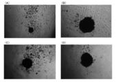
- FIG. 2 is a photograph showing a phase contrast microscopic image of C-MSCs
- FIG. 2 (A) is a photograph of C-MSCs that has not been cryopreserved
- FIG. 2 (B) is a C-MSC that has been cryopreserved in a DMSO cryopreservation solution.
- Photo of MSCs Fig. 2 (C) is a photo of C-MSCs cryopreserved at Bambanker (trade name: Wako Pure Chemical Industries, Ltd.)
- Fig. 2 (D) is Cell Banker (trade name: Takara Bio Inc.) It is a photograph of C-MSCs stored frozen.
- FIG. 2 is a photograph showing a phase contrast microscopic image of C-MSCs
- FIG. 2 (A) is a photograph of C-MSCs that has not been cryopreserved
- FIG. 2 (B) is a C-MSC that has been cryopreserved in a DM
- FIG. 3 (A) is a photograph before freezing
- FIG. 3 (B) is a partially enlarged photograph of FIG. 3 (A)
- FIG. 3 (C) is one day after thawing
- 3D is a partially enlarged photograph of FIG. 3C
- FIG. 3E is a photograph 5 days after melting
- FIG. 3F is a partially enlarged photograph of FIG. 3E.
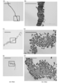
- FIG. 4 (A) is a photograph before freezing
- FIG. 4 (B) is a partially enlarged photograph of FIG. 4 (A)
- FIG. 4 (C) is a photograph one day after thawing
- 4 (D) is a partially enlarged photograph of FIG. 4 (C)
- FIG. 4 (E) is a photograph 5 days after melting
- FIG. 4 (F) is a partially enlarged photograph of FIG. 4 (E).
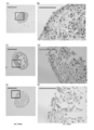
- FIG. 5 (A) is a photograph before freezing
- FIG. 5 (B) is a partially enlarged photograph of FIG. 5 (A)
- FIG. 5 (C) is a photograph one day after thawing.
- 5 (D) is a partially enlarged photograph of FIG. 5 (C)
- FIG. 5 (E) is a photograph 5 days after melting
- FIG. 5 (F) is a partially enlarged photograph of FIG. 5 (E).
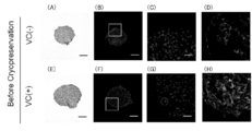
- It is a graph which shows the rate of cell death of C-MSCs before freezing, and C-MSCs, cell sheets and cell spheroids after 5 days of freezing and thawing.
- FIG. 7 (A) to (D) are photographs showing the state of C-MSCs to which collagenase was not added before cryopreservation
- FIG. 7 (A) is a HE-stained photograph
- FIG. 7 (B) is a TUNEL / FIG. 7 (C) is a partially enlarged photograph of FIG. 7 (B)
- FIG. 7 (D) is an immunostained photograph
- FIGS. 7 (E) to (H) are freezing of C-MSCs added with collagenase.
- FIG. 7 (E) is a HE-stained photograph
- FIG. 7 (F) is a TUNEL / DAPI-stained photograph
- FIG. 7 (G) is a partially enlarged photograph of FIG. 7 (F)
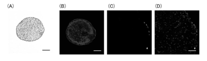
- FIGS. 8 (A) to (C) are photographs showing the state after 5 days of thawing after freezing C-MSCs to which no collagenase was added
- FIG. 8 (A) is a HE-stained photograph
- FIG. 8 (B ) Is a TUNEL / DAPI stained photograph
- FIG. 8 (C) is a partially enlarged photograph of FIG. 8 (B)
- FIGS. 8 (D) to (F) are 5 days after thawing after freezing storage of collagenase-added C-MSCs.
- 8 (D) is a HE-stained photograph
- FIG. 8 (E) is a TUNEL / DAPI-stained photograph
- FIG. 8 (F) is a partially enlarged photograph of FIG. 8 (E).
- 3 is a graph showing the ratio of dead cells 5 days after thawing of C-MSCs.
- FIGS. 10 (A) to (D) are photographs showing the state before cryopreservation of cell spheroids produced without adding ascorbic acid to the medium.
- FIG. 10 (A) is a HE-stained photograph and FIG. 10 (B).
- FIG. 10C is a partially enlarged photograph of FIG. 10B
- FIG. 10D is an immunostained photograph
- FIGS. 10E to 10H are ascorbic acid in the medium.
- FIG. 10 (E) is a photograph showing HE staining
- FIG. 10 (E) is a photograph showing HE staining
- FIG. 10 (F) is a photograph of TUNEL / DAPI staining
- FIG. 10 (G) is a photograph showing FIG. 10 (G).
- F) Partial enlarged photograph and FIG. 10 (H) are immunostained photographs.
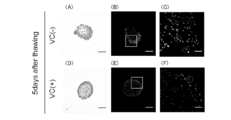
- FIGS. 11 (A) to (C) are photographs showing the state of cell spheroids produced without adding ascorbic acid to the medium after freezing and thawing 5 days
- FIG. 11 (A) is a HE-stained photograph.
- FIG. 11 (B) is a photograph of TUNEL / DAPI staining
- FIG. 11 (C) is a partially enlarged photograph of FIG. 11 (B)
- FIGS. 11 (D) to (F) are cells produced by adding ascorbic acid to the medium.
- FIG. 11 (B) is a photograph of TUNEL / DAPI staining
- FIG. 11 (C) is a partially enlarged photograph of FIG. 11 (B)
- FIG. 11 (D) is a HE-stained photograph
- FIG. 11 (E) is a TUNEL / DAPI-stained photograph
- FIG. 11 (F) is a partially enlarged photograph of FIG. 11 (E). It is. It is a graph which shows the ratio of the dead cell 5 days after the melt
- FIG. 14A is a CT photograph 4 weeks after transplanting C-MSCs into a rat calvaria
- FIG. 14A is a photograph in which transplantation was not performed
- FIG. 14B is a M-MSCs cryopreserved in DMSO cryopreservation solution.
- 14 (C) is a photograph of transplanted C-MSCs cryopreserved by Cell Banker (trade name: Takara Bio Inc.), and FIG. 14 (D) is Bambaker (trade name: Wako Pure Chemical Industries Ltd.). This is a photograph of transplanted C-MSCs cryopreserved by the company.
- FIG. 15A is a photograph showing HE-stained photographs
- FIG. 15B is a TUNEL / DAPI stained photograph
- FIG. 15C is a TUNEL-stained part.
- An enlarged photograph, FIG. 15D is a partially enlarged photograph stained with TUNEL / DAPI.
- FIG. 15A is a photograph showing HE-stained photographs
- FIG. 15B is a TUNEL / DAPI stained photograph
- FIG. 15C is a TUNEL-stained part.
- FIG. 16A is a CT photograph of a rat calvaria showing the transplantation effect of C-MSCs
- FIG. 16A is a photograph in which transplantation was not performed
- FIG. 16B is a photograph four weeks after transplantation of C-MSCs.
- FIG. 17A is a cross-sectional photograph of HE-stained rat calvaria showing the transplantation effect of C-MSCs
- FIG. 17A is a photograph in which transplantation was not performed
- FIG. 17B is a photograph four weeks after transplanting C-MSCs. It is.
- the frozen cell agglomeration according to the present embodiment is a frozen cell agglomeration in which a plurality of mesenchymal stem cells are agglomerated and formed into a granular shape.
- the frozen cell agglomerate may be of a size according to the form to be used.
- a bone destruction disease such as a fracture or periodontitis
- It is preferably a size that can be easily transplanted into a diameter of 0.5 mm to 1.5 mm, more preferably 0.8 mm to 1.2 mm.
- the transplant refers to a form used for tissue regeneration regardless of a technique such as a form in which a bone defect site is directly implanted and used, or a form in which tissue regeneration is performed by injection into the body by injection or the like.
- a technique such as a form in which a bone defect site is directly implanted and used, or a form in which tissue regeneration is performed by injection into the body by injection or the like.
- Frozen cell clumps are thawed before transplant surgery and used for transplant surgery.
- a frozen cell agglomerate retains its form after thawing and does not lose cell function.
- the frozen cell agglomeration exhibits tissue regeneration ability by transplanting into a defective tissue.
- the frozen cell agglomeration can be thawed by various methods such as removing the cryopreservation container containing the frozen cell agglomerate from a freezer and placing it at room temperature or in a warm bath (eg, 40 ° C. to 60 ° C.). It can be carried out.
- the above-mentioned frozen cell agglomeration can be produced as follows. First, the collected mesenchymal stem cells are cultured in a growth medium containing a factor that causes MSC to produce collagen using a culture vessel such as a culture dish. As the mesenchymal stem cells, those collected from various tissues such as a patient's bone marrow, adipose tissue, placental tissue or umbilical cord tissue, dental pulp and the like are used.
- the growth medium may be any medium that allows mesenchymal stem cells to grow, and a commercially available growth medium (for example, DMEM (Dulbecco's Modified a Eagle's Medium) + 10% FBS (fetal bovine serum)) may be used.
- DMEM Denstrength Enzyme
- FBS fetal bovine serum
- Factors that cause MSC to produce collagen are not limited as long as they can cause MSC to produce collagen as a result. Examples include ascorbic acid, steroids such as dexamethasone, cytokines, and the like. An acid is preferred.
- cell sheet a sheet-like cell population (hereinafter cell sheet) is formed. Then, the periphery of the cell sheet adheres to the edge of the incubator and grows until it reaches confluence.
- the edge of the cell sheet adhered to the peripheral wall of the incubator is separated from the incubator.
- the cell sheet can be separated from the incubator by inserting a thin rod into the inner wall of the incubator to which the edge of the cell sheet is adhered and moving the rod once along the inner wall of the incubator. As a result, the cell sheet floats.
- This floating cell sheet is wrapped by self-aggregation. And it becomes a cluster mesenchymal stem cell conglomerate using the extracellular matrix (ECM: extracellular matrix) which a mesenchymal stem cell itself produces. In this way, a granular cell clump can be obtained.
- ECM extracellular matrix
- the incubator is a cylindrical one having a size of 1.5 cm 2 to 2.5 cm 2
- cell agglomerates having a particle size of 0.5 mm to 1.5 mm can be obtained.
- a 24-well plate having a surface area of 2 cm 2 may be used as the incubator.
- a spheroid-shaped cell cluster when obtaining a spheroid-shaped cell cluster, it can be produced as follows.
- MSCs When MSCs are cultured in a growth medium so as to float, the MSCs aggregate and become granular due to cell-cell adhesion. Thereafter, a substance that causes the MSC to produce collagen is added to the growth medium. As a result, MSC produces collagen, and a spheroid-shaped cell cluster containing abundant collagen as compared with normal cell spheroids is obtained.
- the resulting MSC transplant is placed in a cryopreservation container such as a cryopreservation vial together with a cryopreservation agent and cryopreserved. In this way, a frozen transplant can be obtained.
- a cryopreservation container such as a cryopreservation vial together with a cryopreservation agent and cryopreserved.
- cryopreservation agent various preservation solutions for cell freezing may be used.
- a cryopreservation solution containing 10% DMSO (Dimethyl sulfoxide), 20% FBS, 70% DMEM, a commercially available cell bank (product) Name: Takara Bio Inc.), Bambanker (trade name: Wako Pure Chemical Industries, Ltd.) and the like.
- the cryopreservation may be performed at a temperature of -70 ° C to -90 ° C, preferably -75 ° C to -85 ° C.
- the cryopreservation can be performed by various methods such as placing the storage container in a freezer. When long-term storage for one month or longer is performed, the storage container may be placed at the above temperature for 24 hours, and then transferred to a liquid nitrogen tank ( ⁇ 196 ° C.) for storage.
- an MSC graft using an artificial scaffold material for example, hydroxyapatite, tricalcium phosphate apatite, polylactic acid, chitosan, atelocollagen gel, hyaluronic acid gel, etc.
- an artificial scaffold material for example, hydroxyapatite, tricalcium phosphate apatite, polylactic acid, chitosan, atelocollagen gel, hyaluronic acid gel, etc.
- the physical properties of the artificial scaffold material change when frozen. Since it does not function as an MSC transplant after thawing, it has not been cryopreserved.
- cell sheets and cell spheroids are available as transplants that do not use a scaffold material, but these cannot be cryopreserved because they cannot be maintained in their respective forms after freezing and thawing, as described in the examples below.
- the frozen cell agglomeration according to the present embodiment is in a state of being cryopreserved after producing the MSC cell agglomeration, the MSC cell agglomeration is produced from the MSC separated from the patient, and its cell function And cell uniformity can be inspected in advance. And it inspects and selects only the thing of fixed quality, and is cryopreserved as a frozen cell conglomerate.
- the MSC transplant which is a mass-controlled MSC cell cluster, can be reliably supplied on the day of transplantation, and the situation where the transplantation operation is postponed can be prevented.
- bone marrow mesenchymal stem cells isolated from periodontitis patients are formed into MSC transplants, and are cryopreserved after examination of cell function / contamination.
- basic periodontal treatment such as removal of infection source and reduction of inflammation required before transplantation is completed, and then the quality-controlled MSC transplant is melted on the day of transplantation and transplanted to the defective tissue.
- Periodontal tissue regeneration can be realized.
- MSC is considered to be a cell applicable to allogeneic transplantation. Therefore, when an MSC supply system for other transplantation such as an MSC bank is constructed, the MSC is not frozen as it is, but frozen in the form of MSC cell agglomerates so that it can be promptly used when needed by the patient. In addition, it becomes possible to perform cell preparation medical treatment that can reliably provide a good MSC cell cluster.
- GM medium Growth medium in which 10% FBS (Biowest), 100 U / mL penicillin (Sigma), 100 ⁇ g / mL streptomycin (Sigma), and 500 ng / mL amphotericin B (Invitrogen) were added to DMEM (Sigma). Then, the collected mesenchymal stem cells were cultured. After 24 hours, non-adherent cells were removed, and the third passage cells obtained by further culturing the adherent cells were used in the following experiments.
- MSCs were seeded on a 24-well culture plate at a rate of 7.0 ⁇ 10 4 cells / well and cultured in a growth medium (DMEM + 10% FBS) supplemented with L-ascorbic acid (50 ⁇ g / mL) for 7 hours.
- DMEM + 10% FBS a growth medium supplemented with L-ascorbic acid (50 ⁇ g / mL) for 7 hours.
- a cell sheet composed of an MSC / ECM complex is formed by the extracellular matrix produced by MSC itself, and the periphery of the cell sheet comes into contact with the peripheral wall of the 24-well culture plate and reaches a confluent state.
- the periphery of the cell sheet was peeled off from the 24-well culture plate using the tip of. This caused the cell sheet to float and wrap around itself.
- C-MSCs MSC transplants
- C-MSCs (1 ⁇ 10 5 cells), soaked in 500 ⁇ L of cryopreservation solution, and frozen in a deep freezer at ⁇ 80 ° C. without pre-freezing using a 1.5 mL freezing vial .
- DMSO cryopreservation solution a cryopreservation solution (hereinafter, DMSO cryopreservation solution) in which 10% DMSO, 20% FBS, and 70% DMEM are mixed, Cell banker (trade name: Takara Bio Inc.), and Bambanker (trade name: Wako Pure Chemical Industries, Ltd.) was used for each.
- C-MSCs were immersed in a 24-well culture plate containing 500 ⁇ L normal medium (DMEM + 10% FBS) and cultured again.
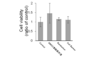
- FIG. 1 shows the result. Note that C-MSCs that were not frozen (control) were also performed, and Cell viability in FIG. 1 is shown as a relative value to the control.
- FIG. 1 shows that C-MSCs frozen in any of the cryopreservation solutions were not significantly different from the control, and the cell survival activity of the frozen C-MSCs was higher than that in the case where freezing was not performed. It was not inferior.
- FIG. 2 it was confirmed that cells were released from all C-MSCs. Thereby, it was confirmed that a normal function as a transplant can be exhibited.
- FIG. 4 shows an HE-stained image of a cell sheet
- FIG. 5 shows an HE-stained image of a cell spheroid.
- Cell sheets and cell spheroids were prepared and used referring to “Akabane M et al., 2008, J Tissue Eng Regen Med” and “Priya R et al., 2012, Cell Tissue Res”, respectively.
- FIG. 4 it can be seen that the morphology of the cell sheet collapses in a time-dependent manner.
- FIG. 5 like the cell sheet, the form of the cell spheroid is decaying in a time-dependent manner.
- the cell sheets and cell spheroids collapsed when stored frozen and cannot be used as transplants.
- the cell sheet had a rough substrate, and the cell spheroids were formed by cell-cell adhesion with less substrate (extracellular matrix). Freezing ice crystals are thought to have damaged the morphology.
- TUNEL assays were performed on C-MSCs before freezing, and C-MSCs, cell sheets, and cell spheroids 5 days after freezing and thawing.
- C-MSCs can be frozen and stored even after freezing and thawing, and can be cryopreserved.
- cell sheets and spheroids are frozen, many cell deaths occur, and cryopreservation occurs. I confirmed that I could not.
- the C-MSCs were soaked one by one in 500 ⁇ l of DMSO cryopreservation solution and cryopreserved by the same method as described above. After 48 hours, the mixture was thawed in the same manner as described above and cultured in 500 ⁇ l of GM medium. Five days after thawing, C-MSCs were fixed with 1% formaldehyde and embedded in paraffin, and 5 ⁇ m and 20 ⁇ m sections were prepared successively. A 5 ⁇ m section was stained with HE, and the histological structure was observed with an optical microscope. 20 ⁇ m sections were stained with TUNEL using DeadEnd® Fluorometric® terminal® deoxynucleotidyl® transferase® System, and the number of dead cells was observed with a confocal laser microscope.
- FIGS. 7 (A) to (D) and FIGS. 8 (A) to (C) show the state of the collagenase ( ⁇ ) group before cryopreservation and after 5 days of thawing
- FIGS. H) and FIGS. 8D to 8F show the state of the collagenase (+) group before cryopreservation and 5 days after thawing.
- a portion with high luminance (as an example, surrounded by a broken line) Represents the nucleus of dead cells.
- FIG. 7 (D) shows that the expression level of Type I collagen decreased before cryopreservation by treating collagenase with C-MSCs. 8 (D) to (F), it was observed that the collagenase (+) group was deformed after freezing and thawing and the number of dead cells increased.
- the collagenase ( ⁇ ) group which is a normal C-MSCs, maintained its form after freezing and thawing, and the number of dead cells was small, as seen in FIGS. 8 (A) to (C). .
- FIG. 9 shows the cell death rate in the collagenase ( ⁇ ) group and the collagenase (+) group. From FIG. 9, a significant difference appears in the rate of cell death between the collagenase ( ⁇ ) group and the collagenase (+). From the above, in C-MSCs, it was suggested that ECM mainly composed of collagen exhibits a protective effect against freezing.
- C-MSCs can be cryopreserved due to the protective effect of ECM, it can be cryopreserved in the form of cell spheroids as long as collagen is produced from the cells. It was verified whether it could be stored.
- MSCs were seeded on an ultra low binding 24 well plate at a cell density of 2 ⁇ 10 5 cells / well and cultured in GM medium for 4 days to obtain cell spheroids (VC ( ⁇ ) group) composed of MSCs.
- VC ( ⁇ ) group cell spheroids
- cell spheroids were also prepared by adding 50 ⁇ g / ml ascorbic acid (Sigma) to GM medium after MSC aggregated during culture (VC (+) group).
- the cell spheroids were soaked one by one in 500 ⁇ l of DMSO cryopreservation solution and frozen at ⁇ 80 ° C. using a 1.5 ml freezing vial (True line). After 48 hours, it was rapidly thawed in a constant temperature water bath set at 37 ° C., replated on a 24-well culture plate, and cultured in 500 ⁇ l GM medium.
- the cell spheroids were thawed and fixed with 1% formaldehyde after 5 days, embedded in paraffin, and continuously sliced at 5 ⁇ m and 20 ⁇ m. A 5 ⁇ m section was stained with hematoxylin-eosin (HE staining), and the histological structure was observed with an optical microscope. 20 ⁇ m sections were stained with DeadEnd Fluorometric terminal deoxynucleotidyl transfer system (Promega) and the number of dead cells was observed with a confocal laser microscope.
- FIGS. 11 (A) to (C) show the state of the VC ( ⁇ ) group before cryopreservation and after 5 days of thawing, and 10 (E) to (H).
- 11 (D) to (F) show the state of the VC (+) group before cryopreservation and 5 days after thawing, respectively.
- 10B, FIG. 10B, FIG. 11B, FIG. 10C, FIG. 10E, and FIG. 10F a portion with high luminance (a portion surrounded by a broken line as an example). Represents the nucleus of dead cells.
- FIG. 10 (D) shows that the expression level of Type I collagen is low in the spheroids of the VC ( ⁇ ) group with no ascorbic acid added.
- 11 (A) to (C) after freezing and thawing, the form collapsed and an increase in dead cells was observed.
- FIG. 10 (H) high type I collagen expression was observed in the spheroids of the VC (+) group added with ascorbic acid.
- 11 (D) to 11 (F) the shape was maintained after freezing and thawing, and the number of dead cells was small.
- FIG. 12 shows the ratio of dead cells 5 days after thawing in the VC (+) group and the VC ( ⁇ ) group. From FIG. 12, there is a significant difference in the rate of cell death between the VC (+) group and the VC ( ⁇ ) group. From the above, it was found that even cell spheroids can be cryopreserved by producing abundant ECM.
- C-MSCs were cultured in a normal medium (GM) and a mineralization induction medium (OIM) using a low adhesion culture dish. Then, the amount of calcium deposition in C-MSCs was quantified after 5 days and 10 days of culture.
- GM normal medium
- OFIM mineralization induction medium
- CT Computerputed Tomography
- Fig. 14 shows a CT photograph. B-regeneration is induced in C-MSCs that have been cryopreserved with any of the cryogens compared to those that were not transplanted.
- C-MSCs were cryopreserved for 6 months using a DMSO cryopreservation solution in the same manner as described above. Then, C-MSCs were melted by the same method as described above. The C-MSCs were subjected to HE staining, TUNEL staining, and DIPI staining. The result is shown in FIG.
- FIG. 15 shows that the morphology of C-MSCs cryopreserved for 6 months was maintained, and no marked increase in dead cells was observed.
- FIG. 16 shows a CT photograph.
- FIG. 17 shows a cross-sectional photograph stained with HE. It can be seen that bone regeneration is promoted in the calvaria transplanted with the C-MSCs of FIGS. 16 (B) and 17 (B) as compared to the controls of FIGS. 16 (A) and 17 (A). Therefore, it was confirmed that even if C-MSCs were cryopreserved for as long as 6 months, they could be thawed and transplanted to promote bone regeneration.
- cryopreserved C-MSCs could be used for regenerative medicine by transplantation surgery without losing the function as a transplant even when thawed.
- the frozen cell agglomeration according to the present invention can be used in transplantation regenerative medicine and the like.
Landscapes
- Health & Medical Sciences (AREA)
- Life Sciences & Earth Sciences (AREA)
- Engineering & Computer Science (AREA)
- Biomedical Technology (AREA)
- Chemical & Material Sciences (AREA)
- Zoology (AREA)
- Cell Biology (AREA)
- General Health & Medical Sciences (AREA)
- Wood Science & Technology (AREA)
- Developmental Biology & Embryology (AREA)
- Genetics & Genomics (AREA)
- Bioinformatics & Cheminformatics (AREA)
- Organic Chemistry (AREA)
- Biotechnology (AREA)
- Epidemiology (AREA)
- Animal Behavior & Ethology (AREA)
- Veterinary Medicine (AREA)
- Public Health (AREA)
- Hematology (AREA)
- Transplantation (AREA)
- Oral & Maxillofacial Surgery (AREA)
- Medicinal Chemistry (AREA)
- Dermatology (AREA)
- Botany (AREA)
- Chemical Kinetics & Catalysis (AREA)
- General Engineering & Computer Science (AREA)
- Biochemistry (AREA)
- Microbiology (AREA)
- Urology & Nephrology (AREA)
- Immunology (AREA)
- Rheumatology (AREA)
- Dentistry (AREA)
- Environmental Sciences (AREA)
- Micro-Organisms Or Cultivation Processes Thereof (AREA)
- Materials For Medical Uses (AREA)
Abstract
凍結細胞集塊は、間葉系幹細胞が粒状に形成され、凍結状態の細胞集塊である。凍結細胞集塊の製造方法は、間葉系幹細胞にコラーゲンを産生させる因子を含有する増殖培地にて間葉系幹細胞を培養し、間葉系幹細胞から産生されるコラーゲンを含有する粒状の細胞集塊を形成させ、増殖培地から分離した細胞集塊を凍結保存剤とともに凍結保存する。
Description
本発明は、凍結細胞集塊及び凍結細胞集塊の製造方法に関する。
間葉系幹細胞(MSC:Mesenchymal stem cell)は多分化能・自己増殖能を有しており、遺伝子導入などが不要であるとともに、患者から確実に得られるため、組織再生療法に適した細胞である。
例えば、骨折や歯周炎などの骨破壊疾患に対して、現在のMSC移植治療法では、患者からMSCを分離、増殖させ、細胞数を十分に確保して一旦凍結保存しておく。その後、患者への移植手術の直前に、凍結保存しておいたMSCを融解し、CPC(Cell Processing Center)で培養してMSCが集塊状の形態となったMSC移植体の形態に加工する。
例えば、融解したMSCを人工足場材料(ハイドロキシアパタイト、リン酸三カルシウムアパタイト、ポリ乳酸、キトサン、アテロコラーゲンゲル、ヒアルロン酸ゲルなど)と混和して培養することで、MSC移植体が作成される。そして、このMSC移植体が患者の欠損組織に移植され、MSCの組織再生能を発揮させる。
また、近年では、人工足場材料を要しないMSC移植体があり、このようなMSC移植体として、細胞シートや細胞スフェロイドのほか、細胞集塊(非特許文献1)がある。
M KITTAKA, M KAJIYA, H SHIBA, M TAKEWAKI, K TAKESHITA, R KHUNG, T FUJITA, T IWATA, T Q NGUYEN, K OUHARA, K TAKEDA, T FUJITA, H KURIHARA; "Clumps of a mesenchymal stromal cell/extracellular matrix complex can be a novel tissue engineering therapy for bone regeneration"; International Society for Cellular Therapy; Cytotherapy, 2015, 17, 860-873
これまでのMSC移植体は、移植手術の直前に、凍結保存しておいたMSCを融解してから製造されている。移植手術の直前の限られた期間内に、その都度MSC移植体を作製することから、MSC移植体に含まれる細胞数や細胞機能を均一にできる保証はない。このため、所望のMSC移植体を製造できなかった場合には、移植手術が延期されることがある。
本発明は上記事項に鑑みてなされたものであり、その目的とするところは、集塊状で凍結保存可能な凍結細胞集塊及び凍結細胞集塊の製造方法を提供することにある。
本発明の第1の観点に係る凍結細胞集塊は、
間葉系幹細胞が粒状に形成され、凍結状態の細胞集塊である、
ことを特徴とする。
間葉系幹細胞が粒状に形成され、凍結状態の細胞集塊である、
ことを特徴とする。
また、前記凍結細胞集塊が0.5mm~1.5mm径の粒状であることが好ましい。
また、前記凍結細胞集塊が0.8mm~1.2mm径の粒状であることが好ましい。
本発明の第2の観点に係る凍結細胞集塊の製造方法は、
間葉系幹細胞にコラーゲンを産生させる因子を含有する増殖培地にて前記間葉系幹細胞を培養し、
前記間葉系幹細胞から産生されるコラーゲンを含有する粒状の細胞集塊を形成させ、
前記増殖培地から分離した前記細胞集塊を凍結保存剤とともに凍結保存する、
ことを特徴とする。
間葉系幹細胞にコラーゲンを産生させる因子を含有する増殖培地にて前記間葉系幹細胞を培養し、
前記間葉系幹細胞から産生されるコラーゲンを含有する粒状の細胞集塊を形成させ、
前記増殖培地から分離した前記細胞集塊を凍結保存剤とともに凍結保存する、
ことを特徴とする。
本発明の第3の観点に係る凍結細胞集塊の製造方法は、
間葉系幹細胞にコラーゲンを産生させる因子を含有する増殖培地にて前記間葉系幹細胞を培養して細胞シートを形成し、
前記細胞シートの周囲を自由縁にして自己凝集作用によって粒状の細胞集塊を形成し、
前記増殖培地から分離した前記細胞集塊を凍結保存剤とともに凍結保存する、
ことを特徴とする。
間葉系幹細胞にコラーゲンを産生させる因子を含有する増殖培地にて前記間葉系幹細胞を培養して細胞シートを形成し、
前記細胞シートの周囲を自由縁にして自己凝集作用によって粒状の細胞集塊を形成し、
前記増殖培地から分離した前記細胞集塊を凍結保存剤とともに凍結保存する、
ことを特徴とする。
また、培養容器にて前記間葉系幹細胞を培養してコンフルエントになるまで増殖させ、
前記培養容器の周壁に接着した前記細胞シートの縁を前記培養容器の周壁から離間させることにより、前記細胞シートの周囲を自由縁にしてもよい。
前記培養容器の周壁に接着した前記細胞シートの縁を前記培養容器の周壁から離間させることにより、前記細胞シートの周囲を自由縁にしてもよい。
1.5cm2~2.5cm2の円筒状の前記培養容器を用い、0.5mm~1.5mm径の粒状の細胞集塊を得てもよい。
本発明の第4の観点に係る凍結細胞集塊の製造方法は、
増殖培地にて間葉系幹細胞を培養して凝集させ、
前記増殖培地に前記間葉系幹細胞にコラーゲンを産生させる因子を添加して、前記間葉系幹細胞から産生されるコラーゲンを含有する粒状の細胞集塊を形成させ、
前記増殖培地から分離した前記細胞集塊を凍結保存剤とともに凍結保存する、
ことを特徴とする。
増殖培地にて間葉系幹細胞を培養して凝集させ、
前記増殖培地に前記間葉系幹細胞にコラーゲンを産生させる因子を添加して、前記間葉系幹細胞から産生されるコラーゲンを含有する粒状の細胞集塊を形成させ、
前記増殖培地から分離した前記細胞集塊を凍結保存剤とともに凍結保存する、
ことを特徴とする。
予備凍結を行わずに凍結保存してもよい。
本発明に係る凍結細胞集塊は、MSCが集塊状の形態で凍結されているので、移植手術等において、直前の限られた期間内にMSC移植体の製造を行う必要がない。このため、移植手術が延期される事態を防ぐことができる。
本実施の形態に係る凍結細胞集塊は、複数の間葉系幹細胞が集塊状に、粒状に形成された凍結状態の細胞集塊である。
凍結細胞集塊は、使用される形態に応じた大きさであればよく、例えば、凍結細胞集塊が骨折や歯周炎などの骨破壊疾患に対する移植体として使用される場合移植手術時に欠損組織に移植しやすい大きさであることが好ましく、0.5mm~1.5mm径、より好ましくは0.8mm~1.2mm径である。
なお、ここで移植体とは、骨欠損部位など直接埋め込まれて使用される形態のほか、注射等による体内への注入により組織再生を図る形態など、手法を問わず、組織再生に用いられる形態全般をいう。
凍結細胞集塊は、移植手術等の前に融解されて、移植手術等に用いられる。凍結細胞集塊は、融解後もその形態を保持し、細胞機能を失うことがないので、例えば、欠損組織に移植することで組織再生能を発揮する。なお、凍結細胞集塊の融解は、凍結細胞集塊が入れられている凍結保存容器をフリーザー等から取り出し、室温に置くことや温浴(例えば、40℃~60℃)に置くなど種々の手法で行うことができる。
上記の凍結細胞集塊は、以下のようにして製造することができる。まず、採取した間葉系幹細胞を培養皿等の培養器を用い、MSCにコラーゲンを産生させる因子を含有する増殖培地で培養する。間葉系幹細胞は、患者の髄、脂肪組織、胎盤組織又は臍帯組織、歯髄等、種々の組織から採取されたものが用いられる。
増殖培地は、間葉系幹細胞を増殖させ得る培地であればよく、一般に市販されている増殖培地(例えば、DMEM(Dulbecco’s Modified Eagle’s Medium)+10%FBS(fetal bovine serum))を用いればよい。また、MSCにコラーゲンを産生させる因子は、化合物やタンパク質など、結果としてMSCにコラーゲンを産生させ得るものであれば限定されず、アスコルビン酸のほか、デキサメタゾン等のステロイドやサイトカイン等が挙げられ、アスコルビン酸であることが好ましい。
間葉系幹細胞が増殖すると、シート状の細胞集団(以下、細胞シート)が形成される。そして、細胞シートの周囲が培養器の縁に接着し、コンフルエントに達するまで増殖させる。
次いで、培養器の周壁に接着した細胞シートの縁を培養器から離間させる。例えば、細い棒体を細胞シートの縁が接着している培養器の内壁に差し込み、培養器の内壁に沿って棒体を一周移動させることにより、細胞シートを培養器から離間させることができる。これにより、細胞シートは浮遊することになる。
この浮遊する細胞シートは自己凝集作用により、くるまってゆく。そして、間葉系幹細胞自身が産生する細胞外マトリクス(ECM:extracellular matrix)を利用して塊状の間葉系幹細胞集塊となる。このようにして、粒状の細胞集塊を得ることができる。
なお、培養器は1.5cm2~2.5cm2の円筒状のものを用いると、0.5mm~1.5mmの粒径の細胞集塊が得られる。培養器として、例えば、表面積2cm2の24ウェルプレートなどを用いればよい。
また、スフェロイド形態の細胞集塊を得る場合、以下のようにして製造することができる。増殖培地でMSCが浮遊するように培養すると、細胞間接着によってMSC同士が凝集、粒状になる。その後、MSCにコラーゲンを産生させる物質を増殖培地に添加する。これによりMSCがコラーゲンを産生し、通常の細胞スフェロイドに比べて、コラーゲンを豊富に含んだスフェロイド形態の細胞集塊が得られる。
得られたMSC移植体を凍結保存剤とともに凍結用バイアル等の凍結保存用の容器に入れ、凍結保存する。このようにして凍結移植体を得ることができる。
凍結保存剤として、種々の細胞凍結用の保存液を用いればよく、例えば、10%DMSO(Dimethyl sulfoxide)、20%FBS、70%DMEMを含有する凍結保存液のほか、市販のCell banker(商品名:タカラバイオ株式会社)、Bambanker(商品名:和光純薬工業株式会社)などの凍結保存液が挙げられる。
凍結保存は、-70℃~-90℃、好ましくは-75℃~-85℃の温度で行えばよい。また、凍結保存は、保存容器をフリーザーに置くなど、種々の手法で行うことができる。また、一ヶ月以上の長期保存を行う場合では、保存容器を上記温度に24時間置いた後、液体窒素タンク(-196℃)に移して保存してもよい。
なお、凍結保存に際し、予備凍結を行わなくてもよい。予備凍結を行わずに直接凍結保存しても、MSC移植体の細胞組織が崩壊することはほぼない。
なお、人工足場材料(例えば、ハイドロキシアパタイト、リン酸三カルシウムアパタイト、ポリ乳酸、キトサン、アテロコラーゲンゲル、ヒアルロン酸ゲルなど)を用いたMSC移植体では、凍結させると人工足場材料の物性が変わってしまい、融解後にMSC移植体としての機能を発揮しないため、凍結保存はなされていない。また、足場材料を用いない移植体として、細胞シートや細胞スフェロイドがあるが、これらは後述の実施例でも記すように、凍結、融解後にそれぞれの形態を維持できないため、凍結保存はできなかった。
一方、本実施の形態に係る凍結細胞集塊については、MSC細胞集塊を作製した後に凍結保存された状態であることから、患者から分離したMSCからMSC細胞集塊を作製し、その細胞機能や細胞の均一性を事前に検査することができる。そして、検査して一定品質のもののみを選別し、凍結細胞集塊として凍結保存される。これにより、品質管理されたMSC細胞集塊であるMSC移植体を移植日当日に確実に供給することが可能になり、移植手術が延期されるという事態を防ぐことができる。
例えば、歯周炎患者から分離した骨髄間葉系幹細胞をMSC移植体の形にし、細胞機能・異物混入などの検査を行った後に凍結保存しておく。その間に移植前に必要となる感染源除去、炎症の軽減などの歯周基本治療を済ませておき、その後、移植日当日に品質管理されたMSC移植体を融解して欠損組織に移植することで歯周組織再生が実現できる。
更には、MSCはその低い抗原性のため、他家移植に応用可能な細胞であると考えられている。したがって、MSCバンクなどの他家移植のためのMSC供給システムが構築された場合、MSCのまま凍結するのではなく、MSC細胞集塊の形態で凍結保存することで、患者に必要な際に速やか且つ確実に良好なMSC細胞集塊を提供できる細胞製剤医療が行えるようにもなる。
(凍結移植体の作製、融解)
3週齢ラットの大腿骨の骨髄から間葉系幹細胞を採取した。DMEM(Sigma)に10%FBS(Biowest)、100U/mLペニシリン(Sigma)、100μg/mLストレプトマイシン(Sigma)、及び、500ng/mLアムホテリシンB(Invitrogen)を添加した増殖培地(以下、GM培地)にて、採取した間葉系幹細胞を培養した。24時間後、非接着性の細胞を取り除き、接着性の細胞を更に培養して得た第三継代細胞を以下の実験に用いた。
3週齢ラットの大腿骨の骨髄から間葉系幹細胞を採取した。DMEM(Sigma)に10%FBS(Biowest)、100U/mLペニシリン(Sigma)、100μg/mLストレプトマイシン(Sigma)、及び、500ng/mLアムホテリシンB(Invitrogen)を添加した増殖培地(以下、GM培地)にて、採取した間葉系幹細胞を培養した。24時間後、非接着性の細胞を取り除き、接着性の細胞を更に培養して得た第三継代細胞を以下の実験に用いた。
24well培養プレートに、MSCを7.0×10⁴cells/wellの割合で播種し、L-アスコルビン酸(50μg/mL)を添加した増殖培地(DMEM+10%FBS)で7時間培養した。
培養することにより、MSC自身が産生した細胞外マトリクスによりMSC/ECM複合体からなる細胞シートが形成され、そして、細胞シートの周囲が24well培養プレートの周壁に接してコンフルエントまで達した後、マイクロピペットの先端を使って、細胞シートの周囲を24well培養プレートから剥がした。これにより、細胞シートは浮遊し、自らくるまっていった。
そして、1日培養した後、直径0.9~1.2mmの粒状の細胞集塊であるMSC移植体(以下、C-MSCsと記す)を得た。
C-MSCsを1塊(2×10⁵cells)、凍結保存液500μLに浸漬し、1.5mL凍結バイアルを用いて、予備凍結を行わずに、-80℃のdeep freezer内に置いて凍結した。
凍結保存液として、10%DMSO、20%FBS、70%DMEMを混合した凍結保存液(以下、DMSO凍結保存液)、Cell banker(商品名:タカラバイオ株式会社)、及び、Bambanker(商品名:和光純薬工業株式会社)の3種を用い、それぞれについて行った。
48時間凍結後、37℃に設定した恒温水槽で急速解凍して融解し、C-MSCsを取り出した。そして、500μL通常培地(DMEM+10%FBS)を入れた24well培養プレートにC-MSCsを浸漬し、再度培養した。
(細胞生存活性の検証)
それぞれのC-MSCsを融解した後、Cell viability kitを用い、細胞生存活性を測定した。図1に、その結果を示す。なお、凍結しなかったC-MSCs(コントロール)についても行い、図1のCell viabilityはコントロールに対する相対値として示している。
それぞれのC-MSCsを融解した後、Cell viability kitを用い、細胞生存活性を測定した。図1に、その結果を示す。なお、凍結しなかったC-MSCs(コントロール)についても行い、図1のCell viabilityはコントロールに対する相対値として示している。
図1を見ると、いずれの凍結保存液で凍結させたC-MSCsについても、コントロールに対して有意差はなく、凍結させたC-MSCsの細胞生存活性は、凍結を行わなかった場合に比べて劣ることはなかった。
また、それぞれについて、位相差顕微鏡像を撮影した。その結果を図2に示す。図2を見ると、全てのC-MSCsから細胞が遊出してくることが確認された。これにより、移植体としての正常な機能を発揮し得ることを確認した。
(HE染色による検証)
C-MSCsの凍結保存前、凍結保存して融解1日後、融解5日後について、HE(Hematoxylin-Eosin)染色を行って、HE染色像を撮影し、組織形態を観察した。HE染色像を図3に示す。
C-MSCsの凍結保存前、凍結保存して融解1日後、融解5日後について、HE(Hematoxylin-Eosin)染色を行って、HE染色像を撮影し、組織形態を観察した。HE染色像を図3に示す。
図3を見ると、凍結保存前、融解1日後、融解5日後にかけて、C-MSCsの形態はほぼ崩れておらず、C-MSCsを凍結保存しても細胞及び基質が残存していることがわかる。
また、C-MSCsに代えて、細胞シート(2×10⁵cells)、細胞スフェロイド(2×10⁵cells)を用い、上記と同様にDMSO凍結保存液を使用して凍結、融解を行った。そして、これらを上記と同様に、HE染色像を撮影した。図4に細胞シートのHE染色像を、図5に細胞スフェロイドのHE染色像を示す。なお、細胞シート、細胞スフェロイドは、それぞれ「Akabane M et al., 2008, J Tissue Eng Regen Med」、「Priya R et al., 2012, Cell Tissue Res」を参照して作製して用いた。
図4を見ると、細胞シートの形態は時間依存的に崩壊していることがわかる。また、図5を見ると細胞シートと同様、細胞スフェロイドの形態は時間依存的に崩壊している。
このように、細胞シート、細胞スフェロイドについては、凍結保存すると形態が崩壊し、移植体として利用できないことがわかる。C-MSCsと比較して、細胞シートは基質が粗であったこと、細胞スフェロイドは基質(細胞外マトリクス)が少なく細胞間接着によって形成されているものであることから、いずれも凍結保存すると、凍結による氷晶がその形態を損傷したものと考えられる。
(TUNEL(TdT-mediated dUTP nick end labeling)法による検証)
また、凍結前のC-MSCs、並びに、凍結して融解した5日後のC-MSCs、細胞シート及び細胞スフェロイドについて、TUNELアッセイを行った。
また、凍結前のC-MSCs、並びに、凍結して融解した5日後のC-MSCs、細胞シート及び細胞スフェロイドについて、TUNELアッセイを行った。
その結果を図6に示す。凍結前のC-MSCsの細胞死の割合は1.96%であり、凍結、融解後のC-MSCsでは5.41%であった。C-MSCsでは凍結、融解を経ても、さほど細胞死は増えていない。一方、凍結、融解後の細胞シート及び細胞スフェロイドでは、細胞死の割合が65.6%、及び44.8%と非常に高く、C-MSCsに比べて非常に高い割合となっている。
このように、C-MSCsでは凍結、融解を経ても、細胞死はさほど生じず、凍結保存が可能であること、一方、細胞シート及び細胞スフェロイドでは凍結させると多くの細胞死が生じ、凍結保存ができないことを確認した。
(凍結C-MSCsにおける細胞外マトリクスの役割の検討)
C-MSCsにおいては、細胞シートや細胞スフェロイドと異なり、細胞から細胞外マトリクスが豊富に産生されていることから凍結保存が可能になったと考えられる。これを検証すべく、細胞外マトリクスの主成分であるコラーゲンの分解酵素を用い、以下の検証実験を行った。
C-MSCsにおいては、細胞シートや細胞スフェロイドと異なり、細胞から細胞外マトリクスが豊富に産生されていることから凍結保存が可能になったと考えられる。これを検証すべく、細胞外マトリクスの主成分であるコラーゲンの分解酵素を用い、以下の検証実験を行った。
上記と同様にしてC-MSCsを作製した後、C-MSCsを凍結する直前に3mg/mlのコラーゲン分解酵素(Collagenase(Sigma))を15分間作用させた(collagenase(+)群)。
このC-MSCsを一塊ずつDMSO凍結保存液500μlに浸漬し、上記と同様の手法で凍結保存した。48時間後に上記と同様の手法で融解し、GM培地500μlで培養した。C-MSCsを解凍5日後、1%ホルムアルデヒドで固定し、パラフィン包埋を行い、連続して5μm、20μmの切片を作製した。5μmの切片をHE染色し、光学顕微鏡で組織学的構造を観察した。20μm切片をDeadEnd Fluorometric terminal deoxynucleotidyl transferase SystemでTUNEL染色し、共焦点レーザー顕微鏡にて死細胞数を観察した。
また、コントロール実験として、コラーゲン分解酵素を作用させないもの(collagenase(-)群)も同様に行った。
図7(A)~(D)、及び、図8(A)~(C)にcollagenase(-)群の凍結保存前、及び、融解5日後の状態を、また、図7(E)~(H)、及び、図8(D)~(F)にcollagenase(+)群の凍結保存前、及び、融解5日後の状態を示す。なお、図7(B)、(C)、(F)、(G)、図8(B)、(C)、(E)、(F)において、輝度が高い部位(一例として、破線で囲っている部位)が死細胞の細胞核を表している。
図7(D)を見ると、C-MSCsをcollagenase処理することで、凍結保存前にTypeI collagenの発現レベルが低下したことがわかる。そして、図8(D)~(F)を見ると、このcollagenase(+)群は、凍結融解後に形態が崩れ、死細胞が増加することが観察された。
一方、通常のC-MSCsであるcollagenase(-)群は、図8(A)~(C)を見ると、凍結融解後もその形態を保持しており、死細胞の数も僅かであった。
また、図9にcollagenase(-)群、及び、collagenase(+)群における細胞死の割合を示す。図9から、collagenase(-)群とcollagenase(+)とでは、細胞死の割合において、有意差が現れている。以上のことから、C-MSCsでは、コラーゲンを主成分とするECMが凍結に対する保護効果を発揮していることが示唆された。
(コラーゲンを産生させて作製した細胞スフェロイド形態のMSCの凍結保存の検証)
C-MSCsではECMの保護効果により、凍結保存が可能であることから、細胞スフェロイドの形態においても、細胞からコラーゲンが産生されていれば、凍結保存が可能であると考え、スフェロイドの形態でも凍結保存が可能か否か検証を行った。
C-MSCsではECMの保護効果により、凍結保存が可能であることから、細胞スフェロイドの形態においても、細胞からコラーゲンが産生されていれば、凍結保存が可能であると考え、スフェロイドの形態でも凍結保存が可能か否か検証を行った。
MSCをultra low binding 24wellプレートに2×10⁵cells/wellの細胞密度で播種し、GM培地で4日間培養することで、MSCから構成される細胞スフェロイド(VC(-)群)を得た。
また、培養中、MSCが凝集した後に、50μg/mlアスコルビン酸(Sigma)をGM培地に添加して培養した細胞スフェロイドも作成した(VC(+)群)。
凍結保存前の細胞スフェロイドの生存性およびECMとしてのTypeI Collagenの発現をHE染色、TUNEL染色、および免疫染色にて観察した。
更に、細胞スフェロイドを一塊ずつDMSO凍結保存液500μlに浸漬し、1.5ml凍結バイアル(True line)を用いて-80℃で凍結した。48時間後、37℃に設定した恒温水槽で急速解凍後、24well培養プレートに再播種し、500μlGM培地で培養した。
細胞スフェロイドを解凍して5日後に1%ホルムアルデヒドで固定し、パラフィン包埋を行い、連続して5μm、20μmの切片を作製した。5μmの切片をヘマトキシリン-エオジン染色(HE染色)し、光学顕微鏡で組織学的構造を観察した。20μm切片をDeadEnd Fluorometric terminal deoxynucleotidyl transferase System (Promega)でTUNEL染色し、共焦点レーザー顕微鏡にて死細胞数を観察した。
図10(A)~(D)及び図11(A)~(C)に、VC(-)群の凍結保存前、及び、融解5日後の状態を、また、10(E)~(H)及び図11(D)~(F)に、VC(+)群の凍結保存前、及び、融解5日後の状態をそれぞれ示す。なお、図10(B)、(F)、(G)、図11(B)、(C)、(E)、(F)において、輝度が高い部位(一例として、破線で囲っている部位)が死細胞の細胞核を表している。
図10(D)を見ると、アスコルビン酸無添加のVC(-)群のスフェロイドでは、TypeI collagenの発現レベルは低い。そして、図11(A)~(C)を見ると、凍結融解後、その形態は崩れ、死細胞の増加が観察された。
一方、図10(H)を見ると、アスコルビン酸添加のVC(+)群のスフェロイドでは高いTypeI collagenの発現が観察された。そして、図11(D)~(F)を見ると、凍結融解後もその形態が保持され、死細胞の数も僅かであった。
また、図12に、VC(+)群及びVC(-)群の融解5日後の死細胞の割合を示す。図12から、VC(+)群とVC(-)群とでは、細胞死の割合において、有意な差が現れている。以上のことから、細胞スフェロイドにおいても、ECMを豊富に産生させておくことで、凍結保存可能であることがわかった。
(石灰分化能の検証)
続いて、C-MSCsの石灰化分化能について検証した。低接着培養皿を用い、通常培地(GM)及び石灰化誘導培地(OIM)にて、それぞれC-MSCsを培養した。そして、培養5日後、及び、10日後に、C-MSCs内のカルシウム沈着量を定量した。
続いて、C-MSCsの石灰化分化能について検証した。低接着培養皿を用い、通常培地(GM)及び石灰化誘導培地(OIM)にて、それぞれC-MSCsを培養した。そして、培養5日後、及び、10日後に、C-MSCs内のカルシウム沈着量を定量した。
その結果を図13に示す。図13を見ると、凍結、融解しなかったC-MSCsと同様、凍結、融解したC-MSCsは、石灰化誘導培地での培養によってカルシウム沈着量が上昇しており、石灰化分化能が失われていないことを確認した。
(骨再生の検証)
続いて、C-MSCsの移植による骨再生の検証を行った。ラットの頭蓋冠1.6mm径骨欠損モデルに対し、凍結保存、融解したC-MSCsを移植した。
続いて、C-MSCsの移植による骨再生の検証を行った。ラットの頭蓋冠1.6mm径骨欠損モデルに対し、凍結保存、融解したC-MSCsを移植した。
そして、移植して4週間後にCT(Computed Tomography)撮影を行い、骨再生の有無について検証した。
図14にCT写真を示す。いずれの凍結剤で凍結保存させたC-MSCsについても、移植を行わなかったものと比較して、骨再生が誘導されている。
(C-MSCsの長期凍結保存による影響の検証)
上記と同様の手法にて、DMSO凍結保存液を用い、C-MSCsを6ヶ月間凍結保存した。そして、上記と同様の手法にて、C-MSCsを融解した。このC-MSCsのHE染色、TUNEL染色、DIPI染色を行った。その結果を図15に示す。
上記と同様の手法にて、DMSO凍結保存液を用い、C-MSCsを6ヶ月間凍結保存した。そして、上記と同様の手法にて、C-MSCsを融解した。このC-MSCsのHE染色、TUNEL染色、DIPI染色を行った。その結果を図15に示す。
図15から、6ヶ月凍結保存されたC-MSCsにおいてもその形態は保持されており、顕著な死細胞の増加は観察されなかった。
更に、この6ヶ月凍結保存の後に融解したC-MSCsを、上記と同様の手法にて、ラット頭蓋冠欠損モデルに移植し、骨再生の有無について検証した。図16にCT写真を示す。また、図17にHE染色した断面写真を示す。図16(A)、図17(A)のコントロールに対して、図16(B)、図17(B)のC-MSCsを移植した頭蓋冠では、骨再生が促進されていることがわかる。したがって、C-MSCsを6ヶ月間もの長期凍結保存をしても、これを融解し、移植することで骨再生を促進させ得ることを確認した。
以上の検証から、凍結保存されたC-MSCsは、融解しても移植体としての機能を損なわず、移植手術による再生医療に利用できることを立証した。
本発明は、本発明の広義の精神と範囲を逸脱することなく、様々な実施の形態及び変形が可能とされるものである。また、上述した実施の形態は、この発明を説明するためのものであり、本発明の範囲を限定するものではない。すなわち、本発明の範囲は、実施の形態ではなく、特許請求の範囲によって示される。そして、特許請求の範囲内及びそれと同等の発明の意義の範囲内で施される様々な変形が、この発明の範囲内とみなされる。
本出願は、2016年4月27日に出願された、日本国特許出願特願2016-89188号に基づく。本明細書中に日本国特許出願特願2016-89188号の明細書、特許請求の範囲、図面全体を参照として取り込むものとする。
本発明に係る凍結細胞集塊は、移植再生医療等にて利用可能である。
Claims (9)
- 間葉系幹細胞が粒状に形成され、凍結状態の細胞集塊である、
ことを特徴とする凍結細胞集塊。 - 前記凍結細胞集塊が0.5mm~1.5mm径の粒状である、
ことを特徴とする請求項1に記載の凍結細胞集塊。 - 前記凍結細胞集塊が0.8mm~1.2mm径の粒状である、
ことを特徴とする請求項2に記載の凍結細胞集塊。 - 間葉系幹細胞にコラーゲンを産生させる因子を含有する増殖培地にて前記間葉系幹細胞を培養し、
前記間葉系幹細胞から産生されたコラーゲンを含有する粒状の細胞集塊を形成させ、
前記増殖培地から分離した前記細胞集塊を凍結保存剤とともに凍結保存する、
ことを特徴とする凍結細胞集塊の製造方法。 - 間葉系幹細胞にコラーゲンを産生させる因子を含有する増殖培地にて前記間葉系幹細胞を培養して細胞シートを形成し、
前記細胞シートの周囲を自由縁にして自己凝集作用によって粒状の細胞集塊を形成し、
前記増殖培地から分離した前記細胞集塊を凍結保存剤とともに凍結保存する、
ことを特徴とする凍結細胞集塊の製造方法。 - 培養容器にて前記間葉系幹細胞を培養してコンフルエントになるまで増殖させ、
前記培養容器の周壁に接着した前記細胞シートの縁を前記培養容器の周壁から離間させることにより、前記細胞シートの周囲を自由縁にする、
ことを特徴とする請求項5に記載の凍結細胞集塊の製造方法。 - 1.5cm2~2.5cm2の円筒状の前記培養容器を用い、0.5mm~1.5mm径の粒状の前記細胞集塊を得る、
ことを特徴とする請求項6に記載の凍結細胞集塊の製造方法。 - 増殖培地にて間葉系幹細胞を培養して凝集させ、
前記増殖培地に前記間葉系幹細胞にコラーゲンを産生させる因子を添加して、前記間葉系幹細胞から産生されるコラーゲンを含有する粒状の細胞集塊を形成させ、
前記増殖培地から分離した前記細胞集塊を凍結保存剤とともに凍結保存する、
ことを特徴とする凍結細胞集塊の製造方法。 - 予備凍結を行わずに凍結保存する、
ことを特徴とする請求項4乃至8のいずれか一項に記載の凍結細胞集塊の製造方法。
Priority Applications (2)
| Application Number | Priority Date | Filing Date | Title |
|---|---|---|---|
| JP2018514236A JP7105487B2 (ja) | 2016-04-27 | 2017-04-07 | 凍結移植体及び凍結移植体の製造方法 |
| US16/095,907 US20210084893A1 (en) | 2016-04-27 | 2017-04-07 | Frozen cell cluster and method for producing frozen cell cluster |
Applications Claiming Priority (2)
| Application Number | Priority Date | Filing Date | Title |
|---|---|---|---|
| JP2016089188 | 2016-04-27 | ||
| JP2016-089188 | 2016-04-27 |
Publications (1)
| Publication Number | Publication Date |
|---|---|
| WO2017187941A1 true WO2017187941A1 (ja) | 2017-11-02 |
Family
ID=60160470
Family Applications (1)
| Application Number | Title | Priority Date | Filing Date |
|---|---|---|---|
| PCT/JP2017/014545 Ceased WO2017187941A1 (ja) | 2016-04-27 | 2017-04-07 | 凍結細胞集塊及び凍結細胞集塊の製造方法 |
Country Status (3)
| Country | Link |
|---|---|
| US (1) | US20210084893A1 (ja) |
| JP (1) | JP7105487B2 (ja) |
| WO (1) | WO2017187941A1 (ja) |
Cited By (3)
| Publication number | Priority date | Publication date | Assignee | Title |
|---|---|---|---|---|
| WO2020226043A1 (ja) * | 2019-05-09 | 2020-11-12 | 国立大学法人広島大学 | 骨様組織及びその製造方法 |
| JP2021517121A (ja) * | 2018-03-06 | 2021-07-15 | エピボーン インコーポレイテッドEpiBone, Inc. | 注射可能な既製の軟骨、腱および靭帯修復組成物およびその使用方法 |
| WO2025183063A1 (ja) * | 2024-03-01 | 2025-09-04 | セントラル硝子株式会社 | 三次元細胞培養体の凍結物、三次元細胞培養体の凍結物を含む容器、細胞凍結キット、凍結保存液、三次元細胞培養体の凍結物の製造方法、三次元細胞培養体の凍結物の使用方法、および凍結保存液の使用方法 |
Families Citing this family (2)
| Publication number | Priority date | Publication date | Assignee | Title |
|---|---|---|---|---|
| JP7840913B2 (ja) | 2023-09-15 | 2026-04-06 | 株式会社クラレ | 細胞凝集体および細胞凝集体の製造方法 |
| WO2026004992A1 (ja) * | 2024-06-26 | 2026-01-02 | セルアクシア株式会社 | 骨芽細胞含有細胞集塊の製造方法 |
Citations (1)
| Publication number | Priority date | Publication date | Assignee | Title |
|---|---|---|---|---|
| JP2015181433A (ja) * | 2014-03-25 | 2015-10-22 | テルモ株式会社 | 凍結保存細胞からの生細胞の回収方法およびシステム |
Family Cites Families (1)
| Publication number | Priority date | Publication date | Assignee | Title |
|---|---|---|---|---|
| JP2011519616A (ja) * | 2008-04-30 | 2011-07-14 | エシコン・インコーポレイテッド | 組織工学による血管 |
-
2017
- 2017-04-07 JP JP2018514236A patent/JP7105487B2/ja active Active
- 2017-04-07 US US16/095,907 patent/US20210084893A1/en not_active Abandoned
- 2017-04-07 WO PCT/JP2017/014545 patent/WO2017187941A1/ja not_active Ceased
Patent Citations (1)
| Publication number | Priority date | Publication date | Assignee | Title |
|---|---|---|---|---|
| JP2015181433A (ja) * | 2014-03-25 | 2015-10-22 | テルモ株式会社 | 凍結保存細胞からの生細胞の回収方法およびシステム |
Non-Patent Citations (3)
| Title |
|---|
| KITTAKA, M. ET AL.: "Clumps of a mesenchymal stromal cell /extracellular matrix complex can be a novel tissue engineering therapy for bone regeneration", CYTOTHERAPY, vol. 17, no. 7, 3 March 2015 (2015-03-03), pages 860 - 873, XP055440052 * |
| SOTA MOTOIKE ET AL.: "Toketsu Hozon shita Kan'yokei Kansaibo Shukai Clumps of MSCs/ECM complex no Hone Saiseino no Kento", JOURNAL OF THE JAPANESE SOCIETY FOR REGENERATIVE MEDICINE, vol. 17, 1 February 2017 (2017-02-01), pages 368 * |
| SOTA MOTOIKE: "Toketsu Hozon ga Kan'yokei Kansaibo Shukai Clumps fo MSCs/ECM complex ni yoru Hone Saisei ni Oyobosu Eikyo", JOURNAL OF THE JAPANESE SOCIETY OF PERIODONTOLOGY, vol. 58, 18 April 2016 (2016-04-18), pages 123 * |
Cited By (6)
| Publication number | Priority date | Publication date | Assignee | Title |
|---|---|---|---|---|
| JP2021517121A (ja) * | 2018-03-06 | 2021-07-15 | エピボーン インコーポレイテッドEpiBone, Inc. | 注射可能な既製の軟骨、腱および靭帯修復組成物およびその使用方法 |
| EP3762480A4 (en) * | 2018-03-06 | 2021-12-22 | Epibone, Inc. | Injectable ready-to-use cartilage, tendon and ligament repair compositions and methods of use |
| US12540307B2 (en) | 2018-03-06 | 2026-02-03 | Epibone, Inc. | Injectable off-the-shelf cartilage, tendon, and ligament repair compositions and methods of use |
| US12595463B2 (en) | 2018-03-06 | 2026-04-07 | Epibone, Inc. | Injectable off-the-shelf cartilage, tendon, and ligament repair compositions and methods of use |
| WO2020226043A1 (ja) * | 2019-05-09 | 2020-11-12 | 国立大学法人広島大学 | 骨様組織及びその製造方法 |
| WO2025183063A1 (ja) * | 2024-03-01 | 2025-09-04 | セントラル硝子株式会社 | 三次元細胞培養体の凍結物、三次元細胞培養体の凍結物を含む容器、細胞凍結キット、凍結保存液、三次元細胞培養体の凍結物の製造方法、三次元細胞培養体の凍結物の使用方法、および凍結保存液の使用方法 |
Also Published As
| Publication number | Publication date |
|---|---|
| US20210084893A1 (en) | 2021-03-25 |
| JP7105487B2 (ja) | 2022-07-25 |
| JPWO2017187941A1 (ja) | 2019-02-28 |
Similar Documents
| Publication | Publication Date | Title |
|---|---|---|
| AU709236B2 (en) | A biologic material comprising an efficient culture of bone marrow stem cells partially or completely differentiated into connective tissue cells and a three-dimensional biocompatible and biodegradable matrix consisting of hyaluronic acid derivative | |
| CN102317445B (zh) | 细胞、人工细胞构建体或三维复合组织装配体的冷冻保存方法 | |
| US9375514B2 (en) | Multicellular tissue and organ culture systems | |
| Guasti et al. | Chondrogenic differentiation of adipose tissue-derived stem cells within nanocaged POSS-PCU scaffolds: a new tool for nanomedicine | |
| US10655120B2 (en) | Transport of cells in hydrogels | |
| Chen et al. | Effect of DMSO concentration, cell density and needle gauge on the viability of cryopreserved cells in three dimensional hyaluronan hydrogel | |
| JP7105487B2 (ja) | 凍結移植体及び凍結移植体の製造方法 | |
| KR20140033057A (ko) | 세포-합성된 입자 | |
| JP5890222B2 (ja) | 培養軟骨組織材料 | |
| US20200163326A1 (en) | Cryopreservation | |
| JP4204784B2 (ja) | 組織等価物の凍結保存方法および凍結保存された組織等価物 | |
| New et al. | Towards reconstruction of epithelialized cartilages from autologous adipose tissue‐derived stem cells | |
| JPWO2001070021A1 (ja) | 組織等価物の凍結保存方法および凍結保存された組織等価物 | |
| WO2020226043A1 (ja) | 骨様組織及びその製造方法 | |
| EP2540819A1 (en) | Method for cultivating cells in platelet-lysate-containing medium | |
| JP2005095152A (ja) | 培養組織の保存方法 | |
| JPWO2001092481A1 (ja) | 凍結保存可能な小型肝細胞の調製方法、およびその凍結保存方法 | |
| RU2586952C1 (ru) | Способ лечения печеночной недостаточности | |
| JP6525282B2 (ja) | 骨分化能を有する脂肪由来幹細胞シート及びその作製方法 | |
| WO2016088373A1 (ja) | 移植用培養細胞シート、移植用培養細胞シートの製造方法、及び、移植用の骨組織作製方法 | |
| JP6851602B2 (ja) | ヒト幹細胞の保存液、ヒト幹細胞懸濁液およびヒト幹細胞の保存方法 | |
| Mutsenko et al. | Cryosensitivity of Mesenchymal Stromal Cells Cryopreserved Within Marine Sponge Ianthella basta Skeleton-Based Carriers | |
| EP1103601B1 (en) | Method for culturing cells | |
| US20250367347A1 (en) | Fibrin patch containing corneal epithelial cells and the method of manufacturing thereof | |
| ES2357387B1 (es) | Gel de fibrina que contiene fibroplastos y células epiteliales del limbo esclerocorneal para bioingenier�?a de la superficie ocular (córnea, conjuntiva y limbo esclerocorneal). |
Legal Events
| Date | Code | Title | Description |
|---|---|---|---|
| ENP | Entry into the national phase |
Ref document number: 2018514236 Country of ref document: JP Kind code of ref document: A |
|
| NENP | Non-entry into the national phase |
Ref country code: DE |
|
| 121 | Ep: the epo has been informed by wipo that ep was designated in this application |
Ref document number: 17789228 Country of ref document: EP Kind code of ref document: A1 |
|
| 122 | Ep: pct application non-entry in european phase |
Ref document number: 17789228 Country of ref document: EP Kind code of ref document: A1 |