WO2017159367A1 - Metallic porous membrane, and classifying method and classifying device using same - Google Patents
Metallic porous membrane, and classifying method and classifying device using same Download PDFInfo
- Publication number
- WO2017159367A1 WO2017159367A1 PCT/JP2017/008145 JP2017008145W WO2017159367A1 WO 2017159367 A1 WO2017159367 A1 WO 2017159367A1 JP 2017008145 W JP2017008145 W JP 2017008145W WO 2017159367 A1 WO2017159367 A1 WO 2017159367A1
- Authority
- WO
- WIPO (PCT)
- Prior art keywords
- metal porous
- cell
- porous membrane
- main surface
- cell aggregate
- Prior art date
Links
- 239000012528 membrane Substances 0.000 title claims abstract description 386
- 238000000034 method Methods 0.000 title claims abstract description 43
- 229910052751 metal Inorganic materials 0.000 claims description 376
- 239000002184 metal Substances 0.000 claims description 376
- 239000007788 liquid Substances 0.000 claims description 92
- 239000012530 fluid Substances 0.000 claims description 24
- 238000011144 upstream manufacturing Methods 0.000 claims description 11
- 238000005406 washing Methods 0.000 claims description 7
- 230000000857 drug effect Effects 0.000 claims 1
- 239000011148 porous material Substances 0.000 abstract description 36
- 238000011084 recovery Methods 0.000 abstract description 13
- 230000035515 penetration Effects 0.000 abstract 1
- 210000004027 cell Anatomy 0.000 description 570
- 230000000694 effects Effects 0.000 description 65
- 239000010408 film Substances 0.000 description 60
- 230000000052 comparative effect Effects 0.000 description 36
- 239000004677 Nylon Substances 0.000 description 34
- 229920001778 nylon Polymers 0.000 description 34
- 238000012258 culturing Methods 0.000 description 29
- 239000002609 medium Substances 0.000 description 26
- 230000007721 medicinal effect Effects 0.000 description 16
- GXJABQQUPOEUTA-RDJZCZTQSA-N bortezomib Chemical compound C([C@@H](C(=O)N[C@@H](CC(C)C)B(O)O)NC(=O)C=1N=CC=NC=1)C1=CC=CC=C1 GXJABQQUPOEUTA-RDJZCZTQSA-N 0.000 description 15
- 229960001467 bortezomib Drugs 0.000 description 15
- 238000001914 filtration Methods 0.000 description 15
- 238000005259 measurement Methods 0.000 description 14
- 238000011835 investigation Methods 0.000 description 11
- PXHVJJICTQNCMI-UHFFFAOYSA-N Nickel Chemical compound [Ni] PXHVJJICTQNCMI-UHFFFAOYSA-N 0.000 description 10
- 201000011510 cancer Diseases 0.000 description 10
- 210000004748 cultured cell Anatomy 0.000 description 9
- 238000011534 incubation Methods 0.000 description 9
- 206010028980 Neoplasm Diseases 0.000 description 8
- 239000003814 drug Substances 0.000 description 8
- 230000002776 aggregation Effects 0.000 description 7
- 238000004220 aggregation Methods 0.000 description 7
- 239000000706 filtrate Substances 0.000 description 7
- 230000001965 increasing effect Effects 0.000 description 7
- 229940079593 drug Drugs 0.000 description 6
- 230000001954 sterilising effect Effects 0.000 description 6
- 238000004659 sterilization and disinfection Methods 0.000 description 6
- 239000006144 Dulbecco’s modified Eagle's medium Substances 0.000 description 5
- 239000001963 growth medium Substances 0.000 description 5
- 229910052759 nickel Inorganic materials 0.000 description 5
- 238000012360 testing method Methods 0.000 description 5
- 238000012054 celltiter-glo Methods 0.000 description 4
- 230000005251 gamma ray Effects 0.000 description 4
- 238000004519 manufacturing process Methods 0.000 description 4
- 238000012207 quantitative assay Methods 0.000 description 4
- 239000000243 solution Substances 0.000 description 4
- 101150040459 RAS gene Proteins 0.000 description 3
- 238000004020 luminiscence type Methods 0.000 description 3
- 230000002093 peripheral effect Effects 0.000 description 3
- 108700042226 ras Genes Proteins 0.000 description 3
- 241000894006 Bacteria Species 0.000 description 2
- IAYPIBMASNFSPL-UHFFFAOYSA-N Ethylene oxide Chemical compound C1CO1 IAYPIBMASNFSPL-UHFFFAOYSA-N 0.000 description 2
- KDLHZDBZIXYQEI-UHFFFAOYSA-N Palladium Chemical compound [Pd] KDLHZDBZIXYQEI-UHFFFAOYSA-N 0.000 description 2
- RTAQQCXQSZGOHL-UHFFFAOYSA-N Titanium Chemical compound [Ti] RTAQQCXQSZGOHL-UHFFFAOYSA-N 0.000 description 2
- 230000015572 biosynthetic process Effects 0.000 description 2
- 230000003833 cell viability Effects 0.000 description 2
- 238000004140 cleaning Methods 0.000 description 2
- PCHJSUWPFVWCPO-UHFFFAOYSA-N gold Chemical compound [Au] PCHJSUWPFVWCPO-UHFFFAOYSA-N 0.000 description 2
- 229910052737 gold Inorganic materials 0.000 description 2
- 239000010931 gold Substances 0.000 description 2
- 230000002401 inhibitory effect Effects 0.000 description 2
- 239000000463 material Substances 0.000 description 2
- 239000011159 matrix material Substances 0.000 description 2
- 238000012986 modification Methods 0.000 description 2
- 230000004048 modification Effects 0.000 description 2
- 238000010899 nucleation Methods 0.000 description 2
- BASFCYQUMIYNBI-UHFFFAOYSA-N platinum Chemical compound [Pt] BASFCYQUMIYNBI-UHFFFAOYSA-N 0.000 description 2
- 238000002360 preparation method Methods 0.000 description 2
- 230000001172 regenerating effect Effects 0.000 description 2
- 229910001220 stainless steel Inorganic materials 0.000 description 2
- 239000010935 stainless steel Substances 0.000 description 2
- 229910052719 titanium Inorganic materials 0.000 description 2
- 239000010936 titanium Substances 0.000 description 2
- 229920001342 Bakelite® Polymers 0.000 description 1
- RYGMFSIKBFXOCR-UHFFFAOYSA-N Copper Chemical compound [Cu] RYGMFSIKBFXOCR-UHFFFAOYSA-N 0.000 description 1
- 102000004889 Interleukin-6 Human genes 0.000 description 1
- 108090001005 Interleukin-6 Proteins 0.000 description 1
- 208000034578 Multiple myelomas Diseases 0.000 description 1
- 108010057466 NF-kappa B Proteins 0.000 description 1
- 102000003945 NF-kappa B Human genes 0.000 description 1
- CBENFWSGALASAD-UHFFFAOYSA-N Ozone Chemical compound [O-][O+]=O CBENFWSGALASAD-UHFFFAOYSA-N 0.000 description 1
- 206010035226 Plasma cell myeloma Diseases 0.000 description 1
- BQCADISMDOOEFD-UHFFFAOYSA-N Silver Chemical compound [Ag] BQCADISMDOOEFD-UHFFFAOYSA-N 0.000 description 1
- 230000004913 activation Effects 0.000 description 1
- 229910045601 alloy Inorganic materials 0.000 description 1
- 239000000956 alloy Substances 0.000 description 1
- 230000033115 angiogenesis Effects 0.000 description 1
- 230000000259 anti-tumor effect Effects 0.000 description 1
- 239000002246 antineoplastic agent Substances 0.000 description 1
- 229940041181 antineoplastic drug Drugs 0.000 description 1
- 230000006907 apoptotic process Effects 0.000 description 1
- 239000004637 bakelite Substances 0.000 description 1
- 230000000903 blocking effect Effects 0.000 description 1
- 239000003560 cancer drug Substances 0.000 description 1
- 238000004113 cell culture Methods 0.000 description 1
- 239000006285 cell suspension Substances 0.000 description 1
- 230000008859 change Effects 0.000 description 1
- 239000003153 chemical reaction reagent Substances 0.000 description 1
- 238000004891 communication Methods 0.000 description 1
- 229910052802 copper Inorganic materials 0.000 description 1
- 239000010949 copper Substances 0.000 description 1
- 238000011161 development Methods 0.000 description 1
- 238000010586 diagram Methods 0.000 description 1
- 210000002242 embryoid body Anatomy 0.000 description 1
- 108010048367 enhanced green fluorescent protein Proteins 0.000 description 1
- 230000004927 fusion Effects 0.000 description 1
- 238000004388 gamma ray sterilization Methods 0.000 description 1
- 210000003494 hepatocyte Anatomy 0.000 description 1
- 230000001939 inductive effect Effects 0.000 description 1
- 239000003112 inhibitor Substances 0.000 description 1
- 230000005764 inhibitory process Effects 0.000 description 1
- 230000010534 mechanism of action Effects 0.000 description 1
- 230000003647 oxidation Effects 0.000 description 1
- 238000007254 oxidation reaction Methods 0.000 description 1
- 229910052763 palladium Inorganic materials 0.000 description 1
- 230000000149 penetrating effect Effects 0.000 description 1
- 239000002504 physiological saline solution Substances 0.000 description 1
- 229910052697 platinum Inorganic materials 0.000 description 1
- 230000035755 proliferation Effects 0.000 description 1
- 230000001105 regulatory effect Effects 0.000 description 1
- 238000011160 research Methods 0.000 description 1
- 229920006395 saturated elastomer Polymers 0.000 description 1
- 230000028327 secretion Effects 0.000 description 1
- 229910052709 silver Inorganic materials 0.000 description 1
- 239000004332 silver Substances 0.000 description 1
- 230000001629 suppression Effects 0.000 description 1
- 239000010409 thin film Substances 0.000 description 1
- 238000009941 weaving Methods 0.000 description 1
Images


Classifications
-
- C—CHEMISTRY; METALLURGY
- C12—BIOCHEMISTRY; BEER; SPIRITS; WINE; VINEGAR; MICROBIOLOGY; ENZYMOLOGY; MUTATION OR GENETIC ENGINEERING
- C12M—APPARATUS FOR ENZYMOLOGY OR MICROBIOLOGY; APPARATUS FOR CULTURING MICROORGANISMS FOR PRODUCING BIOMASS, FOR GROWING CELLS OR FOR OBTAINING FERMENTATION OR METABOLIC PRODUCTS, i.e. BIOREACTORS OR FERMENTERS
- C12M47/00—Means for after-treatment of the produced biomass or of the fermentation or metabolic products, e.g. storage of biomass
- C12M47/04—Cell isolation or sorting
-
- B—PERFORMING OPERATIONS; TRANSPORTING
- B01—PHYSICAL OR CHEMICAL PROCESSES OR APPARATUS IN GENERAL
- B01D—SEPARATION
- B01D39/00—Filtering material for liquid or gaseous fluids
- B01D39/10—Filter screens essentially made of metal
-
- B—PERFORMING OPERATIONS; TRANSPORTING
- B01—PHYSICAL OR CHEMICAL PROCESSES OR APPARATUS IN GENERAL
- B01D—SEPARATION
- B01D39/00—Filtering material for liquid or gaseous fluids
- B01D39/14—Other self-supporting filtering material ; Other filtering material
- B01D39/20—Other self-supporting filtering material ; Other filtering material of inorganic material, e.g. asbestos paper, metallic filtering material of non-woven wires
-
- G—PHYSICS
- G01—MEASURING; TESTING
- G01N—INVESTIGATING OR ANALYSING MATERIALS BY DETERMINING THEIR CHEMICAL OR PHYSICAL PROPERTIES
- G01N15/00—Investigating characteristics of particles; Investigating permeability, pore-volume, or surface-area of porous materials
- G01N15/02—Investigating particle size or size distribution
- G01N15/0272—Investigating particle size or size distribution with screening; with classification by filtering
-
- B—PERFORMING OPERATIONS; TRANSPORTING
- B01—PHYSICAL OR CHEMICAL PROCESSES OR APPARATUS IN GENERAL
- B01D—SEPARATION
- B01D2239/00—Aspects relating to filtering material for liquid or gaseous fluids
- B01D2239/12—Special parameters characterising the filtering material
- B01D2239/1208—Porosity
-
- G—PHYSICS
- G01—MEASURING; TESTING
- G01N—INVESTIGATING OR ANALYSING MATERIALS BY DETERMINING THEIR CHEMICAL OR PHYSICAL PROPERTIES
- G01N1/00—Sampling; Preparing specimens for investigation
- G01N1/28—Preparing specimens for investigation including physical details of (bio-)chemical methods covered elsewhere, e.g. G01N33/50, C12Q
-
- G01N15/01—
-
- G—PHYSICS
- G01—MEASURING; TESTING
- G01N—INVESTIGATING OR ANALYSING MATERIALS BY DETERMINING THEIR CHEMICAL OR PHYSICAL PROPERTIES
- G01N15/00—Investigating characteristics of particles; Investigating permeability, pore-volume, or surface-area of porous materials
- G01N2015/0042—Investigating dispersion of solids
- G01N2015/0053—Investigating dispersion of solids in liquids, e.g. trouble
Definitions
- the present invention relates to a metal porous membrane for classifying cell aggregates, a classification method and a classification apparatus using the metal porous membrane.
- Patent Document 1 discloses classifying a cell mass using a filter.
- An object of the present invention is to provide a metal porous membrane capable of increasing the recovery rate of cell aggregates, a classification method and a classification device using the metal porous membrane.
- the metal porous membrane of one embodiment of the present invention is A metal porous membrane for classifying cell aggregates, A plurality of through-holes having a first main surface in which the cell clumps are captured and a second main surface opposite to the first main surface and communicating the first main surface and the second main surface The film part which has this.
- the classification method includes: A classification method for classifying cell clumps, A plurality of through-holes having a first main surface in which the cell clumps are captured and a second main surface opposite to the first main surface and communicating the first main surface and the second main surface Preparing a metal porous membrane comprising a membrane portion having Classifying the cell aggregate by passing the liquid containing the cell aggregate through the metal porous membrane and capturing the cell aggregate in the metal porous membrane; including.
- a classification device for classifying cell clumps, A plurality of through-holes having a first main surface in which the cell clumps are captured and a second main surface opposite to the first main surface and communicating the first main surface and the second main surface A metal porous membrane provided with a membrane portion having
- the present invention it is possible to provide a metal porous membrane capable of increasing the recovery rate of cell aggregates, a classification method and a classification device using the metal porous membrane.
- FIG. 3 is a cross-sectional view taken along line AA in FIG. 2. It is the schematic which shows the structure of the classification apparatus of Embodiment 1 which concerns on this invention. It is a perspective view which shows the housing of the classification device of Embodiment 1 which concerns on this invention. It is a perspective view which shows a part of housing of FIG. 5 in a cross section. It is a flowchart of the classification method of Embodiment 1 which concerns on this invention. Fig.
- FIG. 6 is a photograph showing a liquid containing cell clumps having different dimensions.
- 2 is a photograph of an enlarged portion of a metal porous membrane that captured cell aggregates in Example 1.
- Example 2 it is an enlarged photograph of the cell aggregate collected by the uppermost metal porous membrane.
- Example 2 is an enlarged photograph of the cell aggregate collect
- Example 2 it is an enlarged photograph of the cell agglomerate collect
- Example 2 it is the photograph which expanded and image
- Example 2 it is the photograph which expanded and image
- Example 2 it is the photograph which expanded and image
- Example 3 it is the photograph which expanded and image
- Example 3 it is an enlarged photograph of the cell agglomerate captured with the metal porous membrane of the center.
- Example 3 it is an enlarged photograph of the cell agglomerate trapped by the lowermost metal porous membrane. In Example 3, it is the photograph which expanded and image
- Example 3 it is a photograph of a set of cell aggregates captured by a metal porous membrane having a pore size of 58 ⁇ m.
- FIG. 26 is a photograph of one cell aggregate produced by culturing three cell aggregates shown in FIG. 25 captured by the uppermost metal porous membrane in Example 3.
- FIG. FIG. 27 is a photograph of one cell aggregate produced by culturing three cell aggregates shown in FIG. 26 captured by a central metal porous membrane in Example 3.
- FIG. In Example 3 it is a photograph of one cell aggregate produced by culturing the three cell aggregates shown in FIG. 27 captured by the lowermost metal porous membrane.
- Example 4 it is a figure which shows the measurement result of the amount of ATP activities of the cell aggregate lump which culture
- Example 4 it is a figure which shows the measurement result of the amount of ATP activities of the cell aggregate lump which culture
- Comparative Example 2 it is a figure which shows the measurement result of the amount of ATP activity of the cell aggregate lump which culture
- Example 2 it is a figure which shows the measurement result of the amount of ATP activity of the cell aggregate lump which cultivated for 48 hours after adding 1, 3, 10, 20, 100 nM bortezomib. It is a figure which shows the culture
- Example 6 it is the photograph which expanded and image
- Example 6 it is an enlarged photograph of the cell aggregate collected by the uppermost metal porous membrane.
- Example 6 it is an enlarged photograph of the cell agglomerate captured with the metal porous membrane of the center.
- Example 6 it is an enlarged photograph of the cell agglomerate captured by the lowermost metal porous membrane. In Example 6, it is the photograph which expanded and image
- Example 6 it is a photograph of a set of cell aggregates captured by a metal porous membrane having a pore size of 58 ⁇ m.
- 43 is a photograph of one cell aggregate produced by culturing three cell aggregates shown in FIG. 43 captured by the uppermost metal porous membrane in Example 6.
- FIG. 45 is a photograph of one cell aggregate produced by culturing three cell aggregates shown in FIG. 44 captured by a central metal porous membrane in Example 6.
- FIG. FIG. 46 is a photograph of one cell aggregate produced by culturing three cell aggregates shown in FIG. 45 captured by the lowermost metal porous membrane in Example 6.
- Cell aggregates are used as models of cancer cells, for example, when investigating the efficacy of cancer. Cancer cells vary in size depending on the degree of progression. Also, different cancer drugs have different effective drugs. For example, a drug effective against early cancer or small cancer tissue may have a small effect on advanced cancer or large cancer tissue. For this reason, in the medicinal effect investigation using the cell aggregate, when the medicinal effect investigation is performed using the cell aggregate of different sizes, the medicinal effect data varies. Therefore, in order to suppress variation in medicinal effect data, it is required to obtain a cell aggregate of uniform size.
- the cell aggregate is going to be used as a tissue for regenerative medicine.
- a tissue of a desired size is required, but it is difficult to create a tissue of a desired size by devising the preparation of a cell aggregate, and cell aggregates of various sizes are prepared in advance. It is more efficient to select a desired size of tissue. Therefore, it is required to select a desired tissue with high efficiency.
- a method for preparing the cultured cell aggregates to a desired size for example, a method of classifying the cell aggregates by capturing the cell aggregates of a desired size using a filter such as a membrane or nylon mesh is used. Yes.
- a filter such as a membrane or nylon mesh
- the present inventors have reached the following invention in order to solve these problems.
- the metal porous membrane of one embodiment of the present invention is A metal porous membrane for classifying cell aggregates, A plurality of through-holes having a first main surface in which the cell clumps are captured and a second main surface opposite to the first main surface and communicating the first main surface and the second main surface The film part which has this.
- Such a configuration can increase the collection rate of cell aggregates.
- the first main surface of the film part is formed in a flat shape
- the plurality of through holes may communicate with each other through a wall surface in which the opening on the first main surface side and the opening on the second main surface side of the film portion are continuous.
- Such a configuration can further improve the recovery rate of cell aggregates.
- the width of the through hole may be less than 100% of the size of the cell aggregate.
- the width of the through hole may be less than 80% of the size of the cell aggregate.
- the width of the through hole may be 20% or more of the size of the cell aggregate.
- Such a configuration makes it easier for a fluid that is not to be captured to pass through, thereby reducing the work time.
- the width of the through hole may be 40% or more of the size of the cell aggregate.
- the method for classifying a cell aggregate includes: A classification method for classifying cell clumps, A plurality of through-holes having a first main surface in which the cell clumps are captured and a second main surface opposite to the first main surface and communicating the first main surface and the second main surface Preparing a metal porous membrane comprising a membrane portion having Classifying the cell aggregate by passing the liquid containing the cell aggregate through the metal porous membrane and capturing the cell aggregate in the metal porous membrane; including.
- Such a configuration can increase the collection rate of cell aggregates.
- the step of preparing the metal porous membrane includes preparing a plurality of metal porous membranes having different through-hole dimensions, and from the upstream side of the flow path through which the liquid containing the cell aggregates flows, A plurality of metal porous membranes may be arranged in series in descending order of the size of the through holes.
- the step of classifying the cell aggregate may be performed by allowing the isolated cells isolated from the cell aggregate to pass through a metal porous membrane located at the lowest stage among the plurality of metal porous membranes. Good.
- the classification method may further include the step of substituting the isolated cells that have passed through the metal porous membrane located at the lowest level.
- the isolated cells contained in the liquid that has passed through the lowermost metal porous membrane can be transferred to a new medium and cultured.
- the isolated cells can be used again for cell aggregate formation.
- the classification method may further include a step of washing the cell aggregate in a state of being captured by the metal porous membrane.
- the classified cell aggregate can be easily washed.
- the classification method may include a step of recovering the cell aggregate captured by the metal porous membrane.
- Such a configuration makes it possible to easily collect classified cell aggregates.
- the step of preparing the metal porous membrane may prepare a sterilized metal porous membrane.
- Such a configuration can prevent the cell aggregate from being contaminated by the bacteria attached to the metal porous membrane before classification.
- the flow path through which the liquid containing the cell aggregates flows through the metal porous membrane may be closed from outside air.
- Such a configuration can prevent the cell aggregate from being contaminated by the outside air.
- An apparatus for classifying a cell aggregate according to one embodiment of the present invention is provided.
- a classification device for classifying cell clumps A plurality of through-holes having a first main surface in which the cell clumps are captured and a second main surface opposite to the first main surface and communicating the first main surface and the second main surface
- a metal porous membrane provided with a membrane portion having
- Such a configuration can increase the collection rate of cell aggregates.
- the plurality of metal porous membranes may be arranged in series in descending order of the size of the through-hole from the upstream side of the flow path through which the liquid containing the cell aggregates flows.
- the size of the through hole of the metal porous membrane located at the bottom of the plurality of metal porous membranes may be equal to or smaller than the size of the isolated cell isolated from the cell aggregate.
- the cell aggregates captured by the lowermost metal porous membrane can be regulated.
- the size of the through hole of the metal porous membrane located at the lowest stage among the plurality of metal porous membranes is a size that allows the isolated cells isolated from the cell aggregates to pass through. There may be.
- a fluid inflow passage enclosing the metal porous membrane and provided to face the first main surface of the metal porous membrane, and provided to face the second main surface of the metal porous membrane
- a housing having a defined fluid discharge path, May be provided.
- Such a configuration makes it possible to easily hold the metal porous membrane and perform classification with a high recovery rate.
- the flow path through which the liquid containing the cell aggregates flows through the metal porous membrane may be closed from outside air.
- Such a configuration can prevent the cell aggregate from being contaminated by the outside air.
- the metal porous membrane may be sterilized.
- Such a configuration can prevent the cell aggregate from being contaminated by the bacteria attached to the metal porous membrane before classification.
- FIG. 1 is an enlarged perspective view of a part of a metal porous film 10 according to Embodiment 1 of the present invention.
- the X, Y, and Z directions in FIG. 1 indicate the vertical direction, the horizontal direction, and the thickness direction of the metal porous film 10, respectively.
- the metal porous film 10 has a first main surface PS1 and a second main surface PS2 facing each other, and a film portion 11 having a plurality of through holes 12 penetrating both main surfaces. Is provided.
- the metal porous membrane 10 is a plate-like structure (lattice-like structure) in which a plurality of through holes 12 are provided at regular intervals in a matrix shape in the film portion 11.
- the metallic porous membrane 10 is a metallic thin film that classifies cell aggregates by passing liquids containing a plurality of cell aggregates having different dimensions.
- the “cell aggregate” means an aggregate of cells formed by bonding a plurality of cells.
- the cell aggregate is a cell aggregate using cancerous cells, hepatocytes, iPS cells, and the like.
- the metal porous membrane 10 is, for example, a circular metal mesh.
- the metal porous membrane 10 has, for example, a diameter of 7.8 mm and a thickness of 20 ⁇ m.
- the material constituting the metal porous film 10 may be gold, silver, copper, platinum, nickel, stainless steel, palladium, titanium, and alloys thereof. In particular, gold, nickel, stainless steel, and titanium are preferable as the material of the metal porous membrane 10 from the viewpoint of biocompatibility with the cell aggregate.
- the metal porous membrane 10 is not limited to a circle, and may be a rectangle such as a rectangle or a square, or a shape such as an ellipse.
- FIG. 2 is a schematic view of a part of the film part 11 of the metal porous film 10 as viewed from the thickness direction (Z direction).
- the plurality of through holes 12 are periodically arranged on the first main surface PS ⁇ b> 1 and the second main surface PS ⁇ b> 2 of the film part 11.
- the plurality of through holes 12 are provided at regular intervals in a matrix in the film portion 11.
- the plurality of through holes 12 have a square shape when viewed from the first main surface PS1 side of the metal porous film, that is, from the Z direction.
- the plurality of through holes 12 are provided at equal intervals in two arrangement directions parallel to each side of the square, that is, the X direction and the Y direction in FIG.
- the through-hole 12 is not limited to a square, For example, shapes, such as a rectangle, a circle, or an ellipse, may be sufficient.
- the arrangement of the holes is not limited to the square lattice arrangement. For example, if the arrangement is a square arrangement, the arrangement may be a rectangular arrangement in which the intervals in the two arrangement directions are not equal, or a triangular lattice arrangement or a quasi-periodic arrangement.
- the through-hole 12 is, for example, a square when viewed from the first main surface PS1 side of the film part 11 of the metal porous membrane 10, that is, the Z direction, and the side d is the size of the cell aggregate. Of less than 100%. Preferably, it is designed to be less than 80% of the size of the cell aggregate, so that even if the cell aggregate is deformed during filtration, it can be reliably captured. Further, by setting the side d to 20% or more of the size of the cell aggregate, it becomes easy for a fluid that is not to be captured to pass through and the working time can be shortened.
- the fluid can further pass through.
- the lattice interval b between the through holes 12 is, for example, greater than 1 time and less than or equal to 10 times the side d of the through hole 12, and more preferably less than or equal to 3 times the side d of the through hole 12.
- the aperture ratio is preferably 10% or more. The aperture ratio is calculated by (area occupied by the through hole 12) / (projected area of the first main surface PS1 when it is assumed that the through hole 12 is not vacant).
- the shape of the through hole 12 is not limited to a square, and may be, for example, a circle, an ellipse, a rectangle, a rhombus, or the like.
- the dimension of the square-shaped through-hole 12 was demonstrated by the one side d, you may define the dimension of the through-hole 12 with the width
- FIG. When the through hole 12 is rectangular, the width of the through hole 12 corresponds to a line segment that maximizes the distance between the opposite sides. Further, the width of the through hole 12 corresponds to the long diameter when the through hole 12 is circular (including an ellipse).
- the metal porous film 10 includes a plurality of through holes 12 having the same size.
- “the same size” means that the variation in the dimensions of the plurality of through holes 12 is within a range of ⁇ 5 ⁇ m.
- some through-holes among the plurality of through-holes 12 may be formed with different dimensions. For example, in order to release the pressure applied to the metal porous membrane 10 without impairing the classification accuracy, the size of some of the through holes 12 is larger than the size of other through holes. It may be formed.
- the longest of the lines connecting any two points on the outer periphery of the cell aggregate in a two-dimensional observation image was the length of the cell aggregate, and the average value of three or more cell aggregates to be captured was defined as “size of cell aggregate”.
- FIG. 3 is a cross-sectional view of a part of the film portion 11 of the metal porous film 10 of FIG. 2 cut along the line AA.
- the through hole 12 communicates through a wall surface in which the opening on the first main surface PS1 side and the opening on the second main surface PS2 side of the film part 11 are continuous.
- the through hole 12 is provided so that the opening on the first main surface PS1 side can be projected onto the opening on the second main surface PS2 side. That is, when the metal porous film 10 is viewed from the first main surface PS1 side, that is, from the Z direction, the through hole 12 has an opening on the first main surface PS1 side overlapping with an opening on the second main surface PS2 side. Is provided.
- the through hole 12 is provided so that the inner wall thereof is perpendicular to the first main surface PS1 and the second main surface PS2.
- the size of the opening on the first main surface PS1 side and the size of the opening on the second main surface PS2 side may be different.
- the first main surface PS1 of the membrane portion 11 where the cell aggregate is captured is formed flat.
- the first main surface PS1 of the film part 11 is formed flush and substantially has no unevenness in the Z direction.
- the second main surface PS2 of the film part 11 is also formed flat.
- the surface accuracy on both main surfaces of the membrane part 11 is smaller than the size of the isolated cells. It is because it can reduce that an isolated cell adheres to both main surfaces of the film
- FIG. 4 is a schematic diagram illustrating a configuration of the classification device 50 according to the first embodiment.
- the classification device 50 includes a plurality of metal porous membranes 10A, 10B, and 10C.
- the plurality of metal porous membranes 10A, 10B, and 10C are arranged in series in a direction 70 in which the liquid 60 including the cell aggregates 61a, 61b, and 61c and the isolated cells 62 flows.
- the metal porous films 10A, 10B, and 10C are arranged in this order from the upstream side in the flow path through which the liquid 60 flows. That is, in the classification device 50, the metal porous film 10A is arranged at the uppermost stage, the metal porous film 10B is arranged at the center, and the metal porous film 10C is arranged at the lowermost stage. Further, the metal porous membranes 10A, 10B, and 10C are arranged so that the first main surfaces PS1 are orthogonal to the direction 70 in which the liquid 60 flows.
- the cell aggregates 61a, 61b, 61c are cell aggregates having different dimensions.
- the size of the cell aggregates 61a, 61b, 61c is larger in the order of the cell aggregates 61a, 61b, 61c. That is, in the cell aggregates 61a, 61b, and 61c, the cell aggregate 61a is the largest and the cell aggregate 61c is the smallest.
- an “isolated cell” is a single cell that forms a cell aggregate and means an independent cell that does not adhere to the cell aggregate. That is, “isolated cell” means one cell isolated from a cell aggregate. Alternatively, it means one isolated cell that was not involved in the formation of cell clumps. In the first embodiment, the size of the isolated cell 62 is smaller than the cell aggregates 61a, 61b, 61c.
- the through holes 12a, 12b, and 12c having different dimensions are provided in the film portions 11a, 11b, and 11c of the metal porous films 10A, 10B, and 10C, respectively.
- the dimensions of the through holes 12a, 12b, and 12c are larger in the order of the through holes 12a, 12b, and 12c. That is, in the through holes 12a, 12b, and 12c, the through hole 12a has the largest dimension and the through hole 12c has the smallest dimension.
- the through-hole 12a of the metal porous membrane 10A is designed with a dimension that allows the cell aggregates 61b and 61c and the isolated cell 62 to pass through without passing through the cell aggregate 61a.
- the through-hole 12a is designed with a size smaller than the cell aggregate 61a and larger than the cell aggregate 61b. For this reason, if the liquid 60 containing the cell aggregates 61a, 61b, 61c and the isolated cells 62 is filtered by passing through the metal porous membrane 10A, the cell aggregate 61a may pass through the through-hole 12a. It cannot be captured on the first main surface PS1 of the metal porous membrane 10A.
- the cell aggregate 61a larger than the dimension of the through hole 12a is captured on the first main surface PS1 of the metal porous membrane 10A.
- the cell aggregates 61b and 61c and the isolated cell 62 contained in the liquid 60 can pass through the through-hole 12a.
- the liquid (filtrate) 60A after being filtered by the metal porous membrane 10A includes the cell aggregates 61b and 61c and the isolated cells 62, but does not include the cell aggregate 61a. Therefore, in the metal porous membrane 10A, the cell aggregate 61a can be classified from the liquid 60 containing the cell aggregates 61a, 61b, 61c and the isolated cells 62.
- the through-hole 12b of the metal porous membrane 10B is designed with a size that allows the cell aggregate 61c and the isolated cell 62 to pass through without passing through the cell aggregate 61b.
- the through-hole 12b is designed with a size smaller than the cell aggregate 61b and larger than the cell aggregate 61c. For this reason, when the liquid 60A filtered by the metal porous membrane 10A is filtered by passing through the metal porous membrane 10B, the cell aggregate 61b cannot pass through the through-hole 12b, and the metal porous membrane It is captured on the first main surface PS1 of 10B. On the other hand, the cell aggregate 61c and the isolated cell 62 contained in the liquid 60A can pass through the through-hole 12b.
- the liquid (filtrate) 60B after being filtered by the metal porous membrane 10B includes the cell aggregate 61c and the isolated cells 62, but does not include the cell aggregate 61b. Therefore, in the metal porous membrane 10B, the cell aggregate 61b can be classified from the liquid 60A containing the cell aggregates 61b and 61c and the isolated cells 62.
- the through hole 12c of the metal porous membrane 10C is designed with a size that allows the isolated cell 62 to pass through without passing through the cell aggregate 61c.
- the through-hole 12c is designed with a size smaller than the cell aggregate 61c and larger than the isolated cell 62. For this reason, when the liquid 60B filtered by the metal porous membrane 10B is filtered by passing through the metal porous membrane 10C, the cell aggregate 61c cannot pass through the through-hole 12c, and the metal porous membrane Captured on the first main surface PS1 of 10C. On the other hand, the isolated cell 62 contained in the liquid 60B can pass through the through hole 12c.
- the liquid (filtrate) 60C after being filtered by the metal porous membrane 10C includes the isolated cells 62 but does not include the cell aggregates 61c. Therefore, in the metal porous membrane 10C, the cell aggregate 61c can be classified from the liquid 60B containing the cell aggregate 61c and the isolated cells 62.
- the isolated cells 62 contained in the liquid (filtrate) 60C after being filtered through the metal porous membrane 10C can be passaged. Or it can utilize for preparation of another cell aggregate.
- the plurality of metal porous membranes 10A, 10B, and 10C are arranged in series in descending order of the dimensions of the through holes 12a, 12b, and 12c from the upstream side of the flow path through which the liquid 60 flows. Yes.
- a cell aggregate of a desired size can be classified stepwise from the liquid 60 including the cell aggregates 61a, 61b, 61c of different sizes and the isolated cells 62.
- the classification device 50 may include a housing that holds the metal porous membranes 10A, 10B, and 10C.
- the classifier 50 filters the object to be filtered in the fluid flowing in from the fluid inflow path by the metallic porous membranes 10A, 10B, and 10C included in the housing provided with the fluid inflow path and the fluid discharge path.
- the fluid that has passed through the metal porous membrane is discharged from the fluid discharge path.
- FIG. 5 is an exploded perspective view showing a schematic structure of the housing 20 holding the metal porous membrane 10A
- FIG. 6 is an exploded sectional view thereof. 5 and 6, the illustration of the metal porous film 10A is omitted.
- the housing 20 includes a substantially cylindrical first housing portion 21 and a substantially cylindrical second housing portion 22.
- the first housing portion 21 includes a fluid inflow passage 21a provided so as to face the first main surface PS1 of the metal porous membrane 10A.
- the first housing portion 21 is formed integrally with a first frame member 51 that sandwiches the outer peripheral portion of the metal porous membrane 10A. That is, the first frame member 51 is configured as a part of the first housing portion 21.
- the inner diameter of the first frame member 51 is, for example, 6.0 mm.
- a flange portion 21b extending in a direction intersecting (for example, orthogonal to) the extending direction of the fluid inflow passage 21a is formed.
- a plurality of through holes 21c are formed in the flange portion 21b so as to penetrate in the thickness direction of the flange portion 21b.
- four through holes 21c are formed at intervals of 90 degrees.
- the thickness of the flange part 21b is 2.1 mm, for example.
- the diameter of the through hole 21c is, for example, 1.42 mm.
- the length of the through hole 21c is, for example, 0.9 mm.
- the second housing portion 22 includes a fluid discharge path 22a provided so as to face the second main surface PS2 of the metal porous membrane 10A.
- the second housing portion 22 is formed integrally with the second frame member 52 that sandwiches the outer peripheral portion of the metal porous membrane 10A. That is, the second frame member 52 is configured as a part of the second housing portion 22.
- the inner diameter of the second frame member 52 is, for example, 6.0 mm.
- a flange part 22b extending in a direction intersecting (for example, orthogonal to) the extending direction of the fluid discharge path 22a is formed.
- a plurality of convex portions 22c projecting in the thickness direction of the flange portion 22b are formed on the flange portion 22b.
- four convex portions 22c are formed at intervals of 90 degrees.
- the diameter of the convex part 22c is 1.4 mm, for example.
- the height of the convex portion 22c is, for example, 0.9 mm.
- 1st housing part 21 and 2nd housing part 22 are comprised so that it may mutually fit by the some convex part 22c being inserted in the some through-hole 21c.
- the outer peripheral portion of the metal porous membrane 10 ⁇ / b> A is held between the first frame member 51 and the second frame member 52.
- the housing 20 can be used by being attached to a luer lock type syringe (not shown), for example.
- a luer lock type syringe (not shown), for example.
- at least one of the end portion 21d (upper end portion in FIG. 6) of the first housing portion 21 and the end portion 22d (lower end portion in FIG. 6) of the second housing portion 22 can be connected to the luer lock syringe.
- a strip or the like may be provided.
- the flow path through which the liquids 60, 60A, 60B, and 60C flow may be closed from the outside air.
- the cell aggregates 61a, 61b, 61c and the isolated cells 62 contained in the liquids 60, 60A, 60B, 60C are contaminated. Can be prevented.
- FIG. 7 is a flowchart showing the classification method according to the first embodiment.
- step ST11 a plurality of metal porous films 10A, 10B, and 10C are prepared.
- the metallic porous membranes 10A, 10B, and 10C are arranged in series in the order from the upstream in the flow path through which the liquid 60 flows (see FIG. 4). Since the metal porous membranes 10A, 10B, and 10C are the same as the configuration of the classification device 50 described above, description thereof is omitted.
- the metal porous membranes 10A, 10B, and 10C prepared in step ST11 may be sterilized.
- the sterilization treatment includes, for example, gamma ray sterilization by gamma ray irradiation, autoclave sterilization with high-temperature and high-pressure saturated steam, ethylene oxide gas sterilization using ethylene oxide gas, or oxidation sterilization with ozone.
- step ST12 the cell aggregates 61a, 61b, 61c are classified by the metal porous membranes 10A, 10B, 10C, respectively.
- step ST12 filtration is performed by passing a liquid 60 containing cell aggregates 61a, 61b, 61c and isolated cells 62 through the metal porous membranes 10A, 10B, 10C.
- filtration is performed by passing the liquid 60 through the metal porous membrane 10A.
- the cell aggregates 61a larger than the dimensions of the through holes 12a are captured on the first main surface PS1 of the metal porous membrane 10A. Thereby, the cell aggregate 61a is classified.
- the filtrate after being filtered by the metal porous membrane 10A that is, the liquid 60A containing the cell aggregates 61b and 61c and the isolated cells 62, is disposed on the downstream side of the metal porous membrane 10A. Filter by passing through 10B. By filtering the liquid 60A through the metal porous membrane 10B, the cell aggregate 61b larger than the dimension of the through-hole 12b is captured on the first main surface PS1 of the metal porous membrane 10B. Thereby, the cell aggregate 61b is classified.
- the filtrate after filtering with the metal porous membrane 10B that is, the liquid 60B containing the cell aggregates 61c and the isolated cells 62 is applied to the metal porous membrane 10C disposed downstream of the metal porous membrane 10B. Filter by passing through.
- the cell aggregate 61c larger than the dimension of the through-hole 12c is captured on the first main surface PS1 of the metal porous membrane 10C. Thereby, the cell aggregate 61c is classified.
- a liquid 60C containing the isolated cells 62 can be obtained as a filtrate.
- the isolated cell 62 taken out from the liquid 60C can be passaged. That is, the isolated cells 62 contained in the liquid 60C can be moved to a new medium and cultured again.
- step ST13 the cell aggregates 61a, 61b, 61c captured by the metal porous membranes 10A, 10B, 10C are washed with a washing solution.
- the cell aggregation mass 61a, 61b, 61c is washed in a state where it is captured by the metal porous membranes 10A, 10B, 10C by flowing a washing liquid in the direction 70 in which the liquid 60 flows.
- cleaning method of the cell aggregate 61a, 61b, 61c it is not limited to this, You may use various washing
- step ST14 the cell aggregates 61a, 61b, 61c captured by the metal porous membranes 10A, 10B, 10C are collected.
- the metal porous membrane 10A in a state where the cell aggregates 61a are captured is removed, and the metal porous membrane 10A is placed in a culture medium and vibrated in the thickness direction of the metal porous membrane 10A.
- the cell aggregate 61a captured by the metal porous membrane 10A can be separated from the first main surface PS1 of the metal porous membrane 10A and recovered.
- the cell aggregation mass 61a is passed through the through hole 12a from the second main surface PS2 to which the cell aggregation mass 61a is not attached to the first main surface PS1, thereby allowing the cell aggregation mass 61a to pass through the first porous metal membrane 10A. It can be separated from the main surface PS1 and recovered.
- the collection method of the cell aggregate 61a, 61b, 61c it is not limited to this, You may use various collection methods.
- the cell aggregates 61a, 61b, 61c collected in this way are used for investigation of drug efficacy.
- the medicinal effect investigation is performed using the cell agglomerate 61c having a smaller size than the cell agglomerates 61a and 61b.
- the efficacy investigation is performed using the cell aggregate 61a.
- the metal porous membrane 10 has a first main surface PS1 on which the cell aggregates 61 are captured and a second main surface PS2 opposite to the first main surface, and the first main surface PS1 and the second main surface.
- a film portion 11 having a plurality of through holes 12 communicating with PS 2 is provided.
- the metal porous membrane 10 has higher rigidity than a filter such as a membrane or nylon mesh. Therefore, when the liquid 60 containing the cell agglomerates 61 is filtered, the pressure of the liquid 60 is applied to the first main surface PS1 of the membrane portion 11 in the metal porous membrane 10 as compared to a filter such as a membrane or nylon mesh. However, the through hole 12 is not easily deformed. Therefore, the metal porous membrane 10 can surely capture the cell aggregate 61 larger than the through hole 12 on the first main surface PS1 of the metal porous membrane 10 as compared with a filter such as a membrane or nylon mesh. it can. In addition, in the filter such as a membrane or nylon mesh, when the pressure by the liquid 60 is applied to the membrane surface, the through hole is likely to be deformed, so that the cell aggregate 61 larger than the through hole may pass through the filter.
- the through hole 12 communicates through a wall surface in which the opening on the first main surface PS1 side and the opening on the second main surface PS2 side of the film portion 11 are continuous. Further, in the through hole 12, the opening on the first main surface PS1 side of the film part 11 is provided so as to be projected onto the opening on the second main surface PS2 side. With such a configuration, the cell aggregate 61 smaller than the through-hole 12 can easily pass through the through-hole 12.
- the through-hole is not in communication through the wall surface where the opening on the first main surface side and the opening on the second main surface side of the membrane portion are continuous, and the cell aggregate 61 smaller than the through-hole 12 penetrates. Difficult to pass through holes. For this reason, in the membrane filter, the cell aggregate 61 remains in the filter.
- the first main surface PS1 of the membrane portion 11 that captures the cell aggregate 61 is formed flat. That is, the first main surface PS1 of the film part 11 of the metal porous film 10 is formed flush. With such a configuration, the cell aggregate 61 smaller than the through hole 12 of the metal porous membrane 10 is easy to flow into the through hole 12. Further, when collecting the cell aggregate 61 captured on the first main surface PS1 of the film part 11 of the metal porous membrane 10, the cell aggregate 61 is easily separated from the first main surface PS1 of the film part 11. be able to. Note that in a filter such as a membrane or nylon mesh, irregularities are formed on the first main surface of the membrane portion that captures the cell aggregate 61.
- the cell aggregate 61 even if it is the cell aggregate 61 smaller than a through-hole, since it may be caught in this uneven
- the metal porous membrane 10 does not change even by flame sterilization, and furthermore, since the thermal conductivity is high, a high sterilization effect is obtained.
- the metal porous membrane 10 can capture the cell aggregate 61 larger than the through-hole 12 more reliably and can easily flow the cell aggregate 61 smaller than the through-hole 12 into the through-hole 12. It has become.
- the cell aggregate 61 can be easily separated and recovered from the first main surface PS ⁇ b> 1 of the membrane portion 11. For this reason, the metal porous membrane 10 can improve the dimensional accuracy of the cell agglomerate 61 to be classified and improve the recovery rate as compared with a filter such as a membrane or nylon mesh.
- the classification device 50 since the cell aggregates 61 are classified using the metal porous membrane 10 described above, the recovery rate of the cell aggregates 61 can be increased.
- the classification device 50 includes metal porous films 10A, 10B, and 10C having through holes 12a, 12b, and 12c having different dimensions.
- the plurality of metal porous membranes 10A, 10B, and 10C are arranged in series in descending order of the dimensions of the through holes 12a, 12b, and 12c from the upstream side of the flow path through which the liquid 60 including the cell aggregates 61a, 61b, and 61c flows. ing. With such a configuration, the cell aggregates 61a, 61b, and 61c can be classified from the liquid 60 using the plurality of metal porous membranes 10A, 10B, and 10C, respectively.
- cell aggregates 61a, 61b, 61c having different dimensions in stages can be obtained. Further, in the classification device 50, the cell aggregates 61a, 61b, 61c that are missed by the upstream metal porous membrane can be reliably recovered by the downstream metal porous membrane.
- the dimension of the through-hole 12c of the metallic porous membrane 10C located at the lowest stage is designed to allow the isolated cell 62 to pass through.
- a liquid 60C containing the isolated cells 62 can be obtained. Therefore, the isolated cell 62 can be passaged. That is, by moving the isolated cell 62 to a new medium and culturing it again, for example, it is possible to determine whether the cell is alive or dead.
- the isolated cell 62 can also be used for the production of another cell aggregate.
- the metal porous membranes 10A, 10B, and 10C include a fluid inflow path 21a provided so as to face the first main surface PS1 of the metal porous membrane, and a second main body of the metal porous membrane. It can be accommodated in a housing 20 having a fluid discharge path 22a provided to face the surface PS2. With such a configuration, the metal porous films 10A, 10B, and 10C can be easily held in the housing 20 having the fluid inflow path 21a and the fluid discharge path 22a.
- the first frame member 51 and the second frame member 52 that sandwich the outer peripheries of the metal porous membranes 10A, 10B, and 10C are formed integrally with the first housing portion 21 and the second housing portion 22, respectively. The number of parts of the classification device 50 can be reduced.
- the classification method since the cell aggregate 61 is classified using the metal porous membrane 10 described above, the recovery rate of the cell aggregate 61 can be increased. Further, the classification method has the same effect as that of the classification device 50 described above.
- the cell aggregates 61a, 61b, 61c can be washed in a state where they are captured by the metal porous membranes 10A, 10B, 10C.
- the cell agglomerates 61a, 61b, and 61c captured by the metal porous membranes 10A, 10B, and 10C can be washed by flowing a washing solution in the direction 70 in which the liquid 60 flows. For this reason, the classified cell aggregates 61a, 61b, 61c can be easily washed.
- the metal porous membranes 10A, 10B, and 10C capturing the cell aggregates 61a, 61b, and 61c are vibrated in the thickness direction of the metal porous membranes 10A, 10B, and 10C in the medium.
- the cell aggregates 61a, 61b, 61c can be easily recovered.
- the cell aggregation mass 61a is passed through the through hole 12a from the second main surface PS2 to which the cell aggregation mass 61a is not attached to the first main surface PS1, thereby allowing the cell aggregation mass 61a to pass through the first porous metal membrane 10A. It can be separated from the main surface PS1 and recovered.
- the cell aggregates 61a, 61b, and 61c captured in the metal porous membranes 10A, 10B, and 10C can be easily separated from the metal porous membranes 10A, 10B, and 10C.
- the medicinal effect can be investigated using the classified cell aggregates 61a, 61b, 61c. Since the classified cell agglomerates 61a, 61b, 61c have uniform dimensions, it is possible to reduce variation in the drug efficacy survey data.
- the classification device 50 has been described with respect to the configuration including the plurality of metal porous films 10A, 10B, and 10C.
- the classification device 50 only needs to include at least one metal porous membrane 10.
- the several metal porous film 10A, 10B, 10C demonstrated the structure which provided the through-hole 12a, 12b, 12c of a respectively different dimension, it is not limited to this.
- the plurality of metal porous membranes 10A, 10B, and 10C may be provided with through holes 12 having the same dimensions. With such a configuration, the cell agglomerate 61a that is missed by the upstream metallic porous membrane 10A can be captured by the downstream metallic porous membrane 10B.
- the through hole 12c of the metal porous membrane 10C may be designed, for example, to be smaller than the size of the isolated cell 62 isolated from the cell aggregate. That is, the through hole 12 c of the metal porous membrane 10 ⁇ / b> C may be designed with the same size as the isolated cell 62, or may be designed with a size smaller than that of the isolated cell 62.
- the first housing portion 21 and the second housing portion 22 are configured to be fitted to each other by inserting the plurality of convex portions 22c into the plurality of through holes 21c.
- the first housing portion 21 is provided with a plurality of through holes
- the second housing portion 22 is provided with a plurality of convex portions
- the plurality of convex portions are inserted into the plurality of through holes
- the 1st frame member 51 and the 2nd frame member 52 demonstrated the structure formed integrally with the 1st housing part 21 and the 2nd housing part 22, respectively, it is not limited to this.
- the first frame member 51 and the second frame member 52 may be configured by separate members from the first housing part 21 and the second housing part 22.
- Example 1 In Example 1, classification of the cell aggregate 61 was performed using the metal porous membrane 10 of the first embodiment. Moreover, as Comparative Example 1, the cell aggregate 61 was classified using a nylon mesh.
- Example 1 and Comparative Example 1 a liquid 60 containing cell aggregates 61 is shown in FIG. As shown in FIG. 8, the liquid 60 includes a plurality of cell aggregates 61 having different dimensions.
- the cell aggregate 61 shown in FIG. 8 was prepared by seeding a cell suspension having a volume of 0.2 ml containing NIH3T3 / ras in a dish having a diameter of 35 mm and culturing it in a general-purpose incubator for one day. At this time, the medium was 3 ml, and the total number of cell aggregates was about 1 ⁇ 10 5 .
- Example 1 used the porous metal film 10 of the first embodiment.
- the metal porous membrane 10 is a nickel circular mesh.
- the metal porous membrane 10 has an outer diameter of 7.8 mm, and a membrane portion 11 having a diameter of 6 mm is formed at the center.
- square through holes 12 are provided in a square lattice arrangement.
- One side of the through hole 12 is 120 ⁇ m.
- the interval between the through holes 12, that is, the distance of the metal part between the two through holes 12, 12 is 50 ⁇ m.
- the lattice spacing of the through holes 12 is 170 ⁇ m.
- the thickness is 17 ⁇ m.
- the metal porous membrane 10 is sterilized by gamma ray irradiation before classification.
- the cell aggregates 61 were classified by passing the liquid 60 containing the cell aggregates 61 having different dimensions shown in FIG. 8 through the metal porous membrane 10 and filtering.
- Comparative Example 1 used a nylon mesh.
- the nylon mesh is a circular mesh made of nylon 6.6.
- the nylon mesh has an outer diameter of 7.8 mm, and a film portion having a diameter of 6 mm is formed at the center.
- the film part is provided with square through holes in a square lattice arrangement.
- One side of the through hole is 131 ⁇ m.
- the interval between the through holes, that is, the diameter of the nylon mesh is 72 ⁇ m.
- the lattice spacing of the through holes is 203 ⁇ m.
- Nylon mesh is not sterilized because gamma rays are used to damage the mesh itself.
- the cell aggregates 61 were classified by passing the liquid 60 containing the cell aggregates 61 having different dimensions shown in FIG. 8 through a nylon mesh and filtering.
- FIG. 9 shows a photograph of a part of the metal porous membrane 10 after classifying the cell aggregates 61 in Example 1.
- FIG. 10 shows a photograph of a part of the nylon mesh after classifying the cell aggregates 61 in Comparative Example 1.
- FIG. 11 shows an enlarged photograph of the intersection Z1 of the nylon mesh. Since the nylon mesh is produced by weaving linear nylon, a stepped portion is formed in the thickness direction of the nylon mesh at the intersection Z1 where the linear nylon intersects. That is, in the nylon mesh, the main surface that captures the cell aggregates 61 is a surface that includes irregularities due to a plurality of step portions. For this reason, the cell aggregate 61 that can pass through the through-hole of the nylon mesh may be caught by the intersection Z1. Thus, the cell aggregate 61 caught on the intersection Z1 of the nylon mesh may not be collected. Even if the cell aggregate 61 caught at the intersection Z1 of the nylon mesh can be recovered, the cell aggregate 61 is not of a desired size, and the cell aggregate 61 to be classified cannot be obtained.
- Example 1 On the other hand, in the metal porous membrane 10 of Example 1, the through-hole 12 is not easily deformed even when pressure is applied to the membrane portion 11 by passing the liquid 60. For this reason, the cell aggregate 61 larger than the through-hole 12 of the metal porous membrane 10 rarely passes through the through-hole 12.
- the first main surface PS1 of the metal porous film 10 is formed in a flat shape.
- the cell agglomerate 61 having a size capable of passing through the through hole 12 can pass through the through hole 12 on the first main surface PS1 of the metal porous membrane 10 without being caught. Therefore, in Example 1, it is considered that more cell aggregates 61 than in Comparative Example 1 could be recovered. Moreover, in Example 1, it is thought that the cell aggregate 61 which improved the dimensional accuracy compared with the comparative example 1 can be collect
- the recovery rate of the cell aggregate 61 is reduced, the classification error, and the reproducibility are reduced. Therefore, in the metal porous membrane 10 of Example 1, the recovery rate, classification accuracy, and reproducibility of the cell aggregate 61 can be improved as compared with the nylon mesh.
- Example 2 In Example 2, using the classification device 50 of the first embodiment, a plurality of cell aggregates 61a, 61b, 61c having different dimensions were classified. In addition, about the dimension of the cell aggregate 61a, 61b, 61c, the cell aggregate 61a is the largest and the cell aggregate 61c is the smallest.
- Example 2 used the classification device 50 of the first embodiment.
- the metal porous membranes 10A, 10B, and 10C of the classification device 50 are nickel circular meshes.
- the metal porous membranes 10A, 10B, and 10C have an outer diameter of 7.8 mm, and film portions 11a, 11b, and 11c having a diameter of 6 mm are formed at the center.
- the film portions 11a, 11b, and 11c are provided with square through holes 12a, 12b, and 12c in a square lattice arrangement.
- the dimensions of the through holes 12a, 12b, and 12c are 180 ⁇ m, 120 ⁇ m, and 58 ⁇ m on each side of the square, the lattice spacing is 260 ⁇ m, 170 ⁇ m, and 76.3 ⁇ m, and the thicknesses are 20 ⁇ m, 17 ⁇ m, and 20 ⁇ m, respectively.
- the metal porous films 10A, 10B, and 10C are sterilized by gamma ray irradiation before classification.
- the metal porous membranes 10A, 10B, and 10C are arranged in series in this order from the upstream side of the flow path of the liquid 60.
- FIG. 12 shows a liquid 60 containing cell aggregates of different sizes in Example 2.
- 1 ml of the liquid 60 containing the cell aggregates of different sizes shown in FIG. 12 is administered to the classification device 50, and the cell aggregates 61a, 61b, 61c are classified by performing filtration in a dead-end manner. went. Five minutes after the start of filtration, the metal porous membranes 10A, 10B, and 10C were respectively taken out.
- the surface (first main surface PS1) on which the cell aggregate is captured is the lower side (the first surface where the cell aggregate is not captured)
- Cell aggregates were collected in a petri dish by passing 5 ml of physiological saline from 2 main surfaces PS2). The collected cell aggregates were observed with a microscope.
- FIG. 13 shows an enlarged photograph of the cell aggregate collected by the metal porous membrane 10A in Example 2. As shown in FIG. 13, in the metal porous membrane 10A, it can be seen that a cell aggregate 61a larger than the through-hole 12a is captured.
- FIG. 14 shows an enlarged photograph of the cell aggregate collected in the metal porous membrane 10B in Example 2. As shown in FIG. 14, in the metal porous membrane 10B, it can be seen that a cell aggregate 61b larger than the through-hole 12b is captured.
- FIG. 15 shows an enlarged photograph of the cell agglomerates collected by the metal porous membrane 10C in Example 2. As shown in FIG. 15, in the metal porous membrane 10C, it can be seen that a cell aggregate 61c larger than the through-hole 12c is captured.
- FIG. 16 shows an enlarged photograph of a part of the liquid 60A after passing through the metal porous membrane 10A in Example 2.
- the liquid 60A that has passed through the metal porous membrane 10A contains cell aggregates 61b and 61c and isolated cells 62, but does not contain the cell aggregate 61a. Recognize.
- FIG. 17 shows an enlarged photograph of a part of the liquid 60B after passing through the metal porous membrane 10B in Example 2. As shown in FIG. 17, it can be seen that the liquid 60B that has passed through the metal porous membrane 10B contains the cell aggregate 61c and the isolated cells 62, but does not contain the cell aggregate 61b.
- FIG. 18 shows an enlarged photograph of a part of the liquid 60C after passing through the metal porous membrane 10C in Example 2. As shown in FIG. 18, it can be seen that the liquid 60C that has passed through the metal porous membrane 10C contains isolated cells 62 but does not contain cell aggregates 61c.
- the cell agglomerates 61a, 61b, 61c having desired dimensions can be captured in the metal porous membranes 10A, 10B, 10C. That is, in the classification device 50 according to the second embodiment, the cell aggregates 61a, 61b, and 61c having different sizes can be classified in stages.
- Example 3 the cultured cell aggregates (spheroids) were classified according to size using the classification device 50 of the first embodiment.
- NIH3T3 cells into which the ras gene was introduced were cultured in a DMEM medium containing 1% PCSM containing 10% FBS.
- the container used was a 3.5 mm dish, and the number of seeded cells was 3 ⁇ 10 5 cells / ml.
- FIG. 19 shows an enlarged photograph of a part of the cultured cell aggregate. As shown in FIG. 19, it can be seen that cell agglomerates 61a, 61b, 61c of different sizes are generated before classification.
- the hole sizes of the through holes 12a, 12b, and 12c of the metal porous membranes 10A, 10B, and 10C of the classification device 50 are 180 ⁇ m, 100 ⁇ m, and 58 ⁇ m, respectively.
- the shape of the through holes 12a, 12b, and 12c is a square, and the hole size means the length d of one side of the square hole.
- Example 3 the cell aggregates 61a, 61b, 61c are passed by passing the medium 60 containing the cell aggregates 61a, 61b, 61c of different sizes shown in FIG. 19 in the order of the metal porous membranes 10A, 10B, 10C. Classification was performed. Specifically, by preparing three 3.5 mm dishes with newly added culture medium, the cell aggregates 61a, 61b, 61c captured by the metal porous membranes 10A, 10B, 10C are back-washed. Transferred to each 3.5 mm dish.
- FIG. 20 shows an enlarged photograph of a part of the cell aggregate 61a captured by the metal porous membrane 10A in Example 3.
- FIG. 21 shows an enlarged photograph of a part of the cell aggregate 61b captured by the metal porous membrane 10B in Example 3.
- FIG. 22 shows a photograph in which a part of the cell aggregate 61c captured by the metal porous membrane 10C in Example 3 is enlarged.
- FIG. 23 shows an enlarged photograph of a part of the liquid 60 after passing through the metal porous membrane 10C in Example 3.
- the liquid 60 after passing through the metal porous membrane 10C does not include the cell aggregates 61a, 61b, 61c, and is smaller than the cell aggregates 61a, 61b, 61c. It can be seen that the size cell aggregate 61d is included. Note that the liquid 60 after passing through the metal porous membrane 10C may contain, for example, isolated cells 62 in addition to the cell aggregates 61d.
- Example 3 by passing the medium 60 (see FIG. 19) containing the cell aggregates 61a, 61b, 61c of different sizes through the metal porous membranes 10A, 10B, 10C, the cell aggregates It can be seen that 61a, 61b, 61c could be classified according to size (see FIGS. 20 to 23).
- the medium containing the cell aggregate was equally divided for each dish, and the amount of ATP activity was measured for one of the equally divided media.
- the amount of ATP activity was measured by an ATP quantitative assay (CellTiter-Glo (registered trademark), Promega).
- CellTiter-Glo registered trademark
- the amount of ATP activity means cell activity, that is, cell viability. That is, it means that there are many living cells, so that the value of ATP activity amount is large.
- the other of the equally divided media was used for re-culture described later.
- the cell aggregates in each dish were randomly selected one by one with a pipette and placed in a U-bottom plate (375 well). And the diameter of each cell aggregate and the amount of ATP activity were measured with Cell Imager.
- the number of cell aggregates 61a captured by the metal porous membrane 10A with a pore size of 180 ⁇ m is 17, the number of cell aggregates 61b captured by the metal porous membrane 10B with a pore size of 100 ⁇ m is 20, and the size of the metal is 61 ⁇ m. Twelve cell aggregates 61c captured by the porous membrane 10C were selected.
- FIG. 24 shows the measurement result of the amount of ATP activity with respect to the diameter of the cell aggregate in Example 3.
- the horizontal axis in FIG. 24 is the diameter of the cell aggregate, and the vertical axis is the amount of ATP activity.
- the square points represent data related to the cell aggregates 61a captured by the metal porous membrane 10A having a pore size of 180 ⁇ m, and the triangular points represent cell aggregates captured by the metal porous membrane 10B having a pore size of 100 ⁇ m.
- Data on 61b, diamond-shaped points indicate data on cell aggregates 61c captured by the metal porous membrane 10C having a pore size of 58 ⁇ m.
- the activity of the cells captured by each of the metal porous membranes 10A, 10B, and 10C is maintained. More specifically, the amount of ATP activity increases as the number of living cells increases.
- the cell aggregate is composed of more cells as the size increases. Therefore, the larger the size of the cell aggregate, the larger the value of the ATP activity amount if the cells contained in the cell aggregate are alive.
- Table 1 shows the maximum value, minimum value, average value, and standard deviation of the diameter of the cell aggregates with respect to the cell aggregates captured by the metal porous membranes 10A, 10B, and 10C.
- the diameter of the trapped cell aggregate in the metal porous membrane 10A having a pore size of 180 ⁇ m, the metal porous membrane 10B having a pore size of 100 ⁇ m, and the metal porous membrane 10C having a pore size of 58 ⁇ m The maximum values were 495 ⁇ m, 268 ⁇ m, and 158 ⁇ m, respectively.
- the minimum value of the diameter of the captured cell aggregate was 267 ⁇ m, 138 ⁇ m, and 65 ⁇ m in the order of the metal porous membranes 10A, 10B, and 10C.
- the average values of the diameters of the captured cell aggregates were 365 ⁇ m, 203 ⁇ m, and 111 ⁇ m in the order of the metal porous membranes 10A, 10B, and 10C.
- the standard deviations of the captured cell aggregates were 68, 36, and 30 in the order of the metal porous membranes 10A, 10B, and 10C.
- the ratio of the standard deviation with respect to the average value of the trapped cell aggregate was 19%, 18%, and 27% in the order of the metal porous membranes 10A, 10B, and 10C.
- the classified cell aggregates 61a, 61b and 61c were re-cultured.
- the other culture medium in the above-mentioned equal division was used for reculture.
- FIG. 25 shows a photograph of a set of cell aggregates 61a captured by the metal porous membrane 10A having a pore size of 180 ⁇ m in Example 3.
- FIG. 26 shows a photograph of a set of cell aggregates 61b captured by the metal porous membrane 10B having a pore size of 100 ⁇ m in Example 3.
- FIG. 27 shows a photograph of a set of cell aggregates 61c captured by the metal porous membrane 10C having a pore size of 58 ⁇ m in Example 3.
- Example 3 33 sets of cell aggregates 61a shown in FIG. 25 captured by the metal porous membrane 10A and 43 sets of cell aggregates 61b shown in FIG. 26 captured by the metal porous membrane 10B having a pore size of 100 ⁇ m were used. 34 sets of cell aggregates 61c shown in FIG. 27 captured by the metal porous membrane 10C having a pore size of 58 ⁇ m were produced. As a result of culturing these cells for 24 hours in a 37-degree incubation, one cell aggregate was able to be produced from three cell aggregates.
- FIG. 28 shows a photograph of one cell aggregate 63 produced by culturing the three cell aggregates 61a shown in FIG. 25 captured by the metal porous membrane 10A in Example 3.
- FIG. 29 shows a photograph of one cell aggregate 64 produced by culturing the three cell aggregates 61b shown in FIG. 26 captured by the metal porous membrane 10B.
- FIG. 30 shows a photograph of one cell aggregate 65 produced by culturing the three cell aggregates 61c shown in FIG. 27 captured by the metal porous membrane 10C.
- the cell aggregates 63, 64, and 65 shown in FIGS. 28 to 30 prepared by re-culturing were measured for size using the Cell Imager described above.
- the average value and standard deviation of the size of the cell aggregates 63, 64, and 65 were 340 ⁇ 81 ⁇ m, 194 ⁇ 34 ⁇ m, and 110 ⁇ 30 ⁇ m, respectively.
- the ratio of the standard deviation with respect to the average value of the size of the cell aggregates 63, 64, and 65 was 24%, 18%, and 27%, respectively.
- the above results show that the cell aggregates of a desired size can be classified from the medium containing the cell aggregates of different sizes using the metal porous membranes 10A, 10B, and 10C in the production of the cell aggregates. Show. Further, it is shown that when the cells are further cultured using the cell aggregates classified by the metal porous membranes 10A, 10B, and 10C, the size of the cultured cell aggregates can be easily made uniform.
- Example 4 In Example 4, a medicinal effect investigation was performed using cell aggregates (spheroids) classified using the classification device 50 of the first embodiment. Moreover, as Comparative Example 2, a medicinal effect investigation was performed using unclassified cell aggregates, that is, cell aggregates of different sizes.
- Example 4 the efficacy of the anticancer drug Bortezomib (Bortezomib) was investigated.
- Bortezomib is a protearm inhibitor used in the treatment of relapsed or refractory multiple myeloma.
- As the mechanism of action it is known to exhibit an antitumor action by inducing apoptosis, inhibiting proliferation, and inhibiting angiogenesis. It is also known to inhibit the activation of NF-kB, causing adhesion inhibition and / or suppression of IL-6 secretion.
- the cell aggregate was cultured in 1% PCSM DMEM medium containing 10% FBS of NIH3T3 / EGFP cells into which the ras gene had been introduced.
- the container used was a 3.5 mm dish, and the number of seeded cells was 1 ⁇ 10 5 cells / ml.
- the medium containing this cell aggregate was divided into two equal parts, one for Example 4 and the other for Comparative Example 2.
- Example 4 after passing through a metal porous membrane 10A having a pore size of 180 ⁇ m and a metal porous membrane 10B having a pore size of 100 ⁇ m, cell aggregates captured on the metal porous membrane 10B are selected one by one, Loaded into U bottom plate (375 well). And the diameter and volume of each cell aggregate were calculated
- Example 4 next, 1, 3, 10, 20, 100 nM of bortezomib was added to each well. Then, after culturing for 24 hours within 37 ° incubation, the amount of ATP activity was measured by an ATP quantitative assay (CellTiter-Glo (registered trademark), Promega). Further, after culturing for 24 hours within a 37-degree incubation (total 48 hours), the amount of ATP activity was measured.
- CellTiter-Glo registered trademark
- Promega Promega
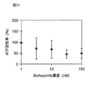
- FIG. 31 shows the measurement results of the amount of ATP activity of cell aggregates obtained by injecting bortezomib in Examples 4, 1, 3, 10, 20, 100 nM and culturing for 24 hours.
- FIG. 32 shows the measurement results of the amount of ATP activity of cell aggregates obtained by injecting bortezomib in Example 4, 1, 3, 10, 20, 100 nM and culturing for 48 hours.
- the vertical axis indicates the ATP activity rate
- the horizontal axis indicates the bortezomib concentration.
- Comparative Example 2 cell agglomerates were selected one by one without being classified by a metal porous membrane, and placed in a U-bottom plate (375 well). And the diameter and volume of each cell aggregate were calculated
- FIG. 33 shows the measurement results of the amount of ATP activity in the cell aggregate obtained by injecting bortezomib at 1, 3, 10, 20, 100 nM in Comparative Example 2 and culturing for 24 hours.
- FIG. 34 shows the measurement results of the amount of ATP activity in the cell aggregate obtained by injecting bortezomib at 1, 3, 10, 20, 100 nM and culturing for 48 hours in Comparative Example 2.
- the vertical axis indicates the ATP activity rate
- the horizontal axis indicates the bortezomib concentration.
- Example 4 When comparing the ATP activity rate of Example 4 shown in FIGS. 31 and 32 with the ATP activity rate of Comparative Example 2 shown in FIGS. 33 and 34, Example 4 has smaller error bars than Comparative Example 2.
- a medicinal effect test medicinal effect research or medicinal effect development
- a cell aggregate of uniform size obtained by classification using the metal porous membrane 10 It shows that reliable results can be obtained. That is, by classifying the cell agglomerates using the metal porous membrane 10, there is an effect that a highly accurate medicinal effect test result can be obtained.
- the metal porous film 10 has higher mechanical strength than nylon mesh or the like, the classification accuracy is high. Therefore, according to the metal porous membrane 10, it is possible to reduce the error in the test result due to the size of the cell aggregate in the medicinal effect test as compared with the nylon mesh or the like.
- Example 5 In Example 5, in order to examine the influence of the metal porous membrane 10 on the culture of the cell aggregate mass, the cell aggregate mass was cultured in a state where the flame-sterilized metal porous membrane 10 was immersed in a culture solution. Moreover, as Comparative Example 3, cell aggregates were cultured without the metal porous membrane 10 being immersed in the culture solution.
- Example 5 NIH3T3 cells into which the ras gene was introduced were cultured in a medium in which 5% FCS and 1% PCSM were added to Dulbecco's modified Eagle medium DMEM (manufactured by Nacalai Tesque).
- FIG. 35 shows the culture state of the cells 66 in Example 5 and Comparative Example 3.
- the well plate 80 (Sumitomo Bakelite Sumiron Tight Plate 24F) was prepared.
- the cells 66 and the medium 60 of Comparative Example 3 were seeded in three wells of the A row, and the cells 66 and the medium 60 of Example 5 were seeded in three wells of the C row.
- the seeding amount per well was 7.0 ⁇ 10 4 cells and 2 ml of the medium.
- Example 5 three metal porous membranes 10 were prepared and exposed with a burner for 1 second. Thereafter, the metal porous membrane 10 was cut and immersed in the medium 60 in the well.
- the metal porous membrane 10 used in Example 5 has an outer diameter of 18 mm and a thickness of 40 ⁇ m.
- the shape of the through hole 12 is a square, and the length of one side of the square is 58 ⁇ m.
- the distance b between the two through holes 12 is 18 ⁇ m.
- Example 5 the metal porous membrane 10 was removed after culturing the cells 66 for 10 minutes while the metal porous membrane 10 was immersed in the medium 60.
- Comparative Example 3 cells 66 were cultured for 10 minutes without immersing the metal porous membrane 10 in the medium. And the active amount of the cell cultured in Example 5 and Comparative Example 3 was confirmed by the following method.
- the medium 60 and the cells 66 of Example 5 and Comparative Example 3 were divided into 20 parts and dispensed into separate plates. After adding 100 ⁇ L of TOYONK ATP reagent to each of the dispensed medium and cells, RLU luminescence (ATP activity) was measured with a plate reader (Perkin Elmer Fusion ⁇ -FP).
- FIG. 36 shows the measurement results of the RLU luminescence amount (ATP activity amount) of cultured cells in Example 5 and Comparative Example 3. As shown in FIG. 36, when Example 5 is compared with Comparative Example 3, it can be seen that the RLU generation amount (ATP activity amount) is equivalent. This indicates that the flame-sterilized metal porous membrane 10 has no influence on cell culture.
- Example 6 In Example 6, using the classification device 50 of Embodiment 1, cell aggregates (spheres or embryoid bodies) derived from cultured mouse ES cells were classified for each size.
- the metal porous membrane 10 is a nickel circular mesh.
- the metal porous membrane 10 has an outer diameter of 7.8 mm, and a membrane portion 11 having a diameter of 6 mm is formed at the center.
- square through holes 12 are provided in a square lattice arrangement. One side of the through hole 12 is 120 ⁇ m.
- the distance between the through holes 12, that is, the distance between the metal parts between the two through holes 12 is 50 ⁇ m. In other words, the lattice spacing of the through holes 12 is 170 ⁇ m.
- the thickness is 17 ⁇ m.
- the metal porous membrane 10 is sterilized by gamma ray irradiation before classification.
- the cell aggregates 61 were classified by passing the liquid 60 containing the cell aggregates 61 having different dimensions shown in FIG. 37 through the metal porous membrane 10 and filtering.
- mouse ES cells were cultured in 1% PCSM DMEM medium containing 10% FBS.
- the container used was a 3.5 mm dish, and the number of seeded cells was 3 ⁇ 10 5 cells / ml.
- cell aggregates of various sizes could be generated.
- FIG. 37 shows an enlarged photograph of a part of the cultured cell aggregate. As shown in FIG. 37, it can be seen that cell agglomerates 61a, 61b, 61c of different sizes are generated before classification.
- the hole sizes of the through holes 12a, 12b, and 12c of the metal porous membranes 10A, 10B, and 10C of the classification device 50 are 180 ⁇ m, 100 ⁇ m, and 58 ⁇ m, respectively.
- the shape of the through holes 12a, 12b, and 12c is a square, and the hole size means the length d of one side of the square hole.
- Example 6 the cell aggregates 61a, 61b, 61c are passed by passing the medium 60 containing the cell aggregates 61a, 61b, 61c of different sizes shown in FIG. 37 in the order of the metal porous membranes 10A, 10B, 10C. Classification was performed. Specifically, by preparing three 3.5 mm dishes with newly added culture medium, the cell aggregates 61a, 61b, 61c captured by the metal porous membranes 10A, 10B, 10C are back-washed. Transferred to each 3.5 mm dish.
- FIG. 38 shows an enlarged photograph of a part of the cell aggregate 61a captured by the metal porous membrane 10A in Example 6.
- FIG. 39 shows an enlarged photograph of a part of the cell aggregate 61b captured by the metal porous membrane 10B in Example 6.
- FIG. 40 shows an enlarged photograph of a part of the cell aggregate 61c captured by the metal porous membrane 10C in Example 6.
- FIG. 41 shows an enlarged photograph of a part of the liquid 60 after passing through the metal porous membrane 10C in Example 6.
- the cell aggregates 61a, 61b, and 61c having substantially the same size are captured.
- the liquid 60 after passing through the metal porous membrane 10C does not contain the cell aggregates 61a, 61b, 61c, and is smaller than the cell aggregates 61a, 61b, 61c.
- the size cell aggregate 61d is included. Note that the liquid 60 after passing through the metal porous membrane 10C may contain, for example, isolated cells 62 in addition to the cell aggregates 61d.
- Example 6 the medium 60 (see FIG. 37) containing the cell aggregates 61a, 61b, and 61c having different sizes is passed through the metal porous membranes 10A, 10B, and 10C, whereby the cell aggregates are obtained. It can be seen that 61a, 61b, 61c could be classified for each size (see FIGS. 38 to 41).
- the medium containing the cell aggregate was equally divided for each dish, and the amount of ATP activity was measured for one of the equally divided media.
- the amount of ATP activity was measured by an ATP quantitative assay (CellTiter-Glo (registered trademark), Promega).
- CellTiter-Glo registered trademark
- the amount of ATP activity means cell activity, that is, cell viability. That is, it means that there are many living cells, so that the value of ATP activity amount is large.
- the other of the equally divided media was used for re-culture described later.
- the cell aggregates in each dish were randomly selected one by one with a pipette and placed in a U-bottom plate (375 well). And the diameter of each cell aggregate and the amount of ATP activity were measured with Cell Imager. Note that 13 cell aggregates 61a captured by the metal porous membrane 10A having a pore size of 180 ⁇ m, 10 cell aggregates 61b captured by the metal porous membrane 10B having a pore size of 100 ⁇ m, and a metal having a pore size of 58 ⁇ m Sixteen cell aggregates 61c captured by the porous membrane 10C were selected.
- FIG. 42 shows the measurement results of the amount of ATP activity with respect to the diameter of the cell aggregate in Example 6.
- the horizontal axis of FIG. 42 is the diameter of the cell aggregate and the vertical axis is the amount of ATP activity.
- square points indicate data related to the cell aggregates 61a captured by the metal porous membrane 10A having a pore size of 180 ⁇ m
- triangular points indicate cell aggregates captured by the metal porous membrane 10B having a pore size of 100 ⁇ m.
- Data on 61b, diamond-shaped points indicate data on cell aggregates 61c captured by the metal porous membrane 10C having a pore size of 58 ⁇ m.
- the activity of the cells captured by each of the metal porous membranes 10A, 10B, and 10C is maintained. More specifically, the amount of ATP activity increases as the number of living cells increases.
- the cell aggregate is composed of more cells as the size increases. Therefore, the larger the size of the cell aggregate, the larger the value of the ATP activity amount if the cells contained in the cell aggregate are alive.
- Table 2 shows the maximum value, the minimum value, the average value, and the standard deviation of the diameter of the cell aggregates with respect to the cell aggregates captured by each of the metal porous membranes 10A, 10B, and 10C.
- the diameter of the trapped cell aggregate in the metal porous membrane 10A having a pore size of 180 ⁇ m, the metal porous membrane 10B having a pore size of 100 ⁇ m, and the metal porous membrane 10C having a pore size of 58 ⁇ m The maximum values were 649 ⁇ m, 211 ⁇ m, and 136 ⁇ m, respectively.
- the minimum value of the diameter of the captured cell aggregate was 276 ⁇ m, 142 ⁇ m, and 59 ⁇ m in the order of the metal porous membranes 10A, 10B, and 10C.
- the average values of the diameters of the captured cell aggregates were 413 ⁇ m, 178 ⁇ m, and 99 ⁇ m in the order of the metal porous membranes 10A, 10B, and 10C.
- the standard deviations of the captured cell aggregates were 68, 36, and 30 in the order of the metal porous membranes 10A, 10B, and 10C.
- the ratio of the standard deviation with respect to the average value of the trapped cell aggregate was 31%, 12%, and 23% in the order of the metal porous membranes 10A, 10B, and 10C.
- the classified cell aggregates 61a, 61b and 61c were re-cultured.
- the other culture medium in the above-mentioned equal division was used for reculture.
- FIG. 43 shows a photograph of a set of cell aggregates 61a captured by the metal porous membrane 10A having a pore size of 180 ⁇ m in Example 6.
- FIG. 44 shows a photograph of a set of cell aggregates 61b captured by the metal porous membrane 10B having a pore size of 100 ⁇ m in Example 6.
- FIG. 45 shows a photograph of a set of cell aggregates 61c captured by the metal porous membrane 10C having a pore size of 58 ⁇ m in Example 6.
- Example 6 30 sets of cell aggregates 61a shown in FIG. 43 captured by the metal porous membrane 10A and 17 sets of cell aggregates 61b shown in FIG. 44 captured by the metal porous membrane 10B having a pore size of 100 ⁇ m were used. Then, 50 sets of cell aggregates 61c shown in FIG. 45 captured by the metal porous membrane 10C having a pore size of 58 ⁇ m were prepared. As a result of culturing these cells for 24 hours in a 37-degree incubation, one cell aggregate was able to be produced from three cell aggregates.
- FIG. 46 shows a photograph of one cell aggregate 63 produced by culturing the three cell aggregates 61a shown in FIG. 43 captured by the metal porous membrane 10A in Example 6.
- FIG. 47 shows a photograph of one cell aggregate 64 produced by culturing the three cell aggregates 61b shown in FIG. 44 captured by the metal porous membrane 10B.
- FIG. 48 shows a photograph of one cell aggregate 65 produced by culturing the three cell aggregates 61c shown in FIG. 45 captured by the metal porous membrane 10C.
- the cell aggregates 63, 64, and 65 shown in FIGS. 46 to 48 prepared by re-culture were measured for size using the Cell Imager described above.
- the average value and standard deviation of the size of the cell aggregates 63, 64, and 65 were 441 ⁇ 146 ⁇ m, 203 ⁇ 26 ⁇ m, and 114 ⁇ 34 ⁇ m, respectively.
- the ratio of the standard deviation with respect to the average value of the size of the cell aggregates 63, 64 and 65 was 33%, 13% and 30%, respectively.
- the above results show that the cell aggregates of a desired size can be classified from the medium containing the cell aggregates of different sizes using the metal porous membranes 10A, 10B, and 10C in the production of the cell aggregates. Show. Further, it is shown that when the cells are further cultured using the cell aggregates classified by the metal porous membranes 10A, 10B, and 10C, the size of the cultured cell aggregates can be easily made uniform.
- the present invention can be classified by increasing the collection rate of cell aggregates, it is useful in fields such as drug efficacy investigations and regenerative medicine production.
Abstract
Provided are a metallic porous membrane that can increase the recovery rate of cell aggregates, and a classifying method and a classifying device using the metallic porous membrane. This metallic porous membrane (10) for classifying cell aggregates (61) has a first primary surface (PS1) where the cell aggregates (61) are captured and a second primary surface (PS2) opposing the first primary surface, and is also provided with a membrane portion (11) having a plurality of penetration pores (12) that connect the first primary surface and the second primary surface. Such a configuration can increase the recovery rate of cell aggregates.
Description
本発明は、細胞凝集塊を分級する金属製多孔膜、それを用いた分級方法および分級装置に関する。
The present invention relates to a metal porous membrane for classifying cell aggregates, a classification method and a classification apparatus using the metal porous membrane.
細胞凝集塊(スフェロイド)を用いた薬効調査等において、均一な寸法の細胞凝集塊が求められている。
In a medicinal effect investigation using a cell aggregate (spheroid), a cell aggregate of a uniform size is required.
例えば、特許文献1には、フィルタを用いて細胞塊を分級することが開示されている。
For example, Patent Document 1 discloses classifying a cell mass using a filter.
近年、フィルタを用いた細胞凝集塊の分級において、細胞凝集塊の回収率を高めることが望まれている。
In recent years, it has been desired to increase the collection rate of cell aggregates in the classification of cell aggregates using a filter.
本発明は、細胞凝集塊の回収率を高めることができる金属製多孔膜、それを用いた分級方法および分級装置を提供することを目的とする。
An object of the present invention is to provide a metal porous membrane capable of increasing the recovery rate of cell aggregates, a classification method and a classification device using the metal porous membrane.
本発明の一態様の金属製多孔膜は、
細胞凝集塊を分級する金属製多孔膜であって、
前記細胞凝集塊が捕捉される第1主面と、前記第1主面に対向する第2主面とを有すると共に、前記第1主面と前記第2主面とを連通する複数の貫通孔を有する膜部を備える。 The metal porous membrane of one embodiment of the present invention is
A metal porous membrane for classifying cell aggregates,
A plurality of through-holes having a first main surface in which the cell clumps are captured and a second main surface opposite to the first main surface and communicating the first main surface and the second main surface The film part which has this.
本発明の一態様の分級方法は、
細胞凝集塊を分級する分級方法であって、
前記細胞凝集塊が捕捉される第1主面と、前記第1主面に対向する第2主面とを有すると共に、前記第1主面と前記第2主面とを連通する複数の貫通孔を有する膜部を備える金属製多孔膜を準備するステップ、
前記細胞凝集塊を含む液体を前記金属製多孔膜に通過させ、前記細胞凝集塊を前記金属製多孔膜に捕捉することによって、前記細胞凝集塊を分級するステップ、
を含む。 The classification method according to one aspect of the present invention includes:
A classification method for classifying cell clumps,
A plurality of through-holes having a first main surface in which the cell clumps are captured and a second main surface opposite to the first main surface and communicating the first main surface and the second main surface Preparing a metal porous membrane comprising a membrane portion having
Classifying the cell aggregate by passing the liquid containing the cell aggregate through the metal porous membrane and capturing the cell aggregate in the metal porous membrane;
including.
本発明の一態様の分級装置は、
細胞凝集塊を分級する分級装置であって、
前記細胞凝集塊が捕捉される第1主面と、前記第1主面に対向する第2主面とを有すると共に、前記第1主面と前記第2主面とを連通する複数の貫通孔を有する膜部を備える金属製多孔膜を備える。 A classification device according to one embodiment of the present invention is provided.
A classification device for classifying cell clumps,
A plurality of through-holes having a first main surface in which the cell clumps are captured and a second main surface opposite to the first main surface and communicating the first main surface and the second main surface A metal porous membrane provided with a membrane portion having
本発明によれば、細胞凝集塊の回収率を高めることができる金属製多孔膜、それを用いた分級方法および分級装置を提供することができる。
According to the present invention, it is possible to provide a metal porous membrane capable of increasing the recovery rate of cell aggregates, a classification method and a classification device using the metal porous membrane.
(本発明に至った経緯)
細胞凝集塊は、例えば、がんの薬効を調査する場合に、がん細胞のモデルとして用いられている。がん細胞は、進行度合いによって細胞の大きさが異なる。また、がん細胞の大きさが異なれば、効果のある薬も異なる。例えば、初期がんや小さながん組織に対して効果のある薬は、進行がんや大きながん組織に対しての効果が小さい場合がある。このため、細胞凝集塊を用いた薬効調査において、異なる寸法の細胞凝集塊を用いて薬効調査を行うと、薬効データにばらつきが生じる。したがって、薬効データのばらつきを抑えるため、均一な寸法の細胞凝集塊を得ることが求められている。 (Background to the present invention)
Cell aggregates are used as models of cancer cells, for example, when investigating the efficacy of cancer. Cancer cells vary in size depending on the degree of progression. Also, different cancer drugs have different effective drugs. For example, a drug effective against early cancer or small cancer tissue may have a small effect on advanced cancer or large cancer tissue. For this reason, in the medicinal effect investigation using the cell aggregate, when the medicinal effect investigation is performed using the cell aggregate of different sizes, the medicinal effect data varies. Therefore, in order to suppress variation in medicinal effect data, it is required to obtain a cell aggregate of uniform size.
また、細胞凝集塊は、細胞凝集塊を再生医療用の組織として用いられようとしている。この場合、所望の大きさの組織が必要となるが、細胞凝集塊の作製の工夫によって所望の大きさの組織を作製することは難しく、様々な大きさの細胞凝集塊を作製しておいてから所望の大きさの組織を選択した方が効率が良い。従って、高効率に所望の組織を選択することが求められている。
Moreover, the cell aggregate is going to be used as a tissue for regenerative medicine. In this case, a tissue of a desired size is required, but it is difficult to create a tissue of a desired size by devising the preparation of a cell aggregate, and cell aggregates of various sizes are prepared in advance. It is more efficient to select a desired size of tissue. Therefore, it is required to select a desired tissue with high efficiency.
培養した細胞凝集塊を所望の寸法に調製する方法として、例えば、メンブレン又はナイロンメッシュ等のフィルタを用いて所望の寸法の細胞凝集塊を捕捉することによって細胞凝集塊を分級する方法が用いられている。しかしながら、これらのフィルタを用いた場合では、捕捉できる細胞凝集塊の寸法精度が低いため、回収率が低くなるといった問題がある。そこで、本発明者らは、これらの問題を解決するため、以下の発明に至った。
As a method for preparing the cultured cell aggregates to a desired size, for example, a method of classifying the cell aggregates by capturing the cell aggregates of a desired size using a filter such as a membrane or nylon mesh is used. Yes. However, when these filters are used, there is a problem that the recovery rate is low because the dimensional accuracy of the cell aggregate that can be captured is low. Accordingly, the present inventors have reached the following invention in order to solve these problems.
本発明の一態様の金属製多孔膜は、
細胞凝集塊を分級する金属製多孔膜であって、
前記細胞凝集塊が捕捉される第1主面と、前記第1主面に対向する第2主面とを有すると共に、前記第1主面と前記第2主面とを連通する複数の貫通孔を有する膜部を備える。 The metal porous membrane of one embodiment of the present invention is
A metal porous membrane for classifying cell aggregates,
A plurality of through-holes having a first main surface in which the cell clumps are captured and a second main surface opposite to the first main surface and communicating the first main surface and the second main surface The film part which has this.
細胞凝集塊を分級する金属製多孔膜であって、
前記細胞凝集塊が捕捉される第1主面と、前記第1主面に対向する第2主面とを有すると共に、前記第1主面と前記第2主面とを連通する複数の貫通孔を有する膜部を備える。 The metal porous membrane of one embodiment of the present invention is
A metal porous membrane for classifying cell aggregates,
A plurality of through-holes having a first main surface in which the cell clumps are captured and a second main surface opposite to the first main surface and communicating the first main surface and the second main surface The film part which has this.
このような構成により、細胞凝集塊の回収率を高めることができる。
Such a configuration can increase the collection rate of cell aggregates.
前記金属製多孔膜において、前記膜部の前記第1主面は、平坦状に形成され、
前記複数の貫通孔は、前記膜部の前記第1主面側の開口と前記第2主面側の開口とが連続した壁面を通じて連通していてもよい。 In the metal porous film, the first main surface of the film part is formed in a flat shape,
The plurality of through holes may communicate with each other through a wall surface in which the opening on the first main surface side and the opening on the second main surface side of the film portion are continuous.
このような構成により、細胞凝集塊の回収率を更に向上させることができる。
Such a configuration can further improve the recovery rate of cell aggregates.
前記金属製多孔膜において、前記貫通孔の幅は、前記細胞凝集塊の大きさの100%未満であってもよい。
In the metal porous membrane, the width of the through hole may be less than 100% of the size of the cell aggregate.
このような構成により、所望の細胞凝集塊を確実に捕捉することができる。
With such a configuration, a desired cell aggregate can be reliably captured.
前記金属製多孔膜において、前記貫通孔の幅は、前記細胞凝集塊の大きさの80%未満であってもよい。
In the metal porous membrane, the width of the through hole may be less than 80% of the size of the cell aggregate.
このような構成により、細胞凝集塊が変形したとしても確実に所望の細胞凝集塊を捕捉することができる。
With such a configuration, even if the cell aggregate is deformed, the desired cell aggregate can be reliably captured.
前記金属製多孔膜において、前記貫通孔の幅は、前記細胞凝集塊の大きさの20%以上であってもよい。
In the metal porous membrane, the width of the through hole may be 20% or more of the size of the cell aggregate.
このような構成により、捕捉対象ではない流体が通過しやすくなり、作業時間を短縮することができる。
Such a configuration makes it easier for a fluid that is not to be captured to pass through, thereby reducing the work time.
前記金属製多孔膜において、前記貫通孔の幅は、前記細胞凝集塊の大きさの40%以上であってもよい。
In the metal porous membrane, the width of the through hole may be 40% or more of the size of the cell aggregate.
このような構成により、捕捉対象ではない流体が更に通過しやすくなり、作業時間を短縮することができる。
Such a configuration makes it easier for fluids that are not to be captured to pass through and shortens the work time.
本発明の一態様の細胞凝集塊の分級方法は、
細胞凝集塊を分級する分級方法であって、
前記細胞凝集塊が捕捉される第1主面と、前記第1主面に対向する第2主面とを有すると共に、前記第1主面と前記第2主面とを連通する複数の貫通孔を有する膜部を備える金属製多孔膜を準備するステップ、
前記細胞凝集塊を含む液体を前記金属製多孔膜に通過させ、前記細胞凝集塊を前記金属製多孔膜に捕捉することによって、前記細胞凝集塊を分級するステップ、
を含む。 The method for classifying a cell aggregate according to one embodiment of the present invention includes:
A classification method for classifying cell clumps,
A plurality of through-holes having a first main surface in which the cell clumps are captured and a second main surface opposite to the first main surface and communicating the first main surface and the second main surface Preparing a metal porous membrane comprising a membrane portion having
Classifying the cell aggregate by passing the liquid containing the cell aggregate through the metal porous membrane and capturing the cell aggregate in the metal porous membrane;
including.
このような構成により、細胞凝集塊の回収率を高めることができる。
Such a configuration can increase the collection rate of cell aggregates.
前記分級方法において、前記金属製多孔膜を準備するステップは、貫通孔の寸法が異なる複数の金属製多孔膜を準備すると共に、前記細胞凝集塊を含む液体が流れる流路の上流側から、前記複数の金属製多孔膜を前記貫通孔の寸法の大きい順に直列に配列してもよい。
In the classification method, the step of preparing the metal porous membrane includes preparing a plurality of metal porous membranes having different through-hole dimensions, and from the upstream side of the flow path through which the liquid containing the cell aggregates flows, A plurality of metal porous membranes may be arranged in series in descending order of the size of the through holes.
このような構成により、段階的に所望の寸法の細胞凝集塊を効率良く得ることができる。
With such a configuration, cell aggregates having desired dimensions can be efficiently obtained step by step.
前記分級方法において、前記細胞凝集塊を分級するステップは、前記複数の金属製多孔膜のうち最下段に位置する金属製多孔膜で前記細胞凝集塊から単離した単離細胞を通過させてもよい。
In the classification method, the step of classifying the cell aggregate may be performed by allowing the isolated cells isolated from the cell aggregate to pass through a metal porous membrane located at the lowest stage among the plurality of metal porous membranes. Good.
このような構成により、細胞凝集塊を含まず、単離細胞を含む液体を得ることができる。
With such a configuration, it is possible to obtain a liquid containing isolated cells without containing cell clumps.
前記分級方法において、更に、最下段に位置する前記金属製多孔膜を通過した前記単離細胞を継代するステップを含んでもよい。
The classification method may further include the step of substituting the isolated cells that have passed through the metal porous membrane located at the lowest level.
このような構成により、最下段の金属製多孔膜を通過した液体に含まれる単離細胞を、新たな培地に移動させて培養することができる。あるいは、単離細胞を細胞凝集塊形成に再び用いることもできる。
With such a configuration, the isolated cells contained in the liquid that has passed through the lowermost metal porous membrane can be transferred to a new medium and cultured. Alternatively, the isolated cells can be used again for cell aggregate formation.
前記分級方法において、更に、前記細胞凝集塊を前記金属製多孔膜に捕捉した状態で洗浄するステップを含んでもよい。
The classification method may further include a step of washing the cell aggregate in a state of being captured by the metal porous membrane.
このような構成により、分級された細胞凝集塊を容易に洗浄することができる。
With such a configuration, the classified cell aggregate can be easily washed.
前記分級方法において、前記金属製多孔膜で捕捉された前記細胞凝集塊を回収するステップを含んでもよい。
The classification method may include a step of recovering the cell aggregate captured by the metal porous membrane.
このような構成により、分級された細胞凝集塊を容易に回収することができる。
Such a configuration makes it possible to easily collect classified cell aggregates.
前記分級方法において、前記金属製多孔膜を準備するステップは、滅菌した金属製多孔膜を準備してもよい。
In the classification method, the step of preparing the metal porous membrane may prepare a sterilized metal porous membrane.
このような構成により、分級する前に金属製多孔膜に付着している菌によって細胞凝集塊が汚染されるのを防ぐことができる。
Such a configuration can prevent the cell aggregate from being contaminated by the bacteria attached to the metal porous membrane before classification.
前記分級方法において、前記細胞凝集塊を含む液体が前記金属製多孔膜を介して流れる流路は、外気から閉鎖されていてもよい。
In the classification method, the flow path through which the liquid containing the cell aggregates flows through the metal porous membrane may be closed from outside air.
このような構成により、外気により細胞凝集塊が汚染されるのを防ぐことができる。
Such a configuration can prevent the cell aggregate from being contaminated by the outside air.
本発明の一態様の細胞凝集塊の分級装置は、
細胞凝集塊を分級する分級装置であって、
前記細胞凝集塊が捕捉される第1主面と、前記第1主面に対向する第2主面とを有すると共に、前記第1主面と前記第2主面とを連通する複数の貫通孔を有する膜部を備える金属製多孔膜を備える。 An apparatus for classifying a cell aggregate according to one embodiment of the present invention is provided.
A classification device for classifying cell clumps,
A plurality of through-holes having a first main surface in which the cell clumps are captured and a second main surface opposite to the first main surface and communicating the first main surface and the second main surface A metal porous membrane provided with a membrane portion having
このような構成により、細胞凝集塊の回収率を高めることができる。
Such a configuration can increase the collection rate of cell aggregates.
前記分級装置において、貫通孔の寸法が異なる複数の金属製多孔膜を備え、
前記複数の金属製多孔膜は、前記細胞凝集塊を含む液体が流れる流路の上流側から、前記貫通孔の寸法の大きい順に直列に配列されていてもよい。 In the classification device, comprising a plurality of metal porous membranes having different through-hole dimensions,
The plurality of metal porous membranes may be arranged in series in descending order of the size of the through-hole from the upstream side of the flow path through which the liquid containing the cell aggregates flows.
このような構成により、段階的に所望の寸法の細胞凝集塊を効率良く得ることができる。
With such a configuration, cell aggregates having desired dimensions can be efficiently obtained step by step.
前記分級装置において、前記複数の金属製多孔膜のうち最下段に位置する金属製多孔膜の貫通孔の寸法は、前記細胞凝集塊から単離した単離細胞の大きさ以下であってもよい。
In the classifying device, the size of the through hole of the metal porous membrane located at the bottom of the plurality of metal porous membranes may be equal to or smaller than the size of the isolated cell isolated from the cell aggregate. .
このような構成により、最下段の金属製多孔膜によって捕捉する細胞凝集塊を規制することができる。
With such a configuration, the cell aggregates captured by the lowermost metal porous membrane can be regulated.
前記分級装置において、前記複数の金属製多孔膜のうち最下段に位置する金属製多孔膜の貫通孔の寸法は、前記細胞凝集塊から単離した単離細胞を通過させることができる大きさであってもよい。
In the classifying device, the size of the through hole of the metal porous membrane located at the lowest stage among the plurality of metal porous membranes is a size that allows the isolated cells isolated from the cell aggregates to pass through. There may be.
このような構成により、細胞凝集塊を含まず、且つ単離細胞を含む液体を得ることができる。
With such a configuration, it is possible to obtain a liquid that does not contain cell clumps and contains isolated cells.
前記分級装置において、更に、
前記金属製多孔膜を内包し、前記金属製多孔膜の前記第1主面に対向するように設けられた流体流入路と、前記金属製多孔膜の前記第2主面に対向するように設けられた流体排出路とを有するハウジング、
を備えてもよい。 In the classification device,
A fluid inflow passage enclosing the metal porous membrane and provided to face the first main surface of the metal porous membrane, and provided to face the second main surface of the metal porous membrane A housing having a defined fluid discharge path,
May be provided.
このような構成により、金属製多孔膜を容易に保持して、回収率の高い分級を行うことができる。
Such a configuration makes it possible to easily hold the metal porous membrane and perform classification with a high recovery rate.
前記分級装置において、前記細胞凝集塊を含む液体が前記金属製多孔膜を介して流れる流路は、外気から閉鎖されていてもよい。
In the classifying device, the flow path through which the liquid containing the cell aggregates flows through the metal porous membrane may be closed from outside air.
このような構成により、外気により細胞凝集塊が汚染されるのを防ぐことができる。
Such a configuration can prevent the cell aggregate from being contaminated by the outside air.
前記分級装置において、前記金属製多孔膜は、滅菌されていてもよい。
In the classifier, the metal porous membrane may be sterilized.
このような構成により、分級する前に金属製多孔膜に付着している菌によって細胞凝集塊が汚染されるのを防ぐことができる。
Such a configuration can prevent the cell aggregate from being contaminated by the bacteria attached to the metal porous membrane before classification.
以下、本発明に係る実施の形態1について、添付の図面を参照しながら説明する。また、各図においては、説明を容易なものとするため、各要素を誇張して示している。
Hereinafter, Embodiment 1 according to the present invention will be described with reference to the accompanying drawings. In each drawing, each element is exaggerated for easy explanation.
(実施の形態1)
[金属製多孔膜]
図1は、本発明に係る実施の形態1における金属製多孔膜10の一部の拡大斜視図である。図1中のX、Y、Z方向は、それぞれ金属製多孔膜10の縦方向、横方向、厚み方向を示している。図1に示すように、金属製多孔膜10は、互いに対向する第1主面PS1と第2主面PS2とを有し、且つ両主面を貫通する複数の貫通孔12を有する膜部11を備える。金属製多孔膜10は、膜部11においてマトリックス状に一定の間隔で複数の貫通孔12を設けた板状構造体(格子状構造体)である。金属製多孔膜10は、異なる寸法を有する複数の細胞凝集塊を含む液体を通過させることによって、細胞凝集塊を分級する金属製薄膜である。 (Embodiment 1)
[Metal porous film]
FIG. 1 is an enlarged perspective view of a part of a metalporous film 10 according to Embodiment 1 of the present invention. The X, Y, and Z directions in FIG. 1 indicate the vertical direction, the horizontal direction, and the thickness direction of the metal porous film 10, respectively. As shown in FIG. 1, the metal porous film 10 has a first main surface PS1 and a second main surface PS2 facing each other, and a film portion 11 having a plurality of through holes 12 penetrating both main surfaces. Is provided. The metal porous membrane 10 is a plate-like structure (lattice-like structure) in which a plurality of through holes 12 are provided at regular intervals in a matrix shape in the film portion 11. The metallic porous membrane 10 is a metallic thin film that classifies cell aggregates by passing liquids containing a plurality of cell aggregates having different dimensions.
[金属製多孔膜]
図1は、本発明に係る実施の形態1における金属製多孔膜10の一部の拡大斜視図である。図1中のX、Y、Z方向は、それぞれ金属製多孔膜10の縦方向、横方向、厚み方向を示している。図1に示すように、金属製多孔膜10は、互いに対向する第1主面PS1と第2主面PS2とを有し、且つ両主面を貫通する複数の貫通孔12を有する膜部11を備える。金属製多孔膜10は、膜部11においてマトリックス状に一定の間隔で複数の貫通孔12を設けた板状構造体(格子状構造体)である。金属製多孔膜10は、異なる寸法を有する複数の細胞凝集塊を含む液体を通過させることによって、細胞凝集塊を分級する金属製薄膜である。 (Embodiment 1)
[Metal porous film]
FIG. 1 is an enlarged perspective view of a part of a metal
本明細書において、「細胞凝集塊」とは、複数の細胞が接着することによって形成される細胞の集合塊を意味する。細胞凝集塊は、例えば、がん化細胞、肝細胞、iPS細胞などを使用した細胞凝集塊である。
In the present specification, the “cell aggregate” means an aggregate of cells formed by bonding a plurality of cells. The cell aggregate is a cell aggregate using cancerous cells, hepatocytes, iPS cells, and the like.
図1では金属製多孔膜10の全体を示していないが、実施の形態1では、金属製多孔膜10は、例えば、円形の金属メッシュである。金属製多孔膜10の寸法は、例えば、直径7.8mm、厚さ20μmである。金属製多孔膜10を構成する材料は、金、銀、銅、白金、ニッケル、ステンレス鋼、パラジウム、チタン、およびこれらの合金であってもよい。特に、金属製多孔膜10の材料としては、細胞凝集塊との生体親和性の観点から、金、ニッケル、ステンレス、チタンがよい。なお、金属製多孔膜10は、円形に限定されず、例えば、長方形、正方形等の矩形形状、又は楕円等の形状であってもよい。
1 does not show the entire metal porous membrane 10, but in the first embodiment, the metal porous membrane 10 is, for example, a circular metal mesh. The metal porous membrane 10 has, for example, a diameter of 7.8 mm and a thickness of 20 μm. The material constituting the metal porous film 10 may be gold, silver, copper, platinum, nickel, stainless steel, palladium, titanium, and alloys thereof. In particular, gold, nickel, stainless steel, and titanium are preferable as the material of the metal porous membrane 10 from the viewpoint of biocompatibility with the cell aggregate. The metal porous membrane 10 is not limited to a circle, and may be a rectangle such as a rectangle or a square, or a shape such as an ellipse.
図2は、金属製多孔膜10の膜部11の一部を厚み方向(Z方向)から見た概略図である。図2に示すように、複数の貫通孔12は、膜部11の第1主面PS1及び第2主面PS2上に周期的に配置されている。具体的には、複数の貫通孔12は、膜部11においてマトリクス状に等間隔で設けられている。複数の貫通孔12は、金属製多孔膜の第1主面PS1側、即ちZ方向から見て、正方形の形状を有する。複数の貫通孔12は、正方形の各辺と平行な2つの配列方向、即ち図2中のX方向とY方向に等しい間隔で設けられている。なお、貫通孔12は、正方形に限定されず、例えば長方形、円、又は楕円などの形状であってもよい。また、孔の配列は、正方格子配列に限定されず、例えば方形配列であれば、2つの配列方向の間隔は等しくない長方形配列でもよく、三角格子配列又は準周期配列などであってもよい。
FIG. 2 is a schematic view of a part of the film part 11 of the metal porous film 10 as viewed from the thickness direction (Z direction). As shown in FIG. 2, the plurality of through holes 12 are periodically arranged on the first main surface PS <b> 1 and the second main surface PS <b> 2 of the film part 11. Specifically, the plurality of through holes 12 are provided at regular intervals in a matrix in the film portion 11. The plurality of through holes 12 have a square shape when viewed from the first main surface PS1 side of the metal porous film, that is, from the Z direction. The plurality of through holes 12 are provided at equal intervals in two arrangement directions parallel to each side of the square, that is, the X direction and the Y direction in FIG. In addition, the through-hole 12 is not limited to a square, For example, shapes, such as a rectangle, a circle, or an ellipse, may be sufficient. Further, the arrangement of the holes is not limited to the square lattice arrangement. For example, if the arrangement is a square arrangement, the arrangement may be a rectangular arrangement in which the intervals in the two arrangement directions are not equal, or a triangular lattice arrangement or a quasi-periodic arrangement.
貫通孔12の形状及び寸法は、細胞凝集塊の大きさ、形状に応じて適宜設計されるものである。実施の形態1において、貫通孔12は、例えば、金属製多孔膜10の膜部11の第1主面PS1側、即ちZ方向から見て正方形であり、一辺dは、細胞凝集塊の大きさの100%未満に設計される。好ましくは細胞凝集塊の大きさの80%未満に設計されることで濾過中に細胞凝集塊が変形したとしても確実に捕捉することができる。また、一辺dを細胞凝集塊の大きさの20%以上とすることで捕捉対象ではない流体が通過しやすくなり、作業時間を短縮することができる。更に好ましくは細胞凝集塊の大きさの40%以上とすることで更に流体が通過しやすくなる。また、貫通孔12間の格子間隔bは、例えば、貫通孔12の一辺dの1倍より大きく10倍以下であり、より好ましくは貫通孔12の一辺dの3倍以下である。あるいは、開口率にして10%以上が好ましい。なお、開口率は、(貫通孔12が占める面積)/(貫通孔12が空いていないと仮定したときの第1主面PS1の投影面積)で計算される。なお、貫通孔12の形状は、正方形に限定されることなく、例えば、円形、楕円形、長方形、菱形などの形状であってもよい。実施の形態1では、正方形状の貫通孔12の寸法を一辺dで説明したが、貫通孔12の寸法を貫通孔12の幅で定義してもよい。貫通孔12の幅とは、貫通孔12が矩形の場合、対向する辺と辺との距離が最大になる線分に相当する。また、貫通孔12の幅は、貫通孔12が円形(楕円含む)の場合、長径に相当する。
The shape and dimensions of the through-hole 12 are appropriately designed according to the size and shape of the cell aggregate. In the first embodiment, the through-hole 12 is, for example, a square when viewed from the first main surface PS1 side of the film part 11 of the metal porous membrane 10, that is, the Z direction, and the side d is the size of the cell aggregate. Of less than 100%. Preferably, it is designed to be less than 80% of the size of the cell aggregate, so that even if the cell aggregate is deformed during filtration, it can be reliably captured. Further, by setting the side d to 20% or more of the size of the cell aggregate, it becomes easy for a fluid that is not to be captured to pass through and the working time can be shortened. More preferably, when the size is 40% or more of the size of the cell aggregate, the fluid can further pass through. Further, the lattice interval b between the through holes 12 is, for example, greater than 1 time and less than or equal to 10 times the side d of the through hole 12, and more preferably less than or equal to 3 times the side d of the through hole 12. Alternatively, the aperture ratio is preferably 10% or more. The aperture ratio is calculated by (area occupied by the through hole 12) / (projected area of the first main surface PS1 when it is assumed that the through hole 12 is not vacant). Note that the shape of the through hole 12 is not limited to a square, and may be, for example, a circle, an ellipse, a rectangle, a rhombus, or the like. In Embodiment 1, although the dimension of the square-shaped through-hole 12 was demonstrated by the one side d, you may define the dimension of the through-hole 12 with the width | variety of the through-hole 12. FIG. When the through hole 12 is rectangular, the width of the through hole 12 corresponds to a line segment that maximizes the distance between the opposite sides. Further, the width of the through hole 12 corresponds to the long diameter when the through hole 12 is circular (including an ellipse).
金属製多孔膜10は、同じ大きさを有する複数の貫通孔12を含む。ここで、「同じ大きさ」とは、複数の貫通孔12の寸法のばらつきが±5μmの範囲内であることを意味する。なお、複数の貫通孔12のうち一部の貫通孔は、異なる寸法で形成されていてもよい。例えば、分級の精度を損なわない程度で、金属製多孔膜10にかかる圧力を逃がすために、複数の貫通孔12のうち一部の貫通孔の寸法を他の貫通孔の寸法と比べて、大きく形成していてもよい。
The metal porous film 10 includes a plurality of through holes 12 having the same size. Here, “the same size” means that the variation in the dimensions of the plurality of through holes 12 is within a range of ± 5 μm. In addition, some through-holes among the plurality of through-holes 12 may be formed with different dimensions. For example, in order to release the pressure applied to the metal porous membrane 10 without impairing the classification accuracy, the size of some of the through holes 12 is larger than the size of other through holes. It may be formed.
なお、本明細書では、細胞凝集塊を液中に配置し、顕微鏡で観察した際、2次元の観察像において、細胞凝集塊の外周の任意の2つの点を結んだ線のうち、最長なものを細胞凝集塊の長さとし、捕捉対象である細胞凝集塊3個以上についての平均値を「細胞凝集塊の大きさ」とした。
In the present specification, when a cell aggregate is placed in a liquid and observed with a microscope, the longest of the lines connecting any two points on the outer periphery of the cell aggregate in a two-dimensional observation image. The cell aggregate was the length of the cell aggregate, and the average value of three or more cell aggregates to be captured was defined as “size of cell aggregate”.
図3は、図2の金属製多孔膜10の膜部11の一部をA-A線で切断した断面図である。図3に示すように、貫通孔12は、膜部11の第1主面PS1側の開口と第2主面PS2側の開口とが連続した壁面を通じて連通している。具体的には、貫通孔12は、第1主面PS1側の開口が第2主面PS2側の開口に投影可能に設けられている。即ち、金属製多孔膜10を第1主面PS1側、即ちZ方向から見た場合に、貫通孔12は、第1主面PS1側の開口が第2主面PS2側の開口と重なるように設けられている。実施の形態11において、貫通孔12は、その内壁が第1主面PS1及び第2主面PS2に対して垂直となるように設けられている。尚、第1主面PS1側の開口の大きさと第2主面PS2側の開口の大きさが異なっていてもよい。
FIG. 3 is a cross-sectional view of a part of the film portion 11 of the metal porous film 10 of FIG. 2 cut along the line AA. As shown in FIG. 3, the through hole 12 communicates through a wall surface in which the opening on the first main surface PS1 side and the opening on the second main surface PS2 side of the film part 11 are continuous. Specifically, the through hole 12 is provided so that the opening on the first main surface PS1 side can be projected onto the opening on the second main surface PS2 side. That is, when the metal porous film 10 is viewed from the first main surface PS1 side, that is, from the Z direction, the through hole 12 has an opening on the first main surface PS1 side overlapping with an opening on the second main surface PS2 side. Is provided. In the eleventh embodiment, the through hole 12 is provided so that the inner wall thereof is perpendicular to the first main surface PS1 and the second main surface PS2. The size of the opening on the first main surface PS1 side and the size of the opening on the second main surface PS2 side may be different.
また、金属製多孔膜10において、細胞凝集塊が捕捉される膜部11の第1主面PS1は、平坦状に形成されている。即ち、膜部11の第1主面PS1は、面一に形成されており、実質的にZ方向に凹凸が形成されていない。また、膜部11の第2主面PS2についても、平坦状に形成されている。さらには、膜部11の両主面における面精度は、単離細胞の大きさよりも小さいことが好ましい。膜部11の両主面へ単離細胞が付着することを低減できるからである。
Further, in the metal porous membrane 10, the first main surface PS1 of the membrane portion 11 where the cell aggregate is captured is formed flat. In other words, the first main surface PS1 of the film part 11 is formed flush and substantially has no unevenness in the Z direction. Further, the second main surface PS2 of the film part 11 is also formed flat. Furthermore, it is preferable that the surface accuracy on both main surfaces of the membrane part 11 is smaller than the size of the isolated cells. It is because it can reduce that an isolated cell adheres to both main surfaces of the film | membrane part 11. FIG.
[分級装置]
本発明の実施の形態1に係る分級装置について、図4を用いて説明する。図4は、実施の形態1に係る分級装置50の構成を示す概略図である。 [Classifier]
A classification device according toEmbodiment 1 of the present invention will be described with reference to FIG. FIG. 4 is a schematic diagram illustrating a configuration of the classification device 50 according to the first embodiment.
本発明の実施の形態1に係る分級装置について、図4を用いて説明する。図4は、実施の形態1に係る分級装置50の構成を示す概略図である。 [Classifier]
A classification device according to
図4に示すように、分級装置50は、複数の金属製多孔膜10A、10B、10Cを備える。複数の金属製多孔膜10A、10B、10Cは、細胞凝集塊61a、61b、61cと単離細胞62とを含む液体60が流れる方向70へ直列に配列されている。実施の形態1においては、液体60が流れる流路において、上流側から金属製多孔膜10A、10B、10Cの順に配置されている。即ち、分級装置50において、金属製多孔膜10Aを最上段に配置し、金属製多孔膜10Bを中央に配置し、金属製多孔膜10Cを最下段に配置している。また、金属製多孔膜10A、10B、10Cは、液体60の流れる方向70に対してそれぞれの第1主面PS1が直交するように配置されている。
As shown in FIG. 4, the classification device 50 includes a plurality of metal porous membranes 10A, 10B, and 10C. The plurality of metal porous membranes 10A, 10B, and 10C are arranged in series in a direction 70 in which the liquid 60 including the cell aggregates 61a, 61b, and 61c and the isolated cells 62 flows. In the first embodiment, the metal porous films 10A, 10B, and 10C are arranged in this order from the upstream side in the flow path through which the liquid 60 flows. That is, in the classification device 50, the metal porous film 10A is arranged at the uppermost stage, the metal porous film 10B is arranged at the center, and the metal porous film 10C is arranged at the lowermost stage. Further, the metal porous membranes 10A, 10B, and 10C are arranged so that the first main surfaces PS1 are orthogonal to the direction 70 in which the liquid 60 flows.
実施の形態1において、細胞凝集塊61a、61b、61cとは、それぞれ寸法が異なる細胞凝集塊である。細胞凝集塊61a、61b、61cの寸法は、細胞凝集塊61a、61b、61cの順に大きい。即ち、細胞凝集塊61a、61b、61cにおいて、細胞凝集塊61aが最も大きく、細胞凝集塊61cが最も小さい。
In the first embodiment, the cell aggregates 61a, 61b, 61c are cell aggregates having different dimensions. The size of the cell aggregates 61a, 61b, 61c is larger in the order of the cell aggregates 61a, 61b, 61c. That is, in the cell aggregates 61a, 61b, and 61c, the cell aggregate 61a is the largest and the cell aggregate 61c is the smallest.
本明細書において、「単離細胞」とは、細胞凝集塊を形成する1つの細胞であり、細胞凝集塊に接着せずに独立した状態の1つの細胞を意味する。即ち、「単離細胞」とは、細胞凝集塊から単離した1つの細胞を意味する。あるいは、細胞凝集塊の形成に関与しなかった単離した1つの細胞を意味する。実施の形態1において、単離細胞62の寸法は、細胞凝集塊61a、61b、61cよりも小さい。
In the present specification, an “isolated cell” is a single cell that forms a cell aggregate and means an independent cell that does not adhere to the cell aggregate. That is, “isolated cell” means one cell isolated from a cell aggregate. Alternatively, it means one isolated cell that was not involved in the formation of cell clumps. In the first embodiment, the size of the isolated cell 62 is smaller than the cell aggregates 61a, 61b, 61c.
金属製多孔膜10A、10B、10Cの膜部11a、11b、11cには、それぞれ異なる寸法の貫通孔12a、12b、12cが設けられている。貫通孔12a、12b、12cの寸法は、貫通孔12a、12b、12cの順に大きい。即ち、貫通孔12a、12b、12cにおいて、貫通孔12aの寸法が最も大きく、貫通孔12cの寸法が最も小さい。
The through holes 12a, 12b, and 12c having different dimensions are provided in the film portions 11a, 11b, and 11c of the metal porous films 10A, 10B, and 10C, respectively. The dimensions of the through holes 12a, 12b, and 12c are larger in the order of the through holes 12a, 12b, and 12c. That is, in the through holes 12a, 12b, and 12c, the through hole 12a has the largest dimension and the through hole 12c has the smallest dimension.
金属製多孔膜10Aの貫通孔12aは、細胞凝集塊61aを通過させずに、細胞凝集塊61b、61cと単離細胞62とを通過させることができる寸法で設計されている。具体的に、貫通孔12aは、細胞凝集塊61aよりも小さく、細胞凝集塊61bよりも大きい寸法で設計されている。このため、細胞凝集塊61a、61b、61cと単離細胞62とを含む液体60が金属製多孔膜10Aを通過することによって濾過されると、細胞凝集塊61aは、貫通孔12aを通り抜けることができず、金属製多孔膜10Aの第1主面PS1上に捕捉される。即ち、金属製多孔膜10Aの第1主面PS1上には、貫通孔12aの寸法より大きい細胞凝集塊61aが捕捉される。一方、液体60に含まれる細胞凝集塊61b、61c及び単離細胞62は、貫通孔12aを通り抜けることができる。このため、金属製多孔膜10Aで濾過された後の液体(濾液)60Aは、細胞凝集塊61b、61c及び単離細胞62を含んでいるが、細胞凝集塊61aを含まない。したがって、金属製多孔膜10Aにおいては、細胞凝集塊61a、61b、61cと単離細胞62とを含む液体60から細胞凝集塊61aを分級することができる。
The through-hole 12a of the metal porous membrane 10A is designed with a dimension that allows the cell aggregates 61b and 61c and the isolated cell 62 to pass through without passing through the cell aggregate 61a. Specifically, the through-hole 12a is designed with a size smaller than the cell aggregate 61a and larger than the cell aggregate 61b. For this reason, if the liquid 60 containing the cell aggregates 61a, 61b, 61c and the isolated cells 62 is filtered by passing through the metal porous membrane 10A, the cell aggregate 61a may pass through the through-hole 12a. It cannot be captured on the first main surface PS1 of the metal porous membrane 10A. That is, a cell aggregate 61a larger than the dimension of the through hole 12a is captured on the first main surface PS1 of the metal porous membrane 10A. On the other hand, the cell aggregates 61b and 61c and the isolated cell 62 contained in the liquid 60 can pass through the through-hole 12a. For this reason, the liquid (filtrate) 60A after being filtered by the metal porous membrane 10A includes the cell aggregates 61b and 61c and the isolated cells 62, but does not include the cell aggregate 61a. Therefore, in the metal porous membrane 10A, the cell aggregate 61a can be classified from the liquid 60 containing the cell aggregates 61a, 61b, 61c and the isolated cells 62.
金属製多孔膜10Bの貫通孔12bは、細胞凝集塊61bを通過させずに、細胞凝集塊61cと単離細胞62とを通過させることができる寸法で設計されている。具体的に、貫通孔12bは、細胞凝集塊61bよりも小さく、細胞凝集塊61cよりも大きい寸法で設計されている。このため、金属製多孔膜10Aで濾過された液体60Aが金属製多孔膜10Bを通過することによって濾過されると、細胞凝集塊61bは、貫通孔12bを通り抜けることができず、金属製多孔膜10Bの第1主面PS1上に捕捉される。一方、液体60Aに含まれる細胞凝集塊61c及び単離細胞62は、貫通孔12bを通り抜けることができる。このため、金属製多孔膜10Bで濾過された後の液体(濾液)60Bは、細胞凝集塊61c及び単離細胞62を含んでいるが、細胞凝集塊61bを含まない。したがって、金属製多孔膜10Bにおいては、細胞凝集塊61b、61cと単離細胞62とを含む液体60Aから細胞凝集塊61bを分級することができる。
The through-hole 12b of the metal porous membrane 10B is designed with a size that allows the cell aggregate 61c and the isolated cell 62 to pass through without passing through the cell aggregate 61b. Specifically, the through-hole 12b is designed with a size smaller than the cell aggregate 61b and larger than the cell aggregate 61c. For this reason, when the liquid 60A filtered by the metal porous membrane 10A is filtered by passing through the metal porous membrane 10B, the cell aggregate 61b cannot pass through the through-hole 12b, and the metal porous membrane It is captured on the first main surface PS1 of 10B. On the other hand, the cell aggregate 61c and the isolated cell 62 contained in the liquid 60A can pass through the through-hole 12b. For this reason, the liquid (filtrate) 60B after being filtered by the metal porous membrane 10B includes the cell aggregate 61c and the isolated cells 62, but does not include the cell aggregate 61b. Therefore, in the metal porous membrane 10B, the cell aggregate 61b can be classified from the liquid 60A containing the cell aggregates 61b and 61c and the isolated cells 62.
金属製多孔膜10Cの貫通孔12cは、細胞凝集塊61cを通過させずに、単離細胞62を通過させることができる寸法で設計されている。具体的に、貫通孔12cは、細胞凝集塊61cよりも小さく単離細胞62よりも大きい寸法で設計されている。このため、金属製多孔膜10Bで濾過された液体60Bが金属製多孔膜10Cを通過することによって濾過されると、細胞凝集塊61cは、貫通孔12cを通り抜けることができず、金属製多孔膜10Cの第1主面PS1上に捕捉される。一方、液体60Bに含まれる単離細胞62は、貫通孔12cを通り抜けることができる。このため、金属製多孔膜10Cで濾過された後の液体(濾液)60Cは、単離細胞62を含んでいるが、細胞凝集塊61cを含まない。したがって、金属製多孔膜10Cにおいては、細胞凝集塊61cと単離細胞62とを含む液体60Bから細胞凝集塊61cを分級することができる。
The through hole 12c of the metal porous membrane 10C is designed with a size that allows the isolated cell 62 to pass through without passing through the cell aggregate 61c. Specifically, the through-hole 12c is designed with a size smaller than the cell aggregate 61c and larger than the isolated cell 62. For this reason, when the liquid 60B filtered by the metal porous membrane 10B is filtered by passing through the metal porous membrane 10C, the cell aggregate 61c cannot pass through the through-hole 12c, and the metal porous membrane Captured on the first main surface PS1 of 10C. On the other hand, the isolated cell 62 contained in the liquid 60B can pass through the through hole 12c. For this reason, the liquid (filtrate) 60C after being filtered by the metal porous membrane 10C includes the isolated cells 62 but does not include the cell aggregates 61c. Therefore, in the metal porous membrane 10C, the cell aggregate 61c can be classified from the liquid 60B containing the cell aggregate 61c and the isolated cells 62.
また、金属製多孔膜10Cで濾過された後の液体(濾液)60Cに含まれる単離細胞62は、継代することができる。あるいは、別の細胞凝集塊の作製に利用することができる。
Moreover, the isolated cells 62 contained in the liquid (filtrate) 60C after being filtered through the metal porous membrane 10C can be passaged. Or it can utilize for preparation of another cell aggregate.
このように、分級装置50においては、液体60が流れる流路の上流側から、複数の金属製多孔膜10A、10B、10Cを貫通孔12a、12b、12cの寸法が大きい順に直列に配列している。このような構成によって、異なる寸法の細胞凝集塊61a、61b、61cと単離細胞62とを含む液体60から所望の寸法の細胞凝集塊を段階的に分級することができる。
Thus, in the classification device 50, the plurality of metal porous membranes 10A, 10B, and 10C are arranged in series in descending order of the dimensions of the through holes 12a, 12b, and 12c from the upstream side of the flow path through which the liquid 60 flows. Yes. With such a configuration, a cell aggregate of a desired size can be classified stepwise from the liquid 60 including the cell aggregates 61a, 61b, 61c of different sizes and the isolated cells 62.
分級装置50は、金属製多孔膜10A、10B、10Cを保持するハウジングを備えてもよい。この場合、分級装置50は、流体流入路と流体排出路とが設けられたハウジングに内包された金属製多孔膜10A、10B、10Cによって流体流入路から流入した流体中の濾過対象物を濾過し、当該金属製多孔膜を通過した流体を流体排出路から排出する。
The classification device 50 may include a housing that holds the metal porous membranes 10A, 10B, and 10C. In this case, the classifier 50 filters the object to be filtered in the fluid flowing in from the fluid inflow path by the metallic porous membranes 10A, 10B, and 10C included in the housing provided with the fluid inflow path and the fluid discharge path. The fluid that has passed through the metal porous membrane is discharged from the fluid discharge path.
図5は金属製多孔膜10Aを保持するハウジング20の概略構造を示す分解斜視図であり、図6はその分解断面図である。なお、図5及び図6においては、金属製多孔膜10Aの図示を省略している。
FIG. 5 is an exploded perspective view showing a schematic structure of the housing 20 holding the metal porous membrane 10A, and FIG. 6 is an exploded sectional view thereof. 5 and 6, the illustration of the metal porous film 10A is omitted.
図5及び図6に示すように、ハウジング20は、略円筒形の第1ハウジング部21と、略円筒形の第2ハウジング部22とを備えている。
As shown in FIGS. 5 and 6, the housing 20 includes a substantially cylindrical first housing portion 21 and a substantially cylindrical second housing portion 22.
第1ハウジング部21は、金属製多孔膜10Aの第1主面PS1に対向するように設けられた流体流入路21aを備えている。第1ハウジング部21は、金属製多孔膜10Aの外周部を挟持する第1枠部材51と一体に形成されている。すなわち、第1枠部材51は、第1ハウジング部21の一部として構成されている。第1枠部材51の内径は、例えば、6.0mmである。
The first housing portion 21 includes a fluid inflow passage 21a provided so as to face the first main surface PS1 of the metal porous membrane 10A. The first housing portion 21 is formed integrally with a first frame member 51 that sandwiches the outer peripheral portion of the metal porous membrane 10A. That is, the first frame member 51 is configured as a part of the first housing portion 21. The inner diameter of the first frame member 51 is, for example, 6.0 mm.
第1ハウジング部21の第1枠部材51の周囲には、流体流入路21aの延在方向と交差(例えば、直交)する方向に延びるフランジ部21bが形成されている。フランジ部21bには、当該フランジ部21bの厚み方向に貫通する複数の貫通穴21cが形成されている。実施の形態1において、複数の貫通穴21cは、90度間隔で4つ形成されている。フランジ部21bの厚さは、例えば、2.1mmである。貫通穴21cの直径は、例えば、1.42mmである。貫通穴21cの長さは、例えば、0.9mmである。
Around the first frame member 51 of the first housing portion 21, a flange portion 21b extending in a direction intersecting (for example, orthogonal to) the extending direction of the fluid inflow passage 21a is formed. A plurality of through holes 21c are formed in the flange portion 21b so as to penetrate in the thickness direction of the flange portion 21b. In the first embodiment, four through holes 21c are formed at intervals of 90 degrees. The thickness of the flange part 21b is 2.1 mm, for example. The diameter of the through hole 21c is, for example, 1.42 mm. The length of the through hole 21c is, for example, 0.9 mm.
第2ハウジング部22は、金属製多孔膜10Aの第2主面PS2に対向するように設けられた流体排出路22aを備えている。第2ハウジング部22は、金属製多孔膜10Aの外周部を挟持する第2枠部材52と一体に形成されている。すなわち、第2枠部材52は、第2ハウジング部22の一部として構成されている。第2枠部材52の内径は、例えば、6.0mmである。
The second housing portion 22 includes a fluid discharge path 22a provided so as to face the second main surface PS2 of the metal porous membrane 10A. The second housing portion 22 is formed integrally with the second frame member 52 that sandwiches the outer peripheral portion of the metal porous membrane 10A. That is, the second frame member 52 is configured as a part of the second housing portion 22. The inner diameter of the second frame member 52 is, for example, 6.0 mm.
第2ハウジング部22の第2枠部材52の周囲には、流体排出路22aの延在方向と交差(例えば、直交)する方向に延びるフランジ部22bが形成されている。フランジ部22bには、当該フランジ部22bの厚み方向に突出する複数の凸部22cが形成されている。実施の形態1において、複数の凸部22cは、90度間隔で4つ形成されている。凸部22cの直径は、例えば、1.4mmである。凸部22cの高さは、例えば、0.9mmである。
Around the second frame member 52 of the second housing part 22, a flange part 22b extending in a direction intersecting (for example, orthogonal to) the extending direction of the fluid discharge path 22a is formed. A plurality of convex portions 22c projecting in the thickness direction of the flange portion 22b are formed on the flange portion 22b. In the first embodiment, four convex portions 22c are formed at intervals of 90 degrees. The diameter of the convex part 22c is 1.4 mm, for example. The height of the convex portion 22c is, for example, 0.9 mm.
第1ハウジング部21と第2ハウジング部22とは、複数の凸部22cが複数の貫通穴21cに挿入されることで、互いに嵌合するように構成されている。第1ハウジング部21と第2ハウジング部22とが互いに嵌合することで、金属製多孔膜10Aの外周部が第1枠部材51と第2枠部材52との間で保持される。
1st housing part 21 and 2nd housing part 22 are comprised so that it may mutually fit by the some convex part 22c being inserted in the some through-hole 21c. When the first housing portion 21 and the second housing portion 22 are fitted to each other, the outer peripheral portion of the metal porous membrane 10 </ b> A is held between the first frame member 51 and the second frame member 52.
ハウジング20は、例えば、ルアーロック型シリンジ(図示せず)に取り付けて使用することができる。この場合、第1ハウジング部21の末端部21d(図6では上端部)と第2ハウジング部22の末端部22d(図6では下端部)の少なくとも一方に、ルアーロック型シリンジと接続可能な突条部等を設ければよい。
The housing 20 can be used by being attached to a luer lock type syringe (not shown), for example. In this case, at least one of the end portion 21d (upper end portion in FIG. 6) of the first housing portion 21 and the end portion 22d (lower end portion in FIG. 6) of the second housing portion 22 can be connected to the luer lock syringe. A strip or the like may be provided.
また、分級装置50においては、液体60、60A、60B、60Cの流れる流路が外気から閉鎖されていてもよい。このように、液体60、60A、60B、60Cを外気から遮断することによって、液体60、60A、60B、60Cに含まれる細胞凝集塊61a、61b、61c及び単離細胞62が汚染されるのを防止することができる。
Further, in the classification device 50, the flow path through which the liquids 60, 60A, 60B, and 60C flow may be closed from the outside air. Thus, by blocking the liquids 60, 60A, 60B, 60C from the outside air, the cell aggregates 61a, 61b, 61c and the isolated cells 62 contained in the liquids 60, 60A, 60B, 60C are contaminated. Can be prevented.
[分級方法]
本発明の実施の形態1に係る分級方法について、図7を用いて説明する。図7は、実施の形態1に係る分級方法を示すフローチャートである。 [Classification method]
A classification method according toEmbodiment 1 of the present invention will be described with reference to FIG. FIG. 7 is a flowchart showing the classification method according to the first embodiment.
本発明の実施の形態1に係る分級方法について、図7を用いて説明する。図7は、実施の形態1に係る分級方法を示すフローチャートである。 [Classification method]
A classification method according to
実施の形態1に係る分級方法は、複数の金属製多孔膜10A、10B、10Cを用いて細胞凝集塊61a、61b、61cと単離細胞62とを含む液体60を、細胞凝集塊61a、61b、61c及び単離細胞62のそれぞれに分級する。
In the classification method according to the first embodiment, the liquid 60 containing the cell aggregates 61a, 61b, 61c and the isolated cells 62 and the cell aggregates 61a, 61b using the plurality of metal porous membranes 10A, 10B, 10C. 61c and isolated cell 62, respectively.
図7に示すように、ステップST11において、複数の金属製多孔膜10A、10B、10Cを準備する。具体的には、液体60が流れる流路に、上流から金属製多孔膜10A、10B、10Cの順に直列に配列する(図4参照)。金属製多孔膜10A、10B、10Cについては、前述した分級装置50の構成と同じであるため、説明を省略する。
As shown in FIG. 7, in step ST11, a plurality of metal porous films 10A, 10B, and 10C are prepared. Specifically, the metallic porous membranes 10A, 10B, and 10C are arranged in series in the order from the upstream in the flow path through which the liquid 60 flows (see FIG. 4). Since the metal porous membranes 10A, 10B, and 10C are the same as the configuration of the classification device 50 described above, description thereof is omitted.
なお、ステップST11で準備する金属製多孔膜10A、10B、10Cは、滅菌処理がされているものであってもよい。滅菌処理は、例えば、ガンマ線照射によるガンマ線滅菌、高温高圧の飽和水蒸気によるオートクレーブ滅菌、酸化エチレンガスを用いた酸化エチレンガス滅菌、又はオゾンによる酸化滅菌などである。
Note that the metal porous membranes 10A, 10B, and 10C prepared in step ST11 may be sterilized. The sterilization treatment includes, for example, gamma ray sterilization by gamma ray irradiation, autoclave sterilization with high-temperature and high-pressure saturated steam, ethylene oxide gas sterilization using ethylene oxide gas, or oxidation sterilization with ozone.
ステップST12において、金属製多孔膜10A、10B、10Cによって細胞凝集塊61a、61b、61cをそれぞれ分級する。ステップST12では、金属製多孔膜10A、10B、10Cに、細胞凝集塊61a、61b、61cと単離細胞62とを含む液体60を通過させることによって濾過を行う。
In step ST12, the cell aggregates 61a, 61b, 61c are classified by the metal porous membranes 10A, 10B, 10C, respectively. In step ST12, filtration is performed by passing a liquid 60 containing cell aggregates 61a, 61b, 61c and isolated cells 62 through the metal porous membranes 10A, 10B, 10C.
より詳しく説明すると、まず、液体60を金属製多孔膜10Aに通過させることによって濾過を行う。金属製多孔膜10Aによる液体60の濾過によって、金属製多孔膜10Aの第1主面PS1上に、貫通孔12aの寸法より大きい細胞凝集塊61aを捕捉する。これにより、細胞凝集塊61aを分級する。
More specifically, first, filtration is performed by passing the liquid 60 through the metal porous membrane 10A. By filtering the liquid 60 with the metal porous membrane 10A, the cell aggregates 61a larger than the dimensions of the through holes 12a are captured on the first main surface PS1 of the metal porous membrane 10A. Thereby, the cell aggregate 61a is classified.
次に、金属製多孔膜10Aで濾過された後の濾液、即ち細胞凝集塊61b、61cと単離細胞62とを含む液体60Aを、金属製多孔膜10Aの下流に配置された金属製多孔膜10Bに通過させることによって濾過を行う。金属製多孔膜10Bによる液体60Aの濾過によって、金属製多孔膜10Bの第1主面PS1上に、貫通孔12bの寸法より大きい細胞凝集塊61bを捕捉する。これにより、細胞凝集塊61bを分級する。
Next, the filtrate after being filtered by the metal porous membrane 10A, that is, the liquid 60A containing the cell aggregates 61b and 61c and the isolated cells 62, is disposed on the downstream side of the metal porous membrane 10A. Filter by passing through 10B. By filtering the liquid 60A through the metal porous membrane 10B, the cell aggregate 61b larger than the dimension of the through-hole 12b is captured on the first main surface PS1 of the metal porous membrane 10B. Thereby, the cell aggregate 61b is classified.
次に、金属製多孔膜10Bで濾過された後の濾液、即ち細胞凝集塊61cと単離細胞62とを含む液体60Bを、金属製多孔膜10Bの下流に配置された金属製多孔膜10Cに通過させることによって濾過を行う。金属製多孔膜10Cによる液体60Bの濾過によって、金属製多孔膜10Cの第1主面PS1上に、貫通孔12cの寸法より大きい細胞凝集塊61cを捕捉する。これにより、細胞凝集塊61cを分級する。
Next, the filtrate after filtering with the metal porous membrane 10B, that is, the liquid 60B containing the cell aggregates 61c and the isolated cells 62 is applied to the metal porous membrane 10C disposed downstream of the metal porous membrane 10B. Filter by passing through. By filtering the liquid 60B with the metal porous membrane 10C, the cell aggregate 61c larger than the dimension of the through-hole 12c is captured on the first main surface PS1 of the metal porous membrane 10C. Thereby, the cell aggregate 61c is classified.
また、金属製多孔膜10Cで液体60Bを濾過することによって、濾液として単離細胞62を含む液体60Cを得ることができる。この液体60Cから取り出した単離細胞62は、継代することができる。即ち、液体60Cに含まれる単離細胞62を新しい培地に移動させて、再度培養することができる。
Further, by filtering the liquid 60B with the metal porous membrane 10C, a liquid 60C containing the isolated cells 62 can be obtained as a filtrate. The isolated cell 62 taken out from the liquid 60C can be passaged. That is, the isolated cells 62 contained in the liquid 60C can be moved to a new medium and cultured again.
ステップST13において、金属製多孔膜10A、10B、10Cに捕捉された細胞凝集塊61a、61b、61cを洗浄液で洗浄する。例えば、液体60が流れる方向70に洗浄液を流して、金属製多孔膜10A、10B、10Cに捕捉された状態で細胞凝集塊61a、61b、61cを洗浄する。なお、細胞凝集塊61a、61b、61cの洗浄方法については、これに限定されるものではなく、様々な洗浄方法を用いてもよい。また、ステップST13を省略しても良い。
In step ST13, the cell aggregates 61a, 61b, 61c captured by the metal porous membranes 10A, 10B, 10C are washed with a washing solution. For example, the cell aggregation mass 61a, 61b, 61c is washed in a state where it is captured by the metal porous membranes 10A, 10B, 10C by flowing a washing liquid in the direction 70 in which the liquid 60 flows. In addition, about the washing | cleaning method of the cell aggregate 61a, 61b, 61c, it is not limited to this, You may use various washing | cleaning methods. Further, step ST13 may be omitted.
ステップST14において、金属製多孔膜10A、10B、10Cに捕捉された細胞凝集塊61a、61b、61cを回収する。例えば、細胞凝集塊61aを捕捉した状態の金属製多孔膜10Aを取り外し、金属製多孔膜10Aを培地に入れて、金属製多孔膜10Aの厚み方向に振動させる。これにより、金属製多孔膜10Aに捕捉されていた細胞凝集塊61aを、金属製多孔膜10Aの第1主面PS1から分離し、回収することができる。あるいは、細胞凝集塊61aが付着していない第2主面PS2から第1主面PS1に向かって貫通孔12aを通じて回収液を通過させることにより、細胞凝集塊61aを金属製多孔膜10Aの第1主面PS1から分離し、回収することができる。なお、細胞凝集塊61a、61b、61cの回収方法については、これに限定されず、様々な回収方法を用いてもよい。
In step ST14, the cell aggregates 61a, 61b, 61c captured by the metal porous membranes 10A, 10B, 10C are collected. For example, the metal porous membrane 10A in a state where the cell aggregates 61a are captured is removed, and the metal porous membrane 10A is placed in a culture medium and vibrated in the thickness direction of the metal porous membrane 10A. As a result, the cell aggregate 61a captured by the metal porous membrane 10A can be separated from the first main surface PS1 of the metal porous membrane 10A and recovered. Alternatively, the cell aggregation mass 61a is passed through the through hole 12a from the second main surface PS2 to which the cell aggregation mass 61a is not attached to the first main surface PS1, thereby allowing the cell aggregation mass 61a to pass through the first porous metal membrane 10A. It can be separated from the main surface PS1 and recovered. In addition, about the collection method of the cell aggregate 61a, 61b, 61c, it is not limited to this, You may use various collection methods.
このようにして回収された細胞凝集塊61a、61b、61cは、薬効の調査に用いられる。例えば、がんに対する薬の効果を調査する場合、初期がんの薬効調査では、細胞凝集塊61a、61bより寸法の小さい細胞凝集塊61cを用いて薬効調査を行う。一方、進行がんの薬効調査では、細胞凝集塊61aを用いて薬効調査を行う。
The cell aggregates 61a, 61b, 61c collected in this way are used for investigation of drug efficacy. For example, when investigating the effect of a drug on cancer, in the medicinal effect investigation of initial cancer, the medicinal effect investigation is performed using the cell agglomerate 61c having a smaller size than the cell agglomerates 61a and 61b. On the other hand, in the investigation of the efficacy of advanced cancer, the efficacy investigation is performed using the cell aggregate 61a.
[効果]
実施の形態1に係る金属製多孔膜10によれば、以下の効果を奏することができる。 [effect]
According to the metalporous membrane 10 according to the first embodiment, the following effects can be obtained.
実施の形態1に係る金属製多孔膜10によれば、以下の効果を奏することができる。 [effect]
According to the metal
金属製多孔膜10においては、細胞凝集塊61が捕捉される第1主面PS1と、第1主面に対向する第2主面PS2とを有すると共に、第1主面PS1と第2主面PS2とを連通する複数の貫通孔12を有する膜部11を備えている。このような構成により、細胞凝集塊61を分級する際の寸法精度を高めることができるため、細胞凝集塊61の回収率を高めることができる。
The metal porous membrane 10 has a first main surface PS1 on which the cell aggregates 61 are captured and a second main surface PS2 opposite to the first main surface, and the first main surface PS1 and the second main surface. A film portion 11 having a plurality of through holes 12 communicating with PS 2 is provided. With such a configuration, since the dimensional accuracy when classifying the cell aggregate 61 can be increased, the recovery rate of the cell aggregate 61 can be increased.
金属製多孔膜10は、メンブレン又はナイロンメッシュ等のフィルタと比べて、剛性が高い。そのため、細胞凝集塊61を含む液体60を濾過する際、金属製多孔膜10では、メンブレン又はナイロンメッシュ等のフィルタと比べて、膜部11の第1主面PS1に液体60による圧力が加わっても貫通孔12が変形しにくい。したがって、金属製多孔膜10は、メンブレン又はナイロンメッシュ等のフィルタと比べて、貫通孔12より大きい細胞凝集塊61を、金属製多孔膜10の第1主面PS1上に確実に捕捉することができる。なお、メンブレン又はナイロンメッシュ等のフィルタは、膜面に液体60による圧力が加わると、貫通孔が変形しやすいため、貫通孔よりも大きい細胞凝集塊61がフィルタを通過してしまう場合がある。
The metal porous membrane 10 has higher rigidity than a filter such as a membrane or nylon mesh. Therefore, when the liquid 60 containing the cell agglomerates 61 is filtered, the pressure of the liquid 60 is applied to the first main surface PS1 of the membrane portion 11 in the metal porous membrane 10 as compared to a filter such as a membrane or nylon mesh. However, the through hole 12 is not easily deformed. Therefore, the metal porous membrane 10 can surely capture the cell aggregate 61 larger than the through hole 12 on the first main surface PS1 of the metal porous membrane 10 as compared with a filter such as a membrane or nylon mesh. it can. In addition, in the filter such as a membrane or nylon mesh, when the pressure by the liquid 60 is applied to the membrane surface, the through hole is likely to be deformed, so that the cell aggregate 61 larger than the through hole may pass through the filter.
金属製多孔膜10において、貫通孔12は、膜部11の第1主面PS1側の開口と第2主面PS2側の開口とが連続した壁面を通じて連通している。また、貫通孔12では、膜部11の第1主面PS1側の開口が第2主面PS2側の開口に投影可能に設けられている。このような構成により、貫通孔12より小さい細胞凝集塊61が貫通孔12を通り抜けやすくなっている。なお、メンブレンフィルタでは、貫通孔が膜部の第1主面側の開口と第2主面側の開口とが連続した壁面を通じて連通しておらず、貫通孔12より小さい細胞凝集塊61が貫通孔を通りにくい。このため、メンブレンフィルタでは、フィルタ内に細胞凝集塊61が残ってしまう。
In the metal porous membrane 10, the through hole 12 communicates through a wall surface in which the opening on the first main surface PS1 side and the opening on the second main surface PS2 side of the film portion 11 are continuous. Further, in the through hole 12, the opening on the first main surface PS1 side of the film part 11 is provided so as to be projected onto the opening on the second main surface PS2 side. With such a configuration, the cell aggregate 61 smaller than the through-hole 12 can easily pass through the through-hole 12. In the membrane filter, the through-hole is not in communication through the wall surface where the opening on the first main surface side and the opening on the second main surface side of the membrane portion are continuous, and the cell aggregate 61 smaller than the through-hole 12 penetrates. Difficult to pass through holes. For this reason, in the membrane filter, the cell aggregate 61 remains in the filter.
また、金属製多孔膜10において、細胞凝集塊61を捕捉する膜部11の第1主面PS1が平坦状に形成されている。即ち、金属製多孔膜10の膜部11の第1主面PS1は、面一に形成されている。このような構成により、金属製多孔膜10の貫通孔12よりも小さい細胞凝集塊61は、貫通孔12へ流入しやすくなっている。また、金属製多孔膜10の膜部11の第1主面PS1上に捕捉された細胞凝集塊61を回収するとき、細胞凝集塊61を膜部11の第1主面PS1から容易に分離することができる。なお、メンブレン又はナイロンメッシュ等のフィルタでは、細胞凝集塊61を捕捉する膜部の第1主面に凹凸が形成されている。このため、貫通孔より小さい細胞凝集塊61であっても、この凹凸部分に引っ掛かることがあるため、所望の寸法以外の細胞凝集塊61を回収することがある。また、細胞凝集塊61を回収するとき、この凹凸部分に細胞凝集塊61が引っ掛かることがある。
Further, in the metal porous membrane 10, the first main surface PS1 of the membrane portion 11 that captures the cell aggregate 61 is formed flat. That is, the first main surface PS1 of the film part 11 of the metal porous film 10 is formed flush. With such a configuration, the cell aggregate 61 smaller than the through hole 12 of the metal porous membrane 10 is easy to flow into the through hole 12. Further, when collecting the cell aggregate 61 captured on the first main surface PS1 of the film part 11 of the metal porous membrane 10, the cell aggregate 61 is easily separated from the first main surface PS1 of the film part 11. be able to. Note that in a filter such as a membrane or nylon mesh, irregularities are formed on the first main surface of the membrane portion that captures the cell aggregate 61. For this reason, even if it is the cell aggregate 61 smaller than a through-hole, since it may be caught in this uneven | corrugated | grooved part, the cell aggregate 61 other than a desired dimension may be collect | recovered. Further, when the cell aggregate 61 is collected, the cell aggregate 61 may be caught on the uneven portion.
金属製多孔膜10は火炎滅菌によっても変化することなく、さらには、熱伝導性も高いため、高い滅菌効果が得られる。
The metal porous membrane 10 does not change even by flame sterilization, and furthermore, since the thermal conductivity is high, a high sterilization effect is obtained.
このように、金属製多孔膜10では、貫通孔12より大きい細胞凝集塊61をより確実に捕捉することができると共に、貫通孔12より小さい細胞凝集塊61を貫通孔12に流入させやすい構成になっている。また、金属製多孔膜10では、膜部11の第1主面PS1から細胞凝集塊61を簡単に分離して回収することができる。このため、金属製多孔膜10は、メンブレン又はナイロンメッシュ等のフィルタに比べて、分級する細胞凝集塊61の寸法精度を向上させると共に、回収率を向上させることができる。
As described above, the metal porous membrane 10 can capture the cell aggregate 61 larger than the through-hole 12 more reliably and can easily flow the cell aggregate 61 smaller than the through-hole 12 into the through-hole 12. It has become. In the metal porous membrane 10, the cell aggregate 61 can be easily separated and recovered from the first main surface PS <b> 1 of the membrane portion 11. For this reason, the metal porous membrane 10 can improve the dimensional accuracy of the cell agglomerate 61 to be classified and improve the recovery rate as compared with a filter such as a membrane or nylon mesh.
実施の形態1に係る分級装置50によれば、以下の効果を奏することができる。
According to the classification device 50 according to the first embodiment, the following effects can be obtained.
分級装置50によれば、前述した金属製多孔膜10を用いて細胞凝集塊61の分級を行うため、細胞凝集塊61の回収率を高めることができる。
According to the classification device 50, since the cell aggregates 61 are classified using the metal porous membrane 10 described above, the recovery rate of the cell aggregates 61 can be increased.
分級装置50は、異なる寸法の貫通孔12a、12b、12cを有する金属製多孔膜10A、10B、10Cを備える。複数の金属製多孔膜10A、10B、10Cは、細胞凝集塊61a、61b、61cを含む液体60が流れる流路の上流側から、貫通孔12a、12b、12cの寸法の大きい順に直列に配列されている。このような構成により、複数の金属製多孔膜10A、10B、10Cを用いて、液体60から細胞凝集塊61a、61b、61cをそれぞれ分級することができる。即ち、分級装置50によれば、段階的に寸法の異なる細胞凝集塊61a、61b、61cを得ることができる。また、分級装置50では、上流側の金属製多孔膜によって捕捉し損ねた細胞凝集塊61a、61b、61cを、下流側の金属製多孔膜によって確実に回収することができる。
The classification device 50 includes metal porous films 10A, 10B, and 10C having through holes 12a, 12b, and 12c having different dimensions. The plurality of metal porous membranes 10A, 10B, and 10C are arranged in series in descending order of the dimensions of the through holes 12a, 12b, and 12c from the upstream side of the flow path through which the liquid 60 including the cell aggregates 61a, 61b, and 61c flows. ing. With such a configuration, the cell aggregates 61a, 61b, and 61c can be classified from the liquid 60 using the plurality of metal porous membranes 10A, 10B, and 10C, respectively. That is, according to the classification device 50, cell aggregates 61a, 61b, 61c having different dimensions in stages can be obtained. Further, in the classification device 50, the cell aggregates 61a, 61b, 61c that are missed by the upstream metal porous membrane can be reliably recovered by the downstream metal porous membrane.
分級装置50において、最下段に位置する金属製多孔膜10Cの貫通孔12cの寸法は、単離細胞62を通過させることができる大きさに設計されている。このような構成により、細胞凝集塊61a、61b、61cを分級した後、単離細胞62を含む液体60Cを得ることができる。そのため、単離細胞62を継代することができる。即ち、単離細胞62を新しい培地に移動させて、再度培養することによって、例えば、細胞の生死判定を行うことができる。あるいは、単離細胞62を別の細胞凝集塊の作製に利用することもできる。
In the classifying device 50, the dimension of the through-hole 12c of the metallic porous membrane 10C located at the lowest stage is designed to allow the isolated cell 62 to pass through. With such a configuration, after classifying the cell aggregates 61a, 61b, 61c, a liquid 60C containing the isolated cells 62 can be obtained. Therefore, the isolated cell 62 can be passaged. That is, by moving the isolated cell 62 to a new medium and culturing it again, for example, it is possible to determine whether the cell is alive or dead. Alternatively, the isolated cell 62 can also be used for the production of another cell aggregate.
分級装置50において、金属製多孔膜10A、10B、10Cは、当該金属製多孔膜の第1主面PS1に対向するように設けられた流体流入路21aと、当該金属製多孔膜の第2主面PS2に対向するように設けられた流体排出路22aとを有するハウジング20に収容することができる。このような構成により、流体流入路21aと流体排出路22aとを有するハウジング20内に金属製多孔膜10A、10B、10Cを容易に保持することができる。また、金属製多孔膜10A、10B、10Cの外周部を挟持する第1枠部材51及び第2枠部材52が、それぞれ第1ハウジング部21及び第2ハウジング部22と一体に形成されているので、分級装置50の部品点数を減らすことができる。
In the classification device 50, the metal porous membranes 10A, 10B, and 10C include a fluid inflow path 21a provided so as to face the first main surface PS1 of the metal porous membrane, and a second main body of the metal porous membrane. It can be accommodated in a housing 20 having a fluid discharge path 22a provided to face the surface PS2. With such a configuration, the metal porous films 10A, 10B, and 10C can be easily held in the housing 20 having the fluid inflow path 21a and the fluid discharge path 22a. In addition, the first frame member 51 and the second frame member 52 that sandwich the outer peripheries of the metal porous membranes 10A, 10B, and 10C are formed integrally with the first housing portion 21 and the second housing portion 22, respectively. The number of parts of the classification device 50 can be reduced.
実施の形態1に係る分級方法によれば、以下の効果を奏することができる。
According to the classification method according to Embodiment 1, the following effects can be achieved.
分級方法によれば、前述した金属製多孔膜10を用いて、細胞凝集塊61の分級を行うため、細胞凝集塊61の回収率を高めることができる。また、分級方法は、前述した分級装置50の効果と同様の効果を有する。
According to the classification method, since the cell aggregate 61 is classified using the metal porous membrane 10 described above, the recovery rate of the cell aggregate 61 can be increased. Further, the classification method has the same effect as that of the classification device 50 described above.
分級方法において、細胞凝集塊61a、61b、61cを金属製多孔膜10A、10B、10Cで捕捉した状態で洗浄することができる。例えば、液体60の流れる方向70の方向に洗浄液を流すことによって、金属製多孔膜10A、10B、10Cで捕捉した細胞凝集塊61a、61b、61cを洗浄することができる。このため、分級した細胞凝集塊61a、61b、61cを容易に洗浄することができる。
In the classification method, the cell aggregates 61a, 61b, 61c can be washed in a state where they are captured by the metal porous membranes 10A, 10B, 10C. For example, the cell agglomerates 61a, 61b, and 61c captured by the metal porous membranes 10A, 10B, and 10C can be washed by flowing a washing solution in the direction 70 in which the liquid 60 flows. For this reason, the classified cell aggregates 61a, 61b, 61c can be easily washed.
分級方法では、例えば、細胞凝集塊61a、61b、61cを捕捉した金属製多孔膜10A、10B、10Cを培地の中で、金属製多孔膜10A、10B、10Cの厚さ方向に振動させる等によって、細胞凝集塊61a、61b、61cを容易に回収することができる。あるいは、細胞凝集塊61aが付着していない第2主面PS2から第1主面PS1に向かって貫通孔12aを通じて回収液を通過させることにより、細胞凝集塊61aを金属製多孔膜10Aの第1主面PS1から分離し、回収することができる。このように、金属製多孔膜10A、10B、10Cのそれぞれに捕捉された細胞凝集塊61a、61b、61cは、金属製多孔膜10A、10B、10Cから容易に分離することができる。
In the classification method, for example, the metal porous membranes 10A, 10B, and 10C capturing the cell aggregates 61a, 61b, and 61c are vibrated in the thickness direction of the metal porous membranes 10A, 10B, and 10C in the medium. The cell aggregates 61a, 61b, 61c can be easily recovered. Alternatively, the cell aggregation mass 61a is passed through the through hole 12a from the second main surface PS2 to which the cell aggregation mass 61a is not attached to the first main surface PS1, thereby allowing the cell aggregation mass 61a to pass through the first porous metal membrane 10A. It can be separated from the main surface PS1 and recovered. As described above, the cell aggregates 61a, 61b, and 61c captured in the metal porous membranes 10A, 10B, and 10C can be easily separated from the metal porous membranes 10A, 10B, and 10C.
分級方法では、分級された細胞凝集塊61a、61b、61cを用いて薬効調査をすることができる。分級された細胞凝集塊61a、61b、61cは、それぞれ均一な寸法となっているため、薬効調査データのばらつきを低減することができる。
In the classification method, the medicinal effect can be investigated using the classified cell aggregates 61a, 61b, 61c. Since the classified cell agglomerates 61a, 61b, 61c have uniform dimensions, it is possible to reduce variation in the drug efficacy survey data.
なお、実施の形態1において、分級装置50は、複数の金属製多孔膜10A、10B、10Cを備える構成について説明したが、これに限定されない。分級装置50は、少なくとも1つ以上の金属製多孔膜10を備えていればよい。また、複数の金属製多孔膜10A、10B、10Cは、それぞれ異なる寸法の貫通孔12a、12b、12cを設けた構成について説明したが、これに限定されない。例えば、複数の金属製多孔膜10A、10B、10Cは、同じ寸法の貫通孔12を設けてもよい。このような構成により、上流側の金属製多孔膜10Aで捕捉し損ねた細胞凝集塊61aを下流側の金属製多孔膜10Bで捕捉することができる。
In the first embodiment, the classification device 50 has been described with respect to the configuration including the plurality of metal porous films 10A, 10B, and 10C. However, the present invention is not limited to this. The classification device 50 only needs to include at least one metal porous membrane 10. Moreover, although the several metal porous film 10A, 10B, 10C demonstrated the structure which provided the through- hole 12a, 12b, 12c of a respectively different dimension, it is not limited to this. For example, the plurality of metal porous membranes 10A, 10B, and 10C may be provided with through holes 12 having the same dimensions. With such a configuration, the cell agglomerate 61a that is missed by the upstream metallic porous membrane 10A can be captured by the downstream metallic porous membrane 10B.
実施の形態1において、最下段の金属製多孔膜10Cの貫通孔12cが、単離細胞62を通過させることができる寸法で設計されている構成について説明したが、これに限定されない。金属製多孔膜10Cの貫通孔12cは、例えば、細胞凝集塊から単離した単離細胞62の大きさ以下で設計されていてもよい。即ち、金属製多孔膜10Cの貫通孔12cは、単離細胞62と同じ寸法で設計されていてもよいし、又は単離細胞62よりも小さい寸法で設計されていてもよい。
In the first embodiment, the configuration in which the through-hole 12c of the lowermost metal porous membrane 10C is designed with a dimension that allows the isolated cell 62 to pass therethrough has been described, but the present invention is not limited to this. The through hole 12c of the metal porous membrane 10C may be designed, for example, to be smaller than the size of the isolated cell 62 isolated from the cell aggregate. That is, the through hole 12 c of the metal porous membrane 10 </ b> C may be designed with the same size as the isolated cell 62, or may be designed with a size smaller than that of the isolated cell 62.
実施の形態1において、第1ハウジング部21と第2ハウジング部22とは、複数の凸部22cが複数の貫通穴21cに挿入されることで、互いに嵌合するように構成したが、本発明はこれに限定されない。例えば、第1ハウジング部21に複数の貫通穴を設け、第2ハウジング部22に複数の凸部を設け、当該複数の凸部を複数の貫通穴に挿入することで、第1ハウジング部21と第2ハウジング部22とが互いに嵌合するように構成されてもよい。第1ハウジング部21と第2ハウジング部22とが互いに嵌合する構造であればよい。
In the first embodiment, the first housing portion 21 and the second housing portion 22 are configured to be fitted to each other by inserting the plurality of convex portions 22c into the plurality of through holes 21c. Is not limited to this. For example, the first housing portion 21 is provided with a plurality of through holes, the second housing portion 22 is provided with a plurality of convex portions, and the plurality of convex portions are inserted into the plurality of through holes, You may be comprised so that the 2nd housing part 22 may mutually fit. What is necessary is just the structure which the 1st housing part 21 and the 2nd housing part 22 fit mutually.
実施の形態1において、第1枠部材51及び第2枠部材52が、それぞれ第1ハウジング部21及び第2ハウジング部22と一体に形成される構成について説明したが、これに限定されない。例えば、第1枠部材51及び第2枠部材52は、第1ハウジング部21及び第2ハウジング部22と別々の部材で構成されていてもよい。
In Embodiment 1, although the 1st frame member 51 and the 2nd frame member 52 demonstrated the structure formed integrally with the 1st housing part 21 and the 2nd housing part 22, respectively, it is not limited to this. For example, the first frame member 51 and the second frame member 52 may be configured by separate members from the first housing part 21 and the second housing part 22.
以下、実施例について説明する。
Hereinafter, examples will be described.
(実施例1)
実施例1においては、実施の形態1の金属製多孔膜10を用いて、細胞凝集塊61の分級を行った。また、比較例1として、ナイロンメッシュを用いて、細胞凝集塊61の分級を行った。 Example 1
In Example 1, classification of thecell aggregate 61 was performed using the metal porous membrane 10 of the first embodiment. Moreover, as Comparative Example 1, the cell aggregate 61 was classified using a nylon mesh.
実施例1においては、実施の形態1の金属製多孔膜10を用いて、細胞凝集塊61の分級を行った。また、比較例1として、ナイロンメッシュを用いて、細胞凝集塊61の分級を行った。 Example 1
In Example 1, classification of the
実施例1及び比較例1において、細胞凝集塊61を含む液体60を図8に示す。図8に示すように、液体60には、異なる寸法の複数の細胞凝集塊61が含まれている。
In Example 1 and Comparative Example 1, a liquid 60 containing cell aggregates 61 is shown in FIG. As shown in FIG. 8, the liquid 60 includes a plurality of cell aggregates 61 having different dimensions.
図8に示す細胞凝集塊61は、直径35mmのディッシュにNIH3T3/rasが含まれた液量0.2mlの細胞懸濁液を播種し、汎用のインキュベーターで1日培養することによって作製した。このとき、培地は3ml、総細胞凝集塊数は約1×105個であった。
The cell aggregate 61 shown in FIG. 8 was prepared by seeding a cell suspension having a volume of 0.2 ml containing NIH3T3 / ras in a dish having a diameter of 35 mm and culturing it in a general-purpose incubator for one day. At this time, the medium was 3 ml, and the total number of cell aggregates was about 1 × 10 5 .
実施例1は、実施の形態1の金属製多孔膜10を用いた。金属製多孔膜10は、ニッケル製の円形のメッシュである。金属製多孔膜10は、外径が7.8mmであり、中央部に直径6mmの膜部11を形成している。膜部11には、正方形の貫通孔12が正方格子配列で設けられている。貫通孔12の一辺は、120μmである。貫通孔12の間隔、即ち2つの貫通孔12、12の間の金属部分の距離は、50μmである。言い換えると、貫通孔12の格子間隔は170μmである。厚みは17μmである。また、金属製多孔膜10は、分級を行う前にガンマ線照射によって滅菌処理が行われている。実施例1では、図8に示す異なる寸法の細胞凝集塊61を含む液体60を、金属製多孔膜10に通過させて濾過することによって、細胞凝集塊61の分級を行った。
Example 1 used the porous metal film 10 of the first embodiment. The metal porous membrane 10 is a nickel circular mesh. The metal porous membrane 10 has an outer diameter of 7.8 mm, and a membrane portion 11 having a diameter of 6 mm is formed at the center. In the film part 11, square through holes 12 are provided in a square lattice arrangement. One side of the through hole 12 is 120 μm. The interval between the through holes 12, that is, the distance of the metal part between the two through holes 12, 12 is 50 μm. In other words, the lattice spacing of the through holes 12 is 170 μm. The thickness is 17 μm. Further, the metal porous membrane 10 is sterilized by gamma ray irradiation before classification. In Example 1, the cell aggregates 61 were classified by passing the liquid 60 containing the cell aggregates 61 having different dimensions shown in FIG. 8 through the metal porous membrane 10 and filtering.
比較例1は、ナイロンメッシュを用いた。ナイロンメッシュは、ナイロン6.6製の円形のメッシュである。ナイロンメッシュは、外径が7.8mmであり、中央部に直径6mmの膜部を形成している。膜部には、正方形の貫通孔が正方格子配列で設けられている。貫通孔の一辺は、131μmである。貫通孔の間隔、即ちナイロンメッシュの線径は、72μmである。言い換えると、貫通孔の格子間隔は203μmである。なお、ナイロンメッシュは、ガンマ線を照射すると、メッシュ自体が損傷するため、減菌処理を行っていない。比較例1では、図8に示す異なる寸法の細胞凝集塊61を含む液体60を、ナイロンメッシュに通過させて濾過することによって、細胞凝集塊61の分級を行った。
Comparative Example 1 used a nylon mesh. The nylon mesh is a circular mesh made of nylon 6.6. The nylon mesh has an outer diameter of 7.8 mm, and a film portion having a diameter of 6 mm is formed at the center. The film part is provided with square through holes in a square lattice arrangement. One side of the through hole is 131 μm. The interval between the through holes, that is, the diameter of the nylon mesh is 72 μm. In other words, the lattice spacing of the through holes is 203 μm. Nylon mesh is not sterilized because gamma rays are used to damage the mesh itself. In Comparative Example 1, the cell aggregates 61 were classified by passing the liquid 60 containing the cell aggregates 61 having different dimensions shown in FIG. 8 through a nylon mesh and filtering.
図9は、実施例1において、細胞凝集塊61を分級した後の金属製多孔膜10の一部の写真を示す。図10は、比較例1において、細胞凝集塊61を分級した後のナイロンメッシュの一部の写真を示す。
FIG. 9 shows a photograph of a part of the metal porous membrane 10 after classifying the cell aggregates 61 in Example 1. FIG. 10 shows a photograph of a part of the nylon mesh after classifying the cell aggregates 61 in Comparative Example 1.
図9に示す実施例1の金属製多孔膜10では、図10に示す比較例1のナイロンメッシュと比べて、より多くの細胞凝集塊61が捕捉されていることがわかる。即ち、実施例1では、比較例1と比べて、より多くの細胞凝集塊61を回収できていることがわかる。
It can be seen that in the metal porous membrane 10 of Example 1 shown in FIG. 9, more cell aggregates 61 are captured as compared with the nylon mesh of Comparative Example 1 shown in FIG. That is, it can be seen that more cell aggregates 61 were recovered in Example 1 than in Comparative Example 1.
比較例1のメッシュ構造体では、液体60が通過することによって膜部に圧力がかかると、貫通孔が変形する。このため、ナイロンメッシュの貫通孔よりも大きい細胞凝集塊61が、変形した貫通孔を通過してしまうことがある。このため、比較例1では、回収されるべき寸法の細胞凝集塊61が、ナイロンメッシュによって回収できなかったものと考えられる。
In the mesh structure of Comparative Example 1, when a pressure is applied to the membrane portion by passing the liquid 60, the through hole is deformed. For this reason, the cell aggregate 61 larger than the through-hole of a nylon mesh may pass through the deformed through-hole. For this reason, in the comparative example 1, it is thought that the cell aggregate 61 of the dimension which should be collect | recovered was not able to be collect | recovered with the nylon mesh.
図11は、ナイロンメッシュの交差部Z1の拡大写真を示す。ナイロンメッシュは、線状のナイロンを編み込んで作製されているため、線状のナイロンが交差する交差部Z1では、ナイロンメッシュの厚み方向に段差部が形成される。即ち、ナイロンメッシュにおいて、細胞凝集塊61を捕捉する主面が、複数の段差部により凹凸を含む面となっている。このため、ナイロンメッシュの貫通孔を通過可能な細胞凝集塊61が、交差部Z1に引っ掛かってしまうことがある。このように、ナイロンメッシュの交差部Z1に引っ掛かった細胞凝集塊61は、回収できなくなることがある。また、ナイロンメッシュの交差部Z1に引っ掛かった細胞凝集塊61を回収できたとしても、この細胞凝集塊61は、所望の寸法のものではないため、分級したい細胞凝集塊61を得ることができない。
FIG. 11 shows an enlarged photograph of the intersection Z1 of the nylon mesh. Since the nylon mesh is produced by weaving linear nylon, a stepped portion is formed in the thickness direction of the nylon mesh at the intersection Z1 where the linear nylon intersects. That is, in the nylon mesh, the main surface that captures the cell aggregates 61 is a surface that includes irregularities due to a plurality of step portions. For this reason, the cell aggregate 61 that can pass through the through-hole of the nylon mesh may be caught by the intersection Z1. Thus, the cell aggregate 61 caught on the intersection Z1 of the nylon mesh may not be collected. Even if the cell aggregate 61 caught at the intersection Z1 of the nylon mesh can be recovered, the cell aggregate 61 is not of a desired size, and the cell aggregate 61 to be classified cannot be obtained.
一方、実施例1の金属製多孔膜10では、液体60が通過することによって膜部11に圧力がかかっても、貫通孔12が変形しにくい。このため、金属製多孔膜10の貫通孔12よりも大きい細胞凝集塊61は、貫通孔12を通過することが少ない。また、実施例1では、金属製多孔膜10の第1主面PS1は、平坦状に形成されている。このため、実施例1では、金属製多孔膜10の第1主面PS1で、貫通孔12を通過可能な寸法の細胞凝集塊61が引っ掛からずに、貫通孔12を通過することができる。したがって、実施例1では、比較例1よりも多くの細胞凝集塊61を回収することができたと考えられる。また、実施例1では、比較例1よりも寸法精度を向上させた細胞凝集塊61を回収することができると考えられる。
On the other hand, in the metal porous membrane 10 of Example 1, the through-hole 12 is not easily deformed even when pressure is applied to the membrane portion 11 by passing the liquid 60. For this reason, the cell aggregate 61 larger than the through-hole 12 of the metal porous membrane 10 rarely passes through the through-hole 12. In Example 1, the first main surface PS1 of the metal porous film 10 is formed in a flat shape. For this reason, in Example 1, the cell agglomerate 61 having a size capable of passing through the through hole 12 can pass through the through hole 12 on the first main surface PS1 of the metal porous membrane 10 without being caught. Therefore, in Example 1, it is considered that more cell aggregates 61 than in Comparative Example 1 could be recovered. Moreover, in Example 1, it is thought that the cell aggregate 61 which improved the dimensional accuracy compared with the comparative example 1 can be collect | recovered.
このように、比較例1のナイロンメッシュでは、細胞凝集塊61の回収率の低下、分級の誤差、再現性の低下が生じる。したがって、実施例1の金属製多孔膜10では、ナイロンメッシュと比べて、細胞凝集塊61の回収率、分級の精度、再現性を向上させることができる。
Thus, in the nylon mesh of Comparative Example 1, the recovery rate of the cell aggregate 61 is reduced, the classification error, and the reproducibility are reduced. Therefore, in the metal porous membrane 10 of Example 1, the recovery rate, classification accuracy, and reproducibility of the cell aggregate 61 can be improved as compared with the nylon mesh.
(実施例2)
実施例2においては、実施の形態1の分級装置50を用いて、異なる寸法の複数の細胞凝集塊61a、61b、61cの分級を行った。なお、細胞凝集塊61a、61b、61cの寸法については、細胞凝集塊61aが最も大きく、細胞凝集塊61cが最も小さい寸法とする。 (Example 2)
In Example 2, using theclassification device 50 of the first embodiment, a plurality of cell aggregates 61a, 61b, 61c having different dimensions were classified. In addition, about the dimension of the cell aggregate 61a, 61b, 61c, the cell aggregate 61a is the largest and the cell aggregate 61c is the smallest.
実施例2においては、実施の形態1の分級装置50を用いて、異なる寸法の複数の細胞凝集塊61a、61b、61cの分級を行った。なお、細胞凝集塊61a、61b、61cの寸法については、細胞凝集塊61aが最も大きく、細胞凝集塊61cが最も小さい寸法とする。 (Example 2)
In Example 2, using the
実施例2は、実施の形態1の分級装置50を用いた。分級装置50の金属製多孔膜10A、10B、10Cは、ニッケル製の円形のメッシュである。金属製多孔膜10A、10B、10Cは、外径が7.8mmであり、中央部に直径6mmの膜部11a、11b、11cを形成している。膜部11a、11b、11cには、正方形の貫通孔12a、12b、12cが正方格子配列で設けられている。貫通孔12a、12b、12cの寸法は、それぞれ正方形の一辺が180μm、120μm、58μm、格子間隔が260μm、170μm、76.3μm、厚みが20μm、17μm、20μmである。また、金属製多孔膜10A、10B、10Cは、分級を行う前にガンマ線照射によって滅菌処理が行われている。実施例2の分級装置50では、液体60の流路の上流側から金属製多孔膜10A、10B、10Cの順で直列に配列されている。
Example 2 used the classification device 50 of the first embodiment. The metal porous membranes 10A, 10B, and 10C of the classification device 50 are nickel circular meshes. The metal porous membranes 10A, 10B, and 10C have an outer diameter of 7.8 mm, and film portions 11a, 11b, and 11c having a diameter of 6 mm are formed at the center. The film portions 11a, 11b, and 11c are provided with square through holes 12a, 12b, and 12c in a square lattice arrangement. The dimensions of the through holes 12a, 12b, and 12c are 180 μm, 120 μm, and 58 μm on each side of the square, the lattice spacing is 260 μm, 170 μm, and 76.3 μm, and the thicknesses are 20 μm, 17 μm, and 20 μm, respectively. Further, the metal porous films 10A, 10B, and 10C are sterilized by gamma ray irradiation before classification. In the classification device 50 of the second embodiment, the metal porous membranes 10A, 10B, and 10C are arranged in series in this order from the upstream side of the flow path of the liquid 60.
図12は、実施例2において異なる寸法の細胞凝集塊を含む液体60を示す。実施例2では、図12に示す異なる寸法の細胞凝集塊を含む液体60を1ml、分級装置50に投与し、デッドエンド方式にて濾過を行うことによって細胞凝集塊61a、61b、61cの分級を行った。濾過を開始してから5分後に、金属製多孔膜10A、10B、10Cをそれぞれ取り出した。次に、各々の金属製多孔膜10A、10B、10Cについて、細胞凝集塊が捕捉された面(第1主面PS1)を下側にした状態で、細胞凝集塊が捕捉されていない面(第2主面PS2)から生理食塩水5mlを通過させることで、シャーレに細胞凝集塊を回収した。回収した細胞凝集塊を顕微鏡にて観察した。
FIG. 12 shows a liquid 60 containing cell aggregates of different sizes in Example 2. In Example 2, 1 ml of the liquid 60 containing the cell aggregates of different sizes shown in FIG. 12 is administered to the classification device 50, and the cell aggregates 61a, 61b, 61c are classified by performing filtration in a dead-end manner. went. Five minutes after the start of filtration, the metal porous membranes 10A, 10B, and 10C were respectively taken out. Next, for each of the metal porous membranes 10A, 10B, and 10C, the surface (first main surface PS1) on which the cell aggregate is captured is the lower side (the first surface where the cell aggregate is not captured) Cell aggregates were collected in a petri dish by passing 5 ml of physiological saline from 2 main surfaces PS2). The collected cell aggregates were observed with a microscope.
図13は、実施例2において金属製多孔膜10Aで回収した細胞凝集塊の拡大写真を示す。図13に示すように、金属製多孔膜10Aでは、貫通孔12aより大きい細胞凝集塊61aが捕捉されていることがわかる。
FIG. 13 shows an enlarged photograph of the cell aggregate collected by the metal porous membrane 10A in Example 2. As shown in FIG. 13, in the metal porous membrane 10A, it can be seen that a cell aggregate 61a larger than the through-hole 12a is captured.
図14は、実施例2において金属製多孔膜10Bで回収した細胞凝集塊の拡大写真を示す。図14に示すように、金属製多孔膜10Bでは、貫通孔12bより大きい細胞凝集塊61bが捕捉されていることがわかる。
FIG. 14 shows an enlarged photograph of the cell aggregate collected in the metal porous membrane 10B in Example 2. As shown in FIG. 14, in the metal porous membrane 10B, it can be seen that a cell aggregate 61b larger than the through-hole 12b is captured.
図15は、実施例2において金属製多孔膜10Cで回収した細胞凝集塊拡大写真を示す。図15に示すように、金属製多孔膜10Cでは、貫通孔12cより大きい細胞凝集塊61cが捕捉されていることがわかる。
FIG. 15 shows an enlarged photograph of the cell agglomerates collected by the metal porous membrane 10C in Example 2. As shown in FIG. 15, in the metal porous membrane 10C, it can be seen that a cell aggregate 61c larger than the through-hole 12c is captured.
また、前述した実施例2の条件と同じ条件で液体60の濾過を行うことによって分級を行い、金属製多孔膜10A、10B、10Cで濾過された後の液体60A、60B、60Cの一部を顕微鏡にて拡大観察した。
Moreover, it classifies by filtering the liquid 60 on the same conditions as the conditions of Example 2 mentioned above, and some liquid 60A, 60B, 60C after filtering by metal porous membrane 10A, 10B, 10C is used. It was magnified and observed with a microscope.
図16は、実施例2において金属製多孔膜10Aを通過した後の液体60Aの一部を拡大した写真を示す。図16に示すように、金属製多孔膜10Aを通過した液体60Aには、細胞凝集塊61b、61cと単離細胞62とが含まれているが、細胞凝集塊61aは含まれていないことがわかる。
FIG. 16 shows an enlarged photograph of a part of the liquid 60A after passing through the metal porous membrane 10A in Example 2. As shown in FIG. 16, the liquid 60A that has passed through the metal porous membrane 10A contains cell aggregates 61b and 61c and isolated cells 62, but does not contain the cell aggregate 61a. Recognize.
図17は、実施例2において金属製多孔膜10Bを通過した後の液体60Bの一部を拡大した写真を示す。図17に示すように、金属製多孔膜10Bを通過した液体60Bには、細胞凝集塊61cと単離細胞62とが含まれているが、細胞凝集塊61bは含まれていないことがわかる。
FIG. 17 shows an enlarged photograph of a part of the liquid 60B after passing through the metal porous membrane 10B in Example 2. As shown in FIG. 17, it can be seen that the liquid 60B that has passed through the metal porous membrane 10B contains the cell aggregate 61c and the isolated cells 62, but does not contain the cell aggregate 61b.
図18は、実施例2において金属製多孔膜10Cを通過した後の液体60Cの一部を拡大した写真を示す。図18に示すように、金属製多孔膜10Cを通過した液体60Cには、単離細胞62が含まれているが、細胞凝集塊61cは含まれていないことがわかる。
FIG. 18 shows an enlarged photograph of a part of the liquid 60C after passing through the metal porous membrane 10C in Example 2. As shown in FIG. 18, it can be seen that the liquid 60C that has passed through the metal porous membrane 10C contains isolated cells 62 but does not contain cell aggregates 61c.
このように、実施例2の分級装置50では、金属製多孔膜10A、10B、10Cにおいて所望の寸法の細胞凝集塊61a、61b、61cを捕捉していることができる。即ち、実施例2の分級装置50では、段階的に異なる寸法の細胞凝集塊61a、61b、61cを分級することができる。
Thus, in the classification device 50 of Example 2, the cell agglomerates 61a, 61b, 61c having desired dimensions can be captured in the metal porous membranes 10A, 10B, 10C. That is, in the classification device 50 according to the second embodiment, the cell aggregates 61a, 61b, and 61c having different sizes can be classified in stages.
(実施例3)
実施例3においては、実施の形態1の分級装置50を用いて、培養した細胞凝集塊(スフェロイド)を大きさ毎に分級した。 (Example 3)
In Example 3, the cultured cell aggregates (spheroids) were classified according to size using theclassification device 50 of the first embodiment.
実施例3においては、実施の形態1の分級装置50を用いて、培養した細胞凝集塊(スフェロイド)を大きさ毎に分級した。 (Example 3)
In Example 3, the cultured cell aggregates (spheroids) were classified according to size using the
細胞凝集塊は、ras遺伝子が導入されたNIH3T3細胞を10%FBSが含まれた1%PCSMのDMEM培地で培養した。使用した容器は、3.5mmディッシュであり、播種細胞数は3×10⁵個/mlであった。37度のインキュベーション内で24時間培養した結果、様々な大きさの細胞凝集塊を生成することができた。
For the cell aggregate, NIH3T3 cells into which the ras gene was introduced were cultured in a DMEM medium containing 1% PCSM containing 10% FBS. The container used was a 3.5 mm dish, and the number of seeded cells was 3 × 10 5 cells / ml. As a result of culturing for 24 hours within a 37-degree incubation, cell aggregates of various sizes could be generated.
図19は、培養した細胞凝集塊の一部を拡大した写真を示す。図19に示すように、分級する前において、異なるサイズの細胞凝集塊61a、61b、61cが生成されていることがわかる。
FIG. 19 shows an enlarged photograph of a part of the cultured cell aggregate. As shown in FIG. 19, it can be seen that cell agglomerates 61a, 61b, 61c of different sizes are generated before classification.
実施例3において、分級装置50の金属製多孔膜10A、10B、10Cの貫通孔12a、12b、12cの孔サイズは、それぞれ、180μm、100μm、58μmである。ここで、貫通孔12a、12b、12cの形状は、正方形であり、孔サイズとは正方形の孔の一辺の長さdを意味する。
In Example 3, the hole sizes of the through holes 12a, 12b, and 12c of the metal porous membranes 10A, 10B, and 10C of the classification device 50 are 180 μm, 100 μm, and 58 μm, respectively. Here, the shape of the through holes 12a, 12b, and 12c is a square, and the hole size means the length d of one side of the square hole.
実施例3では、図19に示す異なるサイズの細胞凝集塊61a、61b、61cを含む培地60を、金属製多孔膜10A、10B、10Cの順に通過させることによって、細胞凝集塊61a、61b、61cの分級を行った。具体的には、新たに培養液を投入した3.5mmディッシュを3個用意し、金属製多孔膜10A、10B、10Cで捕捉された細胞凝集塊61a、61b、61cを、逆洗することによってそれぞれの3.5mmディッシュに移した。
In Example 3, the cell aggregates 61a, 61b, 61c are passed by passing the medium 60 containing the cell aggregates 61a, 61b, 61c of different sizes shown in FIG. 19 in the order of the metal porous membranes 10A, 10B, 10C. Classification was performed. Specifically, by preparing three 3.5 mm dishes with newly added culture medium, the cell aggregates 61a, 61b, 61c captured by the metal porous membranes 10A, 10B, 10C are back-washed. Transferred to each 3.5 mm dish.
図20は、実施例3において金属製多孔膜10Aで捕捉した細胞凝集塊61aの一部を拡大した写真を示す。図21は、実施例3において金属製多孔膜10Bで捕捉した細胞凝集塊61bの一部を拡大した写真を示す。図22は、実施例3において金属製多孔膜10Cで捕捉した細胞凝集塊61cの一部を拡大した写真を示す。図23は、実施例3において金属製多孔膜10Cを通過した後の液体60の一部を拡大した写真を示す。
FIG. 20 shows an enlarged photograph of a part of the cell aggregate 61a captured by the metal porous membrane 10A in Example 3. FIG. 21 shows an enlarged photograph of a part of the cell aggregate 61b captured by the metal porous membrane 10B in Example 3. FIG. 22 shows a photograph in which a part of the cell aggregate 61c captured by the metal porous membrane 10C in Example 3 is enlarged. FIG. 23 shows an enlarged photograph of a part of the liquid 60 after passing through the metal porous membrane 10C in Example 3.
図20~22に示すように、金属製多孔膜10A、10B、10Cでは、それぞれほぼ均等なサイズの細胞凝集塊61a、61b、61cが捕捉されていることがわかる。また、図23に示すように、金属製多孔膜10Cを通過した後の液体60には、細胞凝集塊61a、61b、61cが含まれておらず、細胞凝集塊61a、61b、61cよりも小さいサイズの細胞凝集塊61dが含まれていることがわかる。なお、金属製多孔膜10Cを通過した後の液体60には、細胞凝集塊61dの他に、例えば、単離細胞62などを含んでいてもよい。
As shown in FIGS. 20 to 22, it can be seen that in the metal porous membranes 10A, 10B, and 10C, cell aggregates 61a, 61b, and 61c having almost equal sizes are captured. Further, as shown in FIG. 23, the liquid 60 after passing through the metal porous membrane 10C does not include the cell aggregates 61a, 61b, 61c, and is smaller than the cell aggregates 61a, 61b, 61c. It can be seen that the size cell aggregate 61d is included. Note that the liquid 60 after passing through the metal porous membrane 10C may contain, for example, isolated cells 62 in addition to the cell aggregates 61d.
このように、実施例3において、異なる大きさの細胞凝集塊61a、61b、61cを含む培地60(図19参照)を、金属製多孔膜10A、10B、10Cに通過させることによって、細胞凝集塊61a、61b、61cをサイズ毎に分級することができたことがわかる(図20~23参照)。
Thus, in Example 3, by passing the medium 60 (see FIG. 19) containing the cell aggregates 61a, 61b, 61c of different sizes through the metal porous membranes 10A, 10B, 10C, the cell aggregates It can be seen that 61a, 61b, 61c could be classified according to size (see FIGS. 20 to 23).
次に、細胞凝集塊のサイズと活性の関係を調べるために、細胞凝集塊を含む培地をディッシュ毎に等分し、等分した一方の培地に対してATP活性量を測定した。ここで、ATP活性量の測定は、ATP定量アッセイ(CellTiter-Glo(登録商標),Promega)によって行った。ATP活性量とは、細胞の活性、即ち細胞の生存率を意味している。即ち、ATP活性量の値が大きいほど、生きた細胞が多いことを意味する。なお、等分したうちの他方の培地は、後述の再培養に使用した。
Next, in order to examine the relationship between the size and activity of the cell aggregate, the medium containing the cell aggregate was equally divided for each dish, and the amount of ATP activity was measured for one of the equally divided media. Here, the amount of ATP activity was measured by an ATP quantitative assay (CellTiter-Glo (registered trademark), Promega). The amount of ATP activity means cell activity, that is, cell viability. That is, it means that there are many living cells, so that the value of ATP activity amount is large. The other of the equally divided media was used for re-culture described later.
各ディッシュ内の細胞凝集塊を、ピペットで無作為に1個ずつ選択し、U底プレート(375ウェル)に投入した。そして、Cell Imagerで各細胞凝集塊の直径とATP活性量を測定した。なお、孔サイズ180μmの金属製多孔膜10Aで捕捉された細胞凝集塊61aは17個、孔サイズ100μmの金属製多孔膜10Bで捕捉された細胞凝集塊61bは20個、孔サイズ58μmの金属製多孔膜10Cで捕捉された細胞凝集塊61cは12個を選択した。
The cell aggregates in each dish were randomly selected one by one with a pipette and placed in a U-bottom plate (375 well). And the diameter of each cell aggregate and the amount of ATP activity were measured with Cell Imager. The number of cell aggregates 61a captured by the metal porous membrane 10A with a pore size of 180 μm is 17, the number of cell aggregates 61b captured by the metal porous membrane 10B with a pore size of 100 μm is 20, and the size of the metal is 61 μm. Twelve cell aggregates 61c captured by the porous membrane 10C were selected.
図24は、実施例3において、細胞凝集塊の直径に対するATP活性量の測定結果を示す。図24の横軸は細胞凝集塊の直径、縦軸はATP活性量である。図24において、四角の点は、孔サイズ180μmの金属製多孔膜10Aで捕捉された細胞凝集塊61aに関するデータ、三角の点は、孔サイズ100μmの金属製多孔膜10Bで捕捉された細胞凝集塊61bに関するデータ、菱形の点は、孔サイズ58μmの金属製多孔膜10Cで捕捉された細胞凝集塊61cに関するデータを示す。
FIG. 24 shows the measurement result of the amount of ATP activity with respect to the diameter of the cell aggregate in Example 3. The horizontal axis in FIG. 24 is the diameter of the cell aggregate, and the vertical axis is the amount of ATP activity. In FIG. 24, the square points represent data related to the cell aggregates 61a captured by the metal porous membrane 10A having a pore size of 180 μm, and the triangular points represent cell aggregates captured by the metal porous membrane 10B having a pore size of 100 μm. Data on 61b, diamond-shaped points indicate data on cell aggregates 61c captured by the metal porous membrane 10C having a pore size of 58 μm.
図24に示すように、金属製多孔膜10A、10B、10Cのそれぞれで捕捉された細胞の活性が保たれていることがわかる。具体的に説明すると、ATP活性量は、生きている細胞が多いほど、活性量(RLU発光量)の値が大きくなる。細胞凝集塊は、サイズが大きくなるほど、多くの細胞を含んで構成される。そのため、細胞凝集塊のサイズが大きくなるほど、細胞凝集塊に含まれる細胞が生きていれば、ATP活性量の値は大きくなる。
As shown in FIG. 24, it can be seen that the activity of the cells captured by each of the metal porous membranes 10A, 10B, and 10C is maintained. More specifically, the amount of ATP activity increases as the number of living cells increases. The cell aggregate is composed of more cells as the size increases. Therefore, the larger the size of the cell aggregate, the larger the value of the ATP activity amount if the cells contained in the cell aggregate are alive.
図24に示すように、細胞凝集塊の直径が大きくなるほど、ATPの活性量の値が大きくなっていることがわかる。このことから、金属製多孔膜10A、10B、10Cのそれぞれで捕捉された細胞の活性が保たれていることがわかる。即ち、金属製多孔膜10A、10B、10Cのそれぞれで捕捉された細胞が生存していることがわかる。なお、元来、細胞凝集塊は、表面近傍の細胞のみ活性が保たれており、細胞凝集塊の中心部の細胞の活性は失われている。そのため、細胞凝集塊の直径とATP活性量の関係は、2次から3次の曲線になる。
As shown in FIG. 24, it can be seen that as the diameter of the cell aggregate increases, the activity amount of ATP increases. This shows that the activity of the cells captured by each of the metal porous membranes 10A, 10B, and 10C is maintained. That is, it can be seen that the cells captured by the metal porous membranes 10A, 10B, and 10C are alive. Originally, the activity of the cell aggregate is maintained only in the cells near the surface, and the activity of the cell in the center of the cell aggregate is lost. Therefore, the relationship between the diameter of the cell aggregate and the amount of ATP activity is a second-order to third-order curve.
金属製多孔膜10A、10B、10Cのそれぞれで捕捉された細胞凝集塊に関して、細胞凝集塊の直径の最大値、最小値、平均値、及び標準偏差を表1に示す。
Table 1 shows the maximum value, minimum value, average value, and standard deviation of the diameter of the cell aggregates with respect to the cell aggregates captured by the metal porous membranes 10A, 10B, and 10C.

表1に示すように、孔サイズ180μmの金属製多孔膜金属製多孔膜10A、孔サイズ100μmの金属製多孔膜10B、孔サイズ58μmの金属製多孔膜10Cにおいて、捕捉された細胞凝集塊の直径の最大値は、それぞれ、495μm、268μm、158μmであった。捕捉された細胞凝集塊の直径の最小値は、金属製多孔膜金属製多孔膜10A、10B、10Cの順に、267μm、138μm、65μmであった。捕捉された細胞凝集塊の直径の平均値は、金属製多孔膜金属製多孔膜10A、10B、10Cの順に、365μm、203μm、111μmであった。捕捉された細胞凝集塊の標準偏差は、金属製多孔膜金属製多孔膜10A、10B、10Cの順に、68、36、30であった。また、捕捉された細胞凝集塊の平均値に対する標準偏差の割合は、金属製多孔膜金属製多孔膜10A、10B、10Cの順に、19%、18%、27%であった。
As shown in Table 1, the diameter of the trapped cell aggregate in the metal porous membrane 10A having a pore size of 180 μm, the metal porous membrane 10B having a pore size of 100 μm, and the metal porous membrane 10C having a pore size of 58 μm The maximum values were 495 μm, 268 μm, and 158 μm, respectively. The minimum value of the diameter of the captured cell aggregate was 267 μm, 138 μm, and 65 μm in the order of the metal porous membranes 10A, 10B, and 10C. The average values of the diameters of the captured cell aggregates were 365 μm, 203 μm, and 111 μm in the order of the metal porous membranes 10A, 10B, and 10C. The standard deviations of the captured cell aggregates were 68, 36, and 30 in the order of the metal porous membranes 10A, 10B, and 10C. Moreover, the ratio of the standard deviation with respect to the average value of the trapped cell aggregate was 19%, 18%, and 27% in the order of the metal porous membranes 10A, 10B, and 10C.
更に、分級した細胞凝集塊61a、61b、61cを再培養した。なお、再培養には、前述の等分したうちの他方の培地を用いた。
Furthermore, the classified cell aggregates 61a, 61b and 61c were re-cultured. In addition, the other culture medium in the above-mentioned equal division was used for reculture.
各ディッシュ内の細胞凝集塊61a、61b、61cを、ピペットで無作為に3個ずつ選択して1組とし、この1組をU底プレート(375ウェル)に投入した。図25は、実施例3において、孔サイズ180μmの金属製多孔膜10Aで捕捉された1組の細胞凝集塊61aの写真を示す。図26は、実施例3において、孔サイズ100μmの金属製多孔膜10Bで捕捉された1組の細胞凝集塊61bの写真を示す。図27は、実施例3において、孔サイズ58μmの金属製多孔膜10Cで捕捉された1組の細胞凝集塊61cの写真を示す。
The cell aggregates 61a, 61b, and 61c in each dish were randomly selected by a pipette three by three, and one set was put into a U-bottom plate (375 well). FIG. 25 shows a photograph of a set of cell aggregates 61a captured by the metal porous membrane 10A having a pore size of 180 μm in Example 3. FIG. 26 shows a photograph of a set of cell aggregates 61b captured by the metal porous membrane 10B having a pore size of 100 μm in Example 3. FIG. 27 shows a photograph of a set of cell aggregates 61c captured by the metal porous membrane 10C having a pore size of 58 μm in Example 3.
実施例3では、金属製多孔膜10Aで捕捉された図25に示す細胞凝集塊61aを33組、孔サイズ100μmの金属製多孔膜10Bで捕捉された図26に示す細胞凝集塊61bを43組、孔サイズ58μmの金属製多孔膜10Cで捕捉された図27に示す細胞凝集塊61cを34組作製した。これらを37度のインキュベーション内で24時間培養した結果、3個の細胞凝集塊から1個の細胞凝集塊を作製することができた。
In Example 3, 33 sets of cell aggregates 61a shown in FIG. 25 captured by the metal porous membrane 10A and 43 sets of cell aggregates 61b shown in FIG. 26 captured by the metal porous membrane 10B having a pore size of 100 μm were used. 34 sets of cell aggregates 61c shown in FIG. 27 captured by the metal porous membrane 10C having a pore size of 58 μm were produced. As a result of culturing these cells for 24 hours in a 37-degree incubation, one cell aggregate was able to be produced from three cell aggregates.
図28は、実施例3において、金属製多孔膜10Aで捕捉した図25に示す3個の細胞凝集塊61aを培養して作製した1個の細胞凝集塊63の写真を示す。図29は、金属製多孔膜10Bで捕捉した図26に示す3個の細胞凝集塊61bを培養して作製した1個の細胞凝集塊64の写真を示す。図30は、金属製多孔膜10Cで捕捉した図27に示す3個の細胞凝集塊61cを培養して作製した1個の細胞凝集塊65の写真を示す。
FIG. 28 shows a photograph of one cell aggregate 63 produced by culturing the three cell aggregates 61a shown in FIG. 25 captured by the metal porous membrane 10A in Example 3. FIG. 29 shows a photograph of one cell aggregate 64 produced by culturing the three cell aggregates 61b shown in FIG. 26 captured by the metal porous membrane 10B. FIG. 30 shows a photograph of one cell aggregate 65 produced by culturing the three cell aggregates 61c shown in FIG. 27 captured by the metal porous membrane 10C.
再培養して作製した図28~30に示す細胞凝集塊63、64、65について、前述のCell Imagerを使用して大きさを測定した。細胞凝集塊63、64、65の大きさの平均値と標準偏差は、それぞれ、340±81μm、194±34μm、110±30μmであった。また、細胞凝集塊63、64、65の大きさの平均値に対する標準偏差の割合は、それぞれ、24%、18%、27%であった。これらの値は、金属製多孔膜10A、10B、10Cで分級した表1に示す結果と同等であった。
The cell aggregates 63, 64, and 65 shown in FIGS. 28 to 30 prepared by re-culturing were measured for size using the Cell Imager described above. The average value and standard deviation of the size of the cell aggregates 63, 64, and 65 were 340 ± 81 μm, 194 ± 34 μm, and 110 ± 30 μm, respectively. Moreover, the ratio of the standard deviation with respect to the average value of the size of the cell aggregates 63, 64, and 65 was 24%, 18%, and 27%, respectively. These values were equivalent to the results shown in Table 1 classified by the metal porous membranes 10A, 10B, and 10C.
以上の結果は、細胞凝集塊の作製において、金属製多孔膜10A、10B、10Cを使用して、異なるサイズの細胞凝集塊を含む培地から所望のサイズの細胞凝集塊を分級可能であることを示している。また、金属製多孔膜10A、10B、10Cで分級された細胞凝集塊を用いて更に培養した場合、培養される細胞凝集塊の大きさを均一化させやすいことを示している。
The above results show that the cell aggregates of a desired size can be classified from the medium containing the cell aggregates of different sizes using the metal porous membranes 10A, 10B, and 10C in the production of the cell aggregates. Show. Further, it is shown that when the cells are further cultured using the cell aggregates classified by the metal porous membranes 10A, 10B, and 10C, the size of the cultured cell aggregates can be easily made uniform.
(実施例4)
実施例4においては、実施の形態1の分級装置50を用いて分級した細胞凝集塊(スフェロイド)を用いて薬効調査を行った。また、比較例2として、分級していない細胞凝集塊、即ち異なるサイズの細胞凝集塊を用いて薬効調査を行った。 (Example 4)
In Example 4, a medicinal effect investigation was performed using cell aggregates (spheroids) classified using theclassification device 50 of the first embodiment. Moreover, as Comparative Example 2, a medicinal effect investigation was performed using unclassified cell aggregates, that is, cell aggregates of different sizes.
実施例4においては、実施の形態1の分級装置50を用いて分級した細胞凝集塊(スフェロイド)を用いて薬効調査を行った。また、比較例2として、分級していない細胞凝集塊、即ち異なるサイズの細胞凝集塊を用いて薬効調査を行った。 (Example 4)
In Example 4, a medicinal effect investigation was performed using cell aggregates (spheroids) classified using the
実施例4及び比較例2では、抗がん剤ボルテゾミブ(Bortezomib)に対する薬効を調査した。ボルテゾミブは、再発または難治性多発性骨髄腫の治療に用いられるプロテアーム阻害剤である。作用機序としては、アポトーシス誘導、増殖抑制、血管新生抑制により抗腫瘍作用を示すことが知られている。また、NF-kBの活性化を阻害し、接着阻害及び/又はIL-6分泌の抑制を引き起こすことが知られている。
In Example 4 and Comparative Example 2, the efficacy of the anticancer drug Bortezomib (Bortezomib) was investigated. Bortezomib is a protearm inhibitor used in the treatment of relapsed or refractory multiple myeloma. As the mechanism of action, it is known to exhibit an antitumor action by inducing apoptosis, inhibiting proliferation, and inhibiting angiogenesis. It is also known to inhibit the activation of NF-kB, causing adhesion inhibition and / or suppression of IL-6 secretion.
細胞凝集塊は、ras遺伝子が導入されたNIH3T3/EGFP細胞を10%FBSが含まれた1%PCSMのDMEM培地で培養した。使用した容器は、3.5mmディッシュであり、播種細胞数は1×105個/mlであった。37度のインキュベーション内で24時間培養した結果、様々な大きさの細胞凝集塊を生成することができた。この細胞凝集塊を含む培地を二等分し、一方を実施例4に、他方を比較例2に使用した。
The cell aggregate was cultured in 1% PCSM DMEM medium containing 10% FBS of NIH3T3 / EGFP cells into which the ras gene had been introduced. The container used was a 3.5 mm dish, and the number of seeded cells was 1 × 10 5 cells / ml. As a result of culturing for 24 hours within a 37-degree incubation, cell aggregates of various sizes could be generated. The medium containing this cell aggregate was divided into two equal parts, one for Example 4 and the other for Comparative Example 2.
実施例4では、孔サイズ180μmの金属製多孔膜10A、孔サイズ100μmの金属製多孔膜10Bを通過させた後、金属製多孔膜10B上に捕捉された細胞凝集塊を1個ずつ選択し、U底プレート(375ウェル)に投入した。そして、Cell Imagerで各細胞凝集塊の直径と体積を求めた。
In Example 4, after passing through a metal porous membrane 10A having a pore size of 180 μm and a metal porous membrane 10B having a pore size of 100 μm, cell aggregates captured on the metal porous membrane 10B are selected one by one, Loaded into U bottom plate (375 well). And the diameter and volume of each cell aggregate were calculated | required with Cell Imager.
実施例4では、次に、各ウェルに対して、ボルテゾミブを1、3、10、20、100nM投入した。その後、37度のインキュベーション内で24時間培養した後、ATP定量アッセイ(CellTiter-Glo(登録商標),Promega)によってATP活性量を測定した。さらにその後、37度のインキュベーション内で24時間培養した後(合計48時間)、ATP活性量を測定した。
In Example 4, next, 1, 3, 10, 20, 100 nM of bortezomib was added to each well. Then, after culturing for 24 hours within 37 ° incubation, the amount of ATP activity was measured by an ATP quantitative assay (CellTiter-Glo (registered trademark), Promega). Further, after culturing for 24 hours within a 37-degree incubation (total 48 hours), the amount of ATP activity was measured.
図31は、実施例4において、ボルテゾミブを1、3、10、20、100nM投入して24時間培養した細胞凝集塊のATP活性量の測定結果を示す。図32は、実施例4において、ボルテゾミブを1、3、10、20、100nM投入して48時間培養した細胞凝集塊のATP活性量の測定結果を示す。なお、図31及び図32において、縦軸はATP活性率を示し、横軸はボルテゾミブ濃度を示す。
FIG. 31 shows the measurement results of the amount of ATP activity of cell aggregates obtained by injecting bortezomib in Examples 4, 1, 3, 10, 20, 100 nM and culturing for 24 hours. FIG. 32 shows the measurement results of the amount of ATP activity of cell aggregates obtained by injecting bortezomib in Example 4, 1, 3, 10, 20, 100 nM and culturing for 48 hours. In FIGS. 31 and 32, the vertical axis indicates the ATP activity rate, and the horizontal axis indicates the bortezomib concentration.
一方、比較例2では、金属製多孔膜による分級を行わずに細胞凝集塊を1個ずつ選択し、U底プレート(375ウェル)に投入した。そして、Cell Imagerで各細胞凝集塊の直径と体積を求めた。
On the other hand, in Comparative Example 2, cell agglomerates were selected one by one without being classified by a metal porous membrane, and placed in a U-bottom plate (375 well). And the diameter and volume of each cell aggregate were calculated | required with Cell Imager.
比較例2では、次に、各ウェルに対して、ボルテゾミブを1、3、10、20、100nM投入した。その後、37度のインキュベーション内で24時間培養した後、ATP定量アッセイ(CellTiter-Glo(登録商標),Promega)によってATP活性量を測定した。さらにその後、37度のインキュベーション内で24時間培養した後(合計48時間)、ATP活性量を測定した。
In Comparative Example 2, next, 1, 3, 10, 20, 100 nM of bortezomib was added to each well. Then, after culturing for 24 hours within 37 ° incubation, the amount of ATP activity was measured by an ATP quantitative assay (CellTiter-Glo (registered trademark), Promega). Further, after culturing for 24 hours within a 37-degree incubation (total 48 hours), the amount of ATP activity was measured.
図33は、比較例2において、ボルテゾミブを1、3、10、20、100nM投入して24時間培養した細胞凝集塊のATP活性量の測定結果を示す。図34は、比較例2において、ボルテゾミブを1、3、10、20、100nM投入して48時間培養した細胞凝集塊のATP活性量の測定結果を示す。なお、図33及び図34において、縦軸はATP活性率を示し、横軸はボルテゾミブ濃度を示す。
FIG. 33 shows the measurement results of the amount of ATP activity in the cell aggregate obtained by injecting bortezomib at 1, 3, 10, 20, 100 nM in Comparative Example 2 and culturing for 24 hours. FIG. 34 shows the measurement results of the amount of ATP activity in the cell aggregate obtained by injecting bortezomib at 1, 3, 10, 20, 100 nM and culturing for 48 hours in Comparative Example 2. 33 and FIG. 34, the vertical axis indicates the ATP activity rate, and the horizontal axis indicates the bortezomib concentration.
図31及び図32に示される実施例4のATP活性率と、図33及び図34に示される比較例2のATP活性率とを比較すると、実施例4は比較例2よりもエラーバーが小さいことがわかる。これは、細胞凝集塊を用いた薬効試験(薬効研究又は薬効開発)において、金属製多孔膜10を用いた分級により得られた均一なサイズの細胞凝集塊を用いて薬効試験を行うことにより、信頼性の高い結果が得られることを示している。即ち、金属製多孔膜10を用いて細胞凝集塊の分級を行うことによって、精度の高い薬効試験結果を得られるという効果がある。また、金属製多孔膜10は、ナイロンメッシュ等と比べて機械強度が高いため、分級精度が高い。そのため、金属製多孔膜10によれば、ナイロンメッシュ等と比べて、薬効試験において細胞凝集塊のサイズに起因する試験結果の誤差を小さくすることができる。
When comparing the ATP activity rate of Example 4 shown in FIGS. 31 and 32 with the ATP activity rate of Comparative Example 2 shown in FIGS. 33 and 34, Example 4 has smaller error bars than Comparative Example 2. I understand that. This is because, in a medicinal effect test (medicinal effect research or medicinal effect development) using a cell aggregate, by performing a medicinal effect test using a cell aggregate of uniform size obtained by classification using the metal porous membrane 10, It shows that reliable results can be obtained. That is, by classifying the cell agglomerates using the metal porous membrane 10, there is an effect that a highly accurate medicinal effect test result can be obtained. Moreover, since the metal porous film 10 has higher mechanical strength than nylon mesh or the like, the classification accuracy is high. Therefore, according to the metal porous membrane 10, it is possible to reduce the error in the test result due to the size of the cell aggregate in the medicinal effect test as compared with the nylon mesh or the like.
(実施例5)
実施例5においては、金属製多孔膜10が細胞凝集塊の培養に与える影響を調べるために、火炎滅菌した金属製多孔膜10を培養液に浸した状態で細胞凝集塊を培養した。また、比較例3として、金属製多孔膜10を培養液に浸さない状態で細胞凝集塊を培養した。 (Example 5)
In Example 5, in order to examine the influence of the metalporous membrane 10 on the culture of the cell aggregate mass, the cell aggregate mass was cultured in a state where the flame-sterilized metal porous membrane 10 was immersed in a culture solution. Moreover, as Comparative Example 3, cell aggregates were cultured without the metal porous membrane 10 being immersed in the culture solution.
実施例5においては、金属製多孔膜10が細胞凝集塊の培養に与える影響を調べるために、火炎滅菌した金属製多孔膜10を培養液に浸した状態で細胞凝集塊を培養した。また、比較例3として、金属製多孔膜10を培養液に浸さない状態で細胞凝集塊を培養した。 (Example 5)
In Example 5, in order to examine the influence of the metal
実施例5及び比較例3では、ras遺伝子が導入されたNIH3T3細胞を、ダルベッコ変法イーグル培地DMEM(ナカライテスク製)に5%FCSと1%PCSMを添加した培地で培養した。
In Example 5 and Comparative Example 3, NIH3T3 cells into which the ras gene was introduced were cultured in a medium in which 5% FCS and 1% PCSM were added to Dulbecco's modified Eagle medium DMEM (manufactured by Nacalai Tesque).
図35は、実施例5及び比較例3における細胞66の培養状態を示す。図35に示すように、ウェルプレート80(住友ベークライト製スミロンタイトプレート24F)を準備した。ウェルプレート80において、A列の3つのウェルに比較例3の細胞66及び培地60を播種し、C列の3つのウェルに実施例5の細胞66及び培地60を播種した。なお、1ウェルあたりの播種量は、細胞の数が7.0×10⁴個、培地が2mlであった。
FIG. 35 shows the culture state of the cells 66 in Example 5 and Comparative Example 3. As shown in FIG. 35, the well plate 80 (Sumitomo Bakelite Sumiron Tight Plate 24F) was prepared. In the well plate 80, the cells 66 and the medium 60 of Comparative Example 3 were seeded in three wells of the A row, and the cells 66 and the medium 60 of Example 5 were seeded in three wells of the C row. The seeding amount per well was 7.0 × 10 4 cells and 2 ml of the medium.
実施例5では、金属製多孔膜10を3枚用意し、1秒間バーナーで曝した。その後、金属製多孔膜10を切断し、ウェル内の培地60に浸漬した。なお、実施例5に用いた金属製多孔膜10は、外形18mm、厚み40μmである。貫通孔12の形状は、正方形であり、正方形の一辺の長さは58μmである。また、2つの貫通孔12の間の距離bは、18μmである。
In Example 5, three metal porous membranes 10 were prepared and exposed with a burner for 1 second. Thereafter, the metal porous membrane 10 was cut and immersed in the medium 60 in the well. The metal porous membrane 10 used in Example 5 has an outer diameter of 18 mm and a thickness of 40 μm. The shape of the through hole 12 is a square, and the length of one side of the square is 58 μm. The distance b between the two through holes 12 is 18 μm.
実施例5では、金属製多孔膜10を培地60に浸漬させた状態で、細胞66の培養を10分間行った後、金属製多孔膜10を取り除いた。一方、比較例3では、金属製多孔膜10を培地に浸漬させずに、細胞66の培養を10分間行った。そして、実施例5及び比較例3で培養された細胞の活性量を以下の方法で確認した。
In Example 5, the metal porous membrane 10 was removed after culturing the cells 66 for 10 minutes while the metal porous membrane 10 was immersed in the medium 60. On the other hand, in Comparative Example 3, cells 66 were cultured for 10 minutes without immersing the metal porous membrane 10 in the medium. And the active amount of the cell cultured in Example 5 and Comparative Example 3 was confirmed by the following method.
実施例5及び比較例3の培地60と細胞66を、それぞれ20分割して別のプレートに分注した。分注された培地、細胞それぞれに、TOYOINK製のATP試薬を100μL添加した後に、プレートリーダー(Perkin Elmer製Fusion α-FP)でRLU発光量(ATP活性量)を測定した。
The medium 60 and the cells 66 of Example 5 and Comparative Example 3 were divided into 20 parts and dispensed into separate plates. After adding 100 μL of TOYONK ATP reagent to each of the dispensed medium and cells, RLU luminescence (ATP activity) was measured with a plate reader (Perkin Elmer Fusion α-FP).
図36は、実施例5及び比較例3において、培養された細胞のRLU発光量(ATP活性量)の測定結果を示す。図36に示すように、実施例5と比較例3と比較すると、RLU発生量(ATP活性量)が同等であることがわかる。これは、火炎滅菌された金属製多孔膜10が、細胞の培養に何ら影響を与えないことを示している。
FIG. 36 shows the measurement results of the RLU luminescence amount (ATP activity amount) of cultured cells in Example 5 and Comparative Example 3. As shown in FIG. 36, when Example 5 is compared with Comparative Example 3, it can be seen that the RLU generation amount (ATP activity amount) is equivalent. This indicates that the flame-sterilized metal porous membrane 10 has no influence on cell culture.
(実施例6)
実施例6においては、実施の形態1の分級装置50を用いて、培養したマウスES細胞に由来する細胞凝集塊(スフェアまたは胚様体)を大きさ毎に分級した。金属製多孔膜10は、ニッケル製の円形のメッシュである。金属製多孔膜10は、外径が7.8mmであり、中央部に直径6mmの膜部11を形成している。膜部11には、正方形の貫通孔12が正方格子配列で設けられている。貫通孔12の一辺は、120μmである。貫通孔12の間隔、即ち2つの貫通孔12の間の金属部分の距離は、50μmである。言い換えると、貫通孔12の格子間隔は170μmである。厚みは17μmである。また、金属製多孔膜10は、分級を行う前にガンマ線照射によって滅菌処理が行われている。実施例6では、図37に示す異なる寸法の細胞凝集塊61を含む液体60を、金属製多孔膜10に通過させて濾過することによって、細胞凝集塊61の分級を行った。 (Example 6)
In Example 6, using theclassification device 50 of Embodiment 1, cell aggregates (spheres or embryoid bodies) derived from cultured mouse ES cells were classified for each size. The metal porous membrane 10 is a nickel circular mesh. The metal porous membrane 10 has an outer diameter of 7.8 mm, and a membrane portion 11 having a diameter of 6 mm is formed at the center. In the film part 11, square through holes 12 are provided in a square lattice arrangement. One side of the through hole 12 is 120 μm. The distance between the through holes 12, that is, the distance between the metal parts between the two through holes 12 is 50 μm. In other words, the lattice spacing of the through holes 12 is 170 μm. The thickness is 17 μm. Further, the metal porous membrane 10 is sterilized by gamma ray irradiation before classification. In Example 6, the cell aggregates 61 were classified by passing the liquid 60 containing the cell aggregates 61 having different dimensions shown in FIG. 37 through the metal porous membrane 10 and filtering.
実施例6においては、実施の形態1の分級装置50を用いて、培養したマウスES細胞に由来する細胞凝集塊(スフェアまたは胚様体)を大きさ毎に分級した。金属製多孔膜10は、ニッケル製の円形のメッシュである。金属製多孔膜10は、外径が7.8mmであり、中央部に直径6mmの膜部11を形成している。膜部11には、正方形の貫通孔12が正方格子配列で設けられている。貫通孔12の一辺は、120μmである。貫通孔12の間隔、即ち2つの貫通孔12の間の金属部分の距離は、50μmである。言い換えると、貫通孔12の格子間隔は170μmである。厚みは17μmである。また、金属製多孔膜10は、分級を行う前にガンマ線照射によって滅菌処理が行われている。実施例6では、図37に示す異なる寸法の細胞凝集塊61を含む液体60を、金属製多孔膜10に通過させて濾過することによって、細胞凝集塊61の分級を行った。 (Example 6)
In Example 6, using the
細胞凝集塊は、マウスES細胞を10%FBSが含まれた1%PCSMのDMEM培地で培養した。使用した容器は、3.5mmディッシュであり、播種細胞数は3×10⁵個/mlであった。37度のインキュベーション内で48時間培養した結果、様々な大きさの細胞凝集塊を生成することができた。
For the cell aggregate, mouse ES cells were cultured in 1% PCSM DMEM medium containing 10% FBS. The container used was a 3.5 mm dish, and the number of seeded cells was 3 × 10 5 cells / ml. As a result of culturing for 48 hours in a 37-degree incubation, cell aggregates of various sizes could be generated.
図37は、培養した細胞凝集塊の一部を拡大した写真を示す。図37に示すように、分級する前において、異なるサイズの細胞凝集塊61a、61b、61cが生成されていることがわかる。
FIG. 37 shows an enlarged photograph of a part of the cultured cell aggregate. As shown in FIG. 37, it can be seen that cell agglomerates 61a, 61b, 61c of different sizes are generated before classification.
実施例6において、分級装置50の金属製多孔膜10A、10B、10Cの貫通孔12a、12b、12cの孔サイズは、それぞれ、180μm、100μm、58μmである。ここで、貫通孔12a、12b、12cの形状は、正方形であり、孔サイズとは正方形の孔の一辺の長さdを意味する。
In Example 6, the hole sizes of the through holes 12a, 12b, and 12c of the metal porous membranes 10A, 10B, and 10C of the classification device 50 are 180 μm, 100 μm, and 58 μm, respectively. Here, the shape of the through holes 12a, 12b, and 12c is a square, and the hole size means the length d of one side of the square hole.
実施例6では、図37に示す異なるサイズの細胞凝集塊61a、61b、61cを含む培地60を、金属製多孔膜10A、10B、10Cの順に通過させることによって、細胞凝集塊61a、61b、61cの分級を行った。具体的には、新たに培養液を投入した3.5mmディッシュを3個用意し、金属製多孔膜10A、10B、10Cで捕捉された細胞凝集塊61a、61b、61cを、逆洗することによってそれぞれの3.5mmディッシュに移した。
In Example 6, the cell aggregates 61a, 61b, 61c are passed by passing the medium 60 containing the cell aggregates 61a, 61b, 61c of different sizes shown in FIG. 37 in the order of the metal porous membranes 10A, 10B, 10C. Classification was performed. Specifically, by preparing three 3.5 mm dishes with newly added culture medium, the cell aggregates 61a, 61b, 61c captured by the metal porous membranes 10A, 10B, 10C are back-washed. Transferred to each 3.5 mm dish.
図38は、実施例6において金属製多孔膜10Aで捕捉した細胞凝集塊61aの一部を拡大した写真を示す。図39は、実施例6において金属製多孔膜10Bで捕捉した細胞凝集塊61bの一部を拡大した写真を示す。図40は、実施例6において金属製多孔膜10Cで捕捉した細胞凝集塊61cの一部を拡大した写真を示す。図41は、実施例6において金属製多孔膜10Cを通過した後の液体60の一部を拡大した写真を示す。
FIG. 38 shows an enlarged photograph of a part of the cell aggregate 61a captured by the metal porous membrane 10A in Example 6. FIG. 39 shows an enlarged photograph of a part of the cell aggregate 61b captured by the metal porous membrane 10B in Example 6. FIG. 40 shows an enlarged photograph of a part of the cell aggregate 61c captured by the metal porous membrane 10C in Example 6. FIG. 41 shows an enlarged photograph of a part of the liquid 60 after passing through the metal porous membrane 10C in Example 6.
図38~図41に示すように、金属製多孔膜10A、10B、10Cでは、それぞれほぼ均等なサイズの細胞凝集塊61a、61b、61cが捕捉されていることがわかる。また、図41に示すように、金属製多孔膜10Cを通過した後の液体60には、細胞凝集塊61a、61b、61cが含まれておらず、細胞凝集塊61a、61b、61cよりも小さいサイズの細胞凝集塊61dが含まれていることがわかる。なお、金属製多孔膜10Cを通過した後の液体60には、細胞凝集塊61dの他に、例えば、単離細胞62などを含んでいてもよい。
38 to 41, it can be seen that in the metal porous membranes 10A, 10B, and 10C, the cell aggregates 61a, 61b, and 61c having substantially the same size are captured. As shown in FIG. 41, the liquid 60 after passing through the metal porous membrane 10C does not contain the cell aggregates 61a, 61b, 61c, and is smaller than the cell aggregates 61a, 61b, 61c. It can be seen that the size cell aggregate 61d is included. Note that the liquid 60 after passing through the metal porous membrane 10C may contain, for example, isolated cells 62 in addition to the cell aggregates 61d.
このように、実施例6において、異なる大きさの細胞凝集塊61a、61b、61cを含む培地60(図37参照)を、金属製多孔膜10A、10B、10Cに通過させることによって、細胞凝集塊61a、61b、61cをサイズ毎に分級することができたことがわかる(図38~図41参照)。
As described above, in Example 6, the medium 60 (see FIG. 37) containing the cell aggregates 61a, 61b, and 61c having different sizes is passed through the metal porous membranes 10A, 10B, and 10C, whereby the cell aggregates are obtained. It can be seen that 61a, 61b, 61c could be classified for each size (see FIGS. 38 to 41).
次に、細胞凝集塊のサイズと活性の関係を調べるために、細胞凝集塊を含む培地をディッシュ毎に等分し、等分した一方の培地に対してATP活性量を測定した。ここで、ATP活性量の測定は、ATP定量アッセイ(CellTiter-Glo(登録商標),Promega)によって行った。ATP活性量とは、細胞の活性、即ち細胞の生存率を意味している。即ち、ATP活性量の値が大きいほど、生きた細胞が多いことを意味する。なお、等分したうちの他方の培地は、後述の再培養に使用した。
Next, in order to examine the relationship between the size and activity of the cell aggregate, the medium containing the cell aggregate was equally divided for each dish, and the amount of ATP activity was measured for one of the equally divided media. Here, the amount of ATP activity was measured by an ATP quantitative assay (CellTiter-Glo (registered trademark), Promega). The amount of ATP activity means cell activity, that is, cell viability. That is, it means that there are many living cells, so that the value of ATP activity amount is large. The other of the equally divided media was used for re-culture described later.
各ディッシュ内の細胞凝集塊を、ピペットで無作為に1個ずつ選択し、U底プレート(375ウェル)に投入した。そして、Cell Imagerで各細胞凝集塊の直径とATP活性量を測定した。なお、孔サイズ180μmの金属製多孔膜10Aで捕捉された細胞凝集塊61aは13個、孔サイズ100μmの金属製多孔膜10Bで捕捉された細胞凝集塊61bは10個、孔サイズ58μmの金属製多孔膜10Cで捕捉された細胞凝集塊61cは16個を選択した。
The cell aggregates in each dish were randomly selected one by one with a pipette and placed in a U-bottom plate (375 well). And the diameter of each cell aggregate and the amount of ATP activity were measured with Cell Imager. Note that 13 cell aggregates 61a captured by the metal porous membrane 10A having a pore size of 180 μm, 10 cell aggregates 61b captured by the metal porous membrane 10B having a pore size of 100 μm, and a metal having a pore size of 58 μm Sixteen cell aggregates 61c captured by the porous membrane 10C were selected.
図42は、実施例6において、細胞凝集塊の直径に対するATP活性量の測定結果を示す。図42の横軸は細胞凝集塊の直径、縦軸はATP活性量である。図42において、四角の点は、孔サイズ180μmの金属製多孔膜10Aで捕捉された細胞凝集塊61aに関するデータ、三角の点は、孔サイズ100μmの金属製多孔膜10Bで捕捉された細胞凝集塊61bに関するデータ、菱形の点は、孔サイズ58μmの金属製多孔膜10Cで捕捉された細胞凝集塊61cに関するデータを示す。
FIG. 42 shows the measurement results of the amount of ATP activity with respect to the diameter of the cell aggregate in Example 6. The horizontal axis of FIG. 42 is the diameter of the cell aggregate and the vertical axis is the amount of ATP activity. In FIG. 42, square points indicate data related to the cell aggregates 61a captured by the metal porous membrane 10A having a pore size of 180 μm, and triangular points indicate cell aggregates captured by the metal porous membrane 10B having a pore size of 100 μm. Data on 61b, diamond-shaped points indicate data on cell aggregates 61c captured by the metal porous membrane 10C having a pore size of 58 μm.
図42に示すように、金属製多孔膜10A、10B、10Cのそれぞれで捕捉された細胞の活性が保たれていることがわかる。具体的に説明すると、ATP活性量は、生きている細胞が多いほど、活性量(RLU発光量)の値が大きくなる。細胞凝集塊は、サイズが大きくなるほど、多くの細胞を含んで構成される。そのため、細胞凝集塊のサイズが大きくなるほど、細胞凝集塊に含まれる細胞が生きていれば、ATP活性量の値は大きくなる。
As shown in FIG. 42, it can be seen that the activity of the cells captured by each of the metal porous membranes 10A, 10B, and 10C is maintained. More specifically, the amount of ATP activity increases as the number of living cells increases. The cell aggregate is composed of more cells as the size increases. Therefore, the larger the size of the cell aggregate, the larger the value of the ATP activity amount if the cells contained in the cell aggregate are alive.
図42に示すように、細胞凝集塊の直径が大きくなるほど、ATPの活性量の値が大きくなっていることがわかる。このことから、金属製多孔膜10A、10B、10Cのそれぞれで捕捉された細胞の活性が保たれていることがわかる。即ち、金属製多孔膜10A、10B、10Cのそれぞれで捕捉された細胞が生存していることがわかる。なお、元来、細胞凝集塊は、表面近傍の細胞のみ活性が保たれており、細胞凝集塊の中心部の細胞の活性は失われている。そのため、細胞凝集塊の直径とATP活性量の関係は、2次から3次の曲線になる。
42. As shown in FIG. 42, it can be seen that the larger the diameter of the cell aggregate, the larger the value of ATP activity. This shows that the activity of the cells captured by each of the metal porous membranes 10A, 10B, and 10C is maintained. That is, it can be seen that the cells captured by the metal porous membranes 10A, 10B, and 10C are alive. Originally, the activity of the cell aggregate is maintained only in the cells near the surface, and the activity of the cell in the center of the cell aggregate is lost. Therefore, the relationship between the diameter of the cell aggregate and the amount of ATP activity is a second-order to third-order curve.
金属製多孔膜10A、10B、10Cのそれぞれで捕捉された細胞凝集塊に関して、細
胞凝集塊の直径の最大値、最小値、平均値、及び標準偏差を表2に示す。 Table 2 shows the maximum value, the minimum value, the average value, and the standard deviation of the diameter of the cell aggregates with respect to the cell aggregates captured by each of the metal porous membranes 10A, 10B, and 10C.
胞凝集塊の直径の最大値、最小値、平均値、及び標準偏差を表2に示す。 Table 2 shows the maximum value, the minimum value, the average value, and the standard deviation of the diameter of the cell aggregates with respect to the cell aggregates captured by each of the metal

表2に示すように、孔サイズ180μmの金属製多孔膜金属製多孔膜10A、孔サイズ100μmの金属製多孔膜10B、孔サイズ58μmの金属製多孔膜10Cにおいて、捕捉された細胞凝集塊の直径の最大値は、それぞれ、649μm、211μm、136μmであった。捕捉された細胞凝集塊の直径の最小値は、金属製多孔膜金属製多孔膜10A、10B、10Cの順に、276μm、142μm、59μmであった。捕捉された細胞凝集塊の直径の平均値は、金属製多孔膜金属製多孔膜10A、10B、10Cの順に、413μm、178μm、99μmであった。捕捉された細胞凝集塊の標準偏差は、金属製多孔膜金属製多孔膜10A、10B、10Cの順に、68、36、30であった。また、捕捉された細胞凝集塊の平均値に対する標準偏差の割合は、金属製多孔膜金属製多孔膜10A、10B、10Cの順に、31%、12%、23%であった。
As shown in Table 2, the diameter of the trapped cell aggregate in the metal porous membrane 10A having a pore size of 180 μm, the metal porous membrane 10B having a pore size of 100 μm, and the metal porous membrane 10C having a pore size of 58 μm The maximum values were 649 μm, 211 μm, and 136 μm, respectively. The minimum value of the diameter of the captured cell aggregate was 276 μm, 142 μm, and 59 μm in the order of the metal porous membranes 10A, 10B, and 10C. The average values of the diameters of the captured cell aggregates were 413 μm, 178 μm, and 99 μm in the order of the metal porous membranes 10A, 10B, and 10C. The standard deviations of the captured cell aggregates were 68, 36, and 30 in the order of the metal porous membranes 10A, 10B, and 10C. Moreover, the ratio of the standard deviation with respect to the average value of the trapped cell aggregate was 31%, 12%, and 23% in the order of the metal porous membranes 10A, 10B, and 10C.
更に、分級した細胞凝集塊61a、61b、61cを再培養した。なお、再培養には、前述の等分したうちの他方の培地を用いた。
Furthermore, the classified cell aggregates 61a, 61b and 61c were re-cultured. In addition, the other culture medium in the above-mentioned equal division was used for reculture.
各ディッシュ内の細胞凝集塊61a、61b、61cを、ピペットで無作為に3個ずつ選択して1組とし、この1組をU底プレート(375ウェル)に投入した。図43は、実施例6において、孔サイズ180μmの金属製多孔膜10Aで捕捉された1組の細胞凝集塊61aの写真を示す。図44は、実施例6において、孔サイズ100μmの金属製多孔膜10Bで捕捉された1組の細胞凝集塊61bの写真を示す。図45は、実施例6において、孔サイズ58μmの金属製多孔膜10Cで捕捉された1組の細胞凝集塊61cの写真を示す。
The cell aggregates 61a, 61b, and 61c in each dish were randomly selected by a pipette three by three, and one set was put into a U-bottom plate (375 well). FIG. 43 shows a photograph of a set of cell aggregates 61a captured by the metal porous membrane 10A having a pore size of 180 μm in Example 6. FIG. 44 shows a photograph of a set of cell aggregates 61b captured by the metal porous membrane 10B having a pore size of 100 μm in Example 6. FIG. 45 shows a photograph of a set of cell aggregates 61c captured by the metal porous membrane 10C having a pore size of 58 μm in Example 6.
実施例6では、金属製多孔膜10Aで捕捉された図43に示す細胞凝集塊61aを30組、孔サイズ100μmの金属製多孔膜10Bで捕捉された図44に示す細胞凝集塊61bを17組、孔サイズ58μmの金属製多孔膜10Cで捕捉された図45に示す細胞凝集塊61cを50組作製した。これらを37度のインキュベーション内で24時間培養した結果、3個の細胞凝集塊から1個の細胞凝集塊を作製することができた。
In Example 6, 30 sets of cell aggregates 61a shown in FIG. 43 captured by the metal porous membrane 10A and 17 sets of cell aggregates 61b shown in FIG. 44 captured by the metal porous membrane 10B having a pore size of 100 μm were used. Then, 50 sets of cell aggregates 61c shown in FIG. 45 captured by the metal porous membrane 10C having a pore size of 58 μm were prepared. As a result of culturing these cells for 24 hours in a 37-degree incubation, one cell aggregate was able to be produced from three cell aggregates.
図46は、実施例6において、金属製多孔膜10Aで捕捉した図43に示す3個の細胞凝集塊61aを培養して作製した1個の細胞凝集塊63の写真を示す。図47は、金属製多孔膜10Bで捕捉した図44に示す3個の細胞凝集塊61bを培養して作製した1個の細胞凝集塊64の写真を示す。図48は、金属製多孔膜10Cで捕捉した図45に示す3個の細胞凝集塊61cを培養して作製した1個の細胞凝集塊65の写真を示す。
FIG. 46 shows a photograph of one cell aggregate 63 produced by culturing the three cell aggregates 61a shown in FIG. 43 captured by the metal porous membrane 10A in Example 6. FIG. 47 shows a photograph of one cell aggregate 64 produced by culturing the three cell aggregates 61b shown in FIG. 44 captured by the metal porous membrane 10B. FIG. 48 shows a photograph of one cell aggregate 65 produced by culturing the three cell aggregates 61c shown in FIG. 45 captured by the metal porous membrane 10C.
再培養して作製した図46~図48に示す細胞凝集塊63、64、65について、前述のCell Imagerを使用して大きさを測定した。細胞凝集塊63、64、65の大きさの平均値と標準偏差は、それぞれ、441±146μm、203±26μm、114±34μmであった。また、細胞凝集塊63、64、65の大きさの平均値に対する標準偏差の割合は、それぞれ、33%、13%、30%であった。これらの値は、金属製多孔膜10A、10B、10Cで分級した表2に示す結果と同等であった。
The cell aggregates 63, 64, and 65 shown in FIGS. 46 to 48 prepared by re-culture were measured for size using the Cell Imager described above. The average value and standard deviation of the size of the cell aggregates 63, 64, and 65 were 441 ± 146 μm, 203 ± 26 μm, and 114 ± 34 μm, respectively. Moreover, the ratio of the standard deviation with respect to the average value of the size of the cell aggregates 63, 64 and 65 was 33%, 13% and 30%, respectively. These values were equivalent to the results shown in Table 2 classified by the metal porous membranes 10A, 10B, and 10C.
以上の結果は、細胞凝集塊の作製において、金属製多孔膜10A、10B、10Cを使用して、異なるサイズの細胞凝集塊を含む培地から所望のサイズの細胞凝集塊を分級可能であることを示している。また、金属製多孔膜10A、10B、10Cで分級された細胞凝集塊を用いて更に培養した場合、培養される細胞凝集塊の大きさを均一化させやすいことを示している。
The above results show that the cell aggregates of a desired size can be classified from the medium containing the cell aggregates of different sizes using the metal porous membranes 10A, 10B, and 10C in the production of the cell aggregates. Show. Further, it is shown that when the cells are further cultured using the cell aggregates classified by the metal porous membranes 10A, 10B, and 10C, the size of the cultured cell aggregates can be easily made uniform.
本発明は、添付図面を参照しながら好ましい実施形態に関連して充分に記載されているが、この技術の熟練した人々にとっては種々の変形や修正は明白である。そのような変形や修正は、添付した特許請求の範囲による本発明の範囲から外れない限りにおいて、その中に含まれると理解されるべきである。
Although the present invention has been fully described in connection with preferred embodiments with reference to the accompanying drawings, various variations and modifications will be apparent to those skilled in the art. Such changes and modifications are to be understood as included within the scope of the present invention as long as they do not depart from the scope of the present invention.
本発明は、細胞凝集塊の回収率を高めて分級を行うことができるため、例えば、薬効調査や再生医療薬の製造等の分野に有用である。
Since the present invention can be classified by increasing the collection rate of cell aggregates, it is useful in fields such as drug efficacy investigations and regenerative medicine production.
10、10A、10B、10C 金属製多孔膜
11、11a、11b、11c 膜部
12、12a、12b、12c 貫通孔
20 ハウジング
21 第1ハウジング部
21a 流体流入路
21b フランジ部
21c 貫通穴
21d 末端部
22 第2ハウジング部
22a 流体排出路
22b フランジ部
22c 凸部
22d 末端部
50 分級装置
60、60A、60B、60C 液体
61、61a、61b、61c、61d 細胞凝集塊
62 単離細胞
63、64、65 細胞凝集塊
66 細胞
70 液体の流れる方向
80 ウェルプレート
PS1 第1主面
PS2 第2主面 10, 10A, 10B, 10C Metal porous membrane 11, 11a, 11b, 11c Membrane part 12, 12a, 12b, 12c Through hole 20 Housing 21 First housing part 21a Fluid inflow path 21b Flange part 21c Through hole 21d Terminal part 22 Second housing part 22a Fluid discharge path 22b Flange part 22c Protruding part 22d Terminal part 50 Classifier 60, 60A, 60B, 60C Liquid 61, 61a, 61b, 61c, 61d Cell aggregate 62 Isolated cells 63, 64, 65 cells Agglomerate 66 Cells 70 Flow direction of liquid 80 Well plate PS1 First main surface PS2 Second main surface
11、11a、11b、11c 膜部
12、12a、12b、12c 貫通孔
20 ハウジング
21 第1ハウジング部
21a 流体流入路
21b フランジ部
21c 貫通穴
21d 末端部
22 第2ハウジング部
22a 流体排出路
22b フランジ部
22c 凸部
22d 末端部
50 分級装置
60、60A、60B、60C 液体
61、61a、61b、61c、61d 細胞凝集塊
62 単離細胞
63、64、65 細胞凝集塊
66 細胞
70 液体の流れる方向
80 ウェルプレート
PS1 第1主面
PS2 第2主面 10, 10A, 10B, 10C Metal
Claims (22)
- 細胞凝集塊を分級する金属製多孔膜であって、
前記細胞凝集塊が捕捉される第1主面と、前記第1主面に対向する第2主面とを有すると共に、前記第1主面と前記第2主面とを連通する複数の貫通孔を有する膜部を備える、
金属製多孔膜。 A metal porous membrane for classifying cell aggregates,
A plurality of through-holes having a first main surface in which the cell clumps are captured and a second main surface opposite to the first main surface and communicating the first main surface and the second main surface Comprising a membrane part having
Metal porous membrane. - 前記膜部の前記第1主面は、平坦状に形成され、
前記複数の貫通孔は、前記膜部の前記第1主面側の開口と前記第2主面側の開口とが連続した壁面を通じて連通している、請求項1に記載の金属製多孔膜。 The first main surface of the film part is formed flat,
2. The metal porous film according to claim 1, wherein the plurality of through holes communicate with each other through a wall surface in which the opening on the first main surface side and the opening on the second main surface side of the film portion are continuous. - 前記貫通孔の幅は、前記細胞凝集塊の大きさの100%未満である、請求項1又は2に記載の金属製多孔膜。 The metal porous membrane according to claim 1 or 2, wherein the width of the through-hole is less than 100% of the size of the cell aggregate.
- 前記貫通孔の幅は、前記細胞凝集塊の大きさの80%未満である、請求項1又は2に記載の金属製多孔膜。 The metal porous membrane according to claim 1 or 2, wherein the width of the through hole is less than 80% of the size of the cell aggregate.
- 前記貫通孔の幅は、前記細胞凝集塊の大きさの20%以上である、請求項1~4のいずれか一項に記載の金属製多孔膜。 The metal porous membrane according to any one of claims 1 to 4, wherein the width of the through-hole is 20% or more of the size of the cell aggregate.
- 前記貫通孔の幅は、前記細胞凝集塊の大きさの40%以上である、請求項1~4のいずれか一項に記載の金属製多孔膜。 The metal porous membrane according to any one of claims 1 to 4, wherein the width of the through hole is 40% or more of the size of the cell aggregate.
- 細胞凝集塊を分級する分級方法であって、
前記細胞凝集塊が捕捉される第1主面と、前記第1主面に対向する第2主面とを有すると共に、前記第1主面と前記第2主面とを連通する複数の貫通孔を有する膜部を備える金属製多孔膜を準備するステップ、
前記細胞凝集塊を含む液体を前記金属製多孔膜に通過させ、前記細胞凝集塊を前記金属製多孔膜に捕捉することによって、前記細胞凝集塊を分級するステップ、
を含む、分級方法。 A classification method for classifying cell clumps,
A plurality of through-holes having a first main surface in which the cell clumps are captured and a second main surface opposite to the first main surface and communicating the first main surface and the second main surface Preparing a metal porous membrane comprising a membrane portion having
Classifying the cell aggregate by passing the liquid containing the cell aggregate through the metal porous membrane and capturing the cell aggregate in the metal porous membrane;
Including classification. - 前記金属製多孔膜を準備するステップは、貫通孔の寸法が異なる複数の金属製多孔膜を準備すると共に、前記細胞凝集塊を含む液体が流れる流路の上流側から、前記複数の金属製多孔膜を前記貫通孔の寸法の大きい順に直列に配列する、請求項7に記載の分級方法。 The step of preparing the metal porous membrane comprises preparing a plurality of metal porous membranes having different through-hole dimensions, and the plurality of metal porous membranes from the upstream side of the flow path through which the liquid containing the cell aggregates flows. The classification method according to claim 7, wherein the membranes are arranged in series in descending order of the size of the through holes.
- 前記細胞凝集塊を分級するステップは、前記複数の金属製多孔膜のうち最下段に位置する金属製多孔膜で前記細胞凝集塊から単離した単離細胞を通過させる、請求項8に記載の分級方法。 The step of classifying the cell agglomerates passes the isolated cells isolated from the cell agglomerates through a metal porous membrane located at the lowest stage among the plurality of metal porous membranes. Classification method.
- 更に、最下段に位置する前記金属製多孔膜を通過した前記単離細胞を継代するステップを含む、請求項9に記載の分級方法。 Furthermore, the classification method of Claim 9 including the step which subcultures the said isolated cell which passed the said metal porous membrane located in the lowest stage.
- 更に、前記細胞凝集塊を前記金属製多孔膜に捕捉した状態で洗浄するステップを含む、請求項7~10のいずれか一項に記載の分級方法。 The classification method according to any one of claims 7 to 10, further comprising a step of washing the cell aggregate in a state of being captured by the metal porous membrane.
- 更に、前記金属製多孔膜で捕捉された前記細胞凝集塊を回収するステップを含む、請求項7~11のいずれか一項に記載の分級方法。 The classification method according to any one of claims 7 to 11, further comprising a step of recovering the cell aggregate captured by the metal porous membrane.
- 前記金属製多孔膜を準備するステップは、滅菌した金属製多孔膜を準備する、請求項7~12のいずれか一項に記載の分級方法。 The classification method according to any one of claims 7 to 12, wherein in the step of preparing the metal porous membrane, a sterilized metal porous membrane is prepared.
- 更に、分級された前記細胞凝集塊を用いて、薬効を調査するステップを含む、請求項7~13のいずれか一項に記載の分級方法。 The classification method according to any one of claims 7 to 13, further comprising a step of investigating the drug effect using the classified cell aggregate.
- 前記細胞凝集塊を含む液体が前記金属製多孔膜を介して流れる流路は、外気から閉鎖されている、請求項7~14のいずれか一項に記載の分級方法。 The classification method according to any one of claims 7 to 14, wherein the channel through which the liquid containing the cell aggregates flows through the metal porous membrane is closed from outside air.
- 細胞凝集塊を分級する分級装置であって、
前記細胞凝集塊が捕捉される第1主面と、前記第1主面に対向する第2主面とを有すると共に、前記第1主面と前記第2主面とを連通する複数の貫通孔を有する膜部を備える金属製多孔膜を備える、分級装置。 A classification device for classifying cell clumps,
A plurality of through-holes having a first main surface in which the cell clumps are captured and a second main surface opposite to the first main surface and communicating the first main surface and the second main surface A classification device comprising a metal porous membrane comprising a membrane part having the following. - 貫通孔の寸法が異なる複数の金属製多孔膜を備え、
前記複数の金属製多孔膜は、前記細胞凝集塊を含む液体が流れる流路の上流側から、前記貫通孔の寸法の大きい順に直列に配列されている、請求項16に記載の分級装置。 Provided with a plurality of metal porous membranes with different through-hole dimensions,
The classification device according to claim 16, wherein the plurality of metal porous membranes are arranged in series in descending order of the size of the through hole from the upstream side of the flow path through which the liquid containing the cell aggregates flows. - 前記複数の金属製多孔膜のうち最下段に位置する金属製多孔膜の貫通孔の寸法は、前記細胞凝集塊から単離した単離細胞の大きさ以下である、請求項17に記載の分級装置。 The classification according to claim 17, wherein the size of the through hole of the metal porous membrane located at the lowest stage among the plurality of metal porous membranes is not larger than the size of the isolated cell isolated from the cell aggregate. apparatus.
- 前記複数の金属製多孔膜のうち最下段に位置する金属製多孔膜の貫通孔の寸法は、前記細胞凝集塊から単離した単離細胞を通過させることができる大きさである、請求項17に記載の分級装置。 The dimension of the through-hole of the metal porous membrane located in the lowest stage among the plurality of metal porous membranes is a size capable of allowing the isolated cells isolated from the cell aggregates to pass through. Classification device according to.
- 更に、
前記金属製多孔膜を内包し、前記金属製多孔膜の前記第1主面に対向するように設けられた流体流入路と、前記金属製多孔膜の前記第2主面に対向するように設けられた流体排出路とを有するハウジング、
を備える、請求項16~19のいずれか一項に記載の分級装置。 Furthermore,
A fluid inflow passage enclosing the metal porous membrane and provided to face the first main surface of the metal porous membrane, and provided to face the second main surface of the metal porous membrane A housing having a defined fluid discharge path,
The classification device according to any one of claims 16 to 19, comprising: - 前記細胞凝集塊を含む液体が前記金属製多孔膜を介して流れる流路は、外気から閉鎖されている、請求項16~20のいずれか一項に記載の分級装置。 The classification device according to any one of claims 16 to 20, wherein the flow path through which the liquid containing the cell aggregates flows through the metal porous membrane is closed from outside air.
- 前記金属製多孔膜は、滅菌されている、請求項16~21のいずれか一項に記載の分級装置。 The classification device according to any one of claims 16 to 21, wherein the metal porous membrane is sterilized.
Priority Applications (4)
| Application Number | Priority Date | Filing Date | Title |
|---|---|---|---|
| JP2017534851A JP6256669B1 (en) | 2016-03-18 | 2017-03-01 | METAL POROUS MEMBRANE, CLASSIFICATION METHOD USING SAME, AND CLASSIFICATION DEVICE |
| US16/114,454 US10889796B2 (en) | 2016-03-18 | 2018-08-28 | Metallic porous membrane, classifying method using the same, and classifying device |
| US16/951,420 US20210071132A1 (en) | 2016-03-18 | 2020-11-18 | Metallic porous membrane, classifying method using the same, and classifying device |
| US18/475,846 US20240018462A1 (en) | 2016-03-18 | 2023-09-27 | Metallic porous membrane, classifying method using the same, and classifying device |
Applications Claiming Priority (4)
| Application Number | Priority Date | Filing Date | Title |
|---|---|---|---|
| JP2016055475 | 2016-03-18 | ||
| JP2016-055475 | 2016-03-18 | ||
| JP2016-172839 | 2016-09-05 | ||
| JP2016172839 | 2016-09-05 |
Related Child Applications (1)
| Application Number | Title | Priority Date | Filing Date |
|---|---|---|---|
| US16/114,454 Continuation US10889796B2 (en) | 2016-03-18 | 2018-08-28 | Metallic porous membrane, classifying method using the same, and classifying device |
Publications (1)
| Publication Number | Publication Date |
|---|---|
| WO2017159367A1 true WO2017159367A1 (en) | 2017-09-21 |
Family
ID=59851234
Family Applications (1)
| Application Number | Title | Priority Date | Filing Date |
|---|---|---|---|
| PCT/JP2017/008145 WO2017159367A1 (en) | 2016-03-18 | 2017-03-01 | Metallic porous membrane, and classifying method and classifying device using same |
Country Status (3)
| Country | Link |
|---|---|
| US (3) | US10889796B2 (en) |
| JP (3) | JP6256669B1 (en) |
| WO (1) | WO2017159367A1 (en) |
Cited By (4)
| Publication number | Priority date | Publication date | Assignee | Title |
|---|---|---|---|---|
| WO2019131173A1 (en) * | 2017-12-28 | 2019-07-04 | 株式会社村田製作所 | Distillation recovery system and distillation recovery method |
| WO2020009099A1 (en) * | 2018-07-03 | 2020-01-09 | 株式会社村田製作所 | Filtration recovery device and filtration recovery method |
| WO2020008805A1 (en) * | 2018-07-05 | 2020-01-09 | 富士フイルム株式会社 | Cell division device |
| WO2020039911A1 (en) * | 2018-08-20 | 2020-02-27 | 富士フイルム株式会社 | Cell culture method and cell culture device |
Families Citing this family (4)
| Publication number | Priority date | Publication date | Assignee | Title |
|---|---|---|---|---|
| JP6995975B2 (en) * | 2018-03-12 | 2022-02-04 | 富士フイルム株式会社 | Judgment method |
| JP7021708B2 (en) * | 2018-09-28 | 2022-02-17 | 株式会社村田製作所 | Filtration filter and filtration method |
| US20220298489A1 (en) * | 2019-06-17 | 2022-09-22 | Georgia Tech Research Corporation | Filtration-based systems and methods for isolation of clustered particles |
| CN114206482B (en) | 2019-07-31 | 2023-01-03 | 东丽株式会社 | Separation membrane |
Citations (8)
| Publication number | Priority date | Publication date | Assignee | Title |
|---|---|---|---|---|
| JPH04142441A (en) * | 1990-10-03 | 1992-05-15 | Fujitsu Ltd | Cell separating method |
| US20030134416A1 (en) * | 2001-10-11 | 2003-07-17 | Douglas Yamanishi | Methods, compositions, and automated systems for separating rare cells from fluid samples |
| WO2012002497A1 (en) * | 2010-07-01 | 2012-01-05 | 株式会社カネカ | Disposable set for cell culture, cell culture device and cell preparation method |
| JP2013042689A (en) * | 2011-08-23 | 2013-03-04 | Hitachi Chemical Co Ltd | Cancer cell condensation filter |
| WO2013093954A1 (en) * | 2011-12-19 | 2013-06-27 | ヤマハ発動機株式会社 | Object selecting device and object selecting method |
| JP2014233209A (en) * | 2013-05-31 | 2014-12-15 | 株式会社古河電工アドバンストエンジニアリング | Cell separation method and cell separation device |
| JP2015062400A (en) * | 2013-08-30 | 2015-04-09 | 独立行政法人放射線医学総合研究所 | Method for preparing cell aggregate derived from cancer tissues, and anticancer drug screening method using cell aggregate derived from cancer tissues, quantitative analysis of anticancer agent, or radiosensitivity test of cancer tissues |
| JP2015188316A (en) * | 2014-03-27 | 2015-11-02 | 日立化成株式会社 | Cell-capturing metal filter sheet, cell-capturing metal filter, cell-capturing device, and manufacturing method of cell-capturing metal filter sheet |
Family Cites Families (9)
| Publication number | Priority date | Publication date | Assignee | Title |
|---|---|---|---|---|
| CA2565454A1 (en) * | 2004-05-03 | 2005-11-10 | Friesland Brands B.V. | Device with a membrane on a carrier, as well as a method for manufacturing such a membrane |
| EP1888238B1 (en) * | 2005-04-21 | 2014-08-13 | California Institute of Technology | Parylene membrane filters |
| CN101678250B (en) * | 2007-03-02 | 2013-04-24 | 史密夫及内修公开有限公司 | Apparatus and method for filter cleaning by ultrasound, backwashing and filter movement during the filtration of biological samples |
| CN103270150B (en) | 2010-10-25 | 2016-10-19 | 西托根有限公司 | Cell collection device |
| US10195570B2 (en) * | 2011-01-07 | 2019-02-05 | Creative Micro Tech, Inc. | Fabrication of microfilters and nanofilters and their applications |
| EP2481847A1 (en) | 2011-01-31 | 2012-08-01 | DSM IP Assets B.V. | UV-Stabilized high strength fiber |
| CN103874788B (en) * | 2011-10-14 | 2018-08-21 | 日立化成株式会社 | The manufacturing method of metallic filter |
| SG11201407379VA (en) * | 2012-05-14 | 2014-12-30 | Hitachi Chemical Co Ltd | Cancer cell-trapping metal filter, cancer cell-trapping metal filter sheet, cancer cell-trapping device, and manufacturing methods therefor |
| CN104520420B (en) | 2013-07-24 | 2016-09-07 | 株式会社奥普特尼克斯精密 | Peripheral circulation tumour cell or rare cells separation device and peripheral circulation tumour cell or rare cells separation method |
-
2017
- 2017-03-01 WO PCT/JP2017/008145 patent/WO2017159367A1/en active Application Filing
- 2017-03-01 JP JP2017534851A patent/JP6256669B1/en active Active
- 2017-12-04 JP JP2017232785A patent/JP6521038B2/en active Active
-
2018
- 2018-08-28 US US16/114,454 patent/US10889796B2/en active Active
-
2019
- 2019-04-26 JP JP2019086498A patent/JP6841294B2/en active Active
-
2020
- 2020-11-18 US US16/951,420 patent/US20210071132A1/en not_active Abandoned
-
2023
- 2023-09-27 US US18/475,846 patent/US20240018462A1/en active Pending
Patent Citations (8)
| Publication number | Priority date | Publication date | Assignee | Title |
|---|---|---|---|---|
| JPH04142441A (en) * | 1990-10-03 | 1992-05-15 | Fujitsu Ltd | Cell separating method |
| US20030134416A1 (en) * | 2001-10-11 | 2003-07-17 | Douglas Yamanishi | Methods, compositions, and automated systems for separating rare cells from fluid samples |
| WO2012002497A1 (en) * | 2010-07-01 | 2012-01-05 | 株式会社カネカ | Disposable set for cell culture, cell culture device and cell preparation method |
| JP2013042689A (en) * | 2011-08-23 | 2013-03-04 | Hitachi Chemical Co Ltd | Cancer cell condensation filter |
| WO2013093954A1 (en) * | 2011-12-19 | 2013-06-27 | ヤマハ発動機株式会社 | Object selecting device and object selecting method |
| JP2014233209A (en) * | 2013-05-31 | 2014-12-15 | 株式会社古河電工アドバンストエンジニアリング | Cell separation method and cell separation device |
| JP2015062400A (en) * | 2013-08-30 | 2015-04-09 | 独立行政法人放射線医学総合研究所 | Method for preparing cell aggregate derived from cancer tissues, and anticancer drug screening method using cell aggregate derived from cancer tissues, quantitative analysis of anticancer agent, or radiosensitivity test of cancer tissues |
| JP2015188316A (en) * | 2014-03-27 | 2015-11-02 | 日立化成株式会社 | Cell-capturing metal filter sheet, cell-capturing metal filter, cell-capturing device, and manufacturing method of cell-capturing metal filter sheet |
Cited By (16)
| Publication number | Priority date | Publication date | Assignee | Title |
|---|---|---|---|---|
| JPWO2019131173A1 (en) * | 2017-12-28 | 2020-11-19 | 株式会社村田製作所 | Separation and recovery system and separation and recovery method |
| US11833455B2 (en) | 2017-12-28 | 2023-12-05 | Murata Manufacturing Co., Ltd. | Separation recovery system and separation recovery method |
| CN111526932B (en) * | 2017-12-28 | 2022-02-11 | 株式会社村田制作所 | Separation and recovery system and separation and recovery method |
| WO2019131173A1 (en) * | 2017-12-28 | 2019-07-04 | 株式会社村田製作所 | Distillation recovery system and distillation recovery method |
| CN111526932A (en) * | 2017-12-28 | 2020-08-11 | 株式会社村田制作所 | Separation and recovery system and separation and recovery method |
| JPWO2020009099A1 (en) * | 2018-07-03 | 2021-04-30 | 株式会社村田製作所 | Filtration recovery device and filtration recovery method |
| US20210060457A1 (en) * | 2018-07-03 | 2021-03-04 | Murata Manufacturing Co., Ltd. | Filtration recovery device and filtration recovery method |
| JP7001160B2 (en) | 2018-07-03 | 2022-02-04 | 株式会社村田製作所 | Filtration recovery device and filtration recovery method |
| JP2022033176A (en) * | 2018-07-03 | 2022-02-28 | 株式会社村田製作所 | Filtration recovery device and filtration recovery method |
| JP7248093B2 (en) | 2018-07-03 | 2023-03-29 | 株式会社村田製作所 | Filtration recovery device and filtration recovery method |
| WO2020009099A1 (en) * | 2018-07-03 | 2020-01-09 | 株式会社村田製作所 | Filtration recovery device and filtration recovery method |
| WO2020008805A1 (en) * | 2018-07-05 | 2020-01-09 | 富士フイルム株式会社 | Cell division device |
| US20210115379A1 (en) * | 2018-08-20 | 2021-04-22 | Fujifilm Corporation | Cell culture method and cell culture apparatus |
| WO2020039911A1 (en) * | 2018-08-20 | 2020-02-27 | 富士フイルム株式会社 | Cell culture method and cell culture device |
| JPWO2020039911A1 (en) * | 2018-08-20 | 2021-08-10 | 富士フイルム株式会社 | Cell culture method and cell culture device |
| JP7053853B2 (en) | 2018-08-20 | 2022-04-12 | 富士フイルム株式会社 | Cell culture method and cell culture device |
Also Published As
| Publication number | Publication date |
|---|---|
| JPWO2017159367A1 (en) | 2018-03-22 |
| US10889796B2 (en) | 2021-01-12 |
| JP6841294B2 (en) | 2021-03-10 |
| JP2019141089A (en) | 2019-08-29 |
| JP2018065132A (en) | 2018-04-26 |
| US20180362917A1 (en) | 2018-12-20 |
| JP6521038B2 (en) | 2019-05-29 |
| JP6256669B1 (en) | 2018-01-10 |
| US20240018462A1 (en) | 2024-01-18 |
| US20210071132A1 (en) | 2021-03-11 |
Similar Documents
| Publication | Publication Date | Title |
|---|---|---|
| JP6256669B1 (en) | METAL POROUS MEMBRANE, CLASSIFICATION METHOD USING SAME, AND CLASSIFICATION DEVICE | |
| CN108025238B (en) | Filter filter, filter device, and filtering method using filter | |
| JP6394804B2 (en) | Filtration filter | |
| CN108795723B (en) | Filter for filtering nucleated cells and filtering method using the same | |
| JP6225277B2 (en) | Cell separation filter and cell culture container | |
| WO2016140183A1 (en) | Porous body and filtration device | |
| US20220162539A1 (en) | Filtration and collection device | |
| JP2019162624A (en) | Filtration device and filtration method | |
| US9370736B2 (en) | Flexible filter device for capturing of particles or cells in a fluid | |
| JP6323632B2 (en) | Filtration filter device | |
| US10632427B2 (en) | Filtration filter | |
| JPWO2021054001A5 (en) | ||
| CN111526932B (en) | Separation and recovery system and separation and recovery method | |
| JPWO2019181106A1 (en) | Filtration device | |
| JP6439902B2 (en) | Sampling filter, sampling device and sampling method using the same | |
| JP6669230B2 (en) | Filtration device and filtration method | |
| JP6515675B2 (en) | Filtration container, filtration device, and filtration method | |
| JP7207554B2 (en) | filtration filter | |
| WO2016139987A1 (en) | Structure and collection device | |
| KR20170096811A (en) | Filter and device including the filter |
Legal Events
| Date | Code | Title | Description |
|---|---|---|---|
| ENP | Entry into the national phase |
Ref document number: 2017534851 Country of ref document: JP Kind code of ref document: A |
|
| NENP | Non-entry into the national phase |
Ref country code: DE |
|
| 121 | Ep: the epo has been informed by wipo that ep was designated in this application |
Ref document number: 17766370 Country of ref document: EP Kind code of ref document: A1 |
|
| 122 | Ep: pct application non-entry in european phase |
Ref document number: 17766370 Country of ref document: EP Kind code of ref document: A1 |