WO2017130802A1 - 間葉系幹細胞を含む細胞シート組成物、及び、それを用いた管腔臓器の治癒方法 - Google Patents
間葉系幹細胞を含む細胞シート組成物、及び、それを用いた管腔臓器の治癒方法 Download PDFInfo
- Publication number
- WO2017130802A1 WO2017130802A1 PCT/JP2017/001432 JP2017001432W WO2017130802A1 WO 2017130802 A1 WO2017130802 A1 WO 2017130802A1 JP 2017001432 W JP2017001432 W JP 2017001432W WO 2017130802 A1 WO2017130802 A1 WO 2017130802A1
- Authority
- WO
- WIPO (PCT)
- Prior art keywords
- cell sheet
- sheet composition
- stem cells
- cells
- cell
- Prior art date
- Legal status (The legal status is an assumption and is not a legal conclusion. Google has not performed a legal analysis and makes no representation as to the accuracy of the status listed.)
- Ceased
Links
Images





Classifications
-
- A—HUMAN NECESSITIES
- A61—MEDICAL OR VETERINARY SCIENCE; HYGIENE
- A61K—PREPARATIONS FOR MEDICAL, DENTAL OR TOILETRY PURPOSES
- A61K35/00—Medicinal preparations containing materials or reaction products thereof with undetermined constitution
- A61K35/12—Materials from mammals; Compositions comprising non-specified tissues or cells; Compositions comprising non-embryonic stem cells; Genetically modified cells
- A61K35/28—Bone marrow; Haematopoietic stem cells; Mesenchymal stem cells of any origin, e.g. adipose-derived stem cells
-
- A—HUMAN NECESSITIES
- A61—MEDICAL OR VETERINARY SCIENCE; HYGIENE
- A61K—PREPARATIONS FOR MEDICAL, DENTAL OR TOILETRY PURPOSES
- A61K35/00—Medicinal preparations containing materials or reaction products thereof with undetermined constitution
- A61K35/12—Materials from mammals; Compositions comprising non-specified tissues or cells; Compositions comprising non-embryonic stem cells; Genetically modified cells
- A61K35/48—Reproductive organs
- A61K35/51—Umbilical cord; Umbilical cord blood; Umbilical stem cells
-
- A—HUMAN NECESSITIES
- A61—MEDICAL OR VETERINARY SCIENCE; HYGIENE
- A61K—PREPARATIONS FOR MEDICAL, DENTAL OR TOILETRY PURPOSES
- A61K35/00—Medicinal preparations containing materials or reaction products thereof with undetermined constitution
- A61K35/12—Materials from mammals; Compositions comprising non-specified tissues or cells; Compositions comprising non-embryonic stem cells; Genetically modified cells
- A61K35/48—Reproductive organs
- A61K35/54—Ovaries; Ova; Ovules; Embryos; Foetal cells; Germ cells
- A61K35/545—Embryonic stem cells; Pluripotent stem cells; Induced pluripotent stem cells; Uncharacterised stem cells
-
- A—HUMAN NECESSITIES
- A61—MEDICAL OR VETERINARY SCIENCE; HYGIENE
- A61L—METHODS OR APPARATUS FOR STERILISING MATERIALS OR OBJECTS IN GENERAL; DISINFECTION, STERILISATION OR DEODORISATION OF AIR; CHEMICAL ASPECTS OF BANDAGES, DRESSINGS, ABSORBENT PADS OR SURGICAL ARTICLES; MATERIALS FOR BANDAGES, DRESSINGS, ABSORBENT PADS OR SURGICAL ARTICLES
- A61L27/00—Materials for grafts or prostheses or for coating grafts or prostheses
- A61L27/36—Materials for grafts or prostheses or for coating grafts or prostheses containing ingredients of undetermined constitution or reaction products thereof, e.g. transplant tissue, natural bone, extracellular matrix
- A61L27/38—Materials for grafts or prostheses or for coating grafts or prostheses containing ingredients of undetermined constitution or reaction products thereof, e.g. transplant tissue, natural bone, extracellular matrix containing added animal cells
- A61L27/3804—Materials for grafts or prostheses or for coating grafts or prostheses containing ingredients of undetermined constitution or reaction products thereof, e.g. transplant tissue, natural bone, extracellular matrix containing added animal cells characterised by specific cells or progenitors thereof, e.g. fibroblasts, connective tissue cells, kidney cells
- A61L27/3834—Cells able to produce different cell types, e.g. hematopoietic stem cells, mesenchymal stem cells, marrow stromal cells, embryonic stem cells
-
- A—HUMAN NECESSITIES
- A61—MEDICAL OR VETERINARY SCIENCE; HYGIENE
- A61L—METHODS OR APPARATUS FOR STERILISING MATERIALS OR OBJECTS IN GENERAL; DISINFECTION, STERILISATION OR DEODORISATION OF AIR; CHEMICAL ASPECTS OF BANDAGES, DRESSINGS, ABSORBENT PADS OR SURGICAL ARTICLES; MATERIALS FOR BANDAGES, DRESSINGS, ABSORBENT PADS OR SURGICAL ARTICLES
- A61L27/00—Materials for grafts or prostheses or for coating grafts or prostheses
- A61L27/36—Materials for grafts or prostheses or for coating grafts or prostheses containing ingredients of undetermined constitution or reaction products thereof, e.g. transplant tissue, natural bone, extracellular matrix
- A61L27/38—Materials for grafts or prostheses or for coating grafts or prostheses containing ingredients of undetermined constitution or reaction products thereof, e.g. transplant tissue, natural bone, extracellular matrix containing added animal cells
- A61L27/3839—Materials for grafts or prostheses or for coating grafts or prostheses containing ingredients of undetermined constitution or reaction products thereof, e.g. transplant tissue, natural bone, extracellular matrix containing added animal cells characterised by the site of application in the body
- A61L27/3869—Epithelial tissues other than skin
-
- A—HUMAN NECESSITIES
- A61—MEDICAL OR VETERINARY SCIENCE; HYGIENE
- A61L—METHODS OR APPARATUS FOR STERILISING MATERIALS OR OBJECTS IN GENERAL; DISINFECTION, STERILISATION OR DEODORISATION OF AIR; CHEMICAL ASPECTS OF BANDAGES, DRESSINGS, ABSORBENT PADS OR SURGICAL ARTICLES; MATERIALS FOR BANDAGES, DRESSINGS, ABSORBENT PADS OR SURGICAL ARTICLES
- A61L27/00—Materials for grafts or prostheses or for coating grafts or prostheses
- A61L27/36—Materials for grafts or prostheses or for coating grafts or prostheses containing ingredients of undetermined constitution or reaction products thereof, e.g. transplant tissue, natural bone, extracellular matrix
- A61L27/38—Materials for grafts or prostheses or for coating grafts or prostheses containing ingredients of undetermined constitution or reaction products thereof, e.g. transplant tissue, natural bone, extracellular matrix containing added animal cells
- A61L27/3839—Materials for grafts or prostheses or for coating grafts or prostheses containing ingredients of undetermined constitution or reaction products thereof, e.g. transplant tissue, natural bone, extracellular matrix containing added animal cells characterised by the site of application in the body
- A61L27/3882—Hollow organs, e.g. bladder, esophagus, urether, uterus
-
- A—HUMAN NECESSITIES
- A61—MEDICAL OR VETERINARY SCIENCE; HYGIENE
- A61P—SPECIFIC THERAPEUTIC ACTIVITY OF CHEMICAL COMPOUNDS OR MEDICINAL PREPARATIONS
- A61P1/00—Drugs for disorders of the alimentary tract or the digestive system
-
- A—HUMAN NECESSITIES
- A61—MEDICAL OR VETERINARY SCIENCE; HYGIENE
- A61L—METHODS OR APPARATUS FOR STERILISING MATERIALS OR OBJECTS IN GENERAL; DISINFECTION, STERILISATION OR DEODORISATION OF AIR; CHEMICAL ASPECTS OF BANDAGES, DRESSINGS, ABSORBENT PADS OR SURGICAL ARTICLES; MATERIALS FOR BANDAGES, DRESSINGS, ABSORBENT PADS OR SURGICAL ARTICLES
- A61L2430/00—Materials or treatment for tissue regeneration
- A61L2430/22—Materials or treatment for tissue regeneration for reconstruction of hollow organs, e.g. bladder, esophagus, urether, uterus
Definitions
- the present invention relates to a cell sheet composition containing mesenchymal stem cells for healing or preventing leakage from a wound portion of a luminal organ.
- the present invention also relates to a method for applying a cell sheet composition containing mesenchymal stem cells to a wound part of a luminal organ and healing or preventing leakage of the luminal organ from the wound part.
- EMR endoscopic mucosal resection
- ESD Endoscopic Submucosal Dissection
- Non-patent Document 1 a method of applying an oral mucosal epithelial cell sheet has been developed.
- the oral mucosal epithelial cell sheet was obtained by collecting a tissue piece of the patient's oral mucosa, culturing the obtained oral mucosal epithelial cells on a temperature-responsive culture dish, and culturing at a lower critical temperature.
- This is one type of regenerative medicine in which an oral mucosal epithelial cell sheet is transplanted to the resected surface of the esophageal inner wall after ESD. This prevents esophageal stricture.
- Gastrointestinal suture failure is a condition in which the anastomosis after gastrointestinal anastomosis does not heal and is separated, and the intestinal contents leak into the thoracic cavity and abdominal cavity, causing serious infections. It is one of the complications after gastrointestinal surgery that can sometimes be fatal. Suture failure occurs when the healing process of the anastomotic tissue (restoration phase) is interrupted, but factors that interfere with the healing process during the repair phase include preoperative malnutrition, administration of drugs such as steroids, chronic diseases (diabetes, Systemic factors due to liver / kidney disorders, etc. and local factors such as blood circulation disorders around the anastomosis, hypertension, and infection.
- drugs such as steroids, chronic diseases (diabetes, Systemic factors due to liver / kidney disorders, etc.
- local factors such as blood circulation disorders around the anastomosis, hypertension, and infection.
- Non-patent Document 2 Collagen is produced and increased, and tissue continuity and physical tensile strength are maintained until about 7 days after surgery. Therefore, in the transplantation method in which cells are injected into the anastomosis site, not only are the cells not easily held in place, but the injected cells are exposed to collagenase, which is a proteolytic enzyme in the inflammatory phase. The repair ability could be reduced.
- This invention makes it a subject to provide the cell sheet composition for healing or preventing the leak from the wound part of a luminal organ. It is another object of the present invention to provide a method for applying a cell sheet composition to a wound part of a luminal organ and curing or preventing leakage of the luminal organ from the wound part.
- the present inventors have conducted research and development by adding studies from various angles. As a result, it was surprisingly found that when a cell sheet composition containing mesenchymal stem cells was applied to a wound part of a luminal organ, leakage from the wound part of the luminal organ was cured or prevented. That is, the present invention is as follows.
- a cell sheet composition containing mesenchymal stem cells which is affixed to the wound part of the luminal organ in order to cure or prevent leakage from the wound part of the luminal organ.
- the cell sheet composition according to any one of [1] to [4], wherein the part to be applied is the outer wall of the luminal organ.
- [8] A method for applying a cell sheet composition containing a mesenchymal stem cell to a wound part of a luminal organ to cure or prevent leakage from the wound part of the luminal organ.
- the mesenchymal stem cells are umbilical cord blood, placenta, bone marrow, adipose tissue, synovium, and / or pluripotent stem cells.
- the mesenchymal stem cells are adipose-derived stem cells.
- the present invention has the ability to repair tissue with the cell sheet composition of the present invention and the method using the same for a gastrointestinal suture failure caused by various factors preventing the healing process (restoration stage) of the anastomotic tissue. It is maximally exerted and has an epoch-making effect to prevent or cure suture failure. As a result, there is a possibility that social loss such as a significant decrease in patient QOL due to additional treatments such as a colostomy or intestinal fistula caused by a suture failure, and a lot of medical resources may be reduced.
- FIG. 1 is a diagram showing a histological analysis result of a fat-derived stem cell sheet composition.
- the upper row shows the cell sheet composition.
- the middle row shows a hematoxylin-eosin stain (HE stain) on a tissue section of a four-layer laminate sheet, and the lower row shows a vimentin stain.
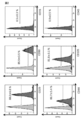
- FIG. 2 shows an analysis of the surface antigen of adipose-derived stem cells.
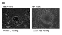
- FIG. 3 is a diagram showing the differentiation-inducing ability of adipose-derived stem cells.
- FIG. 4 is a view showing a colony formation test for confirming the self-renewal ability of adipose-derived stem cells.
- FIG. 1 is a diagram showing a histological analysis result of a fat-derived stem cell sheet composition.
- the middle row shows a hematoxylin-eosin stain (HE stain) on a tissue section of a four-layer laminate sheet, and the lower row
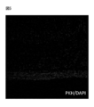
- FIG. 5 is a diagram showing engraftment after transplantation of the adipose-derived stem cell sheet composition.
- FIG. 6A is a diagram showing a small intestine anastomosis.
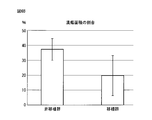
- FIG. 6B is a graph showing the ulcer area of the small intestine anastomosis.
- FIG. 7A is a diagram observing angiogenesis in the small intestine anastomosis. Immunostained with anti-CD31 antibody.
- FIG. 7B is a diagram observing angiogenesis in the small intestine anastomosis. Fluorescent immunostaining with anti-CD31 antibody.
- FIG. 7C is a graph showing angiogenesis at the small intestine anastomosis.
- a luminal organ is an organ having a luminal structure and includes, for example, the digestive tract, vascular system, bladder, vagina, etc., preferably the digestive tract.
- the digestive tract is an organ penetrating from the oral cavity to the anus, and is, for example, the esophagus, stomach, small intestine, and large intestine.
- wound broadly means a state in which a tissue or an organ has been damaged, such as burns, pressure sores, contusions, cuts, abrasions, ulcers, surgical wounds, gun wounds, bruises, stab wounds, This includes wounds and bite wounds.
- the wound is preferably a luminal organ incision, a surgical wound, a stab wound, a heel wound, or a bite wound, and more preferably a surgical wound.
- a surgical wound represents a wound formed as a result of surgery, for example, a wound formed using a scalpel, scissors, medical laser, forceps, snare, etc.
- wound part refers to a surrounding tissue including a wound site in a tissue or organ, for example, within a radius of 10 cm, within 8 cm, within 5 cm, within 3 cm, or within 2 cm, centering on the wound site. .
- the range and shape of the wound portion are not limited because they vary depending on the shape of the wound.
- "leakage" from the wound portion of the luminal organ means that the content of the luminal organ leaks outside the luminal organ, for example, when the luminal organ is the digestive tract, Solids such as food and its digests, liquids such as gastric juice and saliva, and gases such as air leak out from the wound into the thoracic cavity or abdominal cavity.
- Solids such as food and its digests
- liquids such as gastric juice and saliva
- gases such as air leak out from the wound into the thoracic cavity or abdominal cavity.
- the remaining digestive tract needs to be sutured or anastomosed.
- the sutured or anastomosed portion of the gastrointestinal tract may have a suture failure in which contents leak out due to various reasons.
- the present invention by applying a cell sheet composition containing the mesenchymal stem cells of the present invention to a wound site sutured or anastomosed in a luminal organ, particularly the digestive tract, the healing of the wound site is promoted, This has the effect of preventing or healing the suture failure.
- the pressure strength is significantly higher than when not applied, and the recovery of the strength of the suture or anastomosis portion of the digestive tract through which various substances pass is promoted. This contributes to improving the prognosis and quality of life (QOL) of patients after surgery.
- QOL quality of life
- the method for suturing or anastomosing a wound part of a luminal organ is not particularly limited as long as it is a method conventionally used in a surgical operation.
- the suture used for suturing or anastomosing the luminal organ may be either soluble or insoluble, but a soluble suture is preferred from the viewpoint of invasiveness.
- the thickness of the suture thread may be appropriately selected depending on the size and part of the wound.
- the “cell sheet composition” in the present invention refers to a single-layer or multi-layer sheet-like cell group obtained by culturing on a cell culture substrate and peeling off from the cell culture substrate.
- a method for obtaining a cell sheet composition for example, cells are cultured on a stimulus-responsive culture substrate coated with a polymer whose molecular structure is changed by stimulation such as temperature, pH, light, etc., and temperature, pH, light, etc.
- the cells can be detached from the stimulus-responsive culture substrate while maintaining the adhesion state between the cells, or any culture Examples thereof include a method of culturing cells on a base material, and physically detaching from the end of the cell culture base material with tweezers.
- a preferred form is a method in which a temperature-responsive culture substrate having a surface coated with a polymer whose hydration power changes in a temperature range of 0 to 80 ° C. is used as the stimulus-responsive culture substrate.
- the temperature-responsive polymer used in the present invention may be either a homopolymer or a copolymer. Examples of such a polymer include polymers described in JP-A-2-21865.
- poly (N-isopropylacrylamide) is used as a stimulus-responsive polymer, particularly a temperature-responsive polymer is described as an example (temperature-responsive culture dish).
- Poly (N-isopropylacrylamide) is known as a polymer having a lower critical solution temperature at 31 ° C. If in a free state, poly (N-isopropylacrylamide) undergoes dehydration in water at a temperature of 31 ° C. or higher, causing polymer chains to aggregate and become cloudy. Conversely, at a temperature of 31 ° C. or lower, the polymer chain is hydrated and dissolved in water. In the present invention, this polymer is coated and fixed on the surface of a substrate such as a petri dish.
- the polymer on the surface of the culture substrate is similarly dehydrated. However, since the polymer chain is fixed on the surface of the culture substrate, the surface of the culture substrate is hydrophobic. become. Conversely, at a temperature of 31 ° C. or lower, the polymer on the surface of the culture substrate is hydrated, but since the polymer chain is coated on the surface of the culture substrate, the surface of the culture substrate becomes hydrophilic. At this time, the hydrophobic surface is an appropriate surface on which cells can adhere and grow, and the hydrophilic surface is a surface on which cells cannot adhere. Therefore, when the substrate is cooled to 31 ° C. or lower, the cells are detached from the substrate surface.
- the cell sheet composition can be recovered by cooling the substrate to 31 ° C. or lower.
- the temperature-responsive culture dish is not limited as long as it has the same effect, and examples thereof include Up Cell (registered trademark) marketed by Cellseed.
- the origin of the animal species of the cells used in the present invention is, for example, mammals such as humans, rats, mice, guinea pigs, marmosets, rabbits, dogs, cats, sheep, pigs, goats, monkeys, chimpanzees or immunodeficient animals thereof. , Birds, reptiles, amphibians, amphibians, fish and insects.
- the cell sheet composition of the present invention is used for treatment of humans, cells derived from humans, derived from pigs when used for treatment of pigs, derived from monkeys when used for treatment of monkeys, and derived from chimpanzees when used for treatment of chimpanzees. It is preferable to use it.
- cells collected from the patient himself autologous cells
- cells collected from other people's cells transgenic cells
- commercially available cell lines may be used. It may be.
- the “mesenchymal stem cell” is an undifferentiated cell and includes various cells such as adipocytes, chondrocytes, bone cells, myoblasts, fibroblasts, stromal cells, and / or tendon cells.
- Mesenchymal stem cells are cells isolated from tissues such as bone marrow, adipose tissue, umbilical cord blood, dental pulp, synovium, and placenta in vivo, and can be isolated using known methods.
- bone marrow fluid collected from bone marrow is separated from blood cells by density gradient centrifugation, seeded in a plastic culture dish, and cultured in a 37 ° C., 5% CO 2 environment. And can be isolated as adherent cells.
- Adipose tissue-derived mesenchymal stem cells subdivide the collected adipose tissue (mince) and treat the tissue with collagenase type II at 37 ° C. for 1 hour. Digest, add medium and centrifuge. Thereafter, the precipitated cells are washed with a basal medium, filtered through a mesh strainer or the like, seeded on a plastic culture dish, and cultured in a 37 ° C., 5% CO 2 environment to be isolated as adherent cells. Is possible.
- the method for isolating mesenchymal stem cells derived from other tissues is not limited, and any known method may be used.
- the mesenchymal stem cells may be mesenchymal stem cells obtained by inducing differentiation from pluripotent stem cells.
- a pluripotent stem cell is a cell which has a self-replication ability and a pluripotency, and a cell provided with the ability (pluripotent) to form all the cells which comprise a body.
- Self-replicating ability refers to the ability to make two undifferentiated cells from one cell.
- the pluripotent stem cells used in the present invention include embryonic stem cells (embryonic stem cells: ES cells), embryonic carcinoma cells (embryonic carcinoma cells: EC cells), trophoblast stem cells (TS cells), shrimp.
- Blast stem cell epiblast stem cell: EpiS cell
- embryonic germ cell embryonic germ cell
- pluripotent germ cell multipotent stem cell: mGS cell
- induced pluripotent stem cell induced plumped stem cell
- mesenchymal stem cells to be induced to differentiate may be confirmed by a known method. For example, confirmation of differentiation induction from mesenchymal stem cells to adipocytes may be confirmed by culturing in a medium to which insulin and dexamethasone are added, and oil Red O staining. For example, for differentiation induction from mesenchymal stem cells to bone cells, ascorbic acid, ⁇ -glycerophosphate, and dexamethasone may be added to the medium and cultured, and confirmed by alkaline phosphatase staining.
- mesenchymal stem cells in order to induce muscle differentiation from mesenchymal stem cells, it may be cultured in a medium supplemented with horse serum, and it may be confirmed that fused cells specific to muscle cells appear.
- differentiation induction from mesenchymal stem cells may be performed using a known method and is not particularly limited.
- a known method may be used for confirmation of the presence or absence of differentiation induction.
- a gene or protein expressed only after differentiation induction may be detected by a real-time PCR method, a flow cytometer, or the like.
- the origin of the mesenchymal stem cells used in the present invention is not limited, but if it is bone marrow or adipose tissue, a collection method and a separation method are widely established and preferable. If derived from adipose tissue, the number of mesenchymal stem cells collected is greater than that derived from bone marrow, which is preferable.
- a cell adhesion protein such as collagen, laminin, laminin 5, fibronectin, or matrigel may be used alone or in combination of two or more.
- Cells can be cultured on cell culture substrates that have been pre-coated with the mixture.
- These cell adhesion protein coating methods may be in accordance with conventional methods, for example, a method in which an aqueous solution containing the cell adhesion protein is applied to the surface of the cell culture substrate, and then the aqueous solution is removed and rinsed. .
- the number of mesenchymal stem cells contained in the cell sheet composition is not limited because it varies depending on the size and degree of the wound to be applied. Further, the proportion of mesenchymal stem cells contained in the cell sheet composition of the present invention is not limited. For example, 30% or more, 40% or more, 50% or more, 55% or more, 60% or more, 65% or more, 70 %, 75%, 80%, 85%, 90%, 93%, 95%, 97%, 98%, 99% or more. The higher the proportion of mesenchymal stem cells contained in the cell sheet composition, the higher the effect of preventing or treating leakage of the wound part of the luminal organ.
- Cells constituting the cell sheet composition may include cells other than mesenchymal stem cells.
- vascular endothelial cells vascular endothelial precursor cells, fibroblasts, epithelial cells, stromal cells, etc. What is necessary is just to select suitably according to the site
- collected the mesenchymal stem cell may be contained.
- the number of cells to be seeded for preparing the cell sheet composition varies depending on the animal species and cell types, but may be, for example, 0.3 ⁇ 10 4 to 10 ⁇ 10 6 cells / cm 2 , It may be 0.5 ⁇ 10 4 to 8 ⁇ 10 6 pieces / cm 2 , or 0.7 ⁇ 10 4 to 5 ⁇ 10 6 pieces / cm 2 .
- the temperature of the culture substrate to which cells are attached and become confluent or sub-confluent is set to the upper limit of the coating polymer. It can be obtained by peeling off by setting it to a critical melting temperature or higher or a lower critical melting temperature or lower.
- the preparation of the cell sheet composition can be performed in a culture solution or in another isotonic solution, and can be selected according to the purpose.
- a method of tapping or shaking the culture substrate a method of stirring the medium using a pipette, a method of using tweezers, etc. alone or in combination May be used.
- Culture conditions other than temperature may follow conventional methods.
- the medium to be used may be a medium to which serum such as known fetal bovine serum (FBS) is added, or a serum-free medium may be used.
- FBS fetal bovine serum
- the shape of the cell culture substrate for producing the cell sheet composition used in the present invention may be, for example, a dish, a multiplate, a flask, or a flat membrane.
- a dish a multiplate
- a flask a flask
- a flat membrane a flat membrane.
- compounds such as glass, modified glass, polystyrene, polymethyl methacrylate, polycarbonate, etc., which are usually used for cell culture, and substances that can generally be given form, such as polymers other than those described above, are used. Examples thereof include compounds and ceramics.
- the cell culture substrate for producing the cell sheet composition used in the present invention may be a cell culture substrate having both a region to which cells adhere and a region to which cells do not adhere on the same culture surface.
- a cell culture substrate provided with a plurality of circular cell adhesion regions and a region where other cells are not adhered on the same culture surface.
- the shape of the cell adhesion region may be a desired shape according to the purpose, such as a circle, a square, a triangle, a rectangle, or the like, and the size can be changed as appropriate.
- a method for providing a region to which cells do not adhere is not particularly limited.
- poly-N-acryloylmorpholine polyacrylamide, polydimethylacrylamide, polyethylene glycol, cellulose, etc., which are non-cell-adhesive polymers with low affinity to cells.
- the cell sheet composition in the present invention does not use a proteolytic enzyme such as dispase or trypsin conventionally used for recovering adherent cells, the protein expressed on the cell surface is hardly damaged. Therefore, the lower surface of the cell sheet composition peeled from the cell culture substrate (the surface on the side in contact with the cell culture substrate) is rich in undamaged adhesive protein, A cell-to-cell desmosome structure is retained.
- the cell sheet composition is suitable for being attached to a living body affected part or for laminating the cell sheet composition.
- Dispase a proteolytic enzyme
- the cell sheet composition used in the present invention can peel and recover both the desmosome structure and the basement membrane-like protein in a state in which 60% or more remains, and obtains various effects as described above. be able to.
- a laminated cell sheet composition obtained by laminating a plurality of cell sheet compositions may be used.
- the number of cells to be applied increases, and the effect of preventing or treating leakage of the wound part of the luminal organ becomes higher.
- the cell sheet composition floating in the culture solution is sucked together with the culture solution by a pipette or the like, discharged onto the cell sheet composition of another culture dish, and the flow of the liquid medium. Examples include a method of stacking, a method of stacking using a cell movement jig, and the like.
- the method using a cell migration jig is preferable because it can be laminated without damaging the cell sheet composition.
- the cell migration jig only needs to have a function capable of capturing the cell sheet composition.
- the material include polyvinylidene difluoride (PVDF), silicone resin, polyvinyl alcohol, urethane, cellulose, and derivatives thereof. Chitin, chitosan, collagen, gelatin, fibrin gel and the like can be used.
- the shape of the cell migration jig include a stamp shape, a membrane shape, a porous membrane shape, a nonwoven fabric shape, and a woven fabric shape.
- the cell migration jig only needs to have a function of collecting the cell sheet composition without damaging it and laminating it on another cell sheet composition.
- a cultured cell migration jig having a cell attachment part composed of one or more of peptides or hydrophilic polymers is preferred.
- Japanese Patent Application Laid-Open No. 2005-176812 discloses a stamp-type cultured cell moving jig having a cell attachment portion. The stamp-type cultured cell transfer jig prevents the cell sheet composition from being damaged by the cell attachment portion, and prevents the cell sheet composition from shrinking when the cell sheet composition peels from the culture dish. Recovery is possible.
- the cell sheet jig can be laminated without shrinking the cell sheet composition while easily moving the cell sheet composition onto another cell sheet composition.
- the cell sheet compositions are laminated without any gaps, and a laminated cell sheet composition having a high-density three-dimensional structure can be obtained.
- the method for producing the cell sheet composition of the present invention may be further cultured in a medium supplemented with ascorbic acid (reference: Kato Y, et al. Allogeneic Transplantation of Adipose-Derived Stem Cell Sheet Combined With Whiskey. Accelerates Wound Healing in a Rat Wound Model of Type 2 Diabetes and Obesity.Diabetes.2015 Aug; 64 (8): 2723-34).
- a cell sheet composition containing mesenchymal stem cells obtained by culturing in a medium supplemented with ascorbic acid has higher strength and is harder to break than a cell sheet composition obtained by culturing without adding A sheet composition is obtained. Thereby, a cell sheet composition more suitable for transplantation can be obtained.
- the cell sheet composition may further contain a factor that induces angiogenesis.
- factors that induce angiogenesis include vascular endothelial growth factor (VEGF), fibroblast growth factor (FGF), angiopoietin, platelet-derived growth factor (PDGF), transforming growth factor- ⁇ (TGF- ⁇ ), Matrix metalloproteases (MMP), VE-cadherin, ephrin, plasminogen activator, inducible nitric oxide synthase (iNOS), cyclooxygenase-2 (COX-2), placental growth factor (PlGF) and the like.
- VEGF vascular endothelial growth factor
- FGF fibroblast growth factor
- angiopoietin angiopoietin
- PDGF platelet-derived growth factor
- TGF- ⁇ transforming growth factor- ⁇
- MMP Matrix metalloproteases
- VE-cadherin ephrin
- the site to which the cell sheet composition of the present invention is affixed may be a wound part, and preferably a wound part that has been sutured or anastomosed. If it is the site
- the site to which the cell sheet composition of the present invention is affixed may be the inner wall and / or the outer wall of a luminal organ, and may be only the outer wall from the viewpoint of ease of affixing during surgery. .
- HGF hepatocyte growth factor
- adipose-derived stem cell sheet composition (1) Transplantation method to the digestive tract anastomosis site The adipose-derived stem cell sheet composition recovered in the form of a sheet by the above method is obtained by applying the cell sheet composition to the serosal surface of the anastomosis after the digestive tract anastomosis. Transplantation was performed so that the basement membrane surface adhered. Three cell sheet compositions were transplanted so as to cover the entire circumference of the anastomosis.
- HGF and FGF2 were detected by ELISA in a culture medium in which the adipose-derived stem cell sheet composition was cultured for 24 hours (FGF: 106.9 pg / ml). HGF: 1.38 ng / ml).
Landscapes
- Health & Medical Sciences (AREA)
- Life Sciences & Earth Sciences (AREA)
- Engineering & Computer Science (AREA)
- Biomedical Technology (AREA)
- Chemical & Material Sciences (AREA)
- Cell Biology (AREA)
- Medicinal Chemistry (AREA)
- Animal Behavior & Ethology (AREA)
- General Health & Medical Sciences (AREA)
- Veterinary Medicine (AREA)
- Public Health (AREA)
- Developmental Biology & Embryology (AREA)
- Zoology (AREA)
- Epidemiology (AREA)
- Chemical Kinetics & Catalysis (AREA)
- Immunology (AREA)
- Oral & Maxillofacial Surgery (AREA)
- Dermatology (AREA)
- Botany (AREA)
- Transplantation (AREA)
- Pharmacology & Pharmacy (AREA)
- Reproductive Health (AREA)
- Hematology (AREA)
- Biotechnology (AREA)
- Virology (AREA)
- Vascular Medicine (AREA)
- Urology & Nephrology (AREA)
- Nuclear Medicine, Radiotherapy & Molecular Imaging (AREA)
- General Chemical & Material Sciences (AREA)
- Bioinformatics & Cheminformatics (AREA)
- Organic Chemistry (AREA)
- Gynecology & Obstetrics (AREA)
- Medicines Containing Material From Animals Or Micro-Organisms (AREA)
- Materials For Medical Uses (AREA)
- Micro-Organisms Or Cultivation Processes Thereof (AREA)
- Medicinal Preparation (AREA)
- Apparatus Associated With Microorganisms And Enzymes (AREA)
Abstract
本発明は、管腔臓器の創傷部からの漏出を治癒又は予防するための、細胞シート組成物を提供することを課題とする。また、本発明は、細胞シート組成物を管腔臓器の創傷部に貼付し、該管腔臓器の該創傷部からの漏出を治癒又は予防する方法を提供することを課題とする。 本発明は、管腔臓器の創傷部からの漏出を治癒又は予防するために、前記管腔臓器の前記創傷部に貼付されることを特徴とする、間葉系幹細胞を含む細胞シート組成物を提供する。また、本発明は、間葉系幹細胞を含む細胞シート組成物を、管腔臓器の創傷部に貼付し、前記管腔臓器の前記創傷部からの漏出を治癒又は予防する方法を提供する。
Description
本発明は、管腔臓器の創傷部からの漏出を治癒又は予防するための間葉系幹細胞を含む細胞シート組成物に関する。また、本発明は、間葉系幹細胞を含む細胞シート組成物を、管腔臓器の創傷部に貼付し、前記管腔臓器の前記創傷部からの漏出を治癒又は予防する方法に関する。
悪性腫瘍が消化管に生じた場合、早期であれば組織への浸潤が少なく、内視鏡を用いた内視鏡的粘膜切除術(EMR:Endoscopic Mucosal Resection)又は、内視鏡的粘膜切開剥離術(ESD:Endoscopic Submucosal Dissection)等により、表層の悪性腫瘍のみを切除することが可能である。例えば、食道の場合、初期の癌であれば内視鏡を使用したESDにより病変部を切除することができる。しかし、ESD後に切除した部位に潰瘍が生じ、食道の狭窄がしばしば引き起こされる。狭窄が生じるのを予防する目的で、口腔粘膜上皮細胞シートを貼付する方法が開発されている(非特許文献1)。口腔粘膜上皮細胞シートとは、患者本人の口腔粘膜の組織片を採取し、得られた口腔粘膜上皮細胞を、温度応答性培養皿上で培養し、下限臨界温度で培養することで得られた口腔粘膜上皮細胞シートを、ESD後の食道内壁の切除面に移植する再生医療の一つである。これにより、食道の狭窄が予防される。
しかし、進行がんでは、悪性腫瘍が組織の奥深く浸潤して病変が広範に及んでしまっているために、病変部を含む周囲の組織を切除する必要が生じ、病変部を切除後は、消化管の残存部を縫合又は吻合する手術が必要となる。その際、問題となるのが消化管縫合不全である。
消化管縫合不全とは、消化管吻合後の吻合部が治癒しないで、離開してしまう状態のことで、腸管内容物が胸腔内や腹腔内に漏出し、重篤な感染症を起こして、時に致死的な状態になる可能性のある消化管術後の合併症の一つである。縫合不全は、吻合部組織の治癒過程(修復期)が妨げられることで起こるが、修復期の治癒過程を妨げる要因としては、術前低栄養状態やステロイドなどの薬剤投与、慢性疾患(糖尿病、肝・腎障害等)による全身的な要因と吻合部周囲の血行障害や過緊張、感染症などの局所的な要因が挙げられる。縫合不全を予防するために、術前の全身状態をより良い状態に改善して手術に臨む努力や、自動吻合機の開発、部位・状況に応じた適切な吻合法の選択、吻合部の緊張の低減、的確な血流の確保など様々な工夫が行われているが、未だ、縫合不全を完全に予防することは出来ていないのが現状である。
この課題を解決する手段として、組織の創傷治癒を促進する作用のある間葉系幹細胞や脂肪由来幹細胞を用いた動物実験の報告がなされているが、その移植方法の大部分は静脈注射や局所注射などの細胞注入法であった。一般に消化管吻合部の創傷治癒の過程は、術後3~4日までの炎症期と、術後7日程までの修復期に分けられる。術後3~4日までの炎症期には、吻合部粘膜下組織の再構築のために、既存のコラーゲンを炎症細胞由来のコラゲナーゼが分解し、その後の修復期で線維芽細胞が増殖し、コラーゲンが産生されて増加し、術後7日程まで組織の連続性と物理的抗張力が維持される(非特許文献2)。そのため、吻合部位に細胞を注入する移植法では、細胞がその場に保持されにくいだけでなく、注入した細胞が炎症期のタンパク分解酵素であるコラゲナーゼに暴露されるため、コラゲナーゼの細胞傷害により組織修復能が低下する可能性があった。
また、近年、術前化学放射線療法や分子標的療法など局所および全身への強力な治療が進歩して、消化器癌の再発や術後生存率には寄与する一方、創傷治癒遅延に伴う術後縫合不全の発生の増加が問題となっていた。
Ohki T.,et al.Treatment of oesophageal ulcerations using endoscopic transplantation of tissue-engineered autologous oral mucosal epithelial cell sheets in a canine model.Gut.2006;55(12):1704-1710.
Hendriks T.,Mastboom WJ.Healing of experimental intestinal anastomoses.Parameters for repair.Dis Colon Rectum.1990 Oct;33(10):891-901.
上述のように、管腔臓器、特に消化管の縫合不全を予防したり治癒したりする技術の開発が試みられているが、未だ提供されるに至っていない。本発明は、管腔臓器の創傷部からの漏出を治癒又は予防するための、細胞シート組成物を提供することを課題とする。また、本発明は、細胞シート組成物を管腔臓器の創傷部に貼付し、該管腔臓器の該創傷部からの漏出を治癒又は予防する方法を提供することを課題とする。
本発明者らは、上記課題を解決するために、種々の角度から検討を加えて研究開発を行ってきた。その結果、驚くべきことに、間葉系幹細胞を含む細胞シート組成物を管腔臓器の創傷部に貼付したところ、管腔臓器の創傷部からの漏出が治癒又は予防されることを見出した。また、すなわち、本発明は、以下のとおりである。
[1] 管腔臓器の創傷部からの漏出を治癒又は予防するために、前記管腔臓器の前記創傷部に貼付されることを特徴とする、間葉系幹細胞を含む細胞シート組成物。
[2] 前記間葉系幹細胞が、臍帯血、胎盤、骨髄、脂肪組織、滑膜、及び/又は、多能性幹細胞由来の間葉系幹細胞である、[1]に記載の細胞シート組成物。
[3] 前記間葉系幹細胞が、脂肪由来幹細胞である、[1]又は[2]に記載の細胞シート組成物。
[4] 前記創傷部が、縫合又は吻合された創傷部である、[1]~[3]のいずれか1項に記載の細胞シート組成物。
[5] 貼付される部位が、前記管腔臓器の外壁である、[1]~[4]のいずれか1項に記載の細胞シート組成物。
[6] 前記管腔臓器が、消化管である、[1]~[5]のいずれか1項に記載の細胞シート組成物。
[7] 前記管腔臓器が、腸管である、[1]~[6]のいずれか1項に記載の細胞シート組成物。
[8] 間葉系幹細胞を含む細胞シート組成物を、管腔臓器の創傷部に貼付し、前記管腔臓器の前記創傷部からの漏出を治癒又は予防する方法。
[9] 前記間葉系幹細胞が、臍帯血、胎盤、骨髄、脂肪組織、滑膜、及び/又は、多能性幹細胞由来の間葉系幹細胞である、[8]に記載の方法。
[10] 前記間葉系幹細胞が、脂肪由来幹細胞である、[8]又は[9]に記載の方法。
[11] 前記創傷部が、縫合又は吻合された創傷部である、[8]~[10]のいずれか1項に記載の方法。
[12] 貼付する部位が、前記管腔臓器の外壁である、[8]~[11]のいずれか1項に記載の方法。
[13] 前記管腔臓器が、消化管である、[8]~[12]のいずれか1項に記載の方法。
[14] 前記管腔臓器が、腸管である、[8]~[13]のいずれか1項に記載の方法。
[9] 前記間葉系幹細胞が、臍帯血、胎盤、骨髄、脂肪組織、滑膜、及び/又は、多能性幹細胞由来の間葉系幹細胞である、[8]に記載の方法。
[10] 前記間葉系幹細胞が、脂肪由来幹細胞である、[8]又は[9]に記載の方法。
[11] 前記創傷部が、縫合又は吻合された創傷部である、[8]~[10]のいずれか1項に記載の方法。
[12] 貼付する部位が、前記管腔臓器の外壁である、[8]~[11]のいずれか1項に記載の方法。
[13] 前記管腔臓器が、消化管である、[8]~[12]のいずれか1項に記載の方法。
[14] 前記管腔臓器が、腸管である、[8]~[13]のいずれか1項に記載の方法。
本発明は、種々の要因により吻合部組織の治癒過程(修復期)が妨げられて起こる消化管縫合不全に対して、本発明の細胞シート組成物及びそれを用いた方法によって、組織修復能を最大限に発揮し、縫合不全を予防又は治癒する画期的な効果をもたらす。これによって、縫合不全により生じる人工肛門、腸瘻造設などの追加治療による患者QOLの著しい低下、多くの医療資源の投入などの社会的損失を軽減できる可能性がある。
本発明において、管腔臓器とは、管腔構造を有する臓器であって、例えば、消化管、血管系、膀胱、膣などが挙げられるが、好ましくは、消化管である。本発明において、消化管とは、口腔から肛門までを貫通している器官であって、例えば、食道、胃、小腸、大腸である。
本明細書中において、「創傷」とは、組織又は臓器が傷ついた状態を広く意味し、例えば、熱傷、褥瘡、挫傷、切創、擦過傷、潰瘍、手術創、銃創、爆傷、刺創、杙創、咬創等が含まれる。本発明において、創傷とは、好ましくは、管腔臓器の切創、手術創、刺創、杙創、咬創であり、より好ましくは、手術創である。本発明において、手術創とは、外科手術の結果として形成される創傷を表し、例えば、メス、ハサミ、医療用レーザ、鉗子、スネア等を使用して形成される創傷であるが、外科手術で通常使用される器具によって形成される創傷であればよく、限定されない。本発明において、「創傷部」とは、組織又は臓器において、創傷部位を含むその周辺組織をいい、例えば、創傷部位を中心として半径10cm以内、8cm以内、5cm以内、3cm以内、2cm以内である。創傷部の範囲、形状は、創傷の形状によって変わるため、限定されない。
本発明において、管腔臓器の創傷部からの「漏出」とは、管腔臓器の内容物が、管腔臓器の外側へ漏れ出すことをいい、例えば、管腔臓器が消化管である場合、食物やその消化物等の固形物、胃液、唾液等の液体、空気等の気体が、創傷部から胸腔内又は腹腔内へ漏れ出すことをいう。特に、管腔臓器、例えば消化管に生じた癌であって、深層部まで浸潤している場合、消化管の一部を切除する必要がある。その場合、残った消化管を縫合又は吻合する必要がある。上述のように、消化管の縫合又は吻合された部分は、種々の事由により、内容物が漏出してしまう縫合不全が生じる場合がある。本発明は、管腔臓器、特に消化管において、縫合又は吻合された創傷部に、本発明の間葉系幹細胞を含む細胞シート組成物を貼付することにより、該創傷部の治癒が促進され、縫合不全が予防又は治癒される効果をもたらす。また、本発明を適用することにより、適用しない場合と比較して、有意に高い耐圧強度を示し、様々な物質が通過する消化管の縫合又は吻合部の強度の回復を促進する。これによって、術後の患者の予後及び生活の質(QOL)の向上に寄与する。
本発明において、管腔臓器の創傷部の縫合又は吻合の方法については、従来外科手術で用いられる方法であれば、特に限定されない。管腔臓器を縫合又は吻合する時に用いられる縫合糸は、溶解性又は非溶解性のいずれでも良いが、侵襲性の観点からは、溶解性の縫合糸の方が好ましい。縫合糸の太さは、創傷の大きさ、部位等によって適宜選択すればよい。
本発明における「細胞シート組成物」とは、細胞培養基材上で培養し、細胞培養基材上から剥離することで得られる1層又は複数層のシート状の細胞群をいう。細胞シート組成物を得る方法としては、例えば、温度、pH、光等の刺激によって分子構造が変化する高分子を被覆した刺激応答性培養基材上で細胞を培養し、温度、pH、光等の刺激の条件を変えて刺激応答性培養基材表面を変化させることで、細胞同士の接着状態は維持しつつ、刺激応答性培養基材から細胞をシート状に剥離する方法や、任意の培養基材にて細胞を培養し、細胞培養基材の端部から物理的にピンセット等により剥離する方法等が挙げられる。好ましい形態としては、刺激応答性培養基材として、0~80℃の温度範囲で水和力が変化するポリマーを表面に被覆した温度応答性培養基材を用いる方法である。温度応答性培養基材上で、ポリマーの水和力が弱い温度域で細胞を培養し、その後、培養液をポリマーの水和力が強い状態となる温度に変化させることで細胞を培養し、細胞をシート状に剥離して回収する方法である。その際、細胞は0~80℃の温度範囲で水和力が変化するポリマーを表面に被覆した細胞培養基材上で、ポリマーの水和力の弱い温度域で培養する。その温度域とは通常、細胞を培養する温度、例えば33℃~40℃であることが好ましい。本発明に用いる温度応答性高分子はホモポリマー、コポリマーのいずれであってもよい。このような高分子としては、例えば、特開平2-211865号公報に記載されているポリマーが挙げられる。
刺激応答性高分子、特に温度応答性高分子としてポリ(N-イソプロピルアクリルアミド)を用いた場合を例(温度応答性培養皿)に説明する。ポリ(N-イソプロピルアクリルアミド)は31℃に下限臨界溶解温度を有するポリマーとして知られ、遊離状態であれば、水中で31℃以上の温度で脱水和を起こしポリマー鎖が凝集して白濁する。逆に31℃以下の温度ではポリマー鎖は水和し、水に溶解した状態となる。本発明では、このポリマーがシャーレなどの基材表面に被覆されて固定されたものである。したがって、31℃以上の温度であれば、培養基材表面のポリマーも同じように脱水和するが、ポリマー鎖が培養基材表面に固定されているため、培養基材表面が疎水性を示すようになる。逆に、31℃以下の温度では、培養基材表面のポリマーは水和するが、ポリマー鎖が培養基材表面に被覆されているため、培養基材表面が親水性を示すようになる。このときの疎水的な表面は細胞が付着し、増殖できる適度な表面であり、また、親水的な表面は細胞が付着できない表面となる。そのため、該基材を31℃以下に冷却すると、細胞が基材表面から剥離する。細胞が培養面一面にコンフルエントになるまで培養されていれば、該基材を31℃以下に冷却することによって細胞シート組成物が回収できる。温度応答性培養皿は、同一の効果を有するものであれば限定されるものではないが、例えば、セルシード社が市販するUp Cell(登録商標)などが挙げられる。
本発明に用いられる細胞の動物種の由来は、例えば、ヒト、ラット、マウス、モルモット、マーモセット、ウサギ、イヌ、ネコ、ヒツジ、ブタ、ヤギ、サル、チンパンジーあるいはそれらの免疫不全動物などの哺乳類動物、鳥類、爬虫類、両生類、両生類、魚類、昆虫等が挙げられる。本発明の細胞シート組成物をヒトの治療に用いる場合はヒト由来、ブタの治療に用いる場合はブタ由来、サルの治療に用いる場合はサル由来、チンパンジーの治療に用いる場合はチンパンジー由来の細胞を用いる方が好ましい。また、治療を行うのがヒトである場合、患者本人から採取した細胞であってもよく(自家細胞)、他人の細胞から採取した細胞を用いてもよく(他家細胞)、市販の細胞株であってもよい。
本明細書中において、「間葉系幹細胞」とは、未分化な細胞で、脂肪細胞、軟骨細胞、骨細胞、筋芽細胞、線維芽細胞、ストローマ細胞、及び/又は腱細胞等のさまざまな間葉系の細胞へ分化する能力を持ち、且つ自己複製の能力を持つ細胞をいう。The International Society for Cellular Therapy (ISCT)において、間葉系幹細胞を既定する以下の3つの最小限の基準;(1)標準的な培養条件でプラスチックに接着して培養できること、(2)免疫学的特徴として、CD105、CD73、CD90が陽性、CD45、CD34、CD14又はCD11b、CD79a又はCD19、HLA-DRが陰性であること、(3)in vitro分化系で骨芽細胞、脂肪細胞、軟骨芽細胞への分化能を示すこと、を提唱するが、本発明においては、これに限定されない。その他、CD29、CD44、CD106及びSTRO-1も間葉系幹細胞を示す陽性マーカーとして挙げられる。本発明において、「間葉系幹細胞」は、可能な限り最も広く解釈される。
間葉系幹細胞は、生体内においては、骨髄、脂肪組織、臍帯血、歯髄、滑膜、胎盤等の組織から単離される細胞であって、公知の方法を用いて単離することができる。
例えば、骨髄由来間葉系幹細胞は、骨髄から採取した骨髄液を、密度勾配遠心法にて血球系細胞を分離し、プラスチック培養皿に播種し、37℃、5%CO2環境で培養することで、接着する細胞として単離することが可能である。
脂肪組織由来間葉系幹細胞(脂肪組織由来幹細胞;Adipose derived stem cell)は、採取した脂肪組織を細分化(ミンス(mince))し、コラゲナーゼタイプIIによって、37℃1時間処理をして組織を消化し、培地を加えて遠心分離する。その後、沈殿した細胞を基礎培地で洗浄し、セルストレーナー等のメッシュにてろ過し、プラスチック培養皿に播種して、37℃、5%CO2環境で培養することで、接着する細胞として単離することが可能である。その他の組織由来の間葉系幹細胞を単離する方法については、公知の方法を用いればよく、限定されない。
間葉系幹細胞は、多能性幹細胞から分化誘導して得られた間葉系幹細胞であってもよい。本明細書において、多能性幹細胞とは、自己複製能と多分化能を有する細胞であり、体を構成するあらゆる細胞を形成する能力(pluriopotent)を備える細胞をいう。自己複製能とは、1つの細胞から自分と同じ未分化な細胞を2つ作る能力のことをいう。本発明で用いられる多能性幹細胞には、胚性幹細胞(embryonic stem cell:ES細胞)、胚性癌腫細胞(embryonic carcinoma cell:EC細胞)、栄養芽幹細胞(trophoblast stem cell:TS細胞)、エビブラスト幹細胞(epiblast stem cell:EpiS細胞)、胚性生殖細胞(embryonic germ cell:EG細胞)、多能性生殖細胞(multipotent germline stem cell:mGS細胞)、人工多能性幹細胞(induced pluripotent stem cell:iPS細胞)などが含まれる。多能性幹細胞より、公知の方法(例えば、特開2012-120486、Fukuta M.,et al.,Derivation of mesenchymal stromal cells from pluripotent stem cells through a neural crest lineage using small molecule compounds with defined media.PLOS ONE,2014;9(12):e112291.等)を用いて誘導された間葉系幹細胞を用いてもよい。
間葉系幹細胞の分化誘導される能力については、公知の方法によって確認すればよい。例えば、間葉系幹細胞から脂肪細胞への分化誘導の確認には、インスリン及びデキサメサゾンを加えた培地で培養して、Oil Red O染色によって確認すればよい。例えば、間葉系幹細胞から骨細胞への分化誘導では、アスコルビン酸、β-グリセロリン酸、デキサメサゾンを培地に添加して培養し、アルカリフォスファターゼ染色で確認すればよい。例えば、間葉系幹細胞から筋分化誘導には、ウマ血清を加えた培地で培養すればよく、筋細胞に特異的な融合細胞が出現することを確認すればよい。その他、間葉系幹細胞からの分化誘導は、公知の方法を用いればよく、特に限定されない。分化誘導の有無の確認も公知の方法を用いればよく、例えば、分化誘導後にのみ発現する遺伝子又はタンパク質を、リアルタイムPCR法、フローサイトメーター等によって検出すればよい。
本発明に使用される間葉系幹細胞の由来は限定されないが、骨髄又は脂肪組織であれば、採取方法、分離方法が広く確立されており、好ましい。脂肪組織由来であれば、骨髄由来と比較して、採取される間葉系幹細胞の数が多く、好ましい。
細胞の由来によっては、細胞培養基材上に接着しにくい細胞があり、そのような場合は、例えば、コラーゲン、ラミニン、ラミニン5、フィブロネクチン、マトリゲル等の細胞接着性タンパク質を単独又は2種以上の混合物を予め被覆した細胞培養基材上で細胞を培養することができる。これらの細胞接着性タンパク質の被覆方法は常法に従えば良く、例えば、細胞接着性タンパク質を含む水溶液を細胞培養基材表面に塗布し、その後その水溶液を除去し、リンスする方法等が挙げられる。
本発明において、細胞シート組成物に含まれる間葉系幹細胞の数は、貼付する創傷の大きさ、度合いによって変化するため、限定されない。また、本発明の細胞シート組成物に含まれる間葉系幹細胞の割合も限定されないが、例えば、30%以上、40%以上、50%以上、55%以上、60%以上、65%以上、70%以上、75%以上、80%以上、85%以上、90%以上、93%以上、95%以上、97%以上、98%以上、99%以上であってもよい。細胞シート組成物に含まれる間葉系幹細胞の割合が高ければ高いほど、管腔臓器の創傷部の漏出を予防又は治療する効果が、より高くなる。
細胞シート組成物を構成する細胞としては、間葉系幹細胞以外の細胞が含まれてもよく、例えば、血管内皮細胞、血管内皮前駆細胞、線維芽細胞、上皮系細胞、間質細胞等、移植する部位、目的によって適宜選択すればよい。また、間葉系幹細胞を採取した組織由来の細胞が含まれていても良い。
本発明において、細胞シート組成物を作製するために播種する細胞数は動物種や細胞種によって異なるが、例えば、0.3×10⁴~10×106個/cm2であってもよく、0.5×104~8×106個/cm2であってもよく、0.7×104~5×106個/cm2であってもよい。本発明においては、細胞シート組成物を温度応答性培養基材から剥離して回収するためには、細胞が付着し、コンフルエント又はサブコンフルエント状態となった培養基材の温度を、被覆ポリマーの上限臨界溶解温度以上若しくは下限臨界溶解温度以下にすることによって剥離させることで得られる。その際、細胞シート組成物の作製は、培養液中において行うことも、その他の等張液中において行うことも可能であり、目的に合わせて選択することができる。細胞シート組成物をより早く、高効率に剥離、回収するために、培養基材を軽くたたいたり揺らしたりする方法、ピペットを用いて培地を撹拌する方法、ピンセットを用いる方法等を単独又は併用して用いてもよい。温度以外の培養条件は常法に従えばよい。例えば、使用する培地については公知のウシ胎児血清(FBS)等の血清が添加されている培地でもよく、無血清培地を用いてもよい。
本発明に用いられる細胞シート組成物の作製用の細胞培養基材の形状は、例えば、ディッシュ、マルチプレート、フラスコ、又は平膜状などの形状のものを用いることができる。細胞培養基材の材料としては、通常、細胞培養に用いられるガラス、改質ガラス、ポリスチレン、ポリメチルメタクリレート、ポリカーボネート等の化合物や、一般に形態付与が可能である物質、例えば、上記以外の高分子化合物、セラミックス類などが挙げられる。
本発明に用いられる細胞シート組成物の作製用の細胞培養基材は、細胞が接着する領域と細胞が接着しない領域の両方とを同一培養面上に備えた細胞培養基材を用いてもよく、例えば、複数の円形の細胞接着領域と、それ以外の細胞を接着しない領域とを同一培養面に備えた細胞培養基材を用いることで一度に複数の細胞シートを作製することが可能である。この場合、細胞接着領域の形状は円形、正方形、三角形、長方形等、目的に応じて所望の形状であってよく、その大きさも適宜変更することができる。細胞が接着しない領域を設ける方法としては特に限定されないが、例えば細胞と親和性の低い細胞非付着性高分子である、ポリ-N-アクリロイルモルホリン、ポリアクリルアミド、ポリジメチルアクリルアミド、ポリエチレングリコール、セルロース等の親水性高分子、又はシリコーン高分子、フッ素高分子等の強疎水性高分子等を被覆する方法等が挙げられる。
本発明における細胞シート組成物は、従来、接着性細胞を回収する際に用いられるディスパーゼ、トリプシン等のタンパク質分解酵素を用いないため、細胞表面に発現するタンパク質に損傷をほとんど受けていない。そのため、細胞培養基材から剥離された細胞シート組成物の下面(細胞培養基材と接触していた側の面)は、損傷を受けていない接着性タンパク質を豊富に有しており、また、細胞-細胞間のデスモゾーム構造が保持されている。このような構造を有していることにより、細胞シート組成物は生体患部に貼付することや、細胞シート組成物を積層することに適している。タンパク質分解酵素であるディスパーゼは、細胞-細胞間のデスモソーム構造を10~40%保持した状態で細胞を剥離させることができることで知られているが、細胞-培養基材間に存在する基底膜様タンパク質等も同時に破壊してしまう。これに対して、本発明に用いられる細胞シート組成物は、デスモゾーム構造及び基底膜様タンパク質の両者を60%以上残存された状態で剥離・回収可能であり、上述したような種々の効果を得ることができる。
本発明の細胞シート組成物は、複数の細胞シート組成物を積層した積層化細胞シート組成物を用いても良い。本発明において、積層化細胞シート組成物を用いた場合、貼付する細胞数が多くなり、管腔臓器の創傷部の漏出を予防又は治療する効果がより高くなる。積層化細胞シート組成物を得る方法としてはピペット等によって培養液中に浮かんでいる細胞シート組成物を培養液ごと吸い取り、別の培養皿の細胞シート組成物上に放出し、液体培地の流れによって積層する方法、細胞移動治具を用いて積層する方法等が挙げられる。本発明の積層化細胞シート組成物を製造する方法では、細胞移動治具を用いる方法の方が、細胞シート組成物を損傷させることなく積層することが可能となるため、好ましい。細胞移動治具は、細胞シート組成物を捕捉できる機能を有するものであればよく、例えば、材料としては、ポリビニリデンジフルオライド(PVDF)、シリコーン樹脂、ポリビニルアルコール、ウレタン、セルロース及びその誘導体、キチン、キトサン、コラーゲン、ゼラチン、フィブリンゲル等を用いることができる。細胞移動治具の形状としては、例えば、スタンプ型、膜状、多孔膜状、不織布状、織布状の形状で使用される。本発明の実施形態では、細胞移動治具は細胞シート組成物を破損することなく回収し、別の細胞シート組成物に積層する機能を有していればよく、細胞接着性タンパク質、細胞接着性ペプチド、又は親水性ポリマーの1種、若しくは2種以上からなる細胞付着部を有する培養細胞移動治具であることが好ましい。例えば、特開2005-176812号公報には細胞付着部を有したスタンプ型の培養細胞移動治具を開示している。該スタンプ型の培養細胞移動治具は、その細胞付着部によって細胞シート組成物を破損することを防止し、細胞シート組成物が培養皿から剥離するときに起こる細胞シート組成物の縮みを防止しながら回収を可能とする。該細胞移動治具により、細胞シート組成物を別の細胞シート組成物上に容易に移動させつつ、細胞シート組成物を縮ませることなく積層することができる。細胞シート組成物を縮ませずに積層することにより、細胞シート組成物同士が隙間なく積層され、高密度な三次元構造を有する積層化細胞シート組成物を得ることができる。
本発明の細胞シート組成物を製造する方法について、さらに、アスコルビン酸を添加した培地で培養してもよい(参考:Kato Y,et al.Allogeneic Transplantation of an Adipose-Derived Stem Cell Sheet Combined With Artificial Skin Accelerates Wound Healing in a Rat Wound Model of Type 2 Diabetes and Obesity.Diabetes.2015 Aug;64(8):2723-34)。アスコルビン酸を添加した培地で培養することによって得られた、間葉系幹細胞を含む細胞シート組成物は、添加せずに培養して得られた細胞シート組成物よりも強度が高く、破れにくい細胞シート組成物が得られる。これにより、より、移植に適した細胞シート組成物が得られる。
細胞シート組成物には、さらに血管新生を誘導する因子を添加してもよい。血管新生を誘導する因子としては、例えば、血管内皮増殖因子(VEGF)、線維芽細胞増殖因子(FGF)、アンジオポエチン、血小板由来増殖因子(PDGF)、トランスフォーミング増殖因子-β(TGF-β)、マトリックスメタロプロテアーゼ(MMP)、VE-カドヘリン、エフリン、プラスミノーゲンアクチベータ、誘導型一酸化窒素合成酵素(iNOS)、シクロオキシゲナーゼ-2(COX-2)、胎盤成長因子(PlGF)等である。これらが細胞シート組成物に含まれることによって、移植部位における血管新生もより促進されることとなる。
本発明の細胞シート組成物を貼付する部位は創傷部であればよく、好ましくは、縫合又は吻合された創傷部である。縫合又は吻合されている部位であれば、創傷によって炎症をおこした組織同士が密接し、そこに本発明の細胞シート組成物が貼付されることで、治癒効果が高まる。本発明の細胞シートが貼付されることによって、創傷部のコラーゲン等の産生能が高まり、管腔臓器の創傷部周辺の再構築が促進され、管腔内部の圧力上昇にも耐えうる程、創傷部の治癒が促進される。また、本発明の細胞シート組成物を貼付する部位は、管腔臓器の内壁及び/又は外壁であってもよく、外科手術時の貼付のしやすさの点からは外壁のみであってもよい。
以下に、本発明を実施例に基づいて更に詳しく説明するが、これらは本発明を何ら限定するものではない。なお、本実施例におけるミニブタを用いた実験プロトコールは、東京女子医科大学の動物実験に関する倫理委員会によって承認されたものであり、米国国立衛生研究所(NIH)によって刊行された「実験動物の管理と使用に関する指針」(1996年改定版)に沿って実施された。
<使用した動物・試薬・キット>
・ミニブタメス(月齢5~6カ月、NIBS系ミニブタ、日本生物科学研究所)
・ペニシリン・ストレプトマイシン(INVITROGEN、#15140122)
・ウシ胎児血清(FBS;ジャパンバイオシーラム社、#73106-23R1501)
・Trypsin-EDTA(×1)(ナカライテスク社、#32777-44)
・L-アスコルビン酸りん酸エステルマグネシウム塩n水和物(和光純薬社、#013-19641)
・ポピドンヨード(イソジン(登録商標)、明治製菓社、#50400)
・コラゲナーゼ(SERVA社、#17465 NB 4G Proved Grade)
・蒸留水(大塚製薬社)
・RNeasy(登録商標) Fibrous Tissue Mini Kit(QIAGEN社、#74704)
・マイトマイシンC(和光純薬工業社、#134-07911)
・Hepatocyte Growth Factor(Heapapoietin A, Scatter Factor)(HGF) ELISA Kit(antibodies,online.Com社、#ABIN367412)
・FGF basic Pig ELISA Kit(アブカム社、#ab156467)
<使用した抗体>
・Alexa Fluor(登録商標)647 Mouse Anti-Pig CD29(BD Pharmingen、#561496)
・Anti-CD44 antibody[IM7](FITC)(アブカム社、#ab19622)
・APC Mouse Anti-Human CD90(BD Pharmingen社、#561971)
・CD105 Ms mAb to CD105(アブカム社、#ab69772)
・PE Mouse Anti-Rat CD31(BD Pharmingen社、#555027)
・Monoclonal Antibody to CD45/LCA(CD45R)-PE(Acris Antibodies, Inc.社、#SM563R)
・CD31抗体;Anti-CD31 antibody(アブカム社、#ab28364)
・CD29ネガティブコントロール:Alexa Fluor(登録商標)647 Mouse IgG1,κ Isotype Control(BD Pharmingen社、#557714)
・CD44ネガティブコントロール:Mouse IgG(FITC)-Isotype Control(アブカム社、#ab37356)
・CD90ネガティブコントロール:APC Mouse IgG1,κ Isotype Control (BD Pharmingen社、#555751)
・CD105ネガティブコントロール:Mouse IgG2a,κ Isotype (PE/Cy7) (アブカム社、#ab103534)
・CD105 2次抗体:Gout pAb to Ms IgG2a PE/Cy7 (アブカム社、#ab130787)
・CD31ネガティブコントロール: CD29 control(BD Pharmingen社、BD557714)
・CD45 ネガティブコントロール:PE Mouse IgG1,κ Isotype Control(BD Pharmingen社、#550617)
<使用した器材>
・75cm2フラスコ (BDファルコン社、#353810)
・3.5cm(35mm)温度応答性培養皿(UpCell(登録商標))(セルシード社、#CS3007)
・75cm2フラスコ (BDファルコン社、#353810)
・3.5cm(35mm)温度応答性培養皿(UpCell(登録商標))(セルシード社、#CS3007)
<使用した機器>
・マノメーターPG-100B(日本電算コパル社、#PG-100B-102R-MX2T)
・フローサイトメーター(Gallios、BECKMAN COULTER社)
・FACS解析ソフト(Kalusa、BECKMAN COULTER社)
・リアルタイムPCR(Step One PlusTM Real-Time PCR System、Thermo Fisher Scientific社、#4379216)
・マノメーターPG-100B(日本電算コパル社、#PG-100B-102R-MX2T)
・フローサイトメーター(Gallios、BECKMAN COULTER社)
・FACS解析ソフト(Kalusa、BECKMAN COULTER社)
・リアルタイムPCR(Step One PlusTM Real-Time PCR System、Thermo Fisher Scientific社、#4379216)
<使用したプライマー>
実施例中のリアルタイムPCR法に使用したプライマーは、Applied Biosystems社より購入した。それぞれのプライマーの情報を以下に示す。
・ACTB(βアクチン);
Taq Man(登録商標)Gene Expression Assays、β-actin
assay ID: Ss03376081_m1
・Collagen1;
Taq Man(登録商標) Gene Expression Assays、collagen,type I,alpha 1
assay ID: Ss03373340_m1
・Collagen3;
Taq Man(登録商標)Gene Expression Assays、collagen,type III ,alpha 1
assay ID: Ss04323790_m1
1.実験方法
1-1.脂肪由来幹細胞の分離培養および細胞シート組成物の作製
(1)脂肪由来幹細胞の分離
脂肪由来幹細胞の分離はWatanabe N.らの論文(Watanabe N.,et al.,Genetically Modified Adipose Tissue-Derived Stem/Stromal Cells,Using Simian Immunodeficiency Virus-Based Lentiviral Vectors,in the Treatment of Hemophilia B.Hum Gene Ther.2013 Mar;24(3):283-294.)に記載の方法に従った。具体的には、NIBS系ミニブタ(6ヶ月、16~20kg)の腹壁の皮下脂肪を局所麻酔下になるべく血球成分が入らないようにして20g採取した。その後、採取した脂肪組織はポピドンヨードで殺菌消毒を行い、抗生剤入り培地(1% ペニシリン・ストレプトマイシン入りDMEM)で2回洗浄した。洗浄後、ディッシュの上でハサミを用いて組織片を細かくした。4gずつ50mlチューブに入れ抗生剤入り培地を35ml加え、0.27 pzu/mlの濃度のコラゲナーゼを1mlずつ加えた。37℃、150rpmで1時間振盪した。その後4℃で300Gで5分間遠心した。30秒間チューブを用手的に振盪した。再度、4℃、300Gで5分間遠心した。チューブ表面に浮遊する大型の組織片を除去し、100μmのセルストレーナー(BD 日本ベクトン・ディッキンソン社、#352360)に通した。その後、40μmのセルストレーナー(BD 日本ベクトン・ディッキンソン社、#352340)に通した。4℃、1500rpmで5分間遠心した。上清を除去しペレットを10%FBS入り培地で懸濁し、75cm2フラスコ5枚に播種し37℃インキュベーターで培養した。
(2)脂肪由来幹細胞の培養と細胞シート組成物作製
細胞播種後3日目に培地交換を行い、5日目に0.25%トリプシンで細胞を剥がした後に10枚の75cm2フラスコに継代した。継代後は2~3日後で再度継代を行った。2~3日後に再度0.25%トリプシンで細胞を剥がし、細胞カウントを行った後に、2.3×10⁶個の細胞を2mlの培地に懸濁し、35mm UP Cell(登録商標)に播種し、37℃で2日間培養した。2日後に16.4μg/mlアスコルビン酸入り培地で培地交換を行った。更に2日後にアスコルビン酸入り培地で培地交換を行い、細胞シート組成物移植直前に20℃のインキュベーターで20~30分インキュベートすることで細胞をシート状に回収した。
細胞播種後3日目に培地交換を行い、5日目に0.25%トリプシンで細胞を剥がした後に10枚の75cm2フラスコに継代した。継代後は2~3日後で再度継代を行った。2~3日後に再度0.25%トリプシンで細胞を剥がし、細胞カウントを行った後に、2.3×10⁶個の細胞を2mlの培地に懸濁し、35mm UP Cell(登録商標)に播種し、37℃で2日間培養した。2日後に16.4μg/mlアスコルビン酸入り培地で培地交換を行った。更に2日後にアスコルビン酸入り培地で培地交換を行い、細胞シート組成物移植直前に20℃のインキュベーターで20~30分インキュベートすることで細胞をシート状に回収した。
(3)脂肪由来幹細胞シート組成物の組織学的解析
上記方法で回収した細胞シート組成物はコンパウンドで包埋後に液体窒素で凍結し組織切片を作製した。ヘマトキシリン・エオジン染色および抗ビメンチン抗体(アブカム社、#ab8069)にてVimentinの免疫染色を行った。
上記方法で回収した細胞シート組成物はコンパウンドで包埋後に液体窒素で凍結し組織切片を作製した。ヘマトキシリン・エオジン染色および抗ビメンチン抗体(アブカム社、#ab8069)にてVimentinの免疫染色を行った。
(4)培養している細胞が脂肪由来幹細胞であることの確認
3継代目、UP Cell(登録商標)に播種する直前の細胞を用いて培養している細胞が脂肪由来幹細胞であることを確認した。フローサイトメーターで幹細胞マーカー(CD29、CD44、CD90、CD105陽性、CD31、CD45陰性)を用いて存在する細胞の表現型を確認した。一方で幹細胞の機能評価として多分化能に関しては既知の方法で脂肪・骨への分化誘導を確認した(参考:Kato Y,et al.Allogeneic Transplantation of an Adipose-Derived Stem Cell Sheet Combined With Artificial Skin Accelerates Wound Healing in a Rat Wound Model of Type 2 Diabetes and Obesity.Diabetes.2015 Aug;64(8):2723-34)。また既知の方法で自己複製能の評価はコロニー形成試験を用いて行った(参考:Kato Y,et al.Allogeneic Transplantation of an Adipose-Derived Stem Cell Sheet Combined With Artificial Skin Accelerates Wound Healing in a Rat Wound Model of Type 2 Diabetes and Obesity.Diabetes.2015 Aug;64(8):2723-34)。
3継代目、UP Cell(登録商標)に播種する直前の細胞を用いて培養している細胞が脂肪由来幹細胞であることを確認した。フローサイトメーターで幹細胞マーカー(CD29、CD44、CD90、CD105陽性、CD31、CD45陰性)を用いて存在する細胞の表現型を確認した。一方で幹細胞の機能評価として多分化能に関しては既知の方法で脂肪・骨への分化誘導を確認した(参考:Kato Y,et al.Allogeneic Transplantation of an Adipose-Derived Stem Cell Sheet Combined With Artificial Skin Accelerates Wound Healing in a Rat Wound Model of Type 2 Diabetes and Obesity.Diabetes.2015 Aug;64(8):2723-34)。また既知の方法で自己複製能の評価はコロニー形成試験を用いて行った(参考:Kato Y,et al.Allogeneic Transplantation of an Adipose-Derived Stem Cell Sheet Combined With Artificial Skin Accelerates Wound Healing in a Rat Wound Model of Type 2 Diabetes and Obesity.Diabetes.2015 Aug;64(8):2723-34)。
(5)作製した脂肪由来幹細胞シート組成物からのHGF、FGF2の分泌の確認
脂肪由来幹細胞シート組成物を24時間培養した培養培地を採取し、Hepatocyte Growth Factor(Heapapoietin A,Scatter Factor)(HGF)ELISA Kit及びFGF basic Pig ELISA Kitを用いて、キットに添付の手順書に従い、培養液中のHGF、FGF2を測定した。
脂肪由来幹細胞シート組成物を24時間培養した培養培地を採取し、Hepatocyte Growth Factor(Heapapoietin A,Scatter Factor)(HGF)ELISA Kit及びFGF basic Pig ELISA Kitを用いて、キットに添付の手順書に従い、培養液中のHGF、FGF2を測定した。
1-2.脂肪由来幹細胞シート組成物の移植
(1)消化管吻合部への移植方法
上記方法でシート状に回収した脂肪由来幹細胞シート組成物は消化管吻合術後の吻合部漿膜面に細胞シート組成物の基底膜面が接着するように移植した。吻合部全周を覆うように細胞シート組成物を3枚移植した。
(1)消化管吻合部への移植方法
上記方法でシート状に回収した脂肪由来幹細胞シート組成物は消化管吻合術後の吻合部漿膜面に細胞シート組成物の基底膜面が接着するように移植した。吻合部全周を覆うように細胞シート組成物を3枚移植した。
(2)ミニブタ小腸縫合部創傷治癒遅延モデルへの移植
ミニブタ小腸の漿膜下に100μg/mlのマイトマイシンCを2mlずつ局注、更に縫合予定部への血管を6本結紮切離した後に腸間膜対側を2cm切開し、その後に同部位を5針ずつ4-0バイクリルによりGambee縫合で縫合し作製した小腸縫合部創傷治癒遅延モデルに対して上記脂肪由来幹細胞シート組成物を移植することで脂肪由来幹細胞シート組成物移植の縫合部の創傷治癒促進効果および補強効果を検証した。1頭のミニブタにつき8か所の小腸縫合部を作製し、8か所の縫合部の口側と肛門側に側々吻合でバイパスを作製した。4か所を細胞シート組成物移植群、4か所を細胞シート組成物非移植群になるように細胞シート組成物移植直前にランダムに決定し、細胞シート組成物移植群には3枚ずつ脂肪由来幹細胞シート組成物を縫合部が覆われるように移植した。手術当日、翌日に抗生剤を点滴し、術後3日目より食事を開始し術後7日目に再開腹手術を行った。縫合腸管を摘出した後に塩化カリウムを静注し犠牲死させた。
ミニブタ小腸の漿膜下に100μg/mlのマイトマイシンCを2mlずつ局注、更に縫合予定部への血管を6本結紮切離した後に腸間膜対側を2cm切開し、その後に同部位を5針ずつ4-0バイクリルによりGambee縫合で縫合し作製した小腸縫合部創傷治癒遅延モデルに対して上記脂肪由来幹細胞シート組成物を移植することで脂肪由来幹細胞シート組成物移植の縫合部の創傷治癒促進効果および補強効果を検証した。1頭のミニブタにつき8か所の小腸縫合部を作製し、8か所の縫合部の口側と肛門側に側々吻合でバイパスを作製した。4か所を細胞シート組成物移植群、4か所を細胞シート組成物非移植群になるように細胞シート組成物移植直前にランダムに決定し、細胞シート組成物移植群には3枚ずつ脂肪由来幹細胞シート組成物を縫合部が覆われるように移植した。手術当日、翌日に抗生剤を点滴し、術後3日目より食事を開始し術後7日目に再開腹手術を行った。縫合腸管を摘出した後に塩化カリウムを静注し犠牲死させた。
(3)脂肪由来幹細胞シート組成物の移植後生着の検討
移植後生着性の評価には移植直前に蛍光色素PKH26GL(SIGMA社 製品番号:PKH26GL-1KT)を細胞シート組成物に添加し移植した。移植後7日目に摘出した吻合部を凍結組織切片作製用包埋剤(製品名「O.C.T.コンパウンド」、サクラファインテックジャパン社、#4583)に包埋後、液体窒素で凍結し組織切片を作製した。その後、蛍光顕微鏡で脂肪由来幹細胞シート組成物の生着の有無を確認した。
移植後生着性の評価には移植直前に蛍光色素PKH26GL(SIGMA社 製品番号:PKH26GL-1KT)を細胞シート組成物に添加し移植した。移植後7日目に摘出した吻合部を凍結組織切片作製用包埋剤(製品名「O.C.T.コンパウンド」、サクラファインテックジャパン社、#4583)に包埋後、液体窒素で凍結し組織切片を作製した。その後、蛍光顕微鏡で脂肪由来幹細胞シート組成物の生着の有無を確認した。
(4)小腸縫合部の物理的強度の検討(耐圧試験)
参考文献(Ikeda T.,et al.,Evaluation of techniques to prevent colorectal anastomotic leakage.J Surg Res.2015 Apr;194(2):450-7.)に従って、小腸縫合部の耐圧試験を行った。具体的には、摘出した腸管は縫合部の癒着を愛護的に剥離し、縫合部から2cmずつ離して切離した。縫合部口側にエクスエンションチューブ(トップ社 5C13M)を挿入し2号絹糸(日本工業規格JIS-T4101)で結紮し、縫合部肛門側はリスター型腸鉗子で断端を把持した。その後、エクステンションチューブと三方活栓付きの点滴チューブを接続し、圧測定用のマノメーターと接続した。吻合部を1500mlの生理食塩水内に沈め、三方活栓に50ml注射器を接続し空気を注入し縫合部から空気が漏れた瞬間の圧を測定した。
参考文献(Ikeda T.,et al.,Evaluation of techniques to prevent colorectal anastomotic leakage.J Surg Res.2015 Apr;194(2):450-7.)に従って、小腸縫合部の耐圧試験を行った。具体的には、摘出した腸管は縫合部の癒着を愛護的に剥離し、縫合部から2cmずつ離して切離した。縫合部口側にエクスエンションチューブ(トップ社 5C13M)を挿入し2号絹糸(日本工業規格JIS-T4101)で結紮し、縫合部肛門側はリスター型腸鉗子で断端を把持した。その後、エクステンションチューブと三方活栓付きの点滴チューブを接続し、圧測定用のマノメーターと接続した。吻合部を1500mlの生理食塩水内に沈め、三方活栓に50ml注射器を接続し空気を注入し縫合部から空気が漏れた瞬間の圧を測定した。
(5)縫合部周囲の肉眼的評価
上記耐圧試験を行っていないブタの縫合部を摘出後に腸間膜側で切開し標本を粘膜面で拡げ固定しデジタルカメラで写真を撮影した。撮影した写真の縫合部から口側肛門側それぞれ1cmずつの範囲における潰瘍面積の割合を画像解析ソフト(Image J(Ver.1.48))により算出し比較した。
上記耐圧試験を行っていないブタの縫合部を摘出後に腸間膜側で切開し標本を粘膜面で拡げ固定しデジタルカメラで写真を撮影した。撮影した写真の縫合部から口側肛門側それぞれ1cmずつの範囲における潰瘍面積の割合を画像解析ソフト(Image J(Ver.1.48))により算出し比較した。
(6)組織学的評価
手術後7日目に摘出したサンプルをコンパウンドに包埋後、液体窒素で凍結し組織切片を作製した。ヘマトキシリン・エオジン染色、シリウスレッド染色、CD31免疫染色を行った。CD31については1視野あたりの陽性細胞の密度を計測した。検体当たり3視野のCD31陽性細胞の割合の平均値を算出し、各群で平均値を求めた。
手術後7日目に摘出したサンプルをコンパウンドに包埋後、液体窒素で凍結し組織切片を作製した。ヘマトキシリン・エオジン染色、シリウスレッド染色、CD31免疫染色を行った。CD31については1視野あたりの陽性細胞の密度を計測した。検体当たり3視野のCD31陽性細胞の割合の平均値を算出し、各群で平均値を求めた。
(7)分子生物学的解析
手術後7日目に摘出したサンプルの縫合部よりRNA抽出をした。RNAから逆転写反応によりcDNAを合成した。リアルタイムPCR法により1型コラーゲンおよび3型コラーゲン転写量を定量し、内因性コントロールであるβ-アクチン転写量で除算した。
手術後7日目に摘出したサンプルの縫合部よりRNA抽出をした。RNAから逆転写反応によりcDNAを合成した。リアルタイムPCR法により1型コラーゲンおよび3型コラーゲン転写量を定量し、内因性コントロールであるβ-アクチン転写量で除算した。
(8)統計学的分析
データは平均値±標準誤差であらわした。群間の比較はt検定にて行い、P値が0.05未満を有意差ありとした。
データは平均値±標準誤差であらわした。群間の比較はt検定にて行い、P値が0.05未満を有意差ありとした。
2.結果
(1)脂肪由来幹細胞シート組成物の組織学的解析
直径1.3cmの4層積層シートを組織学的に確認した。免疫染色でVimentin陽性で脂肪由来幹細胞シート組成物として矛盾しない結果であった。(図1)
(1)脂肪由来幹細胞シート組成物の組織学的解析
直径1.3cmの4層積層シートを組織学的に確認した。免疫染色でVimentin陽性で脂肪由来幹細胞シート組成物として矛盾しない結果であった。(図1)
(2)培養している細胞が脂肪由来幹細胞であることの確認
フローサイトメーターでCD29、CD44、CD90、CD105陽性、CD31、CD45陰性の表面抗原の発現を確認した。更に分化誘導実験で脂肪および骨への分化、コロニー形成試験で自己複製能を確認した。(図2~4)
フローサイトメーターでCD29、CD44、CD90、CD105陽性、CD31、CD45陰性の表面抗原の発現を確認した。更に分化誘導実験で脂肪および骨への分化、コロニー形成試験で自己複製能を確認した。(図2~4)
(3)作製した脂肪由来幹細胞シート組成物からのHGF、FGF2の分泌
脂肪由来幹細胞シート組成物を24時間培養した培養液中からHGF、FGF2をELISA法で検出した(FGF:106.9pg/ml、HGF:1.38ng/ml)。
脂肪由来幹細胞シート組成物を24時間培養した培養液中からHGF、FGF2をELISA法で検出した(FGF:106.9pg/ml、HGF:1.38ng/ml)。
(4)脂肪由来幹細胞シート組成物の移植後生着の検討
移植後7日目に摘出した吻合部の漿膜面に蛍光顕微鏡でPKH26GLがシート状に残存していることを確認、脂肪由来幹細胞シート組成物が吻合部漿膜面に生着していることが確認できた。(図5)
移植後7日目に摘出した吻合部の漿膜面に蛍光顕微鏡でPKH26GLがシート状に残存していることを確認、脂肪由来幹細胞シート組成物が吻合部漿膜面に生着していることが確認できた。(図5)
(5)小腸縫合部の物理的強度の検討(耐圧試験)
術後7日目の吻合部耐圧試験の結果は非移植群で211.3±25.4mmHg、脂肪由来幹細胞シート組成物移植群で276.0±29.6mmHgであり、脂肪由来幹細胞シート組成物移植群で有意に高い耐圧能を示した(n=4、p<0.05)。
術後7日目の吻合部耐圧試験の結果は非移植群で211.3±25.4mmHg、脂肪由来幹細胞シート組成物移植群で276.0±29.6mmHgであり、脂肪由来幹細胞シート組成物移植群で有意に高い耐圧能を示した(n=4、p<0.05)。
(6)吻合部の肉眼的評価(潰瘍面積)
術後7日目の縫合部周囲粘膜面の潰瘍面積の割合は非移植群で37.3±7.2%、移植群で19.8±13.4%であった(n=4、p<0.05)。(図6A及び図6B)
術後7日目の縫合部周囲粘膜面の潰瘍面積の割合は非移植群で37.3±7.2%、移植群で19.8±13.4%であった(n=4、p<0.05)。(図6A及び図6B)
(7)組織学的評価
CD31免疫染色の結果、1視野あたりのCD31陽性細胞の割合は非移植群で0.95±0.33%、移植群で1.42±0.17%で脂肪由来幹細胞シート組成物移植群は非移植群に比べ有意に血管新生が促進していた(n=4、p<0.05)。(図7A~C)
CD31免疫染色の結果、1視野あたりのCD31陽性細胞の割合は非移植群で0.95±0.33%、移植群で1.42±0.17%で脂肪由来幹細胞シート組成物移植群は非移植群に比べ有意に血管新生が促進していた(n=4、p<0.05)。(図7A~C)
(8)分子生物学的検討
1型コラーゲンの発現は非移植群で0.64±0.32、移植群で1.10±0.40であった(n=4、p>0.05)。3型コラーゲンの発現は非移植群で0.45±0.08、移植群で0.69±0.21で脂肪由来幹細胞シート組成物移植群で有意に高い発現であった(n=4、p<0.05)。このことから、小腸縫合部創傷治癒遅延モデルにおいて脂肪由来幹細胞シート組成物の漿膜面への移植は縫合部の創傷治癒を促進することが示唆された。
1型コラーゲンの発現は非移植群で0.64±0.32、移植群で1.10±0.40であった(n=4、p>0.05)。3型コラーゲンの発現は非移植群で0.45±0.08、移植群で0.69±0.21で脂肪由来幹細胞シート組成物移植群で有意に高い発現であった(n=4、p<0.05)。このことから、小腸縫合部創傷治癒遅延モデルにおいて脂肪由来幹細胞シート組成物の漿膜面への移植は縫合部の創傷治癒を促進することが示唆された。
Claims (14)
- 管腔臓器の創傷部からの漏出を治癒又は予防するために、前記管腔臓器の前記創傷部に貼付されることを特徴とする、間葉系幹細胞を含む細胞シート組成物。
- 前記間葉系幹細胞が、臍帯血、胎盤、骨髄、脂肪組織、滑膜、及び/又は、多能性幹細胞由来の間葉系幹細胞である、請求項1に記載の細胞シート組成物。
- 前記間葉系幹細胞が、脂肪由来幹細胞である、請求項1又は2に記載の細胞シート組成物。
- 前記創傷部が、縫合又は吻合された創傷部である、請求項1~3のいずれか1項に記載の細胞シート組成物。
- 貼付される部位が、前記管腔臓器の外壁である、請求項1~4のいずれか1項に記載の細胞シート組成物。
- 前記管腔臓器が、消化管である、請求項1~5のいずれか1項に記載の細胞シート組成物。
- 前記管腔臓器が、腸管である、請求項1~6のいずれか1項に記載の細胞シート組成物。
- 間葉系幹細胞を含む細胞シート組成物を、管腔臓器の創傷部に貼付し、前記管腔臓器の前記創傷部からの漏出を治癒又は予防する方法。
- 前記間葉系幹細胞が、臍帯血、胎盤、骨髄、脂肪組織、滑膜、及び/又は、多能性幹細胞由来の間葉系幹細胞である、請求項8に記載の方法。
- 前記間葉系幹細胞が、脂肪由来幹細胞である、請求項8又は9に記載の方法。
- 前記創傷部が、縫合又は吻合された創傷部である、請求項8~10のいずれか1項に記載の方法。
- 貼付する部位が、前記管腔臓器の外壁である、請求項8~11のいずれか1項に記載の方法。
- 前記管腔臓器が、消化管である、請求項8~12のいずれか1項に記載の方法。
- 前記管腔臓器が、腸管である、請求項8~13のいずれか1項に記載の方法。
Priority Applications (3)
| Application Number | Priority Date | Filing Date | Title |
|---|---|---|---|
| JP2017564184A JP6884394B2 (ja) | 2016-01-29 | 2017-01-17 | 間葉系幹細胞を含む細胞シート組成物、及び、それを用いた管腔臓器の治癒方法 |
| US16/072,109 US20190054123A1 (en) | 2016-01-29 | 2017-01-17 | Cell sheet composition including mesenchymal stem cells, and method for healing luminal organ using same |
| CN201780008116.1A CN108697739A (zh) | 2016-01-29 | 2017-01-17 | 含有间充质干细胞的细胞片组合物、以及使用该细胞片组合物治愈管腔脏器的方法 |
Applications Claiming Priority (2)
| Application Number | Priority Date | Filing Date | Title |
|---|---|---|---|
| JP2016-016497 | 2016-01-29 | ||
| JP2016016497 | 2016-01-29 |
Publications (1)
| Publication Number | Publication Date |
|---|---|
| WO2017130802A1 true WO2017130802A1 (ja) | 2017-08-03 |
Family
ID=59398046
Family Applications (1)
| Application Number | Title | Priority Date | Filing Date |
|---|---|---|---|
| PCT/JP2017/001432 Ceased WO2017130802A1 (ja) | 2016-01-29 | 2017-01-17 | 間葉系幹細胞を含む細胞シート組成物、及び、それを用いた管腔臓器の治癒方法 |
Country Status (4)
| Country | Link |
|---|---|
| US (1) | US20190054123A1 (ja) |
| JP (1) | JP6884394B2 (ja) |
| CN (1) | CN108697739A (ja) |
| WO (1) | WO2017130802A1 (ja) |
Cited By (4)
| Publication number | Priority date | Publication date | Assignee | Title |
|---|---|---|---|---|
| WO2020022494A1 (ja) | 2018-07-27 | 2020-01-30 | 国立大学法人 長崎大学 | 消化管再生のためのシート状細胞培養物 |
| WO2021149830A1 (ja) | 2020-01-24 | 2021-07-29 | 国立大学法人 長崎大学 | 臓器の表面への貼付のための生着シート状物 |
| WO2023136313A1 (ja) | 2022-01-14 | 2023-07-20 | 国立大学法人大阪大学 | 肝機能障害を処置するための組成物 |
| WO2023136312A1 (ja) | 2022-01-14 | 2023-07-20 | 国立大学法人大阪大学 | 肝機能障害を処置するためのシート状細胞培養物 |
Families Citing this family (4)
| Publication number | Priority date | Publication date | Assignee | Title |
|---|---|---|---|---|
| JP7109719B2 (ja) * | 2018-02-14 | 2022-08-01 | 住友ゴム工業株式会社 | 特定細胞捕捉方法 |
| US11614440B2 (en) | 2019-01-24 | 2023-03-28 | Sumitomo Rubber Industries, Ltd. | Specific cell fractionating and capturing methods |
| KR102565022B1 (ko) * | 2019-02-28 | 2023-08-09 | 보에 테크놀로지 그룹 컴퍼니 리미티드 | 제대 중간엽 줄기 세포 및 그의 세포 시트의 제조 방법 |
| CN115197904B (zh) * | 2022-06-22 | 2024-09-03 | 广东省科学院生物与医学工程研究所 | 一种骨修复材料体外评价模型 |
Citations (1)
| Publication number | Priority date | Publication date | Assignee | Title |
|---|---|---|---|---|
| JP2016022079A (ja) * | 2014-07-17 | 2016-02-08 | 国立大学法人名古屋大学 | 膵液瘻の予防及び治療 |
Family Cites Families (2)
| Publication number | Priority date | Publication date | Assignee | Title |
|---|---|---|---|---|
| CN103585177A (zh) * | 2012-08-13 | 2014-02-19 | 首都医科大学附属北京口腔医院 | 间充质干细胞和基因修饰的间充质干细胞的用途 |
| WO2014185517A1 (ja) * | 2013-05-17 | 2014-11-20 | テルモ株式会社 | シート状細胞培養物の製造方法 |
-
2017
- 2017-01-17 CN CN201780008116.1A patent/CN108697739A/zh active Pending
- 2017-01-17 US US16/072,109 patent/US20190054123A1/en not_active Abandoned
- 2017-01-17 WO PCT/JP2017/001432 patent/WO2017130802A1/ja not_active Ceased
- 2017-01-17 JP JP2017564184A patent/JP6884394B2/ja active Active
Patent Citations (1)
| Publication number | Priority date | Publication date | Assignee | Title |
|---|---|---|---|---|
| JP2016022079A (ja) * | 2014-07-17 | 2016-02-08 | 国立大学法人名古屋大学 | 膵液瘻の予防及び治療 |
Non-Patent Citations (6)
| Title |
|---|
| KANETAKA, K. ET AL.: "Tissue-Engineered Myoblast Cell Sheets or the Treatment of Enteral Defects in a Rat Model", J SURG RES, vol. 186, no. 2, 2014, pages 549 - 50, XP055408223, ISSN: 0022-4804 * |
| MASATO KANZAKI ET AL.: "Kokyuki Geka Shujutsu o Sotei shita Saibo Sheet no Sosho Chiyu, Yuchaku Boshi Koka no Kento", JOURNAL OF THE JAPANESE SOCIETY FOR REGENERATIVE MEDICINE, REGENERATIVE MEDICINE SPECIAL EXTRA, vol. 14, 2015, pages 252, ISSN: 1347-7919 * |
| TAN, B. ET AL.: "Tissue engineered esophagus by mesenchymal stem cell seeding for esophageal repair in a canine model", J SURG RES, vol. 182, 2013, pages 40 - 48, XP055408211, ISSN: 0022-4804 * |
| YASUHIRO MARUYA ET AL., SHIBO YURAI KANSAIBO SHEET ISHOKU NI YORU SHOKAKAN HOGO FUZEN YOBO, June 2016 (2016-06-01), pages 97 * |
| YASUHIRO MARUYA ET AL.: "S hibo Yurai Kansaibo Sheet Ishoku ni yoru Shokudo Saisei Chiryo no Kokorom i", JOURNAL OF THE JAPANESE SOCIETY FOR REGENERATIVE MEDICINE , REGENERATIVE MEDICINE SPECIAL EXTRA, vol. 14, 2015, pages 200, ISSN: 1347-7919 * |
| YASUHIRO MARUYA ET AL.: "Shibo Yurai Kansaibo Sheet o Mochiita Shokakan Hogo Fuzen ni Taisuru Shinki Saisei Chiryoho no Kaihatsu", JOURNAL OF THE JAPANESE SOCIETY FOR REGENERATIVE MEDICINE, REGENERATIVE MEDICINE SPECIAL EXTRA, vol. 15, 2016, pages 199, ISSN: 1347-7919 * |
Cited By (7)
| Publication number | Priority date | Publication date | Assignee | Title |
|---|---|---|---|---|
| WO2020022494A1 (ja) | 2018-07-27 | 2020-01-30 | 国立大学法人 長崎大学 | 消化管再生のためのシート状細胞培養物 |
| JPWO2020022494A1 (ja) * | 2018-07-27 | 2021-08-02 | 国立大学法人 長崎大学 | 消化管再生のためのシート状細胞培養物 |
| WO2021149830A1 (ja) | 2020-01-24 | 2021-07-29 | 国立大学法人 長崎大学 | 臓器の表面への貼付のための生着シート状物 |
| JPWO2021149830A1 (ja) * | 2020-01-24 | 2021-07-29 | ||
| JP7730297B2 (ja) | 2020-01-24 | 2025-08-27 | 国立大学法人 長崎大学 | 臓器の表面への貼付のための生着シート状物 |
| WO2023136313A1 (ja) | 2022-01-14 | 2023-07-20 | 国立大学法人大阪大学 | 肝機能障害を処置するための組成物 |
| WO2023136312A1 (ja) | 2022-01-14 | 2023-07-20 | 国立大学法人大阪大学 | 肝機能障害を処置するためのシート状細胞培養物 |
Also Published As
| Publication number | Publication date |
|---|---|
| JP6884394B2 (ja) | 2021-06-09 |
| US20190054123A1 (en) | 2019-02-21 |
| CN108697739A (zh) | 2018-10-23 |
| JPWO2017130802A1 (ja) | 2018-11-22 |
Similar Documents
| Publication | Publication Date | Title |
|---|---|---|
| JP6884394B2 (ja) | 間葉系幹細胞を含む細胞シート組成物、及び、それを用いた管腔臓器の治癒方法 | |
| JP7027484B2 (ja) | 瘻の治療における脂肪組織由来間質幹細胞の使用 | |
| DK1803472T3 (en) | Biomaterial for suture | |
| JP6215387B2 (ja) | バイオエンジニアリングによって作製された同種異系血管 | |
| JP6781973B2 (ja) | 腎臓病進行抑制細胞シート組成物、その製造方法、及び、それを用いた腎臓病進行抑制方法 | |
| Maruya et al. | Autologous adipose-derived stem cell sheets enhance the strength of intestinal anastomosis | |
| Kim et al. | Engineered mesenchymal stem-cell-sheets patches prevents postoperative pancreatic leakage in a rat model | |
| CN108472319A (zh) | 用于治疗心脏病的移植材料 | |
| JP6494393B2 (ja) | 子宮組織治療用細胞組成物及びその製造方法 | |
| JP2026506084A (ja) | 肛門直腸移行帯に存在する組織再生能を有する細胞集団、ならびにその単離方法および使用方法 | |
| Marzaro et al. | Decellularized esophageal tubular scaffold microperforated by quantum molecular resonance technology and seeded with mesenchymal stromal cells for tissue engineering esophageal regeneration | |
| TWI875793B (zh) | 創傷被覆用片及創傷被覆方法 | |
| WO2020022494A1 (ja) | 消化管再生のためのシート状細胞培養物 | |
| JP7637356B2 (ja) | 創傷被覆用シート、及び創傷被覆方法 | |
| WO2023217129A1 (zh) | 肝内胆管前体样细胞、细胞制剂及制备方法和应用 | |
| CN116437973B (zh) | 使用适配体制备高功能人造器官的方法 | |
| Maruya et al. | Regenerative Therapy | |
| WO2024090312A1 (ja) | 人工臓器及びその製造方法 | |
| CN116848231A (zh) | 用于覆盖胰脏的切面的片状细胞培养物 | |
| 김성룡 | Application of mesenchymal stem cell sheet for prevention of postoperative pancreatic fistula in a rat model | |
| WO2011122318A1 (ja) | 治療用移植材料及びその利用方法 | |
| JP2019146877A (ja) | 外分泌細胞の移植方法 |
Legal Events
| Date | Code | Title | Description |
|---|---|---|---|
| 121 | Ep: the epo has been informed by wipo that ep was designated in this application |
Ref document number: 17744032 Country of ref document: EP Kind code of ref document: A1 |
|
| ENP | Entry into the national phase |
Ref document number: 2017564184 Country of ref document: JP Kind code of ref document: A |
|
| NENP | Non-entry into the national phase |
Ref country code: DE |
|
| 122 | Ep: pct application non-entry in european phase |
Ref document number: 17744032 Country of ref document: EP Kind code of ref document: A1 |