WO2017056362A1 - Méthode, puce, réactif, kit et appareil d'analyse cellulaire - Google Patents
Méthode, puce, réactif, kit et appareil d'analyse cellulaire Download PDFInfo
- Publication number
- WO2017056362A1 WO2017056362A1 PCT/JP2016/003608 JP2016003608W WO2017056362A1 WO 2017056362 A1 WO2017056362 A1 WO 2017056362A1 JP 2016003608 W JP2016003608 W JP 2016003608W WO 2017056362 A1 WO2017056362 A1 WO 2017056362A1
- Authority
- WO
- WIPO (PCT)
- Prior art keywords
- cell
- molecule
- well
- binding
- space
- Prior art date
Links
Images


Classifications
-
- G—PHYSICS
- G01—MEASURING; TESTING
- G01N—INVESTIGATING OR ANALYSING MATERIALS BY DETERMINING THEIR CHEMICAL OR PHYSICAL PROPERTIES
- G01N33/00—Investigating or analysing materials by specific methods not covered by groups G01N1/00 - G01N31/00
- G01N33/48—Biological material, e.g. blood, urine; Haemocytometers
- G01N33/50—Chemical analysis of biological material, e.g. blood, urine; Testing involving biospecific ligand binding methods; Immunological testing
- G01N33/53—Immunoassay; Biospecific binding assay; Materials therefor
- G01N33/569—Immunoassay; Biospecific binding assay; Materials therefor for microorganisms, e.g. protozoa, bacteria, viruses
-
- G—PHYSICS
- G01—MEASURING; TESTING
- G01N—INVESTIGATING OR ANALYSING MATERIALS BY DETERMINING THEIR CHEMICAL OR PHYSICAL PROPERTIES
- G01N33/00—Investigating or analysing materials by specific methods not covered by groups G01N1/00 - G01N31/00
- G01N33/48—Biological material, e.g. blood, urine; Haemocytometers
- G01N33/50—Chemical analysis of biological material, e.g. blood, urine; Testing involving biospecific ligand binding methods; Immunological testing
- G01N33/53—Immunoassay; Biospecific binding assay; Materials therefor
- G01N33/5306—Improving reaction conditions, e.g. reduction of non-specific binding, promotion of specific binding
-
- G—PHYSICS
- G01—MEASURING; TESTING
- G01N—INVESTIGATING OR ANALYSING MATERIALS BY DETERMINING THEIR CHEMICAL OR PHYSICAL PROPERTIES
- G01N33/00—Investigating or analysing materials by specific methods not covered by groups G01N1/00 - G01N31/00
- G01N33/48—Biological material, e.g. blood, urine; Haemocytometers
- G01N33/50—Chemical analysis of biological material, e.g. blood, urine; Testing involving biospecific ligand binding methods; Immunological testing
- G01N33/53—Immunoassay; Biospecific binding assay; Materials therefor
- G01N33/531—Production of immunochemical test materials
- G01N33/532—Production of labelled immunochemicals
-
- G—PHYSICS
- G01—MEASURING; TESTING
- G01N—INVESTIGATING OR ANALYSING MATERIALS BY DETERMINING THEIR CHEMICAL OR PHYSICAL PROPERTIES
- G01N33/00—Investigating or analysing materials by specific methods not covered by groups G01N1/00 - G01N31/00
- G01N33/48—Biological material, e.g. blood, urine; Haemocytometers
- G01N33/50—Chemical analysis of biological material, e.g. blood, urine; Testing involving biospecific ligand binding methods; Immunological testing
- G01N33/53—Immunoassay; Biospecific binding assay; Materials therefor
- G01N33/569—Immunoassay; Biospecific binding assay; Materials therefor for microorganisms, e.g. protozoa, bacteria, viruses
- G01N33/56966—Animal cells
Definitions
- the present technology relates to a method for analyzing a cell, a chip for cell analysis, a reagent for cell analysis, a kit for cell analysis, and an apparatus for cell analysis. More specifically, the present technology relates particularly also to a method for analyzing a single cell, a chip for single-cell analysis, a reagent for single-cell analysis, a kit for single-cell analysis, and an apparatus for single-cell analysis.
- the single-cell analysis in related art has remained analyses taking a limited number of specific genes as a target, such as flow cytometry, or protein analyses using mass cytometry technology.
- the flow cytometry is a method in which, while cells in each of which an antibody with a fluorochrome bound thereto and a cell-surface antigen are specifically bound are passed at high speed one by one, the fluorochrome is excited with a laser or the like and the fluorescence is measured.
- multicolor detection is possible by using a plurality of lasers, only up to 11 colors have been reported as the number of simultaneously measurable colors, due to the complexity of the leakage correction between fluorochromes (the overlapping of fluorescence wavelengths) etc.
- the apparatus is expensive because of the installation of a plurality of lasers.
- the mass cytometry is a method in which an antibody labeled with a metal isotope is bound to each of cells, and then the cells are destroyed while being passed one by one and the metal isotope is detected by TOF-MS.
- 35 types of cell-surface antigens can be measured by using this method, the cell is destroyed during measurement. Hence, it has been unable to obtain the information of the contents of the cell (DNA, RNA, etc.). Furthermore, the cell has been unable to be sent to other processes such as culture.
- the photodegradable linker usable in the method mentioned above for example, one based on an o-nitrobenzyl compound, one based on a nitroveratryl compound (PTL 1), a parahydroxyphenacyl group, a 7-nitroindoline group, a 2-(2-nitrophenyl)ethyl group, a (coumarin-4-yl)methyl group, etc. (PTL 2) have been given.
- PTL 1 nitroveratryl compound
- PTL 2 a parahydroxyphenacyl group
- a 7-nitroindoline group a 2-(2-nitrophenyl)ethyl group
- a (coumarin-4-yl)methyl group etc.
- the comprehensive analysis includes, for example, obtaining the information of cell-surface molecules, cell secretions, intracellular molecules, etc. in a single cell or a plurality of cells.
- a method for analyzing a cell includes trapping the cell by binding a first molecule to the cell and binding a second molecule to the cell.
- the second molecule includes a binding portion capable of specific binding to a cell-surface molecule of the cell, an identifying portion, a labeling portion coupled to the identifying portion, and a stimulus-degradable linker between the binding portion and the identification portion.
- the method further includes detaching the identifying portion from the binding portion by stimulating the stimulus-degradable linker.
- the detached identifying portion is coupled to the labeling portion.
- the method further includes binding the detached identifying portion through specific binding to an identifying portion recognizing molecule and detecting the labeling portion.
- a chip for cell analysis includes a first region where a first molecule capable of binding to a cell is immobilized.
- the chip further includes a second region where an identifying portion recognizing molecule is immobilized.
- the identifying portion recognizing molecule is capable of binding specifically to a second molecule having an identifying portion and a labeling portion that identify information about the cell.
- the chip further includes a detection region configured to detect the labeling portion.
- a reagent for cell analysis includes a molecule that includes a binding portion capable of binding specifically to a molecule selected from the group consisting of a cell-surface molecule, an intracellular molecule, and a cell secretion.
- the molecule further includes an identifying portion, a labeling portion coupled to the identifying portion, and a stimulus-degradable linker between the binding portion and the identification portion.
- a kit for cell analysis includes a chip for cell analysis that includes a first region where a first molecule capable of binding to a cell is immobilized.
- the chip further includes a second region where a molecule capable of binding specifically to a second molecule in which a binding portion capable of binding specifically to the cell, an identifying portion, and a labeling portion are linked is immobilized.
- the chip further includes a detection region where the labeling portion is able to be detected.
- the kit further includes a a reagent selected from the group consisting of a reagent containing a molecule including a binding portion capable of binding specifically to a molecule selected from the group consisting of a cell-surface molecule, an intracellular molecule, and a cell secretion and a labeling portion, a reagent that detects the labeling portion of the preceding reagent, and a reagent containing a substance that stimulates cell secretion.
- a reagent selected from the group consisting of a reagent containing a molecule including a binding portion capable of binding specifically to a molecule selected from the group consisting of a cell-surface molecule, an intracellular molecule, and a cell secretion and a labeling portion
- a reagent that detects the labeling portion of the preceding reagent and a reagent containing a substance that stimulates cell secretion.
- an apparatus for cell analysis includes an insertion unit configured to insert a chip for chip analysis.
- the apparatus further includes a fluid control unit configured to control a movement of a fluid within the chip for cell analysis.
- the apparatus further includes a light irradiation unit configured to apply light to the first region of the chip for cell analysis.
- the apparatus further includes a detection unit configured to detect the labeling portion in the detection region of the chip for cell analysis.
- a large number of cell-surface molecules can be detected simultaneously, with the cell kept undestroyed. Furthermore, the measurement results of a cell-surface molecule, an intracellular molecule, a cell secretion, etc. of the cell can be analyzed integrally.
- the effects described herein are not necessarily limitative ones, and there may be any of the effects described in the present disclosure.
- FIG. 1 is a diagram showing a light absorption pattern of a methoxynitrobenzyl.
- FIG. 2 is a diagram schematically showing the binding of a single cell to a first molecule and a second molecule according to an embodiment of the present technology.
- FIG. 3 is a diagram schematically showing a situation in which different specifically bound molecules are spotted in a well according to an embodiment of the present technology.
- FIG. 4 is vertical cross-sectional views showing configurations of wells according to an embodiment of the present technology.
- FIG. 5A to FIG. 5C are vertical cross-sectional views of wells belonging to a first to a third space according to an embodiment of the present technology.
- FIG. 6C are diagrams schematically showing the release of cells by light irradiation from below the well according to an embodiment of the present technology.
- FIG. 7 is a drawing-substitute photograph imaging the spots of trapped cells according to an embodiment of the present technology.
- FIG. 8 is a block diagram showing the configuration of an apparatus for cell analysis according to an embodiment of the present technology.
- FIG. 9 is a diagram showing an overview of a chip for cell analysis using the first space according to an embodiment of the present technology.
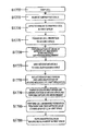
- FIG. 10 is a flow chart showing a method for analyzing a cell using the first space according to an embodiment of the present technology.
- FIG. 11 is a flow chart showing a method for analyzing a cell using the first space according to an embodiment of the present technology.
- FIG. 10 is a flow chart showing a method for analyzing a cell using the first space according to an embodiment of the present technology.
- FIG. 12 is a diagram showing an overview of a chip for cell analysis using the first and second spaces according to an embodiment of the present technology.
- FIG. 13 is a flow chart showing a method for analyzing a cell using the first and second spaces according to an embodiment of the present technology.
- FIG. 14 is a flow chart showing a method for analyzing a cell using the first and second spaces according to an embodiment of the present technology.
- FIG. 15 is a flow chart showing a method for analyzing a cell using the first and second spaces according to an embodiment of the present technology.
- FIG. 16 is a flow chart showing a method for analyzing a cell using the first and second spaces according to an embodiment of the present technology.
- FIG. 17 is a flow chart showing a method for analyzing a cell using the first and second spaces according to an embodiment of the present technology.
- FIG. 18 is a flow chart showing a method for analyzing a cell using the first and second spaces according to an embodiment of the present technology.
- FIG. 19 is a diagram showing an overview of a chip for cell analysis using the first to third spaces according to an embodiment of the present technology.
- FIG. 20 is a flow chart showing a method for analyzing a cell using the first to third spaces according to an embodiment of the present technology.
- FIG. 21 is a diagram showing an overview of a chip for cell analysis using the first to third spaces according to an embodiment of the present technology.
- Step (A) (2) Step (B) (3) Step (C) (4) Step (D) (5) Step (E) (6) Step (F) (7) Step (G) (8) Step (H) (9) Integration of data obtained from the cell 2. Chip for cell analysis 3. Reagent for cell analysis 4. Kit for cell analysis 5. Apparatus for cell analysis 6.
- An embodiment of the present technology includes the following: (A) trapping a cell by means of a first molecule capable of binding to the cell, (B) binding, to a cell-surface molecule of the cell, a second molecule in which a binding portion capable of binding specifically to the cell-surface molecule, an identifying portion, and a labeling portion are linked, (C) detaching the binding portion, and the identifying portion and the labeling portion of the second molecule from each other, (D) binding, out of the identifying portion and the labeling portion detached, the identifying portion specifically to an identifying portion recognizing molecule, and (E) detecting the labeling portion out of the identifying portion and the labeling portion bound to the identifying portion recognizing molecule.
- identifying and/or quantifying the cell-surface molecule specifically bound to the second molecule from the detection result of the labeling portion obtained in the (E) may be included as (F).
- An embodiment of the present technology may further include the following: (G) identifying and/or quantifying a cell secretion from the cell, and/or (H) acquiring a nucleic acid from the cell and analyzing the nucleic acid.
- the order in which the steps mentioned above are performed is not limited to the order of from the (A) to the (H), and may be altered.
- the cell may be trapped after the second molecule is bound to a cell-surface molecule.
- the sequence may be performed in the order of from the (B) to the (A).
- the sequence may be performed in the order of from the (A) through the (G) to the (B).
- Step (A) a cell is trapped by the first molecule capable of binding to the cell.
- the type of the cell to be trapped is not particularly limited, and any cell, such as an animal cell, a plant cell, or fungi, may be the object.
- any cell such as an animal cell, a cell in the blood, a cell taken from a living tissue, a cultured cell, etc. are given. Either of an adherent cell and a non-adherent cell is possible.
- what is called single cell trapping in which one cell is trapped into one well is performed.
- a cell dispersion method such as trypsin treatment
- a plurality of cells of the same type may be trapped simultaneously.
- the first molecule that traps a cell is immobilized in the well (a first space).
- the well is not particularly limited; for example, a well in a flat-bottomed, V-shaped-bottomed, U-shaped-bottomed, plate-like, bead-like, membrane-like, or net-like configuration, etc. may be used.
- a configuration having a recess is preferable in view of the case of forming a flow path described later between wells.
- one cell may be trapped, or a plurality of cells of the same type may be trapped.
- the first molecule be immobilized in the well so as to have a diameter size equal to or less than the diameter size of the one cell to be trapped.
- Cells can be trapped one by one regardless of the size of the cell.
- the size of the well and the width of the flow path described later may be adjusted so that cells flow one by one like in flow cytometry and are trapped into wells one by one.
- Cells can be passed by, for example, a buffer, a cleaning liquid, or the like without using a sheath liquid used in flow cytometry.
- a link is made between wells by a flow path, for example.
- a cell suspension may be passed from the side of the well at one end, and one cell may be trapped into one well and the untrapped cells may be passed from the well to the next well.
- isolation may be made by a valve provided at the flow path or the like.
- one cell may be introduced into the recess of the well by a cell sorter.
- the structure of the first molecule includes a portion capable of binding to a cell.
- a portion capable of binding to a cell for example, an oleyl group, an antibody, an aptamer, or a molecular recognition polymer may be used.
- the oleyl group is hydrophobic, and adheres to a cell surface floating.
- the oleyl group may be provided with, for example, a spacer such as PEG, and an NHS group (N-hydroxysuccinimide group) may be contained in a terminal of the spacer; thus, the first molecule may be formed.
- the first molecule may be immobilized in a well coated with, for example, a collagen or the like.
- an antibody corresponding to, as the antigen, a cell-surface molecule present on the cell desired to be trapped may be used.
- the antibody may be immobilized in the well in a covalent bonding-like manner.
- a nucleic acid molecule or a peptide that binds specifically to a surface molecule present on the cell desired to be trapped may be used.
- the cell-surface molecule of the objective can be trapped with high selectivity even in the presence of a compound having physical and chemical properties similar to those of the cell-surface molecule of the objective.
- the first molecule may have a structure in which a stimulus-degradable linker is interposed between the portion capable of binding to the cell and the well at which the first molecule is immobilized.
- a linker is interposed, when it is desired to recover the cell after various measurements of a cell-surface molecule, a cell secretion, etc., the cell can be recovered with good efficiency. After the recovery, the cell remains alive, and can therefore be cultured or destroyed to perform the measurement, analysis, etc. of an intracellular molecule.
- the stimulus-degradable linker is a linking molecular unit that is degraded by a specific stimulus from the outside.
- a linker that is degraded by light of a specific wavelength a linker that is degraded by an enzyme, a linker that is degraded by temperature, and the like are given.
- the stimulus-degradable linker used in an embodiment of the present technology is not particularly limited, but it is preferable to use a photodegradable linker in terms of preventing the cell from being destroyed or influenced.
- the photodegradable linker is a molecular unit having a structure that is degraded by a specific wavelength, and the wavelength at which the photodegradable linker is degraded almost coincides with the absorption wavelength of the molecular unit.
- a methoxynitrobenzyl group used as the photodegradable linker an absorption pattern like that shown by the solid line of FIG. 1 (before irradiation) is exhibited. Assuming that the absorption at 346 nm is 1, an absorption of 0.89 is exhibited at 364 nm, 0.15 is at 406 nm, and 0.007 is at 487 nm.
- the methoxynitrobenzyl group has the characteristics that, when a light source of 365 nm is used, the efficiency of degradation of the photodegradable linker is good; and when a light source of 488 nm is used, the photodegradable linker is hardly degraded.
- photodegradable linkers for example, the following may be given: a nitrobenzyl group (JP 2010-260831A), a parahydroxyphenacyl group (Tetrahedron Letters, 1962, vol. 1, p. 1), a 7-nitroindoline group (Journal of the American Chemical Society, 1976, vol. 98, p. 843), a 2-(2-nitrophenyl)ethyl group (Tetrahedron Letters, 1997, vol. 53, p. 4247), a (coumarin-4-yl)methyl group (Journal of the American Chemical Society, 1984, vol. 106, p. 6860), etc.
- a nitrobenzyl group JP 2010-260831A
- parahydroxyphenacyl group Tetrahedron Letters, 1962, vol. 1, p. 1
- a 7-nitroindoline group Journal of the American Chemical Society, 1976, vol. 98, p. 843
- the wavelength of the light applied to the photodegradable linker may be a wavelength corresponding to each photodegradable linker. For example, a wavelength near 330 to 450 nm is given. It is preferable to perform irradiation at, for example, 30 mW/cm ⁇ 2 multiplied by 100 sec., i.e. 3 J/cm ⁇ 2, which does not damage the cell. In particular, wavelengths of 300 nm or less may damage the cell, and are therefore preferably not be used. With regard to the cytotoxicity caused by UV, depending on the type of the cell, it is said that the DNA is damaged and the cell growth is inhibited at 500 J/cm ⁇ 2 (Callegari, A. J. & Kelly, T. J.
- Step (B) In the (B), the second molecule in which the binding portion capable of binding specifically to a cell-surface molecule, the identifying portion, and the labeling portion are linked is bound to the cell-surface molecule of the cell.
- the cell-surface molecule is a sugar strand, a protein, a lipid, or the like.
- specific cell-surface molecules for example, a CD antigen, various families of cell-adherent molecules, a cell-surface receptor, etc. are given.
- the second molecule includes the binding portion capable of binding specifically to a cell-surface molecule, the identifying portion, and the labeling portion.
- the binding portion capable of binding specifically to a cell-surface molecule for example, the whole of an antibody, a variable region portion of an antibody, an aptamer, or a molecular recognition polymer may be used.
- the binding reaction between the cell-surface molecule and the second molecule, the binding reaction to the first molecule of the (A), etc. are not particularly limited; the reaction may be performed for a certain period of time while the whole well is cooled at, for example, 4°C or is warmed at approximately 37°C, which is near the body temperature of animals.
- the identifying portion is linked to the binding portion.
- various proteins and peptides, a nucleic acid (a DNA strand or an RNA strand) fragment, etc. may be used.
- a nucleic acid fragment is preferably used.
- the size of the nucleic acid fragment is not particularly limited; for example, a DNA fragment composed of 3 to 100 nucleic acid nucleotide units may be used.
- a DNA fragment is preferable because various sequences can be created in accordance with the combination of A, T, G, and C of the nucleic acid.
- the antibody that binds to a cell-surface molecule is said to have approximately 350 types, and DNA sequences corresponding to all of the antibodies can be created when the label is created by a DNA sequence of an oligo-level length (approximately 20 bases or less).
- the labeling portion is linked to the identifying portion linked to the binding portion.
- the label used for the labeling portion may be a known one; for example, a fluorescent molecule, an RI label, a hapten, an affinity tag, an enzyme, a metal, etc. are given.
- a fluorescent molecule is used for the labeling portion
- the number of colors may be one or may be two or more.
- the detection can be made by TOF-MS.
- the labeling is not limited to fluorescent labeling etc., and a method in which optical detection can be made by making luminescence, color development, or the like on the spot, without diffusion, may be used.
- electrochemiluminescence is possible when a ruthenium complex is used as the label and an electrode is provided on a substrate surface.
- HRP horseradish peroxidase
- a luciferase to the identifying portion, add a chemiluminescent substrate (luminol, lucigenin, an adamantyl dioxetane derivative, etc.) or a bioluminescent substrate (a luciferin, a bacterial luciferase, coelenterazine, etc.), and detect the luminescence intensity.
- chemiluminescent substrate luminol, lucigenin, an adamantyl dioxetane derivative, etc.
- a bioluminescent substrate a luciferin, a bacterial luciferase, coelenterazine, etc.
- the signal can be amplified and thus high-sensitivity detection can be made. Further, the detection may be performed using an intercalator such as SYBR Green I.
- a stimulus-degradable linker may be interposed between the binding portion, and the identifying portion and the labeling portion; as the stimulus-degradable linker, the photodegradable linker described above may be used, for example. Further, the fact that the wavelength suitable for photodegradation varies depending on the type of the photodegradable linker may be utilized. For example, linkers that are degraded by different wavelengths are used for second molecules that bind to different cell-surface molecules. Then, light of a wavelength suitable for any of the photodegradable linkers may be applied; thus, a portion including the identifying portion and the labeling portion of the desired type based on the linker can be isolated from the second molecule bound to the target cell-surface molecule.
- FIG. 2 a schematic diagram in which a single cell 22 is trapped by a first molecule 21 immobilized in a well 26 of a first space and a second molecule 24 is bound to a cell-surface molecule 23 of the trapped single cell is shown.
- a stimulus-degradable linker 25 is included in the first molecule 21 and the second molecule 24 and a metal 27 or a nucleic acid fragment 28 is used as the identifying portion and/or the labeling portion of the second molecule.
- a typical fluorescently labeled substance 29 in related art is used is shown.
- Step (C) In the (C), the binding portion, and the identifying portion and the labeling portion of the second molecule are detached from each other.
- the detachment is performed by stimulating the stimulus-degradable linker.
- the well of the trapped cell may be irradiated such that the photodegradable linker of the second molecule is included in the area of light irradiation.
- the second molecule degraded by the light irradiation is separated into the binding portion, and the identifying portion and the labeling portion.
- the binding portion remains bound to the cell-surface molecule of the trapped cell.
- the identifying portion and the labeling portion detached and isolated may be passed on a solution basis or may be passed by a buffer liquid or the like, and can be transferred to the next well (a second space etc.).
- the identifying portion recognizing molecule includes a recognizing molecular unit corresponding to the identifying portion used for the second molecule.
- an antibody is used for the identifying portion recognizing molecule.
- the identifying portion of the second molecule is a nucleic acid fragment
- a fragment having a nucleic acid sequence complementary to the nucleic acid fragment is used.
- the identifying portion recognizing molecule may be immobilized in the well in which the first molecule is immobilized (the first space), or may be immobilized in another well (the second space etc.).
- a plurality of types of identifying portion recognizing molecules may be immobilized in one well.
- the identifying portion recognizing molecule may be immobilized in an array configuration on a type basis. For example, when fragments having nucleic acid sequences are used as the identifying portion recognizing molecule, a configuration like a DNA chip can be created.
- the immobilization may be made while one well is divided into, for example, a region where a molecule for cell-surface molecule measurement is immobilized (a first region) and a region where a molecule for intracellular molecule measurement is immobilized (a second region).
- a region where a molecule for cell-surface molecule measurement is immobilized a region where a molecule for intracellular molecule measurement is immobilized
- a second region a region where a molecule for intracellular molecule measurement is immobilized
- spots 34 of identifying portion recognizing molecules for cell-surface molecule measurement 32 are arranged in 3 longitudinal rows and 6 transverse rows.
- spots 34 of identifying portion recognizing molecules for intracellular molecule measurement 33 are arranged in 3 longitudinal rows and 6 transverse rows.
- 350 spots are prepared and different molecules are immobilized individually to them.
- the spot size may be not less than the surface area of the cell.
- the configuration of the well is not particularly limited, and may be selected in accordance with the molecule to be immobilized etc.
- the molecule may be immobilized to a flat surface of the well (the upper portion of FIG. 4), may be immobilized to a convex portion of the well (the middle portion of FIG. 4), or may be immobilized to a concave portion of the well (the lower portion of FIG. 4).
- the identifying portion binds specifically to the identifying portion recognizing molecule for cell-surface molecule measurement thus immobilized in the well.
- the identifying portion is a nucleic acid fragment
- the identifying portion binds to a spot to which an identifying portion recognizing molecule (a DNA strand or the like) having a sequence complementary to the nucleic acid fragment is immobilized.
- the identifying portion is a protein or a peptide
- the identifying portion binds to a spot to which an identifying portion recognizing molecule (an antibody) that recognizes the protein or the peptide as the antigen is immobilized.
- Step (E) In the (E), out of the identifying portion and the labeling portion bound specifically to the identifying portion recognizing molecule in the (D), the labeling portion is detected.
- the labeling portion includes, as described above, a labeling molecular unit of a fluorescent molecule, an RI label, a hapten, an affinity tag, an enzyme, a metal, etc.
- the labeling portion is detected by a detection method corresponding to the labeling molecular unit being used. For example, in the case where a fluorescent molecule is used as the labeling portion, light of the excitation wavelength may be applied to the fluorescent molecule.
- the labeling portion is an RI label, a hapten, an affinity tag, or an enzyme, or a metal
- the identification and/or quantification may be performed using radiation, an antibody for detection, or TOF-MS etc., respectively.
- the combination of the molecules used for the identifying portion and the labeling portion is not particularly limited in an embodiment of the present technology.
- a protein, a peptide, a nucleic acid fragment, etc. as the identifying portion and a fluorescent molecule, an RI label, a hapten, an affinity tag, an enzyme, a metal, etc. as the labeling portion may be combined freely.
- a nucleic acid fragment as the identifying portion and a fluorescent molecule as the labeling portion may be combined, for example.
- Step (F) the cell-surface molecule bound specifically to the second molecule is identified and/or quantified from the detection result of the labeling portion obtained in the (E).
- a nucleic acid fragment is used for the identifying portion
- nucleic acid fragments complementary to more than 350 types of nucleic acid fragments are immobilized individually to different spots of the well on a type basis, the type of the nucleic acid fragment and the position of the spot are correlated, and this is grasped as position information.
- the type and expression level of the cell-surface molecule are found by what spot emits what degree of fluorescence.
- the shape of the well is a flat surface
- a shape that can suppress the diffusion may be employed, such as a shape in which the spot portion is recessed.
- the identifying portion and the labeling portion that are isolated from the second molecule and are not bound to the identifying portion recognizing molecule be removed to reduce the background noise and thus high-sensitivity measurement be performed.
- Step (G) a cell secretion from the cell is identified and/or quantified.
- the trapped cell is alive even after undergoing the (A) to (F).
- a cell secretion can be secreted in the well while in the trapped state.
- the trapped cell may be released and put into another container to cause a cell secretion to be secreted.
- the cell secretion is not particularly limited in an embodiment of the present technology, and any cell secretion may be the object.
- various hormones such as insulin and secretin
- various enzymes such as pepsinogen, a cytokine, a chemokine, perforin, a granzyme, an exosome, etc. are given.
- the cell may be stimulated.
- a physical stimulus, a chemical stimulus, or the like may be applied to the cell.
- a cell secretion-stimulating substance may be applied to the cell.
- the cell secretion-stimulating substance varies with the type of the cell, the type of the secretion, etc.; for example, a sugar, an amino acid, a fat, an acid, and an alkali (a pH adjuster) are given.
- the identification and/or quantification of the cell secretion can be performed by a commonly used method.
- a label may be attached to an antibody corresponding to a specific cell secretion as the antigen, and after an antigen-antibody reaction, the detection and/or intensity measurement of the label may be performed.
- an antibody, an aptamer, a molecular recognition polymer, or the like that binds specifically to a cell secretion is immobilized in a specific place.
- a labeled molecule an antibody, an aptamer, a molecular recognition polymer, or the like
- a sandwich immunoassay etc. can be performed.
- the electrochemiluminescence, chemiluminescence, bioluminescence, etc. described above may be used.
- the first molecule that traps the cell is preferably structured to be immobilized via a stimulus-degradable linker, as described above. At the time of releasing the cell, the cell can be easily recovered by applying a stimulus to the stimulus-degradable linker.
- Step (H) a nucleic acid is acquired from the cell and is analyzed. Since the cell is still alive even after undergoing the (A) to (G), the cell may be destroyed and thus a nucleic acid can be acquired and analyzed.
- the acquisition and analysis of a nucleic acid can be performed by a commonly used method. For example, they can be performed by a method in which a nucleic acid is extracted, purified, and amplified (the PCR method etc.) and the nucleic acid sequence is determined. It is also possible to, after the extraction, perform up to preliminary amplification, and recover the amplified substance to determine the nucleic acid sequence with a next-generation sequencer or the like.
- data of the identification and/or quantification of a secretion from the cell are obtained.
- the data obtained in the (F) and the data obtained in the (G) are integrated, the relationship between the cell-surface molecule and the cell secretion of the living cell can be analyzed.
- data of a nucleic acid of the cell can be acquired.
- the data obtained in the (F) and the data obtained in the (H) are integrated, the relationship between the cell-surface molecule and the gene expressed in the living cell can be analyzed.
- the data obtained in the (F), the (G), and the (H) may be integrated. Thereby, the relationships between the cell-surface molecule, the cell secretion, and the gene in the living cell can be made clear.
- cell-surface molecules thereof (antigens etc.) can be measured comprehensively, and also molecules that the cell has secreted due to a stimulus or the like can be measured comprehensively, by the above steps.
- the cell surface information and the cell secretion information can be integrated reliably at a level of what is called a single cell.
- Chip for cell analysis By a chip for cell analysis of an embodiment of the present technology, all the steps from the trapping of a cell from a cell suspension up to various measurements can be performed in the chip.
- a first region, a second region, and a detection region are included.
- the first molecule capable of binding to a cell is immobilized in the first region.
- a molecule capable of binding specifically to the second molecule in which the binding portion capable of binding specifically to the cell, the identifying portion, and the labeling portion are linked is immobilized in the second region.
- a molecule that binds specifically to the identifying portion, for example, is immobilized in the detection region so that the labeling portion can be detected.
- the first region, the second region, and the detection region may be present in one well or may be present individually in different wells, or a plurality of regions may be present in one well and other regions may be present in other wells.
- a link may be made between wells by a flow path, and the flow path may be provided with a valve.
- the chip for cell analysis of an embodiment of the present technology includes a plurality of wells belonging to the first space, and the wells of the first space are linked by a flow path.
- the wells are arranged in one row, they are linked in one row by a flow path.
- a sample such as a cell suspension, a reagent, a cleaning liquid, etc.
- they may be put into the well at one end and passed toward the well at the other end because the wells are linked by the flow path. The passing may be performed at a low rate of several tens of centimeters per second or less and low pressure, for example.
- the flow path is provided with a valve.
- the valve is in a openable and closable structure; for example, it can be closed when the sample and the reagent are reacted in each well, and can be opened when the sample and the reagent are passed to another well.
- the first molecule is immobilized so that one cell is trapped into one well.
- the first molecule may be immobilized in the well so as to have a diameter size equal to or less than the diameter size of one cell to be trapped.
- the first molecule is preferably immobilized in the well via a stimulus-degradable linker.
- the chip for cell analysis may further include a well belonging to another space.
- the chip for cell analysis may include a well belonging to the second space, the third space, etc. on the same plate as the first space.
- a cell-surface molecule, an intracellular molecule, a cell secretion, etc. can be measured on the same plate.
- the wells belonging to the second space may be arranged in one row and linked in one row by a flow path, for example. This similarly applies to the third space etc.
- the wells belonging to the first space and the wells belonging to the second space are arranged parallel in two rows, the wells belonging to the first space and the wells belonging to the second space arranged adjacent thereto may be linked by a flow path.
- the flow path may be provided with a valve.
- the identifying portion recognizing molecule described above may be immobilized.
- a cell is trapped by the first molecule immobilized in the first space.
- the second molecule including the binding portion capable of binding specifically to a cell-surface molecule of the cell is reacted.
- the valve of the flow path between the well of the first space and the well of the second space is closed.
- a stimulus is applied to the second molecule bound to the cell-surface molecule of the cell trapped in the well of the first space to degrade the stimulus-degradable linker, and thus the identifying portion and the labeling portion of the second molecule are isolated.
- the valve of the flow path between the well of the first space and the well of the second space is opened.
- the liquid existing in the well of the first space and containing the identifying portion and the labeling portion flows into the well of the second space.
- the valve is closed.
- the identifying portion out of the identifying portion and the labeling portion isolated from the second molecule and the identifying portion recognizing molecule immobilized in the well of the second space are reacted together.
- the wells belonging to the third space are further arranged in addition to those belonging to the second space
- the wells belonging to the first space may be arranged in one longitudinal row on the center
- the wells belonging to the second space may be arranged in one longitudinal row on the right side of the wells belonging to the first space
- the wells belonging to the third space may be arranged in one longitudinal row on the left side of the wells belonging to the first space.
- a link may be made also between wells belonging to the third space by a flow path, and a valve may be provided.
- the wells belonging to the third space and the wells belonging to the first space immediately on the right side thereof may be linked by a flow path, and a valve may be provided.
- FIG. 5A shows a situation in which cells are allowed to flow into the well belonging to the first space ((1) of FIG. 5A), a single cell is trapped, and the second molecule is bound to a cell-surface molecule of the single cell.
- the valve of the flow path making a link from the well belonging to the first space to the well belonging to the second space is closed.
- the valve of the flow path making a link from the well belonging to the first space to the well belonging to the third space is closed.
- the stimulus-degradable linker of the second molecule in the well belonging to the first space on the center, the stimulus-degradable linker of the second molecule is degraded, and thus the identifying portion and the labeling portion are isolated.
- the valve of the flow path making a link from the well belonging to the first space to the well belonging to the second space on the right side thereof is opened, and the identifying portion and the labeling portion isolated flow into the well belonging to the second space ((2) of FIG. 5B).
- the identifying portion recognizing molecule is immobilized, and the identifying portion that binds specifically to the identifying portion recognizing molecule binds to it; then, measurement is performed.
- the single cell trapped in the well belonging to the first space on the center is dissolved, and the extraction etc. of an intracellular molecule (e.g. a nucleic acid) are performed.
- the valve of the flow path to the well belonging to the third space on the left side is opened, and the nucleic acid flows into the well belonging to the third space ((3) of FIG. 5C).
- an intracellular molecule recognizing molecule is immobilized, and binds specifically to the nucleic acid; then, measurement is performed.
- in-situ hybridization, in-situ PCR, in-situ sequencing, etc. may be performed. After cell membrane permeation treatment, the intracellular molecule may be stained and observed.
- a stimulus may be applied to the first molecule to degrade the stimulus-degradable linker to release the cell.
- the released cell may pass through the flow path and go into the well belonging to the third space, and another analysis, culture, etc. can be performed in the well.
- the released cell may be transferred to a fourth space, another container, etc.
- any molecule may be immobilized other than the first molecule and the second molecule that is the identifying portion recognizing molecule described above.
- a molecule that recognizes a cell secretion, an antibody that recognizes an intracellular molecule of the cell, a molecule that has a sequence complementary to the sequence of a nucleic acid of the cell, etc. are given.
- it is desired to measure the information of a cell-surface molecule and an intracellular molecule as shown in FIG.
- a nucleic acid fragment having a sequence complementary to the nucleic acid fragment bound to the cell-surface molecule may be immobilized in the well belonging to the second space, and a molecule that binds specifically to the intracellular molecule or a molecule that binds complementarily to the intracellular molecule may be immobilized in the well belonging to the third space.
- the wells of the first, second, third, and other spaces may be divided into a plurality of regions, and a different molecule may be immobilized in each region.
- one well of the first space is divided into the first region and the second region; and the first molecule capable of binding to a surface molecule is immobilized in the first region, and the second molecule in which the binding portion capable of binding specifically to a cell-surface molecule, the identifying portion, and the labeling portion are linked and/or a molecule capable of binding specifically to a cell secretion from the cell is immobilized in the second region.
- a nucleic acid fragment complementary to the identifying portion included in the second molecule may be immobilized in the first region, and a nucleic acid fragment that binds specifically to an intracellular molecule may be immobilized in the second region; and in the entire well belonging to the third space, a molecule that binds specifically to a cell secretion may be immobilized.
- the nucleic acid fragment complementary to the identifying portion included in the second molecule is isolated by applying a stimulus to the stimulus-degradable linker, and is sent to the well belonging to the second space.
- a cell secretion secreted by the stimulation or the like of the cell trapped in the well belonging to the first space is sent to the well belonging to the third space.
- the cell is dissolved in the well belonging to the first space, and an intracellular molecule is sent to the well belonging to the second space.
- the well belonging to the second space is cleaned by opening the valve, and next the well belonging to the third space is cleaned by opening the valve.
- the position and amount of luminescence are measured by adding a reactive substrate or the like.
- preliminary amplification if necessary, is performed in the well belonging to the first space. After that, the amplified substance may be transferred to the well belonging to the second space.
- the stimulus-degradable linker of the first molecule trapping the cell may be degraded to recover the cell.
- a photodegradable linker may be used as the stimulus-degradable linker as described above.
- a linker that degrades at a wavelength different from the wavelength at which the photodegradable linker used for the second molecule that binds to a cell-surface molecule degrades may be used.
- the photodegradable linker used for the second molecule is degraded at 365 nm
- the photodegradable linker used for the first molecule is degraded at 700 nm.
- a situation in which light of a specific wavelength can be applied from below the well belonging to the first space is created.
- a light source may be placed below each well.
- the configuration is not limited thereto, and scanning may be used.
- shutters capable of opening and closing in a minute range may be arranged in the area.
- a digital micro-device may be used to perform irradiation on only a specific area.
- FIG. 6 a schematic diagram in which light is applied from below the well belonging to the first space to degrade the photodegradable linker to release the cell is shown.
- FIG. 6A shows a situation in which first molecules are immobilized in the well belonging to the first space. A photodegradable linker is included in the first molecule.
- FIG. 6B shows a situation in which cells are allowed to flow into the well belonging to the first space, cells are trapped by the first molecules, and second molecules are bound specifically to cell-surface molecules.
- FIG. 6C shows a situation in which, for the portion irradiated with light 61 from below the well belonging to the first space, the photodegradable linker is degraded, and thus the trapped cells are released.
- the light irradiation may be performed with, for example, 1 J/cm 2 (in the case of 100 mW/cm 2 , 10 s).
- the timing of light irradiation is varied, the photodegradable linkers of the first molecule and the second molecule can be degraded with a time difference, and therefore also the release can be made with a time difference.
- FIG. 7 an image diagram in which cells are trapped by immobilized first molecules is shown.
- cells can be trapped by a single layer with high density.
- the detection and observation of a cell-surface molecule, an extracellular secreted substance, and an intracellular molecule may be performed while a plurality of cells of the same type are trapped in one well.
- cultured cells from the same cell line, a cell population of which the cells are regarded as identical by flow cytometry (a cell group displayed in the same gate region by flow cytometry), etc. are given.
- CMOS or the like capable of photographing a certain area collectively may be placed below the well, and thereby the wells belonging to the second and third spaces can be scanned and photographed.
- the exposure time may be extended.
- a plurality of molecules of the detection object can be detected collectively, and also imaging analysis becomes possible.
- the apparatus for example, the sCMOS of IN Cell Analyzer 6000 (GE Healthcare) is given.
- one well approximately 0.33 cm 2
- the entire 96-well plate (approximately 31.68 cm 2 ) can be photographed in approximately 4 minutes.
- a reagent of an embodiment of the present technology is a reagent used for the method for analyzing a cell described above, and contains a molecule including a binding portion capable of binding specifically to a molecule selected from the group consisting of a cell-surface molecule, an intracellular molecule, and a cell secretion and a labeling portion.
- a reagent containing the second molecule a reagent containing a molecule that binds specifically to a cell secretion and is labeled
- a reagent containing a molecule that binds specifically to an intracellular molecule and is labeled are given.
- the molecule contained in the reagent one type of molecule or a plurality of types of molecules may be contained in one reagent.
- Kit for cell analysis A kit for cell analysis of an embodiment of the present technology may include the chip for cell analysis mentioned above and at least one reagent or any combination of two or more reagents selected from the group consisting of the reagent for cell analysis mentioned above, a reagent that detects the labeling portion, and a reagent containing a substance that stimulates cell secretion.
- the apparatus includes an insertion unit 84, a fluid control unit 81, a light irradiation unit 83, a detection unit 85, a memory unit 86, and a processing unit 82.
- the insertion unit 84 has the function of inserting the chip for cell analysis mentioned above into the apparatus for cell analysis and setting the chip for cell analysis.
- the fluid control unit 81 has the function of introducing a liquid containing cells, a liquid containing a cell secretion, a reagent, a cleaning liquid, etc. into a well, the function of passing these liquids to another well via a flow path, the function of discharging unnecessary liquids, etc.
- the light irradiation unit 83 includes a light source that irradiates the well with light of a wavelength suitable to degrade the photodegradable linker that is included in the first molecule immobilized in the well of the chip for cell analysis described above or in the second molecule.
- the detection unit 85 has the function of detecting and/or quantifying the cell introduced and trapped in the chip for cell analysis mentioned above, a cell secretion, a cell-surface molecule, etc. by the measurement of the label or the like.
- the operation of the insertion unit 84, the fluid control unit 81, the light irradiation unit 83, and the detection unit 85 may be stored, and in addition, data obtained in the detection unit etc. may be stored.
- the processing unit 82 performs sequence processing mutually with each unit on the basis of the memory of the memory unit 86, and executes the operation of the insertion unit 84, the fluid control unit 81, the light irradiation unit 83, and the detection unit 85.
- the chip for cell analysis mentioned above is installed into the apparatus used in an embodiment of the present technology by the insertion unit 84.
- the fluid control unit 81 is put into operation by the processing unit 82, and a liquid containing cells is introduced into the well at an end of the first space.
- the fluid control unit 81 opens the valve provided at the flow path between wells to pass the liquid containing cells.
- One cell is trapped by the first molecule immobilized in each well, and the untrapped cells are removed by passing a cleaning liquid or the like.
- the fluid control unit 81 is put into operation so that a liquid containing second molecules that bind specifically to a cell-surface molecule is introduced into the well at the end of the first space, and the liquid containing second molecules is passed to the well on the downstream side via the flow path.
- a second molecule binds to the cell trapped by the first molecule, and the unbound second molecules are removed by passing a cleaning liquid or the like by the fluid control unit 81.
- the second molecule bound specifically to a cell-surface molecule is irradiated with light from the light irradiation unit 83 to cut the photodegradable linker of the second molecule, and thus the identifying portion and the labeling portion of the second molecule are isolated.
- the position, timing, wavelength, intensity, time, etc. of the light irradiation are stored as a program in the memory unit 86, and are implemented by driving the light irradiation unit 83 via the processing unit 82.
- the fluid control unit 81 is put into operation to open the valve provided between the well of the first space and the well of the second space, and the liquid containing the identifying portion and the labeling portion isolated is passed from the well of the first space to the well of the second space.
- a molecule that binds specifically to the identifying portion is immobilized, and the identifying portion out of the identifying portion and the labeling portion passed binds to the immobilized molecule.
- the fluid control unit 81 is put into operation to introduce a cleaning liquid or the like into the well at an end of the second space, and the identifying portion and the labeling portion that have not been bound in the well of the second space are removed via the flow path.
- the labeling portion out of the identifying portion and the labeling portion bound in the well of the second space is detected and/or quantified by the detection unit 85.
- the data obtained here may be stored in the memory unit 86.
- the detection method a known method such as detection by applying light of a wavelength suitable for the label may be used when the label is a fluorescent label. Also the light irradiation at this time may be performed by the light irradiation unit 83.
- a cell secretion may be further detected and/or quantified.
- the data of the cell secretion may be stored in the memory unit 86.
- the first molecule may be irradiated with light from the light irradiation unit 83 to release the cell.
- the released cell may be used for the measurement of an intracellular molecule, or may be used for culture, for example.
- Embodiments Embodiments of the present technology will now be specifically described with reference to the drawings.
- (1) Embodiment 1 A case of using only the first space will now be described. In this case, for example, the measurement of a cell secretion, the measurement of a cell-surface molecule, the preliminary amplification of an intracellular mRNA, or cell recovery can be performed, and the conditions of the cell can be found.
- FIG. 9 An overview of a chip for cell analysis is shown in FIG. 9.
- four wells W1 to W4 are arranged longitudinally.
- an antibody that binds to a specific cell-surface molecule is immobilized via a photodegradable linker (the immobilization of the first molecule).
- an antibody that binds to another cell-surface molecule and an antibody that binds to a cell secretion are immobilized in each well.
- the wells are linked by a flow path 16, and valves 3 to 5 are provided individually between adjacent ones of the wells.
- the well W1 may be used as an inlet of cells, cleaning liquids, etc., and the trapped cell may be used as a control for measurement etc.
- FIG. 10 a flow of Embodiment 1 after the chip for cell analysis is inserted into the apparatus for cell analysis is shown.
- a description will now be given with reference to FIG. 9, FIG. 10, and FIG. 8.
- the second molecule in which the binding portion capable of binding specifically to a cell-surface molecule, the identifying portion, and the labeling portion are linked is bound to the cell-surface molecule of the cell, and thus the cell-surface is labeled (S971).
- a photodegradable linker is included in the second molecule.
- the fluid control unit 81 is put into operation by the processing unit 82, and a liquid containing cells labeled by the second molecule is introduced into the well W1.
- the valves 3 to 4 are opened so that the labeled cells flow in the direction from the arrow of FIG. 9 to the wells W2 to W4 sequentially.
- one cell is trapped by the first molecule that is capable of binding to the cell and is immobilized in the well (S972).
- the untrapped cells are washed to the downstream side of W4 by the fluid control unit 81 and are removed (S973).
- a cleaning liquid or the like may be used in order to wash the cells away.
- the valves 3 to 5 are closed by the fluid control unit 81, and by the light irradiation unit 83, the cell-surface label is isolated, that is, the binding portion, and the identifying portion and the labeling portion of the second molecule are detached and isolated from each other (S982).
- the identifying portion and the labeling portion isolated are trapped at the bottom surface of each of the wells W1 to W4, and the detection and measurement of the labeling portion are performed by the detection unit 85 (S983).
- the measurement results may be stored as data in the memory unit 86.
- a cell secretion secreted to the outside of the cell is trapped by the antibody or the like immobilized at the bottom surface of each well, and is identified and/or quantified by the detection unit 85 (S984). Thereby, what kind of cell secretion has been secreted from one cell to what degree can be made clear.
- the measurement results may be stored as data in the memory unit 86.
- a nucleic acid is acquired from the cell and is analyzed.
- a DNA or an mRNA of an intracellular molecule is preliminarily amplified, with the valves 3 to 5 kept closed, preliminary amplification by cell dissolution, the multi-displacement amplification method, etc. is performed (S980), and the solution is recovered.
- the recovered solution may be used for the analysis of the DNA or the mRNA, etc.
- Embodiment 2 A case of using the first and second spaces will now be described.
- the measurement of a cell secretion, the measurement of a cell-surface molecule, the preliminary amplification of an intracellular mRNA, or cell recovery can be performed.
- a cell secretion secreted to the outside of the cell and a cell-surface molecule label can be measured.
- environment control by cell secretion stimulation or the like may be performed, and also the change in conditions of the cell may be measured.
- FIG. 12 An overview of a chip for cell analysis is shown in FIG. 12.
- Four wells W1 to W4 are arranged longitudinally in a first space 11.
- An antibody that binds to a cell-surface molecule is immobilized in the wells W1 to W4 via a photodegradable linker.
- a second space 12 on the right side of the first space 11 four wells W5 to W8 are arranged longitudinally.
- An antibody that binds to a cell secretion and the identifying portion recognizing molecule are immobilized in the wells W5 to W8.
- the wells W1 to W4 and the wells W5 to W8 are linked individually by longitudinal flow paths.
- the wells of the first space and the second space are linked transversely by the flow path 16 except for the wells W1 and W5 at the upper end.
- the flow path is provided with valves 1, 3, 4, and 5.
- the wells W1 and W5 can be linked, by not linking the wells W1 and W5, the well W1 can be used as an inlet of cells, cleaning liquids, etc., or the cell trapped in the well W1 can be used as a control for measurement etc.
- FIG. 13 a flow of Embodiment 2 after the chip for cell analysis is inserted into the apparatus for cell analysis is shown.
- the fluid control unit 81 is put into operation by the processing unit 82 to close the valve 1 and open the valves 3 to 5, and a liquid containing cells is introduced into the well W1 at the upper end of the first space 11 and is passed from the well W1 to the well W4.
- a liquid containing cells is introduced into the well W1 at the upper end of the first space 11 and is passed from the well W1 to the well W4.
- one cell is trapped into each well by the first molecule that is capable of binding to the cell and is immobilized (S1272).
- S1273 The unbound cells are washed to the downstream side of the well W4 and are removed.
- a cleaning liquid or the like may be used.
- the environment of the trapped cell is controlled (S1274).
- a cell secretion-stimulating substance that stimulates cell secretion is introduced by the fluid control unit 81.
- the cell secretion can be made active by the stimulating substance.
- the valves 3 to 5 may be opened to introduce the same stimulating substance, or the valves 3 to 5 may be closed to introduce a different stimulating substance to each well.
- the valve 1 Only the valve 1 is opened, and the liquid containing a cell secretion secreted to the outside of the cell is transferred to the well of the second space by the fluid control unit 81 (S1275).
- the cell secretion of the well W2 is transferred to the well W6, the cell secretion of the well W3 is to the well W7, and the cell secretion of the well W4 is to the well W8.
- the valve 1 is closed.
- the cell secretion is trapped by the antibody or the like immobilized at the bottom surface of the well of the second space, and is measured by the detection unit 85 (S1276). By the measurement, what kind of cell secretion has been secreted from one cell to what degree can be made clear.
- the measurement results may be stored as data in the memory unit 86.
- valve 1 is closed, and a label is attached by the second molecule or the like to a cell-surface molecule of the cell that remains trapped in the well of the first space 11 (S1277).
- a liquid containing second molecules can be passed in the direction from W1 to W4.
- all the valves may be closed, and the second molecule may be added to each of the wells W1 to W4.
- the wells W1 to W4 are irradiated with light from the light irradiation unit 83 to degrade the photodegradable linker of the second molecule, and thus the identifying portion and the labeling portion are detached and isolated (S1278).
- valve 1 Only the valve 1 is opened, and the liquid containing the identifying portion and the labeling portion isolated is transferred to the well of the second space (S1279). After that, the valve 1 is closed.
- the identifying portion recognizing molecule has been immobilized at the bottom surface of the well of the second space as described above; the identifying portion is bound specifically to the identifying portion recognizing molecule, and the labeling portion is detected and measured (S1279). By the measurement, what kind of cell-surface molecule is present on one cell to what degree can be identified and/or quantified.
- the measurement results may be stored as data in the memory unit 86.
- Embodiment 3 A case of using the first and second spaces different from Embodiment 2 will now be described.
- the measurement of a cell secretion the measurement of a cell-surface molecule, the preliminary amplification of an intracellular mRNA, or cell recovery can be performed.
- a cell secretion can be measured in the well of the first space, and the label of a cell-surface molecule can be measured in the well of the second space.
- environment control by cell secretion stimulation or the like may be performed, and also the change in conditions of the cell may be measured.
- FIG. 14 a flow of the steps of Embodiment 3 after a chip for cell analysis is inserted into the apparatus for cell analysis is shown.
- the arrangement of wells of each space and the structure of the flow path and the valve are similar to those of the chip used in Embodiment 2 (FIG. 12).
- the first molecule that binds to a cell-surface molecule and an antibody that binds to a cell secretion are immobilized in the wells W1 to W4, and the identifying portion recognizing molecule is immobilized in the wells W5 to W8.
- the fluid control unit 81 is put into operation by the processing unit 82 to close the valve 1 and open the valves 3 to 5, and a liquid containing cells is introduced into the well W1 of the first space 11 and is passed in the direction from the well W1 to the well W4.
- a liquid containing cells is introduced into the well W1 of the first space 11 and is passed in the direction from the well W1 to the well W4.
- One cell is trapped into each well by the first molecule (S1372).
- the unbound cells are removed by passing a cleaning liquid or the like (S1373).
- the environment of the trapped cell is controlled (S1374).
- a cell secretion-stimulating substance that stimulates cell secretion is introduced by the fluid control unit 81.
- the cell secretion can be made active by the stimulating substance.
- the valves 3 to 5 may be opened to introduce the same stimulating substance, or the valves 3 to 5 may be closed to introduce a different stimulating substance to each well.
- a cell secretion secreted to the outside of the cell is trapped by the antibody or the like immobilized at the bottom surface of the well of the first space, and is measured by the detection unit 85 (S1384). Thereby, what kind of cell secretion has been secreted from one cell to what degree can be identified and/or quantified.
- the measurement results may be stored as data in the memory unit 86.
- the second molecule is bound to a cell-surface molecule of the cell trapped in the well of the first space (S1377).
- a specific type of or various cell-surface molecules can be labeled.
- the fluid control unit 81 may be put into operation, and the valve 1 may be closed and the valves 3 to 5 may be opened to pass a liquid containing second molecules in the direction from the well W1 to the well W4, or all the valves may be closed to add a liquid containing second molecules to each of the wells W1 to W4.
- the wells W1 to W4 are irradiated with light by the light irradiation unit 83 to degrade the photodegradable linker of the second molecule, and thus the identifying portion and the labeling portion are detached and isolated (S1378). At this time, all the valves are closed.
- the valve 1 With the valves 3 to 5 kept closed, the valve 1 is opened, and the liquid containing the identifying portion and the labeling portion isolated is transferred from the wells W2 to W4 of the first space 11 to the wells W6 to W8 of the second space 12 (S1379). After that, the valve 1 is closed.
- the identifying portion recognizing molecule has been immobilized at the bottom surface of the well of the second space as described above; the identifying portion and the labeling portion isolated are trapped, and measurement is performed by the detection unit 85 (S1379). By the measurement, what kind of cell-surface molecule is present on one cell to what degree can be identified and/or quantified.
- the measurement results may be stored as data in the memory unit 86.
- a nucleic acid is acquired from the cell and is analyzed.
- a DNA or an mRNA of an intracellular molecule is preliminarily amplified, with all the valves kept closed, preliminary amplification by cell dissolution, the multi-displacement amplification method, etc. is performed (S1380), and the solution is recovered.
- the recovered solution may be used for the analysis of the DNA or the mRNA, etc.
- Embodiment 4 A case of using the first and second spaces different from Embodiments 2 and 3 will now be described.
- the measurement of a cell secretion, the measurement of a cell-surface molecule, the preliminary amplification of an intracellular mRNA, or cell recovery can be performed.
- a cell secretion and a cell-surface molecule label may be measured in the well of the second space, and a DNA or an mRNA of an intracellular molecule may be measured in the well of the first space.
- environment control by cell secretion stimulation or the like may be performed, and also the change in conditions of the cell may be measured.
- FIG. 16 a flow of the steps of Embodiment 4 after a chip for cell analysis is inserted into the apparatus for cell analysis is shown.
- the arrangement of wells of each space and the structure of the flow path and the valve are similar to those of the chip used in Embodiment 2 (FIG. 12).
- the first molecule that binds to a cell-surface molecule is immobilized in the wells W1 to W4, and an antibody that binds to a cell secretion and the identifying portion recognizing molecule are immobilized in the wells W5 to W8.
- the fluid control unit 81 is put into operation by the processing unit 82 to close the valve 1 and open the valves 3 to 5, and a liquid containing cells is introduced into the well W1 of the first space 11 and is passed in the direction in the well W4 direction.
- a liquid containing cells is introduced into the well W1 of the first space 11 and is passed in the direction in the well W4 direction.
- One cell is trapped into each well by the first molecule (S1572).
- the unbound cells are removed by passing a cleaning liquid or the like (S1573).
- the environment of the trapped cell is controlled (S1574).
- a cell secretion-stimulating substance that stimulates cell secretion is introduced by the fluid control unit 81.
- the cell secretion can be made active by the stimulating substance.
- the valves 3 to 5 may be opened to introduce the same stimulating substance, or the valves 3 to 5 may be closed to introduce a different stimulating substance to each well.
- the valves 3 to 5 are closed and the valve 1 is opened, and a cell secretion secreted to the outside of the cell is transferred to the well of the second space (S1575). After that, all the valves are closed.
- the measurement results may be stored as data in the memory unit 86.
- the second molecule is bound to a cell-surface molecule of the cell trapped in the well of the first space 11 (S1577).
- a specific type of or various cell-surface molecules can be labeled.
- the fluid control unit 81 may be put into operation, and the valve 1 may be closed and the valves 3 to 5 may be opened to pass a liquid containing second molecules in the direction from the well W1 to the well W4, or all the valves may be closed to add a liquid containing second molecules to each of the wells W1 to W4.
- the valves are closed and the wells W1 to W4 are irradiated with light, and thus the identifying portion and the labeling portion are detached and isolated from the second molecule (S1578).
- the valve 1 is opened, and the identifying portion and the labeling portion isolated are transferred to the well of the second space (S1579). After that, the valve 1 is closed.
- the identifying portion and the labeling portion transferred are trapped by the identifying portion recognizing molecule immobilized at the bottom surface of the well of the second space, and measurement is performed by the detection unit 85 (S1579). By the measurement, what kind of cell-surface molecule is present on one cell to what degree can be identified and/or quantified.
- the measurement results may be stored as data in the memory unit 86.
- a nucleic acid is acquired from the cell and is analyzed.
- a DNA or an mRNA of an intracellular molecule is preliminarily amplified, with all the valves kept closed, preliminary amplification by cell dissolution, the multi-displacement amplification method, etc. is performed (S1580), and the solution is recovered.
- the recovered solution may be used for the analysis of the DNA or the mRNA, etc.
- the light irradiation unit 83 is put into operation to degrade the photodegradable linker of the first molecule, and thus the cell is isolated and recovered (S1681).
- the recovered cell may be used for culture etc.
- Embodiment 5 A case of using the first and second spaces different from Embodiments 2 to 4 will now be described.
- the measurement of a cell secretion, the measurement of a cell-surface molecule, the preliminary amplification of an intracellular mRNA, or cell recovery can be performed.
- a cell secretion and a cell-surface molecule label may be measured in the well of the second space, and a protein etc. of intracellular molecules may be measured in the well of the first space.
- environment control by cell secretion stimulation or the like may be performed, and also the change in conditions of the cell may be measured.
- FIG. 18 a flow of Embodiment 5 after a chip for cell analysis is inserted into the apparatus for cell analysis is shown.
- the arrangement of wells of each space, the flow path, and the valve are similar to those of the chip used in Embodiment 2 (FIG. 12); the first molecule that binds to a cell-surface molecule is immobilized in the wells W1 to W4, and an antibody that binds specifically to a cell secretion and the identifying portion recognizing molecule are immobilized in the wells W5 to W8.
- the fluid control unit 81 is put into operation by the processing unit 82 to close the valve 1 and open the valves 3 to 5, and a liquid containing cells is introduced into the well W1 of the first space; and one cell is trapped into each well by the first molecule (S1772). Thereby, the cell to be analyzed can be retained in the well. The unbound cells are removed by passing a cleaning liquid or the like (S1773).
- the environment of the trapped cell is controlled (S1774).
- a cell secretion-stimulating substance that stimulates cell secretion is introduced by the fluid control unit 81. The cell secretion can be made active by the stimulating substance.
- the valves 3 to 5 may be opened to introduce the same stimulating substance, or the valves 3 to 5 may be closed to introduce a different stimulating substance to each well.
- the valves 3 to 5 are closed and the valve 1 is opened, and a cell secretion secreted to the outside of the cell is transferred to the well of the second space 12 (S1775). After that, the valve 1 is closed.
- An antibody that binds specifically to the cell secretion has been immobilized at the bottom surface of the well of the second space 12 as described above; the transferred cell secretion is trapped, and is measured by the detection unit 85 (S1776). By the measurement, what kind of cell secretion has been secreted from one cell to what degree can be identified and/or quantified.
- the measurement results may be stored as data in the memory unit 86.
- the second molecule is bound to a cell-surface molecule of the cell trapped in the well of the first space 11 (S1777).
- a specific type of or various cell-surface molecules can be labeled.
- the valve 1 may be closed and the valves 3 to 5 may be opened by the fluid control unit 81 to pass a liquid containing second molecules in the direction from the well W1 to the well W4, or all the valves may be closed to add a liquid containing second molecules to each of the wells W1 to W4. All the valves are closed and the wells W1 to W4 are irradiated with light from the light irradiation unit 83, and thus the identifying portion and the labeling portion of the second molecule are detached and isolated (S1778).
- valve 1 Only the valve 1 is opened, and the identifying portion and the labeling portion isolated are transferred to the well of the second space (S1779). After that, the valve 1 is closed. The identifying portion and the labeling portion transferred are trapped by the identifying portion recognizing molecule immobilized at the bottom surface of the well of the second space, and measurement is performed by the detection unit 85 (S1779). By the measurement, what kind of cell-surface molecule is present on one cell to what degree can be identified and/or quantified. The measurement results may be stored as data in the memory unit 86.
- the cell is subjected to cell membrane permeation treatment and then subjected to intracellular staining (FISH etc.) (S1785), and intracellular imaging measurement is performed (S1786).
- FISH etc. intracellular staining
- S1786 intracellular imaging measurement
- Embodiment 6 A case of using the first, second, and third spaces will now be described.
- the measurement of a cell secretion for example, the measurement of a cell secretion, the measurement of a cell-surface molecule, the preliminary amplification of an intracellular mRNA, or cell recovery can be performed.
- a cell secretion may be measured in the first space
- a cell-surface molecule label may be measured in the second space
- a cell secretion secreted to the outside of the cell and a cell-surface molecule label may be measured in the third space.
- environment control by cell secretion stimulation or the like may be performed, and also the change in conditions of the cell may be measured.
- FIG. 19 An overview of a chip for cell analysis is shown in FIG. 19.
- Four wells W1 to W4 of the first space 11 are arranged longitudinally on the center, wells W5 to W8 of the second space 12 are arranged on the right side, and four wells W9 to W12 of a third space 13 are arranged longitudinally on the left side.
- the wells of each space are linked longitudinally by the flow path 16.
- the wells of the first space and the second space and the wells of the first space and the third space are linked transversely by the flow path 16 except for the wells at the upper end (W1, W5, and W9).
- the flow path 16 is provided with valves 1, 2, 3, 4, and 5.
- the first molecule and an antibody corresponding to a cell secretion are immobilized in the wells W1 to W4, and the identifying portion recognizing molecule is immobilized in the wells W5 to W8.
- the wells W1, W5, and W9 may be used as an inlet of cells, cleaning liquids, reagents, etc., or may be used as a control for measurement etc. of trapped cells, immobilized antibodies, etc.
- FIG. 20 a flow of Embodiment 6 after the chip for cell analysis is inserted into the apparatus for cell analysis is shown.
- the fluid control unit 81 is put into operation by the processing unit 82 to close the valves 1 and 2 and open the valves 3 to 5, and a liquid containing cells is introduced into the well W1 of the first space and is passed in the direction from the well W1 to the well W4.
- a liquid containing cells is introduced into the well W1 of the first space and is passed in the direction from the well W1 to the well W4.
- one cell is trapped by the first molecule in each of the wells W1 to W4 (S1972).
- S1973 cleaning liquid or the like
- a cell secretion-stimulating substance that stimulates cell secretion is introduced by the fluid control unit 81 (S1974).
- the cell secretion can be made active by the stimulating substance.
- the valves 3 to 5 may be opened to introduce the same stimulating substance, or the valves 3 to 5 may be closed to introduce a different stimulating substance to each well.
- the valves are closed, and a cell secretion secreted to the outside of the cell is trapped by the antibody immobilized at the bottom surface of the well of the first space, and is measured by the detection unit 85 (S1984). By the measurement, what kind of cell secretion has been secreted from one cell to what degree can be identified and/or quantified.
- the measurement results may be stored as data in the memory unit 86.
- the second molecule is bound to a cell-surface molecule of the cell trapped (S1977).
- a specific type of or various cell-surface molecules can be labeled.
- the valve 1 may be closed and the valves 3 to 5 may be opened to pass a liquid containing second molecules in the direction from the well W1 to the well W4, or all the valves may be closed to add a liquid containing second molecules to each of the wells W1 to W4. All the valves are closed and the wells W1 to W4 are irradiated with light by the light irradiation unit 83 to degrade the photodegradable linker of the second molecule, and thus the identifying portion and the labeling portion are detached and isolated (S1978).
- the valve 1 is opened, and the liquid containing the identifying portion and the labeling portion isolated is transferred to each well of the second space 12. After that, the valve 1 is closed.
- the identifying portion and the labeling portion transferred are trapped by the binding of the identifying portion recognizing molecule immobilized at the bottom surface of the well of the second space 12 and the identifying portion, and measurement is performed by the detection unit 85 (S1987). By the measurement, what kind of cell-surface molecule is present on one cell to what degree can be identified and/or quantified.
- the measurement results may be stored as data in the memory unit 86.
- the solution containing the amplified DNA or mRNA is recovered (S1990), and is analyzed by PCR, an NGS (next-generation sequencer), etc.
- Embodiment 7 A case of using the first, second, and third spaces different from Embodiment 6 will now be described. An overview of a chip for cell analysis is shown in FIG. 21.
- three wells W2 to W4 of the first space 11 are arranged longitudinally on the center
- wells W5 to W8 of the second space 12 are arranged on the right side
- four wells W9 to W12 of the third space 13 are arranged longitudinally on the left side.
- the wells of the second and third spaces are linked longitudinally by the flow path 16, and the wells of the first space and the second space and the wells of the first space and the third space are linked transversely by the flow path 16 except for the wells at the upper end.
- the flow path is provided with valves 1, 2, 3, 4, and 5.
- the first molecule that binds to a cell-surface molecule is immobilized in the wells W2 to W4, the identifying portion recognizing molecule is immobilized in the wells W5 to W8, and an antibody that binds specifically to a cell secretion is immobilized in the wells W9 to W12.
- the wells W5 and W9 may be used as an inlet of cleaning liquids, reagents, etc., or may be used as a control for measurement etc. of reagents, immobilized antibodies, etc.
- the fluid control unit 81 is put into operation to close all the valves and a liquid containing cells is introduced into the wells W2, W3, and W4 of the first space 11, and one cell is trapped by the first molecule immobilized in the well. Thereby, the cell to be analyzed can be retained in the well.
- the second molecule composed of an antibody, a photodegradable linker, and a labeled nucleic acid fragment is introduced into the well, and is bound to a cell-surface molecule of the cell. Thereby, a sandwich of the first molecule, the cell, and the second molecule is formed.
- the wells W2 to W4 are irradiated with light by the light irradiation unit 83, and the labeled nucleic acid fragment (the identifying portion and the labeling portion) of the second molecule is isolated.
- the isolated labeled nucleic acid fragment is transferred to the wells W6 to W8 of the second space, and is bound specifically to a complementary nucleic acid fragment (the identifying portion recognizing molecule) immobilized in the wells W6 to W8.
- the unbound labeled nucleic acid fragment is washed down and removed by opening the valves 3, 4, and 5 and passing a cleaning liquid or the like, and then signal amplification is performed and measurement is performed by the detection unit 85. By the measurement, what kind of cell-surface molecule is present on one cell to what degree can be identified and/or quantified.
- the measurement results may be stored as data in the memory unit 86.
- the fluid control unit 81 is used to introduce a cell secretion-stimulating substance into the wells W2 to W4 of the first space 11 in which cells are trapped.
- the cell secretion can be made active by the stimulating substance.
- only the valve 2 is opened, and the secretion is transferred to the wells W10 to W12 of the third space 13 and is bound to the antibody that binds specifically to the cell secretion and is immobilized in the well.
- the unbound secretion is removed by opening the valves 3, 4, and 5. All the valves are closed, and signal amplification is performed and measurement is performed by the detection unit 85. By the measurement, what kind of cell secretion has been secreted from one cell to what degree can be identified and/or quantified.
- the measurement results may be stored as data in the memory unit 86.
- Embodiment 8 A case of using the first, second, and third spaces different from Embodiments 6 and 7 will now be described.
- the arrangement of wells of each space, the flow path, and the valve are similar to those of the chip used in Embodiment 6 (FIG. 19).
- the first molecule is immobilized in the well of the first space 11
- the identifying portion recognizing molecule is in the well of the second space 12
- an antibody that binds specifically to a cell secretion is in the well of the third space 13.
- the fluid control unit 81 is put into operation by the processing unit 82 to close the valves 1 and 2 and open the valves 3 to 5, and a liquid containing cells is introduced into the well W1 of the first space 11; while the liquid is passed from the well W1 to the well W4, one cell is trapped into each well by the first molecule. Thereby, the cell to be analyzed can be retained in the well.
- the second molecule composed of an antibody, a photodegradable linker, and a labeled nucleic acid fragment is introduced into the well by the fluid control unit 81, and is bound to a cell-surface molecule of the cell. Thereby, a sandwich of the first molecule, the cell, and the second molecule is formed. The unbound second molecules are washed down and removed by a cleaning liquid or the like.
- valves All the valves are closed and the wells W1 to W4 of the first space 11 are irradiated with light, and thus the labeled nucleic acid fragment of the second molecule is isolated. Only the valve 1 is opened, and the isolated labeled nucleic acid fragment is transferred from the wells W2 to W4 of the first space 11 to the wells W6 to W8 of the second space 12. The valve 1 is closed, and the transferred labeled nucleic acid fragment is bound specifically to a complementary nucleic acid fragment immobilized in the well of the second space 12. Thereby, a double-strand nucleic acid fragment is formed. After that, the valves 3 to 5 are opened, and the unbound labeled nucleic acid fragment is washed down and removed by a cleaning liquid or the like.
- All the valves are closed, and signal amplification is performed and then the measurement of each well of the second space 12 is performed by the detection unit 85.
- the measurement By the measurement, what kind of cell-surface molecule is present on one cell to what degree can be identified and/or quantified.
- the measurement results may be stored as data in the memory unit 86.
- the fluid control unit 81 is used to close all the valves or to close the valves 1 and 2 and at the same time open the valves 3 to 5, and a cell secretion-stimulating substance is introduced into the well of the first space 11 in which trapped cells are present.
- the cell secretion can be made active by the stimulating substance.
- the valves 1 and 3 to 5 are closed and the valve 2 is opened, and a cell secretion secreted to the outside of the cell is transferred to the wells W10 to W12 of the third space 13.
- the cell secretion is bound to the antibody that binds specifically to the cell secretion and is immobilized in the well of the third space.
- the unbound cell secretion is removed by closing the valves 1 and 2, opening the valves 3 to 5, and passing a cleaning liquid or the liker. After that, all the valves are closed, and signal amplification is performed and measurement is performed by the detection unit 85. By the measurement, what kind of cell secretion has been secreted from one cell to what degreed can be identified and/or quantified.
- the measurement results may be stored as data in the memory unit 86.
- a nucleic acid may be acquired from the cell and analyzed.
- an intracellular molecule DNA or mRNA
- all the valves are closed, and the cell trapped in the first space is dissolved.
- the dissolution may be performed by the introduction of a dissolving liquid, ultrasonic disintegration, etc.
- preliminary amplification is performed by the multi-displacement amplification method or the like, and the liquid is recovered and analyzed by PCR, NGS, etc.
- present technology may also be configured as below.
- a method for analyzing a cell comprising: (A) trapping the cell by binding a first molecule to the cell; (B) binding a second molecule to the cell, the second molecule including: a binding portion capable of specific binding to a cell-surface molecule of the cell; an identifying portion; a labeling portion coupled to the identifying portion; and a stimulus-degradable linker between the binding portion and the identification portion; (C) detaching the identifying portion from the binding portion by stimulating the stimulus-degradable linker, wherein the detached identifying portion is coupled to the labeling portion; (D) binding the detached identifying portion through specific binding to an identifying portion recognizing molecule; and (E) detecting the labeling portion.
- a chip for cell analysis comprising: a first region where a first molecule capable of binding to a cell is immobilized; a second region where an identifying portion recognizing molecule is immobilized, wherein the identifying portion recognizing molecule is capable of binding specifically to a second molecule having an identifying portion and a labeling portion that identify information about the cell; and a detection region configured to detect the labeling portion.
- a reagent for cell analysis comprising: a molecule including: a binding portion capable of binding specifically to a molecule selected from the group consisting of a cell-surface molecule, an intracellular molecule, and a cell secretion; an identifying portion; a labeling portion coupled to the identifying portion; and a stimulus-degradable linker between the binding portion and the identification portion.
- a kit for cell analysis comprising: a chip for cell analysis including: a first region where a first molecule capable of binding to a cell is immobilized, a second region where a molecule capable of binding specifically to a second molecule in which a binding portion capable of binding specifically to the cell, an identifying portion, and a labeling portion are linked is immobilized, and a detection region where the labeling portion is able to be detected; and a reagent selected from the group consisting of a reagent containing a molecule including a binding portion capable of binding specifically to a molecule selected from the group consisting of a cell-surface molecule, an intracellular molecule, and a cell secretion and a labeling portion, a reagent that detects the labeling portion of the preceding reagent, and a reagent containing a substance that stimulates cell secretion.
- An apparatus for cell analysis comprising: an insertion unit configured to insert the chip for cell analysis of claim 14; a fluid control unit configured to control a movement of a fluid within the chip for cell analysis of claim 14; a light irradiation unit configured to apply light to the first region of the chip for cell analysis of claim 14; and a detection unit configured to detect the labeling portion in the detection region of the chip for cell analysis of claim 14.
- valve 11 first space 12 second space 13 third space 16 flow path 21 first molecule 22 cell 23 cell-surface molecule 24 second molecule 25 stimulus-degradable linker 26, 31 well 27 metal 28 nucleic acid fragment 29 fluorescently labeled substance 32 identifying portion recognizing molecule for cell-surface molecule measurement 33 identifying portion recognizing molecule for intracellular molecule measurement 34 spot 61 light 81 fluid control unit 82 processing unit 83 light irradiation unit 84 insertion unit 85 detection unit 86 memory unit 87 apparatus for cell analysis
Abstract
Selon certains aspects, l'invention concerne une méthode d'analyse cellulaire. La méthode consiste à piéger la cellule par liaison d'une première molécule à la cellule. La méthode consiste en outre à lier une seconde molécule à la cellule. La seconde molécule comprend une partie de liaison pouvant se lier spécifiquement à une molécule de surface cellulaire de la cellule. La seconde molécule comprend en outre une partie d'identification, une partie de marquage couplée à la partie d'identification, et un lieur dégradable par stimulus entre la partie de liaison et la partie d'identification. La méthode consiste en outre à séparer la partie d'identification de la partie de liaison par stimulation du lieur dégradable par stimulus là où la partie d'identification séparée est couplée à la partie de marquage. La méthode consiste de plus à lier la partie d'identification séparée par le biais d'une liaison spécifique à une molécule de reconnaissance de partie d'identification et à détecter la partie de marquage.
Priority Applications (2)
| Application Number | Priority Date | Filing Date | Title |
|---|---|---|---|
| US16/065,947 US11199541B2 (en) | 2015-09-30 | 2016-08-04 | Method for analyzing cell, chip for cell analysis, reagent for cell analysis, kit for cell analysis, and apparatus for cell analysis |
| EP16756813.8A EP3362795B1 (fr) | 2015-09-30 | 2016-08-04 | Méthode, puce, réactif, kit et appareil d'analyse cellulaire |
Applications Claiming Priority (2)
| Application Number | Priority Date | Filing Date | Title |
|---|---|---|---|
| JP2015193887A JP6657721B2 (ja) | 2015-09-30 | 2015-09-30 | 細胞の分析方法、細胞分析用チップ、細胞分析用試薬、細胞分析用キット及び細胞分析用装置 |
| JP2015-193887 | 2015-09-30 |
Publications (1)
| Publication Number | Publication Date |
|---|---|
| WO2017056362A1 true WO2017056362A1 (fr) | 2017-04-06 |
Family
ID=56799517
Family Applications (1)
| Application Number | Title | Priority Date | Filing Date |
|---|---|---|---|
| PCT/JP2016/003608 WO2017056362A1 (fr) | 2015-09-30 | 2016-08-04 | Méthode, puce, réactif, kit et appareil d'analyse cellulaire |
Country Status (4)
| Country | Link |
|---|---|
| US (1) | US11199541B2 (fr) |
| EP (1) | EP3362795B1 (fr) |
| JP (1) | JP6657721B2 (fr) |
| WO (1) | WO2017056362A1 (fr) |
Cited By (2)
| Publication number | Priority date | Publication date | Assignee | Title |
|---|---|---|---|---|
| WO2018179735A1 (fr) * | 2017-03-28 | 2018-10-04 | ソニー株式会社 | Dispositif d'analyse de productivité cellulaire, appareil d'analyse de productivité cellulaire, procédé d'analyse de productivité cellulaire, et système d'analyse de productivité cellulaire |
| EP3974844A4 (fr) * | 2019-05-21 | 2022-08-03 | Toppan Inc. | Procédé de détection de substance cible |
Families Citing this family (12)
| Publication number | Priority date | Publication date | Assignee | Title |
|---|---|---|---|---|
| US9103754B2 (en) | 2011-08-01 | 2015-08-11 | Denovo Sciences, Inc. | Cell capture system and method of use |
| US10391490B2 (en) | 2013-05-31 | 2019-08-27 | Celsee Diagnostics, Inc. | System and method for isolating and analyzing cells |
| CN111295245B (zh) | 2017-08-29 | 2021-03-16 | 伯乐实验室有限公司 | 用于分离和分析细胞的系统和方法 |
| CN111788483A (zh) * | 2018-03-12 | 2020-10-16 | 索尼公司 | 细胞评价设备和细胞评价系统 |
| US10633693B1 (en) | 2019-04-16 | 2020-04-28 | Celsee Diagnostics, Inc. | System and method for leakage control in a particle capture system |
| US11273439B2 (en) | 2019-05-07 | 2022-03-15 | Bio-Rad Laboratories, Inc. | System and method for target material retrieval from microwells |
| KR20220016477A (ko) | 2019-05-07 | 2022-02-09 | 바이오 래드 래버러토리스 인코오포레이티드 | 자동화된 단일 세포 처리를 위한 시스템 및 방법 |
| CA3143241A1 (fr) | 2019-06-14 | 2020-12-17 | Bio-Rad Laboratories, Inc. | Systeme et procede de traitement automatique de cellules individuelles et analyses |
| CN110361442B (zh) * | 2019-07-02 | 2021-12-21 | 广州医科大学 | 一种用于质谱流式细胞仪检测的外泌体及其制备方法与应用 |
| JPWO2021132489A1 (fr) * | 2019-12-27 | 2021-07-01 | ||
| WO2023188896A1 (fr) * | 2022-03-29 | 2023-10-05 | ソニーグループ株式会社 | Système d'analyse de bioparticules, dispositif de traitement d'informations et procédé d'analyse de bioparticules |
| KR102543112B1 (ko) * | 2023-04-12 | 2023-06-13 | 주식회사 브레디스헬스케어 | 유체 시료 내에 극미량으로 존재하는 분석 대상 물질의 입자를 검출하기 위한 방법 |
Citations (5)
| Publication number | Priority date | Publication date | Assignee | Title |
|---|---|---|---|---|
| WO2002054065A2 (fr) * | 2000-12-28 | 2002-07-11 | Iba Gmbh | Coloration reversible aux multimeres cmh pour une purification fonctionnelle de lymphocytes t specifiques de l'antigene |
| JP2005511058A (ja) | 2001-12-04 | 2005-04-28 | ソレックサ リミテッド | 標識ヌクレオチド |
| JP2010260831A (ja) | 2009-05-08 | 2010-11-18 | Kanagawa Univ | 光分解性ヘテロ二価性架橋剤 |
| JP2014073092A (ja) | 2012-10-03 | 2014-04-24 | Hitachi High-Technologies Corp | 細胞回収用デバイス、装置及び方法 |
| US20140134645A1 (en) * | 2012-11-14 | 2014-05-15 | Samsung Electronics Co., Ltd. | Method of isolating or counting target cells by using photocleavable linker coupled with fluorescent dye |
Family Cites Families (3)
| Publication number | Priority date | Publication date | Assignee | Title |
|---|---|---|---|---|
| GB9315847D0 (en) | 1993-07-30 | 1993-09-15 | Isis Innovation | Tag reagent and assay method |
| JP2008253176A (ja) | 2007-04-03 | 2008-10-23 | Janusys Kk | 高親和性分子取得のためのリンカー |
| DE102014223430B4 (de) * | 2014-11-17 | 2022-03-03 | Fraunhofer-Gesellschaft zur Förderung der angewandten Forschung e.V. | Kompetitives Immunassay-Testsystem zum Nachweis eines Pyrogens |
-
2015
- 2015-09-30 JP JP2015193887A patent/JP6657721B2/ja active Active
-
2016
- 2016-08-04 WO PCT/JP2016/003608 patent/WO2017056362A1/fr active Application Filing
- 2016-08-04 US US16/065,947 patent/US11199541B2/en active Active
- 2016-08-04 EP EP16756813.8A patent/EP3362795B1/fr active Active
Patent Citations (5)
| Publication number | Priority date | Publication date | Assignee | Title |
|---|---|---|---|---|
| WO2002054065A2 (fr) * | 2000-12-28 | 2002-07-11 | Iba Gmbh | Coloration reversible aux multimeres cmh pour une purification fonctionnelle de lymphocytes t specifiques de l'antigene |
| JP2005511058A (ja) | 2001-12-04 | 2005-04-28 | ソレックサ リミテッド | 標識ヌクレオチド |
| JP2010260831A (ja) | 2009-05-08 | 2010-11-18 | Kanagawa Univ | 光分解性ヘテロ二価性架橋剤 |
| JP2014073092A (ja) | 2012-10-03 | 2014-04-24 | Hitachi High-Technologies Corp | 細胞回収用デバイス、装置及び方法 |
| US20140134645A1 (en) * | 2012-11-14 | 2014-05-15 | Samsung Electronics Co., Ltd. | Method of isolating or counting target cells by using photocleavable linker coupled with fluorescent dye |
Non-Patent Citations (7)
| Title |
|---|
| A. V. ULLALL ET AL.: "Cancer Cell Profiling by Barcoding Allows Multiplexed Protein Analysis in Fine-Needle Aspirates", SCIENCE TRANSLATIONAL MEDICINE, vol. 6, no. 219, 2014, pages 219RA9, XP002739485, DOI: doi:10.1126/scitranslmed.3007361 |
| CALLEGARI, A. J.; KELLY, T. J.: "Shedding light on the DNA damage checkpoint", CELL CYCLE, vol. 6, 2007, pages 660 - 6 |
| JOURNAL OF THE AMERICAN CHEMICAL SOCIETY, vol. 106, 1984, pages 6860 |
| JOURNAL OF THE AMERICAN CHEMICAL SOCIETY, vol. 98, 1976, pages 843 |
| MASATO T ET AL.: "Optical cell separation from three-dimensional environment in photodegradable hydrogels for pure culture techniques", SCIENTIFIC REPORTS 4, 2014 |
| TETRAHEDRON LETTERS, vol. 1, 1962, pages 1 |
| TETRAHEDRON LETTERS, vol. 53, 1997, pages 4247 |
Cited By (2)
| Publication number | Priority date | Publication date | Assignee | Title |
|---|---|---|---|---|
| WO2018179735A1 (fr) * | 2017-03-28 | 2018-10-04 | ソニー株式会社 | Dispositif d'analyse de productivité cellulaire, appareil d'analyse de productivité cellulaire, procédé d'analyse de productivité cellulaire, et système d'analyse de productivité cellulaire |
| EP3974844A4 (fr) * | 2019-05-21 | 2022-08-03 | Toppan Inc. | Procédé de détection de substance cible |
Also Published As
| Publication number | Publication date |
|---|---|
| EP3362795A1 (fr) | 2018-08-22 |
| JP6657721B2 (ja) | 2020-03-04 |
| US11199541B2 (en) | 2021-12-14 |
| US20180372741A1 (en) | 2018-12-27 |
| JP2017063716A (ja) | 2017-04-06 |
| EP3362795B1 (fr) | 2020-04-22 |
Similar Documents
| Publication | Publication Date | Title |
|---|---|---|
| EP3362795B1 (fr) | Méthode, puce, réactif, kit et appareil d'analyse cellulaire | |
| US11866767B2 (en) | Simultaneous spatio-temporal measurement of gene expression and cellular activity | |
| CN109789228B (zh) | 高度复用荧光成像 | |
| KR102608653B1 (ko) | 절편화된 조직의 사용자-한정된 영역에서의 유전자 발현의 동시적인 정량화 | |
| Dixon et al. | Recent developments in multiplexing techniques for immunohistochemistry | |
| Stirman et al. | High-throughput study of synaptic transmission at the neuromuscular junction enabled by optogenetics and microfluidics | |
| JP6109464B2 (ja) | 生体サンプル中の希有な事象の分析のための高感度マルチパラメータ法 | |
| WO2017020617A1 (fr) | Procédé et dispositif de détection de cellule tumorale circulante | |
| KR20180039643A (ko) | 절편화된 조직의 사용자-한정된 영역에서의 복수의 단백질의 동시적인 정량화 | |
| US11426729B2 (en) | Systems and methods for whole cell analysis | |
| DE69732662D1 (de) | Verwendung von antikörpern gegen embryonales hämoglobin zur identifizierung von fötalen zellen | |
| US20170097345A1 (en) | Assay plate and uses thereof | |
| JP6639906B2 (ja) | 生物試料検出方法 | |
| ES2887105T3 (es) | Plataforma integrada para el análisis de células individuales | |
| EP3712599A1 (fr) | Méthode de détection et de typage de cellules tumorales rares dans un échantillon de fluide corporel et kit associé | |
| CN110945359B (zh) | 基于雄激素受体的变异体的前列腺癌患者筛查方法 | |
| US20240060964A1 (en) | Coordinately-ordered single cells with individual identities for high-throughput assay | |
| Wan et al. | smFISH in chips: a microfluidic-based pipeline to quantify in situ gene expression in whole organisms | |
| EP3646010A1 (fr) | Système et procédés de diagnostic de biopsie liquide quantitative | |
| Kim et al. | Automated phenotyping and lifespan assessment of a C. elegans model of Parkinson’s disease | |
| US20150082465A1 (en) | System and Method for Whole-Animal High-Throughput Compound Screening | |
| US11513124B2 (en) | Prostate-specific membrane antigen-based prostate cancer patient screening method | |
| Kopf et al. | Multiplex protein expression profiling with panorama antibody arrays |
Legal Events
| Date | Code | Title | Description |
|---|---|---|---|
| 121 | Ep: the epo has been informed by wipo that ep was designated in this application |
Ref document number: 16756813 Country of ref document: EP Kind code of ref document: A1 |
|
| NENP | Non-entry into the national phase |
Ref country code: DE |
|
| WWE | Wipo information: entry into national phase |
Ref document number: 2016756813 Country of ref document: EP |
|
| ENP | Entry into the national phase |
Ref document number: 2016756813 Country of ref document: EP Effective date: 20180430 |