WO2016162945A1 - 細胞解析装置および細胞解析方法 - Google Patents
細胞解析装置および細胞解析方法 Download PDFInfo
- Publication number
- WO2016162945A1 WO2016162945A1 PCT/JP2015/060836 JP2015060836W WO2016162945A1 WO 2016162945 A1 WO2016162945 A1 WO 2016162945A1 JP 2015060836 W JP2015060836 W JP 2015060836W WO 2016162945 A1 WO2016162945 A1 WO 2016162945A1
- Authority
- WO
- WIPO (PCT)
- Prior art keywords
- image
- cells
- cell
- culture vessel
- reflected
- Prior art date
- Legal status (The legal status is an assumption and is not a legal conclusion. Google has not performed a legal analysis and makes no representation as to the accuracy of the status listed.)
- Ceased
Links
Images



Classifications
-
- G—PHYSICS
- G06—COMPUTING OR CALCULATING; COUNTING
- G06V—IMAGE OR VIDEO RECOGNITION OR UNDERSTANDING
- G06V20/00—Scenes; Scene-specific elements
- G06V20/60—Type of objects
- G06V20/69—Microscopic objects, e.g. biological cells or cellular parts
- G06V20/695—Preprocessing, e.g. image segmentation
-
- C—CHEMISTRY; METALLURGY
- C12—BIOCHEMISTRY; BEER; SPIRITS; WINE; VINEGAR; MICROBIOLOGY; ENZYMOLOGY; MUTATION OR GENETIC ENGINEERING
- C12M—APPARATUS FOR ENZYMOLOGY OR MICROBIOLOGY; APPARATUS FOR CULTURING MICROORGANISMS FOR PRODUCING BIOMASS, FOR GROWING CELLS OR FOR OBTAINING FERMENTATION OR METABOLIC PRODUCTS, i.e. BIOREACTORS OR FERMENTERS
- C12M1/00—Apparatus for enzymology or microbiology
-
- C—CHEMISTRY; METALLURGY
- C12—BIOCHEMISTRY; BEER; SPIRITS; WINE; VINEGAR; MICROBIOLOGY; ENZYMOLOGY; MUTATION OR GENETIC ENGINEERING
- C12M—APPARATUS FOR ENZYMOLOGY OR MICROBIOLOGY; APPARATUS FOR CULTURING MICROORGANISMS FOR PRODUCING BIOMASS, FOR GROWING CELLS OR FOR OBTAINING FERMENTATION OR METABOLIC PRODUCTS, i.e. BIOREACTORS OR FERMENTERS
- C12M41/00—Means for regulation, monitoring, measurement or control, e.g. flow regulation
- C12M41/30—Means for regulation, monitoring, measurement or control, e.g. flow regulation of concentration
- C12M41/36—Means for regulation, monitoring, measurement or control, e.g. flow regulation of concentration of biomass, e.g. colony counters or by turbidity measurements
-
- C—CHEMISTRY; METALLURGY
- C12—BIOCHEMISTRY; BEER; SPIRITS; WINE; VINEGAR; MICROBIOLOGY; ENZYMOLOGY; MUTATION OR GENETIC ENGINEERING
- C12Q—MEASURING OR TESTING PROCESSES INVOLVING ENZYMES, NUCLEIC ACIDS OR MICROORGANISMS; COMPOSITIONS OR TEST PAPERS THEREFOR; PROCESSES OF PREPARING SUCH COMPOSITIONS; CONDITION-RESPONSIVE CONTROL IN MICROBIOLOGICAL OR ENZYMOLOGICAL PROCESSES
- C12Q1/00—Measuring or testing processes involving enzymes, nucleic acids or microorganisms; Compositions therefor; Processes of preparing such compositions
- C12Q1/02—Measuring or testing processes involving enzymes, nucleic acids or microorganisms; Compositions therefor; Processes of preparing such compositions involving viable microorganisms
- C12Q1/04—Determining presence or kind of microorganism; Use of selective media for testing antibiotics or bacteriocides; Compositions containing a chemical indicator therefor
- C12Q1/06—Quantitative determination
-
- G—PHYSICS
- G06—COMPUTING OR CALCULATING; COUNTING
- G06T—IMAGE DATA PROCESSING OR GENERATION, IN GENERAL
- G06T7/00—Image analysis
- G06T7/0002—Inspection of images, e.g. flaw detection
-
- G—PHYSICS
- G06—COMPUTING OR CALCULATING; COUNTING
- G06T—IMAGE DATA PROCESSING OR GENERATION, IN GENERAL
- G06T7/00—Image analysis
- G06T7/10—Segmentation; Edge detection
- G06T7/13—Edge detection
-
- G—PHYSICS
- G06—COMPUTING OR CALCULATING; COUNTING
- G06V—IMAGE OR VIDEO RECOGNITION OR UNDERSTANDING
- G06V20/00—Scenes; Scene-specific elements
- G06V20/60—Type of objects
- G06V20/69—Microscopic objects, e.g. biological cells or cellular parts
- G06V20/693—Acquisition
-
- G—PHYSICS
- G01—MEASURING; TESTING
- G01N—INVESTIGATING OR ANALYSING MATERIALS BY DETERMINING THEIR CHEMICAL OR PHYSICAL PROPERTIES
- G01N15/00—Investigating characteristics of particles; Investigating permeability, pore-volume or surface-area of porous materials
- G01N15/06—Investigating concentration of particle suspensions
- G01N15/075—Investigating concentration of particle suspensions by optical means
-
- G—PHYSICS
- G01—MEASURING; TESTING
- G01N—INVESTIGATING OR ANALYSING MATERIALS BY DETERMINING THEIR CHEMICAL OR PHYSICAL PROPERTIES
- G01N15/00—Investigating characteristics of particles; Investigating permeability, pore-volume or surface-area of porous materials
- G01N15/10—Investigating individual particles
- G01N15/14—Optical investigation techniques, e.g. flow cytometry
- G01N2015/1486—Counting the particles
-
- G—PHYSICS
- G06—COMPUTING OR CALCULATING; COUNTING
- G06T—IMAGE DATA PROCESSING OR GENERATION, IN GENERAL
- G06T2207/00—Indexing scheme for image analysis or image enhancement
- G06T2207/30—Subject of image; Context of image processing
- G06T2207/30004—Biomedical image processing
- G06T2207/30024—Cell structures in vitro; Tissue sections in vitro
-
- G—PHYSICS
- G06—COMPUTING OR CALCULATING; COUNTING
- G06T—IMAGE DATA PROCESSING OR GENERATION, IN GENERAL
- G06T2207/00—Indexing scheme for image analysis or image enhancement
- G06T2207/30—Subject of image; Context of image processing
- G06T2207/30168—Image quality inspection
-
- G—PHYSICS
- G06—COMPUTING OR CALCULATING; COUNTING
- G06T—IMAGE DATA PROCESSING OR GENERATION, IN GENERAL
- G06T2207/00—Indexing scheme for image analysis or image enhancement
- G06T2207/30—Subject of image; Context of image processing
- G06T2207/30242—Counting objects in image
Definitions
- the present invention relates to a cell analysis apparatus and a cell analysis method.
- the cell count is in a state suitable for measuring the number of cells.
- the present invention has been made in view of the above-described circumstances, and an object thereof is to provide a cell analysis device and a cell analysis method that can easily and accurately measure the number of cells in a culture vessel.
- One aspect of the present invention is a cell image acquisition unit that acquires an image of a cell in a culture vessel in which cells are cultured, a use availability determination unit that determines whether or not the image acquired by the cell image acquisition unit is available, A cell number counting unit for counting the number of cells in the image determined to be usable by the usability determining unit, and the number of cells in the culture container based on the cell number counted by the cell number counting unit Is a cell analysis device including a cell number calculation unit for calculating.
- the availability of the image in the culture vessel acquired by the operation of the cell image acquisition unit is determined by the availability determination unit, and the number of cells in the image determined to be available is the number of cells. Since it is calculated by the counting unit, images that do not show cells in an appropriate state for counting the number of cells are excluded from the counting target, and finally the number of cells in the culture vessel calculated by the cell number calculating unit Can be calculated with high accuracy.
- the availability determination unit may detect image blurring and determine that the image can be used when there is no blurring. In this way, when the image is blurred, the image is excluded from the cell count target, so that the number of cells can be accurately calculated based on the unblurred image.
- the said availability determination part detects whether the edge of the said culture container is reflected, It determines with it being usable when the edge of the said culture container is not reflected. Also good. In this way, when the edge of the culture vessel is reflected in the image, the image is excluded from the target of counting the number of cells, so the number of cells can be accurately determined based on the image without the edge reflection. It can be calculated well.
- the availability determination unit detects whether or not the outside of the culture vessel is reflected, and determines that it is available when the outside of the culture vessel is not reflected. Also good. By doing in this way, when the outside of the culture container is reflected in the image, the image is excluded from the target of counting the number of cells. It can be calculated with high accuracy.
- the cell number calculation unit calculates the number of cells using an average value of the number of cells counted from a plurality of images, and the availability determination unit is the same as the already acquired image. It may be determined that the images can be used if they are not the same image. By doing in this way, the number of cells in the culture container is calculated by the cell number calculation unit based on the average value of the number of cells of the plurality of images counted by the cell number counting unit. In this case, even if the same image as the already acquired image is used, it is not possible to improve accuracy by averaging. Therefore, the number of cells can be accurately calculated by excluding such an image.
- an imaging step for acquiring an image of a cell in a culture vessel in which cells are cultured, an availability determination step for determining availability of the image acquired by the imaging step, A cell number counting step for counting the number of cells in the image determined to be usable in the availability determination step, and the number of cells in the culture container based on the number of cells counted in the cell number counting step.
- a cell analysis method including a cell number calculation step to calculate.
- the availability determination step may detect image blur and determine that the image can be used when there is no blur. Further, in the above aspect, the availability determination step detects whether or not the edge of the culture vessel is reflected, and determines that the edge can be used when the edge of the culture vessel is not reflected. Also good.
- the availability determination step detects whether or not the outside of the culture vessel is reflected, and determines that it is available when the outside of the culture vessel is not reflected. Also good. Further, in the above aspect, the cell number calculating step calculates a cell number using an average value of the number of cells counted from a plurality of images, and the availability determination step is the same as the already acquired image. It may be determined that the images can be used if they are not the same image.
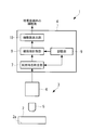
- the cell analysis device 1 is acquired by a cell image acquisition unit 3 that acquires an image of cells cultured in a culture vessel 2 and the cell image acquisition unit 3. And an image processing unit 4 that processes the image and outputs the number of cells in the culture vessel 2.
- the cell image acquisition unit 3 is disposed facing the culture surface 2a of the culture vessel 2 in which cells are cultured, and an objective lens 5 that collects light from the culture surface 2a, and is collected by the objective lens 5. And a light detector 6 that acquires an image over a predetermined visual field range in the culture surface 2a by photographing the collected light.
- the acquired image is, for example, a phase difference image.
- the image processing unit 4 includes a use determination unit 7 that determines whether or not the cell image acquired by the light detection unit 6 is usable, and the number of cells in the image that is determined to be usable by the use determination unit 7.
- Cell number counting unit 8 for counting the number of cells
- memory unit 9 for storing the counted cell number
- cell number calculation for calculating the number of cells in culture vessel 2 based on the cell number counted by cell number counting unit 8 Part 10.
- the availability determination unit 7 determines at least one of the following criteria for the acquired image. (1) The image is not blurred (2) The edge of the culture vessel 2 is not reflected in the image (3) The outside of the culture vessel 2 is not reflected in the image (4) The image already acquired Is not the same image as
- the presence / absence of blur can be determined by determining whether or not there is a linear structure extending in substantially the same direction.
- the presence or absence of a linear structure can be easily detected by Fourier transform of the image signal.
- the availability determination unit 7 determines that the image can be used when it is determined that there is no blur in the image.
- the feature is a geometrical shape such as a straight line or a curve of the edge of the culture vessel 2 and a sufficiently large structure compared to the cell. Therefore, by recognizing a large linear structure extending in substantially the same direction in an image enlarged to such an extent that cells can be observed, the presence or absence of the edge of the culture vessel 2 in the image can be determined.
- the availability determination unit 7 determines that the image can be used when the edge of the culture vessel 2 is not reflected in the image.
- the availability determination unit 7 determines that the image can be used when the outside of the culture vessel 2 is not reflected in the image.
- the determination as to whether or not the image is the same as the already acquired image is made by comparing the number of cells in the immediately preceding image stored in the storage unit 9 with the number of cells in the newly acquired image. If the variation rate is equal to or less than a predetermined threshold, it can be determined that the images are the same part.
- the availability determination unit 7 determines that the image can be used when it is not the same as the already acquired image.
- the cell counting unit 8 recognizes the cells in the image and counts the number of cells when the use determining unit 7 determines that the cells can be used. For the cell recognition, any method such as shape recognition can be adopted.
- the storage unit 9 accumulates and stores the number of cells counted by the cell number counting unit 8.
- the cell number calculation unit 10 averages the integrated value of the number of cells counted by the cell number counting unit 8 and stored in the storage unit 9 for a predetermined number N of images, and the calculated average value is used for the culture surface 2a of the culture vessel 2.
- the number of cells in the entire culture vessel 2 is calculated by multiplying the ratio between the effective area and the area of the visual field range.
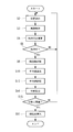
- step S1 the number n is reset (step S1), and the cells are cultured.
- the objective lens 5 of the cell image acquisition unit 3 is made to face the culture surface 2a of the container 2, and the optical axis position of the objective lens 5 is positioned at an appropriate position (step S2).
- the light from the culture surface 2a is detected by the light detection unit 6 to acquire an image (imaging step S3).
- imaging step S3 a phase difference image of a cell obtained by cutting out a partial range in the culture vessel 2 is acquired.
- step S4 On the basis of the luminance value of the image acquired in the photographing step S3, the availability is calculated for at least one of the four criteria (step S4), and the availability is determined (usability determination step S5). .
- step S4 image processing is performed to determine whether or not the image can be used, and information indicating whether or not the image can be used is output.
- step S5 When it is determined that the use is not possible in the availability determination step S5, at least one of the culture vessel 2 and the objective lens 5 is relatively moved in a direction perpendicular to the optical axis of the objective lens 5, and from step S2. These steps are repeated. If it is determined in the availability determination step S5 that the cell is available, the number of cells in the image is counted (cell number counting step S6), and the counted number of cells is stored in the storage unit 9 until then. It is added to the stored number of cells and stored as an integrated value in the storage unit 9 (step S7), and the number n is incremented (step S8).
- step S9 it is determined whether or not the number of cells has been counted for a predetermined number N of images. If not, the processes from step S2 are repeated.
- the integrated value of the number of cells within the visual field range counted for the predetermined number N of images stored in the storage unit 9 in the cell number calculation unit 10 is: An average value is calculated by dividing by the predetermined number N (average value calculating step S10).
- the cell number calculation unit 10 multiplies the average value of the number of cells calculated in the average value calculation step S10 by the ratio between the effective area of the culture surface 2a of the culture container 2 and the area of the visual field range. 2 The number of cells is calculated (cell number calculation step S11).
- the total number of cells in the culture vessel 2 can be calculated easily and accurately.
- the acquired image is suitable for counting the number of cells by the image processing unit 4 before the number of cells is counted. Since the availability indicating whether or not it is used is determined and the image determined to be unusable is excluded from the cell count target, there is an advantage that the accuracy of the counted cell count can be improved.
- the number of cells in the culture vessel 2 being cultured can be estimated, and it is not necessary to detach the cells from the culture vessel 2.
- the number of cells is counted each time for an image that is determined to be usable by the usability determining unit 7, but instead, as shown in FIGS. 3 and 4.
- the image is stored in the storage unit 9 (step S12), and after a predetermined number N of images are acquired, the number of cells is collectively collected. May be counted (cell number counting step S6), and an average value may be calculated (average value calculating step S10).
- the integrated value of the counted number of cells is stored in the storage unit 9, but instead of this, the counted number of cells is stored separately, and the integration and division processing is performed when calculating the average value. It may be done.
- the image acquisition is terminated and the number of cells is calculated.
- FIG. As shown in FIG. 5, the average value of the number of cells may be calculated (step S10) and stored (step S13) without counting the number n, and the variance thereof may be calculated (step S14). Then, on condition that the variance of the average value of the number of cells is equal to or less than a predetermined threshold (step S15), the image acquisition may be terminated and the number of cells may be calculated (step S11).
- the process from step S2 is repeated without calculating the number of cells. According to the dispersion of the average value of the number of cells, it can be determined more reliably that the inside of the culture vessel 2 has been sufficiently sampled. In the present embodiment, the number of colonies may be counted as the number of cells.
Landscapes
- Engineering & Computer Science (AREA)
- Chemical & Material Sciences (AREA)
- Life Sciences & Earth Sciences (AREA)
- Health & Medical Sciences (AREA)
- Organic Chemistry (AREA)
- Zoology (AREA)
- Wood Science & Technology (AREA)
- Bioinformatics & Cheminformatics (AREA)
- Biotechnology (AREA)
- General Health & Medical Sciences (AREA)
- Physics & Mathematics (AREA)
- Biochemistry (AREA)
- Genetics & Genomics (AREA)
- Microbiology (AREA)
- General Engineering & Computer Science (AREA)
- Biomedical Technology (AREA)
- Proteomics, Peptides & Aminoacids (AREA)
- Theoretical Computer Science (AREA)
- General Physics & Mathematics (AREA)
- Analytical Chemistry (AREA)
- Sustainable Development (AREA)
- Molecular Biology (AREA)
- Computer Vision & Pattern Recognition (AREA)
- Toxicology (AREA)
- Biophysics (AREA)
- Immunology (AREA)
- Medicinal Chemistry (AREA)
- Multimedia (AREA)
- Quality & Reliability (AREA)
- Apparatus Associated With Microorganisms And Enzymes (AREA)
- Measuring Or Testing Involving Enzymes Or Micro-Organisms (AREA)
- Investigating Or Analysing Materials By Optical Means (AREA)
- Image Processing (AREA)
Abstract
Description
本発明は上述した事情に鑑みてなされたものであって、簡易に精度よく培養容器内の細胞の細胞数を計測することができる細胞解析装置および細胞解析方法を提供することを目的としている。
このようにすることで、画像がブレている場合に、当該画像が細胞数の計数の対象から除外されるので、ブレのない画像に基づいて細胞数を精度よく算出することができる。
このようにすることで、画像に培養容器のエッジが写り込んでいる場合に、当該画像が細胞数の計数の対象から除外されるので、エッジの写り込みのない画像に基づいて細胞数を精度よく算出することができる。
このようにすることで、画像に培養容器の外部が写り込んでいる場合に、当該画像が細胞数の計数の対象から除外されるので、容器外部の写り込みのない画像に基づいて細胞数を精度よく算出することができる。
このようにすることで、細胞数計数部により計数された複数枚の画像の細胞数の平均値に基づいて、細胞数算出部により培養容器内の細胞数が算出される。この場合には、既に取得された画像と同一の画像を用いても、平均による精度向上を望むことはできないので、このような画像を除外して、細胞数を精度よく算出することができる。
また、上記態様においては、前記利用可否判定ステップが、前記培養容器のエッジが写り込んでいるか否かを検出し、前記培養容器のエッジが写り込んでいない場合に利用可であると判定してもよい。
また、上記態様においては、前記細胞数算出ステップが、複数枚の画像から計数された細胞数の平均値を用いて細胞数を算出し、前記利用可否判定ステップが、既に取得された画像と同一の画像であるか否かを検出し、同一の画像ではない場合に利用可であると判定してもよい。
本実施形態に係る細胞解析装置1は、図1に示されるように、培養容器2内において培養されている細胞の画像を取得する細胞画像取得部3と、該細胞画像取得部3により取得された画像を処理して培養容器2内の細胞数を出力する画像処理部4とを備えている。
取得する画像は、例えば、位相差画像である。
(1) 画像にブレがないか
(2) 画像に培養容器2のエッジが写り込んでいないか
(3) 画像に培養容器2の外部が写り込んでいないか
(4) 既に取得されている画像と同一の画像ではないか
本実施形態に係る細胞解析方法により、培養容器2内の細胞数を算出するには、まず、図2に示されるように、枚数nがリセットされ(ステップS1)、細胞を培養している培養容器2の培養面2aに、細胞画像取得部3の対物レンズ5を対向させ、かつ、対物レンズ5の光軸位置を適当な位置に位置決めし(ステップS2)、対物レンズ5によって集光された培養面2aからの光を光検出部6により検出して画像を取得する(撮影ステップS3)。
撮影ステップS3により、培養容器2内の一部の範囲を切り出した細胞の位相差画像が取得される。
ステップS4において、利用可否の判定のための画像処理が行われ、利用可であるか利用不可であるかの情報が出力される。
利用可否判定ステップS5において、利用可であると判定された場合には、画像内の細胞数の計数が行われ(細胞数計数ステップS6)、計数された細胞数が、それまで記憶部9に記憶されていた細胞数に加算され、積算値として記憶部9に記憶されるとともに(ステップS7)、枚数nがインクリメントされる(ステップS8)。
所定枚数Nの画像について細胞数が計数された場合には、細胞数算出部10において、記憶部9に記憶された所定枚数Nの画像について計数された視野範囲内の細胞数の積算値が、所定枚数Nによって除算されることにより平均値が算出される(平均値算出ステップS10)。
細胞数の平均値の分散によれば、培養容器2内を十分にサンプリングできたことをより確実に判断することができる。
また、本実施形態においては、細胞数としてコロニー数を計数することにしてもよい。
2 培養容器
3 細胞画像取得部
7 利用可否判定部
8 細胞数計数部
10 細胞数算出部
S3 撮影ステップ
S5 利用可否判定ステップ
S6 細胞数計数ステップ
S11 細胞数算出ステップ
Claims (10)
- 細胞を培養している培養容器内の細胞の画像を取得する細胞画像取得部と、
該細胞画像取得部により取得された画像の利用可否を判定する利用可否判定部と、
該利用可否判定部により利用可であると判定された画像内の細胞数を計数する細胞数計数部と、
該細胞数計数部により計数された細胞数に基づいて前記培養容器内の細胞数を算出する細胞数算出部とを備える細胞解析装置。 - 前記利用可否判定部が、画像のブレを検出し、ブレが存在しない場合に利用可であると判定する請求項1に記載の細胞解析装置。
- 前記利用可否判定部が、前記培養容器のエッジが写り込んでいるか否かを検出し、前記培養容器のエッジが写り込んでいない場合に利用可であると判定する請求項1または請求項2に記載の細胞解析装置。
- 前記利用可否判定部が、前記培養容器の外部が写り込んでいるか否かを検出し、前記培養容器の外部が写り込んでいない場合に利用可であると判定する請求項1から請求項3のいずれかに記載の細胞解析装置。
- 前記細胞数算出部が、複数枚の画像から計数された細胞数の平均値を用いて細胞数を算出し、
前記利用可否判定部が、既に取得された画像と同一の画像であるか否かを検出し、同一の画像ではない場合に利用可であると判定する請求項1から請求項4のいずれかに記載の細胞解析装置。 - 細胞を培養している培養容器内の細胞の画像を取得する撮影ステップと、
該撮影ステップにより取得された画像の利用可否を判定する利用可否判定ステップと、
該利用可否判定ステップにより利用可であると判定された画像内の細胞数を計数する細胞数計数ステップと、
該細胞数計数ステップにより計数された細胞数に基づいて前記培養容器内の細胞数を算出する細胞数算出ステップとを含む細胞解析方法。 - 前記利用可否判定ステップが、画像のブレを検出し、ブレが存在しない場合に利用可であると判定する請求項6に記載の細胞解析方法。
- 前記利用可否判定ステップが、前記培養容器のエッジが写り込んでいるか否かを検出し、前記培養容器のエッジが写り込んでいない場合に利用可であると判定する請求項6または請求項7に記載の細胞解析方法。
- 前記利用可否判定ステップが、前記培養容器の外部が写り込んでいるか否かを検出し、前記培養容器の外部が写り込んでいない場合に利用可であると判定する請求項6から請求項8のいずれかに記載の細胞解析方法。
- 前記細胞数算出ステップが、複数枚の画像から計数された細胞数の平均値を用いて細胞数を算出し、
前記利用可否判定ステップが、既に取得された画像と同一の画像であるか否かを検出し、同一の画像ではない場合に利用可であると判定する請求項6から請求項9のいずれかに記載の細胞解析方法。
Priority Applications (5)
| Application Number | Priority Date | Filing Date | Title |
|---|---|---|---|
| PCT/JP2015/060836 WO2016162945A1 (ja) | 2015-04-07 | 2015-04-07 | 細胞解析装置および細胞解析方法 |
| JP2017510830A JP6505830B2 (ja) | 2015-04-07 | 2015-04-07 | 細胞解析装置および細胞解析方法 |
| CN201580078503.3A CN107406811A (zh) | 2015-04-07 | 2015-04-07 | 细胞解析装置和细胞解析方法 |
| DE112015006266.1T DE112015006266T5 (de) | 2015-04-07 | 2015-04-07 | Zellanalysegerät und Zellanalyseverfahren |
| US15/725,413 US10438047B2 (en) | 2015-04-07 | 2017-10-05 | Cell analysis device and cell analysis method |
Applications Claiming Priority (1)
| Application Number | Priority Date | Filing Date | Title |
|---|---|---|---|
| PCT/JP2015/060836 WO2016162945A1 (ja) | 2015-04-07 | 2015-04-07 | 細胞解析装置および細胞解析方法 |
Related Child Applications (1)
| Application Number | Title | Priority Date | Filing Date |
|---|---|---|---|
| US15/725,413 Continuation US10438047B2 (en) | 2015-04-07 | 2017-10-05 | Cell analysis device and cell analysis method |
Publications (1)
| Publication Number | Publication Date |
|---|---|
| WO2016162945A1 true WO2016162945A1 (ja) | 2016-10-13 |
Family
ID=57071860
Family Applications (1)
| Application Number | Title | Priority Date | Filing Date |
|---|---|---|---|
| PCT/JP2015/060836 Ceased WO2016162945A1 (ja) | 2015-04-07 | 2015-04-07 | 細胞解析装置および細胞解析方法 |
Country Status (5)
| Country | Link |
|---|---|
| US (1) | US10438047B2 (ja) |
| JP (1) | JP6505830B2 (ja) |
| CN (1) | CN107406811A (ja) |
| DE (1) | DE112015006266T5 (ja) |
| WO (1) | WO2016162945A1 (ja) |
Cited By (5)
| Publication number | Priority date | Publication date | Assignee | Title |
|---|---|---|---|---|
| WO2018083984A1 (ja) * | 2016-11-02 | 2018-05-11 | ソニー株式会社 | 情報処理装置、情報処理方法及び情報処理システム |
| WO2020203700A1 (ja) * | 2019-04-02 | 2020-10-08 | 富士フイルム株式会社 | 観察制御装置、方法およびプログラム |
| JP2021110561A (ja) * | 2020-01-07 | 2021-08-02 | オリンパス株式会社 | 検査方法、コンピュータ読取可能記録媒体、及び、標準板 |
| JP2023134978A (ja) * | 2022-03-15 | 2023-09-28 | 株式会社Nttドコモ | 穂数予測装置 |
| US12307655B2 (en) | 2019-10-11 | 2025-05-20 | Shimadzu Corporation | Cell image analysis method and cell image analysis device |
Families Citing this family (4)
| Publication number | Priority date | Publication date | Assignee | Title |
|---|---|---|---|---|
| KR102261700B1 (ko) * | 2017-03-30 | 2021-06-04 | 후지필름 가부시키가이샤 | 세포 화상 평가 장치 및 방법과, 프로그램 |
| JP7139359B2 (ja) * | 2018-01-15 | 2022-09-20 | 株式会社エビデント | 細胞解析装置及び細胞解析システム |
| EP3859008B1 (en) * | 2018-11-02 | 2022-11-16 | PHC Holdings Corporation | Method for estimating cell count, and device for estimating cell count |
| TWI824616B (zh) * | 2022-07-12 | 2023-12-01 | 倍利科技股份有限公司 | 基於影像識別的細胞統計之系統及方法 |
Citations (7)
| Publication number | Priority date | Publication date | Assignee | Title |
|---|---|---|---|---|
| JPH1031165A (ja) * | 1996-07-16 | 1998-02-03 | Olympus Optical Co Ltd | 画像入力装置及び走査型顕微鏡装置 |
| JP2004016194A (ja) * | 2002-06-20 | 2004-01-22 | Hitachi Medical Corp | 細胞培養装置 |
| JP2005107302A (ja) * | 2003-09-30 | 2005-04-21 | Olympus Corp | 顕微鏡フォーカス維持装置 |
| JP2006171574A (ja) * | 2004-12-17 | 2006-06-29 | Olympus Corp | 顕微鏡画像処理方法、顕微鏡画像処理プログラムおよび顕微鏡システム |
| WO2007103406A2 (en) * | 2006-03-09 | 2007-09-13 | Cytokinetics, Inc. | Cellular predictive models for toxicities |
| JP2009194584A (ja) * | 2008-02-14 | 2009-08-27 | Olympus Corp | 画像選択装置、画像選択プログラム、および画像選択方法 |
| JP2015091220A (ja) * | 2013-11-08 | 2015-05-14 | 東洋製罐グループホールディングス株式会社 | 細胞計数装置、細胞計数システム、細胞計数方法及び細胞計数プログラム |
Family Cites Families (6)
| Publication number | Priority date | Publication date | Assignee | Title |
|---|---|---|---|---|
| JP2005502369A (ja) | 2001-09-12 | 2005-01-27 | バースタイン テクノロジーズ,インコーポレイティド | 分画細胞計数方法ならびにそれを実行するための関連する装置およびソフトウェア |
| JP4664785B2 (ja) | 2005-09-16 | 2011-04-06 | 株式会社Eci | ディスク及び該ディスクを用いた光学式顕微鏡による細胞の計数観察装置 |
| JP2007124913A (ja) | 2005-11-01 | 2007-05-24 | Olympus Corp | 細胞数計測方法、細胞数推移計測方法、細胞数計測装置および細胞数推移計測装置 |
| WO2012043498A1 (ja) * | 2010-09-30 | 2012-04-05 | 日本電気株式会社 | 情報処理装置、情報処理システム、情報処理方法、プログラム及び記録媒体 |
| JP5447546B2 (ja) * | 2012-01-31 | 2014-03-19 | 東洋製罐グループホールディングス株式会社 | 細胞計数方法、細胞計数装置、及び細胞計数プログラム |
| CN102851208B (zh) * | 2012-03-09 | 2015-03-04 | 上海凯度机电科技有限公司 | 一种高精度细胞统计计数与分析装置 |
-
2015
- 2015-04-07 DE DE112015006266.1T patent/DE112015006266T5/de not_active Withdrawn
- 2015-04-07 JP JP2017510830A patent/JP6505830B2/ja not_active Expired - Fee Related
- 2015-04-07 WO PCT/JP2015/060836 patent/WO2016162945A1/ja not_active Ceased
- 2015-04-07 CN CN201580078503.3A patent/CN107406811A/zh active Pending
-
2017
- 2017-10-05 US US15/725,413 patent/US10438047B2/en active Active
Patent Citations (7)
| Publication number | Priority date | Publication date | Assignee | Title |
|---|---|---|---|---|
| JPH1031165A (ja) * | 1996-07-16 | 1998-02-03 | Olympus Optical Co Ltd | 画像入力装置及び走査型顕微鏡装置 |
| JP2004016194A (ja) * | 2002-06-20 | 2004-01-22 | Hitachi Medical Corp | 細胞培養装置 |
| JP2005107302A (ja) * | 2003-09-30 | 2005-04-21 | Olympus Corp | 顕微鏡フォーカス維持装置 |
| JP2006171574A (ja) * | 2004-12-17 | 2006-06-29 | Olympus Corp | 顕微鏡画像処理方法、顕微鏡画像処理プログラムおよび顕微鏡システム |
| WO2007103406A2 (en) * | 2006-03-09 | 2007-09-13 | Cytokinetics, Inc. | Cellular predictive models for toxicities |
| JP2009194584A (ja) * | 2008-02-14 | 2009-08-27 | Olympus Corp | 画像選択装置、画像選択プログラム、および画像選択方法 |
| JP2015091220A (ja) * | 2013-11-08 | 2015-05-14 | 東洋製罐グループホールディングス株式会社 | 細胞計数装置、細胞計数システム、細胞計数方法及び細胞計数プログラム |
Cited By (9)
| Publication number | Priority date | Publication date | Assignee | Title |
|---|---|---|---|---|
| WO2018083984A1 (ja) * | 2016-11-02 | 2018-05-11 | ソニー株式会社 | 情報処理装置、情報処理方法及び情報処理システム |
| JPWO2018083984A1 (ja) * | 2016-11-02 | 2019-09-19 | ソニー株式会社 | 情報処理装置、情報処理方法及び情報処理システム |
| JP7001060B2 (ja) | 2016-11-02 | 2022-01-19 | ソニーグループ株式会社 | 情報処理装置、情報処理方法及び情報処理システム |
| US11282201B2 (en) | 2016-11-02 | 2022-03-22 | Sony Corporation | Information processing device, information processing method and information processing system |
| WO2020203700A1 (ja) * | 2019-04-02 | 2020-10-08 | 富士フイルム株式会社 | 観察制御装置、方法およびプログラム |
| US12307655B2 (en) | 2019-10-11 | 2025-05-20 | Shimadzu Corporation | Cell image analysis method and cell image analysis device |
| JP2021110561A (ja) * | 2020-01-07 | 2021-08-02 | オリンパス株式会社 | 検査方法、コンピュータ読取可能記録媒体、及び、標準板 |
| JP7286558B2 (ja) | 2020-01-07 | 2023-06-05 | 株式会社エビデント | 検査方法、コンピュータ読取可能記録媒体、及び、標準板 |
| JP2023134978A (ja) * | 2022-03-15 | 2023-09-28 | 株式会社Nttドコモ | 穂数予測装置 |
Also Published As
| Publication number | Publication date |
|---|---|
| US20180032787A1 (en) | 2018-02-01 |
| DE112015006266T5 (de) | 2017-11-30 |
| JP6505830B2 (ja) | 2019-04-24 |
| JPWO2016162945A1 (ja) | 2018-02-08 |
| CN107406811A (zh) | 2017-11-28 |
| US10438047B2 (en) | 2019-10-08 |
Similar Documents
| Publication | Publication Date | Title |
|---|---|---|
| JP6505830B2 (ja) | 細胞解析装置および細胞解析方法 | |
| US8121400B2 (en) | Method of comparing similarity of 3D visual objects | |
| EP2584309B1 (en) | Image capture device and image capture method | |
| US8970770B2 (en) | Continuous autofocus based on face detection and tracking | |
| TWI393980B (zh) | The method of calculating the depth of field and its method and the method of calculating the blurred state of the image | |
| EP2360638A1 (en) | Method, system and computer program product for obtaining a point spread function using motion information | |
| EP4254037A3 (en) | Real-time autofocus scanning | |
| US20120093372A1 (en) | Distance measuring device and distance measuring method | |
| JPWO2011125937A1 (ja) | キャリブレーションデータ選択装置、選択方法、及び選択プログラム、並びに三次元位置測定装置 | |
| JP2011182397A (ja) | ずれ量算出方法およびずれ量算出装置 | |
| JP6598850B2 (ja) | 画像処理装置、画像処理方法および画像処理プログラム | |
| KR20170035844A (ko) | 타임 오브 플라이트 데이터를 비닝하는 방법 | |
| JP2019029948A (ja) | 画像監視装置、画像監視方法および画像監視プログラム | |
| JP2014123069A5 (ja) | ||
| EP3221661B1 (en) | Depth detection apparatus, imaging apparatus and depth detection method | |
| JP2017130976A5 (ja) | 画像処理装置、及び画像処理方法 | |
| JP2017040726A5 (ja) | 焦点検出装置及びその制御方法 | |
| JP2015148783A5 (ja) | 焦点調節装置および制御方法 | |
| JP6449339B2 (ja) | 細胞解析装置および方法 | |
| JP2015201193A (ja) | 画像認識装置及び画像認識方法 | |
| JP2010199701A5 (ja) | ||
| JP2015137987A (ja) | 距離センサおよび距離計測方法 | |
| US8761516B2 (en) | Method and system for detecting the open or closed state of the eyes of a face | |
| CN112330586B (zh) | 一种尿液分析仪诊断提速方法、尿液分析仪及计算机可读存储介质 | |
| JP6621435B2 (ja) | 行列長推定装置、方法、及びプログラム |
Legal Events
| Date | Code | Title | Description |
|---|---|---|---|
| 121 | Ep: the epo has been informed by wipo that ep was designated in this application |
Ref document number: 15888439 Country of ref document: EP Kind code of ref document: A1 |
|
| ENP | Entry into the national phase |
Ref document number: 2017510830 Country of ref document: JP Kind code of ref document: A |
|
| WWE | Wipo information: entry into national phase |
Ref document number: 112015006266 Country of ref document: DE |
|
| 122 | Ep: pct application non-entry in european phase |
Ref document number: 15888439 Country of ref document: EP Kind code of ref document: A1 |