WO2016159376A1 - がんの治療薬のスクリーニング方法 - Google Patents
がんの治療薬のスクリーニング方法 Download PDFInfo
- Publication number
- WO2016159376A1 WO2016159376A1 PCT/JP2016/060974 JP2016060974W WO2016159376A1 WO 2016159376 A1 WO2016159376 A1 WO 2016159376A1 JP 2016060974 W JP2016060974 W JP 2016060974W WO 2016159376 A1 WO2016159376 A1 WO 2016159376A1
- Authority
- WO
- WIPO (PCT)
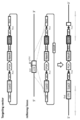
- Prior art keywords
- cell
- cancer
- cells
- expression
- gene
- Prior art date
- Legal status (The legal status is an assumption and is not a legal conclusion. Google has not performed a legal analysis and makes no representation as to the accuracy of the status listed.)
- Ceased
Links
Images





Classifications
-
- G—PHYSICS
- G01—MEASURING; TESTING
- G01N—INVESTIGATING OR ANALYSING MATERIALS BY DETERMINING THEIR CHEMICAL OR PHYSICAL PROPERTIES
- G01N33/00—Investigating or analysing materials by specific methods not covered by groups G01N1/00 - G01N31/00
- G01N33/48—Biological material, e.g. blood, urine; Haemocytometers
- G01N33/50—Chemical analysis of biological material, e.g. blood, urine; Testing involving biospecific ligand binding methods; Immunological testing
- G01N33/5005—Chemical analysis of biological material, e.g. blood, urine; Testing involving biospecific ligand binding methods; Immunological testing involving human or animal cells
- G01N33/5008—Chemical analysis of biological material, e.g. blood, urine; Testing involving biospecific ligand binding methods; Immunological testing involving human or animal cells for testing or evaluating the effect of chemical or biological compounds, e.g. drugs, cosmetics
- G01N33/5011—Chemical analysis of biological material, e.g. blood, urine; Testing involving biospecific ligand binding methods; Immunological testing involving human or animal cells for testing or evaluating the effect of chemical or biological compounds, e.g. drugs, cosmetics for testing antineoplastic activity
-
- C—CHEMISTRY; METALLURGY
- C12—BIOCHEMISTRY; BEER; SPIRITS; WINE; VINEGAR; MICROBIOLOGY; ENZYMOLOGY; MUTATION OR GENETIC ENGINEERING
- C12Q—MEASURING OR TESTING PROCESSES INVOLVING ENZYMES, NUCLEIC ACIDS OR MICROORGANISMS; COMPOSITIONS OR TEST PAPERS THEREFOR; PROCESSES OF PREPARING SUCH COMPOSITIONS; CONDITION-RESPONSIVE CONTROL IN MICROBIOLOGICAL OR ENZYMOLOGICAL PROCESSES
- C12Q1/00—Measuring or testing processes involving enzymes, nucleic acids or microorganisms; Compositions therefor; Processes of preparing such compositions
- C12Q1/68—Measuring or testing processes involving enzymes, nucleic acids or microorganisms; Compositions therefor; Processes of preparing such compositions involving nucleic acids
- C12Q1/6876—Nucleic acid products used in the analysis of nucleic acids, e.g. primers or probes
- C12Q1/6883—Nucleic acid products used in the analysis of nucleic acids, e.g. primers or probes for diseases caused by alterations of genetic material
- C12Q1/6886—Nucleic acid products used in the analysis of nucleic acids, e.g. primers or probes for diseases caused by alterations of genetic material for cancer
-
- G—PHYSICS
- G01—MEASURING; TESTING
- G01N—INVESTIGATING OR ANALYSING MATERIALS BY DETERMINING THEIR CHEMICAL OR PHYSICAL PROPERTIES
- G01N2333/00—Assays involving biological materials from specific organisms or of a specific nature
- G01N2333/435—Assays involving biological materials from specific organisms or of a specific nature from animals; from humans
- G01N2333/475—Assays involving growth factors
- G01N2333/495—Transforming growth factor [TGF]
-
- G—PHYSICS
- G01—MEASURING; TESTING
- G01N—INVESTIGATING OR ANALYSING MATERIALS BY DETERMINING THEIR CHEMICAL OR PHYSICAL PROPERTIES
- G01N2333/00—Assays involving biological materials from specific organisms or of a specific nature
- G01N2333/82—Translation products from oncogenes
-
- G—PHYSICS
- G01—MEASURING; TESTING
- G01N—INVESTIGATING OR ANALYSING MATERIALS BY DETERMINING THEIR CHEMICAL OR PHYSICAL PROPERTIES
- G01N2500/00—Screening for compounds of potential therapeutic value
- G01N2500/04—Screening involving studying the effect of compounds C directly on molecule A (e.g. C are potential ligands for a receptor A, or potential substrates for an enzyme A)
-
- G—PHYSICS
- G01—MEASURING; TESTING
- G01N—INVESTIGATING OR ANALYSING MATERIALS BY DETERMINING THEIR CHEMICAL OR PHYSICAL PROPERTIES
- G01N2500/00—Screening for compounds of potential therapeutic value
- G01N2500/10—Screening for compounds of potential therapeutic value involving cells
Definitions
- the present invention relates to a method for screening for a therapeutic drug for cancer.
- molecular targeting drugs that is, therapeutic drugs that directly target proteins related to important intracellular signals related to cancer cell growth and progression
- searches for proteins that can become new drug discovery targets are being actively pursued.
- target proteins for cancer treatment include proteins encoded by genes that are unique or overexpressed in cancer cells.
- Non-patent Document 1 a technique for reprogramming somatic cells has been developed and attempts have been made to reinitialize cancer cells. Such attempts have been focused on changing the properties of cancer cells themselves. There are no reports on screening for cancer drugs using cancer cell reprogramming.
- An object of the present invention is to provide a method for screening for a therapeutic drug for cancer.
- a known target gene that is, cancer cell growth / Suppressing the expression of genes that are known to be related to important intracellular signals involved in development
- the proteins encoded by these target genes are: It has been found that it has the effect of maintaining the characteristics as a cancer cell and suppressing the fate change of the cancer cell.
- a reprogramming factor is introduced into cancer cells that express the target protein of an existing molecular target drug under contact with the molecular target drug, the reprogramming factor is initialized compared to when it is initialized under non-drug contact.
- the degree of was enhanced. Therefore, when the cancer cell is initialized or differentiated directly into another cell type in a state where an effective target protein is in contact with a known cancer cell and the test substance, the initialization or differentiation is enhanced. It was revealed that the test substance can be confirmed to have a function of suppressing the activity of the target gene. Moreover, even when targeting any cancer cell, if the test substance enhances the initialization or differentiation of the cancer cell, the test substance is an intracellular signal related to the growth and progression of the cancer cell. Since it can be presumed to have a function of suppressing the activity of some protein related to the above, it becomes possible to directly screen candidate substances for cancer therapeutic agents without identifying the target protein (target gene). As a result of further studies based on these findings, the present inventors have completed the present invention.
- a method for screening for a therapeutic drug for cancer comprising the following steps; (I) a step of expressing an exogenous cell regulator in a target cancer cell in contact with or non-contact with a test substance; (Ii) a step of confirming a change in cancer cells, and (iii) in the case where the change in cancer cells is increased in contact with the test substance compared to non-contact, the test substance is treated for cancer.
- the process of selecting as a medicine [2] The method according to [1], wherein the cell regulatory factor is an reprogramming factor, and the change in the cancer cells is reprogramming of cancer cells.
- a method for identifying a protein that can be a drug discovery target of a cancer therapeutic drug comprising the following steps: (I) an exogenous cell regulator in a cancer cell containing a gene encoding a test protein in a form in which expression can be controlled, under conditions under which the gene is expressed or under conditions where expression of the gene is suppressed A step of expressing (Ii) a step of confirming a change in cancer cells, and (iii) when the change in cancer cells is increased under conditions in which expression of the gene is suppressed as compared to conditions under which the gene is expressed, A step of selecting the test protein as a protein that can be a drug discovery target of a cancer therapeutic agent.
- the present invention it is not necessary to identify a target gene, and it becomes possible to directly screen for a therapeutic drug for cancer that becomes a molecular target drug.
- the present invention makes it possible to screen for cancer therapeutic agents having a high effect on cancer cells.
- FIG. 1A shows a schematic diagram describing a method for inducing EWS / ATF1 expression and introducing reprogramming factors.
- FIG. 1B shows a phase contrast microscopic image of sarcoma cell line G1297 after introduction of reprogramming factor when EWS / ATF1 expression was induced (DOXD0.2 ⁇ g / ml) or not (DOX 0 ⁇ g / ml).
- FIG. 1C shows the results of measuring the number of colonies formed after introduction of reprogramming factor when EWS / ATF1 expression was induced (DOX 0.2 or 0.1 ⁇ g / ml) or not (DOX 0 ⁇ g / ml). .
- FIG. 1A shows a schematic diagram describing a method for inducing EWS / ATF1 expression and introducing reprogramming factors.
- FIG. 1B shows a phase contrast microscopic image of sarcoma cell line G1297 after introduction of reprogramming factor when
- FIG. 1D shows RT-PCR of EWS / ATF1 expression induction by adding 0.2 ⁇ g / ml DOX to iPS cell lines (C-1, C-2, C-3 and C-4) established from sarcoma cell line G1297. The result confirmed by is shown.
- FIG. 1E shows the results of chromosome microarray (CGH array) analysis in the sarcoma cell line G1297 and an iPS cell line established from the same.
- FIG. 1F shows the results of measuring the expression of Nanog in the iPS cell lines (C-1, C-2, C-3 and C-4) by RT-PCR (left figure) and staining the iPS cell line with Nanog and DAPI. The stained image (right figure) is shown.
- FIG. 1G shows a stained image of teratoma formed by transplanting iPS cells established from sarcoma cell line G1297 subcutaneously into nude mice.
- FIG. 1H shows a photograph of a chimeric mouse generated by injecting iPS cells established from the sarcoma cell line G1297 into a blastocyst.
- FIG. 1I shows that EWS / ATF1 expression was induced (DOX 0.2 or 0.1 ⁇ g / ml) or not induced (DOX 0 ⁇ g / ml) on day 4 after introduction of reprogramming factor (4F) into sarcoma cell line G1297.
- FIG. 1J shows that EWS / ATF1 expression was induced on mouse embryonic fibroblasts (MEF) 4 days after introduction of reprogramming factor (4F) (DOX 0.2 or 0.1 ⁇ g / ml) or not (DOX (0 ⁇ g / ml) shows the results of FACS in which the positive rate of SSEA1 was measured 10 days after 4F introduction.
- FIG. 1K shows 4F introduction or GFP when EWS / ATF1 expression was induced (ON) or not induced (OFF) on day 4 after introduction of reprogramming factor (4F) or GFP into sarcoma cell line G1297.
- FIG. 1L shows a schematic diagram describing EWS / ATF1 expression induction and a method for introducing MYOD1.
- FIG. 1M shows the results of RT-PCR measurement of Myogenin expression when EWS / ATF1 expression was induced (ON) or not induced (OFF) in cells in which MYOD1 was introduced into the sarcoma cell line G1297.
- FIG. 1L shows a schematic diagram describing EWS / ATF1 expression induction and a method for introducing MYOD1.
- FIG. 1M shows the results of RT-PCR measurement of Myogenin expression when EWS / ATF1 expression was induced (ON) or not induced (OFF) in cells in which MYOD1 was introduced into the sarcoma cell line G1297.
- FIG. 10 shows the 6th day after introduction of MYOD1 or GFP when EWS / ATF1 expression was induced (ON) or not induced (OFF) on day 4 after introduction of MYOD1 or GFP into sarcoma cell line G1297. The results of microarray analysis using RNA recovered from the cells are shown.
- FIG. 2A shows a schematic diagram describing the method of introduction of reprogramming factor, MYOD1 or GFP and siRNA into the MP-CCS-SY cell line.
- FIG. 2B shows that when reprogramming factor (+ 4F) or GFP was introduced into MP-CCS-SY cell line, expression of EWS / ATF1 was suppressed by RNAi (siEWS / ATF1) or expression was not suppressed (siControl).
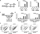
- FIG. 2C shows a schematic diagram describing the introduction of reprogramming factors into HCC827 or SK-BR3 cell lines and the method of adding each drug.
- 2D shows the case where DMSO, 5FU or Lapatinib (Lap) was added to the SK-BR3 cell line when reprogramming factor was introduced (Dox (4F) +) or not (Dox (4F)-) Of PODXL expression in RT-PCR (left figure) and when reprogramming factor was introduced into HCC827 cell line (Dox (4F) +) or not (Dox (4F)-)
- the result (right figure) which measured the expression of PODXL in the case of adding DMSO, 5FU, or Gefitinib (Gef) by RT-PCR is shown.
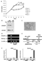
- FIG. 3A shows a growth curve when doxycycline (DOX) 0, 0.1, 0.2, 1 or 2 ⁇ g / ml was added to sarcoma cell line G1297 and cultured.
- DOX doxycycline
- FIG. 3B shows that when the reprogramming factor was introduced into the sarcoma cell line G1297 (+ 4F) or not (-4F), expression of EWS / ATF1 was induced (ON) or not (OFF) ) Shows the results of measuring the amount of Oct3 / 4 in each case by Western blotting.
- FIG. 3C shows a phase contrast microscopic image of iPS cells established after introduction of reprogramming factor when EWS / ATF1 expression was not induced (DOX 0 ⁇ g / ml).
- FIG. 3D shows the results of measuring the expression levels of Oct3 / 4 and Nanog by RT-PCR when EWS / ATF1 expression was induced (ON) or not induced (OFF) in the sarcoma cell line G1297. .
- ESC ES cell
- FIG. 3E shows a schematic diagram describing the method of induction of EWS / ATF1 expression and introduction of reprogramming factors.
- FIG. 3F shows that EWS / ATF1 expression was induced (ON) or not induced (OFF) in cells with or without the reprogramming factor introduced into the sarcoma cell line G1297 (+ 4F).
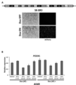
- FIG. 4A shows a PiggyBac vector construct for introducing the reprogramming factor and a phase contrast image and a fluorescence image of the cell after introduction into SK-BR3.
- FIG. 4B shows DMSO, 5FU (1 mM or 10 mM) or Gefitinib (Gef) with or without reprogramming factor introduced into the A549 cell line (Dox (4F) +) or not (Dox (4F) ⁇ ).
- Dox (4F) + the A549 cell line
- Dox (4F) ⁇ Dox (4F) +
- Dox (4F) ⁇ The result of having measured the expression of PODXL by RT-PCR when (10 mM or 50 mM) is added is shown.
- FIG. 4A shows a PiggyBac vector construct for introducing the reprogramming factor and a phase contrast image and a fluorescence image of the cell after introduction into SK-BR3.
- FIG. 4B shows DMSO, 5FU (1 mM or
- FIG. 5A shows a schematic diagram describing the method of induction of reprogramming factors by addition of Dox to HCC827 and SK-BR3 and the introduction of anticancer agents (Gefitinib, Imatinini, Lapatinib, 5-FU).
- FIG. 5A shows a schematic diagram describing the method of induction of reprogramming factors by addition of Dox to HCC827 and SK-BR3 and the introduction of anticancer agents (Gefitinib, Imatinini, Lapatinib, 5-FU).
- 5B shows Gefitinib (0 ⁇ M (DMSO added), 0.005 ⁇ M or 0.05 ⁇ M), Imatinib (0 ⁇ M (DMSO added)) when reprogramming factors were induced into HCC827 and SK-BR3 (Dox (4F) +) RT of Nanog and GDF3 expression when Lapatinib (0 ⁇ M (DMSO added), 0.05 ⁇ M or 0.5 ⁇ M) or 5-FU (0 ⁇ M (DMSO added), 10 ⁇ M or 50 ⁇ M) was added, 1 ⁇ M or 10 ⁇ M) -Measured by PCR and shows the result of normalizing the transcription level with the GAPDH value.
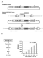
- FIG. 7A shows a schematic diagram describing a construction of a targeting vector for producing reporter cells, and a method for introducing the construct into the PPP1R2C gene locus of HEK293 cells derived from human fetal kidney by genome editing technology using TALEN. .
- FIG. 7B shows a schematic diagram (left diagram) of the luciferase assay performed using the construct of FIG. 7A and the result (right diagram).
- the horizontal axis of the graph indicates the number of cells, and the vertical axis indicates the relative luminescence intensity of luciferase.
- FIG. 8 shows a schematic diagram describing a targeting vector construct for preparing reporter cells and a method for introducing the construct into the Nanog locus of mouse cancer cells by genome editing technology using CRISPR / Cas9.
- FIG. 9 shows a schematic diagram describing a targeting vector construct for preparing reporter cells and a method for introducing the construct into the Nanog locus of mouse cancer cells by genome editing technology using CRISPR / Cas9.
- secNluc represents a secreted NanoLuc TM luciferase gene
- BsdR represents a blasticidin resistance gene.
- FIG. 10 shows a schematic diagram describing a targeting vector construct for preparing reporter cells and a method for introducing the construct into the Nanog locus of human cancer cells by genome editing technology using CRISPR / Cas9.
- the present invention provides a method for screening for a therapeutic agent for cancer, comprising the following steps; (I) a step of expressing an exogenous cell regulator in a target cancer cell in contact with or non-contact with a test substance; (Ii) a step of confirming a change in cancer cells, and (iii) in the case where the change in cancer cells is increased in contact with the test substance compared to non-contact, the test substance is treated for cancer.
- the process of selecting as a medicine comprising the following steps; (I) a step of expressing an exogenous cell regulator in a target cancer cell in contact with or non-contact with a test substance; (Ii) a step of confirming a change in cancer cells, and (iii) in the case where the change in cancer cells is increased in contact with the test substance compared to non-contact, the test substance is treated for cancer.
- cancer means a malignant tumor, including carcinoma (malignant tumor derived from epithelial cells), sarcoma, and other leukemias, and is not limited to a specific cancer.
- the cancer cell used in the present invention is a cell constituting a malignant tumor, and may be a cell line or a cell isolated from a living body.
- cancer cells used in the present invention are cancer cells (for example, EWS / ATF1 fusion gene forced expression) whose effective target protein is known (ie, known to be sensitive to an existing molecular target drug).
- cancer cells gefitinib-sensitive mutant EGFR-expressing cancer cells, lapatinib-sensitive HER2-amplified cancer cells, alectinib-sensitive EML4-ALK fusion gene-expressing cancer cells, imatinib-sensitive chronic myeloid leukemia cells, etc.
- cancer cells whose effective target protein is unknown may be used.
- the cancer cell used in the screening method of the present invention may contain a reporter gene.
- reporter genes include, but are not limited to, luciferase genes, fluorescent protein genes, and drug resistance genes.
- the luciferase gene include a firefly luciferase gene, a synthetic Renilla luciferase gene, a secreted luciferase gene, and the like, but a secreted luciferase gene is preferable in terms of ease of protein recovery.
- fluorescent protein genes include green fluorescent protein genes such as GFP and EGFP, blue fluorescent protein genes such as BFP and TagBFP, and red fluorescent protein genes such as RFP and DsRed.
- drug resistance genes include kanamycin resistance gene, ampicillin resistance gene, neomycin resistance gene, puromycin resistance gene, and blasticidin resistance gene.
- the reporter cell can be prepared, for example, by knocking in the reporter gene to a specific locus in the genome by homologous recombination or the like.
- the homologous recombination may be performed using a genome editing technique.
- a zinc finger nuclease (ZFN) in which a zinc finger DNA binding domain and a non-specific DNA cleavage domain are linked is used as the genome editing technique.
- TALEN TAL effector nuclease
- TAL transcriptional activator-like
- JP 2013-513389 TALEN-like effector
- CRISPR-Cas9 system combining the DNA sequence CRISPR (Clustered Regularly interspaced short palindromic repeats) and the nuclease Cas protein family that plays an important role together with CRISPR (Special Table 2010-519929) It is done.
- Specific loci include open chromatin structures such as the human PPP1R2C locus, which are less susceptible to suppressed expression of inserted genes, and genes that are specifically highly expressed in differentiated pluripotent cells (for example, Nanog , Oct3 / 4, Fbx15, etc., preferably Nanog).
- Recombinant cells containing such a reporter gene include, for example, MEF (Okita et al., Nature, 448, a transgenic mouse derived by incorporating a green fluorescent protein (GFP) gene and a puromycin resistance gene into the Nanog locus. 313-317 (2007)).
- a cell regulator is a factor that changes the properties of the cell by introduction into the cell and changes it to another cell type, and is not particularly limited.
- Nerve cell inducer neural stem cell inducer, neural crest cell inducer, cardiomyocyte inducer, myocyte inducer, chondrocyte inducer, hepatocyte inducer, melanocyte inducer, hematopoietic progenitor inducer, erythroblast Examples include sphere-inducing factors and megakaryocyte progenitor cell-inducing factors.
- reprogramming factors used in the present invention include Oct3 / 4, Sox2, Sox1, Sox3, Sox15, Sox17, Klf4, Klf2, c-Myc, N-Myc, L-Myc, Nanog, Lin28, Fbx15, ERas , ECAT15-2, Tcl1, beta-catenin, Lin28b, Sall1, Sall4, Esrrb, Nr5a2, Tbx3, Glis1 or GDF3 and other genes or gene products are exemplified, and these reprogramming factors may be used alone, You may use it in combination.
- a more preferred reprogramming factor is a combination comprising Oct3 / 4, Sox2, Klf4 and c-Myc.
- the reprogramming factors include histone deacetylase (HDAC) inhibitors [eg small molecule inhibitors such as valproic acid (VPA), trichostatin A, sodium butyrate, MC ⁇ 1293, M344, siRNA and shRNA against HDAC (eg , Nucleic acid expression inhibitors such as HDAC1 siRNA Smartpool (registered trademark) (Millipore), HuSH 29mer shRNA Constructs against HDAC1 (OriGene), etc.], MEK inhibitors (for example, PD184352, PD98059, U0126, SL327 and PD0325901), Glycogen synthase kinase-3 inhibitors (eg, Bio and CHIR99021), DNA methyltransferase inhibitors (eg, 5-azacytidine), histone methyltransferase inhibitors (eg, small molecule inhibitors such as BIX-01294, Suv39hl, Suv39h2, Nucleic acid expression inhibitors such
- Examples of the nerve cell inducer used in the present invention include Lhx3, Ngn2 and Isl1 (WO2014 / 148646), Ascl1, Brn2 and Myt1l (Wapinski OL et al, Cell. 155: 621-635, 2013). .
- Examples of the neural stem cell inducing factor used in the present invention include Brn4 / Pou3f4, Sox2, Klf4, c-Myc and E47 / Tcf3 (Han DW et al, Cell Stem Cell.:10:465-472, 2012) and the like. It is done.
- cardiomyocyte-inducing factor examples include Gata4, Mef2c and Tbx5 (Ieda Met et al., Cell. 142: 375-386, 2010).
- Examples of the myocyte inducing factor used in the present invention include MYOD1 or MYF5 (Tanaka A, et al, PLoS One. 8: e61540, 2013).
- chondrocyte inducing factor examples include c-Myc, Klf4 and SOX9 (Outani H, et al, PLoS One. 8: e77365, 2013).
- hepatocyte-inducing factors used in the present invention include FOXA3, HNF1A, and HNF4A (Huang P, et al, Cell Stem Cell. 14: 370-384, 2014).
- melanocyte-inducing factor examples include MITF, SOX10, and PAX3 (Yang® R, “et” al, “Nat” Commun. “5: 5807,” 2014).
- hematopoietic progenitor cell inducers used in the present invention include ERG, GATA2, LMO2, RUNX1c, and SCL (Batta K, Cell Rep. 9: 1871-84, 2014).
- erythroblast-inducing factor examples include c-MYC and BCL-XL (Hirose S, et al, Stem Cell Reports. 1: 499-508, 2013).
- Examples of the megakaryocyte progenitor cell inducer used in the present invention include BMI1, c-MYC, and BCL-XL (Nakamura S, et al, Cell Stem Cell. 14: 535-548, 2014).
- a cell regulator in the present invention, when the cell regulator is in the form of a protein, for example, lipofection, fusion with a cell membrane-permeable peptide (for example, TAT and polyarginine derived from HIV), microinjection.
- a cell membrane-permeable peptide for example, TAT and polyarginine derived from HIV
- the cell regulatory factor when it is in the form of DNA, it can be introduced into somatic cells by techniques such as vectors such as viruses, plasmids, artificial chromosomes, lipofection, liposomes, and microinjection.
- Virus vectors include retrovirus vectors, lentivirus vectors (cell, 126, pp.663-676, 2006; Cell, 131, pp.861-872, 2007; Science, 318, pp.1917-1920, 2007 ), Adenovirus vectors (Science, 322, 945-949, 2008), adeno-associated virus vectors, Sendai virus vectors (WO 2010/008054) and the like.
- artificial chromosome vectors examples include human artificial chromosomes (HAC), yeast artificial chromosomes (YAC), and bacterial artificial chromosomes (BAC, PAC).
- HAC human artificial chromosomes
- YAC yeast artificial chromosomes
- BAC bacterial artificial chromosomes
- a plasmid a plasmid for mammalian cells can be used (Science, 322: 949-953, 2008).
- the vector can contain regulatory sequences such as a promoter, enhancer, ribosome binding sequence, terminator, polyadenylation site, etc. so that the nuclear reprogramming substance can be expressed.
- It can contain a selection marker sequence such as gene, thymidine kinase gene, diphtheria toxin gene, reporter gene sequence such as fluorescent protein (GFP, etc.), ⁇ -glucuronidase (GUS), FLAG, etc.
- a selection marker sequence such as gene, thymidine kinase gene, diphtheria toxin gene, reporter gene sequence such as fluorescent protein (GFP, etc.), ⁇ -glucuronidase (GUS), FLAG, etc.
- the cell regulator When the cell regulator is in the form of RNA, it may be introduced into somatic cells by, for example, lipofection, microinjection, etc., and RNA that incorporates 5-methylcytidine and pseudoridine (TriLink Biotechnologies) to suppress degradation (Warren L, (2010) Cell Stem Cell. 7: 618-630) may be used.
- RNA that incorporates 5-methylcytidine and pseudoridine (TriLink Biotechnologies) to suppress degradation (Warren L, (2010) Cell Stem Cell. 7: 618-630) may be used.
- Cancer cells after introduction of cell regulatory factors can be cultured in a culture solution prepared using a medium used for culturing animal cells as a basal medium.
- a basal medium examples include IMDM medium, Medium ⁇ 199 medium, Eagle's Minimum Essential Medium (EMEM) medium, ⁇ MEM medium, Dulbecco's modified Eagle's Medium (DMEM) medium, Ham's F12 medium, RPMI 1640 medium, Fischer's medium and the like Is included.
- the medium may contain serum or may be serum-free.
- the medium can be, for example, albumin, insulin, transferrin, selenium, fatty acids, trace elements, 2-mercaptoethanol, thiolglycerol, lipids, amino acids, L-glutamine, non-essential amino acids, vitamins, growth factors, small molecules
- albumin for example, albumin, insulin, transferrin, selenium, fatty acids, trace elements, 2-mercaptoethanol, thiolglycerol, lipids, amino acids, L-glutamine, non-essential amino acids, vitamins, growth factors, small molecules
- One or more substances such as compounds, antibiotics, antioxidants, pyruvate, buffers, inorganic salts, cytokines and the like may also be included.
- cancer cells after introduction of a cell regulatory factor may be cultured by appropriately selecting a culture solution suitable for the characteristics of the cell regulatory factor, and the conditions are exemplified below.
- a culture solution for cells after reprogramming is, for example, DMEM, DMEM / F12 or DME culture solution containing 10 to 15% FBS (in addition to these culture solutions) , LIF, penicillin / streptomycin, puromycin, L-glutamine, non-essential amino acids, ⁇ -mercaptoethanol, etc.) or a commercially available culture solution (for example, a culture solution for mouse ES cell culture (TX-WES Culture medium, Thrombo X), culture medium for primate ES cell culture (culture medium for primate ES / iPS cells, Reprocell), serum-free medium (mTeSR, Stemcell Technology)), and the like.
- DMEM DMEM / F12 or DME culture solution containing 10 to 15% FBS (in addition to these culture solutions) , LIF, penicillin / streptomycin, puromycin, L-glutamine, non-essential amino acids, ⁇ -mercaptoethanol, etc.
- Examples of cell culture methods after initialization include, for example, contacting somatic cells with reprogramming factors on DMEM or DMEM / F12 medium containing 10% FBS in the presence of 5% CO 2 at 37 ° C. Cultivate for about 4-7 days, then re-spread the cells on feeder cells (eg, mitomycin C-treated STO cells, SNL cells, etc.), and bFGF-containing primate ES approximately 10 days after contact between the somatic cells and the reprogramming factor
- the cells can be cultured in a culture medium for cell culture, and iPS-like colonies can be formed about 30 to about 45 days or more after the contact.
- 10% FBS-containing DMEM medium including LIF, penicillin / streptomycin, etc.
- feeder cells eg, mitomycin C-treated STO cells, SNL cells, etc.
- 5% CO 2 at 37 ° C. can be suitably included with puromycin, L-glutamine, non-essential amino acids, ⁇ -mercaptoethanol, etc.
- ES-like colonies after about 25 to about 30 days or more .
- somatic cells to be reprogrammed themselves are used (Takahashi K, et al. (2009), PLoS One. 4: e8067 or WO2010 / 137746), or extracellular matrix (eg, Laminin- 5 (WO2009 / 123349) and Matrigel (BD)) are exemplified.
- iPS cells may be established under hypoxic conditions (oxygen concentration of 0.1% or more and 15% or less) (Yoshida Y, et al. (2009), Cell Stem Cell. 5: 237 -241 or WO2010 / 013845).
- the culture solution and culture conditions for the cells after the introduction of the factor are described, for example, in WO2014 / 148646, Wapinski OL et al, Cell. 155: 621-635, 2013 And the like.
- the culture medium and culture conditions for the cell after the factor introduction are described in, for example, Han DW et al, Cell Stem Cell. 10: 465-472, 2012 And the like.
- the cell regulatory factor is a neural crest cell inducing factor
- the culture medium and culture conditions for the cell after introduction of the factor are described, for example, in Kim YJ, et al, Cell Stem Cell. 15: 497-506, 2014 And the like.
- the cell regulatory factor is a cardiomyocyte-inducing factor
- examples of the culture solution and culture conditions for cells after the introduction of the factor include those described in Ieda M et al, Cell. 142: 375-386, 2010, etc. Is mentioned.
- the cell regulatory factor is a muscle cell inducing factor
- examples of the culture solution and culture conditions for the cells after the introduction of the factor include those described in Tanaka A, et al, PLoS One. 8: e61540, 2013, etc. Is mentioned.
- the cell regulatory factor is a chondrocyte-inducing factor
- examples of the culture solution and culture conditions for the cell after introduction of the factor include those described in Outani H, et al, PLoS One. 8: e77365, 2013, etc. Is mentioned.
- the culture solution and culture conditions for the cell after the factor introduction are described in, for example, HuangHP, et al, Cell Stem Cell. 14: 370-384, 2014 And the like.
- the cell regulatory factor is a melanocyte-inducing factor
- examples of the culture solution and culture conditions for the cells after the introduction of the factor include those described in Yang R, et al, Nat Commun.:5:5807, ⁇ 2014, etc. Is mentioned.
- the cell regulatory factor is a hematopoietic progenitor cell inducing factor
- examples of the culture solution and culture conditions for cells after the introduction of the factor include those described in Batta K, Cell Rep. 9: 1871-84, 2014, etc. Is mentioned.
- the cell regulatory factor is an erythroblast-inducing factor
- the culture solution and culture conditions for the cell after introduction of the factor are described in, for example, Hirose S, et al, Stem Cell Reports. 1: 499-508, 2013 And the like.
- the culture solution and culture conditions for the cell after introduction of the factor are, for example, NakamuraNS, et al, Cell Stem Cell. 14: 535-548, 2014 What is described is mentioned.
- test substance in the screening method of the present invention, any test substance can be used, and any known compound and novel compound may be used.
- the test substance may also be (1) a biological library method, (2) a synthetic library method using deconvolution, (3) “one-bead one-compound” live Can be obtained using any of a number of approaches in combinatorial library methods known in the art, including rally methods, and (4) synthetic library methods using affinity chromatography sorting.
- Biological library methods using affinity chromatography sorting are limited to peptide libraries, but the other four approaches can be applied to small molecule compound libraries of peptides, non-peptide oligomers, or compounds (Lam (1997) Anticancer Drug Des. 12: 145-67).
- Examples of methods for the synthesis of molecular libraries can be found in the art (DeWitt et al. (1993) Proc. Natl. Acad. Sci. USA 90: 6909-13; Erb et al. (1994) Proc. Natl. Acad. Sci. USA 91: 11422-6; Zuckermann et al. (1994) J. Med. Chem. 37: 2678-85; Cho et al.
- the contact between the cancer cell and the test substance can be preferably carried out by culturing the cancer cell into which the cell regulatory factor has been introduced as described above in a culture solution to which the test substance is added.
- concentration of the test substance added to the culture solution can be appropriately selected within a range that does not adversely affect the growth of cells, but is usually 0.1-100 nM.
- Examples of the contact initiation time include immediately after the introduction of the cell regulatory factor or within a few days.
- exogenous cell regulators are introduced into cancer cells in a form in which expression can be controlled (ON / OFF enabled)
- contact can be initiated simultaneously with expression ON treatment or within a few days after the treatment. .
- the contact period is not particularly limited as long as it is a time sufficient for the change of cancer cells to be observed, but it is usually sufficient to allow it to coexist in the culture solution until a positive colony appears.
- Changes in cancer cells In the method for screening a therapeutic drug for cancer of the present invention , changes in cells caused by expression of an exogenous cell regulator in cancer cells under the contact of a test substance are used as an index. be able to. In the present invention, the change in the cancer cells depends on the cell regulatory factor to be introduced.
- initialization means not only complete initialization (ie, establishment of iPS cells with stable pluripotency and undifferentiated state), but also change to a more undifferentiated state compared to the original cancer cells.
- the cell regulatory factor is a factor that directly induces differentiation into other cells without initialization, not only complete differentiation into the target cell, Including a change to a state in which the characteristics of the target cell are acquired as compared to the original cancer cell. Therefore, the initialization of cancer cells can be evaluated using colony formation, undifferentiated specific antigen expression, or undifferentiated specific gene expression as an index.
- undifferentiated specific genes include genes that show specific expression in embryonic stem cells or genes that suggest an important role in maintaining pluripotency. Such genes include Cell. 2005 Sep 23. ; 122 (6): 947-56, Stem Cells. 2004; 22 (1): 51-64., Mol Biol Cell. 2002 Apr; 13 (4): 1274-81., Mol Reprod Dev. 2000 Jun; 56 (2): 113-23., EMBO J. 1998 Apr 1; 17 (7): 2019-32., Proc Natl Acad Sci U S A. 2003 Nov 11; 100 (23): 13350-5., Development. 2005 Mar; 132 (5): 885-96., Blood.
- the undifferentiated specific antigen is selected from the group consisting of, but not limited to, SSEA-1, SSEA-3, SSEA-4, TRA-2-54, TRA-1-60 and TRA-1-80 Examples are antigens. Since SSEA-1 is not detected in undifferentiated cells in humans, SSEA-3 and SSEA-4 are preferably used in place of SSEA-1.
- colonies is not particularly limited, but is measured under a microscope and evaluated by the number. This measurement may be performed mechanically (WO2011 / 010449) or visually.
- cells expressing an undifferentiated specific antigen can be evaluated as the number of cells expressing the antigen using a cell sorter such as FACS. It can be evaluated as the expression level by enzyme PCR analysis, quantitative reverse transcriptase PCR analysis, Northern blot analysis, immunohistochemistry, array analysis, RNA-seq analysis, reporter gene analysis and combinations thereof. Therefore, in the present invention, changes in cancer cells can be paraphrased as an increase in the number of cells expressing an undifferentiated specific antigen, an increased expression level of an undifferentiated specific gene, or a colony formation.
- the change in cancer cells can be evaluated using the expression of a nerve cell specific gene as an index.
- a nerve cell specific gene include genes selected from the group consisting of MFNG, GRIP1, NGFR, Zfp238, GRINT1 and SYT3 (WO2014 / 148646 and Wapinski OL et al, Cell. 155: 621-635, 2013) .
- the cell regulatory factor is a neural stem cell inducing factor
- changes in cancer cells can be evaluated using the expression of neural stem cell specific genes as an index.
- the nerve cell-specific gene include genes selected from the group consisting of Olig2, Sox2 and Mash1 / Ascl1 (Han DW et al, Cell Stem Cell. 10: 465-472, 2012).
- the change in cancer cells can be evaluated using the expression of neural crest cell specific gene as an index.
- neural crest cell-specific genes include genes selected from the group consisting of TWIST1, SNAIL2, ITGA4, ITGA6, SOX5, SOX6, PLP1 and Myelin Protein Zero [MPZ] (Kim YJ, et al, Cell Stem Cell . 15: 497-506, 2014).
- the change in cancer cells can be evaluated using the expression of a cardiomyocyte-specific gene as an index.
- a cardiomyocyte-specific gene examples include ⁇ MHC or cTnT (Ieda M et al, Cell. 142: 375-386, 2010).
- muscle cell-specific genes include genes selected from the group consisting of myogenin, myosin heavy chain (MHC), MEF2C and SIX1 (Tanaka A, et al, PLoS One. 8: e61540, 2013).
- chondrocyte-specific genes include genes selected from the group consisting of COL11A2, COL2A1, and ACAN (Outani H, et al, PLoS One. 8: e77365, 2013).
- hepatocyte-specific genes include genes selected from the group consisting of albumin (ALB), ⁇ -1-antitrypsin (AAT), CYP1A2, CYP2A6, CYP2B6, CYP2C8, CYP2C9 and CYP3A4 (Huang P, et al , Cell Stem Cell. 14: 370-384, 2014).
- melanocyte-specific genes include genes selected from the group consisting of TYR, TYRP1 and DCT (Yang R, et al, Nat Commun. 5: 5807, 2014).
- hematopoietic progenitor cell inducing factor When the cell regulatory factor is a hematopoietic progenitor cell inducing factor, changes in cancer cells can be evaluated using the expression of hematopoietic progenitor cell specific genes as an index.
- hematopoietic progenitor cell-specific genes include genes or antigens selected from the group consisting of CD31, CD41, c-KIT, CD45, CD11b and TER119 (Batta K, Cell Rep. 9: 1871-84, 2014) .
- erythroblast-specific genes include genes or antigens selected from the group consisting of GPA, CD71, GATA1 and RAF1 (Hirose S, et al, Stem Cell Reports. 1: 499-508, 2013).
- megakaryocyte progenitor cell inducer When the cell regulatory factor is a megakaryocyte progenitor cell inducer, changes in cancer cells can be evaluated using the expression of megakaryocyte progenitor cell specific genes as an index.
- megakaryocyte progenitor cell-specific genes include genes or antigens selected from the group consisting of CD41a, CD42a, CD42b and CD9 (Nakamura S, et al, Cell Stem Cell. 53514: 535-548, 2014).
- cells expressing the gene can also be evaluated as the number of cells using a cell sorter such as FACS, and reverse transcriptase PCR analysis
- the expression level can also be evaluated by quantitative reverse transcriptase PCR analysis, Northern blot analysis, immunohistochemistry, array analysis, reporter gene analysis, and combinations thereof. Therefore, in the present invention, “increasing changes in cancer cells” can be paraphrased as the number of cells expressing the gene increases or the expression level of the gene increases.
- Kit for Screening for Drug for Cancer contains the above-mentioned cell regulatory factor.
- the kit for screening for a therapeutic drug for cancer may further include a document and instructions describing the production procedure of cell control and the measurement method and procedure of the index.
- it may further include a panel of various cancer cells having different effective target proteins (wherein the effective target protein has the same meaning as described above), or a cell serving as an indicator of changes in the cancer cells.
- An antibody against a surface antigen and a nucleic acid for detecting a cell-specific gene can be further included.
- a method for identifying a protein that can be a drug discovery target of a cancer therapeutic agent includes the following steps: (I) In a cancer cell containing a gene encoding a test protein (candidate gene) in a form in which expression can be controlled, the gene is exogenous under conditions where the candidate gene is expressed or expression of the gene is suppressed.
- the method includes a step of selecting the test protein as a protein that can be a drug discovery target of a therapeutic drug for cancer.
- candidate genes are identified as a result of exhaustive analysis of gene expression in cancer cells using a microarray or the like, and are highly expressed in genes or cancer cells that are specifically expressed in cancer cells. Genes that are present.
- the expression-controllable form means a form capable of ON / OFF of the expression of the candidate gene.
- the candidate gene is an inducible promoter (eg, metallothionein promoter (induced by heavy metal ions), heat Under the control of shock protein promoter (induced by heat shock), Tet-ON / Tet-OFF system promoter (induced by addition or removal of tetracycline or its derivative), steroid-responsive promoter (induced by steroid hormone or its derivative), etc.) And expression vectors placed in the above.
- metallothionein promoter induced by heavy metal ions
- shock protein promoter induced by heat shock
- Tet-ON / Tet-OFF system promoter induced by addition or removal of tetracycline or its derivative
- steroid-responsive promoter induced by steroid hormone or its derivative
- the EWS / ATF1 fusion gene described in Yamada K, et al, J Clin Invest. 123: 600-610, 2013 is induced in a doxycycline (Dox) -dependent manner
- Dox doxycycline
- a possible mouse sarcoma cell line also referred to as G1297 strain
- the sarcoma cells have been confirmed to stop growth in vitro and to regress tumors in vivo due to EWS / ATF1 expression cessation.
- G1297 was initialized by introducing an initialization factor into G1297.
- EWS / ATF1 sarcoma cells expressing EWS / ATF1
- reprogramming factors 4F: Oct3 / 4, Sox2, Klf4, and c-Myc
- FIGS. 1A to 1C reprogramming factors
- Oct3 / 4 Sox2, Klf4 and c-Myc were each introduced into Plat-E cells using a pMXs-based retroviral vector, and the virus-containing supernatant was collected and filtered through a 0.45 ⁇ m cellulose acetate filter.
- G1297 was seeded at 8 ⁇ 10 5 cells per 60-mm dish, infected with the virus-containing supernatant, and replaced with LIF-containing ES medium on the third day after infection.
- the established iPS cell-like cell line was confirmed to express EWS / ATF1 in a doxycycline-dependent manner, similar to the parent sarcoma cell (FIG. 1D). Furthermore, some chromosomal abnormalities were also observed, confirming that the iPS cell-like cell line was derived from sarcoma cells (FIG. 1E).
- pluripotency-related genes such as Nanog and endogenous Oct3 / 4 (Pou5f1) in iPS cell-like cells derived from the sarcoma cells was compared with ES cells, there was no significant difference (Fig. 1F and FIG. 3D).
- RT-PCR was performed using the following method.
- the efficiency of initialization of sarcoma cells was 0.06%, which was shown to be lower than the initialization efficiency of MEF.
- iPS cell-like cells were administered subcutaneously to immunodeficient mice, teratomas were formed, and chimeric mice could be created by injection into blastocysts (FIGS. 1G and 1H).
- EWS / ATF1 expression-dependent sarcoma cells pluripotent cells can be obtained by reprogramming by suppressing the expression of EWS / ATF1.
- EWS / ATF1 acts suppressively in the reprogramming of EWS / ATF1 expression-dependent sarcoma cells.
- EWS / ATF1 does not suppress the generation of SSEA1-positive cells due to reprogramming in MEF (Fig. 1J), so that the expression of EWS / ATF1 results in the suppression of reprogramming specific to sarcoma cells.
- sarcoma cells when 4F is introduced (4F-sarcoma cells) or sarcoma cells when GFP is introduced as a negative control
- the microarray analysis was performed for each of the cases where EWS / ATF1 was expressed and not expressed (FIG. 3E).
- Microarray analysis was performed using Mouse-Gene-1.0-ST-Array (Affymetrix Inc., Santa Clara, USA). All data analysis was performed using GeneSpring GX software program (version 12; Agilent Technologies, Santa Clara, USA).
- FIG. 1L the influence of EWS / ATF1 expression on skeletal muscle differentiation induction by introduction of MYOD1 was examined.
- MYOG Myogenin
- Fig. 1M MYOD1-introduced sarcoma cells
- MHC myosin heavy chain
- epidermal growth factor receptor (EGFR) mutant lung cancer cell line HCC827 is sensitive to EGFR tyrosine kinase inhibitor Gefitinib and HER2 tyrosine kinase inhibitor Lapatinib, HER2-amplified breast cancer cell line SK-BR3 to Lapatinib It is known to show sensitivity.
- EGFR epidermal growth factor receptor
- Nanog was measured by a method other than quantitative RT-PCR.
- the experimental procedure is the same as the schematic diagram in FIG. 3E.
- Reprogramming factor (+ 4F) or GFP was introduced into G1297 strain expressing EWS / ATF1 by Dox addition by retrovirus, and Dox was added 4 days after the introduction.
- RNA-seq analysis was performed to examine the expression of Nanog in cells (DoxOFF) that stopped and those that continued to add Dox (DoxON). The results are shown in FIG. In DoxOFF + 4F cells, it was confirmed that the expression of the Nanog gene was increased, that is, the expression of the Nanog gene was increased by suppressing the expression of EWS / ATF1.
- RNA-seq analysis was performed using the following method.
- a library was created using TruSeq StrandedandTotal RNA with Ribo-Zero Gold LT sample Prep kit (illumina) .
- concentration quantification using KAPA Library Quantification kits Nippon Genetics
- sequence was performed with Hiseq 2500 (illumina) using Hiseq PE PE Rapid Cluster Kit v2-HS. The sequence data was analyzed using TopHat software and Cufflinks software, and visualized by IGV (Integrative Genomics Viewer).
- reporter cells by genome editing technique using Talen, TagBFP gene as a reporter gene, the targeting vector containing the synthetic Renilla luciferase (hRL) gene and puromycin-resistant gene using, HEK298 A reporter cell that stably expresses TagBFP and synthetic Renilla luciferase (hRL) was prepared by knocking in between exon 1 and exon 2 at the PPP1R2C locus of the cell. After selecting TagBFP-expressing cells by cell sorting using FACS, luciferase assay was performed.
- a reporter gene can be introduced into cancer cells by the same method, and the number of cells of the reporter cells prepared in this way can be calculated from the luminescence intensity of luciferase.
- a targeting vector containing a GFP gene, a modified firefly luciferase (Luc2) gene and a neomycin resistance gene as a reporter gene was prepared.
- the targeting vector by genome editing technology using CRISPR / Cas9, between the 5 'untranslated region (5'UTR) and coding region (CDS) of exon 1 in the Nanog locus of mouse cancer cells
- the reporter gene can be knocked in.
- the reporter is inserted between the CDS of exon 4 and the 3 ′ untranslated region (3′UTR) in the Nanog locus of mouse cancer cells.
- a gene can be knocked in.
- a targeting vector containing a GFP gene, a secreted NanoLuc TM luciferase (secNluc) gene and a neomycin resistance gene as a reporter gene was prepared.
- the reporter is inserted between the CDS of exon 4 and the 3 ′ untranslated region (3′UTR) in the Nanog locus of human cancer cells.
- a gene can be knocked in.
- the cells produced in this manner also express a reporter gene when Nanog is expressed, and therefore, by using the cells, without measuring the expression level of an undifferentiated specific gene such as Nanog, It is suitable for use in the screening method of the present invention.
Landscapes
- Health & Medical Sciences (AREA)
- Life Sciences & Earth Sciences (AREA)
- Chemical & Material Sciences (AREA)
- Engineering & Computer Science (AREA)
- Immunology (AREA)
- Biomedical Technology (AREA)
- Molecular Biology (AREA)
- Pathology (AREA)
- Analytical Chemistry (AREA)
- Organic Chemistry (AREA)
- Proteomics, Peptides & Aminoacids (AREA)
- Hematology (AREA)
- Urology & Nephrology (AREA)
- General Health & Medical Sciences (AREA)
- Physics & Mathematics (AREA)
- Biotechnology (AREA)
- Biochemistry (AREA)
- Bioinformatics & Cheminformatics (AREA)
- Microbiology (AREA)
- Genetics & Genomics (AREA)
- Wood Science & Technology (AREA)
- Zoology (AREA)
- Toxicology (AREA)
- Tropical Medicine & Parasitology (AREA)
- Cell Biology (AREA)
- Food Science & Technology (AREA)
- Medicinal Chemistry (AREA)
- General Physics & Mathematics (AREA)
- Hospice & Palliative Care (AREA)
- General Engineering & Computer Science (AREA)
- Oncology (AREA)
- Biophysics (AREA)
- Measuring Or Testing Involving Enzymes Or Micro-Organisms (AREA)
- Micro-Organisms Or Cultivation Processes Thereof (AREA)
Abstract
Description
本発明者らは、これらの知見に基づいてさらに研究を重ねた結果、本発明を完成するに至った。
[1]下記の工程を含む、がんの治療薬をスクリーニングする方法;
(i)対象となるがん細胞内で、被験物質との接触下または非接触下で、外因性の細胞制御因子を発現させる工程、
(ii)がん細胞の変化を確認する工程、および
(iii)被験物質との接触下において、非接触下と比較してがん細胞の変化が増加した場合、当該被験物質をがんの治療薬として選出する工程。
[2]前記細胞制御因子が、初期化因子であり、前記がん細胞の変化が、がん細胞の初期化である、[1]に記載の方法。
[3]前記初期化因子が、Oct3/4、Sox2、Klf4およびc-Mycである、[2]に記載の方法。
[4]前記がん細胞の初期化が、未分化特異的抗原が陽性である細胞数、または当該がん細胞における未分化特異的遺伝子の発現量を指標として評価される、[2]または[3]に記載の方法。
[5]前記未分化特異的遺伝子が、Nanog、Epcam、Cdh1、Fbxo15、PODXLおよびGDF3から選択される1以上の遺伝子である、[4]に記載の方法。
[6]前記がん細胞の初期化が、形成されるコロニー数を指標として評価される、[2]または[3]に記載の方法。
[7]前記細胞制御因子が、MyoD1であり、前記がん細胞の変化が、がん細胞から筋管細胞への変化である、[1]に記載の方法。
[8]前記がん細胞から筋管細胞への変化が、ミオゲニン(myogenin)またはミオシン重鎖(myosin heavy chain)の発現量を指標として評価される、[7]に記載の方法。
[9]下記の工程を含む、がん治療薬の創薬標的となり得るタンパク質の同定方法:
(i)被験タンパク質をコードする遺伝子を、発現制御可能な形態で含むがん細胞内で、該遺伝子を発現する条件下または該遺伝子の発現が抑制された条件下で、外来性の細胞制御因子を発現させる工程、
(ii)がん細胞の変化を確認する工程、および
(iii)該遺伝子の発現が抑制された条件下において、該遺伝子を発現する条件下と比較してがん細胞の変化が増加した場合、当該被験タンパク質をがんの治療薬の創薬標的となり得るタンパク質として選出する工程。
(i)対象となるがん細胞内で、被験物質との接触下または非接触下で、外因性の細胞制御因子を発現させる工程、
(ii)がん細胞の変化を確認する工程、および
(iii)被験物質との接触下において、非接触下と比較してがん細胞の変化が増加した場合、当該被験物質をがんの治療薬として選出する工程。
本発明において、がんとは、悪性腫瘍を意味し、癌腫(上皮細胞由来の悪性腫瘍)、肉腫、その他白血病などを含み、特定のがんに限定されない。また、本発明で用いるがん細胞とは、悪性腫瘍を構成する細胞であり、株化された細胞であっても生体内から単離された細胞であってもよい。また、本発明で用いるがん細胞は、有効な標的タンパク質が既知(即ち、ある既存の分子標的薬に対して感受性であることが既知)のがん細胞(例えば、EWS/ATF1融合遺伝子強制発現がん細胞、ゲフィチニブ感受性の変異EGFR発現がん細胞、ラパチニブ感受性のHER2-増幅がん細胞、アレクチニブ感受性のEML4-ALK融合遺伝子発現がん細胞、イマチニブ感受性の慢性骨髄性白血病細胞など)であってもよいし、有効な標的タンパク質が不明のがん細胞であってもよい。
本発明において、細胞制御因子とは、細胞内に導入することによって当該細胞の性質を変化させ、他の細胞種へと変化させる因子であり、特に限定されないが、例えば、初期化因子、神経細胞誘導因子、神経幹細胞誘導因子、神経冠細胞誘導因子、心筋細胞誘導因子、筋細胞誘導因子、軟骨細胞誘導因子、肝細胞誘導因子、メラニン細胞誘導因子、造血前駆細胞誘導因子、赤芽球誘導因子および巨核球前駆細胞誘導因子などが例示される。
本発明のスクリーニング方法においては、任意の被験物質を用いることができ、いかなる公知化合物および新規化合物であってもよく、例えば、細胞抽出物、細胞培養上清、微生物発酵産物、海洋生物由来の抽出物、植物抽出物、精製タンパク質または粗タンパク質、ペプチド、非ペプチド化合物、合成低分子化合物、天然化合物等が挙げられる。本発明において、被験物質はまた、(1)生物学的ライブラリー法、(2)デコンヴォルーションを用いる合成ライブラリー法、(3)「1ビーズ1化合物(one-bead one-compound)」ライブラリー法、及び(4)アフィニティクロマトグラフィ選別を使用する合成ライブラリー法を含む当技術分野で公知のコンビナトリアルライブラリー法における多くのアプローチのいずれかを使用して得ることができる。アフィニティクロマトグラフィ選別を使用する生物学的ライブラリー法はペプチドライブラリーに限定されるが、その他の4つのアプローチはペプチド、非ペプチドオリゴマー、または化合物の低分子化合物ライブラリーに適用できる(Lam (1997) Anticancer Drug Des. 12: 145-67)。分子ライブラリーの合成方法の例は、当技術分野において見出され得る(DeWitt et al. (1993) Proc. Natl. Acad. Sci. USA 90: 6909-13; Erb et al. (1994) Proc. Natl. Acad. Sci. USA 91: 11422-6; Zuckermann et al. (1994) J. Med. Chem. 37: 2678-85; Cho et al. (1993) Science 261: 1303-5; Carell et al. (1994) Angew. Chem. Int. Ed. Engl. 33: 2059; Carell et al. (1994) Angew. Chem. Int. Ed. Engl. 33: 2061; Gallop et al. (1994) J. Med. Chem. 37: 1233-51)。化合物ライブラリーは、溶液(Houghten (1992) Bio/Techniques 13: 412-21を参照のこと)またはビーズ(Lam (1991) Nature 354: 82-4)、チップ(Fodor (1993) Nature 364: 555-6)、細菌(米国特許第5,223,409号)、胞子(米国特許第5,571,698号、同第5,403,484号、及び同第5,223,409号)、プラスミド(Cull et al. (1992) Proc. Natl. Acad. Sci. USA 89: 1865-9)若しくはファージ(Scott and Smith (1990) Science 249: 386-90; Devlin (1990) Science 249: 404-6; Cwirla et al. (1990) Proc. Natl. Acad. Sci. USA 87: 6378-82; Felici (1991) J. Mol. Biol. 222: 301-10; 米国特許出願第2002103360号)として作製され得る。
本発明のがんの治療薬をスクリーニングする方法では、被験物質の接触下で、がん細胞内で外来性の細胞制御因子を発現させることで起きる細胞の変化を指標とすることができる。本発明において、当該がん細胞の変化は、導入する細胞制御因子に依存する。
本発明において、がんの治療薬をスクリーニングするためのキットは、上述した細胞制御因子を含む。
(i)被験タンパク質をコードする遺伝子(候補遺伝子)を、発現制御可能な形態で含むがん細胞内で、候補遺伝子を発現する条件下または該遺伝子の発現が抑制された条件下で、外来性の細胞制御因子を発現させる工程、
(ii)がん細胞の変化を確認する工程、および
(iii)候補遺伝子の発現が抑制された条件下において、該遺伝子を発現する条件下と比較してがん細胞の変化が増加した場合、当該被験タンパク質をがんの治療薬の創薬標的となり得るタンパク質として選出する工程
を含むことを特徴とする。
ここで、候補遺伝子としては、がん細胞における遺伝子発現をマイクロアレイ等を用いて網羅的に解析した結果同定される、がん細胞に特有に発現している遺伝子やがん細胞で高発現している遺伝子が挙げられる。
また、ここで、発現制御可能な形態とは、候補遺伝子の発現のON/OFFが可能な形態を意味し、例えば、候補遺伝子が、誘導プロモーター(例、メタロチオネインプロモーター(重金属イオンで誘導)、ヒートショックタンパク質プロモーター(ヒートショックで誘導)、Tet-ON/Tet-OFF系プロモーター(テトラサイクリン又はその誘導体の添加又は除去で誘導)、ステロイド応答性プロモーター(ステロイドホルモン又はその誘導体で誘導)等)の制御下におかれた発現ベクター等が挙げられる。
細胞制御因子、がん細胞の変化、当該変化を確認・評価するための指標等については、上記したがん治療薬のスクリーニング方法と同様である。
図7Aに示されるように、TALENを用いたゲノム編集技術により、レポーター遺伝子としてTagBFP遺伝子、合成ウミシイタケルシフェラーゼ(hRL)遺伝子およびピューロマイシン耐性遺伝子を含有するターゲティングベクターを用いて、HEK298細胞のPPP1R2C遺伝子座におけるエクソン1とエクソン2の間にノックインすることにより、安定的にTagBFPおよび合成ウミシイタケルシフェラーゼ(hRL)を発現するレポーター細胞を作製した。FACSを用いたセルソーティングによりTagBFP発現細胞を選択したのち、ルシフェラーゼアッセイを行った。ルシフェラーゼアッセイは、Dual-Luciferase Reporter Assay System (Promega)を使用し、Passive lysis bufferを用いて細胞を溶解後、Envison 2104 Multilabel Reader (PerkinElmer)を用いて測定した。結果を図7Bに示す。細胞の数と発光強度の間には高い正の相関関係が認められた(R2=0.9864)。
図9に示されるように、レポーター遺伝子として、GFP遺伝子、分泌型のNanoLucTMルシフェラーゼ(secNluc)遺伝子およびブラストサイジン耐性遺伝子を含有するターゲティングベクターを作製した。CRISPR/Cas9を用いたゲノム編集技術により、該ターゲティングベクターを用いることで、マウスがん細胞のNanog遺伝子座におけるエクソン4のCDSと3'非翻訳領域(3'UTR)との間に、該レポーター遺伝子をノックインすることができる。
図10に示されるように、レポーター遺伝子として、GFP遺伝子、分泌型のNanoLucTMルシフェラーゼ(secNluc)遺伝子およびネオマイシン耐性遺伝子を含有するターゲティングベクターを作製した。CRISPR/Cas9を用いたゲノム編集技術により、該ターゲティングベクターを用いることで、ヒトがん細胞のNanog遺伝子座におけるエクソン4のCDSと3'非翻訳領域(3'UTR)との間に、該レポーター遺伝子をノックインすることができる。
Claims (9)
- 下記の工程を含む、がんの治療薬をスクリーニングする方法;
(i)対象となるがん細胞内で、被験物質との接触下または非接触下で、外因性の細胞制御因子を発現させる工程、
(ii)がん細胞の変化を確認する工程、および
(iii)被験物質との接触下において、非接触下と比較してがん細胞の変化が増加した場合、当該被験物質をがんの治療薬として選出する工程。 - 前記細胞制御因子が、初期化因子であり、前記がん細胞の変化が、がん細胞の初期化である、請求項1に記載の方法。
- 前記初期化因子が、Oct3/4、Sox2、Klf4およびc-Mycである、請求項2に記載の方法。
- 前記がん細胞の初期化が、未分化特異的抗原が陽性である細胞数、または当該がん細胞における未分化特異的遺伝子の発現量を指標として評価される、請求項2または3に記載の方法。
- 前記未分化特異的遺伝子が、Nanog、Epcam、Cdh1、Fbxo15、PODXLおよびGDF3から選択される1以上の遺伝子である、請求項4に記載の方法。
- 前記がん細胞の初期化が、形成されるコロニー数を指標として評価される、請求項2または3に記載の方法。
- 前記細胞制御因子が、MyoD1であり、前記がん細胞の変化が、がん細胞から筋管細胞への変化である、請求項1に記載の方法。
- 前記がん細胞から筋管細胞への変化が、ミオゲニン(myogenin)またはミオシン重鎖(myosin heavy chain)の発現量を指標として評価される、請求項7に記載の方法。
- 下記の工程を含む、がん治療薬の創薬標的となり得るタンパク質の同定方法:
(i)被験タンパク質をコードする遺伝子を、発現制御可能な形態で含むがん細胞内で、該遺伝子を発現する条件下または該遺伝子の発現が抑制された条件下で、外来性の細胞制御因子を発現させる工程、
(ii)がん細胞の変化を確認する工程、および
(iii)該遺伝子の発現が抑制された条件下において、該遺伝子を発現する条件下と比較してがん細胞の変化が増加した場合、当該被験タンパク質をがんの治療薬の創薬標的となり得るタンパク質として選出する工程。
Priority Applications (2)
| Application Number | Priority Date | Filing Date | Title |
|---|---|---|---|
| JP2017510268A JP6847374B2 (ja) | 2015-04-03 | 2016-04-01 | がんの治療薬のスクリーニング方法 |
| US15/563,987 US11519901B2 (en) | 2015-04-03 | 2016-04-01 | Method for screening for cancer therapeutic agent |
Applications Claiming Priority (2)
| Application Number | Priority Date | Filing Date | Title |
|---|---|---|---|
| JP2015-077264 | 2015-04-03 | ||
| JP2015077264 | 2015-04-03 |
Publications (1)
| Publication Number | Publication Date |
|---|---|
| WO2016159376A1 true WO2016159376A1 (ja) | 2016-10-06 |
Family
ID=57005099
Family Applications (1)
| Application Number | Title | Priority Date | Filing Date |
|---|---|---|---|
| PCT/JP2016/060974 Ceased WO2016159376A1 (ja) | 2015-04-03 | 2016-04-01 | がんの治療薬のスクリーニング方法 |
Country Status (3)
| Country | Link |
|---|---|
| US (1) | US11519901B2 (ja) |
| JP (1) | JP6847374B2 (ja) |
| WO (1) | WO2016159376A1 (ja) |
Cited By (1)
| Publication number | Priority date | Publication date | Assignee | Title |
|---|---|---|---|---|
| JP2020525031A (ja) * | 2017-06-30 | 2020-08-27 | エタブリスモン フランセ ドュ サンEtablissement Francais Du Sang | 赤血球系前駆細胞を作製する方法 |
Families Citing this family (1)
| Publication number | Priority date | Publication date | Assignee | Title |
|---|---|---|---|---|
| KR102110963B1 (ko) * | 2018-07-19 | 2020-05-14 | 한국과학기술원 | Setdb1 또는 이의 저해제를 포함하는 암세포의 분열 또는 분화 조절용 조성물 |
Citations (1)
| Publication number | Priority date | Publication date | Assignee | Title |
|---|---|---|---|---|
| WO2013126993A1 (en) * | 2011-02-28 | 2013-09-06 | Mcmaster University | Screening assays for identifying and validating agents that target cancer stem cells |
Family Cites Families (1)
| Publication number | Priority date | Publication date | Assignee | Title |
|---|---|---|---|---|
| AU2013203260B2 (en) * | 2012-02-28 | 2016-10-13 | Mcmaster University | Screening assays for identifying and validating agents that target cancer stem cells |
-
2016
- 2016-04-01 US US15/563,987 patent/US11519901B2/en active Active
- 2016-04-01 JP JP2017510268A patent/JP6847374B2/ja active Active
- 2016-04-01 WO PCT/JP2016/060974 patent/WO2016159376A1/ja not_active Ceased
Patent Citations (1)
| Publication number | Priority date | Publication date | Assignee | Title |
|---|---|---|---|---|
| WO2013126993A1 (en) * | 2011-02-28 | 2013-09-06 | Mcmaster University | Screening assays for identifying and validating agents that target cancer stem cells |
Non-Patent Citations (2)
| Title |
|---|
| KATSUNORI SEMI ET AL.: "Reprogramming Gijutsu o Mochiita Gan Epigenome Kenkyu", EXPERIMENTAL MEDICINE, vol. 32, no. 19, December 2014 (2014-12-01), pages 3061 - 3065, ISSN: 0288-5514 * |
| MATSUDA, Y. ET AL.: "Application of iPS cell technology to cancer epigenome study: Uncovering the mechanism of cell status conversion for drug resistance in tumor.", PATHOLOGY INTERNATIONAL, vol. 64, no. 7, July 2014 (2014-07-01), pages 299 - 308, XP055317309, ISSN: 1320-5463 * |
Cited By (2)
| Publication number | Priority date | Publication date | Assignee | Title |
|---|---|---|---|---|
| JP2020525031A (ja) * | 2017-06-30 | 2020-08-27 | エタブリスモン フランセ ドュ サンEtablissement Francais Du Sang | 赤血球系前駆細胞を作製する方法 |
| JP7162625B2 (ja) | 2017-06-30 | 2022-10-28 | エタブリスモン フランセ ドュ サン | 赤血球系前駆細胞を作製する方法 |
Also Published As
| Publication number | Publication date |
|---|---|
| US20180136195A1 (en) | 2018-05-17 |
| JP6847374B2 (ja) | 2021-03-24 |
| US11519901B2 (en) | 2022-12-06 |
| JPWO2016159376A1 (ja) | 2018-02-01 |
Similar Documents
| Publication | Publication Date | Title |
|---|---|---|
| JP6745498B2 (ja) | 神経分化誘導用の多能性幹細胞 | |
| JP7089298B2 (ja) | 多能性幹細胞から生殖細胞への分化誘導方法 | |
| Radzisheuskaya et al. | A defined Oct4 level governs cell state transitions of pluripotency entry and differentiation into all embryonic lineages | |
| JP5936134B2 (ja) | ヒト人工多能性幹細胞の選択方法 | |
| EP3042951B1 (en) | New method for inducing dopamine-producing neural precursor cells | |
| WO2023283631A2 (en) | Methods for differentiating and screening stem cells | |
| JP6090855B2 (ja) | 心筋細胞および/または心筋前駆細胞増殖剤ならびに心筋細胞および/または心筋前駆細胞の増殖方法 | |
| KR101685209B1 (ko) | 유도된 다능성 줄기 세포의 효율적인 확립 방법 | |
| EP4202041A1 (en) | Method for maintaining and amplifying human primordial germ cells / human primordial germ cell-like cells | |
| WO2016104717A1 (ja) | 肝細胞誘導方法 | |
| JP6847374B2 (ja) | がんの治療薬のスクリーニング方法 | |
| US20200165573A1 (en) | Method for maintaining and amplifying colon cancer stem cells and method for inducing colon cancer organoid | |
| WO2017126616A1 (ja) | ユーイング肉腫ファミリー腫瘍モデル細胞とそれを用いた抗腫瘍剤のスクリーニング方法 | |
| JP2022157515A (ja) | 多能性幹細胞、神経細胞及びその応用 | |
| JP6795140B2 (ja) | 誘導型がん幹細胞 | |
| WO2020090836A1 (ja) | 細胞の製造方法 | |
| Conrad et al. | New Insights in Spermatogonial Stem Cells | |
| US20180163180A1 (en) | Method of enhancing somatic cell reprogramming with the acetyllysine reader brd3r | |
| WO2018207789A1 (ja) | 疾患iPS細胞を用いた神経毒性評価モデル系及びその使用 | |
| Kubarakos | A CRISPR/Cas9-mediated FACS screen reveals novel regulators of Wnt/β-catenin signalling in human embryonic stem cells |
Legal Events
| Date | Code | Title | Description |
|---|---|---|---|
| 121 | Ep: the epo has been informed by wipo that ep was designated in this application |
Ref document number: 16773256 Country of ref document: EP Kind code of ref document: A1 |
|
| ENP | Entry into the national phase |
Ref document number: 2017510268 Country of ref document: JP Kind code of ref document: A |
|
| WWE | Wipo information: entry into national phase |
Ref document number: 15563987 Country of ref document: US |
|
| NENP | Non-entry into the national phase |
Ref country code: DE |
|
| 122 | Ep: pct application non-entry in european phase |
Ref document number: 16773256 Country of ref document: EP Kind code of ref document: A1 |