WO2016139941A1 - 抗体、フラグメント、分子及び抗hcv治療剤 - Google Patents
抗体、フラグメント、分子及び抗hcv治療剤 Download PDFInfo
- Publication number
- WO2016139941A1 WO2016139941A1 PCT/JP2016/001125 JP2016001125W WO2016139941A1 WO 2016139941 A1 WO2016139941 A1 WO 2016139941A1 JP 2016001125 W JP2016001125 W JP 2016001125W WO 2016139941 A1 WO2016139941 A1 WO 2016139941A1
- Authority
- WO
- WIPO (PCT)
- Prior art keywords
- occludin
- antibody
- monoclonal antibody
- cells
- human
- Prior art date
- Legal status (The legal status is an assumption and is not a legal conclusion. Google has not performed a legal analysis and makes no representation as to the accuracy of the status listed.)
- Ceased
Links
Images





















Classifications
-
- A—HUMAN NECESSITIES
- A61—MEDICAL OR VETERINARY SCIENCE; HYGIENE
- A61K—PREPARATIONS FOR MEDICAL, DENTAL OR TOILETRY PURPOSES
- A61K39/00—Medicinal preparations containing antigens or antibodies
- A61K39/395—Antibodies; Immunoglobulins; Immune serum, e.g. antilymphocytic serum
-
- A—HUMAN NECESSITIES
- A61—MEDICAL OR VETERINARY SCIENCE; HYGIENE
- A61K—PREPARATIONS FOR MEDICAL, DENTAL OR TOILETRY PURPOSES
- A61K45/00—Medicinal preparations containing active ingredients not provided for in groups A61K31/00 - A61K41/00
-
- C—CHEMISTRY; METALLURGY
- C07—ORGANIC CHEMISTRY
- C07K—PEPTIDES
- C07K16/00—Immunoglobulins [IG], e.g. monoclonal or polyclonal antibodies
- C07K16/18—Immunoglobulins [IG], e.g. monoclonal or polyclonal antibodies against material from animals or humans
- C07K16/28—Immunoglobulins [IG], e.g. monoclonal or polyclonal antibodies against material from animals or humans against receptors, cell surface antigens or cell surface determinants
-
- C—CHEMISTRY; METALLURGY
- C07—ORGANIC CHEMISTRY
- C07K—PEPTIDES
- C07K16/00—Immunoglobulins [IG], e.g. monoclonal or polyclonal antibodies
- C07K16/46—Hybrid immunoglobulins
-
- C—CHEMISTRY; METALLURGY
- C12—BIOCHEMISTRY; BEER; SPIRITS; WINE; VINEGAR; MICROBIOLOGY; ENZYMOLOGY; MUTATION OR GENETIC ENGINEERING
- C12N—MICROORGANISMS OR ENZYMES; COMPOSITIONS THEREOF; PROPAGATING, PRESERVING, OR MAINTAINING MICROORGANISMS; MUTATION OR GENETIC ENGINEERING; CULTURE MEDIA
- C12N15/00—Mutation or genetic engineering; DNA or RNA concerning genetic engineering, vectors, e.g. plasmids, or their isolation, preparation or purification; Use of hosts therefor
- C12N15/02—Preparation of hybrid cells by fusion of two or more cells, e.g. protoplast fusion
Definitions
- the present invention relates to an occludin monoclonal antibody, an antibody fragment thereof (Fab fragment, etc.), a single chain antibody scFv (single-chain variable fragment) in which the variable regions of the L and H chains of the antibody are expressed as a single chain, occludin
- the present invention relates to an anti-HCV therapeutic agent comprising, as an active ingredient, a molecule that binds to an epitope and an antibody thereof.
- Hepatitis C virus (hereinafter sometimes referred to as “HCV” in the present specification) is one of the main causes of the development of chronic liver disease, and it is estimated that approximately 1.5 million people will be infected in Japan. . About 70% of HCV-infected people progress to chronic hepatitis, then about half of them develop cirrhosis over the next 20-30 years, about 10% lead to liver cancer, and about 70% of the annual deaths from liver cancer in Japan. It has been attributed to HCV infection. The number of domestic HCV carriers is estimated to be over 2 million including virus carriers that do not show symptoms of hepatitis, which is mainly due to the absence of effective antiviral agents other than interferon. .
- DAA preparations drugs targeting viral factors
- DAA preparations drugs targeting viral factors
- DAA preparations drugs targeting viral factors
- NS5A inhibitors drugs targeting viral factors
- NS5B inhibitors drugs targeting viral factors
- DAA preparations drugs targeting viral factors
- Treatment that does not use interferon with large side effects has also become realistic.
- the DAA preparation may cause a problem of resistant viruses, and actually multiple resistant virus strains have been isolated.
- many cases have been reported in which DAA preparations cannot completely eliminate viruses. For this reason, an approach targeting host factors that are unlikely to produce drug resistance is considered important.
- an enveloped virus such as HCV infects a host cell, it binds to a specific protein (virus receptor) on the cell surface, and then the virus membrane and the cell membrane fuse to bring the viral gene into the cell. Inject. Therefore, an antibody or the like that can prevent HCV from binding to and entering the viral receptor on the cell surface is required.
- HCV E2 protein expressed in mammals specifically binds to CD81 present on the surface of human cells
- this experimental system is used to inhibit the binding of E2 protein and CD81 from hepatitis C patients.
- Antibodies that exhibit NOB (neutralisation of binding) activity have been developed.
- an antibody gene library is prepared from bone marrow lymphocytes of genotype 1a chronic hepatitis C patients, and antibodies exhibiting NOB activity have been prepared using the phage display method (Patent Document 1).
- Patent Document 1 a monoclonal antibody from the above-mentioned HCV-infected patient, it is limited to a patient having an HCV infection-inhibiting antibody, and the envelope protein is highly mutated. It is difficult to find.
- Occludin is known as a main protein of tight junction formed between cells.
- Ploss et al. Showed that occludin is one of the infection receptors for HCV (Non-patent Document 1).
- Non-Patent Documents 2 and 3 show that it is difficult to produce an antibody against the intact extracellular domain of occludin, which is a multiple transmembrane protein present in the cell membrane.
- Non-Patent Document 4 describes the production of an antibody against an occludin extracellular second loop.
- signal loss decrease
- occludin is specifically recognized in the immunostained image data.
- the immunoblot data which is the result of denatured occludin, shows only that a band can be seen at the same position as the commercially available antibody, and studies using non-occludin-expressing cells or over-expressing cells were conducted. Not going and it is not clear if he really recognizes Occludin.
- the present invention has been made in view of such problems, and an object of the present invention is to provide an anti-occludin monoclonal antibody having HCV infection inhibitory activity, and an anti-HCV therapeutic agent having the anti-occludin monoclonal antibody or the like as an active ingredient. To do.
- the anti-occludin monoclonal antibody according to the present invention is an anti-occludin monoclonal antibody that recognizes an epitope in the extracellular loop of human occludin and binds to intact human occludin.
- the anti-occludin monoclonal antibody according to the present invention is a rat anti-occludin monoclonal antibody, a human chimeric anti-occludin monoclonal antibody, and a humanized anti-occludin monoclonal antibody.
- the anti-HCV therapeutic agent according to the present invention binds to the above-mentioned anti-occludin monoclonal antibody, rat anti-occludin monoclonal antibody, human chimeric anti-occludin monoclonal antibody, humanized anti-occludin monoclonal antibody or a fragment thereof, or an epitope of the anti-occludin monoclonal antibody.
- a molecule is an active ingredient.
- an anti-occludin monoclonal antibody and an anti-occludin single chain antibody scFv having HCV infection inhibitory activity can be obtained. Further, the anti-HCV therapeutic agent according to the present invention has HCV infection inhibitory activity.
- (A) is a view showing the amino acid sequence of VH of each anti-occludin monoclonal antibody.
- FIG. 6 is a view showing the results of flow cytometry analysis using each mutant obtained by substituting (or removing / inserting) one amino acid from the first extracellular region into mouse for human occludin. It is a figure which shows the flow cytometry analysis result using each variant by which 1 amino acid substitution (or removal and insertion) was carried out to the bovine type
- FIG. 4 is a view showing the results of flow cytometry analysis using each mutant in which amino acids 104 to 115 in the first extracellular region of occludin were substituted with alanine for one amino acid.
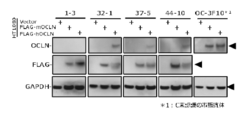
- (C) is an image obtained by staining a sample IP of FLAG-tagged mouse occludin-expressing HT1080 cells with an occludin antibody (C-terminal recognition, commercially available product).
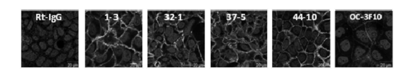
- (D) is an image obtained by staining an IP sample using Caco-2 cells expressing intact occludin with an occludin antibody (C-terminal recognition, commercially available product). It is a figure which shows the result of the immuno-staining by the anti- occludin monoclonal antibody using Caco-2 cell. It is the figure which evaluated the influence which an anti- occludin monoclonal antibody has on a barrier function.
- FIG. 1 It is a figure which shows the in vitro VSV infection inhibitory activity of the anti-occludin monoclonal antibody using VSV (human vesicular stomatitis virus). It is an in vitro HCV infection inhibitory activity of an anti-occludin monoclonal antibody using HCVcc (NS5A inhibitor-resistant mutation-introduced strain), and is a quantitative result of intracellular HCV genomic RNA.
- (A) is a wild type (WT)
- B) is an L31V mutant
- (C) is a Y93H mutant.
- FIG. 5 is a diagram showing the purity of occludin antibody 1-3, which was confirmed by CBB staining, for a sample obtained by purifying the Fab fraction by papain treatment.
- antibody It is difficult to produce an antibody against the intact extracellular domain of occludin, which is a multiple transmembrane protein present in the cell membrane, and so far there has been no report as an occludin ligand antibody that recognizes the extracellular region.
- the anti-occludin monoclonal antibody according to the present invention specifically recognizes the three-dimensional structure of the extracellular region of the polypeptide encoded by the occludin gene and binds to the extracellular region.
- the extracellular region of occludin includes, for example, a ligand binding domain and both the first and second extracellular domains.
- the monoclonal antibody, chimeric antibody, and humanized antibody can be of any isotype.
- the antibody may be, for example, human IgG1, IgG2, IgG3, IgG4, or a variant thereof, mouse IgM, IgG1, IgG2a, IgG2b, IgG3, IgA, IgD, IgE, or a variant thereof.
- Any heavy chain may be paired with a kappa or lambda type light chain.
- Antibodies include Fab fragments, F (ab ') 2 fragments, or Fv fragments, single chain antibodies scFv.
- the present inventors have succeeded in producing four clones as anti-occludin monoclonal antibodies, which are designated as 1-3, 32-1, 37-5, and 44-10, respectively.
- the anti-occludin monoclonal antibody according to the present invention is an anti-occludin monoclonal antibody that binds to an epitope in the first extracellular region and the second extracellular loop of occludin.
- the first extracellular region of human occludin is essential for recognition of 32-1, 37-5, and 44-10, and the second extracellular region of human occludin is essential for recognition of 1-3. is there.
- the epitope in the first extracellular loop is DRGYGTSLLGGSVGYPYGGSGFGSYGSGYGYGYGYGYGYGGYTDPR (Asp Arg Gly Tyr Gly Thr Ser Leu Leu Gly Gly Sly T Gly Tyr Gly Tyr Gly Tyr Gly Gly Tyr Thr Asp Pro Arg) (SEQ ID NO: 1).
- GVNPTAQSSGSLYGSQIYALCNQFYTPAATGLYVDQYLYHYCVVDPQE Gly Val Asn Pro Thr Ala Gln Ser Ser Gly Ser Leu Tyr Gly Ser Gln Ile Tyr Ala Leu Cys Asn Gln Phe Tyr Thr Pro Ala Ala Thr Gly Leu Tyr Val Asp Gln Tyr Leu Tyr His Tyr Cys Val Val Asp Pro Gln Glu
- the complete antibody molecule has two heavy (H) chain variable regions (VH) and two light (L) chain variable regions (VL), whereas in 1-3, VH was QVQLKESGPGLVQPSETLSLTCTVSGFSLSSHHVSWVRQPPGKGLEWIAAISRGGSTYFNSVLKSRLSISRDTSKSQVFLKMNPG (SEQ ID NO: 3). Moreover, in 1-3, VL was YIQMTQSPASLSASPEEIVTITCQASQDIGNWVTWYQQKPGKSPQLLVYSATTLADGIPSRFSGSRSGTQYSLKISRLQVEDTGIYYCLQRYSTPYTFGGGTRLELKR (sequence number 4).
- VH was EVQLVESGGGLVQPRRSLKLSCAASGFTFNKYYMAWVRQTPTKGLEWVASISPRGDNGYYSDSVKGRFTISRDNAKSTQYLQMDSLRSEDTATYYCTTDYGVYWGQGVMVTVSSA (sequence number 5).
- VL was DVQMTQSLSYLAASPGESVSISCKASTSISNYLAWYQQKPGEAYKLLIYSGSTLQSGTPSRFSGSGSGTDFTLTIRSLEPEDFGLYYCQQYYEKPHTFGAGTKLELKR (sequence number 6).
- VH was EVQLVESGGGLLQPGRSLKLSCVASGFTFNHYWMTWVRQAPGKGLEWVASITDIGGATYYLASVKGRFTISRDNTKSTLYLQMNSLRSEDTATYYCTRDDGPFAYWGQGTLVTVSSA (sequence number 7).
- VL was DIQMTQSPSFLSASVGDRVTINCKASQNINRYLNWYQQKLGEAPKLLIYNANSLQTGIPSRFSGSGSGTAFTLTISSLQPEDVATYFCLQHNSWRTFGGGTKLELKR (SEQ ID NO: 8).
- VH was EVKLLESGGGLVQPGGSMRLSCAASGFTFTDFYMNWIRQAAGKAPEWLGFIGNKANGYTTEYIPSVKGRFTISRDNTQNLFFLQMNTLRAEDTGTYYCARGVGDPDYWGQGVMVTVSSA.
- VL was DIVMTQSPSSLAVSAGETVTVNCKSSQSLFSSGNQKNYLAWYQQKPGQSPKLLIYLASNRKSGAPDRFMGSGSGTDFTLTISSMQAEDLAIYYCQQHYDIPPTFGGGTKLELKR (SEQ ID NO: 10).
- VH and VL regions can be subdivided into complementarity determining regions (CDR), and framework regions (FR) are interspersed.
- CDR complementarity determining regions
- FR framework regions
- the present inventors determined the nucleotide sequences of the VH region and VL region from the hybridoma mRNA by the RACE method (rapid amplification of cDNA ends), and analyzed the amino acid sequence. As a result, the sequence was as shown in FIG. .
- Single-chain antibody scFv to the L chain variable region and the H chain of the antibody was expressed as a single chain is a METDTLLLWVLLLWVPGSTGDAAQPARRAVRSLYIQMTQSPASLSASPEEIVTITCQASQDIGNWVTWYQQKPGKSPQLLVYSATTLADGIPSRFSGSRSGTQYSLKISRLQVEDTGIYYCLQRYSTPYTFGGGTRLELKGGGGSGGGGSGGGGSQVQLKESGPGLVQPSETLSLTCTVSGFSLSSHHVSWVRQPPGKGLEWIAAISRGGSTYFNSVLKSRLSISRDTSKSQVFLKMNSLQTEDTAIYFCIGEDWYFDF WGPGTMVTVSSAA RGGP EQKLISEEDL DLNSAVD HHHHHH In ScFv1-3 (SEQ ID NO: 11).
- an amino acid sequence in which one to several amino acids are deleted, substituted or added in the amino acid sequence of the single-chain antibody can also be used as an antibody having HCV infection inhibitory activity.
- the above single-chain antibody and the single-chain antibody with a modified sequence can be used as an antibody having HCV infection inhibitory activity.
- a molecular tag For example, a FLAG tag, a His tag, or a c-Myc tag etc. are mentioned.
- the antibody according to the present embodiment can also include an antibody derivative modified or conjugated by a covalent bond between any kind of molecule and the antibody.
- antibody derivatives include, for example, acetylation, glycosylation, amidation, PEGylation, phosphorylation, derivatization with known protecting / blocking groups, proteolytic cleavage, or intracellular ligands or other proteins Or the antibody modified by the coupling
- a hybridoma that produces the antibody to be obtained can be cultured, and the antibody can be purified and obtained from the obtained culture supernatant by a conventional method.
- the method for collecting the antibody from the obtained hybridoma is not particularly limited, and for example, a normal ascites formation method, a cell culture method, or the like can be used.
- the ascites formation method for example, mineral oil such as pristane is administered into the abdominal cavity of an animal of the same kind as a mammal derived from myeloma cells, and then 1 hybridoma 1 ⁇ 10 6 to 1 ⁇ 10 9 is administered intraperitoneally.
- the hybridoma is grown in large quantities.
- the hybridoma is cultured in an animal cell culture medium such as IMDM containing 10-20% calf serum, RPMI-1640, MEM, E-RDF, or serum-free medium (eg, 37 ° C.). And 5% CO 2 concentration) for 3 to 14 days, and antibodies can be obtained from the culture supernatant.
- Antibody purification includes, for example, ammonium sulfate salting-out method, ion exchange chromatography using anion exchangers such as DEAE cellulose, affinity chromatography using protein G sepharose, molecular sieve chromatography that screens according to molecular weight and structure, etc. It is possible to purify by appropriately selecting a known method.
- Another method is to obtain a gene encoding an antibody from a hybridoma producing the antibody to be obtained, more specifically, a gene encoding an immunoglobulin heavy chain and light chain, and to express the gene.
- a vector can be prepared and introduced into a host cell (mammalian cell, insect cell, microorganism, etc.) to produce the antibody.
- the gene encoding immunoglobulin heavy chain and light chain is modified to introduce a desired trait, or a chimera having a human IgG skeleton using immunoglobulin heavy chain and light chain variable regions
- a person skilled in the art can carry out the production of an antibody, a humanized antibody, a low molecular antibody or a scaffold antibody by using a known technique.
- the present invention also relates to molecules that bind to an occludin epitope.
- An epitope is a specific structural unit of an antigen that is recognized and bound by an antibody. When an antibody binds to a pathogenic microorganism or a macromolecular substance, it does not recognize the whole, but recognizes and binds to an epitope, the epitope is the smallest unit for antigenicity, and the molecule that binds to the epitope is For example, organic synthetic compounds and peptides.
- the anti-HCV therapeutic agent according to this embodiment binds to the anti-occludin monoclonal antibody, human chimeric anti-occludin monoclonal antibody, humanized anti-occludin monoclonal antibody or fragment thereof, single-chain anti-occludin antibody scFv or occludin epitope according to this embodiment.
- a molecule is an active ingredient.
- the anti-HCV therapeutic agent according to the present embodiment may include an active ingredient such as these anti-occludin monoclonal antibodies and a DAA agent (Direct-acting antiviral agent direct acting antiviral agent).
- DAA agents include NS3 / 4A inhibitors, NS5A inhibitors, NS5B inhibitors, or combinations thereof.
- the anti-HCV therapeutic agent according to the present embodiment can be configured as an anti-HCV therapeutic agent having an anti-occludin monoclonal antibody and a DAA agent containing an NS5B inhibitor and an NS3 / 4A inhibitor.
- the effective dose of the anti-HCV therapeutic agent according to this embodiment is not particularly limited, and can be, for example, 0.001 to 1,000 mg / kg body weight per time. Alternatively, the dose may be 0.01 to 100,000 mg / body per patient. Moreover, the anti-HCV therapeutic agent concerning this embodiment can be administered regardless of before and after the clinical symptom of a disease arises as the administration time. The anti-HCV therapeutic agent according to this embodiment can be administered, for example, 1 to 3 times a day, 1 to 7 days a week.
- the anti-HCV therapeutic agent according to this embodiment is usually administered parenterally, for example, by injection (subcutaneous injection, intravenous injection, intramuscular injection, intraperitoneal injection, etc.), but is limited to this dosage form. Instead, for example, it may be administered transdermally, transmucosally, nasally, pulmonary, orally.
- the anti-HCV therapeutic agent according to this embodiment may contain a preservative that suppresses the growth of microorganisms or a buffer that helps keep the pH within an acceptable range.
- Preservatives include sodium azide, octadecyldimethylbenzylammonium chloride, hexamethonium chloride, benzalkonium chloride, benzethonium chloride, alkyl parabens such as phenol, butyl or benzyl alcohol, methyl or propylparaben, catechol, resorcinol, cyclohexanol, Such as 3-pentanol and m-cresol.
- Buffering agents are phosphoric acid, citric acid, and other organic acids.
- the anti-HCV therapeutic agent according to the present embodiment may include, for example, an excipient, a stabilizer, a chelating agent such as EDTA, a salt, or an antibacterial agent.
- antioxidants such as ascorbic acid and methionine, polypeptides, proteins such as serum albumin, gelatin, or nonspecific immunoglobulins, polysorbates, hydrophilic polymers such as polyvinylpyrrolidone, glycine, glutamine, asparagine, histidine, It can contain amino acids such as arginine or lysine, monosaccharides such as glucose, mannose, or dextrin, disaccharides, and other carbohydrates, such as sucrose, mannitol, trehalose, or sorbitol.
- the present invention binds to rat anti-occludin monoclonal antibody, human chimeric anti-occludin monoclonal antibody, humanized anti-occludin monoclonal antibody or fragment thereof, single-chain anti-occludin antibody scFv or occludin epitope in an amount effective for the treatment of hepatitis C. It is also possible to construct a kit containing molecules to be used and instructions for use.
- each genotype is known for HCV, rat occludin monoclonal antibody, human chimeric anti-occludin monoclonal antibody, humanized anti-occludin monoclonal antibody, or fragment thereof, since occludin is involved in infection in all genotypes
- a molecule that binds to a single-chain anti-occludin antibody scFv or an occludin epitope can be used for the treatment of hepatitis C of all genotypes.
- An antibody is also defined to include variants having essentially the same biological activity as the antibody and having one or more amino acid residue mutations in the amino acid sequence.
- CHO cells Chinese hamster ovary cells
- CHO / hOCLN cells were stained with the culture supernatant collected above and PE-labeled PE-labeled anti-rat IgG antibody, and analyzed by flow cytometer (FCM).
- FCM flow cytometer
- the CHO / hOCLN cell is a cell in which human occludin is transiently overexpressed by introducing the pBOMB0 vector encoding the human occludin gene into the CHO cell.
- Hybridoma cells were collected from each well in which a positive shift was confirmed by FCM analysis, and each clone was seeded on a 96-well plate at 1.2 cells / well for 11 days in culture medium 1 *. The cells were cultured at 5 ° C. and 5% CO 2 . After culturing, 20-30 wells where single colony formation was observed under a microscope were selected per plate, and the hybridoma culture supernatant was collected. Chinese hamster ovary cells (CHO cells, hOCLN / CHO cells) were used and stained with the culture supernatant collected above and PE-labeled PE-labeled anti-rat IgG antibody for flow cytometer (FCM) analysis.
- FCM flow cytometer
- hepatoma cells Huh7.5.1-8 cells, OKH-4 cells
- FCM flow cytometer
- Huh7.5.1 cell is a well-differentiated human liver cancer-derived cell line, and is a sub-line of Huh7 cell isolated from liver cancer of a 57-year-old male patient showing high susceptibility to hepatitis C virus (HCV) infection .
- Huh7.5 cells were isolated as a strain with higher HCV replication ability than Huh7 cells, and the replicon was eliminated by interferon gamma treatment from the cell line established by introducing the HCV replication system into Huh7.5 cells (HCV replicon cell line) As a result, Huh7.5.1 cells were established.
- Huh7.5.1-8 cells are strains isolated from Huh7.5.1 cells as cell sublines that are more susceptible to infection with HCV.
- OKH-4 cells are occludin-deficient cells derived from Huh7.5.1-8 cells. It was established by introducing a pX330 vector (CRISRP / Cas9 system) containing a target sequence for human occludin into Huh7.5.1-8 cells. As a result of gene expression analysis, it is known that OKH-4 cells are specifically deficient in occludin expression. Occludin deficiency has also been confirmed by immunoblot analysis, cellular immunostaining, FCM analysis and the like.
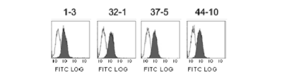
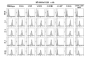
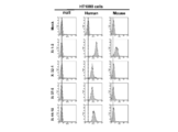
- FIG. 2 shows the results of FCM analysis of the binding of occludin expressed transiently to the cell surface of each antibody clone.
- the area shown in white (hereinafter, the unfilled area) is a staining pattern of CHO cells (occludine negative cells) by each hybridoma culture supernatant, and is filled in on the right side.
- the area shown is a staining pattern of CHO / hOCLN cells (occludin positive cells) by each hybridoma culture supernatant. All clones showed high binding (positive shift) to CHO / hOCLN cells (occludin expressing cells).
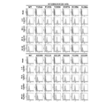
- FIG. 3 shows the results of FCM analysis of the binding of each antibody clone to the occludin on the cell surface expressed intact.
- FIG. 3 shows the results of FCM analysis of the binding of each antibody clone to the occludin on the cell surface expressed intact.
- the area shown in white is the staining pattern of OKH-4 cells (occludin negative cells) by each hybridoma culture supernatant, and the filled area on the right side is Huh7 by each hybridoma culture supernatant. This is a staining pattern of .5.1-8 cells (occludin positive cells). All clones showed high binding (positive shift) to Huh7.5.1-8 cells (Occludin positive cells). In addition, a part of the culture supernatant was used to determine the class and subclass of the antibody in the culture supernatant using a Rat immunoglobulin isotyping ELISA kit. The results are shown in Table 1.
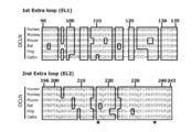
- FIG. 7 shows the homology analysis of the extracellular region sequence.
- human occludin, cynomolgus occludin, mouse occludin, rat occludin, dog occludin, bovine occludin This is an analysis of amino acid sequence homology.
- the epitope in the first extracellular loop is DRGYGTSLLGGSVGYPYGGSGFGSYGSGYGYGYGYGYGYGGYGGYTDPR (Asp Arg Gly Tyr Gly Thr Ser Leu Leu Gly Gly Ser Val Gly Tyr Pro Tyr Gly Gly Ser Gly Phe Gly Ser Tyr Gly Tly Gly Tyr T Tyr Gly Gly Tyr Thr Asp Pro Arg) (SEQ ID NO: 1).
- the epitope in the second extracellular loop is GVNPTAQSSGSLYGSQIYALCNQFYTPAATGLYVDQYLYHYCVVDPQE (Gly Val Asn Pro Thr Ala Gln Ser Ser Gly Ser Leu Tyr Gly Ser Gln Ile Tyr Ala Leu Cys Asn Gln Phe Tyr Thr Pro Aly G Tyr Leu Tyr His Tyr Cys Val Val Asp Pro Gln Glu) (SEQ ID NO: 2). 5-3) Epitope analysis using human occludin extracellular region mutant-1
- FIG. 8 shows the results of preparing and analyzing swap mutants for the extracellular regions (first extracellular region / second extracellular region) of mouse occludin and human occludin.
- Mouse occludin (m / m), mouse / human occludin (m / h), human / mouse occludin (h / m), human occludin (h / h) -expressing HT1080 cells were collected by trypsin treatment. To 5.0 ⁇ 10 5 cells, 100 ⁇ L of each antibody 5 ⁇ g / mL was added, and FCM analysis was performed in the same manner as in 5-1). The results are shown in FIG. Regarding Clone 1-3, binding was confirmed in all swap mutants.
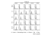
- Clone 32-1, Clone 37-5, and Clone 44-10 showed binding for h / h and h / m mutants, but binding was observed for m / h and m / m mutants. There wasn't. From this result, it was found that Clone32-1, Clone37-5, and Clone44-10 have an epitope in the first extracellular region, and the binding property disappears when converted to a mouse type.
- FIG. 9 shows the results of analysis of antibody binding to cells expressing each occludin mutant, in which the first extracellular region of human occludin was replaced with a mouse type (excluding mutations not common to mice and rats). Mutants (S96G, L98F, V102L, G103N, ⁇ G107, S116G, G119_Y120 insG) expressing HT1080 cells in which the amino acid in the first extracellular region of human occludin was substituted with a mouse type were recovered by trypsin treatment.
- Clone1-3 confirmed binding in all mutants.
- Clone 32-1, Clone 37-5, and Clone 44-10 no change in binding was observed in the amino acid substitutions.
- loss of binding was confirmed by deletion of 107th glycine in the extracellular first region of occludin. This indicates that 107th glycine in the extracellular first region is important for recognition of occludin in Clone 32-1, Clone 37-5, and Clone 44-10.
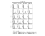
- FIG. 10 shows the results of analysis of antibody binding to each occludin mutant-expressing cell in which the first human occludin extracellular region was replaced with a bovine type. Mutants ( ⁇ S96, L98M, G100A, S101G, G107A, G110A, G129_G130 insT) expressing HT1080 cells in which the amino acid in the first extracellular region of human occludin was substituted with bovine type were recovered by trypsin treatment.
- Clone1-3 confirmed binding in all mutants.
- Clone 32-1 the binding was greatly reduced by the G110A mutation, and the binding was completely lost by the G107A mutation.
- Clone 37-5 binding was not observed in the G107A mutation and the G110A mutation.
- Clone 44-10 the binding could be confirmed by G107A mutation and G110A mutation.
- Other mutants were also binding to 3 Clone. Therefore, in addition to the 107th glycine shown in Fig. 9, the 110th glycine also affects the recognition of occludin in Clone 32-1 and Clone 37-5.
- FIG. 11 shows the results of analysis of antibody binding to each occludin mutant-expressing cell in which human occludin first extracellular region 104-115 was replaced with alanine (Alanine, A).
- HT1080 cells expressing each alanine substitution mutant (Y104A, P105A, Y106A, G107A, G108A, S109A, G110A, F111A, G112A, S113A, Y114A, G115A) were collected by trypsin treatment.
- Y104A, P105A, Y106A, G107A, G108A, S109A, G110A, F111A, G112A, S113A, Y114A, G115A were collected by trypsin treatment.
- 100 ⁇ L of each antibody 5 ⁇ g / mL was added, and FCM analysis was performed in the same manner as in 5-1).
- Clone1-3 confirmed binding in all mutants. For Clone 32-1, binding was completely lost by mutations Y104A, P105A, G107A, G108A, and F111A.
- Each cell was seed
- PVDF polyvinylidene fluoride
- IP Immunoprecipitation
- the barrier function control activity of anti-occludin monoclonal antibody was analyzed using Caco-2 cells widely used as a human intestinal epithelial model. Caco-2 cells were seeded at 8 ⁇ 10 4 cells / 300 ⁇ L in a trans well (CORNING) top well, 700 ⁇ L of medium was added to the bottom well, and cultured in a 37 ° C., 5% CO 2 environment. Every other day, the transepithelial electrical resistance (TEER) value was measured with Millicell-ERS (MILLIPORE), the medium was changed, and the culture was continued in a 37 ° C., 5% CO 2 environment.
- CORNING trans well
- MILLIPORE Millicell-ERS
- an anti-occludin monoclonal antibody was prepared in a medium at 50 ⁇ g / mL, rat antibody (Jackson Immuno Research) was used as a negative control, and venom diarrhea toxin was used as a positive control.
- C-CPE was prepared at 5.0 ⁇ g / mL in each medium, and 300 ⁇ L of medium was added to the top well and 700 ⁇ L of antibody dilution was added to the bottom well, excluding the precultured cell medium.
- the cells were cultured at 37 ° C. in a 5% CO 2 environment, and TEER values were measured over time after antibody addition.
- top well medium and the bottom well antibody dilution were removed and washed with PBS.
- the top well was changed to 300 ⁇ L and the bottom well was changed to 700 ⁇ L, and TEER was measured over time. The results are shown in FIG. No effect was seen when each anti-occludin monoclonal antibody was treated with 5.0 ⁇ g / mL.
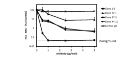
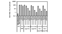
- Huh7.5.1-8 cells were seeded at 5 ⁇ 10 4 cells / 500 ⁇ L / well in a collagen type I-coated 48-well plate (Corning, NCO3548) and cultured at 37 ° C. for 1 day. The medium was removed, and a medium (250 ⁇ L) containing 0.1 to 5 ⁇ g of four kinds of occludin purified antibodies was added, followed by incubation at room temperature (25 ° C.) for 30 minutes.
- HCVcc human immunodeficiency virus
- the cells were washed 3 times with PBS 500 ⁇ L / well, extracted and purified using Blood / Cultured Cell Total RNA Purification Mini Kit (FAVORGEN), dissolved in 50 ⁇ L of sterilized water, and stored at ⁇ 80 ° C. The RNA concentration was measured using Nano Drop.
- FAVORGEN Blood / Cultured Cell Total RNA Purification Mini Kit
- HCV genomic RNA quantification was performed by the Taqman qRT-PCR method (reagents are RNA-direct Realtime PCR Master Mix (Toyobo), and the instrument is LightCycler (Roche)). The reaction solution was adjusted according to the composition shown in Table 2.
- the Sense Primer is 5'-ACGGGGTTAATTATGCAACAGG-3 '(SEQ ID NO: 13)
- the Anitisense Primer is 5'-ACGGTGATGCAGGACAACAG-3' (SEQ ID NO: 14)
- the Taqman Probe is 5 '-[6-FAM] AGCAAGAAGATAGAAAAGGGGAAACCGGGTAG [TAMRA-6-FAM] -3 ′ (SEQ ID NO: 15).
- Huh7.5.1-8 cells were seeded at 1 ⁇ 10 5 cells / well / 500 ⁇ L in a 48-well plate and cultured overnight.
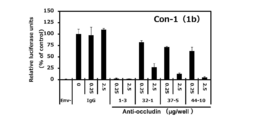
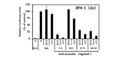
- Anti-Occludin monoclonal antibodies were prepared in various concentrations in a medium, 250 ⁇ L / well antibody solution was added after removing the pre-cultured cell medium, and the mixture was allowed to stand at room temperature for 30 minutes. Each antibody was allowed to act at 0, 0.25 and 2.5 ⁇ g / well.
- HCVpp (genotype 1a (H77 strain), 1b (TH strain, Con-1 strain) or 2a (JFH-1 strain, J6 strain), Microbe and Infection 15, 45-55, 2013) or VSV (bullous mouth) Flame virus) was added at 250 ⁇ L / well and cultured at 37 ° C. for 6 hours.
- the plate was washed 3 times with 500 ⁇ L / well of a medium not containing serum, 500 ⁇ L of an antibody solution having a concentration half that of the pretreatment was added, and the mixture was cultured at 37 ° C. for 3 days.
- Lysis buffer Promega was added at 100 ⁇ L / well, and the lysate was collected in a 1.5 mL tube, centrifuged at 10,000 rpm for 1 minute, and placed on ice. 10 ⁇ L of this supernatant and 50 ⁇ L of luminescent substrate (Piccagene) were mixed, and the luminescence intensity was measured with Luminescencer-PSN.
- FIGS. 21, 22, 23, 24, 25 and 26 The results are shown in FIGS. 21, 22, 23, 24, 25 and 26.
- 21 shows HCVpp (genotypeo1a)
- FIG. 22 shows HCVpp (genotype 1b-TH)
- FIG. 23 shows HCVpp (genotype 1b-Con-1)
- FIG. 24 shows HCVpp (genotype 2a-JFH1)
- FIG. Are HCVpp (genotype 2a-J6)
- NS5A a chimeric full-length replicon
- Huh7.5.1-8 cells recovered by trypsin treatment were washed once with 5 mL of K-PBS, and the number of cells was adjusted to approximately 2 ⁇ 10 7 cells / mL.
- 500 ⁇ L of these cells and 50 ⁇ g of HCV RNA synthesized in vitro were added and allowed to stand on ice for 5 minutes. Suspend and resuspend cells prior to electroporation.
- Electroporation was performed using Gene Pulser X cell (Bio Rad) under the conditions of 975 ⁇ FD and 290 mV. After electroporation, 1.5 mL of serum medium was added and allowed to stand for 15 minutes. Transfected cells were transferred to a 50 mL conical tube and precipitated at room temperature, 200 ⁇ g, 5 minutes. The precipitated cells were suspended in 10 mL of medium, and 3 ⁇ 10 6 cells / dish were seeded on a 10 cm 2 dish of collagen coat and cultured at 37 ° C. On the next day (24 hours or more), the serum medium was removed, fresh serum medium was added, and the mixture was further cultured at 37 ° C. for 4 days.
- the collected virus supernatant was filtered through Millex-HV 0.45 ⁇ m (Millipore), and the virus solution was concentrated about 10 times using Amicon Ultra-15 (Millipore).
- the subsequent operations are based on the infection inhibition activity analysis using Cell culture derived HCV (HCVcc) in the previous section.
- HCVcc Cell culture derived HCV
- Anti-CL1 Clone 2C1 was used as a positive control.
- the results are shown in FIG. The broken line indicates the background.
- the examined anti-occludin monoclonal antibodies Clone 1-3 and Clone 44-10 also had infection inhibitory activity against NS5A inhibitor-resistant mutant strains.
- NS5A inhibitors target viruses, whereas anti-occludin monoclonal antibodies target host factors, so it is expected to have infection inhibitory activity against NS5A inhibitor-resistant mutant strains. It is strongly foreseen that it will be widely effective against other DAA drug resistant viruses.
- Interferon- ⁇ promoter stimulator-1 is a membrane protein present on the outer mitochondrial membrane and induces innate immunity by participating in IFN- ⁇ production during RNA virus infection.
- NS3 / NS4A a non-structural protein of HCV
- the fluorescent protein fusion gene mCherry-NLS-IPS-1 is a gene containing a red fluorescent protein mCherry, a nuclear localization signal (NLS), and IPS-1.
- IPS-1 present on the cytoplasm side is cleaved by NS3 / NS4A produced by HCV infection, and the fluorescent protein is transferred to the nucleus by NLS.
- HCV infection can be evaluated by observing changes in the behavior of the fluorescent protein in this cell (translocation to the nucleus).
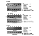
- HCV infectivity was also evaluated by a method using immunoblot analysis.
- HCV-JFH1 was used as a control.
- the results are shown in FIGS.
- the transfer of the fluorescent protein showing HCV infection to the nucleus occurred in all cells.
- Occludin antibodies 37-5 and 44-10 were found to suppress most of the infections, although only a few were infected.
- the occludin antibody 32-1 the TNS2-J1 strain completely blocked the transfer of the fluorescent protein to the nucleus, but Jc-1 was confirmed to be about half of the infection.
- Similar results were obtained by immunoblot analysis and immunostaining for HCV core (FIGS. 29 and 30). From the above results, it was confirmed that established antibodies, particularly 1-3, are probes that can inhibit various HCVcc infections.
- reporter cells were established by introducing mCherry-NLS-IPS-1 into 751r cells that did not cause cell-free infection.
- This evaluation system was used to evaluate whether occludin antibody can inhibit cell-to-cell infection.
- Huh7.5.1-8 cells infected with HCV-JFH-1 and 751r / mCherry-NLS-IPS1 cells cell-to-cell infection was caused.
- FIG. 1 In the rat control antibody-treated group, transfer of the red fluorescent protein to the nucleus was confirmed.
- cell-to-cell infection is known to be less likely to occur compared to cell-free infection.
- IgG4 is known to improve in vivo stability by substituting Pro at 228th Ser in the heavy chain constant region. (Immunology, 105, 9-19, 2002). Therefore, a primer was designed to replace 228th Ser with Pro, 682 bp between NheI site and BsrGI site was amplified by PCR using pFUSE-CHIg-hG4 (Invivogen) as a template, and the PCR product was electrophoresed by electrophoresis. After separation, purification was performed to obtain pFUSE-CHIg-hG4m. The NheI site was added to the primer upstream of the mutation insertion site, and the BsrGI site was added to the downstream primer.
- the gene encoding the amino acid of the VL region and VH region of the variable region of each antibody clone was amplified by PCR.
- the AgeI site was added upstream of the VL gene, the BsiWI site was added downstream, the EcoRI site was added upstream of the VH gene, and the NheI site was added downstream.
- PCR products were separated and purified by electrophoresis.
- PFUSE2-CLIg-hk (Invivogen), a cloning vector having an amplified VL gene and a human IgG kappa chain constant region, was treated with AgeI and BsiWI and then ligated.
- the amplified VH gene and pFUSE-CHIg-hG4m were treated with EcoRI and NheI and then ligated.
- Control antibody, Clone -3 1-3 or Clone ⁇ 37-5 was intraperitoneally administered to mice.
- the first administration day was day 0, and the antibody dose was 50 mg / kg on day 0, 30 mg / kg on day 3, 20 mg / kg on day 7, and 10 mg / kg on day 10.
- Saline was used for dilution of the antibody administration solution.
- mice were anesthetized with isoflurane 8 hours after the first antibody administration, and 100 ⁇ L of HCV (genotype 1b) adjusted to 1.0 ⁇ 10 5 copies / mL with physiological saline was inoculated from the orbital venous plexus.
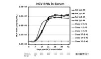
- mice administered with the control antibody, Clone 1-3 or Clone 37-5 were collected daily and HCV infection inhibitory activity was evaluated by measuring the amount of HCV RNA in the serum. The results are shown in FIG. In mice treated with Control antibody, viral RNA was detected in all mice after day 14, and the viral RNA amount reached about 10 7 copies / mL and reached a plateau after day 21, whereas Clone 1-3 or Clone 37-5 The viral RNA level remained below the limit of quantification in all the mice that received the dose. From the above results, it was shown that HCV infection can be completely prevented by administration of the rat anti-occludin monoclonal antibody Clone 1-3 or Clone 37-5.
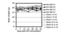
- day 3 day 7, day 10, day 14, day 21, day 28, day 35, day 42, the general state of the mouse was observed and the body weight was measured. Note that day 0, day 3, day 7, and day 10 were performed before antibody administration.
- mice were anesthetized with isoflurane and blood was collected from the orbital venous plexus before antibody administration.
- Serum HCV genomic RNA levels were measured using blood collected on day 0, day 7, day 14, day 21, day 28, day 35, and day 42. RNA was extracted from 5 ⁇ L of the collected serum using SepaGene® RV-R (Adia Corporation, Tokyo). ) Containing 10 ⁇ L of Nuclease-free water (Life Technologies Corporation, CA, USA). The PCR reaction solution was prepared using 2.5 ⁇ L of dissolved RNA stock solution or diluted RNA and TaqMan EZ RT-PCR Core Reagents (Life Technologies Corporation). ABI Prism-7500 (Life Technologies Corporation) was used for PCR reaction and analysis. RT-PCR reaction was performed at 50 ° C. for 2 minutes ⁇ 60 ° C. for 30 minutes ⁇ 95 ° C. for 5 minutes ⁇ (95 ° C. for 20 seconds ⁇ 62 ° C. for 1 minute) ⁇ 50 cycles.
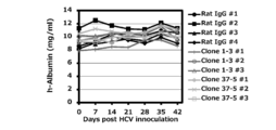
- the blood human albumin concentration was measured using blood collected on day 0, day 7, day 14, day 21, day 28, day 35, and day 42. 2 ⁇ L of blood was mixed with a buffer solution (LX reagent Eiken series shared buffer, Eiken Chemical Co., Ltd., Tokyo), centrifuged at 370 mm x g for 3 minutes, and then latex agglutination immunoturbidimetric method (LX reagent Using “Eiken” Alb-II, Eiken Chemical Co., Ltd., Tokyo), measurement was performed with an absorption microplate reader (Vmax, Nihon Molecular Devices Co., Tokyo).
- Serum ALT and AST activities were measured using blood collected on day 0, day 7, day 14, day 21, day 28, day 35, and day 42.
- Dilute Chem 3500 (Fuji Film, Tokyo) by diluting 10 ⁇ L of the collected serum with physiological saline and using the POP / POD / leuco dye method (the diarylimidazole leuco dye is colored blue with hydrogen peroxide and peroxidase generated by pyruvate oxidase). ).
- FIG. 35 shows changes in body weight when various antibodies were administered to human liver chimeric mice administered with HCV (genotype 1b).
- FIG. 36 shows the concentration of human albumin in the blood when various antibodies were administered to human liver chimeric mice administered with HCV (genotype 1b).
- FIG. 37 shows AST values when various antibodies are administered to human liver chimeric mice administered with HCV (genotype 1b).
- FIG. 38 shows ALT values when various antibodies are administered to human liver chimeric mice administered with HCV (genotype 1b).
- Clone 1-3 and Clone 37-5 are safe because there was no change in general condition, weight loss, blood human albumin concentration decrease, AST value, or ALT value significantly increased by anti-occludin antibody administration In addition, it was considered to be an infection-inhibiting molecule having both effectiveness.
- mice treated with claudin 1 antibody 3A2 there was no change in the amount of viral RNA, whereas in mice treated with occludin antibody 1-3, the amount of viral RNA tended to decrease. It was confirmed that the amount of viral RNA returned to its original value after a delay. From the above results, it was shown that the rat anti-occludin antibody 1-3 can reduce blood viral load even from persistent infection with HCV.
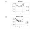
- mice In the Nesbuvir single administration group, all mice (101-104) had breakthroughs, and the blood viral load increased despite the administration of the drug (presumably resistant viruses were growing).
- the Nesbuvir + occludin antibody 1-3 combined administration group one mouse (201) died of unknown cause, and one mouse (204) whose blood viral load did not decrease so much showed a weak breakthrough tendency. The remaining two (202, 203) did not show breakthrough. From the above results, it was shown that breakthrough (generation of resistant virus) can be prevented by coadministration with occludin antibody 1-3. In other words, the occludin antibody was considered to exhibit a useful combination effect when used in combination with the DAA agent.
- the collected sample was added to Ab-Rapid SPiN (Protenova) and allowed to react for 1 hour.
- the column was centrifuged and the passage section was collected as the Fab section. Thereafter, it was replaced with PBS using a PD10 column (GE-Health care).
- SDS-PAGE was performed under reducing conditions. The results are shown in FIG. The Fab band was confirmed at the target position. Undigested Full-body and Fc bands could not be confirmed.
- Huh7.5.1-8 cells were seeded at 5 ⁇ 10 4 cells / 500 ⁇ L / well in a collagen type I-coated 48-well plate (Corning, NCO3548) and cultured at 37 ° C. for 1 day. The medium was removed, a medium (250 ⁇ L) containing 0.5 to 5 ⁇ g of purified Fab1-3 was added, and the mixture was incubated at room temperature (25 ° C.) for 30 minutes.
- HCVcc fragment antigene-binding protein
- the plate was washed 3 times with 500 ⁇ L / well of a medium not containing serum, 500 ⁇ L of purified Fab1-3 solution having a concentration half that of the pretreatment was added, and cultured at 37 ° C. for 4 days.
- the cells were washed 3 times with PBS 500 ⁇ L / well, extracted and purified using Blood / Cultured Cell Total RNA Purification Mini Kit (FAVORGEN), dissolved in 50 ⁇ L of sterilized water, and stored at ⁇ 80 ° C. The RNA concentration was measured using Nano Drop.
- FAVORGEN Blood / Cultured Cell Total RNA Purification Mini Kit
- HCV genomic RNA quantification was performed by the Taqman qRT-PCR method (reagents are RNA-direct Realtime PCR Master Mix (Toyobo), and the instrument is LightCycler (Roche)). The reaction solution was adjusted according to the composition shown in Table 2.
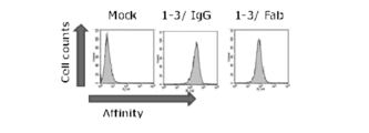
- anti-Occludin Fab1-3 When the HCV infection inhibitory activity of anti-Occludin Fab1-3 was analyzed in an HCVcc infection system, the amount of intracellular HCV RNA was decreased depending on the addition concentration, and a remarkable infection inhibitory action was observed. Note that no cytotoxicity was observed under a microscope. The above results indicate that anti-occludin antibody 1-3 has sufficient ability to inhibit HCV infection even at a monovalent level, and strongly suggests that small molecules that bind to this epitope can function as HCV infection inhibitors. .
- the prepared scFv gene was inserted by ligation between HindIII site and XhoI site in Clone1-3 and between BamHI site and XhoI site in Clone37-5 using pSecTag2 / Hygro A (Invitrogen) as a template.
- Each ligation product was transformed into competent cell DH5 ⁇ , the Escherichia coli clone was independently cultured, the plasmid DNA was recovered, the sequence was confirmed, and pSecTag2 / Hygro-scFv1-3 and scFv37-5 were obtained.
- the amino acid sequence of the constructed construct is shown in FIG.
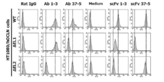
- Binding analysis to various occludins HT1080 cells, human occludin and human occludin first extracellular region deficient ( ⁇ EL1), and human occludin second extracellular region deficient ( ⁇ EL1) -expressing HT1080 cells were collected by trypsin treatment.
- 100 ⁇ L of the culture supernatant containing scFv prepared in the previous section was added, stirred and allowed to stand on ice for 1 hour.
- mouse anti-His tag IgG (MBL) diluted with 1% BSA-PBS was added, and the mixture was stirred and allowed to stand on ice for 60 minutes.
- Huh7.5.1-8 cells were seeded on a 48-well plate at 1 ⁇ 10 5 cells / well / 500 ⁇ L and cultured overnight.
- a medium containing anti-occludin single-chain antibody was prepared at various concentrations, 250 ⁇ L / well of a scFv-containing medium was added except for the precultured cell medium, and the mixture was allowed to stand at room temperature for 30 minutes.
- each culture medium was made to act by undiluted
- HCVpp Genotype 2a (JFH-1 strain), Microbe and Infection 15, 15, 45-55, prepared in accordance with 2013
- HCVpp was added at 250 ⁇ L / well and cultured at 37 ° C. for 6 hours.
- the plate was washed 3 times with 500 ⁇ L / well of a medium containing no serum, 500 ⁇ L of scFv-containing culture solution having a concentration half that of the pretreatment was added, and the mixture was cultured at 37 ° C. for 3 days.
- Lysis buffer Promega was added at 100 ⁇ L / well, and the lysate was collected in a 1.5 ml tube, centrifuged at 10,000 rpm for 1 minute, and placed on ice. 10 ⁇ L of this supernatant and 50 ⁇ L of luminescent substrate (Piccagene) were mixed, and the luminescence intensity was measured with Luminescencer-PSN.
- HRP labeling of anti-occludin antibody Clone1-3 HRP labeling of anti-occludin antibody Clone1-3
- the antibody solution was prepared so as to be 4 mg / mL (3.8-4.2 mg / mL), and the pH of the solution was adjusted to 9.8 using 100 mM Na 2 CO 3 / NaHCO 3 .
- a 2M Triethanolamine (pH 8.0) solution and 200 mM NaBH 4 were added as a reaction stop solution and reacted at 4 ° C. for 2 hours.
- 10 ⁇ L of 1M Glycine solution was added and mixed well.
- the labeled antibody was prepared by performing sufficient dialysis 3 times or more with 200 mL of PBS.
- a screening system was constructed using human occludin-expressing HT1080 cells (HT1080 / hOCLN cells) and HRP-labeled antibodies.
- HT1080 / hOCLN cells were seeded in a 96-well plate (Corning) coated with collagen and cultured at 37 ° C. for 2 days. After immobilization with 3.7% formaldehyde, blocking with 5% skim-milk and labeled anti-occludin monoclonal antibody 1-3-HRP were allowed to react.
- It can be used as a therapeutic agent for hepatitis C.
Landscapes
- Health & Medical Sciences (AREA)
- Chemical & Material Sciences (AREA)
- Life Sciences & Earth Sciences (AREA)
- Organic Chemistry (AREA)
- General Health & Medical Sciences (AREA)
- Medicinal Chemistry (AREA)
- Immunology (AREA)
- Genetics & Genomics (AREA)
- Engineering & Computer Science (AREA)
- Biochemistry (AREA)
- Bioinformatics & Cheminformatics (AREA)
- Molecular Biology (AREA)
- Biophysics (AREA)
- Proteomics, Peptides & Aminoacids (AREA)
- Veterinary Medicine (AREA)
- Public Health (AREA)
- Zoology (AREA)
- Biomedical Technology (AREA)
- Animal Behavior & Ethology (AREA)
- Biotechnology (AREA)
- General Engineering & Computer Science (AREA)
- Epidemiology (AREA)
- Pharmacology & Pharmacy (AREA)
- Microbiology (AREA)
- Wood Science & Technology (AREA)
- Mycology (AREA)
- Plant Pathology (AREA)
- Physics & Mathematics (AREA)
- Cell Biology (AREA)
- Medicines Containing Antibodies Or Antigens For Use As Internal Diagnostic Agents (AREA)
- Peptides Or Proteins (AREA)
- Preparation Of Compounds By Using Micro-Organisms (AREA)
Abstract
HCV感染阻害活性を有する抗オクルディンモノクローナル抗体を提供する。本発明にかかる抗体は、抗オクルディンモノクローナル抗体である。また、ヒトオクルディンの細胞外ループ中のエピトープを認識してヒトオクルディンに結合する抗オクルディンモノクローナル抗体である。抗オクルディンモノクローナル抗体、ラット抗オクルディンモノクローナル抗体、ヒトキメラ抗オクルディンモノクローナル抗体、ヒト化抗オクルディンモノクローナル抗体、抗オクルディン一本鎖抗体scFv、抗オクルディンモノクローナル抗体のエピトープに結合する分子を有効成分とする抗HCV治療剤が提供される。
Description
本発明は、オクルディンモノクローナル抗体、その抗体のフラグメント(Fab fragment等)、抗体のL鎖とH鎖の可変領域が一本鎖として発現させた一本鎖抗体scFv(single-chain variable fragment)、オクルディンエピトープに結合する分子、及びその抗体等を有効成分とする抗HCV治療剤に関する。
C型肝炎ウイルス(以下、本明細書において「HCV」と記載する場合がある)は慢性肝疾患発症の主要因の一つであり、感染者は国内では約150万人に達すると推定される。HCV感染者の約7割は慢性肝炎に進行し、その後20~30年をかけて約半数が肝硬変に、約10%が肝癌に至り、国内の肝癌による年間死亡者数のうち約7割がHCV感染に起因するものとされている。国内のHCV保有者は肝炎の症状を示していないウイルスキャリアーを含めると200万人以上と推定されており、それはこれまでインターフェロン以外の有効な抗ウイルス剤が無かったことが主原因となっている。
近年、ウイルス因子を標的とした薬剤(以下、DAA製剤:NS3/4Aプロテアーゼ阻害剤やNS5A阻害剤、NS5B阻害剤)が開発され、きわめて高い効果を示すことが報告されている。副作用の大きいインターフェロンを用いない治療(インターフェロンフリー療法)も現実的となってきている。しかしながら、DAA製剤では耐性ウイルスの問題が生じる可能性があり、実際に耐性ウイルス株が複数分離されている。またDAA製剤でウイルスを完全に排除できない事例も多数報告されている。そのため、薬剤耐性が生じ難い宿主因子を標的としたアプローチが重要になると考えられる。
HCVのようにエンベロープを持ったウイルスが宿主の細胞に感染する際は、細胞表面の特異的なタンパク質(ウイルス受容体)に結合し、その後、ウイルス膜と細胞膜が融合してウイルス遺伝子を細胞内に注入する。そのため、HCVが細胞表面のウイルス受容体に結合し進入するのを阻止できる抗体等が求められる。
哺乳動物で発現させたHCVのE2タンパク質がヒト細胞表面に存在するCD81に特異的に結合することが示され、この実験系を用いて、C型肝炎患者からE2タンパク質とCD81との結合を阻害するNOB(neutralisation of binding)活性を示す抗体の開発がなされている。例えば遺伝子型1aのC型慢性肝炎患者の骨髄リンパ球から抗体遺伝子ライブラリーを作製し、ファージディスプレイ法を用いて、NOB活性を示す抗体が作製されている(特許文献1)。しかし、上記のHCV感染患者からモノクローナル抗体を取得する方法では、HCV感染阻害抗体が存在する患者に限定されること、エンベロープタンパク質は高率に変異すること等から、抗HCV剤として有用な抗体を見出すことが困難である。
一方、オクルディン(Occludin)は、細胞間に形成されるタイトジャンクションの主要なタンパク質として知られる。2009年にPlossらによって、オクルディンがHCVの感染受容体の一つであることが示された(非特許文献1)。
しかしながら、オクルディンは細胞外領域の小さな四回膜貫通タンパク質であること、細胞外領域の種間ホモロジーが高いことから、抗体を含めてオクルディンのバインダーの創製は立ち後れている。細胞膜に存在する複数膜貫通タンパク質であるオクルディンのインタクト細胞外ドメインに対する抗体の作製が困難であることは、例えば非特許文献2,3に示されている。
非特許文献4には、オクルディン細胞外第2ループに対する抗体の作製が記載されている。しかし、この文献では、免疫染色像のデータで、オクルディンの発現のない(低下した)細胞でのシグナル消失(低下)の検証がなく、オクルディンを特異的に認識しているか明らかでない。また、変性オクルディンについての結果であるイムノブロットのデータでは、市販の抗体と同様の位置にバンドが見られることのみを示しており、オクルディンの発現のない細胞や過剰発現の細胞を用いた検討を行っておらず、オクルディンを真に認識しているか明らかでない。更に、異なる分子量の位置に多数のバンドが認められ、特異性に疑問がある。
Nature, 457,882,2009
YAKUGAKU ZASSHI 133(9) 939-945(2013)
文献2 科学技術動向 2009年10月号 関根進「抗体医薬の現状と課題」4.(3)
Journal of Histochemistry & Cytochemistry Volume 55(7): 735-744, 2007
本発明はかかる問題点に鑑みてなされたものであって、HCV感染阻害活性を有する抗オクルディンモノクローナル抗体、及びその抗オクルディンモノクローナル抗体等を有効成分として有する抗HCV治療剤を提供することを目的とする。
本発明にかかる抗オクルディンモノクローナル抗体は、ヒトオクルディンの細胞外ループ中のエピトープを認識してインタクトなヒトオクルディンに結合する抗オクルディンモノクローナル抗体である。
本発明にかかる抗オクルディンモノクローナル抗体は、ラット抗オクルディンモノクローナル抗体、ヒトキメラ抗オクルディンモノクローナル抗体及びヒト化抗オクルディンモノクローナル抗体である。
また、本発明にかかる抗HCV治療剤は、上述の抗オクルディンモノクローナル抗体、ラット抗オクルディンモノクローナル抗体、ヒトキメラ抗オクルディンモノクローナル抗体、ヒト化抗オクルディンモノクローナル抗体若しくはそのフラグメント又は抗オクルディンモノクローナル抗体のエピトープに結合する分子を有効成分とする。
本発明によれば、HCV感染阻害活性を有する抗オクルディンモノクローナル抗体、抗オクルディン一本鎖抗体scFvが得られる。また、本発明にかかる抗HCV治療剤は、HCV感染阻害活性を有する。
以下、添付の図面を参照して本発明の実施形態について具体的に説明するが、当該実施形態は本発明の原理の理解を容易にするためのものであり、本発明の範囲は、下記の実施形態に限られるものではなく、当業者が以下の実施形態の構成を適宜置換した他の実施形態も、本発明の範囲に含まれる。
(抗体)
細胞膜に存在する複数膜貫通タンパク質であるオクルディンのインタクト細胞外ドメインに対する抗体の作製が困難であり、これまでに細胞外領域を認識するオクルディンリガンド抗体としての報告は皆無である。本発明にかかる抗オクルディンモノクローナル抗体は、オクルディン遺伝子によりコードされるポリペプチドの細胞外領域の立体構造を特異的に認識し、且つ該細胞外領域に結合する。
細胞膜に存在する複数膜貫通タンパク質であるオクルディンのインタクト細胞外ドメインに対する抗体の作製が困難であり、これまでに細胞外領域を認識するオクルディンリガンド抗体としての報告は皆無である。本発明にかかる抗オクルディンモノクローナル抗体は、オクルディン遺伝子によりコードされるポリペプチドの細胞外領域の立体構造を特異的に認識し、且つ該細胞外領域に結合する。
オクルディンの細胞外領域には、例えばリガンド結合ドメイン(Ligand binding domain)、第一及び第二の細胞外ドメインのいずれも包含する。モノクローナル抗体、キメラ型抗体、ヒト化抗体は、任意のアイソタイプであることができる。抗体は、例えば、ヒトIgG1、IgG2、IgG3、IgG4、あるいはそれらの改変体、マウスIgM、IgG1、IgG2a、IgG2b、IgG3、IgA、IgD、IgE、あるいはそれらの改変体であってもよい。任意の重鎖は、κ又はλ型の軽鎖と対形成してもよい。抗体にはFabフラグメント、F (ab’)2フラグメント、又はFvフラグメント、一本鎖抗体scFvが包含される。
本発明者らは、抗オクルディンモノクローナル抗体として4つのクローンの作製に成功しており、それぞれ1-3、32-1、37-5、及び44-10と命名している。
本発明にかかる抗オクルディンモノクローナル抗体は、オクルディンの第一細胞外領域及び第二細胞外ループ中のエピトープに結合する抗オクルディンモノクローナル抗体である。
具体的には、32-1、37-5及び44-10の認識にはヒトオクルディンの第一細胞外領域が必須であり、1-3の認識にはヒトオクルディン第二細胞外領域が必須である。
後述するように、第一細胞外ループ中のエピトープは、DRGYGTSLLGGSVGYPYGGSGFGSYGSGYGYGYGYGYGYGGYTDPR (Asp Arg Gly Tyr Gly Thr Ser Leu Leu Gly Gly Ser Val Gly Tyr Pro Tyr Gly Gly Ser Gly Phe Gly Ser Tyr Gly Ser Gly Tyr Gly Tyr Gly Tyr Gly Tyr Gly Tyr Gly Tyr Gly Gly Tyr Thr Asp Pro Arg)(配列番号1)である。また、第二細胞外ループ中のエピトープは、GVNPTAQSSGSLYGSQIYALCNQFYTPAATGLYVDQYLYHYCVVDPQE (Gly Val Asn Pro Thr Ala Gln Ser Ser Gly Ser Leu Tyr Gly Ser Gln Ile Tyr Ala Leu Cys Asn Gln Phe Tyr Thr Pro Ala Ala Thr Gly Leu Tyr Val Asp Gln Tyr Leu Tyr His Tyr Cys Val Val Asp Pro Gln Glu)(配列番号2)である。
完全な抗体分子は、2つの重(H)鎖可変領域(VH)及び2つの軽(L)鎖可変領域(VL)を有しているところ、1-3では、VHは、QVQLKESGPGLVQPSETLSLTCTVSGFSLSSHHVSWVRQPPGKGLEWIAAISRGGSTYFNSVLKSRLSISRDTSKSQVFLKMNSLQTEDTAIYFCIGEDWYFDFWGPGTMVTVSSAであった(配列番号3)。また、1-3では、VLは、YIQMTQSPASLSASPEEIVTITCQASQDIGNWVTWYQQKPGKSPQLLVYSATTLADGIPSRFSGSRSGTQYSLKISRLQVEDTGIYYCLQRYSTPYTFGGGTRLELKRであった(配列番号4)。また、32-1では、VHは、EVQLVESGGGLVQPRRSLKLSCAASGFTFNKYYMAWVRQTPTKGLEWVASISPRGDNGYYSDSVKGRFTISRDNAKSTQYLQMDSLRSEDTATYYCTTDYGVYWGQGVMVTVSSAであった(配列番号5)。また、32-1では、VLは、DVQMTQSLSYLAASPGESVSISCKASTSISNYLAWYQQKPGEAYKLLIYSGSTLQSGTPSRFSGSGSGTDFTLTIRSLEPEDFGLYYCQQYYEKPHTFGAGTKLELKRであった(配列番号6)。また、37-5では、VHは、EVQLVESGGGLLQPGRSLKLSCVASGFTFNHYWMTWVRQAPGKGLEWVASITDIGGATYYLASVKGRFTISRDNTKSTLYLQMNSLRSEDTATYYCTRDDGPFAYWGQGTLVTVSSAであった(配列番号7)。また、37-5では、VLは、DIQMTQSPSFLSASVGDRVTINCKASQNINRYLNWYQQKLGEAPKLLIYNANSLQTGIPSRFSGSGSGTAFTLTISSLQPEDVATYFCLQHNSWRTFGGGTKLELKRであった(配列番号8)。また、44-10では、VHは、EVKLLESGGGLVQPGGSMRLSCAASGFTFTDFYMNWIRQAAGKAPEWLGFIGNKANGYTTEYIPSVKGRFTISRDNTQNLFFLQMNTLRAEDTGTYYCARGVGDPDYWGQGVMVTVSSAであった(配列番号9)。また、44-10では、VLは、DIVMTQSPSSLAVSAGETVTVNCKSSQSLFSSGNQKNYLAWYQQKPGQSPKLLIYLASNRKSGAPDRFMGSGSGTDFTLTISSMQAEDLAIYYCQQHYDIPPTFGGGTKLELKRであった(配列番号10)。
VH及びVL領域は、相補性決定領域(CDR)に細分することができ、フレームワーク領域(FR)が散在している。本発明者らは、ハイブリドーマmRNAからRACE法(rapid amplification of cDNA ends)にてVH領域及びVL領域の塩基配列を決定し、アミノ酸配列を解析したところ、図1に示されるとおりの配列であった。
抗体のL鎖とH鎖の可変領域が一本鎖として発現させた一本鎖抗体scFv(single-chain variable fragment )は、scFv1-3ではMETDTLLLWVLLLWVPGSTGDAAQPARRAVRSLYIQMTQSPASLSASPEEIVTITCQASQDIGNWVTWYQQKPGKSPQLLVYSATTLADGIPSRFSGSRSGTQYSLKISRLQVEDTGIYYCLQRYSTPYTFGGGTRLELKGGGGSGGGGSGGGGSQVQLKESGPGLVQPSETLSLTCTVSGFSLSSHHVSWVRQPPGKGLEWIAAISRGGSTYFNSVLKSRLSISRDTSKSQVFLKMNSLQTEDTAIYFCIGEDWYFDF WGPGTMVTVSSAA RGGP EQKLISEEDL DLNSAVD HHHHHHである(配列番号11)。
またscFv37-5では、METDTLLLWVLLLWVPGSTGDAAQPARRAVRSLVPSSDPDIQMTQSPSFLSASVGDRVTINCKASQNINRYLNWYQQKLGEAPKLLIYNANSLQTGIPSRFSGSGSGTAFTLTISSLQPEDVATYFCLQHNSWRTFGGGTKLELKGGGGSGGGGSGGGGSEVQLVESGGGLLQPGRSLKLSCVASGFTFNHYWMTWVRQAPGKGLEWVASITDIGGATYYLASVKGRFTISRDNTKSTLYLQMNSLRSEDTATYYCTRDDGPFAYWGQGTLVTVSSAARGGPEQKLISEEDLNSAVDHHHHHHである(配列番号12)。
またscFv37-5では、METDTLLLWVLLLWVPGSTGDAAQPARRAVRSLVPSSDPDIQMTQSPSFLSASVGDRVTINCKASQNINRYLNWYQQKLGEAPKLLIYNANSLQTGIPSRFSGSGSGTAFTLTISSLQPEDVATYFCLQHNSWRTFGGGTKLELKGGGGSGGGGSGGGGSEVQLVESGGGLLQPGRSLKLSCVASGFTFNHYWMTWVRQAPGKGLEWVASITDIGGATYYLASVKGRFTISRDNTKSTLYLQMNSLRSEDTATYYCTRDDGPFAYWGQGTLVTVSSAARGGPEQKLISEEDLNSAVDHHHHHHである(配列番号12)。
また、上記一本鎖抗体のアミノ酸配列において、1乃至数個のアミノ酸が欠失、置換若しくは付加されたアミノ酸配列からなるものもHCV感染阻害活性を有する抗体として利用可能である。更には、上記一本鎖抗体及び配列が改変された一本鎖抗体に分子タグが付加されたものもHCV感染阻害活性を有する抗体として利用可能である。分子タグとしては、特に限定されるものではないが、例えば、FLAGタグ、Hisタグ、又は、c-Mycタグ等が挙げられる。
本実施形態にかかる抗体は、任意の種類の分子と抗体との共有結合により修飾又は複合化された、抗体誘導体を包含することも可能である。このような抗体誘導体として、例えば、アセチル化、グリコシル化、アミド化、PEG化、リン酸化、既知の保護基/ブロック基による誘導体化、タンパク質分解的開裂、又は細胞内配位子又は他のタンパク質あるいは低分子化合物への結合により修飾されている抗体が挙げられる。
本実施形態にかかる抗体を取得する方法としては、取得したい抗体を産生するハイブリドーマを培養し、得られた培養上清から常法によって抗体を精製して取得することができる。取得したハイブリドーマから抗体を採取する方法は、特に限定されるものではないが、例えば通常の腹水形成法や細胞培養法等を用いることが可能である。腹水形成法においては、例えば、骨髄腫細胞由来の哺乳動物と同種の動物の腹腔内にプリスタン等の鉱物油を投与し、その後ハイブリドーマ1×10⁶~1×10⁹個を腹腔内に投与し、ハイブリドーマを大量に増殖させる。そして、1~4週間後に腹水又は血清を採集する。細胞培養法においては、例えば、ハイブリドーマを10~20%仔ウシ血清含有IMDM、RPMI-1640、MEM、E-RDF又は無血清培地等の動物細胞培養培地中で、通常の培養条件(例えば37 ℃、5% CO2濃度)で3~14日間培養し、その培養上清から抗体を取得することができる。抗体の精製は、例えば、硫安塩析法、DEAEセルロース等の陰イオン交換体を利用するイオン交換クロマトグラフィー、プロテインGセファロース等を用いるアフィニティークロマトグラフィー、分子量や構造によってふるい分ける分子ふるいクロマトグラフィー等の公知の方法を適宜に選択して精製することが可能である。
また、別の方法としては、取得したい抗体を産生するハイブリドーマから抗体をコードする遺伝子、より詳細には免疫グロブリンの重鎖及び軽鎖をコードする遺伝子を取得して、該遺伝子を発現するためのベクターを作成し、宿主細胞(哺乳類細胞、昆虫細胞、微生物等)に導入して、該抗体を産生させることも可能である。このとき、免疫グロブリンの重鎖及び軽鎖をコードする遺伝子について、望む形質を導入するための遺伝子改変を行ったり、免疫グロブリンの重鎖及び軽鎖の可変領域を用いてヒトIgG骨格を有するキメラ抗体あるいはヒト化抗体、低分子抗体やスキャフォールド抗体を作製することは、公知の技術を用いることで、当業者であれば実施することができる。
また、本発明は、オクルディンエピトープ(epitope)に結合する分子にも関する。エピトープ(epitope)とは、抗体が認識して結合する抗原の特定の構造単位である。抗体は病原微生物や高分子物質等と結合する際、その全体を認識するわけではなく、エピトープを認識して結合し、エピトープは抗原性のための最小単位であり、エピトープに結合する分子は、例えば有機合成化合物やペプチド等である。
(抗HCV治療剤)
本実施形態にかかる抗HCV治療剤は、本実施形態にかかる抗オクルディンモノクローナル抗体、ヒトキメラ抗オクルディンモノクローナル抗体、ヒト化抗オクルディンモノクローナル抗体若しくはそのフラグメント、一本鎖抗オクルディン抗体scFv又はオクルディンエピトープに結合する分子を有効成分とする。また、本実施形態にかかる抗HCV治療剤は、これら抗オクルディンモノクローナル抗体等の有効成分と、DAA剤(Direct-acting antiviral agent 直接作用型抗ウイルス剤)とを含むものとすることができる。DAA剤にはNS3/4A阻害剤、NS5A阻害剤、NS5B阻害剤、又はこれらの組み合わせが含まれる。例えば、本実施形態にかかる抗HCV治療剤は、抗オクルディンモノクローナル抗体と、NS5B阻害薬及びNS3/4A阻害薬を含むDAA剤を有する抗HCV治療剤として構成することも可能である。
本実施形態にかかる抗HCV治療剤は、本実施形態にかかる抗オクルディンモノクローナル抗体、ヒトキメラ抗オクルディンモノクローナル抗体、ヒト化抗オクルディンモノクローナル抗体若しくはそのフラグメント、一本鎖抗オクルディン抗体scFv又はオクルディンエピトープに結合する分子を有効成分とする。また、本実施形態にかかる抗HCV治療剤は、これら抗オクルディンモノクローナル抗体等の有効成分と、DAA剤(Direct-acting antiviral agent 直接作用型抗ウイルス剤)とを含むものとすることができる。DAA剤にはNS3/4A阻害剤、NS5A阻害剤、NS5B阻害剤、又はこれらの組み合わせが含まれる。例えば、本実施形態にかかる抗HCV治療剤は、抗オクルディンモノクローナル抗体と、NS5B阻害薬及びNS3/4A阻害薬を含むDAA剤を有する抗HCV治療剤として構成することも可能である。
本実施形態にかかる抗HCV治療剤の有効投与量は、特に限定されるものではないが、例えば、1回につき体重1 kgあたり0.001~1,000 mgとすることができる。又は、患者あたり0.01~100,000 mg/bodyの投与量とすることができる。また、本実施形態にかかる抗HCV治療剤は、その投与時期として、疾患の臨床症状が生ずる前後を問わず投与することができる。本実施形態にかかる抗HCV治療剤は、例えば、1日1~3回、1週間に1~7日投与することが可能である。
本実施形態にかかる抗HCV治療剤は、通常、非経口投与で、例えば注射剤(皮下注、静注、筋注、腹腔内注等)で投与されるが、この投与形態に限定されるものではなく、例えば、経皮、経粘膜、経鼻、経肺、経口等で投与してもよい。
本実施形態にかかる抗HCV治療剤は、微生物の増殖を抑制する防腐剤又はpHを許容範囲に保つのに役立つ緩衝剤を含んでもよい。防腐剤は、アジ化ナトリウム、オクタデシルジメチルベンジルアンモニウムクロライド、塩化ヘキサメトニウム、塩化ベンザルコニウム、塩化ベンゼトニウム、フェノール、ブチル若しくはベンジルアルコール、メチル若しくはプロピルパラベン等のアルキルパラベン、カテコール、レゾルシノール、シクロヘキサノール、3-ペンタノール、及びm-クレゾール等である。緩衝剤は、リン酸、クエン酸、及びその他の有機酸である。
また、本実施形態にかかる抗HCV治療剤は、例えば、賦形剤、安定剤、EDTA等のキレート化剤、塩、又は抗菌剤を含んでもよい。他にも、アスコルビン酸及びメチオニン等の酸化防止剤、ポリペプチド、血清アルブミン、ゼラチン、若しくは非特異的免疫グロブリン等のタンパク質、ポリソルベート、ポリビニルピロリドン等の親水性ポリマー、グリシン、グルタミン、アスパラギン、ヒスチジン、アルギニン、若しくはリジン等のアミノ酸、グルコース、マンノース、若しくはデキストリン等の単糖類、二糖類、及びその他の炭水化物、スクロース、マンニトール、トレハロース、若しくはソルビトール等の糖類を含むことが可能である。
なお、本発明は、C型肝炎の治療に有効な量のラット抗オクルディンモノクローナル抗体、ヒトキメラ抗オクルディンモノクローナル抗体、ヒト化抗オクルディンモノクローナル抗体若しくはそのフラグメント、一本鎖抗オクルディン抗体scFv又はオクルディンエピトープに結合する分子及び使用説明書を含む、キットを構成することも可能である。
なお、HCVには各遺伝子型が知られるが、全ての遺伝子型でオクルディンが感染に関与していることから、ラット抗オクルディンモノクローナル抗体、ヒトキメラ抗オクルディンモノクローナル抗体、ヒト化抗オクルディンモノクローナル抗体若しくはそのフラグメント、一本鎖抗オクルディン抗体scFv又はオクルディンエピトープに結合する分子は、全ての遺伝子型のC型肝炎の治療に利用可能である。また、抗体には、該抗体と本質的に同等の生物学的活性をもち且つそのアミノ酸配列中1もしくは複数のアミノ酸残基の変異を有する変異体も含まれるものと定義する。
1)細胞融合
ヒトオクルディン発現プラスミドをWistarラット皮下に免疫し、血清中の抗体価上昇が観察された個体に対し、最終免疫(ブースティング)を行った。最終免疫後、動物からリンパ細胞を回収し、マウスミエローマ細胞(P3U1)と細胞融合を行った。融合後の細胞を96-well plate 10枚に播種し、培養培地1*にて13日間、37 ℃、5% CO2下で培養した。
*培養培地1:D-MEM (Wako, 044-29765) + 10% FCS (Hyclone, Lot.FQF24009), 10% BM condimed H1 Hybridoma cloning supplement (Roche, 1088947), 1×HAT supplement (Life technology, 21060017), 50μg/mL Penicillin/Streptomycin (Life technology, 15140122), 4 mM L-Glutamine (Life technology, 25030081)
2)特異モノクローナル抗体産生ハイブリドーマの樹立
培養後、全てのプレートウェルから培養上清を回収した。チャイニーズハムスター卵巣細胞(CHO細胞、CHO/hOCLN細胞)を用い、上記で回収した培養上清、及びPE標識したPE標識抗ラットIgG抗体で染色し、フローサイトメーター(FCM)解析を行った。ここで、CHO/hOCLN細胞は、CHO細胞にヒトオクルディン遺伝子をコードしたpBOMB0ベクターを遺伝子導入した一過性にヒトオクルディンを過剰に発現した細胞である。
ヒトオクルディン発現プラスミドをWistarラット皮下に免疫し、血清中の抗体価上昇が観察された個体に対し、最終免疫(ブースティング)を行った。最終免疫後、動物からリンパ細胞を回収し、マウスミエローマ細胞(P3U1)と細胞融合を行った。融合後の細胞を96-well plate 10枚に播種し、培養培地1*にて13日間、37 ℃、5% CO2下で培養した。
*培養培地1:D-MEM (Wako, 044-29765) + 10% FCS (Hyclone, Lot.FQF24009), 10% BM condimed H1 Hybridoma cloning supplement (Roche, 1088947), 1×HAT supplement (Life technology, 21060017), 50μg/mL Penicillin/Streptomycin (Life technology, 15140122), 4 mM L-Glutamine (Life technology, 25030081)
2)特異モノクローナル抗体産生ハイブリドーマの樹立
培養後、全てのプレートウェルから培養上清を回収した。チャイニーズハムスター卵巣細胞(CHO細胞、CHO/hOCLN細胞)を用い、上記で回収した培養上清、及びPE標識したPE標識抗ラットIgG抗体で染色し、フローサイトメーター(FCM)解析を行った。ここで、CHO/hOCLN細胞は、CHO細胞にヒトオクルディン遺伝子をコードしたpBOMB0ベクターを遺伝子導入した一過性にヒトオクルディンを過剰に発現した細胞である。
FCM解析にて、陽性を示すシフトが確認されたウェルから、それぞれハイブリドーマ細胞を回収し、各クローンに関し1.2 cells/wellで96-well plate 1枚に撒き、培養培地1*にて11日間、37 ℃、5% CO2下で培養した。培養後、顕微鏡下でシングルコロニー形成の認められるウェルをプレートあたり20~30選択し、そのハイブリドーマ培養上清を回収した。チャイニーズハムスター卵巣細胞(CHO細胞、hOCLN/CHO細胞)を用い、上記で回収した培養上清、及びPE標識したPE標識抗ラットIgG抗体で染色し、フローサイトメーター(FCM)解析を行った。
FCM陽性の確認された4クローン分のプレートから、シフト強度が強く、且つ細胞数の多いウェルを各クローン3 wellずつ選択し、24-well plateに37 ℃、5% CO2下で拡大し培養培地1*にて培養を行った。3日間培養後、全てのウェルを6-well plateに拡大し培養培地2*にて培養を行った。3日間培養後、培養上清を回収した。
また、インタクトな状態のオクルディンへの結合性を調べる目的でHepatoma細胞(Huh7.5.1-8細胞、OKH-4細胞)を用い、上記で回収した培養上清、及びAlexa488標識抗ラットIgG抗体で染色し、フローサイトメーター(FCM)解析を行った。
ここで、Huh7.5.1細胞は、高分化型ヒト肝癌由来細胞株であり、高いC型肝炎ウイルス(HCV)感染感受性を示す、57歳男性患者の肝癌より分離されたHuh7細胞の亜株である。Huh7細胞よりHCV複製能の高い株としてHuh7.5細胞が分離され、更にHuh7.5細胞にHCV複製システムを導入して樹立した細胞株(HCVレプリコン細胞株)からインターフェロンガンマ処理によりレプリコンを排除することでHuh7.5.1細胞は樹立された。Huh7.5.1-8細胞は、HCVに対して、より感染感受性が高い細胞亜株としてHuh7.5.1細胞から分離された株である。また、OKH-4細胞は、Huh7.5.1-8細胞由来のオクルディン欠損細胞である。Huh7.5.1-8細胞にヒトオクルディンへの標的配列を含むpX330ベクター(CRISRP/Cas9システム)を遺伝子導入し、樹立されたものである。遺伝子発現解析を行った結果、OKH-4細胞はオクルディンの発現が特異的に欠損していることがわかっている。イムノブロット解析、細胞免疫染色、FCM解析等によってもオクルディンの欠損が確認されている。
FCM解析の結果、全てのウェルにおいて、オクルディン特異的な陽性を示すシフトが確認された。シフト強度が強く、且つ細胞数の多いウェルを各クローン1 wellずつ選択し、75 cm2 Flaskに37 ℃、5% CO2下で拡大し培養培地2*(下記参照)にて培養を行った。3~5日間培養後、150 cm2プレートに拡大し培養培地2*にて培養を行った。5日間培養後、培養上清を回収した。チャイニーズハムスター卵巣細胞(CHO細胞、CHO/hOCLN細胞)、Hepatoma細胞(Huh7.5.1細胞、OKH-4細胞)を用い、回収した培養上清の一部、及びチャイニーズハムスター卵巣細胞ではPE標識、Hepatoma細胞ではAlexa488標識した抗ラットIgG抗体で染色し、FCM解析を行った。この結果、図1に示される合計4クローンにおいて、各クローンで複数の陽性を示すシフトが確認された(図2、図3)。図2は、各抗体クローンの一過性発現したオクルディンの細胞表面への結合をFCMにより解析した結果である。ここで図2において、白で示されたエリア(以下、塗りつぶされていないエリアを意味する。)は、各ハイブリドーマ培養上清によるCHO細胞(オクルディン陰性細胞)の染色パターンであり、右側の塗りつぶされたエリアは、各ハイブリドーマ培養上清によるCHO/hOCLN細胞(オクルディン陽性細胞)の染色パターンである。全てのクローンはCHO/hOCLN細胞(オクルディン発現細胞)への高い結合性(陽性シフト)を示した。図3は、インタクトに発現している細胞表面のオクルディンへの各抗体クローンの結合をFCMにより解析した結果である。ここで図3において、白で示されたエリアは、各ハイブリドーマ培養上清によるOKH-4細胞(オクルディン陰性細胞)の染色パターンであり、右側の塗りつぶされたエリアは、各ハイブリドーマ培養上清によるHuh7.5.1-8細胞(オクルディン陽性細胞)の染色パターンである。全てのクローンはHuh7.5.1-8細胞(オクルディン陽性細胞)への高い結合性(陽性シフト)を示した。また、培養上清の一部を用いて、Rat immunoglobulin isotyping ELISA kitを用いて培養上清中の抗体のクラス、サブクラス決定を行った。結果を表1に示す。

*培養培地2:D-MEM (Wako, 044-29765) + 10% FCS (Hyclone, Lot.FQF24009), 5% BM condimed H1 Hybridoma cloning supplement (Roche, 1088947), 1×HAT supplement (Life technology, 21060017), 50μg/mL Penicillin/Streptomycin (Life technology, 15140122), 4 mM L-Glutamine (Life technology, 25030081)
3)抗オクルディンモノクローナル抗体の各動物種オクルディンに対する結合性解析
ヒトオクルディン(NP_001192183.1)、カニクイザルオクルディン(XP_005557154.1)、マウスオクルディン(NP_032782.1)、ラットオクルディン(NP_112619.2)、イヌオクルディン(NP_001003195.1)発現HT1080細胞をトリプシン処理により回収した。5.0×10⁵cellsに対し、各抗体5μg/ mLを100 μL添加、撹拌後、氷上で1時間静置した。0.2% BSA-PBSにて1回洗浄後、1% BSA-PBSにて希釈したGoat anti-rat IgG(H+L)-Alexa488抗体(Life technology)を添加、撹拌後、氷上で遮光し30分静置した。0.2% BSA-PBSにて2回洗浄後、0.2% BSA-PBSにて終濃度5μg/mLとなるように希釈したPI(Miltenyi Biotec)を加え、FCM解析を行った。結果を図4に示す。いずれの抗体クローンも、ヒトオクルディン、カニクイザルオクルディン結合した。Clone 1-3は、マウスオクルディン、ラットオクルディンに結合性を示したのに対し、他の3 クローンでは結合性が確認されなかった。イヌオクルディンに対してどのクローンも結合性を有していなかった。
3)抗オクルディンモノクローナル抗体の各動物種オクルディンに対する結合性解析
ヒトオクルディン(NP_001192183.1)、カニクイザルオクルディン(XP_005557154.1)、マウスオクルディン(NP_032782.1)、ラットオクルディン(NP_112619.2)、イヌオクルディン(NP_001003195.1)発現HT1080細胞をトリプシン処理により回収した。5.0×10⁵cellsに対し、各抗体5μg/ mLを100 μL添加、撹拌後、氷上で1時間静置した。0.2% BSA-PBSにて1回洗浄後、1% BSA-PBSにて希釈したGoat anti-rat IgG(H+L)-Alexa488抗体(Life technology)を添加、撹拌後、氷上で遮光し30分静置した。0.2% BSA-PBSにて2回洗浄後、0.2% BSA-PBSにて終濃度5μg/mLとなるように希釈したPI(Miltenyi Biotec)を加え、FCM解析を行った。結果を図4に示す。いずれの抗体クローンも、ヒトオクルディン、カニクイザルオクルディン結合した。Clone 1-3は、マウスオクルディン、ラットオクルディンに結合性を示したのに対し、他の3 クローンでは結合性が確認されなかった。イヌオクルディンに対してどのクローンも結合性を有していなかった。
4)抗オクルディンモノクローナル抗体の各種MARVELファミリーに対する結合性解析
ヒトオクルディン(NP_001192183.1)、ヒトトリセルリン(NP_001231663.1)、ヒトマーベルD3アイソフォーム1(NP_001017967.2)発現HT1080細胞をトリプシン処理により回収した。5.0×10⁵cellsに対し、各抗体5μg/ mLを100 μL添加、撹拌後、氷上で1時間静置した。0.2% BSA-PBSにて1回洗浄後、1% BSA-PBSにて希釈したGoat anti-rat IgG(H+L)-Alexa488抗体(Life technology)を添加、撹拌後、氷上で遮光し30分静置した。0.2% BSA-PBSにて2回洗浄後、0.2% BSA-PBSにて終濃度5μg/mLとなるように希釈したPI(Miltenyi Biotec)を加え、FCM解析を行った。結果を図5に示す。いずれの抗体クローンも、ヒトトリセルリン、ヒトマーベルD3 アイソフォーム1には結合せず、ヒトオクルディンに特異的に結合した。
ヒトオクルディン(NP_001192183.1)、ヒトトリセルリン(NP_001231663.1)、ヒトマーベルD3アイソフォーム1(NP_001017967.2)発現HT1080細胞をトリプシン処理により回収した。5.0×10⁵cellsに対し、各抗体5μg/ mLを100 μL添加、撹拌後、氷上で1時間静置した。0.2% BSA-PBSにて1回洗浄後、1% BSA-PBSにて希釈したGoat anti-rat IgG(H+L)-Alexa488抗体(Life technology)を添加、撹拌後、氷上で遮光し30分静置した。0.2% BSA-PBSにて2回洗浄後、0.2% BSA-PBSにて終濃度5μg/mLとなるように希釈したPI(Miltenyi Biotec)を加え、FCM解析を行った。結果を図5に示す。いずれの抗体クローンも、ヒトトリセルリン、ヒトマーベルD3 アイソフォーム1には結合せず、ヒトオクルディンに特異的に結合した。
5)ラット抗体の認識領域配列解析
5-1)ヒトオクルディン細胞外領域欠損体を用いたエピトープ解析
ヒトオクルディン(NP_001192183.1)(Wild type)及び第一及び第二細胞外領域欠損体(以下、ΔEL1、ΔEL2)ヒトオクルディン発現HT1080細胞をトリプシン処理により回収した。5.0×10⁵cellsに対し、各抗体5μg/ mLを100 μL添加、撹拌後、氷上で1時間静置した。0.2% BSA-PBSにて1回洗浄後、1% BSA-PBSにて希釈したGoat anti-rat IgG(H+L)-Alexa488抗体(Life technology)を添加、撹拌後、氷上で遮光し30分静置した。0.2% BSA-PBSにて2回洗浄後、0.2% BSA-PBSにて終濃度5μg/mLとなるように希釈したPI(Miltenyi Biotec)を加え、FCM解析を行った。結果を図6に示す。Clone 1-3は、ΔEL2で結合性が消失し、他の3 Cloneについては、ΔEL1で結合性が消失した。このことから、Clone1-3は第二細胞外領域、Clone32-1、Clone 37-5、Clone 44-10は第一細胞外領域を認識することが分かった。
5-1)ヒトオクルディン細胞外領域欠損体を用いたエピトープ解析
ヒトオクルディン(NP_001192183.1)(Wild type)及び第一及び第二細胞外領域欠損体(以下、ΔEL1、ΔEL2)ヒトオクルディン発現HT1080細胞をトリプシン処理により回収した。5.0×10⁵cellsに対し、各抗体5μg/ mLを100 μL添加、撹拌後、氷上で1時間静置した。0.2% BSA-PBSにて1回洗浄後、1% BSA-PBSにて希釈したGoat anti-rat IgG(H+L)-Alexa488抗体(Life technology)を添加、撹拌後、氷上で遮光し30分静置した。0.2% BSA-PBSにて2回洗浄後、0.2% BSA-PBSにて終濃度5μg/mLとなるように希釈したPI(Miltenyi Biotec)を加え、FCM解析を行った。結果を図6に示す。Clone 1-3は、ΔEL2で結合性が消失し、他の3 Cloneについては、ΔEL1で結合性が消失した。このことから、Clone1-3は第二細胞外領域、Clone32-1、Clone 37-5、Clone 44-10は第一細胞外領域を認識することが分かった。
5-2)オクルディン変異体に対する結合性解析
図7は、細胞外領域配列の相同性解析であり、Clustal W2を用いて、ヒトオクルディン、カニクイザルオクルディン、マウスオクルディン、ラットオクルディン、イヌオクルディン、ウシオクルディンのアミノ酸配列の相同性を解析したものである。第一細胞外ループ中のエピトープは、DRGYGTSLLGGSVGYPYGGSGFGSYGSGYGYGYGYGYGYGGYTDPR(Asp Arg Gly Tyr Gly Thr Ser Leu Leu Gly Gly Ser Val Gly Tyr Pro Tyr Gly Gly Ser Gly Phe Gly Ser Tyr Gly Ser Gly Tyr Gly Tyr Gly Tyr Gly Tyr Gly Tyr Gly Tyr Gly Gly Tyr Thr Asp Pro Arg)(配列番号1)である。また、第二細胞外ループ中のエピトープは、GVNPTAQSSGSLYGSQIYALCNQFYTPAATGLYVDQYLYHYCVVDPQE (Gly Val Asn Pro Thr Ala Gln Ser Ser Gly Ser Leu Tyr Gly Ser Gln Ile Tyr Ala Leu Cys Asn Gln Phe Tyr Thr Pro Ala Ala Thr Gly Leu Tyr Val Asp Gln Tyr Leu Tyr His Tyr Cys Val Val Asp Pro Gln Glu)(配列番号2)であった。
5-3)ヒトオクルディン細胞外領域変異体を用いたエピトープ解析-1
図8は、マウスオクルディンとヒトオクルディンの細胞外領域(第1細胞外領域/第2細胞外領域)についてそれぞれスワップ変異体を作製して解析した結果である。マウスオクルディン(m/m)、又はマウス/ヒトオクルディン(m/h)、ヒト/マウスオクルディン(h/m)、ヒトオクルディン(h/h)発現HT1080細胞をトリプシン処理により回収した。5.0×105 cellsに対し、各抗体5μg/mLを100μL添加し、5-1)と同様の操作でFCM解析を行った。結果を図8に示す。Clone 1-3に関しては、全てのスワップ変異体で結合性を確認できた。Clone 32-1、Clone 37-5、Clone 44-10に関しては、h/h、h/m変異体に関しては結合性を示したが、m/h及びm/m変異体では結合性は認められなかった。この結果より、Clone32-1、Clone37-5、Clone44-10は、第一細胞外領域にエピトープがあり、それはマウス型に変換することで結合性が消失することが分かった。
図7は、細胞外領域配列の相同性解析であり、Clustal W2を用いて、ヒトオクルディン、カニクイザルオクルディン、マウスオクルディン、ラットオクルディン、イヌオクルディン、ウシオクルディンのアミノ酸配列の相同性を解析したものである。第一細胞外ループ中のエピトープは、DRGYGTSLLGGSVGYPYGGSGFGSYGSGYGYGYGYGYGYGGYTDPR(Asp Arg Gly Tyr Gly Thr Ser Leu Leu Gly Gly Ser Val Gly Tyr Pro Tyr Gly Gly Ser Gly Phe Gly Ser Tyr Gly Ser Gly Tyr Gly Tyr Gly Tyr Gly Tyr Gly Tyr Gly Tyr Gly Gly Tyr Thr Asp Pro Arg)(配列番号1)である。また、第二細胞外ループ中のエピトープは、GVNPTAQSSGSLYGSQIYALCNQFYTPAATGLYVDQYLYHYCVVDPQE (Gly Val Asn Pro Thr Ala Gln Ser Ser Gly Ser Leu Tyr Gly Ser Gln Ile Tyr Ala Leu Cys Asn Gln Phe Tyr Thr Pro Ala Ala Thr Gly Leu Tyr Val Asp Gln Tyr Leu Tyr His Tyr Cys Val Val Asp Pro Gln Glu)(配列番号2)であった。
5-3)ヒトオクルディン細胞外領域変異体を用いたエピトープ解析-1
図8は、マウスオクルディンとヒトオクルディンの細胞外領域(第1細胞外領域/第2細胞外領域)についてそれぞれスワップ変異体を作製して解析した結果である。マウスオクルディン(m/m)、又はマウス/ヒトオクルディン(m/h)、ヒト/マウスオクルディン(h/m)、ヒトオクルディン(h/h)発現HT1080細胞をトリプシン処理により回収した。5.0×105 cellsに対し、各抗体5μg/mLを100μL添加し、5-1)と同様の操作でFCM解析を行った。結果を図8に示す。Clone 1-3に関しては、全てのスワップ変異体で結合性を確認できた。Clone 32-1、Clone 37-5、Clone 44-10に関しては、h/h、h/m変異体に関しては結合性を示したが、m/h及びm/m変異体では結合性は認められなかった。この結果より、Clone32-1、Clone37-5、Clone44-10は、第一細胞外領域にエピトープがあり、それはマウス型に変換することで結合性が消失することが分かった。
5-4)ヒトオクルディン細胞外領域変異体を用いたエピトープ解析-2
図9は、ヒトオクルディン第一細胞外領域をマウス型に置き換えた(マウス、ラットに共通しない変異は除く)オクルディン各変異体発現細胞への抗体の結合を解析した結果である。ヒトオクルディンの第一細胞外領域のアミノ酸をマウス型に置換した変異体(S96G、L98F、V102L、G103N、ΔG107、S116G、G119_Y120 insG)発現HT1080細胞をトリプシン処理により回収した。5.0×10⁵ cellsに対し、各抗体5μg/mLを100μL添加し、5-1)と同様の操作でFCM解析を行った。Clone1-3は全ての変異体での結合を確認した。Clone 32-1、Clone 37-5、Clone 44-10に関して、アミノ酸置換体では結合性の変化は認められなかった。しかし、オクルディンの細胞外第一領域107番目のグリシンを欠損させることで結合性の消失(劇的な低下)が確認された。このことから、Clone 32-1、Clone 37-5、Clone 44-10のオクルディンの認識には細胞外第一領域の107番目のグリシンが重要であることが分かった。(なお、第二細胞外領域を認識するClone 1-3は全ての変異体への結合が確認されており、各変異体の細胞表面への発現には問題がないことを示している。)
5-5)ヒトオクルディン細胞外領域変異体を用いたエピトープ解析-3
図10は、ヒトオクルディン第一細胞外領域をウシ型に置き換えたオクルディン各変異体発現細胞への抗体の結合を解析した結果である。ヒトオクルディンの第一細胞外領域のアミノ酸をウシ型に置換した変異体(ΔS96、L98M、G100A、S101G、G107A、G110A、G129_G130 insT)発現HT1080細胞をトリプシン処理により回収した。5.0×10⁵ cellsに対し、各抗体5μg/mLを100μL添加し、5-1)と同様の操作でFCM解析を行った。Clone1-3は全ての変異体での結合を確認した。Clone 32-1については、G110A変異で大きく結合が低下し、G107A変異で完全に結合性が消失した。Clone 37-5については、G107A変異及びG110A変異で結合性が認められなかった。Clone 44-10に関しては結合性が、G107A変異、G110A変異で確認できた。他の変異体へは3 Cloneとも結合性を有していた。このことから、Clone 32-1、Clone 37-5のオクルディンの認識には、図9でも示した107番目のグリシンに加え、110番目のグリシンも影響を与えること(特に37-5では認識に必須)が分かった。G110A変異体(110番目のグリシンがアラニンに置換)の認識に両者で差があることから、エピトープが若干異なることが示唆される。また、Clone 44-10のオクルディンの認識では、細胞外第一領域の107番目のグリシンがアラニンになっても認識できる一方で図9より107番目のグリシンが欠損すると認識できないこと、また、110番目のグリシンをアラニンに置換しても認識できることから、Clone 44-10のエピトープはClone 32-1、Clone 37-5とは異なることも分かった。(なお、第二細胞外領域を認識するClone 1-3は全ての変異体への結合が確認されており、各変異体の細胞表面への発現には問題がないことを示している。)
5-6)ヒトオクルディン細胞外領域変異体を用いたエピトープ解析-4
図11は、ヒトオクルディン第一細胞外領域104-115についてアラニン(Alanine,A)に置き換えたオクルディン各変異体発現細胞への抗体の結合を解析した結果である。各アラニン置換変異体(Y104A、P105A、Y106A、G107A、G108A、S109A、G110A、F111A、G112A、S113A、Y114A、G115A)発現HT1080細胞をトリプシン処理により回収した。5.0×10⁵ cellsに対し、各抗体5μg/mLを100μL添加し、5-1)と同様の操作でFCM解析を行った。Clone1-3は全ての変異体での結合を確認した。Clone 32-1については、Y104A、P105A、G107A、G108A、F111A変異で完全に結合性が消失した。Clone 37-5については、G107A、G108A、G110A、F111A変異で結合性が認められなかった。Clone 44-10に関しては結合性が、Y104A、G108A、F111A変異で完全に結合性が消失することが確認できた。以上の結果より、Clone32-1、37-5、44-10抗体のエピトープは近傍にあると示唆されるものの、それぞれ異なることが分かった。(なお、第二細胞外領域を認識するClone 1-3は全ての変異体への結合が確認されており、各変異体の細胞表面への発現には問題がないことを示している。)
5-7)ヒトオクルディン細胞外領域変異体を用いたエピトープ解析-5
図12は、第二細胞外領域をイヌ型に置換(マウス、ラットに共通しない変異は除く)した解析結果である。ヒトオクルディンの第二細胞外領域のアミノ酸をイヌ型に置換した変異体(S203A、G209S、L215M、T221A、P222S、A223T、V229M、P222S/A223T)を発現させたHT1080発現をトリプシン処理により回収した。5.0×105 cellsに対し、各抗体5μg/mLを100μL添加し、5-1)と同様の操作でFCM解析を行った。Clone 32-1は作製した全てのヒトオクルディン変異体への結合性を確認した。一方、A223T変異を加えることでClone 1-3の結合性の低下が認められ、近傍のP222S変異をさらに加えることで結合性が完全に消失した。このことから、Clone 1-3の認識には、オクルディンの第二細胞外領域のP222、A223が重要であることが分かった。(なお、第一細胞外領域を認識するClone 32-1は全ての変異体への結合が確認されており、各変異体の細胞表面への発現には問題がないことを示している。)
5-8)ヒトオクルディン細胞外領域変異体を用いたエピトープ解析-6
図13は、第二細胞外領域にある2か所のシステイン(Cysteine、C)残基(216番目及び237番目)それぞれ、あるいは両方をセリン(Serine、S)に変換した変異体(C216S、C237S、C216S/C237S)について解析した結果である。各変異体発現HT1080細胞をトリプシン処理により回収した。5.0×105 cellsに対し、各抗体5μg/mLを100μL添加し、5-1)と同様の操作でFCM解析を行った。Clone 32-1、Clone 37-5、Clone 44-10については全ての変異体で結合性を確認できた。Clone 1-3では、Cysteine残基に変異を加えることで結合性が消失(劇的に低下)した。このことは、216番目システイン及び237番目のシステインが分子間でジスルフィド結合を形成することで生じるループ構造が、Clone1-3の認識に重要であることを示唆している。(なお、第一細胞外領域を認識するClone 32-1、Clone 37-5、Clone 44-10は全ての変異体への結合が確認されており、各変異体の細胞表面への発現には問題がないことを示している。)
6)抗オクルディンモノクローナル抗体の性状解析
6-1)抗オクルディンモノクローナル抗体の結合力解析
HT1080細胞(HT1080 cells)、及びhOCLN発現HT1080細胞(HT1080/hOCLN cells)を用いて、各クローンの結合力を細胞エライザ(Cell ELISA)で評価した。コラーゲンコートを行った96well plate (Corning)に各細胞を播種し、37℃で2日間培養した。3.7% Formaldehydeで固定化した後、5% Skim-milkでBlockingし、各精製抗オクルディンモノクローナル抗体を反応させた。洗浄後、ヒツジ抗ラットIgG-HRP(GE Health care)を反応させた。よく洗浄した後、基質であるOPD溶液を加え、十分反応させた後、プレートリーダーで492nmの波長を測定し、各クローンにおける結合力を評価した。コントロールとして、ラットIgG(Jackson Immuno Research)を用いた。HT1080細胞では、どの抗体の濃度でも結合性は認められなかった(図14A)。一方、HT1080/hOCLN細胞では、用量依存的な吸光度の上昇が認められた(図14B)。各抗オクルディンモノクローナル抗体のKd値を算出した。Clone 1-3 :Kd=0.58±0.03nM、Clone 32-1:Kd=0.59±0.05nM、Clone 37-5:Kd=0.72±0.08nM、Clone 44-10:Kd=0.63±0.05nMであった。各抗オクルディンモノクローナル抗体とも、ヒトオクルディンに対し同等な非常に強い結合力(Kd:ピコモルオーダー)を保持していた。
図9は、ヒトオクルディン第一細胞外領域をマウス型に置き換えた(マウス、ラットに共通しない変異は除く)オクルディン各変異体発現細胞への抗体の結合を解析した結果である。ヒトオクルディンの第一細胞外領域のアミノ酸をマウス型に置換した変異体(S96G、L98F、V102L、G103N、ΔG107、S116G、G119_Y120 insG)発現HT1080細胞をトリプシン処理により回収した。5.0×10⁵ cellsに対し、各抗体5μg/mLを100μL添加し、5-1)と同様の操作でFCM解析を行った。Clone1-3は全ての変異体での結合を確認した。Clone 32-1、Clone 37-5、Clone 44-10に関して、アミノ酸置換体では結合性の変化は認められなかった。しかし、オクルディンの細胞外第一領域107番目のグリシンを欠損させることで結合性の消失(劇的な低下)が確認された。このことから、Clone 32-1、Clone 37-5、Clone 44-10のオクルディンの認識には細胞外第一領域の107番目のグリシンが重要であることが分かった。(なお、第二細胞外領域を認識するClone 1-3は全ての変異体への結合が確認されており、各変異体の細胞表面への発現には問題がないことを示している。)
5-5)ヒトオクルディン細胞外領域変異体を用いたエピトープ解析-3
図10は、ヒトオクルディン第一細胞外領域をウシ型に置き換えたオクルディン各変異体発現細胞への抗体の結合を解析した結果である。ヒトオクルディンの第一細胞外領域のアミノ酸をウシ型に置換した変異体(ΔS96、L98M、G100A、S101G、G107A、G110A、G129_G130 insT)発現HT1080細胞をトリプシン処理により回収した。5.0×10⁵ cellsに対し、各抗体5μg/mLを100μL添加し、5-1)と同様の操作でFCM解析を行った。Clone1-3は全ての変異体での結合を確認した。Clone 32-1については、G110A変異で大きく結合が低下し、G107A変異で完全に結合性が消失した。Clone 37-5については、G107A変異及びG110A変異で結合性が認められなかった。Clone 44-10に関しては結合性が、G107A変異、G110A変異で確認できた。他の変異体へは3 Cloneとも結合性を有していた。このことから、Clone 32-1、Clone 37-5のオクルディンの認識には、図9でも示した107番目のグリシンに加え、110番目のグリシンも影響を与えること(特に37-5では認識に必須)が分かった。G110A変異体(110番目のグリシンがアラニンに置換)の認識に両者で差があることから、エピトープが若干異なることが示唆される。また、Clone 44-10のオクルディンの認識では、細胞外第一領域の107番目のグリシンがアラニンになっても認識できる一方で図9より107番目のグリシンが欠損すると認識できないこと、また、110番目のグリシンをアラニンに置換しても認識できることから、Clone 44-10のエピトープはClone 32-1、Clone 37-5とは異なることも分かった。(なお、第二細胞外領域を認識するClone 1-3は全ての変異体への結合が確認されており、各変異体の細胞表面への発現には問題がないことを示している。)
5-6)ヒトオクルディン細胞外領域変異体を用いたエピトープ解析-4
図11は、ヒトオクルディン第一細胞外領域104-115についてアラニン(Alanine,A)に置き換えたオクルディン各変異体発現細胞への抗体の結合を解析した結果である。各アラニン置換変異体(Y104A、P105A、Y106A、G107A、G108A、S109A、G110A、F111A、G112A、S113A、Y114A、G115A)発現HT1080細胞をトリプシン処理により回収した。5.0×10⁵ cellsに対し、各抗体5μg/mLを100μL添加し、5-1)と同様の操作でFCM解析を行った。Clone1-3は全ての変異体での結合を確認した。Clone 32-1については、Y104A、P105A、G107A、G108A、F111A変異で完全に結合性が消失した。Clone 37-5については、G107A、G108A、G110A、F111A変異で結合性が認められなかった。Clone 44-10に関しては結合性が、Y104A、G108A、F111A変異で完全に結合性が消失することが確認できた。以上の結果より、Clone32-1、37-5、44-10抗体のエピトープは近傍にあると示唆されるものの、それぞれ異なることが分かった。(なお、第二細胞外領域を認識するClone 1-3は全ての変異体への結合が確認されており、各変異体の細胞表面への発現には問題がないことを示している。)
5-7)ヒトオクルディン細胞外領域変異体を用いたエピトープ解析-5
図12は、第二細胞外領域をイヌ型に置換(マウス、ラットに共通しない変異は除く)した解析結果である。ヒトオクルディンの第二細胞外領域のアミノ酸をイヌ型に置換した変異体(S203A、G209S、L215M、T221A、P222S、A223T、V229M、P222S/A223T)を発現させたHT1080発現をトリプシン処理により回収した。5.0×105 cellsに対し、各抗体5μg/mLを100μL添加し、5-1)と同様の操作でFCM解析を行った。Clone 32-1は作製した全てのヒトオクルディン変異体への結合性を確認した。一方、A223T変異を加えることでClone 1-3の結合性の低下が認められ、近傍のP222S変異をさらに加えることで結合性が完全に消失した。このことから、Clone 1-3の認識には、オクルディンの第二細胞外領域のP222、A223が重要であることが分かった。(なお、第一細胞外領域を認識するClone 32-1は全ての変異体への結合が確認されており、各変異体の細胞表面への発現には問題がないことを示している。)
5-8)ヒトオクルディン細胞外領域変異体を用いたエピトープ解析-6
図13は、第二細胞外領域にある2か所のシステイン(Cysteine、C)残基(216番目及び237番目)それぞれ、あるいは両方をセリン(Serine、S)に変換した変異体(C216S、C237S、C216S/C237S)について解析した結果である。各変異体発現HT1080細胞をトリプシン処理により回収した。5.0×105 cellsに対し、各抗体5μg/mLを100μL添加し、5-1)と同様の操作でFCM解析を行った。Clone 32-1、Clone 37-5、Clone 44-10については全ての変異体で結合性を確認できた。Clone 1-3では、Cysteine残基に変異を加えることで結合性が消失(劇的に低下)した。このことは、216番目システイン及び237番目のシステインが分子間でジスルフィド結合を形成することで生じるループ構造が、Clone1-3の認識に重要であることを示唆している。(なお、第一細胞外領域を認識するClone 32-1、Clone 37-5、Clone 44-10は全ての変異体への結合が確認されており、各変異体の細胞表面への発現には問題がないことを示している。)
6)抗オクルディンモノクローナル抗体の性状解析
6-1)抗オクルディンモノクローナル抗体の結合力解析
HT1080細胞(HT1080 cells)、及びhOCLN発現HT1080細胞(HT1080/hOCLN cells)を用いて、各クローンの結合力を細胞エライザ(Cell ELISA)で評価した。コラーゲンコートを行った96well plate (Corning)に各細胞を播種し、37℃で2日間培養した。3.7% Formaldehydeで固定化した後、5% Skim-milkでBlockingし、各精製抗オクルディンモノクローナル抗体を反応させた。洗浄後、ヒツジ抗ラットIgG-HRP(GE Health care)を反応させた。よく洗浄した後、基質であるOPD溶液を加え、十分反応させた後、プレートリーダーで492nmの波長を測定し、各クローンにおける結合力を評価した。コントロールとして、ラットIgG(Jackson Immuno Research)を用いた。HT1080細胞では、どの抗体の濃度でも結合性は認められなかった(図14A)。一方、HT1080/hOCLN細胞では、用量依存的な吸光度の上昇が認められた(図14B)。各抗オクルディンモノクローナル抗体のKd値を算出した。Clone 1-3 :Kd=0.58±0.03nM、Clone 32-1:Kd=0.59±0.05nM、Clone 37-5:Kd=0.72±0.08nM、Clone 44-10:Kd=0.63±0.05nMであった。各抗オクルディンモノクローナル抗体とも、ヒトオクルディンに対し同等な非常に強い結合力(Kd:ピコモルオーダー)を保持していた。
6-2)抗オクルディンモノクローナル抗体を用いたイムノブロット解析
HT1080細胞並びにFLAGタグを付加したヒト及びマウスオクルディンを発現したHT1080細胞を1% Triton-Xを含むPBSで懸濁し、超音波処理することで細胞溶解液を調製した。ポリアクリルアミド電気泳動後、polyvinylidene fluoride(PVDF)膜に転写し、PVDF膜を5% スキムミルク含有T-TBS中でブロッキング(室温、2時間)し、一次抗体(Mouse 抗OCLN抗体(invitrogen)、Clone 1-3、32-1、37-5、44-10、mouse 抗FLAG 抗体(wako)、mouse抗GAPDH 抗体(abcam)を作用(室温、2時間)させ、その後二次抗体(Goat 抗mouse IgG HRP-conjugated(Jackckson)、Goat 抗Rat IgG HRP-conjugated(GE health care))を作用(室温、1時間)させ、十分に洗浄した後に、ECL Western Blotting Detection Reagents(GE Healthcare Bio-Sciences Corp)又はECL plus Western blotting detection system(GE Healthcare Bio-Sciences Corp)を添加し、Image Quant LAS 4010(GE Healthcare Bio-Sciences Corp)を用いて抗体認識バンドを検出した。結果を図15に示す。FLAGタグヒトOCLNを発現しているHT1080細胞の溶解液を用いてウエスタンブロッテイングを行ったところ、Clone 1-3はOCLNと反応せず、Clone 32-1、37-5、44-10ではヒトOCLNとの反応が観察された。マウスOCLNはラットOCLN抗体では検出できなかった。
HT1080細胞並びにFLAGタグを付加したヒト及びマウスオクルディンを発現したHT1080細胞を1% Triton-Xを含むPBSで懸濁し、超音波処理することで細胞溶解液を調製した。ポリアクリルアミド電気泳動後、polyvinylidene fluoride(PVDF)膜に転写し、PVDF膜を5% スキムミルク含有T-TBS中でブロッキング(室温、2時間)し、一次抗体(Mouse 抗OCLN抗体(invitrogen)、Clone 1-3、32-1、37-5、44-10、mouse 抗FLAG 抗体(wako)、mouse抗GAPDH 抗体(abcam)を作用(室温、2時間)させ、その後二次抗体(Goat 抗mouse IgG HRP-conjugated(Jackckson)、Goat 抗Rat IgG HRP-conjugated(GE health care))を作用(室温、1時間)させ、十分に洗浄した後に、ECL Western Blotting Detection Reagents(GE Healthcare Bio-Sciences Corp)又はECL plus Western blotting detection system(GE Healthcare Bio-Sciences Corp)を添加し、Image Quant LAS 4010(GE Healthcare Bio-Sciences Corp)を用いて抗体認識バンドを検出した。結果を図15に示す。FLAGタグヒトOCLNを発現しているHT1080細胞の溶解液を用いてウエスタンブロッテイングを行ったところ、Clone 1-3はOCLNと反応せず、Clone 32-1、37-5、44-10ではヒトOCLNとの反応が観察された。マウスOCLNはラットOCLN抗体では検出できなかった。
6-3)抗オクルディンモノクローナル抗体を用いた免疫沈降法(IP)解析
100mm ディッシュ上で コンフルエントのFLAG(N末)タグヒト及びマウスOCLN発現HT1080細胞又はCaco-2細胞を、IP用RIPA buffer 2mLで溶解し、エッペンドルフチューブに回収した。4℃で2時間反応後(上清100μLをLysateとして保存)、500μlの抗体溶液を終濃度1μg/ tubeになるように加え、更に4℃で2時間反応させた。反応させた溶液に、抗体結合性アガロース担体(ProteinA/G agarose plus, Santa-cruz)を、4℃で2時間反応させた。反応液を遠心し、未反応の細胞溶解液を除いた。得られた沈殿に対し、IP用RIPA bufferで3回洗浄した後、100μLのサンプルバッファーを加え、泳動サンプルとした。ポジティブコントロールとして、市販のmouse抗OCLN抗体(OC-3F10、Zymed)を用いた。ネガティブコントロールとしてラットIgG(Jackson社)を用いた。結果を図16に示す。(A)、(B)双方の結果から、どのクローンでも免疫沈降法に適用できることが示された。マウスOCLN(C)についてはいずれの抗体でも反応性を示さなかった。OCLNをnativeに発現しているCaco-2細胞(D)については、全てのクローンで免疫沈降法に適用できることがわかった。
100mm ディッシュ上で コンフルエントのFLAG(N末)タグヒト及びマウスOCLN発現HT1080細胞又はCaco-2細胞を、IP用RIPA buffer 2mLで溶解し、エッペンドルフチューブに回収した。4℃で2時間反応後(上清100μLをLysateとして保存)、500μlの抗体溶液を終濃度1μg/ tubeになるように加え、更に4℃で2時間反応させた。反応させた溶液に、抗体結合性アガロース担体(ProteinA/G agarose plus, Santa-cruz)を、4℃で2時間反応させた。反応液を遠心し、未反応の細胞溶解液を除いた。得られた沈殿に対し、IP用RIPA bufferで3回洗浄した後、100μLのサンプルバッファーを加え、泳動サンプルとした。ポジティブコントロールとして、市販のmouse抗OCLN抗体(OC-3F10、Zymed)を用いた。ネガティブコントロールとしてラットIgG(Jackson社)を用いた。結果を図16に示す。(A)、(B)双方の結果から、どのクローンでも免疫沈降法に適用できることが示された。マウスOCLN(C)についてはいずれの抗体でも反応性を示さなかった。OCLNをnativeに発現しているCaco-2細胞(D)については、全てのクローンで免疫沈降法に適用できることがわかった。
6-4)抗オクルディンモノクローナル抗体を用いた免疫染色解析
12穴プレート(FALCON)にカバーガラスをのせて、50μg/mLのコラーゲンタイプI(BD Biosciences、溶媒:0.02M CH3COOH)を1 mL/well添加した。室温で1 時間静置した後、PBSで2回洗浄し、Caco-2細胞を1×10⁵ cells/wellで播種し、72 時間前培養した。その後、PBSで2回洗浄を行い、3.7%ホルムアルデヒド(キシダ化学)を500μL/well添加し、室温で30 分静置することで固定を施した。その後、PBSで2回洗浄し、0.1%(v/v) Triton-X-100/PBSを500μL添加し、10分間静置することで浸透化を施し、PBSで2回洗浄を行った。10μg/mLの各種オクルディン抗体を処理し、1 時間反応を行った。その後、ヤギ抗rat IgG-Alexa488(Life Technologies、1/500 in 1%BSA-PBS)を混和した溶液を500μL/well添加し2 時間遮光静置した後、2回洗浄を行った。その後、10 ng/mLのDAPIを添加し、15分室温で静置することで核を染色した。更にPBSによる洗浄を2回行った後、それぞれのカバーガラスをanti-fade(ナカライテスク)を1滴添加したスライドガラスにのせて、封入を行った。共焦点顕微鏡LSM780(Zeiss)にて蛍光を観察した。結果を図17に示す。いずれのクローンも免疫染色に適用できることが分かった。
12穴プレート(FALCON)にカバーガラスをのせて、50μg/mLのコラーゲンタイプI(BD Biosciences、溶媒:0.02M CH3COOH)を1 mL/well添加した。室温で1 時間静置した後、PBSで2回洗浄し、Caco-2細胞を1×10⁵ cells/wellで播種し、72 時間前培養した。その後、PBSで2回洗浄を行い、3.7%ホルムアルデヒド(キシダ化学)を500μL/well添加し、室温で30 分静置することで固定を施した。その後、PBSで2回洗浄し、0.1%(v/v) Triton-X-100/PBSを500μL添加し、10分間静置することで浸透化を施し、PBSで2回洗浄を行った。10μg/mLの各種オクルディン抗体を処理し、1 時間反応を行った。その後、ヤギ抗rat IgG-Alexa488(Life Technologies、1/500 in 1%BSA-PBS)を混和した溶液を500μL/well添加し2 時間遮光静置した後、2回洗浄を行った。その後、10 ng/mLのDAPIを添加し、15分室温で静置することで核を染色した。更にPBSによる洗浄を2回行った後、それぞれのカバーガラスをanti-fade(ナカライテスク)を1滴添加したスライドガラスにのせて、封入を行った。共焦点顕微鏡LSM780(Zeiss)にて蛍光を観察した。結果を図17に示す。いずれのクローンも免疫染色に適用できることが分かった。
6-5)抗オクルディンモノクローナル抗体のバリア機能制御活性解析
ヒト腸管上皮モデルとして汎用されるCaco-2細胞を用いて抗オクルディンモノクローナル抗体のバリア機能制御活性を解析した。Caco-2細胞をtrans well (CORNING) のtop wellに8×10⁴ cells/300 μLで播種し、bottom wellには700 μLの培地を加え、37℃、5% CO2環境下で培養した。1日おきにMillicell-ERS (MILLIPORE)で経上皮電気抵抗 (TEER) 値を測定し、培地交換を行い37℃、5% CO2環境下で培養を続けた。TEER値が安定した細胞播種10日後、抗オクルディンモノクローナル抗体を培地で50 μg/mLに調製し、また、ネガティブコントロールとしてラット抗体(Jackson Immuno Research)を、ポジティブコントロールとしてウェルシュ菌の下痢毒素で毒素領域を除いたC-CPEをそれぞれ培地で5.0 μg/mLに調製し、前培養していた細胞の培地を除いてtop wellに300 μLの培地、bottom wellには700 μLの抗体希釈液をそれぞれ加え、37℃、5% CO2環境下で培養し、抗体添加後経時的にTEER値を測定した。その後、top wellの培地、bottom wellの抗体希釈液を除去し、PBSで洗浄した。Top wellは300 μL、bottom well は700μLの培地に交換し、更に経時的ににTEERを測定した。結果を図18に示す。各抗オクルディンモノクローナル抗体を5.0 μg/mL処理しても影響は見られなかった。
ヒト腸管上皮モデルとして汎用されるCaco-2細胞を用いて抗オクルディンモノクローナル抗体のバリア機能制御活性を解析した。Caco-2細胞をtrans well (CORNING) のtop wellに8×10⁴ cells/300 μLで播種し、bottom wellには700 μLの培地を加え、37℃、5% CO2環境下で培養した。1日おきにMillicell-ERS (MILLIPORE)で経上皮電気抵抗 (TEER) 値を測定し、培地交換を行い37℃、5% CO2環境下で培養を続けた。TEER値が安定した細胞播種10日後、抗オクルディンモノクローナル抗体を培地で50 μg/mLに調製し、また、ネガティブコントロールとしてラット抗体(Jackson Immuno Research)を、ポジティブコントロールとしてウェルシュ菌の下痢毒素で毒素領域を除いたC-CPEをそれぞれ培地で5.0 μg/mLに調製し、前培養していた細胞の培地を除いてtop wellに300 μLの培地、bottom wellには700 μLの抗体希釈液をそれぞれ加え、37℃、5% CO2環境下で培養し、抗体添加後経時的にTEER値を測定した。その後、top wellの培地、bottom wellの抗体希釈液を除去し、PBSで洗浄した。Top wellは300 μL、bottom well は700μLの培地に交換し、更に経時的ににTEERを測定した。結果を図18に示す。各抗オクルディンモノクローナル抗体を5.0 μg/mL処理しても影響は見られなかった。
6-6)抗オクルディンモノクローナル抗体の細胞増殖への影響の解析
96穴プレートにHCVのin vitro 感染系で用いられるHuh7.5.1-8細胞を1.0×10⁴ cells/well播種し、一晩培養した。抗オクルディンモノクローナル抗体の抗体希釈液を調製し、細胞の培養上清を除き、各抗体希釈液を100 μL添加し、4日間培養した。XTT試薬(Roche)を添加し、37℃でincubateした。OD450を測定し、抗体非添加群の吸光度を基準として各抗体濃度における吸光度の相対値を求め、生存率とした。結果を図19に示す。抗オクルディンモノクローナル抗体を処理しても細胞の増殖に影響はなかった。
96穴プレートにHCVのin vitro 感染系で用いられるHuh7.5.1-8細胞を1.0×10⁴ cells/well播種し、一晩培養した。抗オクルディンモノクローナル抗体の抗体希釈液を調製し、細胞の培養上清を除き、各抗体希釈液を100 μL添加し、4日間培養した。XTT試薬(Roche)を添加し、37℃でincubateした。OD450を測定し、抗体非添加群の吸光度を基準として各抗体濃度における吸光度の相対値を求め、生存率とした。結果を図19に示す。抗オクルディンモノクローナル抗体を処理しても細胞の増殖に影響はなかった。
7)ラット抗オクルディンモノクローナル抗体のin vitro HCV感染阻害活性解析(Cell-cultured HCV (HCVcc)を用いた解析)
培地には、10%fetal bovine serum (Cell Culture Bioscience), non-essential amino acid (Hyclone SH30238.01)及びpenicillin/streptomycin(Wako, 168-23191)含有D-MEM(Wako, 044-29765)を用いた。
培地には、10%fetal bovine serum (Cell Culture Bioscience), non-essential amino acid (Hyclone SH30238.01)及びpenicillin/streptomycin(Wako, 168-23191)含有D-MEM(Wako, 044-29765)を用いた。
コラーゲンタイプIコート48穴プレート(Corning, NCO3548)にHuh7.5.1-8細胞を5 × 10⁴cells/500 μL/wellで播種し、37 ℃、1日間培養した。培地を除き、4種のオクルディン精製抗体を0.1~5μg含む培地(250μL)を添加し、室温 (25 ℃)で30分培養した。
その後、HCVcc(genotype 2a)を50倍希釈したものをそれぞれ250μL/well添加し、室温で2時間培養した。血清を含まない培地500μL/wellで3回洗浄し、前処理の1/2の濃度にした抗体液500μLを加え、37 ℃で4日間培養した。
細胞をPBS 500μL/wellで3回洗浄し、Blood/Cultured Cell Total RNA Purification Mini Kit (FAVORGEN)を用いRNA抽出・精製を行い、滅菌水50μLで溶解し、-80℃で保存した。尚、RNA濃度は、Nano Dropを用いて測定した。
精製したRNAを用いて、Taqman qRT-PCR法(試薬はRNA-direct Realtime PCR Master Mix(Toyobo)、機器はLightCycler (Roche))にてHCVゲノムRNA定量を行った。尚、反応溶液は、表2の組成に準じて調整した。
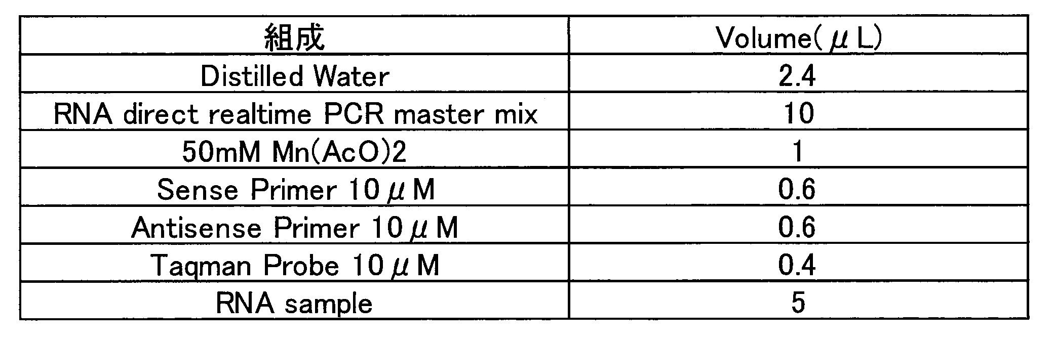
尚、Sense Primerは5’-ACGGGGTTAATTATGCAACAGG-3’(配列番号13)であり、Anitisense Primerは5’-ACGGTGATGCAGGACAACAG-3’(配列番号14)であり、Taqman Probeは5’-[6-FAM]AGCAAGAAGATAGAAAAGGGGAAACCGGGTAG[TAMRA-6-FAM]-3’(配列番号15)であった。
結果を図20に示す。ここで図20は、HCVcc (genotype 2a)を用いたin vitro HCV感染阻害活性であり、細胞内のHCVゲノムRNAの定量結果である。データは抗体非添加群のHCV ゲノムRNAコピー数に対する割合(mean ± SD)で表記した(n=4)。破線はバックグラウンドを示している。
HCVcc感染系にて、抗オクルディンモノクローナル抗体のHCV感染阻害活性を解析したところ、いずれのクローンも添加濃度依存的に細胞内のHCV RNA量が低下しており、顕著な感染阻害作用が認められた(図20)。尚、感染阻害活性は、1-3>37-5=44-10>32-1の順であった。
8)ラット抗オクルディンモノクローナル抗体のin vitro感染阻害活性解析(HCV pseudoparticles(HCVpp、genotype 1a、1b及び2a)を用いた解析)
Huh7.5.1-8細胞を1×10⁵ cells/well/500μLで48穴プレートに播種し、一晩培養した。抗オクルディンモノクローナル抗体を培地で各濃度に調製し、前培養していた細胞の培地を除いて250μL/wellの抗体溶液を加え、室温で30分静置した。なお、各抗体は0、0.25、2.5μg/wellで作用させた。
Huh7.5.1-8細胞を1×10⁵ cells/well/500μLで48穴プレートに播種し、一晩培養した。抗オクルディンモノクローナル抗体を培地で各濃度に調製し、前培養していた細胞の培地を除いて250μL/wellの抗体溶液を加え、室温で30分静置した。なお、各抗体は0、0.25、2.5μg/wellで作用させた。
HCVpp(genotype 1a(H77株)、1b(TH株、Con-1株)又は2a(JFH-1株、J6株)、Microbe and Infection 15, 45-55 , 2013に従い調製)あるいはVSV(水疱性口炎ウイルス)をそれぞれ250μL/well添加し、37 ℃で6時間培養した。血清を含まない培地500μL/wellで3回洗浄し、前処理の1/2の濃度にした抗体液500μLを加え、37 ℃で3日間培養した。
培地を除去後、Lysis Buffer(Promega)を100μL/well加え、溶解液を1.5mLチューブに回収し、10,000 rpm、1分間遠心し、氷上に置いた。本上清10μLと発光基質(ピッカジーン)50μLを混ぜ、Luminescencer-PSNで発光強度の測定を行った。
結果を図21、図22、図23、図24、図25及び図26に示す。図21は、HCVpp(genotype 1a)、図22は、HCVpp(genotype 1b-TH)、図23は、HCVpp(genotype 1b-Con-1)、図24は、HCVpp(genotype 2a-JFH1)、図25はHCVpp(genotype 2a-J6)、図26は、VSVに対する感染阻害活性を示す図である。データは抗体非添加群のルシフェラーゼ活性に対する割合(mean ± SD)で表記した(n=3)。抗オクルディンモノクローナル抗体は評価した全てのgenotype で、添加量依存的な感染阻害活性を示した。またオクルディンを介さずに細胞内へ侵入するVSVに関しては感染阻害活性をいずれの抗体でも確認できなかった。なお、感染阻害活性は、1-3>44-10≧37-5>32-1の順であった。阻害活性の強さの順番は上記7)のHCVccの結果(図20)とよく相関していた。
9)NS5A阻害剤耐性変異導入ウイルス株における抗オクルディンモノクローナル抗体の感染阻害能の解析
NS5A阻害剤耐性変異は、インターフェロン耐性のHCV genotype 1bについて数多く報告されている。しかしながらHCV genotype 1bについて全長のレプリコンを用いたin vitroにおけるウイルスの生活環を効率的に再現する評価系は確立されていない。そこで、in vitroで広く汎用されているJFH-1株(HCV genotype 2a)のNS5A領域について、genotype 1bのCon-1株に置換したキメラ全長レプリコン(JFH-1/NS5A-Con-1)を野生株として用いた。キメラ全長レプリコンのNS5AにNS5A阻害剤耐性変異であるL31V変異、Y93H変異を加え、抗オクルディンモノクローナル抗体における感染阻害能を評価した。トリプシン処理で回収したHuh7.5.1-8細胞をK-PBS 5mLで1回洗浄した後、細胞数がおおよそ2×10⁷cells/mLになるようにした。0.4cmキュベットにこれらの細胞を500μLとin vitroで合成したHCV RNAを50μg加えて氷上で5分間静置した。エレクトロポレーションを行う前にサスペンドして細胞を再懸濁した。Gene Pulser X cell(Bio Rad)を用いて 975μFD, 290mVの条件でエレクトロポレーションを行った。エレクトロポレーション後、1.5 mLの血清培地を加え15分間静置した。トランスフェクションした細胞を50mL コニカルチューブに移して室温、200 × g、5分間で細胞を沈殿させた。沈殿させた細胞は10 mLの培地でサスペンションした後、3×10⁶ cells/dishをコラーゲンコートの10 cm2ディッシュに播いて37℃で培養した。翌日(24時間以上)に血清培地を取り除き、新しい血清培地を加えて37℃で更に4日間培養した。回収したウイルス上清はMillex-HV 0.45μm (Millipore)で濾過した後に、Amicon Ultra-15 (Millipore)を用いて約10倍ウイルス液の濃縮を行った。以降の操作は前項のCell culture derived HCV (HCVcc)を用いた感染阻害活性解析に準ずる。ポジティブコントロールとして、クラウディン-1抗体(Anti-CL1 Clone 2C1)を用いた。結果を図27に示す。破線はバックグラウンドを示している。検討した抗オクルディンモノクローナル抗体Clone 1-3 及びClone 44-10では、NS5A阻害剤耐性変異導入株に対しても感染阻害活性を有していた。NS5A阻害剤はウイルスを標的にしていているのに対し、抗オクルディンモノクローナル抗体は宿主因子を標的にしていることから、NS5A阻害剤耐性変異導入株に対しても感染阻害活性を有することは予想されることであり、他のDAA剤耐性ウイルスにも広く有効であることが強く予見される。
NS5A阻害剤耐性変異は、インターフェロン耐性のHCV genotype 1bについて数多く報告されている。しかしながらHCV genotype 1bについて全長のレプリコンを用いたin vitroにおけるウイルスの生活環を効率的に再現する評価系は確立されていない。そこで、in vitroで広く汎用されているJFH-1株(HCV genotype 2a)のNS5A領域について、genotype 1bのCon-1株に置換したキメラ全長レプリコン(JFH-1/NS5A-Con-1)を野生株として用いた。キメラ全長レプリコンのNS5AにNS5A阻害剤耐性変異であるL31V変異、Y93H変異を加え、抗オクルディンモノクローナル抗体における感染阻害能を評価した。トリプシン処理で回収したHuh7.5.1-8細胞をK-PBS 5mLで1回洗浄した後、細胞数がおおよそ2×10⁷cells/mLになるようにした。0.4cmキュベットにこれらの細胞を500μLとin vitroで合成したHCV RNAを50μg加えて氷上で5分間静置した。エレクトロポレーションを行う前にサスペンドして細胞を再懸濁した。Gene Pulser X cell(Bio Rad)を用いて 975μFD, 290mVの条件でエレクトロポレーションを行った。エレクトロポレーション後、1.5 mLの血清培地を加え15分間静置した。トランスフェクションした細胞を50mL コニカルチューブに移して室温、200 × g、5分間で細胞を沈殿させた。沈殿させた細胞は10 mLの培地でサスペンションした後、3×10⁶ cells/dishをコラーゲンコートの10 cm2ディッシュに播いて37℃で培養した。翌日(24時間以上)に血清培地を取り除き、新しい血清培地を加えて37℃で更に4日間培養した。回収したウイルス上清はMillex-HV 0.45μm (Millipore)で濾過した後に、Amicon Ultra-15 (Millipore)を用いて約10倍ウイルス液の濃縮を行った。以降の操作は前項のCell culture derived HCV (HCVcc)を用いた感染阻害活性解析に準ずる。ポジティブコントロールとして、クラウディン-1抗体(Anti-CL1 Clone 2C1)を用いた。結果を図27に示す。破線はバックグラウンドを示している。検討した抗オクルディンモノクローナル抗体Clone 1-3 及びClone 44-10では、NS5A阻害剤耐性変異導入株に対しても感染阻害活性を有していた。NS5A阻害剤はウイルスを標的にしていているのに対し、抗オクルディンモノクローナル抗体は宿主因子を標的にしていることから、NS5A阻害剤耐性変異導入株に対しても感染阻害活性を有することは予想されることであり、他のDAA剤耐性ウイルスにも広く有効であることが強く予見される。
10)オクルディン抗体による様々なHCVcc株に対する感染阻害解析
HCVccのシステムにおいて、細胞培養で感染系が確立しているTNS2-J1(Genotype 1b/2a)とJc-1(Genotype 2a)の感染に対して、オクルディン抗体で阻害ができるか評価した。評価系としては蛍光蛋白質の細胞内局在を蛍光顕微鏡で観察することでHCV感染を判断できる実験系を用いた。Interferon-β promoter stimulator -1 (IPS-1)は、ミトコンドリア外膜上に存在する膜蛋白質で、RNAウイルス感染時にIFN-β産生に関与することで自然免疫を誘導する。HCVの非構造蛋白質であるNS3/NS4Aはプロテアーゼ活性を有しており、IPS-1切断活性を有している。HCV感染時、HCVはIPS-1を切断することにより、IFN-βの産生を阻害していることが報告されている。蛍光蛋白質融合遺伝子mCherry-NLS-IPS-1は、赤色の蛍光蛋白質mCherry、核移行シグナル(Nuclear localize signal, NLS)、そしてIPS-1を含む遺伝子である。HCV感染により、産生されるNS3/NS4Aにより細胞質側に存在するIPS-1が切断され、NLSにより核へ蛍光蛋白質が移行する。この細胞内における蛍光蛋白質の挙動変化(核への移行)を観察することでHCVの感染を評価できる。また、HCV感染性はイムノブロット解析を用いた方法でも評価した。コントロールとしては、HCV-JFH1を用いた。結果を図28、図29に示す。未処理又はコントロール抗体添加群ではいずれもHCV感染を示す蛍光蛋白質の核への移行が全ての細胞で起こっていた。オクルディン抗体1-3を処理することにより、核への移行が全ての種類のウイルスで完全に抑えられた。オクルディン抗体37-5、44-10についてもごく一部感染が起こっているものの大部分の感染を抑制していることが分かった。オクルディン抗体32-1では、TNS2-J1株では蛍光蛋白質の核への移行を全て阻止したが、Jc-1では半分程度感染が確認された。イムノブロット解析及びHCV coreに対する免疫染色でも同様な結果が得られた(図29及び30)。以上の結果より、樹立抗体、特に1-3は様々なHCVcc感染を阻害できるプローブであることを確認した。
HCVccのシステムにおいて、細胞培養で感染系が確立しているTNS2-J1(Genotype 1b/2a)とJc-1(Genotype 2a)の感染に対して、オクルディン抗体で阻害ができるか評価した。評価系としては蛍光蛋白質の細胞内局在を蛍光顕微鏡で観察することでHCV感染を判断できる実験系を用いた。Interferon-β promoter stimulator -1 (IPS-1)は、ミトコンドリア外膜上に存在する膜蛋白質で、RNAウイルス感染時にIFN-β産生に関与することで自然免疫を誘導する。HCVの非構造蛋白質であるNS3/NS4Aはプロテアーゼ活性を有しており、IPS-1切断活性を有している。HCV感染時、HCVはIPS-1を切断することにより、IFN-βの産生を阻害していることが報告されている。蛍光蛋白質融合遺伝子mCherry-NLS-IPS-1は、赤色の蛍光蛋白質mCherry、核移行シグナル(Nuclear localize signal, NLS)、そしてIPS-1を含む遺伝子である。HCV感染により、産生されるNS3/NS4Aにより細胞質側に存在するIPS-1が切断され、NLSにより核へ蛍光蛋白質が移行する。この細胞内における蛍光蛋白質の挙動変化(核への移行)を観察することでHCVの感染を評価できる。また、HCV感染性はイムノブロット解析を用いた方法でも評価した。コントロールとしては、HCV-JFH1を用いた。結果を図28、図29に示す。未処理又はコントロール抗体添加群ではいずれもHCV感染を示す蛍光蛋白質の核への移行が全ての細胞で起こっていた。オクルディン抗体1-3を処理することにより、核への移行が全ての種類のウイルスで完全に抑えられた。オクルディン抗体37-5、44-10についてもごく一部感染が起こっているものの大部分の感染を抑制していることが分かった。オクルディン抗体32-1では、TNS2-J1株では蛍光蛋白質の核への移行を全て阻止したが、Jc-1では半分程度感染が確認された。イムノブロット解析及びHCV coreに対する免疫染色でも同様な結果が得られた(図29及び30)。以上の結果より、樹立抗体、特に1-3は様々なHCVcc感染を阻害できるプローブであることを確認した。
11)オクルディン抗体によるHCV Cell-to-cell感染阻害解析
HCVには、受容体依存性エンドサイトーシスを介した侵入(Cell-free感染)に加えて、直接細胞から細胞へ移動して広がる感染(Cell-to-cell 感染)が存在する。Cell-to-cell感染は、生体内の持続感染時の感染維持に特に重要と考えられている。In vitro感染培養細胞系では、Cell-free感染とCell-to-cell感染が起こっている。そこでCell-to-cell感染を特異的に評価するために、侵入受容体の一つであるCD81を欠損しているためCell-free感染を起こさないCD81欠損細胞(751r細胞)を用いた。検出系としては、前項の複製能評価システムを用いた。本項ではまず、Cell-free感染を起こさない751r細胞にmCherry-NLS-IPS-1を導入しレポーター細胞を樹立した。本評価系を用いて、Cell-to-cell感染をオクルディン抗体が阻害できるか評価した。HCV-JFH-1感染させたHuh7.5.1-8細胞と751r/ mCherry-NLS-IPS1細胞を共培養することで、Cell-to-cell感染が起こる条件とした。結果を図31で示す。ラットコントロール抗体処理群では、赤色の蛍光蛋白質の核への移行が確認された。一般にCell-free感染に比べ、Cell-to-cell感染は起こりにくいことが知られており、今回の系でも感染は弱く、Huh7.5.1-8細胞と接している751r/ mCherry-NLS-IPS1細胞の一部に感染が広がっているのが見られるのみであった。オクルディン抗体クローン1-3とクローン 37-5処理群では、非感染群と同様に、全て赤色の蛍光蛋白質は細胞質側に局在したままであった(図31)。即ち、オクルディン抗体により、Acceptor細胞である751r/mCherry-NLS-IPS-1細胞内へのHCVの移行が阻止されたことを示している。この事から、オクルディン抗体1-3及び37-5は、Cell-to-cell感染を阻害することがわかった。
HCVには、受容体依存性エンドサイトーシスを介した侵入(Cell-free感染)に加えて、直接細胞から細胞へ移動して広がる感染(Cell-to-cell 感染)が存在する。Cell-to-cell感染は、生体内の持続感染時の感染維持に特に重要と考えられている。In vitro感染培養細胞系では、Cell-free感染とCell-to-cell感染が起こっている。そこでCell-to-cell感染を特異的に評価するために、侵入受容体の一つであるCD81を欠損しているためCell-free感染を起こさないCD81欠損細胞(751r細胞)を用いた。検出系としては、前項の複製能評価システムを用いた。本項ではまず、Cell-free感染を起こさない751r細胞にmCherry-NLS-IPS-1を導入しレポーター細胞を樹立した。本評価系を用いて、Cell-to-cell感染をオクルディン抗体が阻害できるか評価した。HCV-JFH-1感染させたHuh7.5.1-8細胞と751r/ mCherry-NLS-IPS1細胞を共培養することで、Cell-to-cell感染が起こる条件とした。結果を図31で示す。ラットコントロール抗体処理群では、赤色の蛍光蛋白質の核への移行が確認された。一般にCell-free感染に比べ、Cell-to-cell感染は起こりにくいことが知られており、今回の系でも感染は弱く、Huh7.5.1-8細胞と接している751r/ mCherry-NLS-IPS1細胞の一部に感染が広がっているのが見られるのみであった。オクルディン抗体クローン1-3とクローン 37-5処理群では、非感染群と同様に、全て赤色の蛍光蛋白質は細胞質側に局在したままであった(図31)。即ち、オクルディン抗体により、Acceptor細胞である751r/mCherry-NLS-IPS-1細胞内へのHCVの移行が阻止されたことを示している。この事から、オクルディン抗体1-3及び37-5は、Cell-to-cell感染を阻害することがわかった。
12)ヒトIgG4変異体キメラ抗体の作製
12-1)発現ベクター作製
IgG4は、重鎖定常領域内にある228番目のSerをProに置換することで生体内安定性が向上することが知られている(Immunology, 105, 9-19, 2002)。そこで、228番目のSerをProに置換するようにプライマーを設計し、pFUSE-CHIg-hG4 (Invivogen)を鋳型にNheIサイト及びBsrGIサイト間の682 bpをPCR法により増幅、PCR産物を電気泳動により分離後、精製しpFUSE-CHIg-hG4mを得た。尚、変異挿入部上流のプライマーにはNheIサイト、下流のプライマーにはBsrGIサイトを付加した。
12-1)発現ベクター作製
IgG4は、重鎖定常領域内にある228番目のSerをProに置換することで生体内安定性が向上することが知られている(Immunology, 105, 9-19, 2002)。そこで、228番目のSerをProに置換するようにプライマーを設計し、pFUSE-CHIg-hG4 (Invivogen)を鋳型にNheIサイト及びBsrGIサイト間の682 bpをPCR法により増幅、PCR産物を電気泳動により分離後、精製しpFUSE-CHIg-hG4mを得た。尚、変異挿入部上流のプライマーにはNheIサイト、下流のプライマーにはBsrGIサイトを付加した。
各抗体クローンの可変部領域のVL領域及びVH領域のアミノ酸をコードする遺伝子をPCR法により増幅した。なお、VL遺伝子の上流にAgeIサイト、下流にBsiWIサイト、VH遺伝子の上流にEcoRIサイト、下流にNheIサイトを付加した。PCR産物を電気泳動により分離・精製した。
増幅したVL遺伝子及びヒトIgG kappa鎖定常領域をもつクローニングベクターであるpFUSE2-CLIg-hk (Invivogen)をAgeI及びBsiWIで処理後、ライゲーションした。増幅したVH遺伝子及びpFUSE-CHIg-hG4mをEcoRI及びNheIで処理後、ライゲーションした。各ライゲーション産物をコンピテントセルDH5αにトランスフォーメーションし、独立に大腸菌クローンを培養、プラスミドDNAを回収後、シークエンスを確認し、pFUSE2-CLIg-hk-anti-オクルディン及びpFUSE-CHIg-hG4m-anti-オクルディンを得た。
12-2)ヒトIgG4変異体キメラ抗体の精製
培養用6 穴プレートに2× 10⁵ cells/wellのCHO-K1細胞を播種し、37 ℃、5% CO2環境下でサブコンフルエントになるまで培養した。作製した発現ベクター2μg(pFUSE2-CLIg-hk-anti-オクルディンを1.2μg、pFUSE-CHIg-hG4mutant-anti-オクルディンを0.8μg)をOpti-MEM1 (GIBCO) 100μL及びFuGENE(登録商標)HD Transfection Reagent (Roche) 4μLと混合し、15分間常温静置した。CHO細胞の培地を交換し、上記の混合液をウェルに全量加えた。その後、2日間、37 ℃、5% CO2環境下で培養し、上清を回収した。
培養用6 穴プレートに2× 10⁵ cells/wellのCHO-K1細胞を播種し、37 ℃、5% CO2環境下でサブコンフルエントになるまで培養した。作製した発現ベクター2μg(pFUSE2-CLIg-hk-anti-オクルディンを1.2μg、pFUSE-CHIg-hG4mutant-anti-オクルディンを0.8μg)をOpti-MEM1 (GIBCO) 100μL及びFuGENE(登録商標)HD Transfection Reagent (Roche) 4μLと混合し、15分間常温静置した。CHO細胞の培地を交換し、上記の混合液をウェルに全量加えた。その後、2日間、37 ℃、5% CO2環境下で培養し、上清を回収した。
12-3)各種オクルディンに対する結合性解析
HT1080細胞、ヒトオクルディン及びマウスオクルディン発現HT1080細胞をトリプシン処理により回収した。5.0×10⁵ cellsに対し、前項で作製したヒトIgG4変異体キメラ抗体を含む培養上清を100μL添加し、撹拌し氷上で1時間静置した。0.2% BSA-PBSにて1回洗浄後、1% BSA-PBSにて希釈したGoat anti-human IgG(H+L)-FITC抗体(Jackson Immuno Research)を添加、撹拌、氷上で遮光し30分静置した。0.2% BSA-PBSにて2回洗浄後、0.2% BSA-PBSにて終濃度5μg/mLとなるように希釈したPI(Miltenyi Biotec)を加え、FCM解析を行った。結果を図32に示す。作製した全てのキメラ抗体でヒトオクルディンへの結合性を示した。またClone 1-3をキメラ化したものでは、図4に示したラット抗体の場合と同様に、マウスオクルディンへの結合性も認められた。以上の結果から、それぞれのクローンのCDR領域がヒトオクルディン結合に必須であり、その結合性状を反映していることが示された。
HT1080細胞、ヒトオクルディン及びマウスオクルディン発現HT1080細胞をトリプシン処理により回収した。5.0×10⁵ cellsに対し、前項で作製したヒトIgG4変異体キメラ抗体を含む培養上清を100μL添加し、撹拌し氷上で1時間静置した。0.2% BSA-PBSにて1回洗浄後、1% BSA-PBSにて希釈したGoat anti-human IgG(H+L)-FITC抗体(Jackson Immuno Research)を添加、撹拌、氷上で遮光し30分静置した。0.2% BSA-PBSにて2回洗浄後、0.2% BSA-PBSにて終濃度5μg/mLとなるように希釈したPI(Miltenyi Biotec)を加え、FCM解析を行った。結果を図32に示す。作製した全てのキメラ抗体でヒトオクルディンへの結合性を示した。またClone 1-3をキメラ化したものでは、図4に示したラット抗体の場合と同様に、マウスオクルディンへの結合性も認められた。以上の結果から、それぞれのクローンのCDR領域がヒトオクルディン結合に必須であり、その結合性状を反映していることが示された。
12-4)ヒトIgG4変異体キメラ抗体によるin vitro HCV感染阻害
7)と同様の方法にて、ヒトIgG4変異体キメラ抗体のin vitro HCV感染への影響を検討した。結果を図33に示す。ここで図33は、HCVcc (genotype 2a)を用いたin vitro HCV感染阻害活性であり、細胞内のHCVゲノムRNAの定量結果である。データは抗体非添加群のHCV ゲノムRNAコピー数に対する割合(mean ±SD)で表記した(n=4)。破線はバックグラウンドを示している。
7)と同様の方法にて、ヒトIgG4変異体キメラ抗体のin vitro HCV感染への影響を検討した。結果を図33に示す。ここで図33は、HCVcc (genotype 2a)を用いたin vitro HCV感染阻害活性であり、細胞内のHCVゲノムRNAの定量結果である。データは抗体非添加群のHCV ゲノムRNAコピー数に対する割合(mean ±SD)で表記した(n=4)。破線はバックグラウンドを示している。
HCVcc感染系にて、抗オクルディンヒトIgG4変異体キメラ抗体のHCV感染阻害活性を解析したところ、Xi 1-3,Xi 37-5ともに添加濃度依存的に細胞内のHCV RNA量が低下しており、ラット抗体と同様に、顕著な感染阻害作用が認められた(図33)。尚、感染阻害活性の強さは、1-3>37-5であった。
13)ラット抗オクルディンモノクローナル抗体のin vivo HCV感染阻止試験
HCV (genotype 1b)を投与したヒト肝臓キメラマウスを用いて、ラット抗オクルディンモノクローナル抗体のin vivo感染阻害活性を解析した。ヒト肝臓キメラマウス(uPA-SCIDマウスより作製)は株式会社フェニックスバイオより入手した。雄性、12~16週齢、体重15g以上、血中ヒトアルブミン7.0 mg/mL以上の個体を選択した。群編成では、体重及び血中ヒトアルブミン濃度の平均値を考慮してマウスを3群(Control抗体を4匹、オクルディン抗体投与の2群については3匹)に振り分けた。
HCV (genotype 1b)を投与したヒト肝臓キメラマウスを用いて、ラット抗オクルディンモノクローナル抗体のin vivo感染阻害活性を解析した。ヒト肝臓キメラマウス(uPA-SCIDマウスより作製)は株式会社フェニックスバイオより入手した。雄性、12~16週齢、体重15g以上、血中ヒトアルブミン7.0 mg/mL以上の個体を選択した。群編成では、体重及び血中ヒトアルブミン濃度の平均値を考慮してマウスを3群(Control抗体を4匹、オクルディン抗体投与の2群については3匹)に振り分けた。
Control抗体、Clone 1-3又はClone 37-5を、マウスに腹腔内投与した。初回投与日をday 0とし、抗体投与量はday 0に50mg/kg、day3に30mg/kg、day7に20mg/kg、day10に10mg/kgとした。抗体投与液の希釈には生理食塩水を用いた。
初回抗体投与の8時間後にマウスにイソフルラン麻酔を施し、生理食塩水で1.0×10⁵ copies/mLに調製したHCV(genotype 1b)100μLを眼窩静脈叢から接種した。
コントロール抗体、Clone 1-3又はClone 37-5を投与したマウス血清を経日的に回収し、血清中HCV RNA量を測定することでHCV感染阻害活性を評価した。結果を図34に示す。Control抗体投与マウスではday14以降全てのマウスでウイルスRNAが検出され、Day21以降はウイルスRNA量が約107copies/mLを示しプラトーに達したのに対して、Clone 1-3又はClone 37-5を投与した全てのマウスでウイルスRNA量が定量限界以下のままであった。以上の結果から、ラット抗オクルディンモノクローナル抗体Clone 1-3又はClone 37-5投与により、HCV感染が完全に阻止できることが示された。
詳細な手順、他の結果を以下に示す。
day0、day3、day7、day10、day14、day21、day28、day35、day42に、マウスの一般状態を観察し、体重を計測した。なお、day0、day3、day7、day10は抗体投与前に行った。
day0、day3、day7、day10、day14、day21、day28、day35、day42に採血を行った。なお、day0、day3、day7、day10は抗体投与前に、マウスにイソフルラン麻酔を施し、眼窩静脈叢より採血した。
day0、day7、day14、day21、day28、day35、day42の採血液を用いて血清中HCVゲノムRNA量を測定した。採取した血清5μLからSepaGene RV-R(エーディア株式会社、東京)を用いてRNA抽出を行い、RNAを1 mM DTT(プロメガ株式会社、東京)と0.4U/μL ribonuclease inhibitor(タカラバイオ株式会社、滋賀)を含む10μLのNuclease-free water(Life Technologies Corporation、CA、USA)に溶解した。PCR反応液は、溶解したRNA原液もしくは希釈したRNAを2.5μLとTaqMan EZ RT-PCR Core Reagents(Life Technologies Corporation)を用いて調製した。PCR反応と解析にはABI Prism 7500(Life Technologies Corporation)を用いた。RT-PCR反応は、50℃ 2分→60℃ 30分→95℃ 5分→(95℃ 20秒→62℃ 1分)×50サイクルで行った。
day0、day7、day14、day21、day28、day35、day42の採血液を用いて血中ヒトアルブミン濃度を測定した。血液2μLを緩衝液(LX試薬栄研シリーズ共用緩衝液、栄研化学株式会社、東京)と混合し、370 × g、3分間の遠心分離を行った後、ラテックス凝集免疫比濁法(LX試薬’栄研’Alb-II、栄研化学株式会社、東京)を用いて、吸光マイクロプレートリーダー(Vmax、日本モレキュラーデバイス株式会社、東京)で測定した。
day0、day7、day14、day21、day28、day35、day42の採血液を用いて血清中ALT及びAST活性を測定した。採取した血清10μLを生理食塩液で希釈し、POP・POD・ロイコ色素法(ピルビン酸オキシダーゼにより発生する過酸化水素とペルオキシダーゼによりジアリールイミダゾールロイコ色素を青色に発色)により、ドライケム3500(富士フィルム、東京)を用いて測定した。
結果を図35~図38に示す。ここで図35は、HCV (genotype 1b)を投与したヒト肝キメラマウスに各種抗体を投与した際の体重変化である。図36は、HCV (genotype 1b)を投与したヒト肝キメラマウスに各種抗体を投与した際の血中のヒトアルブミン濃度である。図37は、HCV (genotype 1b)を投与したヒト肝キメラマウスに各種抗体を投与した際のAST値である。図38は、HCV (genotype 1b)を投与したヒト肝キメラマウスに各種抗体を投与した際のALT値である。
抗オクルディン抗体投与による一般状態の変化、体重減少、血中ヒトアルブミン濃度減少、AST値、ALT値の有意な増加等は認められなかったことから、Clone 1-3及びClone 37-5は安全性及び有効性を兼ね備えた感染阻害分子であると考えられた。
14)In vivo持続感染状態からのラット抗オクルディンモノクローナル抗体投与の効果
13)の実験で、コントロール抗体を投与した4匹はHCV持続感染状態になっている。これらのマウスに対するラット抗オクルディン抗体1-3及びマウス抗クローディン1抗体3A2の効果を検討した。抗体の投与は、Day42(0), 45(3),49(7),52(10)に、各30mg/kgを腹腔内投与で行った(各2匹ずつ)。投与開始後、1週間ごとに採血をし、血中HCV量を測定した(方法は13)と同様)。結果を図39に示す。クローディン1抗体3A2投与マウスではウイルスRNA量に変動がみられなかったのに対して、オクルディン抗体1-3投与マウスではウイルスRNA量が低下する傾向がみられ、投与を中止すると、10日ほど遅れてウイルスRNA量が元に戻ることが確認された。以上の結果から、ラット抗オクルディン抗体1-3は、HCV持続感染からでも血中ウイルス量低下させることができることが示された。
13)の実験で、コントロール抗体を投与した4匹はHCV持続感染状態になっている。これらのマウスに対するラット抗オクルディン抗体1-3及びマウス抗クローディン1抗体3A2の効果を検討した。抗体の投与は、Day42(0), 45(3),49(7),52(10)に、各30mg/kgを腹腔内投与で行った(各2匹ずつ)。投与開始後、1週間ごとに採血をし、血中HCV量を測定した(方法は13)と同様)。結果を図39に示す。クローディン1抗体3A2投与マウスではウイルスRNA量に変動がみられなかったのに対して、オクルディン抗体1-3投与マウスではウイルスRNA量が低下する傾向がみられ、投与を中止すると、10日ほど遅れてウイルスRNA量が元に戻ることが確認された。以上の結果から、ラット抗オクルディン抗体1-3は、HCV持続感染からでも血中ウイルス量低下させることができることが示された。
15)DAA剤とラット抗オクルディンモノクローナル抗体の併用投与in vivo試験
HCV (genotype 1a)を持続感染させたヒト肝臓キメラマウスを用いて、DAA剤(Direct-acting antiviral agent(直接作用型抗ウイルス剤), Nesbuvir,NS5B阻害剤)とラット抗オクルディンモノクローナル抗体の併用投与試験を行った。基本的な操作は13)14)と同様である。Nesbuvirのみ投与群(101~104)、Nesbuvir+オクルディン抗体1-3投与群(201~204)とした。Nesbuvirは毎日2回50mg/kg投与、オクルディン抗体1-3はDay0, 3, 7に30mg/kg投与で行った。結果を図40に示す。Nesbuvir単独投与群では、全てのマウス(101~104)でブレークスルーがみられ、薬剤を投与しているにも関わらず血中ウイルス量が上昇した(おそらく耐性ウイルスが増殖している)。一方で、Nesbuvir+オクルディン抗体1-3併用投与群では、1匹(201)が原因不明で死亡し、血中ウイルス量があまり下がらなかった1匹(204)では弱いブレークスルー傾向がみられたが、残り2匹(202, 203)ではブレークスルーがみられなかった。以上の結果から、オクルディン抗体1-3併用投与により、ブレークスルー(耐性ウイルスの発生)を阻止できる可能性が示された。つまり、オクルディン抗体はDAA剤との併用で、有用な併用効果を示すことが考えられた。
HCV (genotype 1a)を持続感染させたヒト肝臓キメラマウスを用いて、DAA剤(Direct-acting antiviral agent(直接作用型抗ウイルス剤), Nesbuvir,NS5B阻害剤)とラット抗オクルディンモノクローナル抗体の併用投与試験を行った。基本的な操作は13)14)と同様である。Nesbuvirのみ投与群(101~104)、Nesbuvir+オクルディン抗体1-3投与群(201~204)とした。Nesbuvirは毎日2回50mg/kg投与、オクルディン抗体1-3はDay0, 3, 7に30mg/kg投与で行った。結果を図40に示す。Nesbuvir単独投与群では、全てのマウス(101~104)でブレークスルーがみられ、薬剤を投与しているにも関わらず血中ウイルス量が上昇した(おそらく耐性ウイルスが増殖している)。一方で、Nesbuvir+オクルディン抗体1-3併用投与群では、1匹(201)が原因不明で死亡し、血中ウイルス量があまり下がらなかった1匹(204)では弱いブレークスルー傾向がみられたが、残り2匹(202, 203)ではブレークスルーがみられなかった。以上の結果から、オクルディン抗体1-3併用投与により、ブレークスルー(耐性ウイルスの発生)を阻止できる可能性が示された。つまり、オクルディン抗体はDAA剤との併用で、有用な併用効果を示すことが考えられた。
16)抗オクルディン抗体FabフラグメントによるHCVcc感染阻害アッセイ
16-1)抗オクルディン抗体Fab1-3の作製
15ml tube(FALCON)に抗オクルディン抗体Clone1-3を消化用バッファー(20mM sodium phosphate, 10mM EDTA,20mM Cysteine; pH 7.0)に溶解させ、Immobilized Papain (Agarose Resin)(GE health care)を加えた。37℃で、4時間インキュベートさせた。その後10mM Tris-HCl (pH7.5)を1.0ml加え、遠心し上清を回収した。回収したサンプルをAb-Rapid SPiN(プロテノバ)に加え1時間反応させた。カラムを遠心し、通過区分をFab区分として回収した。その後PD10カラム(GE-Health care)でPBSに置換した。得られた目的タンパク質溶液をもちいて、還元条件下のSDS-PAGEを行った。結果を図41に示す。目的の位置にFabのバンドを確認した。未消化のFull-body及びFcのバンドは確認できなかった。
16-1)抗オクルディン抗体Fab1-3の作製
15ml tube(FALCON)に抗オクルディン抗体Clone1-3を消化用バッファー(20mM sodium phosphate, 10mM EDTA,20mM Cysteine; pH 7.0)に溶解させ、Immobilized Papain (Agarose Resin)(GE health care)を加えた。37℃で、4時間インキュベートさせた。その後10mM Tris-HCl (pH7.5)を1.0ml加え、遠心し上清を回収した。回収したサンプルをAb-Rapid SPiN(プロテノバ)に加え1時間反応させた。カラムを遠心し、通過区分をFab区分として回収した。その後PD10カラム(GE-Health care)でPBSに置換した。得られた目的タンパク質溶液をもちいて、還元条件下のSDS-PAGEを行った。結果を図41に示す。目的の位置にFabのバンドを確認した。未消化のFull-body及びFcのバンドは確認できなかった。
16-2)抗オクルディン抗体Fab1-3のオクルディン結合能の解析
ヒトオクルディンを発現させたHT1080発現をトリプシン処理により回収した。5.0×10⁵ cellsに対し、各抗体及びFab 5μg/mLを100μL添加し、5-1)と同様の操作でFCM解析を行った。結果を図42に示す。作製したFab1-3はヒトオクルディンに対する結合性を有していた。
ヒトオクルディンを発現させたHT1080発現をトリプシン処理により回収した。5.0×10⁵ cellsに対し、各抗体及びFab 5μg/mLを100μL添加し、5-1)と同様の操作でFCM解析を行った。結果を図42に示す。作製したFab1-3はヒトオクルディンに対する結合性を有していた。
16-3)抗オクルディン抗体Fab1-3のin vitro HCV感染阻害活性解析(Cell-cultured HCV (HCVcc)を用いた解析)
培地には、10% fetal bovine serum (Cell Culture Bioscience), non-essential amino acid (Hyclone SH30238.01)及びpenicillin/streptomycin(Wako, 168-23191)含有D-MEM(Wako, 044-29765)を用いた。
培地には、10% fetal bovine serum (Cell Culture Bioscience), non-essential amino acid (Hyclone SH30238.01)及びpenicillin/streptomycin(Wako, 168-23191)含有D-MEM(Wako, 044-29765)を用いた。
コラーゲンタイプIコート48穴プレート(Corning, NCO3548)にHuh7.5.1-8細胞を5 × 10⁴cells/500 μL/wellで播種し、37 ℃、1日間培養した。培地を除き、精製Fab1-3を0.5~5μg含む培地(250μL)を添加し、室温 (25 ℃)で30分培養した。
その後、HCVcc(genotype 2a)を50倍希釈したものをそれぞれ250μL/well添加し、室温で2時間培養した。血清を含まない培地500μL/wellで3回洗浄し、前処理の1/2の濃度にした精製Fab1-3液500μLを加え、37 ℃で4日間培養した。
細胞をPBS 500μL/wellで3回洗浄し、Blood/Cultured Cell Total RNA Purification Mini Kit (FAVORGEN)を用いRNA抽出・精製を行い、滅菌水50μLで溶解し、-80℃で保存した。尚、RNA濃度は、Nano Dropを用いて測定した。
精製したRNAを用いて、Taqman qRT-PCR法(試薬はRNA-direct Realtime PCR Master Mix(Toyobo)、機器はLightCycler (Roche))にてHCVゲノムRNA定量を行った。尚、反応溶液は、表2の組成に準じて調整した。
結果を図43に示す。ここで図43は、HCVcc (genotype 2a)を用いたin vitro HCV感染阻害活性であり、細胞内のHCVゲノムRNAの定量結果である。データは抗体非添加群のHCV ゲノムRNAコピー数に対する割合(mean ±SD)で表記した(n=4)。
HCVcc感染系にて、抗オクルディンFab1-3についてHCV感染阻害活性を解析したところ、添加濃度依存的に細胞内のHCV RNA量が低下しており、顕著な感染阻害作用が認められた。尚、顕鏡下で細胞障害性は観察されなかった。以上の結果から、抗オクルディン抗体1-3は1価でも十分なHCV感染阻害能を有することが示され、本エピトープに結合する低分子がHCV感染阻害剤として機能しうることが強く示唆される。
17)抗オクルディン一本鎖抗体scFvの作製とscFvによるHCV感染阻害
17-1)抗オクルディン一本鎖抗体scFv発現ベクター作製
一本鎖抗体は、抗体の可変領域VH、VLについて、適当なリンカー(汎用されているリンカーとして(GlyGlyGlyGlySer)3等が知られている)で連結させた一つの読み枠で合成される分子である。Clone1-3とClone37-5につてそれぞれVH、VLに対するプライマー及びリンカーに対応するプライマーを設計し、Assembly PCRによりscFv遺伝子を作製した。作製したscFv遺伝子をpSecTag2/Hygro A(Invitrogen)を鋳型にClone1-3ではHindIIIサイト及びXhoIサイト間、Clone37-5ではBamHIサイト及びXhoIサイト間にライゲーションにより挿入した。各ライゲーション産物をコンピテントセルDH5αにトランスフォーメーションし、独立に大腸菌クローンを培養、プラスミドDNAを回収後、シークエンスを確認し、pSecTag2/Hygro-scFv1-3及びscFv37-5を得た。作製したコンストラクトのアミノ酸シーケンスを図44に示す。
17-1)抗オクルディン一本鎖抗体scFv発現ベクター作製
一本鎖抗体は、抗体の可変領域VH、VLについて、適当なリンカー(汎用されているリンカーとして(GlyGlyGlyGlySer)3等が知られている)で連結させた一つの読み枠で合成される分子である。Clone1-3とClone37-5につてそれぞれVH、VLに対するプライマー及びリンカーに対応するプライマーを設計し、Assembly PCRによりscFv遺伝子を作製した。作製したscFv遺伝子をpSecTag2/Hygro A(Invitrogen)を鋳型にClone1-3ではHindIIIサイト及びXhoIサイト間、Clone37-5ではBamHIサイト及びXhoIサイト間にライゲーションにより挿入した。各ライゲーション産物をコンピテントセルDH5αにトランスフォーメーションし、独立に大腸菌クローンを培養、プラスミドDNAを回収後、シークエンスを確認し、pSecTag2/Hygro-scFv1-3及びscFv37-5を得た。作製したコンストラクトのアミノ酸シーケンスを図44に示す。
17-2)scFvを含む培養上清の回収
培養用6 穴プレートに5 × 10⁵ cells/wellのHEK293T細胞を播種し、37 ℃、5% CO2環境下でサブコンフルエントになるまで培養した。作製した発現ベクター2μgをOpti-MEM1 (GIBCO) 100μL及びFuGENE(登録商標)HD Transfection Reagent (Roche) 4μLと混合し、15分間常温静置した。CHO細胞の培地を交換し、上記の混合液をウェルに全量加えた。その後、2日間、37 ℃、5% CO2環境下で培養し、上清を回収した。
培養用6 穴プレートに5 × 10⁵ cells/wellのHEK293T細胞を播種し、37 ℃、5% CO2環境下でサブコンフルエントになるまで培養した。作製した発現ベクター2μgをOpti-MEM1 (GIBCO) 100μL及びFuGENE(登録商標)HD Transfection Reagent (Roche) 4μLと混合し、15分間常温静置した。CHO細胞の培地を交換し、上記の混合液をウェルに全量加えた。その後、2日間、37 ℃、5% CO2環境下で培養し、上清を回収した。
17-3)各種オクルディンに対する結合性解析
HT1080細胞、ヒトオクルディン及びヒトオクルディン第一細胞外領域欠損(ΔEL1)、ヒトオクルディン第二細胞外領域欠損(ΔEL1)発現HT1080細胞をトリプシン処理により回収した。5.0×10⁵ cellsに対し、前項で作製したscFvを含む培養上清を100μL添加し、撹拌し氷上で1時間静置した。0.2% BSA-PBSにて1回洗浄後、1% BSA-PBSにて希釈したマウス抗HisタグIgG(MBL)を添加し、撹拌、氷上で60分静置した。0.2% BSA-PBSにて1回洗浄後、1% BSA-PBSにて希釈したヤギ抗マウスIgG(H+L)-Alexa488抗体(Jackson Immuno Research)を添加、撹拌、氷上で遮光し30分静置した。0.2% BSA-PBSにて2回洗浄後、FCM解析を行った。ポジティブコントロールとして、各ラット抗オクルディン抗体を用いた。またネガティブコントロールとしてラットIgG及びHEK293T細胞の培養上清を使用した。結果を図45に示す。作製したscFvは、ヒトオクルディンに対して結合性を有していた。またそれぞれのエピトープに対する特異性を保持していた。
HT1080細胞、ヒトオクルディン及びヒトオクルディン第一細胞外領域欠損(ΔEL1)、ヒトオクルディン第二細胞外領域欠損(ΔEL1)発現HT1080細胞をトリプシン処理により回収した。5.0×10⁵ cellsに対し、前項で作製したscFvを含む培養上清を100μL添加し、撹拌し氷上で1時間静置した。0.2% BSA-PBSにて1回洗浄後、1% BSA-PBSにて希釈したマウス抗HisタグIgG(MBL)を添加し、撹拌、氷上で60分静置した。0.2% BSA-PBSにて1回洗浄後、1% BSA-PBSにて希釈したヤギ抗マウスIgG(H+L)-Alexa488抗体(Jackson Immuno Research)を添加、撹拌、氷上で遮光し30分静置した。0.2% BSA-PBSにて2回洗浄後、FCM解析を行った。ポジティブコントロールとして、各ラット抗オクルディン抗体を用いた。またネガティブコントロールとしてラットIgG及びHEK293T細胞の培養上清を使用した。結果を図45に示す。作製したscFvは、ヒトオクルディンに対して結合性を有していた。またそれぞれのエピトープに対する特異性を保持していた。
17-4)抗オクルディン一本鎖抗体scFvのin vitro感染阻害活性解析(HCV pseudoparticles(HCVpp、genotype 2a)を用いた解析)
Huh7.5.1-8細胞を1×10⁵ cells/well/500μLで48-well plateに播種し、一晩培養した。抗オクルディン一本鎖抗体を含む培地を各濃度に調製し、前培養していた細胞の培地を除いて250μL/wellのscFv含有培地を加え、室温で30分静置した。なお、各培地は原液又は10倍希釈で作用させた。
Huh7.5.1-8細胞を1×10⁵ cells/well/500μLで48-well plateに播種し、一晩培養した。抗オクルディン一本鎖抗体を含む培地を各濃度に調製し、前培養していた細胞の培地を除いて250μL/wellのscFv含有培地を加え、室温で30分静置した。なお、各培地は原液又は10倍希釈で作用させた。
HCVpp(genotype 2a(JFH-1株)、Microbe and Infection 15, 45-55 , 2013に従い調製)をそれぞれ250μL/well添加し、37 ℃で6時間培養した。血清を含まない培地500μL/wellで3回洗浄し、前処理の1/2の濃度にしたscFv含有培養液500μLを加え、37 ℃で3日間培養した。
培地を除去後、Lysis Buffer(Promega)を100μL/well加え、溶解液を1.5mlチューブに回収し、10,000 rpm、1分間遠心し、氷上に置いた。本上清10μLと発光基質(ピッカジーン)50μLを混ぜ、Luminescencer-PSNで発光強度の測定を行った。
結果を図46に示す。図46はHCVpp(genotype 2a-JFH-1)を用いたin vitro HCV感染阻害活性を示す図である。データは培地非添加群のルシフェラーゼ活性に対する割合(mean ± SD)で表記した(n=3)。抗オクルディン一本鎖抗体は添加量依存的な感染阻害活性を示した(*1は原液、*10は10倍希釈培地)。なお、感染阻害活性は、1-3>37-5の順であった。阻害傾向は図24と相関していた。また培地添加による細胞への影響は顕鏡下で確認できなかった。
18)オクルディン結合分子取得に向けたスクリーニング系への応用
18-1)抗オクルディン抗体Clone1-3のHRPラベル化
抗オクルディン抗体の定量的な検出を簡便にするため、抗体のHRP標識を行った。まず、抗体溶液が4mg/mL(3.8-4.2mg/mL)になるように調製し、溶液のpHを100mM Na2CO3/NaHCO3を用いて9.8に合わせた。300μL抗体溶液(4mg/mL)を100μLのPOD溶液に加え(Ab:POD=1M :5M)、25℃のウォーターバス中で3時間反応させた。
反応停止液として2M Triethanolamine(pH8.0)溶液と200mM NaBH4を加え4℃で2時間反応させた。ラベル化抗体の安定化のため1MのGlycine溶液を10μL加え、よく混合した。200mLのPBSで3回以上十分な透析を行い、ラベル化抗体を調製した。
18-1)抗オクルディン抗体Clone1-3のHRPラベル化
抗オクルディン抗体の定量的な検出を簡便にするため、抗体のHRP標識を行った。まず、抗体溶液が4mg/mL(3.8-4.2mg/mL)になるように調製し、溶液のpHを100mM Na2CO3/NaHCO3を用いて9.8に合わせた。300μL抗体溶液(4mg/mL)を100μLのPOD溶液に加え(Ab:POD=1M :5M)、25℃のウォーターバス中で3時間反応させた。
反応停止液として2M Triethanolamine(pH8.0)溶液と200mM NaBH4を加え4℃で2時間反応させた。ラベル化抗体の安定化のため1MのGlycine溶液を10μL加え、よく混合した。200mLのPBSで3回以上十分な透析を行い、ラベル化抗体を調製した。
18-2)抗オクルディン抗体Clone1-3のHRPラベル化体を用いたスクリーニング系構築
ヒトオクルディン発現HT1080細胞(HT1080/hOCLN細胞)及びHRP標識抗体を用いてスクリーニング系を構築した。コラーゲンコートを行った96well plate (Corning)にHT1080/hOCLN細胞を播種し、37℃で2日間培養した。3.7% Formaldehydeで固定化した後、5% Skim-milkでBlockingし、ラベル化した抗オクルディンモノクローナル抗体1-3-HRPを反応させた。よく洗浄した後、基質であるTMB溶液を加え、十分反応させた後、プレートリーダーで450nmの波長を測定し、ラベル化抗体の結合量を評価した。結合阻害コントロールとして、ラット抗オクルディン抗体1-3を前処理した条件でも行った。またバックグラウンドを見る目的でラベル化抗体処置なしも測定した。結果を図47に示す。ラベル化抗体非添加群ではシグナルは検出されなかった、ラベル化抗体添加群では強いシグナルが検出できた。また非標識の抗体を加えることでこのシグナルは有意に減弱した。つまり本解析系をもちいることで、オクルディンと抗体の結合を阻害するオクルディン結合分子の取得が可能であることが示された。本系を用いたオクルディン結合分子のスクリーニングを通じて、HCV感染阻害を示す分子が新たに見出されることが期待される。
ヒトオクルディン発現HT1080細胞(HT1080/hOCLN細胞)及びHRP標識抗体を用いてスクリーニング系を構築した。コラーゲンコートを行った96well plate (Corning)にHT1080/hOCLN細胞を播種し、37℃で2日間培養した。3.7% Formaldehydeで固定化した後、5% Skim-milkでBlockingし、ラベル化した抗オクルディンモノクローナル抗体1-3-HRPを反応させた。よく洗浄した後、基質であるTMB溶液を加え、十分反応させた後、プレートリーダーで450nmの波長を測定し、ラベル化抗体の結合量を評価した。結合阻害コントロールとして、ラット抗オクルディン抗体1-3を前処理した条件でも行った。またバックグラウンドを見る目的でラベル化抗体処置なしも測定した。結果を図47に示す。ラベル化抗体非添加群ではシグナルは検出されなかった、ラベル化抗体添加群では強いシグナルが検出できた。また非標識の抗体を加えることでこのシグナルは有意に減弱した。つまり本解析系をもちいることで、オクルディンと抗体の結合を阻害するオクルディン結合分子の取得が可能であることが示された。本系を用いたオクルディン結合分子のスクリーニングを通じて、HCV感染阻害を示す分子が新たに見出されることが期待される。
C型肝炎治療薬として利用することができる。
配列番号13,14:プライマー
配列番号15:プローブ
配列番号15:プローブ
Claims (19)
- ヒトオクルディンの細胞外ループ中のエピトープを認識してインタクトなヒトオクルディンに結合する抗オクルディンモノクローナル抗体。
- ヒトオクルディンの第一細胞外ループ中のエピトープ及び第二細胞外ループ中のエピトープに結合する請求項1記載の抗オクルディンモノクローナル抗体。
- 前記第一細胞外ループ中のエピトープが、配列番号1に記載のアミノ酸配列により定義される請求項1又は2に記載の抗オクルディンモノクローナル抗体。
- 前記第二細胞外ループ中のエピトープが、配列番号2に記載のアミノ酸配列により定義される請求項1又は2に記載の抗オクルディンモノクローナル抗体。
- VHの配列が配列番号3、VLの配列が配列番号4である請求項1又は2に記載の抗オクルディンモノクローナル抗体。
- VHの配列が配列番号5、VLの配列が配列番号6である請求項1又は2に記載の抗オクルディンモノクローナル抗体。
- VHの配列が配列番号7、VLの配列が配列番号8である請求項1又は2に記載の抗オクルディンモノクローナル抗体。
- VHの配列が配列番号9、VLの配列が配列番号10である請求項1又は2に記載の抗オクルディンモノクローナル抗体。
- ラット抗オクルディンモノクローナル抗体。
- 請求項1乃至8の何れか1項に記載の抗体の可変領域を有するヒトIgG1キメラ抗体、ヒトIgG4キメラ抗体又はヒトIgG4変異体キメラ抗体。
- 請求項1乃至8の何れか1項に記載の抗体の相補性決定領域を有するヒト化抗体。
- 請求項1乃至11の何れか1項に記載の抗体のFabフラグメント、(Fab’)2フラグメント又はFvフラグメント。
- 配列番号10又は配列番号11である抗オクルディン一本鎖抗体scFv。
- 前記配列番号に示されるアミノ酸配列において1乃至数個のアミノ酸が欠失、置換若しくは付加されたアミノ酸配列からなる請求項13記載の抗オクルディン一本鎖抗体scFv。
- 前記一本鎖抗体scFvに分子タグが付加された請求項13又は14項に記載の一本鎖抗体scFv。
- 請求項1乃至15の何れか1項に記載の抗オクルディンモノクローナル抗体又はそのフラグメントを有効成分とする抗HCV治療剤。
- 更に直接作用型抗ウイルス剤を含む請求項16記載の抗HCV治療剤。
- 請求項1乃至8及び13乃至15の何れか1項に記載の抗オクルディンモノクローナル抗体のエピトープに結合する分子。
- 請求項16に記載の抗オクルディンモノクローナル抗体のエピトープに結合する分子を有効成分とする抗HCV治療剤。
Priority Applications (1)
| Application Number | Priority Date | Filing Date | Title |
|---|---|---|---|
| JP2017503350A JP6771725B2 (ja) | 2015-03-03 | 2016-03-02 | 抗体、フラグメント、分子及び抗hcv治療剤 |
Applications Claiming Priority (2)
| Application Number | Priority Date | Filing Date | Title |
|---|---|---|---|
| JP2015-041198 | 2015-03-03 | ||
| JP2015041198 | 2015-03-03 |
Publications (1)
| Publication Number | Publication Date |
|---|---|
| WO2016139941A1 true WO2016139941A1 (ja) | 2016-09-09 |
Family
ID=56849245
Family Applications (1)
| Application Number | Title | Priority Date | Filing Date |
|---|---|---|---|
| PCT/JP2016/001125 Ceased WO2016139941A1 (ja) | 2015-03-03 | 2016-03-02 | 抗体、フラグメント、分子及び抗hcv治療剤 |
Country Status (2)
| Country | Link |
|---|---|
| JP (1) | JP6771725B2 (ja) |
| WO (1) | WO2016139941A1 (ja) |
Cited By (1)
| Publication number | Priority date | Publication date | Assignee | Title |
|---|---|---|---|---|
| WO2017061599A1 (ja) * | 2015-10-07 | 2017-04-13 | 公立大学法人福島県立医科大学 | C型肝炎ウイルス感染阻害剤 |
Citations (1)
| Publication number | Priority date | Publication date | Assignee | Title |
|---|---|---|---|---|
| JP2012504647A (ja) * | 2008-10-03 | 2012-02-23 | ザ ロックフェラー ユニヴァーシティ | 新規なhcv侵入因子、オクルディン |
-
2016
- 2016-03-02 WO PCT/JP2016/001125 patent/WO2016139941A1/ja not_active Ceased
- 2016-03-02 JP JP2017503350A patent/JP6771725B2/ja active Active
Patent Citations (1)
| Publication number | Priority date | Publication date | Assignee | Title |
|---|---|---|---|---|
| JP2012504647A (ja) * | 2008-10-03 | 2012-02-23 | ザ ロックフェラー ユニヴァーシティ | 新規なhcv侵入因子、オクルディン |
Non-Patent Citations (2)
| Title |
|---|
| LIU SHUFENG. ET AL.: "The second extracellular loop dictates Occludin-mediated HCV entry", VIROLOGY, vol. 407, 2010, pages 160 - 170, XP027318252 * |
| MICHTA MARIA L. ET AL.: "Species-Specific Regions of Occludin Required by Hepatitis C Virus for Cell Entry", JOURNAL OF VIROLOGY, vol. 84, 2010, pages 11696 - 11708 * |
Cited By (1)
| Publication number | Priority date | Publication date | Assignee | Title |
|---|---|---|---|---|
| WO2017061599A1 (ja) * | 2015-10-07 | 2017-04-13 | 公立大学法人福島県立医科大学 | C型肝炎ウイルス感染阻害剤 |
Also Published As
| Publication number | Publication date |
|---|---|
| JPWO2016139941A1 (ja) | 2018-01-11 |
| JP6771725B2 (ja) | 2020-10-21 |
Similar Documents
| Publication | Publication Date | Title |
|---|---|---|
| Mailly et al. | Clearance of persistent hepatitis C virus infection in humanized mice using a claudin-1-targeting monoclonal antibody | |
| JP6522517B2 (ja) | Cdh19およびcd3に対する抗体構築物 | |
| CN102224171B (zh) | 抑制丙型肝炎病毒感染的单克隆抗紧密连接蛋白-1抗体 | |
| KR102640157B1 (ko) | 인간화 항-클라우딘-1 항체 및 이의 용도 | |
| JP6393875B2 (ja) | 抗体 | |
| EP2956171B1 (en) | Methods to protect against and treat multiple sclerosis | |
| CN111808194B (zh) | 一种结合密蛋白的用于治疗癌症的人源化抗体 | |
| JP2023134497A (ja) | Muc17及びcd3に向けられる二重特異性抗体コンストラクト | |
| CN114075289A (zh) | 抗cd73的抗体及其用途 | |
| JP2025506172A (ja) | コロナウイルスを広く標的とするヒトモノクローナル抗体 | |
| WO2021212021A2 (en) | Coronavirus antibodies and methods of use thereof | |
| JP2012504647A (ja) | 新規なhcv侵入因子、オクルディン | |
| EP4206224A1 (en) | Human antibody or antigen-binding fragment thereof against coronavirus spike protein | |
| BR112020021266A2 (pt) | anticorpo anti-tlr7 humano | |
| EP3992205A1 (en) | Sars coronavirus-2 spike protein binding compounds | |
| US10266584B2 (en) | Antibodies specific to glycoprotein (GP) of Ebolavirus and uses for the treatment and diagnosis of ebola virus infection | |
| US20240398911A1 (en) | Engineered receptors and monoclonal antibodies for cronaviruses and uses thereof | |
| JP6771725B2 (ja) | 抗体、フラグメント、分子及び抗hcv治療剤 | |
| EP4585687A1 (en) | Human antibody against coronavirus variants or antigen-binding fragment thereof | |
| US10435463B2 (en) | Hepatitis C virus specific antibody | |
| CN103374069B (zh) | 一种抗狂犬病毒的结合分子2e1 | |
| US20170107273A1 (en) | Antibody having infection-inhibiting activity against hepatitis c virus | |
| US10888615B2 (en) | Neutralizing human monoclonal antibody 8D6 against HCV infection | |
| CN104119436B (zh) | 抗丙型肝炎病毒的中和性全人单克隆抗体 | |
| US20220227843A1 (en) | Coronavirus-binding molecules and methods of use thereof |
Legal Events
| Date | Code | Title | Description |
|---|---|---|---|
| 121 | Ep: the epo has been informed by wipo that ep was designated in this application |
Ref document number: 16758639 Country of ref document: EP Kind code of ref document: A1 |
|
| ENP | Entry into the national phase |
Ref document number: 2017503350 Country of ref document: JP Kind code of ref document: A |
|
| NENP | Non-entry into the national phase |
Ref country code: DE |
|
| 122 | Ep: pct application non-entry in european phase |
Ref document number: 16758639 Country of ref document: EP Kind code of ref document: A1 |