WO2016009789A1 - Cell culture device and image analysis device - Google Patents
Cell culture device and image analysis device Download PDFInfo
- Publication number
- WO2016009789A1 WO2016009789A1 PCT/JP2015/067915 JP2015067915W WO2016009789A1 WO 2016009789 A1 WO2016009789 A1 WO 2016009789A1 JP 2015067915 W JP2015067915 W JP 2015067915W WO 2016009789 A1 WO2016009789 A1 WO 2016009789A1
- Authority
- WO
- WIPO (PCT)
- Prior art keywords
- cells
- cell
- analysis
- light
- distance
- Prior art date
Links
Images


Classifications
-
- G—PHYSICS
- G01—MEASURING; TESTING
- G01N—INVESTIGATING OR ANALYSING MATERIALS BY DETERMINING THEIR CHEMICAL OR PHYSICAL PROPERTIES
- G01N21/00—Investigating or analysing materials by the use of optical means, i.e. using sub-millimetre waves, infrared, visible or ultraviolet light
- G01N21/17—Systems in which incident light is modified in accordance with the properties of the material investigated
-
- C—CHEMISTRY; METALLURGY
- C12—BIOCHEMISTRY; BEER; SPIRITS; WINE; VINEGAR; MICROBIOLOGY; ENZYMOLOGY; MUTATION OR GENETIC ENGINEERING
- C12M—APPARATUS FOR ENZYMOLOGY OR MICROBIOLOGY; APPARATUS FOR CULTURING MICROORGANISMS FOR PRODUCING BIOMASS, FOR GROWING CELLS OR FOR OBTAINING FERMENTATION OR METABOLIC PRODUCTS, i.e. BIOREACTORS OR FERMENTERS
- C12M41/00—Means for regulation, monitoring, measurement or control, e.g. flow regulation
- C12M41/30—Means for regulation, monitoring, measurement or control, e.g. flow regulation of concentration
- C12M41/36—Means for regulation, monitoring, measurement or control, e.g. flow regulation of concentration of biomass, e.g. colony counters or by turbidity measurements
-
- C—CHEMISTRY; METALLURGY
- C12—BIOCHEMISTRY; BEER; SPIRITS; WINE; VINEGAR; MICROBIOLOGY; ENZYMOLOGY; MUTATION OR GENETIC ENGINEERING
- C12M—APPARATUS FOR ENZYMOLOGY OR MICROBIOLOGY; APPARATUS FOR CULTURING MICROORGANISMS FOR PRODUCING BIOMASS, FOR GROWING CELLS OR FOR OBTAINING FERMENTATION OR METABOLIC PRODUCTS, i.e. BIOREACTORS OR FERMENTERS
- C12M21/00—Bioreactors or fermenters specially adapted for specific uses
- C12M21/08—Bioreactors or fermenters specially adapted for specific uses for producing artificial tissue or for ex-vivo cultivation of tissue
-
- C—CHEMISTRY; METALLURGY
- C12—BIOCHEMISTRY; BEER; SPIRITS; WINE; VINEGAR; MICROBIOLOGY; ENZYMOLOGY; MUTATION OR GENETIC ENGINEERING
- C12M—APPARATUS FOR ENZYMOLOGY OR MICROBIOLOGY; APPARATUS FOR CULTURING MICROORGANISMS FOR PRODUCING BIOMASS, FOR GROWING CELLS OR FOR OBTAINING FERMENTATION OR METABOLIC PRODUCTS, i.e. BIOREACTORS OR FERMENTERS
- C12M41/00—Means for regulation, monitoring, measurement or control, e.g. flow regulation
- C12M41/48—Automatic or computerized control
-
- C—CHEMISTRY; METALLURGY
- C12—BIOCHEMISTRY; BEER; SPIRITS; WINE; VINEGAR; MICROBIOLOGY; ENZYMOLOGY; MUTATION OR GENETIC ENGINEERING
- C12M—APPARATUS FOR ENZYMOLOGY OR MICROBIOLOGY; APPARATUS FOR CULTURING MICROORGANISMS FOR PRODUCING BIOMASS, FOR GROWING CELLS OR FOR OBTAINING FERMENTATION OR METABOLIC PRODUCTS, i.e. BIOREACTORS OR FERMENTERS
- C12M41/00—Means for regulation, monitoring, measurement or control, e.g. flow regulation
- C12M41/46—Means for regulation, monitoring, measurement or control, e.g. flow regulation of cellular or enzymatic activity or functionality, e.g. cell viability
-
- G—PHYSICS
- G01—MEASURING; TESTING
- G01N—INVESTIGATING OR ANALYSING MATERIALS BY DETERMINING THEIR CHEMICAL OR PHYSICAL PROPERTIES
- G01N2223/00—Investigating materials by wave or particle radiation
- G01N2223/40—Imaging
Definitions
- the present invention relates to determination of the cell state of a cell sheet.
- FIG. 2 shows the culture stage until the cell sheet is normally produced.
- FIG. 2 shows the cell structure observed from the side when the culture surface 201 is in the depth direction of the drawing.
- the cell sheet becomes a biomimetic tissue through the following steps.
- S104 Cells are stratified into two or more layers. That is, the cells form a laminated structure. Furthermore, cells in the second layer or more differentiate to form a cell sheet. In differentiation, proteins expressed in cells differ depending on layers.
- the quality of cell sheets used for transplantation has been verified by observation with a phase contrast microscope during culture. Alternatively, it has been verified by invasive evaluation such as tissue staining on an evaluation cell sheet prepared under the same conditions as the transplant cell sheet.
- Patent Document 1 describes a method of determining a cell adhering to a culture surface and a detached cell by capturing a plurality of images having different focal positions and Z positions using an optical microscope.
- the cells targeted by Patent Document 1 are cells that adhere to the culture surface in a single layer.
- the optical microscope is the principle that all light other than that from the focal position is reflected in the image, and the Z resolution is low, so when applied to tissues stacked like a cell sheet, the image in the Z direction has multiple layers. It is difficult to image each cell in each layer as a superposed image.
- Patent Document 2 describes a determination protocol for aligning the determination criteria so as to eliminate variations in individual cultured cells. However, a specific culture state cannot be determined.
- a method for determining a cell state by performing imaging of a cell sheet using an optical instrument characterized by high resolution and analyzing an internal structure is provided.
- One aspect of the present invention is a cell culture apparatus for culturing a cell sheet by layering cells on a culture surface, and a light source and a condensing optical system for irradiating light from the light source to cells on the culture surface
- a cell culture device comprising: a detection optical system that detects light from a cell; and an analysis unit that analyzes an image based on information acquired from the detection optical system.
- the analysis unit obtains a plurality of cross-sectional images with different distances in the stratification direction from the culture surface, measures the number of cells included in each of the plurality of cross-sectional images, and based on the number of cells, at least in the stratification direction Analyze cell number distribution.
- One aspect of the present invention is an image analysis apparatus that analyzes an image obtained by optically acquiring a cell sheet layered on a culture surface in a non-invasive manner.
- This device is based on an acquisition unit that acquires a plurality of cross-sectional images with different distances in the stratification direction from the culture surface, a measurement unit that measures the number of cells included in each of the plurality of cross-sectional images, and the number of cells.
- An analysis unit that analyzes at least the distribution of the number of cells in the stratification direction.
- the analysis unit can be configured to determine the position of each of a plurality of layers constituting the layered cell sheet from the distribution of the number of cells in the layering direction. Further, the analysis unit is configured to calculate the distance between cells included in the plurality of cross-sectional images, and to determine the positions of the plurality of layers constituting the layered cell sheet based on the distance between the cells. You can also
- the analysis unit calculates the density of the cells based on the measured number of cells, and displays the graphed image by defining the position in the stratification direction on one axis and the density of the cells at the position on the other axis.
- the generated and graphed image may be displayed on the display device.
- the analysis unit also measures the distance between the cells or the size of the cells included in each of the plurality of images based on the measured number of cells, and calculates the distribution of the distance or size between the cells in the stratification direction. Generates a graphed image by defining the distance between cells or cell size on one axis and the number of cells having the cell distance or size on the other axis, and displays the graphed image as a display device You may comprise so that it may display on.
- the analysis unit generates analysis data regarding the distribution of the number of cells in the stratification direction from the information on the number of cells included in each of the plurality of images, and displays the analysis data or alerts based on the analysis data. Or at least one of outputting a signal based on the analysis data to the cell culture device or other external device.
- the analysis unit measures the distance or size between the cells included in each of the plurality of images from the information on the number of cells included in each of the plurality of images, and the cells of the cells included in the cell sheet Generate analytical data on the distance between cells or the distribution of cell size and display the analytical data, issue an alarm based on the analytical data, or send a signal based on the analytical data to a cell culture device or other external device It may be configured to perform at least one of the following.
- One aspect of the present invention is a cell state analysis method for culturing a cell sheet by layering cells on the culture surface.
- a light source a condensing optical system that irradiates cells on the culture surface with light from the light source, a detection optical system that detects light from the cells, and a detector that detects light from the detection optical system And are used.
- a plurality of images having different distances in the stratification direction from the culture surface are acquired. Then, at least one piece of information on the number of cells, the distance between cells, or the size included in each of the plurality of images is measured.
- the cell state analyzer receives data from a cell culture device that cultivates a cell sheet by layering cells on the culture surface and analyzes the state of the cells cultured in the cell culture device.
- the cell culture device and the cell state analysis device may be integrated, or may be connected via a network and arranged at geographically separated positions.
- the cell culture apparatus includes a light source, a condensing optical system for irradiating cells on the culture surface with light from the light source, a detection optical system for detecting light from the cells, and a detector for detecting light from the detection optical system. And an output device.
- the processing device provided in the cell state analysis device has a function of acquiring a plurality of images having different distances in the stratification direction from the culture surface based on a signal from a detector sent from the output device, and a plurality of images A function of measuring at least one piece of information on the number of cells, the distance between cells, or the size of each cell.
- the distance between cells is the distance between cell nuclei. When cells are growing normally, each cell is formed without a gap. Thus, assuming a normal state, the cell size is approximately equal to the distance between cells. In addition, the number of cells included in the predetermined region, the distance between the cells, the size of the cells, the density of the cells, and the like have a correlation. Therefore, any index may be used as an index used for analysis of the cell membrane structure in the present invention.
- the apparatus includes a light source, a condensing optical system for irradiating cells on the culture surface with light from the light source, a detection optical system for detecting light from the cells, and a detector for detecting light from the detection optical system, And a processing device for processing a signal from the detector, and an output device.
- the processing device has a function of acquiring a plurality of images having different distances in the stratification direction from the culture surface based on a signal from the detector, a number of cells included in each of the plurality of images, a distance between the cells, Alternatively, it has a function of measuring at least one piece of information.
- the information measured above can be displayed on a display device by graphing. Further, it can be stored as data in a storage device. Further, it can be transmitted as data to an external device via a network. Alternatively, at least a part of the cell culture apparatus can be controlled based on the measured information.
- Still another aspect of the present invention relates to a temperature-controlled room, a culture container that is placed in the temperature-controlled room to layer cells and culture cell sheets, a cell bottle that is connected to the culture container and supplies a cell solution, and a culture container
- the present invention relates to an automatic cell culture apparatus having a culture medium bottle that is connected to supply a culture medium and a waste liquid bottle that stores a culture medium that is connected to the culture container and discarded from the culture container.
- This automatic cell culture apparatus has a control unit that controls at least one of a temperature-controlled room, a culture container, a cell bottle, a medium bottle, a waste liquid bottle, an imaging unit, a cell solution supply, a medium supply and disposal.
- an imaging unit that images the cell sheet, a processing unit that processes information obtained from the imaging unit, an output unit that outputs information from the processing unit, and an input unit that inputs information to the processing unit is doing.
- the imaging unit includes a light source, a condensing optical system that irradiates the cell sheet with light from the light source, a detection optical system that detects light from the cell sheet, and detection that detects light from the detection optical system. And a vessel.
- the processing device has a function of acquiring a plurality of images having different positions in the stratification direction in the culture sheet based on a signal from the detector, the number of cells included in each of the plurality of images, the distance between the cells, Alternatively, it has a function of measuring at least one piece of information.
- the output unit displays at least one of the measured information, issues an alarm based on the measured information, outputs to an external device, or provides feedback to the control unit or the input unit. It can be performed.
- the alarm includes both a notification of abnormality and a notification of normality.
- the cell state (stratification, differentiation) can be determined non-invasively when the cell sheet is cultured.
- FIG. 3 is an analysis flow diagram of cell sheet stratification and differentiation in Example 1.
- FIG. 5 is an analysis flow diagram of stratification and differentiation of cell sheets in Example 2. It is a graph figure of the cell number with respect to the distance between cells. It is a block diagram of the basic composition of a reflection type confocal microscope. 1 is a basic configuration diagram of an optical system and a detection system according to an embodiment of the present invention.
- FIG. 2 shows the culture stage until the cell sheet is normally produced.
- Epidermal cells are seeded (S101) and adhere to the culture surface in about 24 hours (S102).
- the adhered cells grow into a flat state until a dense state in a few days thereafter (S103), and this first layer becomes the basal layer of the cell sheet.
- the cells proliferate in layers and are layered, and the cells in the second and higher layers differentiate to form a cell sheet similar to living human epidermal cells after a period of about 1-2 weeks (S104).
- Stratification means that cells form a laminated structure, and differentiation means that different expressed proteins are expressed in the cells depending on the layer.
- the cell shape and size of the cells of the cell sheet differ depending on the differentiation state.
- Living epidermis cells have a layered structure, consisting of the basal layer, spiny layer, granule layer, and keratinized layer from the bottom. As cell differentiation progresses, the formation of spinous and granular layers is observed.
- the cell sheet is generally evaluated by a phase contrast microscope during culture and by tissue staining after culture. In observing a cell sheet with a phase contrast microscope, the number and shape of cells present on the culture surface are confirmed. In this method, it is possible to determine non-invasively whether cell growth is normal, but it is not possible to determine the stratification or differentiation of cell sheets after the middle stage of culture or at the end of culture.
- tissue staining evaluation of a cell sheet a section prepared by fixing the tissue is stained by hematoxylin eosin staining or immunostaining to confirm the stratification and differentiation of the cell sheet. In this method, it is possible to determine the degree of stratification and differentiation of the cell sheet after completion of the culture. However, this is an invasive technique because it requires tissue fixation and staining, and the cell sheet evaluated or carried out during culture cannot be used for transplantation.
- the present invention has been made in view of such a situation.
- the present invention provides a method for determining the state of cell sheet stratification and differentiation by imaging a cell sheet using an optical instrument characterized by high resolution and analyzing the internal structure.
- the three-dimensional structure of the cell sheet can be imaged in cell units, the nucleus and cell membrane can be extracted, and the cell density and cell size of any layer can be analyzed. This makes it possible to determine how many layers the cell sheet has, and to determine the degree of differentiation of the cells constituting each layer.
- the cell sheet is measured three-dimensionally through the culture vessel during the culture or at the end of the culture.
- the measuring device may be an optical device having a high resolution in three dimensions.
- OCT Optical Coherence Tomography
- OCT In the case of OCT, it is based on the principle of detecting the synthesized light generated by splitting the light from the light source into signal light and reference light, irradiating the signal light to the cell, and combining the signal light reflected from the cell with the reference light. is there.
- signal light overlaps and is reflected from various depths of the cell, but the component that interferes with the reference light is limited to the signal light component from a specific depth position. Measurement with high resolution becomes possible.
- the nucleus inside the cell sheet can be imaged from the acquired image. Since the cell nuclei have different contrasts in the acquired image, the density and size, or the distance between the nuclei can be analyzed. With this information, it can be determined whether the process of layering and differentiation of the cell sheet is smooth.
- the cell state determination method can be automated by existing image processing techniques. It is also possible to measure with OCT a cell sheet that is incorporated in an automatic culture device and cultured in a culture vessel in the automatic culture device.
- FIG. 1A shows an automatic culture apparatus incorporating OCT.
- the automatic culture apparatus 201 in FIG. 1 has a temperature-controlled room 202 for cell culture.
- An imaging unit 203 is installed in the temperature-controlled room.
- a computer 206 including an analysis unit 204 and a storage unit 205 and an output device 207 are installed outside the temperature-controlled room.
- the output device 207 includes, for example, an image display device that displays various types of information to an operator, an alarm device that issues a voice alert, and a printer.
- data can be transmitted to an external storage device or information terminal via a network or the like.
- an instruction can be sent to the control unit 208 via various interfaces.
- the automatic culture apparatus is controlled by the control unit 208.
- Cell culture is performed in a plurality of culture vessels 214 installed in the temperature-controlled room 202.
- the necessary cell solution is supplied from the cell bottle 209 through the medium channel 212.
- the medium is supplied from the medium bottle 210 to the culture container 214 through the medium channel 212.
- the unnecessary medium used for the culture is discarded into the waste liquid bottle 211 through the waste liquid channel 213.
- the quality evaluation of the cell sheet can be performed by measurement using an imaging unit 203 that images the cell sheet from the outside of the culture container.
- OCT is used for the imaging unit 203.
- the entire configuration of the non-invasive 3D measurement part is analyzed in advance, an imaging unit 203 that images the cell sheet, an analysis unit 204 that analyzes the captured image and determines the state of cell sheet stratification and differentiation
- a storage unit 205 that stores necessary information
- an output device here, assumed to be an image monitor
- the automatic culture apparatus of FIG. 1 may include an amino acid analysis unit (not shown) including an amino acid analyzer.
- the old culture medium that becomes waste liquid when replacing the medium is discarded from the culture vessel 214 through the waste liquid channel 213 to the waste liquid bottle 211, but part of the culture supernatant flows to the culture supernatant analysis branched from the waste liquid channel 213.
- the amino acid concentration in the supernatant can be analyzed by being transported through a path (not shown) to the amino acid analysis unit.
- the cell state is determined by the analysis unit 204, and is fed back to the control unit 208 of the automatic culture apparatus as determination of the culture end timing and quality evaluation of the cultured tissue.
- the cell state is displayed on the output device 207, and the operator determines the cell state, determines the culture end timing, and evaluates the quality of the cultured tissue.
- the operator inputs to the input unit 215 in order to operate the control unit 208 and the computer 206 of the automatic culture apparatus as necessary.
- the input unit 215 may be configured to be able to input an instruction from a remote place via a network.
- the realization method of the analysis unit 204 is configured as software operating on a general-purpose computer 206, but may be configured by hardware.
- the computer 206, the control unit 208, the input unit 215, and the like are arranged close to or integrated with the automatic culture apparatus 201.
- the positions of the computer 206, the control unit 208, the input unit 215, etc. are not limited to this.
- OCT is used as the imaging unit 203.
- FIG. 1B shows an example of the operation of the automatic culture apparatus.
- a medium solution of cells to be seeded is supplied to a culture container (S201). After incubation for about 1 day until the cells grow and adhere (S202), the medium is changed.
- the medium in the culture container is first transferred to a waste bottle (S203), and then a new medium is transferred from the medium bottle to the culture container (S204).
- cell measurement is performed (S205), and it is determined whether the stratification / differentiation of the cell sheet is sufficient (S206). If it is sufficient, the culture is terminated (S207), and if it is insufficient, the process returns to the incubation (S202). .
- the cell measurement of the present invention corresponds to S205.
- FIG. 3 shows the basic configuration of the OCT that is the imaging unit 203.
- the OCT includes a light source 301, a beam splitter 302, an objective lens 303, a reference light mirror 304, and a detector 305.
- the light from the light source 301 is branched into the signal light 307 and the reference light 308, and the cell 306 is irradiated with the signal light 307.
- the detector 305 detects the interference light 309 generated by combining the signal light reflected from the cells with the reference light. Thereby, the structure of the cell is visualized.
- the numerical aperture of the objective lens 303 is 0.4 or more.
- an interference optical system that generates three or more interference lights having different phases may be provided. These interference lights can be similarly detected by the detector 305.
- the interference optical system for example, the number of interferences generated is four, and the four interference lights may have different interference phases by 90 degrees from each other.
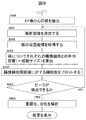
- Fig. 4 shows an outline of the flow of cell sheet measurement, stratification, and differentiation analysis using OCT.
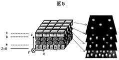
- FIG. 5 shows the measurement image of the cell sheet at that time.
- the left side of FIG. 5 is an image of a perspective view of the cell sheet. Define xyz axes as shown here.
- the right side is an image of the xy image at different z positions obtained from the OCT.
- FIG. 6 shows display screen images of various analysis results displayed on the monitor of the output device (display unit) 207.
- the number of layer structures formed and the number of layers (basal layer, spiny layer, granule layer, etc.) corresponding to each layer are displayed (FIG. 6A).
- FIG. 6A cells are layered into four layers, and a basal layer 601, a spiny layer 602, and a granule layer 603 are formed from the bottom, and two spiny layers 602 exist. Different layers of cells are displayed in different shapes and colors. Instead of this, or in addition to this, the name of the cell layer can also be displayed.
- a three-dimensional image (FIG. 6B), an XY image (FIG. 6C), and an XZ image (FIG. 6D) of the imaged nucleus are displayed.
- an analysis graph FIG. 6E in Example 1 and FIG. 6F in Example 2 can also be displayed.
- results are displayed last as an example (S407), but the results may be displayed together after the analysis is completed, or may be displayed for each item when the analysis is completed.
- the OCT imaging unit 203 installed in the temperature-controlled room 202 captures an XZ tomographic image of the cell sheet (S401).
- an interval smaller than the assumed size of the cell in the Z direction is set.
- the nucleus of the cell is imaged, but only the nucleus is extracted from the image by analyzing the image in the analysis unit 204 (S405). Thereafter, the analysis unit 204 performs an analysis (S406) for determining stratification or differentiation, which will be described in detail later.
- FIG. 7 shows a detailed flow of analysis (S406) for determining stratification or differentiation.
- the analysis region on the XY plane is determined (S701). Desirably, extracting all nuclei from all XY images improves the analysis accuracy.
- the analysis region may be arbitrary as long as it includes a plurality of nuclei at each Z position and does not have a large measurement error. Desirably, the same XY region is good at each Z position.
- nuclei are counted for each XY image (S702). Further, the cell density is calculated for each XY image, and the cell density for each Z position is plotted (S703). The plotted result is, for example, a graph as shown in FIG. 6E.
- FIG. 8 shows a result example of the cell sheet shown in FIG. 5 at this time.
- the horizontal axis indicates the measurement position in the Z direction (the cell membrane layering direction), and the vertical axis defines the cell density.
- the Z positions a, b, and c in FIGS. 5 and 8 correspond to each other.
- the cell sheet is composed of 1 layer of 0 ⁇ Z ⁇ a, 2 layers of spinous layers a ⁇ Z ⁇ b, and 1 layer of b ⁇ Z ⁇ c.
- the size of the cells varies depending on the layer, particularly in the XY direction. For example, the cell size is the smallest in the basal layer, and the cell size increases as the spinous layer, the granule layer, and the upper layer are formed.
- the cells are arranged with almost no gap (see FIG. 2).
- the cell density is highest in the basal layer, and the cell density becomes lower as the spinous layer, the granular layer, and the upper layer are formed. For this reason, when the cell sheet is normally stratified and differentiated, the cell density with respect to the Z position (FIG. 8) is stepped.
- the cell density is shown on the vertical axis, but as described above, assuming a normal state, the number of cells included in the predetermined region, the distance between cells, the size of the cells, Cell density, etc. will have a correlation.
- the number of cells per unit area in the XY plane is the cell density on the surface, and the cell density can also be calculated from the distance between the cells and the size of the cells. Therefore, any index may be used as the vertical axis index used for analyzing the structure of the cell sheet in this example.
- the analysis (S705) for determining stratification and differentiation from here is as follows.
- For proliferation it is determined how many layers the cell sheet is composed of.
- a graph as shown in FIG. 8 is obtained.
- the obtained graph when attention is paid to the Z position where the cell density changes, it can be determined that the cell is composed of at least three layers of 0 ⁇ Z ⁇ a, a ⁇ Z ⁇ b, and b ⁇ Z ⁇ c.
- For differentiation it is determined how many layers the cell sheet is composed of, and what degree of differentiation the cell at which Z position is. In the case of the graph of FIG. 8, it is first determined from the number of asterisks that there are three types of cells with a cell density. At this time, which cell density corresponds to which layer (basal layer, spiny layer, granule layer) is determined from the relationship with the data learned in the storage unit 205 in advance. Alternatively, it can be determined that the first layer having the smallest value of the Z position and the highest cell density is the basal layer, and the cell layer is differentiated into a spiny layer and a granular layer each time the cell density decreases (FIG. 6A). ).
- the cell density information is an index for checking whether normal differentiation is performed. Normal cells fall within a predetermined range for each layer. Therefore, when the cells are cultured without gaps, the density of the cells falls within a predetermined range. Data on the density and size of each layer of cells is stored in the storage unit 205, and the state of the cell layer can be known by collating the cell density of each cell layer in the analysis process (S406). . For example, when the cell density is within a predetermined range, the cell can be determined as a normally differentiated cell. Moreover, when the density of the cells is below a predetermined range, a cell defect (a gap is formed between the cells) and the like can be expected. Cell defects can be confirmed directly by looking at the cell image.
- Table 1 shows an example of information stored in the storage unit 205.
- a data example of the size of the human epidermal cell sheet is shown.
- cell density data the number of layers in each layer, and other data may be stored.
- layer structure data is added as an example.
- S represents a single layer structure
- M represents a multilayer structure. Such data can also be used to determine the state of cells.
- Table 2 shows another example of information stored in the storage unit 205.
- the cell density relative to the Z position may not be stepped.
- the cause may be that there is variation in the progression of differentiation even for cells at the same Z position.
- the region after the change may be a region different from the region analyzed first or a part of the region analyzed first.
- the initial measurement area may be changed and remeasured from S401.
- the analysis unit 204 performs a detailed analysis (S705) using the data learned in the storage unit 205 or the like as the analysis (S406) for determining stratification or differentiation.
- S705 the important point in this example is that a plurality of images with different distances in the stratification direction from the culture surface are acquired, and the number of cells included in each of the plurality of images, It is to measure at least one piece of information about the distance or size between them and enable analysis based on the obtained information.
- information regarding cell stratification in the stratification direction can be obtained.
- the analysis result can be stored in the storage unit 205 as data. If the data content is examined later, it can contribute to the improvement of the cell culture process. Further, it can be displayed on the display unit 207. If displayed in real time, the cell status can be monitored. In addition, remote operation is possible by transmitting to an external device via a network. Alternatively, when the analysis result satisfies a specific condition, an alarm can be issued by sound or video.
- a plurality of images with different distances in the stratification direction from the culture surface are acquired, and at least one information on the number of cells, the distance between cells, or the size included in each of the plurality of images. May be measured and the information may be displayed on the display device 207. For example, it is also effective to plot the cell density for each Z position (S703) and display the graph (for example, FIG. 6E) on the output device 207.
- This configuration is a cheaper device configuration, but the operator can know the outline of the state of cell culture. Such display may be performed in real time, or data may be stored once in the storage unit 205 and checked later.
- the number of cells included in each of a plurality of images is measured, and a plurality of cells having different cell states constituting a layered cell sheet differ from the state of cell distribution in the layering direction.
- the distribution of the number of cells in the stratification direction is graphed by defining the position in the stratification direction on the horizontal axis and the density of cells at the position on the vertical axis
- the first is a relatively large slope.
- the position in the stratification direction corresponding to the first state indicates a boundary between layers having different cell states, and corresponds to the second state. It can be determined that the position in the stratification direction indicates the position of the layer where the cell state is the same.
- Example 2 compared with Example 1, the apparatus used (FIG. 1), the outline of the measurement and analysis flow of the cell sheet by OCT (FIG. 4), and the measurement image of the cell sheet at that time (FIG. 5) ) And the result display screen image (FIG. 6) are common.
- the details of the analysis (S406) for determining stratification or differentiation are different, only different parts are described.
- the second embodiment information that cannot be obtained in the first embodiment can be obtained. For this reason, in Example 1, even when the graph of FIG. 8 does not become stepped and it is difficult to determine the stratification, there is an advantage that the determination of the stratification is possible.
- FIG. 9 shows a detailed flow of analysis (S406) for determining stratification or differentiation.
- an analysis region is determined (S901).
- the position coordinates of all the nuclei in the analysis region are acquired (S902), and the average distance between each nucleus and each adjacent cell is calculated (S903).
- the distance between cell nuclei is used as the distance between cells.
- Each cell has one nucleus, and the nucleus has a different contrast on the image, so that it can be easily extracted by image processing.
- the number of cells against the distance between adjacent cells is plotted (S904) (FIG. 6F)).
- the distance between adjacent cells is obtained by averaging the distances between a certain cell and each of a plurality of adjacent cells. Only the adjacent cells can be extracted by determining a threshold value such as obtaining a distance from the nearest cell and not counting cells that are more than a certain distance as adjacent cells.
- FIG. 10 shows a result example of the cell sheet shown in FIG.
- the horizontal axis indicates the cell size
- the vertical axis indicates the number of cells having the size. From the graph of FIG. 10, it is possible to know the distribution of cells having different characteristics (size in the case of FIG. 10) in the cell membrane. Ideally, it is desirable to count the number of cells in the three-dimensional space occupied by the cell sheet. However, an approximate value can also be obtained by adding the number of cells of a plurality of XY plane images obtained discretely at different locations in the Z direction. If the same cell is included in different XY plane images, counting is repeated, but this is not a big problem when determining the peak of a graph described later. In addition, if the XY coordinates of the cell are used, it is possible to correct the double count.
- the cell sheet is composed of one layer of 0 ⁇ Z ⁇ a, one layer of basal layer, two layers of spiny layer a ⁇ Z ⁇ b, and one layer of granule layer b ⁇ Z ⁇ c.
- the cells are normally stratified and differentiated, the cells are arranged almost without gaps in each layer.
- the cell size varies depending on the layer, especially in the XY direction, and the cell size is the smallest in the basal layer, that is, the distance between adjacent cells (distance between nuclei) is short, and the spine layer, granular layer, and upper layer
- the cell size is large, that is, the distance between adjacent cells is increased.
- the number of cells with respect to the distance between adjacent cells has several peaks. It is determined whether a peak can be detected in the graph (S905). If YES, the process proceeds to analysis (S906) for determining stratification and differentiation.
- the analysis (S906) for determining stratification and differentiation from here is as follows. For proliferation, it is determined how many layers the cell sheet is composed of. From FIG. 10, it can be discriminated that it is composed of at least three layers from the number of peaks in the cell distribution graph with respect to the distance between adjacent cells. However, there are actually two layers in the peak of the spiny layer, and this cannot be identified by the number of peaks. Therefore, the number of cells that can exist in one layer is calculated from the inter-cell distance, and for a peak in which there are more cells, the total number of cells in the peak is divided by the number of cells that can exist in one layer. Thereby, the number of layers included in the peak is calculated, and it is determined how many layers the cell sheet is overlaid (FIG. 6A).
- the data stored in the storage unit 205 shown in Table 1 may be used. That is, when a normal cell membrane is formed, the number of cells that can exist in one layer is experimentally determined and stored in advance in stored data. At the time of analysis, it can be calculated using this data. It is also possible to store in advance in memory data the number of layers of the cell membrane when a normal cell membrane is formed, and compare it with the total number calculated from the observation data.
- For differentiation it is determined how many layers the cell sheet is composed of, and what degree of differentiation the cell at which Z position is. In the case of FIG. 10, it is first determined from the number of peaks that three types of cells have a cell density. At this time, it is determined from the correlation with data (Table 1) that the storage unit 205 has learned in advance which adjacent cell distance corresponds to which layer (basal layer, spiny layer, granule layer). Alternatively, it is determined that the portion with the smallest distance between adjacent cells is the basal layer where the cell size is the smallest, and that each time the distance between adjacent cells increases, it is differentiated into a spiny layer or a granular layer. (FIG. 6A).
- a peak cannot be detected in the graph of the number of cells against the distance between adjacent cells (FIG. 10). In that case, it is determined whether or not the peak of the graph can be detected and the result is NO (S905), so the analysis region is changed (S901).
- the region after the change may be a region different from the region analyzed first or a part of the region analyzed first.
- the initial measurement area may be changed and remeasured from S401.
- the information on the cell size can be an index for checking whether normal differentiation is performed. Normal cells fall within a predetermined range for each layer. Accordingly, the state of the cell layer can be known by storing data related to the cell size in the storage unit 205 and collating the cell size of each cell layer in the analysis process (S705). For example, when the cell size is within a predetermined range, the cell can be determined as a normally differentiated cell.
- various analyzes are performed by the analysis unit 204 using the data learned in the storage unit 205 as analysis (S906) for determining stratification or differentiation.
- it is also effective to plot the number of cells against the distance between adjacent cells and display the graph (FIG. 10) or the like on the output device 207.
- This configuration is a cheaper device configuration, but the operator can know the outline of the state of cell culture.
- Such display may be performed in real time, or data may be stored once in the storage unit 205 and checked later.
- the distance between cells or the size of cells included in each of a plurality of images is measured, and the cell sheet is configured from the distance between cells or the distribution of cell sizes.
- the presence or absence of a plurality of layers having different cell states can be determined. More specifically, a plurality of peaks were observed when the graph was defined by defining the distance between cells or cell size on the horizontal axis and the number of cells having the distance or size between the cells on the vertical axis. In this case, it can be determined that there are a plurality of layers having different cell states.
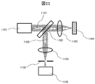
- the measuring device may be an optical device having a high resolution in three dimensions, an optical device other than OCT may be used.
- Fig. 11 shows the basic configuration of a reflective confocal microscope.
- Incident light 1107 from the light source 1101 is applied to the cell sheet 1104 through the objective lens 1103 via the beam splitter 1102.
- the reflected light 1108 is detected by the detection system 1105 through the objective lens 1103 and the pinhole 1106 on the detection system side.
- the imaged nucleus can be extracted and analyzed along the analysis flow of FIG. 7 or FIG. 9 to determine the stratification and differentiation of the cell sheet.
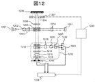
- FIG. 12 is a schematic diagram showing a basic embodiment of an optical measuring device constituting a part of the present invention.
- the interference optical system generates three or more interference lights, specifically four examples.
- the interference phase of the signal light and the reference light is set to be different from each other by an integer multiple of approximately 90 degrees.
- a pair of interference lights in which the interference phases of the signal light and the reference light are approximately 180 degrees different from each other are detected by a current differential type photodetector.
- a laser beam composed of a single wavelength component emitted from the light source 1201 is converted into parallel light by a collimator lens 1202, and the polarization is rotated by a ⁇ / 2 plate 1203 whose optical axis direction can be adjusted, and then a polarization beam splitter. 1204 branches the signal light and the reference light into two.
- the signal light passes through a ⁇ / 4 plate 1205 whose optical axis direction is set to about 22.5 with respect to the horizontal direction, and the polarization state is converted from s-polarized light to circularly-polarized light.
- the light is collected by the lens 1206 and irradiated to the cell sheet 1209 to be measured.
- the objective lens 1206 is scanned at least in the z direction by the lens actuator 1207 under the control of the control unit 1230, thereby scanning the condensing position (measurement position) of the luminous flux signal light by the objective lens 1206.
- the signal light generated by being reflected or scattered from the measurement object is converted into parallel light by the objective lens 1206, the polarization state is converted from circularly polarized light to p-polarized light by the ⁇ / 4 plate 1205, and is incident on the polarization beam splitter 1204. To do.
- the reference light is transmitted through the ⁇ / 4 plate 1210, the polarization state is converted from p-polarized light to circularly polarized light, is incident on the fixed mirror 1211 and reflected, and then the polarization state is converted from circularly polarized light to s-polarized light. And enters the polarization beam splitter 1204.
- the signal light and the reference light are combined by the polarization beam splitter 1204 to generate combined light.
- the combined light is guided to an interference optical system 212 including a half beam splitter 1213, a ⁇ / 2 plate 1214, a ⁇ / 4 plate 1219, condenser lenses 1215 and 1220, and Wollaston prisms 1216 and 1221.
- the combined light incident on the interference optical system 1212 is split into two by the half beam splitter 1213 into transmitted light and reflected light.
- the transmitted light passes through a ⁇ / 2 plate 1214 whose optical axis is set to about 22.5 degrees with respect to the horizontal direction, and is then collected by a condenser lens 1215 and branched into two by a Wollaston prism 1216.
- a first interference light and a second interference light that are 180 degrees out of phase with each other are generated.
- the first interference light and the second interference light are detected by a current differential photodetector 1217, and a signal 1218 proportional to the difference in intensity between them is output.
- the reflected light passes through a ⁇ / 4 plate 1219 whose optical axis is set to about 45 degrees with respect to the horizontal direction, and is then collected by a condenser lens 1220 and branched into two by a Wollaston prism 1221.
- a third interference light and a fourth interference light that are 180 degrees out of phase with each other are generated.
- the third interference light and the fourth interference light are detected by a current differential photodetector 1222, and a signal 1223 proportional to the difference in intensity is output.
- the signals 1218 and 1223 generated in this way are input to the signal processing unit 1224, and a signal proportional to the amplitude of the signal light is obtained by being calculated. Based on this signal, three-dimensional information of the cell sheet 1209 can be obtained.
- information on cell stratification can be obtained by analyzing changes in the number of cells, the distance between cells, or the size in the stratification direction.
- the number of these cells in the stratification direction (depth direction), the distance between the cells, or the change in size is displayed on the display device or stored in the storage device, thereby allowing the operator to stratify the cells. Can inform information.
- information on the number of cells, the distance between cells, and the size is acquired, but it is not measured for one specific cell but obtained from a predetermined range of one or a plurality of images. It should be noted that data is acquired for a plurality of cells. From this data, it is possible to know the distribution of the number and density of cells in the stratification direction, or the distribution of the cell size and the distance between cells in the data of a plurality of cell samples. The point of using statistical data in this way is a big feature.
- the present invention is not limited to the above-described embodiment, and includes various modifications.
- a part of the configuration of one embodiment can be replaced with the configuration of another embodiment, and the configuration of another embodiment can be added to the configuration of one embodiment.
- Interference light 1101 ⁇ ⁇ ⁇ Light source 1102 Beam splitter 1103 ⁇ ⁇ ⁇ Objective lens 1104 ... Cell sheet 1105 ⁇ ⁇ ⁇ Detection system 1106 ⁇ ⁇ ⁇ Pinhole 1107: Incident light 1108: Reflected light 1201 ⁇ ⁇ ⁇ Light source 1202 ... Collimating lens 1203 ⁇ ⁇ ⁇ ⁇ / 2 plate 1204 ... Beam splitter 1205 ... ⁇ / 4 plate 1206 ... Objective lens 1207 ... Lens actuator 1209 ⁇ ⁇ ⁇ Cell sheet 1210 ...
Abstract
Provided is a method for non-invasively and quantitatively determining multilayerization and differentiation when culturing a cell sheet. Provided is a method for determining a cell state by imaging a cell sheet by using an optical instrument characterized by having a high resolution, and then analyzing the inner structure thereof.
Description
本発明は、細胞シートの細胞状態の判定に関するものである。
The present invention relates to determination of the cell state of a cell sheet.
幹細胞から作製した組織を損傷部位に移植することにより,損傷した組織や器官の再生や機能の回復を実現する再生医療が近年注目されている。中でも生体模倣組織である細胞シートの移植は、細胞が単体で存在する細胞溶液と比較した場合に治癒効果がより高いことが知られており、ヒト表皮細胞シートは製品化され重度のやけど治療に使用されるなど細胞シートの臨床応用が進んでいる。現在この細胞シートに関して残された課題のひとつに細胞シートの細胞状態の非侵襲的評価方法の確立がある。
In recent years, regenerative medicine that realizes regeneration of damaged tissues and organs and recovery of function by transplanting tissue prepared from stem cells to the damaged site has attracted attention in recent years. In particular, transplantation of a cell sheet, which is a biomimetic tissue, is known to have a higher healing effect when compared to a cell solution in which cells are present alone, and human epidermal cell sheets have been commercialized for the treatment of severe burns. The clinical application of cell sheets is progressing. One of the remaining issues regarding this cell sheet is the establishment of a noninvasive evaluation method for the cell state of the cell sheet.
図2に細胞シートが正常に作製されるまでの培養段階を示す。図2は培養表面201を紙面奥行方向としたとき、横から細胞の構造を観察したものである。細胞シートは以下の段階を経て生体模倣組織となる。
FIG. 2 shows the culture stage until the cell sheet is normally produced. FIG. 2 shows the cell structure observed from the side when the culture surface 201 is in the depth direction of the drawing. The cell sheet becomes a biomimetic tissue through the following steps.
(S101):細胞が播種される。このとき、単離された幹細胞202が培地中に浮遊している状態である。
(S101): Cells are seeded. At this time, the isolated stem cell 202 is in a suspended state in the medium.
(S102):培養表面201に細胞が接着する。このとき、幹細胞202の密度はまばらである。
(S102): Cells adhere to the culture surface 201. At this time, the density of the stem cells 202 is sparse.
(S103):培養表面一面に細胞が単層に増殖し基底層となる。
(S103): Cells grow into a monolayer on the entire culture surface to become a basal layer.
(S104):細胞が2層以上に重層化する。すなわち、細胞が積層構造を形成する。さらに2層目以上の細胞が分化して細胞シートを形成する。分化では、層によって細胞内に発現するタンパク質が異なる。
(S104): Cells are stratified into two or more layers. That is, the cells form a laminated structure. Furthermore, cells in the second layer or more differentiate to form a cell sheet. In differentiation, proteins expressed in cells differ depending on layers.
(S105):培養表面から細胞シートを剥離し患部へ移植する。
(S105): The cell sheet is detached from the culture surface and transplanted to the affected area.
現状では移植に用いられる細胞シートの品質は,培養中の位相差顕微鏡による観察により検証されている。あるいは、移植用細胞シートと同時に同条件で作製された評価用細胞シートに対する、組織染色などの侵襲的評価によって検証されている。
Currently, the quality of cell sheets used for transplantation has been verified by observation with a phase contrast microscope during culture. Alternatively, it has been verified by invasive evaluation such as tissue staining on an evaluation cell sheet prepared under the same conditions as the transplant cell sheet.
しかし、これらの方法には課題がある。位相差顕微鏡による細胞観察は非侵襲的であり、細胞培養中に随時実施されている。しかしながら、細胞シート表層の観察のみに対応しており、(S104)以降の重層化した細胞シートを評価することはできない。また評価用細胞シートに対して実施されている組織染色評価は、重層化や分化の程度を評価可能であるが、細胞シートを固定するため侵襲的手法であり移植用シートそのものを評価することはできない。これらの課題を解決する非侵襲的計測技術の確立は,移植用細胞シートの細胞状態を直接的に評価可能にすることにより、移植用再生組織の品質向上に貢献するといえる。
However, there are problems with these methods. Cell observation with a phase contrast microscope is non-invasive and is performed as needed during cell culture. However, only the observation of the cell sheet surface layer is supported, and the layered cell sheets after (S104) cannot be evaluated. In addition, the tissue staining evaluation performed on the cell sheet for evaluation can evaluate the degree of stratification and differentiation, but it is an invasive technique for fixing the cell sheet, and it is not possible to evaluate the transplant sheet itself Can not. The establishment of a noninvasive measurement technique that solves these problems can be said to contribute to improving the quality of the regenerative tissue for transplantation by making it possible to directly evaluate the cell state of the cell sheet for transplantation.
これまでにいくつかの文献において非侵襲的な細胞評価手法が述べられている。例えば特許文献1では、光学顕微鏡を用いて焦点位置をZ位置の異なる画像を複数枚撮像し、培養面に接着している細胞と剥離細胞を判定する方法について述べている。
So far, several non-invasive cell evaluation methods have been described. For example, Patent Document 1 describes a method of determining a cell adhering to a culture surface and a detached cell by capturing a plurality of images having different focal positions and Z positions using an optical microscope.
しかしながら、特許文献1が対象としている細胞は細胞シートと異なり、単層で培養面に接着する細胞である。また、光学顕微鏡は焦点位置から以外の光もすべて像に反映される原理でありZ分解能が低いため、細胞シートのように積層している組織に適応した場合、Z方向の像は複数層の重ね合わせの像となり、層ごとの細胞ひとつひとつをイメージングすることは困難である。
However, unlike the cell sheet, the cells targeted by Patent Document 1 are cells that adhere to the culture surface in a single layer. In addition, the optical microscope is the principle that all light other than that from the focal position is reflected in the image, and the Z resolution is low, so when applied to tissues stacked like a cell sheet, the image in the Z direction has multiple layers. It is difficult to image each cell in each layer as a superposed image.
また特許文献2には、個々の培養細胞のばらつきを解消すべく判断基準をそろえるための、判断するプロトコルが記載されているが、具体的な培養状況を判定できるものではない。
In addition, Patent Document 2 describes a determination protocol for aligning the determination criteria so as to eliminate variations in individual cultured cells. However, a specific culture state cannot be determined.
そのため、文献に示された手法では細胞シートを評価する際に不可欠となる細胞の重層化、分化のタイミングと程度を評価しがたい。本発明においては、生体模倣組織である細胞シートの重層化、分化を非侵襲的に評価することが課題となる。
Therefore, it is difficult to evaluate the timing and degree of cell stratification and differentiation, which are indispensable when evaluating a cell sheet, by the method shown in the literature. In this invention, it becomes a subject to non-invasively evaluate the stratification and differentiation of the cell sheet which is a biomimetic tissue.
上記課題を解決するために、高分解能を特徴とする光学機器を用いて細胞シートのイメージングを実施し、内部構造を分析することにより細胞状態を判定する方法を提供する。
In order to solve the above-described problems, a method for determining a cell state by performing imaging of a cell sheet using an optical instrument characterized by high resolution and analyzing an internal structure is provided.
本発明の一つの側面は、培養面に細胞を重層化させて細胞シートを培養する細胞培養装置であって、光源と、培養面上の細胞に光源からの光を照射する集光光学系と、細胞からの光を検出する検出光学系と、検出光学系から取得した情報に基づく画像を解析する解析部と、を備える細胞培養装置である。解析部は、培養面からの重層化方向の距離が異なる複数の断面画像を取得し、複数の断面画像の夫々に含まれる細胞数を計測し、細胞数に基づいて、少なくとも前記重層化方向の細胞数の分布を解析する。
One aspect of the present invention is a cell culture apparatus for culturing a cell sheet by layering cells on a culture surface, and a light source and a condensing optical system for irradiating light from the light source to cells on the culture surface A cell culture device comprising: a detection optical system that detects light from a cell; and an analysis unit that analyzes an image based on information acquired from the detection optical system. The analysis unit obtains a plurality of cross-sectional images with different distances in the stratification direction from the culture surface, measures the number of cells included in each of the plurality of cross-sectional images, and based on the number of cells, at least in the stratification direction Analyze cell number distribution.
本発明の一つの側面は、培養面に重層化された細胞シートを非侵襲で光学的に取得した画像を解析する画像解析装置である。この装置は、培養面からの重層化方向の距離が異なる複数の断面画像を取得する取得部と、複数の断面画像の夫々に含まれる細胞数を計測する計測部と、細胞数に基づいて、少なくとも重層化方向の細胞数の分布を解析する解析部と、を有する。
One aspect of the present invention is an image analysis apparatus that analyzes an image obtained by optically acquiring a cell sheet layered on a culture surface in a non-invasive manner. This device is based on an acquisition unit that acquires a plurality of cross-sectional images with different distances in the stratification direction from the culture surface, a measurement unit that measures the number of cells included in each of the plurality of cross-sectional images, and the number of cells. An analysis unit that analyzes at least the distribution of the number of cells in the stratification direction.
解析部は、重層化方向の細胞数の分布から、重層化した細胞シートを構成する複数の層夫々の位置を判別するように構成することができる。また、解析部は、複数の断面画像に含まれる細胞間の距離を算出し、細胞間の距離に基づいて、重層化した細胞シートを構成する複数の層の夫々の位置を判別するように構成することもできる。
The analysis unit can be configured to determine the position of each of a plurality of layers constituting the layered cell sheet from the distribution of the number of cells in the layering direction. Further, the analysis unit is configured to calculate the distance between cells included in the plurality of cross-sectional images, and to determine the positions of the plurality of layers constituting the layered cell sheet based on the distance between the cells. You can also
また、解析部は、計測した細胞数に基づいて細胞の密度を算出し、一方の軸に重層化方向の位置を、他方の軸に当該位置における細胞の密度を定義してグラフ化した画像を生成し、グラフ化した画像を表示装置に表示するように構成してもよい。また、解析部は、計測した細胞数に基づいて複数の画像の夫々に含まれる細胞の細胞間の距離または細胞の大きさを測定し、重層化方向の細胞間の距離または大きさの分布を、一方の軸に細胞間の距離または細胞の大きさ、他方の軸に当該細胞間距離または大きさをもつ細胞の数を定義してグラフ化した画像を生成し、グラフ化した画像を表示装置に表示するように構成してもよい。
In addition, the analysis unit calculates the density of the cells based on the measured number of cells, and displays the graphed image by defining the position in the stratification direction on one axis and the density of the cells at the position on the other axis. The generated and graphed image may be displayed on the display device. The analysis unit also measures the distance between the cells or the size of the cells included in each of the plurality of images based on the measured number of cells, and calculates the distribution of the distance or size between the cells in the stratification direction. Generates a graphed image by defining the distance between cells or cell size on one axis and the number of cells having the cell distance or size on the other axis, and displays the graphed image as a display device You may comprise so that it may display on.
また、解析部は、複数の画像の夫々に含まれる細胞の数の情報から、重層化方向の細胞の数の分布に関する分析データを生成し、分析データを表示するか、分析データに基づいて警報を発するか、分析データに基づいた信号を細胞培養装置または他の外部の装置に出力するかの少なくとも一つを行うように構成してもよい。また、解析部は、複数の画像の夫々に含まれる細胞の数の情報から、複数の画像の夫々に含まれる細胞の細胞間の距離または大きさを測定し、細胞シートに含まれる細胞の細胞間の距離または細胞の大きさの分布に関する分析データを生成し、分析データを表示するか、分析データに基づいて警報を発するか、分析データに基づいた信号を細胞培養装置または他の外部の装置に出力するかの少なくとも一つを行うように構成してもよい。
In addition, the analysis unit generates analysis data regarding the distribution of the number of cells in the stratification direction from the information on the number of cells included in each of the plurality of images, and displays the analysis data or alerts based on the analysis data. Or at least one of outputting a signal based on the analysis data to the cell culture device or other external device. Further, the analysis unit measures the distance or size between the cells included in each of the plurality of images from the information on the number of cells included in each of the plurality of images, and the cells of the cells included in the cell sheet Generate analytical data on the distance between cells or the distribution of cell size and display the analytical data, issue an alarm based on the analytical data, or send a signal based on the analytical data to a cell culture device or other external device It may be configured to perform at least one of the following.
本発明の一つの側面は、培養面に細胞を重層化させて細胞シートを培養する際の細胞状態解析方法である。この方法では、光源と、培養面上の細胞に前記光源からの光を照射する集光光学系と、細胞からの光を検出する検出光学系と、検出光学系からの光を検出する検出器とを用いる。解析においては、検出器からの信号に基づいて、培養面からの重層化方向の距離が異なる複数の画像を取得する。そして、複数の画像の夫々に含まれる細胞の数、細胞間の距離、あるいは大きさの少なくともひとつの情報を測定する。
One aspect of the present invention is a cell state analysis method for culturing a cell sheet by layering cells on the culture surface. In this method, a light source, a condensing optical system that irradiates cells on the culture surface with light from the light source, a detection optical system that detects light from the cells, and a detector that detects light from the detection optical system And are used. In the analysis, based on the signal from the detector, a plurality of images having different distances in the stratification direction from the culture surface are acquired. Then, at least one piece of information on the number of cells, the distance between cells, or the size included in each of the plurality of images is measured.
本発明の他の側面は、培養面に細胞を重層化させて細胞シートを培養する細胞培養装置からデータを受け取り、細胞培養装置で培養された細胞の状態を解析する細胞状態解析装置である。細胞培養装置と細胞状態解析装置は一体化されていてもよいし、ネットワークで接続され、地理的に離れた位置に配置されてもよい。細胞培養装置は、光源と、培養面上の細胞に光源からの光を照射する集光光学系と、細胞からの光を検出する検出光学系と、検出光学系からの光を検出する検出器と、出力装置を備える。細胞状態解析装置が備える処理装置は、出力装置から送られてくる検出器からの信号に基づいて、培養面からの重層化方向の距離が異なる複数の画像を取得する機能と、複数の画像の夫々に含まれる細胞の数、細胞間の距離、あるいは大きさの少なくともひとつの情報を測定する機能と、を有する。
Another aspect of the present invention is a cell state analyzer that receives data from a cell culture device that cultivates a cell sheet by layering cells on the culture surface and analyzes the state of the cells cultured in the cell culture device. The cell culture device and the cell state analysis device may be integrated, or may be connected via a network and arranged at geographically separated positions. The cell culture apparatus includes a light source, a condensing optical system for irradiating cells on the culture surface with light from the light source, a detection optical system for detecting light from the cells, and a detector for detecting light from the detection optical system. And an output device. The processing device provided in the cell state analysis device has a function of acquiring a plurality of images having different distances in the stratification direction from the culture surface based on a signal from a detector sent from the output device, and a plurality of images A function of measuring at least one piece of information on the number of cells, the distance between cells, or the size of each cell.
細胞間の距離は細胞の核同士の距離とする。正常に細胞が成長している場合、各細胞は隙間なく形成されている。よって、正常な状態を前提とした場合、細胞の大きさは細胞間の距離にほぼ等しい。また、所定領域に含まれる細胞の数、細胞間の距離、細胞の大きさ、細胞の密度、等は相関関係を有することになる。よって、本発明で細胞膜構造の分析に用いる指標としては、いずれを用いてもよい。
The distance between cells is the distance between cell nuclei. When cells are growing normally, each cell is formed without a gap. Thus, assuming a normal state, the cell size is approximately equal to the distance between cells. In addition, the number of cells included in the predetermined region, the distance between the cells, the size of the cells, the density of the cells, and the like have a correlation. Therefore, any index may be used as an index used for analysis of the cell membrane structure in the present invention.
本発明の他の側面は、培養面に細胞を重層化させて細胞シートを培養する細胞培養装置における、培養された細胞の状態解析装置である。この装置は、光源と、培養面上の細胞に光源からの光を照射する集光光学系と、細胞からの光を検出する検出光学系と、検出光学系からの光を検出する検出器と、検出器からの信号を処理する処理装置と、出力装置を備える。処理装置は、検出器からの信号に基づいて、培養面からの重層化方向の距離が異なる複数の画像を取得する機能と、複数の画像の夫々に含まれる細胞の数、細胞間の距離、あるいは大きさの少なくともひとつの情報を測定する機能と、を有する。
Another aspect of the present invention is an apparatus for analyzing the state of a cultured cell in a cell culture device for culturing a cell sheet by layering cells on the culture surface. The apparatus includes a light source, a condensing optical system for irradiating cells on the culture surface with light from the light source, a detection optical system for detecting light from the cells, and a detector for detecting light from the detection optical system, And a processing device for processing a signal from the detector, and an output device. The processing device has a function of acquiring a plurality of images having different distances in the stratification direction from the culture surface based on a signal from the detector, a number of cells included in each of the plurality of images, a distance between the cells, Alternatively, it has a function of measuring at least one piece of information.
上記で測定された情報は、グラフ化をするなどして、表示装置に表示することができる。また、データとして記憶装置に蓄積することができる。また、データとしてネットワークを介して外部の装置に送信することができる。あるいは、測定された情報に基づいて、細胞培養装置の少なくとも一部を制御するように構成することもできる。
The information measured above can be displayed on a display device by graphing. Further, it can be stored as data in a storage device. Further, it can be transmitted as data to an external device via a network. Alternatively, at least a part of the cell culture apparatus can be controlled based on the measured information.
本発明のさらに他の側面は、恒温室と、恒温室内に配置され細胞を重層化させて細胞シートを培養する培養容器と、培養容器に結合され細胞溶液を供給する細胞ボトルと、培養容器に結合され培地を供給する培地ボトルと、培養容器に結合され培養容器から廃棄される培地を格納する廃液ボトルとを有する自動細胞培養装置に関する。この自動細胞培養装置は、恒温室、培養容器、細胞ボトル、培地ボトル、廃液ボトル、撮像部、細胞溶液の供給、培地の供給および廃棄、のうちの少なくとも一つを制御する制御部を有する。また、細胞シートを撮像する撮像部と、撮像部から得られる情報を処理する処理部と、処理部からの情報を出力する出力部と、処理部へ情報を入力する入力部と、を有ししている。撮像部は、光源と、前記細胞シートに前記光源からの光を照射する集光光学系と、前記細胞シートからの光を検出する検出光学系と、前記検出光学系からの光を検出する検出器と、を備えている。また、処理装置は、検出器からの信号に基づいて培養シートにおける重層化方向の位置が異なる複数の画像を取得する機能と、複数の画像の夫々に含まれる細胞の数、細胞間の距離、あるいは大きさの少なくともひとつの情報を測定する機能と、を有ししている。さらに、出力部は、測定した情報を表示するか、測定した情報に基づいて警報を発するか、外部の装置に出力するか、あるいは、制御部または入力部にフィードバックを行うか、の少なくとも一つを行うことができる。ここで、警報とは、異常を知らせるものと正常を知らせるものの両方を含む。
Still another aspect of the present invention relates to a temperature-controlled room, a culture container that is placed in the temperature-controlled room to layer cells and culture cell sheets, a cell bottle that is connected to the culture container and supplies a cell solution, and a culture container The present invention relates to an automatic cell culture apparatus having a culture medium bottle that is connected to supply a culture medium and a waste liquid bottle that stores a culture medium that is connected to the culture container and discarded from the culture container. This automatic cell culture apparatus has a control unit that controls at least one of a temperature-controlled room, a culture container, a cell bottle, a medium bottle, a waste liquid bottle, an imaging unit, a cell solution supply, a medium supply and disposal. In addition, an imaging unit that images the cell sheet, a processing unit that processes information obtained from the imaging unit, an output unit that outputs information from the processing unit, and an input unit that inputs information to the processing unit is doing. The imaging unit includes a light source, a condensing optical system that irradiates the cell sheet with light from the light source, a detection optical system that detects light from the cell sheet, and detection that detects light from the detection optical system. And a vessel. In addition, the processing device has a function of acquiring a plurality of images having different positions in the stratification direction in the culture sheet based on a signal from the detector, the number of cells included in each of the plurality of images, the distance between the cells, Alternatively, it has a function of measuring at least one piece of information. Furthermore, the output unit displays at least one of the measured information, issues an alarm based on the measured information, outputs to an external device, or provides feedback to the control unit or the input unit. It can be performed. Here, the alarm includes both a notification of abnormality and a notification of normality.
上記で説明した機能は、ハードウエアで構成してもよいし、ソフトウエアで構成してもよい。
The functions described above may be configured by hardware or software.
本発明によれば、細胞シートを培養する際に非侵襲的に細胞状態(重層化、分化)を判定できるようになる。
According to the present invention, the cell state (stratification, differentiation) can be determined non-invasively when the cell sheet is cultured.
実施の形態について、図面を用いて詳細に説明する。ただし、本発明は以下に示す実施の形態の記載内容に限定して解釈されるものではない。本発明の思想ないし趣旨から逸脱しない範囲で、その具体的構成を変更し得ることは当業者であれば容易に理解される。
Embodiments will be described in detail with reference to the drawings. However, the present invention is not construed as being limited to the description of the embodiments below. Those skilled in the art will readily understand that the specific configuration can be changed without departing from the spirit or the spirit of the present invention.
図面等において示す各構成の位置、大きさ、形状、範囲などは、発明の理解を容易にするため、実際の位置、大きさ、形状、範囲などを表していない場合がある。このため、本発明は、必ずしも、図面等に開示された位置、大きさ、形状、範囲などに限定されない。
The position, size, shape, range, etc. of each component shown in the drawings and the like may not represent the actual position, size, shape, range, etc. in order to facilitate understanding of the invention. For this reason, the present invention is not necessarily limited to the position, size, shape, range, and the like disclosed in the drawings and the like.
以下、本発明の実施形態を順次説明するが、それに先立ち、本発明が対象とする細胞シート評価時の状況について、表皮細胞を例に説明する。
Hereinafter, embodiments of the present invention will be described in order, but prior to that, the situation at the time of cell sheet evaluation targeted by the present invention will be described using epidermis cells as an example.
図2に細胞シートが正常に作製されるまでの培養段階を示す。表皮細胞は播種され(S101),24時間程度で培養表面に接着する(S102)。接着した細胞は、その後数日程度で密集状態まで平面状に増殖し(S103),この1層目が細胞シートの基底層となる。その後は,細胞が層状に増殖して重層化すると共に、2層目以上の細胞が分化することにより、1-2週間程度の期間を経て生体ヒト表皮細胞と類似した細胞シートとなる(S104)。重層化とは細胞が積層構造を形成すること、分化とは層によって細胞内に異なる発現タンパク質が発現することを意味する。また、細胞シートの細胞は細胞形状や大きさが分化状態によって異なることが知られている。なお,生体表皮細胞は層構造であり,下から基底層,有棘層,顆粒層,角化層から成る。細胞シートにおいても分化が進行すると有棘層,顆粒層の形成が見られる。
FIG. 2 shows the culture stage until the cell sheet is normally produced. Epidermal cells are seeded (S101) and adhere to the culture surface in about 24 hours (S102). The adhered cells grow into a flat state until a dense state in a few days thereafter (S103), and this first layer becomes the basal layer of the cell sheet. Thereafter, the cells proliferate in layers and are layered, and the cells in the second and higher layers differentiate to form a cell sheet similar to living human epidermal cells after a period of about 1-2 weeks (S104). . Stratification means that cells form a laminated structure, and differentiation means that different expressed proteins are expressed in the cells depending on the layer. Moreover, it is known that the cell shape and size of the cells of the cell sheet differ depending on the differentiation state. Living epidermis cells have a layered structure, consisting of the basal layer, spiny layer, granule layer, and keratinized layer from the bottom. As cell differentiation progresses, the formation of spinous and granular layers is observed.
細胞シートが正常に生体模倣組織として形成されていることの品質を保障するために、上記細胞シートの重層化や分化状態を評価することが必要である。細胞シートの評価法は、培養中は位相差顕微鏡による評価、培養後は組織染色による評価が一般的である。位相差顕微鏡による細胞シートの観察においては、培養表面上に存在する細胞の数や形状を確認する。この手法においては細胞増殖が正常かどうかを非侵襲的に判断可能であるが、培養中期以降もしくは終了時点における細胞シートの重層化や分化を判断することができない。また細胞シートの組織染色評価においては、組織を固定し作製した切片をヘマトキシリンエオシン染色または免疫染色によって染色し、細胞シートの重層化や分化を確認する。この手法においては、培養終了後の細胞シートの重層化や分化の程度を判断可能である。しかし、組織の固定や染色が必要となるため侵襲的手法であり、培養中に実施することや評価した細胞シートを移植に用いることができない。
In order to ensure the quality that the cell sheet is normally formed as a biomimetic tissue, it is necessary to evaluate the stratification and differentiation state of the cell sheet. The cell sheet is generally evaluated by a phase contrast microscope during culture and by tissue staining after culture. In observing a cell sheet with a phase contrast microscope, the number and shape of cells present on the culture surface are confirmed. In this method, it is possible to determine non-invasively whether cell growth is normal, but it is not possible to determine the stratification or differentiation of cell sheets after the middle stage of culture or at the end of culture. In the tissue staining evaluation of a cell sheet, a section prepared by fixing the tissue is stained by hematoxylin eosin staining or immunostaining to confirm the stratification and differentiation of the cell sheet. In this method, it is possible to determine the degree of stratification and differentiation of the cell sheet after completion of the culture. However, this is an invasive technique because it requires tissue fixation and staining, and the cell sheet evaluated or carried out during culture cannot be used for transplantation.
本発明はこのような状況に鑑みてなされたものである。高分解能を特徴とする光学機器を用いて細胞シートのイメージングを実施し、内部構造を分析することにより細胞シートの重層化、分化の状態を判定する方法を提供する。細胞シートの3次元構造を細胞単位でイメージングし、核や細胞膜を抽出し、任意の層の細胞密度や細胞サイズを解析することができる。これにより、細胞シートが何層構造から成っているか重層化が判定可能になり、さらに各層を構成する細胞の分化の程度も判定可能になる。
The present invention has been made in view of such a situation. The present invention provides a method for determining the state of cell sheet stratification and differentiation by imaging a cell sheet using an optical instrument characterized by high resolution and analyzing the internal structure. The three-dimensional structure of the cell sheet can be imaged in cell units, the nucleus and cell membrane can be extracted, and the cell density and cell size of any layer can be analyzed. This makes it possible to determine how many layers the cell sheet has, and to determine the degree of differentiation of the cells constituting each layer.
当該それぞれの課題を解決する手段の一例としては、具体的には以下のようになる。培養中もしくは培養終了時に培養容器越しに細胞シートを3次元的に測定する。測定機器は3次元に高分解能を有する光学機器であればよい。ここでは、OCT(Optical Coherence Tomography:光干渉断層計)を例に挙げている。他の構成として、反射型共焦点顕微鏡、多光子励起顕微鏡など、非侵襲(非破壊かつ非染色)且つ3次元に分解能を有する光学機器が使用可能である。OCTの場合、光源の光を信号光と参照光に分岐し、信号光を細胞に照射し、細胞から反射した信号光を参照光と合波することにより生成された合成光を検出する原理である。OCTでは、信号光は細胞の様々な深さから重なり合って反射されるが、参照光と干渉される成分は特定の深さ位置からの信号光成分に限定されるため、光学顕微鏡とは異なりZ分解能の高い測定が可能になる。
Specific examples of means for solving each of the problems are as follows. The cell sheet is measured three-dimensionally through the culture vessel during the culture or at the end of the culture. The measuring device may be an optical device having a high resolution in three dimensions. Here, OCT (Optical Coherence Tomography) is taken as an example. As another configuration, an optical instrument that is non-invasive (non-destructive and non-staining) and has a three-dimensional resolution, such as a reflective confocal microscope or a multiphoton excitation microscope, can be used. In the case of OCT, it is based on the principle of detecting the synthesized light generated by splitting the light from the light source into signal light and reference light, irradiating the signal light to the cell, and combining the signal light reflected from the cell with the reference light. is there. In OCT, signal light overlaps and is reflected from various depths of the cell, but the component that interferes with the reference light is limited to the signal light component from a specific depth position. Measurement with high resolution becomes possible.
10ミクロン以下程度の高い空間分解能を有するOCTの場合、取得した画像から細胞シート内部の核がイメージング可能である。取得した画像では細胞の核はコントラストが異なることから、その密度やサイズ、あるいは核同士の距離を解析することができる。この情報により細胞シートの重層化、分化の過程が順調か否か判定できる。また、この細胞状態の判定法は、既存の画像処理技術により自動化可能である。自動培養装置に組み込み、自動培養装置内の培養容器で培養された細胞シートをOCTで測定することも可能である。
In the case of OCT having a high spatial resolution of about 10 microns or less, the nucleus inside the cell sheet can be imaged from the acquired image. Since the cell nuclei have different contrasts in the acquired image, the density and size, or the distance between the nuclei can be analyzed. With this information, it can be determined whether the process of layering and differentiation of the cell sheet is smooth. The cell state determination method can be automated by existing image processing techniques. It is also possible to measure with OCT a cell sheet that is incorporated in an automatic culture device and cultured in a culture vessel in the automatic culture device.
本実施例では、ヒト表皮細胞シートの非侵襲3次元計測と細胞密度による重層化および分化の判定を例に説明する。
In this example, a non-invasive three-dimensional measurement of a human epidermal cell sheet and determination of stratification and differentiation based on cell density will be described as an example.
図1AはOCTを組み込んだ自動培養装置を示す。図1の自動培養装置201は細胞培養をする恒温室202を有する。恒温室内には撮像部203が設置されている。恒温室の外部には解析部204、記憶部205を含むコンピュータ206と出力装置207が設置されている。出力装置207は、例えばオペレータに各種の情報を表示する画像表示装置、音声で警報を発する警報装置、プリンターなどがある。また、ネットワークなどを介して、外部の記憶装置や情報端末にデータを送信することもできる。あるいは、種々のインターフェースを介して、制御部208に指示を送ることもできる。自動培養装置の制御は制御部208で実施する。細胞培養は恒温室202内部に複数設置された培養容器214中において実施される。必要となる細胞溶液の供給は培地流路212を通って細胞ボトル209から供給される。培地の供給は、培地流路212を通って培地ボトル210から培養容器214に対して実施される。培養に使用された不要な培地は廃液流路213を通って廃液ボトル211へ廃棄される。
FIG. 1A shows an automatic culture apparatus incorporating OCT. The automatic culture apparatus 201 in FIG. 1 has a temperature-controlled room 202 for cell culture. An imaging unit 203 is installed in the temperature-controlled room. A computer 206 including an analysis unit 204 and a storage unit 205 and an output device 207 are installed outside the temperature-controlled room. The output device 207 includes, for example, an image display device that displays various types of information to an operator, an alarm device that issues a voice alert, and a printer. In addition, data can be transmitted to an external storage device or information terminal via a network or the like. Alternatively, an instruction can be sent to the control unit 208 via various interfaces. The automatic culture apparatus is controlled by the control unit 208. Cell culture is performed in a plurality of culture vessels 214 installed in the temperature-controlled room 202. The necessary cell solution is supplied from the cell bottle 209 through the medium channel 212. The medium is supplied from the medium bottle 210 to the culture container 214 through the medium channel 212. The unnecessary medium used for the culture is discarded into the waste liquid bottle 211 through the waste liquid channel 213.
細胞シートの品質評価は、培養容器の外部から細胞シートを撮像する撮像部203を用いた測定により実施可能である。本実施例では、撮像部203にOCTを用いる。非侵襲3次元計測を行う部分の全体構成は、細胞シートを撮像する撮像部203と、撮像した画像を解析して細胞シートの重層化や分化の状態を判定する解析部204と、事前に解析に必要な情報を記憶させておく記憶部205と、解析結果を表示する出力装置(ここでは画像モニタを想定した)207を有する。図1の自動培養装置はアミノ酸分析装置を含むアミノ酸分析ユニット(図示せず)を備えてもよい。培地交換時に廃液となる古い培地は培養容器214から廃液流路213を通って廃液ボトル211へ廃棄されるが、培養上清の一部は廃液流路213から分岐された培養上清分析行き流路(図示せず)を通ってアミノ酸分析ユニットへ運ばれ上清中のアミノ酸濃度を分析することができる。
The quality evaluation of the cell sheet can be performed by measurement using an imaging unit 203 that images the cell sheet from the outside of the culture container. In this embodiment, OCT is used for the imaging unit 203. The entire configuration of the non-invasive 3D measurement part is analyzed in advance, an imaging unit 203 that images the cell sheet, an analysis unit 204 that analyzes the captured image and determines the state of cell sheet stratification and differentiation A storage unit 205 that stores necessary information, and an output device (here, assumed to be an image monitor) 207 that displays an analysis result. The automatic culture apparatus of FIG. 1 may include an amino acid analysis unit (not shown) including an amino acid analyzer. The old culture medium that becomes waste liquid when replacing the medium is discarded from the culture vessel 214 through the waste liquid channel 213 to the waste liquid bottle 211, but part of the culture supernatant flows to the culture supernatant analysis branched from the waste liquid channel 213. The amino acid concentration in the supernatant can be analyzed by being transported through a path (not shown) to the amino acid analysis unit.
細胞状態は解析部204にて判定され、培養終了タイミングの決定や培養組織の品質評価として自動培養装置の制御部208へフィードバックされる。あるいは、細胞状態は出力装置207に表示され、オペレータが細胞状態を判定し、培養終了タイミングの決定や培養組織の品質評価を行う。また、オペレータは、必要に応じて自動培養装置の制御部208やコンピュータ206を操作するために、入力部215に入力を行う。入力部215は、ネットワークを介して遠隔地からの指示を入力できる構成としてもよい。本実施例では、解析部204の実現方法として、汎用のコンピュータ206上で動作するソフトウエアとして構成したが、ハードウエアで構成することも可能である。
The cell state is determined by the analysis unit 204, and is fed back to the control unit 208 of the automatic culture apparatus as determination of the culture end timing and quality evaluation of the cultured tissue. Alternatively, the cell state is displayed on the output device 207, and the operator determines the cell state, determines the culture end timing, and evaluates the quality of the cultured tissue. In addition, the operator inputs to the input unit 215 in order to operate the control unit 208 and the computer 206 of the automatic culture apparatus as necessary. The input unit 215 may be configured to be able to input an instruction from a remote place via a network. In this embodiment, the realization method of the analysis unit 204 is configured as software operating on a general-purpose computer 206, but may be configured by hardware.
なお、図1Aの実施例では、コンピュータ206、制御部208、入力部215等は、自動培養装置201に近接して配置、あるいは一体化した例を示している。しかし、コンピュータ206、制御部208、入力部215等の位置はこれに制限されるものではない。有線あるいは無線のネットワークが発達した現在では、これらを出力装置207を介してネットワークで接続し、遠隔地に配置することも本発明の開示範囲である。
In the embodiment of FIG. 1A, the computer 206, the control unit 208, the input unit 215, and the like are arranged close to or integrated with the automatic culture apparatus 201. However, the positions of the computer 206, the control unit 208, the input unit 215, etc. are not limited to this. At the present time when wired or wireless networks have been developed, it is also within the scope of the present invention to connect them via a network via the output device 207 and dispose them at a remote location.
さらなる本発明の特徴は、以下の具体例によって明らかになるものである。なお、実施例においては、撮像部203としてOCTを用いている。
Further features of the present invention will become apparent from the following specific examples. In the embodiment, OCT is used as the imaging unit 203.
図1Bは自動培養装置の動作の一例を示す。まず播種する細胞の培地溶液を培養容器へ供給する(S201)。細胞が接着増殖するまで1日程度インキュベーションした後(S202)、培地交換を実施する。培地交換は、まず培養容器中の培地を廃液ボトルに移し(S203)、次に新しい培地を培地ボトルから培養容器へ移す(S204)。その後細胞計測を実施し(S205)、細胞シートの重層化・分化が十分であるかを判定し(S206)、十分であれば培養終了(S207)、不十分であればインキュベーションへ戻る(S202)。本発明の細胞計測はS205に相当する。
FIG. 1B shows an example of the operation of the automatic culture apparatus. First, a medium solution of cells to be seeded is supplied to a culture container (S201). After incubation for about 1 day until the cells grow and adhere (S202), the medium is changed. In the medium exchange, the medium in the culture container is first transferred to a waste bottle (S203), and then a new medium is transferred from the medium bottle to the culture container (S204). Thereafter, cell measurement is performed (S205), and it is determined whether the stratification / differentiation of the cell sheet is sufficient (S206). If it is sufficient, the culture is terminated (S207), and if it is insufficient, the process returns to the incubation (S202). . The cell measurement of the present invention corresponds to S205.
図3に撮像部203であるOCTの基本構成を示す。OCTは、光源301、ビームスプリッタ302、対物レンズ303、参照光ミラー304、検出器305から構成される。光源301からの光を信号光307と参照光308に分岐し、信号光307を前記細胞306に照射する。細胞から反射した信号光を参照光と合波することにより生成された干渉光309を、検出器305で検出する。これにより細胞の構造が可視化される。
FIG. 3 shows the basic configuration of the OCT that is the imaging unit 203. The OCT includes a light source 301, a beam splitter 302, an objective lens 303, a reference light mirror 304, and a detector 305. The light from the light source 301 is branched into the signal light 307 and the reference light 308, and the cell 306 is irradiated with the signal light 307. The detector 305 detects the interference light 309 generated by combining the signal light reflected from the cells with the reference light. Thereby, the structure of the cell is visualized.
OCTの好ましい一例として、対物レンズ303の開口数は0.4以上とする。図3では干渉光を1つ生成しているが、互いに位相が異なる3つ以上の干渉光を生成する干渉光学系を備える構成としてもよい。これらの干渉光は、検出器305で同様に検出することができる。干渉光学系としては、例えば生成される干渉の数が4つであり、これら4つの干渉光は互いに干渉の位相がほぼ90度ずつ異なるものが考えられる。このような構成とすることで、広帯域光源や波長掃引光源のような高価な光源を用いることなく従来のOCT装置と同等かそれ以上の高い空間分解能を達成することができる。具体構成は実施例4で説明する。
As a preferred example of OCT, the numerical aperture of the objective lens 303 is 0.4 or more. Although one interference light is generated in FIG. 3, an interference optical system that generates three or more interference lights having different phases may be provided. These interference lights can be similarly detected by the detector 305. As the interference optical system, for example, the number of interferences generated is four, and the four interference lights may have different interference phases by 90 degrees from each other. With such a configuration, high spatial resolution equivalent to or higher than that of a conventional OCT apparatus can be achieved without using an expensive light source such as a broadband light source or a wavelength swept light source. A specific configuration will be described in a fourth embodiment.
図4にOCTによる細胞シートの測定、および重層化、分化解析フローの概略を示す。
Fig. 4 shows an outline of the flow of cell sheet measurement, stratification, and differentiation analysis using OCT.
図5にその際の細胞シートの測定イメージを示す。図5の左側は細胞シートの斜視図のイメージである。ここで図示するようにxyz軸を定義する。右側はOCTから得られる異なるz位置における、xy像のイメージである。
Fig. 5 shows the measurement image of the cell sheet at that time. The left side of FIG. 5 is an image of a perspective view of the cell sheet. Define xyz axes as shown here. The right side is an image of the xy image at different z positions obtained from the OCT.
図6に出力装置(表示部)207のモニタに表示される、各種解析結果の表示画面イメージを示す。重層化、分化の解析結果として、何層の層構造から構成されているか、および各層が何層(基底層、有棘層、顆粒層など)に相当するのか等を表示する(図6A)。図6Aでは、細胞は4層に重層化されており、下から基底層601、有棘層602、顆粒層603が形成されており、有棘層602は2層存在する。異なる層の細胞は、異なる形や色で表示される。これに代えて、あるいは、これに加えて、細胞層の名称を表示させることもできる。また撮像された核の3次元像(図6B)、XY像(図6C)、XZ像(図6D)を表示する。さらに、解析グラフ(実施例1では図6E、実施例2では図6F)についても表示できる。
FIG. 6 shows display screen images of various analysis results displayed on the monitor of the output device (display unit) 207. As an analysis result of stratification and differentiation, the number of layer structures formed and the number of layers (basal layer, spiny layer, granule layer, etc.) corresponding to each layer are displayed (FIG. 6A). In FIG. 6A, cells are layered into four layers, and a basal layer 601, a spiny layer 602, and a granule layer 603 are formed from the bottom, and two spiny layers 602 exist. Different layers of cells are displayed in different shapes and colors. Instead of this, or in addition to this, the name of the cell layer can also be displayed. In addition, a three-dimensional image (FIG. 6B), an XY image (FIG. 6C), and an XZ image (FIG. 6D) of the imaged nucleus are displayed. Furthermore, an analysis graph (FIG. 6E in Example 1 and FIG. 6F in Example 2) can also be displayed.
図4では一例としてこれらの結果表示を最後にしているが(S407)、結果の表示は解析終了後にまとめて表示しても、個々の項目について解析終了時に表示してもよい。
In FIG. 4, these results are displayed last as an example (S407), but the results may be displayed together after the analysis is completed, or may be displayed for each item when the analysis is completed.
図4のフローを説明する。まず、恒温室202内部に設置されたOCT撮像部203において、細胞シートのXZ断層像を撮像する(S401)。XYZの方向は図5に示す通り、XY平面が培養面および細胞シートの層構造に平行な面であり、Z軸は培養面に垂直な軸である。XZ断層像により、細胞シートのZ厚さ(Z=0からN)が判明するため(図6D)、これをXY像取得範囲とする(S402)。次に、同XY視野においてZ=0からNまでXY像を連続撮影する(S403からS404、図5のOCTのXYイメージ、図6C)。このときZ=0-Nまでの撮像間隔は細かい方が高精度な解析が可能であるが、任意に設定可能である。好ましい一例としては、想定される細胞のZ方向の大きさよりも、小さな間隔を設定する。取得した各層の画像には、細胞の核が撮像されているが、解析部204において画像解析することにより画像から核のみを抽出する(S405)。その後、解析部204においてさらに後に詳細を述べる重層化または分化を判断する解析(S406)を実施する。
The flow of FIG. 4 will be described. First, the OCT imaging unit 203 installed in the temperature-controlled room 202 captures an XZ tomographic image of the cell sheet (S401). As shown in FIG. 5, the XYZ direction is a plane in which the XY plane is parallel to the culture plane and the layer structure of the cell sheet, and the Z axis is an axis perpendicular to the culture plane. Since the Z thickness (Z = 0 to N) of the cell sheet is found from the XZ tomogram (FIG. 6D), this is set as the XY image acquisition range (S402). Next, XY images are continuously photographed from Z = 0 to N in the same XY field of view (S403 to S404, XY image of OCT in FIG. 5, FIG. 6C). At this time, the finer the imaging interval up to Z = 0−N, the more accurate analysis is possible, but it can be arbitrarily set. As a preferred example, an interval smaller than the assumed size of the cell in the Z direction is set. In the acquired image of each layer, the nucleus of the cell is imaged, but only the nucleus is extracted from the image by analyzing the image in the analysis unit 204 (S405). Thereafter, the analysis unit 204 performs an analysis (S406) for determining stratification or differentiation, which will be described in detail later.
図7に重層化または分化を判断する解析(S406)の詳細なフローを示す。XY像から細胞の核を抽出(S405)したのち、XY平面上の解析領域を決定する(S701)。望ましくは、すべてのXY像からすべての核を抽出すると、解析精度が向上する。解析領域は各Z位置において核が複数個含まれ計測誤差が大きくない範囲であれば任意で構わない。望ましくは、各Z位置において、同じXY領域がよい。次にXY像についてそれぞれ核をカウントする(S702)。さらに、それぞれのXY像について細胞密度を算出し各Z位置に対する細胞密度をプロットする(S703)。プロットした結果は、例えば図6Eに示すようなグラフとなる。
FIG. 7 shows a detailed flow of analysis (S406) for determining stratification or differentiation. After extracting the cell nucleus from the XY image (S405), the analysis region on the XY plane is determined (S701). Desirably, extracting all nuclei from all XY images improves the analysis accuracy. The analysis region may be arbitrary as long as it includes a plurality of nuclei at each Z position and does not have a large measurement error. Desirably, the same XY region is good at each Z position. Next, nuclei are counted for each XY image (S702). Further, the cell density is calculated for each XY image, and the cell density for each Z position is plotted (S703). The plotted result is, for example, a graph as shown in FIG. 6E.
図8にこのとき図5に示した細胞シートについての結果例を示す。図8では横軸にZ方向(細胞膜の重層化方向)の測定位置を、縦軸に細胞密度を規定している。このグラフを見ると、Z方向で細胞の状態(図8の場合は密度)がどのように変化しているかを知ることができる。
FIG. 8 shows a result example of the cell sheet shown in FIG. 5 at this time. In FIG. 8, the horizontal axis indicates the measurement position in the Z direction (the cell membrane layering direction), and the vertical axis defines the cell density. By looking at this graph, it is possible to know how the cell state (density in the case of FIG. 8) changes in the Z direction.
図5および図8のZ位置a, b, cはそれぞれ対応している。例えば、図5において、細胞シートは、0<Z<aが基底層1層、a<Z<bが有棘層2層、b<Z<cが顆粒層1層により構成されている。細胞の大きさは層により特にXY方向に異なる。例えば、基底層で最も細胞の大きさが小さく、有棘層、顆粒層と上部の層になるほど細胞の大きさが大きくなる。細胞シートが正常に形成されている場合、各細胞はほぼ隙間なく配置されている(図2参照)。よって、細胞シートが正常に形成されている場合、基底層で最も細胞密度が高く、有棘層、顆粒層と上部の層になるほど細胞密度が低くなる。このため、細胞シートが正常に重層化および分化している場合、Z位置に対する細胞密度(図8)は階段状になる。
The Z positions a, b, and c in FIGS. 5 and 8 correspond to each other. For example, in FIG. 5, the cell sheet is composed of 1 layer of 0 <Z <a, 2 layers of spinous layers a <Z <b, and 1 layer of b <Z <c. The size of the cells varies depending on the layer, particularly in the XY direction. For example, the cell size is the smallest in the basal layer, and the cell size increases as the spinous layer, the granule layer, and the upper layer are formed. When the cell sheet is normally formed, the cells are arranged with almost no gap (see FIG. 2). Therefore, when the cell sheet is normally formed, the cell density is highest in the basal layer, and the cell density becomes lower as the spinous layer, the granular layer, and the upper layer are formed. For this reason, when the cell sheet is normally stratified and differentiated, the cell density with respect to the Z position (FIG. 8) is stepped.
なお、図8では細胞密度を縦軸に示したが、先に述べたように、正常な状態を前提とした場合、所定領域に含まれる細胞の数、細胞間の距離、細胞の大きさ、細胞の密度、等は相関関係を有することになる。XY平面の単位面積当たりの細胞数が当該面上の細胞密度であり、細胞間の距離や細胞の大きさから細胞密度を計算することもできる。よって、本実施例で細胞シートの構造の分析に用いる縦軸の指標としては、いずれを用いてもよい。
In FIG. 8, the cell density is shown on the vertical axis, but as described above, assuming a normal state, the number of cells included in the predetermined region, the distance between cells, the size of the cells, Cell density, etc. will have a correlation. The number of cells per unit area in the XY plane is the cell density on the surface, and the cell density can also be calculated from the distance between the cells and the size of the cells. Therefore, any index may be used as the vertical axis index used for analyzing the structure of the cell sheet in this example.
グラフが階段状になっているかを判別し(S704)、YESの場合重層化、分化を判定する解析(S705)へ進む。階段状になっているかの判別は、図8のグラフを理想的な階段状の直線でフィッティングし、隣り合う直線同士の傾きの差が設定する閾値以上であれば階段状と見なす。
It is determined whether the graph is stepped (S704). If YES, the process proceeds to analysis (S705) for determining stratification and differentiation. To determine whether or not a staircase is formed, the graph of FIG. 8 is fitted with an ideal staircase straight line, and if the difference in inclination between adjacent straight lines is equal to or greater than a set threshold value, it is regarded as a staircase.
ここから重層化、分化を判定する解析(S705)は以下の通りである。増殖(重層化)については細胞シートが何層から構成されているのか、を判定する。図5に示す細胞シートの場合、図8のようなグラフが得られる。得られたグラフで、細胞密度が変化するZ位置に着目すると0<Z<a、a<Z<b、b<Z<cの少なくとも3層以上から構成されていることが判別できる。
The analysis (S705) for determining stratification and differentiation from here is as follows. For proliferation (stratification), it is determined how many layers the cell sheet is composed of. In the case of the cell sheet shown in FIG. 5, a graph as shown in FIG. 8 is obtained. In the obtained graph, when attention is paid to the Z position where the cell density changes, it can be determined that the cell is composed of at least three layers of 0 <Z <a, a <Z <b, and b <Z <c.
しかし、図5に示したように、実際にはa<Z<bの間に2層が含まれており、階段数ではこれを識別できない。そのため、図8のa<Z<bのように細胞密度が一定のZ位置の値が1細胞の大きさよりも大きい場合には、細胞密度が一定のZ位置の値(図8の場合、b-aの値)を1細胞の大きさで割った値を算出し、これをその中に含まれる層の数とする(図6A)。
However, as shown in FIG. 5, two layers are actually included between a <Z <b, and this cannot be identified by the number of steps. Therefore, when the value of the Z position where the cell density is constant is larger than the size of one cell as in a <Z <b of FIG. 8, the value of the Z position where the cell density is constant (in the case of FIG. 8, b A value obtained by dividing the value of −a by the size of one cell is calculated, and this value is taken as the number of layers contained therein (FIG. 6A).
分化については細胞シートが何種類の層から構成されているか、またどのZ位置における細胞がどの分化の程度の細胞なのか、を判定する。図8のグラフの場合、まず細胞密度が3種類の細胞が存在していることが星印の階段数から判定される。このときどの細胞密度が何層(基底層、有棘層、顆粒層)に相当するかは、事前に記憶部205に学習させているデータとの関係から判断する。もしくはZ位置の値が最も小さく細胞密度が最も高い1層目を基底層としそこから細胞密度が低くなるごとに有棘層、顆粒層へと分化していると判断することもできる(図6A)。事前に記憶部205に学習させているデータとの相関から判断する方法では、細胞シートの各層が何層(基底層、有棘層、顆粒層など)に帰属しているかまで判定できる利点がある。一方、Z位置の値が最も小さく細胞密度が最も高い1層目を基底層としそこから細胞密度が低くなるごとに有棘層、顆粒層へと分化していると判断する方法では、未知の細胞種の細胞シートであっても何層また何種類の層構造であるかを判定できる利点がある。
For differentiation, it is determined how many layers the cell sheet is composed of, and what degree of differentiation the cell at which Z position is. In the case of the graph of FIG. 8, it is first determined from the number of asterisks that there are three types of cells with a cell density. At this time, which cell density corresponds to which layer (basal layer, spiny layer, granule layer) is determined from the relationship with the data learned in the storage unit 205 in advance. Alternatively, it can be determined that the first layer having the smallest value of the Z position and the highest cell density is the basal layer, and the cell layer is differentiated into a spiny layer and a granular layer each time the cell density decreases (FIG. 6A). ). In the method of judging from the correlation with the data learned in the storage unit 205 in advance, there is an advantage that it is possible to judge how many layers (basal layer, spiny layer, granular layer, etc.) each layer of the cell sheet belongs to . On the other hand, in the method of determining that the first layer with the smallest Z position value and the highest cell density is the basal layer, and the cell density decreases from there, it is differentiated into a spiny layer and a granular layer. Even if it is a cell sheet of a cell type, there is an advantage that it is possible to determine how many layers or what kind of layer structure it is.
また、細胞の密度の情報は、正常な分化が行われているかどうかをチェックする指標にもなる。正常な細胞は各層ごとに、その大きさが所定範囲に収まる。したがって、細胞が隙間なく培養されている場合、細胞の密度は所定範囲に収まる。細胞の各層の密度や大きさに関するデータを記憶部205に格納しておき、解析処理(S406)のなかで各細胞層の細胞の密度を照合することで、細胞層の状態を知ることができる。例えば、細胞の密度が所定範囲の場合、その細胞は正常に分化した細胞として判定することができる。また、細胞の密度が所定範囲以下の場合、細胞欠損(細胞との間に隙間ができている)等が予想できる。細胞欠損は、細胞の画像を見ることにより直接確認することができる。
Also, the cell density information is an index for checking whether normal differentiation is performed. Normal cells fall within a predetermined range for each layer. Therefore, when the cells are cultured without gaps, the density of the cells falls within a predetermined range. Data on the density and size of each layer of cells is stored in the storage unit 205, and the state of the cell layer can be known by collating the cell density of each cell layer in the analysis process (S406). . For example, when the cell density is within a predetermined range, the cell can be determined as a normally differentiated cell. Moreover, when the density of the cells is below a predetermined range, a cell defect (a gap is formed between the cells) and the like can be expected. Cell defects can be confirmed directly by looking at the cell image.
表1に、記憶部205に記憶させておく情報の例を示す。ここではヒト表皮細胞シートのサイズのデータ例を示した。細胞種が複数ある場合は、登録されている細胞種ごとに記憶させておく。また、サイズのデータに代え、あるいは、これに加え、細胞密度のデータ、各層の層の数その他のデータを格納してもよい。表1では一例として層構造のデータが付加されている。Sは単層構造を、Mは多層構造を表す。このようなデータを、細胞の状態の判断に用いることもできる。
Table 1 shows an example of information stored in the storage unit 205. Here, a data example of the size of the human epidermal cell sheet is shown. When there are a plurality of cell types, they are stored for each registered cell type. Further, instead of or in addition to the size data, cell density data, the number of layers in each layer, and other data may be stored. In Table 1, layer structure data is added as an example. S represents a single layer structure, and M represents a multilayer structure. Such data can also be used to determine the state of cells.

表2は、記憶部205に記憶させておく情報の別の例を示す。
Table 2 shows another example of information stored in the storage unit 205.

なお、Z位置に対する細胞密度(図8)が階段状にならない場合もある。その原因として同じZ位置の細胞であっても分化の進行にばらつきがある場合が考えられる。その場合には、グラフが階段状になっているかを判別しNOとなるため(S704)、解析領域を変更する(S701)。変更後の領域は、始めに解析した領域と別の領域であっても、始めに解析した領域の一部であっても、構わない。また、OCTの測定領域が培養面全面ではない場合、初期の測定領域を変更し、S401より再測定しても構わない。
Note that the cell density relative to the Z position (FIG. 8) may not be stepped. The cause may be that there is variation in the progression of differentiation even for cells at the same Z position. In that case, it is determined whether or not the graph is stepped and the result is NO (S704), so the analysis region is changed (S701). The region after the change may be a region different from the region analyzed first or a part of the region analyzed first. When the OCT measurement area is not the entire culture surface, the initial measurement area may be changed and remeasured from S401.
なお、図7の例では、重層化または分化を判断する解析(S406)として、記憶部205に学習させているデータ等を用いて、詳細な解析(S705)を解析部204で行っている。行う解析の種類は種々想定できるが、本実施例で重要な点は、培養面からの重層化方向の距離が異なる複数の画像を取得し、複数の画像の夫々に含まれる細胞の数、細胞間の距離、あるいは大きさの少なくともひとつの情報を測定し、得られた情報を基に解析を可能とすることである。この解析により、重層化方向の細胞の重層化に関する情報を得ることができる。
In the example of FIG. 7, the analysis unit 204 performs a detailed analysis (S705) using the data learned in the storage unit 205 or the like as the analysis (S406) for determining stratification or differentiation. Although various types of analysis can be assumed, the important point in this example is that a plurality of images with different distances in the stratification direction from the culture surface are acquired, and the number of cells included in each of the plurality of images, It is to measure at least one piece of information about the distance or size between them and enable analysis based on the obtained information. By this analysis, information regarding cell stratification in the stratification direction can be obtained.
解析結果は、データとして記憶部205に記憶させることができる。後にデータ内容を検討すれば、細胞培養プロセスの改良に寄与することができる。また、表示部207に表示することができる。リアルタイムに表示すれば、細胞の状況をモニタできる。また、外部の装置にネットワークを介して送信することにより、遠隔操作も可能となる。あるいは、解析結果が特定の条件を満たす場合、音または映像等により警報を発することもできる。
The analysis result can be stored in the storage unit 205 as data. If the data content is examined later, it can contribute to the improvement of the cell culture process. Further, it can be displayed on the display unit 207. If displayed in real time, the cell status can be monitored. In addition, remote operation is possible by transmitting to an external device via a network. Alternatively, when the analysis result satisfies a specific condition, an alarm can be issued by sound or video.
さらに培養装置の自動化を進めるためには、解析結果に基づいて、自動培養装置の制御部208に指示を行い、フィードバック制御を行うことも望ましい。一方、解析部204の処理は複雑になり、装置は高価になる。他の実施例としては、培養面からの重層化方向の距離が異なる複数の画像を取得し、複数の画像の夫々に含まれる細胞の数、細胞間の距離、あるいは大きさの少なくともひとつの情報を測定し、その情報を表示装置207に表示するだけでもよい。たとえば、各Z位置に対する細胞密度をプロットし(S703)、そのグラフ(たとえば図6E)等を出力装置207に表示することも有効である。この構成は、より安価な装置構成であるが、オペレータは細胞培養の状態の概略を知ることができる。このような表示はリアルタイムに行ってもよいし、一度データを記憶部205に記憶し、後でチェックしてもよい。
In order to further automate the culture apparatus, it is also desirable to perform feedback control by giving an instruction to the control unit 208 of the automatic culture apparatus based on the analysis result. On the other hand, the processing of the analysis unit 204 becomes complicated and the apparatus becomes expensive. In another embodiment, a plurality of images with different distances in the stratification direction from the culture surface are acquired, and at least one information on the number of cells, the distance between cells, or the size included in each of the plurality of images. May be measured and the information may be displayed on the display device 207. For example, it is also effective to plot the cell density for each Z position (S703) and display the graph (for example, FIG. 6E) on the output device 207. This configuration is a cheaper device configuration, but the operator can know the outline of the state of cell culture. Such display may be performed in real time, or data may be stored once in the storage unit 205 and checked later.
以上のように、本実施例では複数の画像の夫々に含まれる細胞の数を測定し、重層化方向の細胞の分布の状態から、重層化した細胞シートを構成する細胞の状態が異なる複数の層の位置または存在を知ることができる。典型例では、重層化方向の細胞数の分布を、横軸に重層化方向の位置、縦軸に位置における細胞の密度を定義してグラフ化した際に、相対的に大きな傾きを呈する第1の状態と相対的に小さな傾きを呈する第2の状態を観測した場合、第1の状態に対応する重層化方向の位置が細胞の状態が異なる層の境界を示し、第2の状態に対応する重層化方向の位置が細胞の状態が同じ層の位置を示していると判別することができる。
As described above, in the present embodiment, the number of cells included in each of a plurality of images is measured, and a plurality of cells having different cell states constituting a layered cell sheet differ from the state of cell distribution in the layering direction. Know the position or presence of the layer. In a typical example, when the distribution of the number of cells in the stratification direction is graphed by defining the position in the stratification direction on the horizontal axis and the density of cells at the position on the vertical axis, the first is a relatively large slope. When the second state exhibiting a relatively small inclination with respect to the state of is observed, the position in the stratification direction corresponding to the first state indicates a boundary between layers having different cell states, and corresponds to the second state. It can be determined that the position in the stratification direction indicates the position of the layer where the cell state is the same.
本実施例では、ヒト表皮細胞シートの非侵襲3次元計測と核位置座標による重層化および分化の判定を例に説明する。
In this embodiment, an example of non-invasive three-dimensional measurement of human epidermal cell sheets and determination of stratification and differentiation based on nuclear position coordinates will be described.
実施例2は、実施例1と比較して、使用する装置(図1)と、OCTによる細胞シートの測定および解析フローの概略(図4)と、その際の細胞シートの測定イメージ(図5)と、結果表示画面イメージ(図6)は共通である。一方、重層化または分化を判断する解析(S406)の詳細が異なるため、異なる部分のみ記載する。実施例2においては、実施例1では得られない情報を得ることができる。このため、実施例1で図8のグラフが階段状にならず重層化の判断が困難な場合においても重層化の判定対応が可能であるという利点がある。
In Example 2, compared with Example 1, the apparatus used (FIG. 1), the outline of the measurement and analysis flow of the cell sheet by OCT (FIG. 4), and the measurement image of the cell sheet at that time (FIG. 5) ) And the result display screen image (FIG. 6) are common. On the other hand, since the details of the analysis (S406) for determining stratification or differentiation are different, only different parts are described. In the second embodiment, information that cannot be obtained in the first embodiment can be obtained. For this reason, in Example 1, even when the graph of FIG. 8 does not become stepped and it is difficult to determine the stratification, there is an advantage that the determination of the stratification is possible.
図9に重層化または分化を判断する解析(S406)の詳細なフローを示す。
FIG. 9 shows a detailed flow of analysis (S406) for determining stratification or differentiation.
ここでは一例として、取得したすべてのXY像からすべての核を抽出(S405)したのち、解析領域を決定する(S901)。次に解析領域内のすべての核の位置座標を取得し(S902)、すべての核についてそれぞれの隣接細胞との平均距離を算出する(S903)。ここで細胞間の距離は、細胞の核同士の距離を用いるのが望ましい。各細胞は1個の核を有しており、画像上で核はコントラストが異なっているので、画像処理で容易に抽出できる。さらに、隣接細胞間距離に対する細胞数をプロットする(S904)(図6F))。隣接細胞間距離は、ある細胞に対して、その細胞と隣接する複数の細胞それぞれとの距離の平均で求める。最隣接細胞との距離を求め、それと比較して一定以上離れた細胞は隣接細胞として数えない、などの閾値を定義することにより隣接細胞のみを抽出することができる。
Here, as an example, after extracting all nuclei from all acquired XY images (S405), an analysis region is determined (S901). Next, the position coordinates of all the nuclei in the analysis region are acquired (S902), and the average distance between each nucleus and each adjacent cell is calculated (S903). Here, it is desirable to use the distance between cell nuclei as the distance between cells. Each cell has one nucleus, and the nucleus has a different contrast on the image, so that it can be easily extracted by image processing. Further, the number of cells against the distance between adjacent cells is plotted (S904) (FIG. 6F)). The distance between adjacent cells is obtained by averaging the distances between a certain cell and each of a plurality of adjacent cells. Only the adjacent cells can be extracted by determining a threshold value such as obtaining a distance from the nearest cell and not counting cells that are more than a certain distance as adjacent cells.
図10に、図5に示した細胞シートについての結果例を示す。図10では、横軸に細胞の大きさを、縦軸には当該大きさを有する細胞の数を示している。図10のグラフをみると、細胞膜の中で異なる特性(図10の場合は大きさ)を有する細胞の分布を知ることができる。細胞の個数は、理想的には細胞シートが占める三次元空間中の個数を数えることが望ましい。しかし、Z方向の異なる場所で離散的に取得された、複数のXY平面画像の細胞の数を加算することによって、近似した値を得ることもできる。異なるXY平面画像に同じ細胞が含まれていると重複してカウントすることになるが、後に説明するグラフのピークを判定する場合には大きな問題とはならない。また、細胞のXY座標を用いれば、ダブルカウントを補正することも可能である。
FIG. 10 shows a result example of the cell sheet shown in FIG. In FIG. 10, the horizontal axis indicates the cell size, and the vertical axis indicates the number of cells having the size. From the graph of FIG. 10, it is possible to know the distribution of cells having different characteristics (size in the case of FIG. 10) in the cell membrane. Ideally, it is desirable to count the number of cells in the three-dimensional space occupied by the cell sheet. However, an approximate value can also be obtained by adding the number of cells of a plurality of XY plane images obtained discretely at different locations in the Z direction. If the same cell is included in different XY plane images, counting is repeated, but this is not a big problem when determining the peak of a graph described later. In addition, if the XY coordinates of the cell are used, it is possible to correct the double count.
図5において、細胞シートは、0<Z<aが基底層1層、a<Z<bが有棘層2層、b<Z<cが顆粒層1層により構成されている。細胞が正常に重層化および分化している場合、各層において細胞はほぼ隙間なく配列されている。細胞の大きさは層により特にXY方向に異なり、基底層で最も細胞の大きさが小さく、つまり隣接細胞間距離(核同士の距離)が短く、有棘層、顆粒層と上部の層になるほど細胞の大きさが大きく、つまり隣接細胞間距離が長くなる。このため細胞シートが正常に重層化および分化している場合、隣接細胞間距離に対する細胞数(図10)はいくつかのピークを有する。グラフがピーク検出できるかを判別し(S905)、YESの場合重層化、分化を判定する解析(S906)へ進む。
In FIG. 5, the cell sheet is composed of one layer of 0 <Z <a, one layer of basal layer, two layers of spiny layer a <Z <b, and one layer of granule layer b <Z <c. When the cells are normally stratified and differentiated, the cells are arranged almost without gaps in each layer. The cell size varies depending on the layer, especially in the XY direction, and the cell size is the smallest in the basal layer, that is, the distance between adjacent cells (distance between nuclei) is short, and the spine layer, granular layer, and upper layer The cell size is large, that is, the distance between adjacent cells is increased. Therefore, when the cell sheet is normally stratified and differentiated, the number of cells with respect to the distance between adjacent cells (FIG. 10) has several peaks. It is determined whether a peak can be detected in the graph (S905). If YES, the process proceeds to analysis (S906) for determining stratification and differentiation.
ここから重層化、分化を判定する解析(S906)は以下の通りである。増殖については細胞シートが何層から構成されているのか、を判定する。図10からは、隣接細胞間距離に対する細胞分布グラフのピークの数から、少なくとも3層以上から構成されていることが判別できる。ただし、実際には有棘層のピーク中には2層が含まれており、ピーク数ではこれを識別できない。そのため、細胞間距離から1層に存在しうる細胞数を算出し、それ以上に細胞数が存在しているピークに関しては、ピーク内の総細胞数を1層に存在しうる細胞数で割る。これにより、ピーク内に含まれる層数を算出し、細胞シートの重層化が何層であるか判定する(図6A)。
The analysis (S906) for determining stratification and differentiation from here is as follows. For proliferation, it is determined how many layers the cell sheet is composed of. From FIG. 10, it can be discriminated that it is composed of at least three layers from the number of peaks in the cell distribution graph with respect to the distance between adjacent cells. However, there are actually two layers in the peak of the spiny layer, and this cannot be identified by the number of peaks. Therefore, the number of cells that can exist in one layer is calculated from the inter-cell distance, and for a peak in which there are more cells, the total number of cells in the peak is divided by the number of cells that can exist in one layer. Thereby, the number of layers included in the peak is calculated, and it is determined how many layers the cell sheet is overlaid (FIG. 6A).
このとき、表1に示した記憶部205の記憶データを用いてもよい。すなわち、正常な細胞膜が形成されたとき、1層に存在しうる細胞数を実験的に求め、これあらかじめ記憶データに記憶しておく。分析時には、このデータを用いて算出することができる。また、正常な細胞膜が形成されたとき細胞膜が何層になるかを、あらかじめ記憶データに記憶しておき、観測データから算出して求めた総数との比較を行うこともできる。
At this time, the data stored in the storage unit 205 shown in Table 1 may be used. That is, when a normal cell membrane is formed, the number of cells that can exist in one layer is experimentally determined and stored in advance in stored data. At the time of analysis, it can be calculated using this data. It is also possible to store in advance in memory data the number of layers of the cell membrane when a normal cell membrane is formed, and compare it with the total number calculated from the observation data.
分化については細胞シートが何種類の層から構成されているか、またどのZ位置における細胞がどの分化の程度の細胞なのか、を判定する。図10の場合、まず細胞密度が3種類の細胞が存在していることがピーク数から判定される。このときどの隣接細胞間距離が何層(基底層、有棘層、顆粒層)に相当するかは、事前に記憶部205に学習させているデータ(表1)との相関から判断する。もしくは隣接細胞間距離の値が最も小さく細胞の大きさが最も小さい部分を基底層とし、そこから隣接細胞間距離が大きくなるごとに有棘層、顆粒層へと分化していると判断してもよい(図6A)。
For differentiation, it is determined how many layers the cell sheet is composed of, and what degree of differentiation the cell at which Z position is. In the case of FIG. 10, it is first determined from the number of peaks that three types of cells have a cell density. At this time, it is determined from the correlation with data (Table 1) that the storage unit 205 has learned in advance which adjacent cell distance corresponds to which layer (basal layer, spiny layer, granule layer). Alternatively, it is determined that the portion with the smallest distance between adjacent cells is the basal layer where the cell size is the smallest, and that each time the distance between adjacent cells increases, it is differentiated into a spiny layer or a granular layer. (FIG. 6A).
なお、隣接細胞間距離に対する細胞数(図10)のグラフにおいてピークが検出できない場合もある。その場合には、グラフのピークが検出できるかを判別しNOとなるため(S905)、解析領域を変更する(S901)。変更後の領域は、始めに解析した領域と別の領域であっても、始めに解析した領域の一部であっても、構わない。また、OCTの測定領域が培養面全面ではない場合、初期の測定領域を変更し、S401より再測定しても構わない。
In some cases, a peak cannot be detected in the graph of the number of cells against the distance between adjacent cells (FIG. 10). In that case, it is determined whether or not the peak of the graph can be detected and the result is NO (S905), so the analysis region is changed (S901). The region after the change may be a region different from the region analyzed first or a part of the region analyzed first. When the OCT measurement area is not the entire culture surface, the initial measurement area may be changed and remeasured from S401.
また、細胞の大きさの情報は、正常な分化が行われているかどうかをチェックする指標にもなる。正常な細胞は各層ごとに、その大きさが所定範囲に収まる。したがって、細胞の大きさに関するデータを記憶部205に格納しておき、解析処理(S705)のなかで各細胞層の細胞の大きさを照合することで、細胞層の状態を知ることができる。例えば、細胞の大きさが所定範囲の場合、その細胞は正常に分化した細胞として判定することができる。
Also, the information on the cell size can be an index for checking whether normal differentiation is performed. Normal cells fall within a predetermined range for each layer. Accordingly, the state of the cell layer can be known by storing data related to the cell size in the storage unit 205 and collating the cell size of each cell layer in the analysis process (S705). For example, when the cell size is within a predetermined range, the cell can be determined as a normally differentiated cell.
なお、上記の例では、重層化または分化を判断する解析(S906)として、記憶部205に学習させているデータ等を用いて、種々の解析(S705)を解析部204で行っている。しかし、実施例1と同様に、隣接細胞間距離に対する細胞数をプロットし、そのグラフ(図10)等を出力装置207に表示するだけでも有効である。この構成は、より安価な装置構成であるが、オペレータは細胞培養の状態の概略を知ることができる。このような表示はリアルタイムに行ってもよいし、一度データを記憶部205に記憶し、後でチェックしてもよい。
In the above example, various analyzes (S705) are performed by the analysis unit 204 using the data learned in the storage unit 205 as analysis (S906) for determining stratification or differentiation. However, as in Example 1, it is also effective to plot the number of cells against the distance between adjacent cells and display the graph (FIG. 10) or the like on the output device 207. This configuration is a cheaper device configuration, but the operator can know the outline of the state of cell culture. Such display may be performed in real time, or data may be stored once in the storage unit 205 and checked later.
以上のように本実施例では、複数の画像の夫々に含まれる細胞の細胞間の距離または細胞の大きさを測定し、細胞間の距離または細胞の大きさの分布から、細胞シートを構成する細胞の状態が異なる複数の層の有無を判別することができる。より具体的には、横軸に細胞間の距離または細胞の大きさ、縦軸に当該細胞間距離または大きさをもつ細胞の数を定義してグラフ化した際に、複数のピークを観測した場合、細胞の状態が異なる複数の層が存在すると判別することができる。
As described above, in this embodiment, the distance between cells or the size of cells included in each of a plurality of images is measured, and the cell sheet is configured from the distance between cells or the distribution of cell sizes. The presence or absence of a plurality of layers having different cell states can be determined. More specifically, a plurality of peaks were observed when the graph was defined by defining the distance between cells or cell size on the horizontal axis and the number of cells having the distance or size between the cells on the vertical axis. In this case, it can be determined that there are a plurality of layers having different cell states.
本実施例では、OCT以外(反射型共焦点顕微鏡)による重層化および分化の判定を例に説明する。
In the present embodiment, the determination of stratification and differentiation using a method other than OCT (reflection confocal microscope) will be described as an example.
測定機器は3次元に高分解能を有する光学機器であればよいため、OCT以外の光学機器を用いてもよい。
Since the measuring device may be an optical device having a high resolution in three dimensions, an optical device other than OCT may be used.
図11に反射型共焦点顕微鏡の基本構成を示す。光源1101からの入射光1107が、ビームスプリッタ1102を介し、対物レンズ1103を通して細胞シート1104に照射される。反射光1108が検出系側の対物レンズ1103とピンホール1106を通して検出系1105にて検出される。反射型共焦点顕微鏡の場合、細胞シートからの反射光の情報のうち、焦点位置だけの情報がピンホールを通過して検出系で検出されるため、光学顕微鏡とは異なりZ分解能の高い測定が可能になる。この場合にもイメージングされた核を抽出し、図7または図9の解析フローに沿って解析し、細胞シートの重層化および分化を判定可能である。
Fig. 11 shows the basic configuration of a reflective confocal microscope. Incident light 1107 from the light source 1101 is applied to the cell sheet 1104 through the objective lens 1103 via the beam splitter 1102. The reflected light 1108 is detected by the detection system 1105 through the objective lens 1103 and the pinhole 1106 on the detection system side. In the case of a reflective confocal microscope, only information on the focal position out of the reflected light information from the cell sheet passes through the pinhole and is detected by the detection system. It becomes possible. Also in this case, the imaged nucleus can be extracted and analyzed along the analysis flow of FIG. 7 or FIG. 9 to determine the stratification and differentiation of the cell sheet.
図12は、本発明の一部を構成する光計測装置の基本的な実施形態を示す模式図である。この例では、干渉光学系において、生成される干渉光は3つ以上、具体的には4つの例を示す。信号光と参照光の干渉位相が互いに略90度の整数倍だけ異なるように設定されている。また、信号光と参照光の干渉位相が互いに略180度異なる干渉光の対が、電流差動型の光検出器によって検出されるようになっている。
FIG. 12 is a schematic diagram showing a basic embodiment of an optical measuring device constituting a part of the present invention. In this example, the interference optical system generates three or more interference lights, specifically four examples. The interference phase of the signal light and the reference light is set to be different from each other by an integer multiple of approximately 90 degrees. In addition, a pair of interference lights in which the interference phases of the signal light and the reference light are approximately 180 degrees different from each other are detected by a current differential type photodetector.
光源1201から出射された単一の波長成分からなるレーザ光はコリメートレンズ1202によって平行光に変換され、光学軸方向を調整可能なλ/2板1203によって偏光を回転させられた後、偏光ビームスプリッタ1204によって信号光と参照光に2分岐される。信号光は光学軸方向が水平方向に対して約22.5に設定されたλ/4板1205を透過して偏光状態をs偏光から円偏光に変換された後、開口数が対物0.4以上のレンズ1206によって集光されつつ測定対象である細胞シート1209に照射される。ここで、対物レンズ1206は制御部1230による制御のもとでレンズアクチュエータ1207によって少なくともz方向へ走査され、これにより対物レンズ1206による光束信号光の集光位置(測定位置)の走査がなされる。測定対象から反射又は散乱されることによって発生された信号光は対物レンズ1206によって平行光に変換され、λ/4板1205によって偏光状態を円偏光からp偏光に変換され、偏光ビームスプリッタ1204へ入射する。
A laser beam composed of a single wavelength component emitted from the light source 1201 is converted into parallel light by a collimator lens 1202, and the polarization is rotated by a λ / 2 plate 1203 whose optical axis direction can be adjusted, and then a polarization beam splitter. 1204 branches the signal light and the reference light into two. The signal light passes through a λ / 4 plate 1205 whose optical axis direction is set to about 22.5 with respect to the horizontal direction, and the polarization state is converted from s-polarized light to circularly-polarized light. The light is collected by the lens 1206 and irradiated to the cell sheet 1209 to be measured. Here, the objective lens 1206 is scanned at least in the z direction by the lens actuator 1207 under the control of the control unit 1230, thereby scanning the condensing position (measurement position) of the luminous flux signal light by the objective lens 1206. The signal light generated by being reflected or scattered from the measurement object is converted into parallel light by the objective lens 1206, the polarization state is converted from circularly polarized light to p-polarized light by the λ / 4 plate 1205, and is incident on the polarization beam splitter 1204. To do.
一方、参照光はλ/4板1210を透過し、偏光状態をp偏光から円偏光に変換され、位置が固定のミラー1211に入射し反射された後、偏光状態を円偏光からs偏光へ変換されて偏光ビームスプリッタ1204へ入射する。
On the other hand, the reference light is transmitted through the λ / 4 plate 1210, the polarization state is converted from p-polarized light to circularly polarized light, is incident on the fixed mirror 1211 and reflected, and then the polarization state is converted from circularly polarized light to s-polarized light. And enters the polarization beam splitter 1204.
信号光と参照光は偏光ビームスプリッタ1204で合波され、合成光が生成される。合成光はハーフビームスプリッタ1213、λ/2板1214、λ/4板1219、集光レンズ1215,1220、ウォラストンプリズム1216,1221から成る干渉光学系212へ導かれる。
The signal light and the reference light are combined by the polarization beam splitter 1204 to generate combined light. The combined light is guided to an interference optical system 212 including a half beam splitter 1213, a λ / 2 plate 1214, a λ / 4 plate 1219, condenser lenses 1215 and 1220, and Wollaston prisms 1216 and 1221.
干渉光学系1212へ入射した合成光は、ハーフビームスプリッタ1213によって透過光と反射光に2分岐される。透過光は光学軸が水平方向に対して約22.5度に設定されたλ/2板1214を透過した後、集光レンズ1215によって集光され、ウォラストンプリズム1216によって2分岐されることにより互いに位相関係が180度異なる第一の干渉光と第二の干渉光が生成される。第一の干渉光と第二の干渉光は電流差動型の光検出器1217によって検出され、それらの強度の差に比例した信号1218が出力される。
The combined light incident on the interference optical system 1212 is split into two by the half beam splitter 1213 into transmitted light and reflected light. The transmitted light passes through a λ / 2 plate 1214 whose optical axis is set to about 22.5 degrees with respect to the horizontal direction, and is then collected by a condenser lens 1215 and branched into two by a Wollaston prism 1216. A first interference light and a second interference light that are 180 degrees out of phase with each other are generated. The first interference light and the second interference light are detected by a current differential photodetector 1217, and a signal 1218 proportional to the difference in intensity between them is output.
一方、反射光は光学軸が水平方向に対して約45度に設定されたλ/4板1219を透過した後、集光レンズ1220によって集光され、ウォラストンプリズム1221によって2分岐されることにより互いに位相関係が180度異なる第三の干渉光と第四の干渉光が生成される。第三の干渉光と第四の干渉光は電流差動型の光検出器1222によって検出され、それらの強度の差に比例した信号1223が出力される。このようにして生成された信号1218,1223は信号処理部1224に入力され、演算されることにより信号光の振幅に比例した信号が得られる。この信号に基づいて、細胞シート1209の3次元的な情報を得ることができる。
On the other hand, the reflected light passes through a λ / 4 plate 1219 whose optical axis is set to about 45 degrees with respect to the horizontal direction, and is then collected by a condenser lens 1220 and branched into two by a Wollaston prism 1221. A third interference light and a fourth interference light that are 180 degrees out of phase with each other are generated. The third interference light and the fourth interference light are detected by a current differential photodetector 1222, and a signal 1223 proportional to the difference in intensity is output. The signals 1218 and 1223 generated in this way are input to the signal processing unit 1224, and a signal proportional to the amplitude of the signal light is obtained by being calculated. Based on this signal, three-dimensional information of the cell sheet 1209 can be obtained.
以上のように、本発明の実施例では、重層化方向におけるこれらの細胞の数、細胞間の距離、あるいは大きさの変化を解析することで、細胞の重層化に関する情報を得ることができる。また、重層化方向(深さ方向)におけるこれらの細胞の数、細胞間の距離、あるいは大きさの変化を表示装置に表示し、あるいは記憶装置に記憶することで、オペレータに細胞の重層化に関する情報を知らせることができる。
As described above, in the embodiment of the present invention, information on cell stratification can be obtained by analyzing changes in the number of cells, the distance between cells, or the size in the stratification direction. In addition, the number of these cells in the stratification direction (depth direction), the distance between the cells, or the change in size is displayed on the display device or stored in the storage device, thereby allowing the operator to stratify the cells. Can inform information.
本発明の実施例では、細胞の数、細胞間の距離や大きさの情報を取得しているが、特定の一つの細胞について計測するのではなく、一つあるいは複数の画像の所定範囲から得られる複数の細胞についてデータを取得している点に注意が必要である。このデータから、重層化方向における細胞の数や密度の分布、あるいは、複数の細胞サンプルのデータにおける、細胞の大きさや細胞間の距離の分布を知ることができる。このように統計的なデータを利用する点が大きな特徴である。
In the embodiment of the present invention, information on the number of cells, the distance between cells, and the size is acquired, but it is not measured for one specific cell but obtained from a predetermined range of one or a plurality of images. It should be noted that data is acquired for a plurality of cells. From this data, it is possible to know the distribution of the number and density of cells in the stratification direction, or the distribution of the cell size and the distance between cells in the data of a plurality of cell samples. The point of using statistical data in this way is a big feature.
本発明の実施例に説明した自動細胞培養装置では、細胞の3次元的な情報を非侵襲で取得し、これを基に、自動的に警告または指示を、装置あるいはオペレータにフィードバックをすることが可能となる。
In the automatic cell culture apparatus described in the embodiments of the present invention, three-dimensional information of cells is acquired non-invasively, and based on this information, warnings or instructions can be automatically fed back to the apparatus or the operator. It becomes possible.
本実施例中、ソフトウエアで構成した機能と同等の機能は、ハードウエアでも実現できる。そのような態様も本願発明の範囲に含まれる。
In this embodiment, functions equivalent to those configured by software can also be realized by hardware. Such an embodiment is also included in the scope of the present invention.
本発明は上記した実施形態に限定されるものではなく、様々な変形例が含まれる。例えば、ある実施例の構成の一部を他の実施例の構成に置き換えることが可能であり、また、ある実施例の構成に他の実施例の構成を加えることが可能である。また、各実施例の構成の一部について、他の構成の追加・削除・置換をすることが可能である。
The present invention is not limited to the above-described embodiment, and includes various modifications. For example, a part of the configuration of one embodiment can be replaced with the configuration of another embodiment, and the configuration of another embodiment can be added to the configuration of one embodiment. Further, it is possible to add, delete, and replace other configurations for a part of the configuration of each embodiment.
各種の細胞培養技術分野に利用可能である。
It can be used in various cell culture technology fields.
201・・・自動培養装置
202・・・恒温室
203・・・撮像部
204・・・解析部
205・・・記憶部
206・・・コンピュータ
207・・・出力装置
208・・・制御部
209・・・細胞ボトル
210・・・培地ボトル
211・・・廃液ボトル
212・・・培地流路
213・・・廃液流路
214・・・培養容器
301・・・光源
302・・・ビームスプリッタ
303・・・対物レンズ
304・・・参照光ミラー
305・・・検出系
306・・・細胞シート
307・・・信号光
308・・・参照光
309・・・干渉光
1101・・・光源
1102・・・ビームスプリッタ
1103・・・対物レンズ
1104・・・細胞シート
1105・・・検出系
1106・・・ピンホール
1107・・・入射光
1108・・・反射光
1201・・・光源
1202・・・コリメートレンズ
1203・・・λ/2板
1204・・・ビームスプリッタ
1205・・・λ/4板
1206・・・対物レンズ
1207・・・レンズアクチュエータ
1209・・・細胞シート
1210・・・λ/4板
1211・・・ミラー
1212・・・干渉光学系
1213・・・ハーフビームスプリッタ
1214・・・λ/2板
1215・・・集光レンズ
1216・・・ウォラストンプリズム
1217・・・光検出器
1218・・・信号
1219・・・λ/4板
1220・・・集光レンズ
1221・・・ウォラストンプリズム
1222・・・光検出器
1223・・・信号
1224・・・信号処理部 201 ・ ・ ・ Automatic culture equipment
202 ... constant temperature room
203 ... Imaging unit
204 ・ ・ ・ Analysis unit
205 ・ ・ ・ Storage unit
206 ・ ・ ・ Computer
207 ... Output device
208 ・ ・ ・ Control unit
209 ... Cell bottle
210 ・ ・ ・ Medium bottle
211 ・ ・ ・ Waste liquid bottle
212 ・ ・ ・ Medium flow path
213 ... Waste liquid flow path
214 ・ ・ ・ Culture container
301 ・ ・ ・ Light source
302 ・ ・ ・ Beam splitter
303 ・ ・ ・ Objective lens
304 ・ ・ ・ Reference mirror
305 ... Detection system
306 ... Cell sheet
307 ... Signal light
308 ... Reference light
309 ... Interference light
1101 ・ ・ ・ Light source
1102: Beam splitter
1103 ・ ・ ・ Objective lens
1104 ... Cell sheet
1105 ・ ・ ・ Detection system
1106 ・ ・ ・ Pinhole
1107: Incident light
1108: Reflected light
1201 ・ ・ ・ Light source
1202 ... Collimating lens
1203 ・ ・ ・ λ / 2 plate
1204 ... Beam splitter
1205 ... λ / 4 plate
1206 ... Objective lens
1207 ... Lens actuator
1209 ・ ・ ・ Cell sheet
1210 ... λ / 4 plate
1211 Mirror
1212 ・ ・ ・ Interference optics
1213 ・ ・ ・ Half beam splitter
1214 ... λ / 2 plate
1215 ... Condensing lens
1216: Wollaston prism
1217 ... Photodetector
1218 ... Signal
1219 ・ ・ ・ λ / 4 plate
1220 ... Condensing lens
1221 ・ ・ ・ Wollaston prism
1222 ... Photodetector
1223 ... Signal
1224 ... Signal processing section
Claims (22)
- 培養面に細胞を重層化させて細胞シートを培養する細胞培養装置であって、
光源と、前記培養面上の細胞に前記光源からの光を照射する集光光学系と、前記細胞からの光を検出する検出光学系と、前記検出光学系から取得した情報に基づく画像を解析する解析部と、を備え、
前記解析部は、
前記培養面からの重層化方向の距離が異なる複数の断面画像を取得し、
前記複数の断面画像の夫々に含まれる細胞数を計測し、
前記細胞数に基づいて、少なくとも前記重層化方向の細胞数の分布を解析する細胞培養装置。 A cell culture apparatus for culturing a cell sheet by layering cells on a culture surface,
Analyzing an image based on information obtained from a light source, a condensing optical system for irradiating cells on the culture surface with light from the light source, a detection optical system for detecting light from the cells, and the detection optical system And an analysis unit for
The analysis unit
Obtaining a plurality of cross-sectional images with different distances in the stratification direction from the culture surface;
Measure the number of cells included in each of the plurality of cross-sectional images,
A cell culture device that analyzes at least the distribution of the number of cells in the stratification direction based on the number of cells. - 前記解析部は、
前記重層化方向の細胞数の分布から、前記重層化した細胞シートを構成する複数の層夫々の位置を判別することを特徴とする請求項1記載の細胞培養装置。 The analysis unit
The cell culture device according to claim 1, wherein the position of each of a plurality of layers constituting the layered cell sheet is determined from the distribution of the number of cells in the layering direction. - 前記解析部は、
前記複数の断面画像に含まれる細胞間の距離を算出し、前記細胞間の距離に基づいて、前記重層化した細胞シートを構成する複数の層の夫々の位置を判別することを特徴とする請求項1記載の細胞培養装置。 The analysis unit
The distance between cells included in the plurality of cross-sectional images is calculated, and the positions of the plurality of layers constituting the layered cell sheet are determined based on the distance between the cells. Item 1. The cell culture apparatus according to Item 1. - 前記判別された複数の層のうち、少なくとも一つの層に含まれる細胞の大きさを算出し、
前記細胞の大きさが所定値以上の場合、前記少なくとも一つの層に含まれる細胞は分化した細胞として判定することを特徴とする請求項2または3記載の細胞培養装置。 Calculating the size of cells contained in at least one of the plurality of determined layers;
The cell culture device according to claim 2 or 3, wherein when the size of the cell is a predetermined value or more, the cell contained in the at least one layer is determined as a differentiated cell. - 前記集光光学系は、前記光源からの光を第1の信号光と第1の参照光に分岐する光分岐手段と、前記第1の信号光を前記細胞に集光して照射する対物レンズとを有し、
前記検出光学系は、前記第1の信号光を前記第1の参照光と合波し、互いに位相関係が異なる3つ以上の干渉光を生成する干渉光学系と、前記3つ以上の干渉光を検出し複数の検出信号を電気信号として出力する光検出器とを有することを特徴とする請求項1記載の細胞培養装置。 The condensing optical system includes a light branching unit that branches light from the light source into first signal light and first reference light, and an objective lens that collects and irradiates the first signal light on the cell. And
The detection optical system includes an interference optical system that combines the first signal light with the first reference light to generate three or more interference lights having different phase relationships, and the three or more interference lights. The cell culture apparatus according to claim 1, further comprising: a photodetector that detects a plurality of detection signals and outputs a plurality of detection signals as electrical signals. - 前記前記対物レンズは0.4以上の開口数を有することを特徴とする請求項5記載の細胞培養装置。 The cell culture apparatus according to claim 5, wherein the objective lens has a numerical aperture of 0.4 or more.
- 前記干渉光学系において生成される干渉光は4つであり、
前記第1の信号光と前記第1の参照光の干渉位相が互いに略90度の整数倍だけ異なり、
前記第1の信号光と前記第1の参照光の干渉位相が互いに略180度異なる干渉光の対が電流差動型の光検出器によって検出される、ことを特徴とする請求項5記載の細胞培養装置。 The interference light generated in the interference optical system is four,
The interference phases of the first signal light and the first reference light differ from each other by an integral multiple of approximately 90 degrees,
6. The pair of interference lights in which interference phases of the first signal light and the first reference light are approximately 180 degrees different from each other are detected by a current differential type photodetector. Cell culture device. - 前記解析部は、
前記計測した細胞数に基づいて細胞の密度を算出し、一方の軸に重層化方向の位置を、他方の軸に当該位置における細胞の密度を定義してグラフ化した画像を生成し、
前記グラフ化した画像を表示装置に表示する請求項1記載の細胞培養装置。 The analysis unit
Calculate the density of cells based on the measured number of cells, generate a graph image defining the position in the stratification direction on one axis and the density of cells at that position on the other axis,
The cell culture device according to claim 1, wherein the graphed image is displayed on a display device. - 前記解析部は、
前記計測した細胞数に基づいて前記複数の画像の夫々に含まれる細胞の細胞間の距離または細胞の大きさを測定し、前記重層化方向の細胞間の距離または大きさの分布を、一方の軸に細胞間の距離または細胞の大きさ、他方の軸に当該細胞間距離または大きさをもつ細胞の数を定義してグラフ化した画像を生成し、
前記グラフ化した画像を表示装置に表示する請求項1記載の細胞培養装置。 The analysis unit
Based on the measured number of cells, the distance between the cells or the size of the cells included in each of the plurality of images is measured, and the distance or size distribution between the cells in the stratification direction is Generate a graph image with the distance between cells or cell size on the axis and the number of cells with the cell distance or size on the other axis,
The cell culture device according to claim 1, wherein the graphed image is displayed on a display device. - 前記解析部は、
前記複数の画像の夫々に含まれる細胞の数の情報から、前記重層化方向の細胞の数の分布に関する分析データを生成し、
前記分析データを表示するか、前記分析データに基づいて警報を発するか、前記分析データに基づいた信号を前記細胞培養装置または他の外部の装置に出力するかの少なくとも一つを行う請求項1記載の細胞培養装置。 The analysis unit
From the information on the number of cells contained in each of the plurality of images, generating analysis data regarding the distribution of the number of cells in the stratification direction,
2. At least one of displaying the analysis data, issuing an alarm based on the analysis data, and outputting a signal based on the analysis data to the cell culture device or another external device. The cell culture device described. - 前記解析部は、
前記複数の画像の夫々に含まれる細胞の数の情報から、前記複数の画像の夫々に含まれる細胞の細胞間の距離または大きさを測定し、前記細胞シートに含まれる細胞の細胞間の距離または細胞の大きさの分布に関する分析データを生成し、
前記分析データを表示するか、前記分析データに基づいて警報を発するか、前記分析データに基づいた信号を前記細胞培養装置または他の外部の装置に出力するかの少なくとも一つを行う請求項1記載の細胞培養装置。 The analysis unit
From the information on the number of cells included in each of the plurality of images, the distance or size between cells included in each of the plurality of images is measured, and the distance between cells included in the cell sheet is measured. Or generate analytical data on cell size distribution,
2. At least one of displaying the analysis data, issuing an alarm based on the analysis data, and outputting a signal based on the analysis data to the cell culture device or another external device. The cell culture device described. - 培養面に重層化された細胞シートから非侵襲で光学的に取得した画像を解析する画像解析装置であって、
前記培養面からの重層化方向の距離が異なる複数の断面画像を取得する取得部と、
前記複数の断面画像の夫々に含まれる細胞数を計測する計測部と、
前記細胞数に基づいて、少なくとも前記重層化方向の細胞数の分布を解析する解析部と、を有することを特徴とする画像解析装置。 An image analysis device for analyzing an image acquired non-invasively from a cell sheet layered on a culture surface,
An acquisition unit that acquires a plurality of cross-sectional images having different distances in the stratification direction from the culture surface;
A measuring unit for measuring the number of cells included in each of the plurality of cross-sectional images;
An image analysis apparatus comprising: an analysis unit that analyzes at least a distribution of the number of cells in the stratification direction based on the number of cells. - 前記解析部は、
前記重層化方向の細胞数の分布から、前記重層化した細胞シートを構成する複数の層夫々の位置を判別することを特徴とする請求項12記載の画像解析装置。 The analysis unit
13. The image analysis apparatus according to claim 12, wherein the position of each of a plurality of layers constituting the layered cell sheet is determined from the distribution of the number of cells in the layering direction. - 前記解析部は、
前記複数の断面画像に含まれる細胞間の距離を算出し、前記細胞間の距離に基づいて、前記重層化した細胞シートを構成する複数の層の夫々の位置を判別することを特徴とする請求項12記載の画像解析装置。 The analysis unit
The distance between cells included in the plurality of cross-sectional images is calculated, and the positions of the plurality of layers constituting the layered cell sheet are determined based on the distance between the cells. Item 13. The image analysis device according to Item 12. - 前記判別された複数の層のうち、少なくとも一つの層に含まれる細胞の大きさを算出し、
前記細胞の大きさが所定値以上の場合、前記少なくとも一つの層に含まれる細胞は分化した細胞として判定することを特徴とする請求項13または14記載の画像解析装置。 Calculating the size of cells contained in at least one of the plurality of determined layers;
The image analysis apparatus according to claim 13 or 14, wherein when the size of the cell is equal to or greater than a predetermined value, the cell included in the at least one layer is determined as a differentiated cell. - 光源と、集光光学系と、検出光学系を備え、
前記集光光学系は、前記光源からの光を第1の信号光と第1の参照光に分岐する光分岐手段と、前記第1の信号光を前記細胞シートに集光して照射する対物レンズとを有し、
前記検出光学系は、前記第1の信号光を前記第1の参照光と合波し、互いに位相関係が異なる3つ以上の干渉光を生成する干渉光学系と、前記3つ以上の干渉光を検出し複数の検出信号を電気信号として出力する光検出器とを有することを特徴とする請求項12記載の画像解析装置。 A light source, a condensing optical system, and a detection optical system;
The condensing optical system includes an optical branching unit for branching light from the light source into first signal light and first reference light, and an objective for condensing and irradiating the first signal light on the cell sheet. A lens,
The detection optical system includes an interference optical system that combines the first signal light with the first reference light to generate three or more interference lights having different phase relationships, and the three or more interference lights. The image analysis apparatus according to claim 12, further comprising: a photodetector that detects a plurality of detection signals and outputs a plurality of detection signals as electrical signals. - 前記前記対物レンズは0.4以上の開口数を有することを特徴とする請求項16記載の画像解析装置。 The image analysis apparatus according to claim 16, wherein the objective lens has a numerical aperture of 0.4 or more.
- 前記干渉光学系において生成される干渉光は4つであり、
前記第1の信号光と前記第1の参照光の干渉位相が互いに略90度の整数倍だけ異なり、
前記第1の信号光と前記第1の参照光の干渉位相が互いに略180度異なる干渉光の対が電流差動型の光検出器によって検出される、ことを特徴とする請求項16記載の画像解析装置。 The interference light generated in the interference optical system is four,
The interference phases of the first signal light and the first reference light differ from each other by an integral multiple of approximately 90 degrees,
The pair of interference lights in which the interference phases of the first signal light and the first reference light are approximately 180 degrees different from each other are detected by a current differential photodetector. Image analysis device. - 前記解析部は、
前記計測した細胞数に基づいて細胞の密度を算出し、一方の軸に重層化方向の位置を、他方の軸に当該位置における細胞の密度を定義してグラフ化した画像を生成し、
前記グラフ化した画像を表示装置に表示する請求項12記載の画像解析装置。 The analysis unit
Calculate the density of cells based on the measured number of cells, generate a graph image defining the position in the stratification direction on one axis and the density of cells at that position on the other axis,
The image analysis device according to claim 12, wherein the graphed image is displayed on a display device. - 前記解析部は、
前記計測した細胞数に基づいて前記複数の画像の夫々に含まれる細胞の細胞間の距離または細胞の大きさを測定し、前記重層化方向の細胞間の距離または大きさの分布を、一方の軸に細胞間の距離または細胞の大きさ、他方の軸に当該細胞間距離または大きさをもつ細胞の数を定義してグラフ化した画像を生成し、
前記グラフ化した画像を表示装置に表示する請求項12記載の画像解析装置。 The analysis unit
Based on the measured number of cells, the distance between the cells or the size of the cells included in each of the plurality of images is measured, and the distance or size distribution between the cells in the stratification direction is Generate a graph image with the distance between cells or cell size on the axis and the number of cells with the cell distance or size on the other axis,
The image analysis device according to claim 12, wherein the graphed image is displayed on a display device. - 前記解析部は、
前記複数の画像の夫々に含まれる細胞の数の情報から、前記重層化方向の細胞の数の分布に関する分析データを生成し、
前記分析データを表示するか、前記分析データに基づいて警報を発するか、前記分析データに基づいた信号を前記細胞培養装置または他の外部の装置に出力するかの少なくとも一つを行う請求項12記載の画像解析装置。 The analysis unit
From the information on the number of cells contained in each of the plurality of images, generating analysis data regarding the distribution of the number of cells in the stratification direction,
13. At least one of displaying the analysis data, issuing an alarm based on the analysis data, and outputting a signal based on the analysis data to the cell culture device or another external device is performed. The image analysis apparatus described. - 前記解析部は、
前記複数の画像の夫々に含まれる細胞の数の情報から、前記複数の画像の夫々に含まれる細胞の細胞間の距離または大きさを測定し、前記細胞シートに含まれる細胞の細胞間の距離または細胞の大きさの分布に関する分析データを生成し、
前記分析データを表示するか、前記分析データに基づいて警報を発するか、前記分析データに基づいた信号を前記細胞培養装置または他の外部の装置に出力するかの少なくとも一つを行う請求項12記載の画像解析装置。 The analysis unit
From the information on the number of cells included in each of the plurality of images, the distance or size between cells included in each of the plurality of images is measured, and the distance between cells included in the cell sheet is measured. Or generate analytical data on cell size distribution,
13. At least one of displaying the analysis data, issuing an alarm based on the analysis data, and outputting a signal based on the analysis data to the cell culture device or another external device is performed. The image analysis apparatus described.
Priority Applications (2)
| Application Number | Priority Date | Filing Date | Title |
|---|---|---|---|
| US15/325,670 US10717962B2 (en) | 2014-07-18 | 2015-06-22 | Cell culture device and image analysis device |
| US16/899,939 US20200308530A1 (en) | 2014-07-18 | 2020-06-12 | Cell culture device and image analysis device |
Applications Claiming Priority (2)
| Application Number | Priority Date | Filing Date | Title |
|---|---|---|---|
| JP2014-147870 | 2014-07-18 | ||
| JP2014147870A JP6338955B2 (en) | 2014-07-18 | 2014-07-18 | Cell culture device and image analysis device |
Related Child Applications (2)
| Application Number | Title | Priority Date | Filing Date |
|---|---|---|---|
| US15/325,670 A-371-Of-International US10717962B2 (en) | 2014-07-18 | 2015-06-22 | Cell culture device and image analysis device |
| US16/899,939 Division US20200308530A1 (en) | 2014-07-18 | 2020-06-12 | Cell culture device and image analysis device |
Publications (1)
| Publication Number | Publication Date |
|---|---|
| WO2016009789A1 true WO2016009789A1 (en) | 2016-01-21 |
Family
ID=55078290
Family Applications (1)
| Application Number | Title | Priority Date | Filing Date |
|---|---|---|---|
| PCT/JP2015/067915 WO2016009789A1 (en) | 2014-07-18 | 2015-06-22 | Cell culture device and image analysis device |
Country Status (3)
| Country | Link |
|---|---|
| US (2) | US10717962B2 (en) |
| JP (1) | JP6338955B2 (en) |
| WO (1) | WO2016009789A1 (en) |
Cited By (2)
| Publication number | Priority date | Publication date | Assignee | Title |
|---|---|---|---|---|
| WO2017216930A1 (en) * | 2016-06-16 | 2017-12-21 | 株式会社日立ハイテクノロジーズ | Method for analyzing state of cells in spheroid |
| WO2023153414A1 (en) * | 2022-02-08 | 2023-08-17 | テルモ株式会社 | System, program and method for evaluating sheeted state of sheet-shaped cell culture, and method for producing sheet-shaped cell culture |
Families Citing this family (8)
| Publication number | Priority date | Publication date | Assignee | Title |
|---|---|---|---|---|
| JP6580012B2 (en) | 2016-09-28 | 2019-09-25 | 富士フイルム株式会社 | Photographed image evaluation apparatus and method, and program |
| JP2018174864A (en) * | 2017-04-19 | 2018-11-15 | アズビル株式会社 | Cell survival rate determining device and cell survival rate determining method |
| JP7030423B2 (en) * | 2017-04-27 | 2022-03-07 | シスメックス株式会社 | How to manufacture image analysis methods, equipment, programs and deep learning algorithms |
| JP6960053B2 (en) * | 2018-06-07 | 2021-11-05 | オリンパス株式会社 | Image processing equipment and culture evaluation system |
| WO2020003454A1 (en) * | 2018-06-28 | 2020-01-02 | 株式会社ニコン | Device, system, and program |
| KR102058826B1 (en) * | 2019-08-26 | 2019-12-23 | 한국광기술원 | Cell culture and system and method for monitoring cell culture using the same |
| JPWO2021065991A1 (en) * | 2019-09-30 | 2021-04-08 | ||
| WO2023069755A1 (en) * | 2021-10-21 | 2023-04-27 | Chromologic Llc | Monitoring objects in aqueous media using optical coherence tomography |
Citations (4)
| Publication number | Priority date | Publication date | Assignee | Title |
|---|---|---|---|---|
| WO2012118099A1 (en) * | 2011-02-28 | 2012-09-07 | 学校法人 東京女子医科大学 | Cytokine-producing cell sheet and method for using same |
| WO2013002158A1 (en) * | 2011-06-27 | 2013-01-03 | 株式会社日立製作所 | Cell culture device and cell culture method |
| WO2013136372A1 (en) * | 2012-03-16 | 2013-09-19 | 株式会社日立製作所 | Cell sheet, cell culture method, and cell culture apparatus |
| WO2014002271A1 (en) * | 2012-06-29 | 2014-01-03 | 株式会社日立製作所 | Cell tissue inspection system, cell culturing device, and cell tissue inspection method |
Family Cites Families (5)
| Publication number | Priority date | Publication date | Assignee | Title |
|---|---|---|---|---|
| US6608717B1 (en) * | 1999-01-29 | 2003-08-19 | Colorado State University Research Foundation | Optical coherence microscope and methods of use for rapid in vivo three-dimensional visualization of biological function |
| JP4689171B2 (en) * | 2004-02-06 | 2011-05-25 | オリンパス株式会社 | Method and apparatus for measuring state of cultured cells |
| JP4852890B2 (en) | 2005-05-31 | 2012-01-11 | 株式会社ニコン | Automatic cell pass / fail judgment system |
| EP2463653A1 (en) * | 2009-08-07 | 2012-06-13 | Nikon Corporation | Technique for classifying cells, image processing program and image processing device using the technique, and method for producing cell mass |
| JP5891719B2 (en) | 2011-11-08 | 2016-03-23 | 大日本印刷株式会社 | Cell cross-section analysis apparatus, cell cross-section analysis method, and cell cross-section analysis program |
-
2014
- 2014-07-18 JP JP2014147870A patent/JP6338955B2/en active Active
-
2015
- 2015-06-22 US US15/325,670 patent/US10717962B2/en active Active
- 2015-06-22 WO PCT/JP2015/067915 patent/WO2016009789A1/en active Application Filing
-
2020
- 2020-06-12 US US16/899,939 patent/US20200308530A1/en active Pending
Patent Citations (4)
| Publication number | Priority date | Publication date | Assignee | Title |
|---|---|---|---|---|
| WO2012118099A1 (en) * | 2011-02-28 | 2012-09-07 | 学校法人 東京女子医科大学 | Cytokine-producing cell sheet and method for using same |
| WO2013002158A1 (en) * | 2011-06-27 | 2013-01-03 | 株式会社日立製作所 | Cell culture device and cell culture method |
| WO2013136372A1 (en) * | 2012-03-16 | 2013-09-19 | 株式会社日立製作所 | Cell sheet, cell culture method, and cell culture apparatus |
| WO2014002271A1 (en) * | 2012-06-29 | 2014-01-03 | 株式会社日立製作所 | Cell tissue inspection system, cell culturing device, and cell tissue inspection method |
Non-Patent Citations (1)
| Title |
|---|
| KENTARO OSAWA ET AL.: "Cultured Cell Imaging by High Resolution Optical Coherence Tomography with High Coherence Light Source", THE 61ST JSAP SPRING MEETING KOEN YOKOSHU, March 2014 (2014-03-01), pages 03 - 086 * |
Cited By (7)
| Publication number | Priority date | Publication date | Assignee | Title |
|---|---|---|---|---|
| WO2017216930A1 (en) * | 2016-06-16 | 2017-12-21 | 株式会社日立ハイテクノロジーズ | Method for analyzing state of cells in spheroid |
| CN109196112A (en) * | 2016-06-16 | 2019-01-11 | 株式会社日立高新技术 | The analysis method of cell state inside orbicule |
| JPWO2017216930A1 (en) * | 2016-06-16 | 2019-03-07 | 株式会社日立ハイテクノロジーズ | Analysis method of cell state inside spheroid |
| EP3473727A4 (en) * | 2016-06-16 | 2020-04-01 | Hitachi High-Technologies Corporation | Method for analyzing state of cells in spheroid |
| US10846849B2 (en) | 2016-06-16 | 2020-11-24 | Hitachi High-Tech Corporation | Method for analyzing state of cells in spheroid |
| CN109196112B (en) * | 2016-06-16 | 2022-10-04 | 株式会社日立高新技术 | Method for analyzing state of cells in spheroid |
| WO2023153414A1 (en) * | 2022-02-08 | 2023-08-17 | テルモ株式会社 | System, program and method for evaluating sheeted state of sheet-shaped cell culture, and method for producing sheet-shaped cell culture |
Also Published As
| Publication number | Publication date |
|---|---|
| JP2016021915A (en) | 2016-02-08 |
| US10717962B2 (en) | 2020-07-21 |
| JP6338955B2 (en) | 2018-06-06 |
| US20200308530A1 (en) | 2020-10-01 |
| US20170159004A1 (en) | 2017-06-08 |
Similar Documents
| Publication | Publication Date | Title |
|---|---|---|
| JP6338955B2 (en) | Cell culture device and image analysis device | |
| US9405958B2 (en) | Cell analysis method, cell analysis device, and cell analysis program | |
| EP2446251B1 (en) | Analysis of ova or embryos with digital holographic imaging | |
| Tjin et al. | Quantification of collagen I in airway tissues using second harmonic generation | |
| EP3473727B1 (en) | Method for analyzing state of cells in spheroid | |
| KR20160103175A (en) | A method and system for detecting and/or classifying cancerous cells in a cell sample | |
| JP2012202761A (en) | Optical interference tomographic device | |
| US20140193850A1 (en) | Holographic method and device for cytological diagnostics | |
| JP2014512514A (en) | Method and apparatus for determining physical parameters associated with red blood cells | |
| JP7275849B2 (en) | Method and apparatus for measuring cell number, morphology or shape | |
| JP2009276327A (en) | Optical image measuring device | |
| JP7228189B2 (en) | Method and apparatus for evaluating cytotoxicity | |
| JP2020086204A (en) | Optical image measurement device and optical image measurement method | |
| WO2014037716A1 (en) | Monitoring and/or characterising biological or chemical material | |
| JP2021089244A (en) | Image generation device and image processing system | |
| KR102466497B1 (en) | Method for detecting monosodium urate crystals using refractive index and uses thereof | |
| EP4235568A1 (en) | Analysis method and analysis apparatus | |
| JP6722620B2 (en) | Cell state analyzer and analysis method | |
| JP5898357B2 (en) | Cell analysis method, cell analysis apparatus, and cell analysis program | |
| Chvalova et al. | Comparison of holotomographic microscopy and coherence‐controlled holographic microscopy | |
| Han | Novel techniques for quantitative second harmonic generation microscopy | |
| CN109357975A (en) | A method of measurement biomolecule effective diffusion cofficient |
Legal Events
| Date | Code | Title | Description |
|---|---|---|---|
| 121 | Ep: the epo has been informed by wipo that ep was designated in this application |
Ref document number: 15822863 Country of ref document: EP Kind code of ref document: A1 |
|
| WWE | Wipo information: entry into national phase |
Ref document number: 15325670 Country of ref document: US |
|
| NENP | Non-entry into the national phase |
Ref country code: DE |
|
| 122 | Ep: pct application non-entry in european phase |
Ref document number: 15822863 Country of ref document: EP Kind code of ref document: A1 |