WO2020003454A1 - Device, system, and program - Google Patents
Device, system, and program Download PDFInfo
- Publication number
- WO2020003454A1 WO2020003454A1 PCT/JP2018/024663 JP2018024663W WO2020003454A1 WO 2020003454 A1 WO2020003454 A1 WO 2020003454A1 JP 2018024663 W JP2018024663 W JP 2018024663W WO 2020003454 A1 WO2020003454 A1 WO 2020003454A1
- Authority
- WO
- WIPO (PCT)
- Prior art keywords
- cells
- cell
- distribution information
- representative point
- unit
- Prior art date
Links
- 238000003384 imaging method Methods 0.000 claims description 31
- 230000011748 cell maturation Effects 0.000 claims description 3
- 230000004069 differentiation Effects 0.000 claims 1
- 239000001963 growth medium Substances 0.000 claims 1
- 210000004027 cell Anatomy 0.000 description 303
- 238000010586 diagram Methods 0.000 description 29
- 238000012545 processing Methods 0.000 description 21
- 238000000034 method Methods 0.000 description 20
- 230000008569 process Effects 0.000 description 19
- 230000004048 modification Effects 0.000 description 9
- 238000012986 modification Methods 0.000 description 9
- 230000007704 transition Effects 0.000 description 9
- 238000012731 temporal analysis Methods 0.000 description 5
- 238000000700 time series analysis Methods 0.000 description 5
- 210000002919 epithelial cell Anatomy 0.000 description 4
- 230000035800 maturation Effects 0.000 description 4
- 230000005540 biological transmission Effects 0.000 description 3
- 239000003814 drug Substances 0.000 description 3
- 210000002510 keratinocyte Anatomy 0.000 description 3
- 239000000203 mixture Substances 0.000 description 3
- 230000001172 regenerating effect Effects 0.000 description 3
- 230000008859 change Effects 0.000 description 2
- 238000001514 detection method Methods 0.000 description 2
- 238000005401 electroluminescence Methods 0.000 description 2
- 238000005516 engineering process Methods 0.000 description 2
- 210000001339 epidermal cell Anatomy 0.000 description 2
- 230000006870 function Effects 0.000 description 2
- 230000005484 gravity Effects 0.000 description 2
- 238000009499 grossing Methods 0.000 description 2
- 230000000877 morphologic effect Effects 0.000 description 2
- 102000011782 Keratins Human genes 0.000 description 1
- 108010076876 Keratins Proteins 0.000 description 1
- 238000004364 calculation method Methods 0.000 description 1
- 239000000470 constituent Substances 0.000 description 1
- 238000007877 drug screening Methods 0.000 description 1
- 239000000284 extract Substances 0.000 description 1
- 230000002349 favourable effect Effects 0.000 description 1
- 230000010354 integration Effects 0.000 description 1
- 239000004973 liquid crystal related substance Substances 0.000 description 1
- 238000010186 staining Methods 0.000 description 1
- 239000000126 substance Substances 0.000 description 1
- 238000006467 substitution reaction Methods 0.000 description 1
- 238000010998 test method Methods 0.000 description 1
- 238000002054 transplantation Methods 0.000 description 1
Images


Classifications
-
- G—PHYSICS
- G01—MEASURING; TESTING
- G01N—INVESTIGATING OR ANALYSING MATERIALS BY DETERMINING THEIR CHEMICAL OR PHYSICAL PROPERTIES
- G01N33/00—Investigating or analysing materials by specific methods not covered by groups G01N1/00 - G01N31/00
- G01N33/48—Biological material, e.g. blood, urine; Haemocytometers
- G01N33/483—Physical analysis of biological material
- G01N33/4833—Physical analysis of biological material of solid biological material, e.g. tissue samples, cell cultures
-
- C—CHEMISTRY; METALLURGY
- C12—BIOCHEMISTRY; BEER; SPIRITS; WINE; VINEGAR; MICROBIOLOGY; ENZYMOLOGY; MUTATION OR GENETIC ENGINEERING
- C12M—APPARATUS FOR ENZYMOLOGY OR MICROBIOLOGY; APPARATUS FOR CULTURING MICROORGANISMS FOR PRODUCING BIOMASS, FOR GROWING CELLS OR FOR OBTAINING FERMENTATION OR METABOLIC PRODUCTS, i.e. BIOREACTORS OR FERMENTERS
- C12M1/00—Apparatus for enzymology or microbiology
- C12M1/34—Measuring or testing with condition measuring or sensing means, e.g. colony counters
-
- C—CHEMISTRY; METALLURGY
- C12—BIOCHEMISTRY; BEER; SPIRITS; WINE; VINEGAR; MICROBIOLOGY; ENZYMOLOGY; MUTATION OR GENETIC ENGINEERING
- C12M—APPARATUS FOR ENZYMOLOGY OR MICROBIOLOGY; APPARATUS FOR CULTURING MICROORGANISMS FOR PRODUCING BIOMASS, FOR GROWING CELLS OR FOR OBTAINING FERMENTATION OR METABOLIC PRODUCTS, i.e. BIOREACTORS OR FERMENTERS
- C12M41/00—Means for regulation, monitoring, measurement or control, e.g. flow regulation
- C12M41/46—Means for regulation, monitoring, measurement or control, e.g. flow regulation of cellular or enzymatic activity or functionality, e.g. cell viability
-
- C—CHEMISTRY; METALLURGY
- C12—BIOCHEMISTRY; BEER; SPIRITS; WINE; VINEGAR; MICROBIOLOGY; ENZYMOLOGY; MUTATION OR GENETIC ENGINEERING
- C12Q—MEASURING OR TESTING PROCESSES INVOLVING ENZYMES, NUCLEIC ACIDS OR MICROORGANISMS; COMPOSITIONS OR TEST PAPERS THEREFOR; PROCESSES OF PREPARING SUCH COMPOSITIONS; CONDITION-RESPONSIVE CONTROL IN MICROBIOLOGICAL OR ENZYMOLOGICAL PROCESSES
- C12Q1/00—Measuring or testing processes involving enzymes, nucleic acids or microorganisms; Compositions therefor; Processes of preparing such compositions
- C12Q1/02—Measuring or testing processes involving enzymes, nucleic acids or microorganisms; Compositions therefor; Processes of preparing such compositions involving viable microorganisms
-
- G—PHYSICS
- G01—MEASURING; TESTING
- G01N—INVESTIGATING OR ANALYSING MATERIALS BY DETERMINING THEIR CHEMICAL OR PHYSICAL PROPERTIES
- G01N15/00—Investigating characteristics of particles; Investigating permeability, pore-volume, or surface-area of porous materials
- G01N15/02—Investigating particle size or size distribution
- G01N15/0205—Investigating particle size or size distribution by optical means, e.g. by light scattering, diffraction, holography or imaging
- G01N15/0227—Investigating particle size or size distribution by optical means, e.g. by light scattering, diffraction, holography or imaging using imaging, e.g. a projected image of suspension; using holography
-
- G01N15/1433—
-
- G—PHYSICS
- G01—MEASURING; TESTING
- G01N—INVESTIGATING OR ANALYSING MATERIALS BY DETERMINING THEIR CHEMICAL OR PHYSICAL PROPERTIES
- G01N33/00—Investigating or analysing materials by specific methods not covered by groups G01N1/00 - G01N31/00
- G01N33/48—Biological material, e.g. blood, urine; Haemocytometers
- G01N33/483—Physical analysis of biological material
-
- G—PHYSICS
- G01—MEASURING; TESTING
- G01T—MEASUREMENT OF NUCLEAR OR X-RADIATION
- G01T1/00—Measuring X-radiation, gamma radiation, corpuscular radiation, or cosmic radiation
-
- G—PHYSICS
- G01—MEASURING; TESTING
- G01T—MEASUREMENT OF NUCLEAR OR X-RADIATION
- G01T7/00—Details of radiation-measuring instruments
-
- G—PHYSICS
- G06—COMPUTING; CALCULATING OR COUNTING
- G06T—IMAGE DATA PROCESSING OR GENERATION, IN GENERAL
- G06T7/00—Image analysis
- G06T7/0002—Inspection of images, e.g. flaw detection
- G06T7/0012—Biomedical image inspection
-
- G—PHYSICS
- G06—COMPUTING; CALCULATING OR COUNTING
- G06T—IMAGE DATA PROCESSING OR GENERATION, IN GENERAL
- G06T7/00—Image analysis
- G06T7/0002—Inspection of images, e.g. flaw detection
- G06T7/0012—Biomedical image inspection
- G06T7/0014—Biomedical image inspection using an image reference approach
- G06T7/0016—Biomedical image inspection using an image reference approach involving temporal comparison
-
- G—PHYSICS
- G06—COMPUTING; CALCULATING OR COUNTING
- G06T—IMAGE DATA PROCESSING OR GENERATION, IN GENERAL
- G06T7/00—Image analysis
- G06T7/60—Analysis of geometric attributes
-
- G—PHYSICS
- G06—COMPUTING; CALCULATING OR COUNTING
- G06T—IMAGE DATA PROCESSING OR GENERATION, IN GENERAL
- G06T7/00—Image analysis
- G06T7/60—Analysis of geometric attributes
- G06T7/62—Analysis of geometric attributes of area, perimeter, diameter or volume
-
- G—PHYSICS
- G06—COMPUTING; CALCULATING OR COUNTING
- G06T—IMAGE DATA PROCESSING OR GENERATION, IN GENERAL
- G06T7/00—Image analysis
- G06T7/70—Determining position or orientation of objects or cameras
-
- G—PHYSICS
- G06—COMPUTING; CALCULATING OR COUNTING
- G06V—IMAGE OR VIDEO RECOGNITION OR UNDERSTANDING
- G06V20/00—Scenes; Scene-specific elements
- G06V20/60—Type of objects
- G06V20/69—Microscopic objects, e.g. biological cells or cellular parts
- G06V20/695—Preprocessing, e.g. image segmentation
-
- C—CHEMISTRY; METALLURGY
- C12—BIOCHEMISTRY; BEER; SPIRITS; WINE; VINEGAR; MICROBIOLOGY; ENZYMOLOGY; MUTATION OR GENETIC ENGINEERING
- C12M—APPARATUS FOR ENZYMOLOGY OR MICROBIOLOGY; APPARATUS FOR CULTURING MICROORGANISMS FOR PRODUCING BIOMASS, FOR GROWING CELLS OR FOR OBTAINING FERMENTATION OR METABOLIC PRODUCTS, i.e. BIOREACTORS OR FERMENTERS
- C12M41/00—Means for regulation, monitoring, measurement or control, e.g. flow regulation
- C12M41/30—Means for regulation, monitoring, measurement or control, e.g. flow regulation of concentration
- C12M41/36—Means for regulation, monitoring, measurement or control, e.g. flow regulation of concentration of biomass, e.g. colony counters or by turbidity measurements
-
- G01N15/01—
-
- G—PHYSICS
- G01—MEASURING; TESTING
- G01N—INVESTIGATING OR ANALYSING MATERIALS BY DETERMINING THEIR CHEMICAL OR PHYSICAL PROPERTIES
- G01N15/00—Investigating characteristics of particles; Investigating permeability, pore-volume, or surface-area of porous materials
- G01N15/10—Investigating individual particles
- G01N2015/1006—Investigating individual particles for cytology
-
- G—PHYSICS
- G01—MEASURING; TESTING
- G01N—INVESTIGATING OR ANALYSING MATERIALS BY DETERMINING THEIR CHEMICAL OR PHYSICAL PROPERTIES
- G01N15/00—Investigating characteristics of particles; Investigating permeability, pore-volume, or surface-area of porous materials
- G01N15/10—Investigating individual particles
- G01N15/14—Electro-optical investigation, e.g. flow cytometers
- G01N2015/1486—Counting the particles
-
- G—PHYSICS
- G06—COMPUTING; CALCULATING OR COUNTING
- G06T—IMAGE DATA PROCESSING OR GENERATION, IN GENERAL
- G06T2207/00—Indexing scheme for image analysis or image enhancement
- G06T2207/10—Image acquisition modality
- G06T2207/10016—Video; Image sequence
-
- G—PHYSICS
- G06—COMPUTING; CALCULATING OR COUNTING
- G06T—IMAGE DATA PROCESSING OR GENERATION, IN GENERAL
- G06T2207/00—Indexing scheme for image analysis or image enhancement
- G06T2207/20—Special algorithmic details
- G06T2207/20036—Morphological image processing
-
- G—PHYSICS
- G06—COMPUTING; CALCULATING OR COUNTING
- G06T—IMAGE DATA PROCESSING OR GENERATION, IN GENERAL
- G06T2207/00—Indexing scheme for image analysis or image enhancement
- G06T2207/20—Special algorithmic details
- G06T2207/20092—Interactive image processing based on input by user
-
- G—PHYSICS
- G06—COMPUTING; CALCULATING OR COUNTING
- G06T—IMAGE DATA PROCESSING OR GENERATION, IN GENERAL
- G06T2207/00—Indexing scheme for image analysis or image enhancement
- G06T2207/30—Subject of image; Context of image processing
- G06T2207/30004—Biomedical image processing
- G06T2207/30024—Cell structures in vitro; Tissue sections in vitro
Definitions
- the present invention relates to an apparatus, a system, and a program.
- one embodiment of the present invention provides distribution information acquisition for acquiring distribution information on distribution of a plurality of cells in a predetermined region based on an image of a plurality of cells to be cultured.
- a determination unit configured to determine a culture state of the plurality of cells based on the distribution information acquired by the distribution information acquisition unit.
- An embodiment of the present invention is a determination system including the above-described device and an imaging unit that generates the image by imaging the target.
- a computer obtains distribution information on distribution of the plurality of cells in the predetermined area based on an image of a plurality of cells cultured in the predetermined area.
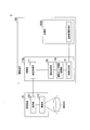
- FIG. 2 is a diagram illustrating an example of a configuration of a determination system 1 according to the first embodiment.
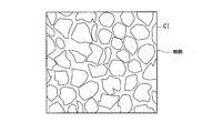
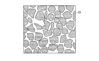
- FIG. 3 is a diagram illustrating an example of a captured image CI according to the first embodiment.
- FIG. 4 is a diagram conceptually illustrating processing of a representative point determination unit 112 according to the first embodiment. It is a figure showing typically an example of distance d between representative points concerning a 1st embodiment. It is a figure which shows typically the process of the cell distribution information acquisition part 114 concerning 1st Embodiment.
- FIG. 9 is a diagram schematically illustrating another example of the processing of the cell distribution information acquisition unit 114 according to the first embodiment. It is a figure showing an example of cell distribution information DI concerning a 1st embodiment.
- FIG. 4 is a flowchart illustrating an example of an operation of the determination device 10 according to the first embodiment. It is a figure showing an example of composition of judgment system 2 concerning a 2nd embodiment. It is a figure showing typically an example of representative point angle ag concerning a 2nd embodiment. It is a flow chart which shows an example of operation of judgment device 10a concerning a 2nd embodiment. It is a figure showing an example of judgment system 3 composition concerning a 3rd embodiment. It is a figure showing typically processing of area acquisition part 130 concerning a 3rd embodiment. It is a figure showing an example of cell size information DI3 concerning a 3rd embodiment. It is a figure showing typically an example of judgment processing of mode judging part 116 concerning a 3rd embodiment.
- FIG. 13 is a flowchart illustrating an example of an operation of the determination device 10b according to the third embodiment.
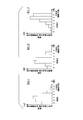
- FIG. 14 is a diagram schematically illustrating another example of the determination processing of the mode determination unit according to the third embodiment. It is a figure showing an example of composition of judgment system 4 concerning a 4th embodiment. It is a figure showing typically processing of quantity acquisition part 140 concerning a 4th embodiment. It is a figure which shows an example of the successive change of the cell distribution information DI.
- FIG. 11 is a diagram illustrating an example of a configuration of a determination system 5 according to a modification 2.
- FIG. 3 is a diagram illustrating an example of a GUI image.
- FIG. 23 is a diagram illustrating an example of a result image of a process executed by the GUI in FIG. 22.
- FIG. 1 is a diagram illustrating an example of a configuration of a determination system 1 according to the first embodiment.
- the determination system 1 includes a determination device 10 and an imaging device 20.
- the imaging device 20 includes an imaging unit 210 and a transmission unit 220.
- the imaging unit 210 captures an image of a subject S, which is an object to be captured.
- the subject S is, for example, a plurality of cells cultured in a petri dish or the like.
- the transmission unit 220 transmits the captured image CI, which is the image of the subject S captured by the imaging unit 210, to the determination device 10.
- FIG. 2 is a diagram illustrating an example of the captured image CI according to the first embodiment.
- the captured image CI is an image showing a part of the subject S captured by the imaging unit 210. That is, the captured image CI includes an image of cells existing in a part of the subject S captured by the imaging unit 210.
- the determination device 10 includes a control unit 110 and a storage unit 800.
- the control unit 110 includes, for example, an image acquisition unit 111, a representative point determination unit 112, a distance acquisition unit 113, and a cell distribution information acquisition unit when a processor such as a CPU (Central Processing Unit) executes a program (software).
- 114 and the mode determination unit 116 are realized as the functional units.
- Some or all of these constituent elements are hardware (circuits) such as LSI (Large Scale Integration), ASIC (Application Specific Integrated Circuit), FPGA (Field-Programmable Gate Array), and GPU (Graphics Processing Unit). (Including circuitry), or may be realized by cooperation of software and hardware.
- the program may be stored in the storage unit 800 in advance, or may be stored in a removable storage medium such as a DVD or a CD-ROM, and may be installed in the storage unit 800 by mounting the storage medium in a drive device. May be done.
- the storage unit 800 is realized by, for example, an HDD, a flash memory, an electrically erasable programmable read only memory (EEPROM), a read only memory (ROM), or a random access memory (RAM).
- the storage unit 800 stores, for example, a program read and executed by the processor.
- the storage unit 800 stores information indicating the calculated distance CD, information indicating the determination distance DD, and number information NI1. Details of each information will be described later.
- the image acquisition unit 111 acquires the captured image CI from the imaging unit 210.
- the image acquisition unit 111 supplies the captured image CI to the representative point determination unit 112.
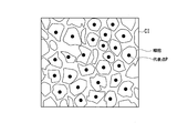
- the representative point determination unit 112 determines, for each cell, a representative point P of a plurality of cells included in the captured image CI acquired from the image acquisition unit 111.
- the representative point P is, for example, a point indicating the center of gravity of a cell for each cell.
- FIG. 3 is a diagram conceptually showing the processing of the representative point determination unit 112 according to the first embodiment.
- the representative point determination unit 112 performs a peak detection process on the acquired captured image CI, and determines a representative point P for each of a plurality of cells included in the captured image CI.
- the representative point determination unit 112 supplies information indicating the representative point P for each of the plurality of cells shown in the captured image CI to the distance acquisition unit 113.
- the representative point determination unit 112 uses, for example, identification information of a pixel indicating the position of the representative point P in the captured image CI (for example, a number assigned to each pixel of the captured image CI) as information indicating the representative point P. It is supplied to the distance acquisition unit 113.
- the distance acquisition unit 113 acquires information indicating the representative point P for each of the plurality of cells determined by the representative point determination unit 112.
- the distance acquisition unit 113 acquires the distance between a certain representative point P included in the captured image CI and another representative point P. More specifically, the distance acquisition unit 113 sets another representative point within a range from the representative point P serving as a reference to the distance indicated by the calculated distance CD, based on one representative point P included in the captured image CI.
- the distance d between the representative points to the point P is calculated.
- the calculated distance CD is a distance that serves as an index when the distance acquisition unit 113 calculates the distance between the representative points P.
- the representative points P This is the distance that can be taken between each other.
- FIG. 4 is a diagram schematically illustrating an example of the distance d between the representative points according to the first embodiment.
- the representative point P serving as a reference is the representative point P1.
- the distance acquisition unit 113 selects (extracts), from the captured image CI, a representative point P existing in the target range ARd from the representative point P1 to the distance indicated by the calculated distance CD.
- the distance acquisition unit 113 calculates the representative point distance d (illustrated, representative point distance) to representative points P (illustrated, representative points P2 to P7) other than the representative point P1 existing in the target range ARd. d1 to d6) are obtained.
- the distance acquisition unit 113 performs the same processing, for example, by setting all the representative points P included in the captured image CI as the representative point P1. Therefore, the distance obtaining unit 113 obtains, for each representative point P, information indicating the distance d1 to d6 between the representative points, where each of the representative points P of the cells captured in the captured image CI is the representative point P1, and obtains the cell distribution.
- the information is supplied to the information acquisition unit 114.
- the cell distribution information acquisition unit 114 generates the cell distribution information DI1 based on the distance d between the representative points acquired by the distance acquisition unit 113 and the determination distance DD.
- the determination distance DD is a distance d between the representative points when it is determined that the cell is appropriately matured.
- the number of representative point distances d equal to or smaller than the determination distance DD and the frequency (the number of reference representative points or the number of reference cells) are associated with each other. Information.
- FIG. 5 is a diagram schematically illustrating a process of the cell distribution information acquisition unit 114 according to the first embodiment.
- the reference representative point P is the representative point P1
- a representative point P (representative points P2 to P7 shown) existing in a range from the representative point P1 to the target range ARd and present in the determination range ARg up to the distance indicated by the determination distance DD.
- the points P are three representative points P4 to P6.
- the cell distribution information acquisition unit 114 is another representative point P present in the target range ARd centered on the representative point P for each of the representative points P of the cells captured in the captured image CI, and is equal to or less than the determination distance DD. Is calculated, and the cell distribution information DI1 in which the number and the frequency of the number are associated with each other is generated. The cell distribution information acquisition unit 114 supplies the generated cell distribution information DI1 to the mode determination unit 116.
- the number of the representative points P of the other objects existing in the target range ARd around the representative point P of a certain cell is an example of the “characteristic of the object”.
- FIG. 6 is a diagram schematically illustrating another example of the process of the cell distribution information acquisition unit 114 according to the first embodiment.
- the cell distribution information acquisition unit 114 calculates the number of the representative points P having the distance d between the representative points equal to or less than the determination distance DD around the representative point P for each of the representative points P of the cells captured in the captured image CI.
- the calculated cell distribution information DI1 may be generated by associating the number with the frequency of the number.
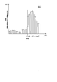
- FIG. 7 is a diagram showing an example of the cell distribution information DI1 according to the first embodiment.
- the cell distribution information acquisition unit 114 determines the representative point distances d1 to d6 acquired for each of the representative points P for the 317 “cells” captured in the captured image CI. And cell distribution information DI1 based on the determination distance DD.
- the cell distribution information DI1 detects 50 “times” of cells in which the number of other representative points P existing in the determination range ARg up to the distance indicated by the determination distance DD is 0 “number”.
- the cell in which the number of the other representative points P existing in the determination range ARg up to the distance indicated by the determination distance DD is 1 “cell” is detected “80 times”, and the cell up to the distance indicated by the determination distance DD is detected.
- the number of other representative points P existing in the determination range ARg up to the distance indicated by the determination distance DD is detected 100 times when the number of other representative points P existing in the determination range ARg is 2 “cells”.
- Are detected 50 times and the number of other representative points P existing in the determination range ARg up to the distance indicated by the determination distance DD is 4 cells 25 times.
- Are detected 10 times, and the number of other representative points P present in the determination range ARg up to the distance indicated by the determination distance DD is 6 “number”. This indicates that there are 2 “cells”.
- the mode determination unit 116 Based on the cell distribution information DI1 generated by the cell distribution information acquiring unit 114 and the number information NI1, the mode determination unit 116 has a favorable mode (for example, the degree of maturity) of the cells captured in the captured image CI. Is determined.
- the number information NI1 indicates the reference representative point P in a range from the reference representative point P to the distance indicated by the determination distance DD (hereinafter, determination range ARg). This is information indicating the number of representative points P other than.
- determination range ARg determination range
- the number information NI1 that can be obtained when cells (representative points P) are arranged in a honeycomb structure is 6 “pieces”. Therefore, when the number of the representative points P present in the determination range ARg matches the number information NI1, it is determined that the cell is appropriately matured.
- the mode determination unit 116 determines that the cell is mature, and determines the number indicated by the number information NI1 and the cell If the number of the highest frequency indicated by the distribution information DI1 does not match, it is determined that the cell is not mature.
- the cell distribution information DI1 is information in which the number of representative point distances d equal to or less than the determination distance DD among the representative point distances d1 to d6 and the frequency are associated with each other.
- the cell distribution information DI1 may be information indicating the number of representative point distances d that are equal to or less than the determination distance DD including the representative point P1 serving as a reference.
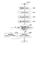
- FIG. 8 is a flowchart illustrating an example of the operation of the determination device 10 according to the first embodiment.
- the determination device 10 executes steps S100 to S170 shown in FIG. 8 based on a determination program Prg10 which is a control program for determining whether the cells are appropriately matured.
- the image acquisition unit 111 acquires the captured image CI from the imaging device 20 and supplies the captured image CI to the representative point determination unit 112 (Step S100).
- the representative point determination unit 112 determines a representative point P of the cell captured in the captured image CI acquired from the image acquisition unit 111 (Step S110).
- the distance acquisition unit 113 determines, from among the representative points P determined by the representative point determination unit 112, a representative point between another representative point P existing within the target range ARd from the reference representative point P and the reference representative point P.
- the distance d is obtained (step S120).
- the cell distribution information acquisition unit 114 generates the cell distribution information DI1 based on the representative point distance d for each representative point P acquired by the distance acquisition unit 113 (Step S130).
- the mode determining unit 116 determines that the cells are mature based on the cell distribution information DI1 generated by the cell distribution information acquiring unit 114 and the number information NI1 (step S150). For example, when the number indicated by the number information NI1 matches the highest frequency number indicated by the cell distribution information DI1 (step S150; YES), the mode determining unit 116 matures the cells captured in the captured image CI. It is determined that there is (step S160). When the number indicated by the number information NI1 does not match the highest frequency number indicated by the cell distribution information DI1 (step S150; NO), the mode determining unit 116 matures the cells captured in the captured image CI. It is determined that it has not been performed (step S170).
- the determination system 1 of the present embodiment includes the determination device 10 and the imaging device 20, and is based on an image in which a plurality of cells to be cultured are captured (in this example, a captured image CI).
- a distribution information acquisition unit (in this example, cell distribution information acquisition unit 114) that acquires distribution information (in this example, cell distribution information DI1) regarding distribution in a predetermined region of a plurality of cells.
- a determination unit in this example, a mode determination unit 116 that determines the culture state of a plurality of cells based on the cell distribution information DI1 acquired by the cell distribution information acquisition unit 114, and (In this example, the degree of maturity) is determined to be good, and the accuracy of the determination of the maturity of the cell can be improved.
- the feature acquiring unit (in this example, the representative point determining unit 112 and the distance acquiring unit 113) is configured such that the cells shown in the captured image CI are within a predetermined range (from a certain cell).
- the feature distance d between representative points in this example
- the determination system 1 of the present embodiment can limit the cells for which the representative point distance d should be obtained for a certain cell, and thus reduce the load of the processing related to the determination of the maturity of the cell. Can be.
- FIG. 9 is a diagram illustrating an example of a configuration of the determination system 2 according to the second embodiment.
- the determination system 2 includes a determination device 10a and an imaging device 20.
- the determination device 10a includes a control unit 110a and a storage unit 800a.
- the control unit 110a replaces (or in addition to) the configuration provided in the control unit 110, and includes an image acquisition unit 111, a representative point determination unit 112, a cell distribution information acquisition unit 114, a mode determination unit 116, an angle acquisition
- the unit 120 is realized as the functional unit.
- the storage unit 800a stores, for example, information indicating a calculated distance CD, information indicating a determination angle DA, and number information NI1. Details of the determination angle DA will be described later.
- the angle acquisition unit 120 acquires information indicating the representative point P for each of the plurality of cells determined by the representative point determination unit 112.
- the angle acquisition unit 120 is based on one representative point P included in the captured image CI, and is another representative point P existing in a range from the representative point P serving as the reference to the distance indicated by the calculated distance CD.
- An angle (hereinafter referred to as a representative point angle) formed by a line segment connecting the representative point P of two other objects and a representative point P adjacent to each other around a vertical axis centered on the representative point P serving as a reference. ag).
- FIG. 10 is a diagram schematically illustrating an example of the representative point angle ag according to the second embodiment.
- the angle acquiring unit 120 determines the representative point P1 and the other representative points P (representative points P2 to P7 shown) in the target range ARd from the representative point P1 to the distance indicated by the calculated distance CD.
- An angle between line segments respectively connecting the other representative points P adjacent around a vertical axis centered on the representative point P1 is acquired.
- the angle acquisition unit 120 acquires an angle (representative point angle ag1 shown) formed by a line connecting the representative point P1 and the representative point P2 and a line connecting the representative point P1 and the representative point P3.
- An angle (representative point angle ag2 shown) formed between a line segment connecting P1 and the representative point P3 and a line segment connecting the representative point P1 and the representative point P4 is obtained, and the representative point P1 and the representative point P4 are connected.
- the angle (representative point angle ag3 shown) formed by the line segment and the line segment connecting the representative point P1 and the representative point P5 is acquired, and the line segment connecting the representative point P1 and the representative point P5, and the representative point P1 are obtained.
- An angle (representative point angle ag4 shown) between the line and the representative point P6 is acquired, and a line connecting the representative point P1 and the representative point P6 and a line connecting the representative point P1 and the representative point P7 are obtained.
- (Representative point angle ag5 shown in the figure) and a line segment connecting the representative point P1 and the representative point P7 Obtains an angle between a line connecting the representative point P2 and the representative point P1 (representative point angle ag6 illustrated).
- the angle acquisition unit 120 performs the same processing, for example, by setting all the representative points P included in the captured image CI as the representative points P1.
- the angle obtaining unit 120 obtains, for each representative point P, information indicating the representative point angles ag1 to ag6 with each of the representative points P of the cell captured in the captured image CI as the representative point P1, and obtains the cell distribution information.
- the information is supplied to the acquisition unit 114.
- the representative points P2 to P7 of other objects existing in the target range ARd centered on the representative point P1 of a certain cell are adjacent to the representative point P1 around the vertical axis centered on the representative point P1.
- the angle formed by the line segments connecting the other matching representative points P2 to P7 is an example of the “characteristic of the object”.
- the cell distribution information acquisition unit 114 acquires the representative point angle ag acquired by the angle acquisition unit 120.
- the cell distribution information acquisition unit 114 generates cell distribution information DI2 based on the representative point angle ag and the determination angle DA.
- the determination angle DA is a representative point angle ag when it is determined that the cell is appropriately matured.
- the cell distribution information DI2 refers to the number of representative point angles ag that match the determination angle DA or the number of representative point angles ag of ⁇ 5 degrees from the representative point angles ag1 to ag6, and the frequency. This is the associated information.
- the process in which the cell distribution information acquiring unit 114 generates the cell distribution information DI2 is the same as the process in which the cell distribution information acquiring unit 114 generates the cell distribution information DI1, and a description thereof will be omitted.
- the determination angle DA that can be taken when cells (representative points P) are arranged in a honeycomb structure is 60 degrees. Therefore, when the representative point angle ag matches or is close to the determination angle DA, it is determined that the cell is appropriately matured.
- the mode determination unit 116 determines that the cells are mature, and When the number indicated by the information NI1 does not match the highest frequency number indicated by the cell distribution information DI2, it is determined that the cell is not mature.
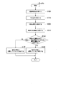
- FIG. 11 is a flowchart illustrating an example of the operation of the determination device 10a according to the second embodiment.
- the determination device 10a executes steps S100 to S210 shown in FIG. 11 based on the determination Prg10a, which is a control program for determining whether the cells are appropriately matured. Also, the processing of steps S100 to S170 shown in FIG. 11 is the same as the processing of the same step numbers in FIG.
- the determination Prg10a executes step S200 instead of (or in addition to) step S120 performed in determination Prg10, and executes step S210 instead of (or in addition to) step S130.
- the angle acquisition unit 120 uses the one representative point P included in the captured image CI as a reference, and sets the other representative points within a range from the reference representative point P to the distance indicated by the calculated distance CD.
- Point P which is the angle formed by the line segments connecting the representative point P of two other objects and the representative point P adjacent to each other around a vertical axis centered on the representative point P serving as a reference.
- a certain representative point angle ag is acquired (step S200).
- the cell distribution information acquisition unit 114 generates the cell distribution information DI2 based on the representative point angle ag for each representative point P acquired by the angle acquisition unit 120 (Step S210).
- the cell distribution information DI2 is a representative of two other cells adjacent to each other around the vertical axis around the representative point P of a certain cell in the captured image CI. It is acquired based on the angle (in this example, the representative point angle ag) formed by the line segments connecting the point P and the representative point P of a certain cell, and the aspect of the cell (in this example, the maturity of the cell) is obtained. It is possible to determine whether the cell is good or not, and improve the accuracy of the determination of the maturity of the cell.
- FIG. 12 is a diagram illustrating an example of the configuration of the determination system 3 according to the third embodiment.
- the determination system 3 includes a determination device 10b and an imaging device 20.
- the determination device 10b includes a control unit 110b and a storage unit 800b.
- the storage unit 800b stores reference area information SI in place of (or in addition to) the information stored in the storage unit 800 and the storage unit 800a. Details of the reference area information SI will be described later.
- the control unit 110b includes an image acquisition unit 111, a cell size information acquisition unit 114a, a mode determination unit 116, and an area acquisition unit 130 instead of (or in addition to) the configuration of the control unit 110 or the control unit 110a. It is realized as the function part.
- the area acquisition unit 130 acquires the area of a cell captured in the captured image CI.
- FIG. 13 is a diagram schematically illustrating a process of the area acquiring unit 130 according to the third embodiment.
- the area acquisition unit 130 identifies, for example, a region of a plurality of cells captured in the captured image CI and a region other than the cells by performing a smoothing process or a morphological filter process on the captured image CI.
- the area acquiring unit 130 acquires an area of each of the plurality of cells based on the identified regions of the plurality of cells.
- the area of the plurality of cells is an example of the “feature of the object”.
- FIG. 14 is a diagram showing an example of the cell size information DI3 according to the third embodiment.
- the cell size information acquisition unit 114a generates cell size information DI3 based on the cell area of each cell acquired by the area acquisition unit 130.
- the cell size information DI3 is information in which the cell area and the number (frequency) of cells having the area included in the captured image CI are associated with each other.
- the mode determining unit 116 determines whether the maturity of the cell is good based on the cell size information DI3 generated by the cell size information obtaining unit 114a and the reference area information SI.
- the reference area information SI is information indicating a cell area (hereinafter, a reference area) when the cell is considered to be appropriately matured.
- FIG. 15 is a diagram schematically illustrating an example of a determination process of the mode determination unit 116 according to the third embodiment. For example, among all the cells captured in the captured image CI (that is, the total number of frequencies indicated by the cell size information DI3), the mode determination unit 116 determines that the cells having an area smaller than the reference area indicated by the reference area information SI ( Is greater than or equal to a predetermined ratio (for example, 90% or more), it is determined that the cell is mature. If the ratio is less than the predetermined ratio, the cell is not mature. Is determined.
- a predetermined ratio for example, 90% or more
- FIG. 16 is a flowchart illustrating an example of the operation of the determination device 10b according to the third embodiment.
- the determination device 10b executes steps S310 to S320 shown in FIG. 16 based on the determination Prg10b which is a control program for determining whether the cells are appropriately matured.
- the image acquisition unit 111 acquires the captured image CI from the imaging device 20 and supplies the captured image CI to the representative point determination unit 112 (Step S310).
- the area acquisition unit 130 acquires, for each cell, the area of a plurality of cells imaged in the captured image CI based on the captured image CI acquired by the image acquisition unit 111 (Step S312).
- the cell size information acquisition unit 114a generates cell size information DI3 based on the area of the cell acquired by the area acquisition unit 130 (Step S314).
- the mode determination unit 116 determines an area smaller than the reference area indicated by the reference area information SI among all the cells captured in the captured image CI. It is determined whether or not the ratio of the possessed cells is equal to or more than a predetermined ratio (90% or more as illustrated) (step S316). If the proportion occupied by the number of cells having an area smaller than the reference area is equal to or greater than a predetermined proportion among all the cells captured in the captured image CI (step S316; YES), the mode determination unit 116 determines that It is determined that the user is mature (step S318).
- step S316 If the ratio of the number of cells having an area smaller than the reference area to all cells captured in the captured image CI is less than the predetermined ratio (step S316; NO), the mode determination unit 116 determines that It is determined that the user has not matured (step S320).
- the features of the plurality of objects include the area of the plurality of cells, and the mode determination unit 116 determines In this example, based on the cell area of each cell acquired by the area acquisition unit 130) and a predetermined area (in this example, reference area information SI), it is determined whether or not the state of the cell is good. It is possible to improve the accuracy of the determination and the determination of the maturity of the cell.
- the mode determination unit 116 determines whether the maturity of the cell is good based on the reference area information SI.
- the mode determining unit 116 may be configured to determine whether the maturity of the cell is good based on the reference average area information ASI instead of the reference area information SI.
- the reference average area information ASI is information indicating the average of the cell area when the cells are considered to be moderately mature (hereinafter, reference average area).
- FIG. 17 is a diagram schematically illustrating another example of the determination process of the mode determination unit 116 according to the third embodiment.
- the mode determining unit 116 determines that the average of the areas of all the cells captured in the captured image CI matches the reference average area indicated by the reference average area information ASI, matches the reference average area, or is equal to or less than the reference average area. In some cases, it is determined that the cells are mature, and when the average of the cell area does not match, does not match, or is larger than the reference average area indicated by the reference average area information ASI. Determine that the cells are not mature.
- the reference average area information ASI may be a median value of the cell area in place of the average of the cell area when the cell is considered to be appropriately matured.
- FIG. 18 is a diagram illustrating an example of the configuration of the determination system 4 according to the fourth embodiment.
- the determination system 4 includes a determination device 10c and an imaging device 20.
- the determination device 10c includes a control unit 110c and a storage unit 800c.
- the storage unit 800c instead of (or in addition to) the information stored in the storage unit 800, the storage unit 800a, and the storage unit 800b, the information indicating the determination range ARg and the number information NI2 are stored in advance. Is done.
- the number information NI2 is information indicating the number of cells existing in the determination range ARg (that is, seven cells) when the cells are considered to be moderately mature.
- the control unit 110c includes, instead of (or in addition to) the configuration included in the control unit 110, the control unit 110a, or the control unit 110b, an image acquisition unit 111, a cell size information acquisition unit 114a, a mode determination unit 116, The number obtaining unit 140 is realized as the functional unit.
- FIG. 19 is a diagram schematically showing the process of the number acquiring unit 140 according to the fourth embodiment.
- the number acquiring unit 140 identifies, for example, a region of a plurality of cells captured in the captured image CI and a region other than the cells by performing a smoothing process or a morphological filter process on the captured image CI.
- the number acquisition unit 140 acquires the number of cells for each determination range ARg based on the identified regions of the plurality of cells. As illustrated in FIG. 19, the number acquiring unit 140 does not count a cell partially existing in the determination range ARg as a cell existing in the determination range ARg.
- the shape of the determination range ARg may be a circle or another shape.
- the number of cells existing in the determination range ARg is an example of “the characteristic of the object”.
- the cell size information acquisition unit 114a generates the cell size information DI4 based on the number of cells existing in the determination range ARg acquired by the number acquisition unit 140.
- the cell size information DI4 is information in which the number of cells existing in the determination range ARg and the frequency of the number of determination ranges ARg are associated with each other.
- the mode determining unit 116 determines whether the cells are appropriately mature based on the cell size information DI4 and the number information NI2. As described above, when the cells are considered to be moderately mature, the number (that is, seven) of the cells existing in the determination range ARg is indicated by the number information NI2. The mode determining unit 116 determines whether the number indicated by the number information NI2 matches the highest frequency indicated by the cell size information DI4, or the number indicated by the number information NI2 indicates the highest frequency indicated by the cell size information DI4. In the above case, it is determined that the cell is mature, and the number indicated by the number information NI2 does not match the highest frequency indicated by the cell size information DI4, or the highest frequency indicated by the cell size information DI4. If the number is smaller than the number indicated by the number information NI2, it is determined that the cells are not mature.
- the characteristics of a plurality of objects include the number of cells existing in a predetermined range (in this example, the determination range ARg).
- the mode determination unit 116 determines the mode of the cell based on the number obtained by the feature obtaining unit (in this example, the number obtaining unit 140) and a predetermined number (in this example, the number information NI2). Is good or not.
- the number acquisition unit 140 acquires the number of cells based on the determination range ARg, but the invention is not limited to this.
- the number acquisition unit 140 may acquire the number of cells based on a predetermined range other than the determination range ARg.
- the predetermined range is required to be a range that always indicates a fixed range regardless of the angle of view of the captured image CI.
- FIG. 20 is a diagram showing an example of successive changes in the cell distribution information DI1.
- the imaging device 20 images the subject S at predetermined time intervals, for example, and generates a captured image CI.
- the determination device 10 generates cell distribution information DI1 based on a plurality of captured images CI generated at predetermined time intervals, and determines whether the cells are mature.
- the cell distribution information DI1-1 is the cell distribution information DI1 generated based on the captured image CI captured at a certain time t1.
- the cell distribution information DI1-2 is cell distribution information DI1 generated based on a captured image CI captured after a predetermined time has elapsed from time t1.
- the cell distribution information DI1-3 is cell distribution information DI1 generated based on a captured image CI captured after a predetermined time has elapsed from time t2.
- the mode determination unit 116 compares, for example, the transition of the frequency indicated in each cell distribution information DI1. When the frequency indicated in the cell distribution information DI1 is a predetermined transition, the mode determination unit 116 determines that the cell is mature.
- the predetermined transition means that, for example, at time t1, the number of representative points P other than the representative point P1 in the determination range ARg has a high ratio of 0 “number” to 2 “number”, and at time t2, The ratio of the number of the representative points P other than the representative point P1 in the determination range ARg being 2 “pieces” to 3 “pieces” is high, and at time t3, other representative points than the representative point P in the determination range ARg.
- the mode determination unit 116 determines that the cell is mature.
- the mode determination unit 116 determines that the cell is not mature (that is, the cell is immature).
- the above-mentioned predetermined transition is an example, and the present invention is not limited to this. Further, in the above description, the cell distribution information DI1 has been described as an example, but the mode determination unit 116 also determines that the frequency transition of the other information (cell distribution information DI2 or cell size information DI3 to DI4) is a predetermined value. Whether or not the cell is mature may be determined based on whether or not the transition has occurred.
- Modification 2 Combination of determination criteria
- Modification 2 according to the above-described embodiment, a case has been described in which it is determined whether or not cells are mature based on one captured image CI captured of the subject S.
- an index for determining whether a cell is mature is selected.
- FIG. 21 is a diagram illustrating an example of a configuration of the determination system 5 according to the second modification.
- the determination system 5 includes a determination device 10d and an imaging device 20.
- the determination device 10d includes a control unit 110d, a storage unit 800d, an operation unit 600, and a display unit 700.
- the operation unit 600 is, for example, an input device such as a keyboard, a touchpad, and a mouse that receives an operation input from a user.
- the display unit 700 is a display device such as a liquid crystal display panel, a plasma display panel, and an organic EL (Electro Luminescence) display panel.
- the control unit 110d corresponds to the image acquisition unit 111, the cell distribution information acquisition unit 114, and the cell size information acquisition unit 114a among the functional units included in the control units 110 and 110a to 110c.
- a function unit at least one of a distance acquisition unit 113, a representative point determination unit 112, an angle acquisition unit 120, an area acquisition unit 130, and a number acquisition unit 140), a mode determination unit 116, and an acquisition unit 150;
- a display control unit 160 causes the operation unit 600 to display a graphical user interface (Graphical User Interface) image previously stored in the storage unit 800d.
- FIG. 22 is a diagram showing an example of a GUI image.
- the GUI image includes an area in which a list of a plurality of captured images CI (illustrated captured images CIa to CIe) is displayed, an area in which a selected image is displayed, and an area in which an index list is displayed.
- the area in which the image list is displayed includes a captured image CI captured by the imaging device 20 in the past, a captured image CI obtained by capturing the subject S with the imaging device 20 at predetermined time intervals, and the like.
- These captured images CI are stored in, for example, the storage unit 800d, and the display control unit 160 causes the display unit 700 to display the captured images CI stored in the storage unit 800d.
- the acquisition unit 150 acquires an operation received by the operation unit 600.
- the display control unit 160 causes the selected captured image CI to be displayed in the area where the selected image is displayed, based on the “operation for selecting the captured image CI” acquired by the acquisition unit 150.
- a check box for selecting whether the captured image CI to be analyzed is one image (that is, a single image) or a plurality of images is shown.
- the determination system 5 determines whether or not the cell is mature based on one captured image CI.
- the determination system 5 determines whether the cells are mature based on the plurality of captured images CI.
- “distance between representative points” and “representative point” indicating options of indices used when determining whether or not cells are matured based on a single or a plurality of captured images CI.
- Check boxes for "Angle”, “Cell Area”, and “Cell Count” are shown.
- the determination system 5 analyzes the captured image CI based on the index selected by the check box, and determines whether the cells are mature. Further, the determination system 5 outputs a histogram (for example, cell distribution information DI1 to DI2, cell size information DI3 to DI4). Note that these indices are merely examples, and the present invention is not limited thereto. At least one of “distance between representative points”, “representative point angle”, “cell area”, and “cell number” is used as a check box. The configuration shown may be used.
- a check box for “time-series analysis” is shown.
- the determination system 5 determines whether the cells are mature based on the time-series image. Further, the determination system 5 outputs the result of the time-series analysis of the captured image CI.
- FIG. 23 is a diagram showing an example of a result image of the processing executed by the GUI of FIG. Specifically, in FIG. 23, “captured image CIa”, “captured image CIb”, “multiple images”, “time-series analysis”, and “distance between representative points” are selected by the GUI in FIG.
- FIG. 9 is a diagram illustrating an example of a result image when the image is displayed.
- the result image for example, the average frequency (or the highest frequency) of the cell distribution information DI generated based on the distance d between the representative points at a certain timing (the first to fourth weeks in the drawing) and the timing are shown. Graphs associated with each other for each timing are shown.
- the result image includes, for example, an image in which an image indicating the cell distribution information DI (histogram) at a predetermined timing is superimposed.
- the determination system 5 When a plurality of check boxes are selected from the check boxes of “distance between representative points”, “representative point angle”, “cell area”, and “cell number”, the determination system 5 performs It is determined based on each of the indices selected according to whether the cells are mature or not, and a result image shown in FIG. 23 is generated for each result. In this case, the determination system 5 determines that the cell is mature when it is determined that the cell has matured based on at least one of the plurality of selected indices. In addition, the determination system 5 determines that the cell is mature when the determination result based on all the indexes among the plurality of indexes indicates the determination result that the cell is matured.
- the acquisition unit 150 acquires an operation of designating the display of “plural images”, “time-series analysis”, and “distance between representative points” indicated in the area where the index list is displayed by the operation unit 600. .
- the control unit 110d acquires characteristics of the cells captured in the captured image CI based on the index indicated by the operation acquired by the acquisition unit 150, and determines whether the cells are mature.
- the object to be imaged by the imaging device 20 may be one that is densely filled and whose filled state is determined.
- the target object may be a substance having a hexagonal close-packed structure or a honeycomb structure.
- the imaging unit 210 may image the entire subject S. Further, here, the case where a plurality of cells are included in the captured image CI captured by the imaging unit 210 has been described, but the invention is not limited thereto. The number of cells included in the captured image CI captured by the imaging unit 210 may be one.
- the representative point determination unit 112 may analyze the captured image CI by contrast processing or may analyze the captured image CI by noise removal. Further, here, the case where the representative point P is the center of gravity of the cells included in the captured image CI has been described, but the present invention is not limited to this.
- the representative point P may be a point indicating a nucleus of a cell included in the captured image CI.
Abstract
Description
まず、細胞の成熟の特性について説明する。
細胞の培養状態を評価するために、その成熟度を的確に判定することが求められる。具体的には、RPE細胞、表皮細胞等の上皮細胞が再生医療を目的として培養される場合、これらの上皮細胞を培養して移植用の細胞、及び細胞シート等を作成する際には、細胞が適度に成熟するまでの間、培養することが一般的である。また、これらの上皮細胞は、適度に成熟すると、複数の細胞がハニカム状になる。実施形態の判定システムは、この細胞の成熟における特性を用いて細胞の状態の判定を行う。 [Characteristics of cell maturation]
First, the characteristics of cell maturation will be described.
In order to evaluate the culture state of a cell, it is required to accurately determine its maturity. Specifically, when epithelial cells such as RPE cells and epidermal cells are cultured for the purpose of regenerative medicine, when these epithelial cells are cultured to prepare cells for transplantation and cell sheets and the like, cells are used. In general, the cells are cultured until they are moderately matured. In addition, when these epithelial cells are appropriately matured, a plurality of cells become honeycomb. The determination system of the embodiment determines the state of the cell by using the characteristics in the maturation of the cell.
以下、図面を参照して本発明の第1の実施形態について説明する。図1は、第1の実施形態に係る判定システム1の構成の一例を示す図である。判定システム1は、判定装置10と、撮像装置20とを備える。 [First Embodiment]
Hereinafter, a first embodiment of the present invention will be described with reference to the drawings. FIG. 1 is a diagram illustrating an example of a configuration of a
次に、図8を参照して判定装置10が細胞の成熟度を判定する動作について説明する。図8は、第1の実施形態に係る判定装置10の動作の一例を示す流れ図である。判定装置10は、細胞が適度に成熟しているか否かを判定するための制御プログラムである判定Prg10に基づいて、図8に示すステップS100からステップS170までを実行する。 <Processing flow>
Next, an operation in which the
以上説明したように、本実施形態の判定システム1は、判定装置10と、撮像装置20とを備え、培養される複数の細胞が撮像された画像(この一例では、撮像画像CI)に基づいて、複数の細胞の所定の領域における分布に関する分布情報(この一例では、細胞分布情報DI1)を取得する分布情報取得部(この一例では、細胞分布情報取得部114
)と、細胞分布情報取得部114によって取得された細胞分布情報DI1に基づいて、複数の細胞の培養状態を判定する判定部(この一例では、態様判定部116)と、を備え、細胞の態様(この一例では、成熟度)が良好であるか否かを判定し、細胞の成熟度の判定の精度を向上することができる。 <Summary of First Embodiment>
As described above, the
) And a determination unit (in this example, a mode determination unit 116) that determines the culture state of a plurality of cells based on the cell distribution information DI1 acquired by the cell distribution
次に、本発明の第2の実施形態について説明する。第1の実施形態では、判定装置10が代表点間距離dに基づいて、細胞分布情報DIを生成し、細胞の成熟度を判定する場合について説明した。第2の実施家形態では、判定装置10aが、基準となる代表点Pと、他の2つの代表点Pとをそれぞれ結んだ線分同士のなす角に基づいて細胞の成熟度を判定する場合について説明する。なお、上述した実施形態と同様の構成については、同一の符号をふして説明を省略する。 [Second embodiment]
Next, a second embodiment of the present invention will be described. In the first embodiment, the case has been described where the
次に、図11を参照して判定装置10aが細胞の成熟度を判定する動作について説明する。図11は、第2の実施形態に係る判定装置10aの動作の一例を示す流れ図である。判定装置10aは、細胞が適度に成熟しているか否かを判定するための制御プログラムである判定Prg10aに基づいて、図11に示すステップS100からステップS210までを実行する。また、図11に示される、ステップS100~S170の処理については、図8の同じステップ番号の処理と同様であるため、説明を省略する。 <Processing flow>
Next, an operation in which the determination device 10a determines the maturity of a cell will be described with reference to FIG. FIG. 11 is a flowchart illustrating an example of the operation of the determination device 10a according to the second embodiment. The determination device 10a executes steps S100 to S210 shown in FIG. 11 based on the determination Prg10a, which is a control program for determining whether the cells are appropriately matured. Also, the processing of steps S100 to S170 shown in FIG. 11 is the same as the processing of the same step numbers in FIG.
以上説明したように、本実施形態の判定システム2において、細胞分布情報DI2は、撮像画像CIにおいて、ある細胞の代表点Pを中心とする鉛直軸まわりに隣り合う、2つの他の細胞の代表点Pと、ある細胞の代表点Pとをそれぞれ結ぶ線分同士のなす角度(この一例では、代表点角度ag)に基づいて取得され、細胞の態様(この一例では、細胞の成熟度)が良好であるか否かを判定し、細胞の成熟度の判定の精度を向上することができる。 <Summary of Second Embodiment>
As described above, in the
次に、本発明の第3の実施形態について説明する。第1の実施形態、及び第2の実施形態では、ある細胞の代表点P1を中心とする対象範囲ARdに存在する細胞を対象に、細胞が成熟しているか否かを判定する場合について説明した。以下、第3の実施形態および第4の実施形態では、撮像画像CIに撮像される細胞の大きさに関する情報に基づいて、細胞が成熟しているか否かを判定する場合について説明する。第3の実施形態では、撮像画像CIに撮像される細胞の面積に基づいて、細胞が成熟しているか否かを判定する場合について説明する。なお、上述した実施形態と同様の構成については、同一の符号を付して説明を省略する。 [Third Embodiment]
Next, a third embodiment of the present invention will be described. In the first embodiment and the second embodiment, a case has been described in which it is determined whether or not the cells are mature with respect to the cells existing in the target range ARd around the representative point P1 of a certain cell. . Hereinafter, in the third embodiment and the fourth embodiment, a case will be described in which it is determined whether or not the cells are mature based on information on the size of the cells captured in the captured image CI. In the third embodiment, a case will be described in which it is determined whether a cell is mature based on the area of the cell captured in the captured image CI. In addition, about the structure similar to embodiment mentioned above, the same code | symbol is attached | subjected and description is abbreviate | omitted.
次に、図16を参照して判定装置10bが細胞の成熟度を判定する動作について説明する。図16は、第3の実施形態に係る判定装置10bの動作の一例を示す流れ図である。判定装置10bは、細胞が適度に成熟しているか否かを判定するための制御プログラムである判定Prg10bに基づいて、図16に示すステップS310からステップS320までを実行する。 <Processing flow>
Next, an operation in which the
以上説明したように、本実施形態の判定システム3において、複数の対象物(この一例では、細胞)の特徴には、複数の細胞の面積が含まれ、態様判定部116は、特徴取得部(この一例では、面積取得部130)によって取得された細胞毎の細胞の面積と、所定の面積(この一例では、基準面積情報SI)とに基づいて、細胞の態様が良好であるか否かを判定し、細胞の成熟度の判定の精度を向上することができる。 <Summary of Third Embodiment>
As described above, in the
次に、本発明の第4の実施形態について説明する。第3の実施形態では、撮像画像CIに撮像される細胞の面積に基づいて、細胞が成熟しているか否かを判定する場合について説明した。第4の実施形態では、撮像画像CIに撮像される細胞の大きさに関する情報を、面積に替えて細胞の数に基づいて判断する。なお、上述した実施形態と同様の構成については、同一の符号を付して説明を省略する。 [Fourth embodiment]
Next, a fourth embodiment of the present invention will be described. In the third embodiment, the case has been described where it is determined whether or not the cells are mature based on the area of the cells captured in the captured image CI. In the fourth embodiment, information on the size of a cell captured in the captured image CI is determined based on the number of cells instead of the area. In addition, about the structure similar to embodiment mentioned above, the same code | symbol is attached | subjected and description is abbreviate | omitted.
以上説明したように、本実施形態の判定システム4おいて、複数の対象物(この一例では、細胞)の特徴には、所定の範囲(この一例では、判定範囲ARg)に存在する細胞の個数が含まれ、態様判定部116は、特徴取得部(この一例では、個数取得部140)によって取得された個数と、所定の個数(この一例では、個数情報NI2)とに基づいて、細胞の態様が良好であるか否かを判定する。 <Summary of Fourth Embodiment>
As described above, in the
以下、上述した実施形態に係る変形例1について説明する。上述した実施形態では、被検体Sについて撮像された、ある1つの撮像画像CIに基づいて、細胞が成熟しているか否かを判定する場合について説明した。変形例1では、被検体Sについて継時的に撮像された複数の撮像画像CIに基づいて、細胞が成熟しているか否かを判定する場合について説明する。 [Variation 1: Successive change of cells]
Hereinafter,
以下、上述した実施形態に係る変形例2について説明する。上述した実施形態では、被検体Sについて撮像された、ある1つの撮像画像CIに基づいて、細胞が成熟しているか否かを判定する場合について説明した。変形例2では、細胞が成熟しているか否かを判定する指標を選択する場合について説明する。 [Modification 2: Combination of determination criteria]
Hereinafter,
Claims (12)
- 培養される複数の細胞が撮像された画像に基づいて、前記複数の細胞の所定の領域における分布に関する分布情報を取得する分布情報取得部と、
前記分布情報取得部によって取得された前記分布情報に基づいて、前記複数の細胞の培養状態を判定する判定部と、
を備える装置。 A distribution information acquisition unit that acquires distribution information regarding distribution in a predetermined region of the plurality of cells based on an image obtained by imaging a plurality of cells to be cultured,
Based on the distribution information acquired by the distribution information acquisition unit, a determination unit that determines the culture state of the plurality of cells,
An apparatus comprising: - 前記培養状態には、分化の状態と、細胞の成熟状態とのうち、少なくとも1つが含まれる、
請求項1に記載の装置。 The culture state includes at least one of a differentiation state and a cell maturation state,
The device according to claim 1. - 前記分布情報には、前記細胞の培地上における前記複数の細胞の互いの相対位置関係に関する情報が含まれる、
請求項1又は請求項2に記載の装置。 The distribution information includes information on the relative positional relationship between the plurality of cells on the culture medium of the cells,
An apparatus according to claim 1 or claim 2. - 前記分布情報は、ある細胞の代表点と、他の細胞の代表点との間の距離に基づいて取得される
請求項1から請求項3のいずれか一項に記載の装置。 The device according to any one of claims 1 to 3, wherein the distribution information is obtained based on a distance between a representative point of a certain cell and a representative point of another cell. - 前記分布情報は、前記複数の細胞のうち、ある細胞の代表点を中心とする所定の範囲に存在する他の細胞の代表点の個数に基づいて取得される、
請求項1から請求項4のいずれか一項に記載の装置。 The distribution information is obtained based on the number of representative points of other cells present in a predetermined range around a representative point of a certain cell among the plurality of cells,
Apparatus according to any one of the preceding claims. - 前記判定部は、
前記分布情報が示す前記所定の範囲に存在する他の細胞の代表点の個数に基づいて、前記細胞の態様を判定する、
請求項5に記載の装置。 The determination unit is
Based on the number of representative points of other cells present in the predetermined range indicated by the distribution information, determine the aspect of the cell,
An apparatus according to claim 5. - 前記分布情報は、前記画像において、ある細胞の代表点を中心とする鉛直軸まわりに隣り合う、2つの他の細胞の代表点と、前記ある細胞の代表点とをそれぞれ結ぶ線分同士のなす角度に基づいて取得される、
請求項1から請求項6のいずれか一項に記載の装置。 In the image, the distribution information is a line segment that connects a representative point of two other cells adjacent to a vertical axis centered on a representative point of a certain cell and a representative point of the certain cell. Obtained based on the angle,
Apparatus according to any one of the preceding claims. - 前記判定部は、
前記分布情報が示す前記角度に基づいて、前記細胞の態様を判定する、
請求項7に記載の装置。 The determination unit is
Based on the angle indicated by the distribution information, determine the aspect of the cell,
An apparatus according to claim 7. - 前記複数の細胞の大きさに関する情報を取得するサイズ情報取得部
をさらに有し、
前記判定部は、
前記細胞の大きさに関する情報に基づいて前記細胞の態様を判定する、
請求項1から請求項8のいずれか一項に記載の装置。 Further comprising a size information acquisition unit for acquiring information on the size of the plurality of cells,
The determination unit is
Determine the aspect of the cell based on information about the size of the cell,
Apparatus according to any one of the preceding claims. - ユーザの操作を受け付ける受付部と、
表示部の表示を制御する表示制御部と、を更に備え、
前記分布情報取得部は、
前記受付部によって受け付けられた前記ユーザの操作によって指定された前記分布情報を取得し、
前記表示制御部は、
前記分布情報を取得する際に用いられる画像、又は前記判定部の判定結果を示す画像のうち、少なくとも1つを前記表示部に表示させる、
請求項1から請求項9のいずれか一項に記載の装置。 A receiving unit that receives a user operation;
A display control unit that controls display on the display unit,
The distribution information acquisition unit,
Obtaining the distribution information specified by the user operation received by the receiving unit,
The display control unit,
Displaying at least one of an image used when acquiring the distribution information or an image indicating a determination result of the determination unit on the display unit,
Apparatus according to any one of the preceding claims. - 請求項1から請求項10のいずれか一項に記載の装置と、
前記細胞を撮像することにより、前記画像を生成する撮像部と
を備えるシステム。 An apparatus according to any one of claims 1 to 10,
An imaging unit that generates the image by imaging the cell. - コンピュータに、
所定の領域において培養される複数の細胞が撮像された画像に基づいて、前記複数の細胞の前記所定の領域における分布に関する分布情報を取得する分布情報取得ステップと、
前記分布情報取得ステップによって取得された前記分布情報に基づいて、前記複数の細胞の培養状態を判定する判定ステップと、
を実行させるためのプログラム。 On the computer,
Distribution information obtaining step of obtaining distribution information regarding distribution of the plurality of cells in the predetermined region based on an image of a plurality of cells cultured in a predetermined region,
Based on the distribution information acquired by the distribution information acquisition step, a determination step of determining the culture state of the plurality of cells,
The program to execute.
Priority Applications (4)
| Application Number | Priority Date | Filing Date | Title |
|---|---|---|---|
| PCT/JP2018/024663 WO2020003454A1 (en) | 2018-06-28 | 2018-06-28 | Device, system, and program |
| JP2020526817A JP7207410B2 (en) | 2018-06-28 | 2018-06-28 | Image analysis method, system and program |
| GB2020538.1A GB2589763B (en) | 2018-06-28 | 2018-06-28 | Device, system, and program |
| US17/128,865 US11941801B2 (en) | 2018-06-28 | 2020-12-21 | Imaging device, system, and program for evaluating cell cultures |
Applications Claiming Priority (1)
| Application Number | Priority Date | Filing Date | Title |
|---|---|---|---|
| PCT/JP2018/024663 WO2020003454A1 (en) | 2018-06-28 | 2018-06-28 | Device, system, and program |
Related Child Applications (1)
| Application Number | Title | Priority Date | Filing Date |
|---|---|---|---|
| US17/128,865 Continuation US11941801B2 (en) | 2018-06-28 | 2020-12-21 | Imaging device, system, and program for evaluating cell cultures |
Publications (1)
| Publication Number | Publication Date |
|---|---|
| WO2020003454A1 true WO2020003454A1 (en) | 2020-01-02 |
Family
ID=68986784
Family Applications (1)
| Application Number | Title | Priority Date | Filing Date |
|---|---|---|---|
| PCT/JP2018/024663 WO2020003454A1 (en) | 2018-06-28 | 2018-06-28 | Device, system, and program |
Country Status (4)
| Country | Link |
|---|---|
| US (1) | US11941801B2 (en) |
| JP (1) | JP7207410B2 (en) |
| GB (1) | GB2589763B (en) |
| WO (1) | WO2020003454A1 (en) |
Citations (6)
| Publication number | Priority date | Publication date | Assignee | Title |
|---|---|---|---|---|
| JP2006079196A (en) * | 2004-09-07 | 2006-03-23 | Olympus Corp | Cell image analysis method |
| JP2009064398A (en) * | 2007-09-10 | 2009-03-26 | Olympus Corp | Cell analysis method, apparatus and program |
| JP2015130806A (en) * | 2014-01-09 | 2015-07-23 | 大日本印刷株式会社 | Growth information management system, and growth information management program |
| JP2015170047A (en) * | 2014-03-05 | 2015-09-28 | 富士フイルム株式会社 | Cell image evaluation device and method as well as program |
| JP2015223174A (en) * | 2014-05-30 | 2015-12-14 | 富士フイルム株式会社 | Cell determination device, method, and program |
| JP2016021915A (en) * | 2014-07-18 | 2016-02-08 | 株式会社日立ハイテクノロジーズ | Cell culture device and image analyzing device |
Family Cites Families (5)
| Publication number | Priority date | Publication date | Assignee | Title |
|---|---|---|---|---|
| JPH0820444B2 (en) | 1986-06-13 | 1996-03-04 | ポーラ化成工業株式会社 | Stratum corneum inspection method |
| EP2444479A1 (en) * | 2009-06-19 | 2012-04-25 | Nikon Corporation | State determination method for cell cluster, image processing program and imaging processing device using said method, and method for producing cell cluster |
| EP2615460B1 (en) * | 2009-08-22 | 2014-06-25 | The Board Of Trustees Of The University Of the Leland Stanford Junior University | Imaging and evaluating embryos, oocytes, and stem cells |
| WO2020003456A1 (en) * | 2018-06-28 | 2020-01-02 | 株式会社ニコン | Device, microscope device, method, and program |
| EP3964562A4 (en) * | 2019-04-26 | 2023-05-24 | Nikon Corporation | Cell tracking method, image processing device, and program |
-
2018
- 2018-06-28 GB GB2020538.1A patent/GB2589763B/en active Active
- 2018-06-28 WO PCT/JP2018/024663 patent/WO2020003454A1/en active Application Filing
- 2018-06-28 JP JP2020526817A patent/JP7207410B2/en active Active
-
2020
- 2020-12-21 US US17/128,865 patent/US11941801B2/en active Active
Patent Citations (6)
| Publication number | Priority date | Publication date | Assignee | Title |
|---|---|---|---|---|
| JP2006079196A (en) * | 2004-09-07 | 2006-03-23 | Olympus Corp | Cell image analysis method |
| JP2009064398A (en) * | 2007-09-10 | 2009-03-26 | Olympus Corp | Cell analysis method, apparatus and program |
| JP2015130806A (en) * | 2014-01-09 | 2015-07-23 | 大日本印刷株式会社 | Growth information management system, and growth information management program |
| JP2015170047A (en) * | 2014-03-05 | 2015-09-28 | 富士フイルム株式会社 | Cell image evaluation device and method as well as program |
| JP2015223174A (en) * | 2014-05-30 | 2015-12-14 | 富士フイルム株式会社 | Cell determination device, method, and program |
| JP2016021915A (en) * | 2014-07-18 | 2016-02-08 | 株式会社日立ハイテクノロジーズ | Cell culture device and image analyzing device |
Also Published As
| Publication number | Publication date |
|---|---|
| US20210110542A1 (en) | 2021-04-15 |
| JPWO2020003454A1 (en) | 2021-08-05 |
| US11941801B2 (en) | 2024-03-26 |
| JP7207410B2 (en) | 2023-01-18 |
| GB2589763B (en) | 2023-05-24 |
| GB202020538D0 (en) | 2021-02-03 |
| GB2589763A (en) | 2021-06-09 |
Similar Documents
| Publication | Publication Date | Title |
|---|---|---|
| US10846853B2 (en) | Medical image processing apparatus, medical image processing method, and medical image processing program | |
| US5933519A (en) | Cytological slide scoring apparatus | |
| US11152105B2 (en) | Information processing apparatus, information processing method, and program | |
| AU2008307261B2 (en) | Methods and systems for processing biological specimens utilizing multiple wavelengths | |
| EP2158575B1 (en) | Detecting haemorrhagic stroke in ct image data | |
| EP3475683A1 (en) | Image atlas systems and methods | |
| US9424640B2 (en) | Pathological diagnosis support apparatus and pathological diagnosis support method | |
| US20150262355A1 (en) | Method and system for processing an image of a biological specimen | |
| US8983166B2 (en) | Method for automatically seeding previously-classified images among images of objects of interest from a specimen | |
| US10776666B2 (en) | Apparatus and method for diagnosis of medical image | |
| AU2004250116B2 (en) | System for determining the stain quality of slides using scatter plot distributions | |
| US20150199587A1 (en) | Image processing device, image processing method, and image processing program | |
| US8755580B2 (en) | Flourescent dot counting in digital pathology images | |
| WO2020003454A1 (en) | Device, system, and program | |
| JP2022112542A (en) | Production device, image processing device, production method, image processing method, and program | |
| CN117437166A (en) | Auxiliary diagnosis method, device, equipment, product, system and medium |
Legal Events
| Date | Code | Title | Description |
|---|---|---|---|
| 121 | Ep: the epo has been informed by wipo that ep was designated in this application |
Ref document number: 18924946 Country of ref document: EP Kind code of ref document: A1 |
|
| ENP | Entry into the national phase |
Ref document number: 2020526817 Country of ref document: JP Kind code of ref document: A |
|
| ENP | Entry into the national phase |
Ref document number: 202020538 Country of ref document: GB Kind code of ref document: A Free format text: PCT FILING DATE = 20180628 |
|
| NENP | Non-entry into the national phase |
Ref country code: DE |
|
| 122 | Ep: pct application non-entry in european phase |
Ref document number: 18924946 Country of ref document: EP Kind code of ref document: A1 |