WO2016006241A1 - 抗イヌpd-1抗体又は抗イヌpd-l1抗体 - Google Patents
抗イヌpd-1抗体又は抗イヌpd-l1抗体 Download PDFInfo
- Publication number
- WO2016006241A1 WO2016006241A1 PCT/JP2015/003444 JP2015003444W WO2016006241A1 WO 2016006241 A1 WO2016006241 A1 WO 2016006241A1 JP 2015003444 W JP2015003444 W JP 2015003444W WO 2016006241 A1 WO2016006241 A1 WO 2016006241A1
- Authority
- WO
- WIPO (PCT)
- Prior art keywords
- amino acid
- acid sequence
- seq
- canine
- sequence shown
- Prior art date
- Legal status (The legal status is an assumption and is not a legal conclusion. Google has not performed a legal analysis and makes no representation as to the accuracy of the status listed.)
- Ceased
Links
Images





Classifications
-
- C—CHEMISTRY; METALLURGY
- C07—ORGANIC CHEMISTRY
- C07K—PEPTIDES
- C07K16/00—Immunoglobulins [IG], e.g. monoclonal or polyclonal antibodies
- C07K16/18—Immunoglobulins [IG], e.g. monoclonal or polyclonal antibodies against material from animals or humans
- C07K16/28—Immunoglobulins [IG], e.g. monoclonal or polyclonal antibodies against material from animals or humans against receptors, cell surface antigens or cell surface determinants
- C07K16/2803—Immunoglobulins [IG], e.g. monoclonal or polyclonal antibodies against material from animals or humans against receptors, cell surface antigens or cell surface determinants against the immunoglobulin superfamily
-
- A—HUMAN NECESSITIES
- A61—MEDICAL OR VETERINARY SCIENCE; HYGIENE
- A61K—PREPARATIONS FOR MEDICAL, DENTAL OR TOILETRY PURPOSES
- A61K39/00—Medicinal preparations containing antigens or antibodies
- A61K39/395—Antibodies; Immunoglobulins; Immune serum, e.g. antilymphocytic serum
-
- A—HUMAN NECESSITIES
- A61—MEDICAL OR VETERINARY SCIENCE; HYGIENE
- A61P—SPECIFIC THERAPEUTIC ACTIVITY OF CHEMICAL COMPOUNDS OR MEDICINAL PREPARATIONS
- A61P35/00—Antineoplastic agents
-
- C—CHEMISTRY; METALLURGY
- C07—ORGANIC CHEMISTRY
- C07K—PEPTIDES
- C07K16/00—Immunoglobulins [IG], e.g. monoclonal or polyclonal antibodies
- C07K16/18—Immunoglobulins [IG], e.g. monoclonal or polyclonal antibodies against material from animals or humans
- C07K16/28—Immunoglobulins [IG], e.g. monoclonal or polyclonal antibodies against material from animals or humans against receptors, cell surface antigens or cell surface determinants
- C07K16/2803—Immunoglobulins [IG], e.g. monoclonal or polyclonal antibodies against material from animals or humans against receptors, cell surface antigens or cell surface determinants against the immunoglobulin superfamily
- C07K16/2818—Immunoglobulins [IG], e.g. monoclonal or polyclonal antibodies against material from animals or humans against receptors, cell surface antigens or cell surface determinants against the immunoglobulin superfamily against CD28 or CD152
-
- C—CHEMISTRY; METALLURGY
- C07—ORGANIC CHEMISTRY
- C07K—PEPTIDES
- C07K16/00—Immunoglobulins [IG], e.g. monoclonal or polyclonal antibodies
- C07K16/18—Immunoglobulins [IG], e.g. monoclonal or polyclonal antibodies against material from animals or humans
- C07K16/28—Immunoglobulins [IG], e.g. monoclonal or polyclonal antibodies against material from animals or humans against receptors, cell surface antigens or cell surface determinants
- C07K16/2803—Immunoglobulins [IG], e.g. monoclonal or polyclonal antibodies against material from animals or humans against receptors, cell surface antigens or cell surface determinants against the immunoglobulin superfamily
- C07K16/2827—Immunoglobulins [IG], e.g. monoclonal or polyclonal antibodies against material from animals or humans against receptors, cell surface antigens or cell surface determinants against the immunoglobulin superfamily against B7 molecules, e.g. CD80, CD86
-
- C—CHEMISTRY; METALLURGY
- C12—BIOCHEMISTRY; BEER; SPIRITS; WINE; VINEGAR; MICROBIOLOGY; ENZYMOLOGY; MUTATION OR GENETIC ENGINEERING
- C12N—MICROORGANISMS OR ENZYMES; COMPOSITIONS THEREOF; PROPAGATING, PRESERVING, OR MAINTAINING MICROORGANISMS; MUTATION OR GENETIC ENGINEERING; CULTURE MEDIA
- C12N15/00—Mutation or genetic engineering; DNA or RNA concerning genetic engineering, vectors, e.g. plasmids, or their isolation, preparation or purification; Use of hosts therefor
- C12N15/09—Recombinant DNA-technology
-
- C—CHEMISTRY; METALLURGY
- C07—ORGANIC CHEMISTRY
- C07K—PEPTIDES
- C07K2317/00—Immunoglobulins specific features
- C07K2317/20—Immunoglobulins specific features characterized by taxonomic origin
-
- C—CHEMISTRY; METALLURGY
- C07—ORGANIC CHEMISTRY
- C07K—PEPTIDES
- C07K2317/00—Immunoglobulins specific features
- C07K2317/30—Immunoglobulins specific features characterized by aspects of specificity or valency
- C07K2317/34—Identification of a linear epitope shorter than 20 amino acid residues or of a conformational epitope defined by amino acid residues
-
- C—CHEMISTRY; METALLURGY
- C07—ORGANIC CHEMISTRY
- C07K—PEPTIDES
- C07K2317/00—Immunoglobulins specific features
- C07K2317/70—Immunoglobulins specific features characterized by effect upon binding to a cell or to an antigen
- C07K2317/76—Antagonist effect on antigen, e.g. neutralization or inhibition of binding
Definitions
- the present invention relates to an anti-canine PD-1 antibody or an anti-canine PD-L1 antibody, a binding inhibitor between canine PD-1 and canine PD-L1 containing such an antibody, and canine PD-1 and canine using such an antibody.
- the present invention relates to a method for inhibiting binding to PD-L1, and a gene encoding such an antibody.
- Non-Patent Documents 1 and 2 In humans, in recent years, antibody drugs targeting immune checkpoint molecules, which are immunosuppressive molecules, have attracted attention, and many clinical trials have been conducted. Especially in clinical trials targeting human cancer targeting PD-1 (Programmed cell death-1) and PD-L1 (programmed cell death-1) and the next generation, It is attracting attention as a cancer treatment (see Non-Patent Documents 1 and 2).
- PD-1 is a receptor present on the surface of T cells. It has a function of suppressing activation of T cells, and functions such as suppression of immune response to self have been clarified. Suppression of such an immune reaction is performed by PD-L1, which is a ligand of PD-1, binding to PD-1.
- PD-L1 is a ligand of PD-1, binding to PD-1.
- cancer cells express PD-L1, and the expressed PD-L1 binds to PD-1, thereby suppressing the activation of T cells and acquiring the ability to escape immune responses. Therefore, inhibiting the binding between PD-1 and PD-L1 is considered effective for the treatment of cancer.
- Patent Document 1 a cancer therapeutic agent having an anti-PD-L1 antibody as an active ingredient and suppressing the growth of cancer cells in vivo and iNKT cell ligand.
- Patent Document 2 an anticancer agent containing an anti-PD-1 antibody or an anti-PD-L1 antibody that restores the reactivity of iNKT cells (see Patent Document 2) has been proposed.
- anti-PD-1 antibodies are being developed as therapeutic agents for melanoma, non-small cell lung cancer, and renal cell cancer (see Non-Patent Document 3).
- An object of the present invention is to provide an anti-canine PD-1 antibody or anti-canine PD-L1 antibody, a binding inhibitor between canine PD-1 and canine PD-L1 containing such an antibody, and canine PD-1 using such an antibody. It is an object to provide a method for inhibiting the binding of canine PD-L1 and a gene encoding such an antibody.
- the inventors first succeeded in cloning canine PD-1 and canine PD-L1 cDNA, and determined the nucleotide sequence and amino acid sequence.
- a canine PD-1 or canine PD-L1-expressing cell line is prepared using canine PD-1 or canine PD-L1 cDNA, and a rat against canine PD-1 or canine PD-L1 is produced using such a cell line.
- the obtained rat monoclonal antibody was found to have the ability to inhibit the binding between canine PD-1 and canine PD-L1, thereby completing the present invention.
- the anti-canine PD-1 antibody described in (a) or (b) below which specifically binds to canine PD-1 consisting of the amino acid sequence represented by SEQ ID NO: 1.
- A the amino acid sequence shown in SEQ ID NO: 2, or the heavy chain variable region having an amino acid sequence having 90% or more identity with the amino acid sequence shown in SEQ ID NO: 2, and the amino acid sequence shown in SEQ ID NO: 3, or An anti-canine PD-1 antibody comprising a light chain variable region having an amino acid sequence having 90% or more identity with the amino acid sequence shown in SEQ ID NO: 3
- An anti-canine PD-1 comprising a light chain variable region comprising CDR1 consisting of the amino acid sequence shown, CDR2 consisting of the amino acid sequence shown in SEQ ID NO: 15, and CDR3 consisting of the amino acid sequence shown in SEQ ID NO: 16.
- (G) a heavy chain variable region comprising CDR1 consisting of the amino acid sequence shown in SEQ ID NO: 23, CDR2 consisting of the amino acid sequence shown in SEQ ID NO: 24 and CDR3 consisting of the amino acid sequence shown in SEQ ID NO: 25;
- An anti-canine PD-L1 comprising a light chain variable region comprising CDR1 consisting of the amino acid sequence shown, CDR2 consisting of the amino acid sequence shown in SEQ ID NO: 27, and CDR3 consisting of the amino acid sequence shown in SEQ ID NO: 28 antibody;
- (H) a heavy chain variable region comprising CDR1 consisting of the amino acid sequence shown in SEQ ID NO: 29, CDR2 consisting of the amino acid sequence shown in SEQ ID NO: 30 and CDR3 consisting of the amino acid sequence shown in SEQ ID NO: 31,
- An anti-canine PD-L1 comprising a light chain variable region comprising CDR1 consisting of the amino acid sequence shown, CDR2 consisting of the amino acid sequence shown in
- the antibody of the present invention can recognize and bind to canine PD-1 or canine PD-L1, it enables expression analysis and functional analysis of canine PD-1 or canine PD-L1, and canine PD- 1 can be used as a binding inhibitor between canine PD-L1.
- FIG. 2 is a view showing the base sequence of canine PD-1 (canine PD-1, hereinafter also referred to as “cPD1”) and the amino acid sequence encoded by the base sequence.
- FIG. 2 is a view showing the base sequence of canine PD-L1 (canine PD-L1, hereinafter also referred to as “cPDL1”) and the amino acid sequence encoded by the base sequence.
- (A) is a figure which shows the result of having investigated the coupling
- (B) is the figure which shows the result of having investigated the coupling
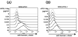
- (A) shows the results of examining the binding between human PD-L1 (hPDL1) -hIg and NRK / cPD1, and (B) shows the results of examining the binding between cPD1-hIg and NRK / cPDL1.
- (A) shows the results of examining the inhibition of binding between hPDL1-hIg and NRK / cPD1 by a rat monoclonal antibody against cPD1
- (B) shows the binding between cPD1-hIg and NRK / cPDL1 by a rat monoclonal antibody against cPDL1. It is a figure which shows the result of having investigated inhibition. It is a figure which shows the result of having investigated the binding inhibition of cPD1-hIg and NRK / cPDL1 by the rat monoclonal antibody with respect to cPD1.
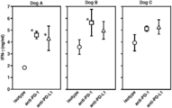
- FIG. 7 is a view showing the results of measuring canine IFN- ⁇ in an obtained culture supernatant by ELISA by adding ConA) to stimulation culture.
- Canine PBMC is cultured in the presence of PMA / ionomycin or ConA, and after recovery of PBMC, expression of PD-1, PD-L1 in the CD3 positive fraction (T cell) is determined by rat monoclonal antibody against cPD1 (4F12-E6) or It is a figure which shows the result of having analyzed the flow cytometry using the rat monoclonal antibody (G11-6) with respect to cPDL1.
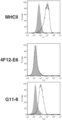
- CD14 positive monocytes separated from canine PBMC by magnetic beads were cultured in the presence of IL-4 and GM-CSF for 6 days to induce immature dendritic cells, and in such immature dendritic cells (MHC Class II antibody positive)
- MHC Class II antibody positive immature dendritic cells
- the anti-canine PD-1 antibody of the present invention specifically binds to canine PD-1 consisting of the amino acid sequence shown in SEQ ID NO: 1, (A) the amino acid sequence shown in SEQ ID NO: 2, or the heavy chain variable region having an amino acid sequence having 90% or more identity with the amino acid sequence shown in SEQ ID NO: 2, and the amino acid sequence shown in SEQ ID NO: 3, or An anti-canine PD-1 antibody comprising a light chain variable region having an amino acid sequence having 90% or more identity with the amino acid sequence shown in SEQ ID NO: 3; (B) the heavy chain variable region having the amino acid sequence shown in SEQ ID NO: 4, or the amino acid sequence shown in SEQ ID NO: 4 having 90% or more identity, and the amino acid sequence shown in SEQ ID NO: 5, or An anti-canine PD-1 antibody comprising a light chain variable region having an amino acid sequence having 90% or more identity with the amino acid sequence shown in SEQ ID NO: 5;
- the anti-canine PD-1 antibody described in (a) or (b) is not
- the anti-canine PD-L1 antibody of the present invention specifically binds to canine PD-L1 consisting of the amino acid sequence shown in SEQ ID NO: 6, (C) the amino acid sequence shown in SEQ ID NO: 7, or the heavy chain variable region having an amino acid sequence having 90% or more identity with the amino acid sequence shown in SEQ ID NO: 7, and the amino acid sequence shown in SEQ ID NO: 8, or An anti-canine PD-L1 antibody comprising a light chain variable region having an amino acid sequence having 90% or more identity with the amino acid sequence shown in SEQ ID NO: 8; (D) the amino acid sequence shown in SEQ ID NO: 9, or the heavy chain variable region having an amino acid sequence having 90% or more identity with the amino acid sequence shown in SEQ ID NO: 9, and the amino acid sequence shown in SEQ ID NO: 10, or An anti-canine PD-L1 antibody comprising a light chain variable region having an amino acid sequence having 90% or more identity with the amino acid sequence shown in SEQ ID NO: 10;
- the anti-canine PD-1 antibody of the present invention specifically binds to canine PD-1 consisting of the amino acid sequence shown in SEQ ID NO: 1, (E) a heavy chain variable region comprising CDR1 consisting of the amino acid sequence shown in SEQ ID NO: 11, CDR2 consisting of the amino acid sequence shown in SEQ ID NO: 12 and CDR3 consisting of the amino acid sequence shown in SEQ ID NO: 13, An anti-canine PD-1 comprising a light chain variable region comprising CDR1 consisting of the amino acid sequence shown, CDR2 consisting of the amino acid sequence shown in SEQ ID NO: 15, and CDR3 consisting of the amino acid sequence shown in SEQ ID NO: 16.
- the anti-canine PD-L1 antibody specifically binds to canine PD-L1 consisting of the amino acid sequence represented by SEQ ID NO: (G) a heavy chain variable region comprising CDR1 consisting of the amino acid sequence shown in SEQ ID NO: 23, CDR2 consisting of the amino acid sequence shown in SEQ ID NO: 24 and CDR3 consisting of the amino acid sequence shown in SEQ ID NO: 25;
- An anti-canine PD-L1 comprising a light chain variable region comprising CDR1 consisting of the amino acid sequence shown, CDR2 consisting of the amino acid sequence shown in SEQ ID NO: 27, and CDR3 consisting of the amino acid sequence shown in SEQ ID NO: 28 antibody;
- the types of the above-mentioned anti-canine PD-1 antibody and anti-canine PD-L1 antibody are not particularly limited, but include monoclonal antibodies.
- a chimeric antibody produced as a recombinant protein from the above sequence a canine antibody, F (ab ′) 2 obtained by digesting the antibody with pepsin, a Fab obtained by digesting the antibody with papain, a heavy chain variable region, Also included are antibody fragments consisting of a part of an antibody such as ScFv in which the light chain variable region is linked by an amino acid bridge, or diabodies of ScFv.
- identity means that the identity is 90% or more, preferably 93% or more, more preferably 95% or more, and still more preferably 98% or more. Means.
- Examples of the gene encoding the anti-canine antibody include, for example, the amino acid sequence represented by SEQ ID NO: 2 or the amino acid sequence represented by SEQ ID NO: 2 by 90% or more as the gene encoding the anti-canine PD-1 antibody.
- a gene that specifically binds to canine PD-1 comprising an amino acid sequence represented by SEQ ID NO: 1 in an antibody obtained by such a gene.
- nucleotide sequence represented by SEQ ID NO: 35 And a gene having the base sequence represented by SEQ ID NO: 36.
- it encodes the amino acid sequence of CDR1-3 of the heavy chain variable region shown in SEQ ID NOs: 11 to 13 and the amino acid sequence of CDR1-3 of the light chain variable region shown in SEQ ID NOs: 14-16.
- a gene with a gene can be mentioned.
- an amino acid sequence represented by SEQ ID NO: 4 or an amino acid sequence having 90% or more identity with the amino acid sequence represented by SEQ ID NO: 4 A gene comprising a coding gene and the amino acid sequence shown in SEQ ID NO: 5 or a gene encoding an amino acid sequence having 90% or more identity with the amino acid sequence shown in SEQ ID NO: 5, A gene that specifically binds to canine PD-1 in which the obtained antibody has the amino acid sequence shown in SEQ ID NO: 1 can be mentioned.
- the nucleotide sequence shown in SEQ ID NO: 37 and the sequence shown in SEQ ID NO: 38 And a gene having the nucleotide sequence shown.
- the gene encoding the anti-canine PD-L1 antibody includes a gene encoding the amino acid sequence shown in SEQ ID NO: 7, or an amino acid sequence having 90% or more identity with the amino acid sequence shown in SEQ ID NO: 7, and a sequence A gene comprising the amino acid sequence shown in No. 8, or a gene encoding an amino acid sequence having 90% or more identity with the amino acid sequence shown in SEQ ID No. 8, wherein an antibody obtained by such a gene is SEQ ID No.
- the gene encoding the anti-canine PD-L1 antibody encodes the amino acid sequence shown in SEQ ID NO: 9 or the amino acid sequence having 90% or more identity with the amino acid sequence shown in SEQ ID NO: 9.
- An expression vector for a gene encoding the anti-canine PD-1 antibody or anti-canine PD-L1 antibody is prepared by incorporating the gene encoding the anti-canine PD-1 antibody or anti-canine PD-L1 antibody into a vector.
- Any vector can be used as such an expression vector as long as it can express the gene encoding the anti-canine PD-1 antibody or anti-canine PD-L1 antibody, and it may be a plasmid vector or a phage vector.
- the above monoclonal antibody can be prepared according to a conventional protocol.
- a recombinant antibody can be produced by expressing a gene encoding the anti-canine antibody of the present invention by a gene recombination technique.
- a method for producing a recombinant antibody for example, a gene encoding the present anti-canine antibody is incorporated into an expression vector, and such expression vector is incorporated into a mammalian cell line such as Chinese hamster ovary (CHO) cell, Escherichia coli, yeast cell, insect cell.
- CHO Chinese hamster ovary
- Escherichia coli Escherichia coli
- yeast cell insect cell.
- a method of introducing a recombinant antibody in a host cell such as a plant cell (see Peter J.
- the base sequence of the gene encoding the present anti-canine antibody to be incorporated into the expression vector may be optimized for the codon sequence according to the host cell to be expressed.
- transgenic animals such as mice, cows, goats, sheep, chickens, pigs, etc., into which the gene encoding the anti-canine antibody has been incorporated, are produced using transgenic animal production technology, and blood, milk of such transgenic animals are produced. It is also possible to produce large quantities from inside.
- the monoclonal antibody is prepared by administering a cell expressing a protein consisting of the amino acid sequence shown in SEQ ID NO: 1 or 6 to a non-human animal such as a mouse or a rat as an antigen, and producing a cell clone that produces the anti-canine antibody.
- the above canine antibody can be produced, for example, by substituting the constant region of an antibody having a heavy chain variable region and a light chain variable region with a constant region of a canine antibody.
- Known constant regions of canine antibodies can be employed.
- the produced anti-canine antibody can be purified using, for example, chromatography using Protein A and Protein G columns, ion exchange chromatography, hydrophobic chromatography, ammonium sulfate salting out method, gel filtration, affinity chromatography, and the like.
- the binding inhibitor between canine PD-1 and canine PD-L1 characterized by containing the present anti-canine antibody is not particularly limited as long as it contains the present anti-canine antibody, and anti-canine PD-1 antibody And / or a plurality of anti-canine PD-L1 antibodies. Since such a binding inhibitor inhibits the binding between canine PD-1 and canine PD-L1, it can also be used as a therapeutic agent for canine cancers expressing PD-L1.
- the binding inhibition in the present invention includes both binding inhibition in vivo and binding inhibition in vitro.
- binding inhibitors are commonly used for formulation and are pharmaceutically acceptable excipients, binders, lubricants, disintegrants, preservatives, isotonic agents, stabilizers, dispersants, oxidation agents. Additives such as an inhibitor, a colorant, a flavoring agent, and a buffer may be included.
- the binding inhibitor can be administered parenterally by injection, infusion, application, suppository, intranasal spray, etc. Can be formulated.
- the amount of the present antibody contained in the binding inhibitor may be 0.0001 to 50% by weight, preferably 0.001 to 5% by weight.
- the method for inhibiting the binding between canine PD-1 and canine PD-L1, characterized by using the present anti-canine antibody is not particularly limited as long as the present anti-canine antibody is used.
- a plurality of canine PD-L1 antibodies may be used.
- the primers used for PCR in the following examples are shown in Table 1.
- the rat kidney cell line NRK cell and the human kidney cell line HEK293T cell used in the following examples are 10% FBS, 100 units / ml penicillin, 100 ⁇ g / ml streptomycin, 55 ⁇ M 2-mercapto. Maintained in DMEM medium supplemented with ethanol. These cell lines used were cultured in a humidified incubator at 37 ° C. under 5% CO 2 gas concentration.
- a forward primer (YTM1144: SEQ ID NO: 45) and a reverse primer (YTM1145: SEQ ID NO: 46) for amplifying cPDL1 cDNAs designed based on the sequence of XM — 543338 are NCBI Accession No. Designed based on the sequence of XM — 005615936.1.
- Accession No. XM — 543338 and Accession No. The sequence of XM — 005615936.1 is a gene whose expression has not been confirmed in dogs.
- PCR reaction Using the above primer pair, normal canine thymus cDNA was used as a template, and KPD-Plus-Neo (manufactured by Toyobo Co., Ltd.) was used to amplify the cPD1 gene and the cPDL1 gene according to the attached protocol.
- pre-denaturation was performed at 94 ° C. for 2 minutes, followed by 35 cycles of denaturation at 98 ° C. for 10 seconds, annealing at 58 ° C. for 30 seconds, extension at 68 ° C. for 60 seconds, and final extension at 68 ° C. I went for a minute.
- the PCR product prepared above was gel-purified and inserted into the SmaI site of the pBluescript SK ( ⁇ ) vector, and construct vectors (pBS-cPD1, pBS-cPDL1) containing cPD1 or cPDL1 gene as inserts were prepared.
- construct vectors uses a forward primer (M13 (-20): SEQ ID NO: 47) and a reverse primer (M13 reverse: SEQ ID NO: 48), and BigDye (registered trademark) Termination v3.1 Cycle Sequencing Kit (manufactured by Perkin-Elmer).
- the nucleotide sequence was determined using an ABI Prism (registered trademark) 377 automatic DNA sequencer (Applied Biosystems).
- the resulting cPD1 base sequence and the amino acid sequence encoded by the base sequence are shown in FIG. 1, the base sequence of cPDL1 and the amino acid sequence encoded by the base sequence are shown in FIG. 2, and the base sequence of cPD1 is SEQ ID NO: 49,
- the amino acid sequence is shown in SEQ ID NO: 1
- the base sequence of cPDL1 is shown in SEQ ID NO: 50
- the amino acid sequence is shown in SEQ ID NO: 6.
- Retroviral vectors that overexpress cPD1 or cPDL1 protein were constructed.
- a forward primer YTM1142: SEQ ID NO: 43
- the CPD1 sequence C A reverse primer containing a FLAG tag sequence at the end (YTM1167: SEQ ID NO: 51) was used.
- pBS-cPDL1 was used as a template, forward primer (YTM1144: SEQ ID NO: 45), and C-terminal of the cPDL1 sequence.
- Primary PCR was performed using a reverse primer containing a FLAG tag sequence (YTM1168: SEQ ID NO: 52).
- PCR secondary PCR is performed using a forward primer similar to the primary PCR and a reverse primer (YTM838: SEQ ID NO: 53) containing a double FLAG tag sequence that anneals to the 1st FLAG tag sequence. It was.
- the obtained PCR products were inserted into the EcoRI and NotI sites of the pMxs-IP vector to prepare pMx-IP-cPD1-FL and pMx-IP-cPDL1-FL # 9.
- pKx-IP-cPD1-FL and pMx-IP-cPDL1-FL # 9 are transfected into NRK cells to produce NRK cells that stably express cPD1 and NRK cells that stably express cPDL1.
- NRK cells 3.5 ⁇ 10 5 cells
- Transfection of cells was performed using Lipofectamine 2000 (Invitrogen) according to the attached protocol.
- the transfected cells were incubated for 48 hours, and then cultured in the presence of 7.5 ⁇ g / ml puromycin (Sigma-Aldrich) to obtain stable transduced cells, and NRK stably expressing cPD1.
- Cells (NRK / cPD1) and NRK cells (NRK / cPDL1) stably expressing cPDL1 were prepared.
- cPD1 and cPDL1 are stably expressed as anti-FLAG M2 antibody (1: 1000 dilution, manufactured by Sigma-Aldrich) as primary antibody and IgG-Dylight as secondary antibody. This was confirmed by immunofluorescence using (registered trademark) 488-labeled anti-mouse (Biolegend).
- NRK / cPD1 or NRK / cPDL1 prepared above (1 ⁇ 10 7 cells in 500 ⁇ l of the above-mentioned DMEM medium) was used in an equal amount of Titer Max® Gold ( CytRx) was emulsified and sarcastically administered to the hind footpad of 7-week-old Sprague-Dawley rats (Kudo Co., Ltd.). Two weeks after administration, popliteal lymph node cells were isolated and fused with P3U1 cells.
- lymph node monocytic cells (1 ⁇ 10 8 cells) and P3U1 cells (2 ⁇ 10 7 cells) were mixed and washed twice with serum-free RPMI 1640 medium. After removing the supernatant, the cells were incubated at 37 ° C. for 2 minutes, 0.5 ml polyethylene glycol 1500 (Roche Diagnostics) at 37 ° C. was added, 9 ml serum-free RPMI 1640 medium at 37 ° C. was added, and 900 rpm for 5 minutes. Centrifuge at room temperature.
- NRK cell positive hybridomas expressing cPD1 or cPDL1 are identified by ELISA or flow cytometry, and such hybridomas are cloned by limited dilution and hybridomas 3B7-D9 as NRK cell positive hybridomas expressing cPD1.
- Hybridoma H7-9 and hybridoma G11-6 were obtained as NRK cell positive hybridomas expressing hybridoma 4F12-E6 and cPDL1.
- the antibody produced by the hybridoma 3B7-D9 is “3B7-D9”
- the antibody produced by the hybridoma 4F12-E6 is “4F12-E6”
- the antibody produced by the hybridoma H7-9 is “H7-9”
- the antibody produced by the hybridoma G11-6 is also referred to as “G11-6”.
- the hybridomas 3B7-D9, 4F12-E6, H7-9, and G11-6 are stored in the National University Corporation Yamaguchi University Joint Veterinary School and can be distributed under certain conditions.
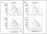
- FIG. 3 (A) shows the results of examining the binding of rat monoclonal antibody (anti-cPD1 mAb) against cPD1 and NRK / cPD1
- FIG. 3 (B) shows the rat monoclonal antibody against cPDL1 (anti-cPD-L1 mAb). It is the result of having investigated the coupling
- the horizontal axis represents the fluorescence intensity of the fluorescently labeled secondary antibody with respect to the prepared antibody, and the vertical axis represents the concentration of each rat monoclonal antibody.
- FIG. 3 (A) shows the results of examining the binding of rat monoclonal antibody (anti-cPD1 mAb) against cPD1 and NRK / cPD1
- FIG. 3 (B) shows the rat monoclonal antibody against cPDL1 (anti-cPD-L1 mAb). It is the result of having investigated the coupling
- the amplified PCR product was introduced into the SmaI site of the pBluescript SK ( ⁇ ) vector.
- This plasmid was digested with EcoRI and NotI, and the resulting fragment was ligated to the EcoRI and NotI sites of the pMxs-IP vector to prepare pMx-IP-hPDL1 # 21, an hPDL1 expression plasmid.
- Fusion protein expression vector production A vector expressing a fusion protein of the extracellular region of cPD1 or hPDL1 and the human IgG2 Fc region was constructed.
- PCR is performed using the forward primer (YTM1153: SEQ ID NO: 56) and reverse primer (YTM1154: SEQ ID NO: 57) using the pMx-IP-cPD1-FL prepared in Example 1 as a template.
- the extracellular region of cPD1 is amplified, and PCR is performed using the forward primer (YTM1157: SEQ ID NO: 58) and reverse primer (YTM1158: SEQ ID NO: 59) using the pMx-IP-hPDL1 # 21 prepared above as a template.
- Each PCR product was cleaved with BamHI for cPD1 and BglII for hPDL1, and cloned into the EcoRV and BgIII sites of the pFUSE-hIgG2-Fc2 vector (Invivogen).
- the extracellular region of cPD1 and the human IgG2 Fc region A vector (pFUSE-cPD1-hIg # 2) that expresses a fusion protein and a vector that expresses a fusion protein of the extracellular region of hPDL1 and the human IgG2 Fc region (pFUSE-hPDL1-hIg # 9) did.
- the HEK293T cell line was transfected with the vector pFUSE-cPD1-hIg # 2, pFUSE-hPDL1-hIg # 9 prepared above or the empty vector pFUSE-hIgG2-Fc2.
- 2 ⁇ 10 6 HEK293T cells were seeded in four 10 cm dishes.
- 375 ⁇ l of OPTI-MEM containing 7.5 ⁇ g of each vector and 30 ⁇ l of 1 mg / ml PEI Max was mixed, incubated at room temperature for 15 minutes, and added to the cells.
- the medium was replaced with a GIT serum-free medium (manufactured by Wako Pure Chemical Industries, Ltd.), and the cells were further cultured for 48 hours.
- Supernatant was collected from each of the transfected cells on the 4th and 8th days and purified by rProtein A agarose (manufactured by GE healthcare).
- the soluble protein was desalted by dialysis, and a fusion protein of the extracellular region of cPD1 and the hIgG2 Fc region (cPD1-hIg) and a fusion protein of the extracellular region of hPDL1 and the hIgG2 Fc region (hPDL1-hIg) ) was produced.
- the purity of the fusion protein was confirmed by SDS-PAGE and Western blotting.
- Binding test of PD-1 and PD-L1 As a preliminary test of inhibition test of PD-1 and PD-L1 by rat monoclonal antibody described later, binding of hPDL1-hIg and NRK / cPD1, or cPD1-hIg and NRK The binding to / cPDL1 was examined by the same method as in the flow cytometry analysis. The concentrations of hPDL1-hIg and cPD1-hIg were 0, 0.04, 0.156, 0.625, 2.5, 10, 40 ⁇ g / ml, and NRK / cPD1 (2 ⁇ 10 5 ) or NRK / cPDL1 ( 2 ⁇ 10 5 ) was used.
- FIG. 4A shows the result of examining the binding between hPDL1-hIg and NRK / cPD1
- FIG. 4B shows the result of examining the binding between cPD1-hIg and NRK / cPDL1.
- the horizontal axis represents the fluorescence intensity of the fluorescently labeled secondary antibody for the prepared fusion protein
- the vertical axis represents the concentration of hPDL1-hIg or cPD1-hIg.
- FIG. 4 it was revealed that hPDL1-hIg binds to NRK / cPD1, and cPD1-hIg binds to NRK / cPDL1.
- Binding inhibition test of PD-1 and PD-L1 with rat monoclonal antibody (flow cytometry analysis) (1) By reacting NRK / cPD1 with a rat monoclonal antibody against cPD1 (3B7-D9, 4F12-E6), and then reacting with hPDL1-hIg, the binding of hPDL1-hIg and NRK / cPD1 to rat against cPD1 (2) By reacting NRK / cPDL1 with rat monoclonal antibodies against cPDL1 (H7-9, G11-6) and then reacting with cPD1-hIg, cPD1-hIg and NRK / cPDL1 Or the rat monoclonal antibody against cPDL1 inhibits the binding with (3) the rat monoclonal antibody against cPD1 (3B7-D9, 4F12-E6) and cPD1-hIg, and the mixed reaction product is then converted to NR
- CPD1-hIg and NRK / cPDL1 the coupling or rat monoclonal antibodies against CPD1 is inhibition of the three points, respectively.
- the inhibition of the binding between PD-1 and PD-L1 by each antibody was examined in the same manner as in the flow cytometry analysis.
- the concentration of each rat monoclonal antibody was 0, 0.04, 0.156, 0.625, 2.5, 10 ⁇ g / ml
- the concentration of hPDL1-hIg, cPD1-hIg was 40 ⁇ g / ml
- NRK / cPD1 (2 ⁇ 10 5 ) or NRK / cPDL1 (2 ⁇ 10 5 ) was used.
- FIGS. FIG. 5 (A) shows the results of examining the inhibition of binding between hPDL1-hIg and NRK / cPD1 by a rat monoclonal antibody against cPD1
- FIG. 5 (B) shows the results of cPD1-hIg and NRK / cPDL1 produced by a rat monoclonal antibody against cPDL1.
- FIG. 6 shows the results of examining the binding inhibition between cPD1-hIg and NRK / cPDL1 by a rat monoclonal antibody against cPD1.
- the abscissa indicates the fluorescence intensity of the fluorescently labeled secondary antibody with respect to the prepared antibody
- the ordinate indicates the concentration of each rat monoclonal antibody
- the ordinate indicates the presence (+) / absence (-) of hPDL1-hIg or cPD1-hIg. .
- the binding between hPDL1-hIg and NRK / cPD1 or the binding between cPD1-hIg and NRK / cPDL1 is 3B7-D9 which is a rat monoclonal antibody against cPD1. It was revealed that 4F12-E6 inhibited, and the binding of cPD1-hIg and NRK / cPDL1 was inhibited by rat monoclonal antibodies H7-9 and G11-6 against cPDL1.
- RNA was isolated from each hybridoma cell line obtained in Example 1, and the nucleotide sequence of the immunoglobulin heavy chain (IgG2a) and the nucleotide sequence of the kappa light chain including the 5 ′ region of all variable regions and constant regions were determined. Obtained by 5'RACE PCR method.
- RNA was reverse transcribed using reverse transcriptase Superscript (registered trademark) III.
- primers YTM171 (SEQ ID NO: 60) was used for light chain amplification, and YTM172 (SEQ ID NO: 61) was used for heavy chain amplification.
- polyC is added by TdT enzyme reaction, then forward primer (YTM166: SEQ ID NO: 62) and reverse primer (YTM171: SEQ ID NO: 60) for light chain amplification, and forward primer for heavy chain amplification.
- Primary PCR was performed using (YTM166: SEQ ID NO: 62) and reverse primer (YTM172: SEQ ID NO: 61).
- the forward primer (YTM170: SEQ ID NO: 63) and reverse primer (YTM173: SEQ ID NO: 64), or the forward primer (YTM170: sequence) No. 63) and reverse primer (YTM174: SEQ ID NO: 65) were used for nested PCR.
- YTM1224 SEQ ID NO: 42 was used instead of YTM171 as a primer in 5'RACE or primary PCR.
- the nested PCR product was cloned into the pBluescript SK ( ⁇ ) vector, and the base sequence and the amino acid sequence encoded by the base sequence were determined.
- the nucleotide sequence of the heavy chain variable region of 3B7-D9 is SEQ ID NO: 35, the amino acid sequence is SEQ ID NO: 2, the nucleotide sequence of the light chain variable region is SEQ ID NO: 36, the amino acid sequence is SEQ ID NO: 3, and the sequence of 4F12-E6
- the nucleotide sequence of the heavy chain variable region is SEQ ID NO: 37, the amino acid sequence is SEQ ID NO: 4, the nucleotide sequence of the light chain variable region is SEQ ID NO: 38, the amino acid sequence is SEQ ID NO: 5, and the heavy chain variable region of H7-9.
- CDR amino acid sequences of each heavy chain variable region and light chain variable region were identified by comparing them with known antibody amino acid sequences.
- the amino acid sequences of CDRs 1 to 3 of the heavy chain variable region of 3B7-D9 are respectively SEQ ID NOs: 11 to 13
- the amino acid sequences of CDRs 1 to 3 of the light chain variable region are respectively SEQ ID NOs: 14 to 16, and the heavy chain variable of 4F12-E6.
- the amino acid sequences of CDR1 to CDR3 of the region are respectively SEQ ID NOs: 17 to 19
- the amino acid sequences of CDR1 to CDR3 of the light chain variable region are respectively SEQ ID NOs: 20 to 22
- the amino acids of CDR1 to CDR3 of the heavy chain variable region of H7-9 The sequence is SEQ ID NO: 23 to 25
- the light chain variable region CDR 1 to 3 amino acid sequence is SEQ ID NO: 26 to 28
- the G11-6 heavy chain variable region CDR 1 to 3 amino acid sequence is SEQ ID NO: 29 to 31
- the amino acid sequences of CDR1 to CDR3 of the light chain variable region are shown in SEQ ID NOs: 32 to 34, respectively.
- PBMC Peripheral blood mononuclear cells
- a rat monoclonal antibody against PD-1 anti-PD-1: 4F12-E6
- a rat monoclonal antibody against cPDL1 anti-PD-L1: G11-6 was added to a concentration of 10 ⁇ g / ml
- concanavalin A ConA was added to 5 ⁇ g / ml and stimulated for 3 days, and canine IFN- ⁇ in the obtained culture supernatant was measured by canine IFN-gamma DuoSet ELISA (manufactured by R & D). The results are shown in FIG.
- IFN ⁇ produced by ConA stimulation tends to be enhanced in the presence of anti-PD-1 4F12-E6 and anti-PD-L1 G11-6. It was. This suggests that ConA stimulation enhances PD-1 and PD-L1 expression in canine T cells, and that PD-L1 binds to PD-1 to prevent excessive T cell activation. It was suggested that the obtained PD-1 and PD-L1 antibodies can inhibit the interaction of both PD-1 and PD-L1 molecules expressed on canine cells.
- PD-1 was not expressed, and it was confirmed that PDL-1 was expressed. Thus, it was revealed that the expression of PD-1 or PD-L1 in immature dendritic cells can be examined by using the antibody of the present invention.
- the rat monoclonal antibody obtained in the present invention can recognize and bind to canine PD-1 or canine PD-L1, it can be used for expression analysis or functional analysis of canine PD-1 or canine PD-L1. In addition, it can be used as a binding inhibitor between canine PD-1 and canine PD-L1.
Landscapes
- Health & Medical Sciences (AREA)
- Chemical & Material Sciences (AREA)
- Immunology (AREA)
- Life Sciences & Earth Sciences (AREA)
- Organic Chemistry (AREA)
- Genetics & Genomics (AREA)
- General Health & Medical Sciences (AREA)
- Medicinal Chemistry (AREA)
- Engineering & Computer Science (AREA)
- Biophysics (AREA)
- Molecular Biology (AREA)
- Biochemistry (AREA)
- Bioinformatics & Cheminformatics (AREA)
- Proteomics, Peptides & Aminoacids (AREA)
- Biomedical Technology (AREA)
- Wood Science & Technology (AREA)
- Zoology (AREA)
- Biotechnology (AREA)
- General Engineering & Computer Science (AREA)
- Microbiology (AREA)
- Pharmacology & Pharmacy (AREA)
- Animal Behavior & Ethology (AREA)
- Public Health (AREA)
- Veterinary Medicine (AREA)
- Plant Pathology (AREA)
- Physics & Mathematics (AREA)
- Epidemiology (AREA)
- Mycology (AREA)
- General Chemical & Material Sciences (AREA)
- Chemical Kinetics & Catalysis (AREA)
- Nuclear Medicine, Radiotherapy & Molecular Imaging (AREA)
- Peptides Or Proteins (AREA)
- Medicines Containing Antibodies Or Antigens For Use As Internal Diagnostic Agents (AREA)
Abstract
Description
(1)配列番号1に示されるアミノ酸配列からなるイヌPD-1に特異的に結合する、以下の(a)又は(b)記載の抗イヌPD-1抗体。
(a)配列番号2に示されるアミノ酸配列、又は配列番号2に示されるアミノ酸配列と90%以上の同一性を有するアミノ酸配列を有する重鎖可変領域、及び配列番号3に示されるアミノ酸配列、又は配列番号3に示されるアミノ酸配列と90%以上の同一性を有するアミノ酸配列を有する軽鎖可変領域を備えたことを特徴とする抗イヌPD-1抗体;
(b)配列番号4に示されるアミノ酸配列、又は配列番号4に示されるアミノ酸配列と90%以上の同一性を有するアミノ酸配列を有する重鎖可変領域、及び配列番号5に示されるアミノ酸配列、又は配列番号5に示されるアミノ酸配列と90%以上の同一性を有するアミノ酸配列を有する軽鎖可変領域を備えたことを特徴とする抗イヌPD-1抗体;
(2)配列番号6に示されるアミノ酸配列からなるイヌPD-L1に特異的に結合する、以下の(c)又は(d)記載の抗イヌPD-L1抗体。
(c)配列番号7に示されるアミノ酸配列、又は配列番号7に示されるアミノ酸配列と90%以上の同一性を有するアミノ酸配列を有する重鎖可変領域、及び配列番号8に示されるアミノ酸配列、又は配列番号8に示されるアミノ酸配列と90%以上の同一性を有するアミノ酸配列を有する軽鎖可変領域を備えたことを特徴とする抗イヌPD-L1抗体;
(d)配列番号9に示されるアミノ酸配列、又は配列番号9に示されるアミノ酸配列と90%以上の同一性を有するアミノ酸配列を有する重鎖可変領域、及び配列番号10に示されるアミノ酸配列、又は配列番号10に示されるアミノ酸配列と90%以上の同一性を有するアミノ酸配列を有する軽鎖可変領域を備えたことを特徴とする抗イヌPD-L1抗体;
(3)配列番号1に示されるアミノ酸配列からなるイヌPD-1に特異的に結合する、以下の(e)又は(f)記載の抗イヌPD-1抗体。
(e)配列番号11に示されるアミノ酸配列からなるCDR1、配列番号12に示されるアミノ酸配列からなるCDR2及び配列番号13に示されるアミノ酸配列からなるCDR3を含む重鎖可変領域と、配列番号14に示されるアミノ酸配列からなるCDR1、配列番号15に示されるアミノ酸配列からなるCDR2及び配列番号16に示されるアミノ酸配列からなるCDR3を含む軽鎖可変領域を備えたことを特徴とする抗イヌPD-1抗体;
(f)配列番号17に示されるアミノ酸配列からなるCDR1、配列番号18に示されるアミノ酸配列からなるCDR2及び配列番号19に示されるアミノ酸配列からなるCDR3を含む重鎖可変領域と、配列番号20に示されるアミノ酸配列からなるCDR1、配列番号21に示されるアミノ酸配列からなるCDR2及び配列番号22に示されるアミノ酸配列からなるCDR3を含む軽鎖可変領域を備えたことを特徴とする抗イヌPD-1抗体;
(4)配列番号6に示されるアミノ酸配列からなるイヌPD-L1に特異的に結合する、以下の(g)又は(h)記載の抗イヌPD-L1抗体。
(g)配列番号23に示されるアミノ酸配列からなるCDR1、配列番号24に示されるアミノ酸配列からなるCDR2及び配列番号25に示されるアミノ酸配列からなるCDR3を含む重鎖可変領域と、配列番号26に示されるアミノ酸配列からなるCDR1、配列番号27に示されるアミノ酸配列からなるCDR2及び配列番号28に示されるアミノ酸配列からなるCDR3を含む軽鎖可変領域を備えたことを特徴とする抗イヌPD-L1抗体;
(h)配列番号29に示されるアミノ酸配列からなるCDR1、配列番号30に示されるアミノ酸配列からなるCDR2及び配列番号31に示されるアミノ酸配列からなるCDR3を含む重鎖可変領域と、配列番号32に示されるアミノ酸配列からなるCDR1、配列番号33に示されるアミノ酸配列からなるCDR2及び配列番号34に示されるアミノ酸配列からなるCDR3を含む軽鎖可変領域を備えたことを特徴とする抗イヌPD-L1抗体;
(5)上記(1)~(4)のいずれか記載の抗体を含有することを特徴とするイヌPD-1とイヌPD-L1との結合阻害剤。
(6)上記(1)~(4)のいずれか記載の抗体を用いることを特徴とするイヌPD-1とイヌPD-L1との結合阻害方法。
(7)上記(1)~(4)のいずれか記載の抗体をコードする遺伝子。
(a)配列番号2に示されるアミノ酸配列、又は配列番号2に示されるアミノ酸配列と90%以上の同一性を有するアミノ酸配列を有する重鎖可変領域、及び配列番号3に示されるアミノ酸配列、又は配列番号3に示されるアミノ酸配列と90%以上の同一性を有するアミノ酸配列を有する軽鎖可変領域を備えたことを特徴とする抗イヌPD-1抗体;
(b)配列番号4に示されるアミノ酸配列、又は配列番号4に示されるアミノ酸配列と90%以上の同一性を有するアミノ酸配列を有する重鎖可変領域、及び配列番号5に示されるアミノ酸配列、又は配列番号5に示されるアミノ酸配列と90%以上の同一性を有するアミノ酸配列を有する軽鎖可変領域を備えたことを特徴とする抗イヌPD-1抗体;
の(a)又は(b)記載の抗イヌPD-1抗体であれば特に制限されないが、
(a’)配列番号2に示されるアミノ酸配列、又は配列番号2に示されるアミノ酸配列と90%以上の同一性を有するアミノ酸配列からなる重鎖可変領域、及び配列番号3に示されるアミノ酸配列、又は配列番号3に示されるアミノ酸配列と90%以上の同一性を有するアミノ酸配列からなる軽鎖可変領域とを備えたことを特徴とする抗イヌPD-1抗体;(b’)配列番号4に示されるアミノ酸配列、又は配列番号4に示されるアミノ酸配列と90%以上の同一性を有するアミノ酸配列からなる重鎖可変領域、及び配列番号5に示されるアミノ酸配列、又は配列番号5に示されるアミノ酸配列と90%以上の同一性を有するアミノ酸配列からなる軽鎖可変領域を備えたことを特徴とする抗イヌPD-1抗体;
の(a’)又は(b’)記載の抗イヌPD-1抗体であることが好ましく、かかる抗イヌPD-1抗体はイヌPD-1を認識して結合可能であることから、イヌPD-1の発現解析や機能解析が可能となるほか、イヌPD-1とイヌPD-L1との結合を阻害することが可能となる。
(c)配列番号7に示されるアミノ酸配列、又は配列番号7に示されるアミノ酸配列と90%以上の同一性を有するアミノ酸配列を有する重鎖可変領域、及び配列番号8に示されるアミノ酸配列、又は配列番号8に示されるアミノ酸配列と90%以上の同一性を有するアミノ酸配列を有する軽鎖可変領域を備えたことを特徴とする抗イヌPD-L1抗体;
(d)配列番号9に示されるアミノ酸配列、又は配列番号9に示されるアミノ酸配列と90%以上の同一性を有するアミノ酸配列を有する重鎖可変領域、及び配列番号10に示されるアミノ酸配列、又は配列番号10に示されるアミノ酸配列と90%以上の同一性を有するアミノ酸配列を有する軽鎖可変領域を備えたことを特徴とする抗イヌPD-L1抗体;
の(c)又は(d)記載の抗イヌPD-L1抗体であれば特に制限されないが、
(c’)配列番号7に示されるアミノ酸配列、又は配列番号7に示されるアミノ酸配列と90%以上の同一性を有するアミノ酸配列からなる重鎖可変領域、及び配列番号8に示されるアミノ酸配列、又は配列番号8に示されるアミノ酸配列と90%以上の同一性を有するアミノ酸配列からなる軽鎖可変領域を備えたことを特徴とする抗イヌPD-L1抗体;(d’)配列番号9に示されるアミノ酸配列、又は配列番号9に示されるアミノ酸配列と90%以上の同一性を有するアミノ酸配列からなる重鎖可変領域、及び配列番号10に示されるアミノ酸配列、又は配列番号10に示されるアミノ酸配列と90%以上の同一性を有するアミノ酸配列からなる軽鎖可変領域を備えたことを特徴とする抗イヌPD-L1抗体;
の(c’)又は(d’)記載の抗イヌPD-L1抗体であることが好ましく、かかる抗イヌPD-L1抗体はイヌPD-L1を認識して結合可能であることから、イヌPD-L1の発現解析や機能解析が可能となるほか、イヌPD-1とイヌPD-L1との結合を阻害することが可能となる。
(e)配列番号11に示されるアミノ酸配列からなるCDR1、配列番号12に示されるアミノ酸配列からなるCDR2及び配列番号13に示されるアミノ酸配列からなるCDR3を含む重鎖可変領域と、配列番号14に示されるアミノ酸配列からなるCDR1、配列番号15に示されるアミノ酸配列からなるCDR2及び配列番号16に示されるアミノ酸配列からなるCDR3を含む軽鎖可変領域を備えたことを特徴とする抗イヌPD-1抗体;
(f)配列番号17に示されるアミノ酸配列からなるCDR1、配列番号18に示されるアミノ酸配列からなるCDR2及び配列番号19に示されるアミノ酸配列からなるCDR3を含む重鎖可変領域と、配列番号20に示されるアミノ酸配列からなるCDR1、配列番号21に示されるアミノ酸配列からなるCDR2及び配列番号22に示されるアミノ酸配列からなるCDR3を含む軽鎖可変領域を備えたことを特徴とする抗イヌPD-1抗体;
の(e)又は(f)記載の抗イヌPD-1抗体を挙げることができる。
(g)配列番号23に示されるアミノ酸配列からなるCDR1、配列番号24に示されるアミノ酸配列からなるCDR2及び配列番号25に示されるアミノ酸配列からなるCDR3を含む重鎖可変領域と、配列番号26に示されるアミノ酸配列からなるCDR1、配列番号27に示されるアミノ酸配列からなるCDR2及び配列番号28に示されるアミノ酸配列からなるCDR3を含む軽鎖可変領域を備えたことを特徴とする抗イヌPD-L1抗体;
(h)配列番号29に示されるアミノ酸配列からなるCDR1、配列番号30に示されるアミノ酸配列からなるCDR2及び配列番号31に示されるアミノ酸配列からなるCDR3を含む重鎖可変領域と、配列番号32に示されるアミノ酸配列からなるCDR1、配列番号33に示されるアミノ酸配列からなるCDR2及び配列番号34に示されるアミノ酸配列からなるCDR3を含む軽鎖可変領域を備えたことを特徴とする抗イヌPD-L1抗体;
の(g)又は(h)記載の抗イヌPD-L1抗体を挙げることができる。

1.cPD1又はcPDL1のcDNAクローニング
(プライマーの設計)
cPD1のcDNAを増幅するためのフォワードプライマー(YTM1142:配列番号43)とリバースプライマー(YTM1143:配列番号44)は、NCBI(http://www.ncbi.nlm.nih.gov/guide/) Accession No.XM_543338の配列に基づいて設計し、cPDL1のcDNAsを増幅するためのフォワードプライマー(YTM1144:配列番号45)とリバースプライマー(YTM1145:配列番号46)は、NCBI Accession No.XM_005615936.1の配列に基づいて設計した。なお、Accession No.XM_543338及びAccession No.XM_005615936.1の配列は、イヌにおいて発現が確認されていない遺伝子である。
上記プライマー対を用い、正常なイヌの胸腺のcDNAをテンプレートとし、KOD-Plus-Neo(東洋紡社製)を用い、その添付のプロトコールに従ってcPD1遺伝子とcPDL1遺伝子を増幅した。PCRは、プレ変性を94℃で2分行い、その後98℃で10秒の変性、58℃で30秒のアニーリング、68℃で60秒の伸長を35サイクル行い、さらに最終伸長を68℃で10分行った。
上記で調製したPCR産物をゲル精製し、pBluescript SK(-)ベクターのSmaI部位に挿入し、インサートとしてcPD1又はcPDL1遺伝子を含むコンストラクトベクター(pBS-cPD1、pBS-cPDL1)を作製した。かかるコンストラクトベクターはフォワードプライマー(M13(-20):配列番号47)とリバースプライマー(M13 reverse:配列番号48)を用い、BigDye(登録商標)Termination v3.1 Cycle Sequencing Kit (Perkin-Elmer社製)及びABI Prism(登録商標)377自動DNAシークエンサー(Applied Biosystems社製)によって塩基配列を決定した。得られたcPD1の塩基配列及びその塩基配列がコードするアミノ酸配列を図1に、cPDL1の塩基配列及びその塩基配列がコードするアミノ酸配列を図2に示すと共に、cPD1の塩基配列を配列番号49、アミノ酸配列を配列番号1に、cPDL1の塩基配列を配列番号50、アミノ酸配列を配列番号6に示す。
(cPD1又はcPDL1発現ベクターの構築)
cPD1又はcPDL1タンパク質を過剰発現するレトロウイルスベクターを構築した。まず、cPD1の配列とcPDL1の配列のC末端に2つのFLAGタグ配列を加えるため、cPD1の増幅には、pBS-cPD1をテンプレートとし、フォワードプライマー(YTM1142:配列番号43)と、cPD1配列のC末端にFLAGタグ配列を含むリバースプライマー(YTM1167:配列番号51)とを用い、cPDL1の増幅には、pBS-cPDL1をテンプレートとし、フォワードプライマー(YTM1144:配列番号45)と、cPDL1配列のC末端にFLAGタグ配列を含むリバースプライマー(YTM1168:配列番号52)を用いて一次PCRを行った。
一般的なトランスフェクション方法によって、pMx-IP-cPD1-FL、pMx-IP-cPDL1-FL#9をNRK細胞にトランスフェクトし、cPD1を安定発現するNRK細胞、cPDL1を安定発現するNRK細胞を作製した。まず、トランスフェクションを行う1日前にNRK細胞(3.5×10⁵個)を6ウェルディッシュに播種した。細胞のトランスフェクションは、Lipofectamine 2000(Invitrogen社製)を用い、添付のプロトコールに従って行った。トランスフェクション後の細胞を48時間インキュベートし、その後、安定な形質導入細胞を得るために7.5μg/mlのプロマイシン(Sigma-Aldrich社製)の存在下で培養し、cPD1を安定発現するNRK細胞(NRK/cPD1)、cPDL1を安定発現するNRK細胞(NRK/cPDL1)を作製した。
cPD1又はcPDL1に対するラットモノクローナル抗体を作製するために、上述で作製したNRK/cPD1又はNRK/cPDL1(500μlの上記DMEM培地中、1×107個)を等量のTiter Max(登録商標)Gold(CytRx社製)で乳化し、7週齢のSprague-Dawleyラット(九動社製)の後足蹠に皮肉投与した。投与から2週間後に、膝窩のリンパ節細胞を単離し、P3U1細胞と融合させた。すなわち、リンパ節単球細胞(1×10⁸個)とP3U1細胞(2×10⁷個)と混合し、無血清のRPMI1640培地で2回洗浄した。上澄みを除去後、細胞を37℃で2分インキュベートし、37℃の0.5mlポリエチレングリコール1500(Roche Diagnostics社製)を加え、さらに37℃の9ml無血清RPMI1640培地を加え、900rpmで5分、室温にて遠心した。上澄みを除去後、10%FBSとhypoxanthine-aminopterin-thymidine(HAT:Life Technologies社製)を含む36mlのGIT培地(和光純薬工業社製)で懸濁し、加湿インキュベーターにより、5%CO2ガス濃度下、37℃で1時間培養した。4mlのBM-Condimed H1培地(Roche Diagnostics社製)を加え、細胞を4つの96ウェル組織培養プレートに播種(100μl/ウェル)して培養した。コロニーが得られた後、cPD1又はcPDL1を発現するNRK細胞陽性ハイブリドーマをELISA法やフローサイトメトリーで同定し、かかるハイブリドーマを限定希釈によりクローニングし、cPD1を発現するNRK細胞陽性ハイブリドーマとしてハイブリドーマ3B7-D9、ハイブリドーマ4F12-E6、cPDL1を発現するNRK細胞陽性ハイブリドーマとしてハイブリドーマH7-9、ハイブリドーマG11-6を得た。以後、ハイブリドーマ3B7-D9によって産生された抗体を「3B7-D9」、ハイブリドーマ4F12-E6によって産生された抗体を「4F12-E6」、ハイブリドーマH7-9によって産生された抗体を「H7-9」、ハイブリドーマG11-6によって産生された抗体を「G11-6」ともいう。なお、ハイブリドーマ3B7-D9、4F12-E6、H7-9、G11-6は国立大学法人山口大学共同獣医学部に保管されており、一定の条件下で分譲可能である。
(抗体濃度及び細胞数)
cPD1に対するラットモノクローナル抗体である3B7-D9、4F12-E6と、上記で作製したNRK/cPD1との結合や、cPDL1に対するラットモノクローナル抗体であるH7-9、G11-6と、上記で作製したNRK/cPDL1との結合を、以下に示すフローサイトメトリー解析により調べた。各ラットモノクローナル抗体の濃度は0、0.04、0.156、0.625、2.5、10μg/mlであり、NRK/cPD1(2×105個)又はNRK/cPDL1(2×105個)を用いた。
フローサイトメトリー解析に用いる細胞株の染色は次の文献(Mizuno et al., J Vet Med Sci, 71(12):1561-1568, 2009)に記載の方法に従って行った。一次抗体としては、精製したcPD1又はcPDL1に対する各ラットモノクローナル抗体を用いた。二次抗体としては抗ラットIgG-PE(Southern biotech社製)を用いた。サンプルはBD AccuriC6 (BD Bioscience社製)を用いて解析し、得られた結果をFlowJo software (Treestar社製)によって解析した。
結果を図3に示す。図3(A)はcPD1に対するラットモノクローナル抗体(anti-cPD1 mAb)とNRK/cPD1との結合を調べた結果であり、図3(B)はcPDL1に対するラットモノクローナル抗体(anti-cPD-L1 mAb)とNRK/cPDL1との結合を調べた結果である。横軸は作製した抗体に対する蛍光標識二次抗体の蛍光強度、縦軸は各ラットモノクローナル抗体の濃度を示す。図3に示すように、いずれのラットモノクローナル抗体を用いた場合でも蛍光強度が増加しており、cPD1に対するラットモノクローナル抗体である3B7-D9、4F12-E6はNRK/cPD1と結合し、cPDL1に対するラットモノクローナル抗体であるH7-9、G11-6は、NRK/cPDL1に結合することが確認された。すなわち、3B7-D9、4F12-E6は抗cPD1抗体であり、H7-9、G11-6は、抗cPDL1抗体であることが明らかとなった。
1.cPD1とヒトIgFc領域との融合タンパク質、ヒトのPD-L1(hPDL1)とヒトIgFc領域との融合タンパク質の作製
(hPDL1発現プラスミドの構築)
hPDL1発現プラスミドを構築するため、ヒトのバーキットリンパ腫細胞株(Raji)由来のcDNAをテンプレートとし、フォワードプライマー(YTM1150:配列番号54)とリバースプライマー(YTM1151:配列番号55)を用いてhPDL1を増幅した。PCRによる増幅は上記実施例1と同様に行った。増幅したPCR産物をpBluescript SK(-)ベクターのSmaI部位に導入した。このプラスミドをEcoRIとNotIで切断し、得られた断片をpMxs-IPベクターのEcoRIとNotI部位に連結し、hPDL1発現プラスミドであるpMx-IP-hPDL1#21を作製した。
cPD1又はhPDL1の細胞外領域と、ヒトIgG2 Fc領域との融合タンパク質とを発現するベクターを構築した。かかる発現ベクターを作製するために、実施例1で作製したpMx-IP-cPD1-FLをテンプレートとしてフォワードプライマー(YTM1153:配列番号56)とリバースプライマー(YTM1154:配列番号57)を用いてPCRを行うことでcPD1の細胞外領域を増幅し、上述で作製したpMx-IP-hPDL1#21をテンプレートとしてフォワードプライマー(YTM1157:配列番号58)とリバースプライマー(YTM1158:配列番号59)を用いてPCRを行うことでhPDL1の細胞外領域を増幅した。それぞれのPCR産物は、cPD1についてはBamHI、hPDL1についてはBglIIで切断し、pFUSE-hIgG2-Fc2ベクター(Invivogen社製)のEcoRVとBgIII部位にクローニングし、cPD1の細胞外領域と、ヒトIgG2 Fc領域との融合タンパク質とを発現するベクター(pFUSE-cPD1-hIg#2)、hPDL1の細胞外領域と、ヒトIgG2 Fc領域との融合タンパク質とを発現するベクター(pFUSE-hPDL1-hIg#9)を作製した。
上述で作製したベクターpFUSE-cPD1-hIg#2、pFUSE-hPDL1-hIg#9、又は空ベクターのpFUSE-hIgG2-Fc2をHEK293T細胞株にトランスフェクトした。まず、トランスフェクションを行う1日前に、2×10⁶個のHEK293T細胞を4つの10cmディッシュに播種した。次に、7.5μgの各ベクターと30μlの1mg/ml PEI Maxを含む375μlのOPTI-MEMを混合して室温で15分インキュベートし、細胞に加えた。トランスフェクションから24時間後に、培地をGIT無血清培地(和光純薬工業社製)に置換し、さらに細胞を48時間培養した。4日目と8日目の各トランスフェクト細胞から上澄みを集め、rProtein A agarose(GE healthcare社製)によって精製した。続いて、可溶性タンパク質を透析によって脱塩し、cPD1の細胞外領域とhIgG2 Fc領域との融合タンパク質(cPD1-hIg)、及び、hPDL1の細胞外領域とhIgG2 Fc領域との融合タンパク質(hPDL1-hIg)を作製した。上記融合タンパク質の純度は、SDS-PAGEとウェスタンブロッティングによって確認した。
後述するラットモノクローナル抗体によるPD-1とPD-L1との阻害試験の予備試験として、hPDL1-hIgとNRK/cPD1との結合、又はcPD1-hIgとNRK/cPDL1との結合を、上記フローサイトメトリー解析と同様の方法で調べた。hPDL1-hIg、cPD1-hIgの濃度は0、0.04、0.156、0.625、2.5、10、40μg/mlとし、NRK/cPD1(2×105個)又はNRK/cPDL1(2×105個)を用いた。結果を図4に示す。図4(A)はhPDL1-hIgとNRK/cPD1との結合を調べた結果であり、図4(B)はcPD1-hIgとNRK/cPDL1との結合を調べた結果である。横軸は作製した融合タンパク質に対する蛍光標識二次抗体の蛍光強度、縦軸はhPDL1-hIg又はcPD1-hIgの濃度を示す。図4に示すように、hPDL1-hIgはNRK/cPD1に結合し、cPD1-hIgはNRK/cPDL1に結合することが明らかとなった。
(フローサイトメトリー解析)
(1)NRK/cPD1に、cPD1に対するラットモノクローナル抗体(3B7-D9、4F12-E6)を反応させ、その後hPDL1-hIgを反応させることで、hPDL1-hIgとNRK/cPD1との結合をcPD1に対するラットモノクローナル抗体が阻害するか、(2)NRK/cPDL1に、cPDL1に対するラットモノクローナル抗体(H7-9、G11-6)を反応させ、その後cPD1-hIgを反応させることで、cPD1-hIgとNRK/cPDL1との結合をcPDL1に対するラットモノクローナル抗体が阻害するか、又は(3)cPD1に対するラットモノクローナル抗体(3B7-D9、4F12-E6)とcPD1-hIgを反応させ、その後その混合反応物をNRK/cPDL1に反応させることで、cPD1-hIgとNRK/cPDL1との結合をcPD1に対するラットモノクローナル抗体が阻害するか、の3点をそれぞれ調べた。各抗体によるPD-1とPD-L1との結合阻害は上記フローサイトメトリー解析と同様に調べた。各ラットモノクローナル抗体の濃度は0、0.04、0.156、0.625、2.5、10μg/mlとし、hPDL1-hIg、cPD1-hIgの濃度は40μg/mlとし、NRK/cPD1(2×105個)又はNRK/cPDL1(2×105個)を用いた。
結果を図5、6に示す。図5(A)はcPD1に対するラットモノクローナル抗体によるhPDL1-hIgとNRK/cPD1との結合阻害を調べた結果であり、図5(B)はcPDL1に対するラットモノクローナル抗体によるcPD1-hIgとNRK/cPDL1との結合阻害を調べた結果であり、図6はcPD1に対するラットモノクローナル抗体によるcPD1-hIgとNRK/cPDL1との結合阻害を調べた結果である。横軸は作製した抗体に対する蛍光標識二次抗体の蛍光強度、縦軸左は各ラットモノクローナル抗体の濃度、縦軸右はhPDL1-hIg又はcPD1-hIgの有り(+)/無し(-)を示す。図5(A)、(B)、図6に示すように、hPDL1-hIgとNRK/cPD1との結合や、cPD1-hIgとNRK/cPDL1との結合をcPD1に対するラットモノクローナル抗体である3B7-D9、4F12-E6が阻害し、cPD1-hIgとNRK/cPDL1との結合をcPDL1に対するラットモノクローナル抗体であるH7-9、G11-6が阻害することが明らかとなった。
トータルRNAを実施例1で得られた各ハイブリドーマ細胞株から単離し、全ての可変領域と定常領域の5’領域を含む、免疫グロブリン重鎖(IgG2a)の塩基配列とκ軽鎖の塩基配列を5’RACE PCR法によって得た。
3頭のイヌ(A,B,C)より末梢血単核細胞(PBMC)を回収し、PBMCを96ウェル丸底プレート中にウェル当たり2×10⁵個、isotype(ラットIgG2a:eBioscience社製)、PD-1に対するラットモノクローナル抗体(anti-PD-1:4F12-E6)、又はcPDL1に対するラットモノクローナル抗体(anti-PD-L1:G11-6)を10μg/mlの濃度となるように加えて、さらにコンカナバリンA(ConA)を5μg/mlとなるように加えて3日間刺激培養し、得られた培養上清中のイヌIFN-γをcanine IFN-gamma DuoSet ELISA(R&D社製)により測定した。結果を図7に示す。
イヌPBMCをPMA/ionomycin又はConA存在下で3日間培養し、PBMCを回収後、CD3陽性分画(T細胞)におけるPD-1、PD-L1の発現を、cPD1に対するラットモノクローナル抗体(4F12-E6)又はcPDL1に対するラットモノクローナル抗体(G11-6)を用いてフローサイトメトリー解析した。結果を図8に示す。
イヌPBMCより磁気ビーズにより分離したCD14陽性単球をIL-4及びGM-CSF存在下で6日間培養し、未成熟樹状細胞を誘導した。未成熟樹状細胞であることは、MHC ClassII抗体(eBioscience社製)を同時に染色することで確認した。得られた未成熟樹状細胞におけるPD-1又はPD-L1の発現をcPD1に対するラットモノクローナル抗体(4F12-E6)、又はcPDL1に対するラットモノクローナル抗体(G11-6)を用いてフローサイトメトリー解析した結果を図9に示す。
Claims (7)
- 配列番号1に示されるアミノ酸配列からなるイヌPD-1に特異的に結合する、以下の(a)又は(b)記載の抗イヌPD-1抗体。
(a)配列番号2に示されるアミノ酸配列、又は配列番号2に示されるアミノ酸配列と90%以上の同一性を有するアミノ酸配列を有する重鎖可変領域、及び配列番号3に示されるアミノ酸配列、又は配列番号3に示されるアミノ酸配列と90%以上の同一性を有するアミノ酸配列を有する軽鎖可変領域を備えたことを特徴とする抗イヌPD-1抗体;
(b)配列番号4に示されるアミノ酸配列、又は配列番号4に示されるアミノ酸配列と90%以上の同一性を有するアミノ酸配列を有する重鎖可変領域、及び配列番号5に示されるアミノ酸配列、又は配列番号5に示されるアミノ酸配列と90%以上の同一性を有するアミノ酸配列を有する軽鎖可変領域を備えたことを特徴とする抗イヌPD-1抗体; - 配列番号6に示されるアミノ酸配列からなるイヌPD-L1に特異的に結合する、以下の(c)又は(d)記載の抗イヌPD-L1抗体。
(c)配列番号7に示されるアミノ酸配列、又は配列番号7に示されるアミノ酸配列と90%以上の同一性を有するアミノ酸配列を有する重鎖可変領域、及び配列番号8に示されるアミノ酸配列、又は配列番号8に示されるアミノ酸配列と90%以上の同一性を有するアミノ酸配列を有する軽鎖可変領域を備えたことを特徴とする抗イヌPD-L1抗体;
(d)配列番号9に示されるアミノ酸配列、又は配列番号9に示されるアミノ酸配列と90%以上の同一性を有するアミノ酸配列を有する重鎖可変領域、及び配列番号10に示されるアミノ酸配列、又は配列番号10に示されるアミノ酸配列と90%以上の同一性を有するアミノ酸配列を有する軽鎖可変領域を備えたことを特徴とする抗イヌPD-L1抗体; - 配列番号1に示されるアミノ酸配列からなるイヌPD-1に特異的に結合する、以下の(e)又は(f)記載の抗イヌPD-1抗体。
(e)配列番号11に示されるアミノ酸配列からなるCDR1、配列番号12に示されるアミノ酸配列からなるCDR2及び配列番号13に示されるアミノ酸配列からなるCDR3を含む重鎖可変領域と、配列番号14に示されるアミノ酸配列からなるCDR1、配列番号15に示されるアミノ酸配列からなるCDR2及び配列番号16に示されるアミノ酸配列からなるCDR3を含む軽鎖可変領域を備えたことを特徴とする抗イヌPD-1抗体;
(f)配列番号17に示されるアミノ酸配列からなるCDR1、配列番号18に示されるアミノ酸配列からなるCDR2及び配列番号19に示されるアミノ酸配列からなるCDR3を含む重鎖可変領域と、配列番号20に示されるアミノ酸配列からなるCDR1、配列番号21に示されるアミノ酸配列からなるCDR2及び配列番号22に示されるアミノ酸配列からなるCDR3を含む軽鎖可変領域を備えたことを特徴とする抗イヌPD-1抗体; - 配列番号6に示されるアミノ酸配列からなるイヌPD-L1に特異的に結合する、以下の(g)又は(h)記載の抗イヌPD-L1抗体。
(g)配列番号23に示されるアミノ酸配列からなるCDR1、配列番号24に示されるアミノ酸配列からなるCDR2及び配列番号25に示されるアミノ酸配列からなるCDR3を含む重鎖可変領域と、配列番号26に示されるアミノ酸配列からなるCDR1、配列番号27に示されるアミノ酸配列からなるCDR2及び配列番号28に示されるアミノ酸配列からなるCDR3を含む軽鎖可変領域を備えたことを特徴とする抗イヌPD-L1抗体;
(h)配列番号29に示されるアミノ酸配列からなるCDR1、配列番号30に示されるアミノ酸配列からなるCDR2及び配列番号31に示されるアミノ酸配列からなるCDR3を含む重鎖可変領域と、配列番号32に示されるアミノ酸配列からなるCDR1、配列番号33に示されるアミノ酸配列からなるCDR2及び配列番号34に示されるアミノ酸配列からなるCDR3を含む軽鎖可変領域を備えたことを特徴とする抗イヌPD-L1抗体; - 請求項1~4のいずれか記載の抗体を含有することを特徴とするイヌPD-1とイヌPD-L1との結合阻害剤。
- 請求項1~4のいずれか記載の抗体を用いることを特徴とするイヌPD-1とイヌPD-L1との結合阻害方法。
- 請求項1~4のいずれか記載の抗体をコードする遺伝子。
Priority Applications (3)
| Application Number | Priority Date | Filing Date | Title |
|---|---|---|---|
| EP15819249.2A EP3168236B1 (en) | 2014-07-09 | 2015-07-08 | Anti-canine pd-1 antibody or anti-canine pd-l1 antibody |
| US15/320,412 US10280223B2 (en) | 2014-07-09 | 2015-07-08 | Anti-canine PD-1 antibody or anti-canine PD-L1 antibody |
| JP2016532441A JP6279733B2 (ja) | 2014-07-09 | 2015-07-08 | 抗イヌpd−1抗体又は抗イヌpd−l1抗体 |
Applications Claiming Priority (2)
| Application Number | Priority Date | Filing Date | Title |
|---|---|---|---|
| JP2014140982 | 2014-07-09 | ||
| JP2014-140982 | 2014-07-09 |
Publications (1)
| Publication Number | Publication Date |
|---|---|
| WO2016006241A1 true WO2016006241A1 (ja) | 2016-01-14 |
Family
ID=55063893
Family Applications (1)
| Application Number | Title | Priority Date | Filing Date |
|---|---|---|---|
| PCT/JP2015/003444 Ceased WO2016006241A1 (ja) | 2014-07-09 | 2015-07-08 | 抗イヌpd-1抗体又は抗イヌpd-l1抗体 |
Country Status (4)
| Country | Link |
|---|---|
| US (1) | US10280223B2 (ja) |
| EP (1) | EP3168236B1 (ja) |
| JP (1) | JP6279733B2 (ja) |
| WO (1) | WO2016006241A1 (ja) |
Cited By (17)
| Publication number | Priority date | Publication date | Assignee | Title |
|---|---|---|---|---|
| CN106632676A (zh) * | 2017-01-20 | 2017-05-10 | 新乡学院 | 一株鼠抗猪pd‑l1单克隆抗体及其应用 |
| US9920123B2 (en) | 2008-12-09 | 2018-03-20 | Genentech, Inc. | Anti-PD-L1 antibodies, compositions and articles of manufacture |
| US9944704B2 (en) | 2013-12-20 | 2018-04-17 | Intervet Inc. | Antibodies against canine PD-1 |
| CN108350082A (zh) * | 2016-06-13 | 2018-07-31 | 爱迈博 | Pd-l1抗体及其用途 |
| WO2018181064A1 (ja) * | 2017-03-27 | 2018-10-04 | 国立大学法人北海道大学 | Pd-l1検出用抗pd-l1抗体 |
| US10336824B2 (en) | 2015-03-13 | 2019-07-02 | Cytomx Therapeutics, Inc. | Anti-PDL1 antibodies, activatable anti-PDL1 antibodies, and methods of thereof |
| WO2020103885A1 (en) * | 2018-11-19 | 2020-05-28 | Beijing Biocytogen Co., Ltd | Anti-pd-1 antibodies and uses thereof |
| US11091556B2 (en) | 2015-12-18 | 2021-08-17 | Intervet Inc. | Caninized human antibodies to human IL-4R alpha |
| US11168144B2 (en) | 2017-06-01 | 2021-11-09 | Cytomx Therapeutics, Inc. | Activatable anti-PDL1 antibodies, and methods of use thereof |
| JP2022542275A (ja) * | 2019-07-30 | 2022-09-30 | キンドレッド バイオサイエンシズ インコーポレイテッド | 動物用パルボウイルス抗体 |
| CN116425877A (zh) * | 2023-05-30 | 2023-07-14 | 武汉百英诺生物科技有限公司 | Pd-l1蛋白抗体、试剂盒和用途 |
| WO2024150525A1 (ja) | 2023-01-12 | 2024-07-18 | 国立大学法人山口大学 | 抗ネコpd-1抗体 |
| WO2024153949A1 (en) | 2023-01-20 | 2024-07-25 | Petmedix Ltd | Therapeutic antibodies |
| US12221486B2 (en) | 2015-12-18 | 2025-02-11 | Intervet Inc. | Caninized human antibodies to human IL-4R alpha |
| WO2025113570A1 (en) * | 2023-11-29 | 2025-06-05 | Vetigenics, Inc. | Fully canine antibodies and uses thereof |
| WO2025127612A1 (ko) * | 2023-12-13 | 2025-06-19 | 주식회사 노드큐어 | 개 pdl-1 항체 및 이의 용도 |
| US20260116957A1 (en) * | 2021-03-19 | 2026-04-30 | Mie University | Pharmaceutical composition for ameliorating acute lung injury and acute worsening of pulmonary fibrosis |
Families Citing this family (10)
| Publication number | Priority date | Publication date | Assignee | Title |
|---|---|---|---|---|
| US10348432B2 (en) | 2013-06-11 | 2019-07-09 | Texas Instruments Incorporated | Network signaling for network-assisted interference cancellation and suppression |
| CA2961987A1 (en) | 2014-09-30 | 2016-04-07 | Intervet International B.V. | Pd-l1 antibodies binding canine pd-l1 |
| CA2991976A1 (en) | 2015-07-13 | 2017-01-19 | Cytomx Therapeutics, Inc. | Anti-pd-1 antibodies, activatable anti-pd-1 antibodies, and methods of use thereof |
| KR20250099459A (ko) | 2015-11-03 | 2025-07-01 | 얀센 바이오테크 인코포레이티드 | Pd-1과 특이적으로 결합하는 항체 및 그의 용도 |
| US12398209B2 (en) | 2018-01-22 | 2025-08-26 | Janssen Biotech, Inc. | Methods of treating cancers with antagonistic anti-PD-1 antibodies |
| CN109452229B (zh) * | 2018-11-19 | 2021-10-22 | 百奥赛图(北京)医药科技股份有限公司 | 狗源化pd-1基因改造动物模型的制备方法及应用 |
| CN113087797B (zh) * | 2019-06-03 | 2021-12-31 | 北京希诺谷生物科技有限公司 | 抗犬pd-1抗体及其应用 |
| MX2022013840A (es) * | 2020-05-04 | 2023-02-09 | Inhibrx Inc | Polipéptidos de unión a pd-1 canina y usos de los mismos. |
| WO2025106547A1 (en) * | 2023-11-14 | 2025-05-22 | The Trustees Of Dartmouth College | Anti-canine pd-1 antibodies, compositions containing, and therapeutic or diagnostic use thereof |
| CN120137035A (zh) * | 2023-12-06 | 2025-06-13 | 北京伟杰信生物科技有限公司 | 一种抗犬pd-1抗体及其用途 |
Citations (1)
| Publication number | Priority date | Publication date | Assignee | Title |
|---|---|---|---|---|
| WO2013173223A1 (en) * | 2012-05-15 | 2013-11-21 | Bristol-Myers Squibb Company | Cancer immunotherapy by disrupting pd-1/pd-l1 signaling |
Family Cites Families (2)
| Publication number | Priority date | Publication date | Assignee | Title |
|---|---|---|---|---|
| PT1537878E (pt) | 2002-07-03 | 2010-11-18 | Ono Pharmaceutical Co | Composições de imunopotenciação |
| KR101050829B1 (ko) | 2008-10-02 | 2011-07-20 | 서울대학교산학협력단 | 항 pd-1 항체 또는 항 pd-l1 항체를 포함하는 항암제 |
-
2015
- 2015-07-08 WO PCT/JP2015/003444 patent/WO2016006241A1/ja not_active Ceased
- 2015-07-08 JP JP2016532441A patent/JP6279733B2/ja active Active
- 2015-07-08 US US15/320,412 patent/US10280223B2/en active Active
- 2015-07-08 EP EP15819249.2A patent/EP3168236B1/en active Active
Patent Citations (1)
| Publication number | Priority date | Publication date | Assignee | Title |
|---|---|---|---|---|
| WO2013173223A1 (en) * | 2012-05-15 | 2013-11-21 | Bristol-Myers Squibb Company | Cancer immunotherapy by disrupting pd-1/pd-l1 signaling |
Non-Patent Citations (2)
| Title |
|---|
| MEAEKAWA ET AL.: "Expression of PD-L1 on canine tumor cells and enhancement of IFN-gamma production from tumor-infiltrating cells by PD-L1 blockade", PLOS ONE, vol. 9, no. Issue 6, June 2014 (2014-06-01), pages e98415, XP055179324 * |
| See also references of EP3168236A4 * |
Cited By (36)
| Publication number | Priority date | Publication date | Assignee | Title |
|---|---|---|---|---|
| US9920123B2 (en) | 2008-12-09 | 2018-03-20 | Genentech, Inc. | Anti-PD-L1 antibodies, compositions and articles of manufacture |
| US10711061B2 (en) | 2013-12-20 | 2020-07-14 | Intervet Inc. | Antibodies against canine PD-1 |
| US10927172B2 (en) | 2013-12-20 | 2021-02-23 | Intervet Inc. | Caninized murine antibodies to human PD-1 |
| US10023636B2 (en) | 2013-12-20 | 2018-07-17 | Intervet Inc. | Caninized murine antibodies to human PD-1 |
| US11248047B2 (en) | 2013-12-20 | 2022-02-15 | Intervet Inc. | Caninized antibodies |
| US11680097B2 (en) | 2013-12-20 | 2023-06-20 | Intervet Inc. | Antibodies against canine PD-1 |
| US10106607B2 (en) | 2013-12-20 | 2018-10-23 | Intervet Inc. | Caninized antibodies |
| US12252536B2 (en) | 2013-12-20 | 2025-03-18 | Intervet Inc. | Caninized antibodies |
| US9944704B2 (en) | 2013-12-20 | 2018-04-17 | Intervet Inc. | Antibodies against canine PD-1 |
| US10336824B2 (en) | 2015-03-13 | 2019-07-02 | Cytomx Therapeutics, Inc. | Anti-PDL1 antibodies, activatable anti-PDL1 antibodies, and methods of thereof |
| US10669339B2 (en) | 2015-03-13 | 2020-06-02 | Cytomx Therapeutics, Inc. | Anti-PDL1 antibodies, activatable anti-PDL1 antibodies, and methods of use thereof |
| US11174316B2 (en) | 2015-03-13 | 2021-11-16 | Cytomx Therapeutics, Inc. | Anti-PDL1 antibodies, activatable anti-PDL1 antibodies, and methods of use thereof |
| US12221486B2 (en) | 2015-12-18 | 2025-02-11 | Intervet Inc. | Caninized human antibodies to human IL-4R alpha |
| US11091556B2 (en) | 2015-12-18 | 2021-08-17 | Intervet Inc. | Caninized human antibodies to human IL-4R alpha |
| CN108350082B (zh) * | 2016-06-13 | 2021-09-24 | 天境生物科技(上海)有限公司 | Pd-l1抗体及其用途 |
| CN108350082A (zh) * | 2016-06-13 | 2018-07-31 | 爱迈博 | Pd-l1抗体及其用途 |
| CN106632676A (zh) * | 2017-01-20 | 2017-05-10 | 新乡学院 | 一株鼠抗猪pd‑l1单克隆抗体及其应用 |
| CN106632676B (zh) * | 2017-01-20 | 2020-05-29 | 新乡学院 | 一株鼠抗猪pd-l1单克隆抗体及其应用 |
| US11697686B2 (en) | 2017-03-27 | 2023-07-11 | National University Corporation Hokkaido University | Anti-PD-L1 antibody for detecting PD-L1 |
| US10865246B2 (en) | 2017-03-27 | 2020-12-15 | Fuso Pharmaceutical Industries, Ltd. | Anti-PD-L1 antibody for detecting PD-L1 |
| KR102402398B1 (ko) | 2017-03-27 | 2022-05-25 | 국립대학법인 홋가이도 다이가쿠 | Pd-l1 검출용 항 pd-l1 항체 |
| RU2758723C2 (ru) * | 2017-03-27 | 2021-11-01 | Нэшнл Юниверсити Корпорейшн Хоккайдо Юниверсити | Анти-pd-l1 антитело для детекции pd-l1 |
| KR20190132651A (ko) * | 2017-03-27 | 2019-11-28 | 국립대학법인 홋가이도 다이가쿠 | Pd-l1 검출용 항 pd-l1 항체 |
| WO2018181064A1 (ja) * | 2017-03-27 | 2018-10-04 | 国立大学法人北海道大学 | Pd-l1検出用抗pd-l1抗体 |
| US11168144B2 (en) | 2017-06-01 | 2021-11-09 | Cytomx Therapeutics, Inc. | Activatable anti-PDL1 antibodies, and methods of use thereof |
| WO2020103885A1 (en) * | 2018-11-19 | 2020-05-28 | Beijing Biocytogen Co., Ltd | Anti-pd-1 antibodies and uses thereof |
| US12180281B2 (en) | 2018-11-19 | 2024-12-31 | Biocytogen Pharmaceuticals (Beijing) Co., Ltd. | Anti-PD-1 antibodies and methods of treating cancer |
| US11352429B2 (en) | 2018-11-19 | 2022-06-07 | Biocytogen Pharmaceuticals (Beijing) Co., Ltd. | Anti-PD-1 antibodies and uses thereof |
| JP2022542275A (ja) * | 2019-07-30 | 2022-09-30 | キンドレッド バイオサイエンシズ インコーポレイテッド | 動物用パルボウイルス抗体 |
| US20260116957A1 (en) * | 2021-03-19 | 2026-04-30 | Mie University | Pharmaceutical composition for ameliorating acute lung injury and acute worsening of pulmonary fibrosis |
| WO2024150525A1 (ja) | 2023-01-12 | 2024-07-18 | 国立大学法人山口大学 | 抗ネコpd-1抗体 |
| EP4650450A1 (en) | 2023-01-12 | 2025-11-19 | Yamaguchi University | Feline anti-pd-1 antibody |
| WO2024153949A1 (en) | 2023-01-20 | 2024-07-25 | Petmedix Ltd | Therapeutic antibodies |
| CN116425877A (zh) * | 2023-05-30 | 2023-07-14 | 武汉百英诺生物科技有限公司 | Pd-l1蛋白抗体、试剂盒和用途 |
| WO2025113570A1 (en) * | 2023-11-29 | 2025-06-05 | Vetigenics, Inc. | Fully canine antibodies and uses thereof |
| WO2025127612A1 (ko) * | 2023-12-13 | 2025-06-19 | 주식회사 노드큐어 | 개 pdl-1 항체 및 이의 용도 |
Also Published As
| Publication number | Publication date |
|---|---|
| JPWO2016006241A1 (ja) | 2017-04-27 |
| JP6279733B2 (ja) | 2018-02-14 |
| US20170158764A1 (en) | 2017-06-08 |
| EP3168236A1 (en) | 2017-05-17 |
| US10280223B2 (en) | 2019-05-07 |
| EP3168236A4 (en) | 2018-01-10 |
| EP3168236B1 (en) | 2019-09-04 |
Similar Documents
| Publication | Publication Date | Title |
|---|---|---|
| JP6279733B2 (ja) | 抗イヌpd−1抗体又は抗イヌpd−l1抗体 | |
| US11198730B2 (en) | Anti-LAG-3 antibody | |
| TWI744242B (zh) | Egfrviii及cd3抗體構築體 | |
| JP2024032975A (ja) | 単一ドメイン抗体に基づくキメラ抗原受容体及びその使用方法 | |
| JP7361610B2 (ja) | 抗pd-l1抗体及びその使用 | |
| US11220535B2 (en) | Anti-BCMA chimeric antigen receptors | |
| KR20240014108A (ko) | Bcma를 표적으로 하는 키메라 항원 수용체 및 이들의 이용 방법 | |
| JP2019193652A (ja) | 前立腺特異幹細胞抗原に対する抗体およびその使用 | |
| CA2876133C (en) | Human bispecific egfrviii antibody engaging molecules | |
| RU2744862C2 (ru) | Антитело против pd-l1 | |
| CN113045661B (zh) | 新型抗cd4抗体 | |
| KR20230154235A (ko) | NKp46에 대한 항체 및 이의 적용 | |
| WO2015172341A1 (zh) | 针对磷脂酰肌醇蛋白多糖-3和t细胞抗原的双特异性抗体 | |
| TWI876227B (zh) | 多特異性抗體及其藥物用途 | |
| JP2024542618A (ja) | 抗trem2抗体による線維症治療法 | |
| WO2022152282A1 (zh) | 抗人cd22的单克隆抗体及其用途 | |
| WO2025026332A1 (zh) | 抗il4r抗体及其应用 | |
| US20250320293A1 (en) | Anti-trem2 single-domain antibody and use thereof | |
| KR20250150019A (ko) | 자가면역질환 치료를 위한 항-cd123 키메라 항원 수용체 t 세포의 용도 | |
| WO2023273762A1 (zh) | 介导细胞内有效滞留cd3的空间构象表位及其应用 | |
| JP2023549977A (ja) | 抗メソテリンscFvを含むキメリック抗原受容体及びその用途 | |
| WO2020078149A1 (zh) | 抗cd137抗体及其应用 | |
| CN116888154A (zh) | Cea6结合分子及其用途 | |
| HK40128094A (zh) | 用於治疗自身免疫性疾病的抗cd123嵌合抗原受体t细胞 | |
| JP2025542097A (ja) | 抗afp/hla02 tcr様抗体及びその使用 |
Legal Events
| Date | Code | Title | Description |
|---|---|---|---|
| 121 | Ep: the epo has been informed by wipo that ep was designated in this application |
Ref document number: 15819249 Country of ref document: EP Kind code of ref document: A1 |
|
| ENP | Entry into the national phase |
Ref document number: 2016532441 Country of ref document: JP Kind code of ref document: A |
|
| REEP | Request for entry into the european phase |
Ref document number: 2015819249 Country of ref document: EP |
|
| WWE | Wipo information: entry into national phase |
Ref document number: 2015819249 Country of ref document: EP |
|
| WWE | Wipo information: entry into national phase |
Ref document number: 15320412 Country of ref document: US |
|
| NENP | Non-entry into the national phase |
Ref country code: DE |