WO2015080441A1 - Nfi-c를 유효성분으로 포함하는 골질환 예방 또는 치료용 약학 조성물 - Google Patents
Nfi-c를 유효성분으로 포함하는 골질환 예방 또는 치료용 약학 조성물 Download PDFInfo
- Publication number
- WO2015080441A1 WO2015080441A1 PCT/KR2014/011299 KR2014011299W WO2015080441A1 WO 2015080441 A1 WO2015080441 A1 WO 2015080441A1 KR 2014011299 W KR2014011299 W KR 2014011299W WO 2015080441 A1 WO2015080441 A1 WO 2015080441A1
- Authority
- WO
- WIPO (PCT)
- Prior art keywords
- bone
- nfi
- cells
- nfic
- mice
- Prior art date
- Legal status (The legal status is an assumption and is not a legal conclusion. Google has not performed a legal analysis and makes no representation as to the accuracy of the status listed.)
- Ceased
Links
Images










Classifications
-
- A—HUMAN NECESSITIES
- A61—MEDICAL OR VETERINARY SCIENCE; HYGIENE
- A61K—PREPARATIONS FOR MEDICAL, DENTAL OR TOILETRY PURPOSES
- A61K38/00—Medicinal preparations containing peptides
-
- A—HUMAN NECESSITIES
- A61—MEDICAL OR VETERINARY SCIENCE; HYGIENE
- A61K—PREPARATIONS FOR MEDICAL, DENTAL OR TOILETRY PURPOSES
- A61K38/00—Medicinal preparations containing peptides
- A61K38/16—Peptides having more than 20 amino acids; Gastrins; Somatostatins; Melanotropins; Derivatives thereof
- A61K38/17—Peptides having more than 20 amino acids; Gastrins; Somatostatins; Melanotropins; Derivatives thereof from animals; from humans
-
- A—HUMAN NECESSITIES
- A01—AGRICULTURE; FORESTRY; ANIMAL HUSBANDRY; HUNTING; TRAPPING; FISHING
- A01K—ANIMAL HUSBANDRY; AVICULTURE; APICULTURE; PISCICULTURE; FISHING; REARING OR BREEDING ANIMALS, NOT OTHERWISE PROVIDED FOR; NEW BREEDS OF ANIMALS
- A01K67/00—Rearing or breeding animals, not otherwise provided for; New or modified breeds of animals
- A01K67/027—New or modified breeds of vertebrates
- A01K67/0275—Genetically modified vertebrates, e.g. transgenic
- A01K67/0276—Knock-out vertebrates
-
- A—HUMAN NECESSITIES
- A61—MEDICAL OR VETERINARY SCIENCE; HYGIENE
- A61K—PREPARATIONS FOR MEDICAL, DENTAL OR TOILETRY PURPOSES
- A61K48/00—Medicinal preparations containing genetic material which is inserted into cells of the living body to treat genetic diseases; Gene therapy
-
- A—HUMAN NECESSITIES
- A61—MEDICAL OR VETERINARY SCIENCE; HYGIENE
- A61P—SPECIFIC THERAPEUTIC ACTIVITY OF CHEMICAL COMPOUNDS OR MEDICINAL PREPARATIONS
- A61P19/00—Drugs for skeletal disorders
- A61P19/08—Drugs for skeletal disorders for bone diseases, e.g. rachitism, Paget's disease
- A61P19/10—Drugs for skeletal disorders for bone diseases, e.g. rachitism, Paget's disease for osteoporosis
-
- A—HUMAN NECESSITIES
- A61—MEDICAL OR VETERINARY SCIENCE; HYGIENE
- A61Q—SPECIFIC USE OF COSMETICS OR SIMILAR TOILETRY PREPARATIONS
- A61Q19/00—Preparations for care of the skin
-
- A—HUMAN NECESSITIES
- A01—AGRICULTURE; FORESTRY; ANIMAL HUSBANDRY; HUNTING; TRAPPING; FISHING
- A01K—ANIMAL HUSBANDRY; AVICULTURE; APICULTURE; PISCICULTURE; FISHING; REARING OR BREEDING ANIMALS, NOT OTHERWISE PROVIDED FOR; NEW BREEDS OF ANIMALS
- A01K2217/00—Genetically modified animals
- A01K2217/07—Animals genetically altered by homologous recombination
- A01K2217/075—Animals genetically altered by homologous recombination inducing loss of function, i.e. knock out
-
- A—HUMAN NECESSITIES
- A01—AGRICULTURE; FORESTRY; ANIMAL HUSBANDRY; HUNTING; TRAPPING; FISHING
- A01K—ANIMAL HUSBANDRY; AVICULTURE; APICULTURE; PISCICULTURE; FISHING; REARING OR BREEDING ANIMALS, NOT OTHERWISE PROVIDED FOR; NEW BREEDS OF ANIMALS
- A01K2227/00—Animals characterised by species
- A01K2227/10—Mammal
- A01K2227/105—Murine
-
- A—HUMAN NECESSITIES
- A01—AGRICULTURE; FORESTRY; ANIMAL HUSBANDRY; HUNTING; TRAPPING; FISHING
- A01K—ANIMAL HUSBANDRY; AVICULTURE; APICULTURE; PISCICULTURE; FISHING; REARING OR BREEDING ANIMALS, NOT OTHERWISE PROVIDED FOR; NEW BREEDS OF ANIMALS
- A01K2267/00—Animals characterised by purpose
- A01K2267/03—Animal model, e.g. for test or diseases
- A01K2267/035—Animal model for multifactorial diseases
-
- A—HUMAN NECESSITIES
- A61—MEDICAL OR VETERINARY SCIENCE; HYGIENE
- A61K—PREPARATIONS FOR MEDICAL, DENTAL OR TOILETRY PURPOSES
- A61K48/00—Medicinal preparations containing genetic material which is inserted into cells of the living body to treat genetic diseases; Gene therapy
- A61K48/005—Medicinal preparations containing genetic material which is inserted into cells of the living body to treat genetic diseases; Gene therapy characterised by an aspect of the 'active' part of the composition delivered, i.e. the nucleic acid delivered
Definitions
- the present invention is in the field of therapeutic agents for bone diseases.
- Osteoporosis is a disease that is generally characterized by a decrease in bone density and problems with the microstructure of bone tissues, which are easily broken (Raisz LG. J Clin Invest. 2005; 115 (12): 3318-3325).
- osteoporosis is a representative senile disease, and according to the 4th National Impact Survey, about 35% of Korean women over 50 years of age have osteoporosis, and elderly people with osteoporosis have three times more tooth loss experience than those who do not. Known.
- osteoporosis treatment has been used to maintain osteoporosis by inhibiting osteoclast differentiation and function to reduce bone resorption.
- Bisphosphonate a typical osteoporosis treatment agent, is known to induce osteoblast differentiation and promote new bone regeneration, but to suppress osteoclast function so that bone density does not decrease.
- long-term use of this drug has been reported to cause side effects of weakening of the alveolar bone and necrosis of the jaw bone, requiring the development of a new treatment for osteoporosis.
- Korean Patent Application No. 2011-0060319 discloses a method for promoting differentiation into osteoblasts by controlling the expression of SMOC1 in stem cells.
- the present application seeks to provide more effective osteoporosis therapeutics that can promote osteoblast differentiation and at the same time inhibit bone loss.
- the present disclosure provides a pharmaceutical composition for preventing or treating bone disease, comprising a Nuclear Factor I-C (NFI-C) protein or a gene thereof as an active ingredient.
- NFI-C Nuclear Factor I-C
- the composition according to the present invention promotes osteoblast differentiation, inhibits adipocyte differentiation and exerts its effects. Further, the promotion of osteoblast differentiation is due to the promotion of Osteirx expression, which is a transcription factor by NFI-C protein or gene, and the inhibition of adipocyte differentiation is due to inhibition of expression of PPAR ⁇ .
- the composition according to the present invention is a bone caused by bone diseases such as osteoporosis, osteomalacia, rickets, osteopenia, fibrotic osteoarthritis, aplastic bone disease, osteoplastic insufficiency, bone atrophy, Paget's disease, or bone metastasis of cancer cells It can be used to damage and include those caused by pathological damage or physical bone damage.
- bone diseases such as osteoporosis, osteomalacia, rickets, osteopenia, fibrotic osteoarthritis, aplastic bone disease, osteoplastic insufficiency, bone atrophy, Paget's disease, or bone metastasis of cancer cells It can be used to damage and include those caused by pathological damage or physical bone damage.
- the present disclosure provides mesenchymal stem cells overexpressing NFI-C; Progenitor cells derived from the mesenchymal stem cells, which can be differentiated into chondrocytes or osteocytes; Or it provides a chondrocyte differentiated from the mesenchymal stem cells or bone cells as an active ingredient, provides a cell therapeutic agent for preventing or treating bone diseases.
- Cells used in the cell therapy according to the present invention include autologous cells, taga cells or heterologous cells.
- the cell therapeutic agent according to the present invention is caused by a bone disease such as osteoporosis, osteomalacia, rickets, osteopenia, fibrous osteoarthritis, aplastic bone disease, osteoplastic insufficiency, bone atrophy, Paget's disease, or bone metastasis of cancer cells It can be used for bone damage, including those caused by pathological damage or physical bone damage.
- a bone disease such as osteoporosis, osteomalacia, rickets, osteopenia, fibrous osteoarthritis, aplastic bone disease, osteoplastic insufficiency, bone atrophy, Paget's disease, or bone metastasis of cancer cells It can be used for bone damage, including those caused by pathological damage or physical bone damage.
- the cell composition or cell therapeutic agent according to the present invention may be administered topically around the bone marrow cavity and / or damaged bone, promoting osteoblast differentiation at the site of administration and adipocyte differentiation by administration of the composition and / or cell therapeutic agent herein. Can be suppressed.
- the present invention also provides a composition for inhibiting adipocyte differentiation, comprising an NFI-C (Nuclear factor I-C) protein or a gene thereof as an active ingredient.
- NFI-C Nuclear factor I-C
- the present invention also provides a composition for promoting osteoblast differentiation comprising a Nuclear Factor I-C (NFI-C) protein or a gene thereof as an active ingredient.
- NFI-C Nuclear Factor I-C
- the present disclosure discloses a method for preventing or treating bone disease, comprising administering a therapeutically effective amount of a Nuclear Factor I-C (NFI-C) protein or a gene thereof to a subject in need thereof.
- NFI-C Nuclear Factor I-C
- the present disclosure provides a method for inhibiting in vivo and in vitro adipocyte differentiation using a Nuclear Factor I-C (NFI-C) protein or a gene thereof.
- NFI-C Nuclear Factor I-C
- the present disclosure provides a method for promoting osteoblast differentiation in vivo and in vitro using a Nuclear Factor I-C (NFI-C) protein or a gene thereof.
- NFI-C Nuclear Factor I-C
- the therapeutic agent for osteoporosis comprising the NFI-C protein or gene according to the present invention may be useful for preventing and treating bone diseases by promoting osteoblast differentiation and inhibiting osteopenia through inhibition of adipocyte differentiation.
- NFI-C involved in promoting bone formation and inhibiting osteopenia according to the present invention may also be usefully used as an osteoporosis cell therapy.
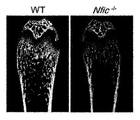
- Figure 1 shows that NFI-C deficiency causes abnormal bone formation during the postnatal bone formation process:
- NFI-C expression in BMSCs from older mice was evaluated by real-time PCR analysis.
- n 3; * P ⁇ 0.05 .;
- MC3T3-E1 cells were incubated for up to 3 weeks in differentiation medium.
- Western blot analysis was used to assess NFI-C, Runx2 and Osx;
- C representative micro-CT images of the mandible;
- D representative micro-CT images of distal femur;
- E 3D micro-CT images of the small and cortical bones distal to the femur;
- F Micro-CT quantification of distal femur.
- Bone mineral density BMD; Small bone volume (BV / TV); Small bone thickness (Tb.Th); Shochu bone number (Tb.N); Osseous bone separation (Tb.SP); Cortical bone volume (Ct. BV / TV); Cortical bone thickness (Ct.Th).
- n 5; * P ⁇ 0.05.
- cf and c'-f 'further enlarged ab and a'-b', respectively.
- FIG. 2 shows that bone marrow fat increases with NFI-C deficiency as seen in osteoporosis patients:
- A H & E staining of femurs of normal and NFIC ⁇ / ⁇ mice at 6 and 28 weeks of age. Scale bar, 50 ⁇ m;
- B Oil Red O Stain. Normal and NFIC -/- BMSCs were incubated for 7 days in adipocyte induction medium. PPAR ⁇ expression in normal and NFIC ⁇ / ⁇ BMSCs was analyzed using real time PCR.
- n 3; * P ⁇ 0.05;
- C H & E staining of bone samples from osteoporosis patients;
- D IHC staining of bone samples from osteoporosis patients;
- E NFI-C mRNA expression was analyzed using the gene expression dataset GSE35959 deposited in GEO. * P ⁇ 0.05;
- Figure 3 shows that NFI-C promotes osteoblast differentiation and inhibits adipocyte differentiation:
- A 7 days in bone formation induction medium after infection with NFIC -/- BMSCs with NFI-C expression vector (ALP staining) ) And 14 days (Alizarin Red S staining).
- B Alp expression was analyzed using real time PCR.
- n 3; * P ⁇ 0.05;
- C Oil Red O Stain.
- NFIC -/- BMSCs were infected with NFI-C expression vectors and then cultured in adipocyte induction media for 7 days;
- D Ppar ⁇ expression was analyzed using real time PCR.
- n 3; * P ⁇ 0.05;
- E Ppar ⁇ promoter activity was assessed in C2C12 cells infected for 48 hours with pGL3-Luc-Ppar ⁇ and NFI-C expression vectors.
- n 3; * P ⁇ 0.05;
- G Histological analysis of distal femur of NFIC -/- mice implanted with NFI-C overexpressing BMSCs.
- n 3; * P ⁇ 0.05;
- D TRAP staining of normal and NFIC -/- mouse femurs at 6 weeks of age.
- the right panel is a further enlargement of the boxed left panel. Scale bar, right panel, 200 ⁇ m; Left panel, 100 ⁇ m;
- E Histomorphologic analysis of normal and NFIC -/- mouse femoral stems at 6 weeks of age. Osteoclasts per tissue area (N.Oc / T.Ar); Osteoclast number per bone parameter (N.Oc / B.Pm); Osteoclast surface area per bone surface area (Oc.S / BS).
- n 5; * P ⁇ 0.05.
- NFI-C mediates BMP2-Runx2-induced Osx expression:
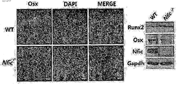
- A Immunofluorescence staining of Osx in the osseous bone of normal and NFIC -/- mouse femoral metaphyseals at 6 weeks of age (red color) ). Scale bar, 100 ⁇ .
- B Real-time PCR and Western blot analysis to assess Osx expression in MC3T3-E1 cells transfected with NFI-C and Runx2 expression vectors and treated with BMP-2 (300 ng / ml) for 48 hours.
- n 3; * P ⁇ 0.05; (C) Osx promoter activity in MC3T3-E1 cells transfected with pGL3-Luc-Osx (-1269- + 91) and NFI-C expression vectors and treated with BMP-2 (300 ng / ml) for 48 hours. Evaluated.
- n 3; * P ⁇ 0.05;
- D detection of NFI-C bound to Osx promoter using ChIP (chromatin immunoprecipitation) assay in MC3T3-E1 cells treated with BMP-2 (300 ng / ml) for 48 hours;
- E NFIC -/- osteoblasts were treated with BMP-2 (300 ng / ml) and / or infected with NFI-C expression vectors. Osx expression was analyzed using real time PCR.
- n 3; * P ⁇ 0.05;
- F Representative micro-CT images and micro-CT quantification of distal femur. Small bone volume (BV / TV); Shochu bone marrow (Tb.N); Shochu bone separation (Tb.SP).
- n 3; * P ⁇ 0.05;
- G Histological analysis of distal femur of NFIC -/- mice implanted with Osx overexpressing BMSCs. Scale bar, 50 ⁇ m;
- H Adipocyte counts in NFIC ⁇ / ⁇ mice implanted with Osx overexpressing BMSCs, compared to NFIC ⁇ / ⁇ mice.
- n 3; * P ⁇ 0.05.
- Runx2 mediates BMP-2 induced NFI-C expression
- C promoter activity and mRNA expression were measured using promoter analysis and real time PCR.
- n 3; * P ⁇ 0.05;
- C Western blot analysis to assess NFI-C expression in MC3T3-E1 cells transfected with Runx2 expression vector and treated with BMP-2 (300 ng / ml) for 48 hours;
- NFI-C and Runx2 expression in control and Runx2 inactivated MC3T3-E1 cells were analyzed using Western blot;
- E Runx2 bound to the NFI-C promoter in MC3T3-E1 cells treated with BMP-2 (300 ng / ml) for 48 hours was detected using ChIP analysis;
- F Immunofluorescence staining (red) of NFI-C in the femur obtained from normal and Runx2 ⁇ / ⁇ mice of E18.5. Scale bar, 100 ⁇ .
- Total RNA and protein isolated from calvarial bones of normal and Runx2 -/- mice. Runx2 and NFI-C expression were evaluated using real time PCR and Western blot analysis. n 3; * P ⁇ 0.05;
- G Model of NFI-C role in Osx expression induced by BMP2-Runx2 during osteogenic differentiation.
- Figure 7 is a NFIC P16 - / - shows that the decreased amount of marrow from mice shochu;
- A normal and NFIC of P16 and 6 weeks of age - / - is a measure of the weight of the mice. The mice were fed a soft dough diet starting 3 days before weaning and up to 6 weeks.
- n 10;
- C Micro-CT quantification of distal femur.
- Bone mineral concentration BMD
- Small bone volume BV / TV
- Shochu bone marrow Tb.N
- Osseous bone separation Tb.SP
- Cortical bone volume Ct. BV / TV
- Cortical bone thickness Ct.Th
- n 5; * P ⁇ 0.05
- D H & E staining of normal of P16 and distal femur of NFIC -/- mice.
- the lower panel is an enlargement of the boxed upper panel. Scale bar, top panel, 500 ⁇ m; Bottom panel, 100 ⁇ m
- Osteoblasts per tissue area (N.Ob / T.Ar); Osteoblasts per bone parameter (N.Ob / B.Pm); Osteoblast surface area per bone surface area (Ob.S / BS).
- N 5; * P ⁇ 0.05.
- FIG. 8 shows the effect of NFI-C deficiency during prenatal bone formation
- A Quantitative micro-CT analysis of normal and NFIC ⁇ / ⁇ mice of E18.5. Micro-CT images of mineralized bone (top panel) and bone volume (BV / TV, bottom panel);
- B Skeletal phenotypes of normal and NFIC ⁇ / ⁇ neonatal mice were analyzed using alizarin red and alcian blue staining;
- C H & E staining of the first molar portion of the mandible of the E18.5 normal and NFIC -/- mice.
- N 5; * P ⁇ 0.05; (C) Normal and NFIC -/- BMMs were incubated with M-CSF (60 ng / ml) and RANKL (100 ng / ml) for 3 days, fixed and TRAP stained.
- NFI-C promotes mandibular formation
- the former being representative x-ray and micro-CT images of distal mandibular
- the latter being the micro-CT quantified graph, where the vertical axis is the small bone volume (BV / TV )to be.
- the present application is based on the finding that NFI-C promotes the differentiation of mesenchymal stem cells into osteoblasts and inhibits the differentiation into adipocytes and ultimately increases bone formation.
- the present invention relates to a pharmaceutical composition for preventing or treating bone disease, comprising a Nuclear Factor I-C (NFI-C) protein or a gene thereof as an active ingredient.
- NFI-C Nuclear Factor I-C
- NFI-C which is included as an active ingredient in the composition of the present application, is a CCAAT-binding transcription factor of promoters belonging to the CTF / NF-I family, which is known to function as a replication factor in DNA replication, but prevents or treats bone diseases. None is known about.
- NFI-C is damaged osteoblast differentiation and bone formation, the bone marrow fat cells increase, can be usefully used for the prevention or treatment of bone diseases.
- Runx2 increases NFI-C expression
- NFI-C increases osteoblast differentiation by increasing Osteirx expression, a transcription factor that regulates osteoblast differentiation, and also adipocyte differentiation. It inhibits the expression of PPAR ⁇ , a regulatory transcription factor, thereby inhibiting adipocyte differentiation, resulting in increased bone formation, which can be used for the prevention or treatment of bone diseases.
- NFI-C genes and / or proteins may be used so long as the effects of the present invention are achieved.
- Genes in which part of the nucleotide sequence is artificially modified, and genes in which a part of the nucleotide sequence is naturally modified, or fragments thereof, are advantageous, as well as wild type sequences, such as characteristics such as expression in cells or stability of proteins. It is to include. Modification of a gene sequence may or may not involve a modification of the corresponding amino acid, where in the case of an amino acid modification, the gene that caused the modification may be substituted, deleted, added, or substituted with one or more amino acids in the protein encoded by it.
- a protein consisting of an inserted amino acid sequence, comprising mutants, derivatives, alleles, variants and homologues. If the mutation of the gene sequence does not involve modification of amino acids in the protein, for example, there are degeneracy mutations, and such degeneracy mutants are also included in the gene of the present invention.
- the gene described above may be provided in the form of a vector operably linked to a promoter to enable expression in the cells of the subject to which the pharmaceutical composition is administered.
- Proteins as used herein can be prepared using methods known in the art.
- the method for producing a protein is by using a genetic recombination technique.
- a vector containing the corresponding gene encoding the protein can be delivered to prokaryotic or eukaryotic cells, for example insect cells, mammalian cells, expressed, and purified.
- the plasmid can be used, for example, by cloning a gene of interest in an expression vector such as pET28b (Novagen), transferring the cell to a cell line, and purifying the expressed protein, but are not limited thereto.
- Synthesized proteins are column chromatography, including precipitation, dialysis, ion exchange chromatography, gel-permeation chromatography, HPLC, reverse phase-HPLC, SDS-PAGE for preparation, affinity columns using anti-screening protein antibodies, and the like. May be separated and purified.
- the NFI-C genes and proteins are of mammalian origin, such as apes, humans, etc., in particular human. NFI-C genes and proteins may also be used as long as the fragment achieves the full length or effects herein, and the full length sequence may be, for example, GenBank access numbers include, but are not limited to, NM_005597 and NP_005588. In one embodiment according to the invention full length NFI-C genes and / or proteins are used.
- bone disease includes both diseases caused by pathological and / or physical bone damage, including, for example, fractures, osteoporosis, osteomalacia, rickets, osteopenia, fibrotic osteoarthritis, aplastic bone disease, bone formation Including but not limited to insufficiency, bone atrophy, Paget's disease, and damage to bone caused by bone metastasis of cancer cells.
- fractures including, for example, fractures, osteoporosis, osteomalacia, rickets, osteopenia, fibrotic osteoarthritis, aplastic bone disease, bone formation Including but not limited to insufficiency, bone atrophy, Paget's disease, and damage to bone caused by bone metastasis of cancer cells.
- osteoporosis including osteoporosis, osteomalacia, rickets, osteopenia, fibrotic osteoarthritis, aplastic bone disease, bone formation Including but not limited to insufficiency, bone atrophy, Paget's disease, and damage
- treatment means any action that ameliorates or beneficially alters the associated symptoms by administration of a composition according to the present application.
- Those skilled in the art to which the present application belongs, will be able to determine the extent to which the composition of the present invention is correct, improved, improved and treated with reference to the data presented by the Korean Medical Association and the like. .
- prevention means any action that inhibits or delays the development of a related disease by administration of a composition according to the present application. It will be apparent to those skilled in the art that the composition of the present invention, which is effective in osteoblast differentiation due to osteoblast differentiation and bone formation damage, can prevent such an early symptom of a disease due to osteogenic abnormality, or when taken before symptoms appear.
- Mesenchymal stem cells of bone marrow can differentiate into various cells such as osteoblasts, adipocytes (Prockop DJ. Science. 1997; 276 (5309): 71-74; Tuli R, Tuli S, Nandi S, Wang ML, Alexander PG, Haleem- Smith H, Hozack WJ, Manner PA, Danielson KG, and Tuan RS Stem Cells. 2003; 21 (6): 681-693). Osteoblast differentiation is regulated by growth factors such as BMP-2, TGF- ⁇ , Wnt and IGF and transcription factors such as Runx2 and Osterix (Osx).
- Adipocyte differentiation is regulated by the peroxisome proliferator-activated receptor gamma (PPAR ⁇ ), a transcription factor.
- PPAR ⁇ peroxisome proliferator-activated receptor gamma
- compositions according to the present disclosure comprising NFI-C, although not limited by this theory, increase the expression of NFI-C by Runx2, which increases the expression of Osteirx, a transcription factor that regulates osteoblast differentiation. It increases osteoblast differentiation and inhibits the expression of PPAR ⁇ , which is a regulatory transcription factor, thereby inhibiting adipocyte differentiation. As a result, bone formation is increased, which can be useful for preventing or treating bone diseases.
- composition of the present application may be prepared as a pharmaceutical composition.
- the pharmaceutical compositions may be administered simultaneously or sequentially and may be used alone or in combination with methods using surgery, other pharmaceutically active ingredients and biological response modifiers for treating the disease.
- compositions according to the invention may be formulated in a suitable form with the pharmaceutically acceptable carriers generally used.
- 'Pharmaceutically acceptable refers to a composition that is physiologically acceptable and does not cause an allergic or similar reaction, such as gastrointestinal disorders, dizziness or the like, when administered to a human.
- pharmaceutically acceptable carriers include, for example, water, suitable oils, saline, carriers for parenteral administration such as aqueous glucose and glycols, and the like, and may further include stabilizers and preservatives. Suitable stabilizers include antioxidants such as sodium hydrogen sulfite, sodium sulfite or ascorbic acid.
- Suitable preservatives include benzalkonium chloride, methyl- or propyl-paraben and chlorobutanol.
- the composition according to the present invention if necessary according to the administration method or dosage form, suspensions, dissolution aids, stabilizers, isotonic agents, preservatives, adsorption agents, surfactants, diluents, excipients, pH adjusters, analgesics, buffers, Antioxidant etc. can be contained suitably.
- Pharmaceutically acceptable carriers and formulations suitable for the present invention including those exemplified above, are described in detail in Remington's Pharmaceutical Sciences, latest edition.
- compositions herein may be prepared in unit dose form by formulating with a pharmaceutically acceptable carrier and / or excipient, according to methods readily available to those of ordinary skill in the art. It can be prepared by incorporation into a dose container. The formulations can then be in the form of solutions, suspensions or emulsions in oil or aqueous media or in the form of powders, granules, tablets or capsules.
- the method of administering the pharmaceutical composition of the present application may be easily selected according to the formulation, and may be administered to mammals such as domestic animals and humans by various routes.
- it may be formulated in the form of powders, tablets, pills, granules, dragees, hard or soft capsules, liquids, emulsions, suspensions, syrups, elixirs, external preparations, suppositories, sterile injectable solutions, and so on.
- Oral or parenteral administration, in particular parenteral administration may be preferred.
- Formulations for parenteral administration include sterile aqueous solutions, non-aqueous solvents, suspensions, emulsions, lyophilized preparations, suppositories.
- non-aqueous solvent and the suspension solvent propylene glycol, polyethylene glycol, vegetable oil such as olive oil, injectable ester such as ethyl oleate, and the like can be used.
- base of the suppository witepsol, macrogol, tween 61, cacao butter, laurin butter, glycerol, gelatin and the like can be used.
- compositions herein can be administered orally or parenterally (eg, applied intravenously, subcutaneously, intraperitoneally or topically) according to the desired method, with parenteral administration being particularly preferred.
- the dosage depends on the condition and weight of the patient, the extent of the disease, the form of the drug, the route of administration and the time of day, and may be appropriately selected by those skilled in the art.
- the composition is administered topically around the bone marrow cavity and around the bones that are physically and / or pathologically damaged.
- composition according to the invention is administered in a pharmaceutically effective amount.
- pharmaceutically or therapeutically effective amount means an amount sufficient to treat a disease at a reasonable benefit / risk ratio applicable to medical treatment, and an effective dose level means the type, severity, It can be determined according to the activity of the drug, sensitivity to the drug, the time of administration, the route of administration and the rate of release, the duration of treatment, factors including the concurrent drug and other factors well known in the medical field.
- the compositions of the present invention may be administered as individual therapeutic agents or in combination with other therapeutic agents, may be administered sequentially or simultaneously with conventional therapeutic agents, and may be single or multiple doses. Taking all of the above factors into consideration, it is important to administer an amount that can obtain the maximum effect in a minimum amount without side effects, which can be easily determined by those skilled in the art.
- the dosage of the pharmaceutical composition of the present application may vary depending on the weight, age, sex, health condition, diet, time of administration, administration method, excretion rate and severity of the disease, etc. 60 kg), about 1 ng to 10 mg / day, in particular about 1 ⁇ g to 1 mg / day. Since the dosage may vary depending on various conditions, it will be apparent to those skilled in the art that the dosage may be added or subtracted, and thus the dosage does not limit the scope of the present invention in any aspect.
- the frequency of administration can be administered once a day or divided into several times within the desired range, the administration period is not particularly limited.
- the present disclosure also relates to mesenchymal stem cells overexpressing NFI-C; Progenitor cells derived from the mesenchymal stem cells, which can be differentiated into chondrocytes or osteocytes; Or it relates to a composition for treating bone diseases or cell therapy comprising chondrocytes or osteoblasts differentiated from the mesenchymal stem cells as an active ingredient.
- cell therapy refers to the proliferation or selection of live autologous, allogenic, xenogenic cells in vitro to alter the function of cells and tissues, or to alter the biological characteristics of the cells by other means. Medicines used for the purpose of treatment, diagnosis and prevention through a series of methods. Since 1993, the United States has managed cell therapy as a medicine since 2002. Such cell therapies include, but are not limited to, stem cell therapies for tissue regeneration or organ function recovery.
- mesenchymal stem cells 'are stem cells that are separated from umbilical cord blood, fat, bone marrow, blood, dermis or periosteum, and include various cells such as adipocytes, chondrocytes and bone cells. By pluripotent or multipotent cells capable of differentiation.
- mesenchymal stem cells are characterized by being able to effectively engraft in allogeneic or heterologous recipients without the use of immunosuppressive agents.
- the cells used in the therapeutic compositions herein are autologous, allogenic, xenogenic cells, in particular autologous cells.
- the mesenchymal stem cells included in the cell therapy of the present application may be mesenchymal stem cells of an animal, preferably may be mesenchymal stem cells of a mammal, more preferably human.
- the mesenchymal stem cells of the present invention may be derived from bone marrow, adipose tissue, peripheral blood, liver.
- cells that may be included in the cell therapy herein include progenitor cells that may differentiate into chondrocytes or osteocytes, derived from mesenchymal stem cells, for example immature bone precursor cells, osteoblasts, Includes chondrocytes.
- the cells that may be included in the cell therapy herein include chondrocytes or osteocytes differentiated from mesenchymal stem cells.
- Mesenchymal stem cells are present in very small amounts, such as bone marrow, but the process of isolating and culturing them is well known in the art and can be referred to, for example, US Pat. No. 5,486,359.
- the mesenchymal stem cells of the present application can be obtained by separating from the hematopoietic stem cells of the bone marrow according to a known method and then proliferating without losing differentiation ability.
- mesenchymal stem cells of a mammal preferably human, including human or mouse mesenchymal stem cells, such as blood or bone marrow Separating the stem cells (the bone marrow can be extracted from the tibia, femur, spinal cord or iliac bone);
- the isolated cells may be obtained by culturing the cells in a suitable medium, and removing the floating cells in the culture process, and subcultured the cells attached to the culture plate to obtain finally constructed mesenchymal stem cells.
- the medium used in the above process any medium commonly used for culturing stem cells can be used.
- the medium contains serum (eg fetal calf serum, horse serum and human serum).
- serum eg fetal calf serum, horse serum and human serum.
- Mediums that can be used in the present invention are, for example, RPMI series, Eagles' MEM (Eagle's minimum essential medium, Eagle, H. Science 130: 432 (1959)), ⁇ -MEM (Stanner, CP et al., Nat New Biol. 230: 52 (1971)), Iscove's MEM (Iscove, N. et al., J. Exp. Med. 147: 923 (1978)), 199 medium (Morgan et al., Proc. Soc.Exp.
- the medium may include other components such as antibiotics or antifungal agents (eg, penicillin, streptomycin), glutamine, and the like.
- identification of mesenchymal stem cells can be made through flow cytometry.
- flow cytometry is performed using specific surface markers of mesenchymal stem cells.
- mesenchymal stem cells have a positive response to CD44, CD29 and / or MHC class I, thereby identifying mesenchymal stem cells.
- NFI-C Cells included in the cell therapy according to the present invention overexpress NFI-C.
- Methods of overexpressing proteins in cells are known.
- the NFI-C gene is cloned into an appropriate eukaryotic expression vector such as phage, virus or retrovirus-derived vector or plasmid so as to be operably linked to the expression promoter, constructing the expression vector and then introducing it into cells according to the present application. can do.
- Methods of introduction are known and include, but are not limited to, for example, calcium phosphate, DEAE-dextran mediated transduction, positively charged lipid mediated transduction, electroporation, transduction using phage systems, or infection with viruses, and the like. It is not.
- the route of administration of the cell therapeutic agent or pharmaceutical composition comprising the cells according to the present application can be administered via any general route as long as it can reach the desired tissue.
- Parenteral administration for example, intraperitoneal administration, intravenous administration, intramuscular administration, subcutaneous administration, intradermal administration may be, but is not limited thereto.
- Therapeutic agents or compositions according to the invention may be formulated in a suitable form with the pharmaceutically acceptable carriers generally used.
- Pharmaceutically acceptable carriers include, for example, water, suitable oils, saline, carriers for parenteral administration such as aqueous glucose and glycols, and the like, and may further include stabilizers and preservatives.
- Suitable stabilizers include antioxidants such as sodium hydrogen sulfite, sodium sulfite or ascorbic acid.
- Suitable preservatives include benzalkonium chloride, methyl- or propyl-paraben and chlorobutanol.
- composition for cell therapy according to the present invention if necessary according to the administration method or dosage form, suspensions, dissolution aids, stabilizers, tonicity agents, preservatives, adsorption inhibitors, surfactants, diluents, excipients, pH adjusters, analgesics , Buffers, antioxidants, and the like may be appropriately included.
- Pharmaceutically acceptable carriers and formulations suitable for the present invention including those exemplified above, are described in detail in Remington's Pharmaceutical Sciences, latest edition.
- the cell therapy composition of the present invention is in unit dose form by formulating with a pharmaceutically acceptable carrier and / or excipient according to a method which can be easily carried out by those skilled in the art. It can be made or prepared by incorporating into a multi-dose container.
- the composition may also be administered by any device in which the cell therapy agent can migrate to the target cell.
- the cell therapy composition of the present invention may include a therapeutically effective amount of cell therapy for the treatment of a disease.
- the term 'therapeutically effective amount' means an amount of an active ingredient or pharmaceutical composition that induces a biological or medical response in a tissue system, animal or human, as thought by a researcher, veterinarian, physician or other clinical person, Amounts that induce alleviation of the symptoms of the disease or disorder. It will be apparent to those skilled in the art that the cell therapy agent included in the composition of the present invention will vary depending on the desired effect.
- the optimal cell therapy content can be readily determined by one skilled in the art and includes the type of disease, the severity of the disease, the amount of other components contained in the composition, the type of formulation, and the age, weight, general health, sex and diet of the patient. It can be adjusted according to various factors including the time of administration, the route of administration and the rate of secretion of the composition, the duration of treatment, and the drugs used simultaneously.
- the cell therapy agent is administered topically to the bone marrow cavity and around the bone which is physically and / or pathologically damaged.
- the dosage of the composition of the present invention is 1.0 ⁇ 10 7 to 1.0 ⁇ 10 8 cells / kg (body weight), more preferably 1.0 ⁇ 10 5 to 1.0 ⁇ based on osteoblasts differentiated from stem cells as active ingredients. 10 8 cells / kg body weight.
- the dosage may be variously prescribed by such factors as the formulation method, the mode of administration, the age, weight, sex, morbidity, food, time of administration, route of administration, rate of excretion, and reaction sensitivity of the patient. These factors can be taken into account to properly adjust the dosage.
- the number of administrations may be one or two or more times within the range of clinically acceptable side effects, and may be administered to one or more than two sites for the administration site.
- the same dosage as that of humans per kg or the volume ratio (for example, average value) of ischemic organs (heart, etc.) between the animal of interest and humans can be adjusted.
- Equivalent amounts may be administered.
- the animal to be treated according to the present invention include humans and mammals for other purposes, and specifically include humans, monkeys, mice, rats, rabbits, sheep, cattle, dogs, horses, pigs, and the like. do.
- NFI-C promotes the differentiation of adipocytes and differentiation of osteoblasts, can be used for various uses that require the inhibition of adipocyte differentiation, or promote the differentiation of osteoblasts.
- the present application may also be provided as an adipocyte differentiation inhibitor or osteoblast differentiation promoting composition comprising an NFI-C protein or a gene thereof as an active ingredient.
- human-derived recombinant BMP-2 (Bone Morphogenic Protein-2) was purchased from R & D Systems.
- NFI-C antibodies were synthesized in rabbits by synthesizing the (amino acid residue NH2-RPTRPLQTVPLWD-COOH) peptide.
- Runx2 (sc-398), PCNA (sc-400), and Osx (sc-22538) antibodies were purchased from Santa Cruz Biotechnology.
- NFIC -/- mice were made by mating female and male NFIC +/- mice.
- Runx2 -/- mice were It was provided by Toshihisa Komori (Nagasaki University graduate School of Biomedical Sciences, Nagasaki, Japan).
- mice At 6 weeks of age, the maxilla, mandible, and femur of normal and NFIC -/- mice were isolated and fixed in 4% paraformaldehyde overnight at 4 ° C. Micro-CT was analyzed with a Skyscan scanner and software. The isotope resolution of the machine was 10 ⁇ m. Six and six weeks old mice were given 10 mg / kg of calcein seven and two days before killing. Histological analysis was performed using OsteoMeasure histomorphometry system (OsteoMetrics) according to the manufacturer's method.
- OsteoMeasure histomorphometry system OsteoMeasure histomorphometry system (OsteoMetrics) according to the manufacturer's method.
- mice The femurs of the mice were fixed at 4 ° C. overnight in 4% paraformaldehyde.
- the non-limeous femur was embedded in destabilized methyl methacrylate resin, cut the femur to a thickness of 5 ⁇ m, and von Kossa staining was performed.
- Demineralized femur specimens were demineralized in a 10% ethylene diamine tetra-acetic acid (EDTA) -1% paraformaldehyde (pH 7.4) solution for 2 weeks, washed, dehydrated, and then paraffinized.
- EDTA ethylene diamine tetra-acetic acid
- Hematoxylin-eosin staining, immunohistochemical staining, and TRAP staining for osteoclasts were performed by embedding and cutting into 5 thicknesses. TRAP staining was performed according to the manufacturer's instructions using a kit (sigma).
- Bone tissue slides of osteoporosis patients were provided by Professor Yang Young-il of Inje University Caucasian Medicine Memorial Institute, and hematoxylin-eosin staining and NFI-C immunochemical tissue staining were performed.
- mice Newborn mice (P0) were subjected to Alcyan blue for cartilage staining and alizarin red S stain for bone staining. Mice were fixed for 2 hours in 95% ethanol and skin and muscles removed. The mice's skeletons were then re-fixed in 95% ethanol for 5 days and then fixed in acetone for 2 days. Next, the mice skeleton was stained for 3 days in a solution containing 0.015% Alcian blue and 0.005% Alizarin Red S in 5% glacial acetic acid and 70% ethanol. The stained samples were washed in 1% KOH solution and then stored in 100% glycerin.
- pCH-NFI-C expression plasmid pSuper and retro-NFI-C siRNA plasmid was previously reported (Lee DS, Yoon WJ, Cho ES, Kim HJ,, Gronostajski RM, ChoM-I, and Park JC. PLoS One 2011, 6 (12): e29160, 2011).
- Runx2, Dlx5, Msx2, and Ppar ⁇ promoter plasmids were provided by Professor Hyun-Ryu Ryu at Seoul National University.
- NFI-C promoters (-2520 ⁇ + 88) amplified 2.6-kb genome fragments by PCR from genomic DNA of mice, and Osx promoters (-1269 ⁇ + 91) were also subjected to PCR from genomic DNA of mice by 1.36. -kb genome sections were amplified.
- GenBank accession numbers of nucleotide sequences of NFI-C and Osx promoters are as follows. NFI-C, NC000076. Osx, DQ229136. PCR was performed using genomic DNA of mice (1 ⁇ l) for amplification of each promoter. The reaction conditions and primer sequences were as follows.
- PCR reaction conditions 94 ° C., 1 min; 60 ° C., 1 min; 72 ° C., 1 min 35 cycles in total.
- Primer sequences NFI-C Fw 5'-CTC GAG AGT GAA TGG ATT GGG GGT GT-3 ', NFI-C Rv 5'-AAG CTT CAG AGC GGG GAG GAA TAC AT-3', Osx Fw 5'-CTC GAG CAC ACA TAC ACG AAC ACA CAT-3 ′, and Osx Rv 5′-AAG CTT TGG GGA CCG GGT CCC AAG GAG-3 ′.
- Amplified genomic DNA fragments were inserted into the PCRR ® 2.1 T vector and then digested with restriction enzymes XhoI and Hind III and inserted into the pGL3 luciferase (LUC) expression vector (Promega).
- Mesenchymal stem cells were isolated from the femurs of mice at 6 weeks of age using a 1 ml syringe. The isolated cells were removed by erythrocyte depletion solution, and then in ⁇ -MEM medium supplemented with 100 IU / ml penicillin, 100 ⁇ g / ml streptomycin (Gibco BRL), and 10% fetal bovine serum in a 100 mm petri dish. Incubated. Primary calvarial osteoblasts from mice were isolated from normal and NFIC ⁇ / ⁇ mice at 5 days of age. After cutting the cranial bones, the skin tissue was removed, chopped and washed three times with PBS.
- Osteoblast differentiation consists of ⁇ -MEM supplemented with 5% fetal bovine serum, ascorbic acid (50 ⁇ g / ml), and beta-glycerophosphate (10 mM) when 80-90% of the cells are grown in a culture dish. Osteoblast differentiation was induced by incubation for 2 weeks in the medium.
- Adipocytes are 10% fetal bovine serum, insulin (10 ⁇ g / ml), dexamethasone (1 ⁇ M), and 3-isobutyl-1-methylxanthine (IBMX, 0.5 mM) if the cells grow 80-90% in a petri dish. Adipocyte differentiation was induced by culturing for 7 days in the DMEM medium.
- MC3T3-E1 cells mouse osteoblasts, were cultured in a medium containing 10% fetal calf serum and antibiotics in ⁇ -MEM.
- C2C12 cells of mouse muscle cells were cultured in DMEM medium supplemented with 100 IU / ml penicillin, 100 ⁇ g / ml streptomycin (Gibco BRL), and 10% fetal calf serum.
- Expression plasmids (2 ⁇ g) described in FIGS. 3, 5, 6, 8 and 10 were transfected with MC3T3-E1 and C2C12 cells using Lipofectamine Plus TM reagent (Invitrogen). It was.
- Human-derived mesenchymal stem cells were purchased from Calbrex and cultured in low glucose DMEM medium supplemented with 100 IU / ml penicillin, 100 ⁇ g / ml streptomycin (Gibco BRL), and 10% fetal calf serum.
- NFIC -/- mesenchymal stem cells were isolated from the femur of 6 weeks old NFIC -/- mice using a 1 ml syringe.
- NFI-C overexpressing retrovirus was added and cultured for 24 hours.
- NFI-C-overexpressing mesenchymal stem cells were injected into the bone marrow cavity of the left femur of mice using a Hamilton syringe (1 ⁇ 10 6 cells / femur in 10 ⁇ l of ⁇ -MEM).
- the femur was separated and subjected to micro-CT and histological analysis.
- Bone marrow cells were cultured in ⁇ -MEM medium containing 10% fetal calf serum and antibiotics, 30 ng / ml M-CSF (R & D Systems) for 1 day to obtain bone marrow monocytes (BMMs).
- BMMs (5 ⁇ 10 4 cells / well) were cultured in ⁇ -MEM medium containing M-CSF (60 ng / ml) and RANKL (100 ng / ml) for 3 days to induce osteoclast differentiation.
- BMMs (5 ⁇ 10 5 cells / well) and calvarial osteoblasts (2.5 ⁇ 10 4 cells / well) were put together in a 48-well plate with 10% fetal bovine serum, 10 -8 M 1,25 Osteoclast differentiation was induced by culturing for 6 days in ⁇ -MEM medium containing -dihydroxyvitamin D3 (Vit D 3 ), and 10 -6 M prostaglandin E2 (PGE 2 ). The identification of osteoclasts was analyzed using TRAP staining kit (Sigma).
- Luciferase assay plated MC3T3-E1 or C2C12 cells at 5 ⁇ 10 4 cells / well in 24-well plates and transformed with Lipofectamine Plus TM reagent after 24 hours.
- pGL3basic control
- pGL3 Ppar ⁇ promoter -1000 to +1
- pGL3 Osx promoter -1269 to +91
- pGL3 NFIC promoter 2520 to +88
- pCMV empty pCMV empty
- Runx2 siRNA and control siRNA were purchased from Dharmacon. Transformed cells were measured luciferase activity after 48 hours.
- CDNA was synthesized using 2 ⁇ g of total RNA and 1 ⁇ l of reverse transcriptase (Superscript III enzyme (Invitrogen)) and 0.5 ⁇ g of oligo (dT).
- the synthesized cDNA was used for real time polymerase chain reaction.
- Real-time polymerase chain reaction was performed in ABI PRISM 7500 sequence detection system (Applied Biosystems) using SYBR GREEN PCR Master Mix (Takara, Japan).
- Real-time polymerase chain reaction was performed 40 minutes at 1 minute at 94 °C, then 9 °C 15 seconds, 60 °C 34 seconds conditions. Analysis of the results was evaluated using a CT (comparative cycle threshold, provided with ABI PRISM 7500 instrument) method, primer sequences are listed in Table 1.
- lysis buffer 100 mM Tris, pH 7.4, 350 mM NaCl, 10% glycerol, 1% Nonidet P-40, 1 mM EDTA, 1 mM dithiothreitol, 1x protease inhibitor. (SigmaAldrich, St. Louis, Mo, USA) and reacted on ice for 15 minutes. Cell debris was removed by centrifugation at 16,000 x g for 15 minutes at low temperature and only supernatant was taken.
- MC3T3-E1 cells were cultured by adding BMP-2 (300 ng) for 48 hours. Then, the cells were washed with PBS and collected and crushed to about 1,000 bp of chromatin by using an ultrasonic device. Chromatin immunoprecipitation was performed by adding NFI-C antiserum (30 ⁇ l) and Runx2 (10 ⁇ l, Santa Cruz) antiserum for 4 hours at 4 ° C. After collecting DNA pellets, each protein was transferred to the promoter region using PCR. Check that it is bound to. PCR primer sequences are shown in Table 2. PCR conditions were as follows: 94 ° C. for 30 s; 60 ° C. for 30 s; And 72 ° C. for 30s (total 35 cycles). PCR products were electrophoresed on 2% agarose gel, stained with ethidium bromide, and identified under UV light.
- Platinum-E cells were transfected with pMxs NFI-C, Osx, and control vectors using Lipofectamine Plus TM reagent (Invitrogen). After 48 hours, the virus-containing medium was collected, centrifuged, filtered with a 0.45- ⁇ m syringe filter, and stored at -80 ° C.
- Gene profiling was analyzed using an mRNA microarray database using mesenchymal stem cells from normal and osteoporosis patients.
- Microarray data was downloaded from NCBI's GEO database (accession number GSE35959), and NFI-C mRNA expression was analyzed using the probe (Probeset ID 213198_at).
- NFIC -/- mesenchymal stem cells were isolated from the femur of 6 weeks old NFIC -/- mice using a 1 ml syringe.
- NFI-C overexpressing retrovirus was added and cultured for 24 hours.
- NFI-C-overexpressing mesenchymal stem cells were injected into the left mandible of mice (1 ⁇ 10 6 cells / mandible in 10 ⁇ l ⁇ -MEM) using a Hamilton syringe. After 4 weeks of implantation, the mandible was separated and subjected to X-ray and micro-CT analysis.
- NFI-C mRNA expression in bone marrow stromal cells obtained from 60 weeks old mice from day 18.5 (E18.5) was examined. Analyzed. From 16 to 6 weeks after birth, NFI-C expression was significantly increased, followed by week 60 (FIG. 1A).
- NFI-C was expressed from day 0 of culture, increased expression from day 4 to day 10 (early osteoblast differentiation) and decreased from day 10 to day 21 (late osteoblast differentiation and petrification). Action).
- Runx2 expression increased from 4 to 7 days and then gradually decreased from 10 to 21 days, corresponding to late osteoblast differentiation and petrification, peaking Osx expression at this time during incubation (FIG. 1B).
- NFIC -/- mice had a normal lifespan, but their growth was slowed due to the slow development of tooth roots, which resulted in a lack of the ability to consume hard normal rodent feed (16). Therefore, in order to alleviate eating disorders in NFIC -/- mice, soft dough dietary supplements were added from 3 days before weaning and continued for 6 weeks. Body weights of normal (WT) mice and NFIC -/- mice were measured. The total body weight difference between normal (WT) mice and NFIC ⁇ / ⁇ mice at 16 and 6 weeks was not significant (FIG. 7A).
- osteoblast function was measured by histological analysis of the femurs of 6-week-old mice. Based on histological analysis and present Cosa staining, the spongy and cortical bone volume of NFIC ⁇ / ⁇ mice was significantly reduced compared to normal mice (FIG. 1G). Osteoblasts per tissue (N.Ob / T.Ar), osteoblasts per bone parameter (N.Ob / B.Pm), and osteoblast surface per bone surface (Ob) in NFIC -/- mice compared to normal mice. S / BS) was significantly reduced (FIG. 1H).
- NFIC ⁇ / ⁇ mice at day P16 also showed similar decreases in the parameters of osteoblast activity than 6 week old mice (FIGS. 7D and E).
- the distance of two calcein labeling of NFIC -/- mice was reduced compared to normal mice, which resulted in a significant decrease in mineral deposition rate (MAR) in NFIC -/- mice (approximately -30%) (FIG. 1I).
- MAR mineral deposition rate
- H & E staining was used to measure bone marrow fat cells in the femur of NFIC -/- mice.
- a large amount of fat cells accumulated, which increased with age (FIG. 2A).
- the effect of NFI-C deficiency on adipocyte differentiation in bone marrow stromal cells was assessed using oil red O staining.
- Oil red O-positive cells increased sharply in NFIC ⁇ / ⁇ myeloid matrix cells compared to normal myeloid matrix cells of control and adipocyte differentiation cultures (FIG. 2B, left panel).
- the expression of adipocyte differentiation marker Ppar ⁇ was significantly increased in NFIC ⁇ / ⁇ myeloid matrix cells (FIG. 2B, right panel).
- NFI-C is a potent candidate gene associated with osteoporosis with increasing age.
- NFIC - / - in order to study the effect of over-expression of the NFIC BMSCs, NFIC - / - BMSCs in the NFIC expression plasmid transient transfection delivered in osteogenic differentiation medium 14 days or local taxes saturated differentiation medium after the user 7 Cells were cultured daily. Osteoblast and adipocyte differentiation were assessed by real-time PCR, alkaline phosphatase staining, alizarin red S mineralization and oil red O staining. When NFI-C was overexpressed, ALP activity and mineralization nodules increased in NFIC ⁇ / ⁇ BMSCs (FIG. 3A).
- NFI-C Alp mRNA expression also increased as NFI-C overexpressed in NFIC ⁇ / ⁇ BMSCs.
- overexpression of NFI-C reduced the number of oil red O-positive adipocytes in NFIC -/- BMSCs.
- Ppar ⁇ expression was also NFIC ⁇ / ⁇
- NFI-C-overexpressing cells had a Ppar ⁇ promoter activity that was approximately 20-fold reduced compared to control cells (FIG. 3E).
- NFIC -/- When NFI-C is expressed in BMSCs, NFIC - /- To determine if mice can relieve osteoporosis-like phenotype, NFIC -/- Overexpresses NFI-C in BMSCs and expresses these cells with NFIC -/- Mice were implanted into the femoral bone marrow. Femur micro-CT image shows NFIC implanted with NFI-C -overexpressed BMSCs -/- NFIC -/-in mice Compared with the mice, there was a marked increase in bone alcohol volume (FIG. 3F, left panel).
- NFIC - the overexpressed BMSCs implanted NFIC - / - Mice showed marked increases in bone volume (BV / TV) and bone number (Tb. N), but bone loss was significantly reduced (FIG. 3F, right panel).
- transplanted BMSCs were analyzed by IHC using NFI-C antibodies. NFI-C-positive cells were observed along the bone trabecular bone surface, indicating that osteoblasts were differentiated in transplanted BMSCs (FIG. 3G).
- Histological analysis shows that NFIC -/- In comparison with mice, the implantation of NFI-C-overexpressing BMSCs increased bone mineral density (FIG.
- mice osteoclasts per tissue area (N.Oc / T.Ar), osteoclasts per bone circumference (N.Oc / BPM) and osteoclasts per bone surface area (Oc.S / BS) were significantly reduced.
- BMMs normal and NFIC -/- Macrophage (BMMs) derived from cultured bone marrow obtained from mice were treated with macrophage colony-stimulating factor (M-CSF) and receptor activator (RANKL) of NF- ⁇ B ligand to induce osteoclasts, which were then TRAP stained. Evaluated.
- M-CSF macrophage colony-stimulating factor
- RNKL receptor activator
- the formation of multinuclear TRAP-positive osteoclasts was identical for both types of mice (FIG. 9C), meaning that osteoclasts were not directly affected by NFI-C deficiency.
- NFIC -/- If according to a rat osteoblast differentiation damage, under normal vitamin D3 (Vt D 3) and prostaglandin E2 (PGE 2) in the presence or NFIC - / - Calvarial osteoblasts were co-cultured with normal BMMs and analyzed for osteoclasts obtained by TRAP staining.
- NFIC -/- normal BMMs compared to normal osteoblasts Coculture with osteoblasts significantly reduced the number of TRAP-positive multinucleated cells (FIG. 4, F and G).
- NFIC -/- Osteoblasts from mice resulted in less osteoclast formation, resulting in decreased TRAP activity (FIG. 4H).
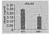
- NFIC -/- Soluble Rankl (sRankl) expression was also decreased in the osteoblast coculture group (FIG. 4I).
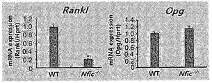
- Real time PCR NFIC -/- Osteoprogenerin ( Opg ) was normally expressed in osteoblasts obtained from mice, but Rankl expression was decreased (FIG. 4J).
- Osx a transcription factor containing zinc fingers
- plays an important role in osteoblast differentiation (Nakashima K, Zhou X, Kunkel G, Zhang Z, Deng JM, Behringer RR, and de Crombrugghe B. Cell. 2002 108 (1): 17-29).
- NFIC NFIC compared to normal BMSCs -/- Osx mRNA expression was reduced in BMSCs (FIG. 4C).
- Western blot analysis to see based on immunofluorescence holding the thigh bone, as compared to normal NFIC - / - Osx protein expression was significantly reduced in osteoblasts (FIG.
- NFIC - / - being Osx is overexpressed in mice after the infection, the retrovirus to find out whether that damaged bone formation to recover them NFIC - / - NFIC through which to implant the femoral space in mice - / - a Osx from BMSCs Overexpressed.
- the decrease in the lumen bone volume was only partially recovered by Osx overexpression (FIG. 5F, left panel). Histomorphologic analysis showed a significant increase in the osseous bone volume (BV / TV) and the osseous bone number (Tb.N) in NFIC -/- mice implanted with Osx - overexpressing BMSCs, but the osseous bone separation (Tb.
- Osteoblastic transcription factors including Runx2, Dlx5, Msx2, and Osx are largely regulated by BMP-2 (Nishimura R, Hata K, Matsubara T, Wakabayashi M, and Yoneda T. J Biochem. 2012; 151 (3) : 247-254).
- BMP-2 significantly increased NFI-C mRNA and protein levels as well as NFI-C promoter activity (FIG. 6, A and C).
- Runx2 stimulated by BMP-2 regulates NFI-C during bone formation was also investigated whether Runx2 stimulated by BMP-2 regulates NFI-C during bone formation.
- Runx2 significantly increased NFI-C promoter activity and NFI-C mRNA and protein expression (FIG. 6, B and C). Knockdown of Runx2 resulted in an approximately 10-fold decrease in NFI-C promoter activity and NFI-C protein expression and inhibited the increase of BMP-2's effect on NFI-C promoter and expression (FIG. 6D).
- BMP-2 was treated to find out whether Runx2 directly binds to the NFI-C promoter portion, followed by ChIP analysis using Runx2 antibodies. In the region between -423 and -246 of the NFI-C promoter, a part assumed to be Runx2 common base was identified.
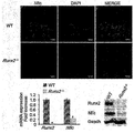
- Runx2 interacted directly with the NFI-C promoter in the presence of BMP-2 (FIG. 6E). NFI-C expression was also examined in Runx2 ⁇ / ⁇ mice. In immunofluorescence studies, NFI-C protein expression was observed but this was drastically reduced in bone and cartilage where Runx2 ⁇ / ⁇ was developing (FIG. 6F, top panel). In real-time PCR and Western blot experiments, NFI-C mRNA and protein expression levels were significantly reduced in Runx2 ⁇ / ⁇ mice compared to normal (FIG. 6F, bottom panel). These results indicate that Runx2 acts upstream of NFI-C and regulates NFI-C expression via the BMP-2 signaling pathway.
- NFIC -/- When NFI-C is expressed in BMSCs, NFIC - /- Overexpresses NFI-C in BMSCs and expresses these cells with NFIC -/- The mouse was implanted into the mandible. The results are described in FIGS. 11A and 11B. As shown in the mandible and a micro X-ray image -CT, NFIC - the overexpressed BMSCs implanted NFIC - / - NFIC -/-in mice Compared with mice, the bone alcohol volume increased (FIG. 11A).
Landscapes
- Health & Medical Sciences (AREA)
- Life Sciences & Earth Sciences (AREA)
- Veterinary Medicine (AREA)
- Animal Behavior & Ethology (AREA)
- General Health & Medical Sciences (AREA)
- Engineering & Computer Science (AREA)
- Public Health (AREA)
- Chemical & Material Sciences (AREA)
- Medicinal Chemistry (AREA)
- Pharmacology & Pharmacy (AREA)
- Bioinformatics & Cheminformatics (AREA)
- Physical Education & Sports Medicine (AREA)
- Epidemiology (AREA)
- Immunology (AREA)
- Environmental Sciences (AREA)
- Orthopedic Medicine & Surgery (AREA)
- Proteomics, Peptides & Aminoacids (AREA)
- Rheumatology (AREA)
- Biotechnology (AREA)
- Zoology (AREA)
- Animal Husbandry (AREA)
- Biodiversity & Conservation Biology (AREA)
- Organic Chemistry (AREA)
- Dermatology (AREA)
- Chemical Kinetics & Catalysis (AREA)
- General Chemical & Material Sciences (AREA)
- Nuclear Medicine, Radiotherapy & Molecular Imaging (AREA)
- Gastroenterology & Hepatology (AREA)
- Genetics & Genomics (AREA)
- Molecular Biology (AREA)
- Medicines Containing Material From Animals Or Micro-Organisms (AREA)
- Medicines That Contain Protein Lipid Enzymes And Other Medicines (AREA)
Abstract
본원은 NFI-C (Nuclear factor I-C) 단백질 또는 그 유전자를 유효성분으로 포함하는, 골질환 예방 또는 치료용 약학 조성물 또는 상기 유전자를 과발현하는 세포 치료제를 개시한다. 본원에 따른 조성물 또는 세포 치료제는 골모세포 분화를 촉진함과 동시에 지방세포의 분화 억제를 통해 골감소를 억제할 수 있는 골다공증을 포함하는 골질환 치료 또는 예방에 유용하게 사용될 수 있다.
Description
본 발명은 골질환 치료제 분야이다.
골다공증은 일반적으로 골 밀도가 감소하고 골 조직의 미세구조에 문제가 발생하여 쉽게 부러지는 특성을 나타내는 질환이다 (Raisz LG. J Clin Invest. 2005; 115(12): 3318-3325). 또한 골다공증은 대표적인 노인성 질환으로 제4차 국민영향 조사에 따르면 50세 이상 우리나라 여성의 약 35%가 골다공증 환자인 것으로 조사 되었고 골다공증을 앓고 있는 노인은 그렇지 않은 노인에 비해 치아 손실 경험이 3배 이상 높다고 알려져있다.
성인의 골은 골을 형성하는 골모세포와 골을 흡수하는 파골세포 사이의 균형에 의해 유지된다 (Deng ZL, et al. Front Biosci. 2008;13:2001-2021). 그러나 노화가 일어나면 호르몬 또는 성장인자의 항상성이 파괴되거나 산화 스트레스 (oxidative stress)의 증가에 의해 골수에서 골 형성이 감소하고 지방세포가 증가하여 결국 골다공증의 위험도가 증가한다 (Kim M, Kim C, Choi YS, Kim M, Park C, and Suh Y. Mech Ageing Dev. 2012;133(5):215-225.; Manolagas SC. Endocr Rev. 2010;31(3):266-300; Wauquier F, Leotoing L, Coxam V, Guicheux J, and Wittrant Y. Trends Mol Med. 2009;15(10):468-477).
아울러 노인의 치과치료 중 많은 부분이 치아 상실에 대한 치료이며, 이러한 치아의 상실은 상하악골 및 치아를 둘러싸고 있는 치조골의 뼈밀도와 연관성이 있다. 상하악골의 치조골의 골밀도가 낮은 노인은 의치가 헐겁거나 잘 맞지 않고, 치아 상실로 인한 임플란트 시술에도 어려움을 겪는 문제점을 가지고 있다.
현재까지 골다공증 치료는 파골세포 분화 및 기능을 억제하여 골 흡수를 감소 시켜 골 밀도를 유지하도록 하는 방법이 이용되고 있다. 대표적인 골다공증 치료제인 비스포스포네이트는 골모세포의 분화를 유도하여 새로운 뼈의 재생을 촉진하는 하는 것이 아니라 파골세포의 기능을 억제하여 골 밀도가 감소하지 않게 유지하도록 하는 것으로 알려져 있다. 그러나 이 약물을 장기간 복용할 경우 치조골이 약해지고 턱 뼈가 괴사하는 부작용을 나타낸다는 보고가 있어, 새로운 골다공증 치료제 개발이 요구된다.
대한민국 특허 출원번호 제2011-0060319호에는 줄기세포에서 SMOC1의 발현을 조절하여 골모세포로의 분화를 촉진하는 방법에 대하여 개시되어 있다.
또한 국제특허출원 공개번호 제2009/087213호에는 성장인자 조합을 사용하는 골수줄기 세포 및 간엽 줄기세포의 골발생 분화에 대하여 개시되어 있다.
골모세포 분화를 촉진함과 동시에 지방세포의 분화 억제를 통해 골감소를 억제할 수 있는 보다 효과적인 골다공증 치료제의 개발이 요구된다.
본원은 골모세포 분화를 촉진함과 동시에 골감소를 억제할 수 있는 보다 효과적인 골다공증 치료제를 제공하고자 한다.
한 양태에서 본원은 NFI-C (Nuclear factor I-C) 단백질 또는 그 유전자를 유효성분으로 포함하는, 골질환 예방 또는 치료용 약학 조성물을 제공한다.
본원에 따른 조성물은 골모세포 분화를 촉진하고, 지방세포 분화를 억제하여 그 효과를 나타낸다. 나아가, 골모세포 분화 촉진은 NFI-C 단백질 또는 유전자에 의한 전사인자인 Osteirx 발현 촉진에 의한 것이고, 상기 지방세포 분화 억제는 PPARγ의 발현의 억제에 의한 것이다.
본원에 따른 조성물은 골질환 예를 들면 골다공증, 골연화증, 구루병, 골감소증, 섬유성 골염, 무형성 골질환, 골형성 부전증, 골위축, 파제트(Paget's disease), 또는 암세포의 골전이에 의해 유발되는 뼈의 손상에 사용될 수 있으며, 병리학적 손상 또는 물리적 뼈의 손상으로 인한 것을 포함한다.
다른 양태에서 본원은 NFI-C를 과발현하는 중간엽줄기세포; 상기 중간엽줄기세포로부터 유래된, 연골세포 또는 골세포로 분화될 수 있는 전구세포; 또는 상기 중간엽줄기세포로부터 분화된 연골세포 또는 골세포를 유효성분으로 포함하는, 골질환 예방 또는 치료용 세포 치료제를 제공한다.
본원에 따른 세포 치료제에 사용되는 세포는 자가세포, 타가세포 또는 이종세포를 포함한다.
본원에 따른 세포 치료제는 골질환 예를 들면 골다공증, 골연화증, 구루병, 골감소증, 섬유성 골염, 무형성 골질환, 골형성 부전증, 골위축, 파제트(Paget's disease), 또는 암세포의 골전이에 의해 유발되는 뼈의 손상에 사용될 수 있으며, 병리학적 손상 또는 물리적 뼈의 손상으로 인한 것을 포함한다.
본원에 따른 세포 조성물 또는 세포 치료제는 골수강 및/또는 손상된 골주위에 국소 투여될 수 있으며, 본원의 조성물 및/또는 세포 치료제의 투여에 의해 상기 투여 부위의 골모세포 분화를 촉진하고, 지방세포 분화를 억제할 수 있다.
다른 양태에서 본원은 또한 NFI-C (Nuclear factor I-C) 단백질 또는 그 유전자를 유효성분으로 포함하는, 지방세포 분화 억제용 조성물을 제공한다.
또 다른 양태에서 본원은 또한 NFI-C (Nuclear factor I-C) 단백질 또는 그 유전자를 유효성분으로 포함하는, 골모세포 분화 촉진용 조성물을 제공한다.
또다른 양태에서 본원은 치료적으로 유효한양의 NFI-C (Nuclear factor I-C) 단백질 또는 그 유전자를 골질환 예방 또는 치료가 필요한 대상체에게 투여하는 단계를 포함하는 골질환 예방 또는 치료 방법을 개시한다.
또다른 양태에서 본원은 NFI-C (Nuclear factor I-C) 단백질 또는 그 유전자를 이용한 인비보 및 인비트로 지방세포 분화 억제 방법을 제공한다.
또다른 양태에서 본원은 NFI-C (Nuclear factor I-C) 단백질 또는 그 유전자를 이용한 인비보 및 인비트로 골모세포 분화 촉진 방법을 제공한다.
본원에 따른 NFI-C 단백질 또는 유전자를 포함하는 골다공증 치료제는 골모세포 분화를 촉진함과 동시에 지방세포의 분화 억제를 통해 골감소를 억제하여, 골질환 예방 및 치료에 유용하게 사용될 수 있다.
본원에 따른 골형성 촉진 및 골감소 억제에 관여하는 NFI-C는 또한 골다공증 세포치료제로서도 유용하게 사용될 수 있다.
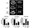
도 1은 NFI-C가 결손되면 생후 골형성 과정 동안 골형성에 이상을 초래하는 결과를 보여준다: (a)나이 든 생쥐에게서 얻은 BMSCs에서의 NFI-C 발현을 실시간 PCR 분석으로 평가하였다. n = 3; *P < 0.05.; (b) MC3T3-E1 세포를 분화 배지에서 최대 3주간 배양하였다. 웨스턴 블랏 분석을 이용하여 NFI-C, Runx2 및 Osx를 평가하였다; (C) 하악골의 대표적 마이크로-CT 이미지; (D) 대퇴골 원위부의 대표적 마이크로-CT 이미지; (E) 대퇴골 원위부에 있는 소주골 및 피질골의 3D 마이크로-CT 이미지; (F) 대퇴골 원위부의 마이크로-CT 정량화. 골 무기질 밀도(BMD); 소주골 부피(BV/TV); 소주골 두께(Tb.Th); 소주골 수(Tb.N); 소주골 분리(Tb.SP); 피질골 부피(Ct.BV/TV); 피질골 두께(Ct.Th). n = 5; *P < 0.05. (G) 6주령 정상 및 NFIC
-/- 생쥐의 대퇴골 원위부의 H&E 염색(a-f) 및 본 코사 염색(a'-f'). c-f 및 c'-f'는 각각 a-b 및 a'-b'를 더 확대하였다. c-d 및 c'-d', 소주골. e-f 및 e'-f', 피질골. 스케일 바, a-b 및 a'-b', 500 μm; c-d 및 c'-d', 200 μm; e-f 및 e'-f', 100 μm. ; (H) 6주령 정상 및 NFIC
-/- 생쥐의 대퇴골 원위부 골간단에 대한 조직형태학적 분석. 조직 면적당 골모세포 수(N.Ob/T.Ar); 골 파라미터 당 골모세포 수(N.Ob/B.Pm); 골 표면 당 골모세포 면적(Ob.S/BS). n = 5; *P < 0.05. ; (I) 6주령 정상 및 NFIC
-/- 생쥐의 대퇴골에 있는 대표적 칼세인 이중 라벨링 부분. 흰색 화살표는 무기질 침착 비율(MAR). n = 5; *P < 0.05.
도 2는 골다공증 환자에게서 나타나는 바와 같이 NFI-C가 결핍되면 골수 지방이 증가한다는 것을 보여준다: (A) 6 및 28주령의 정상 및 NFIC
-/- 생쥐의 대퇴골의 H&E 염색. 스케일 바, 50 μm ; (B) 오일 레드 O 염색. 정상 및 NFIC
-/- BMSCs를 지방세포화 유도 배지에서 7일간 배양하였다. 정상 및 NFIC
-/- BMSCs에서 PPARγ 발현을 실시간 PCR을 사용하여 분석하였다. n = 3; *P < 0.05 ; (C) 골다공증 환자의 골 시료의 H&E 염색; (D) 골다공증 환자의 골 시료의 IHC 염색; (E) NFI-C mRNA 발현을 GEO에 기탁되어 있는 유전자 발현 데이터세트 GSE35959를 이용하여 분석하였다. *P < 0.05 ; (F) hBMSCs에서의 NFI-C 발현에 대한 H2O2의 영향. H2O2의 존재 또는 부존재하에서 5일간 처리한 hBMSCs에서의 NFI-C 발현 레벨을 실시간-PCR 및 웨스턴 블랏 분석을 사용하여 정하였다. n = 3; *P < 0.05.
도 3은 NFI-C가 골모세포 분화를 촉진시키고 지방세포 분화를 억제한다는 것을 나타낸다: (A) NFIC
-/- BMSCs를 NFI-C 발현 벡터로 감염시킨 후 골형성 유도 배지에서 7일(ALP 염색) 및 14일(알리자린 레드 S 염색)동안 배양하였다. (B) Alp 발현을 실시간 PCR을 사용하여 분석하였다. n = 3; *P < 0.05; (C) 오일 레드 O 염색. NFIC
-/- BMSCs를 NFI-C 발현 벡터로 감염시킨 후 지방세포화 유도 배지에서 7일동안 배양하였다; (D) Pparγ 발현을 실시간 PCR을 사용하여 분석하였다. n = 3; *P < 0.05 ; (E) pGL3-Luc-Pparγ 및 NFI-C 발현벡터로 48시간동안 감염시킨 C2C12 세포에서 Pparγ 프로모터 활성을 평가하였다. n = 3; *P < 0.05 ; (F) 대퇴골 원위부의 대표적 마이크로-CT 이미지 및 마이크로-CT 정량화. 소주골 부피 (BV/TV); 소주골 수 (Tb.N); 소주골 분리 (Tb.SP). n = 3; *P < 0.05 ; (G) NFI-C 과발현 BMSCs를 이식한 NFIC
-/- 생쥐의 대퇴골 원위부의 조직학적 분석. 스케일 바, 50 μm ; (H) NFIC
-/- 생쥐와 비교한, NFI-C 과발현 BMSCs를 이식한 NFIC
-/- 생쥐에서의 지방세포의 수. n = 3; *P < 0.05.
도 4는 NFI-C가 결핍되면 골모세포 분화가 손상되고 파골세포 활성이 감소한다는 것을 보여준다: (A) ALP 염색 및 활성; (B) 알리자린 레드 S 염색(ARD)과 알리자린 레드 S 염색된 무기질화 결절의 정량화. 정상 및 NFIC
-/- BMSCs를 골형성 유도 배지에서 7일(ALP) 및 14일(ARS) 동안 배양하였다. n = 3; *P < 0.05 ; (C) 7일간 분화한 후 정상 및 NFIC
-/- BMSCs에서의 Runx2, Osx, Alp, Oc, 및 Bsp 발현을 실시간 PCR을 사용하여 분석하였다. n = 3; *P < 0.05; (D) 6주령의 정상 및 NFIC
-/- 생쥐 대퇴골의 TRAP 염색. 오른쪽 패널은 박스친 왼쪽 패널을 더 확대한 것이다. 스케일 바, 오른쪽 패널, 200 μm; 왼쪽 패널, 100 μm; (E) 6주령의 정상 및 NFIC
-/- 생쥐 대퇴골 골간단부의 조직형태학적 분석. 조직 면적당 파골세포수(N.Oc/T.Ar); 골 파라미터 당 파골세포 수 (N.Oc/B.Pm); 골 표면적 당 파골세포 표면적(Oc.S/BS). n = 5; *P < 0.05. (F) Vit D3 및 PGE2 존재 또는 부존재하에서 정상 BMMs을 정상 및 NFIC
-/- 1차 골모세포와 함께 6일동안 공배양하여, 고정한 후 TRAP 염색을 하였다; (G) TRAP-양성 다핵 세포(MNCs)를 D에서 카운트하였다. n = 3; *P < 0.05 ; (H) TRAP 활성을 세포 용해물로 정량하였다. n = 3; *P < 0.05 ; (I) sRankl 레벨을 ELISA 킷트를 사용하여 세포 배양 배지에서 측정하였다. n = 3; *P < 0.05; (J) 1차 골모세포에서 Rankl 및 Opg 발현을 실시간 PCR을 이용하여 분석하였다. n = 3; *P < 0.05.
도 5는 NFI-C가 BMP2-Runx2로 유도한 Osx 발현을 매개한다는 것을 보여준다: (A) 6주령의 정상 및 NFIC
-/- 생쥐 대퇴골 골간단부의 소주골에 있는 Osx의 면역형광 염색(붉은 색). 스케일 바, 100 μm. 정상 및 NFIC
-/- BMSCs의 Runx2, Osx, 및 NFI-C의 웨스턴 블랏 분석; (B) NFI-C 및 Runx2 발현 벡터로 전달이입하고 48시간 동안 BMP-2(300 ng/ml)로 처리한 MC3T3-E1 세포에서의 Osx 발현을 평가하기 위한 실시간 PCR 및 웨스턴 블랏 분석. n = 3; *P < 0.05 ; (C) pGL3-Luc-Osx (-1269~+91) 및 NFI-C 발현 벡터로 전달이입하고 48시간 동안 BMP-2(300 ng/ml)로 처리한 MC3T3-E1 세포에서의 Osx 프로모터 활성을 평가하였다. n = 3; *P < 0.05; (D) 48시간 동안 BMP-2(300 ng/ml)로 처리한 MC3T3-E1 세포에서 Osx 프로모터에 결합한 NFI-C를 ChIP (chromatin immunoprecipitation) 분석을 사용하여 검출한 것; (E) NFIC
-/- 골모세포를 BMP-2(300 ng/ml)로 처리하거나 및/또는 NFI-C 발현 벡터로 감염시켰다. Osx 발현을 실시간 PCR을 사용하여 분석하였다. n = 3; *P < 0.05; (F) 대퇴골 원위부의 대표적 마이크로-CT 이미지 및 마이크로-CT 정량화. 소주골 부피 (BV/TV); 소주골 수 (Tb.N); 소주골 분리 (Tb.SP). n = 3; *P < 0.05; (G) Osx 과발현 BMSCs를 이식한 NFIC
-/- 생쥐의 대퇴골 원위부의 조직학적 분석. 스케일 바, 50 μm; (H) NFIC
-/- 생쥐와 비교한, Osx 과발현 BMSCs를 이식한 NFIC
-/- 생쥐에서의 지방세포 수. n = 3; *P < 0.05.
도 6은 Runx2가 BMP-2로 유도된 NFI-C 발현을 매개한다는 것을 보여준다; (A 및 B) pGL3-Luc-NFI-C (-2520~+88) 및 Runx2 발현 벡터로 전달이입하고 48시간 동안 BMP-2(300 ng/ml)로 처리한 MC3T3-E1 세포에서의 NFI-C 프로모터 활성 및 mRNA 발현을 프로모터 분석 및 실시간 PCR을 사용하여 측정하였다. n = 3; *P < 0.05 ; (C) Runx2 발현 벡터로 전달이입하고 48시간 동안 BMP-2(300 ng/ml)로 처리한 MC3T3-E1 세포에서의 NFI-C 발현을 평가하기 위한 웨스턴 블랏 분석; (D) 48시간 동안 BMP-2 존재시 또는 부존재시에 pGL3-Luc-NFI-C (-2520~+88) 및 Runx2 siRNA로 전달이입한 MC3T3-E1 세포에서의 NFI-C 프로모터 활성을 평가하였다. n = 3; *P < 0.05. 대조군 및 Runx2가 불활성화된 MC3T3-E1 세포에서의 NFI-C 및 Runx2 발현을 웨스턴 블랏을 사용하여 분석하였다; (E) 48시간동안 BMP-2(300 ng/ml)로 처리한 MC3T3-E1 세포에서 NFI-C 프로모터에 결합한 Runx2를 ChIP 분석을 이용하여 검출하였다; (F) E18.5의 정상 및 Runx2
-/- 생쥐에서 얻은 대퇴골에서의 NFI-C의 면역형광 염색(붉은 색). 스케일 바, 100 μm. 정상 및 Runx2
-/- 생쥐의 calvarial 뼈에서 분리한 총 RNA 및 단백질. Runx2 및 NFI-C 발현을 실시간 PCR 및 웨스턴 블랏 분석을 사용하여 평가하였다. n = 3; *P < 0.05; (G) 골형성 분화 동안 BMP2-Runx2에 의해 유도된 Osx 발현에서의 NFI-C 역할의 모델.
도 7은 P16의 NFIC
-/- 생쥐에게서 소주골 량이 감소하였다는 것을 보여준다;(A) P16 및 6주령의 정상 및 NFIC
-/- 생쥐의 체중을 측정한 것이다. 상기 생쥐는 이유기 3일전부터 시작하여 6주까지 부드러운 도우 식이를 섭취하였다. n = 10; (B) P16의 정상 및 NFIC
-/- 생쥐의 대퇴골 원위부의 정량적 마이크로-CT 분석을 나타낸다. n = 5. 대퇴골 원위부의 소주골의 3D 마이크로-CT 이미지를 나타낸다; (C) 대퇴골 원위부의 마이크로-CT 정량화를 나타낸다. 골 무기질 농도 (BMD); 소주골 부피 (BV/TV); 소주골 두께, Tb.Th (mm); 소주골 수 (Tb.N); 소주골 분리 (Tb.SP); 피질골 부피 (Ct.BV/TV); 피질골 두께 (Ct.Th). n = 5; *P < 0.05; (D) P16의 정상 및 NFIC
-/- 생쥐의 대퇴골 원위부의 조직학적 부분을 H&E 염색한 것이다. 하부 패널은 박스한 상부 패널을 더 확대한 것이다. 스케일 바, 상부 패널, 500 μm; 하부 패널, 100 μm; (E) P16의 정상 및 NFIC
-/- 생쥐의 대퇴골 원위부 골간단을 조직형태학적 분석을 한 것이다. 조직 면적 당 골모세포 수(N.Ob/T.Ar); 골 파라미터 당 골모세포 수(N.Ob/B.Pm); 골 표면적 당 골모세포 표면적 (Ob.S/BS).n = 5; *P < 0.05.
도 8은 태아기 골형성동안 NFI-C 결핍되었을 때의 영향을 나타낸다; (A) E18.5의 정상 및 NFIC
-/- 생쥐의 정량적 마이크로-CT 분석을 나타낸다. 무기질화 골의 마이크로-CT 이미지(상부 패널) 및 골 부피(BV/TV, 하부 패널); (B) 정상 및 NFIC
-/- 신생 생쥐의 골격 표현형태를 알리자린 레드 및 알시안 블루 염색을 사용하여 분석한 것이다; (C) E18.5의 정상 및 NFIC
-/- 생쥐의 하악골 첫 번째 어금니의 화삭모양부분을 H&E 염색한 것이다. 스케일 바, 200 μm; (D) E18.5의 정상 및 NFIC
-/- 생쥐 대퇴골의 H&E 염색(상부 패널), 본 코사 염색(중간 패널), 및 PCNA 면역염색(하부 패널). 스케일 바, 200 μm; (E) 오일 레드 O 염색. 정상 및 NFIC
-/- BMSCs를 7일동안 지방세포화 유도 배지에서 배양하였다. 정상 및 NFIC
-/- BMSCs에서의 Pparγ 발현을 실시간 PCR을 사용하여 분석하였다. n = 3; *P < 0.05.
도 9는 P16의 NFIC
-/- 생쥐에서 파골세포 활성 및 파골세포 수가 감소하였다는 것을 보여준다; (A) P 16의 정상 및 NFIC
-/- 생쥐의 대퇴골을 TRAP 염색한 것이다. 하부 패널은 박스한 상부 패널을 더 확대한 것이다. 스케일 바, 상부 패널, 200 μm; 하부 패널, 100 μm; (B) P 16의 정상 및 NFIC
-/- 생쥐의 대퇴골 원위부 골간단을 조직형태학적으로 분석한 것이다. 조직 면적당 파골세포 수 (N.Oc/T.Ar); 골 파라미터 당 파골세포 수 (N.Oc/B.Pm); 골 표면적 당 파골세포 표면적 (Oc.S/BS).n = 5; *P < 0.05; (C) 정상 및 NFIC
-/- BMMs을 3일간 M-CSF (60 ng/ml) 및 RANKL (100 ng/ml)와 함께 배양하고, 고정하여 TRAP 염색하였다.
도 10은 NFIC
-/- 골모세포에서 NFI-C-독립적 Dlx5 및 Msx2 경로에 의해 Osx 발현이 조절된다는 것을 보여준다; (A) 7일동안 분화한 후 정상 및 NFIC
-/- BMSCs에서의 Dlx5 및 Msx2 발현을 실시간 PCR을 사용하여 분석하였다. n = 3; *P < 0.05; (B) NFIC
-/- 골모세포를 Dlx5 및 Msx2 발현 벡터로 전달이입하였다. 내인성 Osx 발현을 실시간 PCR을 사용하여 분석하였다. n = 3; *P < 0.05.
도 11a 및 11b는 NFI-C가 하악골 형성을 촉진시킨다는 것을 나타내는 것으로 전자는 하악골 원위부의 대표적 엑스레이 및 마이크로-CT 이미지이고, 후자는 상기 마이크로-CT 정량화한 그래프로 세로축은 소주골 부피 (BV/TV)이다.
본원은 NFI-C가 중간엽 줄기세포에서 골모세포로의 분화를 촉진하고 지방세포로의 분화를 억제하여 궁극적으로 골형성을 증가시킨다는 발견에 근거한 것이다.
한 양태에서 본원은 NFI-C (Nuclear factor I-C) 단백질 또는 그 유전자를 유효성분으로 포함하는, 골질환 예방 또는 치료용 약학 조성물에 관한 것이다.
본원의 조성물에 유효성분으로 포함되는 "NFI-C"는 CTF/NF-I 패밀리에 속하는 프로모터의 CCAAT-결합 전사인자로 DNA 복제에서 복제인자로 기능하는 것으로 알려져 있으나, 골질환의 예방 또는 치료 기능에 대하여는 전혀 알려진 바가 없다.
본원에 따른 NFI-C는 결손시 골모세포 분화 및 골 형성이 손상되고, 골수 지방세포가 증가하여, 골질환의 예방 또는 치료에 유용하게 사용될 수 있다. 본 이론으로 한정하는 것은 아니지만, Runx2에 의해 NFI-C의 발현이 증가하고, NFI-C는 골모세포 분화를 조절하는 전사인자인 Osteirx 발현을 증가시켜 골모세포의 분화를 증가시키며, 아울러 지방세포 분화 조절 전사인자인 PPARγ의 발현을 억제시켜 지방세포 분화를 억제하게 되어, 결과적으로 골형성이 증가되어, 골질환 예방 또는 치료에 사용될 수 있다.
따라서, 본원 발명의 목적 달성을 위해, 본원 발명의 효과를 달성하는 한, 다양한 유래 및/또는 형태의 NFI-C 유전자 및/또는 단백질이 사용될 수 있다. 야생형 서열은 물론, 세포내에서의 발현 또는 단백질의 안정성 등과 같은 특징이 유리하도록 염기서열의 일부가 인위적으로 변형된 유전자, 및 자연적으로 발견되는 염기서열 일부가 변형된 유전자 또는 이들의 모두의 단편을 포함하는 것이다. 유전자 염기서열의 변형은 상응하는 아미노산의 변형을 수반하거나 하지 않을 수 있으며, 아미노산의 변형을 수반하는 경우에는 이러한 변형이 유발된 유전자는 이에 의해 코딩되는 단백질에서 하나 이상의 아미노산이 치환, 결실, 부가 및/또는 삽입된 아미노산 서열로 이루어지는 단백질을 암호화하는 것으로, 돌연변이체(mutants), 유도체(derivatives), 대립유전자(alleles), 변형체(variants) 및 동질체 (homologues)를 포함하는 것이다. 유전자 염기서열의 변이가 단백질 중의 아미노산의 변형을 수반하지 않는 경우는 예를 들어 축중변이가 있고, 이러한 축중변이체(degeneracy mutants)도 본 발명의 상기 유전자에 포함된다.
인위적 유전자 염기서열의 변형은 당업자에게 잘 알려져 있는 방법, 예를 들어, 부위-유도성 돌연변이법 (site-directed mutagenesis, Kramer et al, 1987), 에러유발 PCR (Cadwell, R.C. and G.F. Joyce. 1992. PCR methods Appl., 2:28-33.), 점돌연변이법 (Sambrook and Russel, Molecular Cloning: A Laboratory Manual, 3rd Ed. 2001, Cold Spring Harbor Laboratory Press.) 등에 의해 제조될 수 있다.
본원에 따른 일 구현예에서 상술한 유전자는 약학조성물이 투여되는 대상체의 세포내에서 발현이 가능하도록 프로모터에 작동가능하게 연결된 벡터의 형태로 제공될 수 있다.
본원에 사용되는 단백질은 당업계에 공지된 방법을 사용하여 제조될 수 있다. 일 구현예에서는 단백질의 제조방법은 유전자 재조합 기술을 이용하는 것이다. 예를 들면 상기 단백질을 코딩하는 상응하는 유전자를 포함하는 벡터를 원핵 또는 진핵세포 세포 예를 들면 곤충세포, 포유류 세포에 전달하여 발현 시킨 후 정제하여 사용할 수 있다. 상기 플라스미드는 예를 들면 pET28b(Novagen)과 같은 발현 벡터에 해당 유전자를 클로닝하여 세포주에 전달이입한 후, 발현된 단백질을 정제하여 사용할 수 있으나, 이로 제한하는 것은 아니다. 합성된 단백질은 침전, 투석, 이온교환 크로마토그라피, 겔-퍼미에이션 크로마토그라피, HPLC, 역상-HPLC, 프렙용 SDS-PAGE, 항-스크리닝 단백질 항체를 이용한 친화성 컬럼 등을 포함하는 컬럼 크로마토그라피로 분리 정제될 수 있다.
본원에 따른 일 구현예에서, NFI-C 유전자 및 단백질은 유인원, 사람 등과 같은 포유동물 유래이며, 특히 사람 유래이다. NFI-C 유전자 및 단백질은 그 전장 또는 본원의 효과를 달성하는 한 그 단편이 또한 사용될 수 있으며, 전장 서열은 예를 들면 GenBank 접근번호 NM_005597, NP_005588를 포함하나, 이로 제한하는 것은 아니다. 본원에 따른 일구현예에서는 전장의 NFI-C 유전자 또는/및 단백질이 사용된다.
본원에서 사용된 용어 "골질환"은 병리학적 및/또는 물리적 골 손상에 의한 질환을 모두 포함하는 것으로, 예를 들면 골절, 골다공증, 골연화증, 구루병, 골감소증, 섬유성 골염, 무형성 골질환, 골형성 부전증, 골위축, 파제트(Paget's disease), 및 암세포의 골전이에 의해 유발되는 뼈의 손상을 포함하나 이로 제한하는 것은 아니다. 본원에 따른 일 구현예에서는 골다공증에 유용하게 사용될 수 있다.
본원에 사용된 용어 "치료"란 본원에 따른 조성물의 투여로 관련 증세를 호전시키거나 이롭게 변경하는 모든 행위를 의미한다. 본원이 속하는 기술분야에서 통상의 지식을 가진 자라면, 대한의학협회 등에서 제시된 자료를 참조하여 본원의 조성물이 효과가 있는 질환의 정확한 기준을 알고, 개선, 향상 및 치료된 정도를 판단할 수 있을 것이다.
본 명세서에서 사용된 용어 "예방"은 본원에 따른 조성물의 투여로 관련 질환의 발병을 억제 또는 지연시키는 모든 행위를 의미한다. 골모세포 분화 및 골 형성 손상으로 인한 골형성 이상에 효과가 있는 본원 조성물은 골형성 이상으로 인한 질환의 초기 증상, 또는 증상이 나타나기 전에 복용할 경우 이러한 질환을 예방할 수 있다는 것은 당업자에게 자명할 것이다.
골수의 중간엽 줄기 세포는 골모세포, 지방세포와 같은 다양한 세포로 분화 할 수 있다(Prockop DJ. Science. 1997; 276 (5309):71-74 ; Tuli R, Tuli S, Nandi S, Wang ML, Alexander PG, Haleem- Smith H, Hozack WJ, Manner PA, Danielson KG, and Tuan RS. Stem Cells. 2003;21(6):681-693). 골모세포 분화는 BMP-2, TGF-β, Wnt, IGF와 같은 성장 인자와 Runx2, Osterix(Osx)와 같은 전사인자에 의해 조절된다. 반면에 지방세포 분화는 전사인자인 과산화소체 증식 활성화 수용체(peroxisome proliferator-activated receptor gamma(PPARγ))에 의해 조절 된다. 나이가 들수록 골수에서 중간엽 줄기세포는 골모세포로의 분화가 감소하고, 지방세포로의 분화 능력은 증가하여, 결국 골량이 감소하고 골질환이 발생하게 된다.
NFI-C를 포함하는 본원에 따른 조성물은, 본 이론으로 한정하는 것은 아니지만, Runx2에 의해 NFI-C의 발현이 증가하고, NFI-C는 골모세포 분화를 조절하는 전사인자인 Osteirx 발현을 증가시켜 골모세포의 분화를 증가시키며, 아울러 지방세포 분화를 조절 전사인자인 PPARγ의 발현을 억제시켜 지방세포 분화를 억제하여, 결과적으로 골형성이 증가되어, 골질환 예방 또는 치료에 유용하게 사용될 수 있다.
이에 본원의 조성물은 약학 조성물로 제조될 수 있다. 약학 조성물은 동시에 또는 연속적으로 투여가능하며, 단독으로, 또는 수술, 상기 질환 치료를 위한 다른 약학적 활성성분 및 생물학적반응조절제를 사용하는 방법들과 병용하여 사용할 수 있다.
본원에 따른 치료제 또는 약학 조성물은 일반적으로 사용되는 약학적으로 허용가능한 담체와 함께 적합한 형태로 제형화될 수 있다. '약학적으로 허용되는'이란 생리학적으로 허용되고 인간에게 투여될 때, 통상적으로 위장 장애, 현기증 등과 같은 알레르기 반응 또는 이와 유사한 반응을 일으키지 않는 조성물을 말한다. 예로서 약학적으로 허용되는 담체로는 예를 들면, 물, 적합한 오일, 식염수, 수성 글루코스 및 글리콜 등과 같은 비경구 투여용 담체 등이 있으며 안정화제 및 보존제를 추가로 포함할 수 있다. 적합한 안정화제로는 아황산수소나트륨, 아황산나트륨 또는 아스코르브산과 같은 항산화제가 있다. 적합한 보존제로는 벤즈알코늄 클로라이드, 메틸- 또는 프로필-파라벤 및 클로로부탄올이 있다. 또한 본 발명에 따른 조성물은 그 투여방법이나 제형에 따라 필요한 경우, 현탁제, 용해보조제, 안정화제, 등장화제, 보존제, 흡착방지제, 계면활성화제, 희석제, 부형제, pH 조정제, 무통화제, 완충제, 산화방지제 등을 적절히 포함할 수 있다. 상기에 예시된 것들을 비롯하여 본 발명에 적합한 약학적으로 허용되는 담체 및 제제는 문헌[Remington's Pharmaceutical Sciences, 최신판]에 상세히 기재되어 있다.
본원의 조성물은 당해 발명이 속하는 기술 분야에서 통상의 지식을 가진 자가 용이하게 실시할 수 있는 방법에 따라, 약학적으로 허용되는 담체 및/또는 부형제를 이용하여 제제화함으로써 단위 용량 형태로 제조되거나 또는 다용량 용기 내에 내입시켜 제조될 수 있다. 이때 제형은 오일 또는 수성 매질중의 용액, 현탁액 또는 유화액 형태이거나 분말, 과립, 정제 또는 캡슐 형태일 수 있다.
본원의 약학 조성물의 투여방법은 제형에 따라 용이하게 선택될 수 있으며, 가축, 인간 등의 포유동물에 다양한 경로로 투여될 수 있다. 예를 들면, 산제, 정제, 환제, 과립제, 당의정, 경질 또는 연질의 캡슐제, 액, 에멀젼, 현탄액, 시럽제, 엘릭서, 외용제, 좌제, 멸균 주사용액 등의 형태로 제형화되어 전신 또는 국소적으로 경구 또는 비경구 투여될 수 있으며, 특히 비경구 투여가 바람직할 수도 있다.
비경구 투여를 위한 제형에는 멸균된 수용액, 비수성용제, 현탁제, 유제, 동결건조제제, 좌제가 포함된다. 비수성용제, 현탁용제로는 프로필렌글리콜, 폴리에틸렌 글리콜, 올리브 오일과 같은 식물성 기름, 에틸올레이트와 같은 주사 가능한 에스테르 등이 사용될 수 있다. 좌제의 기제로는 위텝솔 (witepsol), 마크로골, 트윈 (tween) 61, 카카오지, 라우린지, 글리세롤, 젤라틴 등이 사용될 수 있다.
본원의 조성물은 목적하는 방법에 따라 경구 투여하거나 비경구투여(예를 들어, 정맥 내, 피하, 복강 내 또는 국소에 적용)할 수 있으며, 특히 비경구 투여가 바람직하다. 투여량은 환자의 상태 및 체중, 질병의 정도, 약물형태, 투여경로 및 시간에 따라 다르지만, 당업자에 의해 적절하게 선택될 수 있다. 본원에 따른 일 구현예에서 조성물은 골수강 및 물리적 및/또는 병리학적으로 손상된 골주위에 국소투여된다.
본 발명에 따른 조성물은 약학적으로 유효한 양으로 투여한다. 본 발명에 있어서, "약학적 또는 치료적으로 유효한 양"은 의학적 치료에 적용 가능한 합리적인 수혜/위험 비율로 질환을 치료하기에 충분한 양을 의미하며, 유효용량 수준은 환자의 질환의 종류, 중증도, 약물의 활성, 약물에 대한 민감도, 투여 시간, 투여 경로 및 배출 비율, 치료기간, 동시 사용되는 약물을 포함한 요소 및 기타 의학 분야에 잘 알려진 요소에 따라 결정될 수 있다. 본 발명의 조성물은 개별 치료제로 투여하거나 다른 치료제와 병용하여 투여될 수 있고 종래의 치료제와는 순차적 또는 동시에 투여될 수 있으며, 단일 또는 다중 투여될 수 있다. 상기한 요소들을 모두 고려하여 부작용 없이 최소한의 양으로 최대 효과를 얻을 수 있는 양을 투여하는 것이 중요하며, 이는 당업자에 의해 용이하게 결정될 수 있다.
본원의 약학 조성물의 투여량은 환자의 체중, 연령, 성별, 건강상태, 식이, 투여시간, 투여방법, 배설률 및 질환의 중증도 등에 따라 그 범위가 다양할 수 있으나, 유효 투여량은 통상적으로 성인(60 kg)의 경우, 약 1 ng 내지 10 mg/일, 특히 약 1 μg 내지 1mg/일이다. 투여량은 여러 가지 조건에 따라 변동가능하기 때문에, 상기 투여량에 가감이 있을 수 있다는 사실은 당업자에게 자명하며, 따라서 상기 투여량은 어떠한 면으로든 본 발명의 범위를 한정하는 것은 아니다.
투여횟수는 원하는 범위 내에서 하루에 1회, 또는 수회로 나누어 투여할 수 있으며, 투여 기간도 특별히 한정되지 않는다.
다른 양태에서 본원은 또한 NFI-C를 과발현하는 중간엽줄기세포; 상기 중간엽줄기세포로부터 유래된, 연골세포 또는 골세포로 분화될 수 있는 전구세포; 또는 상기 중간엽줄기세포로부터 분화된 연골세포 또는 골세포를 유효성분으로 포함하는 골질환 치료용 조성물 또는 세포 치료제에 관한 것이다.
본원에서 "세포치료제" 란 세포와 조직의 기능을 복원시키기 위하여 살아있는 자가 (autologous), 동종 (allogenic), 이종 (xenogenic) 세포를 체외에서 증식 또는 선별하거나 기타 방법으로 세포의 생물학적 특성을 변화시키는 등의 일련의 방법을 통하여 치료, 진단 및 예방의 목적으로 사용되는 의약품을 말한다. 미국은 1993년부터, 우리나라는 2002년부터 세포치료제를 의약품으로 관리하고 있다. 이러한 세포치료제는 조직재생 혹은 장기기능 회복을 위한 줄기세포 치료제를 포함하는 것이나, 이로 제한하는 것은 아니다.
본원에서 중간엽 줄기세포(Mesenchymal Stem Cell; MSC)'는 제대혈, 지방, 골수(bone marrow), 혈액, 진피 또는 골막에서 분리되는 줄기세포로서, 다양한 세포 예컨대 지방세포, 연골세포 및 골세포 등으로 분화할 수 있는 전능성(pluripotent) 또는 다능성(multipotent) 세포를 의미한다. 또한, 중간엽 줄기세포는 면역억제제의 사용 없이도 동종 또는 이종 수혜자에서 효과적으로 생착될 수 있는 특징이 있다.
본원의 치료용 조성물에 사용되는 세포는 자가 (autologous), 동종 (allogenic), 이종 (xenogenic) 세포이며, 특히 자가 세포이다.
일 구현예에서 본원의 세포치료제에 포함되는 중간엽 줄기세포는, 동물의 중간엽 줄기세포일 수 있으며, 바람직하게는 포유동물, 보다 바람직하게는 인간의 중간엽 줄기세포일 수 있다. 또한 본 발명의 중간엽 줄기세포는 골수, 지방조직, 말초혈액, 간 유래 일 수 있다.
다른 구현예에서, 본원의 세포치료제에 포함될 수 있는 세포는 중간엽줄기세포로부터 유래된, 연골세포 또는 골세포로 분화될 수 있는 전구세포를 포함하며, 예를 들면 미성숙 골전구세포, 골모세포 , 연골모세포를 포함한다.
또다른 구현예에서 본원의 세포치료제에 포함될 수 있는 세포는 중간엽줄기세포로부터 분화된 연골세포 또는 골세포를 포함한다.
중간엽 줄기세포는 골수 등에 매우 적은 양으로 존재하지만, 이를 분리 및 배양하는 과정은 당업계에 잘 알려져 있으며 예를 들면 미국특허 제5,486,359호를 참조할 수 있다. 또한, 본원의 중간엽 줄기세포는 공지된 방법에 따라 골수의 조혈모세포로부터 부착특성에 의해 분리한 후 분화능력을 잃지 않은 상태에서 증식시켜 얻을 수 있다.
예를 들면 중간엽 줄기세포를 얻는 과정을 구체적으로 설명하면 다음과 같다: (1) 인간 또는 마우스를 포함하는 포유동물, 바람직하게는, 인간의 중간엽 줄기세포, 예컨대, 혈액 또는 골수로부터 중간엽 줄기세포를 분리하는 단계(상기 골수는 경골, 대퇴골, 척수 또는 장골로부터 추출할 수 있다.); 분리된 세포를 적합한 배지에서 배양하는 단계 및 배양과정에서 부유 세포를 제거하고 배양 플레이트에 부착된 세포들을 계대 배양하여, 최종적으로 구축된 중간엽 줄기세포를 수득하는 단계를 통하여 얻을 수 있다.
상기 과정에서 이용되는 배지로는, 줄기세포의 배양에 이용되는 일반적인 어떠한 배지도 이용할 수 있다. 바람직하게는, 혈청(예컨대, 우태아 혈청, 말 혈청 및 인간 혈청)이 함유된 배지이다. 본 발명에서 이용될 수 있는 배지는, 예를 들어, RPMI 시리즈, Eagles's MEM (Eagle's minimum essential medium, Eagle, H. Science 130:432(1959)), α-MEM (Stanner, C.P. et al., Nat. New Biol. 230:52(1971)), Iscove's MEM (Iscove, N.et al., J. Exp. Med. 147:923(1978)), 199 배지(Morgan et al., Proc. Soc. Exp. Bio. Med., 73:1(1950)), CMRL 1066, RPMI 1640 (Moore et al., J. Amer. Med. Assoc. 199:519(1967)), F12 (Ham, Proc. Natl. Acad. Sci. USA 53:288(1965)), F10 (Ham, R.G. Exp. Cell Res. 29:515(1963)), DMEM (Dulbecco's modification of Eagle's medium, Dulbecco, R. et al., Virology 8:396(1959)), DMEM과 F12의 혼합물(Barnes, D. et al.,Anal. Biochem. 102:255(1980)), Way-mouth's MB752/1 (Waymouth, C. J. Natl. Cancer Inst. 22:1003(1959)), McCoy's 5A (McCoy, T.A., et al., Proc. Soc. Exp. Biol. Med. 100:115(1959)) 및 MCDB 시리즈(Ham, R.G. et al., In Vitro 14:11(1978))을 포함하나, 이에 한정되는 것은 아니다. 상기 배지에는, 다른 성분, 예를 들어, 항생제 또는 항진균제(예컨대, 페니실린, 스트렙토마이신) 및 글루타민 등이 포함될 수 있다.
다른 예에서 중간엽 줄기세포의 확인은 유세포 분석을 통하여 할 수 있다. 이러한 유세포 분석은, 중간엽 줄기세포의 특이한 표면 마커를 이용하여 실시된다. 예컨대, 중간엽 줄기세포는 CD44, CD29 및/또는 MHC 클래스 I에 대하여 양성 반응을 나타내므로, 이를 통해 중간엽 줄기세포를 확인할 수 있다.
본원에 따른 세포 치료제에 포함되는 세포는 NFI-C를 과발현한다. 세포에서 단백질을 과발현하는 방법은 공지되어 있다. NFI-C 유전자를 적절한 진핵세포 발현벡터 예를 들면 파아지, 바이러스 또는 레트로바이러스 유래의 벡터 또는 플라즈미드에 발현 프로모터에 작동가능하도록 연결되도록 클로닝 하여, 발현 벡터를 구축한 후, 이를 본원에 따른 세포에 도입할 수 있다. 도입 방법은 공지되어 있으며 예를 들면 칼슘포스페이트법, DEAE-덱스트란 매개 전달이입, 양하전 지질 매개 전달이입법, 전기천공, 파아지 시스템을 이용한 트랜스덕션 또는 바이러스를 이용한 감염 방법 등을 포함하나 이로 제한하는 것은 아니다.
본원에 따른 세포 치료제 또는 세포를 포함하는 약학 조성물의 투여 경로는 목적 조직에 도달할 수 있는 한 어떠한 일반적인 경로를 통하여 투여될 수 있다. 비경구 투여, 예를 들어, 복강내 투여, 정맥내 투여, 근육내 투여, 피하 투여, 피내 투여될수 있으나, 이에 제한되지는 않는다.
본원에 따른 치료제 또는 조성물은 일반적으로 사용되는 약학적으로 허용가능한 담체와 함께 적합한 형태로 제형화될 수 있다. 약학적으로 허용되는 담체로는 예를 들면, 물, 적합한 오일, 식염수, 수성 글루코스 및 글리콜 등과 같은 비경구 투여용 담체 등이 있으며 안정화제 및 보존제를 추가로 포함할 수 있다. 적합한 안정화제로는 아황산수소나트륨, 아황산나트륨 또는 아스코르브산과 같은 항산화제가 있다. 적합한 보존제로는 벤즈알코늄 클로라이드, 메틸- 또는 프로필-파라벤 및 클로로부탄올이 있다. 또한 본 발명에 따른 세포치료용 조성물은 그 투여방법이나 제형에 따라 필요한 경우, 현탁제, 용해보조제, 안정화제, 등장화제, 보존제, 흡착방지제, 계면활성화제, 희석제, 부형제, pH 조정제, 무통화제, 완충제, 산화방지제 등을 적절히 포함할 수 있다. 상기에 예시된 것들을 비롯하여 본 발명에 적합한 약학적으로 허용되는 담체 및 제제는 문헌[Remington's Pharmaceutical Sciences, 최신판]에 상세히 기재되어 있다.
본 발명의 세포치료용 조성물은 당해 발명이 속하는 기술 분야에서 통상의 지식을 가진 자가 용이하게 실시할 수 있는 방법에 따라, 약학적으로 허용되는 담체 및/또는 부형제를 이용하여 제제화함으로써 단위 용량 형태로 제조되거나 또는 다용량 용기 내에 내입시켜 제조될 수 있다.
또한 상기 조성물은 세포치료제가 표적 세포로 이동할 수 있는 임의의 장치에 의해 투여될 수도 있다. 본 발명의 세포치료제 조성물은 질환의 치료를 위하여 치료학적으로 유효한 양의 세포치료제를 포함할 수 있다. 용어 '치료적으로 유효한 양'은 연구자, 수의사, 의사 또는 기타임상에 의해 생각되는 조직계, 동물 또는 인간에서 생물학적 또는 의학적 반응을 유도하는 유효 성분 또는 약학적 조성물의 양을 의미하는 것으로, 이는 치료되는 질환 또는 장애의 증상의 완화를 유도하는 양을 포함한다. 본 발명의 조성물에 포함되는 세포치료제는 원하는 효과에 따라 변화될 것임은 당업자에게 자명하다.
그러므로 최적의 세포치료제 함량은 당업자에 의해 쉽게 결정될 수 있으며, 질환의 종류, 질환의 중증도, 조성물에 함유된 다른 성분의 함량, 제형의 종류, 및 환자의 연령, 체중, 일반 건강 상태, 성별 및 식이, 투여 시간, 투여 경로 및 조성물의 분비율, 치료기간, 동시 사용되는 약물을 비롯한 다양한 인자에 따라 조절될 수 있다. 본원에 따른 일 구현예에서 세포치료제는 골수강 및 물리적 및/또는 병리학적으로 손상된 골주위에 국소투여된다.
상기 요소를 모두 고려하여 부작용 없이 최소한의 양으로 최대 효과를 얻을 수 있는 양을 포함하는 것이 중요하다. 예컨대, 본 발명의 조성물의 투여량은 유효성분인 줄기세포로부터 분화된 골모세포를 기준으로 1.0× 10⁷ 내지 1.0× 10⁸ 세포/kg(체중), 보다 바람직하게는 1.0× 10⁵ 내지 1.0× 10⁸ 세포/kg(체중)일 수 있다. 다만, 투여량은 제제화 방법, 투여 방식, 환자의 연령, 체중, 성별, 병적 상태, 음식, 투여 시간, 투여 경로, 배설 속도 및 반응 감응성과 같은 요인들에 의해 다양하게 처방될 수 있고, 당업자라면 이러한 요인들을 고려하여 투여량을 적절히 조절할 수 있다. 투여 횟수는 1회 또는 임상적으로 용인가능한 부작용의 범위 내에서 2회 이상이 가능하고, 투여 부위에 대해서도 1개 부위 또는 2개 부위 이상에 투여할 수 있다. 인간 이외의 동물에 대해서도, kg당 인간과 동일한 투여량으로 하거나, 또는 예를 들면 목적의 동물과 인간과의 허혈기관(심장 등)의 용적비(예를 들면, 평균값) 등으로 상기의 투여량을 환산한 양을 투여할 수 있다. 본 발명에 따른 치료의 대상동물로서는, 인간 및 그 밖의 목적으로 하는 포유동물을 예로 들 수 있고, 구체적으로는 인간, 원숭이, 마우스, 래트, 토끼, 양, 소, 개, 말, 돼지 등이 포함된다.
본원에 따른 NFI-C는 지방세포의 분화 억제와 골모세포의 분화를 촉진하여, 지방세포 분화 억제가 필요하거나, 골모세포의 분화 촉진이 필요한 다양한 용도로 사용될 수 있다.
이런 측면에서 본원은 또한 NFI-C 단백질 또는 그 유전자를 유효성분으로 포함하는 지방세포 분화 억제제 또는 골모세포 분화 촉진용 조성물로 제공될 수 있다.
이하, 본 발명의 이해를 돕기 위해서 실시예를 제시한다. 그러나 하기의 실시예는 본 발명을 보다 쉽게 이해하기 위하여 제공되는 것일 뿐 본 발명이 하기의 실시예에 한정되는 것은 아니다.
실 시 예
실험 재료 및 방법
1. 시약 및 항체
본 발명에서 인간유래 재조합 BMP-2(Bone Morphogenic Protein-2)는 R&D Systems에서 구입하였다. NFI-C 항체는 (아미노산 잔기 NH2-RPTRPLQTVPLWD-COOH) 펩타이드를 합성하여 토끼에서 제작하였다. Runx2 (sc-398), PCNA (sc-400), 그리고 Osx (sc-22538) 항체는 Santa Cruz Biotechnology에서 구입하였다.
2. 실험동물
쥐를 포함한 모든 동물 실험은 서울대학교 치의학 연구소 지침에 따라 수행하였다. NFIC
-/- 생쥐는 NFIC
+/- 생쥐의 암컷과 수컷의 교배로 제작하였다. Runx2-/- 생쥐는 Dr. Toshihisa Komori(Nagasaki University Graduate School of Biomedical Sciences, Nagasaki, Japan)에게서 제공 받았다.
3. 마이크로-CT와 조직형태학적 분석
생후 6주 정상 생쥐 및 NFIC
-/- 생쥐의 상악, 하악, 그리고 대퇴골을 분리하여 4% 파라포름 알데하이드에 밤새 4℃에서 고정하였다. 마이크로-CT는 스카이스캔 스캐너와 소프트웨어로 분석하였다. 상기 기계의 동위원소 해상도는 10 μm였다. 죽이기 7일 및 2일 전에 생후 6주 생쥐에게 10 mg/kg의 칼세인은 투여하였다. OsteoMeasure histomorphometry system (OsteoMetrics)을 제조자의 방법대로 이용하여 조직학적 분석을 수행하였다.
4. 조직학적 분석 및 면역화학조직 분석
생쥐의 대퇴골은 4% 파라포름알데하이드에서 밤새 4℃에서 고정하였다. 비탈회 대퇴골은 불안정화 메틸메타크릴레이트 레진(destabilized methyl methacrylate resin)에 포매하여 5 μm 두께로 대퇴골을 절단하여 본 코사 (von Kossa) 염색을 수행하였다. 탈회 대퇴골 표본은 10% 에틸렌 디아민 테트라-아세틱 애시드(ethylene diamine tetra-acetic acid)(EDTA)-1% 파라포름알데히드(pH 7.4) 용액에서 2주간 탈회하고 수세, 탈수 등의 과정을 거친 다음 파라핀 포매하여 5 두께로 절단하여 헤마톡시린-에오신(hematoxylin-eosin) 염색, 면역화학조직 염색, 그리고 파골세포 확인을 위한 TRAP 염색을 수행하였다. TRAP 염색은 키트(sigma)를 사용하여 제조자의 지침에 따라 수행하였다.
골다공증 환자의 골 조직 슬라이드는 인제대학교 백인제기념 임상의학 연구소 양영일 교수님께 제공 받았고, 헤마톡시린-에오신 염색과 NFI-C 면역화학조직 염색을 수행하였다.
5. 생쥐의 골격 분석
갓 태어난 생쥐 (P0)에게 연골 염색을 위한 알시안 블루와 뼈를 염색하기 위한 알리자린 레드 S 염색을 시행하였다. 생쥐는 95% 에탄올에서 2시간 고정한 후 피부와 근육을 제거하였다. 그런 다음 생쥐의 골격은 5일간 95% 에탄올에서 다시 고정한 후 2일간 아세톤에서 다시 고정하였다. 다음으로, 생쥐의 골격은 5% 빙초산과 70% 에탄올에 0.015% 알시안 블루 와 0.005% 알리자린 레드 S가 포함된 용액에서 3일간 염색하였다. 염색된 샘플은 1% KOH 용액에서 수세한 후 100% 글리세린에 보관하였다.
6. 플라스미드 제작.
pCH-NFI-C 발현 플라스미드와 pSuper-retro NFI-C siRNA 플라스미드는 이전에 보고하였다 (Lee DS, Yoon WJ, Cho ES, Kim HJ, ,Gronostajski RM, ChoM-I, and Park JC. PLoS One 2011, 6(12):e29160, 2011). Runx2, Dlx5, Msx2, 와 Pparγ 프로모터 플라스미드는 서울대학교 치과대학 류현모 교수님께 제공 받았다. NFI-C 프로모터(-2520~+88)는 생쥐의 유전체 DNA로부터 PCR를 수행하여 2.6-kb 유전체 절편을 증폭하였고, Osx 프로모터(-1269~+91) 또한 생쥐의 유전체 DNA로부터 PCR를 수행하여 1.36-kb 유전체 절편을 증폭하였다. NFI-C와 Osx 프로모터의 염기서열의 GenBank accession numbers는 다음과 같다. NFI-C, NC000076. Osx, DQ229136. 각각의 프로모터의 증폭을 위해 생쥐의 유전체 DNA (1 μl)를 사용하여 PCR을 수행하였고 반응 조건과 프라이머 염기서열은 아래와 같다. PCR 반응 조건: 94℃, 1 min; 60℃, 1 min; 72℃, 1 min 총 35 주기. 프라이머 염기서열: NFI-C Fw 5'-CTC GAG AGT GAA TGG ATT GGG GGT GT-3', NFI-C Rv 5'-AAG CTT CAG AGC GGG GAG GAA TAC AT-3', Osx Fw 5'-CTC GAG CAC ACA TAC ACG AAC ACA CAT-3', 및 Osx Rv 5'-AAG CTT TGG GGA CCG GGT CCC AAG GAG-3'. 증폭된 유전체 DNA 절편은 PCRR®2.1 T 벡터에 삽입한 후 제한효소 XhoI 및 Hind III로 절단하여 pGL3 루시퍼라제(LUC) 발현 벡터(Promega)에 삽입하였다.
7. 세포배양 및 전달이입 (transfection).
중간엽 줄기세포는 생후 6주 생쥐의 대퇴골에서 1ml 주사기를 이용하여 분리하였다. 분리된 세포는 적혈구제거 용액을 이용하여 적혈구를 제거하고, 100mm 배양 접시에서 100 IU/ml 페니실린, 100 μg/ml 스트렙토마이신 (Gibco BRL), 그리고 10% 우태아혈청이 첨가된 α-MEM 배지에서 배양하였다. 생쥐의 일차 두개관 (Primary calvarial) 골모세포를 생후 5일의 정상과 NFIC
-/- 생쥐에서 분리하였다. 두개관 뼈를 자른 후 피부조직을 제거하고 잘게 자른 다음 PBS로 3번 수세하였다. 뼈 조각은 1% 콜라게나아제 타입 I (Gibco BRL) 과 1.6% 디스파아제 II (Gibco BRL)가 포함된 α-MEM 배지에서 한 시간 동안 반응한 후 calvarial 골모세포를 분리하였다. 골모세포 분화는 배양접시에 세포가 80~90% 자라면 5% 우태아혈청, 아스코르빈산 (50 μg/ml), 그리고 베타 글리세로포스페이트 (β-glycerophosphate, 10 mM)가 첨가된 α-MEM 배지에서 2주간 배양하여 골모세포 분화를 유도하였다. 지방세포는 배양접시에서 세포가 80~90% 자라면 10% 우태아혈청, 인슐린 (10 μg/ml), 덱사메타손 (1 μM), 그리고 3-이소부틸-1-메틸잔틴(IBMX, 0.5 mM)이 포함된 DMEM 배지에서 7일간 배양하여 지방세포 분화를 유도하였다.
생쥐의 골모세포주인 MC3T3-E1 세포는 α-MEM 에 10% 우태아혈청과 항생제가 포함된 배지에 배양하였다. 생쥐의 근육세포주 C2C12 세포는 100 IU/ml 페니실린, 100 μg/ml 스트렙토마이신 (Gibco BRL), 그리고 10% 우태아혈청이 첨가된 DMEM 배지에서 배양하였다. 도3, 도5, 도6, 도8, 그리고 도10 에 기재된 발현 플라스미드 (2 μg)는 MC3T3-E1 과 C2C12 세포에 리포펙타민(Lipofectamine) PlusTM reagent (Invitrogen)을 이용하여 유전자를 전달이입하였다.
인간유래 중간엽줄기세포는 칼브렉스(Cambrex)에서 구입하여 100 IU/ml 페니실린, 100 μg/ml 스트렙토마이신 (Gibco BRL), 그리고 10% 우태아혈청이 첨가된 저포도당 DMEM 배지에서 배양하였다.
8. 중간엽 줄기세포의 골수 이식
NFIC -/- 중간엽줄기세포는 생후 6주 NFIC
-/- 생쥐의 대퇴골에서 1ml 주사기를 이용하여 분리하였다. NFIC
-/- 중간엽줄기세포에 NFI-C를 다시 발현시키기 위하여 NFI-C 과발현 레트로바이러스를 첨가하여 24 시간 동안 배양하였다. NFI-C-과발현 중간엽 줄기세포는 헤밀튼 주사기를 이용하여 (1 × 10⁶ cells/femur in 10 μl of α-MEM) 생쥐의 왼쪽 대퇴골의 골수강에 주입하였다. 이식 4주 후 대퇴골을 분리하여 마이크로-CT와 조직학적 분석을 시행하였다.
9. 시험관에서 파골세포 분화
골수세포를 10% 우태아혈청 및 항생제, 30 ng/ml M-CSF (R & D Systems)가 포함된 α-MEM 배지에서 1일간 배양하여 골수 단핵구(bone marrow monocytes (BMMs))를 얻었다. BMMs (5 × 10⁴ cells/well)은 M-CSF(60 ng/ml)와 RANKL(100 ng/ml)이 포함된 α-MEM 배지에서 3일간 배양하여 파골세포 분화를 유도 하였다. 동시배양을 할 때, BMMs (5 × 10⁵ cells/well)와 calvarial 골모세포 (2.5 × 10⁴ cells/well)는 48-웰 플레이트에 함께 넣어 10% 우태아혈청, 10-8 M 1,25-디히드록시바이타민 D3 (Vit D3), 그리고 10-6 M 프로스타글란딘 E2 (PGE2)가 포함된 α-MEM 배지에서 6일간 배양하여 파골세포 분화를 유도하였다. 파골세포의 확인은 TRAP 염색 키트(Sigma)를 사용하여 분석하였다.
10. 리포터 분석
루시퍼라제 분석은 MC3T3-E1 또는 C2C12 세포를 24-웰 플레이트에 5 × 10⁴ 세포/웰로 플레이팅하고, 24시간 후 리포펙타민 PlusTM 시약으로 형질전환하였다. 각 형질전환에서, 각각 0.4 μg의 pGL3basic (대조군), pGL3 Pparγ 프로모터 (-1000~+1), pGL3 Osx 프로모터(-1269~+91), pGL3 NFIC 프로모터(-2520~+88), pCMV empty, Runx2, 및 NFI-C 발현 플라스미드를 사용하였다. Runx2 siRNA와 대조군 siRNA는 Dharmacon에서 구입하여 사용하였다. 형질전환된 세포는 48시간 후에 루시퍼라제 활성을 측정하였다.
11. cDNA 합성과 실시간 중합효소 연쇄반응
각 세포에서 TRIzol 시약을 이용하여 총 RNA를 분리하였다. 2 μg 의 총 RNA와 역전사효소(Superscript III enzyme(Invitrogen)) 1 μl와 0.5 μg 의 oligo (dT)를 이용하여 cDNA를 합성하였다. 합성된 cDNA를 실시간 중합효소 연쇄반응에 이용하였다. 실시간 중합효소 연쇄반응은 SYBR GREEN PCR Master Mix (Takara, Japan)를 이용하여 ABI PRISM 7500 sequence detection system (Applied Biosystems)에서 진행되었다. 실시간 중합효소 연쇄반응은 94℃에서 1분, 이어 9℃ 15초, 60℃ 34초 조건에서 40주기 수행되었다. 결과의 분석은 CT (comparative cycle threshold, ABI PRISM 7500 장비와 함께 제공됨) 방법을 이용하여 평가하였고, 프라이머 염기서열은 표 1에 기재되어 있다.
[표 1]

12. 웨스턴블랏
단백질을 분리하기 위하여 세포를 PBS로 세척하고, 1,000 x g 에서 5분간 저온에서 원심분리하여 세포를 모았다. 상청액를 제거한 후 펠렛은 융해(lysis) 버퍼(100 mM Tris, pH 7.4, 350 mM NaCl, 10% glycerol, 1% Nonidet P-40, 1 mM EDTA, 1 mM 디치오스레이톨(dithiothreitol), 1x 프로테아제 저해제(SigmaAldrich, St. Louis, Mo, USA))에 녹여 얼음에서 15분간 반응하였다. 세포 잔해(Cell debris)는 16,000 x g, 15분간 저온에서, 원심분리하여 제거하고 상청액만 취하였다. 30 μg의 단백질을 전기영동으로 분리하여 PVDF 멤브레인으로 옮겼다. 멤브레인을 0.1% 트윈 20이 포함된 PBS(PBS-T)에 5% 탈지유(nonfat dry milk)로 블락킹하고 PBS-T로 세척한 후 안티 NFI-C, Runx2, Osx, 그리고 GAPDH 항체에 1:1000으로 반응시켰다. 세척 후 멤브레인은 anti-rabbit IgG conjugated horseradish peroxidase (1:5000) (Santa Cruz Biotechnology)에 한 시간 반응한 후 증강 화학발광 시스템(Amersham Biosciences)으로 밴드를 확인하였다.
13. ChIP assays
MC3T3-E1 세포는 BMP-2 (300 ng)를 48시간 동안 첨가하여 배양하였다. 이어 세포를 PBS로 세척한 후 수거하여 초음파기기를 이용하여 세포의 크로마틴을 약 1,000bp 크기로 분쇄하였다. 크로마틴 면역침강은 NFI-C 항혈청 (30 μl) 과 Runx2 (10 μl, Santa Cruz) 항혈청을 첨가하여 4℃ 에서 4시간 동안 반응 시킨 후 DNA 펠렛을 수거한 후 PCR을 이용하여 각 단백질이 프로모터 지역에 결합되었는지를 확인 하였다. PCR 프라이머 염기서열은 표 2에 나타내었다. PCR 조건은 아래와 같다: 94℃ for 30s; 60℃ for 30s; 및 72℃ for 30s (총 35 cycles). PCR 산물은 2% 아가로스 겔에서 전기영동하여, 에티디움 브로마이트(ethidium bromide)로 염색하여 자외선에서 확인하였다.
[표 2]
14. 레트로 바이러스 생산 및 바이러스 인펙션
Platinum-E 세포는 pMxs NFI-C, Osx, 그리고 대조군 벡터를 리포펙타민 PlusTM 시약(Invitrogen)을 이용하여 전달이입 하였다. 48시간 후, 바이러스가 포함된 배지를 수거하여 원심분리 후 0.45-μm 주사기 필터로 필터한 후 -80℃에 보관하여 사용하였다.
15. 유전자 발현 프로파일링
유전자 프로파일링은 정상과 골다공증 환자의 중간엽 줄기세포을 이용한 mRNA 마이크로 어레이 데이터 베이스를 이용하여 분석하였다. 마이크로 어레이 데이터는 NCBI의 GEO 데이터 베이스 (accession number GSE35959)에서 다운로드받았고, NFI-C mRNA 발현은 상기 탐침자 (Probeset ID 213198_at)를 이용하여 분석하였다.
16. 중간엽줄기세포의 하악골 이식
NFIC -/- 중간엽줄기세포는 생후 6주 NFIC
-/- 생쥐의 대퇴골에서 1ml 주사기를 이용하여 분리하였다. NFIC
-/- 중간엽줄기세포에 NFI-C를 다시 발현시키기 위하여 NFI-C 과발현 레트로바이러스를 첨가하여 24 시간 동안 배양하였다. NFI-C-과발현 중간엽 줄기세포는 헤밀튼 주사기를 이용하여 (10μl α -MEM 중의 1x10⁶ cells/mandible) 생쥐의 왼쪽 하악골에 주입하였다. 이식 4주 후 하악골을 분리하여 엑스레이(X-ray) 및 마이크로-CT 분석을 시행하였다.
실험예
실험예 1. NFI-C의 결손에 의한 출생 후 골형성 손상
첫 번째로, 나이가 들어감에 따른 NFI-C 유전자 발현의 패턴을 조사하기 위하여, 배아 18.5일(E18.5)부터 생후 60주의 생쥐로부터 얻은 골수기질세포(BMSCs)에서의 NFI-C mRNA 발현을 분석하였다. 출생 후 16일부터 6주까지는, NFI-C 발현이 유의하게 증가하였고, 이어서 60주까지는 감소하였다(도 1A). 생체 외 골모세포 분화에서는, NFI-C는 배양 0일째부터 발현되었으며, 4일에서 10일까지는 발현이 증가하였고(초기 골모세포 분화), 10일에서 21일까지는 감소하였다(후기 골모세포 분화 및 석화작용). Runx2 발현은 4일에서 7일까지 증가하였고, 그후 10일부터 21일까지는 점차적으로 감소하였는데, 이는 후기 골모세포 분화 및 석화작용에 상응하는 것으로, 배양동안 이 시기에 Osx 발현이 최고조였다(도 1B).
NFIC -/- 생쥐는 정상적인 수명기간을 가지지만, 치아뿌리의 발달이 늦어서 딱딱한 정상적인 설치류 사료를 섭취하는 능력이 결핍되어 있어 성장이 더딘 것이 관찰되었다(16). 따라서, NFIC
-/- 생쥐에서의 섭식장애를 완화시키기 위하여 이유식기(weaning) 3일 전에부터 부드러운 도우 식이 보충제를 첨가하였고 이를 6주까지 크는 동안 계속 주었다. 정상(WT) 생쥐 및 NFIC
-/- 생쥐의 체중을 측정하였다. 16일째 및 6주째의 정상(WT) 생쥐 및 NFIC
-/- 생쥐의 총 체중 차이는 크기 않았다(도 7A). 부드러운 도우 식이를 사용하여 섭식-의존성 성장지연으로부터 NFIC
-/- 생쥐를 회복시킨 후, 6주 때에 마이크로-CT(micro-computed tomography)를 사용하여 하악골에 대한 NFI-C 손실의 영향을 측정하였다. 그 결과, 이전에 측정한 치아뿌리 결핍이외에도, NFIC
-/- 생쥐의 하악골에서는 피질 골 부피 및 골 두께가 정상 생쥐에 비해 현저히 감소하였다(도 1C). 이러한 결과는 NFI-C가 결핍되면 골 부피가 현저히 감소하며, 골 부피 감소가 기아에 의한 영향이 아님을 의미한다.
뼈에 대한 NFI-C의 역할을 연구하기 위하여, 6주째 NFIC
-/- 생쥐의 대퇴골에서의 골 마이크로구조(micro-architecture)를 조사하였다. 대퇴골의 마이크로씨티 이미지를 보면, NFIC
-/- 생쥐의 골소주 부피(trabecular bone volume) 및 골피질 두께가 정상 생쥐와 비교할 때 현저히 감소한 것을 볼 수 있었다(도 1, D 및 E). 조직학적 분석을 보면, NFIC
-/- 생쥐의 골밀도(BMD, 32%), 골소주 부피(BV/TV, 34%), 골소주 량(Tb.N, 41%), 피질골 부피(Ct. BV/TV, 24%), 및 피질골 두께(Ct. Th, 20%)가 상당히 감소한 것으로 나타났으나, 골소주 분리(Tb.SP, 34%; 도 1F)는 상당히 증가한 것으로 나타났다. 또한 섭식장애가 일어나지 않은 때인 생후 16일(P16)부터 수유시기동안 샘플링한 골에 대한 NFI-C 결핍의 영향을 조사하였다. 예상한 바와 같이, P16 및 6주의 생쥐는 서로 골 특성이 매우 유사한 것으로 나타났는데(도 7 B 및 C), 이는 NFI-C가 결핍되면 생후 골형성 시기에 골 형성이 손상받는다는 것을 나타낸다. 그러나 NFI-C 결핍이 태아기 골 및 치아 발달에는 영향을 미치지 않았다(도 8, A-D). 전체적으로 이러한 결과는, NFI-C가 생후 골 형성 및 치아 뿌리 발달을 조절하지만 태아기 골 발달에는 영향을 미치지 않는다는 것과 일치한다.
NFIC -/- 생쥐의 골량이 감소되는 것이 골형성 능력이 손상되었기 때문인지를 알아내기 위하여, 6주령 생쥐의 대퇴골을 조직학적 분석을 하여 골모세포 기능을 측정하였다. 조직학적 분석 및 본 코사 염색에 기초하여 보면, 정상 생쥐에 비해 NFIC
-/- 생쥐의 해면 및 피질 골 부피가 현저하게 감소하였다(도 1G). 정상 생쥐에 비해 NFIC
-/- 생쥐의 조직당 골모세포 수(N.Ob/T.Ar), 골 파라미터 당 골모세포 수(N.Ob/B.Pm), 및 골 표면당 골모세포 표면(Ob.S/BS)이 상당히 감소하였다(도 1H). P16일째의 NFIC
-/- 생쥐도 유사하게 6주령 생쥐보다 골모세포 활성의 파라미터가 감소한 것으로 나타났다(도 7 D 및 E). 정상 생쥐에 비해 NFIC
-/- 생쥐의 두 개의 칼세인 라벨링의 거리가 감소하였는데, 이는 무기질 침착 속도(MAR)가 NFIC
-/- 생쥐에서는 상당히 감소(대략 ~30%)하였다(도 1i). 이러한 실험결과는, 생쥐에게 NFI-C가 손실되면 골모세포 수가 감소하고 뼈에서의 골모세포 기능이 손상된다는 것을 말한다.
실험예 2.
NFI-C의
결핍에 의한 골수 지방 증가
NFIC -/- 생쥐에게서 골량이 감소하는 것이 골수 지방세포가 증가하였기 때문인지를 알아보기 위하여, H&E 염색을 사용하여 NFIC
-/- 생쥐의 대퇴골에서의 골수 지방세포를 측정하였다. NFIC
-/- 생쥐의 골수 공간에 지방세포가 많이 축적되어 있었는데, 이는 나이에 비례하여 증가하였다(도 2A). 정상 및 NFIC
-/- 골수기질세포를 배양한 후, 골수 기질세포에서의 지방세포 분화에 대한 NFI-C 결핍의 영향을 오일 레드 O 염색을 사용하여 평가하였다. 대조군 및 지방세포 분화배양액의 정상 골수기질세포에 비해 NFIC
-/- 골수기질세포에서 오일 레드 O-양성 세포가 급격히 증가하였다(도 2B, 왼쪽 패널). NFIC
-/- 골수기질세포에서 지방세포 분화 마커인 Pparγ의 발현이 상당히 증가하였다(도 2B, 오른쪽 패널). 그러나, E18.5에서 오일 레드 O 양성 또는 Pparγ 발현에 대하여 정상 및 NFIC
-/- 골수기질세포사이에 차이는 없었다(도 8E).
NFIC -/- 생쥐에게서 관찰된 바와 같이 골다공증 환자의 뼈 부분에서 골수 지방세포 또한 증가하였다(도 2C). 따라서 IHC 분석을 이용하여 골다공증 환자의 뼈에서의 NFI-C 발현을 측정하였다. NFI-C는 정상 뼈의 골모세포에서는 강하게 검출되었지만, 골다공증 뼈에서는 급격히 감소하였다(도 2D). 다음으로, 유전자 발현 옴니버스(GEO, accession no. GSE35959)로 골다공증 hBMSCs (hBMSCs-OP) 및 대조군 hBMSCs (hBMSCs-Cont)사이의 유전자 발현 패턴 차이를 비교하기 위하여, 마이크로어레이 데이터 세트를 사용하여 NFI-C mRNA 발현을 분석하였다. 상기 데이터세트에서, hBMSCs-Cont와 비교하여 hBMSCs-OP에서 NFI-C 유전자 발현이 급격히 감소하였다(도 2E). ROS 또한 타겟 유전자 발현을 조절함으로써 나이에 따른 골다공증에 영향을 미친다(Kim M, Kim C, Choi YS, Kim M, Park C, and Suh Y. Mech Ageing Dev. 2012;133(5):215-225).
따라서, NFI-C 발현에 대한 ROS의 영향을 연구하기 위하여, hBMSCs에 H2O2를 처리한 후 실시간-PCR 및 웨스턴 블랏을 실시하여 NFI-C 발현을 측정하였다. 대조군 세포와 비교하여 H2O2-처리된 hBMSCs에서 NFI-C mRNA 및 단백질 레벨이 급격히 감소하였다(도 2F). 이러한 결과는, NFI-C가 나이 증가에 따른 골다공증과 연관이 있는 유력한 후보유전자라는 것을 의미한다.
실험예 3. NFI-C에 의한 골모세포 분화 촉진 및 지방세포 분화 억제
NFIC -/- BMSCs에 대한 NFI-C 과발현의 효과를 연구하기 위하여, NFIC
-/- BMSCs에 NFI-C 발현 플라스미드를 일시적 전달이입한 후 골형성 분화 배지에서 14일 동안 또는 지방세포화 분화 배지에서 7일간 세포를 배양하였다. 실시간-PCR, 알칼라인 포스파타제 염색, 알리자린 레드 S 무기질화 및 오일 레드 O 염색을 시행하여 골모세포 및 지방세포 분화를 평가하였다. NFI-C가 과발현되었을때 NFIC
-/- BMSCs에서 ALP 활성 및 무기질화 결절이 증가하였다(도 3A). Alp mRNA 발현 또한 NFIC
-/- BMSCs에서 NFI-C가 과발현됨에 따라 증가하였다. 그러나 NFI-C가 과발현되면서 NFIC
-/- BMSCs에서 오일 레드 O-양성 지방세포의 수는 감소하였다. 게다가, Pparγ 발현 또한 NFIC
-/-
BMSCs에서 NFI-C가 과발현됨에 따라 급격히 감소하였다(도 3D). 더욱이 NFI-C-과발현 세포는 대조군 세포와 비교하여 대략 20배 정도 감소된 Pparγ 프로모터 활성을 가지고 있었다(도 3E). 이러한 결과는 NFI-C가 BMSCs에서 Pparγ발현을 억제함으로써 골모세포 분화를 촉진하고 지방세포 분화를 저해한다는 것을 의미한다. BMSCs에서 NFI-C가 발현되면 NFIC
-
/-
생쥐의 골다공증-유사 표현형을 구제할수 있는지를 알아보기 위하여, 레트로바이러스 감염을 통해 NFIC
-/-
BMSCs에서 NFI-C를 과발현시키고 이러한 세포를 NFIC
-/-
생쥐의 대퇴골 골수강내로 이식하였다. 대퇴골 마이크로-CT 이미지를 보면, NFI-C-과발현된 BMSCs가 이식된 NFIC
-/-
생쥐에게서 NFIC
-/-
생쥐와 비교하여 골소주 부피가 현저히 증가하여 있었다(도 3F, 왼쪽 패널). 조직학적 분석을 보면, NFI-C-과발현된 BMSCs가 이식된 NFIC
-/-
생쥐에게서 골소주 부피(BV/TV) 및 골소주 수(Tb. N)가 현저히 증가한 것으로 나타났으나, 골소주 분리는 현저히 감소하였다(도 3F, 오른쪽 패널). 이식 4주 후, 이식된 BMSCs를 NFI-C 항체를 사용하여 IHC로 분석하였다. 골소주 골표면에 따라 NFI-C-양성 세포가 관찰되었는데, 이는 이식된 BMSCs에서 골모세포가 분화된 것을 의미한다(도 3G). 조직학적 분석을 보면, NFIC
-/-
생쥐와 비교하여 NFI-C-과발현 BMSCs를 이식하면 골소주 골량은 증가하였으나(도 3G) 골수 지방세포는 감소하였다(도 3H). 이러한 결과는 NFI-C-과발현에 의하여 NFIC
-/-
생쥐의 골다공증-유사 표현형을 구제할 수 있으므로, NFI-C가 골다공증 치료에서 새로운 타겟이 될 수 있다는 것을 의미한다.
실험예 4. NFI-C 결핍에 의한 파골세포 활성을 감소시키는 골모세포 분화 억제
BMSCs의 골모세포 분화에 대한 NFI-C 결핍의 효과를 알아보기 위하여, 정상 및 NFIC
-/-
생쥐로부터 얻은 BMSCs를 배양한 후 실시간 PCR, ALP 염색 및 알리자린 레드 S 무기질화를 실시하여 골모세포 분화의 발현을 평가하였다. 정상 세포와 비교하여 NFIC
-/-
BMSCs에서, ALP 활성 및 알리자린 레드 S 염색에서의 무기질화 결절 양이 대략 2배정도 감소하였다(도 4, A 및 B). 골모세포 분화 마커인 Runx2의 발현은 NFI-C 결핍에 의해 영향을 받지 않았다. 그러나, Osx, Alp, 오스테오칼신(Oc) 및 Bsp(bone sialoprotein) 발현은 NFIC
-/-
BMSCs에서 상당히 감소하였다(도 4C). 이러한 결과는, NFI-C가 결핍되면 BMSCs의 골모세포 분화가 손상된다는 것을 의미한다.
소주골 골 부피가 감소되는 것이 NFIC
-/-
생쥐에게서 파골세포 활성이 증가하였기 때문인지를 알아보기 위하여, 6주령의 정상 및 NFIC
-/-
생쥐에게서의 타르트레이트-저항 산성 포스파타제(TRAP) 활성을 측정하였다. 정상 생쥐와 비교하여 NFIC
-/-
생쥐에게서 TRAP-양성 파골세포의 수가 현저히 감소하였다(도 4D). 조직 형태학적 분석에서, P16(도 9 A 및 B) 뿐만 아니라 6주령(도 4E)에서도 정상 생쥐와 비교하여 NFIC
-/-
생쥐에게서 조직 면적당 파골세포 수(N.Oc/T.Ar), 골 둘레당 파골세포수(N.Oc/BPM) 및 골 표면적당 파골세포 면적(Oc.S/BS)이 현저히 감소하였다. 파골세포로 분화하는 타고난 능력이 NFIC
-/-
생쥐에게는 손상되어 있는지를 알아보기 위하여, 정상 및 NFIC
-/-
생쥐에게서 얻은 배양된 골수에서 유래된 마크로파아지(BMMs)에 마크로파아지 콜로니-자극 인자(M-CSF) 및 NF-κB 리간드의 수용체 활성인자(RANKL)를 처리하여 파골세포를 유도하였고, 이들을 TRAP 염색하여 평가하였다. 다핵 TRAP-양성 파골세포가 형성되는 것은 생쥐 두 가지 타입 모두에게 동일하였는데(도 9C), 이는 파골세포는 NFI-C 결핍에 대해 직접적으로 영향을 받지 않는다는 것을 의미한다.
NFIC -/- 생쥐에게 있어서 골모세포 분화가 손상되면 파골세포 형성이 감소하게 되는지를 알아보기 위하여, 비타민 D3(Vt D3) 및 프로스타글란딘 E2(PGE2) 존재하에서 정상 또는 NFIC
-/-
calvarial 골모세포를 정상 BMMs과 함께 공배양하였고, TRAP 염색을 하여 얻은 파골세포를 분석하였다. 정상 골모세포와 비교하여볼 때 정상 BMMs를 NFIC
-/-
골모세포와 함께 공배양하였을 때 TRAP-양성 다핵 세포 수가 상당히 감소하였다(도 4, F 및 G). NFIC
-/-
생쥐에게서 얻은 골모세포는 파골세포형성이 덜되도록 하며, 결과적으로 TRAP 활성을 감소시켰다(도 4H). NFIC
-/-
골모세포 공배양 그룹에서 용해성 Rankl(sRankl) 발현 또한 감소하였다(도 4I). 실시간 PCR을 시행하여, NFIC
-/-
생쥐에서 얻은 골모세포에서는 오스테오프로테게린(Opg)이 정상적으로 발현되었지만 Rankl 발현은 감소한 것을 관찰하였다(도 4J). 이러한 결과는, NFIC
-/-
생쥐의 파골세포 활성이 감소되는 것이, Rankl-매개 시그널링이 감소함으로써 정상적 골모세포 분화가 실패하는 것에 기인하는 것일 수 있다는 것을 의미한다.
실험예 5. NFI-C의 BMP2-Runx2 경로를 통한 Osx 발현 유도
아연집게(zinc finger)를 함유하는 전사인자인 Osx는 골모세포 분화에 있어 중요한 역할을 한다(Nakashima K, Zhou X, Kunkel G, Zhang Z, Deng JM, Behringer RR, and de Crombrugghe B. Cell. 2002;108(1):17-29). 골모세포 분화에서의 NFIC의 정확한 역할을 알아보기 위하여, NFI-C 및 Osx사이의 연관성에 대하여 조사하였다. 정상 BMSCs에 비하여 NFIC
-/-
BMSCs에서는 Osx mRNA 발현이 감소하는 것으로 나타났다(도 4C). 웨스턴 블랏 분석뿐만 아니라 대퇴부 지주골의 면역형광에 근거하여 볼 때, 정상에 비교하여 NFIC
-/-
골모세포에서 Osx 단백질의 발현이 현저하게 감소하였다(도 5A, 왼쪽 패널). 따라서 Osx 조절에 대한 NFI-C의 역할을 조사하기 위하여, MC3T3-E1 세포에서 NFIC를 과발현시킨 후 실시간 PCR 및 웨스턴 블랏으로 Osx 발현을 측정하였다. NFI-C 과발현 세포에서 Osx mRNA 및 단백질 레벨이 증가하였다(도 5B). Osx 프로모터의 전사 활성에 대한 NFI-C의 영향을 평가하기 위하여, BMP-2의 존재 또는 부존재시에 MC3T3-E1 세포에서 NFI-C를 과발현한 후의 Osx 프로모터 활성을 측정하였다. NFI-C-과발현 또는 BMP-2 처리된 세포에서 Osx 프로모터 활성이 증가하였다. 게다가, BMP-2를 처리한 후 항-NFIC 항체를 사용하여 크로마틴 면역침강(ChIP) 분석으로 측정한 바와 같이 Osx 프로모터 부분에 직접적으로 결합하는 것을 포함하는 매카니즘을 통해, BMP-2 존재시에 NFI-C가 과발현되면 Osx 프로모터 활성이 강하게 증가하였다(도 5C). BMP-2 시그널링 존재시에 Osx 프로모터의 -359 및 -117 사이의 부분에 있는 것으로 추정되는 NFI-C-결합 모티프를 확인하였다. 또한, NFIC
-/- 골모세포에서 BMP-2를 처리하거나 및/또는 NFI-C를 과발현시킴으로써 Osx 발현을 증가시켰다(도 5E). 이러한 결과는, 골모세포가 분화하는 동안 BMP-2 시그널링 경로를 통해 NFI-C가 직접적으로 Osx 발현을 조절한다는 것을 의미한다.
NFIC -/-생쥐에서 Osx가 과발현됨으로써 손상된 골 형성이 회복되는 것인지를 알아내기 위하여, 레트로바이러스로 감염시킨 후 이를 NFIC
-/-생쥐의 대퇴골 공간에 이식하는 것을 통해 NFIC
-/- BMSCs에서 Osx를 과발현시켰다. 대퇴부 마이크로-CT 이미지를 보면, 소주골 부피가 감소되는 것이 Osx 과발현에 의하여 부분적으로만 회복되었다(도 5F, 왼쪽 패널). 조직형태학적 분석에서, Osx-과발현 BMSCs가 이식된 NFIC
-/-생쥐에게서 소주골 부피(BV/TV) 및 소주골 수(Tb.N)가 상당히 증가하여 나타났으나, 소주골 분리(Tb.SP)는 상당히 감소한 것으로 나타났다(도 5F, 오른쪽 패널). 이식 4주후에 Osx 항체를 사용하여 면역형광함으로써 이식된 BMSCs를 분석하였다. NFIC
-/-생쥐에서는 소주골 표면의 골모세포에서 Osx-양성 BMSCs가 발견되었다(도 5G). 조직학적 분석에서, Osx-과발현 BMSCs를 이식하면 소주골 량이 증가하였으나, 골수 지방축척은 변하지 않았다(도 5G). Osx가 과발현되는 BMSCs가 이식된 NFIC
-/-생쥐에서는 NFIC
-/-생쥐와 비교하여 지방세포수가 변하지 않았다(도 5H). 이러한 결과는, Osx는 BMSCs에서 골모세포 분화를 촉진시키지만 지방세포 분화에는 영향을 미치지 않는다는 것을 의미한다.
실험예 6. Runx2에 의한 NFI-C 발현 조절
Runx2, Dlx5, Msx2, 및 Osx를 포함하는 골형성 전사 인자는 대체로 BMP-2에 의해 조절된다(Nishimura R, Hata K, Matsubara T, Wakabayashi M, and Yoneda T. J Biochem. 2012;151(3):247-254). NFI-C가 BMP-2에 의해 조절되는지 알아보기 위하여, MC3T3-E1 세포에서 BMP-2 자극을 한 후 NFI-C 발현 및 프로모터 활성을 실시간 PCR 및 웨스턴 블랏을 사용하여 측정하였다. BMP-2는 NFI-C mRNA 및 단백질 레벨 뿐만 아니라 NFI-C 프로모터 활성도 상당히 증가시켰다(도 6, A 및 C). 또한 BMP-2에 의해 자극받는 Runx2가 골형성시기동안 NFI-C를 조절하는지를 조사하였다. Runx2가 과발현되면 NFI-C 프로모터 활성 및 NFI-C mRNA 및 단백질 발현이 상당히 증가하였다(도 6, B 및 C). Runx2를 녹다운시키면 NFI-C 프로모터 활성 및 NFI-C 단백질 발현이 대략 10배 정도 감소하였고 NFI-C 프로모터 및 발현에 대한 BMP-2의 영향이 증가하는 것을 저해하였다(도 6D). 다음으로, Runx2가 NFI-C 프로모터 부분에 직접 결합하는지를 알아내기 위하여 BMP-2를 처리한 다음 Runx2 항체를 사용하여 ChIP 분석을 수행하였다. NFI-C 프로모터의 -423 및 -246 사이 부분에서 Runx2 공통염기서열로 추정되는 부분을 확인하였다. Runx2는 BMP-2가 존재할 때 NFI-C 프로모터와 직접적으로 상호작용하였다(도 6E). 또한 Runx2
-/- 생쥐에서의 NFI-C 발현을 조사하였다. 면역형광 연구 결과를 보면, NFI-C 단백질 발현이 관찰되었지만 이는 Runx2
-/- 가 발달되고 있는 뼈 및 연골에서는 급격히 감소하였다(도 6F, 위쪽 패널). 실시간 PCR 및 웨스턴 블랏 실험에서, 정상과 비교하여 Runx2
-/- 생쥐에서는 NFI-C mRNA 및 단백질 발현 레벨이 상당히 감소하였다(도 6F, 아래쪽 패널). 이러한 결과는, Runx2가 NFI-C의 업스트림에서 작용하며 BMP-2 시그널링 경로를 통해 NFI-C 발현을 조절한다는 것을 의미한다.
실험예 7. NFI-C에 의한 하악골 형성 촉진
BMSCs에서 NFI-C가 발현되면 NFIC
-
/-
생쥐의 골다공증-유사 표현형을 구제할수 있는지를 알아보기 위하여, 본 실험에서는 레트로바이러스 감염을 통해 NFIC
-/-
BMSCs에서 NFI-C를 과발현시키고 이러한 세포를 NFIC
-/-
생쥐의 하악골내로 이식하였다. 결과는 도 11a 및 11b에 기재되어 있다. 하악골 엑스레이 및 마이크로-CT 이미지에 나타난 바와 같이, NFI-C-과발현된 BMSCs가 이식된 NFIC
-/-
생쥐에게서 NFIC
-/-
생쥐와 비교하여 골소주 부피가 증가하였다 (도 11a). 조직학적 분석을 보면, NFI-C-과발현된 BMSCs가 이식된 NFIC
-/-
생쥐에게서 골소주 부피(BV/TV)가 현저히 증가한 것으로 나타났으나, 골소주 분리는 현저히 감소하였다(도 11b). 이러한 결과는 본원에 따른 NFI-C에 의해 하악골 형성이 촉진되는 것을 나타낸다.
본 연구는 미래창조과학부의 재원으로 한국연구재단의 지원을 받아 수행된 기초연구사업 (NRF-2013M3A9B2076480)에 의해 수행되었다.
이상에서 본원의 예시적인 실시예에 대하여 상세하게 설명하였지만 본원의 권리범위는 이에 한정되는 것은 아니고 다음의 청구범위에서 정의하고 있는 본원의 기본 개념을 이용한 당업자의 여러 변형 및 개량 형태 또한 본원의 권리범위에 속하는 것이다.
본 발명에서 사용되는 모든 기술용어는, 달리 정의되지 않는 이상, 본 발명의 관련 분야에서 통상의 당업자가 일반적으로 이해하는 바와 같은 의미로 사용된다. 본 명세서에 참고문헌으로 기재되는 모든 간행물의 내용은 본 발명에 도입된다.
Claims (12)
- NFI-C (Nuclear factor I-C) 단백질 또는 그 유전자를 유효성분으로 포함하는, 골질환 예방 또는 치료용 약학 조성물.
- 제 1 항에 있어서,상기 NFI-C 단백질 또는 유전자는 골모세포 분화를 촉진하고, 지방세포 분화를 억제하는 것인, 골질환 예방 또는 치료용 약학 조성물.
- 제 2 항에 있어서,상기 골모세포 분화 촉진은 NFI-C 단백질 또는 유전자에 의한 전사인자인 Osteirx 발현 촉진에 의한 것이고, 상기 지방세포 분화 억제는 PPARγ의 발현의 억제에 의한 것인, 골질환 예방 또는 치료용 약학 조성물.
- 제 1 항에 있어서,상기 조성물은 골수강 또는 손상된 골주위에 국소 투여되는, 골질환 예방 또는 치료용 약학 조성물.
- 제 1 항 내지 제 4 항 중 어느 한 항에 있어서,상기 골질환은 골다공증, 골연화증, 구루병, 골감소증, 섬유성 골염, 무형성 골질환, 골형성 부전증, 골위축, 파제트(Paget's disease), 또는 암세포의 골전이에 의해 유발되는 뼈의 손상인, 골질환 예방 또는 치료용 세포 치료제.
- NFI-C를 과발현하는 중간엽줄기세포; 상기 중간엽줄기세포로부터 유래된, 연골세포 또는 골세포로 분화될 수 있는 전구세포; 또는 상기 중간엽줄기세포로부터 분화된 연골세포 또는 골세포를 유효성분으로 포함하는, 골질환 예방 또는 치료용 세포 치료제.
- 제 6 항에 있어서,상기 세포는 자가세포, 타가세포 또는 이종세포인, 골질환 예방 또는 치료용 세포 치료제.
- 제 6 항에 있어서,상기 골질환은 병리학적 손상 또는 물리적 뼈의 손상으로 인한 것인, 골질환 예방 또는 치료용 세포 치료제.
- 제 6 항에 있어서,상기 골질환은 골다공증, 골연화증, 구루병, 골감소증, 섬유성 골염, 무형성 골질환, 골형성 부전증, 골위축, 파제트(Paget's disease), 또는 암세포의 골전이에 의해 유발되는 뼈의 손상인, 골질환 예방 또는 치료용 세포 치료제.
- 제 6 항에 있어서, 상기 세포 치료제는 골수강 또는 손상된 골주위 국소 투여용으로, 상기 투여에 의해 상기 투여 부위의 골모세포 분화가 촉진되고, 지방세포 분화가 억제되는 것인, 골질환 예방 또는 치료용 세포 치료제.
- NFI-C (Nuclear factor I-C) 단백질 또는 그 유전자를 유효성분으로 포함하는, 지방세포 분화 억제용 조성물.
- NFI-C (Nuclear factor I-C) 단백질 또는 그 유전자를 유효성분으로 포함하는, 골모세포 분화 촉진용 조성물.
Applications Claiming Priority (2)
| Application Number | Priority Date | Filing Date | Title |
|---|---|---|---|
| KR1020130144272A KR20150060162A (ko) | 2013-11-26 | 2013-11-26 | Nfi-c를 유효성분으로 포함하는 골질환 예방 또는 치료용 약학 조성물 |
| KR10-2013-0144272 | 2013-11-26 |
Publications (1)
| Publication Number | Publication Date |
|---|---|
| WO2015080441A1 true WO2015080441A1 (ko) | 2015-06-04 |
Family
ID=53199338
Family Applications (1)
| Application Number | Title | Priority Date | Filing Date |
|---|---|---|---|
| PCT/KR2014/011299 Ceased WO2015080441A1 (ko) | 2013-11-26 | 2014-11-24 | Nfi-c를 유효성분으로 포함하는 골질환 예방 또는 치료용 약학 조성물 |
Country Status (2)
| Country | Link |
|---|---|
| KR (1) | KR20150060162A (ko) |
| WO (1) | WO2015080441A1 (ko) |
-
2013
- 2013-11-26 KR KR1020130144272A patent/KR20150060162A/ko not_active Ceased
-
2014
- 2014-11-24 WO PCT/KR2014/011299 patent/WO2015080441A1/ko not_active Ceased
Non-Patent Citations (3)
| Title |
|---|
| GIMBLE, J. M. ET AL.: "Playing with bone and fat", JOURNAL OF CELLULAR BIOCHEMISTRY, vol. 98, 2006, pages 251 - 266 * |
| LEE, D. ET AL.: "541 Role of NFI-C during osteoblast differentiation and osteogenesis", IADR(THE INTERNATIONAL ASSOCIATION FOR DENTAL RESEARCH)/ LAR(LATIN AMERICA REGION) GENERAL SESSION, IGUACU FALLS, 21 June 2012 (2012-06-21), BRAZIL, Retrieved from the Internet <URL:https://iadr.confex.com/iadr/2012rio/webprogram/Paper162523.html> * |
| STEELE-PERKINS, G. ET AL.: "Essential Role for NFI-C/CTF Transcription- Replication Factor in Tooth Root Development", MOLECULAR AND CELLULAR BIOLOGY, vol. 23, no. 3, February 2003 (2003-02-01), pages 1075 - 1084 * |
Also Published As
| Publication number | Publication date |
|---|---|
| KR20150060162A (ko) | 2015-06-03 |
Similar Documents
| Publication | Publication Date | Title |
|---|---|---|
| Zhou et al. | Kinsenoside attenuates osteoarthritis by repolarizing macrophages through inactivating NF-κB/MAPK signaling and protecting chondrocytes | |
| Xing et al. | Bone-targeted delivery of senolytics to eliminate senescent cells increases bone formation in senile osteoporosis | |
| Liu et al. | The balance between adipogenesis and osteogenesis in bone regeneration by platelet-rich plasma for age-related osteoporosis | |
| EP3549589A1 (en) | Pharmaceutical composition containing mitochondria | |
| KR101788885B1 (ko) | 세포 요법에 사용하기 위한 방법 및 조성물 | |
| WO2015194753A1 (ko) | Cpne7 단백질을 포함하는 비치계 중간엽 줄기세포의 상아모세포로의 분화방법, 조성물 및 이를 이용한 치수조직 재생 및 상아질 지각과민증 치료용 약학적 조성물 | |
| JP7332474B2 (ja) | ミオミキサーにより促進される筋細胞融合に関連する組成物および方法 | |
| Lin et al. | Establishment of NF-κB sensing and interleukin-4 secreting mesenchymal stromal cells as an “on-demand” drug delivery system to modulate inflammation | |
| US11040072B2 (en) | Composition for inducing beige adipocyte differentiation containing exosome derived from stem cells | |
| WO2017014485A1 (ko) | 세포융합능력을 도입한 중간엽줄기세포 및 그 용도 | |
| JP2021505611A (ja) | 免疫疾患の予防または治療のためのripk1阻害剤とikk阻害剤の組み合わせ | |
| Wang et al. | Isopsoralen-mediated suppression of bone marrow adiposity and attenuation of the adipogenic commitment of bone marrow-derived mesenchymal stem cells Retraction in/10.3892/ijmm. 2022.5207 | |
| Rostami et al. | The role of Dental-derived stem cell-based therapy and their derived extracellular vesicles in post-COVID-19 syndrome-induced tissue damage | |
| WO2003106663A1 (ja) | 遺伝子治療用初代培養脂肪細胞 | |
| WO2019132353A1 (ko) | Cpne4 단백질을 포함하는 상아질-치수질환 또는 치주질환 예방 또는 치료용 약학적 조성물 | |
| WO2021045595A1 (ko) | 치아 주변조직 유래 다분화능 줄기세포를 포함하는 비만 또는 비알코올성 지방간의 예방 또는 치료용 약학적 조성물 | |
| CN106062184B (zh) | 牙源性干细胞和基因修饰的牙源性干细胞的用途 | |
| WO2019143070A1 (ko) | Cpne7의 치주인대 재생 용도 | |
| US20190247439A1 (en) | Regenerative cell therapy for musculoskeletal disorders | |
| WO2015023147A1 (ko) | mTOR/STAT3 신호억제제 처리된 면역조절능을 갖는 간엽 줄기세포 및 이를 포함하는 면역질환의 예방 또는 치료용 세포치료제 조성물 | |
| WO2015080441A1 (ko) | Nfi-c를 유효성분으로 포함하는 골질환 예방 또는 치료용 약학 조성물 | |
| WO2018062876A2 (ko) | 통증이 경감되는 골관절염 치료용 키트 | |
| WO2022231388A1 (ko) | 조골세포 미토콘드리아에 작용하여 골형성 촉진 유도용 또는 골질환 치료용 조성물 | |
| US20220378841A1 (en) | Modified cells and related methods | |
| WO2022158698A1 (ko) | Ptx-3, timp1 및 bdnf를 발현하는 간엽줄기세포를 유효성분으로 포함하는 염증 질환 또는 통증의 예방 또는 치료용 약학 조성물 |
Legal Events
| Date | Code | Title | Description |
|---|---|---|---|
| 121 | Ep: the epo has been informed by wipo that ep was designated in this application |
Ref document number: 14865258 Country of ref document: EP Kind code of ref document: A1 |
|
| NENP | Non-entry into the national phase |
Ref country code: DE |
|
| 122 | Ep: pct application non-entry in european phase |
Ref document number: 14865258 Country of ref document: EP Kind code of ref document: A1 |